Lack of Adrenomedullin Results in Microbiota ... - CORE
14
ORIGINAL RESEARCH published: 30 November 2016 doi: 10.3389/fphys.2016.00595 Frontiers in Physiology | www.frontiersin.org 1 November 2016 | Volume 7 | Article 595 Edited by: Catia Sternini, University of California, Los Angeles, USA Reviewed by: Roberto DeGiorgio, University of Bologna, Italy Sunil Yeruva, Brigham and Women’s Hospital, USA Maria Cecilia Giron, University of Padua, Italy *Correspondence: Alfredo Martínez [email protected] Specialty section: This article was submitted to Gastrointestinal Sciences, a section of the journal Frontiers in Physiology Received: 28 June 2016 Accepted: 16 November 2016 Published: 30 November 2016 Citation: Martínez-Herrero S, Larrayoz IM, Narro-Íñiguez J, Villanueva-Millán MJ, Recio-Fernández E, Pérez-Matute P, Oteo JA and Martínez A (2016) Lack of Adrenomedullin Results in Microbiota Changes and Aggravates Azoxymethane and Dextran Sulfate Sodium-Induced Colitis in Mice. Front. Physiol. 7:595. doi: 10.3389/fphys.2016.00595 Lack of Adrenomedullin Results in Microbiota Changes and Aggravates Azoxymethane and Dextran Sulfate Sodium-Induced Colitis in Mice Sonia Martínez-Herrero 1 , Ignacio M. Larrayoz 1 , Judit Narro-Íñiguez 1 , María J. Villanueva-Millán 2 , Emma Recio-Fernández 2 , Patricia Pérez-Matute 2 , José A. Oteo 2 and Alfredo Martínez 1 * 1 Oncology Area, Center for Biomedical Research of La Rioja, Logroño, Spain, 2 Infectious Diseases Department, Center for Biomedical Research of La Rioja, Logroño, Spain The link between intestinal inflammation, microbiota, and colorectal cancer is intriguing and the potential underlying mechanisms remain unknown. Here we evaluate the influence of adrenomedullin (AM) in microbiota composition and its impact on colitis with an inducible knockout (KO) mouse model for AM. Microbiota composition was analyzed in KO and wild type (WT) mice by massive sequencing. Colitis was induced in mice by administration of azoxymethane (AOM) followed by dextran sulfate sodium (DSS) in the drinking water. Colitis was evaluated using a clinical symptoms index, histopathological analyses, and qRT-PCR. Abrogation of the adm gene in the whole body was confirmed by PCR and qRT-PCR. KO mice exhibit significant changes in colonic microbiota: higher proportion of δ-Proteobacteria class; of Coriobacteriales order; and of other families and genera was observed in KO feces. Meanwhile these mice had a lower proportion of beneficial bacteria, such as Lactobacillus gasseri and Bifidobacterium choerinum. TLR4 gene expression was higher (p < 0.05) in KO animals. AM deficient mice treated with DSS exhibited a significantly worse colitis with profound weight loss, severe diarrhea, rectal bleeding, colonic inflammation, edema, infiltration, crypt destruction, and higher levels of pro-inflammatory cytokines. No changes were observed in the expression levels of adhesion molecules. In conclusion, we have shown that lack of AM leads to changes in gut microbiota population and in a worsening of colitis conditions, suggesting that endogenous AM is a protective mediator in this pathology. Keywords: adrenomedullin, intestinal microbiota, inflammatory bowel disease, colitis, colorectal cancer INTRODUCTION Gastrointestinal diseases are emerging as an important global health problem (Ponder and Long, 2013). Many of them are idiopathic, chronic, relapsing disorders of the gastrointestinal tract, such as inflammatory bowel diseases (IBD), which encompass ulcerative colitis and Crohn’s disease. IBD is also widely accepted as one of the main risk factors leading to colorectal cancer (CRC) (Kim and Chang, 2014), mediated by chronic intestinal inflammation (Yashiro, 2014). Adrenomedullin (AM) and proadrenomedullin N-terminal 20 peptide (PAMP) are two biologically active peptides produced by the same gene, adm, with ubiquitous distribution and
-
Upload
khangminh22 -
Category
Documents
-
view
1 -
download
0
Transcript of Lack of Adrenomedullin Results in Microbiota ... - CORE
ORIGINAL RESEARCHpublished: 30 November 2016
doi: 10.3389/fphys.2016.00595
Frontiers in Physiology | www.frontiersin.org 1 November 2016 | Volume 7 | Article 595
Edited by:
Catia Sternini,
University of California, Los Angeles,
USA
Reviewed by:
Roberto DeGiorgio,
University of Bologna, Italy
Sunil Yeruva,
Brigham and Women’s Hospital, USA
Maria Cecilia Giron,
University of Padua, Italy
*Correspondence:
Alfredo Martínez
Specialty section:
This article was submitted to
Gastrointestinal Sciences,
a section of the journal
Frontiers in Physiology
Received: 28 June 2016
Accepted: 16 November 2016
Published: 30 November 2016
Citation:
Martínez-Herrero S, Larrayoz IM,
Narro-Íñiguez J, Villanueva-Millán MJ,
Recio-Fernández E, Pérez-Matute P,
Oteo JA and Martínez A (2016) Lack
of Adrenomedullin Results in
Microbiota Changes and Aggravates
Azoxymethane and Dextran Sulfate
Sodium-Induced Colitis in Mice.
Front. Physiol. 7:595.
doi: 10.3389/fphys.2016.00595
Lack of Adrenomedullin Results inMicrobiota Changes and AggravatesAzoxymethane and Dextran SulfateSodium-Induced Colitis in MiceSonia Martínez-Herrero 1, Ignacio M. Larrayoz 1, Judit Narro-Íñiguez 1,María J. Villanueva-Millán 2, Emma Recio-Fernández 2, Patricia Pérez-Matute 2,José A. Oteo 2 and Alfredo Martínez 1*
1Oncology Area, Center for Biomedical Research of La Rioja, Logroño, Spain, 2 Infectious Diseases Department, Center for
Biomedical Research of La Rioja, Logroño, Spain
The link between intestinal inflammation, microbiota, and colorectal cancer is intriguing
and the potential underlying mechanisms remain unknown. Here we evaluate the
influence of adrenomedullin (AM) in microbiota composition and its impact on colitis with
an inducible knockout (KO) mouse model for AM. Microbiota composition was analyzed
in KO and wild type (WT) mice by massive sequencing. Colitis was induced in mice by
administration of azoxymethane (AOM) followed by dextran sulfate sodium (DSS) in the
drinking water. Colitis was evaluated using a clinical symptoms index, histopathological
analyses, and qRT-PCR. Abrogation of the adm gene in the whole body was confirmed
by PCR and qRT-PCR. KO mice exhibit significant changes in colonic microbiota: higher
proportion of δ-Proteobacteria class; of Coriobacteriales order; and of other families and
genera was observed in KO feces. Meanwhile these mice had a lower proportion of
beneficial bacteria, such as Lactobacillus gasseri and Bifidobacterium choerinum. TLR4
gene expression was higher (p< 0.05) in KO animals. AM deficient mice treated with DSS
exhibited a significantly worse colitis with profound weight loss, severe diarrhea, rectal
bleeding, colonic inflammation, edema, infiltration, crypt destruction, and higher levels
of pro-inflammatory cytokines. No changes were observed in the expression levels of
adhesion molecules. In conclusion, we have shown that lack of AM leads to changes
in gut microbiota population and in a worsening of colitis conditions, suggesting that
endogenous AM is a protective mediator in this pathology.
Keywords: adrenomedullin, intestinal microbiota, inflammatory bowel disease, colitis, colorectal cancer
INTRODUCTION
Gastrointestinal diseases are emerging as an important global health problem (Ponder and Long,2013). Many of them are idiopathic, chronic, relapsing disorders of the gastrointestinal tract, suchas inflammatory bowel diseases (IBD), which encompass ulcerative colitis and Crohn’s disease. IBDis also widely accepted as one of the main risk factors leading to colorectal cancer (CRC) (Kim andChang, 2014), mediated by chronic intestinal inflammation (Yashiro, 2014).
Adrenomedullin (AM) and proadrenomedullin N-terminal 20 peptide (PAMP) are twobiologically active peptides produced by the same gene, adm, with ubiquitous distribution and
Martínez-Herrero et al. Adrenomedullin Regulates Microbiota and Colitis
many physiological functions (López and Martinez, 2002). TheAM receptor consists on a 7-transmembrane domain proteincalled calcitonin receptor-like receptor (CLR) in combinationwith a single transmembrane domain protein known as receptoractivity modifying protein (RAMP) (Poyner et al., 2002). Specificbinding and production sites for AM are located in manytissues and cell types (López and Martinez, 2002), including thegastrointestinal tract (Martínez-Herrero and Martínez, 2016),being especially abundant in the neuroendocrine cells ofthe gastrointestinal mucosa, suggesting that AM and PAMPmay act as gut hormones regulating many physiological andpathological conditions. Furthermore, AM and PAMP areantimicrobial peptides which are found in mostly all epithelialsurfaces and body secretions (Martínez et al., 1997) and ithas been demonstrated that Gram-positive and Gram-negativebacteria isolated from the skin, oral cavity, and respiratory andgastrointestinal tract are sensitive to AM (Allaker et al., 1999).
AM has a protective role in gastrointestinal diseases, includingIBD, and its administration in rodents (Ashizuka et al., 2009) andhumans (Ashizuka et al., 2013, 2016) ameliorates the severity ofthese gut pathologies, emerging as a new promising therapeuticalternative. However, these results should be considered withcare since IBD patients have an elevated risk of developingcolitis-associated CRC, and the involvement of AM in tumorprogression has become evident in the last years (Larráyoz et al.,2014). In the particular case of CRC, several studies postulate thatblocking AM might prevent tumor growth (Nougueréde et al.,2013).
In susceptible hosts an abnormal communication betweengut microbial communities and the mucosal immune systemhas been identified as the core defect that leads to chronicintestinal inflammation (Leone et al., 2013). In normal gut,intestinal epithelial cells maintain a beneficial link with themicroorganisms in the intestinal flora through toll-like receptors(TLRs), which mediate signaling to maintain epithelial cellintegrity and tight junctions (Yesudhas et al., 2014). Stimulationfrom commensal bacteria is finite and should not trigger anexcessive inflammatory response. However, any alteration in thepopulation of luminal bacteria may influence TLR signaling,paving the way to a dysregulated inflammatory response, thusbeing a common denominator of IBD and CRC (Ha et al., 2014).Altered expression patterns of TLRs lead to tumor development,and in this context the contribution of TLR4 is considerablyhigher than those of other TLRs (Yesudhas et al., 2014).
To carry out formal studies on the correlation between AMand microbiota composition and the impact of these changesin colitis and colitis-associated CRC in vivo, a knockout (KO)model for either AM or its receptors is needed. Previous resultshave shown that early abrogation of the gene coding for AMor some of its receptor components results in 100% embryolethality due to serious vascular abnormalities (Shindo et al.,2001). To circumvent this problem, we developed a conditionalknockout for AMusingCre/loxP technology and have shown thatit works well when targeting specific cell types such as neurons(Fernández et al., 2008), endothelial cells (Koyama et al., 2013),or pulmonary club cells (García-Sanmartín et al., 2016). Recentlywe have generated an inducible model in which the AM gene can
be eliminated in adult mice (Martínez-Herrero et al., 2016). Theseanimals survive whole body abrogation of the gene and constitutea perfect model to study the physiological implications of AM,including the impact of the total lack of AM and PAMP on gutmicrobiota, colitis, and CRC initiation.
MATERIALS AND METHODS
Generation and Characterization ofInducible Knockout MiceAll procedures involving animals were carried out inaccordance with the European Communities Council Directive(2010/63/UE) and Spanish legislation (RD53/2013) on animalexperiments and with approval from the ethical committeeon animal welfare of our institution (Órgano Encargado delBienestar Animal del Centro de Investigación Biomédica de LaRioja, OEBA-CIBIR).
An inducible KO mouse model for AM was developedand characterized in our laboratory (Martínez-Herrero et al.,2016). For experiments, the following 2 genotypes were selected:normal wild type (WT) controls (homozygous for the adm wildtype allele, tetO-Cre, and rtTA) and KO animals (homozygousfor the “floxed” adm allele, tetO-Cre, and rtTA). Male andfemale, 9 week-old, mice of both genotypes were exposed to 2mg/ml doxycycline in the drinking water, supplemented with 5%sucrose, for 15 days. After this period, mice were allowed to restfor at least 4 weeks before performing any experiments. At thispoint, a group of animals (n = 5 per sex and genotype) weresacrificed and several organs collected and frozen in liquid N2.DNA was extracted and subjected to PCR with primers locatedoutside the loxP sequences: P1 (sense): AAGGGAAGTCCTGCTCCAGT, and P2 (antisense): GCCTTAGCTCAGGTCCAGTG.The expected amplicon size is 2500 bp for the WT allele and 600bp after the adm gene has been eliminated.
Feces Collection and DNA ExtractionTwenty eight mice (6 months-old) were divided in: WT males(WTM), WT females (WTF), KO males (KOM), and KO females(KOF). All of them were treated with doxycycline, were fed thesame diet, housed in the same room under specific-pathogen-free (SPF) conditions, and maintained by the same personnel inorder to normalize their microbiota. Fresh fecal contents werecollected from each animal and weighed. DNA was subsequentlyextracted from fecal microbiota using the DNeasy Blood & TissueKit (Qiagen, Venlo, Netherlands). DNA purity and concentrationwere determined by a Nanodrop spectrophotometer (ND-1000;Thermo Fisher Scientific, Waltham, MA).
Bacterial 16S rDNA Massive Sequencingand Sequence PostprocessingSamples were amplified for the 16S rDNAhypervariable sequenceV4 using previously described primers (515F-806R) (Caporasoet al., 2012) in a MiSeq Instrument (2× 150 bp reads) (Illumina,INC, San Diego, CA). Nucleotide filiations were assigned usingthe Ribosomal Database Project (RDP) (Cole et al., 2014).Two different taxonomic assignment approaches were used:BBH (Best Blast Hit: each read was assigned to the taxon
Frontiers in Physiology | www.frontiersin.org 2 November 2016 | Volume 7 | Article 595
Martínez-Herrero et al. Adrenomedullin Regulates Microbiota and Colitis
corresponding to the Best Blast Hit over a threshold of similarity)and LCA (Lowest Common Ancestor: adopted by advanced toolsof metagenomics analysis such as the last version of MEGANHuson and Weber, 2013). The direct assignments (calculatedas counting reads specifically assigned to a node, not includingthe reads assigned to the descendant nodes in the taxonomytree) and the cumulative assignment frequencies (calculated byincluding the direct frequencies and also the frequencies of thedescendant nodes) for each taxonomy node (with some readassigned) were analyzed. A β-diversity analysis was carried outin order to deeply analyse the distinctness among communities(Pasari et al., 2013). The METAGENassist web server was usedfor comparative metagenomics (Arndt et al., 2012).
Colitis-Associated Cancer InductionFifty seven 16-week old mice were used in this study. All ofthem had been previously treated with doxycycline. The groupswere formed as follow: control WTM (n = 5), treated WTM(n = 10), control KOM (n = 5), treated KOM (n = 9), controlWTF (n = 4), treated WTF (n = 10), control KOF (n = 4), andtreated KOF (n= 10). The protocol was performed as previouslydescribed (Thaker et al., 2012). Briefly, treated animals receiveda single intraperitoneal injection (10 mg/Kg) of the carcinogenazoxymethane (AOM) (Sigma-Aldrich, Madrid, Spain). Oneweek later, animals were given 2.5% dextran sulfate sodium (DSS)(Sigma-Aldrich) in the drinking water for 1 week followed by2 weeks of tap water. The DSS treatment should be repeatedfor 2 additional cycles and tumorigenesis should be examined 2weeks after the last cycle. However, KO animals experienced anunexpected overreaction to the first DSS cycle and the procedurehad to be terminated at this point according to the animal welfareguidelines because humane endpoints were reached. Untreatedcontrol mice received a saline injection instead of AOM anddrank tap water only.
Clinical Assessment of ColitisMice were weighed and observed daily. Assessments of rectalbleeding, diarrhea, prolapse, inactivity, and percent weight lossrelative to baseline were scored according to the system describedby Gommeaux et al. (2007) and used as a surrogate measure ofcolitis severity.
Mouse Sacrifice, Macroscopic Analysis,and Tissue HarvestingAll mice were sacrificed by an overdose of anesthesia 14 days(males) and 15 days (females) after AOM injection accordingto humane endpoint. Entire colons were dissected, rinsedwith ice-cold phosphate buffer solution (PBS) to remove fecalresidues, and weighed. Photographs of colon samples weretaken using an EOS 50D camera (Canon, Tokyo, Japan). Colonfragments were snap-frozen in liquid N2 and stored at −80◦Cfor further analysis. Central portions of colonic tissue were fixedin 10% buffered formalin, processed for paraffin embedding, andsections were stained with hematoxylin and eosin (H&E) or withimmunohistochemical techniques.
Histopathological Studies of the ColonThree H&E-stained sections from each colon sample were usedfor histological evaluation of colonic damage. The slides werecoded to prevent observer bias during evaluation. All sectionswere examined in an Eclipse 50i microscope (Nikon, Amsterdam,Netherlands). FIJI software was used for characterization ofhistopathological changes. The height of the mucosa and thesubmucosa were used as a surrogate measure of inflammation. Inaddition, tissues were scored using the histopathological colitisscoring method, as described (Hayashi et al., 2011).
ImmunohistochemistryTissue sections (3µm-thick) were dewaxed, exposed for 15min to 3% H2O2 in methanol to block intrinsic peroxidase,rehydrated, and subjected to antigen retrieval (10 mM SodiumCitrate, 0.5% Tween 20, pH 6.0, 20min at 95◦C). Non-specificbinding was blocked by exposure to 10% normal donkeyserum (Jackson Immunoresearch Laboratories, West Grove,PA) for 30 min, and then to 10% Fab fragment donkeyanti-mouse IgG (Jackson) for 1 h to block potential mouseIgGs in the section. Then tissue sections were incubated withprimary mouse monoclonal antibody anti-TLR4 (sc-293072,Santa Cruz, Dallas, TX) overnight at 4◦C. The following day, thepresence of the primary antibody was revealed by a biotinylateddonkey anti-mouse antibody (Jackson), the Vectastain EliteABC Peroxidase Kit (Vector Laboratories, Burlingame, CA), anddiaminobenzidine (Sigma). Sections were lightly counterstainedwith hematoxylin. Adjacent sections were incubated in theabsence of primary antibody as a control.
RNA Isolation and Quantitative Real-TimePCRRNA isolation, cDNA synthesis, and qRT-PCR were performedas described (Larrayoz et al., 2012). Briefly, total RNA wasisolated from distal colon fragments using Qiagen RNAseasyMini Kit with DNAse digestion step performed (Qiagen,Hilden, Germany) according to manufacturer’s instructions.Total RNA (1µg) of each sample was reverse transcribedusing the SuperScript R© III Reverse Transcriptase Kit (ThermoFisher Scientific). The synthesized cDNA was amplified by qRT-PCR with a 7300 real-time PCR System (Applied Biosystems,Foster City, CA, USA) and gene expression was calculatedusing absolute quantification by interpolation into a standardcurve using RQ software (Applied Biosystems), as described(Schmittgen and Livak, 2008). All values were divided by theexpression of the house keeping gene, 18S, to avoid potentialloading errors. Target genes (AM, TNF-α, IL-1β, IL-6, IL-10,IL-17, IL-22, ZO-1, occluding, JAM-A, β-catenin, E-cadherin,desmoglein-2, connexin 26, β-actin, and TLR4) and primers aredescribed in Table 1 of Supplementary Material.
Protein Extraction and Western BlottingSmall fragments of colon were homogenized (1:3, w/v) inlysis buffer (20mM HEPES, 0.2M sucrose, 5mM DTT, 1mMethylenediaminetetraacetic acid (EDTA), 10µg/ml soybeantrypsin, 10µg/ml leupeptin, 2µg/ml pepstatin, 0.1mM PMSF,pH 7.4) at 4◦C. Homogenates were centrifuged for 30 min at
Frontiers in Physiology | www.frontiersin.org 3 November 2016 | Volume 7 | Article 595

Martínez-Herrero et al. Adrenomedullin Regulates Microbiota and Colitis
15,000× g and the supernatants collected. Protein concentrationwas determined by the BCA kit (Pierce, Rockford, IL),with bovine serum albumin as standard, using a NanoDropspectrophotometer (ND100). Then, 25µg of each sample weremixed with 4x sample buffer (Invitrogen) and heated for 10min at 70◦C. Samples were run on 10% SDS–polyacrylamidegels. Seeblue plus 2 Prestained Standards (Invitrogen) wereused as molecular weight markers. For Western blot analysis,proteins were transferred onto 0.2-µm polyvinylidene difluoride(PVDF) membranes (iBlot system, Invitrogen). For proteinidentification, membranes were incubated overnight at 4◦Cwith primary monoclonal antibody anti-TLR4 (sc-293072, SantaCruz) at a dilution 1:200. To standardize the results, amonoclonal IgG anti-β-actin antibody (Sigma) was used ata dilution 1:10,000 in the same membranes. To visualizeimmunoreactivity, membranes were incubated with anti-mouseperoxidase- labeled IgGs, developed with a chemoluminiscencekit (GE Biosciences, Miami, FL), and exposed to X-ray films (GEBiosciences). Developed films were scanned with a computer-assisted densitometer (GS-800, Bio-Rad) and optical densityquantified by NIH ImageJ software.
Statistical AnalysisAll data sets were analyzed for normalcy and homoscedasticity.Normal data were analyzed by Unpaired Student’s t-test or by1-way ANOVA followed by Tukey’s Multiple Comparison Test.Data that did not follow a normal distribution were comparedby Kruskal-Wallis test followed by Dunn’s Post-hoc Test. Allthese studies were performed with GraphPad Prism version5.02 (GraphPad Software, Inc. La Jolla, CA). A p < 0.05 wasconsidered statistically significant. Metagenomic results wereanalyzed by Wilcoxon test using SPSS version 17.0 (SPSS R© Inc.Chicago, IL).
RESULTS
Characterization of the Inducible KnockoutMice with the proper genotypes were obtained and subjected todoxycycline treatment for 2 weeks. Confirmation of adm deletionwas performed by PCR. In all the WT animals a band of around2500 bp was obtained, indicating the presence of the gene despitethe treatment with doxycycline. In contrast, all tissues obtainedfrom the KO mice presented a single band of approximately 600bp, showing a complete deletion of adm (Figure 1A).
Analysis of AM mRNA levels confirmed adm abrogation anddemonstrated a very significant reduction of this molecule in thecolon of KO mice (n = 28) when compared with WT animals(n= 29) (p < 0.001) (Figure 1B).
Metagenomic Analysis of Gut BacterialPopulations from WT and KO MiceUsing our doxycycline-inducible KO model we have studiedthe effects of eliminating the adm gene on gut microbiotacomposition under physiological conditions.
Figure 1C shows the relative abundance of the major bacterialphyla present in the gut. As expected, around 90% of thebacteria detected (89.4 and 87.3% for WT and KO animals
respectively) belong to the Bacteroidetes and Firmicutes phyla(Table 1). At phylum level, no significant differences wereobserved in the most abundant phyla in the gut between WTand KO mice, although a slight decrease in the abundanceof the Bacteroidetes was observed in KO animals (p = 0.07)(Table 1). However, significant changes were observed in otherphyla, less represented in the gut (Table 1). Some of thesedifferences were sex dependent. Thus, a significant reduction inthe abundance of Deferribacteres was observed in WTF whencompared with WTM, and an opposite tendency was obtainedwhen comparing the KO groups. The abundance of Tenericuteswas significantly higher in WTF in comparison with WTM,whereas the abundance of this phylum was slightly decreased inKOF when compared to KOM (Table 1).
At the class level, AMdeletion resulted in a significant increasein the abundance of the δ-Proteobacteria, especially in KOFmice (p < 0.05 vs. WTF mice) (Table 1). A slight decrease wasalso observed in the abundance of β-proteobacteria class in KOanimals (p= 0.086) (Table 1).
At the order level, KO mice showed a significant increasein the abundance of Coriobacteriales, whereas a significantreduction in the abundance of Erysipelotrichales andCaulobacterales was observed (Table 2). The increase observedin the abundance of Coriobacteriales was similar in both malesand females (Table 2, Figure 1A of Supplementary Material)whereas the decrease observed in Erysipelotrichaleswas restrictedto males (Figure 1B of Supplementary Material). In contrast, thedecrease in the abundance of Caulobacterales was only observedin KOF mice but not in KOM (Figure 1C of SupplementaryMaterial). Although no statistical differences were observedin the abundance of Anaeroplasmatales when comparing WTvs. KO mice, it is worth mentioning the significant increaseobserved in the abundance of this bacterial order in KOM miceand WTF in comparison with their corresponding sex controls(Figure 1D of Supplementary Material).
At the family level, KO mice showed a significant increasein Bacteroidaceae, Pseudomonadaceae, and Rikenellaceae familieswith no significant differences according to sex (Table 2).
Concerning lower taxonomic levels (genera), KO miceexhibited a significant increase in the abundance of Clostridium,Lactococcus, Odoribacter, and Alistipes and a significantdecrease in the presence of bacteria belonging to the generaBifidobacterium, Olsenella, and Brevundimonas (Table 2). Theincrease observed in Lactococcus was more evident in males thanin females; however, the increase in Alistipes was only observedin females. In addition, the decrease observed in Bifidobacteriumwas only evident in males and, on the contrary, the decreaseobserved in Olsenella and Brevundimonas were only evidentin females. Although, no differences were observed in theabundance of Acinetobacter genus when comparing the KOs vs.their corresponding WT, a significant increase in the abundanceof this genus was observed in KOM in comparison with theirrespective WT group. Finally, the abundance of several bacterialspecies was also detected using the BBH alignment assignment.Thus, a significant higher presence of Clostridium scindens,Christensenella minuta, and Bacteroides vulgatus was observedin KO mice (Figures 2A–C). A tendency to higher abundance of
Frontiers in Physiology | www.frontiersin.org 4 November 2016 | Volume 7 | Article 595

Martínez-Herrero et al. Adrenomedullin Regulates Microbiota and Colitis
FIGURE 1 | Characterization of the knockout phenotype and analysis of gut bacterial communities by 16S rDNA sequencing. Cerebellum and colon (A)
fresh tissue was collected from 5 WT and 5 KO mice and subjected to PCR with primers which distinguish the unmodified adm gene (∼2500 bp) from the recombined
allele (∼600 bp). Doxycycline administration effectively suppresses AM expression in colonic tissues (B). After DSS treatment, RNA was extracted from colonic biopsy
specimens and analyzed for AM levels by qRT PCR. Panel (C) shows the percentages which each community contributed by the indicated Phyla. Data are shown as
mean ± SEM. Kruskal-Wallis test; ***P < 0.001; 29 WT and 28 KO mice have been included. WT, Wild type mice; KO, Knockout mice; WTM, male wild type mice;
WTF, female wild type mice; KOM, male knockout mice; KOF, female knockout mice.
Lachnospiraceae bacterium and Faecalibacterium prausniztii inKO mice were also detected although it did not reach statisticalsignificance (Figure 2 of Supplementary Material). On the otherhand, decreased abundance of Bifidobacterium choerinum andLactobacillus gasseri (Figures 2D–F) was detected in feces fromKOmice in comparison with WT animals.
KO Mice Express Higher Levels of TLR4 inthe ColonColon tissue sections from WT and KO mice wereimmunostained with an antibody against TLR4. In the WTmice the immunoreactivity was mainly found, as expected, inthe epithelial cells of the colonic mucosa (Figure 3A). In theKO mice the distribution was the same but staining intensitywas higher than in their WT counterparts (Figure 3B). Adjacentsections stained in the absence of primary antibody were used asa negative control (Figure 3C). We also measured colonic TLR4
mRNA expression levels (Figure 3D) in 20-week-old mice (n =
8 WT, n = 8 KO) and these were significantly augmented in KOmice when compared to WTs (P < 0.05). Protein levels were alsomeasured by Western blotting (Figure 3E). Although there wassome apparent increase of TLR4 immunoreactivity in KO mice,the differences did not reach statistical significance.
Lack of Adrenomedullin Worsens ColitisSymptoms in AOM- and DSS-Treated MiceOne of the initial aims of this work was to analyze the impact ofthe lack of AM and PAMP on colitis-associated CRC initiationand progression using a research paradigm consisting in theinjection of AOM and 3 weekly cycles of DSS. However, ourKO mice experienced very extreme symptoms and the protocolhad to be finished at this point. But the experience allowed usto investigate the effect of adm abrogation on the severity ofDSS-induced colitis.
Frontiers in Physiology | www.frontiersin.org 5 November 2016 | Volume 7 | Article 595
Martínez-Herrero et al. Adrenomedullin Regulates Microbiota and Colitis
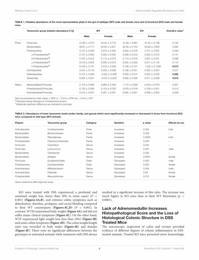
TABLE 1 | Relative abundance of the most representative phyla in the gut of wildtype (WT) male and female mice and of knockout (KO) male and female
mice.
Taxonomic group (relative abundance [%]) WT KO Overall p value†
Male Female Male Female
Phyla Firmicutes 31.68 ± 4.513 25.45 ± 2.770 31.99 ± 4.867 31.05 ± 5.736 0.722
Bacteroidetes 56.61 ± 4.717 64.92 ± 2.621 55.48 ± 4.743 55.99 ± 4.850 0.282
Proteobacteria 3.127 ± 0.403 2.619 ± 0.405 2.652 ± 0.476 3.721 ± 0.297 0.349
α-Proteobacteria@ 0.131 ± 0.050 0.023 ± 0.004 0.038 ± 0.012 0.033 ± 0.015 0.147
β-Proteobacteria@ 0.167 ± 0.012 0.113 ± 0.014 0.110 ± 0.019 0.081 ± 0.041 0.086
γ-Proteobacteria@ 0.018 ± 0.004 0.040 ± 0.013 0.025 ± 0.003 0.271 ± 0.116 0.114
δ-Proteobacteria@ 0.419 ± 0.141 0.478 ± 0.056 0.736 ± 0.157 1.532 ± 0.129# 0.013
Actinobacteria 0.231 ± 0.116 0.220 ± 0.038 0.198 ± 0.041 0.322 ± 0.054 0.227
Deferribacteres 0.107 ± 0.026 0.022 ± 0.009* 0.029 ± 0.014 0.054 ± 0.033 0.026
Tenericutes 0.002 ± 0.001 0.072 ± 0.020* 0.058 ± 0.026 0.011 ± 0.008 0.012
Ratios Bacteroidetes/Firmicutes 2.104 ± 0.390 2.880 ± 0.446 1.712 ± 0.329 2.223 ± 0.579 0.327
Proteobacteria/Firmicutes 0.120 ± 0.026 0.123 ± 0.033 0.079 ± 0.019 0.139 ± 0.031 0.513
Actinobacteria/Firmicutes 0.014 ± 0.007 0.007 ± 0.001 0.005 ± 0.001 0.009 ± 0.002 0.508
Data are presented as mean values ± SEM.*p < 0.05 vs. WTM; #p < 0.05 vs. WTF.@Bacterial classes belonging to Proteobacteria phylum.†Statistically significant differences are indicated by bold type.
TABLE 2 | Abundance of lower taxonomic levels (order, family, and genus) which were significantly increased or decreased in feces from knockout (KO)
mice compared to wild type (WT) animals.
Phylum Taxonomic group Category Variation p value Effects by sex
Actinobacteria Coriobacteriales Order Increased 0.005 both
Bacteroidetes Bacteroidaceae Family Increased 0.028 –
Bacteroidetes Rikenellaceae Family Increased 0.0002 both
Proteobacteria Pseudomonadaceae Family Increased 0.048 –
Firmicutes Clostridium Genus Increased 0.025 –
Firmicutes Lactococcus Genus Increased 0.047 male
Bacteroidetes Odoribacter Genus Increased 0.032 –
Bacteroidetes Alistipes Genus Increased 0.0001 female
Firmicutes Erysipelotrichales Order Decreased 0.020 male
Proteobacteria Caulobacterales Order Decreased 0.003 female
Actinobacteria Bifidobacterium Genus Decreased 0.004 male
Actinobacteria Olsenella Genus Decreased 0.05 female
Proteobacteria Brevundimonas Genus Decreased 0.013 female
Values obtained by BBH alignment assay.
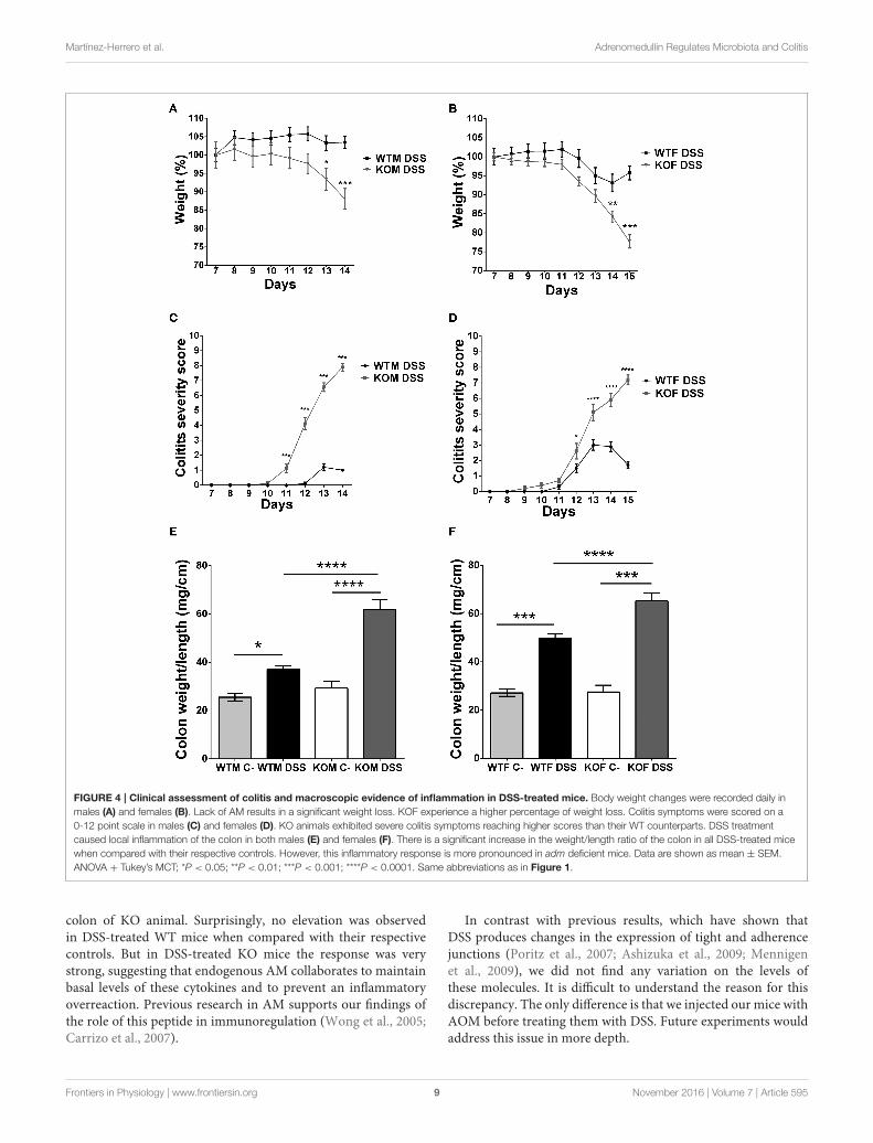
KO mice treated with DSS experienced a profound andsustained weight loss (more than 30% in some cases) (P <
0.001) (Figures 4A,B), and extreme colitis symptoms such asdehydration, diarrhea, prolapses, and rectal bleeding comparedto their WT counterparts (Figures 4C,D) (P < 0.001). Incontrast, WTMmaintained body weight (Figure 4A) and did notsuffer major clinical symptoms (Figure 4C). On the other hand,WTF experienced light weight loss (less than 10%) (Figure 4B)and some colitis symptoms (Figure 4D). The colonweight/lengthratio was recorded in both males (Figure 4E) and females(Figure 4F). There were no significant differences between thegenotypes in untreated animals while treatment with DSS always
resulted in a significant increase of this ratio. The increase wasmuch higher in KO mice than in their WT littermates (p <
0.0001).
Lack of Adrenomedullin IncreasesHistopathological Score and the Loss ofHistological Colonic Structure in DSSTreated MiceThe macroscopic inspection of colon and rectum providedevidence of different degrees of colonic inflammation in DSS-treated animals. Treated KO mice presented more hemorrhagic
Frontiers in Physiology | www.frontiersin.org 6 November 2016 | Volume 7 | Article 595

Martínez-Herrero et al. Adrenomedullin Regulates Microbiota and Colitis
FIGURE 2 | Metagenomic analysis of bacterial species in the colonic microbiota of experimental animals. Several bacterial species (A–E) are shown where
statistically significant differences in abundance were found between genotypes. WT, Wild type mice; KO, Knockout mice. In the case of L. gasseri an additional panel
(F) was added to point out the sex influence. Data are shown as mean ± SEM. Kruskal-Wallis test; *P < 0.05; **P < 0.001; ***P < 0.001. 20-week old WT (n = 8) and
KO (n = 8) mice have been included.
areas (Figures 5A,C) than their WT littermates. Histologicalexamination of the colonic sections of DSS treated micerevealed the presence of inflammatory cells restricted to isolatedareas in WT animals, while most of the tissue maintained itsmicro architecture (Figures 5B,D). In KO animals a transmuralinflammation involving all layers of the colon wall was observed,with massive infiltration in the mucosa and submucosa, anddestruction of the crypts (Figures 5B,D). Histopathologicalscores were calculated and they revealed that DSS administrationcaused a significant increase in the pathological characteristics ofboth genotypes (Figures 5E,F), being this pathology much moresevere in KO than in WT animals (P < 0.001 for males, p < 0.05for females).
Endogenous Adrenomedullin Reduces theExpression of Inflammatory Cytokines inDSS ColitisTo define the potential of endogenous AM to function as an anti-inflammatory agent we evaluated the mRNA expression levelsof different cytokines (Figure 6 and Figure 3 of SupplementaryMaterial).
In KOM, there was an increase in TNF-α (P < 0.0001), IL-1β(P < 0.001), and IL-6 (P < 0.01) levels after DSS administrationcompared with their sham group. In contrast, levels of thesecytokines did not experience any variation in DSS treated WTMwhen compared with their untreated littermates, and weresignificantly lower when compared with KOM (P < 0.001 forTNF-α, P < 0.05 for IL-1β, P < 0.01 for IL-6). Moreover, levelsof the anti-inflammatory cytokines IL-17 and IL-22 were alsosignificantly augmented in treated KOM when compared withDSS treated WTM (P < 0.05 for IL-17, P < 0.05 for IL-22) and
when compared with their sham littermates in the case of IL-22 (P < 0.05). Levels of IL-10 did not experience any significantvariation in males (Figure 3 of Supplementary Material).
Among females, few variations were observed between DSStreated and untreated WT animals. DSS increased pro- and anti-inflammatory cytokine expression in KOFs when compared withtheir untreated controls (P < 0.01 for TNF-α, P < 0.01 for IL-1β,P < 0.05 for IL-17, and P < 0.05 for IL-22) and with their WTcounterparts (P < 0.05 for IL-1β, P < 0.05 for IL-6, P < 0.05 forIL-10, and P < 0.01 for IL-22) (Figure 6).
Lack of Adrenomedullin Does Not Modifythe Expression of Adhesion Molecules inthe ColonIn an attempt to identify the impact of AM in epithelialintegrity we examined gene expression levels of differentadhesion molecules (Figures 4, 5 of Supplementary Material). Nosignificant change was observed in the gene expression patternsof these molecules with the exception of a significant decreaseof β-catenin caused by DSS administration in KOF (P < 0.05)(Figure 5 of Supplementary Material).
DISCUSSION
In this study we have shown that elimination of the gene codingfor AM in mice results in gut microbiota changes. The initialgoal of linking lack of AM with colorectal cancer initiationand progression could not be reached with this methodologybecause AM deficiency results in an unexpected overreactionto DSS-induced colitis and the experimental protocol had to beterminated after the first DSS cycle. A possible alternative to
Frontiers in Physiology | www.frontiersin.org 7 November 2016 | Volume 7 | Article 595

Martínez-Herrero et al. Adrenomedullin Regulates Microbiota and Colitis
FIGURE 3 | Expression of TLR4 is elevated in AM KO mice. TLR4 expression was assessed by immunohistochemistry (A–C), qRT-PCR (D), and Western
blotting (E). Colon sections from WT (A) and KO (B) mice were stained with antibodies against TLR4. Staining was more intense in KO mice. Absence of the primary
antibody was used as a negative control (C). Bar = 100µm. TLR4 mRNA expression was assessed by qRT-PCR (D). Bars represent the mean TLR4/18S quotient ±
SEM. Kruskal-Wallis test; *P < 0.05. TLR4 protein was measured by Western blotting, using β-Actin as a loading control (E). Bars represent the normalized TLR4/
β-Actin value ± SEM. 20-week old WT (n = 8) and KO (n = 8) mice have been included.
study the role of this peptide in CRC initiation and progressionis to use a less aggressive model, as those based on dietswhich recapitulate levels and length of exposure to nutrientslinked to relative risk for human sporadic colon cancer (Yanget al., 2008). Nevertheless, this study provides new data onthe influence of AM on colitis symptoms and colitis-relatedinflammation.
Previous studies have shown a strong positive effect usingAM as treatment for colitis symptoms in rodents (Ashizukaet al., 2009) and humans (Ashizuka et al., 2013). However, noneof the previous studies was able to investigate the oppositesituation; i.e., the impact of the lack of endogenous AM oncolitis. The scientific interest for this investigation is 2-fold.First, it provides a confirmation of AM’s beneficial role inintestinal pathologies. And second, a particular single nucleotidepolymorphism (SNP) close to the adm gene was found to beresponsible for a reduction in the circulating levels of AM(Cheung et al., 2011) and to correlate with cancer susceptibility(Martínez-Herrero and Martínez, 2013). Therefore, it would
be interesting to test whether carriers of this SNP are moresusceptible to develop chronic inflammatory intestinal disordersor have worse symptoms.
This study shows that lack of endogenous AM correlateswith an exacerbation of colitis symptoms at all levels suggestingthat AM prevented the symptoms of the disease in line withprevious experiments by the group in Sevilla (Talero et al.,2008). Microscopic analysis of colonic sections confirmed datafrom macroscopic observations reflecting an aggravation ofmucosal disruption, edema, and inflammatory infiltration inKO mice. These results are in line with previous papers whichdemonstrated that AM exerts a protective action in DSS-inducedexperimental IBD (Ashizuka et al., 2009).
Previous studies have described that the DSS-induced colitismodel induced high amounts of Th1 cytokines (TNF-α, IL-1β, IL-6) (Ashizuka et al., 2009). These cytokines exert potentproinflammatory effects that, when uncontrolled, can lead totissue injury. Our cytokine determination demonstrated thatDSS administration induces an inflammatory response in the
Frontiers in Physiology | www.frontiersin.org 8 November 2016 | Volume 7 | Article 595
Martínez-Herrero et al. Adrenomedullin Regulates Microbiota and Colitis
FIGURE 4 | Clinical assessment of colitis and macroscopic evidence of inflammation in DSS-treated mice. Body weight changes were recorded daily in
males (A) and females (B). Lack of AM results in a significant weight loss. KOF experience a higher percentage of weight loss. Colitis symptoms were scored on a
0-12 point scale in males (C) and females (D). KO animals exhibited severe colitis symptoms reaching higher scores than their WT counterparts. DSS treatment
caused local inflammation of the colon in both males (E) and females (F). There is a significant increase in the weight/length ratio of the colon in all DSS-treated mice
when compared with their respective controls. However, this inflammatory response is more pronounced in adm deficient mice. Data are shown as mean ± SEM.
ANOVA + Tukey’s MCT; *P < 0.05; **P < 0.01; ***P < 0.001; ****P < 0.0001. Same abbreviations as in Figure 1.
colon of KO animal. Surprisingly, no elevation was observedin DSS-treated WT mice when compared with their respectivecontrols. But in DSS-treated KO mice the response was verystrong, suggesting that endogenous AM collaborates to maintainbasal levels of these cytokines and to prevent an inflammatoryoverreaction. Previous research in AM supports our findings ofthe role of this peptide in immunoregulation (Wong et al., 2005;Carrizo et al., 2007).
In contrast with previous results, which have shown thatDSS produces changes in the expression of tight and adherencejunctions (Poritz et al., 2007; Ashizuka et al., 2009; Mennigenet al., 2009), we did not find any variation on the levels ofthese molecules. It is difficult to understand the reason for thisdiscrepancy. The only difference is that we injected our mice withAOM before treating them with DSS. Future experiments wouldaddress this issue in more depth.
Frontiers in Physiology | www.frontiersin.org 9 November 2016 | Volume 7 | Article 595

Martínez-Herrero et al. Adrenomedullin Regulates Microbiota and Colitis
FIGURE 5 | Effect of endogenous AM on colon acute injury by DSS. Representative macroscopic (A,C) and histological (B,D) appearance of the colon in
females (A,B) and males (C,D) of the four experimental groups: untreated WTF (WTF C-), DSS-treated WTF (WTF DSS), untreated KOF (KOF C-), and DSS-treated
KOF (KOF DSS). In untreated animals the characteristic oval shape and size of mice feces can be appreciated. However, DSS administration caused the presence of
diarrhea, with bloody feces. Histological analysis of the colon of mice treated with DSS confirmed the macroscopic study data showing large areas of lymphocyte
infiltrates in the mucosa and submucosa, being the damage greater in DSS-treated KOs with loss of tissue architecture, inflammation, and a more prominent
infiltration when compared with WTs. Untreated KOs did not show any abnormality in the microscopic structure of the colon. Bars = 100µm. The histopathological
score was calculated in both males (E) and females (F). All animals treated with DSS presented a severe inflammation in the colon, which was more severe among the
KO mice. Data are shown as mean ± SEM. ANOVA + Tukey’s MCT; *P < 0.05; **P < 0.01; ***P < 0.001; ****P < 0.0001. Same abbreviations as in Figure 1.
Another explanation for the exacerbation of colitis symptomsobserved in KO animals is the alteration in gut microbiotathat these mice suffer under physiological conditions. To our
knowledge, this is the first study were the effects of AM deficiencyon gut microbiota composition has been tested. Our resultspoint to an important effect of this antimicrobial peptide in
Frontiers in Physiology | www.frontiersin.org 10 November 2016 | Volume 7 | Article 595

Martínez-Herrero et al. Adrenomedullin Regulates Microbiota and Colitis
FIGURE 6 | Effects of endogenous adrenomedullin (AM) on the
mucosal inflammatory responses during acute episode of colitis
caused by DSS administration in females. Expression of the principal pro-
and anti-inflammatory cytokines (A–F) were evaluated by qRT-PCR in colon
biopsies. Lack of AM resulted in a significant increase in the Th1 and Th2
cytokines when compared with their respective control mice and with their WT
littermates. Surprisingly, DSS treatment did not cause any significant variation
among the levels of expression of these cytokines in the DSS-treated WTs. All
data were normalized by their 18S value. Data are shown as mean ± SEM.
ANOVA + Tukey’s MCT; *P < 0.05; **P < 0.01. Same abbreviations as in
Figure 4.
the control of gut bacteria composition which could offer amechanistic insight into the pathogenesis of DSS-induced colitisand also could suggest several microbial interventions to be usedin preventing or managing this condition.
The lack of AM did not translate into significant changesat higher taxonomic levels, suggesting that the worsening ofDSS-induced colitis and inflammation observed in KO miceis more related to changes in lower taxonomic levels anddiversity, as occurs in other metabolic pathologies such as obesity(Villanueva-Millán et al., 2015). It is also interesting to point outthat some of the results obtained differ depending on sex. Inthis line, IBD seems to be more prevalent in women (Burischet al., 2013) and it has been hypothesized that this situationmight
be related with female sex hormones since oral contraceptivesare considered a risk factor for IBD (Ponder and Long, 2013).These data seem to indicate a potential link between sex-specificregulation of microbiota and IBD.
A previous study demonstrated that AM had antimicrobialactivity against gut B. fragilis, E. coli and H. pylori (Allaker et al.,1999). As expected, we did not detect the presence of these specieseither in WT or KO mice, as these animals were maintainedunder SPF barrier conditions. However, we observed that thelack of AM was associated with a decreased abundance of twobeneficial bacteria such as L. gasseri and B. choerinum (and also ofthe Bifidobacterium genus), which could suggest that the decreasein the numbers of these bacteria is associated with the worseningof DSS-induced colitis observed in KO mice. In fact, L. gasserihas anti-inflammatory activity that reduces the severity of colitisin a mouse model (Carroll et al., 2007). Also, administrationof certain strains of Lactobacillus and Bifidobacterium improvedDSS-induced colitis (Toumi et al., 2014).
In contrast, we observed a significant increase in theabundance of two bacteria belonging to the Firmicutes phyla(Clostridium scindens and Christensenella minuta) as well as inB. vulgatus levels, a weak inducer of intestinal inflammation thatbelongs to the Bacteroidetes phylum. The actions of Bacteroidesin the pathogenesis of IBD has received attention (Liang et al.,2014). However, the role of B. vulgatus in these pathologies iscontroversial. Thus, while some studies demonstrated that itsabundance was rather decreased among IBD patients (Takaishiet al., 2008) and that it was able to prevent the developmentof Escherichia coli-induced colitis (Sydora et al., 2007), othershave reported that the administration of Bacteroides, andespecially the administration of B. vulgatus aggravates colitis(Kishi et al., 2000). In our study, the increase observed in KOmice suggests a role of this bacterium in intestinal inflammationand also in the worsening of colitis induced by the lackof AM.
Surprisingly, a significant increase in the abundance ofFaecalibacterium prausnitzii, an important member of mucosa-associated Firmicutes, was observed in male KO mice. Previousstudies had reported that a reduction of F. prausnitzii wasassociated with the recurrence of ileal Crohn’s disease (Sokolet al., 2008) and oral administration of this species can reduce theseverity of experimental colitis in mice. As this phenomenon wasnot observed in femalemice, the increase observed inmales couldsuggest a compensatory mechanism to ameliorate the damageinduced by AM deficiency. In fact, inflammation and severitywas higher in females, where no increases in F. prausnitzii wereobserved.
Therefore, we can conclude that AM deficiency is clearlyassociated with changes in microbiota composition. Thismicrobiota changes may predispose mice to suffer intestinalinflammation and worse DSS-induced colitis symptoms. Butvariations in microbiota may have another effect throughchanges in colonic TLR4 gene expression, although wehave not able to show a clear increase of TLR4 proteinby Western blotting. Hasegawa et al. (2010) demonstratedthat immunostimulatory activity of the intestinal microfloravaried during mouse development, evolving from a largely
Frontiers in Physiology | www.frontiersin.org 11 November 2016 | Volume 7 | Article 595

Martínez-Herrero et al. Adrenomedullin Regulates Microbiota and Colitis
stimulation through TLR4 during breast-feeding to a TLR4-independent process after weaning. In AM deficient micemicroflora there was a higher proportion of δ-Proteobacteria,which may be stimulating TLR4 in the colon and thuspromoting its expression as it happened with mice duringbreast-feeding (Hasegawa et al., 2010). In addition, KO miceexhibited higher levels of other Gram negative bacteria thatare able to stimulate TLR4 and increase its expression, suchas Clostridium (Scarpa et al., 2011), B. vulgates (Haller et al.,2004), Acinetobacter (Erridge et al., 2007), or Pseudomonadaceae(Korneev et al., 2015). Today it is widely accepted that innormal intestinal epithelial cells, TLR4 is marginally expressedbut under pathological conditions such as IBD and CRC anoverexpression of this receptor occurs (Yesudhas et al., 2014).However, it has not been well established whether this TLR4elevation occurs prior to IBD breakout or is a consequenceof the disease. Our results seem to support the idea that analtered microbiota composition and a modified gene expressionof TLR4 would precede and favor the appearance of thepathology.
To confirm the causal relationship between AM deficiency,microbiota changes, and colitis induction, manipulation of thegut microbiota should be performed, as described in other animalmodels (Li et al., 2016; Tian et al., 2016).
In conclusion, using our KO animals we have demonstratedthat lack of endogenous AM causes an evident dysbiosis and an
elevated TLR4 expression which might lead to a worse prognosisduring colitis episodes.
AUTHOR CONTRIBUTIONS
SM-H, IL, JN-I, MV-M, ER-F, PP-M, and JO: Performedexperiments and interpreted data; SM-H and AM: Designed thestudy; AM: Wrote the manuscript and provided funding. Allauthors read and approved the final version of the manuscript.
FUNDING
This study was supported by a grant from the Instituto deSalud Carlos III (PI13/02166), cofinanced by FEDER to AM;by a predoctoral fellowship from the Junta Provincial de LaRioja de la Asociación Española Contra el Cáncer (AECC) toSM-H; and by a predoctoral fellowship from Consejería deIndustria, Innovación y Empleo (Government of La Rioja) toMV-M. Sponsors had no participation in study design, collection,analysis, and interpretation of the data.
SUPPLEMENTARY MATERIAL
The Supplementary Material for this article can be foundonline at: http://journal.frontiersin.org/article/10.3389/fphys.2016.00595/full#supplementary-material
REFERENCES
Allaker, R. P., Zihni, C., and Kapas, S. (1999). An investigation into theantimicrobial effects of adrenomedullin on members of the skin, oral,respiratory tract and gut microflora. FEMS Immunol. Med. Microbiol. 23,289–293. doi: 10.1111/j.1574-695X.1999.tb01250.x
Arndt, D., Xia, J., Liu, Y., Zhou, Y., Guo, A. C., Cruz, J. A., et al.(2012). METAGENassist: a comprehensive web server for comparativemetagenomics. Nucleic Acids Res. 40, W88–W95. doi: 10.1093/nar/gks497
Ashizuka, S., Inagaki-Ohara, K., Kuwasako, K., Kato, J., Inatsu, H., and Kitamura,K. (2009). Adrenomedullin treatment reduces intestinal inflammation andmaintains epithelial barrier function in mice administered dextran sulphatesodium. Microbiol. Immunol. 53, 573–581. doi: 10.1111/j.1348-0421.2009.00159.x
Ashizuka, S., Inatsu, H., Kita, T., and Kitamura, K. (2016). Adrenomedullintherapy in patients with refractory ulcerative colitis: a case series. Dig. Dis. Sci.61, 872–880. doi: 10.1007/s10620-015-3917-0
Ashizuka, S., Kita, T., Inatsu, H., andKitamura, K. (2013). Adrenomedullin: a noveltherapy for intractable ulcerative colitis. Inflamm. Bowel Dis. 19, E26–E27.doi: 10.1002/ibd.22891
Burisch, J., Jess, T., Martinato, M., and Lakatos, P. L. (2013). The burden ofinflammatory bowel disease in Europe. J. Crohns. Colitis 7, 322–337. doi: 10.1016/j.crohns.2013.01.010
Caporaso, J. G., Lauber, C. L., Walters, W. A., Berg-Lyons, D., Huntley, J., Fierer,N., et al. (2012). Ultra-high-throughput microbial community analysis on theIllumina HiSeq andMiSeq platforms. ISME J. 6, 1621–1624. doi: 10.1038/ismej.2012.8
Carrizo, G. J., Wu, R., Cui, X., Dwivedi, A. J., Simms, H. H., andWang, P. (2007). Adrenomedullin and adrenomedullin-binding protein-1 downregulate inflammatory cytokines and attenuate tissue injury aftergut ischemia-reperfusion. Surgery 141, 245–253. doi: 10.1016/j.surg.2006.05.017
Carroll, I. M., Andrus, J. M., Bruno-Bárcena, J. M., Klaenhammer, T. R., Hassan, H.M., and Threadgill, D. S. (2007). Anti-inflammatory properties of Lactobacillusgasseri expressing manganese superoxide dismutase using the interleukin 10-deficient mouse model of colitis. Am. J. Physiol. Gastrointest. Liver Physiol. 293,G729–G738. doi: 10.1152/ajpgi.00132.2007
Cheung, B. M., Ong, K. L., Tso, A. W., Leung, R. Y., Cherny, S. S., Sham, P.C., et al. (2011). Plasma adrenomedullin level is related to a single nucleotidepolymorphism in the adrenomedullin gene. Eur. J. Endocrinol. 165, 571–577.doi: 10.1530/EJE-11-0513
Cole, J. R., Wang, Q., Fish, J. A., Chai, B., McGarrell, D. M., Sun, Y., et al. (2014).Ribosomal database project: data and tools for high throughput rRNA analysis.Nucleic Acids Res. 42, D633–D642. doi: 10.1093/nar/gkt1244
Erridge, C., Moncayo-Nieto, O. L., Morgan, R., Young, M., and Poxton, I. R.(2007). Acinetobacter baumannii lipopolysaccharides are potent stimulatorsof human monocyte activation via Toll-like receptor 4 signalling. J. Med.Microbiol. 56, 165–171. doi: 10.1099/jmm.0.46823-0
Fernández, A. P., Serrano, J., Tessarollo, L., Cuttitta, F., and Martínez, A. (2008).Lack of adrenomedullin in the mouse brain results in behavioral changes,anxiety, and lower survival under stress conditions. Proc. Natl. Acad. Sci. U.S.A.105, 12581–12586. doi: 10.1073/pnas.0803174105
García-Sanmartín, J., Larrayoz, I. M., and Martínez, A. (2016). Adrenomedullinregulates club cell recovery following lung epithelial injury. Histol. Histopathol.31, 663–673. doi: 10.14670/HH-11-704
Gommeaux, J., Cano, C., Garcia, S., Gironella, M., Pietri, S., Culcasi, M., et al.(2007). Colitis and colitis-associated cancer are exacerbated in mice deficientfor tumor protein 53-induced nuclear protein 1.Mol. Cell. Biol. 27, 2215–2228.doi: 10.1128/MCB.01454-06
Ha, C. W., Lam, Y. Y., and Holmes, A. J. (2014). Mechanistic links betweengut microbial community dynamics, microbial functions and metabolichealth. World J. Gastroenterol. 20, 16498–16517. doi: 10.3748/wjg.v20.i44.16498
Haller, D., Holt, L., Parlesak, A., Zanga, J., Bäuerlein, A., Sartor, R. B., et al.(2004). Differential effect of immune cells on non-pathogenic Gram-negative
Frontiers in Physiology | www.frontiersin.org 12 November 2016 | Volume 7 | Article 595
Martínez-Herrero et al. Adrenomedullin Regulates Microbiota and Colitis
bacteria-induced nuclear factor-kappaB activation and pro-inflammatory geneexpression in intestinal epithelial cells. Immunology 112, 310–320. doi: 10.1111/j.1365-2567.2004.01874.x
Hasegawa, M., Osaka, T., Tawaratsumida, K., Yamazaki, T., Tada, H.,Chen, G. Y., et al. (2010). Transitions in oral and intestinal microfloracomposition and innate immune receptor-dependent stimulation duringmouse development. Infect. Immun. 78, 639–650. doi: 10.1128/IAI.01043-09
Hayashi, Y., Narumi, K., Tsuji, S., Tsubokawa, T., Nakaya, M. A., Wakayama, T.,et al. (2011). Impact of adrenomedullin on dextran sulfate sodium-inducedinflammatory colitis in mice: insights from in vitro and in vivo experimentalstudies. Int. J. Colorectal Dis. 26, 1453–1462. doi: 10.1007/s00384-011-1254-0
Huson, D. H., and Weber, N. (2013). Microbial community analysis usingMEGAN. Meth. Enzymol. 531, 465–485. doi: 10.1016/B978-0-12-407863-5.00021-6
Kim, E. R., and Chang, D. K. (2014). Colorectal cancer in inflammatorybowel disease: the risk, pathogenesis, prevention and diagnosis. World J.Gastroenterol. 20, 9872–9881. doi: 10.3748/wjg.v20.i29.9872
Kishi, D., Takahashi, I., Kai, Y., Tamagawa, H., Iijima, H., Obunai, S., et al. (2000).Alteration of V beta usage and cytokine production of CD4+ TCR beta betahomodimer T cells by elimination of Bacteroides vulgatus prevents colitis inTCR alpha-chain-deficient mice. J. Immunol. 16, 5891–5899. doi: 10.4049/jimmunol.165.10.5891
Korneev, K. V., Arbatsky, N. P., Molinaro, A., Palmigiano, A., Shaikhutdinova,R. Z., Shneider, M. M., et al. (2015). Structural relationship of the lipid Aacyl groups to activation of murine toll-like receptor 4 by lipopolysaccharidesfrom pathogenic strains of Burkholderia mallei, Acinetobacter baumannii, andPseudomonas aeruginosa. Front. Immunol. 6:595. doi: 10.3389/fimmu.2015.00595
Koyama, T., Ochoa-Callejero, L., Sakurai, T., Kamiyoshi, A., Ichikawa-Shindo,Y., Iinuma, N., et al. (2013). Vascular endothelial adrenomedullin-RAMP2system is essential for vascular integrity and organ homeostasis.Circulation 127,842–853. doi: 10.1161/CIRCULATIONAHA.112.000756
Larráyoz, I. M., de, L. A., Rúa, O., Velilla, S., and Cabello, J., Martinez, A.(2012). Molecular effects of doxycycline treatment on pterygium as revealed bymassive transcriptome sequencing. PLoS ONE 7:e39359. doi: 10.1371/journal.pone.0039359
Larráyoz, I. M., Martínez-Herrero, S., García-Sanmartín, J., Ochoa-Callejero, L.,and Martínez, A. (2014). Adrenomedullin and tumour microenvironment. J.Transl. Med. 12:339. doi: 10.1186/s12967-014-0339-2
Leone, V., Chang, E. B., and Devkota, S. (2013). Diet, microbes, and host genetics:the perfect storm in inflammatory bowel diseases. J. Gastroenterol. 48, 315–321.doi: 10.1007/s00535-013-0777-2
Li, M., Li, Z., Wen, S., Liu, Y., Wang, Y., and Tang, L. (2016). Transplantation ofa bacterial consortium ameliorates trinitrobenzenesulfonic acid-induced colitisand intestinal dysbiosis in rats. Future Microbiol. 11, 887–902. doi: 10.2217/fmb-2015-0002
Liang, X., Li, H., Tian, G., and Li, S. (2014). Dynamic microbe and moleculenetworks in a mouse model of colitis-associated colorectal cancer. Sci. Rep.4:4985. doi: 10.1038/srep04985
López, J., and Martínez, A. (2002). Cell and molecular biology of themultifunctional peptide, adrenomedullin. Int. Rev. Cytol. 221, 1–92. doi: 10.1016/S0074-7696(02)21010-4
Martínez, A., Elsasser, T. H., Muro-Cacho, C., Moody, T. W., Miller, M. J., Macri,C. J., et al. (1997). Expression of adrenomedullin and its receptor in normaland malignant human skin: a potential pluripotent role in the integument.Endocrinology 138, 5597–5604. doi: 10.1210/endo.138.12.5622
Martínez-Herrero, S., Larrayoz, I. M., Ochoa-Callejero, L., Fernández, L. J.,Allueva, A., Ochoa, I., et al. (2016). Prevention of bone loss in a modelof postmenopausal osteoporosis through adrenomedullin inhibition. Front.Physiol. 7:280. doi: 10.3389/fphys.2016.00280
Martínez-Herrero, S., and Martínez, A. (2013). Cancer protection elicited by asingle nucleotide polymorphism close to the adrenomedullin gene. J. Clin.Endocrinol. Metab. 98, E807–E810. doi: 10.1210/jc.2012-4193
Martínez-Herrero, S., and Martínez, A. (2016). Adrenomedullin regulatesintestinal physiology and pathophysiology. Domest. Anim. Endocrinol. 56,S66–S83. doi: 10.1016/j.domaniend.2016.02.004
Mennigen, R., Nolte, K., Rijcken, E., Utech, M., Loeffler, B., Senninger, N., et al.(2009). Probiotic mixture VSL#3 protects the epithelial barrier by maintainingtight junction protein expression and preventing apoptosis in a murine modelof colitis. Am. J. Physiol. Gastrointest. Liver Physiol. 296, G1140–G1149. doi: 10.1152/ajpgi.90534.2008
Nougueréde, E., Berenguer, C., Garcia, S., Bennani, B., Delfino, C., Nanni, I., et al.(2013). Expression of adrenomedullin in human colorectal tumors and its rolein cell growth and invasion in vitro and in xenograft growth in vivo. CancerMed. 2, 196–207. doi: 10.1002/cam4.51
Pasari, J. R., Levi, T., Zavaleta, E. S., and Tilman, D. (2013). Several scales ofbiodiversity affect ecosystem multifunctionality. Proc. Natl. Acad. Sci. U.S.A.110, 10219–10222. doi: 10.1073/pnas.1220333110
Ponder, A., and Long, M. D. (2013). A clinical review of recent findings in theepidemiology of inflammatory bowel disease. Clin. Epidemiol. 5, 237–247.doi: 10.2147/CLEP.S33961
Poritz, L. S., Garver, K. I., Green, C., Fitzpatrick, L., Ruggiero, F., and Koltun,W. A. (2007). Loss of the tight junction protein ZO-1 in dextran sulfatesodium induced colitis. J. Surg. Res. 140, 12–19. doi: 10.1016/j.jss.2006.07.050
Poyner, D. R., Sexton, P. M., Marshall, I., Smith, D. M., Quirion, R., Born, W.,et al. (2002). International union of pharmacology. XXXII. The mammaliancalcitonin gene-related peptides, adrenomedullin, amylin, and calcitoninreceptors. Pharmacol. Rev. 54, 233–246. doi: 10.1124/pr.54.2.233
Scarpa, M., Grillo, A., Pozza, A., Faggian, D., Ruffolo, C., Scarpa, M., et al.(2011). TLR2 and TLR4 up-regulation and colonization of the ileal mucosa byClostridiaceae spp. in chronic/relapsing pouchitis. J. Surg. Res. 169, e145–e154.doi: 10.1016/j.jss.2011.04.003
Schmittgen, T. D., and Livak, K. J. (2008). Analyzing real-time PCR data by thecomparative C, (T) method. Nat. Protoc. 3, 1101–1108. doi: 10.1038/nprot.2008.73
Shindo, T., Kurihara, Y., Nishimatsu, H., Moriyama, N., Kakoki, M., Wang, Y.,et al. (2001). Vascular abnormalities and elevated blood pressure in micelacking adrenomedullin gene.Circulation 104, 1964–1971. doi: 10.1161/hc4101.097111
Sokol, H., Pigneur, B., Watterlot, L., Lakhdari, O., Bermúdez-Humaran, L.G., Gratadoux, J. J., et al. (2008). Faecalibacterium prausnitzii is an anti-inflammatory commensal bacterium identified by gut microbiota analysis ofCrohn disease patients. Proc. Natl. Acad. Sci. U.S.A. 105, 16731–16736. doi: 10.1073/pnas.0804812105
Sydora, B. C., Macfarlane, S. M., Walker, J. W., Dmytrash, A. L., Churchill, T. A.,Doyle, J., et al. (2007). Epithelial barrier disruption allows nondisease-causingbacteria to initiate and sustain IBD in the IL-10 gene-deficient mouse. Inflamm.Bowel Dis. 13, 947–954. doi: 10.1002/ibd.20155
Takaishi, H., Matsuki, T., Nakazawa, A., Takada, T., Kado, S., Asahara, T., et al.(2008). Imbalance in intestinal microflora constitution could be involved inthe pathogenesis of inflammatory bowel disease. Int. J. Med. Microbiol. 298,463–472. doi: 10.1016/j.ijmm.2007.07.016
Talero, E., Sánchez-Fidalgo, S., de la Lastra, C. A., Illanes, M., Calvo, J.R., and Motilva, V. (2008). Acute and chronic responses associated withadrenomedullin administration in experimental colitis. Peptides 29, 2001–2012.doi: 10.1016/j.peptides.2008.07.013
Thaker, A. I., Shaker, A., Rao, M. S., and Ciorba, M. A. (2012). Modeling colitis-associated cancer with azoxymethane, (AOM) and dextran sulfate sodium,(DSS). J. Vis. Exp. 4100. doi: 10.3791/4100
Tian, Z., Liu, J., Liao, M., Li, W., Zou, J., Han, X., et al. (2016). Beneficial effects offecal microbiota transplantation on ulcerative colitis in mice. Dig. Dis. Sci. 61,2262–2271. doi: 10.1007/s10620-016-4060-2
Toumi, R., Soufli, I., Rafa, H., Belkhelfa, M., Biad, A., and Touil-Boukoffa,C. (2014). Probiotic bacteria lactobacillus and bifidobacterium attenuateinflammation in dextran sulfate sodium-induced experimental colitisin mice. Int. J. Immunopathol. Pharmacol. 27, 615–627. doi: 10.1177/039463201402700418
Villanueva-Millán, M. J., Perez-Matute, P., and Oteo, J. A. (2015). Gutmicrobiota: a key player in health and disease. A review focused onobesity. J. Physiol. Biochem. 71, 509–525. doi: 10.1007/s13105-015-0390-3
Wong, L. Y., Cheung, B. M., Li, Y. Y., and Tang, F. (2005). Adrenomedullin isboth proinflammatory and antiinflammatory: its effects on gene expression
Frontiers in Physiology | www.frontiersin.org 13 November 2016 | Volume 7 | Article 595

Martínez-Herrero et al. Adrenomedullin Regulates Microbiota and Colitis
and secretion of cytokines and macrophage migration inhibitory factor inNR8383 macrophage cell line. Endocrinology 146, 1321–1327. doi: 10.1210/en.2004-1080
Yang, K., Kurihara, N., Fan, K., Newmark, H., Rigas, B., Bancroft, L., et al. (2008).Dietary induction of colonic tumors in amousemodel of sporadic colon cancer.Cancer Res. 68, 7803–7810. doi: 10.1158/0008-5472.CAN-08-1209
Yashiro, M. (2014). Ulcerative colitis-associated colorectal cancer. World J.Gastroenterol. 20, 16389–16397. doi: 10.3748/wjg.v20.i44.16389
Yesudhas, D., Gosu, V., Anwar, M. A., and Choi, S. (2014). Multiple roles of toll-like receptor 4 in colorectal cancer. Front. Immunol. 5:334. doi: 10.3389/fimmu.2014.00334
Conflict of Interest Statement: The authors declare that the research wasconducted in the absence of any commercial or financial relationships that couldbe construed as a potential conflict of interest.
Copyright © 2016 Martínez-Herrero, Larrayoz, Narro-Íñiguez, Villanueva-Millán,Recio-Fernández, Pérez-Matute, Oteo and Martínez. This is an open-access articledistributed under the terms of the Creative Commons Attribution License (CC BY).The use, distribution or reproduction in other forums is permitted, provided theoriginal author(s) or licensor are credited and that the original publication in thisjournal is cited, in accordance with accepted academic practice. No use, distributionor reproduction is permitted which does not comply with these terms.
Frontiers in Physiology | www.frontiersin.org 14 November 2016 | Volume 7 | Article 595