World Journal of Gastrointestinal Surgery
304
2014 Bound Volume 6 Issue 1-12: 1-258 Published by Baishideng Publishing Group Inc Published by Baishideng Publishing Group Inc World Journal of Gastrointestinal Surgery World J Gastrointest Surg 2014 July 27; 6(7): 122-145 ISSN 1948-9366 (online) World Journal of Gastrointestinal Surgery ISSN 1948-9366 (online) Published by Baishideng Publishing Group Inc World Journal of Gastrointestinal Surgery World J Gastrointest Surg 2014 August 27; 6(8): 146-168 ISSN 1948-9366 (online) Published by Baishideng Publishing Group Inc World Journal of Gastrointestinal Surgery World J Gastrointest Surg 2014 September 27; 6(9): 169-189 ISSN 1948-9366 (online) Published by Baishideng Publishing Group Inc World Journal of Gastrointestinal Surgery World J Gastrointest Surg 2014 October 27; 6(10): 190-207 ISSN 1948-9366 (online) Published by Baishideng Publishing Group Inc World Journal of Gastrointestinal Surgery World J Gastrointest Surg 2014 December 27; 6(12): 235-258 ISSN 1948-9366 (online) Volume End Published by Baishideng Publishing Group Inc World Journal of Gastrointestinal Surgery World J Gastrointest Surg 2014 May 27; 6(5): 80-87 ISSN 1948-9366 (online)
-
Upload
khangminh22 -
Category
Documents
-
view
0 -
download
0
Transcript of World Journal of Gastrointestinal Surgery

2014 Bound Volume 6 Issue 1-12: 1-258
Published by Baishideng Publishing Group Inc
Published by Baishideng Publishing Group Inc
World Journal of Gastrointestinal SurgeryWorld J Gastrointest Surg 2014 July 27; 6(7): 122-145
ISSN 1948-9366 (online)
World Journal of Gastrointestinal Surgery
ISSN 1948-9366 (online)
Published by Baishideng Publishing Group Inc
World Journal of Gastrointestinal SurgeryWorld J Gastrointest Surg 2014 August 27; 6(8): 146-168
ISSN 1948-9366 (online)
Published by Baishideng Publishing Group Inc
World Journal of Gastrointestinal SurgeryWorld J Gastrointest Surg 2014 September 27; 6(9): 169-189
ISSN 1948-9366 (online)
Published by Baishideng Publishing Group Inc
World Journal of Gastrointestinal SurgeryWorld J Gastrointest Surg 2014 October 27; 6(10): 190-207
ISSN 1948-9366 (online)
Published by Baishideng Publishing Group Inc
World Journal of Gastrointestinal SurgeryWorld J Gastrointest Surg 2014 December 27; 6(12): 235-258
ISSN 1948-9366 (online)
Volume End
Published by Baishideng Publishing Group Inc
World Journal of Gastrointestinal SurgeryWorld J Gastrointest Surg 2014 May 27; 6(5): 80-87
ISSN 1948-9366 (online)
EDITOR-IN-CHIEFTimothy M Pawlik, Baltimore
STRATEGY ASSOCIATE EDITOR-IN-CHIEFElijah Dixon, CalgaryAntonello Forgione, MilanTobias Keck, FreiburgTsuyoshi Konishi, TokyoNatale Di Martino, Naples
GUEST EDITORIAL BOARD MEMBERSChao-Long Chen, KaohsiungChien-Hung Chen, TaipeiHsin-Yuan Fang, TaichungJong-Shiaw Jin, TaipeiChen-Guo Ker, KaohsiungKing-Teh Lee, KaohsiungWei-Jei Lee, TaoyuanShiu-Ru Lin, KaohsiungWan-Yu Lin, TaichungYan-Shen Shan, TainanYau-Lin Tseng, TainanJaw-Yuan Wang, KaohsiungLi-Wha Wu, Tainan
MEMBERS OF THE EDITORIAL BOARD
Australia
Ned Abraham, Coffs HarbourRobert Gibson, VictoriaMichael Michael, VictoriaDavid Lawson Morris, KogarahJaswinder Singh Samra, LeonardsM Wilhelm Wichmann, Mount Gambier
Austria
Harald R Rosen, ViennaFranz Sellner, Vienna
Belgium
Giovanni Dapri, BrusselsJean-François Gigot, BrusselsLerut Jan Paul Marthe, BrusselsGregory Peter Sergeant, LeuvenHans Van Vlierberghe, GentJean-Louis Vincent, Brussels
Brazil
Jose E Aguilar-Nascimento, CuiabaMario Reis Alvares-da-Silva, Porto AlegreFernando Martín Biscione, Minas GeraisJulio Coelho, CuritibaJosé Sebastião dos Santos, Ribeirão PretoMarcel Autran Machado, São PauloMarcelo AF Ribeiro, Santana de ParnaibaMarcus V Motta Valadão, Rio de JaneiroRicardo Zorron, Rio de Janeiro
Bulgaria
Krassimir Dimitrow Ivanov, VarnaBelev Vasilev Nikolai, Plovdiv Plovdiv
Canada
Runjan Chetty, OntarioLaura Ann Dawson, Ontario
Mahmoud A Khalifa, TorontoPeter C Kim, OntarioPeter Metrakos, QuebecReda S Saad, TorontoManuela Santos, Montreal
China
Yue-Zu Fan, ShanghaiWen-Tao Fang, ShanghaiYong-Song Guan, ChengduShao-Liang Han, WenzhouMichael Garnet Irwin, Hong KongLong Jiang, ShanghaiWai Lun Law, Hong KongTing-Bo Liang, HangzhouQuan-Da Liu, BeijingYu-Bin Liu, GuangdongJian-Yang Ma, ChengduKwan Man, Hong KongTang Chung Ngai, Hong KongYan-Ning Qian, NanjingAi-Wen Wu, BeijingYun-Fei Yuan, Guangzhou
Denmark
Thue Bisgaard, Koge
Finland
Helena Mariitta Isoniemi, HelsinkiIsto Henrik Nordback, Tampere
France
Mustapha Adham, Lyon Cedex
I
Editorial Board2012-2016
The World Journal of Gastrointestinal Surgery Editorial Board consists of 340 members, representing a team of worldwide experts in pediatrics. They are from 37 countries, including Australia (6), Austria (2), Belgium (6), Brazil (9), Bulgaria (2), Canada (8), China (29), Denmark (1), Finland (2), France (9), Germany (21), Greece (7), India (11), Ireland (3), Israel (3), Italy (49), Jamaica (1), Japan (47), Lithuania (1), Malaysia (1), Netherlands (11), Pakistan (1), Poland (1), Portugal (1), Russia (1), Saudi Arabia (1), Serbia (2), Singapore (5), South Korea (8), Spain (5), Sweden (2), Switzerland (3), Thailand (2), Tunisia (1), Turkey (8), United Kingdom (11), and United States (59).
February 27, 2013WJGS|www.wjgnet.com
Chapel Alain, ParisBrice Gayet, ParisJean-François Gillion, AntonyGuilhem Godlewski, Saint ChaptesD Heresbach, Rennes CedexRomaric Loffroy, Dijon CedexJacques Marescaux, Strasbourg CedexAurelie Plessier, Clichy
Germany
Hans G Beger, UlmVollmar Brigitte, RostockDieter C Broering, KielAnsgar Michael Chromik, RegensburgMarc-H Dahlke, RegensburgIrene Esposito, NeuherbergStefan Fichtner-Feigl, RegensburgBenedikt Josef Folz, Bad LippspringeHelmut Friess, MunichReinhart T Grundmann, BurghausenBertram Illert, WürzburgJakob Robert Izbicki, HamburgJörg H Kleeff, MunichAxel Kleespies, MunichUwe Klinge, AachenMartin G Mack, FrankfurtKlaus Erik Mönkemüller, BottropMatthias Peiper, DusseldorfHubert Scheidbach, MagdeburgJoerg Theisen, Munich
Greece
Teni Boulikas, AthensEelco de Bree, HerakleionStavros J Gourgiotis, AthensAndreas Manouras, AthensTheodoros E Pavlidis, ThessalonikiGeorge H Sakorafas, AthensVassilios E Smyrniotis, Athens
India
Anil Kumar Agarwal, New DelhiSamik Kumar Bandyopadhyay, KolkataShams ul Bari, KashmirSomprakas Basu, VaranasiPravin Jaiprakash Gupta, NagpurVinay Kumar Kapoor, LucknowChandra Kant Pandey, LucknowShailesh V Shrikhande, MumbaiSadiq Saleem Sikora, BangaloreRakesh K Tandon, New DelhiImtiaz Ahmed Wani, Srinagar
Ireland
Kevin CP Conlon, DublinPrem Puri, DublinEamonn Martin Quigley, Cork
Israel
Ariel Halevy, Zerifin
Jesse Lachter, HaifaHagit Tulchinsky, Tel Aviv
Italy
Angelo Andriulli, San Giovanni RotondoGiuseppe Aprile, UdineGianni Biancofiore, PisaStefania Boccia, RomeLuigi Bonavina, Piazza MalanPier Andrea Borea, FerraraGiovanni Cesana, MilanoStefano Crippa, VeronaGiovanni D De Palma, NapoliGiovanni de Simone, NapoliGiorgio Di Matteo, RomeGiorgio Ercolani, BolognaCarlo V Feo, FerraraSimone Ferrero, GenovaValenza Franco, MilanoLeandro Gennari, RozzanoFelice Giuliante, RomeCalogero Iacono, VeronaRiccardo Lencioni, PisaDottor Fabrizio Luca, MilanoGiuseppe Malleo, VeronaPaolo Massucco, CandioloGiulio Melloni, MilanPaolo Morgagni, ForliChiara Mussi, RozzanoGabriella Nesi, FlorenceAngelo Nespoli, MonzaGiuseppe R Nigri, RomeFabio Pacelli, RomeCorrado Pedrazzani, SienaRoberto Persiani, RomePasquale Petronella, NapoliPiero Portincasa, BariStefano Rausei, VareseCarla Ida Ripamonti, MilanoAntonio Russo, PalermoGiulio A Santoro, TrevisoStefano Scabini, GenoaGiuseppe S Sica, RomeGianfranco Silecchia, RomeMario Testini, BariGuido Alberto Massimo Tiberio, BresciaUmberto Veronesi, MilanoBruno Vincenzi, RomeMarco Vivarelli, BolognaAlberto Zaniboni, BresciaAlessandro Zerbi, Milano
Jamaica
Joseph Martin Plummer, Kingston
Japan
Yasunori Akutsu, ChibaRyuichiro Doi, KyotoYosuke Fukunaga, SakaiAkira Furukawa, ShigaShigeru Goto, OitaKazuhiko Hayashi, TokyoNaoki Hiki, Tokyo
Takeyama Hiromitsu, NagoyaTsujimoto Hironori, TokorozawaTsukasa Hotta, WakayamaYutaka Iida, Gifu CityKazuaki Inoue, YokohamaMasashi Ishikawa, MasaTatsuo Kanda, NiigataTatsuyuki Kawano, TokyoKeiji Koda, ChibaHajime Kubo, KyotoIruru Maetani, TokyoYoshimasa Maniwa, KobeToru Mizuguchi, HokkaidoZenichi Morise, ToyoakeYoshihiro Moriwaki, YokohamaYoshihiro Moriya, TokyoSatoru Motoyama, AkitaHiroaki Nagano, OsakaMasato Nagino, NagoyaKazuyuki Nakamura, YamaguchiShingo Noura, OsakaKazuo Ohashi, TokyoYoichi Sakurai, AichiHirozumi Sawai, NagoyaShouji Shimoyama, TokyoMasayuki Sho, NaraYasuhiko Sugawara, TokyoHiroshi Takamori, KumamotoSonshin Takao, KagoshimaKuniya Tanaka, YokohamaMasanori Tokunaga, Sunto-gunYasunobu Tsujinaka, ChibaAkira Tsunoda, ChibaToshifumi Wakai, Niigata CityJiro Watari, HyogoShinichi Yachida, KagawaYasushi Yamauchi, FukuokaHiroki Yamaue, WakayamaYutaka Yonemura, Oosaka
Lithuania
Donatas Venskutonis, Kaunas
Malaysia
Way Seah Lee, Kuala Lumpur
Netherlands
Lee H Bouwman, The HagueWim A Buuman, MaastrichtRobert Chamuleau, AmsterdamMiguel A Cuesta, AmsterdamJeroen Heemskerk, RoermondBuis Carlijn Ineke, DeventerWjhj Meijerink, AmsterdamPoortman Pieter, AmsterdamJan Stoot, SittardChj van Eijck, RotterdamAlexander Lucas Vahrmeijer, Leiden
Pakistan
Kamran Khalid, Lahore
II February 27, 2013WJGS|www.wjgnet.com
III February 27, 2013WJGS|www.wjgnet.com
Poland
Bogusław B Machalinski, Szczecin
Portugal
Jorge Correia-Pinto, Braga
Russia
Grigory G Karmazanovsky, Moscow
Saudi Arabia
Salman Y Guraya, Madina Al Munawara
Serbia
Ivan Jovanovic, BelgradeMiroslav Nikola Milicevic, Beograd
Singapore
Brian KP Goh, SingaporeJohn M Luk, SingaporeFrancis Seow-Choen, SingaporeVishalkumar G Shelat, Tan Tock SengMelissa Teo, Singapore
South Korea
Joon Koo Han, SeoulHyung-Ho Kim, SeongnamWoo Ho Kim, SeoulSang Yeoup Lee, Gyeongsangnam-doWoo Yong Lee, SeoulHyo K Lim, SeoulJae Hyung Noh, SeoulSung Hoon Noh, Seoul
Spain
Antonio M Lacy Fortuny, BarcelonaLaura Lladó Garriga, BarcelonaPrieto Jesus, PamplonaDavid Pares, Sant Boi de LlobregatFrancisco José Vizoso, Gijón
Sweden
Helgi Birgisson, UppsalaJörgen Rutegard, Umea
Switzerland
Pascal Gervaz, GenevaBucher Pascal, GenevaMarc Pusztaszeri, Carouge
Thailand
Varut Lohsiriwat, BangkokRungsun Rerknimitr, Bangkok
Tunisia
Nafaa Arfa, Sidi Daoued-Tunis
Turkey
A Ziya Anadol, BesevlerUnal Aydin, GaziantepMehmet Fatih Can, EtlikGozde Kir, Umraniye-IstanbulAdnan Narci, AfyonkarahisarIlgin Ozden, IstanbulMesut Abdulkerim Unsal, TrabzonOmer Yoldas, Ordu
United Kingdom
Graeme Alexander, CambridgeSimon R Bramhall, BirminghamBrian Ritchie Davidson, LondonAndrea Frilling, LondonGiuseppe Fusai, LondonGianpiero Gravante, LeicesterNajib Haboubi, ManchesterMohammad Abu Hilal, SouthamptonAftab Alam Khan, KentAravind Suppiah, ScarboroughCaroline S Verbeke, Leeds
United States
Eddie K Abdalla, Houston
Forse Robert Armour, OmahaMarc D Basson, LansingJames M Becker, BostonThomas David Boyer, TucsonMichael E de Vera, PittsburghAndrew J Duffy, New HavenKelli Bullard Dunn, New YorkThomas Fabian, New HavenP Marco Fisichella, MaywoodRaja M Flores, New YorkMarkus Frank, BostonNiraj J Gusani, HersheyPaul D Hansen, PortlandDouglas W Hanto, BostonJohn P Hoffman, PhiladelphiaScott A Hundahl, SacramentoMichel Kahaleh, CharlottesvilleDavid S Kauvar, San AntonioMary Margaret Kemeny, JamaicaVijay P Khatri, SacramentoJoseph Kim, DuarteAndrew Scott Klein, Los AngelesRichard A Kozarek, SeattleRobert A Kozol, FarmingtonSunil Krishnan, HoustonAtul Kumar, NorthportWei Li, SeattleKeith Douglas Lillemoe, IndianapolisHenry T Lynch, OmahaPaul Ellis Marik, PhiladelphiaRobert Clell Miller, RochesterThomas J Miner, ProvidenceRavi Murthy, HoustonAtsunori Nakao, PittsburghHirofumi Noguchi, DallasJeffrey A Norton, StanfordNicholas J Petrelli, NewarkAlessio Pigazzi, DuarteJames John Pomposelli, CarlisleMitchell C Posner, ChicagoAlexander S Rosemurgy, TampaSukamal Saha, FlintReza F Saidi, BostonAaron R Sasson, OmahaChristian Max Schmidt, IndianapolisPerry Shen, Winston-SalemAli Ahmed Siddiqui, TexasFrank A Sinicrope, RochesterJohn H Stewart, Winston-SalemPaul H Sugarbaker, WashingtonDouglas S Tyler, DurhamVic Velanovich, DetroitAlan Wilkinson, Los AngelesM Michael Wolfe, BostonChristopher L Wolfgang, BaltimoreYou-Min Wu, Little RockZhi Zhong, Charleston

World Journal of Gastrointestinal SurgeryWorld J Gastrointest Surg 2014 January 27; 6(1): 1-13
ISSN 1948-9366 (online)
www.wjgnet.com
1 Tumordifferentiationasrelatedtosentinellymphnodestatusin
gastriccancer
Lavy R, Kapiev A, Hershkovitz Y, Poluksht N, Rabin I, Chikman B, Shapira Z, Was-
serman I, Sandbank J, Halevy A
5 Treatmentofperforatedgiantgastriculcerinanemergencysetting
Kumar P, Khan HM, Hasanrabba S
9 Implicationsofthepresenceofanaberrantrighthepaticarteryin
patientsundergoingpancreaticoduodenectomy
Rammohan A, Palaniappan R, Pitchaimuthu A, Rajendran K, Perumal SK,
Balaraman K, Ramasamy R, Sathyanesan J, Govindan M
Contents Monthly Volume 6 Number 1 January 27, 2014
WJGS|www.wjgnet.com I January 27, 2014|Volume 6|Issue 1|
BRIEF ARTICLE
Contents
APPENDIX
ABOUT COVER
World Journal of Gastrointestinal SurgeryVolume 6 Number 1 January 27, 2014
I-V Instructionstoauthors
EditorialBoardMemberofWorldJournalofGastrointestinalSurgery ,VarutLoh-siriwat,MD,DepartmentofSurgery,FacultyofMedicineSirirajHospital,MahidolUniversity,2Pran-nokRoad,BangkokNoi,Bangkok10700,Thailand
World Journal of Gastrointestinal Surgery (World J Gastrointest Surg, WJGS, online ISSN 1948-9366, DOI: 10.4240) is a peer-reviewed open access academic journal that aims to guide clinical practice and improve diagnostic and therapeutic skills of clinicians.
WJGS covers topics concerning micro-invasive surgery; laparoscopy; hepatic, biliary, pancreatic and splenic surgery; surgical nutrition; portal hypertension, as well as associated subjects. The current columns of WJGS include editorial, frontier, diagnostic advances, therapeutics advances, field of vision, mini-reviews, review, topic highlight, medical ethics, original articles, case report, clinical case conference (Clinicopathological conference), and autobiography. Priority publication will be given to articles concerning diagnosis and treatment of gastrointestinal surgery diseases. The following aspects are covered: Clinical diagnosis, laboratory diagnosis, differential diagnosis, imaging tests, pathological diagnosis, molecular biological diagnosis, immunological diagnosis, genetic diagnosis, functional diagnostics, and physical diagnosis; and comprehensive therapy, drug therapy, surgical therapy, interventional treatment, minimally invasive therapy, and robot-assisted therapy.
We encourage authors to submit their manuscripts to WJGS. We will give priority to manuscripts that are supported by major national and international foundations and those that are of great basic and clinical significance.
World Journal of Gastrointestinal Surgery is now indexed in PubMed Central, PubMed, Digital Object Identifier, and Directory of Open Access Journals.
I-III EditorialBoard
EDITORIALOFFICEJin-Lei Wang, DirectorXiu-Xia Song, Vice DirectorWorld Journal of Gastrointestinal SurgeryRoom 903, Building D, Ocean International Center, No. 62 Dongsihuan Zhonglu, Chaoyang District, Beijing 100025, ChinaTelephone: +86-10-85381891Fax: +86-10-85381893E-mail: [email protected]://www.wjgnet.com
PUBLISHERBaishideng Publishing Group Co., LimitedFlat C, 23/F., Lucky Plaza, 315-321 Lockhart Road, Wanchai, Hong Kong, ChinaFax: +852-31158812Telephone: +852-58042046E-mail: [email protected]://www.wjgnet.com
PUBLICATIONDATEJanuary 27, 2014
COPYRIGHT© 2014 Baishideng. Articles published by this Open-Access journal are distributed under the terms of the Creative Commons Attribution Non-commercial Li-cense, which permits use, distribution, and reproduction in any medium, provided the original work is properly cited, the use is non commercial and is otherwise in compliance with the license.
SPECIALSTATEMENTAll articles published in this journal represent the view-points of the authors except where indicated otherwise.
INSTRUCTIONSTOAUTHORSFull instructions are available online at http://www.wjgnet.com/1948-9366/g_info_20100305152206.htm
ONLINESUBMISSIONhttp://www.wjgnet.com/esps/
NAMEOFJOURNALWorld Journal of Gastrointestinal Surgery
ISSNISSN 1948-9366 (online)
LAUNCHDATENovember 30, 2009
FREQUENCYMonthly
EDITOR-IN-CHIEFTimothy M Pawlik, MD, MPH, FACS, Associate Professor of Surgery and Oncology, Hepatobiliary Surgery Program Director, Director, Johns Hopkins Medicine Liver Tumor Center Multi-Disciplinary Clinic, Co-Director of Center for Surgical Trials and Outcomes Research, Johns Hopkins Hospital, 600 N. Wolfe Street, Harvey 611, Baltimore, MD 21287, United States
EDITORS FOR THIS ISSUE
Responsible Assistant Editor: Xin-Xin Che Responsible Science Editor: Ling-Ling WenResponsible Electronic Editor: Huan-Liang WuProofing Editor-in-Chief: Lian-Sheng Ma
AIM AND SCOPE
FLYLEAF
INDEXING/ABSTRACTING
WJGS|www.wjgnet.com II January 27, 2014|Volume 6|Issue 1|
Ron Lavy, Andronik Kapiev, Yehuda Hershkovitz, Natan Poluksht, Igor Rabin, Bar Chikman, Zahar Shapira, Ilan Wasserman, Judith Sandbank, Ariel Halevy
Tumor differentiation as related to sentinel lymph node status in gastric cancer
Ron Lavy, Andronik Kapiev, Yehuda Hershkovitz, Natan Poluksht, Igor Rabin, Bar Chikman, Zahar Shapira, Ilan Wasserman, Ariel Halevy, Division of Surgery, Assaf Harofeh Medical Center, Sackler Faculty of Medicine, Tel-Aviv Univer-sity, Ramat Aviv, Zerifin 70300, IsraelIgor Rabin, Department of Vascular Surgery, Assaf Harofeh Medical Center, Sackler Faculty of Medicine, Tel-Aviv Univer-sity, Ramat Aviv, Zerifin 70300, IsraelJudith Sandbank, Institute of Pathology, Assaf Harofeh Medical Center, Sackler Faculty of Medicine, Tel-Aviv University, Ramat Aviv, Zerifin 70300, IsraelAuthor contributions: Lavy R, Kapiev A and Halevy A con-ceived and designed the study; Lavy R, Hershkovitz Y, Sandbank J and Halevy A analyzed and interpreted the data; Halevy A per-formed critical revision of the article for important intellectual content; all authors acquired the data, wrote the draft of the article and gave final approval of the version to be published.Correspondence to: Ariel Halevy, Professor, Division of Sur-gery, Assaf Harofeh Medical Center, Tel-Aviv University, Weiz-mann 10, Ramat Aviv, Zerifin 70300, Israel. [email protected]: +972-8-9779222 Fax: +972-8-9779225Received: August 28, 2013 Revised: December 11, 2013Accepted: December 17, 2013Published online: January 27, 2014
AbstractAIM: To investigate the influence of tumor grade on sentinel lymph node (SLN) status in patients with gas-tric cancer (GC).
METHODS: We retrospectively studied 71 patients with GC who underwent SLN mapping during gastric surgery to evaluate the relationship between SLN sta-tus and tumor grade.
RESULTS: Poorly differentiated tumors were detected in 50/71 patients, while the other 21 patients had moderately differentiated tumors. SLNs were identified
in 58/71 patients (82%). In 41 of the 58 patients that were found to have stained nodes (70.7%), the tumor was of the poorly differentiated type (group Ⅰ), while in the remaining patients with stained nodes 17/58 (29.3%), the tumor was of the moderately differenti-ated type (group Ⅱ). Positive SLNs were found in 22/41 patients in group I (53.7%) and in 7/17 patients in group Ⅱ (41.2%) (P = 0.325). The rate of positivity for the SLNs in the two groups (53.7% vs 41.2%) was not statistically significant (P = 0.514).
CONCLUSION: Most of our patients were found to have poorly differentiated adenocarcinoma of the stom-ach and there was no correlation between tumor grade and SLN involvement.
© 2014 Baishideng Publishing Group co., Limited. All rights reserved.
Key words: Gastric cancer; Sentinel lymph nodes; Tumor differentiation; Sentinel lymph node mapping; Prognosis
Core tip: The application of sentinel lymph node (SLN) sampling in gastric cancer is limited to the early stages of the disease. The results of the sampling, which is usually not one node but rather a group of nodes, might influence the extent of lymphadenectomy to be performed. In a previous study, we clearly showed that the accuracy of SLN testing is inversely proportionate to the T stage of the tumor. In this retrospective study, we evaluated the level of tumor differentiation as re-lated to the SLN status. Our study showed that there was no correlation between tumor differentiation and SLN status.
Lavy R, Kapiev A, Hershkovitz Y, Poluksht N, Rabin I, Chik-man B, Shapira Z, Wasserman I, Sandbank J, Halevy A. Tumor differentiation as related to sentinel lymph node status in gastric
BRIEF ARTICLE
Online Submissions: http://www.wjgnet.com/esps/[email protected]:10.4240/wjgs.v6.i1.1
January 27, 2014|Volume 6|Issue 1|WJGS|www.wjgnet.com
World J Gastrointest Surg 2014 January 27; 6(1): 1-4ISSN 1948-9366 (online)
© 2014 Baishideng Publishing Group co., Limited. All rights reserved.
1
cancer. World J Gastrointest Surg 2014; 6(1): 1-4 Available from: URL: http://www.wjgnet.com/1948-9366/full/v6/i1/1.htm DOI: http://dx.doi.org/10.4240/wjgs.v6.i1.1
INTRODUCTIONAlthough first described for patients with penile can-cer[1] and now used routinely in patients with malignant melanoma and breast cancer, the evaluation of sentinel lymph nodes (SLNs) has also gradually entered the field of gastrointestinal cancer[2]. SLN mapping of the gastro-intestinal tract has been studied extensively, especially in patients with gastric cancer (GC) and today, SLN status plays an important role in the decision-making process regarding the extent of lymphadenectomy in selected groups of patients with early stage GC[3,4].
In a previous study, we investigated the accuracy of SLN mapping according to the T stage of the tumor and showed that in T1-2 tumors SLN mapping may be of as-sistance, but that in patients with T3 it will be misleading in a third of the patients and should not be attempted[5].
We used this particular group of patients to retro-spectively study whether or not tumor grade also has an influence on SLN status.
MATERIALS AND METHODSThis study was performed under the authorization of the Institutional Review Board of our medical center (Assaf Harofeh Medical Center; Approval No. 82/12).
Data was retrieved from a computerized data base. Out of 80 patients, nine patients with well differentiated tumors were omitted so that only 71 patients with poorly and moderately differentiated GC entered the study. Pre-op evaluation included gastroscopy, intravenous contrast computed tomography (CT) and endoscopic ultrasound in a selected group of patients with a gastroesophageal junction location.
Surgery started with exploration of the abdominal cavity, disease staging and resectability assessment. Before any dissection was performed, patent blue (Guerbet Pat-ent Blue V Sodium 2.5%; Guerbet, Roissy, France) dilut-ed with 2 mL of normal saline was injected subserosally in four different opposing points adjacent to the tumor site. Ten minutes following dye injection, dye spread was evaluated and blue nodes were marked by a stitch. The type of D2 resection was based on tumor location and the extent of the disease.
A detailed focused pathological assessment was per-formed with special attention to all areas marked by pat-ent blue. All blue-stained lymph nodes were sectioned into 0.2 cm thick slices. Two 3 μm thick sections were se-rially cut at 0.25 mm levels from these lymph node slices: the first was stained with hematoxylin and eosin and the second was placed on a Superfrost Plus Slide (Menzel GmbH and Co KG, Braunschweig, Germany). If the hematoxylin and eosin slides were negative for meta-
static involvement, the unstained consecutive slides were stained with a pan cytokeratin antibody (CKMNF116; Dako, Carpinteria, CA, United States) to highlight micro-metastases. All relevant sections were examined. The total sampling of the SLNs with systematic serial sectioning and cytokeratin immunohistochemistry enabled a rela-tively optimal estimation of the metastatic status of the SLNs.
The non-stained (not sentinel) lymph nodes were routinely submitted either in toto when less than 0.2 cm in diameter or sectioned into 0.2cm thick slices. Two levels of 3 μm thickness were performed on each of these tis-sue fragments, which were then stained with hematoxylin and eosin only.
All pathological slides were re-evaluated by the senior pathologist with respect to tumor grade. Tumor grade was matched to SLN status and statistically evaluated.
Statistical analysisStatistical analysis was performed at the Department of Statistics of the Tel Aviv University using the χ 2 Test, Fisher’s Exact Test and the Mann-Whitney Test.
RESULTSOur cohort included 71 patients (30 women and 41 men) with GC with no evidence of spread (by computer tomography scan). The age range varied from 26 to 88 years (mean, 67.4 years).
The tumor was located in the lower third of the stomach in 32 patients, the middle third in 15 patients and the upper third or the gastroesophageal junction in 18 patients. Four patients had linitis plastica and two pa-tients had gastric stump carcinoma that developed many years after a subtotal gastrectomy for benign disease.
Forty-four patients underwent distal subtotal gastrec-tomy, 14 patients underwent proximal gastrectomy, 11 patients underwent total gastrectomy and two patients underwent gastric stump resection, one of them with en-bloc transverse colon resection.
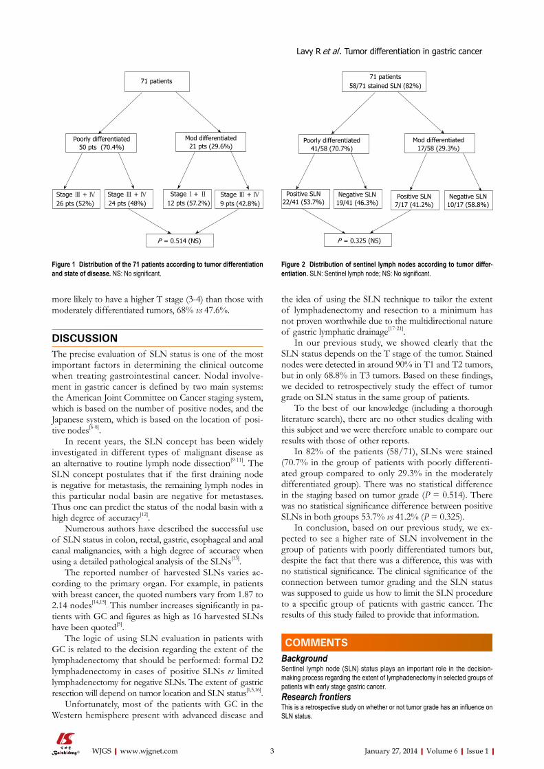
Poorly differentiated tumors were detected in 50/71 patients, while the other 21 patients had moderately dif-ferentiated tumors (Figure 1).
A total of 1114 regional lymph nodes were harvested in the group of patients with poorly differentiated tumors (22.7 nodes per person) with a positivity ratio of 20.3% (226/1114). In the group of patients with moderately differentiated tumors, the overall number of harvested nodes was 401 (19.1 nodes per person) with a positivity ratio of 16.5% (66/401).
SLNs were identified in 58/71 patients (82%), of which 41 (70.7%) were of the poorly differentiated type (group Ⅰ) and 17 (29.3%) were of the moderately differ-entiated type (group Ⅱ).
Positive SLNs were found in 22/41 patients in group I (53.7%) and in 7/17 patients in group Ⅱ (41.2%), P = 0.325 NS (Figure 2).
The patients with poorly differentiated tumors were
January 27, 2014|Volume 6|Issue 1|WJGS|www.wjgnet.com
Lavy R et al . Tumor differentiation in gastric cancer
2
more likely to have a higher T stage (3-4) than those with moderately differentiated tumors, 68% vs 47.6%.
DISCUSSIONThe precise evaluation of SLN status is one of the most important factors in determining the clinical outcome when treating gastrointestinal cancer. Nodal involve-ment in gastric cancer is defined by two main systems: the American Joint Committee on Cancer staging system, which is based on the number of positive nodes, and the Japanese system, which is based on the location of posi-tive nodes[6-8].
In recent years, the SLN concept has been widely investigated in different types of malignant disease as an alternative to routine lymph node dissection[9-11]. The SLN concept postulates that if the first draining node is negative for metastasis, the remaining lymph nodes in this particular nodal basin are negative for metastases. Thus one can predict the status of the nodal basin with a high degree of accuracy[12].
Numerous authors have described the successful use of SLN status in colon, rectal, gastric, esophageal and anal canal malignancies, with a high degree of accuracy when using a detailed pathological analysis of the SLNs[13].
The reported number of harvested SLNs varies ac-cording to the primary organ. For example, in patients with breast cancer, the quoted numbers vary from 1.87 to 2.14 nodes[14,15]
. This number increases significantly in pa-tients with GC and figures as high as 16 harvested SLNs have been quoted[5].
The logic of using SLN evaluation in patients with GC is related to the decision regarding the extent of the lymphadenectomy that should be performed: formal D2 lymphadenectomy in cases of positive SLNs vs limited lymphadenectomy for negative SLNs. The extent of gastric resection will depend on tumor location and SLN status[1,5,16].
Unfortunately, most of the patients with GC in the Western hemisphere present with advanced disease and
the idea of using the SLN technique to tailor the extent of lymphadenectomy and resection to a minimum has not proven worthwhile due to the multidirectional nature of gastric lymphatic drainage[17-21].
In our previous study, we showed clearly that the SLN status depends on the T stage of the tumor. Stained nodes were detected in around 90% in T1 and T2 tumors, but in only 68.8% in T3 tumors. Based on these findings, we decided to retrospectively study the effect of tumor grade on SLN status in the same group of patients.
To the best of our knowledge (including a thorough literature search), there are no other studies dealing with this subject and we were therefore unable to compare our results with those of other reports.
In 82% of the patients (58/71), SLNs were stained (70.7% in the group of patients with poorly differenti-ated group compared to only 29.3% in the moderately differentiated group). There was no statistical difference in the staging based on tumor grade (P = 0.514). There was no statistical significance difference between positive SLNs in both groups 53.7% vs 41.2% (P = 0.325).
In conclusion, based on our previous study, we ex-pected to see a higher rate of SLN involvement in the group of patients with poorly differentiated tumors but, despite the fact that there was a difference, this was with no statistical significance. The clinical significance of the connection between tumor grading and the SLN status was supposed to guide us how to limit the SLN procedure to a specific group of patients with gastric cancer. The results of this study failed to provide that information.
COMMENTSBackgroundSentinel lymph node (SLN) status plays an important role in the decision-making process regarding the extent of lymphadenectomy in selected groups of patients with early stage gastric cancer.Research frontiersThis is a retrospective study on whether or not tumor grade has an influence on SLN status.
January 27, 2014|Volume 6|Issue 1|WJGS|www.wjgnet.com 3
71 patients
Poorly differentiated50 pts (70.4%)
Mod differentiated21 pts (29.6%)
Stage Ⅲ + Ⅳ26 pts (52%)
Stage Ⅲ + Ⅳ24 pts (48%)
Stage Ⅰ+ Ⅱ12 pts (57.2%)
Stage Ⅲ + Ⅳ9 pts (42.8%)
P = 0.514 (NS)
Figure 1 Distribution of the 71 patients according to tumor differentiation and state of disease. NS: No significant.
71 patients58/71 stained SLN (82%)
Poorly differentiated41/58 (70.7%)
Mod differentiated17/58 (29.3%)
Positive SLN22/41 (53.7%)
Negative SLN19/41 (46.3%)
Positive SLN7/17 (41.2%)
Negative SLN10/17 (58.8%)
P = 0.325 (NS)
Figure 2 Distribution of sentinel lymph nodes according to tumor differ-entiation. SLN: Sentinel lymph node; NS: No significant.
COMMENTS
Lavy R et al . Tumor differentiation in gastric cancer

January 27, 2014|Volume 6|Issue 1|WJGS|www.wjgnet.com
Zasshi 2008; 109: 90-94 [PMID: 18409586]10 Snider H, Dowlatshahi K, Fan M, Bridger WM, Rayudu
G, Oleske D. Sentinel node biopsy in the staging of breast cancer. Am J Surg 1998; 176: 305-310 [PMID: 9817244 DOI: 10.1016/S0002-9610(98)00207-4]
11 Barnwell JM, Arredondo MA, Kollmorgen D, Gibbs JF, La-monica D, Carson W, Zhang P, Winston J, Edge SB. Sentinel node biopsy in breast cancer. Ann Surg Oncol 1998; 5: 126-130 [PMID: 9527265 DOI: 10.1007/BF02303845]
12 Kitagawa Y, Burian M, Kitajima M. Methods of sentinel lymph node mapping. Chirurg 2004; 75: 751-755 [PMID: 15241522 DOI: 10.1007/s00104-004-0908-7]
13 Saha S, Dan AG, Bilchik AJ, Kitagawa Y, Schochet E, Choudhri S, Saha LT, Wiese D, Morton D, Kitajima M. Historical review of lymphatic mapping in gastrointestinal malignancies. Ann Surg Oncol 2004; 11: 245S-249S [PMID: 15023761 DOI: 10.1245/ASO.2004.12.931]
14 Nielsen KR, Oturai PS, Friis E, Hesse U, Callesen T, Nielsen MB, Chakera AH, Hesse B. Axillary sentinel node iden-tification in breast cancer patients: degree of radioactiv-ity present at biopsy is critical. Clin Physiol Funct Imaging 2011; 31: 288-293 [PMID: 21672136 DOI: 10.1111/j.1475-097X.2011.01015.x]
15 Hundley JC, Shen P, Shiver SA, Geisinger KR, Levine EA. Lymphatic mapping for gastric adenocarcinoma. Am Surg 2002; 68: 931-935 [PMID: 12455783]
16 Cozzaglio L, Bottura R, Di Rocco M, Gennari L, Doci R. Sentinel lymph node biopsy in gastric cancer: possible ap-plications and limits. Eur J Surg Oncol 2011; 37: 55-59 [PMID: 21115231 DOI: 10.1016/j.ejso.2010.10.012]
17 Kitagawa Y, Kitajima M. Diagnostic validity of radio-guided sentinel node mapping for gastric cancer: a review of current status and future direction. Surg Technol Int 2006; 15: 32-36 [PMID: 17029158]
18 Wong J, Jackson P. Gastric cancer surgery: an American perspective on the current options and standards. Curr Treat Options Oncol 2011; 12: 72-84 [PMID: 21274666 DOI: 10.1007/s11864-010-0136-y]
19 Hundahl SA, Phillips JL, Menck HR. The National Cancer Data Base Report on poor survival of U.S. gastric carcinoma patients treated with gastrectomy: Fifth Edition American Joint Committee on Cancer staging, proximal disease, and the “different disease” hypothesis. Cancer 2000; 88: 921-932 [PMID: 10679663]
20 Tsubono Y, Hisamichi S. Screening for gastric cancer in Japan. Gastric Cancer 2000; 3: 9-18 [PMID: 11984703 DOI: 10.1007/PL00011692]
21 Lambert R, Guilloux A, Oshima A, Pompe-Kirn V, Bray F, Parkin M, Ajiki W, Tsukuma H. Incidence and mortality from stomach cancer in Japan, Slovenia and the USA. Int J Cancer 2002; 97: 811-818 [PMID: 11857360 DOI: 10.1002/ijc.10150]
P- Reviewers: El-Tawil AM, Nowicki MJ, Xia HHX S- Editor: Cui XM L- Editor: A E- Editor: Wu HL
Innovations and breakthroughsIn a previous study, the authors investigated the accuracy of SLN mapping ac-cording to the T stage of the tumor and showed that in T1-2 tumors, SLN map-ping may be of assistance, but in patients with T3 it will be misleading in a third of the patients and should not be attempted.ApplicationsThe evaluation of tumor grade may aid in predicting the outcome in patients with gastric cancer and the need for sentinel node evaluation.Peer reviewThe manuscript titled “Tumor differentiation as related to sentinel lymph node status in patients with gastric cancer” by Lavy et al, was performed to investi-gate the influence of tumor grade on sentinel lymph node status in patients with gastric cancer. The paper is well written.
REFERENCES1 Cabanas RM. An approach for the treatment of penile carci-
noma. Cancer 1977; 39: 456-466 [PMID: 837331]2 Fujii H, Kitagawa Y, Kitajima M, Kubo A. Sentinel nodes
of malignancies originating in the alimentary tract. Ann Nucl Med 2004; 18: 1-12 [PMID: 15072178 DOI: 10.1007/BF02985608]
3 Kitagawa Y, Fujii H, Mukai M, Kubo A, Kitajima M. Senti-nel lymph node mapping in esophageal and gastric cancer. Cancer Treat Res 2005; 127: 123-139 [PMID: 16209080 DOI: 10.1007/0-387-23604-X_6]
4 Kitagawa Y, Fujii H, Kumai K, Kubota T, Otani Y, Saikawa Y, Yoshida M, Kubo A, Kitajima M. Recent advances in sentinel node navigation for gastric cancer: a paradigm shift of surgi-cal management. J Surg Oncol 2005; 90: 147-151; discussion 151-152 [PMID: 15895450 DOI: 10.1002/jso.20220]
5 Rabin I, Chikman B, Lavy R, Poluksht N, Halpern Z, Was-sermann I, Gold-Deutch R, Sandbank J, Halevy A. The ac-curacy of sentinel node mapping according to T stage in patients with gastric cancer. Gastric Cancer 2010; 13: 30-35 [PMID: 20373073 DOI: 10.1007/s10120-009-0532-9]
6 Aikou T, Kitagawa Y, Kitajima M, Uenosono Y, Bilchik AJ, Martinez SR, Saha S. Sentinel lymph node mapping with GI cancer. Cancer Metastasis Rev 2006; 25: 269-277 [PMID: 16770539 DOI: 10.1007/s10555-006-8507-3]
7 Kim DH, Oh CA, Oh SJ, Choi MG, Noh JH, Sohn TS, Bae JM, Kim S. Validation of seventh edition AJCC gastric cancer staging modifications. J Surg Oncol 2012; 105: 26-30 [PMID: 21761411 DOI: 10.1002/jso.22026]
8 Zhang M, Zhu G, Ma Y, Xue Y. Comparison of four staging systems of lymph node metastasis in gastric cancer. World J Surg 2009; 33: 2383-2388 [PMID: 19760313 DOI: 10.1007/s00268-009-0214-0]
9 Takeuchi H, Kitagawa Y. Preoperative diagnosis of lymph node metastases and sentinel node navigation surgery in pa-tients with upper gastrointestinal cancer. Nihon Geka Gakkai
4
Lavy R et al . Tumor differentiation in gastric cancer
Pradeep Kumar, Hosni Mubarak Khan, Safarulla Hasanrabba
Treatment of perforated giant gastric ulcer in an emergency setting
Pradeep Kumar, Hosni Mubarak Khan, Department of General Surgery, ESI-PGIMSR and Medical College, Bangalore 560010, Karnataka, India Safarulla Hasanrabba, Department of General Surgery, Dr.B.R.Ambedkar Medical College, Bangalore 560045, Karna-taka, IndiaAuthor contributions: Kumar P contributed to the concept, research, data and figures, and was the operating surgeon and attending consultant; Khan HM was the attending doctor; and Hasanrabba S contributed to the references.Correspondence to: Dr. Pradeep Kumar, Assistant Profes-sor, Department of General Surgery, ESI-PGIMSR and Medical College, Rajajinagar, Bangalore 560010, Karnataka, India. [email protected]: +91-990-2349960Received: August 21, 2013 Revised: November 13, 2013Accepted: November 18, 2013Published online: January 27, 2014
AbstractAIM: To study and assess clinical outcomes of various modes of treatment for perforated giant gastric ulcer in an emergency setting.
METHODS: From May 2010 to February 2013, 20 cases of perforated giant gastric ulcer (> 2 cm) were operated on in an emergency setting. All the patients presented with features of peritonitis and were resusci-tated aggressively before taking for surgery. In the first 4 cases, primary closure was done after taking a biopsy and among these, the 3rd case also underwent partial distal gastrectomy and gastrojejunostomy and the 4th
case underwent a radical subtotal gastrectomy with D2 lymphadenectomy and gastrojejunostomy for ma-lignancy. All the remaining 16 cases underwent partial distal gastrectomy and gastrojejunostomy.
RESULTS: Among the first 4 cases, 2 had an unevent-ful recovery and were discharged on the 6th postop-erative day. The 3rd and 4th patients developed gastric
fistula, leading to prolonged hospitalization. For the 3rd patient, conservative management was tried for 1 wk, followed by partial distal gastrectomy and gastrojeju-nostomy, and he was discharged on the 20th day after admission, while the 4th patient underwent a radical subtotal gastrectomy with D2 lymphadenectomy and gastrojejunostomy. Postoperatively, he developed adult respiratory distress syndrome, multiorgan dysfunction syndrome and expired on the 3rd postoperative day of the second surgery. All the remaining 16 patients un-derwent partial distal gastrectomy and gastrojejunos-tomy and recovered well. Among these, 4 of them were malignant and the remaining were benign ulcers. All had an uneventful recovery. The percentage of malig-nancy in our series was 30% (6 out of 20 cases). In our study, 86% had an uneventful recovery, complications were seen in about 10%, and mortality was about 5%.
CONCLUSION: In giant gastric ulcer, the chances of malignancy and leak after primary closure are high. So, we feel that partial distal gastrectomy and gastrojeju-nostomy is better.
© 2014 Baishideng Publishing Group co., Limited. All rights reserved.
Key words: Giant; Gastric; Ulcer; Primary closure; Par-tial gastrectomy; Biopsy
Core tip: Giant gastric ulcer is considered to be more prone for perforation because of the large size and it is more likely to be malignant. Delay in seeking surgical care is to be discouraged because of the poor response to medical management. We have shown that with prompt treatment for perforated gastric ulcer, nearly 86% had uneventful recovery, complications were seen in about 10%, and mortality was about 5%. Further-more, the chances of malignancy and leak after pri-mary closure of giant gastric ulcer is high, so we feel partial distal gastrectomy and gastrojejunostomy is a better option, even in an emergency setting if the ex-
BRIEF ARTICLE
Online Submissions: http://www.wjgnet.com/esps/[email protected]:10.4240/wjgs.v6.i1.5
January 27, 2014|Volume 6|Issue 1|WJGS|www.wjgnet.com
World J Gastrointest Surg 2014 January 27; 6(1): 5-8ISSN 1948-9366 (online)
© 2014 Baishideng Publishing Group co., Limited. All rights reserved.
5
pertise is available.
Kumar P, Khan HM, Hasanrabba S. Treatment of perforated gi-ant gastric ulcer in an emergency setting. World J Gastrointest Surg 2014; 6(1): 5-8 Available from: URL: http://www.wjg-net.com/1948-9366/full/v6/i1/5.htm DOI: http://dx.doi.org/10.4240/wjgs.v6.i1.5
INTRODUCTIONGiant gastric ulcer is defined as an ulcer greater than 2 cm in diameter[1]. It is usually found along the lesser cur-vature at incisura angularis. It is considered more prone for perforation because of the large size[2] and is more likely to be malignant, especially when associated with scalloped margins and loss of rugal folds around ulcer. These ulcers were traditionally treated by primary closure after taking a biopsy as it was presumed that patients would not tolerate gastrectomy in an emergency setting as the time taken is longer and prolonged anesthesia is contraindicated in compromised patients. In this paper, we have compared primary closure with partial gastrec-tomy and gastrojejunostomy for perforated giant gastric ulcer in an emergency setting.
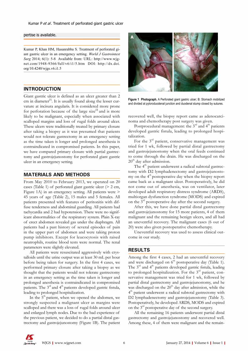
MATERIALS AND METHODSFrom May 2010 to February 2013, we operated on 20 cases (Table 1) of perforated giant gastric ulcer (> 2 cm, Figure 1A) in an emergency setting. All patients were > 45 years of age (Table 2), 15 males and 5 females. All patients presented with features of peritonitis with dif-fuse tenderness and abdominal guarding. All patients had tachycardia and 2 had hypotension. There were no signif-icant abnormalities of the respiratory system. Plain X-ray of erect abdomen revealed gas under the diaphragm. All patients had a past history of several episodes of pain in the upper part of abdomen and were taking proton pump inhibitors. Except for leucocytosis with raised neutrophils, routine blood tests were normal. The renal parameters were slightly elevated.
All patients were resuscitated aggressively with crys-talloids until the urine output was at least 30 mL per hour before being taken for surgery. In the first 4 cases, we performed primary closure after taking a biopsy as we thought that the patients would not tolerate gastrectomy in an emergency setting as the time taken is longer and prolonged anesthesia is contraindicated in compromised patients. The 3rd and 4th patients developed gastric fistula, leading to prolonged hospitalization.
In the 5th patient, when we opened the abdomen, we strongly suspected a malignant ulcer as margins were scalloped and there was a loss of rugal folds around ulcer and enlarged lymph nodes. Due to the bad experience of the previous patient, we decided to do a partial distal gas-trectomy and gastrojejunostomy (Figure 1B). The patient
recovered well, the biopsy report came as adenocarci-noma and chemotherapy post surgery was given.
Postprocedural management: the 3rd and 4th patients developed gastric fistula, leading to prolonged hospi-talization.
For the 3rd patient, conservative management was tried for 1 wk, followed by partial distal gastrectomy and gastrojejunostomy when the oral feeds continued to come through the drain. He was discharged on the 20th day after admission.
The 4th patient underwent a radical subtotal gastrec-tomy with D2 lymphadenectomy and gastrojejunosto-my on the 4th postoperative day when the biopsy report came back as a malignant ulcer. Postoperatively, he did not come out of anesthesia, was on ventilator, later developed adult respiratory distress syndrome (ARDS), multiorgan dysfunction syndrome (MODS) and expired on the 3rd postoperative day after the second surgery.
After this, we have done partial distal gastrectomy and gastrojejunostomy for 15 more patients, 4 of them malignant and the remaining benign ulcers, and all had an uneventful recovery. The malignant cases (6 out of 20) were also given postoperative chemotherapy.
Uneventful recovery was used to assess clinical out-come in our study.
RESULTSAmong the first 4 cases, 2 had an uneventful recovery and were discharged on 6th postoperative day (Table 1). The 3rd and 4th patients developed gastric fistula, leading to prolonged hospitalization. For the 3rd patient, con-servative management was tried for 1 wk, followed by partial distal gastrectomy and gastrojejunostomy, and he was discharged on the 20th day after admission, while the 4th patient underwent a radical subtotal gastrectomy with D2 lymphadenectomy and gastrojejunostomy (Table 3). Postoperatively, he developed ARDS, MODS and expired on the 3rd postoperative day of the second surgery.
All the remaining 16 patients underwent partial distal gastrectomy and gastrojejunostomy and recovered well. Among these, 4 of them were malignant and the remain-
January 27, 2014|Volume 6|Issue 1|WJGS|www.wjgnet.com
Kumar P et al . Treatment of perforated giant gastric ulcer
6
Figure 1 Photograph. A Perforated giant gastric ulcer; B: Stomach mobilized and divided at pyloroduodenal junction and duodenal stump closed by sutures.
A B
ing benign ulcers. All had an uneventful recovery (Table 4).The percentage of malignancy in our series was 30%
(6 out of 20 cases).In our study, 86% had an uneventful recovery, com-
plications were seen in about 10%, and mortality was about 5%.
DISCUSSIONGiant gastric ulcer is defined as an ulcer greater than 2 cm in diameter. It is usually found along the lesser curva-ture at incisura angularis[1,2]. It is considered more prone for perforation because of the large size and is more likely to be malignant[3-5], especially when associated with scalloped margins and loss of rugal folds around ulcer. Most giant ulcers occur beyond the middle span of life (all our patients were > 45 years of age). Indeed, the long history of most of these patients requires that they no longer be in the younger age group. The preponderance
of males is in agreement with the usual sex distribution of gastric ulcer disease. About half of the ulcers were in the antrum and half in the body. The frequency of mas-sive bleeding and perforation indicates that giant ulcers are not immune to the usual complications of gastric ul-cer disease.
The concept that giant gastric ulcers are most often benign presents the patient with an altered prognosis and makes an aggressive surgical attitude towards such a le-sion even more important. Delay in seeking surgical care is to be discouraged because of the poor response to medical management[6].
Undue delay in exploration is no longer justified in a giant ulcer simply because of fear that an inoperable car-cinoma will be found. On the contrary, all such patients should be subjected to exploration as soon as possible with the expectation that beneficial results may be ob-tained in a large percentage of these patients[7,8].
We have shown that with prompt treatment, nearly 86% had uneventful recovery, complications were seen in about 10%, and mortality was about 5%. Furthermore, the chances of malignancy and leak after primary closure of giant gastric ulcer is high, so we feel partial distal gas-trectomy and gastrojejunostomy is a better option, even in an emergency setting if the expertise is available.
In conclusion, the chances of malignancy and leak after primary closure of giant gastric ulcer is high, so we feel partial distal gastrectomy and gastrojejunostomy is a better option, even in an emergency setting if the exper-tise is available.
ACKNOWLEDGMENTSWritten informed consent was obtained from the patients for publication of this case series and accompanying im-ages. A copy of the written consent is available for review by the Editor-in-Chief of this journal on request.
January 27, 2014|Volume 6|Issue 1|WJGS|www.wjgnet.com 7
Type of surgery Number of patients
PC 4 (18.2) DG + GJ 17 (77.3) SG + GJ + D2 1 (4.5) Total 22
Table 3 Distribution of patients according to the type of surgery n (%)
PC: Primary closure; DG: Distal gastrectomy; GJ: Gastrojejunostomy; SG: Subtotal gastrectomy; D2: D2 lymph node dissection for ca stomach.
Type of patient compliance Number of patients
Uneventful recovery 18 (85.72) Complications 2 (9.52) Mortality 1 (4.76) Total 21
Table 4 Distribution of patients according to recovery n (%)
Serial No.
Age, yr
Sex Procedure undergone
Nature of gastric ulcer
Recovery Discharge day post-
operatively
1 47 Male PC BENIGN U 6 2 54 Male PC BENIGN U 6 3 52 Male PC +
DG + GJBENIGN GF 17
4 77 Male PC + SG + GJ + D2
MALIGNANT GF + ARDS+ MODS + M
-
5 63 Male DG + GJ MALIGNANT U 6 6 74 Female DG + GJ BENIGN U 6 7 79 Female DG + GJ BENIGN U 6 8 57 Male DG + GJ BENIGN U 6 9 53 Male DG + GJ MALIGNANT U 8 10 46 Male DG + GJ BENIGN U 11 72 Male DG + GJ MALIGNANT U 8 12 49 Female DG + GJ BENIGN U 6 13 67 Male DG + GJ BENIGN U 6 14 56 Male DG + GJ BENIGN U 7 15 68 Female DG + GJ MALIGNANT U 8 16 63 Male DG + GJ BENIGN U 9 17 61 Male DG + GJ MALIGNANT U 7 18 56 Female DG + GJ BENIGN U 9 19 49 Male DG + GJ BENIGN U 7 20 66 Male DG + GJ BENIGN U 6
Table 1 Patient demography
PC: Primary closure; DG: Distal gastrectomy; GJ: Gastrojejunostomy; SG: Subtotal gastrectomy; D2: D2 lymph node dissection for ca stomach; U: Uneventful recovery; GF: Gastric fistula; ARDS: Adult respiratory distress syndrome; M: Mortality; MODS: Multiorgan dysfunction syndrome.
Age group Number of patients
41-50 4 (20) Mean age = 63 yr 51-60 6 (30) 61-70 6 (30) 71-80 4 (20) Total 20
Table 2 Distribution of patients according to age of the patients n (%)
Kumar P et al . Treatment of perforated giant gastric ulcer
January 27, 2014|Volume 6|Issue 1|WJGS|www.wjgnet.com
contents of stomach will enter the peritoneal cavity and cause peritonitis, a life threatening condition. Primary closure means simply closing the ulcer with sutures (stitches). Partial distal gastrectomy and gastrojejunostomy means removal of part of stomach along with ulcer and joining it to small intestine (jeju-num) to maintain continuity of the gastrointestinal tract.Peer reviewThe study is interesting. The aim of this study is to assess the clinical outcomes of various treatments for perforated giant gastric ulcer in an emergency setting.
REFERENCES1 Lulu DJ. Benign giant gastric ulcer. Am Surg 1971; 37:
357-362 [PMID: 5578526]2 Cohn I, SARTIN J. Giant gastric ulcers. Ann Surg 1958; 147:
749-758; discussion 758-759 [PMID: 13521694 DOI: 10.1097/00000658-195805000-00020]
3 Ferris DO. Gastric cancer. J La State Med Soc 1953; 105: 211-216 [PMID: 13053058]
4 Lumsden K. The problem of the giant gastric ulcer. Gas-troenterologia 1950-1951; 76: 89-93 [PMID: 14813383 DOI: 10.1159/000199136]
5 Smith FH, Boles RS, Jordan SM. Problem of the gastric ulcer reviewed: study of one thousand cases. J Am Med Assoc 1953; 153: 1505-1508 [PMID: 13108635 DOI: 10.1001/jama.1953.02940340007003]
6 Haddad W, Kestenbaum DJ, Wang HS. Effect of cimetidine on healing and surgical treatment of gastric ulcers. Am J Surg 1985; 149: 665-667 [PMID: 3993850 DOI: 10.1016/S0002-9610(85)80151-3]
7 Kukral JC. Gastric ulcer: an appraisal. Surgery 1968; 63: 1024-1036 [PMID: 4871797]
8 Zollinger RM, Watman RN, Denkewalter F. Should all gas-tric ulcers be treated surgically. Gastroenterology 1958; 35: 521-527 [PMID: 13598043]
P- Reviewers: Xu HM, Zhang BB S- Editor: Wen LL L- Editor: Roemmele A E- Editor: Wu HL
COMMENTSBackgroundGiant gastric ulcer is considered more prone for perforation because of the large size and is more likely to be malignant. Delay in seeking surgical care is to be discouraged because of the poor response to medical management. Undue delay in exploration is no longer justified in a giant ulcer simply because of fear that an inoperable carcinoma will be found. On the contrary, all such patients should be subjected to exploration as soon as possible with the ex-pectation that beneficial results may be obtained in a large percentage of these patients.Research frontiersThese ulcers were traditionally treated by primary closure after taking a biopsy as it was presumed that patients would not tolerate gastrectomy in an emer-gency setting as the time taken is longer and prolonged anesthesia is contra-indicated in compromised patients. In this paper, the authors have compared primary closure with partial gastrectomy and gastrojejunostomy for perforated giant gastric ulcer in an emergency setting. Innovations and breakthroughsWith modern intensive care unit care, anesthesia and minimal access surgery, partial gastrectomy and gastrojejunostomy for perforated giant gastric ulcer in an emergency setting is a viable option. ApplicationsThe study results suggest that the chances of malignancy and leak after pri-mary closure of giant gastric ulcer are high, so the authors feel that partial distal gastrectomy and gastrojejunostomy is a better option, even in an emergency setting if the expertise is available. In the study, after this surgery, nearly 86% had an uneventful recovery, complications were seen in about 10%, and mortal-ity was about 5%.TerminologyGiant gastric ulcer is defined as an ulcer greater than 2 cm in diameter. It is usually found along the lesser curvature at incisura angularis. It is considered more prone for perforation because of the large size and is more likely to be malignant. Emergency closure of perforation is essential as otherwise the acidic
8
COMMENTS
Kumar P et al . Treatment of perforated giant gastric ulcer

Ashwin Rammohan, Ravichandran Palaniappan, Anbalagan Pitchaimuthu, Kamalakannan Rajendran, Senthil Kumar Perumal, Kesavan Balaraman, Ravi Ramasamy, Jeswanth Sathyanesan, Manoharan Govindan
Implications of the presence of an aberrant right hepatic artery in patients undergoing pancreaticoduodenectomy
Ashwin Rammohan, Ravichandran Palaniappan, Anbalagan Pitchaimuthu, Kamalakannan Rajendran, Senthil Kumar Perumal, Kesavan Balaraman, Ravi Ramasamy, Jeswanth Sathyanesan, Manoharan Govindan, The Institute of Surgical Gastroenterology and Liver Transplantation, Centre for GI Bleed, Division of HPB diseases, Stanley Medical College Hospital, Chennai 600 001, IndiaAuthor contributions: Rammohan A, Palaniappan R, Pit-chaimuthu A, Rajendran K and Perumal SK contributed to the conception and design, acquisition, analysis and interpretation of data; Rammohan A, Balaraman K, Ramasamy R and Sathy-anesan J drafted the article and revised it critically for important intellectual content; Sathyanesan J, Palaniappan R and Govindan M gave the final approval of the version to be published.Correspondence to: Dr. Ashwin Rammohan, The Institute of Surgical Gastroenterology and Liver Transplantation, Centre for GI Bleed, Division of HPB diseases, Stanley Medical College Hospital, Old Jail Road, Chennai 600 001, India. [email protected]: +91-988-4173583 Fax: +91-44-25289595Received: October 14, 2013 Revised: December 7, 2013Accepted: December 17, 2013Published online: January 27, 2014
AbstractAIM: To analyze the differences in outcomes and the clinical impact following pancreatoduodenectomy (PD) in patients with and without aberrant right hepatic ar-tery (aRHA).
METHODS: All patients undergoing PD between Janu-ary 2008 and December 2012 were divided into two groups, one with aRHA and the other without. These groups were compared to identify differences in the intraoperative variables, the oncological clearance and the postoperative morbidity, mortality and hospital stay.
RESULTS: A total of 225 patients underwent PD, of which 43 (19.1%) patients were found to have either
accessory or replaced right hepatic arteries (aRHA group). The aRHA was preserved in 79% of the pa-tients. There was no significant difference in the intra-operative blood loss but operative time was prolonged, reflecting the complexity of the procedure [420 ± 44 (240-540) min vs 480 ± 45 (300-600) min, P < 0.05)]. There were no differences in the incidence of postoper-ative complications (pancreatic leak, pancreatic fistula, delayed gastric emptying and mortality) and hospital stay. Oncological clearance in the form of positive re-section margins [13 (7.1%) vs 3 (6.9%)] and lymph node yield were also similar in the two groups.
CONCLUSION: An aRHA is found in approximately one fifth of patients undergoing PD. Preservation is technically possible in most patients and can increase the operative complexity but does not negatively affect the safety or oncological outcomes of the procedure.
© 2014 Baishideng Publishing Group co., Limited. All rights reserved.
Key words: Pancreatoduodenectomy; Aberrant right hepatic artery; Arterial anomalies; Outcomes
Core tip: Appreciation and study of hepatic arterial anatomical variability is essential to the successful per-formance of complex pancreaticobiliary procedures. An aberrant right hepatic artery (aRHA) represents the vascular anomaly encountered most frequently dur-ing pancreatoduodenectomy (PD) and, because of its course, is most susceptible to intraoperative damage and tumor involvement. When an aRHA is present, the challenge in peripancreatic malignant disease is to bal-ance its preservation and the need to achieve oncologi-cal clearance. In this study, we analyzed the incidence of aRHA and its relationship with the operative com-plexity, occurrence of complications and oncological clearance in a large cohort of patients undergoing PD.
BRIEF ARTICLE
Online Submissions: http://www.wjgnet.com/esps/[email protected]:10.4240/wjgs.v6.i1.9
January 27, 2014|Volume 6|Issue 1|WJGS|www.wjgnet.com
World J Gastrointest Surg 2014 January 27; 6(1): 9-13ISSN 1948-9366 (online)
© 2014 Baishideng Publishing Group co., Limited. All rights reserved.
9

Rammohan A, Palaniappan R, Pitchaimuthu A, Rajendran K, Perumal SK, Balaraman K, Ramasamy R, Sathyanesan J, Govin-dan M. Implications of the presence of an aberrant right hepatic artery in patients undergoing pancreaticoduodenectomy. World J Gastrointest Surg 2014; 6(1): 9-13 Available from: URL: http://www.wjgnet.com/1948-9366/full/v6/i1/9.htm DOI: http://dx.doi.org/10.4240/wjgs.v6.i1.9
INTRODUCTIONEven although well described in the literature, the surgi-cal anatomy of the hepatic arteries is notoriously variable. Appreciation and study of hepatic arterial anatomical variability is essential to the successful performance of complex pancreaticobiliary procedures, such as the pancreaticoduodenectomy (PD). Although anatomically interesting, the presence of aberrant hepatic arterial anat-omy raises the surgical complexity and increases the po-tential risk of injury to the hepatic arterial supply during a PD[1]. The two most widely accepted classifications of hepatic arterial variations are those by Michels[2], based on 200 autopsies, and Hiatt, based on 1000 donor livers[2,3]. In both series, the most commonly reported vascular anomaly is an aberrant right hepatic artery (aRHA)[2,3].
An aRHA represents the vascular anomaly encoun-tered most frequently during PD. It may have a suprapan-creatic, intrapancreatic or rarely transpancreatic course, and, because of its course, it is most susceptible to intra-operative damage and tumor involvement[4,5]. The inci-dence of an aRHA identified in patients undergoing PD varies from 11%-26.5%[1,6]. When an aRHA is present, the challenge in peripancreatic malignant disease is to bal-ance between its preservation and the need to achieve on-cological clearance, which represents the only chance for prolonged survival[1,5,7]. The presence of an aRHA leads not only to an alteration in the surgical approach, but may also adversely affect the outcomes of the surgical procedure[5,8]. In this study, we analyzed the incidence of aRHA and its relationship with the operative complexity, occurrence of complications and oncological clearance in a large cohort of patients undergoing PD.
MATERIALS AND METHODSThe study was conducted over a five year period (2008-2012) which included all patients who underwent a PD by a single surgical team in a tertiary care center. Detailed information regarding their demography, char-acteristics, imaging, intraoperative findings and operative details were maintained on a prospective database. De-mography and patient characteristics were carefully re-corded. The details of the arterial anatomy, variations and the operative complexities, including duration of surgery and blood loss, were noted. In the specimen, the arterial margin was inked in addition to the resection margins. Postoperative course was recorded, with death during the same hospital stay or within 30 d of surgery being con-
sidered as operative mortality. Histopathology, along with resection margin status and lymph nodal yield, was also documented.
DefinitionsNormal anatomy was defined as when the celiac axis tri-furcated into the left gastric artery, the splenic artery and the common hepatic artery, with the common hepatic artery continuing as a hepatic artery proper after the branching off of the gastroduodenal artery, finally bifur-cating into the right and left hepatic arteries. The term anomalous or aberrant encompassed both the “accessory” and the “replaced” vessels. An extra right hepatic artery that supplied the liver, which also received blood supply from a normally located right hepatic artery, was termed an accessory right hepatic artery. If the liver received its primary blood supply from the aberrant right hepatic ar-tery, it was called a replaced hepatic artery.
Statistical analysisData are reported as frequencies or mean ± SD and ranges. SPSS version 20.0 (SPSS, Inc., Chicago, IL, Unit-ed States) software was used for data analysis. The Student t test was used to test significance for continuous variables and the Fisher’s exact test was used for categorical vari-ables. P values less than 0.05 were considered significant.
RESULTSBetween January 2008 and December 2012, two hundred and twenty-five consecutive patients who underwent PD were included in the study. No significant differences were noted in terms of age, gender, American Society of Anesthesiologists class and indication for pancreati-coduodenectomy (Tables 1 and 2). The most common indication for surgery was ampullary adenocarcinoma, followed by distal cholangiocarcinoma. There were 43 (19.1%) arterial anomalies detected during the procedure. The spectrum of arterial anomalies is shown in Table 3. Replaced RHA from superior mesenteric artery (SMA) was the most common anomaly noted. The artery could be preserved in 79% of the cases (Table 4). In eight pa-tients (6%) with aRHA, the aberrant vessel was sacrificed for oncological (n = 6) or technical (n = 2) reasons. While in 6, the aberrant artery was found to be accessory and was ligated. In two patients, there was an inadvertent
January 27, 2014|Volume 6|Issue 1|WJGS|www.wjgnet.com
Rammohan A et al . Aberrant right hepatic artery during pancreaticoduodenectomy
10
Patient characteristics No arterial anomalyn = 182
Arterial anomalyn = 43
P value
Age 52.1 ± 10.9 52.4 ± 11.2 NS Males/females 135/53 32/11 NS ASA Ⅰ 45 (24.7) 11 (25.6) NS Ⅱ 115 (63.2) 27 (62.7) NS Ⅲ/Ⅳ 22 (12.1) 5 (11.6) NS
Table 1 Patient demography n (%)
ASA: American Society of Anesthesiologists.

ligation which did not result in any ischemia of the liver; following an intraoperative Doppler confirmation, no re-construction was undertaken. In one patient, the replaced right hepatic artery was resected for oncological reasons and a primary anastomosis was performed. Apart from an increase in operating time by approximately an hour, there were no other significant differences in intraopera-tive variables between the two groups (Table 5). There were also no differences in the overall rates of postpan-createctomy hemorrhage, postpancreatectomy fistula, de-layed gastric emptying, positive resection margin, length of hospital stay and mortality between the two groups (Table 5). Seven point one percent of the patients un-dergoing PD had positive resection margins, 10 of them with their SMA margin positive for tumor. None of the arterial margins were positive for tumor (Table 5).
DISCUSSIONThe significance of the aberrant arterial anatomy is enor-mous during surgery, especially PD. The artery could necessitate altering the surgical approach by interfering with the resection and/or lymphadenectomy. These anomalous vessels may interfere with reconstruction of the pancreatic remnant, precluding safe pancreatic stump drainage. Aberrant anatomy increases the risk of injury to the hepatic arterial supply, leading to unexpected bleed-ing (intra- or postoperative) and ischemia[1,5,7,9-11]. The extrahepatic biliary tree receives a substantial portion of its blood supply from the RHA. Any ischemia secondary to hepatic artery injury will lead to ischemia of the biliary anastomosis, resulting in a biliary anastomotic leak. Isch-emic liver dysfunction may also manifest in the form of elevations in hepatic enzymes[1,5,7,9,10]. During dissection of these arteries, excessive handling of the vessel should
be avoided as it may damage the vessel adventitia, thereby increasing the chances of pseudoaneurysm. This can lead to catastrophic complications in the event of pancreatic anastomotic leak[1,5,7,10,11].
A precise knowledge of normal hepatic arterial anatomy is necessary to appreciate abnormal anatomy. Preoperative imaging can detect up to 60%-80% of all arterial anomalies. If the anomaly is detected preopera-tively, embolization of the vessel can be performed with microcoils[12-14]. It also helps to forewarn the surgeon, thereby preventing inadvertent injury to the RHA[1,7]. Multidetector row computed tomography (CT) (MDCT) scan shows enhanced delineation of the pancreatic le-sion and vascular structures along with the benefit of CT angiography in preoperative delineation of the arterial anatomy[4,15]. A visceral angiography is recommended only when very rare or complex visceral arterial anomalies are encountered on noninvasive imaging[15,16]. Although advance planning is ideal, extemporaneous decisions may be required intraoperatively[1,7,10,11]. In our series, imag-ing picked up the anomalies preoperatively in 58% of the cases. Interestingly, Perwaiz et al[11] have shown that the duration of the surgery did not differ significantly between those patients whose arterial anomalies were detected preoperatively compared to those detected in-traoperatively. A recent study from The Netherlands has shown that preservation of an aRHA is technically pos-sible in most patients and does not negatively impact on outcomes in patients undergoing PD. Surgical morbidity is also not higher in patients with an aRHA[6]. Another study, a series from India, has shown that while oncologi-cal outcomes and safety of the procedure are not com-promised, there is an increased operative complexity[11].
This is in concurrence with our study results. A report by Jah et al[17] showed a trend towards prolonged operative
January 27, 2014|Volume 6|Issue 1|WJGS|www.wjgnet.com 11
Indication No arterial anomaly n = 182
Arterial anomalyn = 43
P value
Ampullary adenocarcinoma 99 (54.4) 23 (53.5) NS Distal cholangiocarcinoma 43 (23.6) 10 (23.2) NS Pancreatic adenocarcinoma 25 (13.7) 6 (13.9) NS Duodenal carcinoma 15 (8.2) 3 (7.0) NS GIST 0 1 (2.3) -
Table 2 Underlying disease in patients with and without arterial anomaly n (%)
GIST: Gastrointestinal stromal tumor.
Arterial variations n = 43
Replaced RHA from SMA 31 (72.1) Accessory RHA from SMA 10 (23.2) Replaced CHA from SMA 1 (2.3) Accessory RHA from GDA 1 (2.3)
Table 3 Hepatic arterial variations observed during pancreaticoduodenectomy n (%)
RHA: Right hepatic artery; SMA: Superior mesenteric artery; GDA: Gateway design automation.
Management n = 43
Dissection and preservation 34 (79) Ligation 8 (18.6) Dissection and primary anastomosis 1 (2.3)
Table 4 Intraoperative management of aberrant artery n (%)
Variables No arterial anomalyn = 182
Arterial anomalyn = 43
P value
Duration of surgery (min) 420 ± 44 (240-540) 480 ± 45 (300-600) < 0.05 Blood loss (mL) 360 ± 52 (200-630) 390 ± 45 (300-650) NS Postpancreatectomy hemorrhage
4 (2.1) 1 (2.3) NS
Postpancreatectomy fistula
9 (4.9) 2 (4.65) NS
Delayed gastric emptying 98 (53.8) 23 (53.4) NS Length of hospital stay 13.6 ± 6.0 13.1 ± 5.1 NS Mortality 3 (1.7) 1 (2.3) NS Positive margin 13 (7.1) 3 (6.9) NS Lymph node yield 12 ± 4 12 ± 5 NS
Table 5 Intraoperative and postoperative comparison in patients with or without arterial anomalies n (%)
Rammohan A et al . Aberrant right hepatic artery during pancreaticoduodenectomy
January 27, 2014|Volume 6|Issue 1|WJGS|www.wjgnet.com
pancreaticoduodenectomy.TerminologyThe term anomalous or aberrant artery encompasses both the “accessory” and the “replaced” vessels. An extra right hepatic artery that supplies the liver, which also receives blood supply from a normally located right hepatic artery, is termed an accessory right hepatic artery. If the liver receives its primary blood supply from the aberrant right hepatic artery, it is called a replaced hepatic ar-tery.Peer reviewThis is a brief article that analyzed 225 patients who had a pancreaticoduode-nectomy during the last 5 years. The content is interesting and it implies that aberrant right hepatic artery can be handled with no adverse consequences when treated in high volume centers.
REFERENCES1 Shukla PJ, Barreto SG, Kulkarni A, Nagarajan G, Finger-
hut A. Vascular anomalies encountered during pancre-atoduodenectomy: do they influence outcomes? Ann Surg Oncol 2010; 17: 186-193 [PMID: 19838756 DOI: 10.1245/s10434-009-0757-1]
2 Michels NA. Newer anatomy of the liver and its variant blood supply and collateral circulation. Am J Surg 1966; 112: 337-347 [PMID: 5917302 DOI: 10.1016/0002-9610(66)90201-7]
3 Hiatt JR, Gabbay J, Busuttil RW. Surgical anatomy of the he-patic arteries in 1000 cases. Ann Surg 1994; 220: 50-52 [PMID: 8024358 DOI: 10.1097/00000658-199407000-00008]
4 Song SY, Chung JW, Yin YH, Jae HJ, Kim HC, Jeon UB, Cho BH, So YH, Park JH. Celiac axis and common hepatic artery variations in 5002 patients: systematic analysis with spiral CT and DSA. Radiology 2010; 255: 278-288 [PMID: 20308464 DOI: 10.1148/radiol.09090389.]
5 Rong GH, Sindelar WF. Aberrant peripancreatic arterial anatomy. Considerations in performing pancreatectomy for malignant neoplasms. Am Surg 1987; 53: 726-729 [PMID: 3425998]
6 Eshuis WJ, Olde Loohuis KM, Busch OR, van Gulik TM, Gouma DJ. Influence of aberrant right hepatic artery on peri-operative course and longterm survival after pancreatoduo-denectomy. HPB (Oxford) 2011; 13: 161-167 [PMID: 21309932 DOI: 10.1111/j.1477-2574.2010.00258.x]
7 Chamberlain RS, El-Sedfy A, Rajkumar D. Aberrant he-patic arterial anatomy and the whipple procedure: lessons learned. Am Surg 2011; 77: 517-526 [PMID: 21679581]
8 Volpe CM, Peterson S, Hoover EL, Doerr RJ. Justification for visceral angiography prior to pancreaticoduodenectomy. Am Surg 1998; 64: 758-761 [PMID: 9697907]
9 Suzuki T, Nakayasu A, Kawabe K, Takeda H, Honjo I. Surgical significance of anatomic variations of the hepatic artery. Am J Surg 1971; 122: 505-512 [PMID: 5098656 DOI: 10.1016/0002-9610(71)90476-4]
10 Lee JM, Lee YJ, Kim CW, Moon KM, Kim MW. Clinical implications of an aberrant right hepatic artery in patients undergoing pancreaticoduodenectomy. World J Surg 2009; 33: 1727-1732 [PMID: 19459000 DOI: 10.1007/s00268-009-0063-x]
11 Perwaiz A, Singh A, Singh T, Chaudhary A. Incidence and management of arterial anomalies in patients undergo-ing pancreaticoduodenectomy. JOP 2010; 11: 25-30 [PMID: 20065548]
12 Yamamoto S, Kubota K, Rokkaku K, Nemoto T, Sakuma A. Disposal of replaced common hepatic artery coursing within the pancreas during pancreatoduodenectomy: report of a case. Surg Today 2005; 35: 984-987 [PMID: 16249858 DOI: 10.1007/s00595-005-3040-5]
13 Woods MS, Traverso LW. Sparing a replaced common hepatic artery during pancreaticoduodenectomy. Am Surg 1993; 59: 719-721 [PMID: 7902051]
14 Miyamoto N, Kodama Y, Endo H, Shimizu T, Miyasaka
times and blood loss but these did not reach statistical significance. This is mirrored in a study by Yang et al[18], whose incidence of postoperative complications, operat-ing time and blood loss is similar to ours.
Prevention of the injury is the best policy. Hence, an early step in every PD should be a conscious attempt to define the vascular anatomy. After a complete kocherisa-tion and opening of the pars flaccida, the porta hepatis should be palpated to determine the location of the ar-terial pulsation[1,5,7,10,11,19-24]. Any variation in the normal location of the proper hepatic artery pulsations should raise the suspicion of an aberrant artery. Performing intraoperative liver Doppler ultrasonography is recom-mended to ensure the results of the arterial sacrifice or reconstruction and to prevent postoperative complica-tions[1,5,7,10,11,25,26]. Surgical expertise is a key factor in re-ducing morbidity so these patients should be managed in high volume centers where arterial reconstructions are routinely performed[19,27].
An all-important factor in the management of vas-cular anomalies is its recognition. There are multiple approaches to deal with an anomalous vessel interfering with the pancreatic resection. These include avoidance, ligation, dissection and traction away from the site of dissection, and division and anastomosis. Preservation is technically possible in most patients; this increases the operative complexity but does not negatively affect the safety or oncological outcomes of the procedure. A high index of suspicion in every patient along with an aware-ness of the normal and aberrant anatomy is a sine qua non to the performance of a safe pancreaticoduodenectomy.
COMMENTSBackgroundAn aberrant right hepatic artery (aRHA) is the most frequently encountered vas-cular anomaly during pancreatoduodenectomy (PD) which necessitates control of various arcades of the upper gastrointestinal tract. When an aRHA is pres-ent, the challenge in peripancreatic malignant disease is to balance its preser-vation and the need to achieve oncological clearance which represents the only chance for prolonged survival. The presence of an aRHA leads not only to an alteration in the surgical approach, but may also adversely affect the outcomes of the surgical procedure. This study was performed to analyze the differences in outcomes and the clinical impact following PD in patients with and without aRHA.Research frontiersAlthough well described in the literature, the surgical anatomy of the hepatic arteries is notoriously variable. Appreciation and study of hepatic arterial ana-tomical variability is essential for the successful performance of complex pan-creaticobiliary procedures, such as the PD. Although anatomically interesting, the presence of aberrant hepatic arterial anatomy raises the surgical complexity and increases the potential risk of injury to the hepatic arterial supply during a PD.Innovations and breakthroughsThere are multiple approaches to deal with an anomalous vessel interfering with the pancreatic resection. These include avoidance, ligation, dissection and traction away from the site of dissection, and division and anastomosis. Preser-vation is technically possible in most patients; this increases the operative com-plexity in the form of operating time but does not negatively affect the safety or oncological outcomes of the procedure.ApplicationsAn all-important factor in the management of vascular anomalies is its recogni-tion. A high index of suspicion in every patient along with an awareness of the normal and aberrant anatomy is a sine qua non to the performance of a safe
12
COMMENTS
Rammohan A et al . Aberrant right hepatic artery during pancreaticoduodenectomy
January 27, 2014|Volume 6|Issue 1|WJGS|www.wjgnet.com
21 Furukawa H, Shimada K, Iwata R, Moriyama N. A replaced common hepatic artery running through the pancreatic pa-renchyma. Surgery 2000; 127: 711-712 [PMID: 10840370 DOI: 10.1067/msy.2000.104485]
22 Li B, Chen FZ, Ge XH, Cai MZ, Jiang JS, Li JP, Lu SH. Pan-creatoduodenectomy with vascular reconstruction in treat-ing carcinoma of the pancreatic head. Hepatobiliary Pancreat Dis Int 2004; 3: 612-615 [PMID: 15567757]
23 Rammohan A, Sathyanesan J, Palaniappan R, Govindan M. Transpancreatic hepatomesenteric trunk complicating pancreaticoduodenectomy. JOP 2013; 14: 649-652 [PMID: 24216553 DOI: 10.6092/1590-8577/1641]
24 Kondo S, Katoh H, Hirano S, Ambo Y, Tanaka E, Okushiba S, Morikawa T. Results of radical distal pancreatectomy with en bloc resection of the celiac artery for locally advanced can-cer of the pancreatic body. Langenbecks Arch Surg 2003; 388: 101-106 [PMID: 12684805 DOI: 10.1007/s00423-003-0375-5]
25 Nakano H, Bachellier P, Weber JC, Oussoultzoglou E, Di-eng M, Shimura H, Boudjema K, Wolf P, Jaeck D. Arterial and vena caval resections combined with pancreaticoduo-denectomy in highly selected patients with periampullary malignancies. Hepatogastroenterology 2002; 49: 258-262 [PMID: 11941970]
26 Bold RJ, Charnsangavej C, Cleary KR, Jennings M, Madray A, Leach SD, Abbruzzese JL, Pisters PW, Lee JE, Evans DB. Major vascular resection as part of pancreaticoduodenec-tomy for cancer: radiologic, intraoperative, and pathologic analysis. J Gastrointest Surg 1999; 3: 233-243 [PMID: 10481116 DOI: 10.1016/S1091-255X(99)80065-1]
27 Kim AW, McCarthy WJ, Maxhimer JB, Quiros RM, Hol-linger EF, Doolas A, Millikan KW, Deziel DJ, Godellas CV, Prinz RA. Vascular complications associated with pancreati-coduodenectomy adversely affect clinical outcome. Surgery 2002; 132: 738-744; discussion 744-747 [PMID: 12407360 DOI: 10.1067/msy.2002.127688]
P- Reviewers: Takabe K, Wang CC S- Editor: Cui XM L- Editor: Roemmele A E- Editor: Wu HL
K, Tanaka E, Anbo Y, Hirano S, Kondo S, Katoh H. Em-bolization of the replaced common hepatic artery before surgery for pancreatic head cancer: report of a case. Surg Today 2004; 34: 619-622 [PMID: 15221560 DOI: 10.1007/s00595-004-2785-6]
15 Winston CB, Lee NA, Jarnagin WR, Teitcher J, DeMatteo RP, Fong Y, Blumgart LH. CT angiography for delineation of celiac and superior mesenteric artery variants in patients undergoing hepatobiliary and pancreatic surgery. AJR Am J Roentgenol 2007; 189: W13-W19 [PMID: 17579128]
16 Brennan DD, Zamboni G, Sosna J, Callery MP, Vollmer CM, Raptopoulos VD, Kruskal JB. Virtual Whipple: preoperative surgical planning with volume-rendered MDCT images to identify arterial variants relevant to the Whipple procedure. AJR Am J Roentgenol 2007; 188: W451-W455 [PMID: 17449742]
17 Jah A, Jamieson N, Huguet E, Praseedom R. The implica-tions of the presence of an aberrant right hepatic artery in patients undergoing a pancreaticoduodenectomy. Surg Today 2009; 39: 669-674 [PMID: 19639433 DOI: 10.1007/s00595-009-3947-3]
18 Yang F, Long J, Fu DL, Jin C, Yu XJ, Xu J, Ni QX. Aberrant hepatic artery in patients undergoing pancreaticoduode-nectomy. Pancreatology 2008; 8: 50-54 [PMID: 18230918 DOI: 10.1159/000114867]
19 Kennedy TJ, Cassera MA, Wolf R, Swanstrom LL, Hansen PD. Surgeon volume versus morbidity and cost in patients undergoing pancreaticoduodenectomy in an academic com-munity medical center. J Gastrointest Surg 2010; 14: 1990-1996 [PMID: 20676793 DOI: 10.1007/s11605-010-1280-1]
20 Yang SH, Yin YH, Jang JY, Lee SE, Chung JW, Suh KS, Lee KU, Kim SW. Assessment of hepatic arterial anatomy in keeping with preservation of the vasculature while performing pancreatoduodenectomy: an opinion. World J Surg 2007; 31: 2384-2391 [PMID: 17922256 DOI: 10.1007/s00268-007-9246-5]
13
Rammohan A et al . Aberrant right hepatic artery during pancreaticoduodenectomy
GENERAL INFORMATION World Journal of Gastrointestinal Surgery (World J Gastrointest Surg, WJGS, online ISSN 1948-9366, DOI: 10.4240) is a peer-reviewed open access (OA) academic journal that aims to guide clinical practice and improve diagnostic and therapeutic skills of clinicians.
Aim and scopeWJGS covers topics concerning micro-invasive surgery; laparos-copy; hepatic, biliary, pancreatic and splenic surgery; surgical nutri-tion; portal hypertension, as well as associated subjects. The current columns of WJGS include editorial, frontier, diagnostic advances, therapeutics advances, field of vision, mini-reviews, review, topic highlight, medical ethics, original articles, case report, clinical case conference (clinicopathological conference), and autobiography. Pri-ority publication will be given to articles concerning diagnosis and treatment of gastrointestinal surgery diseases. The following aspects are covered: clinical diagnosis, laboratory diagnosis, differential di-agnosis, imaging tests, pathological diagnosis, molecular biological diagnosis, immunological diagnosis, genetic diagnosis, functional diagnostics, and physical diagnosis; and comprehensive therapy, drug therapy, surgical therapy, interventional treatment, minimally invasive therapy, and robot-assisted therapy.
We encourage authors to submit their manuscripts to WJGS. We will give priority to manuscripts that are supported by major national and international foundations and those that are of great basic and clinical significance.
WJGS is edited and published by Baishideng Publishing Group (BPG). BPG has a strong professional editorial team composed of science editors, language editors and electronic editors. BPG currently publishes 41 OA clinical medical journals, and is one of the leading medical publishers, with the first-class editing and publishing capacity and production.
ColumnsThe columns in the issues of WJGS will include: (1) Editorial: The editorial board members are invited to make comments on an import-ant topic in their field in terms of its current research status and future directions to lead the development of this discipline; (2) Frontier: The editorial board members are invited to select a highly cited cutting-edge original paper of his/her own to summarize major findings, the problems that have been resolved and remain to be resolved, and fu-ture research directions to help readers understand his/her important academic point of view and future research directions in the field; (3) Diagnostic Advances: The editorial board members are invited to write high-quality diagnostic advances in their field to improve the diagnos-tic skills of readers. The topic covers general clinical diagnosis, differ-ential diagnosis, pathological diagnosis, laboratory diagnosis, imaging diagnosis, endoscopic diagnosis, biotechnological diagnosis, functional diagnosis, and physical diagnosis; (4) Therapeutics Advances: The editorial board members are invited to write high-quality therapeutic advances in their field to help improve the therapeutic skills of readers. The topic covers medication therapy, psychotherapy, physical therapy, replacement therapy, interventional therapy, minimally invasive therapy, endoscopic therapy, transplantation therapy, and surgical therapy; (5) Field of Vision: The editorial board members are invited to write com-mentaries on classic articles, hot topic articles, or latest articles to keep
readers at the forefront of research and increase their levels of clinical research. Classic articles refer to papers that are included in Web of Knowledge and have received a large number of citations (ranking in the top 1%) after being published for more than years, reflecting the quality and impact of papers. Hot topic articles refer to papers that are included in Web of Knowledge and have received a large number of citations after being published for no more than 2 years, reflect-ing cutting-edge trends in scientific research. Latest articles refer to the latest published high-quality papers that are included in PubMed, reflecting the latest research trends. These commentary articles should focus on the status quo of research, the most important research topics, the problems that have now been resolved and remain to be resolved, and future research directions. Basic information about the article to be commented (including authors, article title, journal name, year, volume, and inclusive page numbers); (6) Minireviews: The editorial board members are invited to write short reviews on recent advances and trends in research of molecular biology, genomics, and related cutting-edge technologies to provide readers with the latest knowledge and help improve their diagnostic and therapeutic skills; (7) Review: To make a systematic review to focus on the status quo of research, the most important research topics, the problems that have now been resolved and remain to be resolved, and future research dir-ections; (8) Topic Highlight: The editorial board members are invited to write a series of articles (7-10 articles) to comment and discuss a hot topic to help improve the diagnostic and therapeutic skills of readers; (9) Medical Ethics: The editorial board members are invited to write articles about medical ethics to increase readers’ knowledge of medical ethics. The topic covers international ethics guidelines, animal studies, clinical trials, organ transplantation, etc.; (10) Clinical Case Conference or Clinicopathological Conference: The editorial board members are invited to contribute high-quality clinical case conference; (11) Original Articles: To report innovative and original findings in gastrointestinal surgery; (12) Brief Articles: To briefly report the novel and innovative findings in gastrointestinal surgery; (13) Meta-Analysis: Covers the systematic review, mixedtreatment comparison, meta-regression, and overview of reviews, in order to summarize a given quantitative effect, e.g., the clinical effectiveness and safety of clinical treatments by com-bining data from two or more randomized controlled trials, thereby providing more precise and externally valid estimates than those which would stem from each individual dataset if analyzed separately from the others; (14) Case Report: To report a rare or typical case; (15) Letters to the Editor: To discuss and make reply to the contributions published in WJGS, or to introduce and comment on a controversial issue of general interest; (16) Book Reviews: To introduce and com-ment on quality monographs of gastrointestinal surgery; and (17) Autobiography: The editorial board members are invited to write their autobiography to provide readers with stories of success or failure in their scientific research career. The topic covers their basic personal information and information about when they started doing research work, where and how they did research work, what they have achieved, and their lessons from success or failure.
Name of journalWorld Journal of Gastrointestinal Surgery
ISSNISSN 1948-9366 (online)
WJGS|www.wjgnet.com January 27, 2014|Volume 6|Issue 1|I
Online Submissions: http://www.wjgnet.com/esps/[email protected]
World J Gastrointest Surg 2014 January 27; 6(1): I-VISSN 1948-9366 (online)
© 2014 Baishideng Publishing Group Co., Limited. All rights reserved.
INSTRUCTIONS TO AUTHORS

Instructions to authors
Launch dateNovember 30, 2009
FrequencyMonthly
Editorial-in-ChiefTimothy M Pawlik, MD, MPH, FACS, Associate Professor of Surgery and Oncology, Hepatobiliary Surgery Program Director, Di-rector, Johns Hopkins Medicine Liver Tumor Center Multi-Disciplin-ary Clinic, Co-Director of Center for Surgical Trials and Outcomes Research, Johns Hopkins Hospital, 600 N. Wolfe Street, Harvey 611, Baltimore, MD 21287, United States. [email protected]
Editorial OfficeJin-Lei Wang, DirectorXiu-Xia Song, Vice DirectorWorld Journal of Gastrointestinal SurgeryRoom 903, Building D, Ocean International Center,No. 62 Dongsihuan Zhonglu, Chaoyang District,Beijing 100025, ChinaTelephone: +86-10-85381891Fax: +86-10-85381893E-mail: [email protected]://www.wjgnet.com
PublisherBaishideng Publishing Group Co., LimitedFlat C, 23/F., Lucky Plaza, 315-321 Lockhart Road,Wanchai, Hong Kong, ChinaTelephone: +852-31158812Fax: +852-58042046E-mail: [email protected]://www.wjgnet.com
Production centerBeijing Baishideng BioMed Scientific Co., LimitedRoom 903, Building D, Ocean International Center,No. 62 Dongsihuan Zhonglu, Chaoyang District,Beijing 100025, ChinaTelephone: +86-10-85381892Fax: +86-10-85381893
Representative officeUSA Office8226 Regency Drive, Pleasanton, CA 94588-3144, United StatesTelephone: +1-925-2238242Fax: +1-925-2238243
Instructions to authorsFull instructions are available online at http://www.wjgnet.com/ 1948-9366/g_info_20100305152206.htm.
Indexed and Abstracted inPubMed Central, PubMed, Digital Object Identifier, and Direc-tory of Open Access Journals.
SPECIAL STATEMENTAll articles published in this journal represent the viewpoints of the authors except where indicated otherwise.
Biostatistical editingStatisital review is performed after peer review. We invite an ex-pert in Biomedical Statistics to evaluate the statistical method used in the paper, including t-test (group or paired comparisons), chi-squared test, Ridit, probit, logit, regression (linear, curvilinear, or stepwise), correlation, analysis of variance, analysis of covariance, etc. The reviewing points include: (1) Statistical methods should be described when they are used to verify the results; (2) Whether
the statistical techniques are suitable or correct; (3) Only homoge-neous data can be averaged. Standard deviations are preferred to standard errors. Give the number of observations and subjects (n). Losses in observations, such as drop-outs from the study should be reported; (4) Values such as ED50, LD50, IC50 should have their 95% confidence limits calculated and compared by weighted probit analysis (Bliss and Finney); and (5) The word ‘significantly’ should be replaced by its synonyms (if it indicates extent) or the P value (if it indicates statistical significance).
Conflict-of-interest statementIn the interests of transparency and to help reviewers assess any po-tential bias, WJGS requires authors of all papers to declare any com-peting commercial, personal, political, intellectual, or religious interests in relation to the submitted work. Referees are also asked to indi-cate any potential conflict they might have reviewing a particular paper. Before submitting, authors are suggested to read “Uniform Requirements for Manuscripts Submitted to Biomedical Journals: Ethical Considerations in the Conduct and Reporting of Research: Conflicts of Interest” from International Committee of Medical Journal Editors (ICMJE), which is available at: http://www.icmje.org/ethical_4conflicts.html.
Sample wording: [Name of individual] has received fees for serv-ing as a speaker, a consultant and an advisory board member for [names of organizations], and has received research funding from [names of organization]. [Name of individual] is an employee of [name of or-ganization]. [Name of individual] owns stocks and shares in [name of organization]. [Name of individual] owns patent [patent identification and brief description].
Statement of informed consentManuscripts should contain a statement to the effect that all human studies have been reviewed by the appropriate ethics committee or it should be stated clearly in the text that all persons gave their informed consent prior to their inclusion in the study. Details that might disclose the identity of the subjects under study should be omitted. Authors should also draw attention to the Code of Ethics of the World Med-ical Association (Declaration of Helsinki, 1964, as revised in 2004).
Statement of human and animal rightsWhen reporting the results from experiments, authors should follow the highest standards and the trial should conform to Good Clini-cal Practice (for example, US Food and Drug Administration Good Clinical Practice in FDA-Regulated Clinical Trials; UK Medicines Research Council Guidelines for Good Clinical Practice in Clinical Trials) and/or the World Medical Association Declaration of Hel-sinki. Generally, we suggest authors follow the lead investigator’s na-tional standard. If doubt exists whether the research was conducted in accordance with the above standards, the authors must explain the rationale for their approach and demonstrate that the institutional review body explicitly approved the doubtful aspects of the study.
Before submitting, authors should make their study approved by the relevant research ethics committee or institutional review board. If human participants were involved, manuscripts must be accompanied by a statement that the experiments were undertaken with the understanding and appropriate informed consent of each. Any personal item or information will not be published without ex-plicit consents from the involved patients. If experimental animals were used, the materials and methods (experimental procedures) section must clearly indicate that appropriate measures were taken to minimize pain or discomfort, and details of animal care should be provided.
SUBMISSION OF MANUSCRIPTSManuscripts should be typed in 1.5 line spacing and 12 pt. Book Antiqua with ample margins. Number all pages consecutively, and start each of the following sections on a new page: Title Page, Ab-stract, Introduction, Materials and Methods, Results, Discussion, Acknowledgements, References, Tables, Figures, and Figure Leg-ends. Neither the editors nor the publisher are responsible for the
WJGS|www.wjgnet.com II January 27, 2014|Volume 6|Issue 1|

Instructions to authors
opinions expressed by contributors. Manuscripts formally accepted for publication become the permanent property of Baishideng Publishing Group Co., Limited, and may not be reproduced by any means, in whole or in part, without the written permission of both the authors and the publisher. We reserve the right to copy-edit and put onto our website accepted manuscripts. Authors should follow the relevant guidelines for the care and use of laboratory animals of their institution or national animal welfare committee. For the sake of transparency in regard to the performance and reporting of clinical trials, we endorse the policy of the ICMJE to refuse to pub-lish papers on clinical trial results if the trial was not recorded in a publicly-accessible registry at its outset. The only register now avail-able, to our knowledge, is http://www.clinicaltrials.gov sponsored by the United States National Library of Medicine and we encour-age all potential contributors to register with it. However, in the case that other registers become available you will be duly notified. A letter of recommendation from each author’s organization should be provided with the contributed article to ensure the privacy and secrecy of research is protected.
Authors should retain one copy of the text, tables, photographs and illustrations because rejected manuscripts will not be returned to the author(s) and the editors will not be responsible for loss or damage to photographs and illustrations sustained during mailing.
Online submissionsManuscripts should be submitted through the Online Submission System at: http://www.wjgnet.com/esps/. Authors are highly recom-mended to consult the ONLINE INSTRUCTIONS TO AUTHORS (http://www.wjgnet.com/1948-9366/g_info_20100305152206.htm) before attempting to submit online. For assistance, authors encoun-tering problems with the Online Submission System may send an email describing the problem to [email protected], or by tele-phone: +86-10-85381891. If you submit your manuscript online, do not make a postal contribution. Repeated online submission for the same manuscript is strictly prohibited.
MANUSCRIPT PREPARATIONAll contributions should be written in English. All articles must be submitted using word-processing software. All submissions must be typed in 1.5 line spacing and 12 pt. Book Antiqua with ample mar-gins. Style should conform to our house format. Required informa-tion for each of the manuscript sections is as follows:
Title pageTitle: Title should be less than 12 words.
Running title: A short running title of less than 6 words should be provided.
Authorship: Authorship credit should be in accordance with the standard proposed by International Committee of Medical Journal Editors, based on (1) substantial contributions to conception and design, acquisition of data, or analysis and interpretation of data; (2) drafting the article or revising it critically for important intellectual content; and (3) final approval of the version to be published. Au-thors should meet conditions 1, 2, and 3.
Institution: Author names should be given first, then the complete name of institution, city, province and postcode. For example, Xu-Chen Zhang, Li-Xin Mei, Department of Pathology, Chengde Medi-cal College, Chengde 067000, Hebei Province, China. One author may be represented from two institutions, for example, George Sgourakis, Department of General, Visceral, and Transplantation Surgery, Es-sen 45122, Germany; George Sgourakis, 2nd Surgical Department, Korgialenio-Benakio Red Cross Hospital, Athens 15451, Greece
Author contributions: The format of this section should be: Author contributions: Wang CL and Liang L contributed equally to this work; Wang CL, Liang L, Fu JF, Zou CC, Hong F and Wu XM designed the research; Wang CL, Zou CC, Hong F and Wu XM performed the
research; Xue JZ and Lu JR contributed new reagents/analytic tools; Wang CL, Liang L and Fu JF analyzed the data; and Wang CL, Liang L and Fu JF wrote the paper.
Supportive foundations: The complete name and number of sup-portive foundations should be provided, e.g. Supported by National Natural Science Foundation of China, No. 30224801
Correspondence to: Only one corresponding address should be provided. Author names should be given first, then author title, af-filiation, the complete name of institution, city, postcode, province, country, and email. All the letters in the email should be in lower case. A space interval should be inserted between country name and email address. For example, Montgomery Bissell, MD, Professor of Medi-cine, Chief, Liver Center, Gastroenterology Division, University of California, Box 0538, San Francisco, CA 94143, United States. [email protected]
Telephone and fax: Telephone and fax should consist of +, country number, district number and telephone or fax number, e.g. Tele-phone: +86-10-85381891 Fax: +86-10-85381893
Peer reviewers: All articles received are subject to peer review. Nor-mally, three experts are invited for each article. Decision on accept-ance is made only when at least two experts recommend publication of an article. All peer-reviewers are acknowledged on Express Sub-mission and Peer-review System website.
AbstractThere are unstructured abstracts (no less than 200 words) and struc-tured abstracts. The specific requirements for structured abstracts are as follows:
An informative, structured abstract should accompany each manuscript. Abstracts of original contributions should be struc-tured into the following sections: AIM (no more than 20 words; Only the purpose of the study should be included. Please write the Aim in the form of “To investigate/study/…”), METHODS (no less than 140 words for Original Articles; and no less than 80 words for Brief Articles), RESULTS (no less than 150 words for Original Articles and no less than 120 words for Brief Articles; You should present P values where appropriate and must provide relevant data to illustrate how they were obtained, e.g. 6.92 ± 3.86 vs 3.61 ± 1.67, P < 0.001), and CONCLUSION (no more than 26 words).
Key wordsPlease list 5-10 key words, selected mainly from Index Medicus, which reflect the content of the study.
Core tipPlease write a summary of less than 100 words to outline the most innovative and important arguments and core contents in your paper to attract readers.
TextFor articles of these sections, original articles and brief articles, the main text should be structured into the following sections: INTRO-DUCTION, MATERIALS AND METHODS, RESULTS and DIS-CUSSION, and should include appropriate Figures and Tables. Data should be presented in the main text or in Figures and Tables, but not in both. The main text format of these sections, editorial, topic high-light, case report, letters to the editors, can be found at: http://www.wjgnet.com/1948-9366/g_info_list.htm.
IllustrationsFigures should be numbered as 1, 2, 3, etc., and mentioned clearly in the main text. Provide a brief title for each figure on a separate page. Detailed legends should not be provided under the figures. This part should be added into the text where the figures are applicable. Keep-ing all elements compiled is necessary in line-art image. Scale bars should be used rather than magnification factors, with the length of
WJGS|www.wjgnet.com III January 27, 2014|Volume 6|Issue 1|

Instructions to authors
the bar defined in the legend rather than on the bar itself. File names should identify the figure and panel. Avoid layering type directly over shaded or textured areas. Please use uniform legends for the same subjects. For example: Figure 1 Pathological changes in atrophic gas-tritis after treatment. A: ...; B: ...; C: ...; D: ...; E: ...; F: ...; G: …etc. It is our principle to publish high resolution-figures for the E-versions.
TablesThree-line tables should be numbered 1, 2, 3, etc., and mentioned clearly in the main text. Provide a brief title for each table. Detailed legends should not be included under tables, but rather added into the text where applicable. The information should complement, but not duplicate the text. Use one horizontal line under the title, a second under column heads, and a third below the Table, above any footnotes. Vertical and italic lines should be omitted.
Notes in tables and illustrationsData that are not statistically significant should not be noted. aP < 0.05, bP < 0.01 should be noted (P > 0.05 should not be noted). If there are other series of P values, cP < 0.05 and dP < 0.01 are used. A third series of P values can be expressed as eP < 0.05 and fP < 0.01. Other notes in tables or under illustrations should be expressed as 1F, 2F, 3F; or sometimes as other symbols with a superscript (Arabic numer-als) in the upper left corner. In a multi-curve illustration, each curve should be labeled with ●, ○, ■, □, ▲, △, etc., in a certain sequence.
AcknowledgmentsBrief acknowledgments of persons who have made genuine contri-butions to the manuscript and who endorse the data and conclusions should be included. Authors are responsible for obtaining written permission to use any copyrighted text and/or illustrations.
REFERENCESCoding systemThe author should number the references in Arabic numerals ac-cording to the citation order in the text. Put reference numbers in square brackets in superscript at the end of citation content or after the cited author’s name. For citation content which is part of the narration, the coding number and square brackets should be typeset normally. For example, “Crohn’s disease (CD) is associated with increased intestinal permeability[1,2]”. If references are cited directly in the text, they should be put together within the text, for example, “From references[19,22-24], we know that...”
When the authors write the references, please ensure that the order in text is the same as in the references section, and also ensure the spelling accuracy of the first author’s name. Do not list the same citation twice.
PMID and DOIPleased provide PubMed citation numbers to the reference list, e.g. PMID and DOI, which can be found at http://www.ncbi.nlm.nih.gov/sites/entrez?db=pubmed and http://www.crossref.org/Simple-TextQuery/, respectively. The numbers will be used in E-version of this journal.
Style for journal referencesAuthors: the name of the first author should be typed in bold-faced letters. The family name of all authors should be typed with the initial letter capitalized, followed by their abbreviated first and middle ini-tials. (For example, Lian-Sheng Ma is abbreviated as Ma LS, Bo-Rong Pan as Pan BR). The title of the cited article and italicized journal title (journal title should be in its abbreviated form as shown in PubMed), publication date, volume number (in black), start page, and end page [PMID: 11819634 DOI: 10.3748/wjg.13.5396].
Style for book referencesAuthors: the name of the first author should be typed in bold-faced letters. The surname of all authors should be typed with the initial let-
ter capitalized, followed by their abbreviated middle and first initials. (For example, Lian-Sheng Ma is abbreviated as Ma LS, Bo-Rong Pan as Pan BR) Book title. Publication number. Publication place: Publica-tion press, Year: start page and end page.
FormatJournals English journal article (list all authors and include the PMID where applicable)1 Jung EM, Clevert DA, Schreyer AG, Schmitt S, Rennert J,
Kubale R, Feuerbach S, Jung F. Evaluation of quantitative con-trast harmonic imaging to assess malignancy of liver tumors: A prospective controlled two-center study. World J Gastroenterol 2007; 13: 6356-6364 [PMID: 18081224 DOI: 10.3748/wjg.13. 6356]
Chinese journal article (list all authors and include the PMID where applicable)2 Lin GZ, Wang XZ, Wang P, Lin J, Yang FD. Immunologic ef-
fect of Jianpi Yishen decoction in treatment of Pixu-diarrhoea. Shijie Huaren Xiaohua Zazhi 1999; 7: 285-287
In press3 Tian D, Araki H, Stahl E, Bergelson J, Kreitman M. Signature
of balancing selection in Arabidopsis. Proc Natl Acad Sci USA 2006; In press
Organization as author4 Diabetes Prevention Program Research Group. Hyperten-
sion, insulin, and proinsulin in participants with impaired glu-cose tolerance. Hypertension 2002; 40: 679-686 [PMID: 12411462 DOI:10.1161/01.HYP.0000035706.28494.09]
Both personal authors and an organization as author 5 Vallancien G, Emberton M, Harving N, van Moorselaar RJ;
Alf-One Study Group. Sexual dysfunction in 1, 274 Euro-pean men suffering from lower urinary tract symptoms. J Urol 2003; 169: 2257-2261 [PMID: 12771764 DOI:10.1097/01.ju. 0000067940.76090.73]
No author given6 21st century heart solution may have a sting in the tail. BMJ
2002; 325: 184 [PMID: 12142303 DOI:10.1136/bmj.325. 7357.184]
Volume with supplement7 Geraud G, Spierings EL, Keywood C. Tolerability and safety
of frovatriptan with short- and long-term use for treatment of migraine and in comparison with sumatriptan. Headache 2002; 42 Suppl 2: S93-99 [PMID: 12028325 DOI:10.1046/j.1526- 4610.42.s2.7.x]
Issue with no volume8 Banit DM, Kaufer H, Hartford JM. Intraoperative frozen sec-
tion analysis in revision total joint arthroplasty. Clin Orthop Relat Res 2002; (401): 230-238 [PMID: 12151900 DOI:10.1097/00003086-200208000-00026]
No volume or issue9 Outreach: Bringing HIV-positive individuals into care. HRSA
Careaction 2002; 1-6 [PMID: 12154804]
BooksPersonal author(s)10 Sherlock S, Dooley J. Diseases of the liver and billiary system.
9th ed. Oxford: Blackwell Sci Pub, 1993: 258-296Chapter in a book (list all authors)11 Lam SK. Academic investigator’s perspectives of medical treat-
ment for peptic ulcer. In: Swabb EA, Azabo S. Ulcer disease: investigation and basis for therapy. New York: Marcel Dekker, 1991: 431-450
Author(s) and editor(s)12 Breedlove GK, Schorfheide AM. Adolescent pregnancy. 2nd
ed. Wieczorek RR, editor. White Plains (NY): March of Dimes Education Services, 2001: 20-34
Conference proceedings13 Harnden P, Joffe JK, Jones WG, editors. Germ cell tumours
V. Proceedings of the 5th Germ cell tumours Conference; 2001 Sep 13-15; Leeds, UK. New York: Springer, 2002: 30-56
Conference paper14 Christensen S, Oppacher F. An analysis of Koza's computa-
WJGS|www.wjgnet.com IV January 27, 2014|Volume 6|Issue 1|
Instructions to authors
tional effort statistic for genetic programming. In: Foster JA, Lutton E, Miller J, Ryan C, Tettamanzi AG, editors. Genetic programming. EuroGP 2002: Proceedings of the 5th European Conference on Genetic Programming; 2002 Apr 3-5; Kinsdale, Ireland. Berlin: Springer, 2002: 182-191
Electronic journal (list all authors)15 Morse SS. Factors in the emergence of infectious diseases.
Emerg Infect Dis serial online, 1995-01-03, cited 1996-06-05; 1(1): 24 screens. Available from: URL: http://www.cdc.gov/ncidod/eid/index.htm
Patent (list all authors)16 Pagedas AC, inventor; Ancel Surgical R&D Inc., assignee. Flex-
ible endoscopic grasping and cutting device and positioning tool assembly. United States patent US 20020103498. 2002 Aug 1
Statistical dataWrite as mean ± SD or mean ± SE.
Statistical expressionExpress t test as t (in italics), F test as F (in italics), chi square test as χ2 (in Greek), related coefficient as r (in italics), degree of freedom as υ (in Greek), sample number as n (in italics), and probability as P (in italics).
UnitsUse SI units. For example: body mass, m (B) = 78 kg; blood pressure, p (B) = 16.2/12.3 kPa; incubation time, t (incubation) = 96 h, blood glucose concentration, c (glucose) 6.4 ± 2.1 mmol/L; blood CEA mass concentration, p (CEA) = 8.6 24.5 mg/L; CO2 volume fraction, 50 mL/L CO2, not 5% CO2; likewise for 40 g/L formaldehyde, not 10% formalin; and mass fraction, 8 ng/g, etc. Arabic numerals such as 23, 243, 641 should be read 23 243 641.
The format for how to accurately write common units and quan-tums can be found at: http://www.wjgnet.com/1948-9366/g_info_ 20100312191949.htm.
AbbreviationsStandard abbreviations should be defined in the abstract and on first mention in the text. In general, terms should not be abbreviated un-less they are used repeatedly and the abbreviation is helpful to the reader. Permissible abbreviations are listed in Units, Symbols and Ab-breviations: A Guide for Biological and Medical Editors and Authors (Ed. Baron DN, 1988) published by The Royal Society of Medicine, London. Certain commonly used abbreviations, such as DNA, RNA, HIV, LD50, PCR, HBV, ECG, WBC, RBC, CT, ESR, CSF, IgG, ELISA, PBS, ATP, EDTA, mAb, can be used directly without further explanation.
ItalicsQuantities: t time or temperature, c concentration, A area, l length, m mass, V volume.Genotypes: gyrA, arg 1, c myc, c fos, etc.Restriction enzymes: EcoRI, HindI, BamHI, Kbo I, Kpn I, etc.Biology: H. pylori, E coli, etc.
Examples for paper writingAll types of articles’ writing style and requirement will be found in the
link: http://www.wjgnet.com/esps/NavigationInfo.aspx?id=15
SUBMISSION OF THE REVISED MANUSCRIPTS AFTER ACCEPTEDAuthors must revise their manuscript carefully according to the revision policies of Baishideng Publishing Group Co., Limited. The revised version, along with the signed copyright transfer agreement, responses to the reviewers, and English language Grade B certificate (for non-native speakers of English), should be submitted to the online system via the link contained in the e-mail sent by the editor. If you have any questions about the revision, please send e-mail to [email protected].
Language evaluation The language of a manuscript will be graded before it is sent for revision. (1) Grade A: priority publishing; (2) Grade B: minor lan-guage polishing; (3) Grade C: a great deal of language polishing needed; and (4) Grade D: rejected. Revised articles should reach Grade A.
Copyright assignment formPlease download a Copyright assignment form from http://www.wjgnet.com/1948-9366/g_info_20100312191901.htm.
Responses to reviewersPlease revise your article according to the comments/suggestions provided by the reviewers. The format for responses to the reviewers’ comments can be found at: http://www.wjgnet.com/1948-9366/g_info_20100312191818.htm.
Proof of financial supportFor papers supported by a foundation, authors should provide a copy of the approval document and serial number of the foundation.
STATEMENT ABOUT ANONYMOUS PUBLICA-TION OF THE PEER REVIEWERS’ COMMENTSIn order to increase the quality of peer review, push authors to carefully revise their manuscripts based on the peer reviewers' com-ments, and promote academic interactions among peer reviewers, authors and readers, we decide to anonymously publish the review-ers’ comments and author’s responses at the same time the manu-script is published online.
PUBLICATION FEEWJGS is an international, peer-reviewed, OA online journal. Articles published by this journal are distributed under the terms of the Cre-ative Commons Attribution Non-commercial License, which per-mits use, distribution, and reproduction in any medium and format, provided the original work is properly cited. The use is non-com-mercial and is otherwise in compliance with the license. Authors of accepted articles must pay a publication fee. Publication fee: 600 USD per article. All invited articles are published free of charge.
WJGS|www.wjgnet.com V January 27, 2014|Volume 6|Issue 1|

World Journal of Gastrointestinal SurgeryWorld J Gastrointest Surg 2014 February 27; 6(2): 14-37
ISSN 1948-9366 (online)
www.wjgnet.com
14 Predictionanddiagnosisofcolorectalanastomoticleakage:A
systematicreviewofliterature
Daams F, Wu Z, Lahaye MJ, Jeekel J, Lange JF
27 Transversus abdominis plane infiltration for laparoscopic gastric
banding:Apilotstudy
De Oliveira Jr GS, Fitzgerald P, Ahmad S, Kim J, Rahangdale R, McCarthy R
33 Rarediaphragmatictumormimickinglivermass
Thapar S, Ahuja A, Rastogi A
Contents Monthly Volume 6 Number 2 February 27, 2014
WJGS|www.wjgnet.com I February 27, 2014|Volume 6|Issue 2|
REVIEW
BRIEF ARTICLE
CASE REPORT

Contents
APPENDIX
ABOUT COVER
World Journal of Gastrointestinal SurgeryVolume 6 Number 2 Febuary 27, 2014
I-V Instructionstoauthors
EditorialBoardMemberofWorldJournalofGastrointestinalSurgery ,GiuseppeFusai,MD,HPBandLiverTransplantUnit,RoyalFreeHospital,PondStreet,LondonNW32QG,[email protected]
World Journal of Gastrointestinal Surgery (World J Gastrointest Surg, WJGS, online ISSN 1948-9366, DOI: 10.4240) is a peer-reviewed open access academic journal that aims to guide clinical practice and improve diagnostic and therapeutic skills of clinicians.
WJGS covers topics concerning micro-invasive surgery; laparoscopy; hepatic, biliary, pancreatic and splenic surgery; surgical nutrition; portal hypertension, as well as associated subjects. The current columns of WJGS include editorial, frontier, diagnostic advances, therapeutics advances, field of vision, mini-reviews, review, topic highlight, medical ethics, original articles, case report, clinical case conference (Clinicopathological conference), and autobiography. Priority publication will be given to articles concerning diagnosis and treatment of gastrointestinal surgery diseases. The following aspects are covered: Clinical diagnosis, laboratory diagnosis, differential diagnosis, imaging tests, pathological diagnosis, molecular biological diagnosis, immunological diagnosis, genetic diagnosis, functional diagnostics, and physical diagnosis; and comprehensive therapy, drug therapy, surgical therapy, interventional treatment, minimally invasive therapy, and robot-assisted therapy.
We encourage authors to submit their manuscripts to WJGS. We will give priority to manuscripts that are supported by major national and international foundations and those that are of great basic and clinical significance.
World Journal of Gastrointestinal Surgery is now indexed in PubMed Central, PubMed, Digital Object Identifier, and Directory of Open Access Journals.
I-III EditorialBoard
EDITORIALOFFICEJin-Lei Wang, DirectorXiu-Xia Song, Vice DirectorWorld Journal of Gastrointestinal SurgeryRoom 903, Building D, Ocean International Center, No. 62 Dongsihuan Zhonglu, Chaoyang District, Beijing 100025, ChinaTelephone: +86-10-85381891Fax: +86-10-85381893E-mail: [email protected]://www.wjgnet.com
PUBLISHERBaishideng Publishing Group Co., LimitedFlat C, 23/F., Lucky Plaza, 315-321 Lockhart Road, Wanchai, Hong Kong, ChinaFax: +852-31158812Telephone: +852-58042046E-mail: [email protected]://www.wjgnet.com
PUBLICATIONDATEFebruary 27, 2014
COPYRIGHT© 2014 Baishideng. Articles published by this Open-Access journal are distributed under the terms of the Creative Commons Attribution Non-commercial Li-cense, which permits use, distribution, and reproduction in any medium, provided the original work is properly cited, the use is non commercial and is otherwise in compliance with the license.
SPECIALSTATEMENTAll articles published in this journal represent the view-points of the authors except where indicated otherwise.
INSTRUCTIONSTOAUTHORSFull instructions are available online at http://www.wjgnet.com/1948-9366/g_info_20100305152206.htm
ONLINESUBMISSIONhttp://www.wjgnet.com/esps/
NAMEOFJOURNALWorld Journal of Gastrointestinal Surgery
ISSNISSN 1948-9366 (online)
LAUNCHDATENovember 30, 2009
FREQUENCYMonthly
EDITOR-IN-CHIEFTimothy M Pawlik, MD, MPH, FACS, Associate Professor of Surgery and Oncology, Hepatobiliary Surgery Program Director, Director, Johns Hopkins Medicine Liver Tumor Center Multi-Disciplinary Clinic, Co-Director of Center for Surgical Trials and Outcomes Research, Johns Hopkins Hospital, 600 N. Wolfe Street, Harvey 611, Baltimore, MD 21287, United States
EDITORS FOR THIS ISSUE
Responsible Assistant Editor: Xin-Xin Che Responsible Science Editor: Ling-Ling WenResponsible Electronic Editor: Huan-Liang WuProofing Editor-in-Chief: Lian-Sheng Ma
AIM AND SCOPE
FLYLEAF
INDEXING/ABSTRACTING
WJGS|www.wjgnet.com II February 27, 2014|Volume 6|Issue 2|
Freek Daams, Zhouqiao Wu, Max Jef Lahaye, Johannus Jeekel, Johan Frederik Lange
Prediction and diagnosis of colorectal anastomotic leakage: A systematic review of literature
Freek Daams, Department of Surgery, Catharina Ziekenhuis, Postbus 1350, 5602 ZA Eindhoven, The NetherlandsFreek Daams, Zhouqiao Wu, Johan Frederik Lange, Depart-ment of Surgery, Erasmus Medical Center, Postbus 2040, 3000 CA Rotterdam, The NetherlandsMax Jef Lahaye, Department of Radiology, Maastricht Uni-versity Medical Centre, Postbus 5800, 6202 AZ Maastricht, The NetherlandsJohan Frederik Lange, Department of Neuroscience, Erasmus Medical Center, Postbus 2040, 3000 CA Rotterdam, The Nether-landsAuthor contributions: Daams F, Wu Z, Jeekel J and Lange JF designed the paper; Daams F and Wu Z performed data acquisi-tion; Daams F, Wu Z, Lahaye MJ, Jeekel J and Lange JF analysed and interpreted the data; Daams F, Wu Z, Lahaye MJ, Jeekel J and Lange JF drafted the article; Daams F, Wu Z and Lahaye MJ wrote the paper; Jeekel J and Lange JF approved of the paper.Correspondence to: Freek Daams, MD, Department of Sur-gery, Erasmus Medical Center, Secretariaat Chirurgie, Erasmus-MC, Postbus 2040, 3000 CA Rotterdam, The Netherlands. [email protected]: +31-10-7043683 Fax: +31-10-7044746Received: September 10, 2013 Revised: October 30, 2013Accepted: January 13, 2014Published online: February 27, 2014
AbstractAlthough many studies have focused on the preopera-tive risk factors of anastomotic leakage after colorectal surgery (CAL), postoperative delay in diagnosis is com-mon and harmful. This review provides a systematic overview of all available literature on diagnostic tools used for CAL. A systematic search of literature was un-dertaken using Medline, Embase, Cochrane and Web-of-Science libraries. Articles were selected when a diagnostic or prediction tool for CAL was described and tested. Two reviewers separately assessed the eligibility and level of evidence of the papers. Sixty-nine articles were selected (clinical methods: 11, laboratory tests: 12, drain fluid analysis: 12, intraoperative techniques:
22, radiology: 16). Clinical scoring leads to early aware-ness of probability of CAL and reduces delay of diagno-sis. C-reactive protein measurement at postoperative day 3-4 is helpful. CAL patients are characterized by elevated cytokine levels in drain fluid in the very early postoperative phase in CAL patients. Intraoperative testing using the air leak test allows intraoperative repair of the anastomosis. Routine contrast enema is not recommended. If CAL is clinically suspected, rectal contrast-computer tomography is recommended by a few studies. In many studies a “no-test” control group was lacking, furthermore no golden standard for CAL is available. These two factors contributed to a relatively low level of evidence in the majority of the papers. This paper provides a systematic overview of literature on the available tools for diagnosing CAL. The study shows that colorectal surgery patients could benefit from some diagnostic interventions that can easily be performed in daily postoperative care.
© 2014 Baishideng Publishing Group co., Limited. All rights reserved.
Key words: Colorectal; Anastomosis; Leakage; Diagno-sis; Prediction
Core tip: Postoperative delay in diagnosis of colorectal anastomotic leakage is common and harmful. This pa-per provides a systematic overview of literature on the available tools for diagnosing colorectal surgery. The study shows that colorectal surgery patients could ben-efit from some diagnostic interventions that can easily be performed in daily postoperative care.
Daams F, Wu Z, Lahaye MJ, Jeekel J, Lange JF. Prediction and diagnosis of colorectal anastomotic leakage: A systematic re-view of literature. World J Gastrointest Surg 2014; 6(2): 14-26 Available from: URL: http://www.wjgnet.com/1948-9366/full/v6/i2/14.htm DOI: http://dx.doi.org/10.4240/wjgs.v6.i2.14
REVIEW
Online Submissions: http://www.wjgnet.com/esps/[email protected]:10.4240/wjgs.v6.i2.14
February 27, 2014|Volume 6|Issue 2|WJGS|www.wjgnet.com
World J Gastrointest Surg 2014 February 27; 6(2): 14-26ISSN 1948-9366 (online)
© 2014 Baishideng Publishing Group co., Limited. All rights reserved.
14
INTRODUCTIONAnastomotic leakage is the most frequent major adverse event after colorectal surgery and remains a large burden for patients and surgeons[1]. Despite evolutions in sta-pling techniques and operation modalities, incidence of anastomotic leakage after colorectal surgery (CAL) has not decreased over the last decade[1,2]. In the abundant literature on CAL, figures on incidence vary widely, most probably because many studies did not apply the un-equivocal definition of CAL that has been available since 2010[3,4]. Clinical signs of CAL before the fifth postop-erative day (POD) are uncommon, and most studies de-scribed a mean POD of 8 d for CAL to become clinical-ly apparent. However, some studies even show that CAL is diagnosed at mean POD 12[5,6]. Short-term morbidity and mortality, as well as detrimental long-term effects, such as permanent stoma, might be reduced if CAL is detected and treated in an early phase[7]. Many studies have focused on preoperative risk factors, such as age, sex, neoadjuvant therapy, emergency surgery and dis-tance to the anal verge, and should enable an estimation of risk of postoperative CAL[8-11]. Despite this caution, delay in diagnosis is common and has been described to be caused by false negative radiological investigation and intervening weekends[12]. This study was designed to pro-vide colorectal surgeons with a systematic review of the predictive value of the diagnostic techniques for detec-tion of CAL that are currently described in literature.
METHODS OF STUDYSearch methodsA systematic search of literature was undertaken us-ing Medline, Embase, Cochrane and Web-of-Science libraries. No limitations for year of publication were applied. Search terms were: anastomosis, leakage, dehis-cence, colorectal, rectum, resection, anterior resection, diagnosis, sensitivity, specificity, prediction, forecasting, monitoring. The search was restricted to publications in English and French. Full search syntax is shown in Ad-dendum and was carried out lastly on 15 October, 2012. All references in eligible articles were screened for addi-tional publications. Articles were retrieved according to the Preferred Items for Reporting of Systematic Reviews and Meta-Analyses guidelines (Figure 1).
Study selectionArticles were selected if a diagnostic tool or prediction model for CAL was described and tested, preferably us-ing a reference. Furthermore, definition of CAL was re-quired. If an article described more than one diagnostic tool, it was included for all the tools that were addressed, with the exception of the technique serving as refer-ence/golden standard.
Studies were excluded if they reported on risk pre-diction of other complications than CAL. The included anastomosis were ileo-colic, colo-colic, colorectal and co-lo-anal. Total coloproctectomy with ileal pouch anal anas-
tomosis was excluded since etiology, diagnosis and treat-ment are very different from the types of anastomosis mentioned before. Moreover, studies on risk factors for CAL and randomized trials studying treatment modalities were excluded, as were presentations, experimental stud-ies, narrative reviews and letters to the editor.
Data extractionFor all eligible studies, a standard data extraction form was filled in and the following data were extracted: study design, number of patients, percentage of clinically important CAL, diagnostic tool and main results. If published, sensitivity, specificity, positive predictive value and negative predictive value were noted, or, if possible, calculated. If stated, the POD of CAL diagnosis was recorded. Furthermore, two authors (Daams, Wu) sepa-rately determined the level of evidence for validation studies according to the Levels of Evidence 2011 from the Centre for Evidence Based Medicine. In case of inconsistencies, agreement was accomplished by discus-sion.
RESULTS OF STUDYThe abstracts of a total of 859 articles were screened separately by 2 authors (Daams, Wu) for eligibility. Of these article, 771 were excluded, either for being written in a different language than French and English (n = 25), or for description of preoperative risk factors for CAL (n = 90), or due to irrelevance (n = 308), or because they described a patient cohort or randomized trial or experi-mental studies, or for other reasons than early detection of CAL (n = 348). This resulted in 88 articles, 18 of which were excluded after full text examination, either for being a narrative review (n = 3), or abstract (n = 11), or due to irrelevance (n = 4).
The remaining 70 articles were included and subdi-vided into 5 groups, according to type of method used. Two studies were included in two different groups, some studies related to more than one diagnostic tool from one category. (1) Clinical methods: Eleven articles focused on clinical methods, such as the value of physical examina-tion (n = 1), the correlation between clinical symptoms and CAL (n = 5), the application of CAL risk scores (n = 2) or the direct postoperative prediction of the risk of CAL by the surgeon (n = 3); (2) laboratory tests: Twelve articles related to the correlation between CAL and post-operative levels of cytokines (n = 1), C-reactive protein (CRP, n = 10) or coagulation parameters (n = 1); (3) drain fluid analysis: Twelve articles related to diagnosis of CAL by analysing peritoneal drain fluid, in one case using two different methods in one study. The articles focussed on macroscopic findings of drain production (n = 2) or on drain fluid analysis of cytokine levels (n = 6), lipopoly-saccharides levels (n = 1) or lysozym levels (n = 1). One article addressed the topic of intramucosal pH-measure-ment, and two articles focused on microdialysis of the peritoneal cavity; (4) intra-operative techniques: Twenty-three articles investigated the correlation between preop-
February 27, 2014|Volume 6|Issue 2|WJGS|www.wjgnet.com
Daams F et al . Review of diagnostics for anastomotic leakage
15
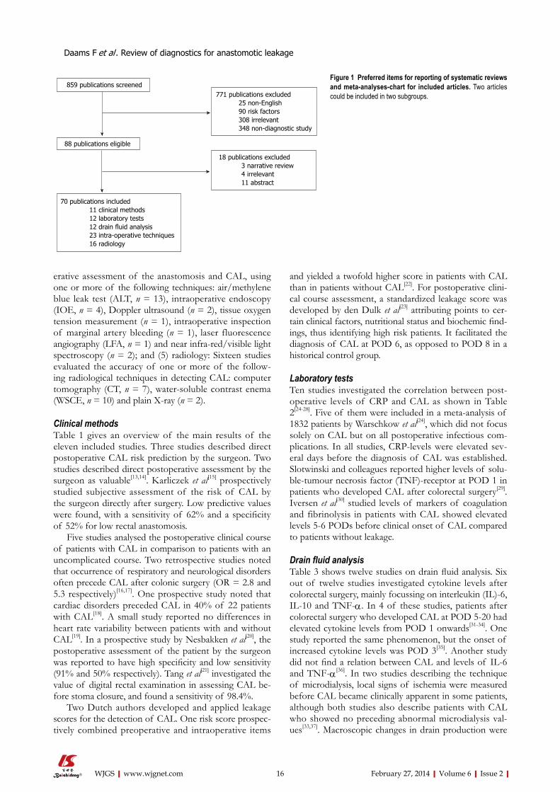
erative assessment of the anastomosis and CAL, using one or more of the following techniques: air/methylene blue leak test (ALT, n = 13), intraoperative endoscopy (IOE, n = 4), Doppler ultrasound (n = 2), tissue oxygen tension measurement (n = 1), intraoperative inspection of marginal artery bleeding (n = 1), laser fluorescence angiography (LFA, n = 1) and near infra-red/visible light spectroscopy (n = 2); and (5) radiology: Sixteen studies evaluated the accuracy of one or more of the follow-ing radiological techniques in detecting CAL: computer tomography (CT, n = 7), water-soluble contrast enema (WSCE, n = 10) and plain X-ray (n = 2).
Clinical methodsTable 1 gives an overview of the main results of the eleven included studies. Three studies described direct postoperative CAL risk prediction by the surgeon. Two studies described direct postoperative assessment by the surgeon as valuable[13,14]. Karliczek et al[15] prospectively studied subjective assessment of the risk of CAL by the surgeon directly after surgery. Low predictive values were found, with a sensitivity of 62% and a specificity of 52% for low rectal anastomosis.
Five studies analysed the postoperative clinical course of patients with CAL in comparison to patients with an uncomplicated course. Two retrospective studies noted that occurrence of respiratory and neurological disorders often precede CAL after colonic surgery (OR = 2.8 and 5.3 respectively)[16,17]. One prospective study noted that cardiac disorders preceded CAL in 40% of 22 patients with CAL[18]. A small study reported no differences in heart rate variability between patients with and without CAL[19]. In a prospective study by Nesbakken et al[20], the postoperative assessment of the patient by the surgeon was reported to have high specificity and low sensitivity (91% and 50% respectively). Tang et al[21] investigated the value of digital rectal examination in assessing CAL be-fore stoma closure, and found a sensitivity of 98.4%.
Two Dutch authors developed and applied leakage scores for the detection of CAL. One risk score prospec-tively combined preoperative and intraoperative items
and yielded a twofold higher score in patients with CAL than in patients without CAL[22]. For postoperative clini-cal course assessment, a standardized leakage score was developed by den Dulk et al[23] attributing points to cer-tain clinical factors, nutritional status and biochemic find-ings, thus identifying high risk patients. It facilitated the diagnosis of CAL at POD 6, as opposed to POD 8 in a historical control group.
Laboratory testsTen studies investigated the correlation between post-operative levels of CRP and CAL as shown in Table 2[24-28]. Five of them were included in a meta-analysis of 1832 patients by Warschkow et al[24], which did not focus solely on CAL but on all postoperative infectious com-plications. In all studies, CRP-levels were elevated sev-eral days before the diagnosis of CAL was established. Slotwinski and colleagues reported higher levels of solu-ble-tumour necrosis factor (TNF)-receptor at POD 1 in patients who developed CAL after colorectal surgery[29]. Iversen et al[30] studied levels of markers of coagulation and fibrinolysis in patients with CAL showed elevated levels 5-6 PODs before clinical onset of CAL compared to patients without leakage.
Drain fluid analysisTable 3 shows twelve studies on drain fluid analysis. Six out of twelve studies investigated cytokine levels after colorectal surgery, mainly focussing on interleukin (IL)-6, IL-10 and TNF-α. In 4 of these studies, patients after colorectal surgery who developed CAL at POD 5-20 had elevated cytokine levels from POD 1 onwards[31-34]. One study reported the same phenomenon, but the onset of increased cytokine levels was POD 3[35]. Another study did not find a relation between CAL and levels of IL-6 and TNF-α[36]. In two studies describing the technique of microdialysis, local signs of ischemia were measured before CAL became clinically apparent in some patients, although both studies also describe patients with CAL who showed no preceding abnormal microdialysis val-ues[33,37]. Macroscopic changes in drain production were
February 27, 2014|Volume 6|Issue 2|WJGS|www.wjgnet.com 16
859 publications screened
88 publications eligible
70 publications included 11 clinical methods 12 laboratory tests 12 drain fluid analysis 23 intra-operative techniques 16 radiology
771 publications excluded 25 non-English 90 risk factors 308 irrelevant 348 non-diagnostic study
18 publications excluded 3 narrative review 4 irrelevant 11 abstract
Figure 1 Preferred items for reporting of systematic reviews and meta-analyses-chart for included articles. Two articles could be included in two subgroups.
Daams F et al . Review of diagnostics for anastomotic leakage
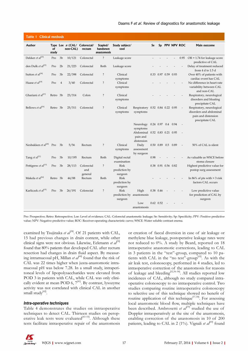
February 27, 2014|Volume 6|Issue 2|WJGS|www.wjgnet.com
or creation of faecal diversion in case of air leakage or methylene blue leakage, postoperative leakage rates were not reduced to 0%. A study by Beard, reported on 18 intraoperative anastomotic corrections, leading to CAL in 3 patients in the “test”-group, compared to 10 pa-tients with CAL in the “no test”-group[43]. As with the air leak test, colonoscopy, performed in 4 studies, led to intraoperative correction of the anastomosis for reasons of leakage and bleeding[52,56-58]. All studies reported low incidences of CAL, although no study compared intra-operative colonoscopy to no intraoperative control. Two studies comparing routine intraoperative colonoscopy to selective use of this technique showed no benefit of routine application of this technique[57,58]. For assessing local anastomotic blood flow, multiple techniques have been described. Ambrosetti et al[59] studied the use of Doppler intraoperatively at the site of the anastomosis, enabling correction of the anastomosis in 10 of 200 patients, leading to CAL in 2 (1%). Vignali et al[60] found
examined by Tsujinaka et al[38]. Of 21 patients with CAL, 15 had previous changes in drain content, while other clinical signs were not obvious. Likewise, Eckmann et al[39] found that 80% patients that developed CAL after rectum resection had changes in drain fluid aspect. By measur-ing intramucosal pH, Millan et al[40] found that the risk of CAL was 22 times higher when juxta-anastomotic intra-mucosal pH was below 7.28. In a small study, intraperi-toneal levels of lipopolysaccharides were elevated from POD 3 in patients with CAL, while CAL was only clini-cally evident at mean POD 6, 7[41]. By contrast, lysozyme activity was not correlated with clinical CAL in another small study[42].
Intra-operative techniquesTable 4 demonstrates the studies on intraoperative techniques to detect CAL. Thirteen studies on perop-erative leak tests were evaluated[43-55]. Although these tests facilitate intraoperative repair of the anastomosis
17
Author Type of
study
Loe n (CAL/non-CAL)
Colorectal/rectum
Stapled/handsewn
anastomosis
Study subject/tool
Se Sp PPV NPV ROC Main outcome
Dekker et al[22] Pro 3b 10/121 Colorectal ? Leakage score - - - - 0.95 OR = 1.74 for leakage score predictive of CAL
den Dulk et al[23] Pro 2b 21/223 Colorectal Both Leakage score - - - - - Delay of treatment reduced from 4 d to 1.5 d
Sutton et al[18] Pro 3b 22/398 Colorectal ? Clinical symptoms
0.33 0.97 0.59 0.93 Over 40% of patients with cardiac event has CAL
Haase et al[19] Pro 4 3/40 Colorectal ? Clinical symptoms
- - - - - No difference in heart rate variability between CAL
and non-CAL Ghariani et al[17] Retro 3b 23/314 Colon ? Clinical
symptoms- - - - - Respiratory, neurological
disorders and bloating precipitate CAL
Bellows et al[16] Retro 3b 25/311 Colorectal ? Clinical symptoms
Respiratory symptoms
0.52 0.84 0.22 0.95 - Respiratory, neurological disorders and abdominal
pain and distension precipitate CAL
Neurology symptoms
0.24 0.97 0.4 0.94 -
Abdominal pain and
distension
0.52 0.83 0.21 0.95 -
Nesbakken et al[20] Pro 3b 5/56 Rectum ? Clinical symptoms
Daily assessment by surgeon
0.50 0.89 0.5 0.89 - 50% of CAL is silent
Tang et al[21] Pro 3b 10/195 Rectum Both Digital rectal examination
0.98 - - - - As valuable as WSCE before stoma closure
Pettigrew et al[13] Pro 3b 28/113 Colorectal and
general
? Risk prediction by
surgeon
0.38 0.91 0.56 0.82 Highest predictive value for postop surg assessment
Makela et al[14] Retro 3b 44/88 Rectum Both Risk prediction by
surgeon
- - - - - In 86% of pts with > 3 risk factors CAL occurs
Karliczek et al[15] Pro 3b 26/191 Colorectal ? Risk prediction by
surgeon
High anastomosis
0.38 0.46 - - - Low predictive value for prediction of CAL by
surgeonLow
anastomosis0.62 0.52 - - -
Table 1 Clinical methods
Pro: Prospective; Retro: Retrospective; Loe: Level of evidence; CAL: Colorectal anastomotic leakage; Se: Sensitivity; Sp: Specificity; PPV: Positive predictive value; NPV: Negative predictive value; ROC: Receiver-operating characteristic curve; WSCE: Water soluble contrast enema.
Daams F et al . Review of diagnostics for anastomotic leakage
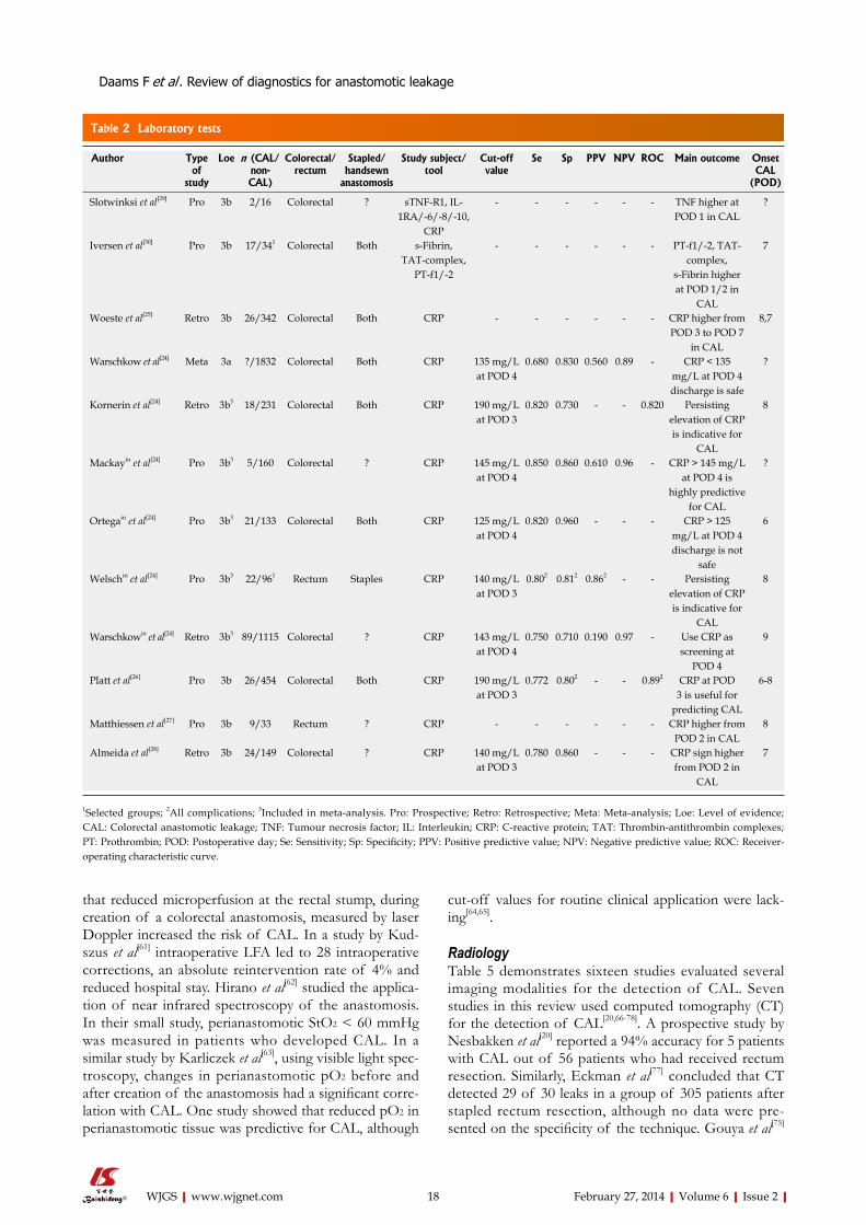
February 27, 2014|Volume 6|Issue 2|WJGS|www.wjgnet.com
cut-off values for routine clinical application were lack-ing[64,65].
RadiologyTable 5 demonstrates sixteen studies evaluated several imaging modalities for the detection of CAL. Seven studies in this review used computed tomography (CT) for the detection of CAL[20,66-78]. A prospective study by Nesbakken et al[20] reported a 94% accuracy for 5 patients with CAL out of 56 patients who had received rectum resection. Similarly, Eckman et al[77] concluded that CT detected 29 of 30 leaks in a group of 305 patients after stapled rectum resection, although no data were pre-sented on the specificity of the technique. Gouya et al[75]
that reduced microperfusion at the rectal stump, during creation of a colorectal anastomosis, measured by laser Doppler increased the risk of CAL. In a study by Kud-szus et al[61] intraoperative LFA led to 28 intraoperative corrections, an absolute reintervention rate of 4% and reduced hospital stay. Hirano et al[62] studied the applica-tion of near infrared spectroscopy of the anastomosis. In their small study, perianastomotic StO2 < 60 mmHg was measured in patients who developed CAL. In a similar study by Karliczek et al[63], using visible light spec-troscopy, changes in perianastomotic pO2 before and after creation of the anastomosis had a significant corre-lation with CAL. One study showed that reduced pO2 in perianastomotic tissue was predictive for CAL, although
18
Author Type of
study
Loe n (CAL/non-CAL)
Colorectal/rectum
Stapled/handsewn
anastomosis
Study subject/tool
Cut-off value
Se Sp PPV NPV ROC Main outcome Onset CAL
(POD)
Slotwinksi et al[29] Pro 3b 2/16 Colorectal ? sTNF-R1, IL-1RA/-6/-8/-10,
CRP
- - - - - - TNF higher at POD 1 in CAL
?
Iversen et al[30] Pro 3b 17/341 Colorectal Both s-Fibrin, TAT-complex,
PT-f1/-2
- - - - - - PT-f1/-2, TAT-complex,
s-Fibrin higher at POD 1/2 in
CAL
7
Woeste et al[25] Retro 3b 26/342 Colorectal Both CRP - - - - - - CRP higher from POD 3 to POD 7
in CAL
8,7
Warschkow et al[24] Meta 3a ?/1832 Colorectal Both CRP 135 mg/L at POD 4
0.680 0.830 0.560 0.89 - CRP < 135 mg/L at POD 4 discharge is safe
?
Kornerin et al[24] Retro 3b3 18/231 Colorectal Both CRP 190 mg/L at POD 3
0.820 0.730 - - 0.820 Persisting elevation of CRP is indicative for
CAL
8
Mackayin et al[24] Pro 3b3 5/160 Colorectal ? CRP 145 mg/L at POD 4
0.850 0.860 0.610 0.96 - CRP > 145 mg/L at POD 4 is
highly predictive for CAL
?
Ortegain et al[24] Pro 3b3 21/133 Colorectal Both CRP 125 mg/L at POD 4
0.820 0.960 - - - CRP > 125 mg/L at POD 4 discharge is not
safe
6
Welschin et al[24] Pro 3b3 22/961 Rectum Staples CRP 140 mg/L at POD 3
0.802 0.812 0.862 - - Persisting elevation of CRP is indicative for
CAL
8
Warschkowin et al[24] Retro 3b3 89/1115 Colorectal ? CRP 143 mg/L at POD 4
0.750 0.710 0.190 0.97 - Use CRP as screening at
POD 4
9
Platt et al[26] Pro 3b 26/454 Colorectal Both CRP 190 mg/L at POD 3
0.772 0.802 - - 0.892 CRP at POD 3 is useful for
predicting CAL
6-8
Matthiessen et al[27] Pro 3b 9/33 Rectum ? CRP - - - - - - CRP higher from POD 2 in CAL
8
Almeida et al[28] Retro 3b 24/149 Colorectal ? CRP 140 mg/L at POD 3
0.780 0.860 - - - CRP sign higher from POD 2 in
CAL
7
Table 2 Laboratory tests
1Selected groups; 2All complications; 3Included in meta-analysis. Pro: Prospective; Retro: Retrospective; Meta: Meta-analysis; Loe: Level of evidence; CAL: Colorectal anastomotic leakage; TNF: Tumour necrosis factor; IL: Interleukin; CRP: C-reactive protein; TAT: Thrombin-antithrombin complexes; PT: Prothrombin; POD: Postoperative day; Se: Sensitivity; Sp: Specificity; PPV: Positive predictive value; NPV: Negative predictive value; ROC: Receiver-operating characteristic curve.
Daams F et al . Review of diagnostics for anastomotic leakage
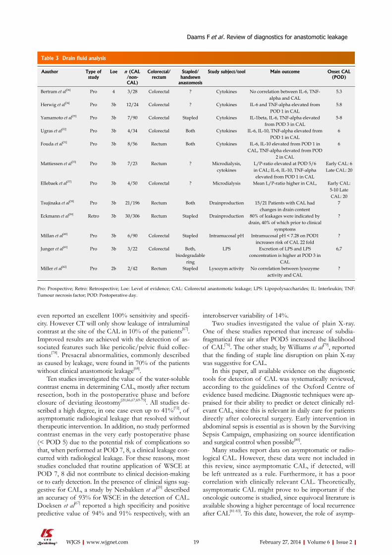
February 27, 2014|Volume 6|Issue 2|WJGS|www.wjgnet.com
interobserver variability of 14%.Two studies investigated the value of plain X-ray.
One of these studies reported that increase of subdia-fragmatical free air after POD5 increased the likelihood of CAL[76]. The other study, by Williams et al[79], reported that the finding of staple line disruption on plain X-ray was suggestive for CAL.
In this paper, all available evidence on the diagnostic tools for detection of CAL was systematically reviewed, according to the guidelines of the Oxford Centre of evidence based medicine. Diagnostic techniques were ap-praised for their ability to predict or detect clinically rel-evant CAL, since this is relevant in daily care for patients directly after colorectal surgery. Early intervention in abdominal sepsis is essential as is shown by the Surviving Sepsis Campaign, emphasizing on source identification and surgical control when possible[80].
Many studies report data on asymptomatic or radio-logical CAL. However, these data were not included in this review, since asymptomatic CAL, if detected, will be left untreated as a rule. Furthermore, it has a poor correlation with clinically relevant CAL. Theoretically, asymptomatic CAL might prove to be important if the oncologic outcome is studied, since equivocal literature is available showing a higher percentage of local recurrence after CAL[81-83]. To this date, however, the role of asymp-
even reported an excellent 100% sensitivity and specifi-city. However CT will only show leakage of intraluminal contrast at the site of the CAL in 10% of the patients[67]. Improved results are achieved with the detection of as-sociated features such like pericolic/pelvic fluid collec-tions[78]. Presacral abnormalities, commonly described as caused by leakage, were found in 70% of the patients without clinical anastomotic leakage[68].
Ten studies investigated the value of the water-soluble contrast enema in determining CAL, mostly after rectum resection, both in the postoperative phase and before closure of deviating ileostomy[20,66,67,69-75]. All studies de-scribed a high degree, in one case even up to 41%[72], of asymptomatic radiological leakage that resolved without therapeutic intervention. In addition, no study performed contrast enemas in the very early postoperative phase (< POD 5) due to the potential risk of complications so that, when performed at POD 7, 8, a clinical leakage con-curred with radiological leakage. For these reasons, most studies concluded that routine application of WSCE at POD 7, 8 did not contribute to clinical decision-making or to early detection. In the presence of clinical signs sug-gestive for CAL, a study by Nesbakken et al[20] described an accuracy of 93% for WSCE in the detection of CAL. Doeksen et al[67] reported a high specificity and positive predictive value of 94% and 91% respectively, with an
19
Aauthor Type of study
Loe n (CAL /non-CAL)
Colorectal/rectum
Stapled/handsewn
anastomosis
Study subject/tool Main outcome Onset CAL (POD)
Bertram et al[36] Pro 4 3/28 Colorectal ? Cytokines No correlation between IL-6, TNF-alpha and CAL
5.3
Herwig et al[34] Pro 3b 12/24 Colorectal ? Cytokines IL-6 and TNF-alpha elevated from POD 1 in CAL
5.8
Yamamoto et al[35] Pro 3b 7/90 Colorectal Stapled Cytokines IL-1beta, IL-6, TNF-alpha elevated from POD 3 in CAL
5-8
Ugras et al[32] Pro 3b 4/34 Colorectal Both Cytokines IL-6, IL-10, TNF-alpha elevated from POD 1 in CAL
6
Fouda et al[31] Pro 3b 8/56 Rectum Both Cytokines IL-6, IL-10 elevated from POD 1 in CAL, TNF-alpha elevated from POD
2 in CAL
6
Mattiessen et al[33] Pro 3b 7/23 Rectum ? Microdialysis, cytokines
L/P-ratio elevated at POD 5/6 in CAL; IL-6, IL-10, TNF-alpha elevated from POD 1 in CAL
Early CAL: 6 Late CAL: 20
Ellebaek et al[37] Pro 3b 4/50 Colorectal ? Microdialysis Mean L/P-ratio higher in CAL, Early CAL: 5-10 Late CAL: 20
Tsujinaka et al[38] Pro 3b 21/196 Rectum Both Drainproduction 15/21 Patients with CAL had changes in drain content
7
Eckmann et al[39] Retro 3b 30/306 Rectum Stapled Drainproduction 80% of leakages were indicated by drain, 40% of which prior to clinical
symptoms
?
Millan et al[40] Pro 3b 6/90 Colorectal Stapled Intramucosal pH Intramucosal pH < 7.28 on POD1 increases risk of CAL 22 fold
?
Junger et al[41] Pro 3b 3/22 Colorectal Both, biodegradable
ring
LPS Excretion of LPS and LPS concentration is higher at POD 3 in
CAL
6,7
Miller et al[42] Pro 2b 2/42 Rectum Stapled Lysozym activity No correlation between lysozyme activity and CAL
?
Table 3 Drain fluid analysis
Pro: Prospective; Retro: Retrospective; Loe: Level of evidence; CAL: Colorectal anastomotic leakage; LPS: Lipopolysaccharides; IL: Interleukin; TNF: Tumour necrosis factor; POD: Postoperative day.
Daams F et al . Review of diagnostics for anastomotic leakage
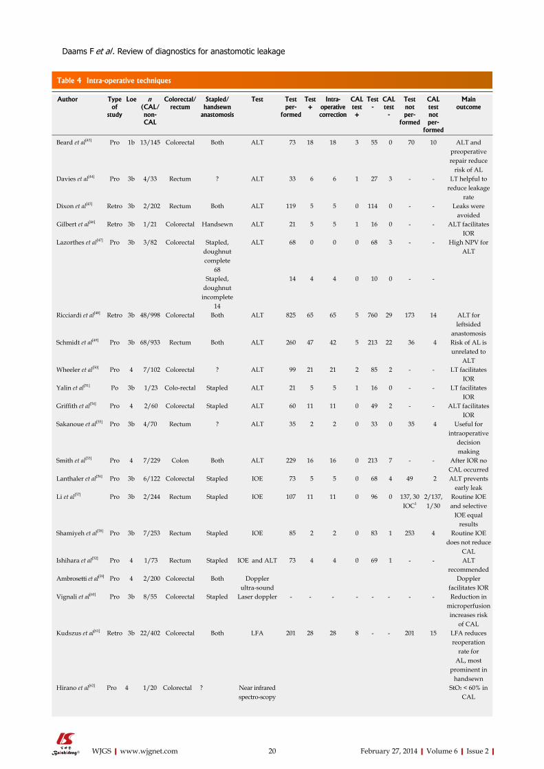
February 27, 2014|Volume 6|Issue 2|WJGS|www.wjgnet.com 20
Author Type of
study
Loe n (CAL/non-CAL
Colorectal/rectum
Stapled/handsewn
anastomosis
Test Test per-
formed
Test +
Intra-operative correction
CAL test +
Test -
CAL test-
Test not per-
formed
CAL test not per-
formed
Main outcome
Beard et al[43] Pro 1b 13/145 Colorectal Both ALT 73 18 18 3 55 0 70 10 ALT and preoperative repair reduce
risk of AL Davies et al[44] Pro 3b 4/33 Rectum ? ALT 33 6 6 1 27 3 - - LT helpful to
reduce leakage rate
Dixon et al[45] Retro 3b 2/202 Rectum Both ALT 119 5 5 0 114 0 - - Leaks were avoided
Gilbert et al[46] Retro 3b 1/21 Colorectal Handsewn ALT 21 5 5 1 16 0 - - ALT facilitates IOR
Lazorthes et al[47] Pro 3b 3/82 Colorectal Stapled, doughnut complete
68
ALT 68 0 0 0 68 3 - - High NPV for ALT
Stapled, doughnut
incomplete 14
14 4 4 0 10 0 - -
Ricciardi et al[48] Retro 3b 48/998 Colorectal Both ALT 825 65 65 5 760 29 173 14 ALT for leftsided
anastomosis Schmidt et al[49] Pro 3b 68/933 Rectum Both ALT 260 47 42 5 213 22 36 4 Risk of AL is
unrelated to ALT
Wheeler et al[50] Pro 4 7/102 Colorectal ? ALT 99 21 21 2 85 2 - - LT facilitates IOR
Yalin et al[51] Po 3b 1/23 Colo-rectal Stapled ALT 21 5 5 1 16 0 - - LT facilitates IOR
Griffith et al[54] Pro 4 2/60 Colorectal Stapled ALT 60 11 11 0 49 2 - - ALT facilitates IOR
Sakanoue et al[55] Pro 3b 4/70 Rectum ? ALT 35 2 2 0 33 0 35 4 Useful for intraoperative
decision making
Smith et al[53] Pro 4 7/229 Colon Both ALT 229 16 16 0 213 7 - - After IOR no CAL occurred
Lanthaler et al[56] Pro 3b 6/122 Colorectal Stapled IOE 73 5 5 0 68 4 49 2 ALT prevents early leak
Li et al[57] Pro 3b 2/244 Rectum Stapled IOE 107 11 11 0 96 0 137, 30 IOC1
2/137, 1/30
Routine IOE and selective
IOE equal results
Shamiyeh et al[58] Pro 3b 7/253 Rectum Stapled IOE 85 2 2 0 83 1 253 4 Routine IOE does not reduce
CAL Ishihara et al[52] Pro 4 1/73 Rectum Stapled IOE and ALT 73 4 4 0 69 1 - - ALT
recommended Ambrosetti et al[59] Pro 4 2/200 Colorectal Both Doppler
ultra-soundDoppler
facilitates IOR Vignali et al[60] Pro 3b 8/55 Colorectal Stapled Laser doppler - - - - - - - - Reduction in
microperfusion increases risk
of CAL Kudszus et al[61] Retro 3b 22/402 Colorectal Both LFA 201 28 28 8 - - 201 15 LFA reduces
reoperation rate for
AL, most prominent in
handsewn Hirano et al[62] Pro 4 1/20 Colorectal ? Near infrared
spectro-scopyStO2 < 60% in
CAL
Table 4 Intra-operative techniques
Daams F et al . Review of diagnostics for anastomotic leakage
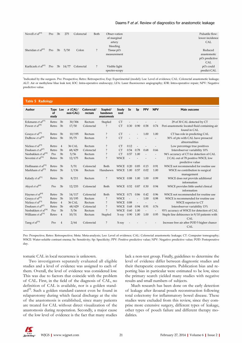
February 27, 2014|Volume 6|Issue 2|WJGS|www.wjgnet.com
lack a non-test group. Finally, guidelines to determine the level of evidence differ between diagnostic studies and their therapeutic counterparts. Publication bias and re-porting bias in particular were estimated to be low, since the primary search yielded many studies with negative results and small numbers of subjects.
Much research has been done on the early detection of leakage after ileoanal pouch reconstruction following total colectomy for inflammatory bowel disease. These studies were excluded from this review, since they com-prise more extensive surgery, different types of leakage, other types of pouch failure and different therapy mo-dalities.
tomatic CAL in local recurrence is unknown.Two investigators separately evaluated all eligible
studies and a level of evidence was assigned to each of them. Overall, the level of evidence was considered low. This was due to factors that coincide with the problem of CAL. First, in the field of the diagnosis of CAL, no definition of CAL is available, nor is a golden stand-ard[3]. Such a golden standard cannot even be found in relaparotomy during which faecal discharge at the site of the anastomosis is established, since many patients are treated for CAL without direct visualization of the anastomosis during reoperation. Secondly, a major cause of the low level of evidence is the fact that many studies
21
Novell et al[64] Pro 3b 275 Colorectal Both Obser-vation of marginal
artery bleeding
Pulsatile flow: lower incidence
CAL
Sheridan et al[65] Pro 3b 5/50 Colon ? Tissue pO2 measurement
Reduced anastomotic
pO2 predictive CAL
Karliczek et al[63] Pro 3b 14/77 Colorectal ? Visible light spectro-scopy
pO2 could predict CAL
1Indicated by the surgeon. Pro: Prospective; Retro: Retrospective; Exp: Experimental (model); Loe: Level of evidence; CAL: Colorectal anastomotic leakage; ALT: Air or methylene blue leak test; IOC: Intra-operative endoscopy; LFA: Laser fluorescence angiography; IOR: Intra-operative repair; NPV: Negative predictive value.
Author Type of
study
Loe n (CAL/non-CAL)
Colorectal/rectum
Stapled/handsewn
anastomosis
Study tool
Se Sp PPV NPV Main outcome
Eckmann et al[77] Retro 3b 30/306 Rectum Stapled CT - - - - 29 of 30 CAL detected by CT Power et al[78] Retro 3b 17/50 Colorectal ? CT 0.30 0.90 0.58 0.74 Peri-anastomotic located fluid containing air
found in CAL Gouya et al[75] Retro 3b 10/195 Rectum ? CT - - 1.00 1.00 CT has role in predicting CAL DuBrow et al[68] Retro 3b 35/75 Rectum ? CT - - - - 30% of pts with CAL have presacral
abnormalities Nicksa et al[73] Retro 4 36 CAL Rectum ? CT 0.12 - - - Low percentage true positives Doeksen et al[67] Retro 3b 68/429 Colorectal ? CT 0.54 0.78 0.68 0.66 Interobserver variability 10% Nesbakken et al[20] Pro 3b 5/56 Rectum ? CT 0.57 1.00 - - 94% accuracy of CT for detection of CAL Severini et al[74] Retro 3b 12/175 Rectum ? WSCE - - - - 2 CAL out of 78 positive WSCE, low
predictive value Hoffmann et al[70] Retro 3b 5/51 Colorectal Both WSCE 0.20 0.85 0.13 0.91 WSCE not recommended for routine use Markham et al[72] Retro 3b 1/136 Rectum Handsewn WSCE 1.00 0.57 0.02 1.00 WSCE no contribution to surgical
management Kalady et al[71] Retro 3b 8/211 Rectum ? WSCE 0.88 1.00 1.00 0.99 WSCE does not provide additional
information Akyol et al[66] Pro 3b 12/233 Colorectal Both WSCE 0.52 0.87 0.30 0.94 WSCE provides little useful clinical
information Haynes et al[69] Retro 3b 14/117 Colorectal Both WSCE 0.71 0.86 0.42 0.96 WSCE not recommended for routine use Gouya et al[75] Retro 3b 10/195 Rectum ? WSCE - - 1.00 0,98 WSCE is recommended for routine use Nicksa et al[73] Retro 4 36 CAL Rectum ? WSCE 0.88 - - - WSCE superior to CT Doeksen et al[67] Retro 3b 68/429 Colorectal ? WSCE 0.68 0.94 0.91 0.76 Interobserver variability 13% Nesbakken et al[20] Pro 3b 5/56 Rectum ? WSCE 0.60 1.00 - - 93% accuracy of WSCE for detection of CAL Williams et al[76] Retro 4 10/31 Rectum Stapled X-ray 0.90 1.00 1.00 0.95 Staple line dehiscence in 9/10 patients with
CAL Tang et al[79] Pro 4 2/64 Colorectal ? X-ray - - - - Increase free air after POD 5 higher chance
CAL
Table 5 Radiology
Pro: Prospective; Retro: Retrospective; Meta: Meta-analysis; Leo: Level of evidence; CAL: Colorectal anastomotic leakage; CT: Computer tomography; WSCE: Water-soluble contrast enema; Se: Sensitivity; Sp: Specificity; PPV: Positive predictive value; NPV: Negative predictive value; POD: Postoperative day.
Daams F et al . Review of diagnostics for anastomotic leakage
February 27, 2014|Volume 6|Issue 2|WJGS|www.wjgnet.com
Other laboratory tests like coagulation factors and cy-tokines show a correlation with occurrence of CAL, but they have been studied sparsely. Since no parameters for their predictive value can be calculated from the available data, there is no basis for incorporating them in the stan-dard postoperative lab tests.
Drain fluid analysisIn this review, the results for cytokine levels in perito-neal drain fluid, as biomarkers for local infection, seem promising. In most studies cytokine levels were elevated from POD 1 in patients with CAL compared to patients without CAL. This finding suggests an early onset of local infection in patients with CAL, or at least a more prominent postoperative reaction in this group. It is hy-pothesised that cytokines are directly elevated postopera-tively and will normalise unless infectious complications occur. Most frequently investigated cytokines are IL-1, IL-6, IL-10 and TNF-α.
Although routine drainage after colorectal surgery does not seem to prevent CAL and is omitted in en-hanced recovery programs, two studies showed that changes of drain production occur frequently and before clinical symptoms. These interesting findings might jus-tify the routine placement of a drain for the first postop-erative days as an indicator for CAL.
Two studies on intraperitoneal microdialysis show, by retrospectively analysing of peritoneal microdialysis sam-ples, that CAL was preceded by changes in local lactate/pyruvate ratio. Although these findings are promising, patient numbers were too low to compute predictive val-ues and cut-off values. Future research should elucidate if prospective, real-time analysis actually leads to early detection and determine whether this technique is cost effective.
For intramucosal pH monitoring, as a measure for mucosal hypoperfusion and subsequent hypoxia, data are limited but promising. The same holds for measurement of lipopolysaccharides, integral components of normal gut flora, and measurements of lysozym in drain fluid, since the studies investigating these biomarkers did nei-ther lead neither to confirmation of these techniques nor to a re-evaluation.
Intra-operative techniquesExcept for one, all studies evaluating the ALT confirm the importance of this simple intervention. Although not completely eliminating the occurrence of CAL, ALT allows intraoperative revision of the anastomosis, is easy to perform and has a high negative predictive value. Un-derstandably, no studies have been performed that relate a positive ALT without intraoperative repair to CAL. All valuable studies, those that use a no-test control group, show a lower percentage of CAL in the group in which ALT was performed; in two out of four papers this dif-ference was significant.
IOE can, apart from direct visualisation of CAL, be of diagnostic and therapeutical importance if the loca-tion of the tumour or of additional lesions is unknown
Clinical methodsClinical factors are objective and easily available for risk prediction. A few problems, however, occur if surgeons rely solely on clinical factors. First, the influence of in-dividual factors is not exactly known. Secondly, by the time signs of septicaemia occur; patients will be in a worse clinical state at the onset of an often prolonged and onerous therapeutic course. Subjective prognosis of leakage at the moment of finishing the anastomosis was proven to have a limited prognostic value[15]. Ob-jective measurements might be of greater prognostic value, as shown by the Colon Leakage Score, in which the presence of objective risk factors leads to a higher score representing a higher chance of CAL[22]. This leak-age score was based on previously identified risk factors and to our knowledge is the first to translate all available literature on risk factors for CAL into an instrument that can easily be implemented in daily practice. In a cohort of 233 patients, using a historical control group of 1066 patients, den Dulk et al[23] developed a similar score system for postoperative clinical evaluation of the colorectal patient. When a high score is found, computer tomography using rectal contrast is warranted. Although this promising method has shown to reduce delay in di-agnosis, no information was provided on the prognostic value of this risk score, nor did the study mention the number of CT-scans and concomitant negative results In a study on tracking of surgical site infections (SSI), van Ramshorst et al[84] found that protocolled tracking yields a higher reported incidence of SSI than self-reported de-tection. We believe that this finding could be applied to the protocolled detection of CAL as described above, as it contributes to increased awareness and early detection.Little is known about the value of physical examination in relation to CAL, except that digital rectal examination has at least the same prognostic value for low anastomo-sis as contrast enema prior to stoma reversal.
Laboratory testsMany investigators have studied the behaviour of CRP during the subclinical phase of CAL. CRP has the ca-pacity to rise quickly after the onset of an inflammatory stimulus, reaching its highest serum level within 48 h. Since it has a short halftime of around 19 h, a drop in CRP corresponds well with the removal of the stimu-lus. Most studies investigating CRP used cut-off values of around 120-190 mg/L at POD 3, 4, and all studies in this review showed a reasonable predictive value of CRP for CAL. Drawbacks of all studies described in this review is that the number of included patients per study is rather small and that none of these studies provide a protocol that structurally describes the postoperative clinical examination, the clinical state of the patients during postoperative follow-up and the type of CAL (i.e., faecal peritonitis, juxta-anastomotic abscess, rectovaginal fistula). Despite these drawbacks, we believe that these studies have indeed shown that measurement of CRP is of great importance in detecting CAL in the preclinical phase.
22
Daams F et al . Review of diagnostics for anastomotic leakage
February 27, 2014|Volume 6|Issue 2|WJGS|www.wjgnet.com
reference; therefore the general level of evidence is rela-tively low. The air leak test is recommended for intraop-erative assessment of CAL. When a leakage score system is used intraoperatively, preoperative preventive measures can be taken. When using a clinical algorithm postopera-tively, delay in diagnosis of CAL might be reduced. CRP measurement should be part of postoperative labora-tory routine at least at POD 3 and 4, since due to a high negative predictive value patients with an uncomplicated course can be identified. Cytokine measurement among other measurements of peritoneal drain fluid is promis-ing and could justify the routine placement of a juxta-anastomotic drain, while peritoneal microdialysis might develop as minimally invasive peritoneal “smart”-drain. When clinical signs are present, CT with rectal contrast is recommended. CT cannot only to detect CAL but also can be used as a therapeutic instrument for percutane-ous drainage of a pericolic/pelvic abscess. We believe that this review reaffirms the importance of early detec-tion of colorectal anastomotic leakage and that it offers colorectal surgeons an overview on easily applicable di-agnostic tools to improve early detection.
ACKNOWLEDGMENTSThe authors would like to thank Wichor Bramer, Bio-medical Information Specialist at the Medical Library at the ErasmusMC Rotterdam the Netherlands, for his contribution during the collection of data.
REFERENCES1 Alves A, Panis Y, Pocard M, Regimbeau JM, Valleur P.
Management of anastomotic leakage after nondiverted large bowel resection. J Am Coll Surg 1999; 189: 554-559 [PMID: 10589591]
2 Platell C, Barwood N, Dorfmann G, Makin G. The incidence of anastomotic leaks in patients undergoing colorectal sur-gery. Colorectal Dis 2007; 9: 71-79 [PMID: 17181849 DOI: 10.1111/j.1463-1318.2006.01002.x]
3 Bruce J, Krukowski ZH, Al-Khairy G, Russell EM, Park KG. Systematic review of the definition and measurement of anastomotic leak after gastrointestinal surgery. Br J Surg 2001; 88: 1157-1168 [PMID: 11531861 DOI: 10.1046/j.0007-1323.2001.01829.x]
4 Rahbari NN, Weitz J, Hohenberger W, Heald RJ, Moran B, Ulrich A, Holm T, Wong WD, Tiret E, Moriya Y, Laurberg S, den Dulk M, van de Velde C, Büchler MW. Definition and grading of anastomotic leakage following anterior resection of the rectum: a proposal by the International Study Group of Rectal Cancer. Surgery 2010; 147: 339-351 [PMID: 20004450 DOI: 10.1016/j.surg.2009.10.012]
5 Hyman N, Manchester TL, Osler T, Burns B, Cataldo PA. Anastomotic leaks after intestinal anastomosis: it’s later than you think. Ann Surg 2007; 245: 254-258 [PMID: 17245179 DOI: 10.1097/01.sla.0000225083.27182.85]
6 Komen N, Dijk JW, Lalmahomed Z, Klop K, Hop W, Klein-rensink GJ, Jeekel H, Ruud Schouten W, Lange JF. After-hours colorectal surgery: a risk factor for anastomotic leak-age. Int J Colorectal Dis 2009; 24: 789-795 [PMID: 19301016 DOI: 10.1007/s00384-009-0692-4]
7 Macarthur DC, Nixon SJ, Aitken RJ. Avoidable deaths still occur after large bowel surgery. Scottish Audit of Surgi-cal Mortality, Royal College of Surgeons of Edinburgh.
or if anastomotic bleeding occurs. More recently, the routine application of IOE has been studied in compari-son to selective IOE. No favourable results in occurrence of CAL were described for routinely performed IOE compared to selective IOE. Apart from the mentioned benefits of IOE, no data are available on the superiority of IOE compared to ALT for intraoperative diagnosis of anastomotic dehiscence. Thus, ALT seems to be favour-able to IOE since it is faster, easier and cheaper.
Some authors have attempted to relate anastomotic perfusion parameters to anastomotic leakage. Except for one, all studies are case controlled without reference and have not been repeated. It has not led to clear cut-off values for any of these techniques that seem not very practical in daily current practice. At least one cohort study with a good reference is needed before clinical implementation.
RadiologyAs far as CT with rectal contrast is concerned, only 7 studies could be included. These studies showed large differences in methodology and lacked generally applied definitions. These differences between several studies, especially in CT criteria for CAL, resulted in equivocal results. Intestinal contrast leakage is not regularly de-picted with CT in patients with CAL. However CT can accurately depict the associated features of anastomotic leakage such like pericolic/pelvic fluid collections and free air. When these additional criteria were used the accuracy improved dramatically with accuracies varying from 80%-100%.
All six studies that were performed on the subject of WSCE over the last two decades concluded that there is no place for routine application of WSCE. In these studies, WSCE did not have a consistently high positive predictive value, and other techniques, such as digital rectal examination in low rectal anastomosis, appeared to provide at least equal results. Furthermore due to the po-tential risk of complications no study performed contrast enemas in the very early postoperative phase. This means that, when performed at POD 7-8, clinical CAL con-curred with radiological leakage. In addition, radiologic signs of CAL do not correlate with clinical CAL and fre-quently do not require any form of treatment. Another drawback of WSCE is that the rectally administered contrast has been diluted and there may be not enough remaining pressure to induce contrast leakage in more proximal anastomoses.
Two older studies describe how plain X-rays can be used in assessment of intra-abdominal free air and staple line integrity in the diagnosis of CAL. Although some-times helpful, modern techniques offer the surgeon much more detailed information on the extend of CAL com-pared to plain X-rays.
CONCLUSIONMany studies have been performed in the field of diag-nosis of CAL. Many lack a no-test control group and
23
Daams F et al . Review of diagnostics for anastomotic leakage
February 27, 2014|Volume 6|Issue 2|WJGS|www.wjgnet.com
BM, Güller U, Tarantino I. Safe and early discharge after colorectal surgery due to C-reactive protein: a diagnostic meta-analysis of 1832 patients. Ann Surg 2012; 256: 245-250 [PMID: 22735714 DOI: 10.1097/SLA.0b013e31825b60f0]
25 Woeste G, Müller C, Bechstein WO, Wullstein C. Increased serum levels of C-reactive protein precede anastomotic leakage in colorectal surgery. World J Surg 2010; 34: 140-146 [PMID: 19953248 DOI: 10.1007/s00268-009-0304-z]
26 Platt JJ, Ramanathan ML, Crosbie RA, Anderson JH, McKee RF, Horgan PG, McMillan DC. C-reactive protein as a pre-dictor of postoperative infective complications after cura-tive resection in patients with colorectal cancer. Ann Surg Oncol 2012; 19: 4168-4177 [PMID: 22805866 DOI: 10.1245/s10434-012-2498-9]
27 Matthiessen P, Henriksson M, Hallböök O, Grunditz E, Norén B, Arbman G. Increase of serum C-reactive protein is an early indicator of subsequent symptomatic anastomotic leakage after anterior resection. Colorectal Dis 2008; 10: 75-80 [PMID: 17666099 DOI: 10.1111/j.1463-1318.2007.01300.x]
28 Almeida AB, Faria G, Moreira H, Pinto-de-Sousa J, Correia-da-Silva P, Maia JC. Elevated serum C-reactive protein as a predictive factor for anastomotic leakage in colorectal surgery. Int J Surg 2012; 10: 87-91 [PMID: 22222182 DOI: 10.1016/j.ijsu.2011.12.006]
29 Slotwiński R, Olszewski WL, Chaber A, Slodkowski M, Za-leska M, Krasnodebski IW. The soluble tumor necrosis factor receptor I is an early predictor of local infective complica-tions after colorectal surgery. J Clin Immunol 2002; 22: 289-296 [PMID: 12405162]
30 Iversen LH, Thomsen GH, Thorlacius-Ussing O. Systemic coagulation activation and anastomotic leakage after colorec-tal cancer surgery. Dis Colon Rectum 1999; 42: 56-65 [PMID: 10211521]
31 Fouda E, El Nakeeb A, Magdy A, Hammad EA, Othman G, Farid M. Early detection of anastomotic leakage after elective low anterior resection. J Gastrointest Surg 2011; 15: 137-144 [PMID: 20978948 DOI: 10.1007/s11605-010-1364-y]
32 Uğraş B, Giriş M, Erbil Y, Gökpinar M, Citlak G, Işsever H, Bozbora A, Oztezcan S. Early prediction of anastomotic leakage after colorectal surgery by measuring peritoneal cytokines: prospective study. Int J Surg 2008; 6: 28-35 [PMID: 18037067 DOI: 10.1016/j.ijsu.2007.10.001]
33 Matthiessen P, Strand I, Jansson K, Törnquist C, Andersson M, Rutegård J, Norgren L. Is early detection of anastomotic leakage possible by intraperitoneal microdialysis and intra-peritoneal cytokines after anterior resection of the rectum for cancer? Dis Colon Rectum 2007; 50: 1918-1927 [PMID: 17763907 DOI: 10.1007/s10350-007-9023-4]
34 Herwig R, Glodny B, Kühle C, Schlüter B, Brinkmann OA, Strasser H, Senninger N, Winde G. Early identification of peritonitis by peritoneal cytokine measurement. Dis Colon Rectum 2002; 45: 514-521 [PMID: 12006934 DOI: 10.1007/s10350-004-6231-z]
35 Yamamoto T, Umegae S, Matsumoto K, Saniabadi AR. Peritoneal cytokines as early markers of peritonitis follow-ing surgery for colorectal carcinoma: a prospective study. Cytokine 2011; 53: 239-242 [PMID: 21075004 DOI: 10.1016/j.cyto.2010.10.006]
36 Bertram P, Junge K, Schachtrupp A, Götze C, Kunz D, Schumpelick V. Peritoneal release of TNFalpha and IL-6 after elective colorectal surgery and anastomotic leakage. J Invest Surg 2003; 16: 65-69 [PMID: 12746189]
37 Ellebaek Pedersen M, Qvist N, Bisgaard C, Kelly U, Bern-hard A, Møller Pedersen S. Peritoneal microdialysis. Early diagnosis of anastomotic leakage after low anterior resection for rectosigmoid cancer. Scand J Surg 2009; 98: 148-154 [PMID: 19919919]
38 Tsujinaka S, Kawamura YJ, Konishi F, Maeda T, Mizokami K. Pelvic drainage for anterior resection revisited: use of drains in anastomotic leaks. ANZ J Surg 2008; 78: 461-465
Br J Surg 1998; 85: 80-83 [PMID: 9462390 DOI: 10.1046/j.1365-2168.1998.00554.x]
8 Canelas A, Bun M, Cabo JK, Laporte M, Peczan C, Rotholtz N. Risk factors associated to anastomotic leakage in lapa-roscopic colorectal surgery. Colorectal Dis 2010; 12: 37 [DOI: 10.1097/01.dcr.0000370595.66829.6d]
9 Isbister WH. Study populations and casemix: influence on analysis of postoperative outcomes. Aust N Z J Surg 2000; 70: 279-284 [PMID: 10779060]
10 Lai R, Lu Y, Li Q, Guo J, Chen G, Zeng W. Risk factors for anastomotic leakage following anterior resection for colorec-tal cancer: the effect of epidural analgesia on occurrence. Int J Colorectal Dis 2013; 28: 485-492 [PMID: 23014977 DOI: 10.1007/s00384-012-1585-5]
11 Warschkow R, Steffen T, Thierbach J, Bruckner T, Lange J, Tarantino I. Risk factors for anastomotic leakage after rectal cancer resection and reconstruction with colorectostomy. A retrospective study with bootstrap analysis. Ann Surg Oncol 2011; 18: 2772-2782 [PMID: 21468782 DOI: 10.1245/s10434-011-1696-1]
12 Doeksen A, Tanis PJ, Vrouenraets BC, Lanschot van JJ, Tets van WF. Factors determining delay in relaparotomy for anastomotic leakage after colorectal resection. World J Gastro-enterol 2007; 13: 3721-3725 [PMID: 17659732]
13 Pettigrew RA, Hill GL. Indicators of surgical risk and clini-cal judgement. Br J Surg 1986; 73: 47-51 [PMID: 3947877]
14 Mäkelä JT, Kiviniemi H, Laitinen S. Risk factors for anasto-motic leakage after left-sided colorectal resection with rectal anastomosis. Dis Colon Rectum 2003; 46: 653-660 [PMID: 12792443 DOI: 10.1097/01.DCR.0000059328.10563.8C]
15 Karliczek A, Harlaar NJ, Zeebregts CJ, Wiggers T, Baas PC, van Dam GM. Surgeons lack predictive accuracy for anastomotic leakage in gastrointestinal surgery. Int J Colorec-tal Dis 2009; 24: 569-576 [PMID: 19221768 DOI: 10.1007/s00384-009-0658-6]
16 Bellows CF, Webber LS, Albo D, Awad S, Berger DH. Early predictors of anastomotic leaks after colectomy. Tech Coloproctol 2009; 13: 41-47 [PMID: 19288246 DOI: 10.1007/s10151-009-0457-7]
17 Ghariani B, Houissa H, Sebai F. Early diagnosis of anasto-motic dehiscence after colonic surgery. Tunis Med 2011; 89: 174-178 [PMID: 21308627]
18 Sutton CD, Marshall LJ, Williams N, Berry DP, Thomas WM, Kelly MJ. Colo-rectal anastomotic leakage often masquerades as a cardiac complication. Colorectal Dis 2004; 6: 21-22 [PMID: 14692947 DOI: 10.1111/j.1463-1318.2004.00574.x]
19 Haase O, Langelotz C, Scharfenberg M, Schwenk W, Tsilim-paris N. Reduction of heart rate variability after colorectal resections. Langenbecks Arch Surg 2012; 397: 793-799 [PMID: 22249435 DOI: 10.1007/s00423-012-0903-2]
20 Nesbakken A, Nygaard K, Lunde OC, Blücher J, Gjertsen Ø, Dullerud R. Anastomotic leak following mesorectal exci-sion for rectal cancer: true incidence and diagnostic chal-lenges. Colorectal Dis 2005; 7: 576-581 [PMID: 16232238 DOI: 10.1111/j.1463-1318.2005.00870.x]
21 Tang CL, Seow-Choen F. Digital rectal examination com-pares favourably with conventional water-soluble contrast enema in the assessment of anastomotic healing after low rectal excision: a cohort study. Int J Colorectal Dis 2005; 20: 262-266 [PMID: 15455246 DOI: 10.1007/s00384-004-0652-y]
22 Dekker JW, Liefers GJ, de Mol van Otterloo JC, Putter H, Tollenaar RA. Predicting the risk of anastomotic leakage in left-sided colorectal surgery using a colon leakage score. J Surg Res 2011; 166: e27-e34 [PMID: 21195424]
23 den Dulk M, Noter SL, Hendriks ER, Brouwers MA, van der Vlies CH, Oostenbroek RJ, Menon AG, Steup WH, van de Velde CJ. Improved diagnosis and treatment of anastomotic leakage after colorectal surgery. Eur J Surg Oncol 2009; 35: 420-426 [PMID: 18585889 DOI: 10.1016/j.ejso.2008.04.009]
24 Warschkow R, Beutner U, Steffen T, Müller SA, Schmied
24
Daams F et al . Review of diagnostics for anastomotic leakage
February 27, 2014|Volume 6|Issue 2|WJGS|www.wjgnet.com
doscopy in elective laparoscopic colorectal surgery: can it further avoid anastomotic failure? Surg Endosc 2009; 23: 2459-2465 [PMID: 19301071 DOI: 10.1007/s00464-009-0416-4]
58 Shamiyeh A, Szabo K, Ulf Wayand W, Zehetner J. Intra-operative endoscopy for the assessment of circular-stapled anastomosis in laparoscopic colon surgery. Surg Laparosc Endosc Percutan Tech 2012; 22: 65-67 [PMID: 22318063 DOI: 10.1097/SLE.0b013e3182401e20]
59 Ambrosetti P, Robert J, Mathey P, Rohner A. Left-sided co-lon and colorectal anastomoses: Doppler ultrasound as an aid to assess bowel vascularization. A prospective evaluation of 200 consecutive elective cases. Int J Colorectal Dis 1994; 9: 211-214 [PMID: 7876727]
60 Vignali A, Gianotti L, Braga M, Radaelli G, Malvezzi L, Di Carlo V. Altered microperfusion at the rectal stump is pre-dictive for rectal anastomotic leak. Dis Colon Rectum 2000; 43: 76-82 [PMID: 10813128]
61 Kudszus S, Roesel C, Schachtrupp A, Höer JJ. Intraoperative laser fluorescence angiography in colorectal surgery: a non-invasive analysis to reduce the rate of anastomotic leakage. Langenbecks Arch Surg 2010; 395: 1025-1030 [PMID: 20700603 DOI: 10.1007/s00423-010-0699-x]
62 Hirano Y, Omura K, Tatsuzawa Y, Shimizu J, Kawaura Y, Watanabe G. Tissue oxygen saturation during colorectal surgery measured by near-infrared spectroscopy: pilot study to predict anastomotic complications. World J Surg 2006; 30: 457-461 [PMID: 16479348]
63 Karliczek A, Benaron DA, Baas PC, Zeebregts CJ, Wiggers T, van Dam GM. Intraoperative assessment of micrope-rfusion with visible light spectroscopy for prediction of anastomotic leakage in colorectal anastomoses. Colorectal Dis 2010; 12: 1018-1025 [PMID: 19681979 DOI: 10.1111/j.1463-1318.2009.01944.x]
64 Novell JR, Lewis AA. Peroperative observation of marginal artery bleeding: a predictor of anastomotic leakage. Br J Surg 1990; 77: 137-138 [PMID: 2317669]
65 Sheridan WG, Lowndes RH, Young HL. Tissue oxygen ten-sion as a predictor of colonic anastomotic healing. Dis Colon Rectum 1987; 30: 867-871 [PMID: 3677962]
66 Akyol AM, McGregor JR, Galloway DJ, George WD. Early postoperative contrast radiology in the assessment of colorectal anastomotic integrity. Int J Colorectal Dis 1992; 7: 141-143 [PMID: 1402311 DOI: 10.1007/bf00360354]
67 Doeksen A, Tanis PJ, Wüst AF, Vrouenraets BC, van Lan-schot JJ, van Tets WF. Radiological evaluation of colorectal anastomoses. Int J Colorectal Dis 2008; 23: 863-868 [PMID: 18560844 DOI: 10.1007/s00384-008-0487-z]
68 DuBrow RA, David CL, Curley SA. Anastomotic leaks af-ter low anterior resection for rectal carcinoma: evaluation with CT and barium enema. AJR Am J Roentgenol 1995; 165: 567-571 [PMID: 7645472]
69 Haynes IG, Goldman M, Silverman SH, Alexander-Williams J, Keighley MR. Water-soluble contrast enema after colonic anastomosis. Lancet 1986; 1: 675-676 [PMID: 2869360]
70 Hoffmann J, Jensen RH, Shokouh-Amiri MH, Damm P. Clinical value of water-soluble contrast enema in assessing the integrity of left colonic anastomoses. J R Coll Surg Edinb 1988; 33: 23-24 [PMID: 3418572]
71 Kalady MF, Mantyh CR, Petrofski J, Ludwig KA. Routine contrast imaging of low pelvic anastomosis prior to closure of defunctioning ileostomy: is it necessary? J Gastrointest Surg 2008; 12: 1227-1231 [PMID: 18368457 DOI: 10.1007/s11605-008-0510-2]
72 Markham NI, Greatorex RA, Everett WG. The value and sig-nificance of the limited barium enema examination follow-ing restorative resection for carcinoma of the rectum. Ann R Coll Surg Engl 1987; 69: 116-118 [PMID: 3605996]
73 Nicksa GA, Dring RV, Johnson KH, Sardella WV, Vignati PV, Cohen JL. Anastomotic leaks: what is the best diagnostic imaging study? Dis Colon Rectum 2007; 50: 197-203 [PMID:
[PMID: 18522566 DOI: 10.1111/j.1445-2197.2008.04535.x]39 Eckmann C, Kujath P, Kraus M, Schwandner O, Bruch HP,
Shekarriz H. Therapeutic strategy for anastomotic leakage following low anterior resection. Viszeralchirurgie 2005; 40: 17-21 [DOI: 10.1055/s-2005-836313]
40 Millan M, García-Granero E, Flor B, García-Botello S, Lledo S. Early prediction of anastomotic leak in colorectal cancer sur-gery by intramucosal pH. Dis Colon Rectum 2006; 49: 595-601 [PMID: 16575621 DOI: 10.1007/s10350-006-0504-7]
41 Junger W, Junger WG, Miller K, Bahrami S, Redl H, Schlag G, Moritz E. Early detection of anastomotic leaks after colorec-tal surgery by measuring endotoxin in the drainage fluid. Hepatogastroenterology 1996; 43: 1523-1529 [PMID: 8975959]
42 Miller K, Arrer E, Leitner C. Early detection of anastomotic leaks after low anterior resection of the rectum. Dis Colon Rectum 1996; 39: 1081-1085 [PMID: 8831519]
43 Beard JD, Nicholson ML, Sayers RD, Lloyd D, Everson NW. Intraoperative air testing of colorectal anastomoses: a prospective, randomized trial. Br J Surg 1990; 77: 1095-1097 [PMID: 2136198]
44 Davies AH, Bartolo DC, Richards AE, Johnson CD, McC Mortensen NJ. Intra-operative air testing: an audit on rectal anastomosis. Ann R Coll Surg Engl 1988; 70: 345-347 [PMID: 3207322]
45 Dixon AR, Holmes JT. Colorectal anastomotic integrity after anterior resection: is there a role for intraoperative testing? J R Coll Surg Edinb 1991; 36: 35-36 [PMID: 2037996]
46 Gilbert JM, Trapnell JE. Intraoperative testing of the integ-rity of left-sided colorectal anastomoses: a technique of value to the surgeon in training. Ann R Coll Surg Engl 1988; 70: 158-160 [PMID: 3408174]
47 Lazorthes F, Chiotassol P. Stapled colorectal anastomoses: peroperative integrity of the anastomosis and risk of post-operative leakage. Int J Colorectal Dis 1986; 1: 96-98 [PMID: 3611941]
48 Ricciardi R, Roberts PL, Marcello PW, Hall JF, Read TE, Schoetz DJ. Anastomotic leak testing after colorectal resec-tion: what are the data? Arch Surg 2009; 144: 407-411; discus-sion 411-2 [PMID: 19451481 DOI: 10.1001/archsurg.2009.43]
49 Schmidt O, Merkel S, Hohenberger W. Anastomotic leak-age after low rectal stapler anastomosis: significance of in-traoperative anastomotic testing. Eur J Surg Oncol 2003; 29: 239-243 [PMID: 12657233]
50 Wheeler JM, Gilbert JM. Controlled intraoperative water test-ing of left-sided colorectal anastomoses: are ileostomies avoid-able? Ann R Coll Surg Engl 1999; 81: 105-108 [PMID: 10364966]
51 Yalin R, Aktan AO, Yeğen C, Döşlüoğlu H, Okboy N. Importance of testing stapled rectal anastomoses with air. Eur J Surg 1993; 159: 49-51 [PMID: 8095807]
52 Ishihara S, Watanabe T, Nagawa H. Intraoperative colo-noscopy for stapled anastomosis in colorectal surgery. Surg Today 2008; 38: 1063-1065 [PMID: 18958570 DOI: 10.1007/s00595-007-3740-0]
53 Smith S, McGeehin W, Kozol RA, Giles D. The efficacy of intraoperative methylene blue enemas to assess the integ-rity of a colonic anastomosis. BMC Surg 2007; 7: 15 [PMID: 17683526 DOI: 10.1186/1471-2482-7-15]
54 Griffith CD, Hardcastle JD. Intraoperative testing of anasto-motic integrity after stapled anterior resection for cancer. J R Coll Surg Edinb 1990; 35: 106-108 [PMID: 2355372]
55 Sakanoue Y, Nakao K, Shoji Y, Yanagi H, Kusunoki M, Ut-sunomiya J. Intraoperative colonoscopy. Surg Endosc 1993; 7: 84-87 [PMID: 8456374]
56 Lanthaler M, Biebl M, Mittermair R, Ofner D, Nehoda H. Intraoperative colonoscopy for anastomosis assessment in laparoscopically assisted left-sided colon resection: is it worthwhile? J Laparoendosc Adv Surg Tech A 2008; 18: 27-31 [PMID: 18266570 DOI: 10.1089/lap.2007.0058]
57 Li VK, Wexner SD, Pulido N, Wang H, Jin HY, Weiss EG, Nogeuras JJ, Sands DR. Use of routine intraoperative en-
25
Daams F et al . Review of diagnostics for anastomotic leakage

February 27, 2014|Volume 6|Issue 2|WJGS|www.wjgnet.com
80 Dellinger RP, Carlet JM, Masur H, Gerlach H, Calandra T, Cohen J, Gea-Banacloche J, Keh D, Marshall JC, Parker MM, Ramsay G, Zimmerman JL, Vincent JL, Levy MM. Surviving Sepsis Campaign guidelines for management of severe sep-sis and septic shock. Crit Care Med 2004; 32: 858-873 [PMID: 15090974]
81 Law WL, Choi HK, Lee YM, Ho JW, Seto CL. Anastomotic leakage is associated with poor long-term outcome in pa-tients after curative colorectal resection for malignancy. J Gastrointest Surg 2007; 11: 8-15 [PMID: 17390180 DOI: 10.1007/s11605-006-0049-z]
82 Merkel S, Wang WY, Schmidt O, Dworak O, Wittekind C, Hohenberger W, Hermanek P. Locoregional recurrence in patients with anastomotic leakage after anterior resection for rectal carcinoma. Colorectal Dis 2001; 3: 154-160 [PMID: 12790981]
83 den Dulk M, Marijnen CA, Collette L, Putter H, Påhlman L, Folkesson J, Bosset JF, Rödel C, Bujko K, van de Velde CJ. Multicentre analysis of oncological and survival outcomes following anastomotic leakage after rectal cancer surgery. Br J Surg 2009; 96: 1066-1075 [PMID: 19672927 DOI: 10.1002/bjs.6694]
84 van Ramshorst GH, Vos MC, den Hartog D, Hop WC, Jeekel J, Hovius SE, Lange JF. A comparative assessment of surgeons’ tracking methods for surgical site infections. Surg Infect (Larchmt) 2013; 14: 181-187 [PMID: 23485257 DOI: 10.1089/sur.2012.045]
P- Reviewers: Akiyoshi T, Rutegard M S- Editor: Cui XM L- Editor: A E- Editor: Wu HL
17164970]74 Severini A, Civelli EM, Uslenghi E, Cozzi G, Salvetti M,
Milella M, Gallino G, Bonfanti G, Belli F, Leo E. Diagnostic and interventional radiology in the post-operative period and follow-up of patients after rectal resection with coloanal anastomosis. Eur Radiol 2000; 10: 1101-1105 [PMID: 11003405]
75 Gouya H, Oudjit A, Leconte M, Coste J, Vignaux O, Dousset B, Legmann P. CT antegrade colonography to assess proc-tectomy and temporary diverting ileostomy complications before early ileostomy takedown in patients with low rectal endometriosis. AJR Am J Roentgenol 2012; 198: 98-105 [PMID: 22194484 DOI: 10.2214/ajr.10.5916]
76 Tang CL, Yeong KY, Nyam DCNK, Eu KW, Ho YH, Leong AFPK, Tsang CBS, Seow-Choen F. Postoperative intra-abdominal free gas after open colorectal resection. Dis Colon Rectum 2000; 43: 1116-1120 [DOI: 10.1007/BF02236559]
77 Eckmann C, Kujath P, Schiedeck TH, Shekarriz H, Bruch HP. Anastomotic leakage following low anterior resection: results of a standardized diagnostic and therapeutic ap-proach. Int J Colorectal Dis 2004; 19: 128-133 [PMID: 14752675 DOI: 10.1007/s00384-003-0498-8]
78 Power N, Atri M, Ryan S, Haddad R, Smith A. CT assess-ment of anastomotic bowel leak. Clin Radiol 2007; 62: 37-42 [PMID: 17145262 DOI: 10.1016/j.crad.2006.08.004]
79 Williams CE, Makin CA, Reeve RG, Ellenbogen SB. Over-utilisation of radiography in the assessment of stapled colon-ic anastomoses. Eur J Radiol 1991; 12: 35-37 [PMID: 1999207]
26
Daams F et al . Review of diagnostics for anastomotic leakage
Gildasio S De Oliveira Jr, Paul Fitzgerald, Shireen Ahmad, John Kim, Rohit Rahangdale, Robert McCarthy
Transversus abdominis plane infiltration for laparoscopic gastric banding: A pilot study
Gildasio S De Oliveira Jr, Paul Fitzgerald, Shireen Ahmad, Rohit Rahangdale, Robert McCarthy, Department of Anesthe-siology, Feinberg School of Medicine, Northwestern University, Chicago, IL 60611, United StatesJohn Kim, Department of Surgery, Northwestern University, Chicago, IL 60611, United StatesAuthor contributions: De Oliveira Jr GS and McCarthy R anal-ysed data; De Oliveira Jr GS and Fitzgerald P contributed to con-duct of the study; De Oliveira Jr GS, Ahmad S, Rahangdale R and McCarthy R participated in study design and manuscript prepara-tion; Kim J amd Fitzgerald P participated in manuscript preparation.Supported by Department of Anesthesiology, Northwestern UniversityCorrespondence to: Gildasio S De Oliveira Jr, MD, MSCI, Associate Chair for Research, Department of Anesthesiology, Feinberg School of Medicine, Northwestern University, 241 East Huron, St F5-704, Chicago, IL 60611, United States. [email protected]: +1-312-4733573 Fax: +1-312-4733573Received: September 16, 2013 Revised: November 21, 2013Accepted: January 13, 2014Published online: February 27, 2014
AbstractAIM: To estimate an effect size for the transversus ab-dominis plane (TAP) infiltration on quality of recovery in patients undergoing laparoscopic gastric band surgery.
METHODS: The pilot study was a randomized, double blinded, placebo controlled trial. Patients undergoing laparoscopic gastric band surgery were randomized to receive a bilateral TAP infiltration with 20 mL of 0.5% ropivacaine or saline. The evaluated outcomes included quality of recovery-40 (QoR-40) at 24 h, postoperative opioid consumption and pain. Data was examined using the Mann-Whitney U test.
RESULTS: Nineteen subjects were recruited. There was a positive trend favoring the TAP infiltration group in global QoR-40 scores at 24 h after surgery, median [interquartile range (IQR)] of 175.5 (170-189) com-
pared to 170 (160-175) in the control group (P = 0.06). There also a positive trend toward a lower cumulative opioid consumption in the TAP infiltration group, medi-an (IQR) of 7.5 (2.5-11.5) mg iv morphine equivalents compared to 13 (7-21.5) in the control group (P = 0.07). Correlation analysis (Spearman’s Rho) demonstrated an inverse relationship between 24 h cumulative opi-oid consumption and global QoR-40 scores, -0.49 (P = 0.03).
CONCLUSION: The use of multimodal analgesic tech-niques to reduce opioid related side effects is particu-larly desirable in morbidly obese patients undergoing gastric reduction surgery. The TAP infiltration seems to have a clinically important effect in reducing postopera-tive opioid consumption and improve quality of recov-ery after laparoscopic gastric band surgery in morbid obese patients. Future studies to confirm the beneficial effects of the TAP infiltration in these patients are war-ranted.
© 2014 Baishideng Publishing Group co., Limited. All rights reserved.
Key words: Transversus abdominis plane; Infiltration; Gastric band; Pain; Recovery
Core tip: In this current randomized, double blinded, placebo controlled pilot study we estimated the effect of a transversus abdominis plane block on postopera-tive quality of recovery in morbidly obese patients undergoing laparoscopic gastric band surgery. Post-operative opioid consumption was inversely correlated to quality of recovery in this surgical population. The transversus abdominis plane block seems to be an ef-fective strategy to improve quality of recovery in those patients.
De Oliveira Jr GS, Fitzgerald P, Ahmad S, Kim J, Rahangdale R, McCarthy R. Transversus abdominis plane infiltration for laparoscopic gastric banding: A pilot study. World J Gastrointest
BRIEF ARTICLE
Online Submissions: http://www.wjgnet.com/esps/[email protected]:10.4240/wjgs.v6.i2.27
February 27, 2014|Volume 6|Issue 2|WJGS|www.wjgnet.com
World J Gastrointest Surg 2014 February 27; 6(2): 27-32ISSN 1948-9366 (online)
© 2014 Baishideng Publishing Group co., Limited. All rights reserved.
27
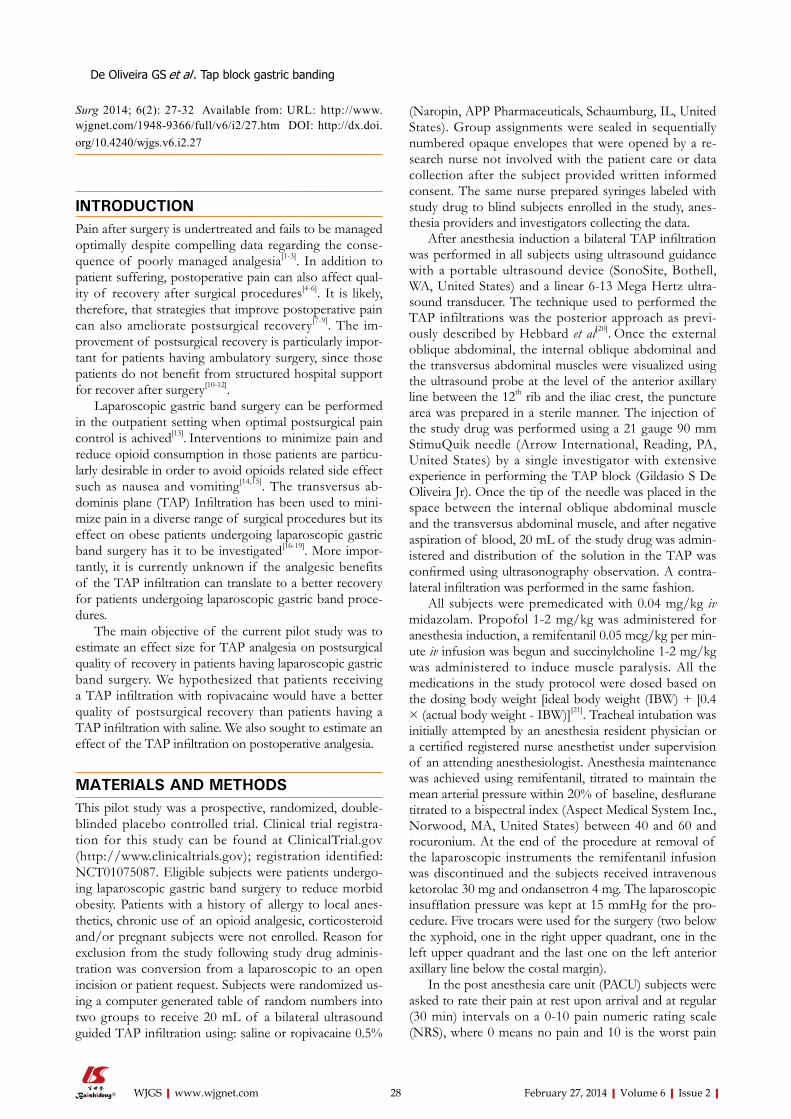
Surg 2014; 6(2): 27-32 Available from: URL: http://www.wjgnet.com/1948-9366/full/v6/i2/27.htm DOI: http://dx.doi.org/10.4240/wjgs.v6.i2.27
INTRODUCTIONPain after surgery is undertreated and fails to be managed optimally despite compelling data regarding the conse-quence of poorly managed analgesia[1-3]. In addition to patient suffering, postoperative pain can also affect qual-ity of recovery after surgical procedures[4-6]. It is likely, therefore, that strategies that improve postoperative pain can also ameliorate postsurgical recovery[7-9]. The im-provement of postsurgical recovery is particularly impor-tant for patients having ambulatory surgery, since those patients do not benefit from structured hospital support for recover after surgery[10-12].
Laparoscopic gastric band surgery can be performed in the outpatient setting when optimal postsurgical pain control is achived[13]. Interventions to minimize pain and reduce opioid consumption in those patients are particu-larly desirable in order to avoid opioids related side effect such as nausea and vomiting[14,15]. The transversus ab-dominis plane (TAP) Infiltration has been used to mini-mize pain in a diverse range of surgical procedures but its effect on obese patients undergoing laparoscopic gastric band surgery has it to be investigated[16-19]. More impor-tantly, it is currently unknown if the analgesic benefits of the TAP infiltration can translate to a better recovery for patients undergoing laparoscopic gastric band proce-dures.
The main objective of the current pilot study was to estimate an effect size for TAP analgesia on postsurgical quality of recovery in patients having laparoscopic gastric band surgery. We hypothesized that patients receiving a TAP infiltration with ropivacaine would have a better quality of postsurgical recovery than patients having a TAP infiltration with saline. We also sought to estimate an effect of the TAP infiltration on postoperative analgesia.
MATERIALS AND METHODSThis pilot study was a prospective, randomized, double-blinded placebo controlled trial. Clinical trial registra-tion for this study can be found at ClinicalTrial.gov (http://www.clinicaltrials.gov); registration identified: NCT01075087. Eligible subjects were patients undergo-ing laparoscopic gastric band surgery to reduce morbid obesity. Patients with a history of allergy to local anes-thetics, chronic use of an opioid analgesic, corticosteroid and/or pregnant subjects were not enrolled. Reason for exclusion from the study following study drug adminis-tration was conversion from a laparoscopic to an open incision or patient request. Subjects were randomized us-ing a computer generated table of random numbers into two groups to receive 20 mL of a bilateral ultrasound guided TAP infiltration using: saline or ropivacaine 0.5%
(Naropin, APP Pharmaceuticals, Schaumburg, IL, United States). Group assignments were sealed in sequentially numbered opaque envelopes that were opened by a re-search nurse not involved with the patient care or data collection after the subject provided written informed consent. The same nurse prepared syringes labeled with study drug to blind subjects enrolled in the study, anes-thesia providers and investigators collecting the data.
After anesthesia induction a bilateral TAP infiltration was performed in all subjects using ultrasound guidance with a portable ultrasound device (SonoSite, Bothell, WA, United States) and a linear 6-13 Mega Hertz ultra-sound transducer. The technique used to performed the TAP infiltrations was the posterior approach as previ-ously described by Hebbard et al[20]. Once the external oblique abdominal, the internal oblique abdominal and the transversus abdominal muscles were visualized using the ultrasound probe at the level of the anterior axillary line between the 12th rib and the iliac crest, the puncture area was prepared in a sterile manner. The injection of the study drug was performed using a 21 gauge 90 mm StimuQuik needle (Arrow International, Reading, PA, United States) by a single investigator with extensive experience in performing the TAP block (Gildasio S De Oliveira Jr). Once the tip of the needle was placed in the space between the internal oblique abdominal muscle and the transversus abdominal muscle, and after negative aspiration of blood, 20 mL of the study drug was admin-istered and distribution of the solution in the TAP was confirmed using ultrasonography observation. A contra-lateral infiltration was performed in the same fashion.
All subjects were premedicated with 0.04 mg/kg iv midazolam. Propofol 1-2 mg/kg was administered for anesthesia induction, a remifentanil 0.05 mcg/kg per min-ute iv infusion was begun and succinylcholine 1-2 mg/kg was administered to induce muscle paralysis. All the medications in the study protocol were dosed based on the dosing body weight [ideal body weight (IBW) + [0.4 × (actual body weight - IBW)][21]. Tracheal intubation was initially attempted by an anesthesia resident physician or a certified registered nurse anesthetist under supervision of an attending anesthesiologist. Anesthesia maintenance was achieved using remifentanil, titrated to maintain the mean arterial pressure within 20% of baseline, desflurane titrated to a bispectral index (Aspect Medical System Inc., Norwood, MA, United States) between 40 and 60 and rocuronium. At the end of the procedure at removal of the laparoscopic instruments the remifentanil infusion was discontinued and the subjects received intravenous ketorolac 30 mg and ondansetron 4 mg. The laparoscopic insufflation pressure was kept at 15 mmHg for the pro-cedure. Five trocars were used for the surgery (two below the xyphoid, one in the right upper quadrant, one in the left upper quadrant and the last one on the left anterior axillary line below the costal margin).
In the post anesthesia care unit (PACU) subjects were asked to rate their pain at rest upon arrival and at regular (30 min) intervals on a 0-10 pain numeric rating scale (NRS), where 0 means no pain and 10 is the worst pain
February 27, 2014|Volume 6|Issue 2|WJGS|www.wjgnet.com
De Oliveira GS et al . Tap block gastric banding
28
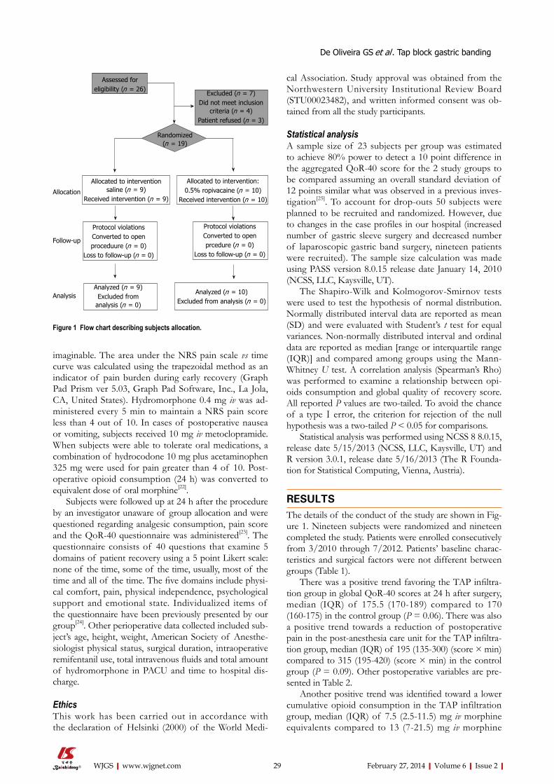
imaginable. The area under the NRS pain scale vs time curve was calculated using the trapezoidal method as an indicator of pain burden during early recovery (Graph Pad Prism ver 5.03, Graph Pad Software, Inc., La Jola, CA, United States). Hydromorphone 0.4 mg iv was ad-ministered every 5 min to maintain a NRS pain score less than 4 out of 10. In cases of postoperative nausea or vomiting, subjects received 10 mg iv metoclopramide. When subjects were able to tolerate oral medications, a combination of hydrocodone 10 mg plus acetaminophen 325 mg were used for pain greater than 4 of 10. Post-operative opioid consumption (24 h) was converted to equivalent dose of oral morphine[22].
Subjects were followed up at 24 h after the procedure by an investigator unaware of group allocation and were questioned regarding analgesic consumption, pain score and the QoR-40 questionnaire was administered[23]. The questionnaire consists of 40 questions that examine 5 domains of patient recovery using a 5 point Likert scale: none of the time, some of the time, usually, most of the time and all of the time. The five domains include physi-cal comfort, pain, physical independence, psychological support and emotional state. Individualized items of the questionnaire have been previously presented by our group[24]. Other perioperative data collected included sub-ject’s age, height, weight, American Society of Anesthe-siologist physical status, surgical duration, intraoperative remifentanil use, total intravenous fluids and total amount of hydromorphone in PACU and time to hospital dis-charge.
EthicsThis work has been carried out in accordance with the declaration of Helsinki (2000) of the World Medi-
cal Association. Study approval was obtained from the Northwestern University Institutional Review Board (STU00023482), and written informed consent was ob-tained from all the study participants.
Statistical analysisA sample size of 23 subjects per group was estimated to achieve 80% power to detect a 10 point difference in the aggregated QoR-40 score for the 2 study groups to be compared assuming an overall standard deviation of 12 points similar what was observed in a previous inves-tigation[25]. To account for drop-outs 50 subjects were planned to be recruited and randomized. However, due to changes in the case profiles in our hospital (increased number of gastric sleeve surgery and decreased number of laparoscopic gastric band surgery, nineteen patients were recruited). The sample size calculation was made using PASS version 8.0.15 release date January 14, 2010 (NCSS, LLC, Kaysville, UT).
The Shapiro-Wilk and Kolmogorov-Smirnov tests were used to test the hypothesis of normal distribution. Normally distributed interval data are reported as mean (SD) and were evaluated with Student’s t test for equal variances. Non-normally distributed interval and ordinal data are reported as median [range or interquartile range (IQR)] and compared among groups using the Mann-Whitney U test. A correlation analysis (Spearman’s Rho) was performed to examine a relationship between opi-oids consumption and global quality of recovery score. All reported P values are two-tailed. To avoid the chance of a type Ⅰ error, the criterion for rejection of the null hypothesis was a two-tailed P < 0.05 for comparisons.
Statistical analysis was performed using NCSS 8 8.0.15, release date 5/15/2013 (NCSS, LLC, Kaysville, UT) and R version 3.0.1, release date 5/16/2013 (The R Founda-tion for Statistical Computing, Vienna, Austria).
RESULTSThe details of the conduct of the study are shown in Fig-ure 1. Nineteen subjects were randomized and nineteen completed the study. Patients were enrolled consecutively from 3/2010 through 7/2012. Patients’ baseline charac-teristics and surgical factors were not different between groups (Table 1).
There was a positive trend favoring the TAP infiltra-tion group in global QoR-40 scores at 24 h after surgery, median (IQR) of 175.5 (170-189) compared to 170 (160-175) in the control group (P = 0.06). There was also a positive trend towards a reduction of postoperative pain in the post-anesthesia care unit for the TAP infiltra-tion group, median (IQR) of 195 (135-300) (score × min) compared to 315 (195-420) (score × min) in the control group (P = 0.09). Other postoperative variables are pre-sented in Table 2.
Another positive trend was identified toward a lower cumulative opioid consumption in the TAP infiltration group, median (IQR) of 7.5 (2.5-11.5) mg iv morphine equivalents compared to 13 (7-21.5) mg iv morphine
February 27, 2014|Volume 6|Issue 2|WJGS|www.wjgnet.com 29
Allocated to interventionsaline (n = 9)
Received intervention (n = 9)
Assessed for eligibility (n = 26)
Excluded (n = 7)Did not meet inclusion
criteria (n = 4)Patient refused (n = 3)
Randomized(n = 19)
Allocated to intervention:0.5% ropivacaine (n = 10)
Received intervention (n = 10)
Protocol violationsConverted to open proceduure (n = 0)
Loss to follow-up (n = 0)
Protocol violationsConverted to open prcedure (n = 0)
Loss to follow-up (n = 0)
Analyzed (n = 9)Excluded from
analysis (n = 0)
Analyzed (n = 10)Excluded from analysis (n = 0)
Allocation
Follow-up
Analysis
Figure 1 Flow chart describing subjects allocation.
De Oliveira GS et al . Tap block gastric banding

February 27, 2014|Volume 6|Issue 2|WJGS|www.wjgnet.com
surgery as an outpatient procedure[35,36].It is important to note that the transversus abdominis
block successfully treats parietal pain but visceral pain is not treated by a TAP block. This is likely the reason why even patients in the TAP block group required the use of systemic opioids. Nevertheless, it seems that the reduc-tion in the parietal pain is an important step to improve quality of recovery in patients undergoing laparoscopic gastric bending.
The TAP infiltration has been previously evaluated in morbidly obese patients undergoing different surgical procedures than the one presented in the current inves-tigation. Sinha et al[37] detected a large effect on analgesic outcomes of the TAP infiltration in patients undergoing laparoscopic gastric bypass. The authors’ results were interesting since laparoscopic gastric bypass surgery re-quires a more extensive procedure which can result in greater visceral pain and a potential lower benefit of the TAP infiltration. In contrast, a retrospective evaluation of obese patients undergoing abdominal plastic surgery did not detect a benefit of the TAP infiltration compared to control for postoperative pain[38]. The effect of the TAP infiltration on postoperative pain outcomes in obese patients may, therefore, be dependent on the type of sur-gical procedure.
Our current study should only be interpreted accord-ing to its limitations. We were unable to complete recruit-ment and we were underpowered to achieve statistically significant results in our main outcomes. Based on the differences observed in the current study, group sample sizes of 24 per group would have been needed to show a difference in opioid consumption between groups if the actual difference was 5.5 as observed in the current study. Nevertheless, we believe that our current results are clini-cally important and will stimulate future studies involving regional and local anesthesia techniques in the morbidly obese patient population.
In summary, we report an important beneficial clinical effect of TAP infiltration on quality of recovery in mor-bidly obese patients after laparoscopic gastric band sur-gery. It seems that the reduction in postoperative opioids by the TAP infiltration resulted in a better recovery in that patient population. Future studies are warranted to
equivalents in the control group (P = 0.07)Correlation analysis (Spearman’s Rho) demonstrated an
inverse relationship between 24 h cumulative opioid con-sumption and global QoR-40 scores, ρ = -0.49 (P = 0.03).
DISCUSSIONThe most important finding of the current study was a probable benefit of the TAP infiltration to improve post-operative quality of recovery after laparoscopic gastric band surgery in morbidly obese patients. In addition, a similar trend toward a reduction in pain burden and opi-oid consumption was also detected in the TAP infiltra-tion group compared to saline. The inverse relationship between postoperative opioid consumption and quality of recovery in the subjects in this study support our previous findings of reduced opioid use and improved postoperative recovery[16,23,25]. Our results suggest that the TAP infiltration may be a clinically significant multimodal strategy to reduce pain and improve quality of postsurgi-cal recovery in patients undergoing laparoscopic gastric band surgery.
Our results are particularly important since the devel-opment of opioid related side effects such as vomiting in gastric reduction surgery can lead to surgical complica-tions[26]. In addition greater opioid consumption in obese patients with obstructive sleep apnea can also result in postoperative hypoxemic events[27-29]. It remains to be determined if the opioid reduction due to the TAP infil-tration can minimize opioid related side effects in morbid obese patients undergoing gastric reduction surgery.
Despite a possible benefit on postoperative analgesia, we did not observe a parallel reduction in hospital stay in our study population. Hospital stay, in the inpatient pop-ulation, has been criticized as a reliable outcome due to the innumerous potential confounders that can alter that outcome[30-32]. Even established analgesic interventions, often fail to demonstrate a beneficial effect on duration of hospital stay[33,34]. Nevertheless, the optimal postop-erative pain control in patients undergoing laparoscopic gastric band surgery may help the performance of this
30
TAP infiltration(n = 10)
Saline(n = 9)
P value
Age (yr) 47 (39-53) 50 (36-54) 0.90 Gender 1.00 Male 2 2 Female 8 7 BMI (kg/m2) 44.2 (39.0-45.7) 40.1(38.5-44.3) 0.46 ASA class 0.37 Ⅱ 3 5 Ⅲ 7 4 Surgical duration (min) 76.5 (51-106) 92 (61-120) 0.53 Remifentanil (mcg) 620 (500-750) 850 (600-1100) 0.23 Intravenous fluids (mL) 1050 (585-1600) 1300 (1200-1500) 0.43
Table 1 Baseline patient and surgical characteristics
TAP infiltration(n = 10)
Saline(n = 9)
P value
Nausea in PACU 0.33 Yes 1 3 No 9 6 Vomiting/Retching in PACU 0.47 Yes 0 1 No 10 8 Time to discharge from PACU (min)
60 (60-75) 75 (60-90) 0.41
Time to discharge from the hospital (h)
32.1 (13.9-52.6) 22.5 (19-26) 0.47
Table 2 Postoperative data
Data are expressed as median [interquartile range (IQR)]. TAP: Transversus abdominis plane; ASA: American society of anesthesiologists; BMI: Body mass index.
TAP: Transversus abdominis plane; PACU: Post anesthesia care unit.
De Oliveira GS et al . Tap block gastric banding
February 27, 2014|Volume 6|Issue 2|WJGS|www.wjgnet.com
8 Bertoglio S, Fabiani F, Negri PD, Corcione A, Merlo DF, Cafiero F, Esposito C, Belluco C, Pertile D, Amodio R, Man-nucci M, Fontana V, Cicco MD, Zappi L. The postoperative analgesic efficacy of preperitoneal continuous wound infu-sion compared to epidural continuous infusion with local an-esthetics after colorectal cancer surgery: a randomized con-trolled multicenter study. Anesth Analg 2012; 115: 1442-1450 [PMID: 23144438 DOI: 10.1213/ANE.0b013e31826b4694]
9 De Oliveira GS, Fitzgerald P, Streicher LF, Marcus RJ, Mc-Carthy RJ. Systemic lidocaine to improve postoperative quality of recovery after ambulatory laparoscopic surgery. Anesth Analg 2012; 115: 262-267 [PMID: 22584558 DOI: 10.1213/ANE.0b013e318257a380]
10 White PF, Zhao M, Tang J, Wender RH, Yumul R, Slonin-sky AV, Naruse R, Kariger R, Cunneen S. Use of a dispos-able acupressure device as part of a multimodal antiemetic strategy for reducing postoperative nausea and vomiting. Anesth Analg 2012; 115: 31-37 [PMID: 22504214 DOI: 10.1213/ANE.0b013e3182536f27]
11 Joshi GP, Ankichetty SP, Gan TJ, Chung F. Society for Ambulatory Anesthesia consensus statement on preop-erative selection of adult patients with obstructive sleep apnea scheduled for ambulatory surgery. Anesth Analg 2012; 115: 1060-1068 [PMID: 22886843 DOI: 10.1213/ANE.0b013e318269cfd7]
12 White PF, White LM, Monk T, Jakobsson J, Raeder J, Mulroy MF, Bertini L, Torri G, Solca M, Pittoni G, Bettelli G. Periop-erative care for the older outpatient undergoing ambulatory surgery. Anesth Analg 2012; 114: 1190-1215 [PMID: 22467899 DOI: 10.1213/ANE.0b013e31824f19b8]
13 Robert M, Denis A, Badol-Van Straaten P, Jaisson-Hot I, Gouillat C. Prospective longitudinal assessment of change in health-related quality of life after adjustable gastric banding. Obes Surg 2013; 23: 1564-1570 [PMID: 23515974]
14 De Oliveira GS, Castro-Alves LJ, Ahmad S, Kendall MC, McCarthy RJ. Dexamethasone to prevent postoperative nausea and vomiting: an updated meta-analysis of random-ized controlled trials. Anesth Analg 2013; 116: 58-74 [PMID: 23223115 DOI: 10.1213/ANE.0b013e31826f0a0a]
15 De Oliveira GS, Castro-Alves LJ, Chang R, Yaghmour E, McCarthy RJ. Systemic metoclopramide to prevent postop-erative nausea and vomiting: a meta-analysis without Fujii’s studies. Br J Anaesth 2012; 109: 688-697 [PMID: 23015617 DOI: 10.1093/bja/aes325]
16 Petersen PL, Stjernholm P, Kristiansen VB, Torup H, Hansen EG, Mitchell AU, Moeller A, Rosenberg J, Dahl JB, Mathiesen O. The beneficial effect of transversus abdominis plane block after laparoscopic cholecystectomy in day-case surgery: a randomized clinical trial. Anesth Analg 2012; 115: 527-533 [PMID: 22763903]
17 Freir NM, Murphy C, Mugawar M, Linnane A, Cunning-ham AJ. Transversus abdominis plane block for analgesia in renal transplantation: a randomized controlled trial. Anesth Analg 2012; 115: 953-957 [PMID: 22763899 DOI: 10.1213/ANE.0b013e3182642117]
18 Abdallah FW, Halpern SH, Margarido CB. Transversus abdominis plane block for postoperative analgesia after Caesarean delivery performed under spinal anaesthesia? A systematic review and meta-analysis. Br J Anaesth 2012; 109: 679-687 [PMID: 22907337 DOI: 10.1093/bja/aes279]
19 McDermott G, Korba E, Mata U, Jaigirdar M, Narayanan N, Boylan J, Conlon N. Should we stop doing blind transver-sus abdominis plane blocks? Br J Anaesth 2012; 108: 499-502 [PMID: 22236911 DOI: 10.1093/bja/aer422]
20 Hebbard P, Fujiwara Y, Shibata Y, Royse C. Ultrasound-guided transversus abdominis plane (TAP) block. Anaesth Intensive Care 2007; 35: 616-617 [PMID: 18020088]
21 Carron M, Guzzinati S, Ori C. Simplified estimation of ideal and lean body weights in morbidly obese patients. Br J An-aesth 2012; 109: 829-830 [PMID: 23066004 DOI: 10.1093/bja/
confirm the beneficial effect of the TAP infiltration on postoperative recovery and analgesia in the same patient population.
COMMENTSBackgroundOpioids are commonly used to treat postoperative pain in patients undergo-ing laparoscopic gastric banding. The transversus abdominis plane block has been used successfully to improve recovery in other patient populations but it is unknown if it can also improve recovery for obese patients undergoing laparo-scopic gastric banding.Research frontiersThe transversus abdominis plane (TAP) block seems to improve quality of re-covery in patients undergoing laparoscopic gastric banding through a reduction in the consumption of opioids.Innovations and breakthroughsThis is the first study to demonstrate a possible benefit of the TAP block on obese patients undergoing laparoscopic gastric bandingApplicationsPatients undergoing laparoscopic gastric banding may have better quality of postsurgical recovery if they receive a TAP block before the surgical procedureTerminologyTransversus abdominis block is the application of local anesthetics in the ab-dominal wall using ultrasound. Peer reviewThis manuscript is described as a pilot study but realistically is an underpow-ered study that if recruitment had been successfully would have realised signifi-cant results.
REFERENCES1 Ohri R, Wang JC, Blaskovich PD, Pham LN, Costa DS,
Nichols GA, Hildebrand WP, Scarborough NL, Herman CJ, Strichartz GR. Inhibition by local bupivacaine-releasing mi-crospheres of acute postoperative pain from hairy skin inci-sion. Anesth Analg 2013; 117: 717-730 [PMID: 23921651 DOI: 10.1213/ANE.0b013e3182a00851]
2 Cohen SP, Galvagno SM, Plunkett A, Harris D, Kurihara C, Turabi A, Rehrig S, Buckenmaier CC, Chelly JE. A multi-center, randomized, controlled study evaluating preventive etanercept on postoperative pain after inguinal hernia re-pair. Anesth Analg 2013; 116: 455-462 [PMID: 23302973 DOI: 10.1213/ANE.0b013e318273f71c]
3 Yuan Y, Wang JY, Yuan F, Xie KL, Yu YH, Wang GL. Glyco-gen synthase kinase-3β contributes to remifentanil-induced postoperative hyperalgesia via regulating N-methyl-D-as-partate receptor trafficking. Anesth Analg 2013; 116: 473-481 [PMID: 23267003 DOI: 10.1213/ANE.0b013e318274e3f1]
4 Gornall BF, Myles PS, Smith CL, Burke JA, Leslie K, Pereira MJ, Bost JE, Kluivers KB, Nilsson UG, Tanaka Y, Forbes A. Measurement of quality of recovery using the QoR-40: a quantitative systematic review. Br J Anaesth 2013; 111: 161-169 [PMID: 23471753 DOI: 10.1093/bja/aet014]
5 Benn J, Arnold G, Wei I, Riley C, Aleva F. Using quality indicators in anaesthesia: feeding back data to improve care. Br J Anaesth 2012; 109: 80-91 [PMID: 22661749 DOI: 10.1093/bja/aes173]
6 Cook TM, Coupe M, Ku T. Shaping quality: the use of per-formance polygons for multidimensional presentation and interpretation of qualitative performance data. Br J Anaesth 2012; 108: 953-960 [PMID: 22451507 DOI: 10.1093/bja/aes026]
7 Gardiner S, Rudkin G, Cooter R, Field J, Bond M. Paraverte-bral blockade for day-case breast augmentation: a random-ized clinical trial. Anesth Analg 2012; 115: 1053-1059 [PMID: 22984150 DOI: 10.1213/ANE.0b013e318264ba33]
31
COMMENTS
De Oliveira GS et al . Tap block gastric banding

February 27, 2014|Volume 6|Issue 2|WJGS|www.wjgnet.com
30 Fawcett WJ, Mythen MG, Scott MJ. Enhanced recovery: more than just reducing length of stay? Br J Anaesth 2012; 109: 671-674 [PMID: 23065999 DOI: 10.1093/bja/aes358]
31 Wongyingsinn M, Baldini G, Stein B, Charlebois P, Liber-man S, Carli F. Spinal analgesia for laparoscopic colonic resection using an enhanced recovery after surgery pro-gramme: better analgesia, but no benefits on postoperative recovery: a randomized controlled trial. Br J Anaesth 2012; 108: 850-856 [PMID: 22408272 DOI: 10.1093/bja/aes028]
32 Grady MV, Mascha E, Sessler DI, Kurz A. The effect of perioperative intravenous lidocaine and ketamine on recovery after abdominal hysterectomy. Anesth Analg 2012; 115: 1078-1084 [PMID: 23011561 DOI: 10.1213/ANE.0b013e3182662e01]
33 De Oliveira GS, Agarwal D, Benzon HT. Perioperative single dose ketorolac to prevent postoperative pain: a meta-analysis of randomized trials. Anesth Analg 2012; 114: 424-433 [PMID: 21965355 DOI: 10.1213/ANE.0b013e3182334d68]
34 Michelet D, Andreu-Gallien J, Bensalah T, Hilly J, Wood C, Nivoche Y, Mantz J, Dahmani S. A meta-analysis of the use of nonsteroidal antiinflammatory drugs for pediatric postoperative pain. Anesth Analg 2012; 114: 393-406 [PMID: 22104069 DOI: 10.1213/ANE.0b013e31823d0b45]
35 Krenk L, Jennum P, Kehlet H. Sleep disturbances after fast-track hip and knee arthroplasty. Br J Anaesth 2012; 109: 769-775 [PMID: 22831887 DOI: 10.1093/bja/aes252]
36 de Silva E, Plaat F. Postoperative analgesia - still failing to meet the standard. Anaesthesia 2012; 67: 801-802 [PMID: 22670746 DOI: 10.1111/j.1365-2044.2012.07210.x]
37 Sinha A, Jayaraman L, Punhani D. Efficacy of ultrasound-guided transversus abdominis plane block after laparoscopic bariatric surgery: a double blind, randomized, controlled study. Obes Surg 2013; 23: 548-553 [PMID: 23361468 DOI: 10.1007/s11695-012-0819-5]
38 Gravante G, Castrì F, Araco F, Araco A. A comparative study of the transversus abdominis plane (TAP) block effica-cy on post-bariatric vs aesthetic abdominoplasty with flank liposuction. Obes Surg 2011; 21: 278-282 [PMID: 20517653 DOI: 10.1007/s11695-010-0203-2]
P- Reviewers: Dubost C, McCaulCL, Mukhtar K S- Editor: Zhai HH L- Editor: A E- Editor: Wu HL
aes368]22 Macintyre PE, Ready LB. Pharmacology of opioids. In:
Acute pain management-a practical guide. 2nd edition. Phila-delphia: WB Saunders, 2001: 15-49
23 De Oliveira GS, Milad MP, Fitzgerald P, Rahmani R, Mc-Carthy RJ. Transversus abdominis plane infiltration and quality of recovery after laparoscopic hysterectomy: a ran-domized controlled trial. Obstet Gynecol 2011; 118: 1230-1237 [PMID: 22105251 DOI: 10.1097/AOG.0b013e318236f67f]
24 De Oliveira GS, Ahmad S, Fitzgerald PC, Marcus RJ, Alt-man CS, Panjwani AS, McCarthy RJ. Dose ranging study on the effect of preoperative dexamethasone on postoperative quality of recovery and opioid consumption after ambula-tory gynaecological surgery. Br J Anaesth 2011; 107: 362-371 [PMID: 21669954 DOI: 10.1093/bja/aer156]
25 De Oliveira GS, Fitzgerald PC, Marcus RJ, Ahmad S, Mc-Carthy RJ. A dose-ranging study of the effect of transver-sus abdominis block on postoperative quality of recovery and analgesia after outpatient laparoscopy. Anesth Analg 2011; 113: 1218-1225 [PMID: 21926373 DOI: 10.1213/ANE.0b013e3182303a1a]
26 Lloret-Linares C, Lopes A, Declèves X, Serrie A, Mouly S, Bergmann JF, Perrot S. Challenges in the optimisation of post-operative pain management with opioids in obese patients: a literature review. Obes Surg 2013; 23: 1458-1475 [PMID: 23700237 DOI: 10.1007/s11695-013-0998-8]
27 Kaw R, Chung F, Pasupuleti V, Mehta J, Gay PC, Hernandez AV. Meta-analysis of the association between obstructive sleep apnoea and postoperative outcome. Br J Anaesth 2012; 109: 897-906 [PMID: 22956642 DOI: 10.1093/bja/aes308]
28 Chung F, Subramanyam R, Liao P, Sasaki E, Shapiro C, Sun Y. High STOP-Bang score indicates a high probability of ob-structive sleep apnoea. Br J Anaesth 2012; 108: 768-775 [PMID: 22401881 DOI: 10.1093/bja/aes022]
29 Aldenkortt M, Lysakowski C, Elia N, Brochard L, Tramèr MR. Ventilation strategies in obese patients undergoing sur-gery: a quantitative systematic review and meta-analysis. Br J Anaesth 2012; 109: 493-502 [PMID: 22976857 DOI: 10.1093/bja/aes338]
32
De Oliveira GS et al . Tap block gastric banding
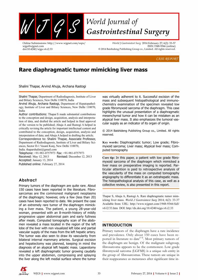
Shalini Thapar, Arvind Ahuja, Archana Rastogi
Rare diaphragmatic tumor mimicking liver mass
Shalini Thapar, Department of Radiodiagnosis, Institute of Liver and Biliary Sciences, New Delhi 110070, IndiaArvind Ahuja, Archana Rastogi, Department of Hepatopathol-ogy, Institute of Liver and Biliary Sciences, New Delhi 110070, IndiaAuthor contributions: Thapar S made substantial contributions to the conception and design, acquisition, analysis and interpreta-tion of data, and drafted the article and helped in final approval of the version to be published; Ahuja A and Rastogi A helped in critically revising the article for important intellectual content and contributed to the conception, design, acquisition, analysis and interpretation of data; and Ahuja A helped in drafting the article. Correspondence to: Shalini Thapar, Associate Professor, Department of Radiodiagnosis, Institute of Liver and Biliary Sci-ences, Sector D-1 Vasant Kunj, New Delhi 110070,India. [email protected]: +91-981-0757973 Fax: +91-981-0757973Received: May 12, 2013 Revised: December 12, 2013Accepted: January 13, 2014Published online: February 27, 2014
AbstractPrimary tumors of the diaphragm are quite rare. About 150 cases have been reported in the literature. Fibro-sarcomas are the commonest malignant neoplasms of the diaphragm; however, only a few (less than 20) cases have been reported to date. We present the case of an extremely rare tumor of the diaphragm mimick-ing a liver mass. The patient, a young 28-year-old woman, presented with an 8-month-history of mildly progressive upper abdominal pain and early fullness after meals. Computed tomography scan of the abdo-men revealed a mass located in the region of the left lobe of the liver with non visualized left lobe and partial vascular supply of the mass from the left hepatic artery. The tumor was also seen to draw its vascularity from bilateral internal mammary arteries. Surgical excision and hepatectomy was planned, keeping in mind the diagnosis of an atypical left hepatic mass. Laparotomy revealed a left diaphragmatic tumor growing caudally into the upper abdomen, compressing and splaying the liver along the left medial surface where the tumor
was virtually adherent to it. Successful excision of the mass and subsequent histopathological and immuno-chemistry examination of the specimen revealed low grade fibromyxoid sarcoma of the diaphragm. This case highlights the unusual presentation of a diaphragmatic mesenchymal tumor and how it can be mistaken as an atypical liver mass. It also emphasizes the tumoral vas-cular supply as an indicator of its organ of origin.
© 2014 Baishideng Publishing Group co., Limited. All rights reserved.
Key words: Diaphragmatic tumor; Low grade; Fibro-myxoid sarcoma; Liver mass; Atypical liver mass; Com-puted tomography
Core tip: In this paper, a patient with low grade fibro-myxoid sarcoma of the diaphragm which mimicked a liver mass on preoperative imaging is reported. Par-ticular attention is paid to retrospective analysis of the vascularity of the mass on computed tomography angiography to differentiate it as an extrahepatic mass. The histopathological analysis of this case, as well as a collective review, is also presented in this report.
Thapar S, Ahuja A, Rastogi A. Rare diaphragmatic tumor mim-icking liver mass. World J Gastrointest Surg 2014; 6(2): 33-37 Available from: URL: http://www.wjgnet.com/1948-9366/full/v6/i2/33.htm DOI: http://dx.doi.org/10.4240/wjgs.v6.i2.33
INTRODUCTIONPrimary tumors of the diaphragm have a rare incidence and prevalence. Only about 150 cases have been re-ported in literature to date[1-5]. Most primary tumors of the diaphragm are benign. Of the malignant subgroup, fibrosarcoma appears to be the commonest. Low grade fibromyxoid sarcoma (LGFMS) is a unique sub entity in the group of fibrosarcomas. These tumors are unique in their reappearance as metastases after significant time in-
CASE REPORT
Online Submissions: http://www.wjgnet.com/esps/[email protected]:10.4240/wjgs.v6.i2.33
February 27, 2014|Volume 6|Issue 2|WJGS|www.wjgnet.com
World J Gastrointest Surg 2014 February 27; 6(2): 33-37ISSN 1948-9366 (online)
© 2014 Baishideng Publishing Group co., Limited. All rights reserved.
33

tervals. Around 20 cases of the same have been reported in the English literature so far, to the best of our knowl-edge[1-7]. LGFMS shows characteristic histopathological and immunochemistry features. In the last decade, spo-radic cases of fibromyxoid sarcoma have been reported in the literature. Even after recent major advances in imaging technology, such as the advent of multidetector, multi slice computed tomography (CT) and 3Tesla mag-netic resonance imaging (MRI), diaphragmatic tumors still present as a diagnostic dilemna and are difficult to diagnose preoperatively or without a pathological diagno-sis. They are known to mimic other large masses arising from the mediastinum in the majority of cases. In this paper, a patient with low grade fibromyxoid sarcoma of the diaphragm which mimicked a liver mass on preop-erative imaging is reported. Particular attention is paid to retrospective analysis of the vascularity of the mass on CT angiography to differentiate it as an extrahepatic mass. The histopathological analysis of this case, as well as a collective review, is also presented in this report.
CASE REPORTA 28-year-old lady presented to the hospital with an 8 mo progressive history of nagging upper abdominal pain and a feeling of early satiety. The pain was localized without associated vomiting, aggravating or relieving factors. The patient complained of feeling a gradually increasing lump in the upper abdomen for 6 mo. However, she had no
definite history of cough, hiccups or difficulty in breath-ing. The lady had loss of appetite with significant weight loss of 5 kg over 6 mo. On general examination, the lady was well preserved. Local palpation revealed a firm lump in the epigastrium and left hypochondrium with a percep-tible lower border. Palpating fingers could not be insinu-ated between the lump and the upper border, as well as the costal margin. Laboratory tests showed normal serum alpha fetoprotein and carcinoembryonic antigen levels (1.16 U) and (1.8 mg/L) respectively. All other tests, such as hemogram, kidney function tests and liver function tests, were within the normal reference ranges.
A dynamic triple phase CT scan of the upper abdo-men was performed and showed a homogenous soft tissue mass lesion measuring approximately 25 cm × 20 cm × 13.5 cm in the left upper hypochondrium, almost entirely replacing the left lobe of the liver (Figure 1A). The left hepatic vein and portal vein were only partially visualized and were grossly attenuated by the mass (Figure 1E-G). Enlarged left supra-diaphragmatic nodes were present (Figure 2A). The sections of bilateral lower lobes of chest did not show evidence of basal atelectasis or pleural effusion (Figure 2B). The tumor showed arterial feeders from the left hepatic artery which was splayed but showed normal patency (Figure 1B). The major chunk of tumor vascularity appeared to be from bilateral internal mammary arteries (Figure 1C). The tumor appearance was thought to be of an atypical hepatic mass and a deci-sion for surgical laparotomy with surgical excision of the
February 27, 2014|Volume 6|Issue 2|WJGS|www.wjgnet.com
Thapar S et al . Diaphragmatic tumor mimicking liver mass
34
LHV
LHVRHV
Figure 1 A 28-year-old lady with low grade fibromyxoid sarcoma on dynamic computed tomography. A: Soft tissue homogenously enhancing mildly hypodense mass (arrowhead) in the region of left lobe of liver is seen on the arterial phase of the dynamic triple phase computed tomography scan; B: The mass shows small arterial feeders from the left hepatic artery (dotted arrow); C: Large feeders from bilateral internal mammary arteries (solid arrows) to the mass are seen; D: Middle hepatic vein and right hepatic vein show normal patency and course; E: Left hepatic vein is only seen at the ostium, appears compressed by the mass and is not seen beyond the ostium; F: Left portal vein (solid arrow) is also splayed and partially attenuated by the tumor.
A CB
D E F
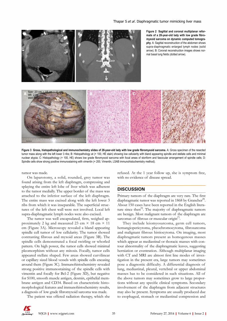
tumor was made. On laparotomy, a solid, rounded, grey tumor was
found arising from the left diaphragm, compressing and splaying the entire left lobe of liver which was adherent to the tumor medially. The upper border of the mass was attached to the inferior surface of the left diaphragm. The entire mass was excised along with the left lower 3 ribs from which it was inseparable. The superficial struc-tures of the left chest wall were not involved. Local left supra-diaphragmatic lymph nodes were also excised.
The tumor was well encapsulated, firm, weighed ap-proximately 2 kg and measured 23 cm × 18 cm × 11 cm (Figure 3A). Microscopy revealed a bland appearing spindle cell tumor of low cellularity. The tumor showed contrasting fibrous and myxoid areas (Figure 3B). The spindle cells demonstrated a focal swirling or whorled pattern. On high power, the tumor cells showed minimal pleomorphism without any mitosis. Focally, tumor cells appeared stellate shaped. Few areas showed curvilinear or capillary sized blood vessels with spindle cells encasing around them (Figure 3C). Immunohistochemistry revealed strong positive immunostaining of the spindle cells with vimentin and focally for Bcl-2 (Figure 3D), but negative for S100, smooth muscle antigen, desmin, epithelial mem-brane antigen and CD34. Based on characteristic histo-morphological features and immunohistochemistry results, a diagnosis of low grade fibromyxoid sarcoma was made.
The patient was offered radiation therapy, which she
refused. At the 1 year follow up, she is symptom free, with no evidence of disease spread.
DISCUSSIONPrimary tumors of the diaphragm are very rare. The first diaphragmatic tumor was reported in 1868 by Grancher[8]. About 150 cases have been reported in the English litera-ture since then[9]. The majority of diaphragmatic tumors are benign. Most malignant tumors of the diaphragm are sarcomas of fibrous or muscular origin[7].
They include leiomyosarcoma, germ cell tumors, hemangiopericytoma, pheochromocytoma, fibrosarcoma and malignant fibrous histiocytoma. On imaging, most diaphragmatic tumors present as homogenous masses which appear as mediastinal or thoracic masses with con-tour abnormality of the diaphragmatic leaves, suggesting herniation or eventration. Although multiplanar imaging with CT and MRI are almost first line modes of inves-tigation in the present era, large tumors may sometimes pose a diagnostic difficulty. A differential diagnosis of lung, mediastinal, pleural, vertebral or upper abdominal masses has to be considered in such situations. All of the above tumors may sometimes grow to large propor-tions without any specific clinical symptoms. Secondary involvement of the diaphragm from adjacent structures may also be present. Symptoms are usually produced due to esophageal, stomach or mediastinal compression and
February 27, 2014|Volume 6|Issue 2|WJGS|www.wjgnet.com 35
Figure 2 Sagittal and coronal multiplanar refor-mats of a 28-year-old lady with low grade fibro-myxoid sarcoma on dynamic computed tomogra-phy. A: Sagittal reconstruction of the abdomen shows supra-diaphragmatic enlarged lymph nodes (solid arrow); B: Coronal reconstruction images shows nor-mal basal lung fields (dotted arrow).
Figure 3 Gross, histopathological and immunochemistry slides of 28-year-old lady with low grade fibromyxoid sarcoma. A: Gross specimen of the resected tumor mass along with the left lower 3 ribs; B: Histopathology at (× 100, HE stain) showing low cellularity with bland appearing spindle and stellate cells and minimal nuclear atypia; C: Histopathology (× 100, HE) shows low grade fibromyxoid sarcoma with focal areas of storiform and fascicular arrangement of spindle cells; D: Spindle cells show strong positive immunostaining with vimentin (× 200, Vimentin, LSAB immunohistochemistry method).
A B
A B C DSize- 23 cm × 18 cm × 11 cmWt- 2 kgWell circumscribed
Thapar S et al . Diaphragmatic tumor mimicking liver mass
February 27, 2014|Volume 6|Issue 2|WJGS|www.wjgnet.com
coma centre in Denmark where 14 patients of LGFMS were studied for the disease and metastases treatment strategies, multi-agent chemotherapy was found useful, especially in the setting of metastases. The best possible response to chemotherapy was only a short term disease non progression with the use of trabectedin[12].
ACKNOWLEDGMENTSWe thank Rita Gulabani for transcription help and Ab-hishek Anand and Dhananjay Kumar for their technical assistance.
COMMENTSCase characteristicsThe patient, a young-28-year old woman, presented with an 8-mo-history of mildly progressive upper abdominal pain and early fullness after meals.Clinical diagnosisThe patient complained of feeling a gradually increasing lump in the upper ab-domen for 6 mo.Laboratory diagnosisLaboratory tests showed normal serum alpha fetoprotein and carcinoembryonic antigen levels (1.16 U) and (1.8 mg/L) respectively. Imaging diagnosisComputed tomography scan of the abdomen revealed a mass located in the re-gion of left lobe of liver with non visualized left lobe and partial vascular supply of the mass from the left hepatic artery. Pathological diagnosisImmunohistochemistry revealed strong positive immunostaining of the spindle cells with vimentin and focally for Bcl-2, but negative for S100, smooth muscle antigen, desmin, epithelial membrane antigen and CD34.TreatmentSurgical excision and hepatectomy was planned, keeping in mind the diagnosis of an atypical left hepatic mass.Experiences and lessonsThis case highlights that extrahepatic vascular supply may act as an indicator of the extrahepatic origin of a tumor showing indistinct fat planes from the liver.Peer reviewThe authors need to discuss pathological findings in more detail to substantiate the diagnosis of Low grade fibromyxoid sarcoma at this relatively uncommon location.
REFERENCES1 Evans HL. Low-grade fibromyxoid sarcoma: a clinicopatho-
logic study of 33 cases with long-term follow-up. Am J Surg Pathol 2011; 35: 1450-1462 [PMID: 21921785 DOI: 10.1097/PAS.0b013e31822b3687]
2 Wiener MF, Chou wh. primary tumors of the diaphragm. Arch Surg 1965; 90: 143-152 [PMID: 14220631 DOI: 10.1001/archsurg.1965.01320070145031]
3 Olafsson G, Rausing A, Holen O. Primary tumors of the diaphragm. Chest 1971; 59: 568-570 [PMID: 4952560 DOI: 10.1378/chest.59.5.568]
4 Mandal AK, Lee H, Salem F. Review of primary tumors of the diaphragm. J Natl Med Assoc 1988; 80: 214-217 [PMID: 3071608]
5 Midorikawa Y, Kubota K, Mori M, Koyama H, Aihara N, Makuuchi M, Kajiura N. Rhabdomyosarcoma of the dia-phragm: report of an adult case. Jpn J Clin Oncol 1998; 28: 222-226 [PMID: 9614447]
6 Zhang H, Wu D, Wang Z, Xu J, Wang H. Primary fibrosar-coma of the diaphragm with pleural effusion. Clin Respir J 2010; 4: 127-128 [PMID: 20565488 DOI: 10.1111/j.1752-
not due to the tumor bulk. The patient in our study also reported vague symp-
toms, which did not raise any suspicion about the extent of the mass. Imaging provided the only clue to the large size and extent of the tumor, which was solely limited to the upper abdomen. The initial suspicion of a liver mass was compounded by the vascular supply of the mass from the left hepatic artery. However, in retrospect, the majority of the tumor blood supply from bilateral inter-nal mammary arteries was indicative of the extrahepatic origin of the tumor. The sharp interface of the mass with the liver parenchyma despite the lack of a proper cleav-age plane was also a soft indicator of the extrahepatic origin.
Low-grade fibromyxoid sarcomas are distinctive, in-dolent soft-tissue sarcomas, first described by Evans in 1987. Low grade fibromyxoid sarcoma of the diaphragm is extremely rare. From 1868 to 1982, only 11 cases of low grade fibrosarcoma were reported[4]. Less than 20 cases have been reported in literature to date, to the best of our knowledge[1-3,6,7]. The sites described in literature so far have been commonly in the shoulder, neck or thigh[1]. On histopathology, low-grade fibromyxoid sarcomas demonstrate contrasting fibrous and myxoid areas, a swirl-ing, whorled growth pattern and bland benign-appearing fibroblastic spindle cells. Cellularity is low to moderate, mitotic figures are uncommon, and nuclear pleomorphism is usually absent or slight. In addition, LFGMS shows a specific genetic transcript fused in sarcoma-cAMP respon-sive element binding protein 3 (FUS-CREB3)L2 and FUS-CREB3L1 in up to 95%-100% of cases[10]. The common differentials of low grade fibromyxoid sarcoma include myxoid neurofibroma, malignant peripheral nerve sheath tumor of low grade, myxofibrosarcoma and spindle cell liposarcoma. Myxoid neurofibroma and low grade ma-lignant peripheral nerve sheath tumor show slender and wavy nuclei which are positive for S100. Myxofibrosar-coma is mostly a subcutaneous tumor of the elderly and shows a greater degree of nuclear pleomorphism, hyper-chromasia and brisk mitosis. Spindle cell liposarcoma is cellular and shows more nuclear atypia. It always contains an atypical adipocytic component which includes the pres-ence of lipoblasts and shows positivity for S100.
Primary diaphragmatic fibromyxoid sarcomas, espe-cially low grade like in this case, have a good prognosis. One of the reports has shown survival even up to 10 years[7]. Treatment options include surgery followed by radiotherapy and, in some cases, chemotherapy to shrink tumor bulk, followed by resection. These tumors are usually resistant to chemotherapy and radiotherapy with surgical resection being the treatment of choice[11].
In conclusion, this case highlights that an extrahepatic vascular supply may act as an indicator of the extrahe-patic origin of a tumor showing indistinct fat planes from the liver. Also, although rare, diaphragmatic mesenchy-mal tumors should also be included in the differential of atypical appearing hepatic or extrahepatic masses in the upper abdomen. In a recent study at a dedicated sar-
36
COMMENTS
Thapar S et al . Diaphragmatic tumor mimicking liver mass
February 27, 2014|Volume 6|Issue 2|WJGS|www.wjgnet.com
gopoulos I. Clinicopathologic and molecular genetic charac-terization of low-grade fibromyxoid sarcoma, and cloning of a novel FUS/CREB3L1 fusion gene. Lab Invest 2005; 85: 408-415 [PMID: 15640831 DOI: 10.1038/labinvest.3700230]
11 Menon S, Krivanek M, Cohen R. Low-grade fibromyxoid sarcoma, a deceptively benign tumor in a 5-year-old child. Pediatr Surg Int 2012; 28: 211-213 [PMID: 22130782 DOI: 10.1007/s00383-011-3024-z]
12 Maretty-Nielsen K, Baerentzen S, Keller J, Dyrop HB, Saf-wat A. Low-Grade Fibromyxoid Sarcoma: Incidence, Treat-ment Strategy of Metastases, and Clinical Significance of the FUS Gene. Sarcoma 2013; 2013: 256280 [PMID: 23818812 DOI: 10.1155/2013/256280]
P- Reviewer: Rekhi B S- Editor: Zhai HH L- Editor: Roemmele A E- Editor: Wu HL
699X.2009.00165.x]7 Sbokos CG, Salama FD, Powell V, McMillan IK. Primary
fibrosarcoma of the diaphragm. Br J Dis Chest 1977; 71: 49-52 [PMID: 831767 DOI: 10.1016/0007-0971(77)90077-8]
8 Weksler B, Ginsberg RJ. Tumors of the diaphragm. Chest Surg Clin N Am 1998; 8: 441-447 [PMID: 9619315]
9 Cada M, Gerstle JT, Traubici J, Ngan BY, Capra ML. Ap-proach to diagnosis and treatment of pediatric primary tumors of the diaphragm. J Pediatr Surg 2006; 41: 1722-1726 [PMID: 17011277 DOI: 10.1016/j.jpedsurg.2006.05.073]
10 Mertens F, Fletcher CD, Antonescu CR, Coindre JM, Colec-chia M, Domanski HA, Downs-Kelly E, Fisher C, Goldblum JR, Guillou L, Reid R, Rosai J, Sciot R, Mandahl N, Pana-
37
Thapar S et al . Diaphragmatic tumor mimicking liver mass

World Journal of Gastrointestinal SurgeryWorld J Gastrointest Surg 2014 March 27; 6(3): 38-54
ISSN 1948-9366 (online)
www.wjgnet.com

38 Theroleofstentinginthepalliationofgastroesophagealjunction
cancer:Abriefreview
Pavlidis TE, Pavlidis ET
42 Giantmucinouscysticadenomawithpancreaticatrophymimicking
dorsalagenesisofthepancreas
Gagnière J, Dupré A, Da Ines D, Tixier L, Pezet D, Buc E
47 GiantMeckel’sdiverticulum:Anexceptionalcauseofintestinal
obstruction
Akbulut S, Yagmur Y
51 Coexistenceofabdominalcocoon,intestinalperforationand
incarceratedMeckel’sdiverticuluminaninguinalhernia:A
troublesomecondition
Akbulut S, Yagmur Y, Babur M
Contents Monthly Volume 6 Number 3 March 27, 2014
WJGS|www.wjgnet.com I March 27, 2014|Volume 6|Issue 3|
MINIREVIEWS
CASE REPORT

Contents
APPENDIX
ABOUT COVER
World Journal of Gastrointestinal SurgeryVolume 6 Number 3 March 27, 2014
I-V Instructionstoauthors
EditorialBoardMemberofWorldJournalofGastrointestinalSurgery ,MichelKahaleh,MD,FACG,DivisionofGastroenterologyandHepatology,UniversityofVirginiaHealthSystem,POBox800708,Charlottesville,VA22908,UnitedStates
World Journal of Gastrointestinal Surgery (World J Gastrointest Surg, WJGS, online ISSN 1948-9366, DOI: 10.4240) is a peer-reviewed open access academic journal that aims to guide clinical practice and improve diagnostic and therapeutic skills of clinicians.
WJGS covers topics concerning micro-invasive surgery; laparoscopy; hepatic, biliary, pancreatic and splenic surgery; surgical nutrition; portal hypertension, as well as associated subjects. The current columns of WJGS include editorial, frontier, diagnostic advances, therapeutics advances, field of vision, mini-reviews, review, topic highlight, medical ethics, original articles, case report, clinical case conference (Clinicopathological conference), and autobiography. Priority publication will be given to articles concerning diagnosis and treatment of gastrointestinal surgery diseases. The following aspects are covered: Clinical diagnosis, laboratory diagnosis, differential diagnosis, imaging tests, pathological diagnosis, molecular biological diagnosis, immunological diagnosis, genetic diagnosis, functional diagnostics, and physical diagnosis; and comprehensive therapy, drug therapy, surgical therapy, interventional treatment, minimally invasive therapy, and robot-assisted therapy.
We encourage authors to submit their manuscripts to WJGS. We will give priority to manuscripts that are supported by major national and international foundations and those that are of great basic and clinical significance.
World Journal of Gastrointestinal Surgery is now indexed in PubMed Central, PubMed, Digital Object Identifier, and Directory of Open Access Journals.
I-III EditorialBoard
EDITORIALOFFICEJin-Lei Wang, DirectorXiu-Xia Song, Vice DirectorWorld Journal of Gastrointestinal SurgeryRoom 903, Building D, Ocean International Center, No. 62 Dongsihuan Zhonglu, Chaoyang District, Beijing 100025, ChinaTelephone: +86-10-85381891Fax: +86-10-85381893E-mail: [email protected]://www.wjgnet.com
PUBLISHERBaishideng Publishing Group Co., LimitedFlat C, 23/F., Lucky Plaza, 315-321 Lockhart Road, Wanchai, Hong Kong, ChinaFax: +852-31158812Telephone: +852-58042046E-mail: [email protected]://www.wjgnet.com
PUBLICATIONDATEMarch 27, 2014
COPYRIGHT© 2014 Baishideng. Articles published by this Open-Access journal are distributed under the terms of the Creative Commons Attribution Non-commercial Li-cense, which permits use, distribution, and reproduction in any medium, provided the original work is properly cited, the use is non commercial and is otherwise in compliance with the license.
SPECIALSTATEMENTAll articles published in this journal represent the view-points of the authors except where indicated otherwise.
INSTRUCTIONSTOAUTHORSFull instructions are available online at http://www.wjgnet.com/1948-9366/g_info_20100305152206.htm
ONLINESUBMISSIONhttp://www.wjgnet.com/esps/
NAMEOFJOURNALWorld Journal of Gastrointestinal Surgery
ISSNISSN 1948-9366 (online)
LAUNCHDATENovember 30, 2009
FREQUENCYMonthly
EDITOR-IN-CHIEFTimothy M Pawlik, MD, MPH, FACS, Associate Professor of Surgery and Oncology, Hepatobiliary Surgery Program Director, Director, Johns Hopkins Medicine Liver Tumor Center Multi-Disciplinary Clinic, Co-Director of Center for Surgical Trials and Outcomes Research, Johns Hopkins Hospital, 600 N. Wolfe Street, Harvey 611, Baltimore, MD 21287, United States
EDITORS FOR THIS ISSUE
Responsible Assistant Editor: Xiang Li Responsible Science Editor: Ling-Ling WenResponsible Electronic Editor: Su-Qing LiuProofing Editor-in-Chief: Lian-Sheng Ma
AIM AND SCOPE
FLYLEAF
INDEXING/ABSTRACTING
WJGS|www.wjgnet.com II March 27, 2014|Volume 6|Issue 3|
Role of stenting in the palliation of gastroesophageal junction cancer: A brief review
Theodoros E Pavlidis, Efstathios T Pavlidis
Theodoros E Pavlidis, Efstathios T Pavlidis, Second Surgical Propedeutic Department, Medical School, Aristotle University of Thessaloniki, Hippocration Hospital, 54642 Thessaloniki, GreeceAuthor contributions: Pavlidis TE designed the research and wrote the paper; Pavlidis ET performed the research and analyzed the data.Correspondence to: Theodoros E Pavlidis, MD, PhD, Pro-fessor, Second Surgical Propedeutic Department, Medical School, Aristotle University of Thessaloniki, Hippocration Hos-pital, Konstantinoupoleos 49, A Samothraki 23, 54642 Thessa-loniki, Greece. [email protected]: +30-2310-992861 Fax: +30-2310-992932Received: October 7, 2013 Revised: December 21, 2013Accepted: January 17, 2014Published online: March 27, 2014
AbstractGastroesophageal junction cancer has an increasing in-cidence in western countries. It is inoperable when first manifested in more than 50% of cases. So, palliation is the only therapeutic option for the advanced disease to relieve dysphagia and its consequences in weakened patients with an estimated mean survival under 6 mo. This article has tried to identify trends focusing on cur-rent information about the best palliative treatment, with an emphasis on the role of stenting. Self-expand-ing stent placement, either metal or plastic, is the main management option. However, this anatomical loca-tion creates some particular problems for stent safety and effectiveness which may be overcome by properly designed novel stents. The stents ensure a good qual-ity of life and must be preferred over other alterna-tive methods of loco-regional modalities, i.e. , external radiation, laser thermal or photodynamic therapy. Al-though stent placement is generally a simple, safe and effective method, there are sometimes complications, increasing the morbidity and mortality rate. Bypass operative procedures have now been abandoned as a first choice. The stomach instead of the colon must be
used for a bypass operation when it is needed. Chemo-therapy, despite the toxicity, and intraluminal radiation (brachytherapy) have a well-defined role.
© 2014 Baishideng Publishing Group Co., Limited. All rights reserved.
Key words: Gastroesophageal junction cancer; Palliative therapy; Stent placement; Dysphagia relief; Esophageal carcinoma; Gastric carcinoma
Core tip: The topic is interesting and this manuscript contains the most recent data briefly highlighting it. More than half of the patients with gastroesophageal junction cancer present with inoperable disease at the time of diagnosis so they need palliative treatment to relieve dysphagia and its consequences. Stent place-ment ensures good quality of life during the short sur-vival time but it has some additional specific problems in this particular location.
Pavlidis TE, Pavlidis ET. Role of stenting in the palliation of gas-troesophageal junction cancer: A brief review. World J Gastrointest Surg 2014; 6(3): 38-41 Available from: URL: http://www.wjgnet.com/1948-9366/full/v6/i3/38.htm DOI: http://dx.doi.org/10.4240/wjgs.v6.i3.38
INTRODUCTIONCancer of the gastroesophageal junction has had an in-creasing incidence over the past thirty years in western countries and is now the eighth most common malig-nancy. However, the exact incidence cannot be precisely assessed because it can be allocated as either gastric or esophageal cancer. This confusion is not of particular importance since in both cases the management is the same. Its prognosis is not good, with a 5 year survival less than 20% and even less in younger patients under 35
MINIREVIEWS
Online Submissions: http://www.wjgnet.com/esps/[email protected]:10.4240/wjgs.v6.i3.38
38 March 27, 2014|Volume 6|Issue 3|WJGS|www.wjgnet.com
World J Gastrointest Surg 2014 March 27; 6(3): 38-41ISSN 1948-9366 (online)
© 2014 Baishideng Publishing Group Co., Limited. All rights reserved.
years old[1,2].More than half of these patients present with inoper-
able disease at the time of diagnosis. Hence, they need palliative treatment in order to alleviate dysphagia and its further consequences for the life expectancy, given that the average survival time does not exceed 6 mo[1-4]. Although the kind of palliation must be individualized, generally stent placement is the first choice. There are also other alternatives, such as bypass operation, external radiation, intraluminal radiation (brachytherapy), chemo-therapy, laser ablation (thermal Nd: YAG or photody-namic), dilatations, chemical substance injection, mainly ethanol, as well as nutritional support via a nasoenteric feeding tube or percutaneous endoscopic gastrostomy[1,4].
STENT PLACEMENTEndoscopic self-expanding metal stent (SEMS) place-ment provides a rapid relief of dysphagia and it is cur-rently the most frequently chosen method[3,5,6]. However, according to the relevant researchers, the recurrence of dysphagia varies between 22%-50%. This recurrence may occur due to various reasons, such as tissue growth in the stent space, stent migration or its obstruction by bolus. The incidence of cancerous tissue overgrowth via the stent is 26%-36%, while the non-cancerous granulo-matous tissue overgrowth is 20%. The former has been estimated to occur after 18 wk and the latter after 22 wk following the stent placement. This tissue overgrowth can be managed either by thermal ablation or the recently introduced self-expanding plastic stent (SEPS) placement, with the advantage of lower cost[3].
The placement of SEMS has also been proposed for relieving dysphagia due to local recurrence after esopha-gogastrectomy, in which the mean survival time has been limited to 4-6 mo or even less[7].
The application of SEMSs does not require dilatation of the stricture before placement; they are also flexible and ensure a diameter of patent lumen from 16 to 24 mm.
Dysphagia is staged in 5 grades: (0) Ability to swallow normal diet; (1) Ability to swallow part of solid diet; (2) Ability to swallow part of semi-solid diet; (3) Ability to swallow only liquids; and (4) Complete obstruction. The indication for stent placement includes grade 3 and 4 or the presence of tracheobronchial fistula irrespective of the grade[5].
In a recent meta-analysis on published reports in-cluding 1027 patients in 16 randomized controlled tri-als, it was concluded that endoscopic placement of self-expanding stents is the most widely used method for the management of dysphagia in comparison to other alter-native methods of loco-regional modalities, i.e., radiation, laser thermal or photodynamic therapy. Despite its high cost, it is a simple and effective method (with minimal invasiveness and discomfort) to ameliorate dysphagia in the vast majority of patients with a mean survival of no more than six months. In addition, its superiority is mainly associated with the fact that, unlike what is com-monly observed in alternative methods, there is no need
for re-interventions. However, in patients with one year survival, a loco-regional palliation seems better despite the need for further re-intervention. Furthermore, their higher life expectancy is possibly associated with the ap-plication of loco-regional treatment[4]. According to the aforementioned meta-analysis, the choice of conventional self-expanding stents vs modern anti-reflux stents has been found to be equally effective in relieving reflux since there was no difference between them. There are mini-mal differences among the various types of stents with regards to the outcome[4].
COMPLICATIONS OF STENTSSignificant differences have been noted between endo-scopic and radiographic stent placements regarding the short-term complications. They encompass hemorrhage, pneumonia, exhaustion, heart abnormalities, perforation and sepsis. The morbidity and mortality rate exceeds 45% and 9%, respectively[6].
Due to the high frequency of the manifestation of long-term complications, i.e., stent migration, hemorrhage and protrusion of gastroesophageal junction mucosa, opposition to the use of metal stents has been recently expressed and the individualized design of nitinol stents in special anatomical conditions has been proposed[8]. The placement of SEMSs for palliative management of obstruction due to gastroesophageal junction cancer has often been related to stent migration, as well as with symptoms of gastroesophageal reflux. The stent place-ment in this particular location has some additional spe-cific problems compared to proximal esophageal cancer and therefore it implies less palliation and a higher rate of complications. Stent migration is more frequent due to the fact that the distal end of the stent protruding freely into the stomach fundus cannot be fixed to the wall. Hemorrhage is even more frequent in such cases. Firstly, the distal end of the stent may corrode the posterior wall of the stomach resulting in ulceration and subsequent bleeding. Secondly, the stent via the gastroesophageal junction cannot remain straight due to the angle of his, resulting in high pressure and ulceration with subsequent bleeding. The stent angulations are mainly responsible for the lack of significant improvement in the swallow quality. Moreover, gastroesophageal reflux is particularly common. Novel stents with specific design have certain advantages in overcoming those difficulties of the gastro-esophageal junction.
TYPES OF STENTSThus, such stents have been designed, including the anti-reflux Z mechanism[9]. There are different types available: (1) Z-stent with the Korean modification (Choo stent) composed of nitinol or the European version of stain-less steel, both covered by polyethylene; and (2) Flamingo Wallstent (available only in Europe) composed of cobalt mixture covered by polyurethane specifically designed for gastroesophageal junction with diameter of either 30
39 March 27, 2014|Volume 6|Issue 3|WJGS|www.wjgnet.com
Pavlidis TE et al . Gastroesophageal Ca palliation

mm (distal 20 mm) or 24 mm (distal 16 mm). Some other stent types include Ultraflex composed of a plexus of ni-tinol wire covered by polyurethane with a diameter of 28 mm (distal 23 mm) or 23 mm (distal 18 mm), Wallstent Ⅱ composed of cobalt mixture covered by silicone with a diameter of 28 mm in both ends (20 mm in the middle) and the latest nitinol stents double layer composition (inner of polyurethane and outer of uncovered nitinol wire preventing stent migration). Reflux is prevented by a glove of polyurethane extended into the stomach[9] or a membrane into the lower end functioning as a valve[10]. Additionally, there are available novel SEPSs (Polyflex) consisting of polyester covered by silicone which have been used with satisfactory results[11].
OTHER MODALITIESThe combination of stricture dilatation by balloon, che-motherapy and/or radiation, with additional metal stent placement has been proposed in some cases[12].
Radiation and intraluminal high dose brachytherapy is indicated in advanced cancer, offering palliation by relieving dysphagia and improving the quality of life[13-17]. Despite the toxicity and its complications, chemotherapy has a place in such condemned patients[18-20].
The cooperation of various medical specialities is mandatory since the use of all available current diagnostic and therapeutic tools improves the outcome[21,22]. Nowa-days, in western countries, adenocarcinoma represents two thirds of esophageal cancer. The current modalities of palliative treatment are equally effective in both ad-enocarcinoma and squamous cell carcinoma, contributing to a satisfactory quality of life[23].
BYPASS OPERATIONWith the introduction of modern, safe and effective non-operative alternative interventional methods for the man-agement of dysphagia, the palliative bypass operation has been applied even less, mainly due to its high morbidity and mortality rates. On the other hand, the average sur-vival time of these patients is limited to about 6 mo. The stomach has been preferred to be used for a bypass pro-cedure and when the stomach cannot be used, we use the colon, which historically was first used[24-26]. The gastric conduit was introduced first by Kirschner in 1920 and so the operation was established using his name. However, it should be mentioned that this operation is now consid-ered an obsolete planned procedure although it remains a reasonable choice in cases when unexpected findings ap-pear that exclude any radical operation during the opera-tive exploration[25].
CONCLUSIONIn conclusion, palliative treatment of inoperable gastro-esophageal junction cancer aims at managing dysphagia. It can now mainly be achieved by interventional stent placement, ensuring good quality of life during the short
survival time. The bypass operation must be avoided as a first choice in these severely affected patients. The gastric conduit is preferred instead of colon interposition.
REFERENCES1 Siersema PD. New developments in palliative therapy. Best
Pract Res Clin Gastroenterol 2006; 20: 959-978 [PMID: 16997172 DOI: 10.1016/j.bpg.2006.07.005]
2 Donohoe CL, MacGillycuddy E, Reynolds JV. The impact of young age on outcomes in esophageal and junctional can-cer. Dis Esophagus 2011; 24: 560-568 [PMID: 21385286 DOI: 10.1111/j.1442-2050.2011.01183.x]
3 Conio M, Blanchi S, Filiberti R, De Ceglie A. Self-expanding plastic stent to palliate symptomatic tissue in/overgrowth after self-expanding metal stent placement for esophageal cancer. Dis Esophagus 2010; 23: 590-596 [PMID: 20545980 DOI: 10.1111/j.1442-2050.2010.01068.x]
4 Sgourakis G, Gockel I, Radtke A, Dedemadi G, Goumas K, Mylona S, Lang H, Tsiamis A, Karaliotas C. The use of self-expanding stents in esophageal and gastroesophageal junc-tion cancer palliation: a meta-analysis and meta-regression analysis of outcomes. Dig Dis Sci 2010; 55: 3018-3030 [PMID: 20440646 DOI: 10.1007/s10620-010-1250-1]
5 Madhusudhan C, Saluja SS, Pal S, Ahuja V, Saran P, Dash NR, Sahni P, Chattopadhyay TK. Palliative stenting for relief of dysphagia in patients with inoperable esophageal cancer: impact on quality of life. Dis Esophagus 2009; 22: 331-336 [PMID: 19473211 DOI: 10.1111/j.1442-2050.2008.00906.x]
6 Burstow M, Kelly T, Panchani S, Khan IM, Meek D, Memon B, Memon MA. Outcome of palliative esophageal stent-ing for malignant dysphagia: a retrospective analysis. Dis Esophagus 2009; 22: 519-525 [PMID: 19302213 DOI: 10.1111/j.1442-2050.2009.00948.x]
7 Tong DK, Law S, Wong KH. The use of self-expanding me-tallic stents (SEMS) is effective in symptom palliation from recurrent tumor after esophagogastrectomy for cancer. Dis Esophagus 2010; 23: 660-665 [PMID: 20545971 DOI: 10.1111/j.1442-2050.2010.01077.x]
8 Aymaz S, Dormann AJ. A new approach to endoscopic treatment of tumors of the esophagogastric junction with individually designed self-expanding metal stents. World J Gastroenterol 2008; 14: 3919-3921 [PMID: 18609720 DOI: 10.3748/wjg.14.3919]
9 Schoppmeyer K, Golsong J, Schiefke I, Mössner J, Caca K. Antireflux stents for palliation of malignant esophagocardial stenosis. Dis Esophagus 2007; 20: 89-93 [PMID: 17439590 DOI: 10.1111/j.1442-2050.2007.00646.x]
10 Power C, Byrne PJ, Lim K, Ravi N, Moore J, Fitzgerald T, Keeling PW, Reynolds JV. Superiority of anti-reflux stent compared with conventional stents in the palliative manage-ment of patients with cancer of the lower esophagus and esophago-gastric junction: results of a randomized clinical trial. Dis Esophagus 2007; 20: 466-470 [PMID: 17958720 DOI: 10.1111/j.1442-2050.2007.00696.x]
11 Conigliaro R, Battaglia G, Repici A, De Pretis G, Ghezzo L, Bittinger M, Messmann H, Demarquay JF, Togni M, Blanchi S, Filiberti R, Conio M. Polyflex stents for malignant oesoph-ageal and oesophagogastric stricture: a prospective, mul-ticentric study. Eur J Gastroenterol Hepatol 2007; 19: 195-203 [PMID: 17301645 DOI: 10.1097/MEG.0b013e328013a418]
12 Kim JH, Song HY, Shin JH, Kim TW, Kim KR, Kim SB, Park SI, Kim JH, Choi E. Palliative treatment of unresectable esophagogastric junction tumors: balloon dilation combined with chemotherapy and/or radiation therapy and metallic stent placement. J Vasc Interv Radiol 2008; 19: 912-917 [PMID: 18503907 DOI: 10.1016/j.jvir.2008.02.020]
13 Frobe A, Jones G, Jaksić B, Bokulić T, Budanec M, Iva M, Stancić-Rokotov D, Hrabar D, Bolanca A, Rosenblatt E, Kusić Z. Intraluminal brachytherapy in the management of squa-
40 March 27, 2014|Volume 6|Issue 3|WJGS|www.wjgnet.com
Pavlidis TE et al . Gastroesophageal Ca palliation
41 March 27, 2014|Volume 6|Issue 3|WJGS|www.wjgnet.com
semradonc.2012.10.001]20 Ku GY, Ilson DH. Chemotherapeutic options for gastroesoph-
ageal junction tumors. Semin Radiat Oncol 2013; 23: 24-30 [PMID: 23207044 DOI: 10.1016/j.semradonc.2012.09.003]
21 Moehler M, Lyros O, Gockel I, Galle PR, Lang H. Multi-disciplinary management of gastric and gastroesophageal cancers. World J Gastroenterol 2008; 14: 3773-3780 [PMID: 18609699 DOI: 10.3748/wjg.14.3773]
22 Fox MP, van Berkel V. Management of gastroesophageal junc-tion tumors. Surg Clin North Am 2012; 92: 1199-1212 [PMID: 23026278 DOI: 10.1016/j.suc.2012.07.011]
23 Homs MY, Kuipers EJ, Siersema PD. Palliative therapy. J Surg Oncol 2005; 92: 246-256 [PMID: 16299791 DOI: 10.1002/jso.20366]
24 Shirakawa Y, Naomoto Y, Noma K, Sakurama K, Nishikawa T, Nobuhisa T, Kobayashi M, Okawa T, Asami S, Yamatsuji T, Haisa M, Matsuoka J, Hanazaki M, Morita K, Hiraki T, Tana-ka N. Colonic interposition and supercharge for esophageal reconstruction. Langenbecks Arch Surg 2006; 391: 19-23 [PMID: 16411140 DOI: 10.1007/s00423-005-0010-8]
25 Whooley BP, Law S, Murthy SC, Alexandrou A, Chu KM, Wong J. The Kirschner operation in unresectable esophageal cancer: current application. Arch Surg 2002; 137: 1228-1232 [PMID: 12413307 DOI: 10.1001/archsurg.137.11.1228]
26 Meunier B, Spiliopoulos Y, Stasik C, Lakéhal M, Malledant Y, Launois B. Retrosternal bypass operation for unresectable squamous cell cancer of the esophagus. Ann Thorac Surg 1996; 62: 373-377 [PMID: 8694594 DOI: 10.1016/0003-4975(96)00236-6]
P- Reviewer: Ding MX S- Editor: Song XX L- Editor: Roemmele A E- Editor: Liu SQ
mous carcinoma of the esophagus. Dis Esophagus 2009; 22: 513-518 [PMID: 19302221 DOI: 10.1111/j.1442-2050.2009.00958.x]
14 Bhatt L, Tirmazy S, Sothi S. Intraluminal high-dose-rate brachytherapy for palliation of dysphagia in cancer of the esophagus: initial experience at a single UK center. Dis Esophagus 2013; 26: 57-60 [PMID: 22404484 DOI: 10.1111/j.1442-2050.2012.01333.x]
15 Pepek JM, Willett CG, Czito BG. Technical considerations in radiation therapy for gastroesophageal junction cancer. Semin Radiat Oncol 2013; 23: 51-59 [PMID: 23207047 DOI: 10.1016/j.semradonc.2012.09.005]
16 Buergy D, Lohr F, Baack T, Siebenlist K, Haneder S, Mi-chaely H, Wenz F, Boda-Heggemann J. Radiotherapy for tumors of the stomach and gastroesophageal junction--a review of its role in multimodal therapy. Radiat Oncol 2012; 7: 192 [PMID: 23157945 DOI: 10.1186/1748-717X-7-192]
17 Sterzing F, Grenacher L, Debus J. Radiotherapy of gastro-esophageal junction cancer. Recent Results Cancer Res 2012; 196: 187-199 [PMID: 23129375 DOI: 10.1007/978-3-642-31629-6_13]
18 Monjazeb AM, Blackstock AW. The impact of multimo-dality therapy of distal esophageal and gastroesophageal junction adenocarcinomas on treatment-related toxicity and complications. Semin Radiat Oncol 2013; 23: 60-73 [PMID: 23207048 DOI: 10.1016/j.semradonc.2012.09.006]
19 Kleinberg L. Therapy for locally advanced adenocarcinoma of the gastroesophageal junction: optimizing outcome. Semin Radiat Oncol 2013; 23: 38-50 [PMID: 23207046 DOI: 10.1016/j.
Pavlidis TE et al . Gastroesophageal Ca palliation
Giant mucinous cystic adenoma with pancreatic atrophy mimicking dorsal agenesis of the pancreas
Johan Gagnière, Aurélien Dupré, David Da Ines, Lucie Tixier, Denis Pezet, Emmanuel Buc
Johan Gagnière, Aurélien Dupré, Denis Pezet, Emmanuel Buc, Department of Digestive and Hepatobiliary Surgery, Cen-tre Hospitalier Universitaire Estaing, 63003 Clermont-Ferrand, FranceDavid Da Ines, Lucie Tixier, Department of Radiology, Cen-tre Hospitalier Universitaire Estaing, 63003 Clermont-Ferrand, FranceAuthor contributions: Gagnière J and Dupré A contributed equally to this work; Gagnière J, Dupré A and Buc E designed the research; Gagniere J, Dupré A, Da Ines D and Tixier L performed the research; Da Ines D and Tixier L supervised the iconography and Gagnière J, Dupré A, Pezet D and Buc E wrote the paper.Correspondence to: Emmanuel Buc, PhD, Department of Di-gestive and Hepatobiliary Surgery, Centre Hospitalier Universi-taire Estaing, place Lucie et Raymond Aubrac, 63003 Clermont-Ferrand, France. [email protected]: +33-473-750494 Fax: + 33-473-750459Received: December 1, 2013 Revised: January 15, 2014Accepted: February 16, 2014Published online: March 27, 2014
AbstractMucinous cystic adenoma (MCA) of the pancreas is a rare benign cystic tumor with ovarian-like stroma and lack of communication with the pancreatic ductal sys-tem. The ovarian tissue is incorporated from the left gonad within the dorsal pancreas during embryogen-esis. Consequently, congenital dorsal agenesis of the pancreas (DAP) cannot be associated with MCA. We report the case of a giant MCA associated with atro-phy of the dorsal pancreas mimicking complete DAP. Pancreato-magnetic resonance imaging failed to iden-tify the dorsal pancreas but the absence of diabetes mellitus and compression of the splenic vein with major tributaries rectified the diagnosis of secondary atrophy of the distal pancreas. Unusual proximal location of the cyst in the pancreas may have induced chronic obstruc-tion of both the dorsal pancreatic duct and the splenic vein, with secondary atrophy of the distal pancreas.
© 2014 Baishideng Publishing Group Co., Limited. All rights reserved.
Key words: Dorsal agenesis; Pancreas; Cystic tumor; Diabetes; Surgery
Core tip: Mucinous cystic adenoma (MCA) of the pan-creas is a benign tumor with ovarian-like tissue located in the body or the tail of the pancreas. We report the first case of atrophy of the distal pancreas secondary to compression by a giant MCA. We raise the question of underlying dorsal agenesis of the pancreas (DAP) but as ovarian-like tissue of MCA comes from the close migration of the left gonad and the dorsal pancreas during embryogenesis MCA can not be associated with true DAP. Finally, the absence of diabetes mellitus, and thrombosis of the splenic vein confirmed the secondary atrophy caused by a mechanism of compression.
Gagnière J, Dupré A, Da Ines D, Tixier L, Pezet D, Buc E. Gi-ant mucinous cystic adenoma with pancreatic atrophy mim-icking dorsal agenesis of the pancreas. World J Gastrointest Surg 2014; 6(3): 42-46 Available from: URL: http://www.wjgnet.com/1948-9366/full/v6/i3/42.htm DOI: http://dx.doi.org/10.4240/wjgs.v6.i3.42
INTRODUCTIONDorsal agenesis of the pancreas (DAP) is a rare disease that is frequently asymptomatic except when associated with polysplenia syndrome. Etiology remains unclear, but dysgenesis of the dorsal bud during embryogenesis seems to be the most plausible explanation. Confounding diag-noses include pseudo-agenesis of the dorsal pancreas fol-lowing acute pancreatitis or compression by a tumor[1,2]. In such cases, the mechanism involves pancreatic duct obstruction with atrophy of pancreatic acini replaced by
CASE REPORT
Online Submissions: http://www.wjgnet.com/esps/[email protected]:10.4240/wjgs.v6.i3.42
42 March 27, 2014|Volume 6|Issue 3|WJGS|www.wjgnet.com
World J Gastrointest Surg 2014 March 27; 6(3): 42-46ISSN 1948-9366 (online)
© 2014 Baishideng Publishing Group Co., Limited. All rights reserved.
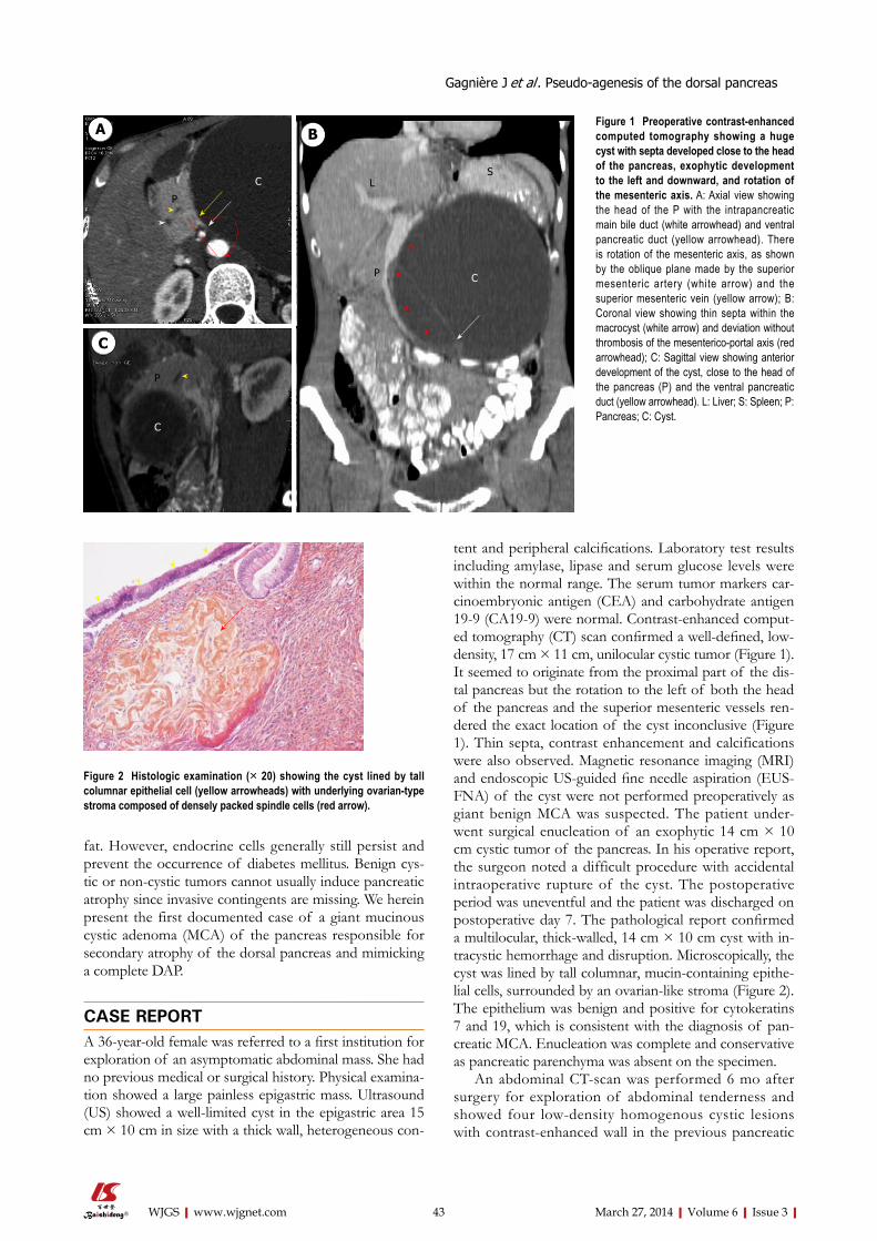
fat. However, endocrine cells generally still persist and prevent the occurrence of diabetes mellitus. Benign cys-tic or non-cystic tumors cannot usually induce pancreatic atrophy since invasive contingents are missing. We herein present the first documented case of a giant mucinous cystic adenoma (MCA) of the pancreas responsible for secondary atrophy of the dorsal pancreas and mimicking a complete DAP.
CASE REPORTA 36-year-old female was referred to a first institution for exploration of an asymptomatic abdominal mass. She had no previous medical or surgical history. Physical examina-tion showed a large painless epigastric mass. Ultrasound (US) showed a well-limited cyst in the epigastric area 15 cm × 10 cm in size with a thick wall, heterogeneous con-
tent and peripheral calcifications. Laboratory test results including amylase, lipase and serum glucose levels were within the normal range. The serum tumor markers car-cinoembryonic antigen (CEA) and carbohydrate antigen 19-9 (CA19-9) were normal. Contrast-enhanced comput-ed tomography (CT) scan confirmed a well-defined, low-density, 17 cm × 11 cm, unilocular cystic tumor (Figure 1). It seemed to originate from the proximal part of the dis-tal pancreas but the rotation to the left of both the head of the pancreas and the superior mesenteric vessels ren-dered the exact location of the cyst inconclusive (Figure 1). Thin septa, contrast enhancement and calcifications were also observed. Magnetic resonance imaging (MRI) and endoscopic US-guided fine needle aspiration (EUS-FNA) of the cyst were not performed preoperatively as giant benign MCA was suspected. The patient under-went surgical enucleation of an exophytic 14 cm × 10 cm cystic tumor of the pancreas. In his operative report, the surgeon noted a difficult procedure with accidental intraoperative rupture of the cyst. The postoperative period was uneventful and the patient was discharged on postoperative day 7. The pathological report confirmed a multilocular, thick-walled, 14 cm × 10 cm cyst with in-tracystic hemorrhage and disruption. Microscopically, the cyst was lined by tall columnar, mucin-containing epithe-lial cells, surrounded by an ovarian-like stroma (Figure 2). The epithelium was benign and positive for cytokeratins 7 and 19, which is consistent with the diagnosis of pan-creatic MCA. Enucleation was complete and conservative as pancreatic parenchyma was absent on the specimen.
An abdominal CT-scan was performed 6 mo after surgery for exploration of abdominal tenderness and showed four low-density homogenous cystic lesions with contrast-enhanced wall in the previous pancreatic
43 March 27, 2014|Volume 6|Issue 3|WJGS|www.wjgnet.com
Gagnière J et al . Pseudo-agenesis of the dorsal pancreas
Figure 1 Preoperative contrast-enhanced computed tomography showing a huge cyst with septa developed close to the head of the pancreas, exophytic development to the left and downward, and rotation of the mesenteric axis. A: Axial view showing the head of the P with the intrapancreatic main bile duct (white arrowhead) and ventral pancreatic duct (yellow arrowhead). There is rotation of the mesenteric axis, as shown by the oblique plane made by the superior mesenteric artery (white arrow) and the superior mesenteric vein (yellow arrow); B: Coronal view showing thin septa within the macrocyst (white arrow) and deviation without thrombosis of the mesenterico-portal axis (red arrowhead); C: Sagittal view showing anterior development of the cyst, close to the head of the pancreas (P) and the ventral pancreatic duct (yellow arrowhead). L: Liver; S: Spleen; P: Pancreas; C: Cyst.
Figure 2 Histologic examination (× 20) showing the cyst lined by tall columnar epithelial cell (yellow arrowheads) with underlying ovarian-type stroma composed of densely packed spindle cells (red arrow).
A B
C
P
C
C
P
LS
P C

enucleation area (Figure 3). The body and tail of the pancreas were not visible on CT nor on the upper part of the head of the pancreas, suggesting complete DAP. Magnetic resonance cholangiopancreatography (MR-CP) showed absence of the body and tail of the pancreas with no accessory pancreatic duct that would confirm the diagnosis of DAP (Figure 4). There was no other pan-creatic anomaly and no polysplenia. The patient was then referred to our institution. A second interpretation of the scan pictures showed splenic vein obstruction with major tributaries around the stomach, suggesting segmental portal hypertension (Figure 3). These features were also present on the initial CT. The serum levels of glucose, amylase and lipase were still normal. The suspected diag-noses were either recurrence of the MCA following dif-ficult and incomplete primary resection, as suggested by intraoperative rupture, or multiple pseudocysts due to a latent post-operative pancreatic leak. A second-look lapa-roscopy was advocated because of abdominal tenderness, risk of recurrence of the MCA and because EUS-FNA failed to distinguish MCA from pancreatic pseudocysts. Laparoscopic exploration showed extra-pancreatic mul-tiple cysts close to the first duodenum at the anterior part of the head of the pancreas, without pancreatic leak. The body and tail of the pancreas were also absent. Intraop-erative pathology examination of the cysts confirmed pseudocysts with fat necrosis. Postoperative course was uneventful and the patient was discharged on postopera-tive day 3. One year after initial resection the patient had no diabetes mellitus, and routine blood parameters, in particular serum glucose level, were normal.
DISCUSSIONTo the best of our knowledge, there are no documented reports of pancreatic cyst-including benign MCA-associated with congenital or secondary atrophy of the distal pancreas. MCA is a rare benign cystic tumor char-acterized by an ovarian stroma underlying the epithelium
of the cyst. Differential diagnosis includes other benign cystic lesions such as serous cystic neoplasm, intraductal papillary mucinous neoplasm and post-pancreatitis pseu-docysts[3]. Clinical presentation (female sex, location in the distal pancreas and no history of pancreatitis) and paraclinical investigations (MRI and EUS-FNA showing no pancreatic duct communication, wall calcifications and high level of intra-cystic CA19-9 and CEA) are sugges-tive of MCA[4]. Prophylactic resection is warranted as ma-lignant transformation can occur in 6%-27% of cases[5,6]. The origin of ovarian stroma remains unclear. It has been suggested to derive from ectopic tissue within the pancreas incorporated throughout close migration of the left primordial gonad and dorsal pancreatic bud during embryogenesis[7-9], which would explain the predilection of MCA for the body-tail region of the pancreas. Conse-quently, the association of complete DAP with MCA is theoretically not possible.
DAP is a congenital agenesis of the pancreas that can be partial or complete. It is a rare event since only 54 cases have been reported in the literature[10]. The pancreas develops from ventral and dorsal endodermal buds dur-ing embryogenesis. The ventral bud gives rise to the ma-jor part of the head and uncinate process, which drains through the duct of Wirsung (i.e., the main pancreatic duct). The dorsal bud forms the upper part of the head, body and tail of the pancreas and drains through the duct of Santorini (i.e., the accessory pancreatic duct). Each
44 March 27, 2014|Volume 6|Issue 3|WJGS|www.wjgnet.com
C C CP
Figure 3 Post-operative contrast-enhanced computed tomography show-ing three low-density homogenous cystic lesions suggesting secondary dissemination of the resected cyst. Major tributaries were also present around the stomach (red arrowheads). The P has a total distal atrophy as shown by lack of pancreatic tissue behind the mesenterico-portal axis (white arrow). P: Pancreas; C: Cyst.
Figure 4 Coronal magnetic resonance imagery showing lack of the body and tail of the pancreas and of the splenic vein (A), magnetic resonance cholangiopancreatography showing the common bile duct joining the ven-tral pancreatic duct at the posterior part of the head of the pancreas (B). The dorsal pancreatic duct is not visible.
A
B
Left gastric vein
Portal vein
Pancreatic head
Gallbladder Main bile duct
Ventral pancreatic duct
Gagnière J et al . Pseudo-agenesis of the dorsal pancreas
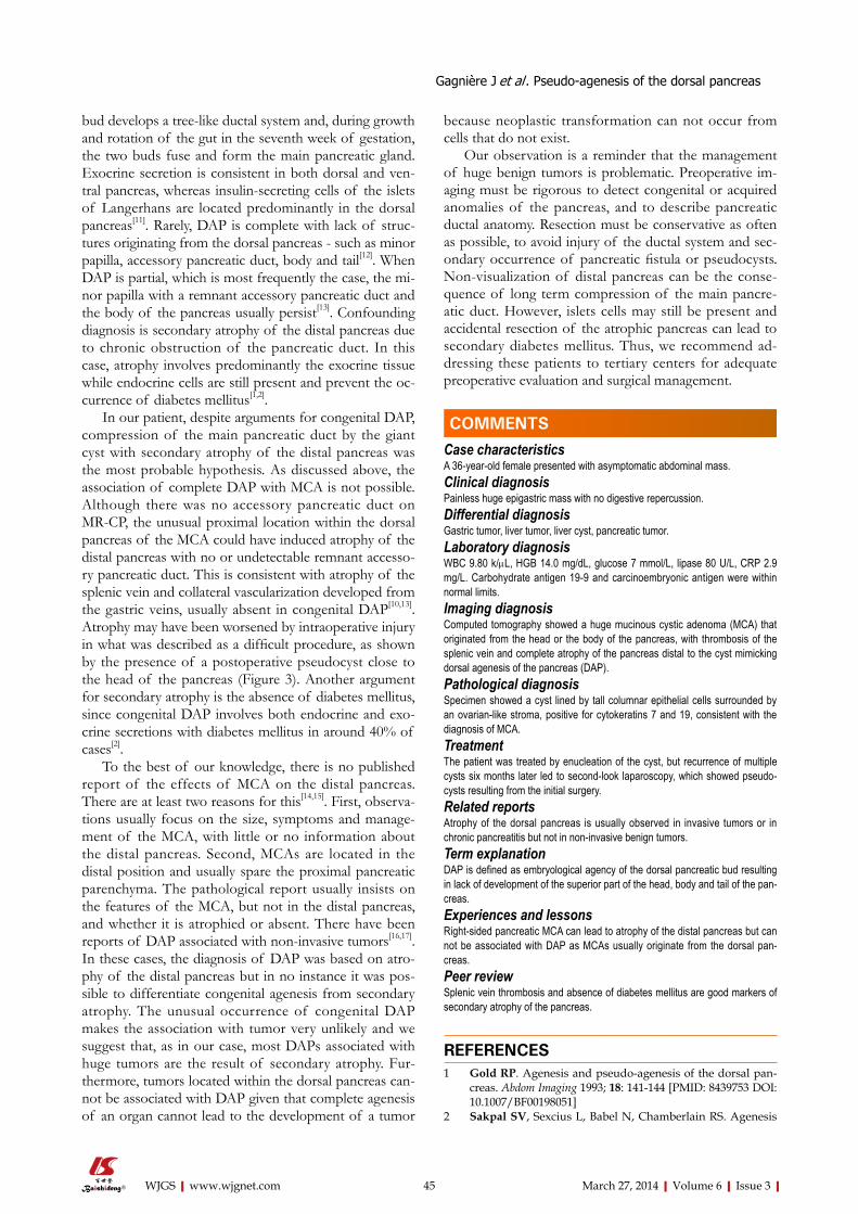
45 March 27, 2014|Volume 6|Issue 3|WJGS|www.wjgnet.com
because neoplastic transformation can not occur from cells that do not exist.
Our observation is a reminder that the management of huge benign tumors is problematic. Preoperative im-aging must be rigorous to detect congenital or acquired anomalies of the pancreas, and to describe pancreatic ductal anatomy. Resection must be conservative as often as possible, to avoid injury of the ductal system and sec-ondary occurrence of pancreatic fistula or pseudocysts. Non-visualization of distal pancreas can be the conse-quence of long term compression of the main pancre-atic duct. However, islets cells may still be present and accidental resection of the atrophic pancreas can lead to secondary diabetes mellitus. Thus, we recommend ad-dressing these patients to tertiary centers for adequate preoperative evaluation and surgical management.
COMMENTSCase characteristicsA 36-year-old female presented with asymptomatic abdominal mass.Clinical diagnosisPainless huge epigastric mass with no digestive repercussion.Differential diagnosisGastric tumor, liver tumor, liver cyst, pancreatic tumor.Laboratory diagnosisWBC 9.80 k/μL, HGB 14.0 mg/dL, glucose 7 mmol/L, lipase 80 U/L, CRP 2.9 mg/L. Carbohydrate antigen 19-9 and carcinoembryonic antigen were within normal limits.Imaging diagnosisComputed tomography showed a huge mucinous cystic adenoma (MCA) that originated from the head or the body of the pancreas, with thrombosis of the splenic vein and complete atrophy of the pancreas distal to the cyst mimicking dorsal agenesis of the pancreas (DAP).Pathological diagnosisSpecimen showed a cyst lined by tall columnar epithelial cells surrounded by an ovarian-like stroma, positive for cytokeratins 7 and 19, consistent with the diagnosis of MCA.TreatmentThe patient was treated by enucleation of the cyst, but recurrence of multiple cysts six months later led to second-look laparoscopy, which showed pseudo-cysts resulting from the initial surgery.Related reportsAtrophy of the dorsal pancreas is usually observed in invasive tumors or in chronic pancreatitis but not in non-invasive benign tumors.Term explanationDAP is defined as embryological agency of the dorsal pancreatic bud resulting in lack of development of the superior part of the head, body and tail of the pan-creas.Experiences and lessonsRight-sided pancreatic MCA can lead to atrophy of the distal pancreas but can not be associated with DAP as MCAs usually originate from the dorsal pan-creas.Peer reviewSplenic vein thrombosis and absence of diabetes mellitus are good markers of secondary atrophy of the pancreas.
REFERENCES1 Gold RP. Agenesis and pseudo-agenesis of the dorsal pan-
creas. Abdom Imaging 1993; 18: 141-144 [PMID: 8439753 DOI: 10.1007/BF00198051]
2 Sakpal SV, Sexcius L, Babel N, Chamberlain RS. Agenesis
bud develops a tree-like ductal system and, during growth and rotation of the gut in the seventh week of gestation, the two buds fuse and form the main pancreatic gland. Exocrine secretion is consistent in both dorsal and ven-tral pancreas, whereas insulin-secreting cells of the islets of Langerhans are located predominantly in the dorsal pancreas[11]. Rarely, DAP is complete with lack of struc-tures originating from the dorsal pancreas - such as minor papilla, accessory pancreatic duct, body and tail[12]. When DAP is partial, which is most frequently the case, the mi-nor papilla with a remnant accessory pancreatic duct and the body of the pancreas usually persist[13]. Confounding diagnosis is secondary atrophy of the distal pancreas due to chronic obstruction of the pancreatic duct. In this case, atrophy involves predominantly the exocrine tissue while endocrine cells are still present and prevent the oc-currence of diabetes mellitus[1,2].
In our patient, despite arguments for congenital DAP, compression of the main pancreatic duct by the giant cyst with secondary atrophy of the distal pancreas was the most probable hypothesis. As discussed above, the association of complete DAP with MCA is not possible. Although there was no accessory pancreatic duct on MR-CP, the unusual proximal location within the dorsal pancreas of the MCA could have induced atrophy of the distal pancreas with no or undetectable remnant accesso-ry pancreatic duct. This is consistent with atrophy of the splenic vein and collateral vascularization developed from the gastric veins, usually absent in congenital DAP[10,13]. Atrophy may have been worsened by intraoperative injury in what was described as a difficult procedure, as shown by the presence of a postoperative pseudocyst close to the head of the pancreas (Figure 3). Another argument for secondary atrophy is the absence of diabetes mellitus, since congenital DAP involves both endocrine and exo-crine secretions with diabetes mellitus in around 40% of cases[2].
To the best of our knowledge, there is no published report of the effects of MCA on the distal pancreas. There are at least two reasons for this[14,15]. First, observa-tions usually focus on the size, symptoms and manage-ment of the MCA, with little or no information about the distal pancreas. Second, MCAs are located in the distal position and usually spare the proximal pancreatic parenchyma. The pathological report usually insists on the features of the MCA, but not in the distal pancreas, and whether it is atrophied or absent. There have been reports of DAP associated with non-invasive tumors[16,17]. In these cases, the diagnosis of DAP was based on atro-phy of the distal pancreas but in no instance it was pos-sible to differentiate congenital agenesis from secondary atrophy. The unusual occurrence of congenital DAP makes the association with tumor very unlikely and we suggest that, as in our case, most DAPs associated with huge tumors are the result of secondary atrophy. Fur-thermore, tumors located within the dorsal pancreas can-not be associated with DAP given that complete agenesis of an organ cannot lead to the development of a tumor
COMMENTS
Gagnière J et al . Pseudo-agenesis of the dorsal pancreas
46 March 27, 2014|Volume 6|Issue 3|WJGS|www.wjgnet.com
of the dorsal pancreas and its association with pancreatic tumors. Pancreas 2009; 38: 367-373 [PMID: 19390403 DOI: 10.1097/MPA.0b013e318196c401]
3 Scoazec JY, Vullierme MP, Barthet M, Gonzalez JM, Sau-vanet A. Cystic and ductal tumors of the pancreas: diag-nosis and management. J Visc Surg 2013; 150: 69-84 [PMID: 23518192 DOI: 10.1016/j.jviscsurg.2013.02.003]
4 Goh BK, Tan YM, Chung YF, Chow PK, Cheow PC, Wong WK, Ooi LL. A review of mucinous cystic neoplasms of the pancreas defined by ovarian-type stroma: clinicopathologi-cal features of 344 patients. World J Surg 2006; 30: 2236-2245 [PMID: 17103100 DOI: 10.1007/s00268-006-0126-1]
5 Le Borgne J, de Calan L, Partensky C. Cystadenomas and cystadenocarcinomas of the pancreas: a multiinstitutional retrospective study of 398 cases. French Surgical Association. Ann Surg 1999; 230: 152-161 [PMID: 10450728 DOI: 10.1097/00000658-199908000-00004]
6 Tanaka M, Chari S, Adsay V, Fernandez-del Castillo C, Falconi M, Shimizu M, Yamaguchi K, Yamao K, Matsuno S. International consensus guidelines for management of intraductal papillary mucinous neoplasms and mucinous cystic neoplasms of the pancreas. Pancreatology 2006; 6: 17-32 [PMID: 16327281 DOI: 10.1159/000090023]
7 Nishimura H. Atlas of human prenatal histology. Tokyo-New York, Igaku-Shoin, 1983
8 Tuchmann-Duplessis H. Embryologie travaux pratiques et enseignement dirigé. Paris: Masson, 1968
9 Zamboni G, Scarpa A, Bogina G, Iacono C, Bassi C, Talamini G, Sessa F, Capella C, Solcia E, Rickaert F, Mariuzzi GM, Klöppel G. Mucinous cystic tumors of the pancreas: clinico-pathological features, prognosis, and relationship to other mucinous cystic tumors. Am J Surg Pathol 1999; 23: 410-422 [PMID: 10199470 DOI: 10.1097/00000478-199904000-00005]
10 Schnedl WJ, Piswanger-Soelkner C, Wallner SJ, Reittner P, Krause R, Lipp RW, Hohmeier HE. Agenesis of the dor-sal pancreas and associated diseases. Dig Dis Sci 2009; 54: 481-487 [PMID: 18618254 DOI: 10.1007/s10620-008-0370-3]
11 Wittingen J, Frey CF. Islet concentration in the head, body, tail and uncinate process of the pancreas. Ann Surg 1974; 179: 412-414 [PMID: 4593099 DOI: 10.1097/00000658-197404000-00005]
12 Mohapatra M, Mishra S, Dalai PC, Acharya SD, Nahak B, Ibrarullah M, Panda K, Mishra SS. Imaging findings in agen-esis of the dorsal pancreas. Report of three cases. JOP 2012; 13: 108-114 [PMID: 22233961]
13 Schnedl WJ, Reisinger EC, Schreiber F, Pieber TR, Lipp RW, Krejs GJ. Complete and partial agenesis of the dorsal pan-creas within one family. Gastrointest Endosc 1995; 42: 485-487 [PMID: 8566643 DOI: 10.1016/S0016-5107(95)70055-2]
14 Teixeira F, Moutinho V, Ushinohama A, Akaishi E, Utiyama E, Rasslan S. Giant mucinous cystic neoplasm of the pan-creas. J Gastrointest Surg 2010; 14: 1197-1198 [PMID: 19960269 DOI: 10.1007/s11605-009-1117-y]
15 Mizutani S, Nakamura Y, Ogata M, Watanabe M, Tokunaga A, Tajiri T. A case of giant mucinous cystic neoplasm of the pancreas resected with laparoscopic surgery. J Nippon Med Sch 2009; 76: 212-216 [PMID: 19755797 DOI: 10.1272/jnms.76.212]
16 Nakamura Y, Egami K, Maeda S, Hosone M, Onda M. Solid and papillary tumor of the pancreas complicating agenesis of the dorsal pancreas. J Hepatobiliary Pancreat Surg 2001; 8: 485-489 [PMID: 11702261 DOI: 10.1007/s005340100014]
17 Ulusan S, Bal N, Kizilkilic O, Bolat F, Yildirim S, Yildirim T, Niron EA. Case report: solid-pseudopapillary tumour of the pancreas associated with dorsal agenesis. Br J Radiol 2005; 78: 441-443 [PMID: 15845940 DOI: 10.1259/bjr/91312352]
P- Reviewers: Azhar R, Guan YS, Klinge U, Pavlidis TE S- Editor: Qi Y L- Editor: A E- Editor: Liu SQ
Gagnière J et al . Pseudo-agenesis of the dorsal pancreas

Giant Meckel’s diverticulum: An exceptional cause of intestinal obstruction
Sami Akbulut, Yusuf Yagmur
Sami Akbulut, Yusuf Yagmur, Department of Surgery, Di-yarbakir Education and Research Hospital, Diyarbakir 21400, TurkeyAuthor contributions: Akbulut S and Yagmur Y designed, or-ganized and wrote the report, and were attending doctors for the patients; Akbulut S performed surgical operation.Correspondence to: Sami Akbulut, MD, FICS, FACS, Depart-ment of Surgery, Diyarbakir Education and Research Hospital, Uckuyular Mevki, Kayapinar, Diyarbakir 21400, Turkey. [email protected]: +90-412-2580075 Fax: +90-412-2580070 Received: November 19, 2013 Revised: January 5, 2014Accepted: February 16, 2014Published online: March 27, 2014
Abstract Meckel’s diverticulum (MD) results from incomplete involution of the proximal portion of the vitelline (also known as the omphalomesenteric) duct during weeks 5-7 of foetal development. Although MD is the most commonly diagnosed congenital gastrointestinal anom-aly, it is estimated to affect only 2% of the population worldwide. Most cases are asymptomatic, and diagno-sis is often made following investigation of unexplained gastrointestinal bleeding, perforation, inflammation or obstruction that prompt clinic presentation. While MD range in size from 1-10 cm, cases of giant MD (≥ 5 cm) are relatively rare and associated with more severe forms of the complications, especially for obstruction. Herein, we report a case of giant MD with secondary small bowel obstruction in an adult male that was suc-cessfully managed by surgical resection and anasto-mosis created with endoscopic stapler device (80 mm, endo-GIA stapler). Patient was discharged on post-operative day 6 without any complications. Histopatho-logic examination indicated Meckel’s diverticulitis with-out gastric or pancreatic metaplasia.
© 2014 Baishideng Publishing Group Co., Limited. All rights reserved.
Key words: Meckel’s diverticulum; Giant Meckel’s diver-ticulum; Intestinal obstruction; Small bowel
Core tip: The most commonly diagnosed congenital anomaly of the gastrointestinal tract is Meckel’s diver-ticulum (MD), which occurs upon failure of the om-phalomesenteric duct to regress and involute. MD can remain asymptomatic, and cases are generally diag-nosed incidentally or upon investigation of unexplained gastrointestinal bleeding, perforation, inflammation, or obstruction for both paediatric and adult cases. It is es-timated that as little as 4% of cases manifest complica-tions, and obstruction is the most common presenting symptom in adults. In this case study, we report a case of giant MD with secondary small bowel obstruction in an adult male that was successfully managed by surgi-cal resection and anastomosis created with endoscopic stapler.
Akbulut S, Yagmur Y. Giant Meckel’s diverticulum: An ex-ceptional cause of intestinal obstruction. World J Gastrointest Surg 2014; 6(3): 47-50 Available from: URL: http://www.wjgnet.com/1948-9366/full/v6/i3/47.htm DOI: http://dx.doi.org/10.4240/wjgs.v6.i3.47
INTRODUCTIONThe most commonly diagnosed congenital anomaly of the gastrointestinal tract is Meckel’s diverticulum (MD), which occurs upon failure of the vitelline (also known as the omphalomesenteric) duct to regress and involute[1-3]. Accumulated experience with surgical treatment of MD (using both open and laparoscopic procedures) has led to the clinical “rule of 2” for symptomatic cases, whereby the anatomical deformity (with estimated prevalence in 2% of the population) is most frequently located 2 feet from the ileocaecal junction and is 2 inches long[2]. MD can remain asymptomatic, and cases are generally diag-
CASE REPORT
Online Submissions: http://www.wjgnet.com/esps/[email protected]:10.4240/wjgs.v6.i3.47
47 March 27, 2014|Volume 6|Issue 3|WJGS|www.wjgnet.com
World J Gastrointest Surg 2014 March 27; 6(3): 47-50ISSN 1948-9366 (online)
© 2014 Baishideng Publishing Group Co., Limited. All rights reserved.

nosed incidentally or upon investigation of unexplained gastrointestinal bleeding, perforation, inflammation, or obstruction for both paediatric and adult cases[1].
It is estimated that as little as 4% of cases manifest complications, and obstruction is the most common presenting symptom in adults[1]. There is evidence that severity of symptoms correlates with MD size. Ninety percent of the reported MDs are between 1 and 10 cm, the average size being 3 cm. MDs ≥ 5 cm are classified as giant MD, are relatively rare, and may be more prone to complications[1]. Here, we report a case of giant MD which was diagnosed in an adult male with small bowel obstruction and successfully managed by resection.
CASE REPORTA 23-year-old male patient presented at the Emergency Department with a complaint of abdominal pain, nausea, and vomiting that had persisted for 5 d and increased in severity over the last 24 h. The patient reported no faecal or gas discharge during the previous 48 h. History taking upon admission revealed that the patient had visited hos-pitals frequently for many years with similar gastrointesti-nal complaints as well as bloating. The patient’s abdomen was remarkably distended and initial clinical assessment indicated hypovolemia. Physical examination revealed sig-nificant bowel sounds and substantial abdominal rebound pain, both more robust in the periumbilical area. Labo-ratory testing showed increased white blood cell count (11.8 × 10³/μL; normal range: 4.1 × 10³-11.2 × 10³), haemoglobin (17.0 g/dL; 12.5-16.0), haematocrit (49.6%; 37.0-47.0) and creatinine (1.4 mg/dL; 0.4-1.2), but nor-mal blood urea nitrogen (27 mg/dL; 10-50). Abdominal X-ray indicated remarkably high air-fluid levels (Figure 1).
An emergency laparotomy was performed and re-vealed oedema throughout the entire small bowel, dila-tion of small bowel segments, and a giant MD (27 cm long and 6 cm wide) on the antimesenteric border of the small bowel at 80 cm proximal to the ileocaecal valve (Figure 2). The diverticulum’s tip was strongly adhered to the parietal peritoneum of the abdominal wall at the site of the pelvis, having been pushed up against this site due to the MD’s excessively large size and high-volume intestinal content. No other obstruction was observed in the gastrointestinal tract. Resection of the small bowel was performed with a linear stapler and an ileoileal anas-tomosis was generated using a 80 mm endo-GIA stapler (Figure 3). The resection was completed without incident, and the patient was discharged on post-operative day 6 without any complications. Pathology findings indicated diverticulitis without gastric or pancreatic metaplasia.
DISCUSSIONMD is a true diverticulum, comprising all three layers of the small intestine. Compared to the overall incidence of 0.14%-4.50% (estimated by autopsy findings and retro-spective studies)[4], giant MD are rare[5]. The largest giant
MDs reported have been > 100 cm long[6], 96 cm long[7], 85 cm long[8], and 66 cm long[9,10].
In adult cases of MD, obstruction is the most fre-quently reporting presenting symptom[11-14] and can be caused by either the diverticulum’s attachment to the um-bilicus, abdominal wall or other viscera by a fibrous band or by interference due to the mobility of an unattached diverticulum[11]. Though first hypothesized in 1902[13], these potential reasons for MD-caused intestinal obstruc-
48 March 27, 2014|Volume 6|Issue 3|WJGS|www.wjgnet.com
Akbulut S et al . Intestinal obstruction due to Giant’s Meckel’s diverticulum
Figure 1 Abdominal X-ray radiography showing air-fluid levels represen-tative of intestinal obstruction.
Figure 2 Giant Meckel's diverticulum causing gastrointestinal obstruc-tion. White arrow: Diameter of Meckel’s diverticulum; Black arrow: Proximal ileal segment; Yellow arrow: Distal ileal segment.
Figure 3 Giant Meckel's diverticulum view after resection. Black arrow: Proximal ileal segment; Yellow arrow: Distal ileal segment.
tion remain the features by which MD cases are classified. The obstructions associated with a free or unattached diverticulum, or having only one attachment to the intes-tine, represent first MD type, and obstructions associated with an attached diverticulum, including through its ter-minal ligament, to the abdominal wall or intestinal viscus, represent the second type. Between these two types, the former is much rarer.
When the congenital malformation occurs, the free di-verticulum forms a volvulus with a loop, twisting the gut structure. Adhesions commonly form between the two arms of the twist, making an obstruction. Subsequent inflammation of the diverticulum further promotes con-striction of the bowel. Furthermore, an unattached, dis-tended diverticulum may cause movement of the looped intestine so that a kink forms in the intestine at the at-tachment point of the diverticulum; this event could lead to an obstruction without any concomitant structural changes in the intestinal wall. Persistence of such kinking may ultimately cause necrosis of the involved and proxi-mal gut tissues. Other potential aetiologies of MD-related intestinal obstructions exist. For example, the obstruction may be caused by twisting of the bowel along its long axis at the point of the diverticulum’s origin, by chronic inflammation of the diverticulum and its adjacent bowel, or by inversion of the mucous membrane alone, or of the entire diverticulum, with or without invagination.
Several case reports of MD-related obstructions have described strangulation caused by an adherent di-verticulum. Many causes of such an event have been proposed. First, the adherent diverticulum itself may act as a constricting band, such as an adventitious band or a peritoneal adhesion. Second, the adherent diverticulum may have resulted from looping and twisting of the gut in upon itself, forming a volvulus. Third, a volvulus of the attached diverticulum may itself represent a physical obstruction of the intestine. Finally, the diverticular band may become tensely drawn under certain conditions[13].
In a review of 402 patients with MD, 16.9% of the patients were found to have demonstrated symptoms that are considered clinical references for diverticulum[14], with obstruction of the small intestine, and inflammation and bleeding of the lower gastrointestinal tract account-ing for 90% of those presenting symptoms. In another study of 34 MD cases, the most common complications were intestinal obstruction (37%), intussusception (14%), inflammation, and rectal bleeding (12%); interestingly, intussusception and volvulus were associated with those patients having intestinal obstruction[15].
For the current case of giant MD, the diverticulum was large in diameter, long in length, and adherent (caus-ing a small bowel obstruction). The structural features of a MD provide clues to the type of complications it may cause. For example, diverticulitis and torsion are com-mon complications observed with long MDs that have a narrow base, while short MDs that have a stumpy base are more often associated with intussusception[16]. Thus, an elongated variant with a narrow neck is more likely to
result in torsion, whereas a short, wide-base diverticula may promote foreign body entrapment.
Cullen et al[17] studied the outcomes of diverticulec-tomy surgical management of MD-related complications and determined that the operative mortality and mor-bidity rates were 2% and 12%, respectively, and that the cumulative risk of long-term post-operative complica-tions was 7%; in contrast, analysis of patients receiving incidental diverticulectomy showed that the operative mortality, morbidity, and risk of long-term post-operative complications were lower (1%, 2%, and 2%, respectively). It is generally recommended that MD discovered inciden-tally during operation should be removed, regardless of the patient’s age.
In conclusion, this report describes a very rare form of acute small bowel obstruction secondary to giant MD encircling the terminal ileum, providing novel insights into this condition and describing its successful manage-ment by surgical resection.
COMMENTSCase characteristicsClinical symptoms include abdominal pain, nausea, vomiting, and no faecal or gas discharge.Clinical diagnosisAcute abdomen, mechanical small bowel obstructions.Differential diagnosisIntestinal malrotation, congenital anomalous bands, tumor obstruction.Laboratory diagnosisLaboratory tests showed a leukocytosis (11800/μL; 4100-11200), haemoglobin (17.0 g/dL; 12.5-16.0), haematocrit (49.6%; 37.0-47.0) and creatinine (1.4 mg/dL; 0.4-1.2).Imaging diagnosisAn abdominal X-ray radiography indicated remarkably high air-fluid levels.Pathological diagnosisPathology findings indicated Meckel’s diverticulitis without gastric or pancreatic metaplasia.TreatmentLimited ileal resection and end-to-end anastomosis created with stapler device.Term explanationMeckel’s diverticulum (MD), a remnant of the vitelline duct that normally disap-pears at the end of the seventh week of gestation, is the most common congenital abnormality of the small intestine. It arises from the antimesenteric border of the terminal ileum as a true diverticulum that contains all layers of the intestinal wall.Peer reviewThis is a well written case report on a farly common subject. It is well know that MD can couse intestinal obstruction and can reach fairly large dimensions, de-pending on the duration of the sub-occlusive symptoms.
REFERENCES1 Elsayes KM, Menias CO, Harvin HJ, Francis IR. Imaging
manifestations of Meckel’s diverticulum. AJR Am J Roentgenol 2007; 189: 81-88 [PMID: 17579156 DOI: 10.2214/AJR.06.1257]
2 Sagar J, Kumar V, Shah DK. Meckel’s diverticulum: a system-atic review. J R Soc Med 2006; 99: 501-505 [PMID: 17021300 DOI: 10.1258/jrsm.99.10.501]
3 Cartanese C, Petitti T, Marinelli E, Pignatelli A, Martignetti D, Zuccarino M, Ferrozzi L. Intestinal obstruction caused by torsed gangrenous Meckel’s diverticulum encircling termi-nal ileum. World J Gastrointest Surg 2011; 3: 106-109 [PMID: 21860699 DOI: 10.4240/wjgs.v3.i7.106]
49 March 27, 2014|Volume 6|Issue 3|WJGS|www.wjgnet.com
COMMENTS
Akbulut S et al . Intestinal obstruction due to Giant’s Meckel’s diverticulum
50 March 27, 2014|Volume 6|Issue 3|WJGS|www.wjgnet.com
251-253 [PMID: 13999637 DOI: 10.1001/jama.1962.03050420027007]
12 Turgeon DK, Barnett JL. Meckel’s diverticulum. Am J Gas-troenterol 1990; 85: 777-781 [PMID: 2196781]
13 Halstead AE. IV. Intestinal Obstruction from Meckel’s Di-verticulum. Ann Surg 1902; 35: 471-494 [PMID: 17861102]
14 Mackey WC, Dineen P. A fifty year experience with Meck-el’s diverticulum. Surg Gynecol Obstet 1983; 156: 56-64 [PMID: 6600203]
15 Yamaguchi M, Takeuchi S, Awazu S. Meckel’s diverticulum: Investigation of 600 patients in Japanese literature. Am J Surg 1978: 136: 247-249 [PMID: 208325 DOI: 10.1016/0002-9610(78)90238-6]
16 Caiazzo P, Albano M, Del Vecchio G, Calbi F, Loffredo A, Pastore M, De Martino C, Di Lascio P, Tramutoli PR. Intesti-nal obstruction by giant Meckel’s diverticulum. Case report. G Chir 2011; 32: 491-494 [PMID: 22217379]
17 Cullen JJ, Kelly KA, Moir CR, Hodge DO, Zinsmeister AR, Melton LJ 3rd. Surgical management of Meckel's diverticu-lum. An epidemiologic, population-based study. Ann Surg 1994; 220: 564-568; discussion 568-569 [PMID: 7944666 DOI: 10.1097/00000658-199410000-00014]
P- Reviewers: Hiraki M, Iacono C, Nigri G S- Editor: Qi Y L- Editor: A E- Editor: Liu SQ
4 Limas C, Seretis K, Soultanidis C, Anagnostoulis S. Axial torsion and gangrene of a giant Meckel’s diverticulum. J Gas-trointestin Liver Dis 2006; 15: 67-68 [PMID: 16680236]
5 Torii Y, Hisatsune I, Imamura K, Morita K, Kumagaya N, Nakata H. Giant Meckel diverticulum containing entero-liths diagnosed by computed tomography and sonography. Gastrointest Radiol 1989; 14: 167-169 [PMID: 2651200 DOI: 10.1007/BF01889186]
6 Tisdall FF. An unusual Meckel’s diverticulum as a cause of intestinal hemorrhage. Am J Dis Child 1928; 36: 1218-1223
7 Chaffin L. Surgical emergencies during childhood caused by meckel’s diverticulum. Ann Surg 1941; 113: 47-56 [PMID: 17857714 DOI: 10.1097/00000658-194101000-00006]
8 Moll HH. Giant Meckel’s diverticulum (33 ½ inches long). Br J Surg 1926; 14: 176-179 [DOI: 10.1002/bjs.1800145317]
9 Goldstein M, Cragg RW. Elongated Meckel's Diverticulum In A Chıld. Am J Dis Child 1938; 55: 128-134
10 Moses WR. Meckel’s diverticulum; report of two unusual cases. N Engl J Med 1947; 237: 118-122 [PMID: 20252118 DOI: 10.1056/NEJM194707242370403]
11 Weinstein EC, Cain JC, Remine WH. Meckel‘s diverticulum: 55 years of clinical and surgical experience. JAMA 1962; 182:
Akbulut S et al . Intestinal obstruction due to Giant’s Meckel’s diverticulum
Coexistence of abdominal cocoon, intestinal perforation and incarcerated Meckel’s diverticulum in an inguinal hernia: A troublesome condition
Sami Akbulut, Yusuf Yagmur, Mehmet Babur
Sami Akbulut, Yusuf Yagmur, Mehmet Babur, Department of Surgery, Diyarbakir Education and Research Hospital, Uckuyular Mevki, Diyarbakir 21400, TurkeyAuthor contributions: Akbulut S and Yagmur Y designed the report, were the attending doctors for the patient, and wrote the manuscript; Akbulut S and Babur M performed the surgical operation.Correspondence to: Sami Akbulut, MD, FICS, FACS, Depart-ment of Surgery, Diyarbakir Education and Research Hospital, Uckuyular Mevki, Kayapinar, Diyarbakir 21400, Turkey. [email protected]: +90-412- 2580075 Fax: +90-412-2580070 Received: November 22, 2013 Revised: January 5, 2014Accepted: February 16, 2014Published online: March 27, 2014
AbstractSclerosing encapsulating peritonitis (SEP) is a rare dis-ease entity, in which the small intestine becomes en-cased and mechanically obstructed by a dense, fibrotic membrane. The disorder is characterized as either primary (idiopathic) or secondary to other causes. The idiopathic cases of SEP, which lack any identifiable eti-ology according to clinical, radiological and histopatho-logical findings, are also reported under the designation of abdominal cocoon syndrome. The most frequent presenting symptoms of all SEP cases are nausea, vom-iting, abdominal distention and inability to defecate, all of which are associated with the underlying intestinal obstruction. Persistent untreated SEP may advance to intestinal perforation, representing a life-threatening condition. However, preoperative diagnosis remains a particular clinical challenge, and most diagnoses are confirmed only when the typical fibrous membrane en-casing the small intestine is discovered by laparotomy. Here, we report the clinical presentation of an 87-year-old male with signs of intestinal obstruction and the ul-timate diagnosis of concurrent abdominal cocoon, right
incarcerated Meckel’s diverticulum, and gastrointestinal perforation in laparotomy.
© 2014 Baishideng Publishing Group Co., Limited. All rights reserved.
Key words: Sclerosing encapsulating peritonitis; Cocoon syndrome; Perforation; Meckel’s diverticulum
Core tip: Abdominal cocoon syndrome, also known as idiopathic sclerosing encapsulating peritonitis, is a rare disease entity, in which the small intestine becomes en-cased and mechanically obstructed by a dense, fibrotic membrane. While some patients with cocoon syndrome remain asymptomatic, the majority experience gas-trointestinal symptoms, including recurrent attacks of acute, sub-acute or chronic gastrointestinal obstruction, weight loss, loss of appetite, and development of a pal-pable abdominal mass. Herein, we describe an elderly patient who presented with signs of intestinal obstruc-tion and who was diagnosed with concurrent abdominal cocoon, right incarcerated Meckel’s diverticulum, and gastrointestinal perforation by exploratory laparotomy.
Akbulut S, Yagmur Y, Babur M. Coexistence of abdominal cocoon, intestinal perforation and incarcerated Meckel’s diver-ticulum in an inguinal hernia: A troublesome condition. World J Gastrointest Surg 2014; 6(3): 51-54 Available from: URL: http://www.wjgnet.com/1948-9366/full/v6/i3/51.htm DOI: http://dx.doi.org/10.4240/wjgs.v6.i3.51
INTRODUCTIONSclerosing encapsulating peritonitis (SEP) is a rare struc-tural abnormality whereby the small intestine becomes encased (or cocooned) by a dense fibrocollagenous
CASE REPORT
Online Submissions: http://www.wjgnet.com/esps/[email protected]:10.4240/wjgs.v6.i3.51
51 March 27, 2014|Volume 6|Issue 3|WJGS|www.wjgnet.com
World J Gastrointest Surg 2014 March 27; 6(3): 51-54ISSN 1948-9366 (online)
© 2014 Baishideng Publishing Group Co., Limited. All rights reserved.
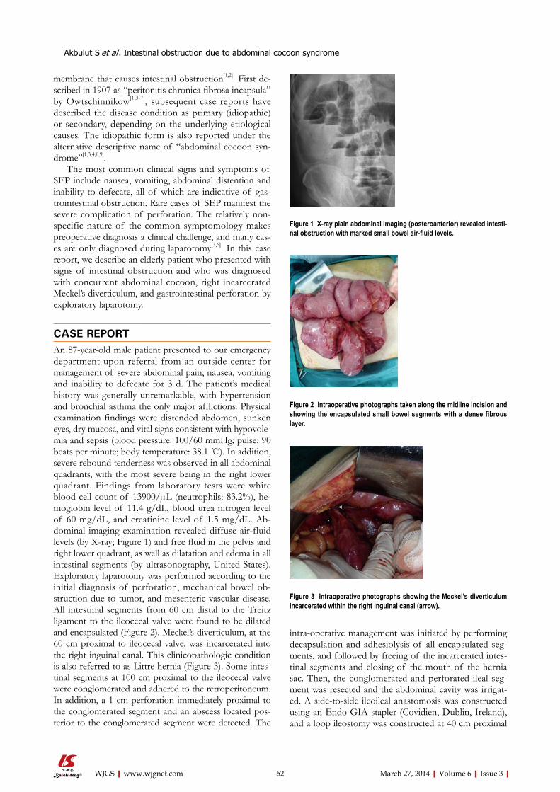
membrane that causes intestinal obstruction[1,2]. First de-scribed in 1907 as “peritonitis chronica fibrosa incapsula” by Owtschinnikow[1,3-7], subsequent case reports have described the disease condition as primary (idiopathic) or secondary, depending on the underlying etiological causes. The idiopathic form is also reported under the alternative descriptive name of “abdominal cocoon syn-drome”[1,3,4,8,9].
The most common clinical signs and symptoms of SEP include nausea, vomiting, abdominal distention and inability to defecate, all of which are indicative of gas-trointestinal obstruction. Rare cases of SEP manifest the severe complication of perforation. The relatively non-specific nature of the common symptomology makes preoperative diagnosis a clinical challenge, and many cas-es are only diagnosed during laparotomy[3,6]. In this case report, we describe an elderly patient who presented with signs of intestinal obstruction and who was diagnosed with concurrent abdominal cocoon, right incarcerated Meckel’s diverticulum, and gastrointestinal perforation by exploratory laparotomy.
CASE REPORTAn 87-year-old male patient presented to our emergency department upon referral from an outside center for management of severe abdominal pain, nausea, vomiting and inability to defecate for 3 d. The patient’s medical history was generally unremarkable, with hypertension and bronchial asthma the only major afflictions. Physical examination findings were distended abdomen, sunken eyes, dry mucosa, and vital signs consistent with hypovole-mia and sepsis (blood pressure: 100/60 mmHg; pulse: 90 beats per minute; body temperature: 38.1 ℃). In addition, severe rebound tenderness was observed in all abdominal quadrants, with the most severe being in the right lower quadrant. Findings from laboratory tests were white blood cell count of 13900/μL (neutrophils: 83.2%), he-moglobin level of 11.4 g/dL, blood urea nitrogen level of 60 mg/dL, and creatinine level of 1.5 mg/dL. Ab-dominal imaging examination revealed diffuse air-fluid levels (by X-ray; Figure 1) and free fluid in the pelvis and right lower quadrant, as well as dilatation and edema in all intestinal segments (by ultrasonography, United States). Exploratory laparotomy was performed according to the initial diagnosis of perforation, mechanical bowel ob-struction due to tumor, and mesenteric vascular disease. All intestinal segments from 60 cm distal to the Treitz ligament to the ileocecal valve were found to be dilated and encapsulated (Figure 2). Meckel’s diverticulum, at the 60 cm proximal to ileocecal valve, was incarcerated into the right inguinal canal. This clinicopathologic condition is also referred to as Littre hernia (Figure 3). Some intes-tinal segments at 100 cm proximal to the ileocecal valve were conglomerated and adhered to the retroperitoneum. In addition, a 1 cm perforation immediately proximal to the conglomerated segment and an abscess located pos-terior to the conglomerated segment were detected. The
intra-operative management was initiated by performing decapsulation and adhesiolysis of all encapsulated seg-ments, and followed by freeing of the incarcerated intes-tinal segments and closing of the mouth of the hernia sac. Then, the conglomerated and perforated ileal seg-ment was resected and the abdominal cavity was irrigat-ed. A side-to-side ileoileal anastomosis was constructed using an Endo-GIA stapler (Covidien, Dublin, Ireland), and a loop ileostomy was constructed at 40 cm proximal
52 March 27, 2014|Volume 6|Issue 3|WJGS|www.wjgnet.com
Akbulut S et al . Intestinal obstruction due to abdominal cocoon syndrome
Figure 1 X-ray plain abdominal imaging (posteroanterior) revealed intesti-nal obstruction with marked small bowel air-fluid levels.
Figure 2 Intraoperative photographs taken along the midline incision and showing the encapsulated small bowel segments with a dense fibrous layer.
Figure 3 Intraoperative photographs showing the Meckel’s diverticulum incarcerated within the right inguinal canal (arrow).
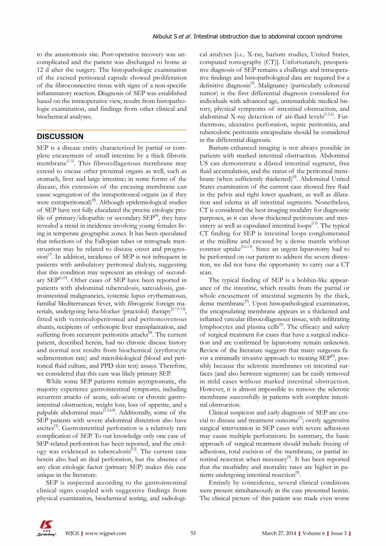
to the anastomosis site. Post-operative recovery was un-complicated and the patient was discharged to home at 12 d after the surgery. The histopathologic examination of the excised peritoneal capsule showed proliferation of the fibroconnective tissue with signs of a non-specific inflammatory reaction. Diagnosis of SEP was established based on the intraoperative view, results from histopatho-logic examination, and findings from other clinical and biochemical analyses.
DISCUSSIONSEP is a disease entity characterized by partial or com-plete encasement of small intestine by a thick fibrotic membrane[1,5]. This fibrocollagenous membrane may extend to encase other proximal organs as well, such as stomach, liver and large intestine; in some forms of the disease, this extension of the encasing membrane can cause segregation of the intraperitoneal organs (as if they were extraperitoneal)[8]. Although epidemiological studies of SEP have not fully elucidated the precise etiologic pro-file of primary/idiopathic or secondary SEP[4], they have revealed a trend in incidence involving young females liv-ing in temperate geographic zones. It has been speculated that infections of the Fallopian tubes or retrograde men-struation may be related to disease onset and progres-sion[7]. In addition, incidence of SEP is not infrequent in patients with ambulatory peritoneal dialysis, suggesting that this condition may represent an etiology of second-ary SEP[6,10]. Other cases of SEP have been reported in patients with abdominal tuberculosis, sarcoidosis, gas-trointestinal malignancies, systemic lupus erythematosus, familial Mediterranean fever, with fibrogenic foreign ma-terials, undergoing beta-blocker (practolol) therapy[4,7,9-12], fitted with ventriculoperitoneal and peritoneovenous shunts, recipients of orthotopic liver transplantation, and suffering from recurrent peritonitis attacks[6]. The current patient, described herein, had no chronic disease history and normal test results from biochemical (erythrocyte sedimentation rate) and microbiological (blood and peri-toneal fluid culture, and PPD skin test) assays. Therefore, we considered that this case was likely primary SEP.
While some SEP patients remain asymptomatic, the majority experience gastrointestinal symptoms, including recurrent attacks of acute, sub-acute or chronic gastro-intestinal obstruction, weight loss, loss of appetite, and a palpable abdominal mass[1,3,6,8]. Additionally, some of the SEP patients with severe abdominal distention also have ascites[3]. Gastrointestinal perforation is a relatively rare complication of SEP. To our knowledge only one case of SEP-related perforation has been reported, and the etiol-ogy was evidenced as tuberculosis[12]. The current case herein also had an ileal perforation, but the absence of any clear etiologic factor (primary SEP) makes this case unique in the literature.
SEP is suspected according to the gastrointestinal clinical signs coupled with suggestive findings from physical examination, biochemical testing, and radiologi-
cal analyses [i.e., X-ray, barium studies, United States, computed tomography (CT)]. Unfortunately, preopera-tive diagnosis of SEP remains a challenge and intraopera-tive findings and histopathological data are required for a definitive diagnosis[9]. Malignancy (particularly colorectal tumor) is the first differential diagnosis considered for individuals with advanced age, unremarkable medical his-tory, physical symptoms of intestinal obstruction, and abdominal X-ray detection of air-fluid levels[1,2,6]. Fur-thermore, ulcerative perforation, septic peritonitis, and tuberculotic peritonitis encapsulans should be considered in the differential diagnosis.
Barium-enhanced imaging is not always possible in patients with marked intestinal obstruction. Abdominal US can demonstrate a dilated intestinal segment, free fluid accumulation, and the status of the peritoneal mem-brane (when sufficiently thickened)[4]. Abdominal United States examination of the current case showed free fluid in the pelvis and right lower quadrant, as well as dilata-tion and edema in all intestinal segments. Nonetheless, CT is considered the best imaging modality for diagnostic purposes, as it can show thickened peritoneum and mes-entery as well as capsulated intestinal loops[13]. The typical CT finding for SEP is intestinal loops conglomerated at the midline and encased by a dense mantle without contrast uptake[5,6,13]. Since an urgent laparotomy had to be performed on our patient to address the severe disten-tion, we did not have the opportunity to carry out a CT scan.
The typical finding of SEP is a bobbin-like appear-ance of the intestine, which results from the partial or whole encasement of intestinal segments by the thick, dense membrane[9]. Upon histopathological examination, the encapsulating membrane appears as a thickened and inflamed vascular fibrocollagenous tissue, with infiltrating lymphocytes and plasma cells[5]. The efficacy and safety of surgical treatment for cases that have a surgical indica-tion and are confirmed by laparotomy remain unknown. Review of the literature suggests that many surgeons fa-vor a minimally invasive approach to treating SEP[6], pos-sibly because the sclerotic membranes on intestinal sur-faces (and also between segments) can be easily removed in mild cases without marked intestinal obstruction. However, it is almost impossible to remove the sclerotic membrane successfully in patients with complete intesti-nal obstruction.
Clinical suspicion and early diagnosis of SEP are cru-cial to disease and treatment outcome[7]; overly aggressive surgical intervention in SEP cases with severe adhesions may cause multiple perforations. In summary, the basic approach of surgical treatment should include freeing of adhesions, total excision of the membrane, or partial in-testinal resection when necessary[9]. It has been reported that the morbidity and mortality rates are higher in pa-tients undergoing intestinal resection[9].
Entirely by coincidence, several clinical conditions were present simultaneously in the case presented herein. The clinical picture of this patient was made even worse
53 March 27, 2014|Volume 6|Issue 3|WJGS|www.wjgnet.com
Akbulut S et al . Intestinal obstruction due to abdominal cocoon syndrome
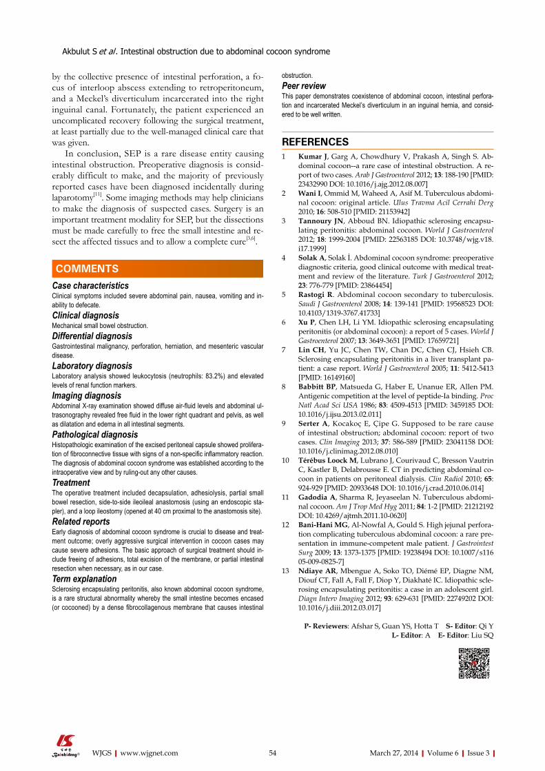
54 March 27, 2014|Volume 6|Issue 3|WJGS|www.wjgnet.com
obstruction.Peer reviewThis paper demonstrates coexistence of abdominal cocoon, intestinal perfora-tion and incarcerated Meckel’s diverticulum in an inguinal hernia, and consid-ered to be well written.
REFERENCES1 Kumar J, Garg A, Chowdhury V, Prakash A, Singh S. Ab-
dominal cocoon--a rare case of intestinal obstruction. A re-port of two cases. Arab J Gastroenterol 2012; 13: 188-190 [PMID: 23432990 DOI: 10.1016/j.ajg.2012.08.007]
2 Wani I, Ommid M, Waheed A, Asif M. Tuberculous abdomi-nal cocoon: original article. Ulus Travma Acil Cerrahi Derg 2010; 16: 508-510 [PMID: 21153942]
3 Tannoury JN, Abboud BN. Idiopathic sclerosing encapsu-lating peritonitis: abdominal cocoon. World J Gastroenterol 2012; 18: 1999-2004 [PMID: 22563185 DOI: 10.3748/wjg.v18.i17.1999]
4 Solak A, Solak İ. Abdominal cocoon syndrome: preoperative diagnostic criteria, good clinical outcome with medical treat-ment and review of the literature. Turk J Gastroenterol 2012; 23: 776-779 [PMID: 23864454]
5 Rastogi R. Abdominal cocoon secondary to tuberculosis. Saudi J Gastroenterol 2008; 14: 139-141 [PMID: 19568523 DOI: 10.4103/1319-3767.41733]
6 Xu P, Chen LH, Li YM. Idiopathic sclerosing encapsulating peritonitis (or abdominal cocoon): a report of 5 cases. World J Gastroenterol 2007; 13: 3649-3651 [PMID: 17659721]
7 Lin CH, Yu JC, Chen TW, Chan DC, Chen CJ, Hsieh CB. Sclerosing encapsulating peritonitis in a liver transplant pa-tient: a case report. World J Gastroenterol 2005; 11: 5412-5413 [PMID: 16149160]
8 Babbitt BP, Matsueda G, Haber E, Unanue ER, Allen PM. Antigenic competition at the level of peptide-Ia binding. Proc Natl Acad Sci USA 1986; 83: 4509-4513 [PMID: 3459185 DOI: 10.1016/j.ijsu.2013.02.011]
9 Serter A, Kocakoç E, Çipe G. Supposed to be rare cause of intestinal obstruction; abdominal cocoon: report of two cases. Clin Imaging 2013; 37: 586-589 [PMID: 23041158 DOI: 10.1016/j.clinimag.2012.08.010]
10 Térébus Loock M, Lubrano J, Courivaud C, Bresson Vautrin C, Kastler B, Delabrousse E. CT in predicting abdominal co-coon in patients on peritoneal dialysis. Clin Radiol 2010; 65: 924-929 [PMID: 20933648 DOI: 10.1016/j.crad.2010.06.014]
11 Gadodia A, Sharma R, Jeyaseelan N. Tuberculous abdomi-nal cocoon. Am J Trop Med Hyg 2011; 84: 1-2 [PMID: 21212192 DOI: 10.4269/ajtmh.2011.10-0620]
12 Bani-Hani MG, Al-Nowfal A, Gould S. High jejunal perfora-tion complicating tuberculous abdominal cocoon: a rare pre-sentation in immune-competent male patient. J Gastrointest Surg 2009; 13: 1373-1375 [PMID: 19238494 DOI: 10.1007/s11605-009-0825-7]
13 Ndiaye AR, Mbengue A, Soko TO, Diémé EP, Diagne NM, Diouf CT, Fall A, Fall F, Diop Y, Diakhaté IC. Idiopathic scle-rosing encapsulating peritonitis: a case in an adolescent girl. Diagn Interv Imaging 2012; 93: 629-631 [PMID: 22749202 DOI: 10.1016/j.diii.2012.03.017]
P- Reviewers: Afshar S, Guan YS, Hotta T S- Editor: Qi Y L- Editor: A E- Editor: Liu SQ
by the collective presence of intestinal perforation, a fo-cus of interloop abscess extending to retroperitoneum, and a Meckel’s diverticulum incarcerated into the right inguinal canal. Fortunately, the patient experienced an uncomplicated recovery following the surgical treatment, at least partially due to the well-managed clinical care that was given.
In conclusion, SEP is a rare disease entity causing intestinal obstruction. Preoperative diagnosis is consid-erably difficult to make, and the majority of previously reported cases have been diagnosed incidentally during laparotomy[11]. Some imaging methods may help clinicians to make the diagnosis of suspected cases. Surgery is an important treatment modality for SEP, but the dissections must be made carefully to free the small intestine and re-sect the affected tissues and to allow a complete cure[3,6].
COMMENTSCase characteristicsClinical symptoms included severe abdominal pain, nausea, vomiting and in-ability to defecate.Clinical diagnosisMechanical small bowel obstruction.Differential diagnosisGastrointestinal malignancy, perforation, herniation, and mesenteric vascular disease.Laboratory diagnosisLaboratory analysis showed leukocytosis (neutrophils: 83.2%) and elevated levels of renal function markers.Imaging diagnosisAbdominal X-ray examination showed diffuse air-fluid levels and abdominal ul-trasonography revealed free fluid in the lower right quadrant and pelvis, as well as dilatation and edema in all intestinal segments.Pathological diagnosisHistopathologic examination of the excised peritoneal capsule showed prolifera-tion of fibroconnective tissue with signs of a non-specific inflammatory reaction. The diagnosis of abdominal cocoon syndrome was established according to the intraoperative view and by ruling-out any other causes.TreatmentThe operative treatment included decapsulation, adhesiolysis, partial small bowel resection, side-to-side ileoileal anastomosis (using an endoscopic sta-pler), and a loop ileostomy (opened at 40 cm proximal to the anastomosis site).Related reportsEarly diagnosis of abdominal cocoon syndrome is crucial to disease and treat-ment outcome; overly aggressive surgical intervention in cocoon cases may cause severe adhesions. The basic approach of surgical treatment should in-clude freeing of adhesions, total excision of the membrane, or partial intestinal resection when necessary, as in our case.Term explanation Sclerosing encapsulating peritonitis, also known abdominal cocoon syndrome, is a rare structural abnormality whereby the small intestine becomes encased (or cocooned) by a dense fibrocollagenous membrane that causes intestinal
COMMENTS
Akbulut S et al . Intestinal obstruction due to abdominal cocoon syndrome

World Journal of Gastrointestinal SurgeryWorld J Gastrointest Surg 2014 April 27; 6(4): 55-79
ISSN 1948-9366 (online)
www.wjgnet.com

55 Multimodaltreatmentofgastriccancer
Proserpio I, Rausei S, Barzaghi S, Frattini F, Galli F, Iovino D, Rovera F, Boni L,
Dionigi G, Pinotti G
59 Short-termefficacyoflaparoscopy-assistedvsopenradical
gastrectomyingastriccancer
Li HT, Han XP, Lin S, Zhu WK, Xu W, Li K, Zhao QC, Yang H, Liu HB
65 Pancreaticrecurrenceofintrahepaticcholangiocarcinoma:Case
reportandreviewoftheliterature
Labgaa I, Carrasco-Avino G, Fiel MI, Schwartz ME
70 Retroperitonealparagangliomas:Reportof4cases
Kallel H, Hentati H, Baklouti A, Gassara A, Saadaoui A, Halek G, Landolsi S, El
Ouaer MA, Chaieb W, Maamouri F, Mannaï S
74 Recurrenceofgastriccancerinthejejunalstumpafterradicaltotal
gastrectomy
Yoo JH, Seo SH, An MS, Ha TK, Kim KH, Bae KB, Choi CS, Oh SH, Choi YK
77 Neuroendocrinecarcinomaofthestomach:Acasereport
Kang SH, Kim KH, Seo SH, An MS, Ha TK, Park HK, Bae KB, Choi CS, Oh SH,
Choi YK
Contents Monthly Volume 6 Number 4 April 27, 2014
WJGS|www.wjgnet.com I April 27, 2014|Volume 6|Issue 4|
MINIREVIEWS
RETROSPECTIVE STUDY
CASE REPORT
Contents
APPENDIX
ABOUT COVER
World Journal of Gastrointestinal SurgeryVolume 6 Number 4 April 27, 2014
I-V Instructionstoauthors
EditorialBoardMemberofWorldJournalofGastrointestinalSurgery ,StefanoRausei,MD,DepartmentofSurgicalSciences,UniversityofInsubria,VialeBorri57,Varese21100,Italy
World Journal of Gastrointestinal Surgery (World J Gastrointest Surg, WJGS, online ISSN 1948-9366, DOI: 10.4240) is a peer-reviewed open access academic journal that aims to guide clinical practice and improve diagnostic and therapeutic skills of clinicians.
WJGS covers topics concerning micro-invasive surgery; laparoscopy; hepatic, biliary, pancreatic and splenic surgery; surgical nutrition; portal hypertension, as well as associated subjects. The current columns of WJGS include editorial, frontier, diagnostic advances, therapeutics advances, field of vision, mini-reviews, review, topic highlight, medical ethics, original articles, case report, clinical case conference (Clinicopathological conference), and autobiography. Priority publication will be given to articles concerning diagnosis and treatment of gastrointestinal surgery diseases. The following aspects are covered: Clinical diagnosis, laboratory diagnosis, differential diagnosis, imaging tests, pathological diagnosis, molecular biological diagnosis, immunological diagnosis, genetic diagnosis, functional diagnostics, and physical diagnosis; and comprehensive therapy, drug therapy, surgical therapy, interventional treatment, minimally invasive therapy, and robot-assisted therapy.
We encourage authors to submit their manuscripts to WJGS. We will give priority to manuscripts that are supported by major national and international foundations and those that are of great basic and clinical significance.
World Journal of Gastrointestinal Surgery is now indexed in PubMed Central, PubMed, Digital Object Identifier, and Directory of Open Access Journals.
I-III EditorialBoard
EDITORIALOFFICEJin-Lei Wang, DirectorXiu-Xia Song, Vice DirectorWorld Journal of Gastrointestinal SurgeryRoom 903, Building D, Ocean International Center, No. 62 Dongsihuan Zhonglu, Chaoyang District, Beijing 100025, ChinaTelephone: +86-10-85381891Fax: +86-10-85381893E-mail: [email protected]://www.wjgnet.com
PUBLISHERBaishideng Publishing Group Co., LimitedFlat C, 23/F., Lucky Plaza, 315-321 Lockhart Road, Wanchai, Hong Kong, ChinaFax: +852-31158812Telephone: +852-58042046E-mail: [email protected]://www.wjgnet.com
PUBLICATIONDATEApril 27, 2014
COPYRIGHT© 2014 Baishideng. Articles published by this Open-Access journal are distributed under the terms of the Creative Commons Attribution Non-commercial Li-cense, which permits use, distribution, and reproduction in any medium, provided the original work is properly cited, the use is non commercial and is otherwise in compliance with the license.
SPECIALSTATEMENTAll articles published in this journal represent the view-points of the authors except where indicated otherwise.
INSTRUCTIONSTOAUTHORSFull instructions are available online at http://www.wjgnet.com/1948-9366/g_info_20100305152206.htm
ONLINESUBMISSIONhttp://www.wjgnet.com/esps/
NAMEOFJOURNALWorld Journal of Gastrointestinal Surgery
ISSNISSN 1948-9366 (online)
LAUNCHDATENovember 30, 2009
FREQUENCYMonthly
EDITOR-IN-CHIEFTimothy M Pawlik, MD, MPH, FACS, Associate Professor of Surgery and Oncology, Hepatobiliary Surgery Program Director, Director, Johns Hopkins Medicine Liver Tumor Center Multi-Disciplinary Clinic, Co-Director of Center for Surgical Trials and Outcomes Research, Johns Hopkins Hospital, 600 N. Wolfe Street, Harvey 611, Baltimore, MD 21287, United States
EDITORS FOR THIS ISSUE
Responsible Assistant Editor: Xiang Li Responsible Science Editor: Fang-Fang JiResponsible Electronic Editor: Huan-Liang WuProofing Editor-in-Chief: Lian-Sheng Ma
AIM AND SCOPE
FLYLEAF
INDEXING/ABSTRACTING
WJGS|www.wjgnet.com II April 27, 2014|Volume 6|Issue 4|

Ilaria Proserpio, Stefano Rausei, Sabrina Barzaghi, Francesco Frattini, Federica Galli, Domenico Iovino, Francesca Rovera, Luigi Boni, Gianlorenzo Dionigi, Graziella Pinotti
Multimodal treatment of gastric cancer
Ilaria Proserpio, Sabrina Barzaghi, Graziella Pinotti, Depart-ment of Oncology, Ospedale di Circolo and University of Insub-ria, 21100 Varese, ItalyStefano Rausei, Francesco Frattini, Federica Galli, Domeni-co Iovino, Francesca Rovera, Luigi Boni, Gianlorenzo Dio-nigi, Department of Surgery, Ospedale di Circolo and University of Insubria, 21100 Varese, ItalyAuthor contributions: Proserpio I and Rausei S contributed to the design and conceptualization of the study and editing of the manuscript; Barzaghi S and Frattini F contributed to the drafting of the manuscript; Galli F and Iovino D contributed to the paper collection and analysis; Rovera F and Boni L contributed to the revision of the manuscript; Dionigi G and Pinotti G gave final ap-proval of the manuscript.Correspondence to: Stefano Rausei, MD, PhD, Department of Surgery, University of Insubria, Viale Borri 57, 21100 Varese, Italy. [email protected]: +39-332-278577 Fax: +39-332-260260Received: October 29, 2013 Revised: January 30, 2014 Accepted: February 16, 2014 Published online: April 27, 2014
AbstractGastric cancer is the second leading cause of death from malignant disease worldwide. Although complete surgical resection remains the only curative modality for early stage gastric cancer, surgery alone only provides long-term survival in 20% of patients with advanced-stage disease. To improve current results, it is neces-sary to consider multimodality treatment, including chemotherapy, radiotherapy and surgery. Recent clinical trials have shown survival benefit of combining differ-ent neoadjuvant or adjuvant protocols compared with surgery with curative intent. Furthermore, the imple-mentation of chemotherapy with novel targeted agents could play an important role in the multimodal manage-ment of advanced gastric cancer. In this paper, we fo-cus on a multidisciplinary approach in the treatment of gastric cancer and discuss future strategies to improve the outcome for these patients.
© 2014 Baishideng Publishing Group co., Limited. All rights reserved.
Key words: Gastric cancer; Gastrectomy; Lymphadenec-tomy; Multimodal treatment; Adjuvant therapy; Neoad-juvant therapy; Chemotherapy; Radiotherapy; Targeted therapy
Core tip: It is necessary to consider multimodality treat-ment, including chemotherapy, radiotherapy and sur-gery, to improve current results of gastric cancer treat-ment. Recent clinical trials have shown survival benefit combining different neoadjuvant or adjuvant protocols compared with curative surgery. Furthermore, the implementation of chemotherapy with novel targeted agents could play an important role in the multimodal management of advanced gastric cancer. In this paper, we focus on a multidisciplinary approach in the treat-ment of gastric cancer and discuss future strategies to improve the outcome for these patients.
Proserpio I, Rausei S, Barzaghi S, Frattini F, Galli F, Iovino D, Rovera F, Boni L, Dionigi G, Pinotti G. Multimodal treatment of gastric cancer. World J Gastrointest Surg 2014; 6(4): 55-58 Available from: URL: http://www.wjgnet.com/1948-9366/full/v6/i4/55.htm DOI: http://dx.doi.org/10.4240/wjgs.v6.i4.55
INTRODUCTIONGastric cancer is one of the most common cancers worldwide and the second leading cause of death from malignant disease. This mortality data is explained by a late diagnosis. The incidence justifies screening programs only in Asia; in other parts of the world, gastric cancer remains a healthcare dilemma. In fact, in Japan and South Korea, the diffusion of endoscopy for gastric cancer resulted in 50% of patients with early disease (i.e., T1 tu-mors). Conversely, in Europe and the United States, more
MINIREVIEWS
Online Submissions: http://www.wjgnet.com/esps/[email protected]:10.4240/wjgs.v6.i4.55
April 27, 2014|Volume 6|Issue 4|WJGS|www.wjgnet.com
World J Gastrointest Surg 2014 April 27; 6(4): 55-58ISSN 1948-9366 (online)
© 2014 Baishideng Publishing Group co., Limited. All rights reserved.
55
than two thirds of gastric cancers are found in advanced stages and most of these patients have a locally advanced resectable disease. Surgery with D2 nodal dissection is the primary treatment for patients with resectable can-cer, with only a-5-year survival rate of 25.7% in locally advanced disease in these countries. To improve survival multimodal treatment has been used as an adjunct to surgery in recent years. In this review, we present a short analysis of high evidence level contributions published in the literature (phase-Ⅲ randomized controlled trials) on this topic.
POSTOPERATIVE THERAPY: CHEMORADIOTHERAPYThe role of adjuvant chemoradiotherapy (CRT) was established by the SWOG 9008/INT-0116 trial[1]. In this study, patients with completely resected gastric and esophagogastric junction (EGJ) adenocarcinoma were randomized to receive surgery alone or surgery plus post-operative chemoradiation [bolus 5-fluorouracil (5-FU) and leucovorin before and after chemoradiation with the same combination]. Overall survival was 27 mo in the group that received surgery alone and 36 mo in the group that received adjuvant CRT. After ten years follow-up, overall survival advantage is confirmed in favor of adjuvant CRT[2]. This trial has been criticized because the surgical procedure was considered inadequate since only 10% of patients had the recommended extended lymph node dissection (D2) and the combined modality arm reported a high rate of acute toxicity, probably due to the large field of irradiation and to the RTX technique used.
In the CALGB 80101 trial[3], postoperative CRT with epirubicin, cisplatin and 5fluorouracil (ECF) before and after CRT with concurrent infusional fluorouracil did not improve survival compared to bolus 5-FU-LV before and after 5-FU-RT (INT regimen).
More recently, the role of adjuvant CRT has not been confirmed. In the ARTIST trial[4], the authors in-vestigated the role of postoperative CRT in addition to chemotherapy (cisplatin, capecitabine) in patients with curatively resected gastric cancer with D2 lymph node dissection. In this study, CTR did not significantly reduce recurrence compared to chemotherapy alone. Stratified analysis showed that the 3 year disease free survival rate was better in the CRT group in patients with positive lymph nodes.
Pending the results of ongoing clinical trials, we can conclude that while postoperative CRT is considered a standard therapy in the United States, in Europe it remains an effective and preferred treatment after D0 or D1 dissec-tion and R1 resection, but not after D2 dissection[5], when the role of adjuvant chemotherapy is demonstrated.
POSTOPERATIVE THERAPY: CHEMOTHERAPYThe role of adjuvant therapy in GC has been studied
during the past three decades in an attempt to improve the prognosis of patients who have undergone curative surgery. A recent meta-analysis[6] suggested a survival benefit with adjuvant chemotherapy based on fluoroura-cil regimens (HR = 0.82, 95%CI: 0.75-0.9, P < 0.001).
These results were recently confirmed by the CLAS-SIC and the ACTC-GC trial. The ACTS-GC study con-ducted in Japan demonstrated that adjuvant chemotherapy with 1 year treatment of S-1, an oral fluoropyrimidine, showed a significant benefit for gastric cancer with stage II and III who underwent gastrectomy with extended (D2) lymph node dissection, with a 3-year-overall survival (OS) for S-1 group of 80.1% compared with 70.1% for controls. The study was prematurely stopped by the Data and Safety Monitoring Committee because active treatment exceeded the efficacy threshold. The comparison of this study with those done in Western countries is difficult because of dif-ferences in survival rates, early detection rates and surgical techniques between Western and Asian countries.
Furthermore, S-1 remains an investigational agent in North America due to biological differences of how the drug is metabolized between patient populations[7].
In the CLASSIC trial[8] conducted in South Korea, China and Taiwan, patients with stage Ⅱ-ⅢB gastric can-cer who underwent curative gastrectomy (D2 dissection) were randomized to surgery alone or postoperative che-motherapy with capecitabine and oxaliplatin (XELOX). The primary endpoint of the 3 year disease free survival (DSF) rate was 74% in the XELOX group and 59% in the surgery only group (HR = 0.56); stratified analysis re-vealed a significant difference between the two groups in stage Ⅲ disease.
However, there is no currently recognized standard regimen, particularly in countries where D2 dissection is a routine procedure.
The ITACA-S trial[9] was published during the last year in which the authors assessed whether a more inten-sive postoperative chemotherapy than fluoropyrimidine improves effectiveness. Patients radically resected for gastric or GEJ (≥ D1 node dissection) pN0 with pT > 2b or pN+ were randomized to receive CPT-11, LV, 5-FU for 4 cycles (FOLFIRI regimen) followed by docetaxel, cisplatin for 3 cycles or to LV, 5-FU (De-Gramont regi-men) for 9 cycles. With a median follow-up of 49 mo, the use of an intensive treatment did not result in a sig-nificant prolongation of DFS and OS when compared to the De-Gramont regimen.
In conclusion, adjuvant chemotherapy with fluoropy-rimidine is associated with improvement in overall survival and is recommended after complete resection in patients with stage ≥ ⅠB who have not received perioperative treatment. The data seem to also confirm this benefit in patients treated with extended lymph node dissection.
PERIOPERATIVE THERAPY: NEOADJUVANT CHEMOTHERAPYNeo-adjuvant chemotherapy (CHT) has been shown to
April 27, 2014|Volume 6|Issue 4|WJGS|www.wjgnet.com
Proserpio I et al . Multimodal treatment of gastric cancer
56
increase the rate of complete tumor resection, to reduce the incidence of systemic metastases and, probably, to prolong survival. Overall, the data indicate that neo-ad-juvant CHT is feasible, does not increase post-operative morbidity and mortality, and is able to increase the rate of R0 resection.
The MAGIC trial[10] evaluated the efficacy of a peri-operative CHT. Five hundred and three patients with potentially resectable GC were randomly assigned to both preoperative and postoperative cisplatin, epirubicin and 5-FU (ECF) CHT versus surgery alone. The results evidenced a statistically significant improvement of the ECF arm in progression free survival (PFS) (HR = 0.66; 95%CI 0.53-0.81) and OS (HR = 0.75; 95%CI: 0.60-0.93; 5 year OS 36% vs 23%). The resected tumors were sig-nificantly smaller and less advanced in the perioperative CHT group.
The two groups had a similar incidence of postopera-tive complications and mortality rates and, additionally, the completion rate of 3 course preoperative CHT was 86%, while only 42% of the patients completed postop-erative ECF therapy.
Recently, in the FNCLCC/FFCD TRIAL[11], 224 patients with resectable adenocarcinoma of the lower esophagus, GEJ or stomach were randomized to either perioperative chemotherapy with cisplatin and 5fluor-ouracil continuous intravenous infusion plus surgery or surgery alone. The multimodal treatment significantly increased the curative resection (84% vs 74%; P = 0.04), disease free (5 year rate: 34% vs 19%; P = 0.003) and overall survival (5 year rate: 38% vs 24%; P = 0.02) rates.
We are awaiting the results of the ongoing CRITICS trial that compares three cycles of preoperative polyche-motherapy followed by surgery and then randomised between adjuvant chemotherapy and CRT.
In our institution, we are involved in the multicentric randomized phase Ⅲ study ITACA-S-2 that compares the efficacy of a perioperative versus a postoperative CHT treatment in patients with operable gastric cancer and assesses the benefit of a postoperative CRT.
According to published data, perioperative chemo-therapy is considered the preferred option in most of Europe and the United Kingdom, but we believe that each patient should be assessed within a multidisciplinary team, waiting the pending data of ongoing trials.
MOLECULAR TARGETED AGENTS Recently, new elements have emerged which have shown the benefit of molecular targeted agents (MTA) in the treatment of advanced gastric cancer. human epidermal growth factor receptor 2 (HER2) overexpression has been reported in 13%-20% of gastric cancer specimens and is associated with a poor prognosis. Trastuzumab is a humanized monoclonal antibody that selectively blinds to the human epidermal growth factor receptor type 2. Based on results obtained in the treatment of HER2 pos-itive breast cancer, the role of trastuzumab has also been
studied in gastric cancer. The ToGA trial[12] randomised 594 patients with HER2 positive locally advanced, recur-rent and metastatic gastric and EGJ cancer to receive trastuzumab, plus chemotherapy (cisplatin and fluorour-acil or capecitabine) or CHT alone. Overall survival was 11.1 mo in patients who received chemotherapy alone and 13.8 mo in patients who received chemotherapy plus trastuzumab. This result established trastuzumab in com-bination with chemotherapy as the standard of care for first line treatment of HER2 positive advanced gastric cancer. According to the results obtained in metastatic settings, further clinical trials should be undertaken to evaluate the role of MTA in the perioperative setting.
Conversely, anti epidermal growth factor receptor and vascular endothelial growth factor antibodies that are widely used in advanced colon cancer have failed to improve overall survival of patients in association with chemotherapy.
CONCLUSIONThe management of gastric cancer has been evolving during the last years. Clinical data demonstrated that a multimodal approach is mandatory to achieve maximum clinical benefit; therefore, it is desirable that each center has a multidisciplinary team which should include a sur-geon, gastroenterologist, medical and radiation oncologist and pathologist. An adequate selection of the patients is mandatory to optimize clinical results. To obtain this end-point, it is critical to make an accurate and strict patient selection by a correct staging of the disease, which has to take laparoscopy into account.
We recognize that increasing numbers of patients in controlled clinical trials is essential to improve our knowl-edge about the best clinical practice.
REFERENCES1 Macdonald JS, Smalley SR, Benedetti J, Hundahl SA, Estes
NC, Stemmermann GN, Haller DG, Ajani JA, Gunderson LL, Jessup JM, Martenson JA. Chemoradiotherapy after surgery compared with surgery alone for adenocarcinoma of the stomach or gastroesophageal junction. N Engl J Med 2001; 345: 725-730 [PMID: 11547741 DOI: 10.1056/NEJMoa010187]
2 Smalley SR, Benedetti JK, Haller DG, Hundahl SA, Estes NC, Ajani JA, Gunderson LL, Goldman B, Martenson JA, Jessup JM, Stemmermann GN, Blanke CD, Macdonald JS. Updated analysis of SWOG-directed intergroup study 0116: a phase III trial of adjuvant radiochemotherapy versus observation after curative gastric cancer resection. J Clin Oncol 2012; 30: 2327-2333 [PMID: 22585691 DOI: 10.1200/JCO.2011.36.7136]
3 Leong T, Joon DL, Willis D, Jayamoham J, Spry N, Harvey J, Di Iulio J, Milner A, Mann GB, Michael M. Adjuvant chemo-radiation for gastric cancer using epirubicin, cisplatin, and 5-fluorouracil before and after three-dimensional conformal radiotherapy with concurrent infusional 5-fluorouracil: a multicenter study of the Trans-Tasman Radiation Oncology Group. Int J Radiat Oncol Biol Phys 2011; 79: 690-695 [PMID: 20472363 DOI: 10.1016/j.ijrobp.2009.11.042]
4 Lee J, Lim do H, Kim S, Park SH, Park JO, Park YS, Lim HY, Choi MG, Sohn TS, Noh JH, Bae JM, Ahn YC, Sohn I, Jung
April 27, 2014|Volume 6|Issue 4|WJGS|www.wjgnet.com 57
Proserpio I et al . Multimodal treatment of gastric cancer
April 27, 2014|Volume 6|Issue 4|WJGS|www.wjgnet.com
Santoro A, Casaretti R, Pasquini E, Fabio FD, Rondini E, Pinotti G, Bidoli G, Rosati G , Mambrini A, Ciarlo A, Cordio SS, Ricci S, Frassineti L, Costanzo FD, Bochicchio AM (on behalf of ITACA-S Study Group). Intergroup Trial of Ad-juvant Chemotherapy in Adenocarcinoma of the Stomach (ITACA-S) trial: Comparison of a sequential treatment with irinotecan (CPT-11) plus 5-fluorouracil (5FU)/folinic acid (LV) followed by docetaxel and cisplatin versus a 5-FU/LV regimen as postoperative treatment for radically resected gastric cancer: J Clin Oncol 30, 2012 (suppl; abstr LBA4001; 2012 ASCO Annual Meeting. Available from: URL: http://meetinglibrary.asco.org/content/99730-114
10 Cunningham D, Allum WH, Stenning SP, Thompson JN, Van de Velde CJ, Nicolson M, Scarffe JH, Lofts FJ, Falk SJ, Iveson TJ, Smith DB, Langley RE, Verma M, Weeden S, Chua YJ. Perioperative chemotherapy versus surgery alone for resectable gastroesophageal cancer. N Engl J Med 2006; 355: 11-20 [PMID: 16822992 DOI: 10.1056/NEJMoa055531]
11 Ychou M, Boige V, Pignon JP, Conroy T, Bouché O, Lebreton G, Ducourtieux M, Bedenne L, Fabre JM, Saint-Aubert B, Genève J, Lasser P, Rougier P. Perioperative chemotherapy compared with surgery alone for resectable gastroesopha-geal adenocarcinoma: an FNCLCC and FFCD multicenter phase III trial. J Clin Oncol 2011; 29: 1715-1721 [PMID: 21444866 DOI: 10.1200/JCO.2010.33.0597]
12 Bang YJ, Van Cutsem E, Feyereislova A, Chung HC, Shen L, Sawaki A, Lordick F, Ohtsu A, Omuro Y, Satoh T, Aprile G, Kulikov E, Hill J, Lehle M, Rüschoff J, Kang YK. Trastuzum-ab in combination with chemotherapy versus chemotherapy alone for treatment of HER2-positive advanced gastric or gastro-oesophageal junction cancer (ToGA): a phase 3, open-label, randomised controlled trial. Lancet 2010; 376: 687-697 [PMID: 20728210 DOI: 10.1016/S0140-6736(10)61121-X]
P- Reviewers: Capobianco G, Kayaalp C, Mayol J S- Editor: Song XX L- Editor: Roemmele A E- Editor: Wu HL
SH, Park CK, Kim KM, Kang WK. Phase III trial comparing capecitabine plus cisplatin versus capecitabine plus cisplatin with concurrent capecitabine radiotherapy in completely resected gastric cancer with D2 lymph node dissection: the ARTIST trial. J Clin Oncol 2012; 30: 268-273 [PMID: 22184384 DOI: 10.1200/JCO.2011.39.1953]
5 Dikken JL, Jansen EP, Cats A, Bakker B, Hartgrink HH, Kranenbarg EM, Boot H, Putter H, Peeters KC, van de Velde CJ, Verheij M. Impact of the extent of surgery and postop-erative chemoradiotherapy on recurrence patterns in gastric cancer. J Clin Oncol 2010; 28: 2430-2436 [PMID: 20368551 DOI: 10.1200/JCO.2009.26.9654]
6 GASTRIC (Global Advanced/Adjuvant Stomach Tumor Research International Collaboration) Group, Paoletti X, Oba K, Burzykowski T, Michiels S, Ohashi Y, Pignon JP, Rougier P, Sakamoto J, Sargent D, Sasako M, Van Cutsem E, Buyse M. Benefit of adjuvant chemotherapy for resectable gastric cancer: a meta-analysis. JAMA 2010; 303: 1729-1737 [PMID: 20442389 DOI: 10.1001/jama.2010.534]
7 Sakuramoto S, Sasako M, Yamaguchi T, Kinoshita T, Fujii M, Nashimoto A, Furukawa H, Nakajima T, Ohashi Y, Imamura H, Higashino M, Yamamura Y, Kurita A, Arai K. Adjuvant chemotherapy for gastric cancer with S-1, an oral fluoropy-rimidine. N Engl J Med 2007; 357: 1810-1820 [PMID: 17978289 DOI: 10.1056/NEJMoa072252]
8 Bang YJ, Kim YW, Yang HK, Chung HC, Park YK, Lee KH, Lee KW, Kim YH, Noh SI, Cho JY, Mok YJ, Kim YH, Ji J, Yeh TS, Button P, Sirzén F, Noh SH. Adjuvant capecitabine and oxaliplatin for gastric cancer after D2 gastrectomy (CLAS-SIC): a phase 3 open-label, randomised controlled trial. Lancet 2012; 379: 315-321 [PMID: 22226517 DOI: 10.1016/S0140-6736(11)61873-4]
9 Bajetta E, Floriani I, Bartolomeo MD, Labianca R, Landi L,
58
Proserpio I et al . Multimodal treatment of gastric cancer

Hong-Tao Li, Xiao-Peng Han, Lin Su, Wan-Kun Zhu, Wei Xu, Kun Li, Qing-Chuan Zhao, Hua Yang, Hong-Bin Liu
Short-term efficacy of laparoscopy-assisted vs open radical gastrectomy in gastric cancer
Hong-Tao Li, Xiao-Peng Han, Lin Su, Wan-Kun Zhu, Wei Xu,
Kun Li, Hong-Bin Liu, Department of General Surgery, General Hospital of Lanzhou Military Region, Lanzhou 730050, Gansu Province, ChinaQing-Chuan Zhao, Xijing Hospital of Digestive Disease, The Fourth Military Medical University, Xi’an 710032, Shaanxi Prov-ince, ChinaHua Yang, Xinqiao Hospital, The Third Military Medical Uni-versity, Chongqing 400037, ChinaAuthor contributions: Li HT and Liu HB designed the study and wrote the manuscript; Han XP, Su L, Zhu WK, Xu W, Li K performed the majority of the experiments; Zhao QC, Yang H were also involved in editing the manuscript.Correspondence to: Hong-Bin Liu, Professor, Department of General Surgery, General Hospital of Lanzhou Military Region, #333 Binhe South Road, Lanzhou 730050, Gansu Province, China. [email protected]: +86-931-8994364 Fax: +86-931-8994002Received: November 28, 2013 Revised: February 16, 2014Accepted: March 13, 2014Published online: April 27, 2014
AbstractAIM: To investigate the short-term benefits of lapa-roscopic radical gastrectomy (LARG) and open radical gastrectomy (ORG) in patients with gastric cancer.
METHODS: A total of 400 patients with gastric cancer aged ≤ 65 years who were treated at General Hospi-tal of Lanzhou Military Region were enrolled. Among these, 200 patients underwent LARG between October 2008 and August 2012 (LARG group); and 200 patients underwent ORG between March 2000 and September 2008 (ORG group). The short-term therapeutic benefits between the two groups were analyzed.
RESULTS: The LARG procedure offered significantly better benefits to the patients compared to the ORG procedure, including less intraoperative blood loss
(103.1 ± 19.5 mL vs 163.0 ± 32.9 mL, P < 0.0001), shorter postoperative hospital stay (6.8 ± 1.2 d vs 9.5 ± 1.6 d, P < 0.0001), less frequent occurrence of post-operative complications (6.5% vs 13.5%, P = 0.02), shorter time to mobilization (1.0 ± 0.3 vs 3.3 ± 0.4 d, P < 0.0001), shorter time to bowel opening (3.3 ± 0.7 d vs 4.5 ± 0.7 d, P < 0.0001), and shorter time to normal diet (3.0 ± 0.4 vs d 3.8 ± 0.5 d, P < 0.0001). However, LARG required a longer time to complete than the ORG procedure (192.3 ± 20.9 min vs 180.0 ± 26.9 min, P < 0.0001).
CONCLUSION: Compared to ORG, LARG is safer, more effective, and less invasive for treating gastric cancer, with better short-term efficacy.
© 2014 Baishideng Publishing Group co., Limited. All rights reserved.
Key words: Laparoscopic surgery; Gastric cancer; Short-term efficacy; Open surgery
Core tip: We compared patients who underwent laparo-scopic-assisted radical gastrectomy (LARG) with those who underwent open radical surgery (ORG) in terms of intra- and postoperative benefits. LARG was success-fully completed without needing to convert to laparot-omy in all patients, and no residual cancerous tissues were noted in the surgical margins. LARG offered the patients several better short-term benefits compared to the ORG procedure, such as less intraoperative blood loss, shorter hospitalization time, shorter time to mobi-lization, and shorter time to bowel opening. Addition-ally, LARG was also associated with fewer postoperative complications.
Li HT, Han XP, Su L, Zhu WK, Xu W, Li K, Zhao QC, Yang H, Liu HB. Short-term efficacy of laparoscopy-assisted vs open radical gastrectomy in gastric cancer. World J Gastrointest
RETROSPECTIVE STUDY
Online Submissions: http://www.wjgnet.com/esps/[email protected]:10.4240/wjgs.v6.i4.59
April 27, 2014|Volume 6|Issue 4|WJGS|www.wjgnet.com
World J Gastrointest Surg 2014 April 27; 6(4): 59-64ISSN 1948-9366 (online)
© 2014 Baishideng Publishing Group co., Limited. All rights reserved.
59
Surg 2014; 6(4): 59-64 Available from: URL: http://www.wjgnet.com/1948-9366/full/v6/i4/59.htm DOI: http://dx.doi.org/10.4240/wjgs.v6.i4.59
INTRODUCTIONGastric cancer is one of the most common malignant tumors worldwide, with a yearly incidence of about 900000. In China, > 400000 cases of gastric cancer are diagnosed annually, and the mortality rate is estimated to be 25.2/100000, which accounts for 23.3% of cancer-related mortality[1]. The 5-year survival rate is about 95% for early gastric cancer but, in patients with advanced gastric carcinoma, the 5-year survival falls to < 50%[2]. In China, > 90% of gastric cancer patients are diagnosed at an advanced stage when they first present[1].
At the present time, radical surgery is the only effec-tive treatment for early gastric cancer with a potential to cure the disease[3]. Since its seminal application in patients with advanced gastric cancer in 1991 by a group of Japa-nese surgeons[4], laparoscopic radical gastrectomy (LARG) has been increasingly used as a promising approach for the management of gastric cancer because of its minimal invasiveness and its potential to treat successfully patients with lymph node metastasis[5,6]. However, LARG is tech-nically demanding and requires a long learning curve[4,7].
Although LARG and laparoscopic D2 gastrectomy have now been widely used in the treatment of gastric cancer, including advanced gastric carcinoma, the short- and long-term benefits are unclear. The short-term out-comes of LARG have recently been reported, although these studies were based on small samples.
In the current study, we compared the short-term outcomes between LARG and open radical gastrectomy (ORG) in patients with gastric cancer in our department.
MATERIALS AND METHODSPatient selection criteria A total of 200 patients with gastric cancer who were treated with LARG after 2008 were randomly selected (LARG group). Tumors were located in the antrum (n = 95), cardia-fundus (n = 56), and corpus (n = 49) of the stomach. Pathological diagnosis of gastric cancer was confirmed in all patients using gastroscopic biopsy speci-mens. These samples included adenocarcinoma (n = 156), signet ring cell carcinoma (n = 15), adenosquamous carci-noma (n = 6), squamous cell carcinoma (n = 8), carcinoid (n = 4), and undifferentiated carcinoma (n = 11). The pathological classification was based on the 2010 World Health Organization (WHO) classification[8].
We also randomly selected 200 patients with gastric cancer who were treated with ORG between 2000 and 2008 (ORG group). Within this group, tumors were lo-cated in the antrum (n = 91), cardia-fundus (n = 58), and corpus (n = 51) of the stomach. Pathological diagnosis was confirmed in all patients using gastroscopic biopsy
specimens, including adenocarcinoma (n = 162), signet ring cell carcinoma (n = 10), adenosquamous carcinoma (n = 7), squamous cell carcinoma (n = 6), carcinoid (n = 7), and undifferentiated carcinoma (n = 8), based on the 2010 WHO classification[8].
Surgical proceduresPatients fasted for 24 h prior to surgery. Following gen-eral anesthesia and endotracheal intubation, patients were placed in the supine position with their legs apart. A small subumbilical arc incision of 1 cm was made, and a pneumascos needle was inserted to generate CO2 pneu-moperitoneum, which was maintained at 12-14 mmHg during surgery. A 10-mm trocar was inserted into the same incision, and laparoscopy-assisted examination was performed to assess visually the extension, diameter, and location of the lesion. Tumor metastasis, serous layer invasion, adhesion to adjacent tissues, and organs were also carefully examined to determine the best angle of approach. A 12-mm trocar was inserted at the junction of the left lower costal margin and anterior axillary line to conduct the operation. Three 5-mm trocars were inserted through the abdominal wall; one at the level of the umbi-licus at the left midclavicular line; one at the junction of the right lower costal margin and right midclavicular line; and one at the junction of the right lower costal margin and anterior axillary line. The operator was standing on the left side of the patient, while one assistant was stand-ing on the right side, and another assistant who was hold-ing the laparoscope was standing between the patient’s legs. Blocking glue was used at the serous layer of the tumor to prevent implantation metastasis, and biological glue was used to seal the anastomosis after the tumor was removed completely to prevent the formation of anasto-motic leakage or stump fistula.
Radical distal subtotal gastrectomyThe greater omentum was resected off the transverse colon using an ultrasonic knife along the border of the transverse colon. The dissection was continued to the left toward the splenic flexure, and to the right toward the origin of right gastroepiploic artery. The anterior lobe of the transverse mesocolon and pancreatic capsule were also resected, and lymph nodes along the middle colic artery were removed. The right gastroepiploic ar-tery and the right gastro-omental vein were isolated and resected after ligation using titanium clips, and lymph nodes (Group 6) were removed. The greater omentum was pulled to the front of the stomach, and the stom-ach was gently picked up. The common hepatic artery, splenic artery, and left gastric artery were dissected, and the lymph nodes of Groups 8, 11, 7 and 9 were removed. The left gastric artery was ligated by two titanium clips and resected. The hepatogastric ligament and right gastric artery were resected along the lesser curvature, and the lymph nodes of Groups 5, 12, 3, 1 and 4 were removed. A longitudinal incision of 5 cm was made in the center of the upper abdomen. After an incision protective layer
April 27, 2014|Volume 6|Issue 4|WJGS|www.wjgnet.com
Li HT et al . Short-term efficacy of LARG vs ORG in GC
60
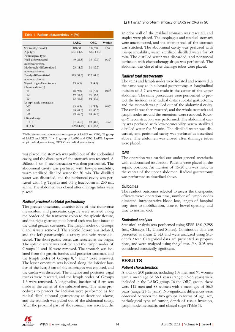
was placed, the stomach was pulled out of the abdominal cavity, and the distal part of the stomach was resected. A Billroth Ⅰ or Ⅱ reconstruction was then performed. The abdominal cavity was perfused with low-permeability, warm sterilized distilled water for 30 min. The distilled water was discarded, and the peritoneal cavity was per-fused with 1 g Tegafur and 0.3 g leucovorin in 250 mL saline. The abdomen was closed after drainage tubes were placed.
Radical proximal subtotal gastrectomyThe greater omentum, anterior lobe of the transverse mesocolon, and pancreatic capsule were isolated along the border of the transverse colon to the splenic flexure, and the right gastroepiploic hemal arch was kept intact at the distal greater curvature. The lymph nodes of Groups 6 and 4 were removed. The splenic flexure was isolated, and the left gastroepiploic artery and vein were dis-sected. The short gastric vessel was resected at the origin. The splenic artery was isolated and the lymph nodes of Groups 11 and 10 were removed. The stomach was iso-lated from the gastric fundus and posterior stomach, and the lymph nodes of Groups 8, 9 and 7 were removed. The lesser omentum was isolated along the inferior bor-der of the liver, 5 cm of the esophagus was exposed, and the cardia was dissected. The anterior and posterior vagal trunks were resected, and the lymph nodes of Groups 1-3 were removed. A longitudinal incision of 5 cm was made in the center of the subcostal area. The same pro-cedures to protect the incision were performed as for radical distal subtotal gastrectomy as described above, and the stomach was pulled out of the abdominal cavity. After the proximal part of the stomach was resected, the
anterior wall of the residual stomach was resected, and staples were placed. The esophagus and residual stomach were anastomosed, and the anterior wall of the stomach was stitched. The abdominal cavity was perfused with low-permeability, warm sterilized distilled water for 30 min. The distilled water was discarded, and peritoneal perfusion with chemotherapy drugs was performed. The abdomen was closed after drainage tubes were placed.
Radical total gastrectomyThe veins and lymph nodes were isolated and removed in the same way as in subtotal gastrectomy. A longitudinal incision of 5-7 cm was made in the center of the upper abdomen. The same procedures were performed to pro-tect the incision as in radical distal subtotal gastrectomy, and the stomach was pulled out of the abdominal cavity. The cardia was then resected, and the whole stomach and lymph nodes around the omentum were removed. Roux-en-Y reconstruction was performed. The abdominal cav-ity was perfused with low-permeability, warm sterilized distilled water for 30 min. The distilled water was dis-carded, and peritoneal cavity was perfused as described above. The abdomen was closed after drainage tubes were placed.
ORGThe operation was carried out under general anesthesia with endotracheal intubation. Patients were placed in the supine position. An incision of 15-20 cm was made in the center of the upper abdomen. Radical gastrectomy was performed as described above.
OutcomesThe readout outcomes selected to assess the therapeutic efficacy were: operation time, number of lymph nodes dissected, intraoperative blood loss, length of hospital stay, time to mobilization, time to bowel opening, and time to normal diet.
Statistical analysisStatistical analysis was performed using SPSS 18.0 (SPSS Inc., Chicago, IL, United States). Continuous data are presented as mean ± SD, and were analyzed using Stu-dent’s t test. Categorical data are presented as propor-tions, and were analyzed using the χ 2 test. P < 0.05 was considered statistically significant.
RESULTSPatient characteristicsA total of 200 patients, including 109 men and 91 women with a mean age of 56.1 years (range: 23-63 years) were included in the LARG group. In the ORG group, there were 112 men and 88 women with a mean age of 56.3 years (range: 21-65 years). No significant differences were observed between the two groups in terms of age, sex, pathological type of tumor, depth of tissue invasion, lymph node metastasis, and clinical stage (Table 1).
April 27, 2014|Volume 6|Issue 4|WJGS|www.wjgnet.com 61
LARG ORG P value
Sex (male/female) 109/91 112/88 0.84 Age (yr) 58.3 ± 6.5 58.6 ± 6.3 Pathological type Well-differentiated adenocarcinoma
49 (24.5) 38 (19.0) 0.321
Moderately-differentiated adenocarcinoma
23 (11.5) 31 (15.5)
Poorly-differentiated adenocarcinoma
115 (57.5) 122 (61.0)
Signet ring cell carcinoma 13 (6.5) 9 (4.5) Classification (T) T1 18 (9.0) 15 (7.5) 0.862
T2 89 (44.5) 91 (45.5) T3 93 (46.5) 94 (47.0) Lymph node metastasis N0 13 (6.5) 11 (5.5) 0.903
N1 88 (44.0) 91 (45.5) N2 99 (49.5) 98 (49.0) Clinical stage Ⅰ + Ⅱ 91 (45.5) 89 (44.5) 0.92 Ⅲ + Ⅳ 109 (54.5%) 111 (55.5)
Table 1 Patients characteristics n (%)
1Well-differentiated adenocarcinoma group of LARG and ORG; 2T1 group of LARG and ORG; 3Ⅰ + Ⅱ group of LARG and ORG. LARG: Laparo-scopic radical gastrectomy; ORG: Open radical gastrectomy.
Li HT et al . Short-term efficacy of LARG vs ORG in GC
April 27, 2014|Volume 6|Issue 4|WJGS|www.wjgnet.com
laparotomy in all patients in the LARG group, and no residual cancerous tissues were noted in the surgical mar-gins. Despite a significantly longer operation time with LARG, this approach offered the patients several better short-term benefits compared to the ORG procedure, such as less intraoperative blood loss, shorter hospitaliza-tion time, shorter time to mobilization, shorter time to bowel opening, and shorter time to normal dietary intake. Additionally, LARG was also associated with fewer post-operative complications.
Well-exposed surgical fields could help reduce blood vessel damage and reduce intraoperative blood loss. A satisfactory surgical field can be obtained using laparos-copy with the assistance of an ultrasonic knife and tita-nium clips, and this could greatly reduce intraoperative blood loss. Currently, the same surgical processes used in traditional radical gastrectomy are still used in LARG, including blood vessel ligation at the origin, excessive margin resection, and removal of perigastric lymphoid tissues. However, because LARG is performed with lapa-roscopic instruments inside the abdominal cavity, me-chanic organ damage by direct contact with the stomach during surgery can be minimized.
The indications for LARG in the treatment of pa-tients with gastric cancer vary among different centers. For example, Kitano et al[9] have suggested that LARG could be indicated for patients with advanced-stage gas-tric carcinoma with an invasion depth lower than T2[9,10], whereas Huscher et al[11] have suggested that LARG is the best choice for patients with advanced gastric carcinoma in whom tumor invasion has reached T3[11]. Based on our study, we believe that LARG is a safe, effective, and minimally invasive approach for treatment of gastric can-cer, as previously reported[7,12,13]. The long-term efficacy of LARG in patients with gastric cancer has also been reported[10]. In this long-term follow-up study, no signifi-cant difference in the 5-year survival rate was observed between 136 patients with gastric cancer who underwent LARG and 120 who underwent ORG.
Currently, CO2 pneumoperitoneum is widely used in LARG procedures. However, the use of CO2 pneu-moperitoneum could result in inhibition of the immune response in the abdominal cavity. In an animal experi-ment[14], a significant decrease in the number of CD4/CD8 cells was observed after the induction of CO2 pneu-
Major intraoperative characteristics and outcomesLARG was successfully performed in the 200 patients without conversion to laparotomy. ORG was also suc-cessfully performed in 200 patients. No malignant tissues were found at the lower or upper resection margin in any of the patients.
Table 2 shows the treatment characteristics between the two groups. Longer time was needed for LARG than for ORG (192.3 ± 20.9 min vs 180.0 ± 26.9 min, respec-tively, P < 0.0001). A similar number of lymph nodes was removed by both approaches (P = 0.62). The LARG procedure was superior to ORG for several outcomes, including: less intraoperative blood loss (103.1 ± 19.5 mL vs 163.0 ± 32.9 mL, P < 0.0001); less bedbound time (1.0 ± 0.3 d vs 3.3 ± 0.4 d, P < 0.0001); less time to bowel opening (3.3 ± 0.7 d vs 4.5 ± 0.7 d, P < 0.0001); less time to normal diet (3.0 ± 0.4 d vs 3.8 ± 0.5 d, P < 0.0001); and shorter hospital stay (6.8 ± 1.2 d vs 9.5 ± 1.6 d, P < 0.0001). In addition, the incision length was shorter in the LARG group than in the ORG group (5.2 ± 0.7 vs 17.8 ± 1.0 cm, P < 0.0001), and fewer patients required special pain control in the LARG group than in the ORG group (39.5% vs 56.5%, P = 0.0007).
Postoperative complicationsAs shown in Table 3, significantly fewer patients in the LARG group suffered from poor incision healing (2.5% vs 8.0%, P = 0.01) and pulmonary infection (2.0% vs 9.5%, P = 0.001). Fewer patients experienced anasto-motic leakage or stump fistula in the LARG group than ORG group, although the difference was not significant (3.5% vs 7.5%, P = 0.08). No difference was observed in the incidence of decreased gastrointestinal motility and acute organ (liver or kidney) failure between the LARG and ORG groups (P > 0.05). Overall, the LARG group was associated with less frequent complications than the ORG group was (13% vs 27%, P = 0.02).
DISCUSSIONWe compared 200 patients who underwent LARG and 200 who underwent ORG in terms of their intra- and postoperative benefits. The laparoscopic procedures were successfully completed without needing to convert to
62
Outcomes LARG ORG P value
Operation time (min) 192.3 ± 20.9 180.0 ± 26.9 < 0.0001 No. of lymph nodes removed 28.5 ± 4.5 28.3 ± 3.4 0.62 Intraoperative blood loss (mL) 103.1 ± 19.5 163.0 ± 32.9 < 0.0001 Bedbound time (d) 1.0 ± 0.3 3.3 ± 0.4 < 0.0001 Time to bowel opening (d) 3.3 ± 0.7 4.5 ± 0.7 < 0.0001 Length of incision (cm) 5.2 ± 0.7 17.8 ± 1.0 < 0.0001 Time to normal diet (d) 3.0 ± 0.4 3.8 ± 0.5 < 0.0001 Total hospital stay (d) 6.8 ± 1.2 9.5 ± 1.6 < 0.0001
Table 2 Comparison of surgical outcomes between laparo-scopic radical gastrectomy and open radical gastrectomy groups
LARG: Laparoscopic radical gastrectomy; ORG: Open radical gastrectomy.
Postoperative complications LARG ORG P value
Decreased gastrointestinal motility 3 (1.5) 7 (3.5) 0.2 Anastomotic leakage/stump fistula 7 (3.5) 15 (7.5) 0.08 Poor incision healing 5 (2.5) 16 (8.0) 0.01 Pulmonary infection 4 (2.0) 19 (9.5) 0.001 Acute liver/kidney failure 1 (0.5) 4 (2.0) 0.18 Total 131 (6.5) 271 (13.5) 0.02
Table 3 Comparison of postoperative complications between the two groups n (%)
1Some patients had two or more concomitant complications. LARG: Lapa-roscopic radical gastrectomy; ORG: Open radical gastrectomy.
Li HT et al . Short-term efficacy of LARG vs ORG in GC

April 27, 2014|Volume 6|Issue 4|WJGS|www.wjgnet.com
ApplicationsThe results suggest that LARG is a safer, more effective, and less-invasive ap-proach for treating gastric cancer with a better short-term efficacy.TerminologyLARG: LARG is a novel minimally invasive surgical technique. It is associated with such advantages as less injury, reduced postoperative pain, lower impact on immune function, rapid recovery of gastrointestinal function, and short hospi-tal study. Peer reviewThe authors have performed a well-designed study and submitted a full detailed manuscript. The overall body of the article is fine and they have presented the results and discussion well.
REFERENCES1 Siegel R, Naishadham D, Jemal A. Cancer statistics, 2013. CA
Cancer J Clin 2013; 63: 11-30 [PMID: 23335087 DOI: 10.3322/caac.21166]
2 WHO (2013) International Agency for Research on Cancer: GLOBOCAN 2008. Available from: URL: http://www.wpro.who.int
3 Ajani JA, Bentrem DJ, Besh S, D’Amico TA, Das P, Denlinger C, Fakih MG, Fuchs CS, Gerdes H, Glasgow RE, Hayman JA, Hofstetter WL, Ilson DH, Keswani RN, Kleinberg LR, Korn WM, Lockhart AC, Meredith K, Mulcahy MF, Orringer MB, Posey JA, Sasson AR, Scott WJ, Strong VE, Varghese TK, Warren G, Washington MK, Willett C, Wright CD, McMil-lian NR, Sundar H. Gastric cancer, version 2.2013: featured updates to the NCCN Guidelines. J Natl Compr Canc Netw 2013; 11: 531-546 [PMID: 23667204]
4 Kitano S, Iso Y, Moriyama M, Sugimachi K. Laparoscopy-assist-ed Billroth I gastrectomy. Surg Laparosc Endosc 1994; 4: 146-148 [PMID: 8180768 DOI: 10.1097/00129689-200206000-00021]
5 Kim HH, Hyung WJ, Cho GS, Kim MC, Han SU, Kim W, Ryu SW, Lee HJ, Song KY. Morbidity and mortal-ity of laparoscopic gastrectomy versus open gastrectomy for gastric cancer: an interim report--a phase III multi-center, prospective, randomized Trial (KLASS Trial). Ann Surg 2010; 251: 417-420 [PMID: 20160637 DOI: 10.1097/SLA.0b013e3181cc8f6b]
6 Nakajima T. Gastric cancer treatment guidelines in Japan. Gastric Cancer 2002; 5: 1-5 [PMID: 12021853 DOI: 10.1007/s101200200000]
7 Lee JH, Han HS, Lee JH. A prospective randomized study comparing open vs laparoscopy-assisted distal gastrectomy in early gastric cancer: early results. Surg Endosc 2005; 19: 168-173 [PMID: 15580441 DOI: 10.1007/s00464-004-8808-y]
8 Li ZS, Li Q. The latest 2010 WHO classification of tumors of digestive system. Zhonghua Binglixue Zazhi 2011; 40: 351-354 [PMID: 21756837]
9 Kitano S, Shiraishi N. Minimally invasive surgery for gas-tric tumors. Surg Clin North Am 2005; 85: 151-164, xi [PMID: 15619536 DOI: 10.1016/j.suc.2004.09.004]
10 Ziqiang W, Feng Q, Zhimin C, Miao W, Lian Q, Huaxing L, Peiwu Y. Comparison of laparoscopically assisted and open radical distal gastrectomy with extended lymphadenec-tomy for gastric cancer management. Surg Endosc 2006; 20: 1738-1743 [PMID: 17024529 DOI: 10.1007/s00464-006-0031-6]
11 Huscher CG, Mingoli A, Sgarzini G, Sansonetti A, Di Paola M, Recher A, Ponzano C. Laparoscopic versus open subtotal gastrectomy for distal gastric cancer: five-year results of a randomized prospective trial. Ann Surg 2005; 241: 232-237 [PMID: 15650632 DOI: 10.1097/01.sla.0000151892.35922.f2]
12 Du Y, Cheng X, Xu Z, Yang L, Huang L, Wang B, Yu P, Dong R. Laparoscopic-assisted radical gastrectomy for distal gastric cancer. Chin J Cancer Res 2013; 25: 460-462 [PMID: 23997536 DOI: 10.3978/j.issn.1000-9604.2013.08.15]
13 Lee SI, Choi YS, Park DJ, Kim HH, Yang HK, Kim MC. Com-parative study of laparoscopy-assisted distal gastrectomy and
moperitoneum. CO2 pneumoperitoneum was also shown to inhibit macrophage activation in the abdominal cavity, and thus inhibit the release of tumor necrosis factor (TNF)-α and interleukin-1 by macrophages. Both mac-rophages and TNF-α play a potent role in the antitumor activity in the abdominal cavity[15].
During the treatment of malignant tumors using lap-aroscopy-assisted approaches, potential tumor implanta-tion induced by the operation has been a major concern. Based on our study, the possible impact of CO2 pneu-moperitoneum on immune disturbance and possible im-plantation metastasis could not be determined. However, previous studies have reported no significant increase in metastasis implantation after LARG, and the rate of incision metastasis did not differ between patients who underwent and those who did not undergo CO2 pneumo-peritoneum during surgery[16]. Similarly, no implantation metastasis was found in patients treated with LARG in our study. We believe that gentle surgical maneuver with-out squeezing the tumor tissues, and withdrawal of the laparoscopic instruments only after the intra-abdominal gas is completely removed, are important.
In conclusion, LARG is a safe, effective, and minimal-ly invasive approach for the treatment of gastric cancer. LARG may offer better short-term benefits to patients than ORG offers. Further studies are needed to investi-gate the long-term efficacy of the LARG approach.
COMMENTSBackgroundGastric cancer is one of the most common malignant tumors worldwide, with a yearly incidence of about 900000. Laparoscopic radical gastrectomy (LARG) has been increasingly used as a promising approach for the management of gastric cancer because of its minimal invasiveness and a potential to treat successfully patients with lymph node metastasis. Although LARG and laparo-scopic D2 gastrectomy have now been widely used in the treatment of gastric cancer, including advanced gastric carcinoma, the short- and long-term benefits are unclear. The short-term outcomes of LARG have recently been reported, although these studies were based on small samples. In the current study, authors compared the short-term outcomes between LARG and open radical gastrectomy (ORG) performed in patients with gastric cancer in our department.Research frontiersLARG and laparoscopic D2 gastrectomy have now been widely used in the treatment of gastric cancer, including advanced gastric carcinoma. The re-search hotspot is how to investigate the short-term benefits of LARG and ORG in patients with gastric cancer.Innovations and breakthroughsBased on this study, authors believe that LARG is a safe, effective, and mini-mally invasive approach for the treatment of gastric cancer, as previously re-ported. The long-term efficacy of LARG in patients with gastric cancer has also been reported. In this long-term follow-up study, no significant difference in the 5-year survival rate was observed between 136 patients with gastric cancer who underwent LARG and 120 patients who underwent ORG. Well-exposed surgical fields could help reduce blood vessel damage and reduce intraoperative blood loss. A satisfactory surgical field can be obtained using laparoscopy with the as-sistance of an ultrasonic knife and titanium clips, and this could greatly reduce intraoperative blood loss. Currently, the same surgical processes used in tradi-tional radical gastrectomy are still used in LARG, including blood vessel ligation at the origin, excessive margin resection, and removal of perigastric lymphoid tissues. However, because LARG is performed with laparoscopic instruments inside the abdominal cavity, mechanical organ damage by direct contact with the stomach during surgery can be minimized.
63
COMMENTS
Li HT et al . Short-term efficacy of LARG vs ORG in GC
April 27, 2014|Volume 6|Issue 4|WJGS|www.wjgnet.com
source of transitional-cell carcinoma port-site metastasis, with prophylactic irrigation strategies to decrease laparo-scopic oncologic risks. J Endourol 2008; 22: 105-112 [PMID: 18315481 DOI: 10.1089/end.2007.9858]
16 Nakamura T, Mitomi H, Ohtani Y, Kokuba Y, Sato T, Ozawa H, Ihara A, Watanabe M. Comparison of long-term outcome of laparoscopic and conventional surgery for advanced co-lon and rectosigmoid cancer. Hepatogastroenterology 2006; 53: 351-353 [PMID: 16795970]
P- Reviewer: Maleki AR S- Editor: Wen LL L- Editor: Kerr C E- Editor: Wu HL
open distal gastrectomy. J Am Coll Surg 2006; 202: 874-880 [PMID: 16735200 DOI: 10.1016/j.jamcollsurg.2006.02.028]
14 Gutt CN, Hollander D, Brier CH, Kim ZG, Lorenz M. Influ-ence of laparoscopy and laparotomy on systemic and perito-neal T lymphocytes in a rat model. Int J Colorectal Dis 2001; 16: 216-220 [PMID: 11515680 DOI: 10.1007/s003840100296]
15 Ost MC, Patel KP, Rastinehad AR, Chu PY, Anderson AE, Smith AD, Lee BR. Pneumoperitoneum with carbon dioxide inhibits macrophage tumor necrosis factor-alpha secretion:
64
Li HT et al . Short-term efficacy of LARG vs ORG in GC
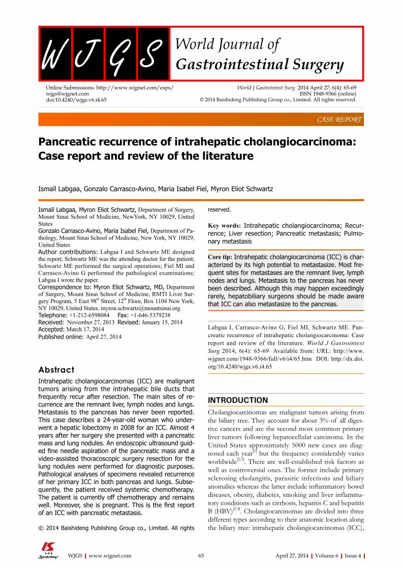
Ismaïl Labgaa, Gonzalo Carrasco-Avino, Maria Isabel Fiel, Myron Eliot Schwartz
Pancreatic recurrence of intrahepatic cholangiocarcinoma: Case report and review of the literature
Ismaïl Labgaa, Myron Eliot Schwartz, Department of Surgery, Mount Sinai School of Medicine, NewYork, NY 10029, United StatesGonzalo Carrasco-Avino, Maria Isabel Fiel, Department of Pa-thology, Mount Sinai School of Medicine, New York, NY 10029, United StatesAuthor contributions: Labgaa I and Schwartz ME designed the report; Schwartz ME was the attending doctor for the patient; Schwartz ME performed the surgical operations; Fiel MI and Carrasco-Avino G performed the pathological examinations; Labgaa I wrote the paper.Correspondence to: Myron Eliot Schwartz, MD, Department of Surgery, Mount Sinai School of Medicine, RMTI Liver Sur-gery Program, 5 East 98th Street, 12th Floor, Box 1104 New York, NY 10029, United States. [email protected]: +1-212-6598084 Fax: +1-646-5379238Received: November 27, 2013 Revised: January 15, 2014Accepted: March 17, 2014Published online: April 27, 2014
AbstractIntrahepatic cholangiocarcinomas (ICC) are malignant tumors arising from the intrahepatic bile ducts that frequently recur after resection. The main sites of re-currence are the remnant liver, lymph nodes and lungs. Metastasis to the pancreas has never been reported. This case describes a 24-year-old woman who under-went a hepatic lobectomy in 2008 for an ICC. Almost 4 years after her surgery she presented with a pancreatic mass and lung nodules. An endoscopic ultrasound guid-ed fine needle aspiration of the pancreatic mass and a video-assisted thoracoscopic surgery resection for the lung nodules were performed for diagnostic purposes. Pathological analyses of specimens revealed recurrence of her primary ICC in both pancreas and lungs. Subse-quently, the patient received systemic chemotherapy. The patient is currently off chemotherapy and remains well. Moreover, she is pregnant. This is the first report of an ICC with pancreatic metastasis.
© 2014 Baishideng Publishing Group co., Limited. All rights
reserved.
Key words: Intrahepatic cholangiocarcinoma; Recur-rence; Liver resection; Pancreatic metastasis; Pulmo-nary metastasis
Core tip: Intrahepatic cholangiocarcinoma (ICC) is char-acterized by its high potential to metastasize. Most fre-quent sites for metastases are the remnant liver, lymph nodes and lungs. Metastasis to the pancreas has never been described. Although this may happen exceedingly rarely, hepatobiliary surgeons should be made aware that ICC can also metastasize to the pancreas.
Labgaa I, Carrasco-Avino G, Fiel MI, Schwartz ME. Pan-creatic recurrence of intrahepatic cholangiocarcinoma: Case report and review of the literature. World J Gastrointest Surg 2014; 6(4): 65-69 Available from: URL: http://www.wjgnet.com/1948-9366/full/v6/i4/65.htm DOI: http://dx.doi.org/10.4240/wjgs.v6.i4.65
INTRODUCTIONCholangiocarcinomas are malignant tumors arising from the biliary tree. They account for about 3% of all diges-tive cancers and are the second most common primary liver tumors following hepatocellular carcinoma. In the United States approximately 5000 new cases are diag-nosed each year[1] but the frequency considerably varies worldwide[2,3]. There are well-established risk factors as well as controversial ones. The former include primary sclerosing cholangitis, parasitic infections and biliary anomalies whereas the latter include inflammatory bowel diseases, obesity, diabetes, smoking and liver inflamma-tory conditions such as cirrhosis, hepatitis C and hepatitis B (HBV)[2-4]. Cholangiocarcinomas are divided into three different types according to their anatomic location along the biliary tree: intrahepatic cholangiocarcinomas (ICC),
CASE REPORT
Online Submissions: http://www.wjgnet.com/esps/[email protected]:10.4240/wjgs.v6.i4.65
April 27, 2014|Volume 6|Issue 4|WJGS|www.wjgnet.com
World J Gastrointest Surg 2014 April 27; 6(4): 65-69ISSN 1948-9366 (online)
© 2014 Baishideng Publishing Group co., Limited. All rights reserved.
65
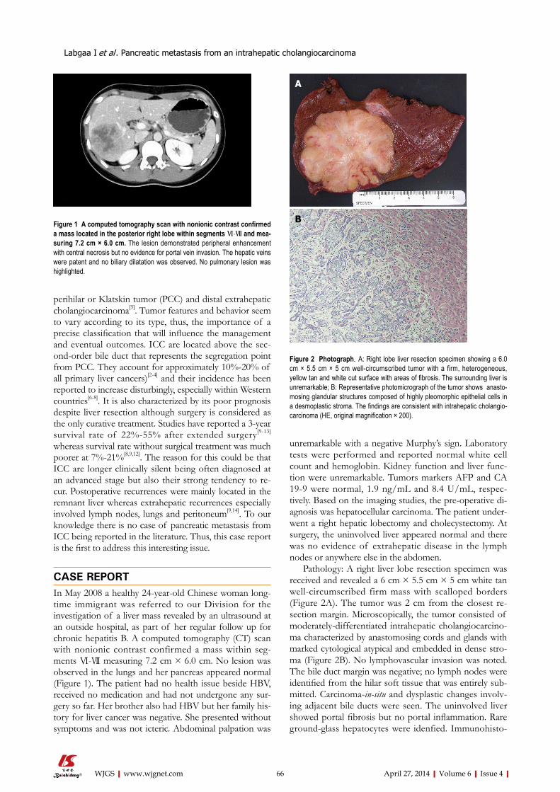
perihilar or Klatskin tumor (PCC) and distal extrahepatic cholangiocarcinoma[5]. Tumor features and behavior seem to vary according to its type, thus, the importance of a precise classification that will influence the management and eventual outcomes. ICC are located above the sec-ond-order bile duct that represents the segregation point from PCC. They account for approximately 10%-20% of all primary liver cancers)[2-4] and their incidence has been reported to increase disturbingly, especially within Western countries[6-8]. It is also characterized by its poor prognosis despite liver resection although surgery is considered as the only curative treatment. Studies have reported a 3-year survival rate of 22%-55% after extended surgery[9-13] whereas survival rate without surgical treatment was much poorer at 7%-21%[8,9,12]. The reason for this could be that ICC are longer clinically silent being often diagnosed at an advanced stage but also their strong tendency to re-cur. Postoperative recurrences were mainly located in the remnant liver whereas extrahepatic recurrences especially involved lymph nodes, lungs and peritoneum[9,14]. To our knowledge there is no case of pancreatic metastasis from ICC being reported in the literature. Thus, this case report is the first to address this interesting issue.
CASE REPORTIn May 2008 a healthy 24-year-old Chinese woman long-time immigrant was referred to our Division for the investigation of a liver mass revealed by an ultrasound at an outside hospital, as part of her regular follow up for chronic hepatitis B. A computed tomography (CT) scan with nonionic contrast confirmed a mass within seg-ments Ⅵ-Ⅶ measuring 7.2 cm × 6.0 cm. No lesion was observed in the lungs and her pancreas appeared normal (Figure 1). The patient had no health issue beside HBV, received no medication and had not undergone any sur-gery so far. Her brother also had HBV but her family his-tory for liver cancer was negative. She presented without symptoms and was not icteric. Abdominal palpation was
unremarkable with a negative Murphy’s sign. Laboratory tests were performed and reported normal white cell count and hemoglobin. Kidney function and liver func-tion were unremarkable. Tumors markers AFP and CA 19-9 were normal, 1.9 ng/mL and 8.4 U/mL, respec-tively. Based on the imaging studies, the pre-operative di-agnosis was hepatocellular carcinoma. The patient under-went a right hepatic lobectomy and cholecystectomy. At surgery, the uninvolved liver appeared normal and there was no evidence of extrahepatic disease in the lymph nodes or anywhere else in the abdomen.
Pathology: A right liver lobe resection specimen was received and revealed a 6 cm × 5.5 cm × 5 cm white tan well-circumscribed firm mass with scalloped borders (Figure 2A). The tumor was 2 cm from the closest re-section margin. Microscopically, the tumor consisted of moderately-differentiated intrahepatic cholangiocarcino-ma characterized by anastomosing cords and glands with marked cytological atypical and embedded in dense stro-ma (Figure 2B). No lymphovascular invasion was noted. The bile duct margin was negative; no lymph nodes were identified from the hilar soft tissue that was entirely sub-mitted. Carcinoma-in-situ and dysplastic changes involv-ing adjacent bile ducts were seen. The uninvolved liver showed portal fibrosis but no portal inflammation. Rare ground-glass hepatocytes were idenfied. Immunohisto-
April 27, 2014|Volume 6|Issue 4|WJGS|www.wjgnet.com
Labgaa I et al . Pancreatic metastasis from an intrahepatic cholangiocarcinoma
66
Figure 1 A computed tomography scan with nonionic contrast confirmed a mass located in the posterior right lobe within segments Ⅵ-Ⅶ and mea-suring 7.2 cm × 6.0 cm. The lesion demonstrated peripheral enhancement with central necrosis but no evidence for portal vein invasion. The hepatic veins were patent and no biliary dilatation was observed. No pulmonary lesion was highlighted.
Figure 2 Photograph. A: Right lobe liver resection specimen showing a 6.0 cm × 5.5 cm × 5 cm well-circumscribed tumor with a firm, heterogeneous, yellow tan and white cut surface with areas of fibrosis. The surrounding liver is unremarkable; B: Representative photomicrograph of the tumor shows anasto-mosing glandular structures composed of highly pleomorphic epithelial cells in a desmoplastic stroma. The findings are consistent with intrahepatic cholangio-carcinoma (HE, original magnification × 200).
A
B
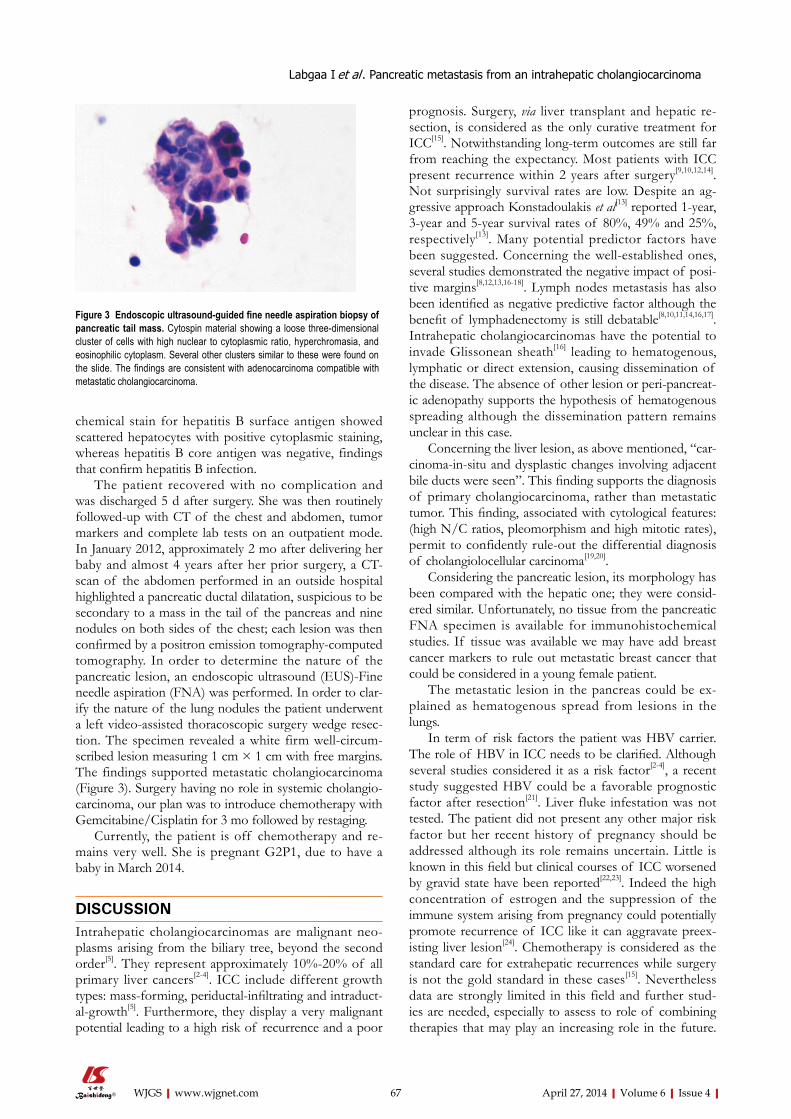
chemical stain for hepatitis B surface antigen showed scattered hepatocytes with positive cytoplasmic staining, whereas hepatitis B core antigen was negative, findings that confirm hepatitis B infection.
The patient recovered with no complication and was discharged 5 d after surgery. She was then routinely followed-up with CT of the chest and abdomen, tumor markers and complete lab tests on an outpatient mode. In January 2012, approximately 2 mo after delivering her baby and almost 4 years after her prior surgery, a CT-scan of the abdomen performed in an outside hospital highlighted a pancreatic ductal dilatation, suspicious to be secondary to a mass in the tail of the pancreas and nine nodules on both sides of the chest; each lesion was then confirmed by a positron emission tomography-computed tomography. In order to determine the nature of the pancreatic lesion, an endoscopic ultrasound (EUS)-Fine needle aspiration (FNA) was performed. In order to clar-ify the nature of the lung nodules the patient underwent a left video-assisted thoracoscopic surgery wedge resec-tion. The specimen revealed a white firm well-circum-scribed lesion measuring 1 cm × 1 cm with free margins. The findings supported metastatic cholangiocarcinoma (Figure 3). Surgery having no role in systemic cholangio-carcinoma, our plan was to introduce chemotherapy with Gemcitabine/Cisplatin for 3 mo followed by restaging.
Currently, the patient is off chemotherapy and re-mains very well. She is pregnant G2P1, due to have a baby in March 2014.
DISCUSSIONIntrahepatic cholangiocarcinomas are malignant neo-plasms arising from the biliary tree, beyond the second order[5]. They represent approximately 10%-20% of all primary liver cancers[2-4]. ICC include different growth types: mass-forming, periductal-infiltrating and intraduct-al-growth[5]. Furthermore, they display a very malignant potential leading to a high risk of recurrence and a poor
prognosis. Surgery, via liver transplant and hepatic re-section, is considered as the only curative treatment for ICC[15]. Notwithstanding long-term outcomes are still far from reaching the expectancy. Most patients with ICC present recurrence within 2 years after surgery[9,10,12,14]. Not surprisingly survival rates are low. Despite an ag-gressive approach Konstadoulakis et al[13] reported 1-year, 3-year and 5-year survival rates of 80%, 49% and 25%, respectively[13]. Many potential predictor factors have been suggested. Concerning the well-established ones, several studies demonstrated the negative impact of posi-tive margins[8,12,13,16-18]. Lymph nodes metastasis has also been identified as negative predictive factor although the benefit of lymphadenectomy is still debatable[8,10,11,14,16,17]. Intrahepatic cholangiocarcinomas have the potential to invade Glissonean sheath[16] leading to hematogenous, lymphatic or direct extension, causing dissemination of the disease. The absence of other lesion or peri-pancreat-ic adenopathy supports the hypothesis of hematogenous spreading although the dissemination pattern remains unclear in this case.
Concerning the liver lesion, as above mentioned, “car-cinoma-in-situ and dysplastic changes involving adjacent bile ducts were seen”. This finding supports the diagnosis of primary cholangiocarcinoma, rather than metastatic tumor. This finding, associated with cytological features: (high N/C ratios, pleomorphism and high mitotic rates), permit to confidently rule-out the differential diagnosis of cholangiolocellular carcinoma[19,20].
Considering the pancreatic lesion, its morphology has been compared with the hepatic one; they were consid-ered similar. Unfortunately, no tissue from the pancreatic FNA specimen is available for immunohistochemical studies. If tissue was available we may have add breast cancer markers to rule out metastatic breast cancer that could be considered in a young female patient.
The metastatic lesion in the pancreas could be ex-plained as hematogenous spread from lesions in the lungs.
In term of risk factors the patient was HBV carrier. The role of HBV in ICC needs to be clarified. Although several studies considered it as a risk factor[2-4], a recent study suggested HBV could be a favorable prognostic factor after resection[21]. Liver fluke infestation was not tested. The patient did not present any other major risk factor but her recent history of pregnancy should be addressed although its role remains uncertain. Little is known in this field but clinical courses of ICC worsened by gravid state have been reported[22,23]. Indeed the high concentration of estrogen and the suppression of the immune system arising from pregnancy could potentially promote recurrence of ICC like it can aggravate preex-isting liver lesion[24]. Chemotherapy is considered as the standard care for extrahepatic recurrences while surgery is not the gold standard in these cases[15]. Nevertheless data are strongly limited in this field and further stud-ies are needed, especially to assess to role of combining therapies that may play an increasing role in the future.
April 27, 2014|Volume 6|Issue 4|WJGS|www.wjgnet.com 67
Figure 3 Endoscopic ultrasound-guided fine needle aspiration biopsy of pancreatic tail mass. Cytospin material showing a loose three-dimensional cluster of cells with high nuclear to cytoplasmic ratio, hyperchromasia, and eosinophilic cytoplasm. Several other clusters similar to these were found on the slide. The findings are consistent with adenocarcinoma compatible with metastatic cholangiocarcinoma.
Labgaa I et al . Pancreatic metastasis from an intrahepatic cholangiocarcinoma
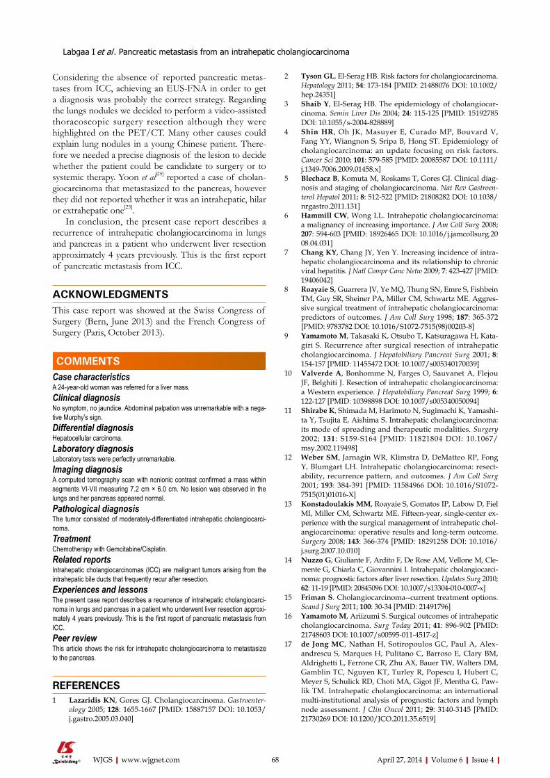
April 27, 2014|Volume 6|Issue 4|WJGS|www.wjgnet.com
2 Tyson GL, El-Serag HB. Risk factors for cholangiocarcinoma. Hepatology 2011; 54: 173-184 [PMID: 21488076 DOI: 10.1002/hep.24351]
3 Shaib Y, El-Serag HB. The epidemiology of cholangiocar-cinoma. Semin Liver Dis 2004; 24: 115-125 [PMID: 15192785 DOI: 10.1055/s-2004-828889]
4 Shin HR, Oh JK, Masuyer E, Curado MP, Bouvard V, Fang YY, Wiangnon S, Sripa B, Hong ST. Epidemiology of cholangiocarcinoma: an update focusing on risk factors. Cancer Sci 2010; 101: 579-585 [PMID: 20085587 DOI: 10.1111/j.1349-7006.2009.01458.x]
5 Blechacz B, Komuta M, Roskams T, Gores GJ. Clinical diag-nosis and staging of cholangiocarcinoma. Nat Rev Gastroen-terol Hepatol 2011; 8: 512-522 [PMID: 21808282 DOI: 10.1038/nrgastro.2011.131]
6 Hammill CW, Wong LL. Intrahepatic cholangiocarcinoma: a malignancy of increasing importance. J Am Coll Surg 2008; 207: 594-603 [PMID: 18926465 DOI: 10.1016/j.jamcollsurg.2008.04.031]
7 Chang KY, Chang JY, Yen Y. Increasing incidence of intra-hepatic cholangiocarcinoma and its relationship to chronic viral hepatitis. J Natl Compr Canc Netw 2009; 7: 423-427 [PMID: 19406042]
8 Roayaie S, Guarrera JV, Ye MQ, Thung SN, Emre S, Fishbein TM, Guy SR, Sheiner PA, Miller CM, Schwartz ME. Aggres-sive surgical treatment of intrahepatic cholangiocarcinoma: predictors of outcomes. J Am Coll Surg 1998; 187: 365-372 [PMID: 9783782 DOI: 10.1016/S1072-7515(98)00203-8]
9 Yamamoto M, Takasaki K, Otsubo T, Katsuragawa H, Kata-giri S. Recurrence after surgical resection of intrahepatic cholangiocarcinoma. J Hepatobiliary Pancreat Surg 2001; 8: 154-157 [PMID: 11455472 DOI: 10.1007/s005340170039]
10 Valverde A, Bonhomme N, Farges O, Sauvanet A, Flejou JF, Belghiti J. Resection of intrahepatic cholangiocarcinoma: a Western experience. J Hepatobiliary Pancreat Surg 1999; 6: 122-127 [PMID: 10398898 DOI: 10.1007/s005340050094]
11 Shirabe K, Shimada M, Harimoto N, Sugimachi K, Yamashi-ta Y, Tsujita E, Aishima S. Intrahepatic cholangiocarcinoma: its mode of spreading and therapeutic modalities. Surgery 2002; 131: S159-S164 [PMID: 11821804 DOI: 10.1067/msy.2002.119498]
12 Weber SM, Jarnagin WR, Klimstra D, DeMatteo RP, Fong Y, Blumgart LH. Intrahepatic cholangiocarcinoma: resect-ability, recurrence pattern, and outcomes. J Am Coll Surg 2001; 193: 384-391 [PMID: 11584966 DOI: 10.1016/S1072-7515(01)01016-X]
13 Konstadoulakis MM, Roayaie S, Gomatos IP, Labow D, Fiel MI, Miller CM, Schwartz ME. Fifteen-year, single-center ex-perience with the surgical management of intrahepatic chol-angiocarcinoma: operative results and long-term outcome. Surgery 2008; 143: 366-374 [PMID: 18291258 DOI: 10.1016/j.surg.2007.10.010]
14 Nuzzo G, Giuliante F, Ardito F, De Rose AM, Vellone M, Cle-mente G, Chiarla C, Giovannini I. Intrahepatic cholangiocarci-noma: prognostic factors after liver resection. Updates Surg 2010; 62: 11-19 [PMID: 20845096 DOI: 10.1007/s13304-010-0007-x]
15 Friman S. Cholangiocarcinoma--current treatment options. Scand J Surg 2011; 100: 30-34 [PMID: 21491796]
16 Yamamoto M, Ariizumi S. Surgical outcomes of intrahepatic cholangiocarcinoma. Surg Today 2011; 41: 896-902 [PMID: 21748603 DOI: 10.1007/s00595-011-4517-z]
17 de Jong MC, Nathan H, Sotiropoulos GC, Paul A, Alex-andrescu S, Marques H, Pulitano C, Barroso E, Clary BM, Aldrighetti L, Ferrone CR, Zhu AX, Bauer TW, Walters DM, Gamblin TC, Nguyen KT, Turley R, Popescu I, Hubert C, Meyer S, Schulick RD, Choti MA, Gigot JF, Mentha G, Paw-lik TM. Intrahepatic cholangiocarcinoma: an international multi-institutional analysis of prognostic factors and lymph node assessment. J Clin Oncol 2011; 29: 3140-3145 [PMID: 21730269 DOI: 10.1200/JCO.2011.35.6519]
Considering the absence of reported pancreatic metas-tases from ICC, achieving an EUS-FNA in order to get a diagnosis was probably the correct strategy. Regarding the lungs nodules we decided to perform a video-assisted thoracoscopic surgery resection although they were highlighted on the PET/CT. Many other causes could explain lung nodules in a young Chinese patient. There-fore we needed a precise diagnosis of the lesion to decide whether the patient could be candidate to surgery or to systemic therapy. Yoon et al[25] reported a case of cholan-giocarcinoma that metastasized to the pancreas, however they did not reported whether it was an intrahepatic, hilar or extrahepatic one[25].
In conclusion, the present case report describes a recurrence of intrahepatic cholangiocarcinoma in lungs and pancreas in a patient who underwent liver resection approximately 4 years previously. This is the first report of pancreatic metastasis from ICC.
ACKNOWLEDGMENTSThis case report was showed at the Swiss Congress of Surgery (Bern, June 2013) and the French Congress of Surgery (Paris, October 2013).
COMMENTSCase characteristicsA 24-year-old woman was referred for a liver mass.Clinical diagnosisNo symptom, no jaundice. Abdominal palpation was unremarkable with a nega-tive Murphy’s sign. Differential diagnosisHepatocellular carcinoma.Laboratory diagnosisLaboratory tests were perfectly unremarkable.Imaging diagnosisA computed tomography scan with nonionic contrast confirmed a mass within segments VI-VII measuring 7.2 cm × 6.0 cm. No lesion was observed in the lungs and her pancreas appeared normal.Pathological diagnosisThe tumor consisted of moderately-differentiated intrahepatic cholangiocarci-noma.TreatmentChemotherapy with Gemcitabine/Cisplatin.Related reportsIntrahepatic cholangiocarcinomas (ICC) are malignant tumors arising from the intrahepatic bile ducts that frequently recur after resection. Experiences and lessonsThe present case report describes a recurrence of intrahepatic cholangiocarci-noma in lungs and pancreas in a patient who underwent liver resection approxi-mately 4 years previously. This is the first report of pancreatic metastasis from ICC.Peer reviewThis article shows the risk for intrahepatic cholangiocarcinoma to metastasize to the pancreas.
REFERENCES1 Lazaridis KN, Gores GJ. Cholangiocarcinoma. Gastroenter-
ology 2005; 128: 1655-1667 [PMID: 15887157 DOI: 10.1053/j.gastro.2005.03.040]
68
COMMENTS
Labgaa I et al . Pancreatic metastasis from an intrahepatic cholangiocarcinoma
April 27, 2014|Volume 6|Issue 4|WJGS|www.wjgnet.com
QQ, Wang Q, Zou SS, Wu MC, Hu HP. Hepatitis B virus infection: a favorable prognostic factor for intrahepatic chol-angiocarcinoma after resection. World J Gastroenterol 2011; 17: 1292-1303 [PMID: 21455328 DOI: 10.3748/wjg.v17.i10.1292]
22 Marasinghe JP, Karunananda SA, Angulo P. Cholangio-carcinoma in pregnancy: a case report. J Obstet Gynaecol Res 2008; 34: 635-637 [PMID: 18840169 DOI: 10.1111/j.1447-0756.2008.00810.x]
23 Balderston KD, Tewari K, Azizi F, Yu JK. Intrahepatic chol-angiocarcinoma masquerading as the HELLP syndrome (he-molysis, elevated liver enzymes, and low platelet count) in pregnancy: case report. Am J Obstet Gynecol 1998; 179: 823-824 [PMID: 9758000 DOI: 10.1016/S0002-9378(98)70093-6]
24 Joshi D, James A, Quaglia A, Westbrook RH, Heneghan MA. Liver disease in pregnancy. Lancet 2010; 375: 594-605 [PMID: 20159293 DOI: 10.1016/S0140-6736(09)61495-1]
25 Yoon WJ, Ryu JK, Kim YT, Yoon YB, Kim SW, Kim WH. Clinical features of metastatic tumors of the pancreas in Korea: a single-center study. Gut Liver 2011; 5: 61-64 [PMID: 21461074 DOI: 10.5009/gnl.2011.5.1.61]
P- Reviewers: Komuta M, Mott JL, Wang HLL S- Editor: Wen LL L- Editor: A E- Editor: Wu HL
18 Sotiropoulos GC, Miyazaki M, Konstadoulakis MM, Paul A, Molmenti EP, Gomatos IP, Radtke A, Baba HA, Beckebaum S, Brokalaki EI, Ohtsuka M, Schwartz ME, Broelsch CE, Sgourakis G. Multicentric evaluation of a clinical and prog-nostic scoring system predictive of survival after resection of intrahepatic cholangiocarcinomas. Liver Int 2010; 30: 996-1002 [PMID: 20141593 DOI: 10.1111/j.1478-3231.2010.02203.x]
19 Komuta M, Spee B, Vander Borght S, De Vos R, Verslype C, Aerts R, Yano H, Suzuki T, Matsuda M, Fujii H, Desmet VJ, Kojiro M, Roskams T. Clinicopathological study on cholan-giolocellular carcinoma suggesting hepatic progenitor cell origin. Hepatology 2008; 47: 1544-1556 [PMID: 18393293 DOI: 10.1002/hep.22238]
20 Komuta M, Govaere O, Vandecaveye V, Akiba J, Van Steen-bergen W, Verslype C, Laleman W, Pirenne J, Aerts R, Yano H, Nevens F, Topal B, Roskams T. Histological diversity in cholangiocellular carcinoma reflects the different cholan-giocyte phenotypes. Hepatology 2012; 55: 1876-1888 [PMID: 22271564 DOI: 10.1002/hep.25595]
21 Zhou HB, Wang H, Li YQ, Li SX, Wang H, Zhou DX, Tu
69
Labgaa I et al . Pancreatic metastasis from an intrahepatic cholangiocarcinoma
Helmi Kallel, Hassen Hentati, Amine Baklouti, Ali Gassara, Ahmed Saadaoui, Ghassen Halek, Sana Landolsi, MA El Ouaer, Wajdi Chaieb, Fethia Maamouri, Saber Mannaï
Retroperitoneal paragangliomas: Report of 4 cases
Helmi Kallel, Hassen Hentati, Amine Baklouti, Ali Gassara, Ahmed Saadaoui, Ghassen Halek, Sana Landolsi, MA El Ouaer, Wajdi Chaieb, Saber Mannaï, Department of Visceral Surgery, Hospital of Jendouba, Faculty of Medicine of Tunis, University of Tunis El Manar, 2083 Ariana, TunisFethia Maamouri, Department of Histopathology, Hospital of Jendouba, Faculty of Medicine of Tunis, University of Tunis El Manar, 2083 Ariana, TunisAuthor contributions: All the authors contributed to this manuscript.Correspondence to: Dr. Kallel Helmi, Department of Visceral Surgery, Hospital of Jendouba, Faculty of Medicine of Tunis, University of Tunis El Manar, Residence Omrane 6, App A8, Cité el Ghazela, 2083 Ariana, Tunis. [email protected]: +216-98-670457 Fax: +216-70-681281Received: July 8, 2013 Revised: October 24, 2013 Accepted: February 20, 2014 Published online: April 27, 2014
AbstractWe reviewed the data of all patients managed for retro-peritoneal paragangliomas (PGLs) between June 2010 and June 2011 to present our experience concerning this uncommon entity to highlight diagnostic and thera-peutic challenges of retroperitoneal PGLs. All patients were admitted to the department of general and hepa-tobiliary surgery in the regional hospital of Jendouba, Tunisia. The size of the tumor was taken at its largest dimension, as determined in a computed tomography (CT) scan and pathological reports. There were 4 pa-tients (all women) with a median age of 48 years (range 46-56 years). Abdominal pain was the commonest pre-sentation. CT showed and localized the tumors which were all retroperitoneal. All patients had successful surgical resection of the tumors under invasive arte-rial blood pressure monitoring. One patient underwent surgery for a presumed tumor of the pancreatic head. The fresh-mount microscopic study of the peroperative biopsy yielded inflammatory tissue without malignancy and no resection was performed. Final histological examination of the biopsy concluded PGL. A second
laparotomy was performed and the tumor was entirely resected. The diagnosis was made after surgery by his-tology in all patients. The control of the blood pressure was improved after surgery in 3 patients. Paraganglio-mas are rare tumors. The retroperitoneal localization is uncommon. Complete surgical resection remains the only curative treatment but it is often challenging as these tumors are located near multiple vital blood ves-sels.
© 2014 Baishideng Publishing Group co., Limited. All rights reserved.
Key words: Paraganglioma; Extra adrenal pheochromo-cytoma, Retroperitoneal; Surgery; Histology
Core tip: Retroperitoneal paragangliomas are uncom-mon tumors causing considerable difficulty in both diagnosis and treatment. Its complete surgical removal is often challenging as these tumors are located near multiple vital blood vessels. Any surgeon could face such a rare tumor.
Kallel H, Hentati H, Baklouti A, Gassara A, Saadaoui A, Halek G, Landolsi S, El Ouaer MA, Chaieb W, Maamouri F, Mannaï S. Retroperitoneal paragangliomas: Report of 4 cases. World J Gastrointest Surg 2014; 6(4): 70-73 Available from: URL: http://www.wjgnet.com/1948-9366/full/v6/i4/70.htm DOI: http://dx.doi.org/10.4240/wjgs.v6.i4.70
INTRODUCTIONParagangliomas (PGLs), or extra-adrenal phaeochro-mocytomas, are tumors arising from chromaffin tissues. Abdominal localizations are less frequent than head and neck. Retroperitoneal PGLs are more uncommon, caus-ing considerable difficulty in diagnosis and treatment. In this article, we present our experience concerning this uncommon entity to highlight diagnostic and therapeutic
CASE REPORT
Online Submissions: http://www.wjgnet.com/esps/[email protected]:10.4240/wjgs.v6.i4.70
April 27, 2014|Volume 6|Issue 4|WJGS|www.wjgnet.com
World J Gastrointest Surg 2014 April 27; 6(4): 70-73ISSN 1948-9366 (online)
© 2014 Baishideng Publishing Group co., Limited. All rights reserved.
70
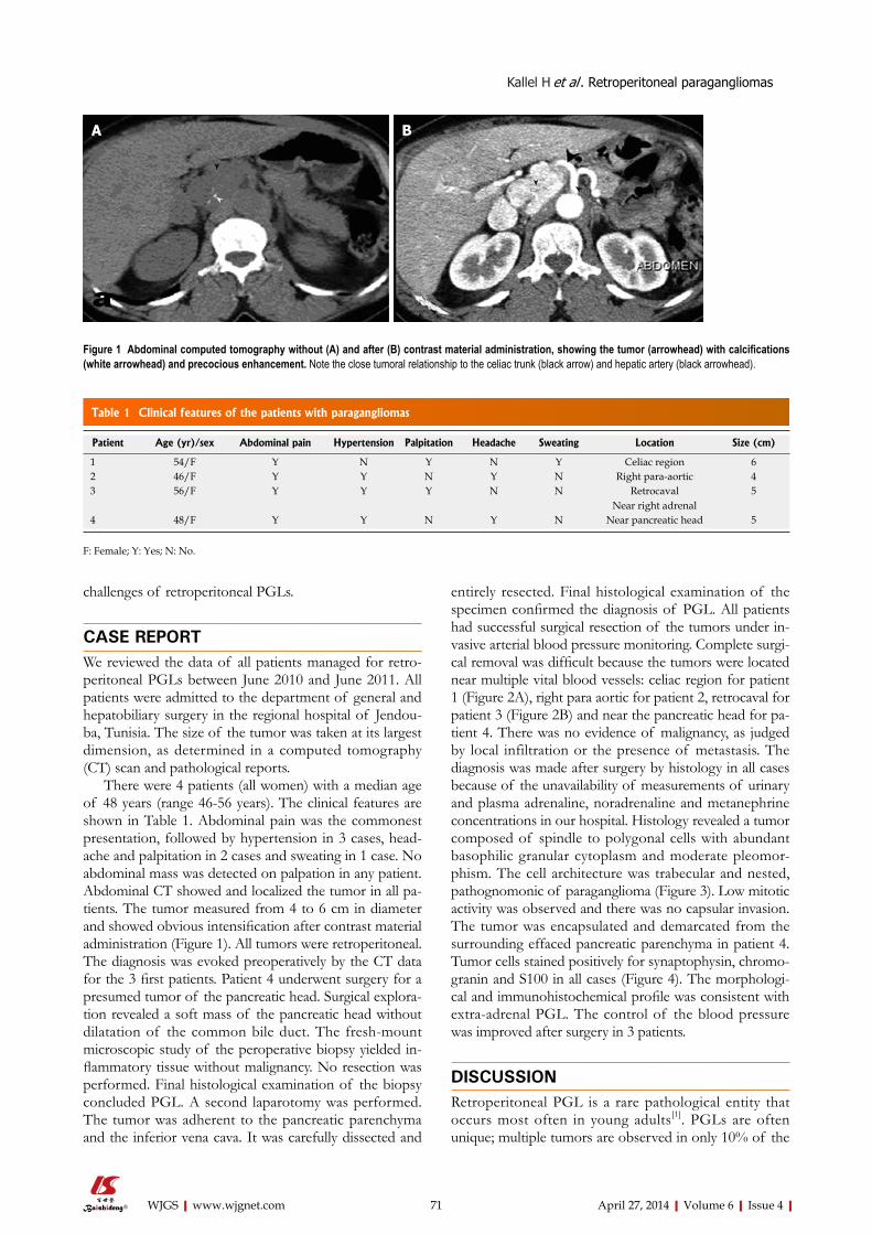
challenges of retroperitoneal PGLs.
CASE REPORTWe reviewed the data of all patients managed for retro-peritoneal PGLs between June 2010 and June 2011. All patients were admitted to the department of general and hepatobiliary surgery in the regional hospital of Jendou-ba, Tunisia. The size of the tumor was taken at its largest dimension, as determined in a computed tomography (CT) scan and pathological reports.
There were 4 patients (all women) with a median age of 48 years (range 46-56 years). The clinical features are shown in Table 1. Abdominal pain was the commonest presentation, followed by hypertension in 3 cases, head-ache and palpitation in 2 cases and sweating in 1 case. No abdominal mass was detected on palpation in any patient. Abdominal CT showed and localized the tumor in all pa-tients. The tumor measured from 4 to 6 cm in diameter and showed obvious intensification after contrast material administration (Figure 1). All tumors were retroperitoneal. The diagnosis was evoked preoperatively by the CT data for the 3 first patients. Patient 4 underwent surgery for a presumed tumor of the pancreatic head. Surgical explora-tion revealed a soft mass of the pancreatic head without dilatation of the common bile duct. The fresh-mount microscopic study of the peroperative biopsy yielded in-flammatory tissue without malignancy. No resection was performed. Final histological examination of the biopsy concluded PGL. A second laparotomy was performed. The tumor was adherent to the pancreatic parenchyma and the inferior vena cava. It was carefully dissected and
entirely resected. Final histological examination of the specimen confirmed the diagnosis of PGL. All patients had successful surgical resection of the tumors under in-vasive arterial blood pressure monitoring. Complete surgi-cal removal was difficult because the tumors were located near multiple vital blood vessels: celiac region for patient 1 (Figure 2A), right para aortic for patient 2, retrocaval for patient 3 (Figure 2B) and near the pancreatic head for pa-tient 4. There was no evidence of malignancy, as judged by local infiltration or the presence of metastasis. The diagnosis was made after surgery by histology in all cases because of the unavailability of measurements of urinary and plasma adrenaline, noradrenaline and metanephrine concentrations in our hospital. Histology revealed a tumor composed of spindle to polygonal cells with abundant basophilic granular cytoplasm and moderate pleomor-phism. The cell architecture was trabecular and nested, pathognomonic of paraganglioma (Figure 3). Low mitotic activity was observed and there was no capsular invasion. The tumor was encapsulated and demarcated from the surrounding effaced pancreatic parenchyma in patient 4. Tumor cells stained positively for synaptophysin, chromo-granin and S100 in all cases (Figure 4). The morphologi-cal and immunohistochemical profile was consistent with extra-adrenal PGL. The control of the blood pressure was improved after surgery in 3 patients.
DISCUSSIONRetroperitoneal PGL is a rare pathological entity that occurs most often in young adults[1]. PGLs are often unique; multiple tumors are observed in only 10% of the
April 27, 2014|Volume 6|Issue 4|WJGS|www.wjgnet.com
Kallel H et al . Retroperitoneal paragangliomas
71
Figure 1 Abdominal computed tomography without (A) and after (B) contrast material administration, showing the tumor (arrowhead) with calcifications (white arrowhead) and precocious enhancement. Note the close tumoral relationship to the celiac trunk (black arrow) and hepatic artery (black arrowhead).
Patient Age (yr)/sex Abdominal pain Hypertension Palpitation Headache Sweating Location Size (cm)
1 54/F Y N Y N Y Celiac region 6 2 46/F Y Y N Y N Right para-aortic 4 3 56/F Y Y Y N N Retrocaval
Near right adrenal5
4 48/F Y Y N Y N Near pancreatic head 5
Table 1 Clinical features of the patients with paragangliomas
F: Female; Y: Yes; N: No.
A B
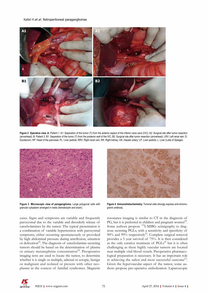
cases. Signs and symptoms are variable and frequently paroxysmal due to the variable and disorderly release of catecholamines by the tumor. The typical presentation is a combination of variable hypertension with paroxysmal symptoms, either occurring spontaneously or provoked by high abdominal pressure during anteflexion, urination or defecation[2]. The diagnosis of catecholamine-secreting tumors should be based on the determination of plasma or urinary metanephrine concentration[2]. Preoperative imaging tests are used to locate the tumor, to determine whether it is single or multiple, adrenal or ectopic, benign or malignant and isolated or present with other neo-plasms in the context of familial syndromes. Magnetic
resonance imaging is similar to CT in the diagnosis of PG, but it is preferred in children and pregnant women[2]. Some authors propose 123I-MIBG scintigraphy to diag-nose secreting PGLs, with a sensitivity and specificity of 90% and 99% respectively[1]. Complete surgical removal provides a 5 year survival of 75%. It is then considered as the only curative treatment of PGLs[1] but it is often challenging as these highly vascular tumors are located near multiple vital blood vessels. Preoperative pharmaco-logical preparation is necessary. It has an important role in achieving the safest and most successful outcome[3]. Given the hypervascular aspect of the tumor, some au-thors propose pre-operative embolization. Laparoscopic
April 27, 2014|Volume 6|Issue 4|WJGS|www.wjgnet.com 72
Figure 2 Operative view. A: Patient 1. A1: Separation of the tumor (T) from the anterior aspect of the inferior vena cava (IVC); A2: Surgical site after tumor resection (arrowhead); B: Patient 3. B1: Separation of the tumor (T) from the posterior wall of the IVC; B2: Surgical site after tumor resection (arrowhead). LRV: Left renal vein; D: Duodenum; HP: Head of the pancreas; PL: Liver pedicle; RRV: Right renal vein; RK: Right kidney; HA: Hepatic artery; LP: Liver pedicle; L: Liver (Lobe of Spiegel).
Figure 3 Microscopic view of paraganglioma. Large polygonal cells with granular cytoplasm arranged in nests (hematoxylin and eosin).
Figure 4 Immunohistochemistry. Tumoral cells strongly express anti-chromo-granin antibody.
PL
D
HP
LRVT
IVC
L
LPHA
IVC
TRRV
RK
A1 A2
B1 B2
Kallel H et al . Retroperitoneal paragangliomas
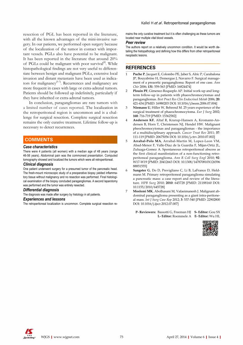
April 27, 2014|Volume 6|Issue 4|WJGS|www.wjgnet.com
mains the only curative treatment but it is often challenging as these tumors are located near multiple vital blood vessels.Peer reviewThe authors report on a relatively uncommon condition. It would be worth de-tailing the histopathology and defining how this differs from other retroperitoneal neoplastic lesions.
REFERENCES1 Puche P, Jacquet E, Colombo PE, Jaber S, Alric P, Carabalona
JP, Bouyabrine H, Domergue J, Navarro F. Surgical manage-ment of a preaortic paraganglioma: Report of one case. Ann Chir 2006; 131: 559-563 [PMID: 16824474]
2 Plouin PF, Gimenez-Roqueplo AP. Initial work-up and long-term follow-up in patients with phaeochromocytomas and paragangliomas. Best Pract Res Clin Endocrinol Metab 2006; 20: 421-434 [PMID: 16980203 DOI: 10.1016/j.beem.2006.07.004]
3 Niemann U, Hiller W, Behrend M. 25 years experience of the surgical treatment of phaeochromocytoma. Eur J Surg 2002; 168: 716-719 [PMID: 15362582]
4 Andersen KF, Altaf R, Krarup-Hansen A, Kromann-An-dersen B, Horn T, Christensen NJ, Hendel HW. Malignant pheochromocytomas and paragangliomas - the importance of a multidisciplinary approach. Cancer Treat Rev 2011; 37: 111-119 [PMID: 20675056 DOI: 10.1016/j.ctrv.2010.07.002]
5 Arrabal-Polo MA, Arrabal-Martin M, Lopez-Leon VM, Abad-Menor F, Valle-Diaz de la Guardia F, Mijan-Ortiz JL, Zuluaga-Gomez A. Spontaneous retroperitoneal abscess as the first clinical manifestation of a non-functioning retro-peritoneal paraganglioma. Ann R Coll Surg Engl 2010; 92: W17-W19 [PMID: 20412663 DOI: 10.1308/147870810X12659688851555]
6 Sangster G, Do D, Previgliano C, Li B, LaFrance D, Held-mann M. Primary retroperitoneal paraganglioma simulating a pancreatic mass: a case report and review of the litera-ture. HPB Surg 2010; 2010: 645728 [PMID: 21188160 DOI: 10.1155/2010/645728]
7 Moslemi MK, Abolhasani M, Vafaeimanesh J. Malignant ab-dominal paraganglioma presenting as a giant intra-peritone-al mass. Int J Surg Case Rep 2012; 3: 537-540 [PMID: 22902800 DOI: 10.1016/j.ijscr.2012.07.007]
P- Reviewers: Bassotti G, Freeman HJ S- Editor: Gou SX L- Editor: Roemmele A E- Editor: Wu HL
resection of PGL has been reported in the literature, with all the known advantages of the mini-invasive sur-gery. In our patients, we performed open surgery because of the localization of the tumor in contact with impor-tant vessels. PGLs also have potential to be malignant. It has been reported in the literature that around 20% of PGLs could be malignant with poor survival[4]. While histopathological findings are not very useful to differen-tiate between benign and malignant PGLs, extensive local invasion and distant metastasis have been used as indica-tors for malignancy[5-7]. Recurrences and malignancy are more frequent in cases with large or extra-adrenal tumors. Patients should be followed up indefinitely, particularly if they have inherited or extra-adrenal tumors.
In conclusion, paragangliomas are rare tumors with a limited number of cases reported. The localization in the retroperitoneal region is uncommon and is a chal-lenge for surgical resection. Complete surgical resection remains the only curative treatment. Lifetime follow-up is necessary to detect recurrences.
COMMENTSCase characteristicsThere were 4 patients (all women) with a median age of 48 years (range 46-56 years). Abdominal pain was the commonest presentation. Computed tomography showed and localized the tumors which were all retroperitoneal. Clinical diagnosisOne patient underwent surgery for a presumed tumor of the pancreatic head. The fresh-mount microscopic study of a preoperative biopsy yielded inflamma-tory tissue without malignancy and no resection was performed. Final histologi-cal examination of the biopsy concluded paragangliomas. A second laparotomy was performed and the tumor was entirely resected. Differential diagnosisThe diagnosis was made after surgery by histology in all patients. Experiences and lessonsThe retroperitoneal localization is uncommon. Complete surgical resection re-
73
COMMENTS
Kallel H et al . Retroperitoneal paragangliomas

Jong Han Yoo, Sang Hyuk Seo, Min Sung An, Tae Kwun Ha, Kwang Hee Kim, Ki Beom Bae, Chang Soo Choi, Sang Hun Oh, Young Kil Choi
Recurrence of gastric cancer in the jejunal stump after radical total gastrectomy
Jong Han Yoo, Sang Hyuk Seo, Min Sung An, Tae Kwun Ha, Kwang Hee Kim, Ki Beom Bae, Chang Soo Choi, Sang Hun Oh, Young Kil Choi, Department of Surgery, Busan Paik Hospi-tal, Inje University College of Medicine, Busan 614-735, South KoreaAuthor contributions: Yoo JH and Kim KH designed the re-port; Kim KH, Choi CS and Choi YK were the attending doctors for the patient; Kim KH and Choi YK performed the surgical operation; An MS, Ha TK and Bae KB organized the report; and Yoo JH and Seo SH wrote the paper; Oh SH organized the report.Correspondence to: Kwang Hee Kim, MD, Department of Surgery, Busan Paik Hospital, Inje University College of Medi-cine, Gaegum 2-dong, Busanjingu, Busan 614-735,South Korea. [email protected]: +82-51-8906352 Fax: +82-51-8989427Received: August 19, 2013 Revised: January 16, 2014Accepted: February 18, 2014Published online: April 27, 2014
AbstractThis is a very rare case of the recurrence of gastric cancer in the jejunal stump after radical total gastrec-tomy with Roux-en-Y reconstruction. In January 2008, a 65-year-old man underwent radical total gastrectomy with Roux-en-Y reconstruction for stage ⅠB gastric cancer of the upper body. At a follow-up in December 2011, the patient had a recurrence of gastric cancer on gastroduodenal fibroscopy. The gastroduodenal fibro-scopic biopsy specimens show a well-differentiated tu-bular adenocarcinoma. Computed tomography showed no lymphadenopathy or hepatic metastases. At laparot-omy, there was a tumor in the jejunal stump involving the pancreatic tail and spleen. Therefore, the patient underwent jejunal pouch resection, distal pancreatec-tomy and splenectomy. The patient was diagnosed with gastric cancer on histopathological examination.
© 2014 Baishideng Publishing Group co., Limited. All rights reserved.
Key words: Gastric cancer; Recurrence; Jejunal stump
Core tip: In our study, there was a case of recurrence of gastric cancer in the jejunal stump after radical total gastrectomy with Roux-en-Y reconstruction. The patient underwent jejunal pouch resection, distal pancreatecto-my and splenectomy. On histopathologic examinations, the patient was diagnosed with gastric cancer.
Yoo JH, Seo SH, An MS, Ha TK, Kim KH, Bae KB, Choi CS, Oh SH, Choi YK. Recurrence of gastric cancer in the jejunal stump after radical total gastrectomy. World J Gastrointest Surg 2014; 6(4): 74-76 Available from: URL: http://www.wjgnet.com/1948-9366/full/v6/i4/74.htm DOI: http://dx.doi.org/10.4240/wjgs.v6.i4.74
INTRODUCTIONGastric cancer is a highly prevalent cancer that occurs the most commonly in Korea. It shows a very good prog-nosis when detected earlier in a regular medical check-up. In advanced cancer, however, a poor prognosis has been well documented. There are many recurrent cases of gastric cancer despite radical surgery. Its recurrence occurs through hematogenous, peritoneal dissemination or via the lymph nodes. We report a case of recurrence of gastric cancer in the jejunal stump after radical total gastrectomy with Roux-en-Y reconstruction.
CASE REPORTA 65-year-old man presented with a recurrence on gastro-duodenal fibroscopy (Figure 1) at a follow-up after gastric cancer surgery. He underwent radical total gastrectomy with Roux-en-Y reconstruction (end to side esophago-jejunostomy with circular stapler), for gastric cancer de-tected on gastroduodenal fibroscopy in January 2008. The
CASE REPORT
Online Submissions: http://www.wjgnet.com/esps/[email protected]:10.4240/wjgs.v6.i4.74
April 27, 2014|Volume 6|Issue 4|WJGS|www.wjgnet.com
World J Gastrointest Surg 2014 April 27; 6(4): 74-76ISSN 1948-9366 (online)
© 2014 Baishideng Publishing Group co., Limited. All rights reserved.
74
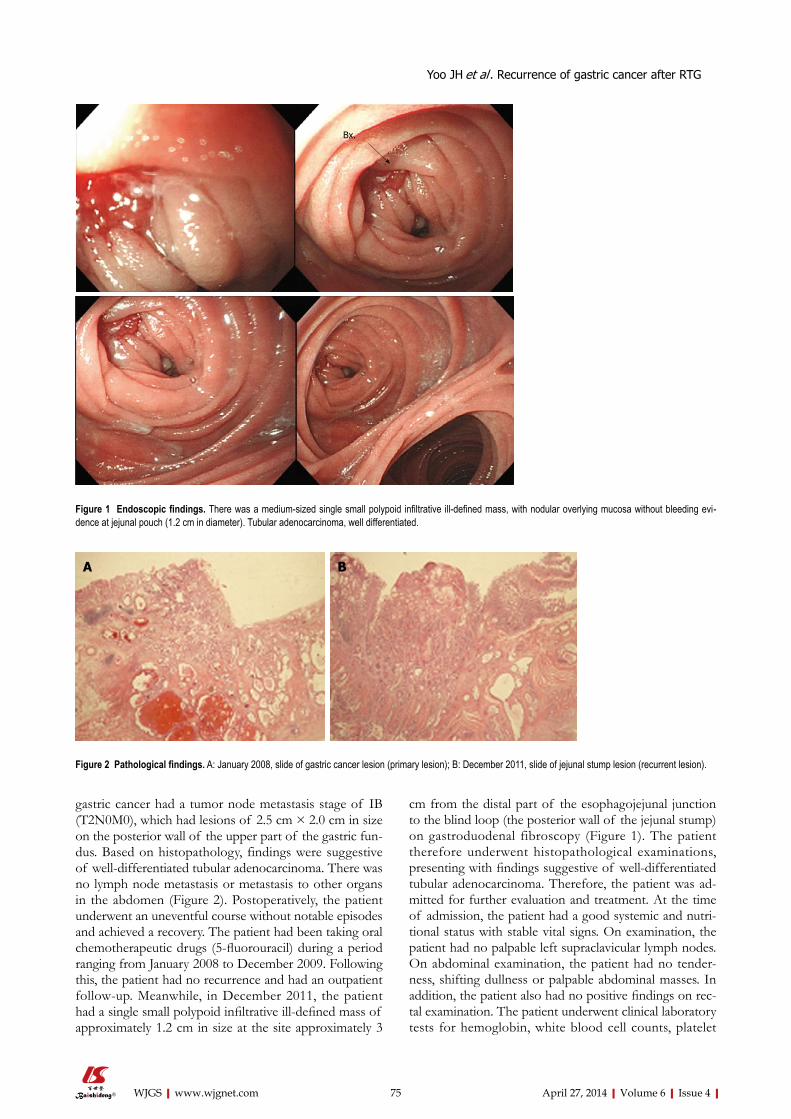
gastric cancer had a tumor node metastasis stage of IB (T2N0M0), which had lesions of 2.5 cm × 2.0 cm in size on the posterior wall of the upper part of the gastric fun-dus. Based on histopathology, findings were suggestive of well-differentiated tubular adenocarcinoma. There was no lymph node metastasis or metastasis to other organs in the abdomen (Figure 2). Postoperatively, the patient underwent an uneventful course without notable episodes and achieved a recovery. The patient had been taking oral chemotherapeutic drugs (5-fluorouracil) during a period ranging from January 2008 to December 2009. Following this, the patient had no recurrence and had an outpatient follow-up. Meanwhile, in December 2011, the patient had a single small polypoid infiltrative ill-defined mass of approximately 1.2 cm in size at the site approximately 3
cm from the distal part of the esophagojejunal junction to the blind loop (the posterior wall of the jejunal stump) on gastroduodenal fibroscopy (Figure 1). The patient therefore underwent histopathological examinations, presenting with findings suggestive of well-differentiated tubular adenocarcinoma. Therefore, the patient was ad-mitted for further evaluation and treatment. At the time of admission, the patient had a good systemic and nutri-tional status with stable vital signs. On examination, the patient had no palpable left supraclavicular lymph nodes. On abdominal examination, the patient had no tender-ness, shifting dullness or palpable abdominal masses. In addition, the patient also had no positive findings on rec-tal examination. The patient underwent clinical laboratory tests for hemoglobin, white blood cell counts, platelet
April 27, 2014|Volume 6|Issue 4|WJGS|www.wjgnet.com
Yoo JH et al . Recurrence of gastric cancer after RTG
75
Figure 1 Endoscopic findings. There was a medium-sized single small polypoid infiltrative ill-defined mass, with nodular overlying mucosa without bleeding evi-dence at jejunal pouch (1.2 cm in diameter). Tubular adenocarcinoma, well differentiated.
Figure 2 Pathological findings. A: January 2008, slide of gastric cancer lesion (primary lesion); B: December 2011, slide of jejunal stump lesion (recurrent lesion).
Bx.
A B
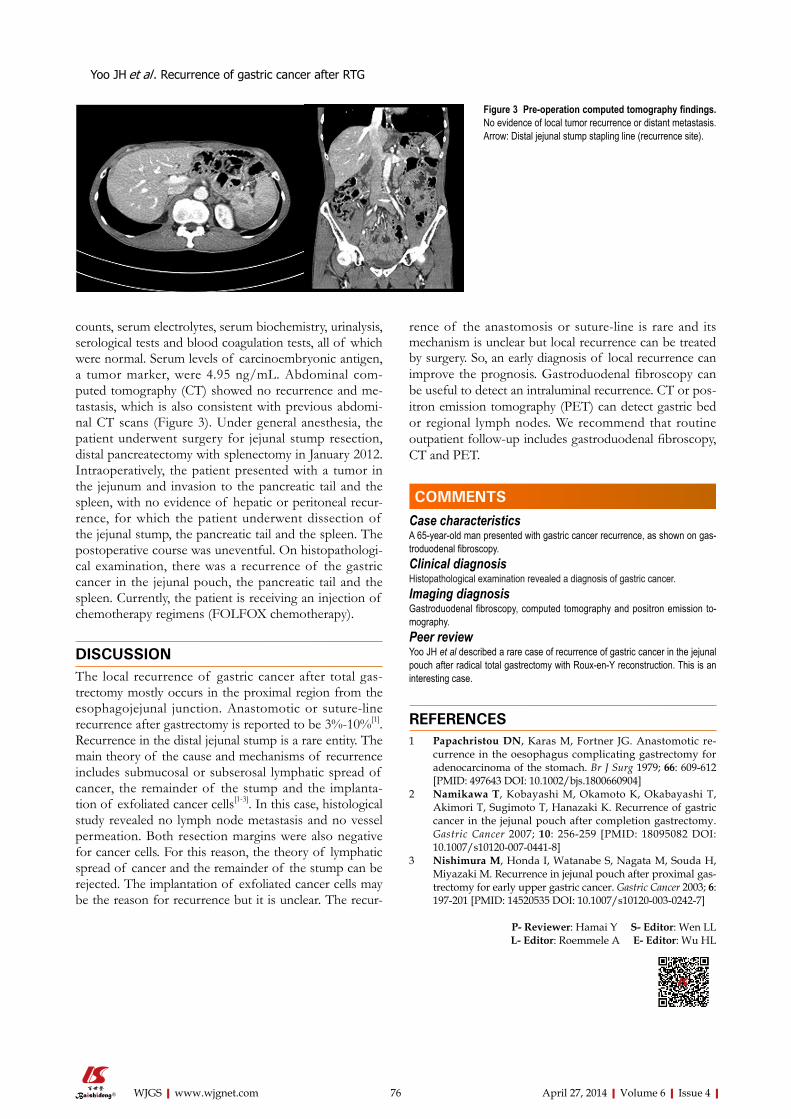
counts, serum electrolytes, serum biochemistry, urinalysis, serological tests and blood coagulation tests, all of which were normal. Serum levels of carcinoembryonic antigen, a tumor marker, were 4.95 ng/mL. Abdominal com-puted tomography (CT) showed no recurrence and me-tastasis, which is also consistent with previous abdomi-nal CT scans (Figure 3). Under general anesthesia, the patient underwent surgery for jejunal stump resection, distal pancreatectomy with splenectomy in January 2012. Intraoperatively, the patient presented with a tumor in the jejunum and invasion to the pancreatic tail and the spleen, with no evidence of hepatic or peritoneal recur-rence, for which the patient underwent dissection of the jejunal stump, the pancreatic tail and the spleen. The postoperative course was uneventful. On histopathologi-cal examination, there was a recurrence of the gastric cancer in the jejunal pouch, the pancreatic tail and the spleen. Currently, the patient is receiving an injection of chemotherapy regimens (FOLFOX chemotherapy).
DISCUSSIONThe local recurrence of gastric cancer after total gas-trectomy mostly occurs in the proximal region from the esophagojejunal junction. Anastomotic or suture-line recurrence after gastrectomy is reported to be 3%-10%[1]. Recurrence in the distal jejunal stump is a rare entity. The main theory of the cause and mechanisms of recurrence includes submucosal or subserosal lymphatic spread of cancer, the remainder of the stump and the implanta-tion of exfoliated cancer cells[1-3]. In this case, histological study revealed no lymph node metastasis and no vessel permeation. Both resection margins were also negative for cancer cells. For this reason, the theory of lymphatic spread of cancer and the remainder of the stump can be rejected. The implantation of exfoliated cancer cells may be the reason for recurrence but it is unclear. The recur-
rence of the anastomosis or suture-line is rare and its mechanism is unclear but local recurrence can be treated by surgery. So, an early diagnosis of local recurrence can improve the prognosis. Gastroduodenal fibroscopy can be useful to detect an intraluminal recurrence. CT or pos-itron emission tomography (PET) can detect gastric bed or regional lymph nodes. We recommend that routine outpatient follow-up includes gastroduodenal fibroscopy, CT and PET.
COMMENTSCase characteristicsA 65-year-old man presented with gastric cancer recurrence, as shown on gas-troduodenal fibroscopy.Clinical diagnosisHistopathological examination revealed a diagnosis of gastric cancer.Imaging diagnosisGastroduodenal fibroscopy, computed tomography and positron emission to-mography.Peer reviewYoo JH et al described a rare case of recurrence of gastric cancer in the jejunal pouch after radical total gastrectomy with Roux-en-Y reconstruction. This is an interesting case.
REFERENCES1 Papachristou DN, Karas M, Fortner JG. Anastomotic re-
currence in the oesophagus complicating gastrectomy for adenocarcinoma of the stomach. Br J Surg 1979; 66: 609-612 [PMID: 497643 DOI: 10.1002/bjs.1800660904]
2 Namikawa T, Kobayashi M, Okamoto K, Okabayashi T, Akimori T, Sugimoto T, Hanazaki K. Recurrence of gastric cancer in the jejunal pouch after completion gastrectomy. Gastric Cancer 2007; 10: 256-259 [PMID: 18095082 DOI: 10.1007/s10120-007-0441-8]
3 Nishimura M, Honda I, Watanabe S, Nagata M, Souda H, Miyazaki M. Recurrence in jejunal pouch after proximal gas-trectomy for early upper gastric cancer. Gastric Cancer 2003; 6: 197-201 [PMID: 14520535 DOI: 10.1007/s10120-003-0242-7]
P- Reviewer: Hamai Y S- Editor: Wen LL L- Editor: Roemmele A E- Editor: Wu HL
April 27, 2014|Volume 6|Issue 4|WJGS|www.wjgnet.com 76
Figure 3 Pre-operation computed tomography findings. No evidence of local tumor recurrence or distant metastasis. Arrow: Distal jejunal stump stapling line (recurrence site).
COMMENTS
Yoo JH et al . Recurrence of gastric cancer after RTG

Sang hyun Kang, Kwang hee Kim, Sang hyuk Seo, Min Sung An, Tae Kwun ha, ha Kyung Park, Ki Beom Bae, Chang Soo Choi, Sang hun Oh, Young Kil Choi
Neuroendocrine carcinoma of the stomach: A case report
Sang hyun Kang, Kwang hee Kim, Sang hyuk Seo, Min Sung An, Tae Kwun ha, ha Kyung Park, Ki Beom Bae, Chang Soo Choi, Sang hun Oh, Young Kil Choi, Department of Surgery, Busan Paik Hospital, Inje University College of Med-icine, Busan 614-735, South KoreaAuthor contributions: Kang SH and Kim KH designed the report; Kim KH, Oh SH and Park HK were the attending doctors for the patient; Kang SH and Kim KH performed the surgical operation; An MS, Ha TK, Bae KB and Choi CS organized the report; and Kang SH, Seo SH and Choi YK wrote the paper.Correspondence to: Kwang hee Kim, MD, Department of Surgery, Busan Paik Hospital, Inje University College of Medi-cine, Gaegum 2-dong, Busanjingu, Busan 614-735, South Korea. [email protected]: +82-51-8906352 Fax: +82-51-8989427Received: October 14, 2013 Revised: December 2, 2013Accepted: February 18, 2014Published online: April 27, 2014
AbstractNeuroendocrine carcinoma (NEC) is a rare tumor, com-prising < 1% of stomach cancers. A 55-year-old woman was referred to our hospital with biopsy-proven gastric cancer. A shallow ulcerative lesion was detected in the lesser curvature of the lower body. It was suspected to be early gastric cancer ⅡA + ⅡC type. Thus, endo-scopic submucosal dissection was performed. She was subsequently diagnosed with NEC, which is aggressive and carries a poor prognosis. We conducted a radical resection and a laparoscopic-assisted distal gastrecto-my. The tumor had infiltrated the subserosal layer and 6/42 lymph nodes were involved. The mitotic index was 16/10 high power fields and the Ki-67 labeling index was 26%-50%. The final diagnosis of NEC was made according to the World health Organization 2010 crite-ria. She was suspected of having jumping metastasis to the proximal margin. The patient was treated with an oral anticancer drug (5-flurouracil based drug) for 2 years. The patient has been followed up for 3 years without recurrence.
© 2014 Baishideng Publishing Group co., Limited. All rights reserved.
Key words: Neuroendocrine carcinoma; Mitosis; Ki-67; Gastrectomy; Prognosis
Core tip: Some studies argue that neuroendocrine car-cinoma (NEC) can be removed by endoscopic resection. however, in this case, we found that NEC can have jumping metastasis. Thus, NEC must be removed by radical surgical resection.
Kang SH, Kim KH, Seo SH, An MS, Ha TK, Park HK, Bae KB, Choi CS, Oh SH, Choi YK. Neuroendocrine car-cinoma of the stomach: A case report. World J Gastrointest Surg 2014; 6(4): 77-79 Available from: URL: http://www.wjgnet.com/1948-9366/full/v6/i4/77.htm DOI: http://dx.doi.org/10.4240/wjgs.v6.i4.77
INTRODUCTIONNeuroendocrine carcinoma (NEC) is rare tumor that in-cludes < 1% of stomach cancers. It is aggressive and has a poor prognosis[1-3]. NEC is classified as neuroendocrine carcinoma G3 according to The World Health Organi-zation (WHO) classification of tumors of the digestive system, 2010[4].
In this report, we describe a patient with NEC who underwent endoscopic submucosal dissection (ESD) and laparoscopic assisted distal gastrectomy (LADG) for re-moval of a tumor.
CASE REPORTA 55-year-old woman with acid reflux underwent an esoph-agogastroduodenoscopy (EGD) for a checkup. A shallow ulcerative lesion was detected in the lesser curvature of the lower body (Figure 1). It was suspected to be early gastric
CASE REPORT
Online Submissions: http://www.wjgnet.com/esps/[email protected]:10.4240/wjgs.v6.i4.77
April 27, 2014|Volume 6|Issue 4|WJGS|www.wjgnet.com
World J Gastrointest Surg 2014 April 27; 6(4): 77-79ISSN 1948-9366 (online)
© 2014 Baishideng Publishing Group co., Limited. All rights reserved.
77

cancer ⅡA + ⅡC type. A biopsy was done and it was di-agnosed as a well differentiated adenocarcinoma. She was transferred to the digestive department of our hospital.
On July 13, she underwent ESD. The specimen was 6.3 cm × 3.8 cm and a pathological examination revealed a 1.2 cm × 1.4 cm NEC that had invaded the submu-cosal layer. The tumor cells exhibited mitosis in 16/10 high power fields (HPF). The resection margin was clear (Figure 2) and no lymphatic, vascular or neural invasion was observed. She was advised to undergo an operation due to possible neural invasion by the NEC. On July 24, she vomited blood from an ulcer because of the weak-ened mucosa after ESD. The bleeding was stopped under emergency EGD. She underwent conservative treatment with a proton-pump inhibitor and no oral intake.
On August 9, she underwent LADG with a D2 lymphadenectomy. A Billroth type Ⅰ anastomosis was done. A frozen biopsy revealed that the proximal and distal resection margins were clear of lesions. The mass was 2.2 cm × 1.3 cm in size and limited to the subserosa (Figure 3). The proximal resection margin was very close to the lesion but the distal resection margin was clear. Neural and lymphatic invasion was observed with 6 of 42 metastatic lymph nodes harvested. The tumor cells were positive for synaptophysin, chromogranin and CD56. The Ki-67 labeling index was 2+ (26%-50%). These find-ings led to the diagnosis of NEC, according to the 2010 WHO criteria[4]. The proximal margin was clear but the final pathology showed that some cancer cells were found between the mucosa and submucosa.
Minor bleeding was detected through the drain dur-ing the first 3 d. After an antihemorrhagic treatment and a transfusion, the blood tests were stable and the drain color changed to clear. She was discharged after the drain was removed.
She was treated with an oral anticancer drug [5-flu-rouracil (5-FU) based drug] for 2 years. No recurrence at the anastomosis or other site in the stomach was ob-served 3 years later.
DISCUSSIONNeuroendocrine neoplasm (NEN) is an epithelial neo-
plasm with predominant neuroendocrine differentiation and is an uncommon tumor with multiple sites of occur-rence[5].
NENs are commonly divided by origin as located in the foregut (lung, bronchus, stomach or duodenum), midgut (jejunum, ileum, appendix or proximal colon) and hindgut (distal colon or rectum). The percentage of fore-gut cases is 34%, midgut 30% and hindgut 36%[6].
Gastric NEN is classified into neuroendocrine tumor (NET), neuroendocrine carcinoma (NEC), mixed ad-enoneuroendocrine carcinoma, enterochromaffin cells, serotonin-producing NETs and gastrin-producing NETs. NETs include NET G1 (carcinoid) and NET G2 (well-differentiated neuroendocrine tumor/carcinoma). NECs include NEC G3 (poorly differentiated neuroendocrine carcinoma small cell type/large cell type)[4]. NEN is posi-tive for synaptophysin and chromogranin A[7].
NEN is classified based on the level of cellular pro-liferation, including the mitotic and Ki-67 indices[4]. In our case, the mitotic index was 16/10 HPF and the Ki-67 labeling index was 26%-50%. Thus, she was diagnosed with NEC. We suspected jumping metastasis from the main lesion to the proximal margin.
April 27, 2014|Volume 6|Issue 4|WJGS|www.wjgnet.com
Kang Sh et al . Neuroendocrine carcinoma of the stomach
78
Figure 1 Endoscopic findings. Ulcerative lesion in the lesser curvature of the lower body.
Figure 2 Endoscopic submucosal dissection specimen. A hypercellular le-sion was detected in the mucosa and submucosal layers.
Figure 3 Laparoscopic assisted distal gastrectomy specimen. The ulcer-ative lesion due to mucosal detachment after endoscopic submucosal dissec-tion is distinguished from normal mucosa (right side). Fibrosis was observed in the submucosal layer and a hypercellular lesion that was the same as the endoscopic submucosal dissection specimen in the muscle and subserosa lay-ers.
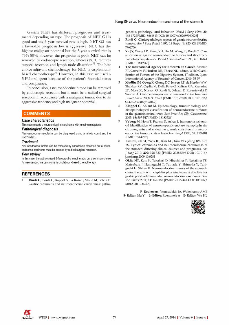
Gastric NEN has different prognoses and treat-ments depending on type. The prognosis of NET G1 is good and the 5 year survival rate is high. NET G2 has a favorable prognosis but is aggressive. NEC has the highest malignant potential but the 5 year survival rate is 75%-80%; however, the prognosis is poor. NET can be removed by endoscopic resection, whereas NEC requires surgical resection and lymph node dissection[8]. The best choice adjuvant chemotherapy for NEC is cisplatinum-based chemotherapy[9]. However, in this case we used a 5-FU oral agent because of the patient’s financial status and compliance.
In conclusion, a neuroendocrine tumor can be removed by endoscopic resection but it must be a radical surgical resection in accordance with a malignant tumor, due to its aggressive tendency and high malignant potential.
COMMENTSCase characteristicsThis case reports a neuroendocrine carcinoma with jumping metastasis.Pathological diagnosisNeuroendocrine neoplasm can be diagnosed using a mitotic count and the Ki-67 index.TreatmentNeuroendocrine tumors can be removed by endoscopic resection but a neuro-endocrine carcinoma must be excised by radical surgical resection.Peer reviewIn this case, the authors used 5-flurouracil chemotherapy, but a common choice for neuroendocrine carcinoma is cisplatinum-based chemotherapy.
REFERENCES1 Rindi G, Bordi C, Rappel S, La Rosa S, Stolte M, Solcia E.
Gastric carcinoids and neuroendocrine carcinomas: patho-
genesis, pathology, and behavior. World J Surg 1996; 20: 168-172 [PMID: 8661813 DOI: 10.1007/s002689900026]
2 Rindi G. Clinicopathologic aspects of gastric neuroendocrine tumors. Am J Surg Pathol 1995; 19 Suppl 1: S20-S29 [PMID: 7762736]
3 Yu JY, Wang LP, Meng YH, Hu M, Wang JL, Bordi C. Clas-sification of gastric neuroendocrine tumors and its clinico-pathologic significance. World J Gastroenterol 1998; 4: 158-161 [PMID: 11819263]
4 The International Agency for Research on Cancer. Bosman FT, Carneiro F, Hruban RH, Theise ND, editor. WHO Classi-fication of Tumors of the Digestive System. 4th edition, Lyon: International Agency of Research of Cancer, 2010: 53-57
5 Modlin IM, Oberg K, Chung DC, Jensen RT, de Herder WW, Thakker RV, Caplin M, Delle Fave G, Kaltsas GA, Krenning EP, Moss SF, Nilsson O, Rindi G, Salazar R, Ruszniewski P, Sundin A. Gastroenteropancreatic neuroendocrine tumours. Lancet Oncol 2008; 9: 61-72 [PMID: 18177818 DOI: 10.1016/S1470-2045(07)70410-2]
6 Klöppel G, Anlauf M. Epidemiology, tumour biology and histopathological classification of neuroendocrine tumours of the gastrointestinal tract. Best Pract Res Clin Gastroenterol 2005; 19: 507-517 [PMID: 16183524]
7 Vyberg M, Horn T, Francis D, Askaa J. Immunohistochemi-cal identification of neuron-specific enolase, synaptophysin, chromogranin and endocrine granule constituent in neuro-endocrine tumours. Acta Histochem Suppl 1990; 38: 179-181 [PMID: 1964227]
8 Kim BS, Oh ST, Yook JH, Kim KC, Kim MG, Jeong JW, Kim BS. Typical carcinoids and neuroendocrine carcinomas of the stomach: differing clinical courses and prognoses. Am J Surg 2010; 200: 328-333 [PMID: 20385369 DOI: 10.1016/j.amjsurg.2009.10.028]
9 Okita NT, Kato K, Takahari D, Hirashima Y, Nakajima TE, Matsubara J, Hamaguchi T, Yamada Y, Shimada Y, Tani-guchi H, Shirao K. Neuroendocrine tumors of the stomach: chemotherapy with cisplatin plus irinotecan is effective for gastric poorly-differentiated neuroendocrine carcinoma. Gas-tric Cancer 2011; 14: 161-165 [PMID: 21327441 DOI: 10.1007/s10120-011-0025-5]
P- Reviewers: Voutsadakis IA, Walenkamp AME S- Editor: Ma YJ L- Editor: Roemmele A E- Editor: Wu HL
April 27, 2014|Volume 6|Issue 4|WJGS|www.wjgnet.com 79
COMMENTS
Kang Sh et al . Neuroendocrine carcinoma of the stomach

Published by Baishideng Publishing Group Inc
World Journal of Gastrointestinal SurgeryWorld J Gastrointest Surg 2014 May 27; 6(5): 80-87
ISSN 1948-9366 (online)
80 Rarecauseofabdominalincidentaloma:Hepatoduodenalligament
teratoma
Jeismann VB, Dumarco RB, di Loreto C, Barbuti RC, Jukemura J
84 Single-incisionlaparoscopiccecectomyforlow-gradeappendiceal
mucinousneoplasmafterlaparoscopicrectectomy
Fujino S, Miyoshi N, Noura S, Shingai T, Tomita Y, Ohue M, Yano M
Contents Monthly Volume 6 Number 5 May 27, 2014
WJGS|www.wjgnet.com I May 27, 2014|Volume 6|Issue 5|
CASE REPORT
Contents
APPENDIX
ABOUT COVER
World Journal of Gastrointestinal SurgeryVolume 6 Number 5 May 27, 2014
I-V Instructionstoauthors
EditorialBoardMemberofWorldJournalofGastrointestinalSurgery ,ChapelAlain,PhD,DepartmentofMenRadioprotection,LaboratoryofRadioPathologyandInnovativeTherapy,InstituteofNuclearSafetyandRadioprotection,POBox17,Paris92262,France
World Journal of Gastrointestinal Surgery (World J Gastrointest Surg, WJGS, online ISSN 1948-9366, DOI: 10.4240) is a peer-reviewed open access academic journal that aims to guide clinical practice and improve diagnostic and therapeutic skills of clinicians.
WJGS covers topics concerning micro-invasive surgery; laparoscopy; hepatic, biliary, pancreatic and splenic surgery; surgical nutrition; portal hypertension, as well as associated subjects. The current columns of WJGS include editorial, frontier, diagnostic advances, therapeutics advances, field of vision, mini-reviews, review, topic highlight, medical ethics, original articles, case report, clinical case conference (Clinicopathological conference), and autobiography. Priority publication will be given to articles concerning diagnosis and treatment of gastrointestinal surgery diseases. The following aspects are covered: Clinical diagnosis, laboratory diagnosis, differential diagnosis, imaging tests, pathological diagnosis, molecular biological diagnosis, immunological diagnosis, genetic diagnosis, functional diagnostics, and physical diagnosis; and comprehensive therapy, drug therapy, surgical therapy, interventional treatment, minimally invasive therapy, and robot-assisted therapy.
We encourage authors to submit their manuscripts to WJGS. We will give priority to manuscripts that are supported by major national and international foundations and those that are of great basic and clinical significance.
World Journal of Gastrointestinal Surgery is now indexed in PubMed Central, PubMed, Digital Object Identifier, and Directory of Open Access Journals.
I-III EditorialBoard
Xiu-Xia Song, Vice DirectorWorld Journal of Gastrointestinal SurgeryRoom 903, Building D, Ocean International Center, No. 62 Dongsihuan Zhonglu, Chaoyang District, Beijing 100025, ChinaTelephone: +86-10-85381891Fax: +86-10-85381893E-mail: [email protected] desk: http://www.wjgnet.com/esps/helpdesk.aspxhttp://www.wjgnet.com
PUBLISHERBaishideng Publishing Group Inc8226 Regency Drive, Pleasanton, CA 94588, USATelephone: +1-925-223-8242Fax: +1-925-223-8243E-mail: [email protected] desk: http://www.wjgnet.com/esps/helpdesk.aspxhttp://www.wjgnet.com
PUBLICATIONDATEMay 27, 2014
COPYRIGHT© 2014 Baishideng Publishing Group Inc. Articles pub-lished by this Open-Access journal are distributed under the terms of the Creative Commons Attribution Non-commercial License, which permits use, distribution, and reproduction in any medium, provided the original work is properly cited, the use is non commercial and is otherwise in compliance with the license.
SPECIALSTATEMENTAll articles published in journals owned by the Baishideng Publishing Group (BPG) represent the views and opinions of their authors, and not the views, opinions or policies of the BPG, except where otherwise explicitly indicated.
INSTRUCTIONSTOAUTHORSFull instructions are available online at http://www.wjgnet.com/1948-9366/g_info_20100305152206.htm
ONLINESUBMISSIONhttp://www.wjgnet.com/esps/
NAMEOFJOURNALWorld Journal of Gastrointestinal Surgery
ISSNISSN 1948-9366 (online)
LAUNCHDATENovember 30, 2009
FREQUENCYMonthly
EDITOR-IN-CHIEFTimothy M Pawlik, MD, MPH, FACS, Associate Professor of Surgery and Oncology, Hepatobiliary Surgery Program Director, Director, Johns Hopkins Medicine Liver Tumor Center Multi-Disciplinary Clinic, Co-Director of Center for Surgical Trials and Outcomes Research, Johns Hopkins Hospital, 600 N. Wolfe Street, Harvey 611, Baltimore, MD 21287, United States
EDITORIALOFFICEJin-Lei Wang, Director
EDITORS FOR THIS ISSUE
Responsible Assistant Editor: Xiang Li Responsible Science Editor: Ling-Ling WenResponsible Electronic Editor: Su-Qing Liu Proofing Editorial Office Director: Xiu-Xia SongProofing Editor-in-Chief: Lian-Sheng Ma
AIM AND SCOPE
FLYLEAF
INDEXING/ABSTRACTING
WJGS|www.wjgnet.com II May 27, 2014|Volume 6|Issue 5|
Rare cause of abdominal incidentaloma: Hepatoduodenal ligament teratoma
Vagner Birk Jeismann, Rodrigo Blanco Dumarco, Celso di Loreto, Ricardo Correa Barbuti, José Jukemura
Vagner Birk Jeismann, Rodrigo Blanco Dumarco, Ricardo Correa Barbuti, José Jukemura, Department of Gastroenterol-ogy, School of Medicine, University of São Paulo, São Paulo, SP 05403-900, BrazilCelso di Loreto, CICAP Pathology Laboratory, Oswaldo Cruz German Hospital, Sao Paulo, SP 01323-903, BrazilCelso di Loreto, The Adolfo Lutz Institute, Sao Paulo, SP 01246-902, BrazilAuthor contributions: Jeismann VB, Dumarco RB and Juke-mura J performed the procedure; Jeismann VB and Dumarco RB reviewed the literature; di Loreto C, Barbuti RC and Jukemura J analyzed and reviewed the paper; Jeismann VB and Jukemura J wrote the paper.Correspondence to: Vagner Birk Jeismann, MD, Depart-ment of Gastroenterology, School of Medicine, University of São Paulo, Avenida Doutor Enéas de Carvalho Aguiar, 255 Central Institute, Room 9074, São Paulo, SP 05403-900, Brazil. [email protected]: +55-11-26617560 Fax: +55-11-26617560Received: November 21, 2013 Revised: January 10, 2014Accepted: April 17, 2014Published online: May 27, 2014
AbstractThe occurrence of a hepatoduodenal ligament teratoma is extremely rare, with only a few cases reported in the literature. This case report describes the discovery of a hepatoduodenal ligament lesion revealed during abdominal ultrasonography for cholelithiasis-related abdominal pain in a 27-year-old female. Cross-sectional imaging identified a 5 cm × 4 cm heterogeneous mass of fat tissue with irregular calcification located in the posterior-superior aspect of the head of the pancreas. An encapsulated lesion showing no invasion to the common bile duct or adjacent organs and vessels was exposed during laparotomy and resected. Intraop-erative cholangiography during the cholecystectomy showed no abnormalities. The postoperative course was uneventful. Pathological analysis of the resected mass indicated hepatoduodenal ligament teratoma.
This case report demonstrates that cross-sectional im-aging, such as computed tomography, can reveal sus-pected incidences of this rare type of teratoma, which can then be confirmed after pathologic analysis of the specimen. The prognosis after complete surgical resec-tion of lesions presenting with benign pathological fea-tures is excellent.
© 2014 Baishideng Publishing Group Inc. All rights reserved.
Key words: Abdominal incidentaloma; Teratoma; Hepa-toduodenal ligament; Surgery; Hepatobiliary surgery
Core tip: The 10th reported case of hepatoduodenal ligament teratoma is presented in a patient who un-derwent cross-sectional imaging for the evaluation of an abdominal mass. As incidences of hepatoduodenal ligament teratoma are extremely rare, this report may help physicians to suspect this disorder in an emergent group of patients with abdominal incidentaloma.
Jeismann VB, Dumarco RB, di Loreto C, Barbuti RC, Jukemura J. Rare cause of abdominal incidentaloma: Hepatoduodenal ligament teratoma. World J Gastrointest Surg 2014; 6(5): 80-83 Available from: URL: http://www.wjgnet.com/1948-9366/full/v6/i5/80.htm DOI: http://dx.doi.org/10.4240/wjgs.v6.i5.80
INTRODUCTIONTeratomas are neoplasms comprised of mixed dermal el-ements derived from the three germ cell layers. Although the majority of teratomas are congenitally present in the gonads of men and women, they have been identified in extra-gonadal sites, such as the anterior mediastinum, retroperitoneum and sacrococcygeal regions[1]. Teratomas in the hepatoduodenal ligament are extremely rare, with only nine cases described in the literature[2-10] (Table 1). We report a case of hepatoduodenal ligament teratoma
CASE REPORT
Submit a Manuscript: http://www.wjgnet.com/esps/Help Desk: http://www.wjgnet.com/esps/helpdesk.aspxDOI: 10.4240/wjgs.v6.i5.80
World J Gastrointest Surg 2014 May 27; 6(5): 80-83ISSN 1948-9366 (online)
© 2014 Baishideng Publishing Group Inc. All rights reserved.
80 May 27, 2014|Volume 6|Issue 5|WJGS|www.wjgnet.com
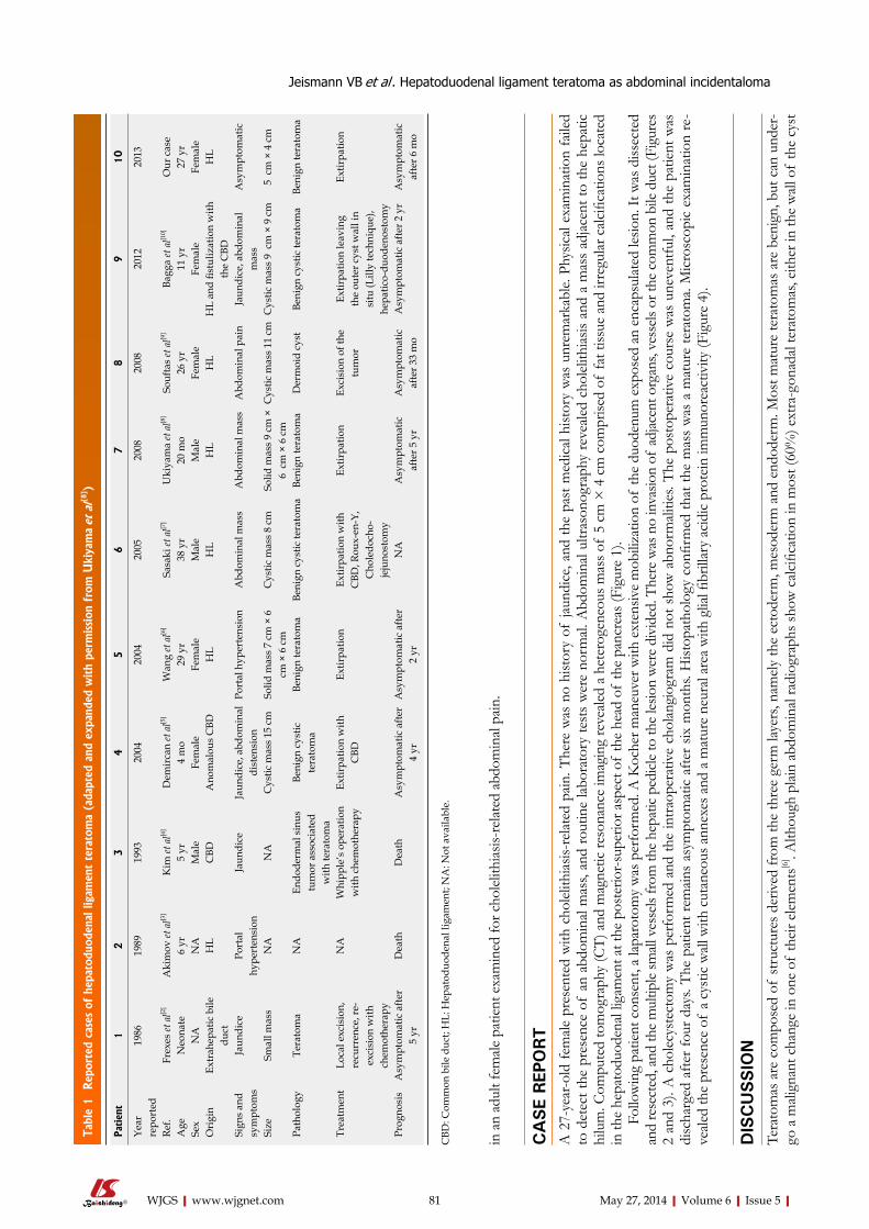
in a
n ad
ult f
emale
pat
ient
exa
min
ed fo
r cho
lelit
hias
is-re
lated
abd
omin
al pa
in.
CA
SE
REP
ORT
A 2
7-ye
ar-o
ld fe
male
pre
sent
ed w
ith c
hole
lithi
asis-
relat
ed p
ain. T
here
was
no
hist
ory
of ja
undi
ce, a
nd th
e pa
st m
edic
al hi
stor
y w
as u
nrem
arka
ble.
Phys
ical
exam
inat
ion
faile
d to
det
ect t
he p
rese
nce
of a
n ab
dom
inal
mas
s, an
d ro
utin
e lab
orat
ory
test
s w
ere
norm
al. A
bdom
inal
ultra
sono
grap
hy re
veale
d ch
olel
ithias
is an
d a
mas
s ad
jacen
t to
the
hepa
tic
hilu
m. C
ompu
ted
tom
ogra
phy
(CT)
and
mag
netic
reso
nanc
e im
agin
g re
veale
d a
hete
roge
neou
s mas
s of
5 cm
× 4
cm
com
prise
d of
fat t
issue
and
irre
gular
calc
ifica
tions
loca
ted
in th
e he
pato
duod
enal
ligam
ent a
t the
pos
terio
r-sup
erio
r asp
ect o
f th
e he
ad o
f th
e pa
ncre
as (F
igur
e 1)
.Fo
llow
ing
patie
nt c
onse
nt, a
lapa
roto
my
was
per
form
ed. A
Koc
her m
aneu
ver w
ith e
xten
sive
mob
iliza
tion
of th
e du
oden
um e
xpos
ed a
n en
caps
ulat
ed le
sion.
It w
as d
issec
ted
and
rese
cted
, and
the
mul
tiple
small
ves
sels
from
the
hepa
tic p
edicl
e to
the
lesio
n w
ere
divi
ded.
The
re w
as n
o in
vasio
n of
adj
acen
t org
ans,
vess
els o
r the
com
mon
bile
duc
t (Fi
gure
s 2
and
3). A
cho
lecy
stec
tom
y w
as p
erfo
rmed
and
the
intra
oper
ativ
e ch
olan
giog
ram
did
not
sho
w a
bnor
mali
ties.
The
post
oper
ativ
e co
urse
was
une
vent
ful,
and
the
patie
nt w
as
disc
harg
ed a
fter f
our d
ays.
The
patie
nt re
main
s as
ympt
omat
ic a
fter s
ix m
onth
s. H
istop
atho
logy
con
firm
ed th
at th
e m
ass
was
a m
atur
e te
rato
ma.
Mic
rosc
opic
exa
min
atio
n re
-ve
aled
the
pres
ence
of
a cy
stic
wall
with
cut
aneo
us a
nnex
es a
nd a
mat
ure
neur
al ar
ea w
ith g
lial fi
brill
ary
acid
ic p
rote
in im
mun
orea
ctiv
ity (F
igur
e 4)
.
DIS
CU
SSIO
NTe
rato
mas
are
com
pose
d of
stru
ctur
es d
eriv
ed fr
om th
e th
ree
germ
laye
rs, n
amel
y th
e ec
tode
rm, m
esod
erm
and
end
oder
m. M
ost m
atur
e te
rato
mas
are
ben
ign,
but
can
und
er-
go a
mali
gnan
t cha
nge
in o
ne o
f th
eir e
lem
ents
[6] . A
lthou
gh p
lain
abdo
min
al ra
diog
raph
s sho
w c
alcifi
catio
n in
mos
t (60
%) e
xtra
-gon
adal
tera
tom
as, e
ither
in th
e w
all o
f th
e cy
st
Jeismann VB et al . Hepatoduodenal ligament teratoma as abdominal incidentaloma
81 May 27, 2014|Volume 6|Issue 5|WJGS|www.wjgnet.com
Tabl
e 1 Rep
orte
d ca
ses
of h
epat
oduo
dena
l lig
amen
t te
rato
ma
(ada
pted
and
exp
ande
d w
ith
perm
ission
fro
m U
kiya
ma et
al[8
] )
Patien
t1
23
45
67
89
10
Year
re
port
ed19
8619
8919
9320
0420
0420
0520
0820
0820
1220
13
Ref.
Frex
es et
al[2
]A
kim
ov et
al[3
]K
im et
al[4
]D
emir
can
et a
l[5]
Wan
g et
al[6
]Sa
saki
et a
l[7]
Uki
yam
a et
al[8
]So
ufta
s et
al[9
]Ba
gga
et a
l[10]
Our
cas
eA
geN
eona
te6
yr5
yr4
mo
29 y
r38
yr
20 m
o26
yr
11 y
r27
yr
Sex
NA
NA
Mal
eFe
mal
eFe
mal
eM
ale
Mal
eFe
mal
eFe
mal
eFe
mal
eO
rigi
nEx
trah
epat
ic b
ile
duct
HL
CBD
Ano
mal
ous
CBD
HL
HL
HL
HL
HL
and
fistu
lizat
ion
with
th
e C
BDH
L
Sign
s an
d sy
mpt
oms
Jaun
dice
Port
al
hype
rten
sion
Jaun
dice
Jaun
dice
, abd
omin
al
dist
ensi
onPo
rtal
hyp
erte
nsio
nA
bdom
inal
mas
sA
bdom
inal
mas
sA
bdom
inal
pai
nJa
undi
ce, a
bdom
inal
m
ass
Asy
mpt
omat
ic
Size
Smal
l mas
sN
AN
AC
ystic
mas
s 15
cm
Solid
mas
s 7
cm ×
6
cm ×
6 c
mC
ystic
mas
s 8
cmSo
lid m
ass
9 cm
×
6 c
m ×
6 c
mC
ystic
mas
s 11
cm
Cys
tic m
ass
9 c
m ×
9 c
m5
cm
× 4
cm
Path
olog
yTe
rato
ma
NA
Endo
derm
al s
inus
tu
mor
ass
ocia
ted
with
tera
tom
a
Beni
gn c
ystic
te
rato
ma
Beni
gn te
rato
ma
Beni
gn c
ystic
tera
tom
aBe
nign
tera
tom
aD
erm
oid
cyst
Beni
gn c
ystic
tera
tom
aBe
nign
tera
tom
a
Trea
tmen
tLo
cal e
xcis
ion,
re
curr
ence
, re-
exci
sion
with
ch
emot
hera
py
NA
Whi
pple
’s o
pera
tion
with
che
mot
hera
pyEx
tirpa
tion
with
C
BDEx
tirpa
tion
Extir
patio
n w
ith
CBD
, Rou
x-en
-Y,
Cho
ledo
cho-
jeju
nost
omy
Extir
patio
nEx
cisi
on o
f the
tu
mor
Extir
patio
n le
avin
g th
e ou
ter c
yst w
all i
n si
tu (L
illy
tech
niqu
e),
hepa
tico-
duod
enos
tom
y
Extir
patio
n
Prog
nosi
sA
sym
ptom
atic
afte
r 5
yrD
eath
Dea
thA
sym
ptom
atic
afte
r 4
yrA
sym
ptom
atic
afte
r 2
yrN
AA
sym
ptom
atic
af
ter 5
yr
Asy
mpt
omat
ic
afte
r 33
mo
Asy
mpt
omat
ic a
fter 2
yr
Asy
mpt
omat
ic
afte
r 6 m
o
CBD
: Com
mon
bile
duc
t; H
L: H
epat
oduo
dena
l lig
amen
t; N
A: N
ot a
vaila
ble.
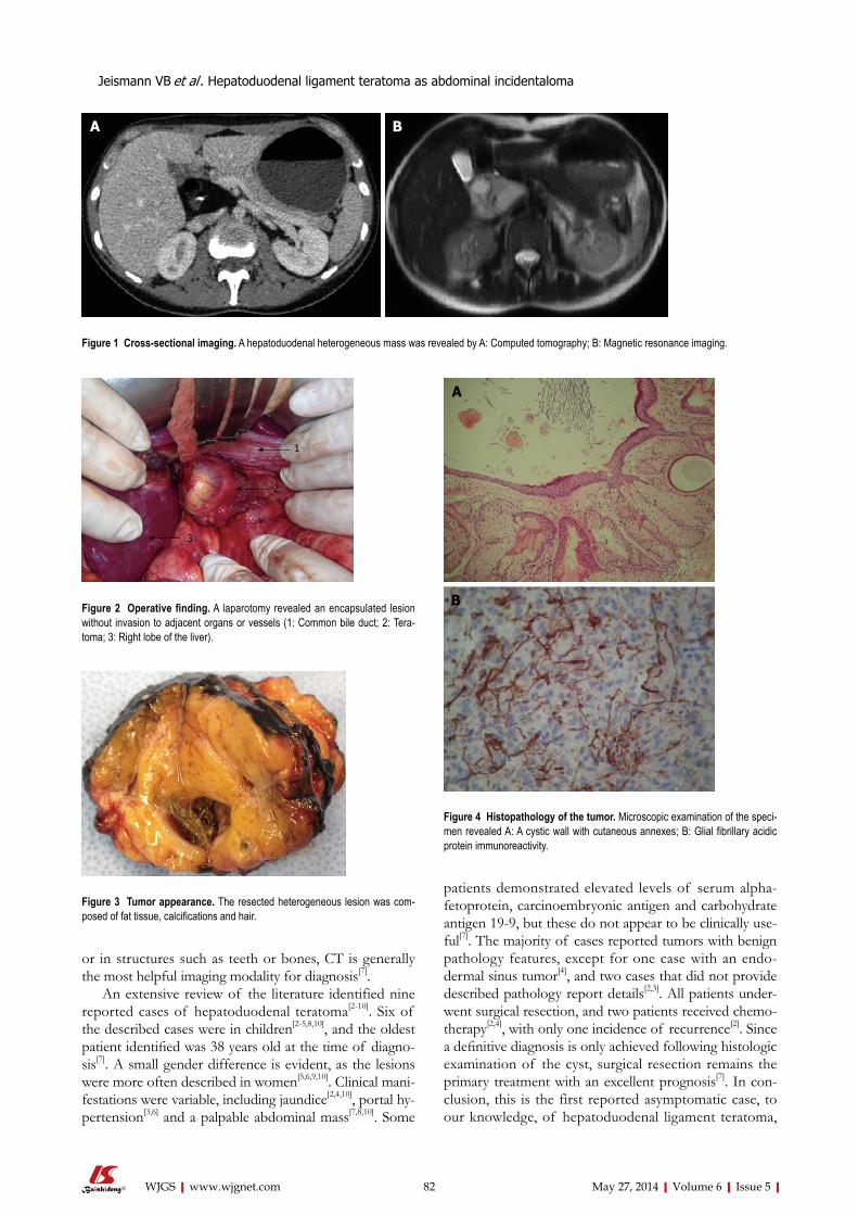
or in structures such as teeth or bones, CT is generally the most helpful imaging modality for diagnosis[7].
An extensive review of the literature identified nine reported cases of hepatoduodenal teratoma[2-10]. Six of the described cases were in children[2-5,8,10], and the oldest patient identified was 38 years old at the time of diagno-sis[7]. A small gender difference is evident, as the lesions were more often described in women[5,6,9,10]. Clinical mani-festations were variable, including jaundice[2,4,10], portal hy-pertension[3,6] and a palpable abdominal mass[7,8,10]. Some
patients demonstrated elevated levels of serum alpha-fetoprotein, carcinoembryonic antigen and carbohydrate antigen 19-9, but these do not appear to be clinically use-ful[7]. The majority of cases reported tumors with benign pathology features, except for one case with an endo-dermal sinus tumor[4], and two cases that did not provide described pathology report details[2,3]. All patients under-went surgical resection, and two patients received chemo-therapy[2,4], with only one incidence of recurrence[2]. Since a definitive diagnosis is only achieved following histologic examination of the cyst, surgical resection remains the primary treatment with an excellent prognosis[7]. In con-clusion, this is the first reported asymptomatic case, to our knowledge, of hepatoduodenal ligament teratoma,
82 May 27, 2014|Volume 6|Issue 5|WJGS|www.wjgnet.com
Figure 2 Operative finding. A laparotomy revealed an encapsulated lesion without invasion to adjacent organs or vessels (1: Common bile duct; 2: Tera-toma; 3: Right lobe of the liver).
Figure 3 Tumor appearance. The resected heterogeneous lesion was com-posed of fat tissue, calcifications and hair.
1
2
3
Figure 4 Histopathology of the tumor. Microscopic examination of the speci-men revealed A: A cystic wall with cutaneous annexes; B: Glial fibrillary acidic protein immunoreactivity.
A
B
A B
Figure 1 Cross-sectional imaging. A hepatoduodenal heterogeneous mass was revealed by A: Computed tomography; B: Magnetic resonance imaging.
Jeismann VB et al . Hepatoduodenal ligament teratoma as abdominal incidentaloma

surgical findings are well documented.
REFERENCES1 Engel RM, Elkins RC, Fletcher BD. Retroperitoneal teratoma.
Review of the literature and presentation of an unusual case. Cancer 1968; 22: 1068-1073 [PMID: 5686638 DOI: 10.1002/1097-0142(196811)22:5<1068::AID-CNCR2820220525>3.0.CO;2-3]
2 Frexes M, Neblett WW, Holcomb GW. Spectrum of biliary disease in childhood. South Med J 1986; 79: 1342-1349 [PMID: 3775460 DOI: 10.1097/00007611-198611000-00007]
3 Akimov OV. [Hepatoduodenal ligament teratoma followed by hypertensive syndrome of the portal vein]. Arkh Patol 1989; 51: 60-62 [PMID: 2719564]
4 Kim WS, Choi BI, Lee YS, Chi JG, Park HR, Kim I, Yeon KM, Han MC. Endodermal sinus tumour associated with benign teratoma of the common bile duct. Pediatr Radiol 1993; 23: 59-60 [PMID: 8469596 DOI: 10.1007/BF02020227]
5 Demircan M, Uguralp S, Mutus M, Kutlu R, Mizrak B. Tera-toma arising from anomalous common bile ducts: a case report. J Pediatr Surg 2004; 39: e1-e2 [PMID: 15065072 DOI: 10.1016/j.jpedsurg.2003.12.036]
6 Wang H, Dong J. Teratoma in the hepatoduodenal ligament followed by portal hypertension syndrome. J Gastroenterol Hepatol 2004; 19: 477-479 [PMID: 15012796 DOI: 10.1111/j.1440-1746.2004.03366.x]
7 Sasaki H, Ajiki T, Takase S, Fujino Y, Suzuki Y, Tominaga M, Ku Y, Kuroda Y. Images of interest. Hepatobiliary and pan-creatic: mature cystic teratoma in the hepatoduodenal liga-ment. J Gastroenterol Hepatol 2005; 20: 317 [PMID: 15683440 DOI: 10.1111/j.1440-1746.2005.03784.x]
8 Ukiyama E, Endo M, Yoshida F. Hepatoduodenal ligament teratoma with hepatic artery running inside. Pediatr Surg Int 2008; 24: 1239-1242 [PMID: 18807051 DOI: 10.1007/s00383-008-2205-x]
9 Souftas V, Polychronidis A, Giatromanolaki A, Perente S, Simopoulos C. Dermoid cyst in the hepatoduodenal liga-ment: report of a case. Surg Today 2008; 38: 959-961 [PMID: 18820876 DOI: 10.1007/s00595-007-3744-9]
10 Bagga D, Jindal B, Naredi BK, Yadav DK, Acharya SK, Ma-hato R, Gupta K. Portal teratoma causing obstructive jaun-dice in children: a rarity. J Pediatr Surg 2012; 47: 1449-1452 [PMID: 22813813 DOI: 10.1016/j.jpedsurg.2012.04.016]
P- Reviewers: Scheidbach H, Sugawara Y, Seow-Choen F S- Editor: Qi Y L- Editor: A E- Editor: Liu SQ
indicating that teratomas should not be ruled out in cases of abdominal incidentaloma.
COMMENTSCase characteristicsThe patient was asymptomatic.Clinical diagnosisThe patient was diagnosed with abdominal incidentaloma uncovered during investigation of cholelithiasis-related abdominal pain.Differential diagnosisBenign and malignant abdominal tumors were alternative diagnoses.Imaging diagnosisA heterogeneous mass of 5 cm × 4 cm with fat tissue and irregular calcifications was located in the posterior-superior aspect of the head of the pancreas, into the hepatoduodenal ligament.Pathological diagnosisAnalysis by microscopy revealed a mature teratoma cystic wall with cutaneous annexes and glial fibrillary acidic protein staining of a mature neural area, find-ings that are consistent with a teratoma.TreatmentThe patient was treated by surgical resection of the tumor.Related reportsA hepatoduodenal teratoma is a rare occurrence and, to the best of the authors’ knowledge, this is the 10th reported case and the 1st asymptomatic reported case.Term explanation Hepatoduodenal ligament teratoma refers to a neoplasm that is comprised of mixed dermal elements derived from the three germ cell layers and located at the portion of the lesser omentum extending between the porta hepatis of the liver and superior part of the duodenum. Computer tomography is a technology that uses computer-processed X-rays to produce cross-sectional imaging of the human body. Abdominal incidentaloma has been defined as an intraabdominal tumor found in a patient without symptoms, usually during evaluation of unre-lated diseases or screening programs. Cholelithiasis is defined by the pres-ence/formation of stones within the biliary tract, most commonly the gallbladder.Experiences and lessonsTeratomas must be included on differential diagnosis of all abdominal incidentalomas.Peer reviewThis case report provides a description of a rare disease that may be under-diagnosed due to a low index of suspicion. The pathological, radiological and
83 May 27, 2014|Volume 6|Issue 5|WJGS|www.wjgnet.com
COMMENTS
Jeismann VB et al . Hepatoduodenal ligament teratoma as abdominal incidentaloma
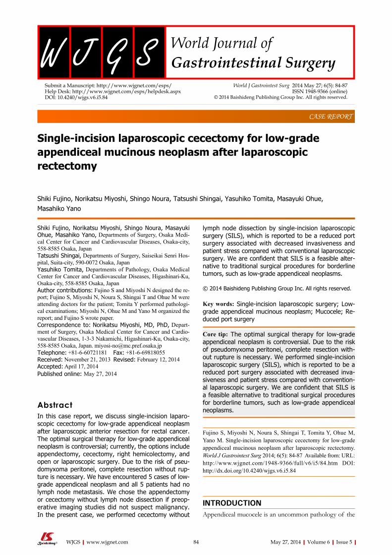
Single-incision laparoscopic cecectomy for low-grade appendiceal mucinous neoplasm after laparoscopic rectectomy
Shiki Fujino, Norikatsu Miyoshi, Shingo Noura, Tatsushi Shingai, Yasuhiko Tomita, Masayuki Ohue, Masahiko Yano
Shiki Fujino, Norikatsu Miyoshi, Shingo Noura, Masayuki Ohue, Masahiko Yano, Departments of Surgery, Osaka Medi-cal Center for Cancer and Cardiovascular Diseases, Osaka-city, 558-8585 Osaka, JapanTatsushi Shingai, Departments of Surgery, Saiseikai Senri Hos-pital, Suita-city, 590-0072 Osaka, JapanYasuhiko Tomita, Departments of Pathology, Osaka Medical Center for Cancer and Cardiovascular Diseases, Higashinari-Ku, Osaka-city, 558-8585 Osaka, JapanAuthor contributions: Fujino S and Miyoshi N designed the re-port; Fujino S, Miyoshi N, Noura S, Shingai T and Ohue M were attending doctors for the patient; Tomita Y performed pathologi-cal examinations; Miyoshi N, Ohue M and Yano M organized the report; and Fujino S wrote paper.Correspondence to: Norikatsu Miyoshi, MD, PhD, Depart-ment of Surgery, Osaka Medical Center for Cancer and Cardio-vascular Diseases, 1-3-3 Nakamichi, Higashinari-Ku, Osaka-city, 558-8585 Osaka, Japan. [email protected]: +81-6-60721181 Fax: +81-6-69818055Received: November 21, 2013 Revised: February 12, 2014Accepted: April 17, 2014Published online: May 27, 2014
AbstractIn this case report, we discuss single-incision laparo-scopic cecectomy for low-grade appendiceal neoplasm after laparoscopic anterior resection for rectal cancer. The optimal surgical therapy for low-grade appendiceal neoplasm is controversial; currently, the options include appendectomy, cecectomy, right hemicolectomy, and open or laparoscopic surgery. Due to the risk of pseu-domyxoma peritonei, complete resection without rup-ture is necessary. We have encountered 5 cases of low-grade appendiceal neoplasm and all 5 patients had no lymph node metastasis. We chose the appendectomy or cecectomy without lymph node dissection if preop-erative imaging studies did not suspect malignancy. In the present case, we performed cecectomy without
lymph node dissection by single-incision laparoscopic surgery (SILS), which is reported to be a reduced port surgery associated with decreased invasiveness and patient stress compared with conventional laparoscopic surgery. We are confident that SILS is a feasible alter-native to traditional surgical procedures for borderline tumors, such as low-grade appendiceal neoplasms.
© 2014 Baishideng Publishing Group Inc. All rights reserved.
Key words: Single-incision laparoscopic surgery; Low-grade appendiceal mucinous neoplasm; Mucocele; Re-duced port surgery
Core tip: The optimal surgical therapy for low-grade appendiceal neoplasm is controversial. Due to the risk of pseudomyxoma peritonei, complete resection with-out rupture is necessary. We performed single-incision laparoscopic surgery (SILS), which is reported to be a reduced port surgery associated with decreased inva-siveness and patient stress compared with convention-al laparoscopic surgery. We are confident that SILS is a feasible alternative to traditional surgical procedures for borderline tumors, such as low-grade appendiceal neoplasms.
Fujino S, Miyoshi N, Noura S, Shingai T, Tomita Y, Ohue M, Yano M. Single-incision laparoscopic cecectomy for low-grade appendiceal mucinous neoplasm after laparoscopic rectectomy. World J Gastrointest Surg 2014; 6(5): 84-87 Available from: URL: http://www.wjgnet.com/1948-9366/full/v6/i5/84.htm DOI: http://dx.doi.org/10.4240/wjgs.v6.i5.84
INTRODUCTIONAppendiceal mucocele is an uncommon pathology of the
CASE REPORT
Submit a Manuscript: http://www.wjgnet.com/esps/Help Desk: http://www.wjgnet.com/esps/helpdesk.aspxDOI: 10.4240/wjgs.v6.i5.84
World J Gastrointest Surg 2014 May 27; 6(5): 84-87ISSN 1948-9366 (online)
© 2014 Baishideng Publishing Group Inc. All rights reserved.
84 May 27, 2014|Volume 6|Issue 5|WJGS|www.wjgnet.com

appendix (0.08%-0.15% of all appendectomies) that is characterized by the accumulation of mucus in the appen-diceal lumen[1,2]. The term mucocele, includes cystadenoma and low-grade appendiceal neoplasm, and is the same as the World Health Organization (WHO)’s classification of low-grade appendiceal neoplasm. Several optimal surgical methods are reported but are still controversial. Complete resection without rupture is definitely necessary because the dissemination of neoplastic cells and mucus in the abdominal cavity, which is often be caused by appendiceal perforation, clinically results in pseudomyxoma peritonei in 10%-15% of cases[3]. Therefore, low-grade appendi-ceal neoplasms are classified into carcinoma groups in the WHO’s classification. In several reports, the surgical procedures used vary from simple appendectomy to right hemicolectomy[4]. Here, to identify the optimal surgical method to treat appendiceal mucocele, we report the case of a 49-year-old woman with an appendiceal lesion that was laparoscopically resected by a single incision and summarize other cases that involved surgical resections.
CASE REPORTA 49-year-old woman underwent laparoscopic anterior resection for rectal cancer at Osaka Medical Center for Cancer and Cardiovascular Diseases in February 2009. During routine postoperative care, a mucus-filled lesion in the appendix was detected by computed tomography (CT). The patient’s past medical history was not remark-able, except for the rectal cancer. Upon physical exami-nation, there were no remarkable findings. Laboratory tests were within normal ranges. CT revealed a 55 mm × 25 mm tumor that presented as a blind-ended, tubular-shaped, fluid-filled structure in the cecum (Figure 1). Colonoscopy showed that the lesion was covered by normal colonic mucosa in the closed appendix. A colo-noscopic biopsy revealed normal mucosa (Figure 2). The lesion was thought to be an appendiceal mucocele of the appendix, and single-incision laparoscopic surgery (SILS) was performed. The surgical choice was made because ultrasound examination showed a movable ap-pendix including the surrounding intestine and no adhe-sion around the umbilical portion, which had an old scar from the primary operation. Under general anesthesia, the operation started with a trans-umbilical, 2.5 cm-diameter incision (SILS port). A Lap Protector (Hakko Co. Ltd., Nagano, Japan) was folded and the bottom half was inserted into the abdomen through the umbilical in-cision. The EZ Access (Hakko) was adjusted, and three devices were introduced through it: a flexible laparoscope (Olympus, Tokyo, Japan) and two operating forceps. The pneumoperitoneum was set at 10 mmHg. A smooth and mucus-filled lesion of the appendix was found and there were no ascites or peritoneal nodules indicating malig-nancy (Figure 3). To excise the tumor without rupture, we cut the peritoneum around the cecum and mobilized it without touching the tumor. After pulling the cecum with the tumor through the SILS port, a cecectomy which included the swollen appendix was performed using a linear stapler (Endo GIA universal; Covidien, Mansfield,
MA, United States). The resected bowel contained the appendix (8 cm) and cecum (1 cm). The total operating time was 57 min, and the blood loss was minimal. Histo-logical examination showed low grade epithelial dysplasia, a feature diagnostic of a low-grade appendiceal mucinous neoplasm (Figure 4). Surgical margin was negative and no lymph node metastasis was discovered. The patient recovered without any complications, and was discharged on postoperative day 6. She returned to work, and she is now doing well without any complaint postoperatively.
DISCUSSIONAppendiceal mucocele itself does not have typical clini-
Fujino S et al . SILS for appendiceal tumor
85 May 27, 2014|Volume 6|Issue 5|WJGS|www.wjgnet.com
Figure 1 Computed tomography. The arrow shows a 55 mm × 25 mm, low-density and no-contrast lesion at the appendix.
Figure 2 Colonoscopy. A: Normal image; B: Indigo carmine-stained image. There was an inflated lesion covered with normal mucosa in the appendix, sug-gesting a tumor under the cecal mucosa (arrows).
A
B
cal features; more than two-thirds of patients have their appendiceal mucocele removed based on incidental find-ings, as was the case for our patient[4]. Surgical resection without rupture is necessary, and laparoscopic appendec-tomy is often used in accordance with the accepted treat-ment of mucocele[2]. Based on the tumor characteristics, including the location and size, surgical management should differ. Using either an open or a minimally inva-sive technique still depends on the situation and the pref-
erence and experience of the surgeon. Although an open procedure is still recommended in certain appendiceal cases, especially for those with suspected malignancy[5], the laparoscopic technique has been described and rec-ommended as the first choice for treating this disease, rather than conventional open laparotomy. This recom-mendation is due to the method’s ability to provide useful information regarding the entire abdominal cavity, short recovery post-surgery, and a minimized risk of seeding the tumor during laparoscopic manipulation[6]. Further-more, SILS has been often applied in several fields, and in colectomies[7-11]. Recent reports show single incision approach through the umbilicus that is called SILS port or grove method. The narrow working space for surgical manipulation for surgical manipulation presents a techni-cal difficulty; however, in certain cases, it is still easy and safe to convert to the conventional laparoscopic surgery with multiple ports or open laparotomy. Laparoscopic surgery has the added benefit of the laparoscope magni-fying the surgical field, keeping the surgical space wide by aeroperitoneum. Additionally, in our case, the SILS op-eration was started but could have been changed to any operation, such as conventional laparoscopic surgery or open surgery, if we had encountered any difficulties (e.g., bleeding, injury to other organs, or difficult surgical ma-nipulation). In a case using the McBurney method, a tra-
86 May 27, 2014|Volume 6|Issue 5|WJGS|www.wjgnet.com
Figure 3 Photograph of the operation. A: A 2.5 cm-diameter incision was in the navel; B: The swollen appendix (arrows) and cecum were mobilized by non-touch technique.
A B
Fujino S et al . SILS for appendiceal tumor
Figure 4 Macroscopic image of the removed specimen. The appendix was about 8 cm long and swollen and exhibited a fibrotic wall (arrow; the mucus in the lumen was removed).
Table 1 Six cases of mucocele of appendix in our hospital
Case Age (yr) Chief complaint Tumor size Pathological findings Operative procedures Bleeding Operation Time after Recurrence/
/sex (mm) (lymph node dissection1) (mL) time (min) surgery (mo) survival1 49/F None 55 × 25 Low-grade appendiceal
mucinous neoplasmSingle incision
laparoscopic cecectomy 5 57 6 None/alive
2 61/M Right lower abdominal pain
90 × 40 Low-grade appendiceal mucinous neoplasm
Laparoscopy-assisted ileocecal resection (D3)
30 251 14 None/alive
3 61/F None 30 × 15 Low-grade appendiceal mucinous neoplasm
Open appendectomy 70 140 25 None/alive
4 81/F None 150 × 40 Low-grade appendiceal mucinous neoplasm
Open ileocecal resection (D3)
70 125 29 None/alive
5 69/F None 46 × 27 Low-grade appendiceal mucinous neoplasm
Open appendectomy 50 134 32 None/alive
6 51/F Right hypochondrial pain
30 × 30 Mucinous cystadenoma Laparoscopy-assisted ileocecal resection (D1)
65 280 74 None/alive
1The defined lymph node dissection was performed according to the JSCCR guideline[12].
for low-grade neoplasm, such as mucocele.Peer reviewReport is well written. This is an interesting well-described study. The review is well conducted. It addresses an interesting clinical area in an application that has not received a great deal of attention.
REFERENCES1 Marudanayagam R, Williams GT, Rees BI. Review of the pa
thological results of 2660 appendicectomy specimens. J Gas-troenterol 2006; 41: 745-749 [PMID: 16988762 DOI: 10.1007/s00535-006-1855-5]
2 Rangarajan M, Palanivelu C, Kavalakat AJ, Parthasarathi R. Laparoscopic appendectomy for mucocele of the appen-dix: Report of 8 cases. Indian J Gastroenterol 2006; 25: 256-257 [PMID: 17090846]
3 Caracappa D, Gullà N, Gentile D, Listorti C, Boselli C, Ciroc-chi R, Bellezza G, Noya G. Appendiceal mucocele. A case report and literature review. Ann Ital Chir 2011; 82: 239-245 [PMID: 21780569]
4 Stocchi L, Wolff BG, Larson DR, Harrington JR. Surgical treatment of appendiceal mucocele. Arch Surg 2003; 138: 585-589; discussion 589-590 [PMID: 12799327]
5 Sturniolo G, Barbuscia M, Taranto F, Tonante A, Paparo D, Romeo G, Nucera D, Lentini M. Mucocele of the appendix. Two case reports. G Chir 2011; 32: 487-490 [PMID: 22217378]
6 Chiu CC, Wei PL, Huang MT, Wang W, Chen TC, Lee WJ. Laparoscopic resection of appendiceal mucinous cystadeno-ma. J Laparoendosc Adv Surg Tech A 2005; 15: 325-328 [PMID: 15954839 DOI: 10.1089/lap.2005.15.325]
7 Makino T, Milsom JW, Lee SW. Single-incision laparoscopic surgeries for colorectal diseases: early experiences of a novel surgical method. Minim Invasive Surg 2012; 2012: 783074 [PMID: 22888419 DOI: 10.1155/2012/783074]
8 Vestweber B, Galetin T, Lammerting K, Paul C, Giehl J, Straub E, Kaldowski B, Alfes A, Vestweber KH. Singleincision laparoscopic surgery: outcomes from 224 colonic re-sections performed at a single center using SILS. Surg Endosc 2013; 27: 434-442 [PMID: 22806519 DOI: 10.1007/s00464-012-2454-6]
9 Makino T, Milsom JW, Lee SW. Feasibility and safety of single-incision laparoscopic colectomy: a systematic review. Ann Surg 2012; 255: 667-676 [PMID: 22258065 DOI: 10.1097/SLA.0b013e31823fbae7]
10 Champagne BJ, Papaconstantinou HT, Parmar SS, Nagle DA, YoungFadok TM, Lee EC, Delaney CP. Singleincision versus standard multiport laparoscopic colectomy: a multi-center, case-controlled comparison. Ann Surg 2012; 255: 66-69 [PMID: 22104563 DOI: 10.1097/SLA.0b013e3182378442]
11 Yang TX, Chua TC. Singleincision laparoscopic colectomy versus conventional multiport laparoscopic colectomy: a me-taanalysis of comparative studies. Int J Colorectal Dis 2013; 28: 89-101 [PMID: 22828958 DOI: 10.1007/s00384-012-1537-0]
12 Japanese Society for Cancer of the Colon and Rectum (2010). JSCCR Guidelines 2010 for the Treatment of Colorectal Can-cer. Tokyo: Kanehara, 2010: 1315
P- Reviewers: Chapel A, Ngai TC, Yoldas O S- Editor: Qi Y L- Editor: A E- Editor: Liu SQ
ditional approach for treating an appendiceal locus with a minimized incision, it would be difficult to change the surgical procedure, leaving only the option of increasing the size of the incision.
We have encountered 6 cases of low-grade appendi-ceal neoplasm (Table 1); three cases were treated by open surgery for the reason described above, whereas the other 3 cases underwent laparoscopic surgery. In these cases, we chose the appendectomy or cecectomy without lymph node dissection if preoperative imaging studies did not suspect malignancy. In the present case, CT revealed no involvement of the mesoappendix or local lymph node metastasis. All 6 patients are doing well, without recur-rence for 6-74 mo after surgery.
It is thought that minimally invasive procedures, including SILS appendectomy and cecectomy, for low-grade neoplasm, such as mucocele will be considered as the primary treatment choice in several years. The poten-tial benefits include the superior cosmetic results, reduced postoperative pain, faster recovery, and shorter hospital stays.
COMMENTSCase characteristicsA 49-year-old woman had no symptom and the disease was detected by com-puted tomography (CT) during the follow-up course of rectal cancer.Clinical diagnosisPhysical examination shows no remarkable findings.Differential diagnosisAppendiceal cancer, carcinoid and cystadenoma.Laboratory diagnosisAll laboratory tests were within normal ranges.Imaging diagnosisCT revealed a 55 mm × 25 mm tumor, which presented as a blind-ended, tubular-shaped, fluid-filled structure in the cecum and colonoscopy showed the lesion covered by normal colon mucosa in the closed appendix.Pathological diagnosisHistological examination showed low-grade epithelial dysplasia, a feature diag-nostic of a low-grade appendiceal mucinous neoplasm.TreatmentThe tumor was resected by single-incision laparoscopic surgery (SILS).Related reportsSILS has been often applied in several fields, and in colectomies.Term explanation SILS is used as a reduced port surgery compared to conventional laparoscopic surgery (mostly 5 ports).Experiences and lessonsThis case report shows that SILS is considered as the primary treatment choice
87 May 27, 2014|Volume 6|Issue 5|WJGS|www.wjgnet.com
Fujino S et al . SILS for appendiceal tumor
COMMENTS

Published by Baishideng Publishing Group Inc
World Journal of Gastrointestinal SurgeryWorld J Gastrointest Surg 2014 June 27; 6(6): 88-121
ISSN 1948-9366 (online)
88 Sentinelnodenavigationsurgeryingastriccancer:Currentstatus
Symeonidis D, Koukoulis G, Tepetes K
94 Pathologicalfactorsaffectinggastricadenocarcinomasurvivalina
Caribbeanpopulationfrom2000-2010
Roberts PO, Plummer J, Leake PA, Scott S, de Souza TG, Johnson A, Gibson TN,
Hanchard B, Reid M
101 Laparoscopicre-sleevegastrectomyasatreatmentofweightregain
aftersleevegastrectomy
Cesana G, Uccelli M, Ciccarese F, Carrieri D, Castello G, Olmi S
107 Downstagingandresectionafterneoadjuvanttherapyfor
fibrolamellar hepatocellular carcinoma
Fonseca GM, Varella AD, Coelho FF, Abe ES, Dumarco RB, Herman P
112 Intra-abdominalesophagealduplicationcyst:Acasereportand
reviewoftheliterature
Castelijns PSS, Woensdregt K, Hoevenaars B, Nieuwenhuijzen GAP
117 Repair of an aberrant subclavian arterioesophageal fistula following
esophagealstentplacement
Hosn MA, Haddad F, El-Merhi F, Safadi B, Hallal A
Contents Monthly Volume 6 Number 6 June 27, 2014
WJGS|www.wjgnet.com I June 27, 2014|Volume 6|Issue 6|
MINIREVIEWS
RETROSPECTIVE STUDY
CLINICAL TRIALS STUDY
CASE REPORT

Contents
APPENDIX
ABOUT COVER
World Journal of Gastrointestinal SurgeryVolume 6 Number 6 June 27, 2014
I-V Instructionstoauthors
EditorialBoardMemberofWorldJournalofGastrointestinalSurgery ,GiovanniCesana,MD,DepartmentofLaparoscopicandMini-InvasiveSurgery,SanGi-useppeHospital,viaSanVittore12,Milano20123,Italy
World Journal of Gastrointestinal Surgery (World J Gastrointest Surg, WJGS, online ISSN 1948-9366, DOI: 10.4240) is a peer-reviewed open access academic journal that aims to guide clinical practice and improve diagnostic and therapeutic skills of clinicians.
WJGS covers topics concerning micro-invasive surgery; laparoscopy; hepatic, biliary, pancreatic and splenic surgery; surgical nutrition; portal hypertension, as well as associated subjects. The current columns of WJGS include editorial, frontier, diagnostic advances, therapeutics advances, field of vision, mini-reviews, review, topic highlight, medical ethics, original articles, case report, clinical case conference (Clinicopathological conference), and autobiography. Priority publication will be given to articles concerning diagnosis and treatment of gastrointestinal surgery diseases. The following aspects are covered: Clinical diagnosis, laboratory diagnosis, differential diagnosis, imaging tests, pathological diagnosis, molecular biological diagnosis, immunological diagnosis, genetic diagnosis, functional diagnostics, and physical diagnosis; and comprehensive therapy, drug therapy, surgical therapy, interventional treatment, minimally invasive therapy, and robot-assisted therapy.
We encourage authors to submit their manuscripts to WJGS. We will give priority to manuscripts that are supported by major national and international foundations and those that are of great basic and clinical significance.
World Journal of Gastrointestinal Surgery is now indexed in PubMed Central, PubMed, Digital Object Identifier, and Directory of Open Access Journals.
I-III EditorialBoard
Xiu-Xia Song, Vice DirectorWorld Journal of Gastrointestinal SurgeryRoom 903, Building D, Ocean International Center, No. 62 Dongsihuan Zhonglu, Chaoyang District, Beijing 100025, ChinaTelephone: +86-10-85381891Fax: +86-10-85381893E-mail: [email protected] Desk: http://www.wjgnet.com/esps/helpdesk.aspxhttp://www.wjgnet.com
PUBLISHERBaishideng Publishing Group Inc8226 Regency Drive, Pleasanton, CA 94588, USATelephone: +1-925-223-8242Fax: +1-925-223-8243E-mail: [email protected] Desk: http://www.wjgnet.com/esps/helpdesk.aspxhttp://www.wjgnet.com
PUBLICATIONDATEJune 27, 2014
COPYRIGHT© 2014 Baishideng Publishing Group Inc. Articles pub-lished by this Open-Access journal are distributed under the terms of the Creative Commons Attribution Non-commercial License, which permits use, distribution, and reproduction in any medium, provided the original work is properly cited, the use is non commercial and is otherwise in compliance with the license.
SPECIALSTATEMENTAll articles published in journals owned by the Baishideng Publishing Group (BPG) represent the views and opinions of their authors, and not the views, opinions or policies of the BPG, except where otherwise explicitly indicated.
INSTRUCTIONSTOAUTHORSFull instructions are available online at http://www.wjgnet.com/1948-9366/g_info_20100305152206.htm
ONLINESUBMISSIONhttp://www.wjgnet.com/esps/
NAMEOFJOURNALWorld Journal of Gastrointestinal Surgery
ISSNISSN 1948-9366 (online)
LAUNCHDATENovember 30, 2009
FREQUENCYMonthly
EDITOR-IN-CHIEFTimothy M Pawlik, MD, MPH, FACS, Associate Professor of Surgery and Oncology, Hepatobiliary Surgery Program Director, Director, Johns Hopkins Medicine Liver Tumor Center Multi-Disciplinary Clinic, Co-Director of Center for Surgical Trials and Outcomes Research, Johns Hopkins Hospital, 600 N. Wolfe Street, Harvey 611, Baltimore, MD 21287, United States
EDITORIALOFFICEJin-Lei Wang, Director
EDITORS FOR THIS ISSUE
Responsible Assistant Editor: Xiang Li Responsible Science Editor: Ling-Ling WenResponsible Electronic Editor: Huan-Liang Wu Proofing Editorial Office Director: Xiu-Xia SongProofing Editor-in-Chief: Lian-Sheng Ma
AIM AND SCOPE
FLYLEAF
INDEXING/ABSTRACTING
WJGS|www.wjgnet.com II June 27, 2014|Volume 6|Issue 6|
Sentinel node navigation surgery in gastric cancer: Current status
Dimitrios Symeonidis, George Koukoulis, Konstantinos Tepetes
Dimitrios Symeonidis, George Koukoulis, Konstantinos Te-petes, Department of Surgery, University Hospital of Larissa, 41110 Larissa, GreeceAuthor contributions: Symeonidis D and Tepetes K contributed equally to this work; Symeonidis D, Koukoulis G and Tepetes K performed the literature review; Symeonidis D, Koukoulis G and Tepetes K analyzed the data; Symeonidis D and Koukoulis G drafted the article; Symeonidis D and Tepetes K critically revised the final form of the article; all authors have read and accepted the final version.Correspondence to: Dimitrios Symeonidis, MD, PhD, Gen-eral Surgeon, Department of Surgery, University Hospital of Larissa, Mezourlo, 41110 Larissa, Greece. [email protected]: +30-235-1020730 Fax: +30-235-1020741Received: February 18, 2014 Revised: April 16, 2014Accepted: June 10, 2014Published online: June 27, 2014
AbstractThe theory behind using sentinel node mapping and biopsy in gastric cancer surgery, the so-called sentinel node navigation surgery, is to limit the extent of surgi-cal tissue dissection around the affected organ and subsequently the accompanied morbidity. However, ob-stacles on the clinical correspondence of sentinel node navigation surgery in everyday practice have occasion-ally alleviated researchers’ interest on the topic. Only recently with the widespread use of minimally invasive surgical techniques, i.e. , laparoscopic gastric cancer resections, surgical community’s interest on the topic have been unavoidably reflated. Double tracer methods appear superior compared to single tracer techniques. Ongoing research is now focused on the invention of new lymph node detection methods utilizing sophisti-cated technology such as infrared ray endoscopy, flo-rescence imaging and near-infrared technology. Despite its notable limitations, hematoxylin/eosin is still the mainstay staining for assessing the metastatic status of an identified lymph node. An intra-operatively verified metastatic sentinel lymph node will dictate the need
for further conventional lymph node dissection. Thus, laparoscopic resection of the gastric primary tumor combined with the appropriate lymph node dissection as determined by the process of sentinel lymph node status characterization represents an option for early gastric cancer. Patients with T3 or more advanced dis-ease should still be managed conventionally with resec-tion plus standard lymph node dissection.
© 2014 Baishideng Publishing Group Inc. All rights reserved.
Key words: Sentinel node; Gastric cancer; Minimally in-vasive surgery
Core tip: Sentinel node navigation surgery can change the current surgical treatment of gastric cancer expand-ing the indications of minimally invasive surgical options such laparoscopic techniques. However, the complex lymphatic drainage of the stomach and the ubiquitous fear of skip metastasis make the selection of patients extremely important. Currently, laparoscopic resection of the tumor from the stomach with lymph node dis-section navigated by sentinel lymph node identifica-tion represents an option only for early gastric cancer patients. Unfortunately, patients with T3 or more ad-vanced disease should still be managed conventionally with resection plus lymph node dissection.
Symeonidis D, Koukoulis G, Tepetes K. Sentinel node naviga-tion surgery in gastric cancer: Current status. World J Gastrointest Surg 2014; 6(6): 88-93 Available from: URL: http://www.wjgnet.com/1948-9366/full/v6/i6/88.htm DOI: http://dx.doi.org/10.4240/wjgs.v6.i6.88
INTRODUCTIONMelanoma was the first malignancy that the concept of sentinel node found application for. However, the indi-cations and uses of this attractive procedure have been
MINIREVIEWS
Submit a Manuscript: http://www.wjgnet.com/esps/Help Desk: http://www.wjgnet.com/esps/helpdesk.aspxDOI: 10.4240/wjgs.v6.i6.88
World J Gastrointest Surg 2014 June 27; 6(6): 88-93ISSN 1948-9366 (online)
© 2014 Baishideng Publishing Group Inc. All rights reserved.
June 27, 2014|Volume 6|Issue 6|WJGS|www.wjgnet.com 88
recently expanded in many fields of surgical oncology such as breast cancer, thyroid cancer, gynecological ma-lignancies, colorectal and, recently, gastric cancer. Sentinel node mapping and biopsy in gastric cancer surgery, the so-called sentinel node navigation surgery, aimed to limit the extent of surgical tissue dissection around the af-fected organ. By convection, any unnecessary dissection, i.e., dissection of virgin-tumor free areas unrelentingly in-crease morbidity without always respective survival ben-efits. Within this context, sentinel lymph node navigation surgery could, at least theoretically, facilitate precise and sufficient resections. However, in some instances, insur-mountable obstacles on the clinical correspondence of the sentinel node navigation surgery concept in everyday practice have occasionally alleviated researchers’ interest on the topic. Only recently with the widespread use of minimally invasive surgical techniques, i.e., laparoscopic gastric cancer resections, surgical community’s interest on the topic have been unavoidably reflated.
Nowadays, the following questions regarding the util-ity of sentinel lymph node mapping and biopsy in clinical practice need to be precisely answered: (1) which are the available techniques for sentinel lymph node mapping? (2) which is the best way to administer the tracer? (3) which is the optimal method to verify the presence of metasta-sis in the identified sentinel lymph node? (4) which is the gastric cancer patient subgroup suitable for sentinel node mapping and biopsy? and (5) which are the available op-tions for primary tumor control?
LYMPH NODE STATIONSIn 1973, the Japanese Research Society for the study of gastric cancer published a manual standardizing lymph node dissections in gastric cancer by recognizing 16 dis-
tinct anatomic lymph node stations. Further grouping of these lymph node stations took place, i.e., N1, N2, N3 and N4 to achieve correspondence with respective lymph node dissection extents, i.e., D1, D2, D3 and D4[1] (Table 1).
LYMPHATIC STREAM IN GASTRIC CANCERTrying to decipher the lymph route out of a malignant le-sion within the stomach, a few anatomical considerations are of paramount importance. Briefly, from the anatomic viewpoint, lymph from the gastric wall is drained via lymphatic vessels which form a complex sub-peritoneal plexus surrounding the stomach both anteriorly and posteriorly. Depending on the location, the lymph of the upper left part of the stomach is routed to the left gastric and pericardial nodes. Lymph originated from the pylorus is filtered through the supra-pyloric and the right supra-pancreatic nodes. The region of the fundus filters lymph along the gastrosplenic ligament and splits with lymph flowing to the left supra-pancreatic nodes and the left gastroepiploic nodes via the splenic nodes. Lymph from the pyloric and the distal portion of the corpus col-lects in the right gastroepiploic nodes and then flows to the sub-pyloric nodes. From all regions, the lymph stream continues to the celiac nodes[1].
When dealing with malignant lesions, clarifying the lymphatic drainage pattern is crucial for performing proper lymph node dissections especially from sites “susceptible” to metastasis. However, as briefly dis-cussed above, the lymphatic stream of the stomach ap-pears particularly complex and multidirectional and in many occasions ill-investigated. Certainly, having even a rough idea of how lymph drains out of the stomach will render upper gastrointestinal (GI) surgeons capable of performing effective and, up to a point, targeted lymph node dissections[2]. Nevertheless, tumors at any loca-tion within the stomach have a non-negligible chance of atypical metastasis. Tumors located longitudinally or circumferentially in the lower part of the lesser curvature appear to be of higher chance for an atypical metastasis compared to other locations[3]. It becomes obvious that the efficiency of the sentinel node concept is compro-mised when dealing with tumors at these locations as an unacceptable increase of false-negative results should be anticipated. Studies raise the incidence of skip metastasis up to 29%[3]. Apart from the location, the degree of tu-mor differentiation has been inconsistently implicated as to increase skip metastasis potential[4].
Generally, the severity of gastric malignancy, i.e., tu-mor size and depth of invasion is positively correlated with the lymph node metastasis rate[5]. In addition, stud-ies using a retrospective methodology and including pa-tients with sole lymph node involvement have shown that the majority of sentinel lymph nodes are located in the regional area at a close proximity to the tumor[6]. It is rec-ommended, that if nodes are not identified in the usual locations, then No. 7, 8 and 9 lymph node stations should
June 27, 2014|Volume 6|Issue 6|WJGS|www.wjgnet.com 89
Symeonidis D et al . Sentinel node navigation surgery
Lymph node stations
Anatomic location Group Lymphadenectomy
1 Right cardia N1 D1 2 Left cardia 3 Lesser curvature 4 Greater curvature 4a Short gastric vessels 4b Left gastroepiploic vessels 4c Right gastroepiploic vessels 5 Suprapyloric 6 Infrapyloric 7 Left gastric artery N2 D2
(N1 + N2) 8 Common hepatic artery 9 Celiac trunk 10 Splenic hilus 11 Splenic artery 12 Hepatoduodenal ligament N3 D3
(N1 + N2 + N3) 13 Posterior surface of the head of the pancreas
14 Root of the mesentery 14A Superior mesenteric artery 14V Superior mesenteric vein 15 Para-aortic N4 D4
(N1 +N2 + N3 +N4) 16 Paracolic
Table 1 Lymph node stations of the stomach
be investigated as well[7].
WHICH ARE THE AVAILABLE TECHNIQUES FOR SENTINEL LYMPH NODE MAPPING?Numerous methods in order to increase the usefulness and effectiveness of sentinel node mapping have been proposed to date[8-18]. The clinical evaluation and as-sessment of these modalities within studies have led to a breathtaking progress in the field rendering sentinel lymph node tracking techniques familiar to surgeons. However, the main problem is on the logistics of each technique. Identifying sentinel lymph nodes intra-operatively in a timely and effective pattern is by defini-tion a challenging process. The tracer used should meet the minimum requirements of (1) non-toxicity; (2) easy availability; and (3) cost-effectiveness. Ideally, the tracer should accumulate within the sentinel nodes for a period of time long enough to render detection possible. Fur-thermore, it should be readily identifiable without the need for using sophisticated and unfamiliar to surgeons equipment. As no single tracer to date incorporates all of the above characteristics, the quest for the optimal com-pound seems to be ongoing.
Dye-based and radioisotope-based techniques have been the mainstay for lymph node detection so far[8-18]. Dye agents include isosulfan blue, patent blue and indo-cyanine green (currently, the most commonly used dye). On the other hand, technetium 99 m represents the most commonly used radioisotope. The use of infrared ray beam via endoscopy can, at least theoretically, facilitate the visualization of the used tracer increasing the accu-racy of the detection[19,20]. Similarly, fluorescence imaging is another available adjunct which is suggested to increase the detection rates of traditional dye agents such as indo-cyanine green[21,22].
However, sentinel lymph node mapping of the GI tract by using available techniques is often limited by vari-ous factors. The multidirectional lymph drainage patterns and, practically, the inability to image surgical anatomy in real time in relation to the used tracer can compromise the whole process. In this direction, the use of invisible near-infrared light might have the answers. In this tech-nique, an intraoperative near-infrared fluorescence imag-ing system that simultaneously displays surgical anatomy is utilized. Near-infrared fluorescence images of the sur-gical field are generated to illustrate intra-parenchymally injected near-infrared fluorescent quantum dots. The final result is the visualization of the draining lymphatic tree and of the nodes as well. The technique promises dissec-tion under real time vision[23].
Generally, there is a trend for combining tracers in order to increase the detection accuracy. Double tracer techniques (dye plus isotope), almost consistently, seem to increase the rate of sentinel lymph node identifica-tion[24-30], however there are indeed studies which question
this finding[31,32]. In addition, pre-clinical research is in progress for inventing the optimal tracer and visualization system. It seems pretty likely at this point that research will overcome the traditional dye-based techniques and it will open new perspectives in sentinel node mapping.
WHICH IS THE BEST WAY TO ADMINISTER THE TRACER?Traditionally, endoscopy has been used in order to inject the tracer sub-mucosally around the primary tumor. The administration was carried out either preoperatively in case of isotopes and intra-operatively in case where a dye was the used tracer. Sub-serosal injection of dye has been tested, as well, without however notably superior results compared to the standard sub-mucosal injection[33,34].
WHICH IS THE OPTIMAL METHOD TO VERIFY THE PRESENCE OF METASTASIS IN THE IDENTIFIED SENTINEL LYMPH NODE?The traditional practice of sentinel node biopsy for gas-tric cancer has been largely based on the use of hematox-ylin and eosin (HE) staining for histological examination of frozen section slices. As the accuracy of intraoperative diagnosis of metastasis based on Hematoxylin/Eosin staining ranges significantly in the literature (74%-100%), the issue of whether this certain staining is efficient as a standalone modality remains controversial[35-42]. Because of this controversy, efforts have been directed towards identifying more reliable histopathological methods. Im-munohistochemical staining and reverse transcription-polymerase chain reaction have been both tested in this direction yielding a significantly higher metastasis detec-tion rate than the standard staining technique.
Having this comparative principles, Arigami et al[43] reported the following metastatic detection rates: 8.2% for hematoxylin/eosin, 13.1% for immunohistological staining and 36.1% for reverse transcriptase polymerase chain reaction. These major differences in the detection rates can be explained by the fact that the more sensitive and sophisticated the technique used is, the more likely the detection of micrometastasis is. As the prognostic significance of micrometastasis in gastric cancer has yet to be confirmed, the aforementioned differences require careful interpretation. However, whatever the natural his-tory of gastric cancer micrometastasis is, the widespread use of these sophisticated techniques is quite problem-atic. Firstly, the penetrability of these techniques among institutions is still poor because of the unavailability of the technical equipment. Secondly, due to the logistics, obtaining a definite result in a timely manner, i.e., before the end of the procedure is still mainly futile. Thus, de-spite its limitations hematoxylin/eosin staining remains the standard method for examining the detected sentinel
June 27, 2014|Volume 6|Issue 6|WJGS|www.wjgnet.com 90
Symeonidis D et al . Sentinel node navigation surgery
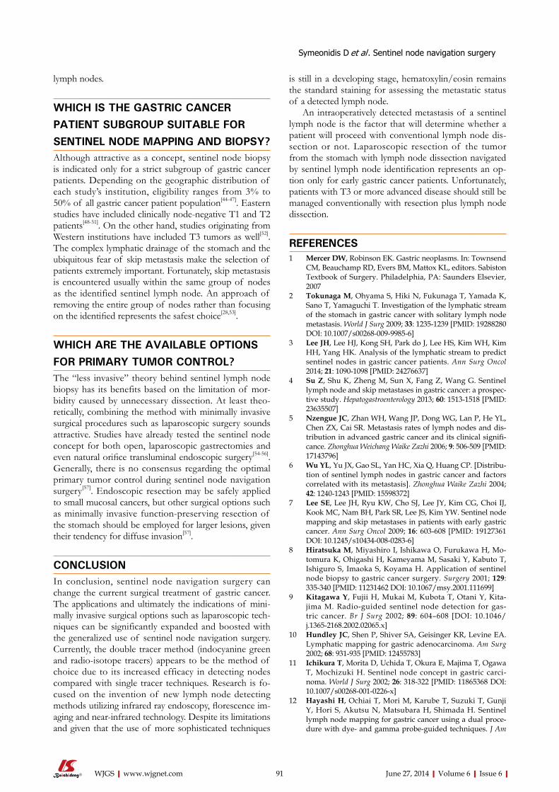
is still in a developing stage, hematoxylin/eosin remains the standard staining for assessing the metastatic status of a detected lymph node.
An intraoperatively detected metastasis of a sentinel lymph node is the factor that will determine whether a patient will proceed with conventional lymph node dis-section or not. Laparoscopic resection of the tumor from the stomach with lymph node dissection navigated by sentinel lymph node identification represents an op-tion only for early gastric cancer patients. Unfortunately, patients with T3 or more advanced disease should still be managed conventionally with resection plus lymph node dissection.
REFERENCES1 Mercer DW, Robinson EK. Gastric neoplasms. In: Townsend
CM, Beauchamp RD, Evers BM, Mattox KL, editors. Sabiston Textbook of Surgery. Philadelphia, PA: Saunders Elsevier, 2007
2 Tokunaga M, Ohyama S, Hiki N, Fukunaga T, Yamada K, Sano T, Yamaguchi T. Investigation of the lymphatic stream of the stomach in gastric cancer with solitary lymph node metastasis. World J Surg 2009; 33: 1235-1239 [PMID: 19288280 DOI: 10.1007/s00268-009-9985-6]
3 Lee JH, Lee HJ, Kong SH, Park do J, Lee HS, Kim WH, Kim HH, Yang HK. Analysis of the lymphatic stream to predict sentinel nodes in gastric cancer patients. Ann Surg Oncol 2014; 21: 1090-1098 [PMID: 24276637]
4 Su Z, Shu K, Zheng M, Sun X, Fang Z, Wang G. Sentinel lymph node and skip metastases in gastric cancer: a prospec-tive study. Hepatogastroenterology 2013; 60: 1513-1518 [PMID: 23635507]
5 Nzengue JC, Zhan WH, Wang JP, Dong WG, Lan P, He YL, Chen ZX, Cai SR. Metastasis rates of lymph nodes and dis-tribution in advanced gastric cancer and its clinical signifi-cance. Zhonghua Weichang Waike Zazhi 2006; 9: 506-509 [PMID: 17143796]
6 Wu YL, Yu JX, Gao SL, Yan HC, Xia Q, Huang CP. [Distribu-tion of sentinel lymph nodes in gastric cancer and factors correlated with its metastasis]. Zhonghua Waike Zazhi 2004; 42: 1240-1243 [PMID: 15598372]
7 Lee SE, Lee JH, Ryu KW, Cho SJ, Lee JY, Kim CG, Choi IJ, Kook MC, Nam BH, Park SR, Lee JS, Kim YW. Sentinel node mapping and skip metastases in patients with early gastric cancer. Ann Surg Oncol 2009; 16: 603-608 [PMID: 19127361 DOI: 10.1245/s10434-008-0283-6]
8 Hiratsuka M, Miyashiro I, Ishikawa O, Furukawa H, Mo-tomura K, Ohigashi H, Kameyama M, Sasaki Y, Kabuto T, Ishiguro S, Imaoka S, Koyama H. Application of sentinel node biopsy to gastric cancer surgery. Surgery 2001; 129: 335-340 [PMID: 11231462 DOI: 10.1067/msy.2001.111699]
9 Kitagawa Y, Fujii H, Mukai M, Kubota T, Otani Y, Kita-jima M. Radio-guided sentinel node detection for gas-tric cancer. Br J Surg 2002; 89: 604–608 [DOI: 10.1046/j.1365-2168.2002.02065.x]
10 Hundley JC, Shen P, Shiver SA, Geisinger KR, Levine EA. Lymphatic mapping for gastric adenocarcinoma. Am Surg 2002; 68: 931-935 [PMID: 12455783]
11 Ichikura T, Morita D, Uchida T, Okura E, Majima T, Ogawa T, Mochizuki H. Sentinel node concept in gastric carci-noma. World J Surg 2002; 26: 318-322 [PMID: 11865368 DOI: 10.1007/s00268-001-0226-x]
12 Hayashi H, Ochiai T, Mori M, Karube T, Suzuki T, Gunji Y, Hori S, Akutsu N, Matsubara H, Shimada H. Sentinel lymph node mapping for gastric cancer using a dual proce-dure with dye- and gamma probe-guided techniques. J Am
lymph nodes.
WHICH IS THE GASTRIC CANCER PATIENT SUBGROUP SUITABLE FOR SENTINEL NODE MAPPING AND BIOPSY?Although attractive as a concept, sentinel node biopsy is indicated only for a strict subgroup of gastric cancer patients. Depending on the geographic distribution of each study’s institution, eligibility ranges from 3% to 50% of all gastric cancer patient population[44-47]. Eastern studies have included clinically node-negative T1 and T2 patients[48-51]. On the other hand, studies originating from Western institutions have included T3 tumors as well[52]. The complex lymphatic drainage of the stomach and the ubiquitous fear of skip metastasis make the selection of patients extremely important. Fortunately, skip metastasis is encountered usually within the same group of nodes as the identified sentinel lymph node. An approach of removing the entire group of nodes rather than focusing on the identified represents the safest choice[28,53].
WHICH ARE THE AVAILABLE OPTIONS FOR PRIMARY TUMOR CONTROL?The “less invasive” theory behind sentinel lymph node biopsy has its benefits based on the limitation of mor-bidity caused by unnecessary dissection. At least theo-retically, combining the method with minimally invasive surgical procedures such as laparoscopic surgery sounds attractive. Studies have already tested the sentinel node concept for both open, laparoscopic gastrectomies and even natural orifice transluminal endoscopic surgery[54-56]. Generally, there is no consensus regarding the optimal primary tumor control during sentinel node navigation surgery[57]. Endoscopic resection may be safely applied to small mucosal cancers, but other surgical options such as minimally invasive function-preserving resection of the stomach should be employed for larger lesions, given their tendency for diffuse invasion[57].
CONCLUSIONIn conclusion, sentinel node navigation surgery can change the current surgical treatment of gastric cancer. The applications and ultimately the indications of mini-mally invasive surgical options such as laparoscopic tech-niques can be significantly expanded and boosted with the generalized use of sentinel node navigation surgery. Currently, the double tracer method (indocyanine green and radio-isotope tracers) appears to be the method of choice due to its increased efficacy in detecting nodes compared with single tracer techniques. Research is fo-cused on the invention of new lymph node detecting methods utilizing infrared ray endoscopy, florescence im-aging and near-infrared technology. Despite its limitations and given that the use of more sophisticated techniques
June 27, 2014|Volume 6|Issue 6|WJGS|www.wjgnet.com 91
Symeonidis D et al . Sentinel node navigation surgery

S, Inazawa J, Mochizuki H, Matsubara O. Analysis of sen-tinel node involvement in gastric cancer. Clin Gastroenterol Hepatol 2007; 5: 1046-1052 [PMID: 17632042 DOI: 10.1016/j.cgh.2007.05.001]
28 Lee YJ, Ha WS, Park ST, Choi SK, Hong SC, Park JW. Which biopsy method is more suitable between a basin dissec-tion and pick-up biopsy for sentinel nodes in laparoscopic sentinel-node navigation surgery (LSNNS) for gastric can-cer? J Laparoendosc Adv Surg Tech A 2008; 18: 357-363 [PMID: 18503367 DOI: 10.1089/lap.2007.0024]
29 Park do J, Kim HH, Park YS, Lee HS, Lee WW, Lee HJ, Yang HK. Simultaneous indocyanine green and (99m)Tc-antimony sulfur colloid-guided laparoscopic sentinel basin dissection for gastric cancer. Ann Surg Oncol 2011; 18: 160-165 [PMID: 20652640 DOI: 10.1245/s10434-010-1221-y]
30 Kitagawa Y, Fujii H, Kumai K, Kubota T, Otani Y, Saikawa Y, Yoshida M, Kubo A, Kitajima M. Recent advances in sentinel node navigation for gastric cancer: a paradigm shift of surgi-cal management. J Surg Oncol 2005; 90: 147-151; discussion 151-152 [PMID: 15895450 DOI: 10.1002/jso.20220]
31 Gretschel S, Bembenek A, Hünerbein M, Dresel S, Schneider W, Schlag PM. Efficacy of different technical procedures for sentinel lymph node biopsy in gastric cancer staging. Ann Surg Oncol 2007; 14: 2028-2035 [PMID: 17453300 DOI: 10.1245/s10434-007-9367-y]
32 Can MF, Yagci G, Cetiner S. Sentinel lymph node biopsy for gastric cancer: Where do we stand? World J Gastrointest Surg 2011; 3: 131-137 [PMID: 22007282 DOI: 10.4240/wjgs.v3.i9.131]
33 Yaguchi Y, Ichikura T, Ono S, Tsujimoto H, Sugasawa H, Sakamoto N, Matsumoto Y, Yoshida K, Kosuda S, Hase K. How should tracers be injected to detect for sentinel nodes in gastric cancer--submucosally from inside or subserosally from outside of the stomach? J Exp Clin Cancer Res 2008; 27: 79 [PMID: 19055749 DOI: 10.1186/1756-9966-27-79]
34 Lee JH, Ryu KW, Kim CG, Kim SK, Choi IJ, Kim YW, Chang HJ, Bae JM, Hong EK. Comparative study of the subserosal versus submucosal dye injection method for sentinel node biopsy in gastric cancer. Eur J Surg Oncol 2005; 31: 965-968 [PMID: 15908163 DOI: 10.1016/j.ejso.2005.03.006]
35 Stojanovic D, Milenkovic SM, Mitrovic N, Marinkovic D, Stevanovic D, Radovanovic D. The feasibility of sentinel lymph node biopsy for gastric cancer: the experience from Serbia. J BUON 2013; 18: 162-168 [PMID: 23613402]
36 Yano K, Nimura H, Mitsumori N, Takahashi N, Kashiwagi H, Yanaga K. The efficiency of micrometastasis by senti-nel node navigation surgery using indocyanine green and infrared ray laparoscopy system for gastric cancer. Gastric Cancer 2012; 15: 287-291 [PMID: 22041868 DOI: 10.1007/s10120-011-0105-6]
37 Tajima Y, Murakami M, Yamazaki K, Masuda Y, Kato M, Sato A, Goto S, Otsuka K, Kato T, Kusano M. Sentinel node mapping guided by indocyanine green fluorescence imag-ing during laparoscopic surgery in gastric cancer. Ann Surg Oncol 2010; 17: 1787-1793 [PMID: 20162462 DOI: 10.1245/s10434-010-0944-0]
38 Miyashiro I, Hiratsuka M, Sasako M, Sano T, Mizusawa J, Nakamura K, Nashimoto A, Tsuburaya A, Fukushima N. High false-negative proportion of intraoperative histologi-cal examination as a serious problem for clinical application of sentinel node biopsy for early gastric cancer: final results of the Japan Clinical Oncology Group multicenter trial JCOG0302. Gastric Cancer 2014; 17: 316-323 [PMID: 23933782]
39 Park DJ, Lee HJ, Lee HS, Kim WH, Kim HH, Lee KU, Choe KJ, Yang HK. Sentinel node biopsy for cT1 and cT2a gastric cancer. Eur J Surg Oncol 2006; 32: 48-54 [PMID: 16269225 DOI: 10.1016/j.ejso.2005.09.006]
40 Hanisch E, Batsis C. Sentinel node biopsy in laparoscopic surgical oncology. Surg Endosc 2011; 25: 3713-3714 [PMID: 21594737 DOI: 10.1007/s00464-011-1747-5]
Coll Surg 2003; 196: 68-74 [PMID: 12517553 DOI: 10.1016/S1072-7515(02)01594-6]
13 Ajisaka H, Miwa K. Micrometastases in sentinel nodes of gastric cancer. Br J Cancer 2003; 89: 676-680 [PMID: 12915877 DOI: 10.1038/sj.bjc.6601183]
14 Miwa K, Kinami S, Taniguchi K, Fushida S, Fujimura T, Nonomura A. Mapping sentinel nodes in patients with ear-ly-stage gastric carcinoma. Br J Surg 2003; 90: 178-182 [PMID: 12555293 DOI: 10.1002/bjs.4031]
15 Ishigami S, Natsugoe S, Uenosono Y, Hata Y, Nakajo A, Miyazono F, Matsumoto M, Hokita S, Aikou T. Infiltration of antitumor immunocytes into the sentinel node in gastric cancer. J Gastrointest Surg 2003; 7: 735-739 [PMID: 13129549 DOI: 10.1016/S1091-255X(03)00076-3]
16 Ryu KW, Lee JH, Kim HS, Kim YW, Choi IJ, Bae JM. Pre-diction of lymph nodes metastasis by sentinel node biopsy in gastric cancer. Eur J Surg Oncol 2003; 29: 895-899 [PMID: 14624784 DOI: 10.1016/j.ejso.2003.09.008]
17 Tonouchi H, Mohri Y, Tanaka K, Konishi N, Ohmori Y, Ko-bayashi M, Watanabe Y, Matsumura K, Takeda K, Kusunoki M. Lymphatic mapping and sentinel node biopsy during laparoscopic gastrectomy for early cancer. Dig Surg 2003; 20: 421-427 [PMID: 12900533 DOI: 10.1159/000072710]
18 Isozaki H, Kimura T, Tanaka N, Satoh K, Matsumoto S, Ninomiya M, Ohsaki T, Mori M. An assessment of the fea-sibility of sentinel lymph node-guided surgery for gastric cancer. Gastric Cancer 2004; 7: 149-153 [PMID: 15449202 DOI: 10.1007/s10120-004-0283-6]
19 Nimura H, Narimiya N, Mitsumori N, Yamazaki Y, Yanaga K, Urashima M. Infrared ray electronic endoscopy combined with indocyanine green injection for detection of sentinel nodes of patients with gastric cancer. Br J Surg 2004; 91: 575-579 [PMID: 15122608 DOI: 10.1002/bjs.4470]
20 Ishikawa K, Yasuda K, Shiromizu A, Etoh T, Shiraishi N, Kitano S. Laparoscopic sentinel node navigation achieved by infrared ray electronic endoscopy system in patients with gastric cancer. Surg Endosc 2007; 21: 1131-1134 [PMID: 17180275 DOI: 10.1007/s00464-006-9062-2]
21 Miyashiro I, Miyoshi N, Hiratsuka M, Kishi K, Yamada T, Ohue M, Ohigashi H, Yano M, Ishikawa O, Imaoka S. Detection of sentinel node in gastric cancer surgery by in-docyanine green fluorescence imaging: comparison with infrared imaging. Ann Surg Oncol 2008; 15: 1640-1643 [PMID: 18379850 DOI: 10.1245/s10434-008-9872-7]
22 Tajima Y, Yamazaki K, Masuda Y, Kato M, Yasuda D, Aoki T, Kato T, Murakami M, Miwa M, Kusano M. Sentinel node mapping guided by indocyanine green fluorescence imaging in gastric cancer. Ann Surg 2009; 249: 58-62 [PMID: 19106676 DOI: 10.1097/SLA.0b013e3181927267]
23 Soltesz EG, Kim S, Kim SW, Laurence RG, De Grand AM, Parungo CP, Cohn LH, Bawendi MG, Frangioni JV. Sentinel lymph node mapping of the gastrointestinal tract by us-ing invisible light. Ann Surg Oncol 2006; 13: 386-396 [PMID: 16485157 DOI: 10.1245/ASO.2006.04.025]
24 Lee JH, Ryu KW, Kim CG, Kim SK, Lee JS, Kook MC, Choi IJ, Kim YW, Chang HJ, Bae JM. Sentinel node biopsy us-ing dye and isotope double tracers in early gastric cancer. Ann Surg Oncol 2006; 13: 1168-1174 [PMID: 16924376 DOI: 10.1245/s10434-006-9038-4]
25 Mura G, Vagliasindi A, Framarini M, Mazza P, Solfrini G, Verdecchia GM. The sentinel node biopsy in early gastric cancer: a preliminary study. Langenbecks Arch Surg 2006; 391: 113-117 [PMID: 16525854 DOI: 10.1007/s00423-005-0018-0]
26 Saikawa Y, Otani Y, Kitagawa Y, Yoshida M, Wada N, Kubota T, Kumai K, Sugino Y, Mukai M, Kameyama K, Kubo A, Kitajima M. Interim results of sentinel node biopsy during laparoscopic gastrectomy: possible role in function-preserving surgery for early cancer. World J Surg 2006; 30: 1962-1968 [PMID: 17043938 DOI: 10.1007/s00268-006-0142-1]
27 Morita D, Tsuda H, Ichikura T, Kimura M, Aida S, Kosuda
June 27, 2014|Volume 6|Issue 6|WJGS|www.wjgnet.com 92
Symeonidis D et al . Sentinel node navigation surgery
Roh MS, Jeong JS. Lymphatic mapping and sentinel node biopsy using 99mTc tin colloid in gastric cancer. Ann Surg 2004; 239: 383-387 [PMID: 15075656 DOI: 10.1097/01.sla.0000114227.70480.14]
51 Can MF, Yagci G, Cetiner S. Systematic review of studies investigating sentinel node navigation surgery and lym-phatic mapping for gastric cancer. J Laparoendosc Adv Surg Tech A 2013; 23: 651-662 [PMID: 23755853 DOI: 10.1089/lap.2012.0311]
52 Gretschel S, Bembenek A, Ulmer Ch, Hünerbein M, Mark-wardt J, Schneider U, Schlag PM. Prediction of gastric can-cer lymph node status by sentinel lymph node biopsy and the Maruyama computer model. Eur J Surg Oncol 2005; 31: 393-400 [PMID: 15837046 DOI: 10.1016/j.ejso.2004.11.014]
53 Cozzaglio L, Bottura R, Di Rocco M, Gennari L, Doci R. Sentinel lymph node biopsy in gastric cancer: possible ap-plications and limits. Eur J Surg Oncol 2011; 37: 55-59 [PMID: 21115231 DOI: 10.1016/j.ejso.2010.10.012]
54 Kelder W, Nimura H, Takahashi N, Mitsumori N, van Dam GM, Yanaga K. Sentinel node mapping with indocyanine green (ICG) and infrared ray detection in early gastric can-cer: an accurate method that enables a limited lymphadenec-tomy. Eur J Surg Oncol 2010; 36: 552-558 [PMID: 20452171 DOI: 10.1016/j.ejso.2010.04.007]
55 Kim HH, Ahn SH. The current status and future perspec-tives of laparoscopic surgery for gastric cancer. J Korean Surg Soc 2011; 81: 151-162 [PMID: 22066116 DOI: 10.4174/jkss.2011.81.3.151]
56 Cahill RA, Asakuma M, Perretta S, Dallemagne B, Mares-caux J. Gastric lymphatic mapping for sentinel node biopsy by natural orifice transluminal endoscopic surgery (NOTES). Surg Endosc 2009; 23: 1110-1116 [PMID: 18813997 DOI: 10.1007/s00464-008-0124-5]
57 Park JY, Ryu KW, Eom BW, Yoon HM, Kim SJ, Cho SJ, Lee JY, Kim CG, Kook MC, Choi IJ, Nam BH, Kim YW. Proposal of the surgical options for primary tumor control during sentinel node navigation surgery based on the discrepancy between preoperative and postoperative early gastric can-cer diagnoses. Ann Surg Oncol 2014; 21: 1123-1129 [PMID: 24366418]
P- Reviewer: Zhu YL S- Editor: Song XX L- Editor: A E- Editor: Wu HL
41 Miyashiro I. What is the problem in clinical application of sentinel node concept to gastric cancer surgery? J Gas-tric Cancer 2012; 12: 7-12 [PMID: 22500258 DOI: 10.5230/jgc.2012.12.1.7]
42 Ichikura T, Sugasawa H, Sakamoto N, Yaguchi Y, Tsujimoto H, Ono S. Limited gastrectomy with dissection of sentinel node stations for early gastric cancer with negative sentinel node biopsy. Ann Surg 2009; 249: 942-947 [PMID: 19474686 DOI: 10.1097/SLA.0b013e3181a77e7e]
43 Arigami T, Natsugoe S, Uenosono Y, Mataki Y, Ehi K, Higashi H, Arima H, Yanagida S, Ishigami S, Hokita S, Aikou T. Evaluation of sentinel node concept in gastric cancer based on lymph node micrometastasis determined by reverse transcription-polymerase chain reaction. Ann Surg 2006; 243: 341-347 [PMID: 16495698 DOI: 10.1097/01.sla.0000201453.65534.f1]
44 Scabini S. Sentinel node biopsy in colorectal cancer: Must we believe it? World J Gastrointest Surg 2010; 2: 6-8 [PMID: 21160827 DOI: 10.4240/wjgs.v2.i1.6]
45 Rabin I, Chikman B, Lavy R, Poluksht N, Halpern Z, Was-sermann I, Gold-Deutch R, Sandbank J, Halevy A. The ac-curacy of sentinel node mapping according to T stage in patients with gastric cancer. Gastric Cancer 2010; 13: 30-35 [PMID: 20373073 DOI: 10.1007/s10120-009-0532-9]
46 Hamashima C, Shibuya D, Yamazaki H, Inoue K, Fukao A, Saito H, Sobue T. The Japanese guidelines for gastric cancer screening. Jpn J Clin Oncol 2008; 38: 259-267 [PMID: 18344316 DOI: 10.1093/jjco/hyn017]
47 Yalcin S. Gastric cancer in Turkey-a bridge between west and East. Gastrointest Cancer Res 2009; 3: 29-32 [PMID: 19343135]
48 Rausei S, Dionigi G, Rovera F, Boni L, Valerii C, Giavarini L, Frattini F, Dionigi R. A decade in gastric cancer curative surgery: Evidence of progress (1999-2009). World J Gastroin-test Surg 2012; 4: 45-54 [PMID: 22530078 DOI: 10.4240/wjgs.v4.i3.45]
49 Takeuchi H, Kitagawa Y. New sentinel node mapping tech-nologies for early gastric cancer. Ann Surg Oncol 2013; 20: 522-532 [PMID: 22941161 DOI: 10.1245/s10434-012-2602-1]
50 Kim MC, Kim HH, Jung GJ, Lee JH, Choi SR, Kang DY,
June 27, 2014|Volume 6|Issue 6|WJGS|www.wjgnet.com 93
Symeonidis D et al . Sentinel node navigation surgery

Pathological factors affecting gastric adenocarcinoma survival in a Caribbean population from 2000-2010
Patrick O Roberts, Joseph Plummer, Pierre-Anthony Leake, Shane Scott, Tamara G de Souza, Ayesha Johnson, Tracey N Gibson, Barrie Hanchard, Marvin Reid
Patrick O Roberts, Joseph Plummer, Pierre-Anthony Leake, Shane Scott, Tamara G de Souza, The Department of Surgery, Radiology, Anaesthesia and Intensive Care, The University of the West Indies, Jamaica 999172, West Indies Ayesha Johnson, College of Public Health, University of South Florida, Tampa, FL 33620-9951, United StatesTracey N Gibson, Barrie Hanchard, The Department of Pathol-ogy, The University of the West Indies, Jamaica 999172, West Indies Marvin Reid, Tropical Medicine Research Institute, The Univer-sity of the West Indies, Jamaica 999172, West IndiesAuthor contributions: Roberts PO designed the study, analysed data, critically revised and approved the final version of the study; Johnson A statistically analysed data, contributed to drafting and the final approval of the study version for publication; Leake PA assisted in designing the study and critical revision; Plummer J critically revised the study; de Souza TG interpreted data, drafted, revised and approved the final manuscript; Reid M assisted in study design; Gibson TN and Hanchard B collected data and ed-ited the final version; Scott S acquired data. Correspondence to: Dr. Patrick O Roberts, The Department of Surgery, Radiology, Anaesthesia and Intensive Care, The Uni-versity of the West Indies, Mona, Kingston 7, Jamaica,West Indies. [email protected]: +1-876-9271620 Fax: +1-876-9704302Received: December 2, 2013 Revised: February 14, 2014Accepted: May 15, 2014Published online: June 27, 2014
AbstractAIM: To investigate pathological factors related to long term patient survival post surgical management of gas-tric adenocarcinoma in a Caribbean population.
METHODS: This is a retrospective, observational study of all patients treated surgically for gastric adenocarci-noma from January 1st 2000 to December 31st 2010 at The University Hospital of the West Indies, an urban Jamaican hospital. Pathological reports of all gastrecto-my specimens post gastric cancer resection during the
specified interval were accessed. Patients with a final diagnosis other than adenocarcinoma, as well as pa-tients having undergone surgery at an external institu-tion were excluded. The clinical records of the selected cohort were reviewed. The following variables were analysed; patient gender, patient age, the number of gastrectomies previous performed by the lead surgeon, the gross anatomical location and appearance of the tumour, the histological appearance of the tumour, infil-tration of the tumour into stomach wall and surround-ing structures, presence of Helicobacter pylori and the presence of gastritis. Patient status as dead vs alive was documented for the end of the interval. The effect of the aforementioned factors on patient survival were analysed using Logrank tests, Cox regression models, Ranksum tests, Kruskal-Wallis tests and Kaplan-Meier curves.
RESULTS: A total of 79 patients, 36 males and 43 fe-males, were included. Their median age was 67 years (range 36-86 years). Median survival time from surgery was 70 mo with 40.5% of patients dying before the termination date of the study. Tumours ranged from 0.8 cm in size to encompassing the entire stomach speci-men, with a median tumour size of 6 cm. The median number of nodes removed at surgery was 8 with a maximum of 28. The median number of positive lymph nodes found was 2, with a range of 0 to 22. Patients’ median survival time was approximately 70 mo, with 40.5% of the patients in this cohort dying before the terminal date. An increase in the incidence of cardiac tumours was noted compared to the previous 10 year interval (7.9% to 9.1%). Patients who had serosal involvement of the tumour did have a significantly shorter survival than those who did not (P = 0.017). A significant increase in the hazard ratio (HR), 2.424, for patients with circumferential tumours was found (P = 0.044). Via Kaplan-Meier estimates, the presence of venous infiltration as well as involvement of the circum-ferential resection margin were found to be poor prog-
RETROSPECTIVE STUDY
Submit a Manuscript: http://www.wjgnet.com/esps/Help Desk: http://www.wjgnet.com/esps/helpdesk.aspxDOI: 10.4240/wjgs.v6.i6.94
World J Gastrointest Surg 2014 June 27; 6(6): 94-100ISSN 1948-9366 (online)
© 2014 Baishideng Publishing Group Inc. All rights reserved.
June 27, 2014|Volume 6|Issue 6|WJGS|www.wjgnet.com 94
nostic markers, decreasing survival at 50 mo by 46.2% and 36.3% respectively. The increased HR for venous infiltration, 2.424, trended toward significant (P = 0.055) Age, size of tumour, number of positive nodes found and total number of lymph nodes removed were not useful predictors of survival. It is noted that the results were mostly negative, that is many tumour character-istics did not indicate any evidence of affecting patient survival. The current sample, with 30 observed events (deaths), would have about 30% power to detect a HR of 2.5.
CONCLUSION: This study mirrors pathological factors used for gastric cancer prognostication in other popu-lations. As evaluation continues, a larger cohort will strengthen the significance of observed trends.
© 2014 Baishideng Publishing Group Inc. All rights reserved.
Key words: Gastric adenocarcinoma; Caribbean; Jamai-ca; Pathologic; Survival; Gastrectomy; Gastritis; Helico-bacter pylori ; Cardia; Circumferential resection margin
Core tip: This ten year retrospective analysis of patho-logic factors affecting the survival of gastric cancer patients is the first ever to be done in a Caribbean population. Significant findings meriting publication in-clude increasing incidence of proximal tumours and de-creased survival with involvement of the circumferential resection margin. Among other factors also examined, are the impact of surgeon and pathologist training on patient survival. By describing the current state of gas-tric cancer management in this population, this study aspires to lay the foundation for further work enhanc-ing gastric cancer care in this region.
Roberts PO, Plummer J, Leake PA, Scott S, de Souza TG, John-son A, Gibson TN, Hanchard B, Reid M. Pathological factors affecting gastric adenocarcinoma survival in a Caribbean popula-tion from 2000-2010. World J Gastrointest Surg 2014; 6(6): 94-100 Available from: URL: http://www.wjgnet.com/1948-9366/full/v6/i6/94.htm DOI: http://dx.doi.org/10.4240/wjgs.v6.i6.94
INTRODUCTIONGastric cancer is the fourth most common malignancy in men, and the fifth commonest in women diagnosed worldwide[1]. In Jamaica it is the seventh commonest cancer in men and the ninth most common in women[2]. Worldwide, gastric cancer is the third most frequent cause of cancer death in men and the fifth most common in women[1]. In Jamaica it is the fourth leading cause of can-cer related death[3].
These figures prompt scrutiny and characterization of the Jamaican gastric cancer patient population. Ac-cordingly, Plummer et al[4] had previously outlined age and sex related incidence, histological appearance, as well as tumour location in a surgically treated cohort of gastric
adenocarcinoma patients in Jamaica from 1993-2002, and found that the antrum of the stomach was most often in-volved, with a trend toward increased incidence of more proximal tumours in the latter 5 years studied, and that lymph node metastases were common[4].
It was our aim to build on this base of information and to explore prognostication in this patient demo-graphic. Our study seeks, for the first time in the Jamai-can population, to describe the post-surgical survival rate of gastric adenocarcinoma patients and to elucidate related pathological factors.
MATERIALS AND METHODSThis retrospective, observational study summarizes the analyses conducted on patients diagnosed with gastric ad-enocarcinoma at the University Hospital of the West In-dies (UHWI) from 2000 to 2010. Analysis was conducted using STATA 9. Approval was granted from the Ethics Committee of the hospital (file number ECP334, 12/13). This study was carried out in accordance with the Second International Helsinki Declaration[5].
All patients who were diagnosed with gastric cancer at UHWI during the period January 1st 2000 to December 31st 2010 were enrolled via review of pathology reports. UHWI is a type A teaching hospital and a referral centre, accepting the management of patients exceeding the re-source capability of several smaller hospitals throughout Jamaica. These patients had all been seen at the General Surgery outpatient clinic, and subsequently undergone upper gastrointestinal endoscopy with biopsy proving gastric cancer. Other forms of gastric cancer besides adenocarcinoma were excluded. Patients with a final diag-nosis of gastric adenocarcinoma, but who had undergone surgery at other institutions were excluded. This exclu-sion came as a matter of access to the clinical records of those patients. Demographically, the patients receiving definitive management at external institutions are not anticipated to greatly differ from the patient population investigated in this study. Of note, the population of Ja-maica is predominantly Afro-Caribbean.
Survival data was analysed with the event being death and the time to death being defined as the time in months from surgery until the patient’s death. Persons who did not die were censored at the date of their last visit or if confirmed to be alive, they were censored at December 31, 2011.
Logrank tests were conducted to determine whether the survival rate was different across various groupings within variables. Cox regression models were built to determine whether various pathological characteristics would be able to predict a person’s risk of death. Rank-sum tests as well as Kruskal-Wallis tests of association were done to determine whether the relationships be-tween clinicopathological factors and survival were differ-ent for persons who were dead as opposed to confirmed alive or censored prior to the end of the studied inter-val[6,7].
June 27, 2014|Volume 6|Issue 6|WJGS|www.wjgnet.com 95
Roberts PO et al . Gastric cancer in the Caribbean
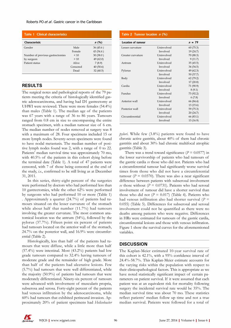
RESULTSThe surgical notes and pathological reports of the 79 pa-tients meeting the criteria of histologically identified gas-tric adenocarcinoma, and having had D1 gastrectomy at UHWI were reviewed. There were more females (54.4%) than males (Table 1). The median age of the patients was 67 years with a range of 36 to 86 years. Tumours ranged from 0.8 cm in size to encompassing the entire stomach specimen, with a median tumour size of 6 cm. The median number of nodes removed at surgery was 8 with a maximum of 28. Four specimens included 15 or more lymph nodes. Seventy-seven specimens were found to have nodal metastasis. The median number of posi-tive lymph nodes found was 2, with a range of 0 to 22. Patients’ median survival time was approximately 70 mo, with 40.5% of the patients in this cohort dying before the terminal date (Table 1). A total of 47 patients were censored, with 7 of those being censored at the end of the study, i.e., confirmed to be still living as at December 31, 2011.
In this series, thirty-eight percent of the surgeries were performed by doctors who had performed less than 10 gastrectomies, while the other 62% were performed by surgeons who had performed 10 or more (Table 1) . Approximately a quarter (24.7%) of patients had tu-mours situated on the lesser curvature of the stomach while about half that number (11.7%) had tumours involving the greater curvature. The most common ana-tomical location was the antrum (54%), followed by the pylorus (37.7%). Fifteen point six percent of patients had tumours located on the anterior wall of the stomach, 24.7% on the posterior wall, and 16.9% were circumfer-ential (Table 2).
Histologically, less than half of the patients had tu-mours that were diffuse, while a little more than half (57.4%) were intestinal. Most (43.2%) patients had low grade tumours compared to 32.4% having tumours of moderate grade and the remainder of high grade. More than half of the patients had ulcerative lesions. Few (5.7%) had tumours that were well differentiated, while the majority (50.9%) of patients had tumours that were moderately differentiated. Ninety-six percent of tumours were advanced with involvement of muscularis propria, subserosa and serosa. Forty-eight percent of the patients had venous infiltration by the adenocarcinoma, while 60% had tumours that exhibited perineural invasion. Ap-proximately 20% of patient specimens had Helicobacter
pylori. While few (3.8%) patients were found to have chronic active gastritis, about 40% of them had chronic gastritis and about 30% had chronic multifocal atrophic gastritis (Table 3).
There was a trend toward significance (P = 0.0577) in the lower survivorship of patients who had tumours of the gastric cardia vs those who did not. Patients who had a circumferential tumour had significantly worse survival times from those who did not have a circumferential tumour (P = 0.0370). There was also a near significant difference between patients with subserosal involvement vs those without (P = 0.0731). Patients who had serosal involvement of tumour did have a shorter survival than those who did not (P = 0.017) (Table 4). Patients who had venous infiltration also had shorter survival (P = 0.055) (Table 5). Differences for subserosal and serosal involvement could not be quantified as there were no deaths among patients who were negative. Differences in HRs were estimated for tumours of the gastric cardia, circumferential lesions and those with venous infiltration. Figure 1 show the survival curves for the aforementioned variables.
DISCUSSIONThe Kaplan-Meier estimated 10-year survival rate of this cohort is 42.1%, with a 95% confidence interval of 24.4%-58.7%. This Kaplan-Meier estimate accounts for the varying risks within the population with respect to their clinicopathological factors. This is appropriate as we have noted statistically significant impact of certain pa-rameters on patient survival. If it were assumed that each patient was at an equivalent risk for mortality following surgery the incidental survival rate would be 35%. The median survival time was about 70 mo. These statistics reflect patients’ median follow up time and not a true median survival. Patients were followed for a total of
June 27, 2014|Volume 6|Issue 6|WJGS|www.wjgnet.com 96
Characteristic n (%)
Gender Male 36 (45.6 )Female 43 (54.4 )
Number of previous gastrectomies by surgeon
< 10 30 (38.0 )> 10 49 (62.0)
Patient status Alive 7 (8.9)Censored 40 (50.6)
Dead 32 (40.5)
Table 1 Clinical characteristics
Location of tumour n = 79
Lesser curvature Uninvolved 60 (75.3)Involved 19 (24.7)
Greater curvature Uninvolved 70 (88.3)Involved 9 (11.7)
Antrum Uninvolved 35 (45.5)Involved 34 (54.5)
Pylorus Uninvolved 49 (62.3)Involved 50 (37.7)
Body Uninvolved 62 (79.2)Involved 17 (20.8)
Cardia Uninvolved 71 (90.9)Involved 8 (9.1)
Fundus Uninvolved 73 (92.2)Involved 6 (7.8)
Anterior wall Uninvolved 66 (84.4)Involved 13 (15.6)
Posterior wall Uninvolved 59 (75.3)Involved 20 (24.7)
Circumferential Uninvolved 66 (83.1)Involved 13 (16.9)
Table 2 Tumour location n (%)
Roberts PO et al . Gastric cancer in the Caribbean

dence of affecting patient survival. Table 3 shows the results of a power analysis to determine the power of our study to detect varying hazard rates. We see that, at best, the current sample with 30 observed events (deaths) would have about 30% power to detect a HR of 2.5. It is likely that the ability of this study to demonstrate patho-logical prognosticators already commonly accepted in the literature was limited by this relatively low power. We see that we could detect a hazard rate of 2 with power 80%, if we observed 202 deaths. As the examined cohort con-tinues to grow yearly we can expect more robust statistics in future analyses. Furthermore, this study included all cause mortality. As the cohort continues to grow, exact causes of death can be better examined and adjusted for according to their likely direct relation to gastric cancer and therapeutic intervention for this disease process.
Age, size of tumour, number of positive nodes found and total number of lymph nodes removed were not use-ful predictors of survival. However, it was noted that pa-tients who had a circumferential tumour were almost two and a half times more likely to die than those without. There was a similar increase in the likelihood of death for patients with tumours of the cardia, as well as in those with venous infiltration (Table 3).
In a previous clinicopathological audit of gastric car-cinomas at this institution between 1993-2002, the major-ity of tumours (56%) were of the antrum. The cardia was the third most involved site at 7.9%[4]. The current study, 2000-2010, noted 9.1% of cases to involve the gastric cardia and 54.5% involving the antrum. While the major-ity of gastric cancers in our population continue to be found in the antrum, it is worth noting the increased inci-dence of cancers of the cardia. This early trend parallels the 29.0% to 52.2% increase in incidence of adenocarci-nomas of the gastric cardia noted in the United Kingdom from 1984 to 1993[8]. Similarly the incidence of cardiac lesions increased from 29% to 52%, in the United States of America, between 1984-1993[9]. McLoughlin purports a generally poorer prognosis associated with cancers of the gastric cardia as opposed to other anatomical sites[10].
1912.115 person months during which time there were 30 deaths. This yields an estimated death rate of 0.0157 deaths per person-month, or 18.8% per person-year (95%CI: 0.132-0.269). This means that if we observed 100 persons with the disease for 1 year we would expect approximately 19 of them to die.
It is noted that the results were mostly negative, that is many tumour characteristics did not indicate any evi-
June 27, 2014|Volume 6|Issue 6|WJGS|www.wjgnet.com 97
Gross appearance Percentage
Configuration Ulcerative 56.00Infiltrative 22.70Exophytic 21.30
n 75 Gross location Proximal Uninvolved 88.20
Involved 11.80n 76
Distal Uninvolved 84.20Involved 15.80
n 76 Histological appearance Intestinal No 42.60
Yes 57.40n 68
Differentiation Poor 43.40Moderate 50.90
Well 5.70n 53
Grade Low 43.20Moderate 32.40
High 24.30 Maximum extent of invasion through stomach wall Mucosa Involved 100.00
n 78
Muscularis propria Uninvolved 3.80Involved 96.20
n 78
Sub–Serosa Uninvolved 9.00Involved 91.00
n 78
Serosa Uninvolved 13.30Involved 86.70
Histological appearance Venous infiltration Absent 51.90
Present 48.10n 52
Perineural infiltration Absent 40.00Present 60.00
n 35 H. Pylori Absent 80.50
Present 19.50n 77
Chronic active gastritis Absent 96.20 Present 3.80 n 78 Chronic gastritis Absent 59.00 Present 41.00
n 78
Chronic multifocal Atrophic gastritis
Absent 70.50Present 29.50
n 78
Table 3 Pathological characteristics
Variable P value
Gender 0.9688 Number of gastrectomies previously performed 0.6725 Lesser curvature 0.9716 Greater curvature 0.7928 Antrum 0.2613 Pylorus 0.2486 Body 0.2581 Cardia 0.0577 Fundus 0.9704 Anterior wall 0.3977 Posterior wall 0.9507 Circumferential 0.0370 Diffuse 0.9389 Intestinal 0.6020 Grade 0.2436 Configuration 0.2598 Differentiation 0.2416
Table 4 Logrank test of survival functions
Roberts PO et al . Gastric cancer in the Caribbean

Siewert I adenocarcinomas of the gastro-oesophageal junction[12,13]. It would appear that the findings of our study reflect such trends.
As, both globally and locally, more proximal gastric tumour sites become increasingly prevalent, it is impor-tant to note the relatively poor outcomes of such cases post gastrectomy. As alluded to above, lymphatic drain-age is thought to be a key contributor to such poor out-comes. Accordingly it is prudent to examine the approach to lymph node dissection and excision employed locally, as well as the correlation between lymph node positiv-ity and survivorship. Our study did not demonstrate any significant influence of either lymph node positivity or total number of nodes resected on survivorship. How-ever, Shimada et al[14] (2001) with a much larger cohort (982) concluded that the involvement of three or more metastatic lymph nodes is an independent prognostic factor with a sharp decrease in 5-year survivorship of pa-tients with greater than two nodes. An Italian randomised trial involving 615 gastric cancer patients demonstrated significant decrease in all-cause mortality varying propor-tionately with number of lymph nodes removed up to 25. The authors suggest a minimum of 25 nodes be removed for better long term survivorship[15]. Our study saw an average of only 8 nodes being removed. During this time at our institution the standard procedure performed was that of D1 gastrectomy. This was due to lack of person-nel trained in D2 gastrectomy technique. The American Joint Committee on Cancer recommends a minimum of 15 lymph nodes be examined for accurate staging of gas-tric cancer as retrieved by D2 lymphadenectomy[16]. Only 4 of the 79 specimens reviewed met this recommenda-tion, as D1 gastrectomies were performed. Interestingly, Schoenleber et al[17] suggest that the single most influen-tial factor in optimising the number of lymph nodes re-trieved, and consequently the number of positive lymph
This prognosis is likely a consequence of more extensive lymphatic drainage of the region, and later diagnosis[10,11]. In this study a HR of 2.685 (P = 0.069) reflected, al-though not significantly, this increased risk of mortality. Kaplan-Meier curves demonstrate the decreased survi-vorship of cardiac gastric cancers in this Jamaican cohort. Finally, the Logrank test trended toward significance with a P value of 0.0577.
This study made evident a significant HR of 2.424 (P = 0.044) for patients with circumferential resection margin (CRM) involvement post-gastrectomy. Logrank survival tests reflect this decreased survivorship with a P value of 0.0370. This applies to cancers of the cardia with extension into the distal oesophagus. CRM involve-ment has been shown to be an important prognostication factor for oesophageal cancer. Several studies have shown a poor prognosis in patients with potentially resectable malignancies of the distal 5 cm of the oesophagus and
June 27, 2014|Volume 6|Issue 6|WJGS|www.wjgnet.com 98
Prop
ortio
n su
rviv
al
1.00
0.75
0.50
0.25
0.000 50 100 150
t /mo
Prop
ortio
n su
rviv
al
1.00
0.75
0.50
0.25
0.000 50 100 150
0 50 100 150
Prop
ortio
n su
rviv
al
1.00
0.75
0.50
0.25
0.000 50 100 150
Prop
ortio
n su
rviv
al
1.00
0.75
0.50
0.25
0.00
Kaplan-Meier estimate of overall survivorship Kaplan-Meier estimate of survivorship with involvement of the cardia
Kaplan-Meier estimate of survivorship with tumours involving the CRM Kaplan-Meier estimate of survivorship with venous infiltration
No Yes
t /mo
No YesNo Yes
t /mo t /mo
A B
C D
Figure 1 Kaplan-Meier survival. A: Estimate of gastric cancer (2000–2010); B: Estimate of patients with involvement of the Cardia; C: Estimate of patients with tu-mour involving the circumferential resection margin; D: Estimate of patients with venous infiltration. CRM: Circumferential resection margin.
Predictor HR P value 95%CI
Cardia Involved vs uninvolved 2.685 0.069 0.927 7.779 Circumferential Involved vs uninvolved 2.424 0.044 1.025 5.732 Venous infiltration Present vs absent 2.502 0.055 0.98 6.384 Age 1.007 0.643 0.978 1.037 Size 0.992 0.897 0.877 1.122 Positive lymph nodes 1.059 0.118 0.986 1.137 Total lymph nodes removed
0.981 0.603 0.911 1.055
Type of surgery Total gastrectomy vs subtotal gastrectomy
1.45 0.417 0.591 3.553
Presence of Helicobacter pylori 0.456 0.198 0.138 1.508
Table 5 Single predictor cox regression models
Roberts PO et al . Gastric cancer in the Caribbean
Finally, modification of not only surgical but pathol-ogy staff practices may improve survivorship. Alteration of D2 gastrectomy to preserve pancreas and spleen has been shown to increase survival of stage three gastric cancer patients without any added risk of mortality or morbidity compared to D1 gastrectomy[24]. As previously mentioned, level of training of the pathology staff signif-icantly affects the rate of retrieval of lymph nodes from surgical specimens. Education of relevant staff should be of benefit in that regard. Also, implementation of a surgical protocol ensuring AJCC recommendations, such as appropriate lymph node dissection, are followed, may improve specimen yield and thus diagnostic relevance. Epidemiological mapping of gastric cancer in the Afro-Caribbean population is of worldwide significance as, interestingly, relative rates of gastric cancer by ethnicity in England have been shown to be higher in black persons of Caribbean heritage[25].
COMMENTSBackgroundGastric adenocarcinoma is the most common type of cancer occurring the stomach. Despite robust clinical research, predominantly out of Japan, it re-mains a leading cause of cancer death both worldwide and in Jamaica. Before therapeutic strategies can be devised to reduce this burden in Jamaica, it is important to thoroughly describe both the pathological patterns seen here and the efficaciousness of current surgical management. Research frontiersD2 gastrectomy, especially as performed by well practiced experts, has been shown to improve patient survivorship. The role of adjuvant chemotherapy is gaining international acceptance as beneficial to survival following the MAGIC trial. The use of software, such as the Maruyama computer program, has shown reproducible and accurate retrospective prediction of lymph node involvement by level. This is a potential means of minimising perioperative morbidity and mortality, by obviating unnecessarily extensive lymph node dissection during gastrectomy.Innovations and breakthroughsIn Asia, Europe and North America clinicopathological factors such as tumour size, lymph node metastases, vascular and serosal invasion have been demon-strated as significant factors affecting patient prognosis. In addition a trend has been noted of increased incidence of more proximal anatomic location of these tumours in the stomach. This study sought to see whether these findings were similar in a Caribbean population.ApplicationsThe findings of this study will be used to refine and standardise lymphadenec-tomy technique at the institution and in the region. This is the first study of its kind from the Caribbean region which differs significantly in racial composition, dietary practice and resource availability from the regions of origin of most pub-lished data. TerminologyD1 gastrectomy–surgical resection of the stomach plus dissection of all group one or perigastric lymph nodes (left cardia, right cardia, lesser curve, greater curve, suprapy-loric, infrapyloric); D2 gastrectomy–surgical resection of the stomach plus dissection of all perigastric lymph nodes plus those about the hepatic, left gastric, celiac, and splenic arteries, as well as those in the splenic hilum. Peer reviewIt has been well written and presents the valid issues regarding association of various factors with the survival of gastric cancer patients.
REFERENCES1 Jemal A, Bray F, Center MM, Ferlay J, Ward E, Forman
D. Global cancer statistics. CA Cancer J Clin 2011; 61: 69-90
nodes identified from a gastric specimen, is the technique of the pathology technician in trimming the gross speci-men. Thus, the relatively low yield of lymph nodes from the surgical specimens in this study may be a function of either surgeon skill or the technical ability of the patholo-gist trimming the specimen.
It is important to note that meta-analyses of D1 vs D2 lymphadenectomy for gastric cancer have failed to demonstrate any survival benefit of extended resection[18]. The reason for this may be the increased surgical morbid-ity and mortality that is expected when less experienced staff perform the more technically complex D2 resection classically inclusive of splenectomy and pancreatectomy. At our institution 38% of the reviewed cases had been done by a surgeon who had previously performed less than 10 gastrectomies. No significant change in survivor-ship was attributable to surgeon experience. The ideal answer to balancing the risk of increased operative mor-bidity, against the need for oncological clearance of in-volved nodes, would be to identify involved nodes preop-eratively. Currently, algorithms utilising prognostic factors such as age, sex, Borrmann classification and histology are being developed to do just that. The Maruyama com-puter program was used retrospectively to predict lymph node involvement by level and the results compared to actual pathological specimen findings. The prediction rate was highly accurate (82%-96%)[19]. This high predictive accuracy has been reproduced[20].
This study has also demonstrated an almost sig-nificant HR of 2.5 with regard to the survivorship of patients with tumours featuring venous infiltration (P = 0.055). A retrospective analysis, done in China, of 487 gastric cancer patients showed that lymphatic or blood vessel invasion in the presence of metastatic lymph node disease significantly decreased survivorship post gastrec-tomy[21]. In our study, all but two of the histological speci-mens had positive nodes, therefore a similar overall trend was observed. Additionally, a study, done in South Korea, of 280 node negative patients demonstrated significant worsened prognosis for patients with blood vessel inva-sion[22].
Given that there is sufficient evidence in the literature that some of these characteristics are clear indicators of survival, we could conclude that our study lacked the power to sufficiently detect the results. Specifically, a haz-ard rate of 2 with power of 80 would require 202 deaths to be observed and this would take about 30 years at our institution. This type of study would provide useful information for the Caribbean. We believe that it would be prudent to continue following this cohort of persons as well as enrolling new patients who are diagnosed with gastric cancer in order to better understand factors affect-ing survival in our population.
Of note, in this retrospective study we did not inves-tigate the effect of chemotherapeutic interventions on survivorship. Numerous clinical trials have demonstrated the positive effect of such treatment on postoperative survival times of gastric cancer patients vs having just surgery[23].
June 27, 2014|Volume 6|Issue 6|WJGS|www.wjgnet.com 99
COMMENTS
Roberts PO et al . Gastric cancer in the Caribbean
therapeutic implications. Eur J Surg Oncol 2002; 28: 406-412 [PMID: 12099651 DOI: 10.1053/ejso.2001.1247]
16 Dikken JL, van de Velde CJ, Gönen M, Verheij M, Bren-nan MF, Coit DG. The New American Joint Committee on Cancer/International Union Against Cancer staging system for adenocarcinoma of the stomach: increased complexity without clear improvement in predictive accuracy. Ann Surg Oncol 2012; 19: 2443-2451 [PMID: 22618718 DOI: 10.1245/s10434-012-2403-6]
17 Schoenleber SJ, Schnelldorfer T, Wood CM, Qin R, Sarr MG, Donohue JH. Factors influencing lymph node recovery from the operative specimen after gastrectomy for gastric adeno-carcinoma. J Gastrointest Surg 2009; 13: 1233-1237 [PMID: 19367436 DOI: 10.1007/s11605-009-0886-7]
18 Memon MA, Subramanya MS, Khan S, Hossain MB, Osland E, Memon B. Meta-analysis of D1 versus D2 gastrectomy for gastric adenocarcinoma. Ann Surg 2011; 253: 900-911 [PMID: 21394009 DOI: 10.1097/SLA.0b013e318212bff6]
19 Bollschweiler E, Boettcher K, Hoelscher AH, Sasako M, Kinoshita T, Maruyama K, Siewert JR. Preoperative assessment of lymph node metastases in patients with gastric cancer: evalu-ation of the Maruyama computer program. Br J Surg 1992; 79: 156-160 [PMID: 1555065 DOI: 10.1002/bjs.1800790221]
20 Guadagni S, de Manzoni G, Catarci M, Valenti M, Amicucci G, De Bernardinis G, Cordiano C, Carboni M, Maruyama K. Evaluation of the Maruyama computer program accu-racy for preoperative estimation of lymph node metastases from gastric cancer. World J Surg 2000; 24: 1550-1558 [PMID: 11193722 DOI: 10.1007/s002680010276]
21 Du CY, Chen JG, Zhou Y, Zhao GF, Fu H, Zhou XK, Shi YQ. Impact of lymphatic and/or blood vessel invasion in stage II gastric cancer. World J Gastroenterol 2012; 18: 3610-3616 [PMID: 22826628 DOI: 10.3748/wjg.v18.i27.3610]
22 Hyung WJ, Lee JH, Choi SH, Min JS, Noh SH. Prognostic im-pact of lymphatic and/or blood vessel invasion in patients with node-negative advanced gastric cancer. Ann Surg Oncol 2002; 9: 562-567 [PMID: 12095972 DOI: 10.1007/BF02573892]
23 Cunningham D, Allum WH, Stenning SP, Thompson JN, Van de Velde CJ, Nicolson M, Scarffe JH, Lofts FJ, Falk SJ, Iveson TJ, Smith DB, Langley RE, Verma M, Weeden S, Chua YJ. Perioperative chemotherapy versus surgery alone for resectable gastroesophageal cancer. N Engl J Med 2006; 355: 11-20 [PMID: 16822992 DOI: 10.1056/NEJMoa055531]
24 Edwards P, Blackshaw GR, Lewis WG, Barry JD, Allison MC, Jones DR. Prospective comparison of D1 vs modified D2 gastrectomy for carcinoma. Br J Cancer 2004; 90: 1888-1892 [PMID: 15138467 DOI: 10.1038/sj.bjc.6601790]
25 Coupland VH, Lagergren J, Konfortion J, Allum W, Men-dall MA, Hardwick RH, Linklater KM, Møller H, Jack RH. Ethnicity in relation to incidence of oesophageal and gastric cancer in England. Br J Cancer 2012; 107: 1908-1914 [PMID: 23059745 DOI: 10.1038/bjc.2012.465]
P- Reviewers: Shi C, Thakur B S- Editor: Ji FF L- Editor: A E- Editor: Wu HL
[PMID: 21296855 DOI: 10.3322/caac.20107]2 Gibson TN, Hanchard B, Waugh N, McNaughton D. Age-
specific incidence of cancer in Kingston and St. Andrew, Ja-maica, 2003-2007. West Indian Med J 2010; 59: 456-464 [PMID: 21473389]
3 Blake G, Hanchard B, Mitchell K, Simpson D, Waugh N, Wolff C, Samuels E. Jamaica cancer mortality statistics, 1999. West Indian Med J 2002; 51: 64-67 [PMID: 12232943]
4 Plummer JM, Gibson TN, McFarlane ME, Hanchard B, Mar-tin A, McDonald AH. Clinicopathologic profile of gastric carcinomas at the University Hospital of the West Indies. West Indian Med J 2005; 54: 364-368 [PMID: 16642652 DOI: 10.1590/S0043-31442005000600004]
5 Puri KS, Suresh KR, Gogtay NJ, Thatte UM. Declaration of Helsinki, 2008: implications for stakeholders in research. J Postgrad Med 2009; 55: 131-134 [PMID: 19550060]
6 Kalbfleisch JD, Prentice RL. The Statistical Analysis of Failure Time Data. Wiley-Interscience 2002 [DOI: 10.1002/9781118032985]
7 Klein J P, Moeschberger ML. Survival Analysis. Wikipedia: Springer, 2003
8 Wayman J, Forman D, Griffin SM. Monitoring the changing pattern of esophago-gastric cancer: data from a UK regional cancer registry. Cancer Causes Control 2001; 12: 943-949 [PMID: 11808714]
9 Corley DA, Buffler PA. Oesophageal and gastric cardia adenocarcinomas: analysis of regional variation using the Cancer Incidence in Five Continents database. Int J Epidemiol 2001; 30: 1415-1425 [PMID: 11821356]
10 McLoughlin JM. Adenocarcinoma of the stomach: a re-view. Proc (Bayl Univ Med Cent) 2004; 17: 391-399 [PMID: 16200126]
11 Aikou T, Natugoe S, Tenabe G, Baba M, Shimazu H. Lymph drainage originating from the lower esophagus and gastric cardia as measured by radioisotope uptake in the regional lymph nodes following lymphoscintigraphy. Lymphology 1987; 20: 145-151 [PMID: 3682938]
12 Scheepers JJ, van der Peet DL, Veenhof AA, Cuesta MA. In-fluence of circumferential resection margin on prognosis in distal esophageal and gastroesophageal cancer approached through the transhiatal route. Dis Esophagus 2009; 22: 42-48 [PMID: 19196265 DOI: 10.1111/j.1442-2050.2008.00898.x]
13 Dexter SP, Sue-Ling H, McMahon MJ, Quirke P, Mapstone N, Martin IG. Circumferential resection margin involvement: an independent predictor of survival following surgery for oesophageal cancer. Gut 2001; 48: 667-670 [PMID: 11302966 DOI: 10.1136/gut.48.5.667]
14 Shimada S, Yagi Y, Honmyo U, Shiomori K, Yoshida N, Ogawa M. Involvement of three or more lymph nodes predicts poor prognosis in submucosal gastric carcinoma. Gastric Cancer 2001; 4: 54-59 [PMID: 11706761 DOI: 10.1007/PL00011724]
15 Marubini E, Bozzetti F, Miceli R, Bonfanti G, Gennari L. Lymphadenectomy in gastric cancer: prognostic role and
June 27, 2014|Volume 6|Issue 6|WJGS|www.wjgnet.com 100
Roberts PO et al . Gastric cancer in the Caribbean
Laparoscopic re-sleeve gastrectomy as a treatment of weight regain after sleeve gastrectomy
Giovanni Cesana, Matteo Uccelli, Francesca Ciccarese, Domenico Carrieri, Giorgio Castello, Stefano Olmi
Giovanni Cesana, Matteo Uccelli, Francesca Ciccarese, Do-menico Carrieri, Giorgio Castello, Stefano Olmi, Department of General and Oncologic Surgery, Centre of Laparoscopic and Bariatric Surgery, Istituti Ospedalieri Bergamaschi-Policlinico San Marco, 24040 Zingonia-Osio Sotto, ItalyAuthor contributions: Cesana G, Ciccarese F and Olmi S per-formed the interventions; Uccelli M, Carrieri D and Castello G provided the collection and the analysis of the data; Cesana G designed the study and wrote the manuscript; Olmi S edited the manuscript.Correspondence to: Dr. Giovanni Cesana, Department of General and Oncologic Surgery, Centre of Laparoscopic and Bar-iatric Surgery, Istituti Ospedalieri Bergamaschi-Policlinico San Marco, Corso Europa 7, 24040 Zingonia-Osio Sotto, Italy. [email protected]: +39-331-6526615 Fax: +39-35-885789Received: December 2, 2013 Revised: April 17, 2014Accepted: May 28, 2014Published online: June 27, 2014
AbstractAIM: To evaluate laparoscopic re-sleeve gastrectomy as a treatment of weight regain after Sleeve.
METHODS: Laparoscopic sleeve gastrectomy is a com-mon bariatric procedure. Weight regain after long-term follow-up is reported. Patients were considered for laparoscopic re-sleeve gastrectomy when we observed progressive weight regain and persistence of comorbid-ities associated with evidence of dilated gastric fundus and/or antrum on upper gastro-intestinal series. Follow-up visits were scheduled at 1, 3, 6 and 12 mo after surgery and every 6 mo thereafter. Measures of change from baseline at different times were analyzed with the paired samples t test.
RESULTS: We observed progressive weight regain after sleeve in 11 of the 201 patients (5.4%) who had a mean follow-up of 21.1 ± 9.7 mo (range 6-57 mo). Three patients started to regain weight after 6 mo fol-
lowing Sleeve, 5 patients after 12 mo, 3 patients after 18 m. Re-sleeve gastrectomy was always performed by laparoscopy. The mean time of intervention was 55.8 ± 29.1 min. In all cases, neither intra-operative nor post-operative complications occurred. After 1 year follow-up we observed a significant (P < 0.05) mean body mass index reduction (-6.6 ± 2.7 kg/m2) and mean % ex-cess weight loss (%EWL) increase (+31.0% ± 15.8%). An important reduction of antihypertensive drugs and hypoglycemic agents was observed after re-sleeve in those patients affected by hypertension and diabetes. Joint problems and sleep apnea syndrome improved in all 11 patients.
CONCLUSION: Laparoscopic re-sleeve gastrectomy is a feasible and effective intervention to correct weight regain after sleeve.
© 2014 Baishideng Publishing Group Inc. All rights reserved.
Key words: Obesity; Bariatric surgery; Laparoscopic surgery; Stomach stapling; Gastrectomy; Surgery; Re-peat
Core tip: Laparoscopic sleeve gastrectomy is gaining an important role in bariatric surgery because it may have similar results to gastric by-pass and duodenal switch, without problems of malabsorption and digestive anas-tomosis. However, weight regain after a long-term follow-up is reported. In this paper we show that re-sleeve gastrectomy is a valid and effective intervention to correct weight regain after sleeve.
Cesana G, Uccelli M, Ciccarese F, Carrieri D, Castello G, Olmi S. Laparoscopic re-sleeve gastrectomy as a treatment of weight regain after sleeve gastrectomy. World J Gastrointest Surg 2014; 6(6): 101-106 Available from: URL: http://www.wjgnet.com/1948-9366/full/v6/i6/101.htm DOI: http://dx.doi.org/10.4240/wjgs.v6.i6.101
CLINICAL TRIALS STUDY
Submit a Manuscript: http://www.wjgnet.com/esps/Help Desk: http://www.wjgnet.com/esps/helpdesk.aspxDOI: 10.4240/wjgs.v6.i6.101
World J Gastrointest Surg 2014 June 27; 6(6): 101-106ISSN 1948-9366 (online)
© 2014 Baishideng Publishing Group Inc. All rights reserved.
June 27, 2014|Volume 6|Issue 6|WJGS|www.wjgnet.com 101
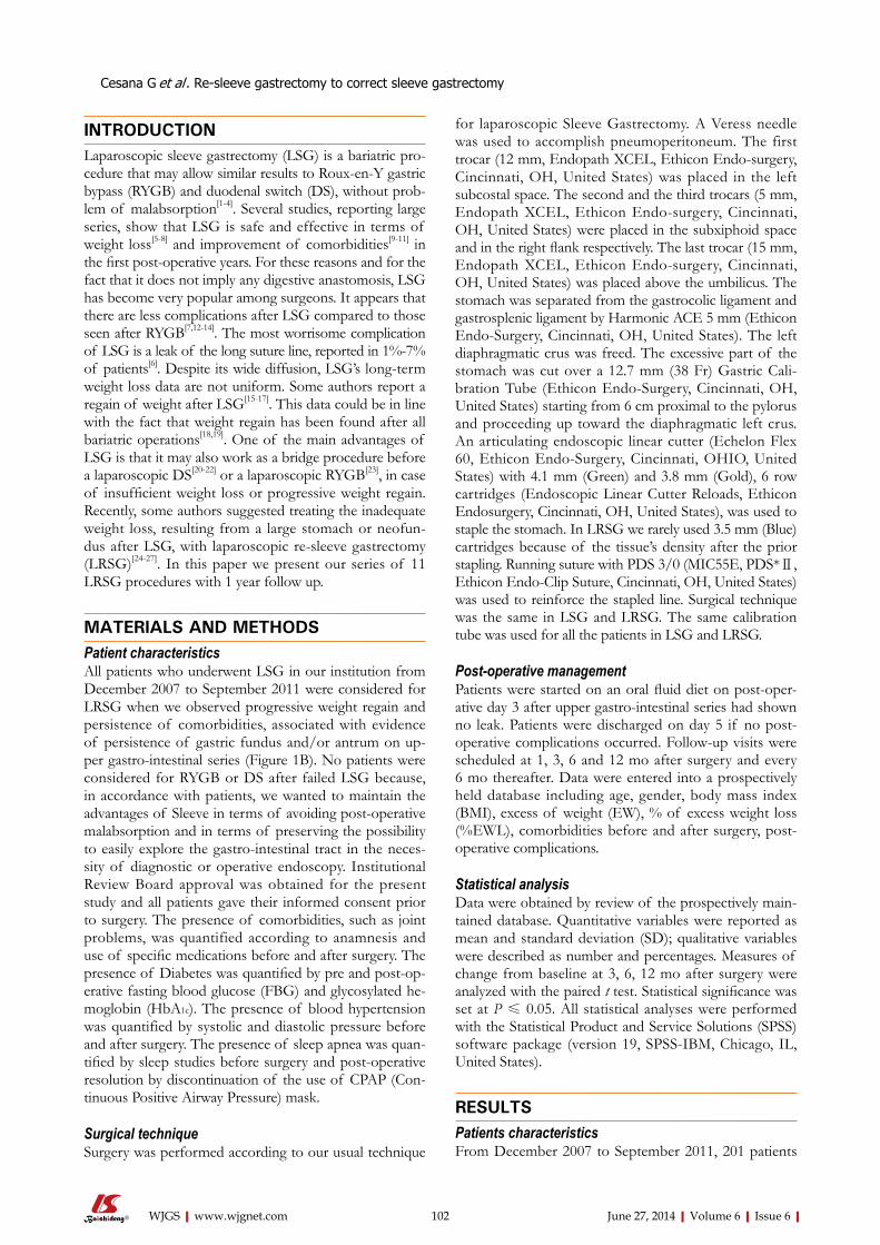
INTRODUCTIONLaparoscopic sleeve gastrectomy (LSG) is a bariatric pro-cedure that may allow similar results to Roux-en-Y gastric bypass (RYGB) and duodenal switch (DS), without prob-lem of malabsorption[1-4]. Several studies, reporting large series, show that LSG is safe and effective in terms of weight loss[5-8] and improvement of comorbidities[9-11] in the first post-operative years. For these reasons and for the fact that it does not imply any digestive anastomosis, LSG has become very popular among surgeons. It appears that there are less complications after LSG compared to those seen after RYGB[7,12-14]. The most worrisome complication of LSG is a leak of the long suture line, reported in 1%-7% of patients[6]. Despite its wide diffusion, LSG’s long-term weight loss data are not uniform. Some authors report a regain of weight after LSG[15-17]. This data could be in line with the fact that weight regain has been found after all bariatric operations[18,19]. One of the main advantages of LSG is that it may also work as a bridge procedure before a laparoscopic DS[20-22] or a laparoscopic RYGB[23], in case of insufficient weight loss or progressive weight regain. Recently, some authors suggested treating the inadequate weight loss, resulting from a large stomach or neofun-dus after LSG, with laparoscopic re-sleeve gastrectomy (LRSG)[24-27]. In this paper we present our series of 11 LRSG procedures with 1 year follow up.
MATERIALS AND METHODSPatient characteristicsAll patients who underwent LSG in our institution from December 2007 to September 2011 were considered for LRSG when we observed progressive weight regain and persistence of comorbidities, associated with evidence of persistence of gastric fundus and/or antrum on up-per gastro-intestinal series (Figure 1B). No patients were considered for RYGB or DS after failed LSG because, in accordance with patients, we wanted to maintain the advantages of Sleeve in terms of avoiding post-operative malabsorption and in terms of preserving the possibility to easily explore the gastro-intestinal tract in the neces-sity of diagnostic or operative endoscopy. Institutional Review Board approval was obtained for the present study and all patients gave their informed consent prior to surgery. The presence of comorbidities, such as joint problems, was quantified according to anamnesis and use of specific medications before and after surgery. The presence of Diabetes was quantified by pre and post-op-erative fasting blood glucose (FBG) and glycosylated he-moglobin (HbA1c). The presence of blood hypertension was quantified by systolic and diastolic pressure before and after surgery. The presence of sleep apnea was quan-tified by sleep studies before surgery and post-operative resolution by discontinuation of the use of CPAP (Con-tinuous Positive Airway Pressure) mask.
Surgical techniqueSurgery was performed according to our usual technique
for laparoscopic Sleeve Gastrectomy. A Veress needle was used to accomplish pneumoperitoneum. The first trocar (12 mm, Endopath XCEL, Ethicon Endo-surgery, Cincinnati, OH, United States) was placed in the left subcostal space. The second and the third trocars (5 mm, Endopath XCEL, Ethicon Endo-surgery, Cincinnati, OH, United States) were placed in the subxiphoid space and in the right flank respectively. The last trocar (15 mm, Endopath XCEL, Ethicon Endo-surgery, Cincinnati, OH, United States) was placed above the umbilicus. The stomach was separated from the gastrocolic ligament and gastrosplenic ligament by Harmonic ACE 5 mm (Ethicon Endo-Surgery, Cincinnati, OH, United States). The left diaphragmatic crus was freed. The excessive part of the stomach was cut over a 12.7 mm (38 Fr) Gastric Cali-bration Tube (Ethicon Endo-Surgery, Cincinnati, OH, United States) starting from 6 cm proximal to the pylorus and proceeding up toward the diaphragmatic left crus. An articulating endoscopic linear cutter (Echelon Flex 60, Ethicon Endo-Surgery, Cincinnati, OHIO, United States) with 4.1 mm (Green) and 3.8 mm (Gold), 6 row cartridges (Endoscopic Linear Cutter Reloads, Ethicon Endosurgery, Cincinnati, OH, United States), was used to staple the stomach. In LRSG we rarely used 3.5 mm (Blue) cartridges because of the tissue’s density after the prior stapling. Running suture with PDS 3/0 (MIC55E, PDS*Ⅱ, Ethicon Endo-Clip Suture, Cincinnati, OH, United States) was used to reinforce the stapled line. Surgical technique was the same in LSG and LRSG. The same calibration tube was used for all the patients in LSG and LRSG.
Post-operative managementPatients were started on an oral fluid diet on post-oper-ative day 3 after upper gastro-intestinal series had shown no leak. Patients were discharged on day 5 if no post-operative complications occurred. Follow-up visits were scheduled at 1, 3, 6 and 12 mo after surgery and every 6 mo thereafter. Data were entered into a prospectively held database including age, gender, body mass index (BMI), excess of weight (EW), % of excess weight loss (%EWL), comorbidities before and after surgery, post-operative complications.
Statistical analysisData were obtained by review of the prospectively main-tained database. Quantitative variables were reported as mean and standard deviation (SD); qualitative variables were described as number and percentages. Measures of change from baseline at 3, 6, 12 mo after surgery were analyzed with the paired t test. Statistical significance was set at P ≤ 0.05. All statistical analyses were performed with the Statistical Product and Service Solutions (SPSS) software package (version 19, SPSS-IBM, Chicago, IL, United States).
RESULTSPatients characteristicsFrom December 2007 to September 2011, 201 patients
June 27, 2014|Volume 6|Issue 6|WJGS|www.wjgnet.com 102
Cesana G et al . Re-sleeve gastrectomy to correct sleeve gastrectomy

underwent LSG at our Institution. We observed progres-sive weight regain in 11 patients (5.4%). Three patients started to regain weight after 6 mo post-LSG, 5 patients after 12 mo, 3 patients after 18 mo. An upper gastro-in-testinal series showed gastric dilatation in all 11 patients. Three patients (27.3%) had another bariatric surgery prior to LSG: 2 patients had an adjustable gastric band (AGB) already removed before LSG and one patient underwent surgical intervention of laparoscopic Band removal and LSG at the same time. The AGB was removed because of dysfunction associated with weight regain.
Four patients (45.5%) were affected by at least 1 comorbidity (Table 1). Two of them (a female with BMI = 54.1 kg/m2 and a male with BMI = 48.5 kg/m2)
were affected by blood hypertension, type II diabetes and joint problems. A third patient, a female with BMI = 52.7 kg/m2, was affected by blood hypertension and joint problems. A fourth patient, a male with BMI = 43.3 kg/m2, was affected by sleep apnea syndrome. In all patients, pre-operative blood hypertension was well con-trolled by drugs (mean systolic 123.3 ± 2.9 mmHg and mean diastolic 78.3 ± 2.9 mmHg). Two patients were in therapy with combination diuretics and ACE inhibitors; one patients with ACE inhibitors alone. Regarding the treatment of diabetes, the two patients affected used oral hypoglycemic agents. The average FBG before surgery was 147.5 ± 3.5 mg/dL and HbA1c averaged 6.9% ± 0.1%.
The mean age of the patients (3 males and 8 females) was 40.6 ± 10.2 years (Table 1).
Findings after LSGBefore LSG, mean absolute weight was 116.4 ± 21.5 kg, mean EW was 59.3 ± 16 kg and mean BMI was 45.2 ± 5.6 kg/m2 (Table 1). One patient developed a high gastric leak after LSG and underwent a second operation six days later. She was a female with BMI = 41 kg/m2 and no comorbidities. She had surgical revision of the gas-tric staple line without resewing it. A perigastric abscess was drained and a drain tube was left in place. The leak resolved in 15 d and the patient was discharged on day 18. BMI and %EWL variations after LSG are collected in Figure 2. After an initial decrease, mean BMI start to increase after 6 mo.
After LSG, systolic and diastolic pressure values did not differ significantly to prior LSG; however a reduction in requirement of antihypertensive drugs was observed. One patient suspended therapy and the others 2 reduced therapy. After LSG, FBG and HbA1c showed an impor-tant decrease (respectively 105.5 ± 28.9 mg/dL and 6.2% ± 0.5%). One of two patients (50%) suspended oral hy-poglycemic agents. Joint problems and sleep apnea syn-drome improved in all (100%).
Findings after LRSGLRSG was performed at a mean interval of 21.1 ± 9.7 mo after LSG. The mean BMI before LRSG was 38.9 ± 3.8 kg/m2 and the mean %EWL was 25.3% ± 14.2% (Figure 2). LRSG was completed laparoscopically in all cases and no intra-operative or post-operative complica-tions occurred. The mean time of intervention was 55.8 ± 29.1 min. The mean operative time for LSG in the same patients was longer: 65.4 ± 17.4 min. This finding could be related to the fact that when we perform LRSG the stomach is already dissected and prepared. We only cut off the exceeding part of the stomach over the boo-gie. No significative blood loss occurred either in LSG or in LRSG. No patient showed leakage from the stapled line at upper gastro-intestinal series scheduled on day 2. All patients resumed an oral liquid diet on day 3 and they were discharged from the hospital on day 5 after LRSG. At 1, 6, 12 mo after LRSG the BMI progressively de-creased and %EWL increased in each patient. As shown
June 27, 2014|Volume 6|Issue 6|WJGS|www.wjgnet.com 103
A BFigure 1 Comparison of upper gastro-intestinal series in the same patient on post-operative day 2 after sleeve gastrectomy (A) and one year later (B). After one year we see a dilated stom-ach (B) that allows weight regain.
Characteristics Value, mean ± SD n = 11
Age (yr) 40.6 ± 10.2 Height (m) 1.60 ± 0.1 Ideal body weight (kg) 57.4 ± 8.9 Excess body weight (kg) 59.3 ± 16 Excess body weight (%) 104.2 ± 25.5 Body mass index (kg/m2) 45.2 ± 5.6 Gender Male 3 (27.3) Female 8 (72.7) Comorbidities At least 1 comorbidity 4 (36.4) Blood hypertension 3 (27.3) Type 2 diabetes mellitus 2 (18.2) Sleep apnea syndrome 1 (9.1) Joint problems 3 (27.3)
Table 1 Pre-operative patients’ characteristics n (%)
Cesana G et al . Re-sleeve gastrectomy to correct sleeve gastrectomy
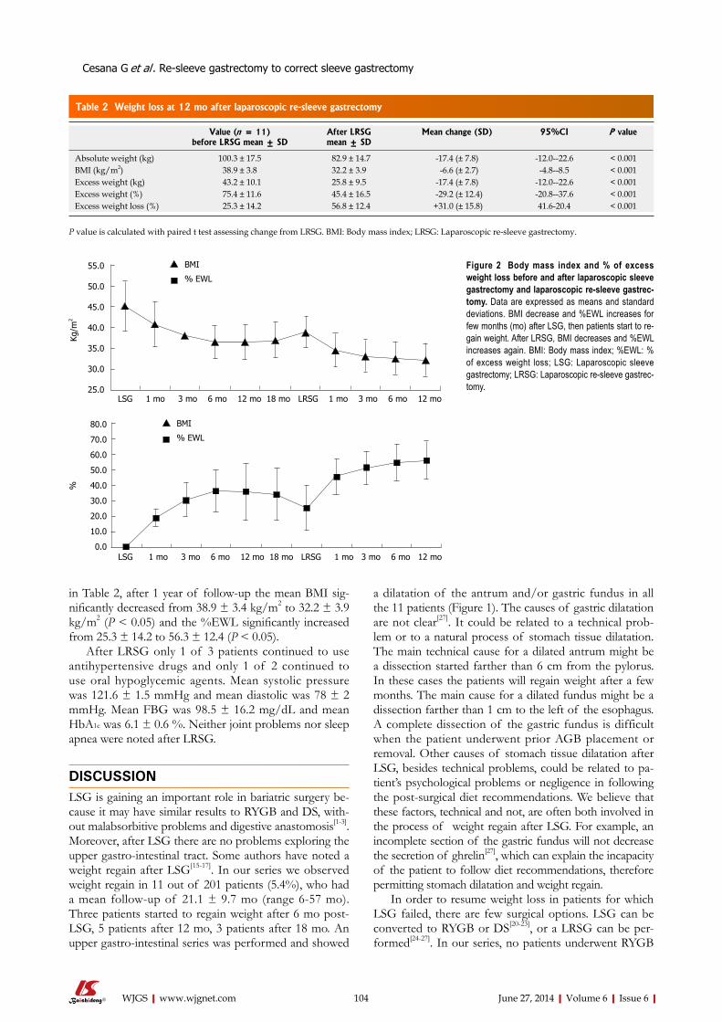
a dilatation of the antrum and/or gastric fundus in all the 11 patients (Figure 1). The causes of gastric dilatation are not clear[27]. It could be related to a technical prob-lem or to a natural process of stomach tissue dilatation. The main technical cause for a dilated antrum might be a dissection started farther than 6 cm from the pylorus. In these cases the patients will regain weight after a few months. The main cause for a dilated fundus might be a dissection farther than 1 cm to the left of the esophagus. A complete dissection of the gastric fundus is difficult when the patient underwent prior AGB placement or removal. Other causes of stomach tissue dilatation after LSG, besides technical problems, could be related to pa-tient’s psychological problems or negligence in following the post-surgical diet recommendations. We believe that these factors, technical and not, are often both involved in the process of weight regain after LSG. For example, an incomplete section of the gastric fundus will not decrease the secretion of ghrelin[27], which can explain the incapacity of the patient to follow diet recommendations, therefore permitting stomach dilatation and weight regain.
In order to resume weight loss in patients for which LSG failed, there are few surgical options. LSG can be converted to RYGB or DS[20-23], or a LRSG can be per-formed[24-27]. In our series, no patients underwent RYGB
in Table 2, after 1 year of follow-up the mean BMI sig-nificantly decreased from 38.9 ± 3.4 kg/m2 to 32.2 ± 3.9 kg/m2 (P < 0.05) and the %EWL significantly increased from 25.3 ± 14.2 to 56.3 ± 12.4 (P < 0.05).
After LRSG only 1 of 3 patients continued to use antihypertensive drugs and only 1 of 2 continued to use oral hypoglycemic agents. Mean systolic pressure was 121.6 ± 1.5 mmHg and mean diastolic was 78 ± 2 mmHg. Mean FBG was 98.5 ± 16.2 mg/dL and mean HbA1c was 6.1 ± 0.6 %. Neither joint problems nor sleep apnea were noted after LRSG.
DISCUSSIONLSG is gaining an important role in bariatric surgery be-cause it may have similar results to RYGB and DS, with-out malabsorbitive problems and digestive anastomosis[1-3]. Moreover, after LSG there are no problems exploring the upper gastro-intestinal tract. Some authors have noted a weight regain after LSG[15-17]. In our series we observed weight regain in 11 out of 201 patients (5.4%), who had a mean follow-up of 21.1 ± 9.7 mo (range 6-57 mo). Three patients started to regain weight after 6 mo post-LSG, 5 patients after 12 mo, 3 patients after 18 mo. An upper gastro-intestinal series was performed and showed
June 27, 2014|Volume 6|Issue 6|WJGS|www.wjgnet.com 104
Value (n = 11)before LRSG mean ± SD
After LRSGmean ± SD
Mean change (SD) 95%CI P value
Absolute weight (kg) 100.3 ± 17.5 82.9 ± 14.7 -17.4 (± 7.8) -12.0--22.6 < 0.001 BMI (kg/m2) 38.9 ± 3.8 32.2 ± 3.9 -6.6 (± 2.7) -4.8--8.5 < 0.001 Excess weight (kg) 43.2 ± 10.1 25.8 ± 9.5 -17.4 (± 7.8) -12.0--22.6 < 0.001 Excess weight (%) 75.4 ± 11.6 45.4 ± 16.5 -29.2 (± 12.4) -20.8--37.6 < 0.001 Excess weight loss (%) 25.3 ± 14.2 56.8 ± 12.4 +31.0 (± 15.8) 41.6-20.4 < 0.001
Table 2 Weight loss at 12 mo after laparoscopic re-sleeve gastrectomy
P value is calculated with paired t test assessing change from LRSG. BMI: Body mass index; LRSG: Laparoscopic re-sleeve gastrectomy.
55.0
50.0
45.0
40.0
35.0
30.0
25.0
Kg/m
2
80.0
70.0
60.0
50.0
40.0
30.0
20.0
10.0
0.0
%
LSG 1 mo 3 mo 6 mo 12 mo 18 mo LRSG 1 mo 3 mo 6 mo 12 mo
LSG 1 mo 3 mo 6 mo 12 mo 18 mo LRSG 1 mo 3 mo 6 mo 12 mo
% EWL
BMI
% EWL
BMI
Figure 2 Body mass index and % of excess weight loss before and after laparoscopic sleeve gastrectomy and laparoscopic re-sleeve gastrec-tomy. Data are expressed as means and standard deviations. BMI decrease and %EWL increases for few months (mo) after LSG, then patients start to re-gain weight. After LRSG, BMI decreases and %EWL increases again. BMI: Body mass index; %EWL: % of excess weight loss; LSG: Laparoscopic sleeve gastrectomy; LRSG: Laparoscopic re-sleeve gastrec-tomy.
Cesana G et al . Re-sleeve gastrectomy to correct sleeve gastrectomy

years, different types of surgery have been developed to resolve this problem when it could not be treated by diets or drugs. Sleeve gastrectomy is an inter-vention that allows for good results in term of weight loss without problems of malabsorption. Sleeve became very popular among surgeons. Despite its wide diffusion, sleeve’s long-term weight loss data are not uniform. Some authors report a regain of weight after Sleeve.Research frontiersTo show that it is feasible and effective to correct weight regain after sleeve through a re-sleeve gastrectomy.Innovations and breakthroughsRe-sleeve gastrectomy can correct weight regain after sleeve. It avoids convert-ing sleeve in a malabsorbitive intervention, loosing the advantages of sleeve. They describe the surgical technique, which is valid and without major compli-cations. ApplicationsSleeve gastrectomy has advantages in terms of quality of life for obese pa-tients, avoiding problems of malabsorption and allowing weight lost. The pos-sibility of managing weight regain with a re-sleeve, without converting it in a malabsorbitive intervention, can allow surgeons to choose this type of surgery in the cure of obesity. TerminologySleeve gastrectomy is a vertical resection of stomach. Re-sleeve gastrectomy is the resection of those parts of stomach that underwent dilatation after sleeve.Peer reviewThis is a case series of laparoscopic re-sleeve gastrectomy in obese patients who showed weight regain after laparoscopic sleeve gastrectomy. The present study is important because there are few data on this type of bariatric surgery.
REFERENCES1 Moy J, Pomp A, Dakin G, Parikh M, Gagner M. Laparoscopic
sleeve gastrectomy for morbid obesity. Am J Surg 2008; 196: e56-e59 [PMID: 18954593 DOI: 10.1016/j.amjsurg.2008.04.008]
2 Nocca D, Krawczykowsky D, Bomans B, Noël P, Picot MC, Blanc PM, de Seguin de Hons C, Millat B, Gagner M, Monni-er L, Fabre JM. A prospective multicenter study of 163 sleeve gastrectomies: results at 1 and 2 years. Obes Surg 2008; 18: 560-565 [PMID: 18317859 DOI: 10.1007/s11695-007-9288-7]
3 Gagner M, Gumbs AA, Milone L, Yung E, Goldenberg L, Pomp A. Laparoscopic sleeve gastrectomy for the super-super-obese (body mass index & gt; 60 kg/m(2)). Surg Today 2008; 38: 399-403 [PMID: 18560961 DOI: 10.1007/s00595-007-3645-y]
4 Fischer L, Hildebrandt C, Bruckner T, Kenngott H, Linke GR, Gehrig T, Büchler MW, Müller-Stich BP. Excessive weight loss after sleeve gastrectomy: a systematic review. Obes Surg 2012; 22: 721-731 [PMID: 22411568 DOI: 10.1007/s11695-012-0616-1]
5 Boza C, Salinas J, Salgado N, Pérez G, Raddatz A, Funke R, Pimentel F, Ibáñez L. Laparoscopic sleeve gastrectomy as a stand-alone procedure for morbid obesity: report of 1,000 cases and 3-year follow-up. Obes Surg 2012; 22: 866-871 [PMID: 22438219 DOI: 10.1007/s11695-012-0591-6]
6 Deitel M, Gagner M, Erickson AL, Crosby RD. Third In-ternational Summit: Current status of sleeve gastrectomy. Surg Obes Relat Dis 2011; 7: 749-759 [PMID: 21945699 DOI: 10.1016/j.soard.2011.07.017]
7 Brethauer SA, Hammel JP, Schauer PR. Systematic review of sleeve gastrectomy as staging and primary bariatric pro-cedure. Surg Obes Relat Dis 2009; 5: 469-475 [PMID: 19632646 DOI: 10.1016/j.soard.2009.05.011]
8 Shi X, Karmali S, Sharma AM, Birch DW. A review of laparoscopic sleeve gastrectomy for morbid obesity. Obes Surg 2010; 20: 1171-1177 [PMID: 20379795 DOI: 10.1007/s11695-010-0145-8]
9 Gill RS, Birch DW, Shi X, Sharma AM, Karmali S. Sleeve gastrectomy and type 2 diabetes mellitus: a systematic re-view. Surg Obes Relat Dis 2010; 6: 707-713 [PMID: 20947447 DOI: 10.1016/j.soard.2010.07.011]
or DS because we wanted to maintain the advantages of LSG in terms of avoiding malabsorption or gastro-intestinal anastomosis, and of preserving the possibility to easily explore the gastro-intestinal tract in the neces-sity of diagnostic or operative endoscopy. Our patients underwent LRSG over the same calibration tube of LSG: 12.7 cm boogie (38 Fr). After LSG, the gastric tis-sue around the stapled line is denser because of the scar’s healing and remodeling. For this reason, we used only Green (4.1 mm) and Gold (3.8 mm) cartridges to dissect the stomach in the LRSG. An oversewing suture of the stapled line was always performed.
LRSG was feasible in all patients in our series. Al-though we had neither intra-operative nor post-operative complications, the main post-operative problem after LRSG may be the same of LSG: leakage from the long stapled line. LRSG was effective for weight loss in all patients (Table 2). After one year follow-up we noted a significant decrease in mean BMI from 38.9 ± 3.4 kg/m2 to 32.2 ± 3.9 kg/m2 (P < 0.05) and a significant increase in mean %EWL from 25.3 ± 14.2 to 56.3 ± 12.4 (P < 0.05) (Figure 2). Although median BMI was > 30 kg/m2 after one year, we noted a significative trend in decreasing weight after LRSG. We need a longer follow-up to deter-mine if the treatment is really effective, but these prelimi-nary data are encouraging in not substituting LSG with a malabsorbitive intervention, loosing the advantages of LSG, especially in terms of quality of life. If weight re-gain will still occur after a longer follow-up post-LRSG, patients will undergo upper gastro-intestinal series. If the series will show a dilatation of the stomach, the patient will undergo another LRSG. If patients will regain weight without signs of stomach dilatation, he will be led to a malabsorbitive intervention.
Comorbidities improved: sleep apneas and joint prob-lems disappeared completely 12 mo after LSG. Blood hypertension values did not differ significantly before surgery, after LSG and after LRSG, but a significant re-duction in the requirement of antihypertensive drugs was observed. FBG and HbA1c gradually decreased after LSG and LRSG. One of two patient suspended oral hypogly-cemic agents.
In conclusion, like the other major bariatric interven-tions[18,19] LSG can result in weight regain. Other bariatric procedures can be performed to correct it[20-27]. In our series of patients who regained weight after LSG, LRSG was performed. The result was a safe procedure which al-lowed a significant weight loss in each patient. LRSG ap-pears to be a valid correction for post-LSG weight regain. Our study is limited by the fact that it is retrospective, involves few patients and has a limited follow-up (12 mo after LRSG). We believe that these preliminary data can be a promising start for further studies, which are needed to confirm the initial results.
COMMENTSBackgroundObesity is an increasing problem in modern western society. In the past few
June 27, 2014|Volume 6|Issue 6|WJGS|www.wjgnet.com 105
COMMENTS
Cesana G et al . Re-sleeve gastrectomy to correct sleeve gastrectomy
19 Roslin M, Damani T, Oren J, Andrews R, Yatco E, Shah P. Abnormal glucose tolerance testing following gastric bypass demonstrates reactive hypoglycemia. Surg Endosc 2011; 25: 1926-1932 [PMID: 21184112 DOI: 10.1007/s00464-010-1489-9]
20 Iannelli A, Dainese R, Piche T, Facchiano E, Gugenheim J. Laparoscopic sleeve gastrectomy for morbid obesity. World J Gastroenterol 2008; 14: 821-827 [PMID: 18240338 DOI: 10.3748/wjg.14.821]
21 Iannelli A, Schneck AS, Dahman M, Negri C, Gugenheim J. Two-step laparoscopic duodenal switch for superobesity: a feasibility study. Surg Endosc 2009; 23: 2385-2389 [PMID: 19263140 DOI: 10.1007/s00464-009-0363-0]
22 Gagner M, Deitel M, Kalberer TL, Erickson AL, Crosby RD. The Second International Consensus Summit for Sleeve Gastrectomy, March 19-21, 2009. Surg Obes Relat Dis 2009; 5: 476-485 [PMID: 19632647 DOI: 10.1016/j.soard.2009.06.001]
23 Regan JP, Inabnet WB, Gagner M, Pomp A. Early experience with two-stage laparoscopic Roux-en-Y gastric bypass as an alternative in the super-super obese patient. Obes Surg 2003; 13: 861-864 [PMID: 14738671 DOI: 10.1381/096089203322618669]
24 Dapri G, Cadière GB, Himpens J. Laparoscopic repeat sleeve gastrectomy versus duodenal switch after isolated sleeve gastrectomy for obesity. Surg Obes Relat Dis 2011; 7: 38-43 [PMID: 21115409 DOI: 10.1016/j.soard.2010.08.005]
25 Iannelli A, Schneck AS, Noel P, Ben Amor I, Krawczykowski D, Gugenheim J. Re-sleeve gastrectomy for failed laparoscopic sleeve gastrectomy: a feasibility study. Obes Surg 2011; 21: 832-835 [PMID: 20924713 DOI: 10.1007/s11695-010-0290-0]
26 Baltasar A, Serra C, Pérez N, Bou R, Bengochea M. Re-sleeve gastrectomy. Obes Surg 2006; 16: 1535-1538 [PMID: 17132421 DOI: 10.1381/096089206778869924]
27 Noel P, Nedelcu M, Nocca D, Schneck AS, Gugenheim J, Iannelli A, Gagner M. Revised sleeve gastrectomy: another option for weight loss failure after sleeve gastrectomy. Surg Endosc 2014; 28: 1096-1102 [PMID: 24170068 DOI: 10.1007/s00464-013-3277-9]
P- Reviewers: Kanda T, Kim HH, Teo M S- Editor: Song XX L- Editor: A E- Editor: Wu HL
10 Kehagias I, Karamanakos SN, Argentou M, Kalfarentzos F. Randomized clinical trial of laparoscopic Roux-en-Y gastric bypass versus laparoscopic sleeve gastrectomy for the manage-ment of patients with BMI & lt; 50 kg/m2. Obes Surg 2011; 21: 1650-1656 [PMID: 21818647 DOI: 10.1007/s11695-011-0479-x]
11 Leyba JL, Aulestia SN, Llopis SN. Laparoscopic Roux-en-Y gastric bypass versus laparoscopic sleeve gastrectomy for the treatment of morbid obesity. A prospective study of 117 patients. Obes Surg 2011; 21: 212-216 [PMID: 20835778 DOI: 10.1007/s11695-010-0279-8]
12 Bennett JM, Mehta S, Rhodes M. Surgery for morbid obe-sity. Postgrad Med J 2007; 83: 8-15 [PMID: 17267672 DOI: 10.1136/pgmj.2006.048868]
13 Frezza EE, Reddy S, Gee LL, Wachtel MS. Complications af-ter sleeve gastrectomy for morbid obesity. Obes Surg 2009; 19: 684-687 [PMID: 18923879 DOI: 10.1007/s11695-008-9677-6]
14 Hallowell PT, Stellato TA, Schuster M, Graf K, Robinson A, Jasper JJ. Avoidance of complications in older patients and Medicare recipients undergoing gastric bypass. Arch Surg 2007; 142: 506-510; discussion 510-512 [PMID: 17576885 DOI: 10.1001/archsurg.142.6.506]
15 Himpens J, Dobbeleir J, Peeters G. Long-term results of lapa-roscopic sleeve gastrectomy for obesity. Ann Surg 2010; 252: 319-324 [PMID: 20622654 DOI: 10.1097/SLA.0b013e3181e90b31]
16 Bohdjalian A, Langer FB, Shakeri-Leidenmühler S, Gfrerer L, Ludvik B, Zacherl J, Prager G. Sleeve gastrectomy as sole and definitive bariatric procedure: 5-year results for weight loss and ghrelin. Obes Surg 2010; 20: 535-540 [PMID: 20094819 DOI: 10.1007/s11695-009-0066-6]
17 Santoro S. Technical aspects in sleeve gastrectomy. Obes Surg 2007; 17: 1534-1535 [PMID: 18219786 DOI: 10.1007/s11695-008-9417-y]
18 Langer FB , Bohdjalian A, Shakeri-Leidenmühler S, Schoppmann SF, Zacherl J, Prager G. Conversion from sleeve gastrectomy to Roux-en-Y gastric bypass--indications and outcome. Obes Surg 2010; 20: 835-840 [PMID: 20393810 DOI: 10.1007/s11695-010-0125-z]
June 27, 2014|Volume 6|Issue 6|WJGS|www.wjgnet.com 106
Cesana G et al . Re-sleeve gastrectomy to correct sleeve gastrectomy
Downstaging and resection after neoadjuvant therapy for fibrolamellar hepatocellular carcinoma
Gilton Marques Fonseca, Antonio Drauzio Varella, Fabricio Ferreira Coelho, Emerson Shigueaki Abe, Rodrigo Blanco Dumarco, Paulo Herman
Gilton Marques Fonseca, Fabricio Ferreira Coelho, Emerson Shigueaki Abe, Rodrigo Blanco Dumarco, Paulo Herman, Digestive Surgery Division, Department of Gastroenterology, University of São Paulo School of Medicine, Avenida Doutor Enéas de Carvalho Aguiar 255, Instituto Central, 05403-900, São Paulo, BrazilAntonio Drauzio Varella, Clinical Oncologist, Hospital Sírio-Libanês, 01308-050, São Paulo, BrazilAuthor contributions: Fonseca GM and Herman P designed the study and wrote the manuscript; Fonseca GM, Coelho FF, Abe ES and Dumarco RB performed revision research; Coelho FF, Abe ES, Dumarco RB and Varella AD reviewed the manuscript.Correspondence to: Gilton Marques Fonseca, MD, Digestive Surgery Division, Department of Gastroenterology, University of São Paulo School of Medicine, Avenida Doutor Enéas de Carvalho Aguiar, 255, Instituto Central, 9° Andar, Sala 9074, 05403-900, São Paulo, Brazil. [email protected]: +55-11-26617560 Fax: +55-11-26617560Received: February 20, 2014 Revised: June 4, 2014Accepted: June 10, 2014Published online: June 27, 2014
AbstractFibrolamellar hepatocellular carcinoma (FLHCC) is a rare malignant liver neoplasm, commonly observed in adolescents and young adults of both genders. The disease is more common in Caucasians and in patients without a prior history of liver disease. The best treat-ment option is a surgical resection associated with liver hilum lymph node dissection. However, there is no established systemic drug treatment for patients with locally advanced or metastatic disease. We report on a patient with advanced FLHCC, initially considered un-resectable due to invasion of the right and the middle hepatic veins and circumferential involvement of the left hepatic vein. Following the treatment with gem-citabine-oxaliplatin systemic chemotherapy, the patient exhibited a significant tumor reduction. As a result, a complete resection was performed with an extended right hepatectomy associated with a partial resection of
the inferior vena cava, a wedge resection in segment 2, and lymphadenectomy of the hepatic hilum. The case was unusual due to the significant tumor downstaging with gemcitabine-oxaliplatin, potentially enabling cura-tive resection. More studies are needed to confirm the efficacy of the systemic drug treatment for FLHCC.
© 2014 Baishideng Publishing Group Inc. All rights reserved.
Key words: Chemotherapy; Gemcitabine; Oxaliplatin; Hepatectomy; Hepatic veins; Fibrolamellar hepatocel-lular carcinoma
Core tip: Fibrolamellar hepatocellular carcinoma (FL-HCC) is a rare malignant liver neoplasm. The best treatment option is a surgical resection with liver hilum lymph node dissection. Currently there is no established systemic drug treatment for patients with locally ad-vanced or metastatic disease. In this report, a patient with advanced FLHCC, initially considered unresect-able due to vascular invasion, exhibited a significant tumor reduction following systemic chemotherapy with gemcitabine-oxaliplatin, allowing resection. This was an unusual case where gemcitabine-oxaliplatin treatment led to a significant tumor downstaging enabling cura-tive resection. Additional studies are needed to confirm the efficacy of the systemic drug treatment for FLHCC.
Fonseca GM, Varella AD, Coelho FF, Abe ES, Dumarco RB, Herman P. Downstaging and resection after neoadjuvant therapy for fibrolamellar hepatocellular carcinoma. World J Gastrointest Surg 2014; 6(6): 107-111 Available from: URL: http://www.wjgnet.com/1948-9366/full/v6/i6/107.htm DOI: http://dx.doi.org/10.4240/wjgs.v6.i6.107
INTRODUCTIONFibrolamellar hepatocellular carcinoma (FLHCC) is
CASE REPORT
Submit a Manuscript: http://www.wjgnet.com/esps/Help Desk: http://www.wjgnet.com/esps/helpdesk.aspxDOI: 10.4240/wjgs.v6.i6.107
World J Gastrointest Surg 2014 June 27; 6(6): 107-111ISSN 1948-9366 (online)
© 2014 Baishideng Publishing Group Inc. All rights reserved.
June 27, 2014|Volume 6|Issue 6|WJGS|www.wjgnet.com 107

an uncommon primary liver neoplasm, representing 0.6%-8.6% of all hepatocellular carcinomas (HCC)[1]. It was first described in 1956 by Edmonson as a rare dis-tinctive form of HCC[2]. In general, it is a vascular tumor with prominent fibrosis. Microscopically, FLHCC ap-pears as a well-differentiated tumor comprised of large polygonal cells with large nuclei and nucleoli, as well as an abundant eosinophilic cytoplasm, arranged in lamellar bands of collagen fibers[3]. FLHCC most often affects adolescents and young adults of both genders, often Caucasian, and without a prior history of liver disease[1,4]. Liver function tests are typically normal or only mildly el-evated. Commonly used HCC markers, such as alpha-fe-toprotein, are of little help in diagnosing and monitoring disease progression in the majority of patients, as only a small proportion of patients (7%-11%) show elevation in alpha-fetoprotein levels[5,6].
FLHCC is believed to more commonly metastasize to regional lymph nodes. The cornerstone for FLHCC treatment is a surgical resection associated with lymph node dissection[3,4]. Patients with advanced FLHCC rep-resent a population in need of novel and effective treat-ments. Due to the lack of data on effective systemic drug treatments as well as the FLHCC patients’ paucity, it is difficult to conduct clinical trials[1]. The authors report an unusual case of a young patient, initially with an unresect-able FLHCC, treated with gemcitabine-oxaliplatin (GE-MOX), resulting in an excellent response and complete resection of the tumor.
CASE REPORTA previously healthy 35-year-old Caucasian female com-plaining of abdominal pain, 3 kg weight loss, weakness, back pain, and a palpable mass in the right upper quad-rant, was referred for evaluation. Physical examination disclosed a palpable hard mass 20 cm below the right costal margin. There were no signs of liver disease or other relevant findings. A computed tomography scan (Figure 1) showed a suggestive FLHCC mass (17 cm × 15 cm) affecting the right liver lobe. In addition, the mass affected segment 4b by obstructing the right por-tal branch, invading both the right and middle hepatic veins, circumferentially wrapping the left hepatic vein, and compressing the inferior vena cava. A lesion (ø 6.5 cm) in segment 2 displayed the same characteristics, as well as lymphadenopathy at the liver hilum up to 2.7 cm in size. Laboratory tests revealed elevated levels of alka-line phosphatase, gamma-glutamyl transpeptidase, and alpha-fetoprotein (44.395 ng/mL). Both hepatitis B and C serologies were negative. Colonoscopy and endoscopy results were normal. Percutaneous biopsy of the tumor confirmed FLHCC. The lesion was considered unre-sectable because of the extensive vascular involvement, especially of the hepatic veins; therefore, the patient was referred to an oncologist for a systemic drug treatment. Transplantation was eliminated due to the presence of the hilar lymph nodes (extra hepatic disease).
The patient received 100 mg of oxaliplatin and 1400
mg of gemcitabine (20 mg/kg) every two weeks during an 11-mo treatment period, with good tolerance. The staging intervals, scheduled every four months, included either a computed tomography scan or magnetic reso-nance imaging. After one year of treatment, magnetic resonance imaging showed significant tumor size reduction (the larger lesion size was 8.5 cm and the smaller 2.2 cm), decrease in contact with the left hepatic vein and inferior vena cava, disappearance of the lymphadenopathy, and hy-pertrophy of the left lateral segment of the liver (38% of total liver volume) (Figure 1). In addition, there was a sig-nificant decrease in alpha-fetoprotein levels to 45 ng/mL.
The patient was submitted to an extended right hepatectomy with partial resection of the inferior vena cava associated with a wedge resection in segment 2 and lymphadenectomy of the hepatic hilum. The procedure lasted 450 min with the patient receiving two units of packed red blood cells and staying in the intensive care unit for two days. The patient developed a postopera-tive biliary fistula, which was treated conservatively, with spontaneous closure after 28 d. Our patient was dis-charged on the 17th postoperative day in a good clinical condition.
Histology examination confirmed FLHCC with mi-croscopically free margins (R0 resection). The hepatic hi-lum lymph nodes were free of the disease. At discharge, the level of alpha-fetoprotein was within normal limits (4.7 ng/mL). The patient had no signs of the disease re-currence at the 14-mo follow-up.
DISCUSSIONThe best treatment option for FLHCC is a resection with adequate lymph node dissection[3,5]. However, a resection is not always possible due to locally advanced disease. The best therapy for these patients has not been well established[1]. Several chemotherapy agents have been used for FLHCC treatment, including fluoropyrimidines, doxorubicin, cisplatin, oxaliplatin, gemcitabine, and irino-tecan, as well as interferon, bevacizumab, and sorafenib in either combination regimens or separately[1,5]. Locore-gional therapies, such as transarterial chemoembolization, radiofrequency ablation, external beam radiation, percu-taneous ethanol injection, and hepatic arterial infusion of cisplatin, had disappointing results[1,5]. A study from the Fibrolamellar Carcinoma Consortium, which contained 99 patients diagnosed with FLHCC, showed that from the 73 patients who underwent surgery, 13% (10/73) received preoperative chemotherapy, external beam radia-tion, and/or transarterial chemoembolization. Twenty one percent (20/99) were considered to have unresect-able disease and 13 of them were treated with various combinations of systemic drug therapy with or without locoregional therapies. Chemotherapy agents used in the study included fluoropyrimidines, doxorubicin, cisplatin, oxaliplatin, gemcitabine, and irinotecan. However, a mul-tivariate analysis showed a lack of surgery to be an inde-pendently poor overall survival predictor[1]. Kaseb et al[7]
studied 94 FLHCC patients and found tumor resection
June 27, 2014|Volume 6|Issue 6|WJGS|www.wjgnet.com 108
Fonseca GM et al . Fibrolamellar hepatocellular carcinoma downstaging
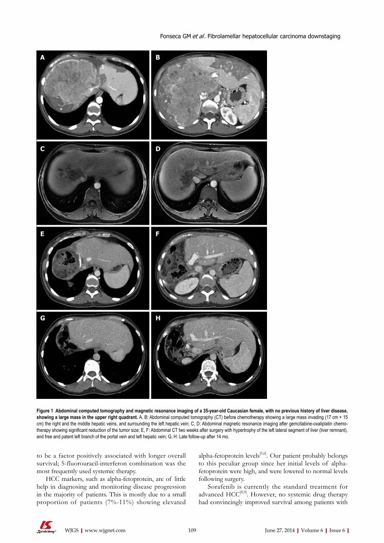
to be a factor positively associated with longer overall survival; 5-fluorouracil-interferon combination was the most frequently used systemic therapy.
HCC markers, such as alpha-fetoprotein, are of little help in diagnosing and monitoring disease progression in the majority of patients. This is mostly due to a small proportion of patients (7%-11%) showing elevated
alpha-fetoprotein levels[5,6]. Our patient probably belongs to this peculiar group since her initial levels of alpha-fetoprotein were high, and were lowered to normal levels following surgery.
Sorafenib is currently the standard treatment for advanced HCC[8,9]. However, no systemic drug therapy had convincingly improved survival among patients with
June 27, 2014|Volume 6|Issue 6|WJGS|www.wjgnet.com 109
A B
C D
E F
G H
Figure 1 Abdominal computed tomography and magnetic resonance imaging of a 35-year-old Caucasian female, with no previous history of liver disease, showing a large mass in the upper right quadrant. A, B: Abdominal computed tomography (CT) before chemotherapy showing a large mass invading (17 cm × 15 cm) the right and the middle hepatic veins, and surrounding the left hepatic vein; C, D: Abdominal magnetic resonance imaging after gemcitabine-oxaliplatin chemo-therapy showing significant reduction of the tumor size; E, F: Abdominal CT two weeks after surgery with hypertrophy of the left lateral segment of liver (liver remnant), and free and patent left branch of the portal vein and left hepatic vein; G, H: Late follow-up after 14 mo.
Fonseca GM et al . Fibrolamellar hepatocellular carcinoma downstaging
segment 4b, obstructing the right portal branch, invading the right and middle hepatic veins, circumferentially wrapping the left hepatic vein, and compressing the inferior vena cava; the second lesion (6.5 cm) in segment 2 had the same characteristics and lymphadenopathy at the liver hilum (up to 2.7 cm), with colonoscopy and endoscopy results being normal.Pathological diagnosisFLHCC was confirmed by percutaneous biopsy of the tumor.TreatmentThe patient received 1400 mg of gemcitabine (20 mg/kg) and 100 mg of ox-aliplatin (GEMOX) every 2 wk over 11 mo, with a significant tumor reduction, allowing for resection.Related reportsThe best treatment option for FLHCC is resection with adequate lymph node dissection. However, resection is not always possible due to local disease advancement. There has not been well-established disease therapy for these patients. Term explanation FLHCC is an uncommon primary liver neoplasm. In general, it is a vascular tumor with prominent fibrosis. Microscopically, FLHCC appears as a well-differ-entiated tumor comprised of large polygonal cells with large nuclei and nucleoli, as well as an abundant eosinophilic cytoplasm, arranged in lamellar bands of collagen fibers.Experiences and lessonsIn the present case, the chemotherapy regimen with GEMOX promoted FLHCC downstaging, potentially allowing for curative treatment and suggesting GE-MOX as a possible chemotherapy treatment for patients with advanced FLHCC; however, more studies are needed to confirm GEMOX efficacy.Peer reviewThis is an interesting case of a young female with an initially considered unre-sectable FLHCC. Significant tumor downstaging with GEMOX allowed surgical resection.
REFERENCES1 Ang CS, Kelley RK, Choti MA, Cosgrove DP, Chou JF,
Klimstra D, Torbenson MS, Ferrell L, Pawlik TM, Fong Y, O’Reilly EM, Ma J, McGuire J, Vallarapu GP, Griffin A, Stipa F, Capanu M, Dematteo RP, Venook AP, Abou-Alfa GK. Clinicopathologic characteristics and survival outcomes of patients with fibrolamellar carcinoma: data from the fibrola-mellar carcinoma consortium. Gastrointest Cancer Res 2013; 6: 3-9 [PMID: 23505572]
2 Edmondson HA. Differential diagnosis of tumors and tumor-like lesions of liver in infancy and childhood. AMA J Dis Child 1956; 91: 168-186 [PMID: 13282629]
3 Mavros MN, Mayo SC, Hyder O, Pawlik TM. A systematic review: treatment and prognosis of patients with fibrola-mellar hepatocellular carcinoma. J Am Coll Surg 2012; 215: 820-830 [PMID: 22981432 DOI: 10.1016/j.jamcollsurg.2012.08.001]
4 Mayo SC, Mavros MN, Nathan H, Cosgrove D, Herman JM, Kamel I, Anders RA, Pawlik TM. Treatment and prognosis of patients with fibrolamellar hepatocellular carcinoma: a national perspective. J Am Coll Surg 2014; 218: 196-205 [PMID: 24315886 DOI: 10.1016/j.jamcollsurg.2013.10.011]
5 Liu S, Chan KW, Wang B, Qiao L. Fibrolamellar hepatocel-lular carcinoma. Am J Gastroenterol 2009; 104: 2617-2624; quiz 2625 [PMID: 19638962 DOI: 10.1038/ajg.2009.440]
6 Stipa F, Yoon SS, Liau KH, Fong Y, Jarnagin WR, D’Angelica M, Abou-Alfa G, Blumgart LH, DeMatteo RP. Outcome of pa-tients with fibrolamellar hepatocellular carcinoma. Cancer 2006; 106: 1331-1338 [PMID: 16475212 DOI: 10.1002/cncr.21703]
7 Kaseb AO, Shama M, Sahin IH, Nooka A, Hassabo HM, Vauthey JN, Aloia T, Abbruzzese JL, Subbiah IM, Janku F, Curley S, Hassan MM. Prognostic indicators and treat-ment outcome in 94 cases of fibrolamellar hepatocellular carcinoma. Oncology 2013; 85: 197-203 [PMID: 24051705 DOI: 10.1159/000354698]
8 Cheng AL, Kang YK, Chen Z, Tsao CJ, Qin S, Kim JS, Luo R, Feng J, Ye S, Yang TS, Xu J, Sun Y, Liang H, Liu J, Wang
advanced HCC. There have been attempts to evaluate the efficacy of new drugs or combination treatments in clinical trials. Some of the examples of these trials include: (1) doxorubicin alone and/or with gemcitabine; (2) combination of cisplatin, doxorubicin, 5-fluorouracil, and alpha-interferon; and (3) combination of irinotecan, taxanes, gemcitabine, topotecan, and thymidilate synthase inhibitors[10]. The GEMOX regimen appeared to be the most promising, based on a lack of renal and hepatic toxicity in cirrhotic patients, promising efficacy in the phase II trials for advanced HCC, and with possible ex-tended benefit for Child B cirrhosis[10-13]. Louafi et al[12], studying 32 patients treated for advanced HCC, had two patients that underwent HCC curative resection after par-tial response to GEMOX. In a retrospective multicenter study, Zaanan et al[10] observed tumor responses in 204 patients with advanced HCC treated with GEMOX. In 10 patients, tumor response either permitted secondary surgical resection of residual tumors or orthotopic liver transplantation. Radiofrequency ablation was performed in one patient, transcatheter arterial chemoembolization in three patients, cyberknife treatment in one patient, and radioembolization in two patients. There was also a case report on a patient with metastatic HCC without liver disease with a complete response after 12 cycles of GE-MOX[14].
There has been only one reported case of FLHCC with a complete response after GEMOX treatment. In this case, a young woman had a histologically proven metastatic lymph node relapse after resection of the primary tumor. GEMOX regimen lead to a complete response without relapse five years after chemotherapy discontinuation[15].
HCC patients have poor tolerance to systemic che-motherapy, mostly due to cirrhotic liver[15]. In most cases FLHCC patients do not have a prior history of liver disease[4]. This may explain the better tolerance to che-motherapy, hence leading to a better response, as is the case in our patient. In the present case, the chemotherapy regimen with GEMOX promoted FLHCC downstaging, potentially allowing for a curative treatment. However, more studies are needed to confirm GEMOX efficacy.
COMMENTSCase characteristicsA previously healthy 35-year-old Caucasian female complained of abdominal pain, 3 kg weight loss, weakness, back pain, and a palpable mass in the right upper quadrant.Clinical diagnosisA palpable hard mass was found to be located 20 cm below the right costal margin with no signs of liver disease.Differential diagnosisHepatocellular carcinoma, cholangiocarcinoma, liver metastasis.Laboratory diagnosisThe patient had elevated levels of alkaline phosphatase, gamma-glutamyl transpeptidase, and alpha-fetoprotein, and negative serologies for both hepati-tis B and C.Imaging diagnosisA computed tomography scan showed a suggestive fibrolamellar hepatocellular carcinoma (FLHCC) mass (17 cm × 15 cm) affecting the right liver lobe and
June 27, 2014|Volume 6|Issue 6|WJGS|www.wjgnet.com 110
COMMENTS
Fonseca GM et al . Fibrolamellar hepatocellular carcinoma downstaging
12 Louafi S, Boige V, Ducreux M, Bonyhay L, Mansourbakht T, de Baere T, Asnacios A, Hannoun L, Poynard T, Taïeb J. Gemcitabine plus oxaliplatin (GEMOX) in patients with ad-vanced hepatocellular carcinoma (HCC): results of a phase II study. Cancer 2007; 109: 1384-1390 [PMID: 17330837 DOI: 10.1002/cncr.22532]
13 Dhooge M, Coriat R, Mir O, Perkins G, Brezault C, Boudou-Rouquette P, Goldwasser F, Chaussade S. Feasibility of gemcitabine plus oxaliplatin in advanced hepatocellular car-cinoma patients with Child-Pugh B cirrhosis. Oncology 2013; 84: 32-38 [PMID: 23076239 DOI: 10.1159/000342763]
14 Boschetti G, Walter T, Hervieu V, Cassier P, Lombard-Bohas C, Adham M, Scoazec JY, Dumortier J. Complete response of hepatocellular carcinoma with systemic combination chemo-therapy: not to get out the chemotherapy? Eur J Gastroenterol Hepatol 2010; 22: 1015-1018 [PMID: 20075738 DOI: 10.1097/MEG.0b013e328336565a]
15 Gras P, Truant S, Boige V, Ladrat L, Rougier P, Pruvot FR, Hebbar M. Prolonged Complete Response after GEMOX Chemotherapy in a Patient with Advanced Fibrolamellar Hepatocellular Carcinoma. Case Rep Oncol 2012; 5: 169-172 [PMID: 22666208 DOI: 10.1159/000338242]
P- Reviewers: Boeck S, Ghinolfi D S- Editor: Song XX L- Editor: A E- Editor: Wu HL
J, Tak WY, Pan H, Burock K, Zou J, Voliotis D, Guan Z. Ef-ficacy and safety of sorafenib in patients in the Asia-Pacific region with advanced hepatocellular carcinoma: a phase III randomised, double-blind, placebo-controlled trial. Lancet Oncol 2009; 10: 25-34 [PMID: 19095497 DOI: 10.1016/S1470-2045(08)70285-7]
9 Trevisani F, Frigerio M, Santi V, Grignaschi A, Bernardi M. Hepatocellular carcinoma in non-cirrhotic liver: a reap-praisal. Dig Liver Dis 2010; 42: 341-347 [PMID: 19828388 DOI: 10.1016/j.dld.2009.09.002]
10 Zaanan A, Williet N, Hebbar M, Dabakuyo TS, Fartoux L, Mansourbakht T, Dubreuil O, Rosmorduc O, Cattan S, Bonnetain F, Boige V, Taïeb J. Gemcitabine plus oxaliplatin in advanced hepatocellular carcinoma: a large multicenter AGEO study. J Hepatol 2013; 58: 81-88 [PMID: 22989572 DOI: 10.1016/j.jhep.2012.09.006]
11 Taïeb J, Bonyhay L, Golli L, Ducreux M, Boleslawski E, Tigaud JM, de Baere T, Mansourbakht T, Delgado MA, Han-noun L, Poynard T, Boige V. Gemcitabine plus oxaliplatin for patients with advanced hepatocellular carcinoma using two different schedules. Cancer 2003; 98: 2664-2670 [PMID: 14669287 DOI: 10.1002/cncr.11869]
June 27, 2014|Volume 6|Issue 6|WJGS|www.wjgnet.com 111
Fonseca GM et al . Fibrolamellar hepatocellular carcinoma downstaging
Intra-abdominal esophageal duplication cyst: A case report and review of the literature
Petrus Sebastianus Simon Castelijns, Karlijn Woensdregt, Brigiet Hoevenaars, Gelde Arie Pieter Nieuwenhuijzen
Petrus Sebastianus Simon Castelijns, Karlijn Woensdregt, Gelde Arie Pieter Nieuwenhuijzen, Department of Surgery, Catharina Hospital Eindhoven, 5623 EJ, Eindhoven, The Nether-landsBrigiet Hoevenaars, Department of Pathology, Catharina Hospi-tal Eindhoven, 5623 EJ, Eindhoven, The NetherlandsAuthor contributions: Nieuwenhuijzen GAP performed the sur-gical operation and revised the paper; Hoevenaars B performed pathological examinations; Castelijns PSS and Woensdregt K wrote the paper.Correspondence to: Karlijn Woensdregt, MD, Department of Surgery, Catharina Hospital Eindhoven, Michelangelolaan 2, 5623 EJ, Eindhoven, The Netherlands. [email protected]: +31-40-2399111 Fax: +31-40-2443370Received: October 26, 2013 Revised: April 17, 2014 Accepted: June 10, 2014Published online: June 27, 2014
AbstractIntra-abdominal esophageal duplications are rare en-tities in adults. They are mostly asymptomatic, but since they can lead to complications surgical excision is advised for all duplication cysts. We present a case of a 20-year-old male with colic-like abdominal pain, mimicking symptoms of cholecystolithiasis. However after cholecystectomy the symptoms were still present. A computed tomography-scan of the abdomen and an endoscopic ultrasound revealed a cyst of the esophagus of 3.0 cm × 2.3 cm in size. Diagnostic laparoscopy was planned, during which we observed a para-esophageal cyst at the gastro-esophageal junction. Laparoscopic excision of this cyst was performed. Pathophysiological examination revealed an esophageal duplication cyst. We report a rare case of a symptomatic intra-abdominal esophageal duplication cyst in an adult. One must con-sider this diagnosis when more common diagnoses to account for the patient’s symptoms are excluded. Re-moval of duplication cysts can be done laparoscopically.
© 2014 Baishideng Publishing Group Inc. All rights reserved.
Key words: Esophageal duplication cyst; Intra-abdomi-nal; Symptomatic; Laparoscopy
Core tip: Intra-abdominal esophageal duplication cysts are rare entities in adults, especially when they are symptomatic. Since these anomalies can lead to com-plications surgical excision is advised for all of them. We present a case of a 20-year-old male with colic-like abdominal pain, mimicking symptoms of cholecystolithi-asis, but caused by an intra-abdominal duplication cyst of the esophagus. The cyst was excised laparoscopi-cally. The procedure was uneventful and the patient was free of symptoms. This case shows that one must consider the diagnosis of a symptomatic intra-abdom-inal esophageal duplication cyst when more common diagnoses to account for the patient’s symptoms are excluded.
Castelijns PSS, Woensdregt K, Hoevenaars B, Nieuwenhui-jzen GAP. Intra-abdominal esophageal duplication cyst: A case report and review of the literature. World J Gastrointest Surg 2014; 6(6): 112-116 Available from: URL: http://www.wjgnet.com/1948-9366/full/v6/i6/112.htm DOI: http://dx.doi.org/10.4240/wjgs.v6.i6.112
INTRODUCTIONGastroenteric duplication cysts are congenital malfor-mations that may affect the gastro-intestinal tract from mouth to anus. They are usually attached to the gastro-intestinal tract, have smooth muscle cells in the wall of the cyst and are lined with gastrointestinal epithelium. Most of them are found in the small intestine (44%), the esophagus (20%) or in the large intestine (15%)[1]. Esoph-ageal duplication cysts mostly arise in the mediastinum.
CASE REPORT
Submit a Manuscript: http://www.wjgnet.com/esps/Help Desk: http://www.wjgnet.com/esps/helpdesk.aspxDOI: 10.4240/wjgs.v6.i6.112
World J Gastrointest Surg 2014 June 27; 6(6): 112-116ISSN 1948-9366 (online)
© 2014 Baishideng Publishing Group Inc. All rights reserved.
June 27, 2014|Volume 6|Issue 6|WJGS|www.wjgnet.com 112

There are only a few case reports that describe intra-abdominal esophageal duplication.
Duplication cysts may be symptomatic, but even then definitive pre-operative diagnosis can be difficult because of non-specific clinical symptoms. Also, symptoms might be mistaken for other more common abdominal condi-tions. Because of potential complications such as bleed-ing, infection or conversion to a malignancy, currently the most common treatment of these anomalies is surgical excision, even if they are not symptomatic.
CASE REPORTWe present a 20-year-old male with no medical or surgi-cal history who was evaluated because of nausea and recurrent colic pain in the right upper abdomen radiating towards his back, but no vomiting or fever. Ultrasound of the upper abdomen was performed, which showed gallstones but no other abnormalities. Initially, symp-tomatic cholecystolithiasis was considered as the primary diagnosis and subsequently our patient underwent a lapa-roscopic cholecystectomy. The procedure was performed without complications and the patient was initially free from symptoms.
However, one year later he presented again with the same symptoms as before. Physical examination and lab-oratory testing did not reveal any abnormalities. The dif-ferential diagnosis at this point was choledocholithiasis or urolithiasis. Ultrasound of the abdomen showed no dila-
tation of intra- or extrahepatic bile ducts or choledocho-lithiasis. A computed tomography (CT)-scan of the ab-domen revealed no urolithiasis, but it did show a mass at the gastro-esophageal junction that was further evaluated with endoscopic ultrasonography (EUS). EUS identified a 3.2 cm × 2.8 cm sized smooth lined lesion (Figure 1) in the distal esophagus just below the diaphragm (Figure 2) We could not differentiate between a gastrointestinal cyst or a leiomyoma, necessitating further analysis.
Endoscopic fine needle aspiration revealed mucinous material without any signs of malignant cells on histo-pathological examination. Gastro-intestinal duplication cyst was considered as one of the possible diagnoses and diagnostic laparoscopy was planned. During laparoscopic exploration we observed a 3 cm × 3 cm sized para-esophageal cystic lesion at the gastro-esophageal junc-tion, 2-3 cm above the Z-line (Figure 3). The cyst was dissected free and the connection with the oesophagus was excised with an endoGIA© after which the staple line was oversewn with a V-loc© suture. The procedure went uneventful.
Histopathological analysis of the specimen revealed a 30 mm × 30 mm × 25 mm sized cyst filled with clear fluid. The wall had a thickness of 3 mm and consisted of two muscular layers lined by respiratory columnar epithelium without any signs of cartilage tissue found in the cyst (typical for bronchogenic cysts) and therefore the final diagnosis of esophageal duplication cyst was sug-gested (Figure 4).
The postoperative course was uncomplicated and the patient could be discharged on day two postoperatively. At follow up 5 mo later the patient did not display any of the earlier experienced abdominal symptoms.
DISCUSSIONGastrointestinal duplication cysts are very rare abnormali-ties. Autopsy studies have shown a prevalence of approx-imately 1:4500 and esophageal duplications occur even less frequently, 1:8200[2,3]. Only a few cases are described in which the cyst is connected to the intra-abdominal part of the esophagus. Only ten percent of the esophageal cysts communicate with the lumen of the esophagus[4].
June 27, 2014|Volume 6|Issue 6|WJGS|www.wjgnet.com 113
Castelijns PSS et al . Symptomatic intra-abdominal esophageal duplication cyst
A B
Figure 1 Computed tomography images of the esophageal duplication cyst (a). A: Frontal view; B: Sagittal view.
0
1
2
3
4
Figure 2 Endoscopic ultrasound image of the cyst.
a b
Figure 3 Preoperative image with the cyst (a) medial to the esophagus and stomach (b).
a a
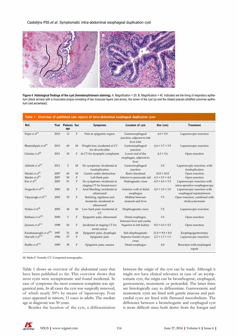
Table 1 shows an overview of the abdominal cases that have been published so far. This overview shows that most cysts were asymptomatic and found incidental. In case of symptoms the most common complaint was epi-gastrical pain. In all cases the cyst was surgically removed, of which nearly 50% by means of laparoscopy. Four cases appeared in minors, 11 cases in adults. The median age at diagnosis was 50 years.
Besides the location of the cyst, a differentiation
between the origin of the cyst can be made. Although it might not have clinical relevance in case of an asymp-tomatic cyst, the origin can be bronchogenic, esophageal, gastroenteric, neurenteric or pericardial. The latter three are histologically easy to differentiate. Gastroenteric and neurenteric cysts are lined with gastric mucosa and peri-cardial cysts are lined with flattened mesothelium. The difference between a bronchogenic and esophageal cyst is more difficult since both derive from the foregut and
June 27, 2014|Volume 6|Issue 6|WJGS|www.wjgnet.com 114
A B
aa
Figure 4 Histological findings of the cyst (hematoxylin/eosin staining). A: Magnification × 20; B: Magnification × 40. Indicated are the lining of respiratory epithe-lium (black arrows) with a muscularis propia consisting of two muscular layers (red arrow), the lumen of the cyst (a) and the ciliated pseudo stratified columnar epithe-lium (red arrowhead).
Ref. Year Patients age
Sex Symptoms Location of cyst Size (cm) Treatment
Pujar et al[1] 2013 13 F Pain in epigastric region Gastroesophageal junction, adjacent to left
liver lobe
4.0 × 5.0 Laparoscopic resection
Bhamidipati et al[7] 2013 69 M Weight loss, incidental at CT for diverticulitis
Gastroesophageal junction
4.4 × 3.7 × 3.9 Laparoscopic resection
Gümüş et al[8] 2011 18 F At CT for dyspeptic complaints Lower end of the esophagus, adjecent to
liver
4.2 × 3.6 Open resection
Aldrink et al[2] 2011 2 M No symptoms. Incidental at fundoplication
Gastroesophageal junction
3.0 Laparoscopic resection, with fundoplication
Martin et al[5] 2007 60 M Gastric outlet obstruction Retro-duodenal 10.0 × 10.0 Open resection Martin et al[5] 2007 50 F Left flank pain Inferior to pancreatic tail 6.5 × 5.5 × 4.2 Open resection Kin et al[9] 2003 51 F No symptoms: incidental at
staging CT for breastcancerDiafragmatic crura 4.5 × 4.0 × 3.5 Laparoscopic resection with
intra-operative esophagoscopy Noguchi et al[10] 2003 26 F Anal bleeding: incidental at
ultrasoundAnterior wall of distal
esophagus4.0 × 3.0 × 3.0 Laparoscopic resection with
esophageal repair(nissen) Vijayaragh et al[11] 2002 70 F Retching, rigidness and
headache, incidental at ultrasound
Midline between stomach and liver
7.5 Open resection, combined with cholecystectomie
Nelms et al[4] 2002 44 M Low back pain: incidental at CT
Diaphragmatic crura 7.0 Laparoscopic resection
Rathaus et al[12] 2000 5 F Epigastric pain, ultrasound Distal esophagus, between liver and cardia
1.0 Open resection
Janssen et al[13] 1998 56 F Incidental at staging CT for rectal cancer
Superior to left kidney 8.0 × 6.0 × 4.5 Open resection
Karahasanoglu et al[14] 1997 51 M Epigastric pain, dysphagia Sub-diaphragmatic 11.0 × 9.0 × 8.0 Esophagogastrectomy Harvell et al[15] 1996 57 F Epigastric pain Superior border of pan-
creas2.2 × 1.7 × 1.5 Laparoscopic resection
Ruffin et al[16] 1989 38 F Epigastric pain, nausea Distal esophagus 4.0 Resection with esophageal repair
Table 1 Overview of published case reports of intra-abdominal esophageal duplication cysts
M: Male; F: Female; CT: Computed tomography.
Castelijns PSS et al . Symptomatic intra-abdominal esophageal duplication cyst
mm esophageal duplication cyst filled with clear fluid and a double muscular layer lined with respiratory columnar epithelium without any signs of cartilage.TreatmentLaparoscopic excision of this cyst was performed.Related reportsAn esophageal duplication cyst is a rare diagnosis and is often asymptomatic.Term explanation An EUS is an endoscopic ultrasonography, which is an ultrasound made through the esophagus.Experiences and lessonsAlthough these cysts are very rare and mostly asymptomatic, they can mimic symptoms of common gastro-intestinal conditions. Therefore, one should consider these anomalies as a possible diagnosis if more obvious causes are excluded. Peer reviewThe paper describes a rare case of a symptomatic intra-abdominal esophageal duplication cyst and gives a complete overview of published cases until now. This case is very interesting for gastrointestinal surgeons. The paper is well written and concise.
REFERENCES1 Pujar VC, Kurbet S, Kaltari DK. Laparoscopic excision of
intra-abdominal oesophageal duplication cyst in a child. J Minim Access Surg 2013; 9: 34-36 [PMID: 23626419 DOI: 10.4103/0972-9941.107137]
2 Aldrink JH, Kenney BD. Laparoscopic excision of an esophageal duplication cyst. Surg Laparosc Endosc Percutan Tech 2011; 21: e280-e283 [PMID: 22002296 DOI: 10.1097/SLE.0b013e31822f1e67]
3 Kim YW, Sohn TI, Shim HS, Kim CB. Intra-abdominal esophageal duplication cyst in an adult. Yonsei Med J 2005; 46: 859-861 [PMID: 16385665 DOI: 10.3349/ymj.2005.46.6.859]
4 Nelms CD, White R, Matthews BD, Ballinger WE, Sing RF, Heniford BT. Thoracoabdominal esophageal duplication cyst. J Am Coll Surg 2002; 194: 674-675 [PMID: 12022610 DOI: 10.1016/S1072-7515(02)01164-X]
5 Martin ND, Kim JC, Verma SK, Rubin R, Mitchell DG, Bergin D, Yeo CJ. Intra-abdominal esophageal duplication cysts: a review. J Gastrointest Surg 2007; 11: 773-777 [PMID: 17562119 DOI: 10.1007/s11605-007-0108-0]
6 Tapia RH, White VA. Squamous cell carcinoma arising in a duplication cyst of the esophagus. Am J Gastroenterol 1985; 80: 325-329 [PMID: 3922217]
7 Bhamidipati C, Smeds M, Dexter E, Kowalski M, Bazaz S. Laparoscopic excision of gastric mass yields intra-abdominal esophageal duplication cyst. Thorac Cardiovasc Surg 2013; 61: 502-504 [PMID: 23171952 DOI: 10.1055/s-0032-1322617]
8 Gümüş M, Önder A, Firat U, Kapan M, Önder H, Gırgın S. Hydatid cyst-like intra-abdominal esophageal duplication cyst in an endemic region. Turk J Gastroenterol 2011; 22: 557-558 [PMID: 22234770]
9 Kin K, Iwase K, Higaki J, Yoon HE, Mikata S, Miyazaki M, Imakita M, Kamiike W. Laparoscopic resection of intra-abdominal esophageal duplication cyst. Surg Laparosc Endosc Percutan Tech 2003; 13: 208-211 [PMID: 12819507 DOI: 10.1097/00129689-200306000-00013]
10 Noguchi T, Hashimoto T, Takeno S, Wada S, Tohara K, Uchida Y. Laparoscopic resection of esophageal duplica-tion cyst in an adult. Dis Esophagus 2003; 16: 148-150 [PMID: 12823217 DOI: 10.1046/j.1442-2050.2003.00314.x]
11 Vijayaraghavan R, Belagavi CS. True giant intra-abdominal esophageal cyst. Indian J Gastroenterol 2002; 21: 198-199 [PMID: 12416752]
12 Rathaus V, Feinberg MS. Subdiaphragmatic esophageal du-plication cyst in a child. J Clin Ultrasound 2000; 28: 264 [PMID: 10800007 DOI: 10.1002/(SICI)1097-0096(200006)28:5<264: AID-JCU10>3.0.CO; 2-O]
13 Janssen H, Fiedler PN. Isolated intraabdominal esophageal
contain ciliated epithelium. To differentiate bronchogenic from esophageal duplication cysts, Palmer’s pathologic criteria are useful. These criteria include: (1) attachment to the esophageal wall; (2) presence of gastrointestinal tract epithelium; and (3) presence of two layers of mus-cularis propria[5]. Duplication cysts of bronchogenic origin do not have these two layers of smooth muscle, in-stead they contain cartilage, bronchogenic glands or both.
In intra-abdominal esophageal duplication cysts Palm-er’s criteria are not always applicable[4]. For example, in 2007 Martin et al[5] presented two cases of isolated intra-abdominal esophageal duplication cyst with no connec-tion to any luminal organ.
The majority (80%) of esophageal duplication cysts becomes symptomatic during childhood and will be re-moved for that reason[1]. As a consequence these types of cysts are rarely seen in adults. If they are found during adulthood, they are mostly asymptomatic and most case reports are therefore based on an incidental finding dur-ing some kind of routine radiological investigation. How-ever cysts can become symptomatic, like in our patient, causing abdominal pain, vomiting, dysphagia or reflux[1,2]. The growing cyst may also cause some mechanical prob-lems of the passage of solid food and fluids because of compression of the esophagus[2]. Finally these cysts can perforate, they can cause an upper gastrointestinal bleed-ing or can get infected. Although a causal relationship is difficult to define, one case report has described a patient with a duplication cyst with an associated intracystic ma-lignancy[6]. Because of these complications it is recom-mended to excise the cyst, whether it is symptomatic or not.
In our patient, symptoms of colic-like abdominal pain were initially addressed to symptomatic cholecysto-lithiasis, but eventually an esophageal duplication cyst ap-peared to be the actual cause of his symptoms. Although these cysts are very rare and mostly asymptomatic, they can mimic symptoms of common gastro-intestinal condi-tions. Therefore, one should consider these anomalies as a possible diagnosis if more obvious causes are excluded.
COMMENTSCase characteristicsA 20-year-old male with a history of a cholecystectomy presents with colic like symptoms mimicking a cholecystolithiasis.Clinical diagnosisNo abnormalities were found during physical examination, especially no pain during palpation of the right upper region of the abdomen.Differential diagnosisCholedocholithiasis, urolithiasis, esophageal duplication cystLaboratory diagnosisLab results showed no abnormalities in particular no signs of inflammation nor infection.Imaging diagnosisComputed tomography-scan and endoscopic ultrasonography (EUS) showed a 3.2 cm × 2.8 cm sized smooth lined lesion in the distal esophagus just below the diaphragm.Pathological diagnosisHistopathological analysis of the specimen revealed a 30 mm × 30 mm × 25
June 27, 2014|Volume 6|Issue 6|WJGS|www.wjgnet.com 115
COMMENTS
Castelijns PSS et al . Symptomatic intra-abdominal esophageal duplication cyst
esophageal cyst. Case report and review of the literature. Am J Surg Pathol 1996; 20: 476-479 [PMID: 8604815 DOI: 10.1097/00000478-199604000-00011]
16 Ruffin WK, Hansen DE. An esophageal duplication cyst presenting as an abdominal mass. Am J Gastroenterol 1989; 84: 571-573 [PMID: 2655437]
P- Reviewers: Ma JY, Wang YD S- Editor: Ji FF L- Editor: A E- Editor: Wu HL
cyst. AJR Am J Roentgenol 1998; 170: 389-390 [PMID: 9456951 DOI: 10.2214/ajr.170.2.9456951]
14 Karahasanoglu T, Ozbal A, Alcicek S, Goksel S, Altun M. Gi-ant intra-abdominal esophageal duplication cyst. Endoscopy 1997; 29: S54-S55 [PMID: 9476780 DOI: 10.1055/s-2007-1004332]
15 Harvell JD, Macho JR, Klein HZ. Isolated intra-abdominal
June 27, 2014|Volume 6|Issue 6|WJGS|www.wjgnet.com 116
Castelijns PSS et al . Symptomatic intra-abdominal esophageal duplication cyst

Repair of an aberrant subclavian arterioesophageal fistula following esophageal stent placement
Maen Aboul Hosn, Fady Haddad, Fadi El-Merhi, Bassem Safadi, Ali Hallal
Maen Aboul Hosn, Bassem Safadi, Ali Hallal, Division of General Surgery, Department of Surgery, American University of Beirut, Riad El-Solh, Beirut 1107 2020, LebanonFady Haddad, Division of Vascular Surgery, Department of Sur-gery, American University of Beirut, Riad El-Solh, Beirut 1107 2020, LebanonFadi El-Merhi, Department of Diagnostic Radiology, American University of Beirut, Riad El-Solh, Beirut 1107 2020, LebanonAuthor contributions: All authors actively participated in the design, drafting, revision and final approval of the manuscript prior to submission; Hosn MA and Safadi B did most of the liter-ature review; Haddad F and El-Merhi F were involved in writing the manuscript and evaluating the relevance of the sources ob-tained; Hallal A did the manuscript editing and critical appraisal. Correspondence to: Maen Aboul Hosn, Chief Resident, Division of General Surgery, Department of Surgery, American University of Beirut, P.O.Box 11-0236, Riad El-Solh, Beirut 1107 2020, Lebanon. [email protected]: +961-3-079863 Fax: +961-1-363291Received: January 23, 2014 Revised: April 2, 2014 Accepted: May 29, 2014Published online: June 27, 2014
AbstractA fistula formation between the esophagus and an aberrant right subclavian artery is a rare but fatal com-plication that has been mostly described in the setting of prolonged nasogastric intubation and foreign body erosion. We report a case of a young morbidly obese patient who underwent sleeve gastrectomy that was complicated by a postoperative leak at the level of the gastroesophageal junction. A covered esophageal stent was placed endoscopically to treat the leak. The pa-tient developed massive upper gastrointestinal bleeding secondary to the erosion of the stent into an aberrant retroesophageal right subclavian artery twelve days after stent placement. She was ultimately treated by endovascular stenting of the aberrant right subclavian artery followed by thoracotomy and esophageal repair over a T-tube. This case report highlights the multidis-ciplinary approach needed to diagnose and manage
such a devastating complication. It also emphasizes the need for imaging studies prior to stent deployment to delineate the vascular anatomy and rule out the possi-bility of such an anomaly in view of the growing popu-larity of esophageal stents, especially in the setting of a leak.
© 2014 Baishideng Publishing Group Inc. All rights reserved.
Key words: Aberrant subclavian artery; Arterioesopha-geal fistula; Esophageal stent; Esophageal repair; An-gioplasty; Sleeve gastrectomy; Leak
Core tip: The use of esophageal covered stents to treat leaks following sleeve gastrectomy has increased sig-nificantly over the past years. However, their possible complications have not yet been fully explored. As dem-onstrated by our case report, the presence of an aber-rant retroesophageal Subclavian artery can predispose to the formation of a fistula with the esophagus sec-ondary to stent erosion, thereby leading to catastrophic hemorrhage and death. Our approach in this case was to start with stent angioplasty of the Subclavian artery followed by thoracotomy and esophageal repair over a T-tube and this approach proved successful in saving the patient’s life.
Hosn MA, Haddad F, El-Merhi F, Safadi B, Hallal A. Repair of an aberrant subclavian arterioesophageal fistula following esophageal stent placement. World J Gastrointest Surg 2014; 6(6): 117-121 Available from: URL: http://www.wjgnet.com/1948-9366/full/v6/i6/117.htm DOI: http://dx.doi.org/10.4240/wjgs.v6.i6.117
INTRODUCTIONAorto-esophageal and arterial-esophageal fistulae have both been described in the literature. These usually arise in the setting of arterial aneurysms and esophageal malig-nancy. An aberrant subclavian arterioesophageal fistula,
CASE REPORT
Submit a Manuscript: http://www.wjgnet.com/esps/Help Desk: http://www.wjgnet.com/esps/helpdesk.aspxDOI: 10.4240/wjgs.v6.i6.117
World J Gastrointest Surg 2014 June 27; 6(6): 117-121ISSN 1948-9366 (online)
© 2014 Baishideng Publishing Group Inc. All rights reserved.
June 27, 2014|Volume 6|Issue 6|WJGS|www.wjgnet.com 117

on the other hand, is an exceedingly rare occurrence and has been mostly reported in association with prolonged nasogastric intubation and erosion of esophageal instru-mentation into the retroesophageal vessel. The mortality rate in such cases is extremely high as the bleeding is usu-ally massive and sudden and ultimately leads to hemor-rhagic shock and death. There are only a handful of re-ported cases of successful management of such a fistula. This paper describes the case of a young female who developed this devastating complication following the placement esophageal covered stent to treat a leak after sleeve gastrectomy. It also highlights the multidisciplinary approach used to diagnose and treat this complication.
CASE REPORTA 29-year-old morbidly obese female developed a leak at the gastroesophageal junction following laparoscopic sleeve gastrectomy and underwent jejunostomy feeding tube insertion to allow spontaneous closure of the fistula. A gastrografin swallow performed a month later showed persistence of the leak. The patient then underwent an uneventful endoscopic stenting to exclude the leak and was discharged home on the same day. Twelve days later, she developed massive hematemesis and was taken to a district general hospital in a state of shock, where she was intubated and resuscitated. She underwent endoscop-ic removal of the stent followed by right thoracotomy, esophageal exploration and suturing of the fistula open-ing from the esophageal mucosal side thus obliterating and controlling the bleeding, followed by primary repair of the esophagus. Twelve hours later, the patient had a second episode of massive upper gastrointestinal (GI) bleeding and was in shock. She was taken back to the op-erating room where she was re-explored through a right thoracotomy. The esophagus was opened again and the bleeding site at the posterior wall of the esophagus was overrun and buttressed with a Teflon patch. She received a total of 29 units of packed red blood cells and 15 units of fresh frozen plasma. She was transferred to the intensive care unit at our medical center in critical condi-tion. Computed tomography (CT) angiography revealed
the presence of an esophageal perforation and a large pseudoaneurysm involving a right aberrant subclavian artery but there was no evidence of active bleeding at that point in time (Figure 1). Upper endoscopy revealed the presence of a Teflon patch within the lumen of the esophagus. The Teflon patch measured 2 cm × 3 cm and was placed at 22 cm from the incisors (Figure 2). After stabilizing the patient, she was taken to the interventional radiology suite for endovascular stenting and exclusion of the subclavian pseudoaneurysm to be followed by de-finitive surgical repair of the esophagus. Thoracic aorta angiography was performed and it revealed the presence of a right aberrant subclavian artery and a diverticulum of Kommerell as well as a 7 mm pseudoaneurysm ap-proximately 1 cm from the origin of the right aberrant subclavian artery (Figure 3A). A right brachial artery cut down was then performed and a 6F sheath was used to deploy an 8 mm × 38 mm ATRIUM covered stent over the targeted area. Sequential balloon dilatation of the stent was then performed using 12 mm × 2 mm and 12 mm × 3 mm balloons and completion angiography demonstrated total occlusion of the pseudoaneurysm (Figure 3B). Following angioplasty, a right posterolateral thoracotomy with esophageal exploration was performed. The esophagotomy site was located above the division of the azygous vein. It was opened longitudinally and the Teflon patch was released from the posterior wall of the esophagus on the mucosal side (Figure 4). There was no evidence of bleeding through the surgically obliterated fistulous opening. The esophagus and its mucosa looked healthy and therefore primary closure over a 19F T-tube was performed. The esophagotomy was closed as a single layer using interrupted Polydioxanone sutures and was then buttressed with a fourth intercostal muscle flap.
The patient had a complicated postoperative course as she developed Acute Respiratory Distress Syndrome and was kept on mechanical ventilation for 10 d. She was fed through the previously inserted jejunostomy feed-ing tube. A gastrografin swallow performed two weeks after her surgery showed no evidence of contrast leak at the level of the esophageal repair (Figure 5). She was discharged home 33 d after her last operation. Twelve weeks later, the T tube was removed and she was started
June 27, 2014|Volume 6|Issue 6|WJGS|www.wjgnet.com 118
Hosn MA et al . Aberrant subclavian arterioesophageal fistula repair
Figure 1 Computed tomography angiography showing a retroesophageal right suclavian artery with a pseudoaneurysm (arrow) around 1cm from its takeoff.
Figure 2 Upper endoscopy showing a 2 cm × 3 cm Teflon patch 22 cm from the incisor.
L226

on regular oral feeding after a CT scan with oral contrast revealed no evidence of contrast extravasation (Figure 6).
DISCUSSIONAn aberrant retroesophageal subclavian artery, also known as arteria lusoria, is the most common aortic arch anomaly. It has an incidence of 0.2% to 2% in the gener-al population and is even more prevalent in patients with Down syndrome[1]. The persistence of the right dorsal aorta (known as Kommerell’s diveritculum) and involu-
tion of the right 4th embryologic arch causes the advent of an aberrant right subclavian artery (ARSA)[2]. It is usu-ally asymptomatic but may cause dysphagia (dysphagia lu-soria) and may demonstrate symptomatic atherosclerotic and aneurysmal degeneration[3].
Aorto-esophageal, arterial-esophageal, and aorto-enteric fistulae have all been described in the literature mainly in the setting aortic aneurysms, aortitis, athero-sclerosis, and gastrointestinal malignancies[4]. Such fistulae have also been described in otherwise healthy people sec-ondary to sharp trauma to the esophagus after swallowing animal bones, dentures, coins and other sharp objects[5]. A fistula between the esophagus and an ARSA, however, remains an exceptional event that has mostly been de-scribed in association with prolonged NG intubation[6]. The abnormal anatomic proximity to the esophagus or trachea likely renders the aberrant subclavian artery vul-nerable to extrinsic compression and pressure necrosis by indwelling NG or endotracheal (ET) tubes[7]. The se-quence of events may involve occlusion and thrombosis of the vasa vasorum, leading to vessel wall infarction and eventual wall dissolution. Moreover, ischemia and bacte-rial invasion of the vessel wall have also been suggested as important etiologic factors[8]. Unlike aorto-esophageal fistulae which present with sentinel bleeding in around 63% of cases, ARSA-esophageal fistulae present with
June 27, 2014|Volume 6|Issue 6|WJGS|www.wjgnet.com 119
A B
Figure 3 Thoracic aortic angiography. A: Showing the pseudoaneurysm (arrow); B: Angiography after stent deployment.
Figure 4 Thoracotomy with the Teflon patch (arrowhead) and nasogastric tube (arrow) visible through the esophagotomy.
Figure 5 Gastrografin swallow performed postoperatively showing no evidence of a contrast leak at the site of the esophageal repair.
Figure 6 Computed tomography scan with oral contrast done after re-moval of the T-Tube with both the remnant tract of the T-tube (arrow) and the subclavian artery stent (arrowhead) seen.
Hosn MA et al . Aberrant subclavian arterioesophageal fistula repair
structed between the lateral aspect of the ascending aorta and the axillary artery. Postoperative endoscopy revealed erosion of the deployed endovascular coils into the lu-men of the esophagus.
Complete endovascular repair with covered stents without dividing the fistulous tract theoretically carries a risk of graft infection and should be reserved to ex-tremely critical patients when a rapid and minimally inva-sive procedure could positively affect patient’s outcome. Magagna et al[12] reported the placement of a 14 mm endovascular prosthesis in an ARSA to cover a fistula be-tween the artery and the oesophagus in an elderly female with a tracheostomy and history of laryngectomy and ra-diotherapy who presented with massive upper GI bleed-ing. The procedure was performed after the bleeding was controlled by positioning a Sengstaken-Blakemore tube and the patient was stabilized.
The surgical rationale followed in our case which included stenting of the artery followed by thoracotomy and primary esophageal repair over T tube and but-tressing with an intercostal muscle flap has never been attempted in the literature but appears to be an equally effective and successful option. Although our patient’s initial operation to control her bleeding stabilized her enough to allow for endovascular stenting of the pseu-doaneurysm, it also necessitated another esophageal exploration for removal of the prosthetic patch and definitive esophageal repair. As the patient had two previ-ous thoracotomies, it was of paramount importance to make sure that the esophageal repair is solid and to rule out a communication between the posterior wall of the esophagus and the stent that was placed in the subclavian artery. The Teflon patch that was previously placed in the lumen of the esophagus prevented us from assessing the posterior lumen of the esophagus and the integrity of the esophageal wall. It is also important to point to the fact that our patient had a sleeve gastrectomy, which precluded the option of using the stomach as conduit in the future should the esophageal repair fail. It was there-fore extremely important to preserve the esophagus and as such, exploration and repair over a T-tube rather than a diverting cervical esphagostomy seemed the only treat-ment option.
ARSA-esophageal fistula is a rare but devastating complication of prolonged esophageal stenting and intubation and should be included in the differential di-agnosis when investigating upper GI bleeding in patients with prolonged NG and ET intubation. It requires a mul-tidisciplinary approach of different specialties including general and cardiothoracic surgery, interventional radiol-ogy, vascular surgery and intensive care. Angiography is an essential diagnostic and therapeutic tool as endolumi-nal balloons and stent grafts can be used as temporizing measures when feasible. Thoracotomy with ligation of the bleeding vessel and repair of the esophagotomy fol-lowed by revascularization of the right arm appears to be the most successful approach described in the literature. However, as demonstrated in our case, endovascular stenting of the fistulous tract followed by esophageal re-
massive upper GI bleeding with an associated mortality approaching 100%[5]. As such, early diagnosis is of para-mount importance.
In our literature review, we were able to find 17 re-ported cases of ARSA-esophageal fistulae out of which, only 4 patients survived the acute event[9-12]. In all re-ported cases, bleeding was sudden, unheralded by prior symptoms, and massive. In the surviving patients, the upper GI bleeding was controlled by a multidisciplinary approach consisting of endoscopic balloon compres-sion or angiographic control or both, and the defini-tive therapy was provided by urgent surgery. Described successful surgical interventions include ligation of the subclavian artery after angiographic or endoscopic con-trol followed mostly by subclavian artery bypass. Left thoracotomy has been advocated for satisfactory control of the aorta and ligation of the origin of the ARSA, but other recommendations include right thoracotomy for optimal exposure for both the arterial resection and the esophageal repair[10]. Miller et al[9] described the first suc-cessful repair of such a fistula in a young girl who devel-oped massive hematemesis two weeks after insertion of an Endotracheal (ET) and NG tubes. She underwent left thoracotomy with esophagotomy and intraesophageal balloon tamponade by positioning a 30F Foley catheter at the bleeding point to control the hemorrhage and allow the patient to be stabilized hemodynamically. Following that, aortic arch arteriography through a right femoral ap-proach was performed demonstrating an aberrant right subclavian artery as well as contrast extravasation consis-tent with a fistula between the anomalous vessel and the esophagus. The involved segment of the aberrant vessel was then ligated and resected via right thoracotomy, and the esophagus was repaired primarily. No revasculariza-tion of the right upper extremity was performed and the patient was reported to be symptom free. A similar suc-cessful repair was reported by Feugier et al[10] in which a 24-year-old male with prolonged Intensive Care Unit stay following polytrauma developed massive upper GI bleed-ing 31 d after insertion of ET and NG tubes. Urgent CT angiography revealed an ARSA in direct contact with the NG tube. Angiography was then performed through the right brachial artery confirming contrast extravasation and the bleeding was controlled by inflating a 7 mm × 20 mm balloon. The patient then underwent left anterolatral thoracotomy with ligation of the subclavian origin and carotid to subclavian bypass.
Embolization of the bleeding vessel has been de-scribed but it may not always be successful in stopping the bleeding and may even create further complications. This was demonstrated by Vanden Eynden et al[11] in a pa-tient with an esophageal stent that was placed to manage an esophageal stricture and eroded into a retroesophageal subclavian artery. Attempted endovascular coil emboliza-tion failed to control the bleeding vessel and the patient underwent urgent exploration with ligation of the artery at its takeoff while the endoesophageal prosthesis was left in place followed by construction of an aorto-axillary bypass using an 8-mm polytetrafluoroethylene graft con-
June 27, 2014|Volume 6|Issue 6|WJGS|www.wjgnet.com 120
Hosn MA et al . Aberrant subclavian arterioesophageal fistula repair

is well written and the images are nice. The subject is nicely reviewed in the discussion.
REFERENCES1 Molz G, Burri B. Aberrant subclavian artery (arteria lusoria):
sex differences in the prevalence of various forms of the mal-formation. Evaluation of 1378 observations. Virchows Arch A Pathol Anat Histol 1978; 380: 303-315 [PMID: 153045 DOI: 10.1007/BF00431315]
2 van Son JA, Konstantinov IE. Burckhard F. Kommerell and Kommerell’s diverticulum. Tex Heart Inst J 2002; 29: 109-112 [PMID: 12075866]
3 Fockens P, Kisman K, Tytgat GNJ. Endosonographic imaging of an aberrant right subclavian (lusorian) artery. Gastrointesti-nal Endosc 1996; 43: 419 [DOI: 10.1016/S0016-5107(96)80512-8]
4 Dossa CD, Pipinos II, Shepard AD, Ernst CB. Primary aorto-enteric fistula: Part I. Ann Vasc Surg 1994; 8: 113-120 [PMID: 8192994 DOI: 10.1007/BF02133413]
5 Hollander JE, Quick G. Aortoesophageal fistula: a compre-hensive review of the literature. Am J Med 1991; 91: 279-287 [PMID: 1892150 DOI: 10.1016/0002-9343(91)90129-L]
6 Inman JC, Kim P, McHugh R. Retroesophageal subclavian artery--esophageal fistula: a rare complication of a salivary bypass tube. Head Neck 2008; 30: 1120-1123 [PMID: 18446837 DOI: 10.1002/hed.20854]
7 Heck HA, Moore HV, Lutin WA, Leatherbury L, Truemper EJ, Steinhart CM, Pearson-Shaver AL. Esophageal-aortic ero-sion associated with double aortic arch and tracheomalacia. Experience with 2 infants. Tex Heart Inst J 1993; 20: 126-129 [PMID: 8334365]
8 Gable DS, Stoddard LD. Acute bacterial aortitis resulting in an aortoesophageal fistula. A fatal complication of untreated esophageal carcinoma. Pathol Res Pract 1989; 184: 318-324 [PMID: 2748456 DOI: 10.1016/S0344-0338(89)80093-7]
9 Miller RG, Robie DK, Davis SL, Cooley DA, Klish WJ, Skolkin MD, Kearney DL, Jaksic T. Survival after aberrant right sub-clavian artery-esophageal fistula: case report and literature review. J Vasc Surg 1996; 24: 271-275 [PMID: 8752039 DOI: 10.1016/S0741-5214(96)70103-9]
10 Feugier P, Lemoine L, Beaudoin N, Chevalier JM. Aberrant right subclavian arterioesophageal fistula: endovascular oc-clusion via a transbrachial approach. Eur J Vasc Endovasc Surg 2002; 23: 77-78 [PMID: 11748953 DOI: 10.1053/ejvs.2001.1512]
11 Vanden Eynden F, Devière J, Laureys M, de Cannière D. Erosion of a retroesophageal subclavian artery by an esopha-geal prosthesis. J Thorac Cardiovasc Surg 2006; 131: 1183-1184.e1 [PMID: 16678615 DOI: 10.1016/j.jtcvs.2005.12.026]
12 Magagna P, Abbiate N, Mansi G, D’Onofrio A, Auriemma S, Piccin C, Savastano S, Fabbri A. Endovascular treatment of aberrant right subclavian (lusorian) artery to oesophagus fistula: a case report. Vasc Endovascular Surg 2008; 42: 394-396 [PMID: 18728041 DOI: 10.1177/1538574408315993]
13 Simon F, Siciliano I, Gillet A, Castel B, Coffin B, Msika S. Gastric leak after laparoscopic sleeve gastrectomy: early covered self-expandable stent reduces healing time. Obes Surg 2013; 23: 687-692 [PMID: 23315096 DOI: 10.1007/s11695-012-0861-3]
P- Reviewers: Azevedo CF, Sarkodieh JE S- Editor: Ji FF L- Editor: A E- Editor: Wu HL
pair offers an alternate effective treatment modality. Tak-ing into account the poor survival after massive bleeding caused by an esophageal stent erosion into a major medi-astinal vessels, and given the fact that esophageal stenting is gaining ground in the management of esophageal and gastroesophageal junction leaks post bariatric surgery[13], we recommend CT angiography of the chest to rule out the presence of the not uncommon ARSA in patients considered for prolonged esophageal stent placement.
COMMENTSCase characteristicsTwenty-nine years old obese female with history of leak following sleeve gas-trectomy underwent esophageal stent placement.Clinical diagnosisMassive upper gastrointestinal bleeding with hemorrhagic shock 12 d after stent placement.Differential diagnosisBleeding gastric ulcer, esophageal bleeding, erosion of the stent into a major vessel.Laboratory diagnosisWhite blood cells 10.1 k/μL; HGB 6 mg/dL; hematocrit 18%; the electrolytes and metabolic panel were within normal limits.Imaging diagnosisComputed tomography angiography showed a communication between the esophagus and an aberrant retroesophageal right subclavian artery around 1 cm from the takeoff of the artery.Pathological diagnosisThe esophageal covered stent eroded through the layers of the esophagus posteriorly reaching the aberrant Subclavian artery.TreatmentEndovascular stenting of the Subclavian artery was first performed followed by thoracotomy and repair of the esophageal perforation over a T tube with inter-costal muscle flap coverage.Related reportsAberrant right Subclavian rterioesophageal fistulas have been reported in the setting of prolonged nasogastric and endotracheal intubation causing erosion of the tube through the esophageal wall into the adjacent vessel with a high as-sociated mortality rate.Term explanation An aberrant right Subclavian artery describes an anatomic variant in which the right Subclavian artery arises distal to the left Subclavian artery and courses behind the esophagus.Experiences and lessonsThis case report is the fifth reported case in literature of successful treatment of an aberrant subclavian arterioesophageal fistula. This was achieved by adopt-ing a multidisciplinary approach spanning over different medical and surgical specialties. To prevent such a devastating complication, computed tomography angiography should be done to rule out the presence of an aberrant retro-esophageal artery when contemplating the use of an esophageal stent for a prolonged period of time.Peer reviewThe case is very interesting and the subject clinically relevant. The manuscript
June 27, 2014|Volume 6|Issue 6|WJGS|www.wjgnet.com 121
COMMENTS
Hosn MA et al . Aberrant subclavian arterioesophageal fistula repair

Published by Baishideng Publishing Group Inc
World Journal of Gastrointestinal SurgeryWorld J Gastrointest Surg 2014 July 27; 6(7): 122-145
ISSN 1948-9366 (online)
122 Mechanismsofhepaticischemia-reperfusioninjuryandprotectiveeffects
ofnitricoxide
Guan LY, Fu PY, Li PD, Li ZN, Liu HY, Xin MG, Li W
129 Chronicpancreatitis:Asurgicaldisease?RoleoftheFreyprocedure
Roch A, Teyssedou J, Mutter D, Marescaux J, Pessaux P
136 Lymphoepithelialcystsandcysticlymphangiomas:Under-recognized
benigncysticlesionsofthepancreas
Konstantinidis IT, Kambadakone A, Catalano OA, Sahani DV, Deshpande V,
Forcione DG, Wargo JA, Fernandez-del Castillo C, Lillemoe KD, Warshaw AL,
Ferrone CR
142 Isotretinoinandulcerativecolitis:Acasereportandreviewofthe
literature
Papaconstantinou I, Stefanopoulos A, Papailia A, Zeglinas C, Georgopoulos I, Mi-
chopoulos S
Contents Monthly Volume 6 Number 7 July 27, 2014
WJGS|www.wjgnet.com I July 27, 2014|Volume 6|Issue 7|
REVIEW
MINIREVIEWS
ORIGINAL ARTICLE
CASE REPORT

Contents
APPENDIX
ABOUT COVER
World Journal of Gastrointestinal SurgeryVolume 6 Number 7 July 27, 2014
I-V Instructionstoauthors
EditorialBoardMemberofWorldJournalofGastrointestinalSurgery ,WeiLi,MD,PhD,Professor,DepartmentofSurgery,UniversityofWashington,1959NEPacific Street, Box 356410, Seattle, WA 98195, United States
World Journal of Gastrointestinal Surgery (World J Gastrointest Surg, WJGS, online ISSN 1948-9366, DOI: 10.4240) is a peer-reviewed open access academic journal that aims to guide clinical practice and improve diagnostic and therapeutic skills of clinicians.
WJGS covers topics concerning micro-invasive surgery; laparoscopy; hepatic, biliary, pancreatic and splenic surgery; surgical nutrition; portal hypertension, as well as associated subjects. The current columns of WJGS include editorial, frontier, diagnostic advances, therapeutics advances, field of vision, mini-reviews, review, topic highlight, medical ethics, original articles, case report, clinical case conference (Clinicopathological conference), and autobiography. Priority publication will be given to articles concerning diagnosis and treatment of gastrointestinal surgery diseases. The following aspects are covered: Clinical diagnosis, laboratory diagnosis, differential diagnosis, imaging tests, pathological diagnosis, molecular biological diagnosis, immunological diagnosis, genetic diagnosis, functional diagnostics, and physical diagnosis; and comprehensive therapy, drug therapy, surgical therapy, interventional treatment, minimally invasive therapy, and robot-assisted therapy.
We encourage authors to submit their manuscripts to WJGS. We will give priority to manuscripts that are supported by major national and international foundations and those that are of great basic and clinical significance.
World Journal of Gastrointestinal Surgery is now indexed in PubMed Central, PubMed, Digital Object Identifier, and Directory of Open Access Journals.
I-III EditorialBoard
Xiu-Xia Song, Vice DirectorWorld Journal of Gastrointestinal SurgeryRoom 903, Building D, Ocean International Center, No. 62 Dongsihuan Zhonglu, Chaoyang District, Beijing 100025, ChinaTelephone: +86-10-85381891Fax: +86-10-85381893E-mail: [email protected] Desk: http://www.wjgnet.com/esps/helpdesk.aspxhttp://www.wjgnet.com
PUBLISHERBaishideng Publishing Group Inc8226 Regency Drive, Pleasanton, CA 94588, USATelephone: +1-925-223-8242Fax: +1-925-223-8243E-mail: [email protected] Desk: http://www.wjgnet.com/esps/helpdesk.aspxhttp://www.wjgnet.com
PUBLICATIONDATEJuly 27, 2014
COPYRIGHT© 2014 Baishideng Publishing Group Inc. Articles pub-lished by this Open-Access journal are distributed under the terms of the Creative Commons Attribution Non-commercial License, which permits use, distribution, and reproduction in any medium, provided the original work is properly cited, the use is non commercial and is otherwise in compliance with the license.
SPECIALSTATEMENTAll articles published in journals owned by the Baishideng Publishing Group (BPG) represent the views and opin-ions of their authors, and not the views, opinions or policies of the BPG, except where otherwise explicitly indicated.
INSTRUCTIONSTOAUTHORSFull instructions are available online at http://www.wjgnet.com/1948-9366/g_info_20100305152206.htm
ONLINESUBMISSIONhttp://www.wjgnet.com/esps/
NAMEOFJOURNALWorld Journal of Gastrointestinal Surgery
ISSNISSN 1948-9366 (online)
LAUNCHDATENovember 30, 2009
FREQUENCYMonthly
EDITOR-IN-CHIEFTimothy M Pawlik, MD, MPH, FACS, Associate Professor of Surgery and Oncology, Hepatobiliary Surgery Program Director, Director, Johns Hopkins Medicine Liver Tumor Center Multi-Disciplinary Clinic, Co-Director of Center for Surgical Trials and Outcomes Research, Johns Hopkins Hospital, 600 N. Wolfe Street, Harvey 611, Baltimore, MD 21287, United States
EDITORIALOFFICEJin-Lei Wang, Director
EDITORS FOR THIS ISSUE
Responsible Assistant Editor: Xiang Li Responsible Science Editor: Ling-Ling WenResponsible Electronic Editor: Cai-Hong Wang Proofing Editorial Office Director: Xiu-Xia SongProofing Editor-in-Chief: Lian-Sheng Ma
AIM AND SCOPE
FLYLEAF
INDEXING/ABSTRACTING
WJGS|www.wjgnet.com II July 27, 2014|Volume 6|Issue 7|
Mechanisms of hepatic ischemia-reperfusion injury and protective effects of nitric oxide
Lian-Yue Guan, Pei-Yao Fu, Pei-Dong Li, Zhuo-Nan Li, Hong-Yu Liu, Min-Gang Xin, Wei Li
Lian-Yue Guan, Pei-Yao Fu, Pei-Dong Li, Zhuo-Nan Li, Hong-Yu Liu, Wei Li, Department of Hepatobiliary-Pancreatic Surgery, Third Hospital (China-Japan Union Hospital) of Jilin University, Changchun 130033, Jilin Province, ChinaMin-Gang Xin, Departments of Anesthesiology, Third Hospital (China-Japan Union Hospital) of Jilin University, Changchun 130033, Jilin Province, ChinaAuthor contributions: Guan LY and Fu PY contributed equally to the paper; Guan LY primarily drafted the article; Fu PY per-formed the intensive revision and drew the two figures; Li PD drafted the nitric oxide portion of the manuscript; Li ZN, Liu HY and Xin MG conducted the data collection and interpretation, selected the references, and drafted the discussion; Li W contrib-uted to the concept design, critical revision and finalization of the manuscript. Supported by National Natural Science Foundation of China, No. 81170416 and No. 81273264; Doctoral Fund of Ministry of Education of China, No. 20100061110069; Jilin Province Science and Technology Bureau International Cooperation Fund, No. 2011742; Techpool Research Fund, No. 01201046; Jilin Province Nature Science Foundation, No. 201015178 Correspondence to: Wei Li, MD, PhD, Department of Hepa-tobiliary-Pancreatic Surgery, Third Hospital (China-Japan Union Hospital) of Jilin University, 126 Xiantai Street, Changchun 130033, Jilin Province, China. [email protected]: +86-431-89876816 Fax: +86-431-84641026Received: January 4, 2014 Revised: May 26, 2014Accepted: June 20, 2014Published online: July 27, 2014
Abstract Hepatic ischemia-reperfusion injury (IRI) is a patho-physiological event post liver surgery or transplantation and significantly influences the prognosis of liver func-tion. The mechanisms of IRI remain unclear, and effec-tive methods are lacking for the prevention and therapy of IRI. Several factors/pathways have been implicated in the hepatic IRI process, including anaerobic metabo-lism, mitochondria, oxidative stress, intracellular cal-cium overload, liver Kupffer cells and neutrophils, and cytokines and chemokines. The role of nitric oxide (NO)
in protecting against liver IRI has recently been report-ed. NO has been found to attenuate liver IRI through various mechanisms including reducing hepatocellular apoptosis, decreasing oxidative stress and leukocyte adhesion, increasing microcirculatory flow, and enhanc-ing mitochondrial function. The purpose of this review is to provide insights into the mechanisms of liver IRI, indicating the potential protective factors/pathways that may help to improve therapeutic regimens for control-ling hepatic IRI during liver surgery, and the potential therapeutic role of NO in liver IRI.
© 2014 Baishideng Publishing Group Inc. All rights reserved.
Key words: Liver; Ischemia-reperfusion injury; Cyto-kine; Chemokine; Kupffer cells; Mitochondria; Nitric oxide
Core tip: This review provides insights into several key mechanisms of liver ischemia-reperfusion injury, includ-ing the effects of anaerobic metabolism and the role of mitochondria, oxidative stress, intracellular calcium overload, liver Kupffer cells and neutrophils, and cyto-kines and chemokines; and summarizes the protective effects of nitric oxide.
Guan LY, Fu PY, Li PD, Li ZN, Liu HY, Xin MG, Li W. Mecha-nisms of hepatic ischemia-reperfusion injury and protective ef-fects of nitric oxide. World J Gastrointest Surg 2014; 6(7): 122-128 Available from: URL: http://www.wjgnet.com/1948-9366/full/v6/i7/122.htm DOI: http://dx.doi.org/10.4240/wjgs.v6.i7.122
INTRODUCTIONIn recent years, liver resection and liver transplantation have been widely adopted in clinical practice for the treatment of liver diseases. Hepatic ischemia-reperfusion injury (IRI) occurs substantially during liver resection or
REVIEW
Submit a Manuscript: http://www.wjgnet.com/esps/Help Desk: http://www.wjgnet.com/esps/helpdesk.aspxDOI: 10.4240/wjgs.v6.i7.122
122 July 27, 2014|Volume 6|Issue 7|WJGS|www.wjgnet.com
World J Gastrointest Surg 2014 July 27; 6(7): 122-128ISSN 1948-9366 (online)
© 2014 Baishideng Publishing Group Inc. All rights reserved.
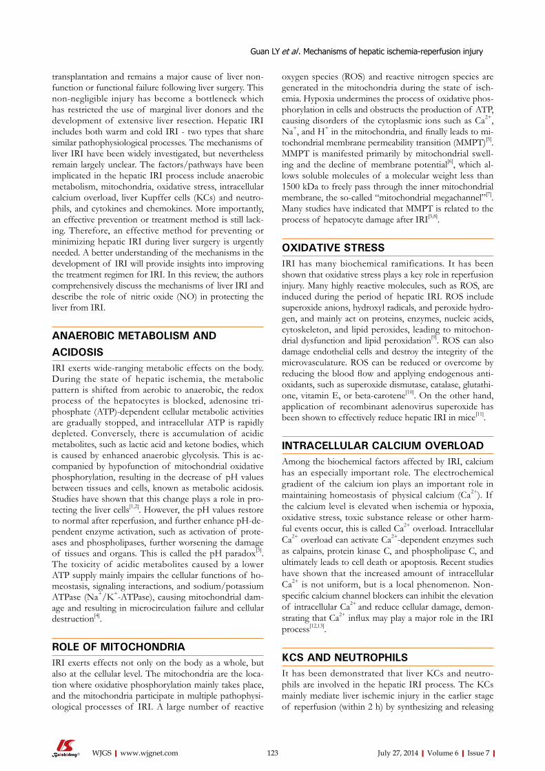
transplantation and remains a major cause of liver non-function or functional failure following liver surgery. This non-negligible injury has become a bottleneck which has restricted the use of marginal liver donors and the development of extensive liver resection. Hepatic IRI includes both warm and cold IRI - two types that share similar pathophysiological processes. The mechanisms of liver IRI have been widely investigated, but nevertheless remain largely unclear. The factors/pathways have been implicated in the hepatic IRI process include anaerobic metabolism, mitochondria, oxidative stress, intracellular calcium overload, liver Kupffer cells (KCs) and neutro-phils, and cytokines and chemokines. More importantly, an effective prevention or treatment method is still lack-ing. Therefore, an effective method for preventing or minimizing hepatic IRI during liver surgery is urgently needed. A better understanding of the mechanisms in the development of IRI will provide insights into improving the treatment regimen for IRI. In this review, the authors comprehensively discuss the mechanisms of liver IRI and describe the role of nitric oxide (NO) in protecting the liver from IRI.
ANAEROBIC METABOLISM AND ACIDOSISIRI exerts wide-ranging metabolic effects on the body. During the state of hepatic ischemia, the metabolic pattern is shifted from aerobic to anaerobic, the redox process of the hepatocytes is blocked, adenosine tri-phosphate (ATP)-dependent cellular metabolic activities are gradually stopped, and intracellular ATP is rapidly depleted. Conversely, there is accumulation of acidic metabolites, such as lactic acid and ketone bodies, which is caused by enhanced anaerobic glycolysis. This is ac-companied by hypofunction of mitochondrial oxidative phosphorylation, resulting in the decrease of pH values between tissues and cells, known as metabolic acidosis. Studies have shown that this change plays a role in pro-tecting the liver cells[1,2]. However, the pH values restore to normal after reperfusion, and further enhance pH-de-pendent enzyme activation, such as activation of prote-ases and phospholipases, further worsening the damage of tissues and organs. This is called the pH paradox[3]. The toxicity of acidic metabolites caused by a lower ATP supply mainly impairs the cellular functions of ho-meostasis, signaling interactions, and sodium/potassium ATPase (Na+/K+-ATPase), causing mitochondrial dam-age and resulting in microcirculation failure and cellular destruction[4].
ROLE OF MITOCHONDRIAIRI exerts effects not only on the body as a whole, but also at the cellular level. The mitochondria are the loca-tion where oxidative phosphorylation mainly takes place, and the mitochondria participate in multiple pathophysi-ological processes of IRI. A large number of reactive
oxygen species (ROS) and reactive nitrogen species are generated in the mitochondria during the state of isch-emia. Hypoxia undermines the process of oxidative phos-phorylation in cells and obstructs the production of ATP, causing disorders of the cytoplasmic ions such as Ca2+, Na+, and H+ in the mitochondria, and finally leads to mi-tochondrial membrane permeability transition (MMPT)[5]. MMPT is manifested primarily by mitochondrial swell-ing and the decline of membrane potential[6], which al-lows soluble molecules of a molecular weight less than 1500 kDa to freely pass through the inner mitochondrial membrane, the so-called “mitochondrial megachannel”[7].
Many studies have indicated that MMPT is related to the process of hepatocyte damage after IRI[5,8].
OXIDATIVE STRESSIRI has many biochemical ramifications. It has been shown that oxidative stress plays a key role in reperfusion injury. Many highly reactive molecules, such as ROS, are induced during the period of hepatic IRI. ROS include superoxide anions, hydroxyl radicals, and peroxide hydro-gen, and mainly act on proteins, enzymes, nucleic acids, cytoskeleton, and lipid peroxides, leading to mitochon-drial dysfunction and lipid peroxidation[9]. ROS can also damage endothelial cells and destroy the integrity of the microvasculature. ROS can be reduced or overcome by reducing the blood flow and applying endogenous anti-oxidants, such as superoxide dismutase, catalase, glutathi-one, vitamin E, or beta-carotene[10]. On the other hand, application of recombinant adenovirus superoxide has been shown to effectively reduce hepatic IRI in mice[11].
INTRACELLULAR CALCIUM OVERLOADAmong the biochemical factors affected by IRI, calcium has an especially important role. The electrochemical gradient of the calcium ion plays an important role in maintaining homeostasis of physical calcium (Ca2+). If the calcium level is elevated when ischemia or hypoxia, oxidative stress, toxic substance release or other harm-ful events occur, this is called Ca2+ overload. Intracellular Ca2+ overload can activate Ca2+-dependent enzymes such as calpains, protein kinase C, and phospholipase C, and ultimately leads to cell death or apoptosis. Recent studies have shown that the increased amount of intracellular Ca2+ is not uniform, but is a local phenomenon. Non-specific calcium channel blockers can inhibit the elevation of intracellular Ca2+ and reduce cellular damage, demon-strating that Ca2+ influx may play a major role in the IRI process[12,13].
KCS AND NEUTROPHILSIt has been demonstrated that liver KCs and neutro-phils are involved in the hepatic IRI process. The KCs mainly mediate liver ischemic injury in the earlier stage of reperfusion (within 2 h) by synthesizing and releasing
123 July 27, 2014|Volume 6|Issue 7|WJGS|www.wjgnet.com
Guan LY et al . Mechanisms of hepatic ischemia-reperfusion injury
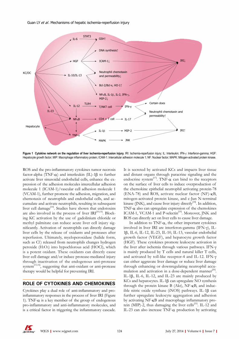
ROS and the pro-inflammatory cytokines tumor necrosis factor-alpha (TNF-α) and interleukin (IL)-1β to further activate liver sinusoidal endothelial cells, enhance the ex-pression of the adhesion molecules intercellular adhesion molecule 1 (ICAM-1)/vascular cell adhesion molecule 1 (VCAM-1), further promote the adhesion, migration, and chemotaxis of neutrophils and endothelial cells, and ac-cumulate and activate neutrophils, resulting in subsequent liver cell damage[14]. Studies have shown that endotoxins are also involved in the process of liver IRI[10,15]. Block-ing KC activation by the use of gadolinium chloride or methyl palmitate can reduce acute liver cell injury sig-nificantly. Activation of neutrophils can directly damage liver cells by the release of oxidants and proteases after reperfusion. Ultimately, myeloperoxidase (halide form, such as Cl-) released from neutrophils changes hydrogen peroxide (H2O2) into hypochlorous acid (HOCl), which is a potent oxidant. These oxidants can directly cause liver cell damage and/or induce protease-mediated injury through inactivation of the endogenous anti-protease system[15,16], suggesting that anti-oxidant or anti-protease therapy would be helpful for preventing IRI.
ROLE OF CYTOKINES AND CHEMOKINES Cytokines play a dual role of anti-inflammatory and pro-inflammatory responses in the process of liver IRI (Figure 1). TNF-α is a key member of the group of endogenous pro-inflammatory and anti-inflammatory molecules, and is a critical factor in triggering the inflammatory cascade.
It is secreted by activated KCs and impacts liver tissue and distant organs through paracrine signaling and the endocrine system[17]. TNF-α can bind to the receptors on the surface of liver cells to induce overproduction of the chemokine epithelial neutrophil activating protein-78 (ENA-78) and ROS, activate nuclear factor (NF)-κB, mitogen-activated protein kinase, and c-Jun N-terminal kinase (JNK), and cause liver injury directly[18]. In addition, TNF-α also can upregulate expression of the chemokines ICAM-1, VCAM-1 and P-selectin[19]. Moreover, JNK and ROS can directly act on liver cells to cause liver damage.
In addition to TNF-α, the other important cytokines involved in liver IRI are interferon-gamma (IFN-γ), IL-1β, IL-6, IL-12, IL-23, IL-10, IL-13, vascular endothelial growth factor (VEGF), and hepatocyte growth factor (HGF). These cytokines promote leukocyte activation in the liver after ischemia through various pathways. IFN-γ is mainly produced by T cells and natural killer T cells, and activated by toll-like receptor-4 and IL-12. IFN-γ can either aggravate liver damage or reduce liver damage through enhancing or downregulating neutrophil accu-mulation and activation in a dose-dependent manner[20]. IL-1β, IL-6, IL-12, and IL-23 are mainly produced by KCs and hepatocytes. IL-1β can upregulate NO synthesis through the protein kinase B (Akt), NF-κB, and induc-ible nitric oxide synthase (iNOS) pathways. IL-1β can further upregulate leukocyte aggregation and adhesion by activating NF-κB and macrophage inflammatory pro-tein (MIP)-2, thus damaging the liver cells[21]. IL-12 and IL-23 can also increase TNF-α production by activating
124 July 27, 2014|Volume 6|Issue 7|WJGS|www.wjgnet.com
KC/DC
IL-6STAT3
GSH↑
HGF
IL-10/IL-13
IL-12
IL-23
NFκB
TNF-α
MAPK
IL-1β
CD4 + T cell
T/NKT cellTLR4
IFN-γ
IL-17
MIP-2
JNK
IRI↑
Neutrophil chemotaxin and permeability↑
Certain does
NFκB, IL-1β, IL-2, IFN-γ, MIP-2↓
Bcl-2/Bcl-x, HO-1↑
Neutrophil chemotaxin and permeability↓
ICAM-1↓
DNA synthesis↑
Hepatocyte
IRI↓
Figure 1 Cytokine network on the regulation of liver ischemia-reperfusion injury. IRI: Ischemia-reperfusion injury; IL: Interleukin; IFN-γ: Interferon-gamma; HGF: Hepatocyte growth factor; MIP: Macrophage inflammatory protein; ICAM-1: Intercellular adhesion molecule 1; NF: Nuclear factor; MAPK: Mitogen-activated protein kinase.
Guan LY et al . Mechanisms of hepatic ischemia-reperfusion injury
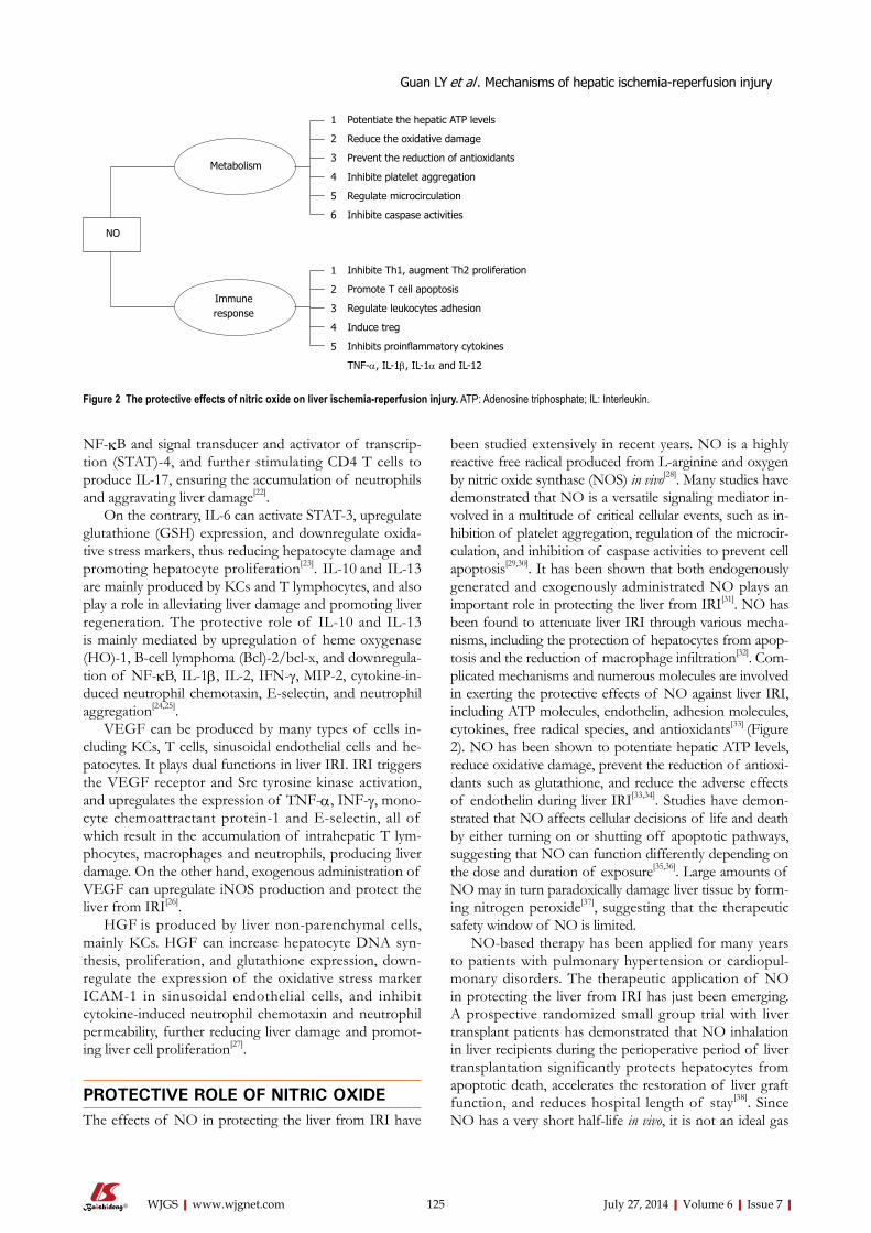
125 July 27, 2014|Volume 6|Issue 7|WJGS|www.wjgnet.com
been studied extensively in recent years. NO is a highly reactive free radical produced from L-arginine and oxygen by nitric oxide synthase (NOS) in vivo[28]. Many studies have demonstrated that NO is a versatile signaling mediator in-volved in a multitude of critical cellular events, such as in-hibition of platelet aggregation, regulation of the microcir-culation, and inhibition of caspase activities to prevent cell apoptosis[29,30]. It has been shown that both endogenously generated and exogenously administrated NO plays an important role in protecting the liver from IRI[31]. NO has been found to attenuate liver IRI through various mecha-nisms, including the protection of hepatocytes from apop-tosis and the reduction of macrophage infiltration[32]. Com-plicated mechanisms and numerous molecules are involved in exerting the protective effects of NO against liver IRI, including ATP molecules, endothelin, adhesion molecules, cytokines, free radical species, and antioxidants[33] (Figure 2). NO has been shown to potentiate hepatic ATP levels, reduce oxidative damage, prevent the reduction of antioxi-dants such as glutathione, and reduce the adverse effects of endothelin during liver IRI[33,34]. Studies have demon-strated that NO affects cellular decisions of life and death by either turning on or shutting off apoptotic pathways, suggesting that NO can function differently depending on the dose and duration of exposure[35,36]. Large amounts of NO may in turn paradoxically damage liver tissue by form-ing nitrogen peroxide[37], suggesting that the therapeutic safety window of NO is limited.
NO-based therapy has been applied for many years to patients with pulmonary hypertension or cardiopul-monary disorders. The therapeutic application of NO in protecting the liver from IRI has just been emerging. A prospective randomized small group trial with liver transplant patients has demonstrated that NO inhalation in liver recipients during the perioperative period of liver transplantation significantly protects hepatocytes from apoptotic death, accelerates the restoration of liver graft function, and reduces hospital length of stay[38]. Since NO has a very short half-life in vivo, it is not an ideal gas
NF-κB and signal transducer and activator of transcrip-tion (STAT)-4, and further stimulating CD4 T cells to produce IL-17, ensuring the accumulation of neutrophils and aggravating liver damage[22].
On the contrary, IL-6 can activate STAT-3, upregulate glutathione (GSH) expression, and downregulate oxida-tive stress markers, thus reducing hepatocyte damage and promoting hepatocyte proliferation[23]. IL-10 and IL-13 are mainly produced by KCs and T lymphocytes, and also play a role in alleviating liver damage and promoting liver regeneration. The protective role of IL-10 and IL-13 is mainly mediated by upregulation of heme oxygenase (HO)-1, B-cell lymphoma (Bcl)-2/bcl-x, and downregula-tion of NF-κB, IL-1β, IL-2, IFN-γ, MIP-2, cytokine-in-duced neutrophil chemotaxin, E-selectin, and neutrophil aggregation[24,25].
VEGF can be produced by many types of cells in-cluding KCs, T cells, sinusoidal endothelial cells and he-patocytes. It plays dual functions in liver IRI. IRI triggers the VEGF receptor and Src tyrosine kinase activation, and upregulates the expression of TNF-α, INF-γ, mono-cyte chemoattractant protein-1 and E-selectin, all of which result in the accumulation of intrahepatic T lym-phocytes, macrophages and neutrophils, producing liver damage. On the other hand, exogenous administration of VEGF can upregulate iNOS production and protect the liver from IRI[26].
HGF is produced by liver non-parenchymal cells, mainly KCs. HGF can increase hepatocyte DNA syn-thesis, proliferation, and glutathione expression, down-regulate the expression of the oxidative stress marker ICAM-1 in sinusoidal endothelial cells, and inhibit cytokine-induced neutrophil chemotaxin and neutrophil permeability, further reducing liver damage and promot-ing liver cell proliferation[27].
PROTECTIVE ROLE OF NITRIC OXIDEThe effects of NO in protecting the liver from IRI have
Metabolism
Immune response
Potentiate the hepatic ATP levels
Reduce the oxidative damage
Prevent the reduction of antioxidants
Inhibite platelet aggregation
Regulate microcirculation
Inhibite caspase activities
Inhibite Th1, augment Th2 proliferation
Promote T cell apoptosis
Regulate leukocytes adhesion
Induce treg
Inhibits proinflammatory cytokines
TNF-α, IL-1β, IL-1α and IL-12
1
2
3
4
5
6
1
2
3
4
5
NO
Figure 2 The protective effects of nitric oxide on liver ischemia-reperfusion injury. ATP: Adenosine triphosphate; IL: Interleukin.
Guan LY et al . Mechanisms of hepatic ischemia-reperfusion injury

126 July 27, 2014|Volume 6|Issue 7|WJGS|www.wjgnet.com
for the treatment of IRI. NO drugs administered to liver donors, such as organic nitrates and sodium nitroprus-side, are now being explored as an alternative choice for NO delivery.
Sodium nitrite, a storage form of NO, can release NO during hypoxia and acidosis[39]. Sodium nitrite has now been identified as an important storage reservior of bioavailable NO in the blood and tissues[40]. The reduc-tion of nitrite to NO has been demonstrated to confer cytoprotection against IRI in the heart, liver, brain, and kidney[40]. Interventions that increase NO production by the use of sodium nitrite before the occurrence of ischemia, either through intraperitoneal injection or oral administration, can mediate significant cytoprotection. This strategy has been demonstrated to potently limit acute IRI in both the heart and liver in murine warm IRI models, with the ability to decrease myocardial infarction and hepatocyte apoptosis[40-43].
NO is also an important effector molecule, produced by KCs and dendritic cells (DCs), and is involved in immune regulation and host innate and adaptive immu-nity[44]. NO inhibits proinflammatory cytokines, including TNF-α, IL-1β, IL-1α and IL-12, which may induce the inflammatory cascade during liver IRI [24-26,33]. It has been reported that NO exerts multiple effects on immune cells, decreasing the number of T helper (Th)1 cells and augmenting Th2 cell proliferation and their cytokine syn-thesis, regulating leukocyte adhesion and recruitment to the site of infection[45-47], inhibiting Th1 proliferation, and promoting T cell apoptosis[48,49]. Moreover, NO also con-tributes to the immunosuppressive function of induced T regulatory cells (Treg)[50]. Therefore, NO is involved in the regulation of liver IRI-associated immune responses. The underlying mechanisms are largely unknown and warrant further investigation.
CONCLUSIONHepatic IRI is not only a pathophysiological process
involving the liver itself, but also a complex systemic process affecting multiple tissues and organs. Hepatic IRI can seriously impair liver function, even producing irreversible damage, which causes a cascade of multiple organ dysfunction. Many factors, including anaerobic metabolism, mitochondrial damage, oxidative stress, intracellular Ca2+ overload, cytokines and chemokines produced by KCs and neutrophils, and NO, are all in-volved in the regulation of liver IRI processes. The most important pathways of liver IRI are initiated by oxidative stress, anaerobic metabolism and acidosis, further result-ing in the cellular damage through induction of apopto-sis, immune responses, and cytokine regulations (Figure 3). Inhaled NO or NO-producing drugs have shown positive effects on IRI protection in clinical practice, and may be a good choice for liver IRI therapy in the future. Therefore, further exploration of the mechanisms of IRI on animal models focusing on the regulatory path-way of IRI development, with concomitant development of a more effective method of controlling IRI, will help overcome the challenges in the prevention of IRI and therapeutic strategies.
REFERENCES1 Kanoria S, Glantzounis G, Quaglia A, Dinesh S, Fusai G, Da-
vidson BR, Seifalian AM. Remote preconditioning improves hepatic oxygenation after ischaemia reperfusion injury. Transpl Int 2012; 25: 783-791 [PMID: 22533545 DOI: 10.1111/j.1432-2277.2012.01481.x]
2 Guan YF, Pritts TA, Montrose MH. Ischemic post-condi-tioning to counteract intestinal ischemia/reperfusion injury. World J Gastrointest Pathophysiol 2010; 1: 137-143 [PMID: 21607154 DOI: 10.4291/wjgp.v1.i4.137]
3 Datta G, Fuller BJ, Davidson BR. Molecular mechanisms of liver ischemia reperfusion injury: insights from transgenic knockout models. World J Gastroenterol 2013; 19: 1683-1698 [PMID: 23555157 DOI: 10.3748/wjg.v19.i11.1683]
4 Siriussawakul A, Zaky A, Lang JD. Role of nitric oxide in hepatic ischemia-reperfusion injury. World J Gastroenterol 2010; 16: 6079-6086 [PMID: 21182222 DOI: 10.3748/wjg.v16.i48.6079]
IRI
Anaerobicmetabolism and acidosis
Cytotoxicity
Inhibits pH-dependent enzyme
ATP↓ Cellular edema
Metabolic dysfunction
Mitochondrial damage Apoptosis
Organelles damageInduce innate immue response
Chemokines ICAM/VCAM
T cells Cytokins
TNF-α, IFN-γ
IL-1β, IL-6, IL-12, IL-23
IL-10, IL-13
VEGF, HGF
ROS↑
Kupffer cellsand neutriphils
Intracellular Ca2+ overload
Oxidativestress
Cellular damage
Figure 3 Mechanisms of hepatic ischemia reperfusion injury. ATP: Adenosine triphosphate; IL: Interleukin; ROS: Reactive oxygen species; IRI: Ischemia-reperfu-sion injury; IFN-γ: Interferon-gamma; ICAM: Intercellular adhesion molecule; VCAM: Vascular cell adhesion molecule; TNF: Tumor necrosis factor.
Guan LY et al . Mechanisms of hepatic ischemia-reperfusion injury
127 July 27, 2014|Volume 6|Issue 7|WJGS|www.wjgnet.com
5 Sastre J, Serviddio G, Pereda J, Minana JB, Arduini A, Ven-demiale G, Poli G, Pallardo FV, Vina J. Mitochondrial func-tion in liver disease. Front Biosci 2007; 12: 1200-1209 [PMID: 17127373 DOI: 10.2741/2138]
6 Aguilar HI, Botla R, Arora AS, Bronk SF, Gores GJ. Induc-tion of the mitochondrial permeability transition by protease activity in rats: a mechanism of hepatocyte necrosis. Gastro-enterology 1996; 110: 558-566 [PMID: 8566604 DOI: 10.1053/gast.1996.v110.pm8566604]
7 Bernardi P, Broekemeier KM, Pfeiffer DR. Recent progress on regulation of the mitochondrial permeability transition pore; a cyclosporin-sensitive pore in the inner mitochondrial membrane. J Bioenerg Biomembr 1994; 26: 509-517 [PMID: 7896766 DOI: 10.1007/BF00762735]
8 Abu-Amara M, Yang SY, Tapuria N, Fuller B, Davidson B, Seifalian A. Liver ischemia/reperfusion injury: processes in inflammatory networks--a review. Liver Transpl 2010; 16: 1016-1032 [PMID: 20818739 DOI: 10.1002/lt.22117]
9 Brass CA, Roberts TG. Hepatic free radical production af-ter cold storage: Kupffer cell-dependent and -independent mechanisms in rats. Gastroenterology 1995; 108: 1167-1175 [PMID: 7698585 DOI: 10.1016/0016-5085(95)90216-3]
10 Jaeschke H. Reactive oxygen and ischemia/reperfusion in-jury of the liver. Chem Biol Interact 1991; 79: 115-136 [PMID: 1884426 DOI: 10.1016/0009-2797(91)90077-K]
11 McCord JM. Oxygen-derived free radicals in postischemic tissue injury. N Engl J Med 1985; 312: 159-163 [PMID: 2981404 DOI: 10.1056/NEJM198501173120305]
12 Ikeda M, Ariyoshi H, Sakon M, Kambayashi J, Yoshikawa N, Shinoki N, Kawasaki T, Monden M. A role for local calcium gradients upon hypoxic injury in human umbilical vein en-dothelial cells (HUVEC). Cell Calcium 1998; 24: 49-57 [PMID: 9793688 DOI: 10.1016/S0143-4160(98)90088-4]
13 Wang HG, Pathan N, Ethell IM, Krajewski S, Yamaguchi Y, Shibasaki F, McKeon F, Bobo T, Franke TF, Reed JC. Ca2+-induced apoptosis through calcineurin dephosphorylation of BAD. Science 1999; 284: 339-343 [PMID: 10195903 DOI: 10.1126/science.284.5412.339]
14 Zhou W, Zhang Y, Hosch MS, Lang A, Zwacka RM, Engel-hardt JF. Subcellular site of superoxide dismutase expression differentially controls AP-1 activity and injury in mouse liver following ischemia/reperfusion. Hepatology 2001; 33: 902-914 [PMID: 11283855 DOI: 10.1053/jhep.2001.23073]
15 Boury NM, Czuprynski CJ. Listeria monocytogenes infec-tion increases neutrophil adhesion and damage to a murine hepatocyte cell line in vitro. Immunol Lett 1995; 46: 111-116 [PMID: 7590905 DOI: 10.1016/0165-2478(95)00027-3]
16 Nagendra AR, Mickelson JK, Smith CW. CD18 integrin and CD54-dependent neutrophil adhesion to cytokine-stimulat-ed human hepatocytes. Am J Physiol 1997; 272: G408-G416 [PMID: 9124560]
17 Gujral JS, Bucci TJ, Farhood A, Jaeschke H. Mechanism of cell death during warm hepatic ischemia-reperfusion in rats: apoptosis or necrosis? Hepatology 2001; 33: 397-405 [PMID: 11172341 DOI: 10.1053/jhep.2001.22002]
18 Redaelli CA, Tian YH, Schaffner T, Ledermann M, Baer HU, Dufour JF. Extended preservation of rat liver graft by induc-tion of heme oxygenase-1. Hepatology 2002; 35: 1082-1092 [PMID: 11981758 DOI: 10.1053/jhep.2002.33067]
19 Peralta C, Fernández L, Panés J, Prats N, Sans M, Piqué JM, Gelpí E, Roselló-Catafau J. Preconditioning protects against systemic disorders associated with hepatic ischemia-reperfusion through blockade of tumor necrosis factor-induced P-selectin up-regulation in the rat. Hepatology 2001; 33: 100-113 [PMID: 11124826 DOI: 10.1053/jhep.2001.20529]
20 Schwabe RF, Brenner DA. Mechanisms of Liver Injury. I. TNF-alpha-induced liver injury: role of IKK, JNK, and ROS pathways. Am J Physiol Gastrointest Liver Physiol 2006; 290: G583-G589 [PMID: 16537970]
21 Oe S, Hiros T, Fujii H, Yasuchika K, Nishio T, Iimuro Y,
Morimoto T, Nagao M, Yamaoka Y. Continuous intrave-nous infusion of deleted form of hepatocyte growth factor attenuates hepatic ischemia-reperfusion injury in rats. J Hepatol 2001; 34: 832-839 [PMID: 11451166 DOI: 10.1016/S0168-8278(01)00030-7]
22 Hamada T, Tsuchihashi S, Avanesyan A, Duarte S, Moore C, Busuttil RW, Coito AJ. Cyclooxygenase-2 deficiency en-hances Th2 immune responses and impairs neutrophil re-cruitment in hepatic ischemia/reperfusion injury. J Immunol 2008; 180: 1843-1853 [PMID: 18209082 DOI: 10.4049/jimmu-nol.180.3.1843]
23 Welborn MB, Moldawer LL, Seeger JM, Minter RM, Hu-ber TS. Role of endogenous interleukin-10 in local and distant organ injury after visceral ischemia-reperfusion. Shock 2003; 20: 35-40 [PMID: 12813366 DOI: 10.1097/01/SHK.0000071062.67193.b6]
24 Teoh N, Field J, Farrell G. Interleukin-6 is a key media-tor of the hepatoprotective and pro-proliferative effects of ischaemic preconditioning in mice. J Hepatol 2006; 45: 20-27 [PMID: 16600417 DOI: 10.1016/j.jhep.2006.01.039]
25 Husted TL, Blanchard J, Schuster R, Shen H, Lentsch AB. Po-tential role for IL-23 in hepatic ischemia/reperfusion injury. Inflamm Res 2006; 55: 177-178 [PMID: 16830103 DOI: 10.1007/s00011-006-0073-1]
26 Ke B, Shen XD, Lassman CR, Gao F, Busuttil RW, Kupiec-Weglinski JW. Cytoprotective and antiapoptotic effects of IL-13 in hepatic cold ischemia/reperfusion injury are heme oxygenase-1 dependent. Am J Transplant 2003; 3: 1076-1082 [PMID: 12919086 DOI: 10.1034/j.1600-6143.2003.00147.x]
27 Shen XD, Ke B, Zhai Y, Gao F, Tsuchihashi S, Lassman CR, Busuttil RW, Kupiec-Weglinski JW. Absence of toll-like receptor 4 (TLR4) signaling in the donor organ reduces isch-emia and reperfusion injury in a murine liver transplantation model. Liver Transpl 2007; 13: 1435-1443 [PMID: 17902130 DOI: 10.1002/lt.21251]
28 Diesen DL, Kuo PC. Nitric oxide and redox regulation in the liver: Part I. General considerations and redox biology in hepatitis. J Surg Res 2010; 162: 95-109 [PMID: 20444470 DOI: 10.1016/j.jss.2009.09.019]
29 Pacher P, Beckman JS, Liaudet L. Nitric oxide and peroxyni-trite in health and disease. Physiol Rev 2007; 87: 315-424 [PMID: 17237348 DOI: 10.1152/physrev.00029.2006]
30 Mocellin S, Bronte V, Nitti D. Nitric oxide, a double edged sword in cancer biology: searching for therapeutic opportu-nities. Med Res Rev 2007; 27: 317-352 [PMID: 16991100 DOI: 10.1002/med.20092]
31 Abu-Amara M, Yang SY, Seifalian A, Davidson B, Fuller B. The nitric oxide pathway--evidence and mechanisms for protection against liver ischaemia reperfusion injury. Liver Int 2012; 32: 531-543 [PMID: 22316165 DOI: 10.1111/j.1478-3231.2012.02755.x]
32 Koti RS, Yang W, Dashwood MR, Davidson BR, Seifalian AM. Effect of ischemic preconditioning on hepatic microcir-culation and function in a rat model of ischemia reperfusion injury. Liver Transpl 2002; 8: 1182-1191 [PMID: 12474159 DOI: 10.1053/jlts.2002.36846]
33 Liu P, Xu B, Spokas E, Lai PS, Wong PY. Role of endog-enous nitric oxide in TNF-alpha and IL-1beta generation in hepatic ischemia-repefusion. Shock 2000; 13: 217-223 [PMID: 10718379 DOI: 10.1097/00024382-200003000-00008]
34 Hsu CM, Wang JS, Liu CH, Chen LW. Kupffer cells protect liver from ischemia-reperfusion injury by an inducible nitric oxide synthase-dependent mechanism. Shock 2002; 17: 280-285 [PMID: 11954827 DOI: 10.1097/00024382-200204000-00007]
35 Brüne B. Nitric oxide: NO apoptosis or turning it ON? Cell Death Differ 2003; 10: 864-869 [PMID: 12867993 DOI: 10.1038/sj.cdd.4401261]
36 Bolli R. Cardioprotective function of inducible nitric oxide synthase and role of nitric oxide in myocardial ischemia and preconditioning: an overview of a decade of research.
Guan LY et al . Mechanisms of hepatic ischemia-reperfusion injury
128 July 27, 2014|Volume 6|Issue 7|WJGS|www.wjgnet.com
J Mol Cell Cardiol 2001; 33: 1897-1918 [PMID: 11708836 DOI: 10.1006/jmcc.2001.1462]
37 Miyake T, Yokoyama Y, Kokuryo T, Mizutani T, Imamura A, Nagino M. Endothelial nitric oxide synthase plays a main role in producing nitric oxide in the superacute phase of hepatic ischemia prior to the upregulation of inducible nitric oxide synthase. J Surg Res 2013; 183: 742-751 [PMID: 23485075 DOI: 10.1016/j.jss.2013.01.048]
38 Lang JD, Teng X, Chumley P, Crawford JH, Isbell TS, Chacko BK, Liu Y, Jhala N, Crowe DR, Smith AB, Cross RC, Frenette L, Kelley EE, Wilhite DW, Hall CR, Page GP, Fallon MB, Bynon JS, Eckhoff DE, Patel RP. Inhaled NO accelerates restoration of liver function in adults following orthotopic liver transplantation. J Clin Invest 2007; 117: 2583-2591 [PMID: 17717604 DOI: 10.1172/JCI31892]
39 Bryan NS, Rassaf T, Maloney RE, Rodriguez CM, Saijo F, Rodriguez JR, Feelisch M. Cellular targets and mechanisms of nitros(yl)ation: an insight into their nature and kinetics in vivo. Proc Natl Acad Sci USA 2004; 101: 4308-4313 [PMID: 15014175 DOI: 10.1073/pnas.0306706101]
40 Sinha SS, Shiva S, Gladwin MT. Myocardial protection by nitrite: evidence that this reperfusion therapeutic will not be lost in translation. Trends Cardiovasc Med 2008; 18: 163-172 [PMID: 18790386 DOI: 10.1016/j.tcm.2008.05.001]
41 Duranski MR, Greer JJ, Dejam A, Jaganmohan S, Hogg N, Langston W, Patel RP, Yet SF, Wang X, Kevil CG, Gladwin MT, Lefer DJ. Cytoprotective effects of nitrite during in vivo ischemia-reperfusion of the heart and liver. J Clin Invest 2005; 115: 1232-1240 [PMID: 15841216 DOI: 10.1172/JCI22493]
42 Kumar D, Branch BG, Pattillo CB, Hood J, Thoma S, Simp-son S, Illum S, Arora N, Chidlow JH, Langston W, Teng X, Lefer DJ, Patel RP, Kevil CG. Chronic sodium nitrite therapy augments ischemia-induced angiogenesis and arteriogenesis. Proc Natl Acad Sci USA 2008; 105: 7540-7545 [PMID: 18508974 DOI: 10.1073/pnas.0711480105]
43 Li W, Meng Z, Liu Y, Patel RP, Lang JD. The hepatoprotec-
tive effect of sodium nitrite on cold ischemia-reperfusion injury. J Transplant 2012; 2012: 635179 [PMID: 22530108 DOI: 10.1155/2012/635179]
44 Panjwani NN, Popova L, Srivastava PK. Heat shock pro-teins gp96 and hsp70 activate the release of nitric oxide by APCs. J Immunol 2002; 168: 2997-3003 [PMID: 11884472 DOI: 10.4049/jimmunol.168.6.2997]
45 Chen C, Lee WH, Zhong L, Liu CP. Regulatory T cells can mediate their function through the stimulation of APCs to produce immunosuppressive nitric oxide. J Immunol 2006; 176: 3449-3460 [PMID: 16517713 DOI: 10.4049/jimmu-nol.176.6.3449]
46 Taylor-Robinson AW, Liew FY, Severn A, Xu D, McSorley SJ, Garside P, Padron J, Phillips RS. Regulation of the im-mune response by nitric oxide differentially produced by T helper type 1 and T helper type 2 cells. Eur J Immunol 1994; 24: 980-984 [PMID: 8149966 DOI: 10.1002/eji.1830240430]
47 Fox-Robichaud A, Payne D, Hasan SU, Ostrovsky L, Fair-head T, Reinhardt P, Kubes P. Inhaled NO as a viable anti-adhesive therapy for ischemia/reperfusion injury of distal microvascular beds. J Clin Invest 1998; 101: 2497-2505 [PMID: 9616221 DOI: 10.1172/JCI2736]
48 Roland CR, Walp L, Stack RM, Flye MW. Outcome of Kupffer cell antigen presentation to a cloned murine Th1 lymphocyte depends on the inducibility of nitric oxide syn-thase by IFN-gamma. J Immunol 1994; 153: 5453-5464 [PMID: 7527442]
49 Monsonego A, Imitola J, Zota V, Oida T, Weiner HL. Mi-croglia-mediated nitric oxide cytotoxicity of T cells following amyloid beta-peptide presentation to Th1 cells. J Immunol 2003; 171: 2216-2224 [PMID: 12928365 DOI: 10.4049/jimmu-nol.171.5.2216]
50 Wood KJ, Sawitzki B. Interferon gamma: a crucial role in the function of induced regulatory T cells in vivo. Trends Immunol 2006; 27: 183-187 [PMID: 16527542 DOI: 10.1016/j.it.2006.02.008]
P- Reviewer: Chapel A, Cnossen WR S- Editor: Wen LL L- Editor: Wang TQ E- Editor: Wang CH
Guan LY et al . Mechanisms of hepatic ischemia-reperfusion injury
Chronic pancreatitis: A surgical disease? Role of the Frey procedure
Alexandra Roch, Jérome Teyssedou, Didier Mutter, Jacques Marescaux, Patrick Pessaux
Alexandra Roch, Jérome Teyssedou, Didier Mutter, Jacques Marescaux, Patrick Pessaux, Digestive and Endocrine Surgery, Nouvel Hôpital Civil, University of Strasbourg, 67000 Strabourg, FranceAlexandra Roch, Jérome Teyssedou, Didier Mutter, Jacques Marescaux, Patrick Pessaux, Institut de recherche en cancéro-logie de l’appareil digestif (IRCAD), 67000 Strasbourg, FranceAlexandra Roch, Jérome Teyssedou, Didier Mutter, Jacques Marescaux, Patrick Pessaux, IHU-Strasbourg, Minimally-Inva-sive Image-Guided Surgical Institute, University of Strasbourg, 67000 Strabourg, FranceAuthor contributions: All the authors contributed to this paper.Correspondence to: Patrick Pessaux, MD, PhD, Professor, Head of the Hepatobiliary and Pancreatic Surgical Unit, Diges-tive and Endocrine Surgery, Nouvel Hôpital Civil, University of Strasbourg, 1 place de l’hopital, 67000 Strasbourg, France. [email protected]: +33-3-69551563 Received: March 21, 2014 Revised: April 26, 2014Accepted: July 12, 2014Published online: July 27, 2014
AbstractAlthough medical treatment and endoscopic interven-tions are primarily offered to patients with chronic pancreatitis, approximately 40% to 75% will ultimately require surgery during the course of their disease. Al-though pancreaticoduodenectomy has been considered the standard surgical procedure because of its favorable results on pain control, its high postoperative complica-tion and pancreatic exocrine or/and endocrine dysfunc-tion rates have led to a growing enthusiasm for duodenal preserving pancreatic head resection. The aim of this review is to better understand the rationale underlying of the Frey procedure in chronic pancreatitis and to ana-lyze its outcome. Because of its hybrid nature, combin-ing both resection and drainage, the Frey procedure has been conceptualized based on the pathophysiology of chronic pancreatitis. The short and long-term outcome, especially pain relief and quality of life, are better after the Frey procedure than after any other surgical proce-
dure performed for chronic pancreatitis.
© 2014 Baishideng Publishing Group Inc. All rights reserved.
Key words: Chronic pancreatitis; Frey procedure; Sur-gery; Complication; Outcome
Core tip: The management and the treatment of chronic pancreatitis are challenging. Many surgical procedures were described with 2 different types of concepts: re-section vs drainage. The Frey procedure is an associa-tion of these 2 concepts. This manuscript contains the most recent data about the technique, the short and long-term outcomes of this technique. In addition, there is a review of the literature of series comparing this technique with the other surgical procedures.
Roch A, Teyssedou J, Mutter D, Marescaux J, Pessaux P. Chronic pancreatitis: A surgical disease? Role of the Frey procedure. World J Gastrointest Surg 2014; 6(7): 129-135 Available from: URL: http://www.wjgnet.com/1948-9366/full/v6/i7/129.htm DOI: http://dx.doi.org/10.4240/wjgs.v6.i7.129
INTRODUCTIONChronic pancreatitis is a progressive inflammatory dis-ease characterized by debilitating pain and pancreatic insufficiency (nutritional deficiency and glucose deregula-tion)[1,2]. The enormous personal and socioeconomic im-pact comprises impairment of quality of life, inability to work and even shortening in life expectancy[3]. Although medical treatment and endoscopic interventions are pri-marily offered to patients with chronic pancreatitis[4,5], approximately 40% to 75% will ultimately require surgery during the course of their disease[6,7].
Although pancreaticoduodenectomy has been con-sidered the standard surgical procedure for patients with chronic pancreatitis because of its favorable results
MINIREVIEWS
Submit a Manuscript: http://www.wjgnet.com/esps/Help Desk: http://www.wjgnet.com/esps/helpdesk.aspxDOI: 10.4240/wjgs.v6.i7.129
129 July 27, 2014|Volume 6|Issue 7|WJGS|www.wjgnet.com
World J Gastrointest Surg 2014 July 27; 6(7): 129-135ISSN 1948-9366 (online)
© 2014 Baishideng Publishing Group Inc. All rights reserved.
on pain control, its high postoperative complication and pancreatic exocrine or/and endocrine dysfunction rates[8,9] have led to a growing enthusiasm for duodenal preserving pancreatic head resection[10,11]. When in 1987 Frey et al[12] described a novel hybrid procedure com-bining local resection of the head of the pancreas and longitudinal pancreatico-jejunostomy, surgeons favorably welcomed it because of its technical feasibility and low surgical risk. Since 1987, numerous studies have analyzed the short and long-term outcome following the Frey pro-cedure and have compared it to other surgical procedures commonly performed for chronic pancreatitis. The aim of this review is to better understand the rationale under-lying of the Frey procedure in chronic pancreatitis and to analyze its outcome.
WHY CAN CHRONIC PANCREATITIS BE CONSIDERED A SURGICAL DISEASE?Mechanisms of pain in chronic pancreatitis Although pain is the most common symptom (85% of patients)[2] in chronic pancreatitis, its mechanism remains unclear and debated[13-15]. Several concepts have been hy-pothesized and pain probably results from a combination of them. The intraductal and interstitial hypertension theory is similar to a compartment syndrome[16,17]. In-creased ductal pressure related to duct stricture or calculi and intraparenchymal hypertension as a result of fibrosis and edema can activate intrapancreatic nociceptors. The neurogenic theory focuses on intrapancreatic neural dam-age[18]. Inflammatory mediators from infiltrating lympho-cytes are responsible for increased signals along the axons of pain-sensitive neurons, which ultimately can result in a “centrally sensitized” pain state[19]. Traditionally, the head of the pancreas is called the “pacemaker” of pain in chronic pancreatitis. It is often enlarged and can be replaced by an inflammatory mass that can lead to com-mon bile duct or duodenal stenosis[20]. Another explana-tion to this pain is the compression of adjacent organs by a pseudocyst.
Indications for surgerySurgical management is usually offered to patients after medical treatment and endoscopic intervention have failed[4,5], and is considered the last option of this step-up approach[21]. Medical treatment for pain related to chronic pancreatitis usually fails, as narcotic dependency occurs in most patients[11]. Longitudinal studies have shown that 40% to 75% of all patients with chronic pancreatitis will require surgery in the course of the disease[7]. The main indications for surgery are intractable pain, non-resolving common bile duct or duodenal stenosis and suspicion of malignancy. The objective of surgery is to relieve in-tractable pain while preserving pancreatic endocrine and exocrine functions.
Rationale for surgery in chronic pancreatitisFirst, surgery has been proved superior to endoscopic
treatment in 2 main randomized controlled trials[22,23]. Moreover, several studies have suggested that surgery early in the course of chronic pancreatitis is beneficial in terms of pain control and pancreatic function[21,24]. One experimental and three clinical observational cohort stud-ies have concluded that surgery, especially drainage pro-cedures, can delay the natural course and progressive loss of pancreatic function in chronic pancreatitis. In an ex-perimental model of early vs late surgical drainage in pigs, early surgery resulted in less pathological cell damage and better exocrine function[25]. When Nealon et al[26] com-pared the outcomes of conservative treatment vs surgery, they reported a delay in pancreatic function impairment after surgical treatment. They concluded that early opera-tive drainage should be performed before the pancreas shows morphological and functional irreversible damage. Ihse et al[27] also have recommended surgical treatment to be performed before nutritional or metabolic disorders develop.
Prolonged periods of pain can be associated with pe-ripheral and central nerve sensitization, leading to a per-manent state of pain impossible to reverse[19]. A recent observational study suggests that longstanding disease is associated with poor pain control after surgical interven-tion[28]. In 266 consecutive patients undergoing surgery for chronic pancreatitis, surgery after 3 years of onset of symptoms was independently associated with impaired pain relief and increased rate of endocrine pancreatic insufficiency. A small pilot trial randomized 32 patients with early stage chronic pancreatitis and dilated pancreatic duct between upfront surgical drainage and conservative approach[29]. Significant pain relief was observed in 94% patients in the surgical group compared to 13% patients in the conservative group. New onset pancreatic insuf-ficiency was significantly less frequently observed in the early surgical group compared conservative group. De-spite the evidence suggesting a benefit of early surgery, most patients are still managed by a conservative step-up approach. To evaluate the benefits, risks and costs of early surgical intervention, the Dutch Pancreatitis Study Group is currently conducting a multicentric randomized controlled trial (the Early Surgery vs Optimal Current Step-up Practice for Chronic Pancreatitis trial) [21].
The role of chronic pancreatitis as a risk factor for pancreatic carcinogenesis has been supported by numer-ous studies since 1993[30-32]. Lowenfels et al[30] published an international cohort study of 2015 patients that reported a cumulative risk of pancreatic cancer in subjects with chronic pancreatitis of 1.8% after 10 years and 4%, after 20 years with a standardized incidence ratio of 14.4. A re-cent multicentric Japanese study[33] of 506 patients found that the incidence of pancreatic cancer was significantly lower in patients who underwent surgery for chronic pancreatitis than in patients who had a conservative treat-ment (0.7% vs 5.1%, P = 0.03, HR = 0.11). Although this study shows a protective effect of surgery in the devel-opment of pancreatic cancer from chronic pancreatitis, the exact mechanism remains unclear probably through reduction in pancreatic inflammation.
130 July 27, 2014|Volume 6|Issue 7|WJGS|www.wjgnet.com
Roch A et al . Frey procedure

FREY PROCEDURE: SURGICAL TECHNIQUERationale for the Frey procedureBased on the pathophysiological mechanisms described above[13-19], two main surgical procedure types have been described in patients with chronic pancreatitis: drain-age and resection procedures[11,34,35]. Until the late 80s, pancreaticoduodenectomy was the resection procedure of choice for “head-dominant” disease[11]. The Frey procedure was first described in 1987 by Frey et al[12] and combines partial resection of the head of the pancreas (resection) with lateral pancreatico-jejunostomy (drain-age). The rationale for this hybrid procedure[12, 36-38] is that it improves the overall pancreatic ductal drainage by de-compressing both the duct of Santorini and ducts in the uncinate process. It also allows removal of calculi. More-over, the partial pancreatic head resection removes what is thought to be the “epicenter” of chronic pain and can relieve symptoms related to ductal stricture.
The Frey procedure was originally applied to patients with an enlarged fibrotic head of the pancreas and an associated dilated main pancreatic duct. It has since then been described in various indications, including patients who have had prior lateral pancreatico-jejunostomy (Pu-estow or Partington and Rochelle procedures) with no relief of symptoms[38].
Surgical techniqueThrough a bilateral subcostal incision and after exposure of the pancreas (Kocher maneuver), the main pancreatic duct is located using a syringe aiming toward the tail of the pancreas[12,36-40]. The pancreatic duct is then opened longitudinally (the incision in the tail of the pancreas is extended to within 1-2 cm of the distal portion of the gland and the incision in the head to within 1 cm of the inner aspect of the duodenum). When the main pancre-atic duct is exposed, it can be inspected and all calculi removed. The head of the pancreas is partially cored-out while preserving a rim of pancreatic tissue along the inner aspect of the duodenum (to allow blood supply to the duodenum from superior and inferior pancreatico-duodenal arteries), along the pancreatic medial margin (to avoid injuring the superior mesenteric/portal vein) and posteriorly (between the head excavation and the unci-nate process and vena cava). During the local excision of the head of the pancreas, the intrapancreatic portion of the common bile duct is freed from inflamed and fibrotic periodical tissue. In about 70% of cases, resection of the fibrotic pancreatic parenchyma is sufficient to relieve a common bile duct stricture. If the obstruction cannot be relieved, a choledocho-duodenostomy or a choledocho-jejunostomy can be performed. The cored-out head of the pancreas and the open main duct are drained into Roux-en-Y limb of jejunum. The Roux-en-Y limb is passed through the transverse mesocolon to lie over the pancreas. A two-layer pancreatico-jejunostomy is per-formed. The gastrointestinal tract continuity is restored
by and end-to-side jejuno-jejunostomy. Owing to the in-creased risk of pancreatic cancer in patients with chronic pancreatitis, the cored tissue from the pancreatic head is routinely sent for pathological analysis.
Technical key pointsCompared to other surgical procedures (especially pan-creaticoduodenectomy and Beger procedure), the Frey procedure is easier to perform by avoiding the transsec-tion of the pancreas neck over the superior mesenteric/portal vein.
Although Frey et al[37-39] analyzed the relation between weight of the cored pancreatic head tissue and pain relief, this amount of tissue depends on the size of the head of the pancreas, which is highly variable. Some studies sug-gested that a mean volume percent of head mass resected between 60% and 65% allowed better pain relief. Exten-sive pancreatic head excision should not be performed as it may lead to increased parenchymal loss and ultimately pancreatic exocrine insufficiency.
Current data suggest that the Frey procedure in small duct chronic pancreatitis is associated with a significantly increased operative time[41]. Difficulty in locating the main pancreatic duct contributes to the delay and intra opera-tive ultrasound in those cases proves useful[42].
Because the Frey procedure can be technically chal-lenging due to major chronic inflammation, it is tradition-ally performed as an open surgery. Surgeons from John Hopkins recently published a case report describing a total laparoscopic Frey procedure for chronic pancreati-tis caused by recurrent pancreatic ductal stones[43]. The laparoscopic approach confers the benefits of magnified visualization while reducing the rate of postoperative wound infection, incisional hernia, bowel obstruction and pain[44]. As laparoscopic Frey procedure is very demand-ing, the selection of patients that can benefit from it is very important. This approach will less likely be offered to obese patients, as visualization can be impaired by ret-roperitoneal fat. Similarly, this approach does not fit pa-tients with a highly vascular head of the pancreas because of the increased risk of bleeding.
RESULTS OF THE FREY PROCEDUREComplicationsThe Frey procedure can be performed with low mortality (< 2%). The published complication rates range from 7% to 42%[45-50]. The most common complications include hemorrhage, pancreatic fistula and intra-abdominal ab-scess. Arterial bleeding is the major life-threatening com-plication (2%-3%). It can occur several days from surgery after erosion of per pancreatic vessels by pancreatic fluid from an anastomotic leakage, or due to the rupture of a pseudoaneurysm[41,49]. Late complications rate after the Frey procedure is high, probably because of comorbidi-ties (alcohol, smoking) in most patients with chronic pan-creatitis. The main medical complication is pulmonary infection and/or insufficiency[50]. In 2006, Pessaux et al[49]
131 July 27, 2014|Volume 6|Issue 7|WJGS|www.wjgnet.com
Roch A et al . Frey procedure
132 July 27, 2014|Volume 6|Issue 7|WJGS|www.wjgnet.com
choose the surgical procedure more likely to achieve pain relief. They also found an association between ductal di-lation ≥ 4 mm and better pain relief. However, they be-lieve that the influence of main pancreatic duct diameter on outcome following the Frey procedure may be biased, as ductal dilation is usually the consequence of progres-sive fibrosis. In these cases, an alternative could be an extended drainage by “V-shaped excision” advocated by Izbicki et al[59] and Yekebas et al[60] with a partial head re-section. This technique seems to be a secure and effective approach for small duct chronic pancreatitis achieving significant improvement in quality of life and pain relief.
Comparison Frey vs other surgical procedures for chronic pancreatitisFrey procedure vs pancreaticoduodenectomy: Opera-tion time is shorter with the Frey procedure, with lower intraoperative blood loss and perioperative transfusion requirements[61]. Chiang et al[62], in a prospective study comparing the Frey procedure to pancreaticoduodenec-tomy found no difference in mortality, morbidity, pain relief or improvement in pancreatic function 3 and 6 mo after surgery. One randomized controlled trial including 61 patients compared the outcome of pancreaticoduode-nectomy and Frey procedure[63]. In this trial (follow-up of 2 years), Izbicki et al[63] found better results after Frey pro-cedure regarding quality of life, although pain relief was similar after both procedures. Additionally, the rate of complications after the Frey procedure was significantly lower than after pancreaticoduodenectomy (19% vs 53%). Farkas et al[64] supported those results concluded that the Frey procedure led to better long-term quality of life. In the long-term follow-up study (mean of 7 years) pub-lished by Strate et al[65], there was no difference between Frey and pancreaticoduodenectomy regarding late mor-tality, survival rate, exocrine and endocrine insufficiency (although the rates of new onset diabetes after both pro-cedures were twice higher than preoperatively) and need for reintervention. The initial favorable results of quality of life and pain after Frey procedure still existed but were not statistically significant. Interestingly, Aspelund et al[66] found however a significantly lower incidence of new onset diabetes after the Frey procedure (8%) than after pancreaticoduodenectomy (25%). A recent randomized controlled trial presented at the European Surgical As-sociation in 2013 reported the 15-year follow-up of the Frey procedure vs pancreaticoduodenectomy for chronic pancreatitis[67]. They concluded that long-term pain relief was comparable after both surgical procedures but the quality of life was better after the Frey procedure. More-over, mean survival was significantly shorter after pan-creaticoduodenectomy because of a higher delayed and long-term mortality rate. Regarding weight gain and work rehabilitation, the Frey procedure also showed better out-come than pancreaticoduodenectomy[53] (Table 1).
Frey procedure vs Beger procedure: A randomized controlled trial comparing the Frey procedure with Be-
recommended preoperative respiratory physiotherapy for all patients before the Frey procedure to avoid postopera-tive respiratory complications.
Short and long-term outcomeExocrine insufficiency has been described in up to 79% of patients following the Frey procedure, whereas de novo diabetes occurs in only 8% to 34% of patients[45-50].
Keck et al[47] showed that 62% of patients were com-pletely pain free 5 years after the Frey procedure. Simi-larly, Negi et al [51] showed that the Frey procedure led to significant and sustained complete or partial pain relief in 75% over a median follow-up of 6 years. This study sug-gests that the Frey procedure significantly decreases the severity of recurrent exacerbations and also the number of acute episodes requiring hospital readmission. Falconi et al[52] reported up to 90% of partially or completely pain-free patients after the Frey procedure. Hildebrand et al[53] showed that the indices for global quality of life and for physical and emotional status increased after the Frey procedure.
Factors predicting outcomeTen to 20% of patients demonstrate persistent pain after the Frey procedure[44-50]. Several risk factors for poor pain relief have been described in the literature, with contro-versial results[54]. In 1999, Frey and Amikura[38] found that chronic narcotic use, multiple abdominal interventions before pancreatic surgery were associated with poor out-come, whereas Riediger et al[55] found that preoperative exocrine insufficiency and postoperative surgical compli-cations were the strongest predictors of poor pain relief. In an Indian study[41], preoperative use of opiates, con-tinuous pattern of pain and postoperative complications were significant predictive factors of failure to achieve complete pain relief after surgery. However, even patients who used opiate medication preoperatively benefited from surgery (significant reduction in pain score, number of pain exacerbation and hospital readmissions). These results suggest that preoperative narcotic use should not be considered a contraindication to the Frey procedure although patients should be referred for surgery early in the course of chronic pancreatitis before drug addiction becomes an issue.
The correlation between main pancreatic duct diam-eter and pain relief after the Frey procedure remains de-bated[38,56,57]. A recent study from John Hopkins showed that the degree of pancreatic fibrosis correlated with the resolution of pain in a series of 35 patients treated with the Frey procedure[58]. Their results suggest that pain in patients with extensive pancreatic fibrosis is significantly better relieved by the Frey procedure than in patients with mild or minimal fibrosis. They implied that patients with mild or minimal fibrosis may respond more favor-ably to other procedures such as total pancreatectomy with islet auto-transplantation. Determination of pancre-atic fibrosis extent preoperatively, thanks to improving imaging technologies, might be an important variable to
Roch A et al . Frey procedure
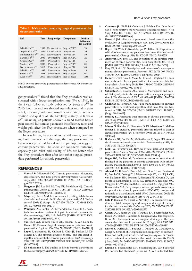
133 July 27, 2014|Volume 6|Issue 7|WJGS|www.wjgnet.com
ger procedure[46] found that the Frey procedure was as-sociated with a lower complication rate (9% vs 15%). In the 8-year follow-up study published by Strate et al[68] in 2005, both procedures showed equivalent mortality, pain relief, exocrine/endocrine insufficiency, rate of reinter-vention and quality of life. Similarly, a study by Keck et al[47] including 92 patients showed a trend toward better pain control but similar pancreatic insufficiency rates and weight gain after the Frey procedure when compared to the Beger procedure.
In conclusion, because of its hybrid nature, combin-ing both resection and drainage, the Frey procedure has been conceptualized based on the pathophysiology of chronic pancreatitis. The short and long-term outcome, especially pain relief and quality of life, are better after the Frey procedure than after any other surgical proce-dure performed for chronic pancreatitis.
REFERENCES1 Etemad B, Whitcomb DC. Chronic pancreatitis: diagnosis,
classification, and new genetic developments. Gastroenter-ology 2001; 120: 682-707 [PMID: 11179244 DOI: 10.1053/gast.2001.22586]
2 Braganza JM, Lee SH, McCloy RF, McMahon MJ. Chronic pancreatitis. Lancet 2011; 377: 1184-1197 [PMID: 21397320 DOI: 10.1016/S0140-6736(10)61852-1]
3 Mayerle J, Lerch MM. Is it necessary to distinguish between alcoholic and nonalcoholic chronic pancreatitis? J Gastro-enterol 2007; 42 Suppl 17: 127-130 [PMID: 17238041 DOI: 10.1007/s00535-006-1920-0]
4 Warshaw AL, Banks PA, Fernández-Del Castillo C. AGA technical review: treatment of pain in chronic pancreatitis. Gastroenterology 1998; 115: 765-776 [PMID: 9721175 DOI: 10.1016/S0016-5085(98)70157-X]
5 van Esch AA, Wilder-Smith OH, Jansen JB, van Goor H, Drenth JP. Pharmacological management of pain in chronic pancreatitis. Dig Liver Dis 2006; 38: 518-526 [PMID: 16627019]
6 Layer P, Yamamoto H, Kalthoff L, Clain JE, Bakken LJ, Di-Magno EP. The different courses of early- and late-onset id-iopathic and alcoholic chronic pancreatitis. Gastroenterology 1994; 107: 1481-1487 [PMID: 7926511 DOI: 10.1016/0016-5085(94)90553-3]
7 Di Sebastiano P. The quality of life in chronic pancreatitis: the role of surgery. JOP 2006; 7: 120-121 [PMID: 16407633]
8 Cameron JL, Riall TS, Coleman J, Belcher KA. One thou-sand consecutive pancreaticoduodenectomies. Ann Surg 2006; 244: 10-15 [PMID: 16794383 DOI: 10.1097/01.sla.0000217673.04165.ea]
9 Howard JM. History of pancreatic head resection – the evaluation of surgical technique. Am J Surg 2007; 194: S6-S10 [DOI: 10.1016/j.amjsurg.2007.05.029]
10 Beger HG, Witte C, Krautzberger W, Bittner R. [Experiences with duodenum-sparing pancreas head resection in chronic pancreatitis]. Chirurg 1980; 51: 303-307 [PMID: 7408575]
11 Andersen DK, Frey CF. The evolution of the surgical treat-ment of chronic pancreatitis. Ann Surg 2010; 251: 18-32 [PMID: 20009754 DOI: 10.1097/SLA.0b013e3181ae3471]
12 Frey CF, Smith GJ. Description and rationale of a new opera-tion for chronic pancreatitis. Pancreas 1987; 2: 701-707 [PMID: 3438308 DOI: 10.1097/00006676-198711000-00014]
13 Demir IE, Tieftrunk E, Maak M, Friess H, Ceyhan GO. Pain mechanisms in chronic pancreatitis: of a master and his fire. Langenbecks Arch Surg 2011; 396: 151-160 [PMID: 21153480 DOI: 10.1007/s00423-010-0731-1]
14 Sakorafas GH, Tsiotou AG, Peros G. Mechanisms and natu-ral history of pain in chronic pancreatitis: a surgical perspec-tive. J Clin Gastroenterol 2007; 41: 689-699 [PMID: 17667054 DOI: 10.1097/MCG.0b013e3180301baf]
15 Chauhan S, Forsmark CE. Pain management in chronic pancreatitis: A treatment algorithm. Best Pract Res Clin Gas-troenterol 2010; 24: 323-335 [PMID: 20510832 DOI: 10.1016/j.bpg.2010.03.007]
16 Bradley EL. Pancreatic duct pressure in chronic pancreatitis. Am J Surg 1982; 144: 313-316 [PMID: 7114368 DOI: 10.1016/0002-9610(82)90008-3]
17 Manes G, Büchler M, Pieramico O, Di Sebastiano P, Malfer-theiner P. Is increased pancreatic pressure related to pain in chronic pancreatitis? Int J Pancreatol 1994; 15: 113-117 [PMID: 8071569]
18 Bockman DE, Buchler M, Malfertheiner P, Beger HG. Analy-sis of nerves in chronic pancreatitis. Gastroenterology 1988; 94: 1459-1469 [PMID: 3360267]
19 Lieb JG, Forsmark CE. Review article: pain and chronic pancreatitis. Aliment Pharmacol Ther 2009; 29: 706-719 [PMID: 19284407 DOI: 10.1111/j.1365-2036.2009.03931.x]
20 Beger HG, Büchler M. Duodenum-preserving resection of the head of the pancreas in chronic pancreatitis with inflam-matory mass in the head. World J Surg 1990; 14: 83-87 [PMID: 2305590 DOI: 10.1007/BF01670550]
21 Ahmed Ali U, Issa Y, Bruno MJ, van Goor H, van Santvoort H, Busch OR, Dejong CH, Nieuwenhuijs VB, van Eijck CH, van Dullemen HM, Fockens P, Siersema PD, Gouma DJ, van Hooft JE, Keulemans Y, Poley JW, Timmer R, Besselink MG, Vleggaar FP, Wilder-Smith OH, Gooszen HG, Dijkgraaf MG, Boermeester MA. Early surgery versus optimal current step-up practice for chronic pancreatitis (ESCAPE): design and rationale of a randomized trial. BMC Gastroenterol 2013; 13: 49 [PMID: 23506415 DOI: 10.1186/1471-230X-13-49]
22 Díte P, Ruzicka M, Zboril V, Novotný I. A prospective, ran-domized trial comparing endoscopic and surgical therapy for chronic pancreatitis. Endoscopy 2003; 35: 553-558 [PMID: 12822088 DOI: 10.1055/s-2003-40237]
23 Cahen DL, Gouma DJ, Nio Y, Rauws EA, Boermeester MA, Busch OR, Stoker J, Laméris JS, Dijkgraaf MG, Huibregtse K, Bruno MJ. Endoscopic versus surgical drainage of the pan-creatic duct in chronic pancreatitis. N Engl J Med 2007; 356: 676-684 [PMID: 17301298 DOI: 10.1056/NEJMoa060610]
24 Rutter K, Ferlitsch A, Sautner T, Püspök A, Götzinger P, Gangl A, Schindl M. Hospitalization, frequency of interven-tions, and quality of life after endoscopic, surgical, or conser-vative treatment in patients with chronic pancreatitis. World J Surg 2010; 34: 2642-2647 [PMID: 20645098 DOI: 10.1007/s00268-010-0713-z]
25 Lamme B, Boermeester MA, Straatsburg IH, van Buijtenen JM, Boerma D, Offerhaus GJ, Gouma DJ, van Gulik TM. Ear-
Table 1 Main studies comparing surgical procedures for chronic pancreatitis
Ref. Year Study design Comparison Median follow-up
(in months)
Izbicki et al[63] 1998 Retrospective Frey vs PPPD 24Aspelund et al[66] 2005 Retrospective Frey vs PD 36Hildebrand et al[53] 2010 Retrospective Frey vs PD 50Farkas et al[64] 2006 Prospective Frey vs PPPD 24Chiang et al[62] 2007 Prospective Frey vs PD 6Strate et al[65] 2008 Prospective Frey vs PPPD 84Bachmann et al[67] 2013 Prospective Frey vs PD 180Izbicki et al[46] 1995 Prospective Frey vs Beger 18Strate et al[68] 2005 Prospective Frey vs Beger 104Keck et al[47] 2010 Retrospective Frey vs Beger 20.6
PPPD: Pylorus-preserving pancreaticoduodenectomy; PD: Pancreatic-oduodenectomy.
Roch A et al . Frey procedure
134 July 27, 2014|Volume 6|Issue 7|WJGS|www.wjgnet.com
ly versus late surgical drainage for obstructive pancreatitis in an experimental model. Br J Surg 2007; 94: 849-854 [PMID: 17335122 DOI: 10.1002/bjs.5722]
26 Nealon WH, Townsend CM, Thompson JC. Operative drain-age of the pancreatic duct delays functional impairment in patients with chronic pancreatitis. A prospective analysis. Ann Surg 1988; 208: 321-329 [PMID: 3421756 DOI: 10.1097/00000658-198809000-00009]
27 Ihse I, Borch K, Larsson J. Chronic pancreatitis: results of op-erations for relief of pain. World J Surg 1990; 14: 53-58 [PMID: 2407038 DOI: 10.1007/BF01670546]
28 Ahmed Ali U, Nieuwenhuijs VB, van Eijck CH, Gooszen HG, van Dam RM, Busch OR, Dijkgraaf MG, Mauritz FA, Jens S, Mast J, van Goor H, Boermeester MA. Clinical out-come in relation to timing of surgery in chronic pancreatitis: a nomogram to predict pain relief. Arch Surg 2012; 147: 925-932 [PMID: 23117832 DOI: 10.1001/archsurg.2012.1094]
29 Nealon WH, Thompson JC. Progressive loss of pancreatic function in chronic pancreatitis is delayed by main pancre-atic duct decompression. A longitudinal prospective analysis of the modified puestow procedure. Ann Surg 1993; 217: 458-66; discussion 466-8 [PMID: 8489308 DOI: 10.1097/00000658-199305010-00005]
30 Lowenfels AB, Maisonneuve P, Cavallini G, Ammann RW, Lankisch PG, Andersen JR, Dimagno EP, Andrén-Sandberg A, Domellöf L. Pancreatitis and the risk of pancreatic cancer. International Pancreatitis Study Group. N Engl J Med 1993; 328: 1433-1437 [PMID: 8479461 DOI: 10.1056/NEJM199305203282001]
31 Talamini G, Falconi M, Bassi C, Sartori N, Salvia R, Caldiron E, Frulloni L, Di Francesco V, Vaona B, Bovo P, Vantini I, Pederzoli P, Cavallini G. Incidence of cancer in the course of chronic pancreatitis. Am J Gastroenterol 1999; 94: 1253-1260 [PMID: 10235203 DOI: 10.1111/j.1572-0241.1999.01075.x]
32 Malka D, Hammel P, Maire F, Rufat P, Madeira I, Pessione F, Lévy P, Ruszniewski P. Risk of pancreatic adenocarci-noma in chronic pancreatitis. Gut 2002; 51: 849-852 [PMID: 12427788 DOI: 10.1136/gut.51.6.849]
33 Ueda J, Tanaka M, Ohtsuka T, Tokunaga S, Shimosegawa T; Research Committee of Intractable Diseases of the Pan-creas. Surgery for chronic pancreatitis decreases the risk for pancreatic cancer: a multicenter retrospective analysis. Surgery 2013; 153: 357-364 [PMID: 22989892 DOI: 10.1016/j.surg.2012.08.005]
34 Bachmann K, Kutup A, Mann O, Yekebas E, Izbicki JR. Sur-gical treatment in chronic pancreatitis timing and type of procedure. Best Pract Res Clin Gastroenterol 2010; 24: 299-310 [PMID: 20510830 DOI: 10.1016/j.bpg.2010.03.003]
35 Bell RH. Current surgical management of chronic pancreati-tis. J Gastrointest Surg 2005; 9: 144-154 [PMID: 15623456 DOI: 10.1016/j.gassur.2004.08.003]
36 Ho HS, Frey CF. The Frey procedure: local resection of pancreatic head combined with lateral pancreaticojejunos-tomy. Arch Surg 2001; 136: 1353-1358 [PMID: 11735858 DOI: 10.1001/archsurg.136.12.1353]
37 Frey CF. The surgical management of chronic pancreatitis: the Frey procedure. Adv Surg 1999; 32: 41-85 [PMID: 9891739]
38 Frey CF, Amikura K. Local resection of the head of the pan-creas combined with longitudinal pancreaticojejunostomy in the management of patients with chronic pancreatitis. Ann Surg 1994; 220: 492-504; discussion 504-507 [PMID: 7524454 DOI: 10.1097/00000658-199410000-00008]
39 Frey CF, Reber HA. Local resection of the head of the pan-creas with pancreaticojejunostomy. J Gastrointest Surg 2005; 9: 863-868 [PMID: 16189887 DOI: 10.1016/j.gassur.2004.09.029]
40 Pessaux P, Arnaud JP. [The Frey technique (ninety-five per-cent distal pancreatectomy) for chronic pancreatitis]. J Chir (Paris) 2007; 144: 135-138 [PMID: 17607230 DOI: 10.1016/S0021-7697(07)89487-7]
41 Amudhan A, Balachandar TG, Kannan DG, Rajarathinam G,
Vimalraj V, Rajendran S, Ravichandran P, Jeswanth S, Suren-dran R. Factors affecting outcome after Frey procedure for chronic pancreatitis. HPB (Oxford) 2008; 10: 477-482 [PMID: 19088936 DOI: 10.1080/13651820802392338]
42 Brennan DD, Kriskal JB, Kane AR. Intraoperative ultra-sound of the pancreas. Ultra Sound Clin 2006; 1: 533-45 [DOI: 10.1016/j.cult.2006.05.005]
43 Cooper MA, Datta TS, Makary MA. Laparoscopic frey procedure for chronic pancreatitis. Surg Laparosc Endosc Per-cutan Tech 2014; 24: e16-e20 [PMID: 24487169 DOI: 10.1097/SLE.0b013e31828f6edf]
44 Venkat R, Edil BH, Schulick RD, Lidor AO, Makary MA, Wolfgang CL. Laparoscopic distal pancreatectomy is associ-ated with significantly less overall morbidity compared to the open technique: a systematic review and meta-analysis. Ann Surg 2012; 255: 1048-1059 [PMID: 22511003 DOI: 10.1097/SLA.0b013e318251ee09]
45 Frey CF, Mayer KL. Comparison of local resection of the head of the pancreas combined with longitudinal pancreati-cojejunostomy (frey procedure) and duodenum-preserving resection of the pancreatic head (beger procedure). World J Surg 2003; 27: 1217-1230 [PMID: 14534821 DOI: 10.1007/s00268-003-7241-z]
46 Izbicki JR, Bloechle C, Knoefel WT, Kuechler T, Binmoeller KF, Broelsch CE. Duodenum-preserving resection of the head of the pancreas in chronic pancreatitis. A prospec-tive, randomized trial. Ann Surg 1995; 221: 350-358 [PMID: 7726670 DOI: 10.1097/00000658-199504000-00004]
47 Keck T, Wellner UF, Riediger H, Adam U, Sick O, Hopt UT, Makowiec F. Long-term outcome after 92 duodenum-preserving pancreatic head resections for chronic pancreati-tis: comparison of Beger and Frey procedures. J Gastrointest Surg 2010; 14: 549-556 [PMID: 20033344 DOI: 10.1007/s11605-009-1119-9]
48 Chaudhary A, Negi SS, Masood S, Thombare M. Complica-tions after Frey’s procedure for chronic pancreatitis. Am J Surg 2004; 188: 277-281 [PMID: 15450834 DOI: 10.1016/j.amjsurg.2004.06.012]
49 Pessaux P, Kianmanesh R, Regimbeau JM, Sastre B, Del-censerie R, Sielezneff I, Arnaud JP, Sauvanet A. Frey pro-cedure in the treatment of chronic pancreatitis: short-term results. Pancreas 2006; 33: 354-358 [PMID: 17079939 DOI: 10.1097/01.mpa.0000236736.77359.3a]
50 Roch AM, Brachet D, Lermite E, Pessaux P, Arnaud JP. Frey procedure in patients with chronic pancreatitis: short and long-term outcome from a prospective study. J Gastrointest Surg 2012; 16: 1362-1369 [PMID: 22580839 DOI: 10.1007/s11605-012-1904-8]
51 Negi S, Singh A, Chaudhary A. Pain relief after Frey’s pro-cedure for chronic pancreatitis. Br J Surg 2010; 97: 1087-1095 [PMID: 20632276 DOI: 10.1002/bjs.7042]
52 Falconi M, Bassi C, Casetti L, Mantovani W, Mascetta G, Sartori N, Frulloni L, Pederzoli P. Long-term results of Frey’s procedure for chronic pancreatitis: a longitudinal prospec-tive study on 40 patients. J Gastrointest Surg 2006; 10: 504-510 [PMID: 16627215 DOI: 10.1016/j.gassur.2005.09.011]
53 Hildebrand P, Duderstadt S, Jungbluth T, Roblick UJ, Bruch HP, Czymek R. Evaluation of the quality of life after surgi-cal treatment of chronic pancreatitis. JOP 2011; 12: 364-371 [PMID: 21737898]
54 Hartel M, Tempia-Caliera AA, Wente MN, Z’graggen K, Friess H, Büchler MW. Evidence-based surgery in chronic pancreatitis. Langenbecks Arch Surg 2003; 388: 132-139 [PMID: 12712343]
55 Riediger H, Adam U, Fischer E, Keck T, Pfeffer F, Hopt UT, Makowiec F. Long-term outcome after resection for chronic pancreatitis in 224 patients. J Gastrointest Surg 2007; 11: 949-959; discussion 959-960 [PMID: 17534689 DOI: 10.1007/s11605-007-0155-6]
56 Ramesh H, Jacob G, Lekha V, Venugopal A. Ductal drain-
Roch A et al . Frey procedure
135 July 27, 2014|Volume 6|Issue 7|WJGS|www.wjgnet.com
age with head coring in chronic pancreatitis with small-duct disease. J Hepatobiliary Pancreat Surg 2003; 10: 366-372 [PMID: 14598137 DOI: 10.1007/s00534-002-0827-2]
57 Shrikhande SV, Kleeff J, Friess H, Büchler MW. Manage-ment of pain in small duct chronic pancreatitis. J Gastrointest Surg 2006; 10: 227-233 [PMID: 16455455 DOI: 10.1016/j.gassur.2005.09.004]
58 Cooper MA, Makary MA, Ng J, Cui Y, Singh VK, Matsu-kuma K, Andersen DK. Extent of pancreatic fibrosis as a de-terminant of symptom resolution after the Frey procedure: a clinico-pathologic analysis. J Gastrointest Surg 2013; 17: 682-687 [PMID: 23345052 DOI: 10.1007/s11605-012-2110-4]
59 Izbicki JR, Bloechle C, Broering DC, Kuechler T, Broelsch CE. Longitudinal V-shaped excision of the ventral pancreas for small duct disease in severe chronic pancreatitis: pro-spective evaluation of a new surgical procedure. Ann Surg 1998; 227: 213-219 [PMID: 9488519 DOI: 10.1097/00000658-199802000-00010]
60 Yekebas EF, Bogoevski D, Honarpisheh H, Cataldegirmen G, Habermann CR, Seewald S, Link BC, Kaifi JT, Wolfram L, Mann O, Bubenheim M, Izbicki JR. Long-term follow-up in small duct chronic pancreatitis: A plea for extended drain-age by “V-shaped excision” of the anterior aspect of the pan-creas. Ann Surg 2006; 244: 940-946; discussion 946-948 [PMID: 17122619 DOI: 10.1097/01.sla.0000246914.25884.e9]
61 Yin Z, Sun J, Yin D, Wang J. Surgical treatment strategies in chronic pancreatitis: a meta-analysis. Arch Surg 2012; 147: 961-968 [PMID: 23070412 DOI: 10.1001/archsurg.2012.2005]
62 Chiang KC, Yeh CN, Hsu JT, Chen HM, Chen HY, Hwang TL, Jan YY, Chen MF. Pancreaticoduodenectomy versus Frey’s procedure for chronic pancreatitis: preliminary data on outcome and pancreatic function. Surg Today 2007; 37: 961-966 [PMID: 17952526 DOI: 10.1007/s00595-007-3539-z]
63 Izbicki JR, Bloechle C, Broering DC, Knoefel WT, Kuechler T,
Broelsch CE. Extended drainage versus resection in surgery for chronic pancreatitis: a prospective randomized trial com-paring the longitudinal pancreaticojejunostomy combined with local pancreatic head excision with the pylorus-preserv-ing pancreatoduodenectomy. Ann Surg 1998; 228: 771-779 [PMID: 9860476 DOI: 10.1097/00000658-199812000-00008]
64 Farkas G, Leindler L, Daróczi M, Farkas G. Prospective randomised comparison of organ-preserving pancreatic head resection with pylorus-preserving pancreaticoduode-nectomy. Langenbecks Arch Surg 2006; 391: 338-342 [PMID: 16680474 DOI: 10.1007/s00423-006-0051-7]
65 Strate T, Bachmann K, Busch P, Mann O, Schneider C, Bruhn JP, Yekebas E, Kuechler T, Bloechle C, Izbicki JR. Resection vs drainage in treatment of chronic pancreatitis: long-term results of a randomized trial. Gastroenterology 2008; 134: 1406-1411 [PMID: 18471517 DOI: 10.1053/j.gastro.2008.02.056]
66 Aspelund G, Topazian MD, Lee JH, Andersen DK. Im-proved outcomes for benign disease with limited pancreatic head resection. J Gastrointest Surg 2005; 9: 400-409 [PMID: 15749604 DOI: 10.1016/j.gassur.2004.08.015]
67 Bachmann K, Tomkoetter L, Kutup A, Erbes J, Vashist Y, Mann O, Bockhorn M, Izbicki JR. Is the Whipple procedure harmful for long-term outcome in treatment of chronic pancreatitis? 15-years follow-up comparing the outcome after pylorus-preserving pancreatoduodenectomy and Frey procedure in chronic pancreatitis. Ann Surg 2013; 258: 815-820; discussion 820-821 [PMID: 24096767 DOI: 10.1097/SLA.0b013e3182a655a8]
68 Strate T, Taherpour Z, Bloechle C, Mann O, Bruhn JP, Schneider C, Kuechler T, Yekebas E, Izbicki JR. Long-term follow-up of a randomized trial comparing the beger and frey procedures for patients suffering from chronic pancre-atitis. Ann Surg 2005; 241: 591-598 [PMID: 15798460 DOI: 10.1097/01.sla.0000157268.78543.03]
P- Reviewer: Bradley EL, Sumi S S- Editor: Song XX L- Editor: A E- Editor: Wang CH
Roch A et al . Frey procedure
Contrast enhanced computed tomography demonstrat-ed that the cysts had thin walls, without calcifications, pancreatic duct dilation or pancreatic parenchyma inva-sion. Endoscopic ultrasound with fine needle aspiration (EUS/FNA) confirmed the diagnosis of a lymphoepithe-lial cyst in 3 patients, one of whom was spared an op-eration and continues to do well after 6 years. Eleven patients had a resection: 3 pancreaticoduodenecto-mies, 7 distal pancreatectomies, and 1 enucleation. The median size of the cysts was 3 cm (range 2-20 cm). At a median follow-up of 57 mo no recurrences or other pancreas-related conditions occurred.
CONCLUSION: Lymphoepithelial cysts and cystic lymphangiomas of the pancreas can be diagnosed with a combination of contrast-enhanced computed tomog-raphy scans and EUS/FNA. If the lesion is asymptom-atic, an operation might be avoided.
© 2014 Baishideng Publishing Group Inc. All rights reserved.
Key words: Lymphoepithelial cysts; Cystic lymphangio-mas; Pancreas; Asymptomatic cysts; Benign cystic le-sions of the pancreas
Core tip: Lymphoepithelial cysts and cystic lymphan-giomas of the pancreas represent rare, benign cystic lesions. The experience with their diagnosis and treat-ment is limited mostly to case reports. This report describes our experience with twelve lymphoepithelial cysts and cystic lymhangiomas of the pancreas, analyz-ing their clinicopathologic characteristics, the role of contrast enhanced computed tomography and endo-scopic ultrasound with fine needle aspiration for their diagnosis with an emphasis on non-surgical manage-ment when a correct diagnosis can be established in an asymptomatic patient.
Lymphoepithelial cysts and cystic lymphangiomas: Under-recognized benign cystic lesions of the pancreas
Ioannis T Konstantinidis, Avinash Kambadakone, Onofrio A Catalano, Dushyant V Sahani, Vikram Deshpande, David G Forcione, Jennifer A Wargo, Carlos Fernandez-del Castillo, Keith D Lillemoe, Andrew L Warshaw, Cristina R Ferrone
Ioannis T Konstantinidis, Jennifer A Wargo, Carlos Fernan-dez-del Castillo, Keith D Lillemoe, Andrew L Warshaw, Cris-tina R Ferrone, Departments of Surgery, Massachusetts General Hospital, Boston, MA 02114, United StatesIoannis T Konstantinidis, Department of Surgery, University of Arizona, Tucson, AZ 85724, United StatesVikram Deshpande, Departments of Pathology, Massachusetts General Hospital, Boston, MA 02114, United StatesAvinash Kambadakone, Onofrio A Catalano, Dushyant V Sahani, Departments of Radiology, Massachusetts General Hos-pital, Boston, MA 02114, United StatesOnofrio A Catalano, Department of Radiology, University of Napoli, 80138 Napoli, ItalyDavid G Forcione, Departments of Gastroenterology, Massachu-setts General Hospital, Boston, MA 02114, United StatesAuthor contributions: All the authors contributed to this work.Correspondence to: Cristina R Ferrone, MD, Assistant Pro-fessor, Departments of Surgery, Massachusetts General Hospital, Wang Ambulatory Care Center 460, 15 Parkman Street, Boston, MA 02114, United States. [email protected]: +1-617-6436189 Fax: +1-617-6436116Received: March 10, 2014 Revised: May 5, 2014Accepted: July 12, 2014Published online: July 27, 2014
Abstract AIM: To identify their diagnostic and prognostic clinical characteristics in a large series.
METHODS: Retrospective review of clinicopathologic and imaging characteristics of patients diagnosed with lymphoepithelial cysts and cystic lymphangiomas of the pancreas at Massachusetts General Hospital.
RESULTS: Twelve patients were identified between 1/1/1997 and 8/1/2007. Their median age was 55.5 years (range 19-78 years), and 6 were females. The le-sion was incidentally discovered in half of the patients.
ORIGINAL ARTICLE
Submit a Manuscript: http://www.wjgnet.com/esps/Help Desk: http://www.wjgnet.com/esps/helpdesk.aspxDOI: 10.4240/wjgs.v6.i7.136
136 July 27, 2014|Volume 6|Issue 7|WJGS|www.wjgnet.com
World J Gastrointest Surg 2014 July 27; 6(7): 136-141ISSN 1948-9366 (online)
© 2014 Baishideng Publishing Group Inc. All rights reserved.

Konstantinidis IT, Kambadakone A, Catalano OA, Sahani DV, Deshpande V, Forcione DG, Wargo JA, Fernandez-del Castillo C, Lillemoe KD, Warshaw AL, Ferrone CR. Lymphoepithelial cysts and cystic lymphangiomas: Under-recognized benign cystic lesions of the pancreas. World J Gastrointest Surg 2014; 6(7): 136-141 Available from: URL: http://www.wjgnet.com/1948-9366/full/v6/i7/136.htm DOI: http://dx.doi.org/10.4240/wjgs.v6.i7.136
INTRODUCTIONThe differential diagnosis of cystic lesions of the pan-creas includes a variety of inflammatory and neoplastic lesions, some of which have malignant potential, any of which may be symptomatic or not[1].
Among the least known pancreatic cystic lesions are the lymphoepithelial cysts and the cystic lymphangiomas[2]. Their natural history is unknown, but asymptomatic cysts might be left alone if diagnosed accurately. A diagnosis may be possible with a combination of imaging and endo-scopic ultrasound with fine needle aspiration (EUS/FNA).
In this study we report our experience with twelve patients diagnosed with lymphoepithelial cysts or cystic lymphangiomas over a period of more than 10 years, focusing on diagnostic evaluation and surgical treatment and exploring the potential of avoiding an operation on asymptomatic patients.
MATERIALS AND METHODSStudy designReview of a pathology database was performed to iden-tify patients diagnosed with lymphoepithelial cysts and cystic lymphangiomas between 1/1/1997-8/1/2007. A prospectively maintained surgical database since 1/2001 was used to identify the relative frequency of the lym-phoepithelial cysts and cystic lymphangiomas among the pancreatic cysts who underwent surgical resection.
Clinical data evaluated included gender, age, presenting symptoms, past medical history, laboratory values, operative procedures and pathology reports. The available computed tomography (CT) scans and endoscopic ultrasound studies performed at the Massachusetts General Hospital were re-reviewed. Pancreatic fistula was defined according to the international study group definition[3]. Operative mortality was defined as death within 30 d of the operation.
Ethics This study was approved by the institutional review board (IRB) of the Massachusetts General Hospital.
Statistical analysisStatistical analysis was conducted using SPSS software (version 20.0; SPSS, Chicago, Ill). Continuous variables are shown as median and range. Categorical or dichoto-mous data are presented in frequencies and percentage (%) as appropriate. This study was approved by the IRB of the Massachusetts General Hospital (MGH).
RESULTSBetween 1997 and 2007, 12 patients with cystic lymphan-giomas or lymphoepithelial cysts were identified, repre-senting approximately 2% of the pancreatic cystic lesions resected during this period. Median age for these 12 patients was 55.5 years (range 19-78) and 6 (50%) were females. The clinicopathologic data of these patients are shown in Table 1. Half of the patients were asymptom-atic. Of the 6 symptomatic patients, 4 presented with abdominal pain (two patients presented with severe, abrupt, abdominal pain and the other two with episodes of right lower and left upper quadrant abdominal pain), 1 with fever, and 1 with nausea. One patient had previously undergone resection of ruptured lymphangiomas at an outside hospital 5 and 10 mo prior to presentation. He underwent a distal pancreatectomy for a second recur-rence of a retroperitoneal lymphangioma.
Diagnostic evaluation The results of the diagnostic evaluation of these patients are shown in Table 2. All of the patients had normal
137 July 27, 2014|Volume 6|Issue 7|WJGS|www.wjgnet.com
Konstantinidis IT et al . Lymphoepithelial cysts/lymhangiomas pancreas
Table 1 Clinicopathologic characteristics of 12 patients n (%)
Factors LECP (n = 8) Lymphangioma (n = 4)
Median age, yr (range) 60.5 (24-78) 37.5 (19-69)Females 2 (25) 4 (100)Symptomatic 4 (50) 2 (50) Abdominal pain 2 (25) 2 (50) Other (nausea, fever) 2 (25) 0Operation 7 (87.5) 4 (100) Distal pancreatectomy 4 (50) 3 (75) Pancreaticoduodenectomy 2 (25) 1 (25) Enucleation 1 (12.5) 0Pathology Median size, cm (range) 2 (2-7.6) 10.5 (2.5-20)
LECP: Lymphoepithelial cyst of the pancreas.
Table 2 Diagnostic studies n (%)
Factors LECP (n = 8)
Lymphangioma (n = 4)
Median CA 19-9, U/mL (range) 35 (3-79) 10 (9-36)CT characteristics Mean size, cm (range) 2.3 (1.5-2.9) 9.2 (2.3-16.5) Loculations 1 (25) 0 Microcystic 1 (25) 0 Central scar 0 0 Septations 3 (75) 2 (50) Calcifications 0 0 Mural nodules 0 1 (25) Thin cyst wall 4 (100) 4 (100) Pancreatic Duct Dilatation 0 0 Other (vascular invasion, lymphadenopathy)
0 0
FNA (n = 6) (n = 1) Sufficient 4 (67) 1 (100) Diagnostic 3 (50) 0
LECP: Lymphoepithelial cyst of the pancreas; FNA: Fine needle aspiration; CT: Computed tomography.
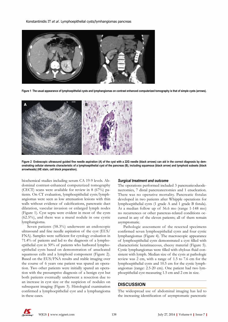
biochemical studies including serum CA 19-9 levels. Ab-dominal contrast-enhanced computerized tomography (CECT) scans were available for review in 8 (67%) pa-tients. On CT evaluation, lymphoepithelial cysts/lymph-angiomas were seen as low attenuation lesions with thin walls without evidence of calcifications, pancreatic duct dilatation, vascular invasion or enlarged lymph nodes (Figure 1). Cyst septa were evident in most of the cysts (62.5%), and there was a mural nodule in one cystic lymphangioma.
Seven patients (58.3%) underwent an endoscopic ultrasound and fine needle aspiration of the cyst (EUS/FNA). Samples were sufficient for cytology evaluation in 71.4% of patients and led to the diagnosis of a lympho-epithelial cyst in 50% of patients who harbored lympho-epithelial cysts based on demonstration of anucleated squamous cells and a lymphoid component (Figure 2). Based on the EUS/FNA results and stable imaging over the course of 6 years one patient was spared an opera-tion. Two other patients were initially spared an opera-tion with the presumptive diagnosis of a benign cyst but both patients eventually underwent a resection due to an increase in cyst size or the suspicion of nodules on subsequent imaging (Figure 3). Histological examination confirmed a lymphoepithelial cyst and a lymphangioma in these cases.
Surgical treatment and outcomeThe operations performed included 3 pancreaticoduode-nectomies, 7 distal pancreatectomies and 1 enucleation. There was no operative mortality. Pancreatic fistulas developed in two patients after Whipple operations for lymphoepithelial cysts (1 grade A and 1 grade B fistula). At a median follow up of 56.6 mo (range 1-148 mo) no recurrences or other pancreas-related conditions oc-curred in any of the eleven patients; all of them remain asymptomatic.
Pathologic assessment of the resected specimens confirmed seven lymphoepithelial cysts and four cystic lymphangiomas (Figure 4). The macroscopic appearance of lymphoepithelial cysts demonstrated a cyst filled with characteristic keratinaceous, cheesy material (Figure 5). Cystic lymphangiomas were filled with chylous fluid con-sistent with lymph. Median size of the cysts at pathologic review was 2 cm, with a range of 1.5 to 7.6 cm for the lymphoepithelial cysts and 10.5 cm for the cystic lymph-angiomas (range: 2.5-20 cm). One patient had two lym-phoepithelial cyst measuring 1.5 cm and 2 cm in size.
DISCUSSION
The widespread use of abdominal imaging has led to the increasing identification of asymptomatic pancreatic
138 July 27, 2014|Volume 6|Issue 7|WJGS|www.wjgnet.com
Figure 1 The usual appearance of lymphoepithelial cysts and lymphangiomas on contrast enhanced computerized tomography is that of simple cysts (arrows).
Figure 2 Endoscopic ultrasound guided fine needle aspiration (A) of the cyst with a 22G needle (black arrows) can aid in the correct diagnosis by dem-onstrating cellular elements characteristic of a lymphoepithelial cyst of the pancreas (B), including squamous (black arrow) and lymphoid subsets (black arrowheads) (HE stain, cell block preparation).
A B
Konstantinidis IT et al . Lymphoepithelial cysts/lymhangiomas pancreas

139 July 27, 2014|Volume 6|Issue 7|WJGS|www.wjgnet.com
ten found in men in their fifth and six decades of life; 75% of our patients were males who had a median age of 60.5 years. Their characteristic pathologic features are a squamous epithelial lining and surrounding lymphoid tissue, which can be identified on EUS/FNA[1].
Cystic lymphangiomas are benign multicystic lesions that are believed to result from blockage of the lymphat-ic system and, for unknown reasons, are more common in young females. They can be very large in size and they are frequently located in the peripancreatic tissues in close association with the pancreas. They harbor an endothelial cell lining[10]. In our series four patients were female and had median age of 37.5 years. The largest le-sion was 16.5 cm.
The preoperative diagnostic evaluation of the patients in our study included a combination of biochemical and tumor markers, CECT scans of the abdomen and EUS/FNA. Although elevations of serum CA 19-9[11], cyst flu-id CA 19-9[12] and persistence of elevated CA 19-9 post-resection[13] have been described in lymphoepithelial cysts, none of our patients had such abnormal levels. Thus, we believe that this marker has no utility in the clinical assessment of these cystic lesions[14]. Consistent with published reports[15], CECT scans in our patients demon-strated cysts with thin walls, without evidence of calcifi-
cystic lesions. Seventy-one percent of cysts in a recent report from our institution were serendipitous imaging findings[4]. It becomes likely that uncommon lymphoepi-thelial cysts and cystic lymphangiomas will increasingly be found incidentally. Although an accurate preoperative diagnosis of pancreatic cysts is not always feasible, crite-ria associated with an increased risk of malignancy have been established by expert consensus[5] and subsequently validated[6,7] and updated[8]. Establishing a correct diagno-sis in a benign asymptomatic cyst can spare the patient a pancreatectomy, which even in specialized tertiary centers carries significant morbidity[9].
This report, representing one of the largest case se-ries describing the clinicopathologic characteristics of lymphoepithelial cysts and cystic lymphangiomas, pro-vides insights into their correct management and spot-lights establishment of an accurate diagnosis to avoid an operation in asymptomatic patients. In 3 (50%) of our asymptomatic patients a non-surgical approach was ini-tially chosen on the basis of the probability of a benign lesion in an asymptomatic patient. Two of those eventu-ally underwent resection because of new suspicious im-aging findings, but the third continues to do well without intervention during follow-up of 6 years.
Lymphoepithelial cysts of the pancreas are most of-
Figure 3 Lymphoepithelial cyst of the pancreas may increase in size during observation. Contrast enhanced computed tomography images demonstrate that the cyst increased from 2.6 cm (A, black arrow) to 4.2 cm (B, white arrow) over a two-month period.
Figure 4 Lymphoepithelial cyst of the pancreas lined by squamous epi-thelium (black arrows), underlying lymphoid tissue (white arrowheads). Pancreatic ductules are also identified (black arrowheads) (HE stain).
Figure 5 Photo of a transected lymphoepithelial cyst. The characteristic keratinaceous cheesy material is shown (arrows). Inset: the multilocular cyst (arrowheads).
A B
Konstantinidis IT et al . Lymphoepithelial cysts/lymhangiomas pancreas

140 July 27, 2014|Volume 6|Issue 7|WJGS|www.wjgnet.com
cations, pancreatic duct dilation or local invasion. Cystic lymphangiomas additionally contain septa, appear multi-loculated, and may have papillary projections[16]. A mural nodule (which proved to be an organizing hematoma) was seen in one of our patients who had a lymphangio-ma. On EUS/FNA lymphoepithelial cysts demonstrate anucleated squamous cells on cytology while their pathol-ogy is consistent with a keratinized squamous lining with a lymphocytic infiltrate in the cyst wall[1]. This character-istic, identifiable in 75% of the fine needle aspirations in our study, helps the differentiation from other squamous epithelium-lined cysts (dermoid cysts, splenic epidermoid cysts, squamous cell cancer, primary or metastatic)[12,17-20]. The cyst aspirates may be thick, milky, gray or frothy[2]. Cystic lymphangiomas characteristically contain chylous, milky fluid with a very high triglyceride level, commonly > 3000-5000 mg/dL, consistent with lymph. Cytologic features are consistent with lymphoid tissue[21-23]. They are lined by endothelium with immunohistochemical markers including factor VIII-R Ag, CD 31 and CD 34[10].
Our experience with these uncommon benign pan-creatic cysts demonstrates that accurate diagnosis may be feasible through a combination of a contrast-enhanced abdominal CT scan and cyst fluid analyses acquired via endoscopic ultrasound and fine needle aspiration. These results can direct the appropriate management strategy, and asymptomatic patients can be spared an operation.
ACKNOWLEDGMENTSPart of this work was presented at the 2008 Annual Meeting of the American Pancreatic Association Novem-ber 6-7, 2008 Chicago Illinois.
COMMENTSBackgroundThe treatment of pancreatic cysts is a continuously evolving field. Amongst the least well studied pancreatic cystic lesions are lymphoepithelial cysts and cystic lymphangiomas of the pancreas. Familiarity with their correct diagnosis is cru-cial as they can be followed non-operatively in asymptomatic patients. Research frontiersThe existing literature on lymphoepithelial cysts and cystic lymphangiomas of the pancreas is limited to case reports. In this report the authors demonstrate our experience with twelve patients, one of the largest single institution experi-ence reported, emphasizing on their correct diagnosis and treatment. Innovations and breakthroughsLymphoepithelial cysts and cystic lymphangiomas of the pancreas can be diag-nosed with a combination of contrast-enhanced computed tomography scans and endoscopic ultrasound with fine needle aspiration. Conservative management avoid-ing a major surgery can be followed as long as the lesions remain asymptomatic. Applications The description of the experience with the diagnosis and treatment of these rare pancreatic cystic lesions will aid in their safe management. TerminologyLymphoepithelial cysts and cystic lymphangiomas of the pancreas represent rare, benign cystic lesions. Their lining is being characterized by squamous epi-thelial lining with surrounding lymphoid tissue and endothelial cells respectively. Peer reviewThis is a retrospective study regarding lymphoepithelial cysts or cystic lymphan-giomas in 12 patients. This is a very uncommon pancreatic pathology and the paper carries a significant number of patients.
REFERENCES1 Adsay NV, Hasteh F, Cheng JD, Bejarano PA, Lauwers GY,
Batts KP, Klöppel G, Klimstra DS. Lymphoepithelial cysts of the pancreas: a report of 12 cases and a review of the lit-erature. Mod Pathol 2002; 15: 492-501 [PMID: 12011254 DOI: 10.1038/modpathol.3880553]
2 Sakorafas GH, Smyrniotis V, Reid-Lombardo KM, Sarr MG. Primary pancreatic cystic neoplasms of the pancreas revisited. Part IV: rare cystic neoplasms. Surg Oncol 2012; 21: 153-163 [PMID: 21816607 DOI: 10.1016/j.suronc.2011.06.007]
3 Bassi C, Dervenis C, Butturini G, Fingerhut A, Yeo C, Izbicki J, Neoptolemos J, Sarr M, Traverso W, Buchler M; Interna-tional Study Group on Pancreatic Fistula D. Postoperative pancreatic fistula: an international study group (ISGPF) definition. Surgery 2005; 138: 8-13 [PMID: 16003309 DOI: 10.1016/j.surg.2005.05.001]
4 Ferrone CR, Correa-Gallego C, Warshaw AL, Brugge WR, Forcione DG, Thayer SP, Fernández-del Castillo C. Current trends in pancreatic cystic neoplasms. Arch Surg 2009; 144: 448-454 [PMID: 19451487 DOI: 10.1001/archsurg.2009.36]
5 Tanaka M, Chari S, Adsay V, Fernandez-del Castillo C, Falco-ni M, Shimizu M, Yamaguchi K, Yamao K, Matsuno S; Inter-national Association of P. International consensus guidelines for management of intraductal papillary mucinous neoplasms and mucinous cystic neoplasms of the pancreas. Pancreatology 2006; 6: 17-32 [PMID: 16327281 DOI: 10.1159/000090023]
6 Rodriguez JR, Salvia R, Crippa S, Warshaw AL, Bassi C, Fal-coni M, Thayer SP, Lauwers GY, Capelli P, Mino-Kenudson M, Razo O, McGrath D, Pederzoli P, Fernández-Del Castillo C. Branch-duct intraductal papillary mucinous neoplasms: observations in 145 patients who underwent resection. Gas-troenterology 2007; 133: 72-79; quiz 309-310 [PMID: 17631133 DOI: 10.1053/j.gastro.2007.05.010]
7 Pelaez-Luna M, Chari ST, Smyrk TC, Takahashi N, Clain JE, Levy MJ, Pearson RK, Petersen BT, Topazian MD, Vege SS, Kendrick M, Farnell MB. Do consensus indications for resection in branch duct intraductal papillary mucinous neoplasm predict malignancy? A study of 147 patients. Am J Gastroenterol 2007; 102: 1759-1764 [PMID: 17686073 DOI: 10.1111/j.1572-0241.2007.01224.x]
8 Tanaka M, Fernández-del Castillo C, Adsay V, Chari S, Falconi M, Jang JY, Kimura W, Levy P, Pitman MB, Schmidt CM, Shimizu M, Wolfgang CL, Yamaguchi K, Yamao K; International Association of Pancreatology. International consensus guidelines 2012 for the management of IPMN and MCN of the pancreas. Pancreatology 2012; 12: 183-197 [PMID: 22687371 DOI: 10.1016/j.pan.2012.04.004]
9 Fernández-del Castillo C, Morales-Oyarvide V, McGrath D, Wargo JA, Ferrone CR, Thayer SP, Lillemoe KD, Warshaw AL. Evolution of the Whipple procedure at the Massachu-setts General Hospital. Surgery 2012; 152: S56-S63 [PMID: 22770961 DOI: 10.1016/j.surg.2012.05.022]
10 Paal E, Thompson LD, Heffess CS. A clinicopathologic and immunohistochemical study of ten pancreatic lymphangio-mas and a review of the literature. Cancer 1998; 82: 2150-2158 [PMID: 9610694]
11 Yamaguchi T, Takahashi H, Kagawa R, Takeda R, Sakata S, Yamamoto M, Nishizaki D. Lymphoepithelial cyst of the pancreas associated with elevated CA 19-9 levels. J Hepato-biliary Pancreat Surg 2008; 15: 652-654 [PMID: 18987938 DOI: 10.1007/s00534-007-1314-6]
12 Centeno BA, Stockwell JW, Lewandrowski KB. Cyst fluid cytology and chemical features in a case of lymphoepithelial cyst of the pancreas: A rare and difficult preoperative diag-nosis. Diagn Cytopathol 1999; 21: 328-330 [PMID: 10527479]
13 Yamamoto K, Fujimoto K, Matsushiro T, Ota K. Lympho-epithelial cyst in the pancreas: a case report. Gastroenterol Jpn 1990; 25: 758-761 [PMID: 2279638]
14 Tsuchiya Y, Suzuki S, Sakaguchi T, Kojima Y, Okamoto K,
COMMENTS
Konstantinidis IT et al . Lymphoepithelial cysts/lymhangiomas pancreas
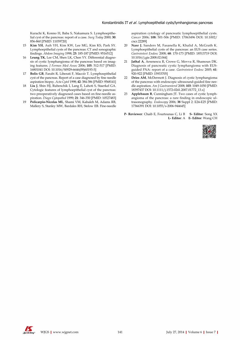
141 July 27, 2014|Volume 6|Issue 7|WJGS|www.wjgnet.com
Kurachi K, Konno H, Baba S, Nakamura S. Lymphoepithe-lial cyst of the pancreas: report of a case. Surg Today 2000; 30: 856-860 [PMID: 11039720]
15 Kim YH, Auh YH, Kim KW, Lee MG, Kim KS, Park SY. Lymphoepithelial cysts of the pancreas: CT and sonographic findings. Abdom Imaging 1998; 23: 185-187 [PMID: 9516512]
16 Leung TK, Lee CM, Shen LK, Chen YY. Differential diagno-sis of cystic lymphangioma of the pancreas based on imag-ing features. J Formos Med Assoc 2006; 105: 512-517 [PMID: 16801041 DOI: 10.1016/S0929-6646(09)60193-5]
17 Bolis GB, Farabi R, Liberati F, Macciò T. Lymphoepithelial cyst of the pancreas. Report of a case diagnosed by fine needle aspiration biopsy. Acta Cytol 1998; 42: 384-386 [PMID: 9568141]
18 Liu J, Shin HJ, Rubenchik I, Lang E, Lahoti S, Staerkel GA. Cytologic features of lymphoepithelial cyst of the pancreas: two preoperatively diagnosed cases based on fine-needle as-piration. Diagn Cytopathol 1999; 21: 346-350 [PMID: 10527483]
19 Policarpio-Nicolas ML, Shami VM, Kahaleh M, Adams RB, Mallery S, Stanley MW, Bardales RH, Stelow EB. Fine-needle
aspiration cytology of pancreatic lymphoepithelial cysts. Cancer 2006; 108: 501-506 [PMID: 17063496 DOI: 10.1002/cncr.22289]
20 Nasr J, Sanders M, Fasanella K, Khalid A, McGrath K. Lymphoepithelial cysts of the pancreas: an EUS case series. Gastrointest Endosc 2008; 68: 170-173 [PMID: 18513719 DOI: 10.1016/j.gie.2008.02.044]
21 Jathal A, Arsenescu R, Crowe G, Movva R, Shamoun DK. Diagnosis of pancreatic cystic lymphangioma with EUS-guided FNA: report of a case. Gastrointest Endosc 2005; 61: 920-922 [PMID: 15933705]
22 Dries AM, McDermott J. Diagnosis of cystic lymphangioma of the pancreas with endoscopic ultrasound-guided fine nee-dle aspiration. Am J Gastroenterol 2008; 103: 1049-1050 [PMID: 18397437 DOI: 10.1111/j.1572-0241.2007.01772_13.x]
23 Applebaum B, Cunningham JT. Two cases of cystic lymph-angioma of the pancreas: a rare finding in endoscopic ul-trasonography. Endoscopy 2006; 38 Suppl 2: E24-E25 [PMID: 17366391 DOI: 10.1055/s-2006-944645]
P- Reviewer: Chaib E, Fourtounas C, Li B S- Editor: Song XX L- Editor: A E- Editor: Wang CH
Konstantinidis IT et al . Lymphoepithelial cysts/lymhangiomas pancreas

Core tip: Case reports suggest that isotretinoin ad-ministration may trigger inflammatory bowel diseases. This hypothesis has raised great scientific interest and numerous propositions addressing the pathophysiology of this potent association have been made. However, demographic data do not support a correlation between isotretinoin and inflammatory bowel disease. The cur-rent case describes a patient who developed ulcerative colitis while on isotretinoin administration. We hope that this case report may contribute to future epide-miological studies with scope to clarify the association between isotretinoin and inflammatory bowel diseases and more specifically ulcerative colitis.
Papaconstantinou I, Stefanopoulos A, Papailia A, Zeglinas C, Georgopoulos I, Michopoulos S. Isotretinoin and ulcerative coli-tis: A case report and review of the literature. World J Gastrointest Surg 2014; 6(7): 142-145 Available from: URL: http://www.wjgnet.com/1948-9366/full/v6/i7/142.htm DOI: http://dx.doi.org/10.4240/wjgs.v6.i7.142
INTRODUCTIONUlcerative colitis (UC) is an idiopathic inflammatory bowel disease affecting mostly young adults[1]. Acne is a skin disease that occurs commonly in adolescents and young adults[2]. Isotretinoin is a synthetic analogue of vitamin A and it is approved as treatment of severe acne that is resistant to standard therapy[3]. It has been prescribed to many patients worldwide since its introduc-tion in 1982. Isotretinoin use has some known and well described severe adverse effects. Therefore the onset of ulcerative colitis after isotretinoin use had been reported only in case reports and therefore the risk had not been assessed[4]. Although the association of isotretinoin with
Isotretinoin and ulcerative colitis: A case report and review of the literature
Ioannis Papaconstantinou, Anastasios Stefanopoulos, Aspasia Papailia, Christos Zeglinas, Ioannis Georgopoulos, Spyridon Michopoulos
Ioannis Papaconstantinou, Anastasios Stefanopoulos, As-pasia Papailia, Ioannis Georgopoulos, Second Department of Surgery, Aretaieion Hospital, Athens 11528, GreeceChristos Zeglinas, Department of Gastroenterology, Tzaneio General Hospital, Piraeus 18536, Greece Spyridon Michopoulos, Department of Gastrenterology, General Hospital of Athens “Aleksandra”, Athens 11528, Greece Author contributions: Papaconstantinou I, Stefanopoulos A, Papailia A, Zeglinas C, Georgopoulos I and Michopoulos S de-signed research; Papaconstantinou I, Stefanopoulos A, Papailia A and Georgopoulos I performed research; Papaconstantinou I, Ste-fanopoulos A, Papailia A and Zeglinas C analyzed data and wrote the paper.Correspondence to: Ioannis Georgopoulos, General Sur-geon, Second Department of Surgery, Aretaieion Hospital, Vas. Sofias Ave. 76, Athens 11528, Greece. [email protected] Telephone: +30-210-7286193 Fax: +30-210-7286128Received: May 2, 2014 Revised: May 31, 2014 Accepted: June 27, 2014Published online: July 27, 2014
AbstractThis case report describes a case of ulcerative colitis the onset of which occurred after the use of isotreti-noin for acne treatment. Our patient, a healthy male young adult, after several months of isotretinoin use, developed gastrointestinal disorders and after thorough medical workup was diagnosed with ulcerative colitis. The literature regarding a possible correlation between isotretinoin use and ulcerative colitis is scarce. Never-theless, recent epidemiological studies have shed more light on this possible association.
© 2014 Baishideng Publishing Group Inc. All rights reserved.
Key words: Ulcerative colitis; Isotretinoin; Acne; Inflam-matory bowel disease; Heroin addiction
CASE REPORT
Submit a Manuscript: http://www.wjgnet.com/esps/Help Desk: http://www.wjgnet.com/esps/helpdesk.aspxDOI: 10.4240/wjgs.v6.i7.142
142 July 27, 2014|Volume 6|Issue 7|WJGS|www.wjgnet.com
World J Gastrointest Surg 2014 July 27; 6(7): 142-145ISSN 1948-9366 (online)
© 2014 Baishideng Publishing Group Inc. All rights reserved.
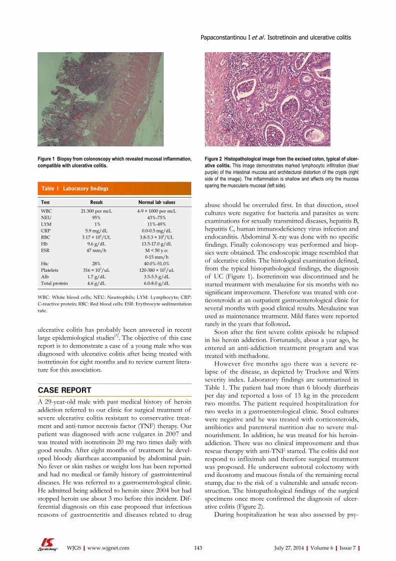
ulcerative colitis has probably been answered in recent large epidemiological studies[1]. The objective of this case report is to demonstrate a case of a young male who was diagnosed with ulcerative colitis after being treated with isotretinoin for eight months and to review current litera-ture for this association.
CASE REPORTA 29-year-old male with past medical history of heroin addiction referred to our clinic for surgical treatment of severe ulcerative colitis resistant to conservative treat-ment and anti-tumor necrosis factor (TNF) therapy. Our patient was diagnosed with acne vulgares in 2007 and was treated with isotretinoin 20 mg two times daily with good results. After eight months of treatment he devel-oped bloody diarrheas accompanied by abdominal pain. No fever or skin rashes or weight loss has been reported and had no medical or family history of gastrointestinal diseases. He was referred to a gastroenterological clinic. He admitted being addicted to heroin since 2004 but had stopped heroin use about 3 mo before this incident. Dif-ferential diagnosis on this case proposed that infectious reasons of gastroenteritis and diseases related to drug
abuse should be overruled first. In that direction, stool cultures were negative for bacteria and parasites as were examinations for sexually transmitted diseases, hepatitis B, hepatitis C, human immunodeficiency virus infection and endocarditis. Abdominal X-ray was done with no specific findings. Finally colonoscopy was performed and biop-sies were obtained. The endoscopic image resembled that of ulcerative colitis. The histological examination defined, from the typical histopathological findings, the diagnosis of UC (Figure 1). Isotretinoin was discontinued and he started treatment with mesalazine for six months with no significant improvement. Therefore was treated with cor-ticosteroids at an outpatient gastroenterological clinic for several months with good clinical results. Mesalazine was used as maintenance treatment. Mild flares were reported rarely in the years that followed.
Soon after the first severe colitis episode he relapsed in his heroin addiction. Fortunately, about a year ago, he entered an anti-addiction treatment program and was treated with methadone.
However five months ago there was a severe re-lapse of the disease, as depicted by Truelove and Witts severity index. Laboratory findings are summarized in Table 1. The patient had more than 6 bloody diarrheas per day and reported a loss of 15 kg in the precedent two months. The patient required hospitalization for two weeks in a gastroenterological clinic. Stool cultures were negative and he was treated with corticosteroids, antibiotics and parenteral nutrition due to severe mal-nourishment. In addition, he was treated for his heroin-addiction. There was no clinical improvement and thus rescue therapy with anti-TNF started. The colitis did not respond to infliximab and therefore surgical treatment was proposed. He underwent subtotal colectomy with end ileostomy and mucous fistula of the remaining rectal stump, due to the risk of a vulnerable and unsafe recon-struction. The histopathological findings of the surgical specimens once more confirmed the diagnosis of ulcer-ative colitis (Figure 2).
During hospitalization he was also assessed by psy-
143 July 27, 2014|Volume 6|Issue 7|WJGS|www.wjgnet.com
Papaconstantinou I et al . Isotretinoin and ulcerative colitis
Figure 1 Biopsy from colonoscopy which revealed mucosal inflammation, compatible with ulcerative colitis.
Figure 2 Histopathological image from the excised colon, typical of ulcer-ative colitis. This image demonstrates marked lymphocytic infiltration (blue/purple) of the intestinal mucosa and architectural distortion of the crypts (right side of the image). The inflammation is shallow and affects only the mucosa sparing the muscularis mucosal (left side). Table 1 Laboratory findings
Test Result Normal lab values
WBC 21.300 per mcL 4-9 × 1000 per mcLNEU 95% 43%-75%LYM 1% 11%-49%CRP 5.9 mg/dL 0.0-0.5 mg/dLRBC 3.17 × 10⁶/UL 3.8-5.3 × 10⁶/ULHb 9.6 g/dL 13.5-17.0 g/dLESR 47 mm/h M < 50 y.o:
0-15 mm/hHtc 28% 40.0%-51.0%Platelets 316 × 103/uL 120-380 × 103/uLAlb 1.7 g/dL 3.5-5.5 g/dLTotal protein 4.6 g/dL 6.0-8.0 g/dL
WBC: White blood cells; NEU: Neutrophils; LYM: Lymphocyte; CRP: C-reactive protein; RBC: Red blood cells; ESR: Erythrocyte sedimentation rate.
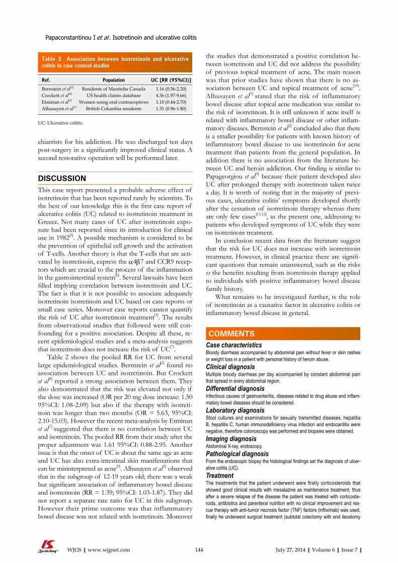
chiatrists for his addiction. He was discharged ten days post-surgery in a significantly improved clinical status. A second restorative operation will be performed later.
DISCUSSIONThis case report presented a probable adverse effect of isotretinoin that has been reported rarely by scientists. To the best of our knowledge this is the first case report of ulcerative colitis (UC) related to isotretinoin treatment in Greece. Not many cases of UC after isotretinoin expo-sure had been reported since its introduction for clinical use in 1982[5]. A possible mechanism is considered to be the prevention of epithelial cell growth and the activation of T-cells. Another theory is that the T-cells that are acti-vated by isotretinoin, express the α4β7 and CCR9 recep-tors which are crucial to the process of the inflammation in the gastrointestinal system[6]. Several lawsuits have been filled implying correlation between isotretinoin and UC. The fact is that it is not possible to associate adequately isotretinoin isotretinoin and UC based on case reports or small case series. Moreover case reports cannot quantify the risk of UC after isotretinoin treatment[5]. The results from observational studies that followed were still con-founding for a positive association. Despite all these, re-cent epidemiological studies and a meta-analysis suggests that isotretinoin does not increase the risk of UC[7].
Table 2 shows the pooled RR for UC from several large epidemiological studies. Bernstein et al[2] found no association between UC and isotretinoin. But Crockett et al[8] reported a strong association between them. They also demonstrated that the risk was elevated not only if the dose was increased (OR per 20 mg dose increase: 1.50 95%CI: 1.08-2.09) but also if the therapy with isotreti-noin was longer than two months (OR = 5.63, 95%CI: 2.10-15.03). However the recent meta-analysis by Etminan et al[7] suggested that there is no correlation between UC and isotretinoin. The pooled RR from their study after the proper adjustment was 1.61 95%CI: 0.88-2.95. Another issue is that the onset of UC is about the same age as acne and UC has also extra-intestinal skin manifestations that can be misinterpreted as acne[9]. Alhusayen et al[1] observed that in the subgroup of 12-19 years old; there was a weak but significant association of inflammatory bowel disease and isotretinoin (RR = 1.39; 95%CI: 1.03-1.87). They did not report a separate rate ratio for UC in this subgroup. However their prime outcome was that inflammatory bowel disease was not related with isotretinoin. Moreover
the studies that demonstrated a positive correlation be-tween isotretinoin and UC did not address the possibility of previous topical treatment of acne. The main reason was that prior studies have shown that there is no as-sociation between UC and topical treatment of acne[10]. Alhusayen et al[1] stated that the risk of inflammatory bowel disease after topical acne medication was similar to the risk of isotretinoin. It is still unknown if acne itself is related with inflammatory bowel disease or other inflam-matory diseases. Bernstein et al[2] concluded also that there is a smaller possibility for patients with known history of inflammatory bowel disease to use isotretinoin for acne treatment than patients from the general population. In addition there is no association from the literature be-tween UC and heroin addiction. Our finding is similar to Papageorgiou et al[9] because their patient developed also UC after prolonged therapy with isotretinoin taken twice a day. It is worth of noting that in the majority of previ-ous cases, ulcerative colitis’ symptoms developed shortly after the cessation of isotretinoin therapy whereas there are only few cases[11,12], as the present one, addressing to patients who developed symptoms of UC while they were on isotretinoin treatment.
In conclusion recent data from the literature suggest that the risk for UC does not increase with isotretinoin treatment. However, in clinical practice there are signifi-cant questions that remain unanswered, such as the risks vs the benefits resulting from isotretinoin therapy applied to individuals with positive inflammatory bowel disease family history.
What remains to be investigated further, is the role of isotretinoin as a causative factor in ulcerative colitis or inflammatory bowel disease in general.
COMMENTSCase characteristics Bloody diarrheas accompanied by abdominal pain without fever or skin rashes or weight loss in a patient with personal history of heroin abuse.Clinical diagnosis Multiple bloody diarrheas per day accompanied by constant abdominal pain that spread in every abdominal region.Differential diagnosis Infectious causes of gastroenteritis, diseases related to drug abuse and inflam-matory bowel diseases should be considered. Laboratory diagnosis Stool cultures and examinations for sexually transmitted diseases, hepatitis B, hepatitis C, human immunodeficiency virus infection and endocarditis were negative, therefore colonoscopy was performed and biopsies were obtained.Imaging diagnosis Abdominal X-ray, endoscopy. Pathological diagnosis From the endoscopic biopsy the histological findings set the diagnosis of ulcer-ative colitis (UC). Treatment The treatments that the patient underwent were firstly corticosteroids that showed good clinical results with mesalazine as maintenance treatment, thus after a severe relapse of the disease the patient was treated with corticoste-roids, antibiotics and parenteral nutrition with no clinical improvement and res-cue therapy with anti-tumor necrosis factor (TNF) factors (infliximab) was used, finally he underwent surgical treatment (subtotal colectomy with end ileostomy
144 July 27, 2014|Volume 6|Issue 7|WJGS|www.wjgnet.com
Table 2 Association between isotretinoin and ulcerative colitis in case control studies
Ref. Population UC [RR (95%CI)]
Bernstein et al[2] Residents of Manitoba Canada 1.16 (0.56-2.20)Crockett et al[8] US health claims database 4.36 (1.97-9.66)Etminan et al[7] Women using oral contraceptives 1.10 (0.44-2.70)Alhusayen et al[1] British Columbia residents 1.31 (0.96-1.80)
UC: Ulcerative colitis.
COMMENTS
Papaconstantinou I et al . Isotretinoin and ulcerative colitis

145 July 27, 2014|Volume 6|Issue 7|WJGS|www.wjgnet.com
3 Passier JL, Srivastava N, van Puijenbroek EP. Isotretinoin-induced inflammatory bowel disease. Neth J Med 2006; 64: 52-54 [PMID: 16517990]
4 Bergstrom KG. Isotretinoin and inflammatory bowel dis-ease. J Drugs Dermatol 2010; 9: 278-280 [PMID: 20232592]
5 Popescu CM, Bigby M. The weight of evidence on the asso-ciation of isotretinoin use and the development of inflamma-tory bowel disease. JAMA Dermatol 2013; 149: 221-222 [PMID: 23426480 DOI: 10.1001/jamadermatol.2013.1348]
6 Mora JR. Homing imprinting and immunomodulation in the gut: role of dendritic cells and retinoids. Inflamm Bowel Dis 2008; 14: 275-289 [PMID: 17924560]
7 Etminan M, Bird ST, Delaney JA, Bressler B, Brophy JM. Isotretinoin and risk for inflammatory bowel disease: a nest-ed case-control study and meta-analysis of published and unpublished data. JAMA Dermatol 2013; 149: 216-220 [PMID: 23426479 DOI: 10.1001/jamadermatol.2013.1344]
8 Crockett SD, Porter CQ, Martin CF, Sandler RS, Kappelman MD. Isotretinoin use and the risk of inflammatory bowel disease: a case-control study. Am J Gastroenterol 2010; 105: 1986-1993 [PMID: 20354506 DOI: 10.1038/ajg.2010.124]
9 Papageorgiou NP, Altman A, Shoenfeld Y. Inflammatory bowel disease: adverse effect of isotretinoin. Isr Med Assoc J 2009; 11: 505-506 [PMID: 19891242]
10 Gilat T, Hacohen D, Lilos P, Langman MJ. Childhood fac-tors in ulcerative colitis and Crohn’s disease. An international cooperative study. Scand J Gastroenterol 1987; 22: 1009-1024 [PMID: 3685876]
11 Martin P, Manley PN, Depew WT, Blakeman JM. Isotreti-noin-associated proctosigmoiditis. Gastroenterology 1987; 93: 606-609 [PMID: 3475230]
12 Brodin MB. Inflammatory bowel disease and isotretinoin. J Am Acad Dermatol 1986; 14: 843 [PMID: 2940270]
P- Reviewer: Bresci G, Kirshtein B, Sipos F S- Editor: Ji FF L- Editor: A E- Editor: Wang CH
and mucous fistula of the remaining rectal stump).Related reports UC is a bowel disease that in not quite sure what are its triggering factors. Though many factors are assumed to be associated to the onset of the disease. Some factors are very well studied but others need more research in order to be blamed. The use of isotretinoin, a widely used treatment for acne is associ-ated to UC even though the case reports describing the linkage are few. Term explanation Isotretinoin is a medication related to vitamin A used primarily for severe cystic acne and acne that has not responded to other treatments. Anti-TNF therapy (anti-tumor necrosis factor therapy) is a new class of drugs that are approved in treating moderate-to-severe UC. The most common drug used is infliximab, that is a monoclonal antibody that binds to TNF. Experiences and lessons UC is a disease that is maybe associated and triggered by more drugs and factors than people already know so in the differential diagnosis of bloody diar-rheas in any young patient, should always be the inflammatory bowel diseases.Peer review This case report is interesting and well written.
REFERENCES1 Alhusayen RO, Juurlink DN, Mamdani MM, Morrow RL,
Shear NH, Dormuth CR. Isotretinoin use and the risk of in-flammatory bowel disease: a population-based cohort study. J Invest Dermatol 2013; 133: 907-912 [PMID: 23096714 DOI: 10.1038/jid.2012.387]
2 Bernstein CN, Nugent Z, Longobardi T, Blanchard JF. Isotret-inoin is not associated with inflammatory bowel disease: a population-based case-control study. Am J Gastroenterol 2009; 104: 2774-2778 [PMID: 19623167 DOI: 10.1038/ajg.2009.417]
Papaconstantinou I et al . Isotretinoin and ulcerative colitis

Published by Baishideng Publishing Group Inc
World Journal of Gastrointestinal SurgeryWorld J Gastrointest Surg 2014 August 27; 6(8): 146-168
ISSN 1948-9366 (online)

146 Simultaneousoperationforcardiacdiseaseandgastrointestinal
malignancy
Komokata T, Fukueda M, Kaieda M, Ueno T, Iguro Y, Imoto Y, Sakata R
151 Neonatalgastricperforation:Asinglecenterexperience
Byun J, Kim HY, Noh SY, Kim SH, Jung SE, Lee SC, Park KW
156 Blindloopperforationafterside-to-sideileocolonicanastomosis
Dalla Valle R, Zinicola R, Iaria M
160 Massivesurgicalemphysemafollowingtransanalendoscopicmicrosurgery
Simkens GAAM, Nienhuijs SW, Luyer MDP, de Hingh IHJT
164 Solitarymediastinallymphnoderecurrenceaftercurativeresectionof
coloncancer
Matsuda Y, Yano M, Miyoshi N, Noura S, Ohue M, Sugimura K, Motoori M, Kishi K,
Fujiwara Y, Gotoh K, Marubashi S, Akita H, Takahashi H, Sakon M
Contents Monthly Volume 6 Number 8 August 27, 2014
WJGS|www.wjgnet.com I August 27, 2014|Volume 6|Issue 8|
RETROSPECTIVE STUDY
OBSERVATIONAL STUDY
CASE REPORT
Contents
APPENDIX
ABOUT COVER
World Journal of Gastrointestinal SurgeryVolume 6 Number 8 August 27, 2014
I-V Instructionstoauthors
EditorialBoardMemberofWorldJournalofGastrointestinalSurgery ,BelevVasilevNikolai,MD,Head,MilitaryMedicalAcademy,HospitalFacilityforActiveTreatment,PlovdivPlovdiv4000,Bulgaria
World Journal of Gastrointestinal Surgery (World J Gastrointest Surg, WJGS, online ISSN 1948-9366, DOI: 10.4240) is a peer-reviewed open access academic journal that aims to guide clinical practice and improve diagnostic and therapeutic skills of clinicians.
WJGS covers topics concerning micro-invasive surgery; laparoscopy; hepatic, biliary, pancreatic and splenic surgery; surgical nutrition; portal hypertension, as well as associated subjects. The current columns of WJGS include editorial, frontier, diagnostic advances, therapeutics advances, field of vision, mini-reviews, review, topic highlight, medical ethics, original articles, case report, clinical case conference (Clinicopathological conference), and autobiography. Priority publication will be given to articles concerning diagnosis and treatment of gastrointestinal surgery diseases. The following aspects are covered: Clinical diagnosis, laboratory diagnosis, differential diagnosis, imaging tests, pathological diagnosis, molecular biological diagnosis, immunological diagnosis, genetic diagnosis, functional diagnostics, and physical diagnosis; and comprehensive therapy, drug therapy, surgical therapy, interventional treatment, minimally invasive therapy, and robot-assisted therapy.
We encourage authors to submit their manuscripts to WJGS. We will give priority to manuscripts that are supported by major national and international foundations and those that are of great basic and clinical significance.
World Journal of Gastrointestinal Surgery is now indexed in PubMed Central, PubMed, Digital Object Identifier, and Directory of Open Access Journals.
I-III EditorialBoard
Xiu-Xia Song, Vice DirectorWorld Journal of Gastrointestinal SurgeryRoom 903, Building D, Ocean International Center, No. 62 Dongsihuan Zhonglu, Chaoyang District, Beijing 100025, ChinaTelephone: +86-10-85381891Fax: +86-10-85381893E-mail: [email protected] Desk: http://www.wjgnet.com/esps/helpdesk.aspxhttp://www.wjgnet.com
PUBLISHERBaishideng Publishing Group Inc8226 Regency Drive, Pleasanton, CA 94588, USATelephone: +1-925-223-8242Fax: +1-925-223-8243E-mail: [email protected] Desk: http://www.wjgnet.com/esps/helpdesk.aspxhttp://www.wjgnet.com
PUBLICATIONDATEAugust 27, 2014
COPYRIGHT© 2014 Baishideng Publishing Group Inc. Articles pub-lished by this Open-Access journal are distributed under the terms of the Creative Commons Attribution Non-commercial License, which permits use, distribution, and reproduction in any medium, provided the original work is properly cited, the use is non commercial and is otherwise in compliance with the license.
SPECIALSTATEMENTAll articles published in journals owned by the Baishideng Publishing Group (BPG) represent the views and opin-ions of their authors, and not the views, opinions or policies of the BPG, except where otherwise explicitly indicated.
INSTRUCTIONSTOAUTHORSFull instructions are available online at http://www.wjgnet.com/1948-9366/g_info_20100305152206.htm
ONLINESUBMISSIONhttp://www.wjgnet.com/esps/
NAMEOFJOURNALWorld Journal of Gastrointestinal Surgery
ISSNISSN 1948-9366 (online)
LAUNCHDATENovember 30, 2009
FREQUENCYMonthly
EDITOR-IN-CHIEFTimothy M Pawlik, MD, MPH, FACS, Associate Professor of Surgery and Oncology, Hepatobiliary Surgery Program Director, Director, Johns Hopkins Medicine Liver Tumor Center Multi-Disciplinary Clinic, Co-Director of Center for Surgical Trials and Outcomes Research, Johns Hopkins Hospital, 600 N. Wolfe Street, Harvey 611, Baltimore, MD 21287, United States
EDITORIALOFFICEJin-Lei Wang, Director
EDITORS FOR THIS ISSUE
Responsible Assistant Editor: Xiang Li Responsible Science Editor: Xue-Mei GongResponsible Electronic Editor: Su-Qing Liu Proofing Editorial Office Director: Xiu-Xia SongProofing Editor-in-Chief: Lian-Sheng Ma
AIM AND SCOPE
FLYLEAF
INDEXING/ABSTRACTING
WJGS|www.wjgnet.com II August 27, 2014|Volume 6|Issue 8|
Simultaneous operation for cardiac disease and gastrointestinal malignancy
Teruo Komokata, Mikio Fukueda, Mamoru Kaieda, Takayuki Ueno, Yoshihumi Iguro, Yutaka Imoto, Ryuzo Sakata
Teruo Komokata, Mamoru Kaieda, Takayuki Ueno, Depart-ment of Surgery, Kagoshima Medical Center, National Hospital Organization, Kagoshima 892-0853, JapanMikio Fukueda, Yoshihumi Iguro, Yutaka Imoto, Cardiovas-cular and Gastroenterological Surgery, Kagoshima University Graduate School of Medical and Dental Sciences, Kagoshima 890-8520, JapanRyuzo Sakata, Cardiovascular Surgery, Kyoto University Hos-pital, Kyoto 606-8397, JapanAuthor contributions: Komokata T designed the study and provided the collection of the clinical data; Komokata T and Fu-kueda M wrote the manuscript; Kaieda M, Ueno T, Iguro Y, and Sakata R participated in the simultaneous surgery; Imoto Y and Sakata R were involved in editing the manuscript.Correspondence to: Teruo Komokata, MD, PhD, Department of Surgery, Kagoshima Medical Center, National Hospital Orga-nization, 8-1 Shiroyama, Kagoshima 892-0853, Japan. [email protected]: +81-99-2231151 Fax: +81-99-2269246Received: March 6, 2014 Revised: June 20, 2014Accepted: July 12, 2014Published online: August 27, 2014
AbstractAIM: To investigate the safety of performing simulta-neous cardiac surgery and a resection of a gastrointes-tinal malignancy.
METHODS: Among 3664 elective cardiac operations performed in adults at Kagoshima University Hospi-tal from January 1991 to October 2009, this study reviewed the clinical records of the patients who un-derwent concomitant cardiac surgery and a gastroin-testinal resection. Such simultaneous surgeries were performed in 15 patients between January 1991 and October 2009. The cardiac diseases included 8 cases of coronary artery disease and 7 cases with valvular heart disease. Gastrointestinal malignancies included 11 gas-tric and 4 colon cancers. Immediate postoperative and
long-term outcomes were evaluated.
RESULTS: Postoperative complications occurred in 5 patients (33.3%), including strokes (n = 1), respiratory failure requiring re-intubation (n = 1), hemorrhage (n = 2), hyperbilirubinemia (n = 1) and aspiration pneu-monia (n = 1). There was 1 hospital death caused by the development of adult respiratory distress syndrome after postoperative surgical bleeding followed aortic valve replacement plus gastrectomy. There was no car-diovascular event in the patients during the follow-up period. The cumulative survival rate for all patients was 69.2% at 5 years.
CONCLUSION: Simultaneous procedures are accept-able for the patients who require surgery for both car-diac diseases and gastrointestinal malignancy. In par-ticular, the combination of a standard cardiac operation, such as coronary artery bypass grafting or an isolated valve replacement and simple gastrointestinal resection, such as gastrectomy or colectomy can therefore be safely performed.
© 2014 Baishideng Publishing Group Inc. All rights reserved.
Key words: Simultaneous operation; Cardiac disease; Gastrointestinal malignancy; Gastric cancer; Colon cancer
Core tip: Simultaneous surgical interventions for cardio-vascular and gastrointestinal pathology have not been well adapted so far. Staged procedure, depending on the clinical priority is usually preferred. Concomitant cardiac and gastrointestinal surgery holds a bundle of advantages with some challenges. We reviewed the outcome in 15 patients who underwent simultaneous cardiovascular and gastrointestinal surgery at Ka-goshima University Hospital. Postoperative complica-tions were noted in 5 cases, with 1 death. No adverse cardiovascular events were noted during follow up. The cumulative survival rate for all patients was 69.2% at 5
RETROSPECTIVE STUDY
Submit a Manuscript: http://www.wjgnet.com/esps/Help Desk: http://www.wjgnet.com/esps/helpdesk.aspxDOI: 10.4240/wjgs.v6.i8.146
World J Gastrointest Surg 2014 August 27; 6(8): 146-150ISSN 1948-9366 (online)
© 2014 Baishideng Publishing Group Inc. All rights reserved.
146 August 27, 2014|Volume 6|Issue 8|WJGS|www.wjgnet.com
years. Synchronized cardiovascular and gastrointestinal procedures can be safely performed when needed.
Komokata T, Fukueda M, Kaieda M, Ueno T, Iguro Y, Imoto Y, Sakata R. Simultaneous operation for cardiac disease and gastroin-testinal malignancy. World J Gastrointest Surg 2014; 6(8): 146-150 Available from: URL: http://www.wjgnet.com/1948-9366/full/v6/i8/146.htm DOI: http://dx.doi.org/10.4240/wjgs.v6.i8.146
INTRODUCTIONThe prevalence of cardiovascular disease and malignant disease has been increasing as the proportion of elderly individuals in the general population increases in devel-oped countries[1]. Therefore, physicians may encounter a patient with concomitant occurrence of cardiovascular disease and malignant diseases. In general, patients with both cardiovascular disease that requires surgery and a surgically resectable malignancy are treated in a staged procedure depending on the clinical priority. Simultane-ous procedure are rare because the surgeon’s anxiety that the incidence of complications may be higher in patients undergoing a simultaneous procedure than in those un-dergoing isolated cardiac surgery or gastrointestinal resec-tion. However, a simultaneous approach would be attrac-tive for such patients, because limiting them to a single exposure to anesthesia and one recovery period remove the potential opportunity for perioperative cardiovascular events, and moreover provide economic benefits. The recent advances in anesthesia, surgical techniques and perioperative management have allowed for combined operations to be conducted, so several studies have vali-dated the concept that a simultaneous operation can be safely performed in a limited population of patients[2-9].
Most of them are reports concerning a combined procedure that includes a pulmonary resection and car-diac operation[2-5], and there are a few studies on the com-bination surgery of the heart and a gastrointestinal ma-lignancy[6-9]. This report presents a series of 15 patients who underwent concomitant cardiac surgery and gastro-intestinal resection to assess the safety of a simultaneous procedure.
MATERIALS AND METHODSThere were 3664 elective cardiac operations performed in adults at Kagoshima University Hospital from January 1991 to October 2009. This study reviewed the clini-cal records of the patients who underwent concomitant cardiac surgery and a gastrointestinal resection. Fifteen of these patients (0.4%) underwent a simultaneous pro-cedure. The records of the patients were reviewed for the type of surgical procedure, complications, and the dura-tion of the stays in the intensive care unit (ICU) and hos-pital. The long-term outcome was determined using hos-pital records and through direct contact with the patients
and their family physicians. The values are expressed as the mean ± SD. Survival was estimated by the Kaplan-Meier method using the date of the combined procedure as the starting point and the date of death or last follow-up as the end-point.
RESULTSClinical presentationThe preoperative characteristics of the patients are sum-marized in Table 1. The subjects included of 11 males and 4 females with an average age of 72.3 ± 7.4 years, of whom 8 were treated for coronary artery disease (CAD) and 7 impaired valvular heart disease. CAD involved two vessels in 2 patients, three vessels in 4 patients, and the left main trunk in 2 patients. Valvular heart diseases in-cluded aortic stenosis in 4 patients, mitral stenosis accom-panying with left atrial thrombus in 2 patients, and severe mitral regurgitation in one patient. The left ventricular ejection fraction assessed by ventriculography or echocar-diography ranged from 36% to 81% with an average of 63.6% ± 12.1%.
Gastrointestinal malignancies included 11 patients with gastric cancer and 4 patients with colon cancer. Only one gastric cancer was located in the fundus, the other 10 lesions were in the body or antrum of the stomach. All gastric cancers were preoperatively diagnosed as early stage and no distant metastasis or multiple lymph nodes metastasis was detected radiologically. All 4 colon cancers (transverse 1, descending 1, sigmoid 1, cecum 1) were in advanced stages and one of them had multiple liver metastases preoperatively. The most common symptoms associated with the gastrointestinal malignancies were anemia, weight loss, and gastrointestinal tract bleeding. Five patients were first diagnosed as having cancer and cardiac disease was discovered during the preoperative assessment. The remaining 10 patients presented with cardiac symptoms, resulting from myocardial ischemia or valvular heart disease, classified as NYHA functional class from Ⅰ to Ⅲ, and thus were found incidentally to have an asymptomatic gastrointestinal cancer.
Preoperative comorbidities included renal failure in 2 patients (1 patient required hemodialysis), diabetes con-trolled by oral agents in 2 patients, and chronic obstruc-tive pulmonary disease (COPD) in one patient, in which the percentage of vital capacity was 81.1% and the forced expiratory volume at 1 s was 54.9%.
Surgical proceduresThe surgical procedures carried out are listed in Table 2. All were elective procedures. The simultaneous pro-cedures included aortic valve replacement (AVR) plus gastrectomy in 4 patients, mitral valve replacement plus gastrectomy in 3 patients, AVR plus colectomy in 1 pa-tients, off-pump coronary artery bypass grafting (OP-CAB) plus gastrectomy in 4 patients, and coronary artery bypass grafting (CABG) plus colectomy in 3 patients. The cardiac surgery was performed first in all but 1 (case
Komokata T et al . Simultaneous cardiac and gastrointestinal operation
147 August 27, 2014|Volume 6|Issue 8|WJGS|www.wjgnet.com
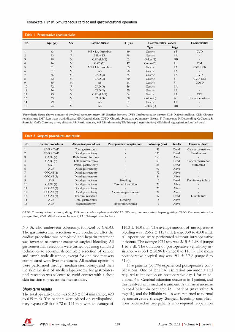
No. 3), who underwent colectomy, followed by CABG. The gastrointestinal resections were conducted after the cardiac procedure was completed and heparin treatment was reversed to prevent excessive surgical bleeding. All gastrointestinal resections were carried out using standard techniques to accomplish complete resection of cancer and lymph node dissection, except for one case that was complicated with liver metastasis. All cardiac operation were performed through median sternotomy, and then the skin incision of median laparotomy for gastrointes-tinal resection was selected to avoid contact with a chest skin incision to prevent the mediastinitis.
Short-term resultsThe total operative time was 512.8 ± 85.4 min (range, 420 to 635 min). Ten patients were placed on cardiopulmo-nary bypass (CPB) for 72 to 144 min, with an average of
116.3 ± 16.0 min. The average amount of intraoperative bleeding was 1256.2 ± 1127 mL (range 330 to 4200 mL). All operations were performed without intraoperative incidents. The average ICU stay was 3.15 ± 1.98 d (range 1 to 8 d). The duration of postoperative ventilatory as-sistance was 35.1 ± 28.96 h (range 8 to 116 h). The mean postoperative hospital stay was 19.1 ± 2.7 d (range 8 to 51 d).
Five patients (33.3%) experienced postoperative com-plications. One patient had aspiration pneumonia and required re-intubation on postoperative day 4 for an ad-ditional 6 d. Cerebral infarction occurred in 1 patient, and this resolved with medical treatment. A transient increase in total bilirubin occurred in 1 patient (max value: 8 mg/dL), and the bililubin values were returned to normal by conservative therapy. Surgical bleeding complica-tions occurred in two patients who required reoperation
148 August 27, 2014|Volume 6|Issue 8|WJGS|www.wjgnet.com
Table 1 Preoperative characteristics
No. Age (yr) Sex Cardiac disease EF (%) Gastrointestinal cancer Comorbidities
Type Stage1 63 F MS + LA thrombus 69 Gastric ⅠB CVD2 73 F MR + TR 78 Gastric ⅠA -3 78 M CAD (LMT) 61 Colon (T) ⅢB -4 76 M CAD (2)1 47 Colon (D) Ⅱ DM5 61 M MS + LA thrombus 65 Gastric ⅠA CRF (HD)6 81 M AS 78 Gastric ⅠA -7 66 M CAD (3) 65 Gastric ⅠA CVD8 62 M CAD (3) 70 Gastric Ⅱ CVD, DM9 85 M AS 64 Gastric Ⅱ COPD10 72 F CAD (3) 36 Gastric ⅠA -11 76 M CAD (2) 55 Gastric ⅠA -12 73 M CAD (LMT) 54 Gastric ⅠA CRF13 65 M CAD (3) 60 Colon (C) Ⅳ Liver metastasis14 79 F AS 81 Gastric ⅠB -15 74 M AS 71 Colon (S) ⅢB -
1Parenthetic figure shows number of involved coronary artery. EF: Ejection fraction; CVD: Cerebrovascular disease; DM: Diabetis mellitus; CRF: Chronic renal failure; LMT: Left main trunk disease; HD: Hemodialysis; COPD: Chronic obstructive pulmonary disease; T: Transverse; D: Descending; C: Cecum; S: Sigmoid; CAD: Coronary artery disease; AS: Aortic stenosis; MS: Mitral stenosis; TR: Tricuspid regurgitation; MR: Mitral regurgitation; LA: Left atrial.
Table 2 Surgical procedures and results
No. Cardiac procedures Abdominal procedures Postoperative complications Follow-up (mo) Results Causes of death
1 MVR + TAP Total gastrectomy - 81 Dead Cancer recurrence2 MVR + TAP Distal gastrectomy - 135 Dead Renal failure3 CABG (2) Right hemicolectomy - 150 Alive -4 CABG (3) Left hemicolectomy - 53 Dead Cancer recurrence5 MVR Partial gastrectomy - 84 Dead Suffocated6 AVR Distal gastrectomy - 90 Alive -7 OPCAB (4) Distal gastrectomy - 72 Alive -8 OPCAB (5) Distal gastrectomy - 84 Alive -9 AVR Distal gastrectomy Bleeding 2 Dead Respiratory failure10 CABG (4) Distal gastrectomy Cerebral infarction 28 Alive -11 OPCAB (2) Distal gastrectomy - 25 Alive -12 OPCAB (3) Distal gastrectomy Aspiration pneumonia 21 Alive -13 OPCAB (3) Ileocecal resection - 17 Dead Liver failure14 AVR Total gastrectomy Bleeding 8 Alive -15 AVR Sigmoidectomy Hyperbilirubinmia 3 Alive -
CABG: Coronary artery bypass grafting; AVR: Aortic valve replacement; OPCAB: Off-pump coronary artery bypass grafting; CABG: Coronary artery by-pass grafting; MVR: Mitral valve replacement; TAP: Tricuspid annuloplasty.
Komokata T et al . Simultaneous cardiac and gastrointestinal operation
safety so that this approach can be generally accepted. This study focused on the safety and indications of si-multaneous procedures.
Most of the simultaneous procedures in the present series were performed successfully in short-term period. The hospital mortality and morbidity rate was 6.6% and 33.3%, respectively, and the outcome was quite acceptable.
One of surgeon’s prime concerns about the safety of simultaneous operations is that CPB has several disad-vantageous systemic effects including bleeding disorders, thrombotic complications, massive fluid shifts and im-munosuppression, all associated with homeostatic disrup-tion. Furthermore, CPB is also associated with mesen-teric ischemia, which may contribute to varying degrees of organ damage[11,12].
Cerebral infarction and hyperbilirubinemia, which are the most common complications associated with CPB, occurred in one patient each, but those resolved with medical treatment. There were no postoperative gastro-intestinal complications related to resection and recon-struction, such as anastomosis leakage, ileus and bleeding. Only one patient unfortunately died of respiratory failure after an early reoperation for postoperative bleeding from the chest, and he was a person of advanced age with COPD and was a high risk case. The postoperative occurrence of bleeding associated with abnormal blood coagulation following CPB and systemic hepariniza-tion, which is one of the most dreaded complications, may frequently be life-threatening for high risk patients in disrupted homeostasis. Therefore, the management of bleeding should be ensured with caution at the end of cardiac surgery. OPCAB is preferable to avoid the adverse effects of CPB, and improve the outcome of a combined operation by allowing gastrointestinal resection to be performed safely[13,14]. OPCAB was performed in all of the 4 patients in the current series, and their post-operative courses were favorable. OPCAB should be se-lected whenever possible in patients with severe coronary disease undergoing combined procedure.
Most cardiovascular surgeons have concern that the addition of clean-contaminated operation to cardiac operation would increase the incidence of postoperative infection; wound infection, mediastinitis and mechanical valve infection. However, there was no incidence of sur-gical site infection in this series, thus routine management of infection, using prophylactic antibiotics, sterilization of the surgical field, and separation of an each skin inci-sion, was able to adequately prevent postoperative infec-tious complications in simultaneous operation.
This study cannot conclude that the hospital morbid-ity and mortality of patients undergoing simultaneous procedure are better than that of staged procedures, and cannot clarify whether simultaneous operations contrib-ute benefits to patients over a long term period. However the current experience demonstrated that the combina-tion of standard cardiac operations, such as CABG or single valve replacement, and uncomplicated gastroin-testinal resections such as gastrectomy or colectomy was
for bleeding from the chest or abdomen. One was an 83-year-old man who had COPD, underwent AVR and distal gastrectomy simultaneously, and suffered massive bleeding from chest drainage tube postoperatively. He temporarily recovered after the reoperation to treat the bleeding. However, Methicillin-resistant Staphylococcus aureus pneumonitis developed shortly after the surgery and he died of respiratory failure 10 wk after undergoing the initial surgery. No clinical anastomotic leaks related to the gastrointestinal surgery and cardiovascular events were observed in the early postoperative period. In addi-tion no mediastinitis, both wound infection and mechani-cal valve infection occurred. The remaining 10 patients were uneventful during postoperative course.
The surgical stage of the gastric cancers was stage ⅠA (defined by tumor-node-metastasis classifications) in 7 patients, stage ⅠB in 1 patient, and stage Ⅱ in 2 patients. All patients with colon cancer were in advanced stages, and thus one patient was in Stage Ⅱ, two patients were in stage ⅢB, and one patient was in stage Ⅳ. Three received post-operative chemotherapy for the treatment of colon cancer.
Long-term resultsFourteen of 15 patients were discharged, and 6 of them died during the follow-up period, which averaged 56.9 ± 47.1 mo (range, 2 to 149 mo). The patient with multiple bilobar liver metastases before the surgery died of liver failure at 17.2 mo. Two patients died of cancer recur-rence at 80.8 and 53.4 mo after operation.
The causes of death of the other 2 patients were suf-focation and renal failure. Six of 14 patients are currently alive without evidence of recurrence after surgery, and have experienced no cardiovascular events during the long-term follow-up. The cumulative survival rates for all patients undergoing simultaneous procedures were 69.2% at 5 years.
DISCUSSIONThe treatment of patients who require surgery for both cardiac disease and gastrointestinal malignancy is still controversial. A staged procedure depending on the clini-cal priority is usually preferred because most surgeons assume that the prolonged operative time and extensive surgical invasiveness associated with simultaneous proce-dures must increase the surgical morbidity and mortality. However, there are also several unfavorable issues as-sociated with a staged procedure. For instance, when an isolated cardiac operation is performed before gastroin-testinal resection, the malignant lesion may develop mas-sive gastrointestinal bleeding due to the heparinization required for CPB, and moreover untreated cancer may progress during the interval prior to the second opera-tion. On the contrary, postoperative cardiovascular com-plication may occur during gastrointestinal resection and that could be fatal[10]. The concomitant approach, could resolve those problem. Although a simultaneous proce-dure is apparently attractive, it is necessary to ensure the
149 August 27, 2014|Volume 6|Issue 8|WJGS|www.wjgnet.com
Komokata T et al . Simultaneous cardiac and gastrointestinal operation
safe and favorable excluding patients with serious comor-bidities or poor cardiac function.
The result of this study suggest that, the simultaneous procedures are an acceptable alternative for selected pa-tients, but further studies are necessary to determine the role of the two strategies for patients with cardiovascular disease and concurrent gastrointestinal malignancy.
COMMENTSBackgroundPatients requiring surgical intervention for gastroenterological pathology and car-diac events mostly entail careful attention. A surgeon has to weigh the pros and cons, and decisions are sometimes cumbersome especially when simultaneous surgery has to be performed. However concomitant cardiac and gastrointestinal surgery holds some advantages at times, like limiting the patients to single expo-sure of anesthesia, same recovery time along with economic benefits. The aim of the study was to assess the safety and outcome of simultaneous procedure. On reviewing the outcome in 15 patients who underwent simultaneous surgical procedure, the overall outcome was found to be satisfactory. Research frontiersThe authors retrospectively studied the outcome in the patients who underwent concomitant cardiac and gastrointestinal surgery. Same time, the authors ana-lyzed the publish literature on concomitant surgery. Innovations and breakthroughsDespite advances in surgery, anesthesia, surgical skills and the high tech set-tings; the surgeons may find themselves reluctant to perform concomitant car-diac and gastrointestinal surgery. This study evaluated the outcome in 15 cases of simultaneous surgery. Immediate and delayed postoperative outcomes were found to be satisfactory.ApplicationsThe result suggests that concomitant cardiac and gastrointestinal surgery can be safely performed when required.TerminologySimultaneous operation, cardiac disease, gastrointestinal malignancy, gastric cancer, colon cancer. Peer reviewThis is a retrospective study to assess the outcomes of concomitant surgery for gastrointestinal pathology with cardiac events. Simultaneous gastrointestinal and cardiac surgery may have an appropriate outcome and can be safely performed.
REFERENCES1 Miniño AM, Murphy SL, Xu J, Kochanek KD. Deaths: fi-
nal data for 2008. Natl Vital Stat Rep 2011; 59: 1-126 [PMID: 22808755]
2 Rao V, Todd TR, Weisel RD, Komeda M, Cohen G, Ikono-midis JS, Christakis GT. Results of combined pulmonary resection and cardiac operation. Ann Thorac Surg 1996; 62: 342-346; discussion 346-347 [PMID: 8694588 DOI: 10.1016/00
03-4975(96)00349-9]3 Thomas P, Giudicelli R, Guillen JC, Fuentes P. Is lung cancer
surgery justified in patients with coronary artery disease? Eur J Cardiothorac Surg 1994; 8: 287-91; discussion 292 [PMID: 8086174 DOI: 10.1016/S1010-7940(05)80087-9]
4 Brutel de la Rivière A, Knaepen P, Van Swieten H, Vander-schueren R, Ernst J, Van den Bosch J. Concomitant open heart surgery and pulmonary resection for lung cancer. Eur J Cardiothorac Surg 1995; 9: 310-313; discussion 313-314 [PMID: 7546803 DOI: 10.1016/S1010-7940]
5 Hosoba S, Hanaoka J, Suzuki T, Takashima N, Kambara A, Matsubayashi K, Asai T. Early to midterm results of cardiac surgery with concomitant pulmonary resection. Ann Thorac Cardiovasc Surg 2012; 18: 8-11 [PMID: 21921358 DOI: 10.5761/atcs.oa.11.01717]
6 Takahashi T, Nakano S, Shimazaki Y, Kaneko M, Nakahara K, Miyata M, Kamiike W, Matsuda H. Concomitant coronary bypass grafting and curative surgery for cancer. Surg Today 1995; 25: 131-135 [PMID: 7772915 DOI: 10.1007/BF00311084]
7 Tsuji Y, Morimoto N, Tanaka H, Okada K, Matsuda H, Tsukube T, Watanabe Y, Okita Y. Surgery for gastric can-cer combined with cardiac and aortic surgery. Arch Surg 2005; 140: 1109-1114 [PMID: 16301450 DOI: 10.1001/arch-surg.140.11.1109]
8 Fu Q, Li QZ, Liang DG, Ruan XH, Wang ZX, Wei MX. Early and long-term results of combined cardiac surgery and neoplastic resection in patients with concomitant severe heart disease and neoplasms. Chin Med J (Engl) 2011; 124: 1939-1942 [PMID: 22088450 DOI: 10.3760/cma.j.issn.0366-6999.2011.13.004]
9 Zielinski J, Jaworski R, Pawlaczyk R, Swierblewski M, Ka-bata P, Jaskiewicz J, Rogowski J. Simultaneous surgery for critical aortic stenosis and gastric cancer: a case report. World J Gastroenterol 2010; 16: 1161-1164 [PMID: 20205291 DOI: 10.3748/wjg.v16.i9.1161]
10 Mangano DT, Browner WS, Hollenberg M, Li J, Tateo IM. Long-term cardiac prognosis following noncardiac surgery. The Study of Perioperative Ischemia Research Group. JAMA 1992; 268: 233-239 [PMID: 1608143 DOI: 10.1001/jama.1992.03490020081035]
11 Gaer JA, Shaw AD, Wild R, Swift RI, Munsch CM, Smith PL, Taylor KM. Effect of cardiopulmonary bypass on gastro-intestinal perfusion and function. Ann Thorac Surg 1994; 57: 371-375 [PMID: 8311598 DOI: 10.1016/0003-4975(94)90999-7]
12 Ohri SK, Becket J, Brannan J, Keogh BE, Taylor KM. Effects of cardiopulmonary bypass on gut blood flow, oxygen utiliza-tion, and intramucosal pH. Ann Thorac Surg 1994; 57: 1193-1199 [PMID: 8179384 DOI: 10.1016/0003-4975(94)91355-2]
13 Ochi M, Yamada K, Fujii M, Ohkubo N, Ogasawara H, Tanaka S. Role of off-pump coronary artery bypass grafting in patients with malignant neoplastic disease. Jpn Circ J 2000; 64: 13-17 [PMID: 10651200 DOI: 10.1253/jcj.64.13]
14 Nurözler F, Kutlu ST, Kücük G. Off-pump coronary bypass for patients with concomitant malignancy. Circ J 2006; 70: 1048-1051 [PMID: 16864940 DOI: 10.1253/circj.70.1048]
P- Reviewer: Lai S, Said SAM S- Editor: Wen LL L- Editor: A E- Editor: Liu SQ
150 August 27, 2014|Volume 6|Issue 8|WJGS|www.wjgnet.com
COMMENTS
Komokata T et al . Simultaneous cardiac and gastrointestinal operation
Neonatal gastric perforation: A single center experience
Jeik Byun, Hyun Young Kim, Seung Yeon Noh, Soo Hong Kim, Sung Eun Jung, Seong Cheol Lee, Kwi Won Park
Jeik Byun, Hyun Young Kim, Seung Yeon Noh, Soo Hong Kim, Sung Eun Jung, Seong Cheol Lee, Kwi Won Park, Department of Pediatric Surgery, Seoul National University Chil-dren’s Hospital, Seoul 110-744, South KoreaAuthor contributions: All the authors contributed to this paper.Correspondence to: Sung Eun Jung, MD, Department of Pediatric Surgery, Seoul National University Children’s Hospital, 101 Daehang-ro, Yeongeon-dong, Jongro-gu, Seoul 110-744, South Korea. [email protected]: +82-02-20722478 Fax: +82-02-7405130Received: February 19, 2014 Revised: June 12, 2014Accepted: July 12, 2014Published online: August 27, 2014
AbstractAIM: To determine the etiology and prognostic factors for neonatal gastric perforation (NGP), a rare but life-threatening disease.
METHODS: Between 1980 and 2011, nine patients un-derwent surgical intervention for NGP at Seoul National University Children’s Hospital. The characteristics and prognosis of the patients were retrospectively analyzed.
RESULTS: Among the nine patients, three (33.3%) were preterm babies and five (55.5%) had associated anomalies, which included diaphragmatic eventration (n = 2), congenital diaphragmatic hernia, esophageal atresia with tracheoesophageal fistula, and antral web. Three (33.3%) patients were born before 1990 and three (33.3%) had a birth weight < 2500 g. Pneumo-peritoneum was found on preoperative images in six (66.7%) patients, and incidentally in the other three (33.3%) patients. Surgery was performed within 24 h after the onset of symptoms in seven (77.8%) patients. The overall mortality rate was 22.2% (2/9). The time between symptoms and surgical intervention was the only prognostic factor for survival, whereas premature birth and birth weight were not.
CONCLUSION: Early detection and advances in neo-natal intensive care may improve the prognosis of NGP.
© 2014 Baishideng Publishing Group Inc. All rights reserved.
Key words: Neonate; Gastric perforation; Etiology; Prognosis; Surgical intervention
Core tip: Neonatal gastric perforation (NGP) is an ex-tremely rare condition and very few cases have been reported to date. We determined the etiology and prog-nostic factors for NGP in nine cases who were treated at a single center. Early detection and prompt surgical intervention are essential to improve the outcomes of NGP.
Byun J, Kim HY, Noh SY, Kim SH, Jung SE, Lee SC, Park KW. Neonatal gastric perforation: A single center experience. World J Gastrointest Surg 2014; 6(8): 151-155 Available from: URL: http://www.wjgnet.com/1948-9366/full/v6/i8/151.htm DOI: http://dx.doi.org/10.4240/wjgs.v6.i8.151
INTRODUCTIONNeonatal gastric perforation (NGP) accounts for ap-proximately 7% of all gastrointestinal perforations in neonates, and has a poor prognosis with a high mortality rate[1,2]. Factors associated with NGP include prematurity, asphyxia, congenital anomalies, stress at birth, vigorous respiratory resuscitative measures, increased intragastric pressure caused by distal obstruction, and anatomic ab-normalities of the stomach[3-7]. Male gender, metabolic acidosis, premature birth, and low birth weight are associ-ated with worse outcomes[8,9]. However, the etiology and prognostic factors of NGP are still widely debated. Here, we describe our experience of treating nine patients with NGP at a single center. The aim of this study was to review patients with NGP and discuss its etiology and
OBSERVATIONAL STUDY
Submit a Manuscript: http://www.wjgnet.com/esps/Help Desk: http://www.wjgnet.com/esps/helpdesk.aspxDOI: 10.4240/wjgs.v6.i8.151
World J Gastrointest Surg 2014 August 27; 6(8): 151-155ISSN 1948-9366 (online)
© 2014 Baishideng Publishing Group Inc. All rights reserved.
151 August 27, 2014|Volume 6|Issue 8|WJGS|www.wjgnet.com
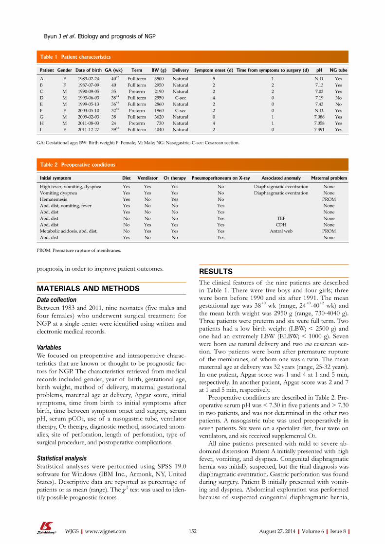
prognosis, in order to improve patient outcomes.
MATERIALS AND METHODSData collectionBetween 1983 and 2011, nine neonates (five males and four females) who underwent surgical treatment for NGP at a single center were identified using written and electronic medical records.
VariablesWe focused on preoperative and intraoperative charac-teristics that are known or thought to be prognostic fac-tors for NGP. The characteristics retrieved from medical records included gender, year of birth, gestational age, birth weight, method of delivery, maternal gestational problems, maternal age at delivery, Apgar score, initial symptoms, time from birth to initial symptoms after birth, time between symptom onset and surgery, serum pH, serum pCO2, use of a nasogastric tube, ventilator therapy, O2 therapy, diagnostic method, associated anom-alies, site of perforation, length of perforation, type of surgical procedure, and postoperative complications.
Statistical analysisStatistical analyses were performed using SPSS 19.0 software for Windows (IBM Inc., Armonk, NY, United States). Descriptive data are reported as percentage of patients or as mean (range). The χ 2 test was used to iden-tify possible prognostic factors.
RESULTSThe clinical features of the nine patients are described in Table 1. There were five boys and four girls; three were born before 1990 and six after 1991. The mean gestational age was 38+0 wk (range, 24+0-40+2 wk) and the mean birth weight was 2950 g (range, 730-4040 g). Three patients were preterm and six were full term. Two patients had a low birth weight (LBW; < 2500 g) and one had an extremely LBW (ELBW; < 1000 g). Seven were born via natural delivery and two via cesarean sec-tion. Two patients were born after premature rupture of the membranes, of whom one was a twin. The mean maternal age at delivery was 32 years (range, 25-32 years). In one patient, Apgar score was 1 and 4 at 1 and 5 min, respectively. In another patient, Apgar score was 2 and 7 at 1 and 5 min, respectively.
Preoperative conditions are described in Table 2. Pre-operative serum pH was < 7.30 in five patients and > 7.30 in two patients, and was not determined in the other two patients. A nasogastric tube was used preoperatively in seven patients. Six were on a specialist diet, four were on ventilators, and six received supplemental O2.
All nine patients presented with mild to severe ab-dominal distension. Patient A initially presented with high fever, vomiting, and dyspnea. Congenital diaphragmatic hernia was initially suspected, but the final diagnosis was diaphragmatic eventration. Gastric perforation was found during surgery. Patient B initially presented with vomit-ing and dyspnea. Abdominal exploration was performed because of suspected congenital diaphragmatic hernia,
Byun J et al . Etiology and prognosis of NGP
152 August 27, 2014|Volume 6|Issue 8|WJGS|www.wjgnet.com
Table 1 Patient characteristics
Patient Gender Date of birth GA (wk) Term BW (g) Delivery Symptom onset (d) Time from symptoms to surgery (d) pH NG tube
A F 1983-02-24 40+2 Full term 3500 Natural 5 1 N.D. YesB F 1987-07-09 40 Full term 2950 Natural 2 2 7.13 YesC M 1990-09-05 35 Preterm 2190 Natural 2 2 7.03 YesD M 1993-06-03 38+4 Full term 2950 C-sec 4 0 7.19 NoE M 1999-05-13 36+3 Full term 2860 Natural 2 0 7.43 NoF F 2003-05-10 32+6 Preterm 1960 C-sec 2 0 N.D. YesG M 2009-02-03 38 Full term 3620 Natural 0 1 7.086 YesH M 2011-08-03 24 Preterm 730 Natural 4 1 7.058 YesI F 2011-12-27 39+3 Full term 4040 Natural 2 0 7.391 Yes
GA: Gestational age; BW: Birth weight; F: Female; M: Male; NG: Nasogastric; C-sec: Cesarean section.
Table 2 Preoperative conditions
Initial symptom Diet Ventilator O2 therapy Pneumoperitoneum on X-ray Associated anomaly Maternal problem
High fever, vomiting, dyspnea Yes Yes Yes No Diaphragmatic eventration NoneVomiting dyspnea Yes Yes Yes No Diaphragmatic eventration NoneHematemesis Yes No Yes No PROMAbd. dist, vomiting, fever Yes No No Yes NoneAbd. dist Yes No No Yes NoneAbd. dist No No No Yes TEF NoneAbd. dist No Yes Yes Yes CDH NoneMetabolic acidosis, abd. dist, No Yes Yes Yes Antral web PROMAbd. dist Yes No No Yes None
PROM: Premature rupture of membranes.

which was ultimately diagnosed as diaphragmatic even-tration. Gastric perforation was also found intraopera-tively. Patient C initially presented with dyspnea and was intubated at birth. At 3 d of age, the patient exhibited hematemesis and severe abdominal distension. Paracen-tesis revealed bloody ascites, and explorative surgery was performed 2 d after the onset of symptoms. All of the patients, except for Patients B and C, underwent surgery within 24 h of the onset of symptoms. The mean age of symptom onset was 3 d (range, 0-5 d). Five patients had associated anomalies, which included diaphragmatic eventration, congenital diaphragmatic hernia, esophageal atresia with a tracheoesophageal fistula, and antral web.
The intraoperative and postoperative findings are summarized in Table 3. The mean size of the perfora-tion was 4.5 cm (0.5-10 cm). The body (n = 5, 55.5%) of the stomach was the most common site of perforation. The perforation was located in the greater curvature in four patients and in the lesser curvature in three patients. Primary repair was performed in six patients, while resec-tion and anastomosis were performed in three patients. Postoperative complications occurred in five patients (55.6%), which included wound problems in two patients and recurrence in one patient. The other four patients were discharged without any complications.
Patient H, who had recurrence, was male and was born at a gestational age of 24 wk with a birth weight of 730 g. He presented with pneumoperitoneum on infan-togram and underwent surgery at 5 d old. A 5 cm long laceration on the lesser curvature and pyloric thickening were found during explorative surgery. Therefore, prima-ry repair was done. However, the postoperative infanto-gram and sonogram revealed an increase in free air. Small bowel series suggested obstruction of the gastric outlet. Three days after initial surgery, the patient underwent a second operation that revealed another perforation of the upper body of the anterior wall and prepyloric antral web. Primary repair and Heineke-Mikulicz pyloroplasty were performed, and an ileostomy was formed because of enteritis of the entire small bowel. Ileostomy repair was done 3 mo after surgery.
The overall mortality rate was 22.2% (2/9). Patient B was born at a gestational age of 40 wk and the birth weight was 2950 g. The patient was diagnosed with Bo-
chdalek hernia at another hospital and was transferred to our institute for surgery. On surgical exploration, the an-terolateral portion of the diaphragm was eventrated, and a 2.5 cm long laceration was found in the posterior wall of the stomach. Diaphragm repair and primary suturing of the stomach were done. The patient died 5 d after surgery because of septic shock. Patient C was born at a gestational age of 35 wk and with a birth weight of 2190 g. Hematemesis and hematochezia were found at 3 d of age. The patient received conservative therapies, including transfusion for 24 h. Diagnostic paracentesis performed at 4 d of age revealed bloody ascites. Explorative laparot-omy was done to evaluate the patient’s hemoperitoneum and gastrointestinal bleeding. On exploration, a 10 cm long laceration with a necrotic margin was found on the greater curvature of the stomach, and primary repair was performed. Fifteen days later, the patient suffered from abrupt onset of abdominal distension and vomiting, and an erythematous discoloration was found on the left flank. Necrotizing enterocolitis was suspected based on infantogram, and the patient underwent surgery to repair multiple small bowel perforations. Gross fecal spillage into the abdominal cavity and multiple perforations of the small bowel were found, and approximately 20 cm of the ileum was resected. Despite intensive postoperative care, the patient’s septic condition, hepatic dysfunction, and renal dysfunction resulted in death 29 d after the sec-ond surgery.
When we performed analyses to identify factors as-sociated with survival, the time between symptoms and surgical intervention was the only prognostic factor for survival (P < 0.05) (Table 4). However, factors that ap-peared to show some association with survival included the presence of pneumoperitoneum on preoperative imaging (P = 0.083) and the year of birth (P = 0.083). Prematurity and birth weight were not associated with survival (P = 1.000 for both).
DISCUSSIONSince Herbut first suggested that the congenital absence of muscular structures of the stomach may result in perforation[10], multiple theories have been proposed to describe the etiology of NGP. High gastric acid produc-
153 August 27, 2014|Volume 6|Issue 8|WJGS|www.wjgnet.com
Table 3 Intraoperative and postoperative outcomes
Patient Surgical procedure Perforated site Length (cm) NEC Complications Hospital stay (d) Survival
A RA Body, LC, PW 4 Yes Wound problem 18 AliveB RA Body, PW 2.5 No Sepsis 4 DeceasedC Primary repair Whole, GC 10 Yes Sepsis 46 DeceasedD Primary repair Whole, GC 10 No 11 AliveE RA Body, GC 5 Yes 28 AliveF Primary repair LC 3 No 40 AliveG Primary repair Body, GC 1 No 24 AliveH Primary repair (1) LC (2) body, AW 5 No 131 AliveI Primary repair Antrum, AW 0.5 No Wound problem 11 Alive
NEC: Necrotizing enterocolitis; RA: Resection and anastomosis; LC: Lesser curvature; PW: Posterior wall; GC: Greater curvature; AW: Anterior wall.
Byun J et al . Etiology and prognosis of NGP
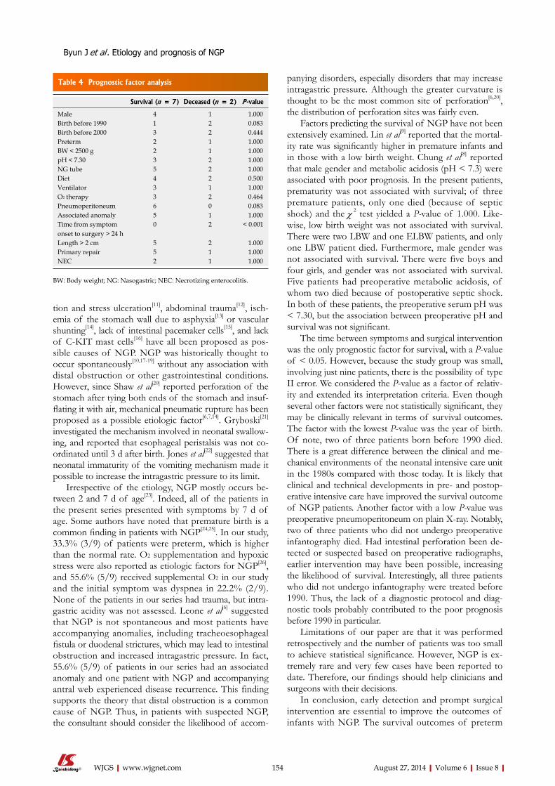
panying disorders, especially disorders that may increase intragastric pressure. Although the greater curvature is thought to be the most common site of perforation[6,20], the distribution of perforation sites was fairly even.
Factors predicting the survival of NGP have not been extensively examined. Lin et al[9] reported that the mortal-ity rate was significantly higher in premature infants and in those with a low birth weight. Chung et al[8] reported that male gender and metabolic acidosis (pH < 7.3) were associated with poor prognosis. In the present patients, prematurity was not associated with survival; of three premature patients, only one died (because of septic shock) and the χ 2 test yielded a P-value of 1.000. Like-wise, low birth weight was not associated with survival. There were two LBW and one ELBW patients, and only one LBW patient died. Furthermore, male gender was not associated with survival. There were five boys and four girls, and gender was not associated with survival. Five patients had preoperative metabolic acidosis, of whom two died because of postoperative septic shock. In both of these patients, the preoperative serum pH was < 7.30, but the association between preoperative pH and survival was not significant.
The time between symptoms and surgical intervention was the only prognostic factor for survival, with a P-value of < 0.05. However, because the study group was small, involving just nine patients, there is the possibility of type II error. We considered the P-value as a factor of relativ-ity and extended its interpretation criteria. Even though several other factors were not statistically significant, they may be clinically relevant in terms of survival outcomes. The factor with the lowest P-value was the year of birth. Of note, two of three patients born before 1990 died. There is a great difference between the clinical and me-chanical environments of the neonatal intensive care unit in the 1980s compared with those today. It is likely that clinical and technical developments in pre- and postop-erative intensive care have improved the survival outcome of NGP patients. Another factor with a low P-value was preoperative pneumoperitoneum on plain X-ray. Notably, two of three patients who did not undergo preoperative infantography died. Had intestinal perforation been de-tected or suspected based on preoperative radiographs, earlier intervention may have been possible, increasing the likelihood of survival. Interestingly, all three patients who did not undergo infantography were treated before 1990. Thus, the lack of a diagnostic protocol and diag-nostic tools probably contributed to the poor prognosis before 1990 in particular.
Limitations of our paper are that it was performed retrospectively and the number of patients was too small to achieve statistical significance. However, NGP is ex-tremely rare and very few cases have been reported to date. Therefore, our findings should help clinicians and surgeons with their decisions.
In conclusion, early detection and prompt surgical intervention are essential to improve the outcomes of infants with NGP. The survival outcomes of preterm
tion and stress ulceration[11], abdominal trauma[12], isch-emia of the stomach wall due to asphyxia[13] or vascular shunting[14], lack of intestinal pacemaker cells[15], and lack of C-KIT mast cells[16] have all been proposed as pos-sible causes of NGP. NGP was historically thought to occur spontaneously[10,17-19] without any association with distal obstruction or other gastrointestinal conditions. However, since Shaw et al[20] reported perforation of the stomach after tying both ends of the stomach and insuf-flating it with air, mechanical pneumatic rupture has been proposed as a possible etiologic factor[6,7,14]. Gryboski[21] investigated the mechanism involved in neonatal swallow-ing, and reported that esophageal peristalsis was not co-ordinated until 3 d after birth. Jones et al[22] suggested that neonatal immaturity of the vomiting mechanism made it possible to increase the intragastric pressure to its limit.
Irrespective of the etiology, NGP mostly occurs be-tween 2 and 7 d of age[23]. Indeed, all of the patients in the present series presented with symptoms by 7 d of age. Some authors have noted that premature birth is a common finding in patients with NGP[24,25]. In our study, 33.3% (3/9) of patients were preterm, which is higher than the normal rate. O2 supplementation and hypoxic stress were also reported as etiologic factors for NGP[26], and 55.6% (5/9) received supplemental O2 in our study and the initial symptom was dyspnea in 22.2% (2/9). None of the patients in our series had trauma, but intra-gastric acidity was not assessed. Leone et al[6] suggested that NGP is not spontaneous and most patients have accompanying anomalies, including tracheoesophageal fistula or duodenal strictures, which may lead to intestinal obstruction and increased intragastric pressure. In fact, 55.6% (5/9) of patients in our series had an associated anomaly and one patient with NGP and accompanying antral web experienced disease recurrence. This finding supports the theory that distal obstruction is a common cause of NGP. Thus, in patients with suspected NGP, the consultant should consider the likelihood of accom-
154 August 27, 2014|Volume 6|Issue 8|WJGS|www.wjgnet.com
Table 4 Prognostic factor analysis
Survival (n = 7) Deceased (n = 2) P -value
Male 4 1 1.000Birth before 1990 1 2 0.083Birth before 2000 3 2 0.444Preterm 2 1 1.000BW < 2500 g 2 1 1.000pH < 7.30 3 2 1.000NG tube 5 2 1.000Diet 4 2 0.500Ventilator 3 1 1.000O2 therapy 3 2 0.464Pneumoperitoneum 6 0 0.083Associated anomaly 5 1 1.000Time from symptom onset to surgery > 24 h
0 2 < 0.001
Length > 2 cm 5 2 1.000Primary repair 5 1 1.000NEC 2 1 1.000
BW: Body weight; NG: Nasogastric; NEC: Necrotizing enterocolitis.
Byun J et al . Etiology and prognosis of NGP
infants or LBW infants were not inferior to those of other patients. NGP can accompany other significant anomalies. Therefore, careful examination of the patient, together with imaging studies, may lead to early detection and improve the outcomes of NGP.
COMMENTSBackgroundNeonatal gastric perforation (NGP) is a very rare but life-threatening disease.Research frontiersBecause of its rarity, the etiology and prognostic factors of NGP are debated.Innovations and breakthroughsThe time between symptoms and surgical intervention was the only prognostic factor for survival; premature birth and birth weight were not associated with the survival of patients with NGP.Applications Early detection and advances in neonatal intensive care may improve the prog-nosis of NGP.TerminologyNGP: Neonatal gastric perforation; LBW: Low birth weight; ELBW: Extremely low birth weight.Peer reviewThis is an important case series to publish as it deals with a rare but important neonatal emergent disorder. The abstract, introduction, methods and results are all well written.
REFERENCES1 St-Vil D, LeBouthillier G, Luks FI, Bensoussan AL, Bla-
nchard H, Youssef S. Neonatal gastrointestinal perforations. J Pediatr Surg 1992; 27: 1340-1342 [PMID: 1403517 DOI: 10.1016/0022-3468(92)90292-f]
2 Rosser SB, Clark CH, Elechi EN. Spontaneous neonatal gastric perforation. J Pediatr Surg 1982; 17: 390-394 [PMID: 7120006 DOI: 10.1016/s0022-3468(82)80496-x]
3 Holgersen LO. The etiology of spontaneous gastric perfora-tion of the newborn: a reevaluation. J Pediatr Surg 1981; 16: 608-613 [PMID: 7277163 DOI: 10.1016/0022-3468(81)90014-2]
4 Tan CE, Kiely EM, Agrawal M, Brereton RJ, Spitz L. Neonatal gastrointestinal perforation. J Pediatr Surg 1989; 24: 888-892 [PMID: 2674391 DOI: 10.1016/s0022-3468(89)80589-5]
5 Kara CS, Ilçe Z, Celayir S, Sarimurat N, Erdogan E, Yeker D. Neonatal gastric perforation: review of 23 years’ experience. Surg Today 2004; 34: 243-245 [PMID: 14999537 DOI: 10.1007/s00595-003-2675-3]
6 Leone RJ, Krasna IH. ‘Spontaneous’ neonatal gastric per-foration: is it really spontaneous? J Pediatr Surg 2000; 35: 1066-1069 [PMID: 10917298 DOI: 10.1053/jpsu.2000.7773]
7 Jawad AJ, Al-Rabie A, Hadi A, Al-Sowailem A, Al-Rawaf A, Abu-Touk B, Al-Karfi T, Al-Sammarai A. Spontaneous neonatal gastric perforation. Pediatr Surg Int 2002; 18: 396-399 [PMID: 12415364 DOI: 10.1007/s00383-002-0749-8]
8 Chung MT, Kuo CY, Wang JW, Hsieh WS, Huang CB, Lin JN. Gastric perforation in the neonate: clinical analysis of 12 cases. Zhonghua Minguo Xiaoerke Yixuehui Zazhi 1994; 35: 460-465 [PMID: 7942035 DOI: 10.1002/ccd.20116]
9 Lin CM, Lee HC, Kao HA, Hung HY, Hsu CH, Yeung CY, Sheu JC, Wang NL. Neonatal gastric perforation: report of 15
cases and review of the literature. Pediatr Neonatol 2008; 49: 65-70 [PMID: 18947001 DOI: 10.1016/s1875-9572(08)60015-7]
10 Herbut PA. Congenital defect in musculature of stomach with rupture in newborn Infant. Arch Pathol 1943; 36: 91
11 Kiesewetter WB. Spontaneous rupture of the stomach in the newborn. AMA J Dis Child 1956; 91: 162-167 [PMID: 13282628 DOI: 10.1001/archpedi.1956.02060020164013]
12 Arnold GG. Perforation of the stomach in the neonatal pe-riod; report of a survival in a premature infant. J Pediatr 1955; 46: 276-279 [PMID: 14354581 DOI: 10.1016/s0022-3476(55)80281-8]
13 Touloukian RJ, Posch JN, Spencer R. The pathogenesis of ischemic gastroenterocolitis of the neonate: selective gut mu-cosal ischemia in asphyxiated neonatal piglets. J Pediatr Surg 1972; 7: 194-205 [PMID: 5023196 DOI: 10.1016/0022-3468(72)90496-4]
14 Houck WS, Griffin JA. Spontaneous linear tears of the stomach in the newborn infant. Ann Surg 1981; 193: 763-768 [PMID: 7247521 DOI: 10.1097/00000658-198106000-00012]
15 Ohshiro K, Yamataka A, Kobayashi H, Hirai S, Miyahara K, Sueyoshi N, Suda K, Miyano T. Idiopathic gastric perforation in neonates and abnormal distribution of intestinal pace-maker cells. J Pediatr Surg 2000; 35: 673-676 [PMID: 10813320 DOI: 10.1053/jpsu.2000.5940]
16 Yamataka A, Yamataka T, Kobayashi H, Sueyoshi N, Mi-yano T. Lack of C-KIT+ mast cells and the development of idiopathic gastric perforation in neonates. J Pediatr Surg 1999; 34: 34-37; discussion 37-38 [PMID: 10022139 DOI: 10.1016/s0022-3468(99)90224-5]
17 Touloukian RJ. Gastric ischemia: the primary factor in neo-natal perforation. Clin Pediatr (Phila) 1973; 12: 219-225 [PMID: 4701092]
18 Meyer JL. Congenital defect in the musculature of the stom-ach resulting in spontaneous gastric perforation in the neo-natal period; a report of two cases. J Pediatr 1957; 51: 416-421 [PMID: 13476323 DOI: 10.1016/s0022-3476(57)80126-7]
19 Amadeo JH, Ashmore HW, Aponte GE. Neonatal gastric perforation caused by congenital defects of the gastric mus-culature. Surgery 1960; 47: 1010-1017 [PMID: 13793066]
20 Shaw A, Blanc WA, Santulli TV, Kaiser G. Spontaneous rup-ture of the stomach in the newborn: A clinical and experi-mental study. Surgery 1965; 58: 561-571 [PMID: 14338551]
21 Gryboski JD. The swallowing mechanism of the neonate. i. esophageal and gastric motility. Pediatrics 1965; 35: 445-452 [PMID: 14260304]
22 Jones TB, Kirchner SG, Lee FA, Heller RM. Stomach rup-ture associated with esophageal atresia, tracheoesophageal fistula, and ventilatory assistance. AJR Am J Roentgenol 1980; 134: 675-677 [PMID: 6767350 DOI: 10.2214/ajr.134.4.675]
23 Saracli T, Mann M, French DM, Booker CR, Scott RB. Rup-ture of the stomach in the newborn infant. Report of three cases and review of the world literature. Clin Pediatr (Phila) 1967; 6: 583-588 [PMID: 6077501 DOI: 10.1177/000992286700601014]
24 Bruce J, Bianchi A, Doig CM. Gastric perforation in the neo-nates. Pediatr Surg Int 1993; 8: 17-19 [DOI: 10.1007/bf02352993]
25 Young AE, Sury MR. Spontaneous neonatal gastric perfora-tion. Paediatr Anaesth 1996; 6: 143-145 [PMID: 8846280 DOI: 10.1111/j.1460-9592.1996.tb00378.x]
26 Hood JH. Clinical considerations of intestinal gas. Ann Surg 1966; 163: 359-366 [PMID: 5907560 DOI: 10.1097/00000658-196603000-00006]
P- Reviewer: Khassawneh M S- Editor: Song XX L- Editor: Wang TQ E- Editor: Liu SQ
155 August 27, 2014|Volume 6|Issue 8|WJGS|www.wjgnet.com
COMMENTS
Byun J et al . Etiology and prognosis of NGP
Blind loop perforation after side-to-side ileocolonic anastomosis
Raffaele Dalla Valle, Roberto Zinicola, Maurizio Iaria
Raffaele Dalla Valle, Maurizio Iaria, Department of Surgery, Division of General Surgery and Organ Transplantation, Parma University Hospital, 43121 Parma, ItalyRoberto Zinicola, Department of Emergency, Division of Acute Care Surgery, Parma University Hospital, 43121 Parma, ItalyAuthor contributions: All authors contributed to this work.Correspondence to: Maurizio Iaria, MD, PhD, Department of Surgery, Division of General Surgery and Organ Transplantation, Parma University Hospital, Via Linati, 6, 43121 Parma, Italy. [email protected]: +39-05-21702006 Fax: +39-05-21704870Received: February 26, 2014 Revised: May 11, 2014Accepted: July 15, 2014Published online: August 27, 2014
AbstractBlind loop syndrome after side-to-side ileocolonic anas-tomosis is a well-recognized entity even though its in-cidence and complication rates are not clearly defined. The inevitable dilation of the ileal cul-de-sac leads to stasis and bacterial overgrowth which eventually leads to mucosal ulceration and even full-thickness perfora-tion. Blind loop syndrome may be an underestimated complication in the setting of digestive surgery. It should always be taken into account in cases of acute abdomen in patients who previously underwent right hemicolectomy. We herein report 3 patients who were diagnosed with perforative blind loop syndrome a few years after standard right hemicolectomy followed by a side-to-side ileocolonic anastomosis.
© 2014 Baishideng Publishing Group Inc. All rights reserved.
Key words: Blind loop syndrome; Blind loop pouch; Perforation; Ileocolonic anastomosis; Laparoscopy
Core tip: The authors suggest that we are likely to see more and more cases of blind loop syndrome in the fu-ture because more side-to-side ileocolonic anastomoses
will be performed in the setting of colonic laparoscopic surgery. A blind loop perforation should immediately be investigated in a patient who presents with acute abdo-men years after a right hemicolectomy. Ideally, more end-to-end anastomoses should be performed, when-ever suitable, in an effort to prevent the development of a blind loop.
Dalla Valle R, Zinicola R, Iaria M. Blind loop perforation af-ter side-to-side ileocolonic anastomosis. World J Gastrointest Surg 2014; 6(8): 156-159 Available from: URL: http://www.wjgnet.com/1948-9366/full/v6/i8/156.htm DOI: http://dx.doi.org/10.4240/wjgs.v6.i8.156
INTRODUCTIONIn the past, digestive continuity after right hemicolectomy was often restored through either end-to-end or end-to-side ileocolonic anastomosis. Due to the extensive imple-mentation of laparoscopy in colonic surgery, side-to-side ileocolonic anastomosis has gained popularity because of the widespread use of linear staplers in this setting. Even in open surgery, mechanical side-to-side anastomoses are easy, quick and cost-effective because these can be per-formed using a single linear stapling device, while an end-to-side recontruction would require both linear and circu-lar staplers to complete the anastomosis with additional costs.
Concurrently, intracorporeal anastomoses during lapa-roscopic right hemicolectomies can be exclusively carried out with linear staplers (endoGIA).
Yet, in 1906, Cannon and Murphy judged side-to-side anastomoses to be far from physiological, and advocated end-to-end reconstruction instead[1]. The same recom-mendation was reinforced by Pearse[2], Estes et al[3] and Holme[4] in further studies. A key reason why side-to-side anastomoses have been questioned lies in their substan-
CASE REPORT
Submit a Manuscript: http://www.wjgnet.com/esps/Help Desk: http://www.wjgnet.com/esps/helpdesk.aspxDOI: 10.4240/wjgs.v6.i8.156
World J Gastrointest Surg 2014 August 27; 6(8): 156-159ISSN 1948-9366 (online)
© 2014 Baishideng Publishing Group Inc. All rights reserved.
156 August 27, 2014|Volume 6|Issue 8|WJGS|www.wjgnet.com

tial risk of progressive dilation at the level of the cul-de-sac, which might lead to enlarged pockets prone to stasis and bacterial overgrowth. Such alterations seem to pre-dispose to the so-called “blind loop syndrome”[5,6]. The continuous enlargement of the blind loop may eventually cause mucosal ulcerations, intestinal bleeding and/or full-thickness viscus perforation.
There are limited data in the literature about the actu-al incidence of blind loop syndrome and its related mor-bidities because, generally, studies on intestinal anastomo-ses are exclusively focused on short-term postoperative complications (3 mo), whereas blind loop syndrome tends to develop years after surgery. We retrospectively reviewed 3 cases of blind loop perforation which oc-curred during long-term follow-up after standard right hemicolectomy followed by mechanical side-to-side ileo-colonic anastomosis.
CASE REPORTCase 1A 76-year-old woman underwent a right hemicolectomy in 2007 for a Duke’s stage C adenocarcinoma of the as-cending colon followed by a mechanical isoperistaltic side-to-side ileocolonic anastomosis. In September 2009, she was admitted for acute and diffuse abdominal pain, high-grade fever and leukocytosis. An abdominal X-ray series displayed free intra-peritoneal subphrenic air bilaterally. Since a computed tomography (CT) scan was not immedi-ately available, an exploratory laparoscopy was performed followed by laparotomy. A generalized purulent peritonitis due to a pinpoint perforation of the ileocolonic blind loop was identified. The same blind loop appeared extremely enlarged, measuring about 7 cm, and was resected using a 75 mm GIA stapler (United States Surgical, Norwalk, CT, United States). The patient had an uneventful postopera-tive course and was discharged home 6 d after surgery. At the 12-mo outpatient clinic follow-up, the patient was asymptomatic with normal bowel function.
Case 2An 80-year-old woman had a right hemicolectomy in
2008 for a Duke’s stage B adenocarcinoma of the ileoce-cal junction, with a mechanical isoperistaltic side-to-side ileocolonic anastomosis. In January 2011, she was admit-ted with worsening generalized abdominal pain, fever and leukocytosis. Before seeking medical attention, the patient experienced loose stools and mild non-localized abdomi-nal pain for almost 1 wk. Plain abdominal X-rays revealed free subphrenic air. A CT scan detected free air in the abdominal cavity and a slightly dilated small bowel loop. A subsequent laparoscopy identified generalized purulent peritonitis due to a tiny perforation of a 6 cm blind loop. The perforated pouch was resected with a 45 mm en-doGIA stapler. She had a straightforward postoperative course and was discharged after 5 d. At a scheduled 6-mo follow-up visit, she was symptom-free except for mild sporadic diarrhea.
Case 3A 58-year-old woman had a right hemicolectomy in 2009 for a Duke’s stage B adenocarcinoma of the he-patic flexure, with a hand-sewn antiperistaltic side-to-side ileocolonic anastomosis. In November 2011, she presented with severe epigastric pain, fever and leukocy-tosis. Plain abdominal X-rays failed to show free air. A contrast-enhanced CT scan revealed intra-peritoneal free air localized in the lower abdomen with air bubbles near the anastomosis. The blind loop showed thick walls as-sociated with mucosal hyperemia along with focal areas of perivisceral soft tissue fat necrosis (Figure 1). Imaging was compatible with blind loop perforation. The patient underwent laparotomy, which demonstrated generalized purulent peritonitis due to microperforation (Figure 2) of the enlarged blind ileal pouch (about 10 cm long). The blind loop was resected with a 75 mm GIA stapler. She recovered well after surgery and was discharged home in 10 d. At the 6-mo follow-up visit, she had no symptoms and regular bowel movements.
DISCUSSIONNowadays, mechanical side-to-side anastomosis after right hemicolectomy is commonly performed either in open or laparoscopic surgery due to its simplicity and
Dalla Valle R et al . Blind loop perforation after side-to-side ileocolonic anastomosis
157 August 27, 2014|Volume 6|Issue 8|WJGS|www.wjgnet.com
Figure 1 Computed tomography. The scan shows intraperitoneal free air with air bubbles around the ileocolonic anastomosis and a dilated blind loop with thickened bowel walls and mucosal hyperemia.
Figure 2 The image shows a long blind loop with signs of microperfora-tion on the suture line.

speed. In the short-term there is no clear advantage of a specific anastomotic configuration over others. On the other hand, side-to-side anastomoses have been criticized for being theoretically anti-physiologic and for the long-term risk of a blind loop[1].
Blind loops result in abnormal peristalsis causing fill-ing rather than emptying of the pouch. Such dismotility-related stasis predisposes to bacterial overgrowth eventu-ally followed by mucosal and/or transmural inflammatory changes of the intestinal wall. The consequent clinical scenario is referred as blind loop syndrome and may present with a wide spectrum of morbidities such as diar-rhea, vitamin B12 deficiency, iron-deficiency anemia, ul-cerations, bleeding and enteroliths[5]. At times, a pinpoint perforation of the blind loop may occur[5,7,8]. Partial dis-ruption of the muscle layer in the side-to-side anastomo-sis causes dysmotility, diverting intestinal content more easily unto the blind loop. The cul-de-sac dilation may occur even after an end-to-side recontruction, though it appears more likely to happen after a side-to-side anas-tomosis[6]. Conversely, blind loops do not develop after end-to-end anastomoses. Stellamor et al[9] analyzed 66 ileocolonic resections, and identified 9 blind loops (aver-age transverse diameter between 5 and 11 cm) out of 31 side-to-side anastomoses, whereas no blind loops were observed after 12 end-to-end anastomoses.
Only a few cases have been accurately described in literature. Estes et al[3] reported about a 10 cm blind loop which had increased to 46 cm after only a few months. Pollock described a blind loop which started as 2 cm in length and stretched to 15 cm after 1 year[10].
We believe this critical aspect of side-to-side anasto-moses will be more intensively taken into consideration in light of their expanding use in the laparoscopic era. The likelihood of enlargement and tearing appear propor-tional to the span of the blind loop, the bigger the pouch the higher the risk of further lengthening and stretching. It is advisable to leave the bowel stump as short as pos-sible, even if that does not necessarily prevent the loop from enlarging in the long-term. In addition, antiperi-staltic anastomoses do not seem to abolish the risk of developing a cul-de-sac, as shown in one of our cases. Some authors reported incidental findings of long blind loops discovered during either autopsy studies or surgi-cal interventions in asymptomatic individuals[6]. Thus, the sole development of a blind loop does not cause clinical manifestations per se.
We found in the literature only 11 cases of blind loop perforation, some presenting with acute, generalized peri-tonitis due to pinpoint perforation[5,6,8]. Lack of data pre-cludes an accurate appraisal on those factors which might cause symptomatic complications in those with a blind loop.
Usually, blind loop syndrome occurs many years after bowel surgery. In a French review, 45 out of 69 patients with a blind loop developed abdominal symptoms more than 5 years after surgery[5]. In a retrospective study based on abdominal CT examinations of 30 patients with radio-logical features compatible with a blind loop (eventually
resected in 4 cases), the mean and median time between surgery and imaging were 49.4 and 32.2 mo, respective-ly[11]. In our own series, all patients presented with perfo-ration at least 2 years after a side-to-side ileocolonic anas-tomosis. A CT scan seems helpful and a focally dilated loop of the small bowel adjacent to surgical clips is a re-current finding[11]. Even so, blind loops may be mistaken for diverticula, abscesses or obstructed bowel segments. Blind loop syndrome is primarily managed through a redo anastomosis in an end-to-end fashion. We chose to resect the perforated pouch sparing a new anastomosis, deeming this limited procedure safer in the emergency setting. Our patients are free of symptoms albeit after a short clinical follow-up (6-12 mo). Blind loop syndrome along with its complications is probably underestimated and further research is needed to define the real extent of the problem. In addition, a colorectal surgeon should be aware of this potential issue before choosing the ileoco-lonic anastomosis reconstructive technique.
COMMENTSCase characteristicsThree patients presented with perforation of a blind loop years after right hemi-colectomy followed by side-to-side ileocolonic anastomosis.Clinical diagnosisThey displayed peritoneal signs along with high-grade fever, one had localized epigastric pain, and the others suffered diffuse abdominal pain.Differential diagnosisComplicated acute diverticulitis, peptic ulcer perforation.Laboratory diagnosisAll had leukocytosis.Imaging diagnosisPlain abdominal X-rays usually show peritoneal free-air. Contrast-enhanced computed tomography identified an enlarged blind loop with thick walls, muco-sal hyperemia and perivisceral fat necrosis along with intra-abdominal free-air and fluid. Pathological diagnosisIleal stump enlargement with full-thickness pinpoint perforation.TreatmentLaparoscopic or laparotomic resection of the perforated blind loop with linear stapler.Related reportsOnly a few series and isolated case reports are available in the literature about the long-term complications of ileocolonic anastomoses associated with a spe-cific anastomosis configuration. The true incidence of blind loop syndrome is probably underestimated but blind loop enlargement after side-to-side digestive anastomoses is a well-known phenomenon. Besides, only 11 cases of blind loop perforation are described in literature thus far.Term explanationBlind loop syndrome after right hemicolectomy develops when bacterial over-growth occurs in the previously interrupted bowel stump, whose dysmotility tends to divert the intestinal contents away from the physiologic route. Clinically it may manifest itself with symptoms of vitamin malabsorption, malnutrition, weight loss, digestive bleeding and even viscus perforation.Experiences and lessonsSide-to-side anastomoses are strongly related to the development of blind loop syndrome in the long-term. In open right hemicolectomy, end-to-end or end-to-side anastomoses can be used, while in the case of a full laparoscopic proce-dure requiring a side-to-side reconstruction, surgeons should resect the ileal stump as short as possible.Peer reviewIn general the side-to-side anastomosis is a bad technique to restore intestinal
158 August 27, 2014|Volume 6|Issue 8|WJGS|www.wjgnet.com
COMMENTS
Dalla Valle R et al . Blind loop perforation after side-to-side ileocolonic anastomosis

drome after side-to-side intestinal anastomosis]. Chirurgie 1990; 116: 586-596 [PMID: 2129971]
6 Schlegel DM, Maglinte DD. The blind pouch syndrome. Surg Gynecol Obstet 1982; 155: 541-544 [PMID: 6981863]
7 Hensler MK, Roosen JU. [A case of perforation of a blind loop secondary to ileocolic side-to-side anastomosis]. Ugeskr Laeger 1991; 153: 2835-2836 [PMID: 1926622]
8 Tsugu Y, Shimada N, Kawakami H, Kadomatsu T, Morita H. [Blind loop syndrome after intestinal anastomosis (case of perforated blind loop)]. Shujutsu 1966; 20: 650-656 [PMID: 5975502]
9 Stellamor K, Hochberger O. [To the cognizance of the “blind pouch syndrome” following intestinal anastomoses (author’s transl)]. Rontgenblatter 1974; 27: 82-90 [PMID: 4820534]
10 Pollock LH. Blind-pouch formation following lateral anas-tomosis. AMA Arch Surg 1958; 76: 536-541 [PMID: 13520085 DOI: 10.1001/archsurg.1958.01280220056011]
11 Sandrasegaran K, Maglinte DD, Rajesh A, Tann M, Kopecky KK. CT findings for postsurgical blind pouch of small bowel. AJR Am J Roentgenol 2006; 186: 110-113 [PMID: 16357387 DOI: 10.2214/AJR.04.1628]
P- Reviewer: Actis GC, Goetze TO, Rabago L S- Editor: Ji FF L- Editor: Cant MR E- Editor: Liu SQ
continuity but not unusual in times of laparoscopic surgery of the right colon. It leads to a progressive distention of the cul-de-sac, which produces definite pockets of stasis and infection.
REFERENCES1 Cannon WB, Murphy FT. IV. The Movements of the Stom-
ach and Intestines in Some Surgical Conditions. Ann Surg 1906; 43: 512-536 [PMID: 17861784 DOI: 10.1097/00000658-190604000-00004]
2 Pearse AE. Experimental Chronic Intestinal Obstruction from Blind Loops. Surg Gynecol Obstet 1934; 59: 726
3 Estes WL, Holm CE. The Fate of the Obstructed Loop in Intestinal Obstruction following an Anastomosis around the Obstruction without Resection. Ann Surg 1932; 96: 924-929 [PMID: 17866883 DOI: 10.1097/00000658-193211000-00012]
4 Holm CE. The Fate of the Sidetracted Loop of Ileum Follow-ing Lateral Anastomosis for Complete Benign Obstruction. Surg Gynecol Obstet 1933; 56: 746
5 Frank P, Batzenschlager A, Philippe E. [Blind-pouch syn-
159 August 27, 2014|Volume 6|Issue 8|WJGS|www.wjgnet.com
Dalla Valle R et al . Blind loop perforation after side-to-side ileocolonic anastomosis

Massive surgical emphysema following transanal endoscopic microsurgery
Geert AAM Simkens, Simon W Nienhuijs, Misha DP Luyer, Ignace HJT de Hingh
Geert AAM Simkens, Simon W Nienhuijs, Misha DP Luyer, Ignace HJT de Hingh, Department of Surgical Oncology, Cath-arina Hospital Eindhoven, 5623 EJ Eindhoven, The NetherlandsAuthor contributions: Simkens GAAM, Nienhuijs SW, Luyer MDP and de Hingh IHJT contributed equally to this work.Correspondence to: Ignace HJT de Hingh, MD, PhD, De-partment of Surgical Oncology, Catharina Hospital Eindhoven, Michelangelolaan 2, 5623 EJ Eindhoven, The Netherlands. [email protected] Telephone: +31-40-2399111 Fax: +31-40-2455035Received: January 25, 2014 Revised: May 18, 2014Accepted: July 12, 2014Published online: August 27, 2014
AbstractWe describe an impressive and rare case of surgical emphysema after minimally invasive rectal surgery. This case reports on a patient who developed mas-sive retroperitoneal, intraperitoneal and subcutaneous emphysema directly following a transanal endoscopic microsurgery (TEM) procedure for a rectal intramuco-sal carcinoma. Free intra-abdominal air after gastro-intestinal surgery can be a sign of a bowel perforation or anastomotic leakage. This is a serious complication often requiring immediate surgery. In our patient an abdominal computed tomography-scan with rectal con-trast showed no signs of a rectal perforation. Therefore this emphysema was caused by the insufflation of CO2 gas in the rectum during the TEM-procedure. Conserva-tive treatment resulted in an uneventful recovery. With the increasing usage of TEM for rectal lesions we ex-pect this complication to occur more often. After ruling out a full thickness rectal wall perforation in patients with surgical emphysema following TEM, conservative treatment is the treatment of choice.
© 2014 Baishideng Publishing Group Inc. All rights reserved.
Key words: Transanal endoscopic microsurgery; Mi-crosurgery; Gastrointestinal endoscopy; Colorectal
neoplasms; Retropneumoperitoneum; Intraperitoneal emphysema; Subcutaneous emphysema
Core tip: Surgical emphysema after transanal endo-scopic microsurgery (TEM) can be a sign of a rectal perforation. This report describes a patient with impres-sive retroperitoneal, intraperitoneal and subcutaneous emphysema directly following TEM without a full thick-ness rectal wall perforation. This is a rare complication after TEM in which conservative treatment resulted in an uneventful recovery.
Simkens GAAM, Nienhuijs SW, Luyer MDP, de Hingh IHJT. Massive surgical emphysema following transanal endoscopic microsurgery. World J Gastrointest Surg 2014; 6(8): 160-163 Available from: URL: http://www.wjgnet.com/1948-9366/full/v6/i8/160.htm DOI: http://dx.doi.org/10.4240/wjgs.v6.i8.160
INTRODUCTIONTransanal endoscopic microsurgery (TEM) is a minimal-ly invasive technique developed for the resection of rec-tal neoplasms. This technique was first described in the early 1980’s and was initially exclusively intended for be-nign adenomas[1]. By providing good stereoscopic views and allowing use of both hands, better exposure and more precise excision are achievable as compared to pre-viously used techniques for local excision. With increas-ing experience, TEM is now considered safe and may also be offered for low-grade malignant rectal lesions without signs of lymphatic or systemic dissemination (T1-2, N0, M0)[2]. The major advantage of TEM is the significantly lower morbidity and mortality as compared to traditional treatment for malignant rectal lesions such as abdominoperineal resection and low anterior resec-tion[2]. Furthermore, functional outcome is better since the sphincteric complex is preserved, the abdominal wall is spared and damage to autonomous pelvic nerves is
CASE REPORT
Submit a Manuscript: http://www.wjgnet.com/esps/Help Desk: http://www.wjgnet.com/esps/helpdesk.aspxDOI: 10.4240/wjgs.v6.i8.160
World J Gastrointest Surg 2014 August 27; 6(8): 160-163ISSN 1948-9366 (online)
© 2014 Baishideng Publishing Group Inc. All rights reserved.
160 August 27, 2014|Volume 6|Issue 8|WJGS|www.wjgnet.com

absent. Postoperative complications are rare after TEM and include postoperative bleeding (1.7%-2.7%) and pelvic sepsis due to perforation (1%-2.7%). TEM asso-ciated mortality is low (0%-2%)[3]. In this case report, a rare and impressive complication after TEM is described being the occurrence of massive retroperitoneal, intra-peritoneal and subcutaneous emphysema directly follow-ing a TEM-procedure.
CASE REPORTA 60-year-old woman was referred to our hospital with changed bowel habits including frequent defecation and soiling. Her relevant medical history includes a uterus ex-tirpation, alcohol abuse and smoking. On a colonoscopy several sessile polyps throughout the colon were discov-ered which could be removed endoscopically. Pathology reports showed that these were all tubulovillous adeno-mas without signs of malignancy. However, in the rectum also a large villous lesion measuring 5 cm in diameter was found, located approximately 8-10 cm from the anal verge. This lesion could not be removed endoscopically due to its size and appearance. Biopsies showed at least intramucosal carcinoma but additional computed tomog-raphy (CT)-scan of the abdomen, pelvic magnetic reso-nance imaging and rectal endo-echography did not show signs of invasive growth, pathologic lymph nodes or systemic metastases. The tumor was therefore staged as a cT1N0M0 rectal intramucosal carcinoma and the patient was referred for a TEM-procedure.
Prior to the surgical procedure antibiotic prophylaxis was given (Cefazolin 1000 mg and Metronidazole 1500 mg both once) and the tumor was visualised on the ventral side of the rectum using rectoscopy. The patient was placed in prone position, the extended TEM-tube (TEO 15 cm, Karl Storz, Germany) was inserted and a pneumorectum was applied by insufflation of CO2 at 6 L/min with a pressure limit of 12 mmHg using a Karl Storz insufflator. Using ultracision, the lesion was macro-scopically radical removed. No rectal perforation into the peritoneal cavity was observed and the defect in the rectal wall was closed with a running suture. Duration of the procedure was 115 min. Pathological examination con-
firmed the diagnosis of a tubulovillous adenoma with the presence of intra-mucosal carcinoma and clear resection margins. Immediately after the procedure massive sub-cutaneous emphysema of the face, neck and body was noticed (Figure 1). Given the amount of emphysema in the face it was decided to keep the patient intubated and ventilated and transfer to the intensive care unit. Here, blood analysis revealed a slight leucocytosis (11.0 k/μL) and a C-reactive protein (CRP) level of < 6 mg/L. Chest X-ray showed subcutaneous emphysema of the thorax, signs of “free air” beneath the left diaphragm and a small pneumothorax of the right sinus (Figure 2). After a few hours emphysema in the face was dissolving and the pa-tient was awake. She was then detubated and discharged from the intensive care unit. At this moment she did not experience any abdominal discomfort, systemic reaction or fever. Intravenous antibiotics (Metronidazole 500 mg, Cefuroxime 1500 mg, both 3 times daily for 3 d) were started empirically and the patient was kept hospitalized with solid diet until all subcutaneous air had resolved three days later.
One day after discharge the patient visited the emer-gency ward with complaints of dyspnoea. Again, no ab-dominal discomfort or nausea were present, the patient had no fever and solid food was tolerated. However, blood analysis now showed an elevated CRP-level of 160 mg/L and based on the thoracic X-ray an increase in the amount of “free air” in the intra-abdominal space was suspected. Given the dyspnoea, “free abdominal air” and elevated inflammatory parameters a rectal perforation with leakage was suspected and an abdominal CT-scan with rectal contrast was performed. This revealed a large amount of air in the mesorectal fascia and especially in the retroperitoneal space and only a small amount of free air in the abdomen (Figure 3A). This retroperitoneal air had erroneously suggested the presence of free air in the abdomen. No signs of leakage from the rectal wound were found. In the lower thoracic field, pleural effusion and signs of a pneumonia were revealed (Figure 3B). This eventually explained for the dyspnoea and elevated inflammatory parameters. Conservative treatment with continuation of intravenous antibiotics was installed. In the following days, the dyspnoea disappeared and also,
Simkens GAAM et al . Surgical emphysema following TEM-procedure
161 August 27, 2014|Volume 6|Issue 8|WJGS|www.wjgnet.com
Figure 1 Right-sided peri-orbital subcutaneous emphysema directly post-operative (arrow).
Figure 2 Thoracic X-ray suggesting free intraperitoneal air beneath the left diaphragm (arrow).
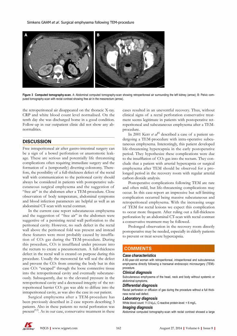
the retroperitoneal air disappeared on the thoracic X-ray. CRP and white blood count level normalised. On the tenth day she was discharged home in a good condition. Follow-up in our outpatient clinic did not show any ab-normalities.
DISCUSSIONFree intraperitoneal air after gastro-intestinal surgery can be a sign of a bowel perforation or anastomotic leak-age. These are serious and potentially life threatening complications often requiring immediate surgery and the formation of a (temporarily) diverting colostomy. There-fore, the possibility of a full-thickness defect of the rectal wall with communication to the peritoneal cavity should always be considered in patients with postoperative sub-cutaneous surgical emphysema and the suggestion of “free air” in the abdomen after a TEM-procedure. Close observation of body temperature, abdominal symptoms and blood infection parameters are helpful as well as an abdominal CT-scan with rectal contrast.
In the current case report subcutaneous emphysema and the suggestion of “free air” in the abdomen were suggestive of a persisting rectal wall perforation to the peritoneal cavity. However, no such defect in the rectal wall above the peritoneal fold was present and instead these features were most probably caused by insuffla-tion of CO2 gas during the TEM-procedure. During this procedure, CO2 is insufflated under pressure into the rectum to create a pneumorectum. A full-thickness defect in the rectal wall is created on purpose during this procedure. Usually the mesorectal fat will seal the defect and prevent the CO2 from entering the body but in this case CO2 “escaped” through the loose connective tissue into the retroperitoneal cavity and eventually subcutane-ously. Subsequently, due to the elevated pressure in the retroperitoneal cavity and a decreased integrity of the ret-roperitoneal barrier CO2 gas was able to diffuse into the intraperitoneal cavity, as was also the case in our patient.
Surgical emphysema after a TEM-procedure has been previously described in 2 case reports describing 3 patients. Also in these patients no rectal perforation was present[4,5]. As in our case, conservative treatment in these
cases resulted in an uneventful recovery. Thus, without clinical signs of a rectal perforation conservative treat-ment seems legitimate in patients with postoperative ret-roperitoneal and subcutaneous emphysema after a TEM-procedure.
In 2001 Kerr et al[6] described a case of a patient un-dergoing a TEM-procedure with intra-operative subcu-taneous emphysema. Interestingly, this patient developed life-threatening hypercapnia in the early postoperative period. They hypothesize these complications were due to the insufflation of CO2 gas into the rectum. They con-clude that a patient with arterial hypercapnia or surgical emphysema after TEM should be observed for a pro-longed period in the recovery room with regular arterial carbon dioxide analysis.
Postoperative complications following TEM are rare and often mild, but life-threatening complications may occur. In this case-report an impressive but self-limiting complication occurred being massive subcutaneous and retroperitoneal emphysema. With the increasing usage of TEM for rectal lesions we expect this complication to occur more frequent. After ruling out a full-thickness perforation by an abdominal CT-scan with rectal contrast a conservative treatment may be followed.
Prolonged observation in the recovery room directly postoperative may be needed, especially in elderly patients to prevent or treat severe hypercapnia.
COMMENTSCase characteristicsA 60-year-old woman with retroperitoneal, intraperitoneal and subcutaneous emphysema directly following a transanal endoscopic microsurgery (TEM)-procedure.Clinical diagnosisSubcutaneous emphysema of the head, neck and body without systemic or abdominal symptoms.Differential diagnosisRectal perforation or diffusion of gas during the procedure without a full thick-ness rectal wall defect.Laboratory diagnosisWhite blood count: 11.0 k/μL; C-reactive protein-level: < 6 mg/L.Imaging diagnosisAbdominal computed tomography-scan with rectal contrast showed a large
162 August 27, 2014|Volume 6|Issue 8|WJGS|www.wjgnet.com
Figure 3 Computed tomography-scan. A: Abdominal computed tomography-scan showing retroperitoneal air surrounding the left kidney (arrow); B: Pelvic com-puted tomography-scan with rectal contrast showing free air in the mesorectum (arrow).
A B
COMMENTS
Simkens GAAM et al . Surgical emphysema following TEM-procedure

maier H. [A system for a transanal endoscopic rectum opera-tion]. Chirurg 1984; 55: 677-680 [PMID: 6510078]
2 Léonard D, Remue C, Kartheuser A. The transanal endo-scopic microsurgery procedure: standards and extended indications. Dig Dis 2012; 30 Suppl 2: 85-90 [PMID: 23207938 DOI: 10.1159/000342033]
3 Bignell MB, Ramwell A, Evans JR, Dastur N, Simson JN. Complications of transanal endoscopic microsurgery (TEMS): a prospective audit. Colorectal Dis 2010; 12: e99-103 [PMID: 19843114 DOI: 10.1111/j.1463-1318.2009.02071.x]
4 Cantos M, Bruna M, García-Coret MJ, Villalba FL, Roig JV. [Pneumomediastinum and subcutaneous emphysema like strange complications after transanal endoscopic mi-crosurgery]. Rev Esp Enferm Dig 2009; 101: 445-446 [PMID: 19630473]
5 Franken RJ, Moes DE, Acherman YI, Derksen EJ. Free Intra-Abdominal Air without Peritoneal Perforation after TEM: A Report of Two Cases. Case Rep Surg 2012; 2012: 185429 [PMID: 23346448 DOI: 10.1155/2012/185429]
6 Kerr K, Mills GH. Intra-operative and post-operative hyper-capnia leading to delayed respiratory failure associated with transanal endoscopic microsurgery under general anaesthe-sia. Br J Anaesth 2001; 86: 586-589 [PMID: 11573640]
P- Reviewer: Arezzo A, Agresta F, George V, Han X, Kita H S- Editor: Song XX L- Editor: A E- Editor: Liu SQ
amount of air in the mesorectal fascia and especially in the retroperitoneal space and only a small amount of free air in the abdomen. No signs of leakage from the rectal wound were found.Pathological diagnosiscT1N0M0 rectal intramucosal carcinoma.TreatmentConservative treatment with intravenous antibiotics.Related reportsTwo reports previously described similar cases, with uneventful recovery after conservative treatment.Experiences and lessonsFree intra-abdominal air after TEM can occur without a full thickness rectal wall perforation and may be treated conservative.Peer reviewTransanal endoscopic microsurgery is a minimally invasive technique devel-oped for the resection of rectal neoplasms. This paper is very interesting case report of something observed not so rarely, even though not so evident.
REFERENCES1 Buess G, Hutterer F, Theiss J, Böbel M, Isselhard W, Pichl-
163 August 27, 2014|Volume 6|Issue 8|WJGS|www.wjgnet.com
Simkens GAAM et al . Surgical emphysema following TEM-procedure
Solitary mediastinal lymph node recurrence after curative resection of colon cancer
Yasuhiro Matsuda, Masahiko Yano, Norikatsu Miyoshi, Shingo Noura, Masayuki Ohue, Keijiro Sugimura, Masaaki Motoori, Kentaro Kishi, Yoshiyuki Fujiwara, Kunihito Gotoh, Shigeru Marubashi, Hirofumi Akita, Hidenori Takahashi, Masato Sakon
Yasuhiro Matsuda, Masahiko Yano, Norikatsu Miyoshi, Shingo Noura, Masayuki Ohue, Keijiro Sugimura, Masaaki Motoori, Kentaro Kishi, Yoshiyuki Fujiwara, Kunihito Gotoh, Shigeru Marubashi, Hirofumi Akita, Hidenori Takahashi, Masato Sakon, Department of Surgery, Osaka Medical Center for Cancer and Cardiovascular Disease, Osaka 537-8511, JapanAuthor contributions: Matsuda Y and Yano M designed the re-port; Miyoshi N, Noura S, Ohue M, Sugimura K, Motoori M and Sakon M made a decision of the treatment; Kishi K, Fujiwara Y, Gotoh K, Marubashi S, Akita H and Takahashi H collected the patient’s clinical data; Matsuda Y and Yano M analyzed the data and wrote the paper.Supported by Department of Surgery, Osaka Medical Center for Cancer and Cardiovascular Disease, Osaka 537-8511, JapanCorrespondence to: Masahiko Yano, MD, PhD, Department of Surgery, Osaka Medical Center for Cancer and Cardiovascular Disease, 1-3-3 Nakamichi, Higashinari-ku, Osaka 537-8511, Japan. [email protected]: +81-6-69721181 Fax: +81-6-69818055Received: February 26, 2014 Revised: March 27, 2014Accepted: July 18, 2014Published online: August 27, 2014
AbstractWe report two cases of solitary mediastinal lymph node recurrence after colon cancer resection. Both cases had para-aortic lymph node metastasis at the time of initial surgery and received adjuvant chemotherapy for 4 years in case 1 and 18 mo in case 2. The time to recurrence was more than 8 years in both cases. After resection of the recurrent tumor, the patient is doing well with no recurrence for 6 years in case 1 and 4 mo in case 2. Patients should be followed up after colon cancer surgery considering the possibility of solitary mediastinal lymph node recurrence if they had para-aortic node metastasis at the time of initial surgery.
© 2014 Baishideng Publishing Group Inc. All rights reserved.
Key words: Solitary mediastinal lymph node recurrence; Colon cance
Core tip: The first site of recurrence of colon cancer is rarely the mediastinal lymph nodes, and solitary recur-rence at this site without another site of recurrence is extremely rare. The operative indication and prognostic significance of the resection of mediastinal lymph node metastasis has not been determined because such re-currence is very rare. Patients should be followed up after colon cancer surgery considering the possibility of solitary mediastinal lymph node recurrence if they had para-aortic node metastasis at the time of initial surgery.
Matsuda Y, Yano M, Miyoshi N, Noura S, Ohue M, Sugimura K, Motoori M, Kishi K, Fujiwara Y, Gotoh K, Marubashi S, Akita H, Takahashi H, Sakon M. Solitary mediastinal lymph node recurrence after curative resection of colon cancer. World J Gastrointest Surg 2014; 6(8): 164-168 Available from: URL: http://www.wjgnet.com/1948-9366/full/v6/i8/164.htm DOI: http://dx.doi.org/10.4240/wjgs.v6.i8.164
INTRODUCTIONDistant organ recurrence after resection of colon cancer is frequently seen in the liver, lung, bone, and brain[1]. The first site of recurrence is rarely the mediastinal lymph nodes, and solitary recurrence at this site without another site of recurrence is extremely rare[2,3]. With respect to recurrence of colon cancer in the lung and liver, surgical resection is recommended if curative resection of the re-current tumors is possible. However, the operative indica-tion and prognostic significance of the resection of me-diastinal lymph node metastasis has not been determined
CASE REPORT
Submit a Manuscript: http://www.wjgnet.com/esps/Help Desk: http://www.wjgnet.com/esps/helpdesk.aspxDOI: 10.4240/wjgs.v6.i8.164
World J Gastrointest Surg 2014 August 27; 6(8): 164-168ISSN 1948-9366 (online)
© 2014 Baishideng Publishing Group Inc. All rights reserved.
164 August 27, 2014|Volume 6|Issue 8|WJGS|www.wjgnet.com

because such recurrence is very rare. We report two cases of resection for isolated colon cancer metastasis to the mediastinal lymph nodes.
CASE REPORTCase 1A 65-year-old man visited our hospital for treatment of colon cancer in June 1998. Elevated serum carcinoem-bryonic antigen (CEA, 17.0 ng/dL; normal range, 0-5 ng/dL) was found at the time of diagnosis. Colonoscopy revealed a type 2 tumor in the sigmoid colon. Computed tomography (CT) showed a T3 tumor in the sigmoid colon with multiple lymph node metastases (cT3N2bM0 cStage Ⅲc). In September 1998, we performed extended left hemicolectomy (D3 dissection) and para-aortic lymph node dissection. Histological examination revealed mod-erately differentiated adenocarcinoma invading the mus-cularis propria without exposure on the serosal surface (n 2b, ly 2, v 1). Metastasis to the para-aortic lymph nodes was also found. After surgery, the patient’s serum CEA level normalized. Adjuvant chemotherapy with tegafur-uracil (100 mg/m2) was given to the patient for 4 years until 2003.
In February 2007, the patient’s tumor markers were normal but CT showed a tumor in the lower mediasti-
num. The tumor was 16 mm in size and located in the retrocrural space with suspicion of enlarged lymph nodes (Figure 1). CT, magnetic resonance imaging (MRI), and positron emission tomography-CT (PET-CT) did not identify any other primary cancer or any other site of recurrence, such as distant organ or locoregional recur-rence. We considered that the tumor was probably a soli-tary recurrence of the colon cancer and planned a surgi-cal resection because the tumor had enlarged over 3 mo.
In May 2007, the patient underwent surgical resection of the tumor. We decided against a laparotomy, but per-formed a left thoracotomy to avoid adhesion due to the previous operation. After thoracotomy, the tumor was identified on the right-anterior surface of the descending aorta. The tumor was easily separated from neighboring organs, such as the aorta or vertebrae, but had extensively invaded the thoracic duct. Therefore, we resected the thoracic duct along with the mediastinal tumor. Histopa-thology revealed the widespread metastasis of moderately differentiated adenocarcinoma accompanied by necrosis, and it was diagnosed as mediastinal metastases of colon cancer (Figure 2).
The patient was given adjuvant chemotherapy with S-1 (100 mg/m2) and irinotecan (115 mg/m2) for 6 mo. He is doing well with no recurrence for 6 years after the surgery for the recurrent tumor.
Matsuda Y et al . Solitary mediastinal lymph node recurrence
165 August 27, 2014|Volume 6|Issue 8|WJGS|www.wjgnet.com
Figure 1 The tumor was 16 mm in size and located in the retrocrural space with suspicion of enlarged lymph nodes. A: Computed tomography (CT) scan showing a 16-mm tumor in the retrocrural space of the lower mediastinum; B: Positron emission tomography-CT did not reveal any other abnormal accumulation. The mediastinal mass had 4.232 of the sum of the maximum standard uptake values. SUV: Standardized uptake value.
Figure 2 Histopathology revealed the widespread metastasis of moderately differentiated adenocarcinoma and necrosis. Hematoxylin and eosin stain, A: × 10, B: × 50.
#1/1Max SUV-Philips4.232
A B
A B

Case 2In November 2004, a 50-year-old man was admitted to our hospital for treatment of rectal cancer. Colonoscopy revealed a tumor that occupied the entire circumfer-ence of the rectum (type 2). Pre-operative CT and MRI showed a T3 tumor in the rectal colon with multiple lymph node metastasis (cT3, N2b, M0 cStage Ⅲc). In January 2005, we performed para-aortic lymph node and lateral lymph node dissection, in addition to low anterior resection (D3 dissection). Histological examination re-vealed moderately > poorly differentiated adenocarcino-ma invading the muscularis propria without involvement of the serosa (n 2b, ly 0, v 0). Metastasis to para-aortic and lateral lymph nodes was also found. After surgery, the patient’s tumor marker normalized (serum CEA 80.9 ng/dL to 3.5 ng/dL). Three regimens of adjuvant che-motherapy were carried out for 18 mo. The first regimen was irinotecan (100 mg/m2), levofolinate (250 mg/m2), and fluorouracil (500 mg/m2) for 6 mo, the second was S-1 (80 mg/m2) and irinotecan (80 mg/m2) for 4 mo, and the last was S-1 (120 mg/m2) for 8 mo.
In January 2013, we noted swelling of a para-esoph-ageal lymph node on CT (Figure 3) and elevation of serum CEA (19.0 ng/dL). We performed an endoscopic ultrasound-guided fine-needle aspiration biopsy and diag-nosed mediastinal metastasis of rectal cancer. No other
site of distant metastasis or local recurrence was found. The patient complained of dysphagia. Therefore, we con-sidered that the tumor was a solitary recurrence of the rectal cancer and decided to perform tumor resection.
In March 2013, using video-assisted thoracic surgery, we resected the mediastinal tumor, lower esophagus, and thoracic duct, because the tumor located in the posterior mediastinum had widely invaded the right side of the esophagus and thoracic duct, and performed reconstruc-tion with a subtotal gastric tube. Histopathology revealed no neoplastic change in the esophageal mucosal surface, compression from the esophageal adventitia, or infiltra-tion of the muscularis propria (Figure 4). Immunohis-tochemical analysis showed that the metastatic tumor was positive for cytokeratin (CK) AE1/AE3, CK20 and caudal-type homeobox transcription factor 2 (cdx-2) and negative for CK7, confirming that it was metastasis from rectal cancer.
Adjuvant chemotherapy was not given because the patient did not want it. He is doing well with no recur-rence for 4 mo after the surgery for the recurrent tumor.
DISCUSSIONAccording to the 2010 guidelines for the treatment of colorectal cancer in Japan, the incidence of first site of
166 August 27, 2014|Volume 6|Issue 8|WJGS|www.wjgnet.com
Figure 3 Computed tomography scan showing swelling of a para-esophageal lymph node (A), positron emission tomography-computed tomography did not reveal any other abnormal accumulation (B). The mediastinal mass had 4.520 of the sum of the maximum standard uptake values. SUV: Standardized uptake value.
Figure 4 Histopathology showing no neoplastic change in the esophageal mucosal surface, compression from the esophageal adventitia, or infiltration of the muscularis propria. Hematoxylin and eosin stain, A: × 10, B: × 50.
A B
A B
Max SUV-Philips4.520
Para-esophageal lymph
Matsuda Y et al . Solitary mediastinal lymph node recurrence
node recurrence was very long, more than 8 years. In ad-dition, both of the cases received adjuvant chemotherapy for a relatively long time: 4 years in case 1 and 18 mo in case 2. Adjuvant chemotherapy over a very long time may play a role in delayed recurrence by inducing tumor dormancy or other unknown mechanism.
The prognosis of patients with lymph node recur-rence after surgery for colon cancer depends on the number of organs or site of recurrence[4]. Patients with mediastinal lymph node recurrence plus lung metastasis or with multiple lymph node metastases have a poor prognosis[12,13]. Solitary lymph node metastasis without other non-curative factors has generally been reported to be a good indication for surgical treatment[14]. Min et al[15] investigated the outcome of 38 patients (1.3%) who developed isolated para-aortic lymph node recur-rence (IPLR) from among 2916 patients who underwent curative surgery for colorectal carcinoma. The median survival time from IPLR was 34 mo, but it was 12 mo for those who did not undergo resection, suggesting the use-fulness of surgical resection. Although no report has ad-dressed the surgical indication and prognosis of patients with solitary mediastinal node recurrence, our case report indicates that surgical resection may prolong the survival of patients, considering that case 1 has survived 6 years without repeated recurrence after resection of the recur-rent mediastinal node.
In conclusion, we report two very rare cases of soli-tary mediastinal lymph node recurrence after colon can-cer surgery. Patients should be followed up after colon cancer surgery considering the possibility of mediastinal node recurrence if they had para-aortic node metastasis at the time of initial surgery.
COMMENTSCase characteristicsA 65-year-old male and a 50-year-old male with a history of resected colon can-cer had no symptoms. Clinical diagnosisClinical findings were not particular.Differential diagnosisEsophagus cancer, metastasis of head and neck cancer or urological cancer, mediastinal tumor.Laboratory diagnosisTumor markers were normal in case 1 and serum carcinoembryonic antigen elevated 19.0 ng/dL in case 2.Imaging diagnosisPositron emission tomography/computed tomography scan showed lower medi-astinal mass (16 mm) with standardized uptake value (SUV) of 4.232 in case 1 and a para-esophageal lymph node with SUV of 4.520 in case 2.Pathological diagnosisHistopathology revealed the widespread metastasis of moderately differentiated adenocarcinoma in case 1 and immunohistochemical analysis showed that the metastatic tumor was positive for cytokeratin (CK), AE1/AE3, CK20 and caudal-type homeobox transcription factor 2 and negative for CK7, confirming that it was metastasis from rectal cancer in case 2.TreatmentAfter surgical resection, the patient in case 1 was given adjuvant chemotherapy with S-1 (100 mg/m2) and irinotecan (115 mg/m2) for 6 mo and the patient in case 2 was not given adjuvant chemotherapy.
recurrence after curative resection of colon cancer is 41.2% in the liver, 27.6% in a lung, 23.1% for local recur-rence, and 22.0% for others[4]. Eisenberg et al[1] reviewed 844 cancer-related deaths from colorectal cancer. Among the patients with evidence of recurrent metastatic diseas-es, the liver was the most common site (50%), followed by lung (25%), bone (10%), and brain (5%). As reported above, lymph node recurrence from colon cancer is rela-tively rare, especially mediastinal lymph node recurrence. We searched PubMed using keywords (colon cancer, me-tastasis and solitary mediastinal lymph node). To the best of our knowledge, only two cases have been reported thus far. One was a combined recurrence in an ovary and mediastinal lymph node, and another was solitary medias-tinal lymph node recurrence[5,6].
Malignant intra-thoracic tumors, such as lung and esophageal cancers, often metastasize to mediastinal lymph nodes, but malignant extra-thoracic tumors are not likely to metastasize to lymph nodes. Although head and neck cancers and urogenital tumors sometimes metasta-size to mediastinal lymph nodes, gastrointestinal cancers do not[7-9]. Libson et al[10] reported 15 cases of mediastinal lymphadenopathy among 1194 cases of gastrointestinal carcinoma. The primary tumor was colonic in nine cases, gastric in four cases, and pancreatic in two cases.
Confirmation of mediastinal lymph node metastasis is more difficult than confirmation of abdominal lymph node metastasis because more non-malignant lymph node enlargement or accumulation is present on PET-CT due to inflammation and granulation in the medias-tinum[11]. Mediastinoscopy is often needed for accurate diagnosis[6]. In addition, a primary malignant tumor in the head and neck region or urological region also needs to be ruled out as a differential diagnosis.
The final pathological diagnosis of our two cases was mediastinal lymph node recurrence from colon cancer because both cases exhibited a glandular cavity forma-tion with necrosis and histological similarity to the initial colon cancer. Furthermore, in case 2, the recurrent tumor and primary colonic tumor had the same immunostaining pattern: positive for CKAE1/AE3 CK20 and cdx-2 and negative for CK7.
There are two possible routes for mediastinal lymph node metastasis from the primary colon cancer: retro-grade lymph node metastasis and hematogenous me-tastasis. The former occurs via the thoracic duct from a retroperitoneal lymph node. Thus far, several cases of gastrointestinal cancer have been reported to metastasize to mediastinal nodes via this route[10]. The latter occurs via the paravertebral venous plexus and is often seen in cases in which the patients have distant organ metastases, such as ovarian metastasis[5]. We speculated that, in our cases, the mediastinal lymph node metastasis developed via ret-rograde lymph node metastasis, as both of the cases had retroperitoneal metastasis at the time of the initial colon cancer surgery and no distant organ metastasis at the time of recurrence.
Interesting findings in our cases are that the time interval from colon cancer surgery to mediastinal lymph
167 August 27, 2014|Volume 6|Issue 8|WJGS|www.wjgnet.com
COMMENTS
Matsuda Y et al . Solitary mediastinal lymph node recurrence
Related reportsMediastinal lymph node recurrence is very rare and only two cases have been reported thus far. One was a combined recurrence in an ovary and mediastinal lymph node, and another was solitary mediastinal lymph node recurrence.Term explanation Retrograde lymph node metastasis occurs via the thoracic duct from a retro-peritoneal lymph node. Hematogenous metastasis occurs via the paravertebral venous plexus.Experiences and lessonsPatients should be followed up after colon cancer surgery considering the pos-sibility of solitary mediastinal lymph node recurrence if they had para-aortic node metastasis at the time of initial surgery.Peer reviewThis article highlights a rare entity, metastatic solitary mediastinal lymph node after resection of colon cancer in the form of two cases.
REFERENCES1 Eisenberg B, Decosse JJ, Harford F, Michalek J. Carcinoma
of the colon and rectum: the natural history reviewed in 1704 patients. Cancer 1982; 49: 1131-1134 [DOI: 10.1186/1471-2407-12-471]
2 August DA, Sugarbaker PH, Schneider PD. Lymphatic dissemination of hepatic metastases. Implications for the follow-up and treatment of patients with colorectal cancer. Cancer 1985; 55: 1490-1494 [PMID: 3978541]
3 Vetto JT, Cohen AM. Isolated spread of hepatic metastatic disease to a mediastinal lymph node. Report of a case and review of pertinent anatomy and literature. Dis Colon Rectum 1991; 34: 1128-1130 [PMID: 1959465 DOI: 10.1007/BF02050077]
4 Watanabe T, Itabashi M, Shimada Y, Tanaka S, Ito Y, Ajioka Y, Hamaguchi T, Hyodo I, Igarashi M, Ishida H, Ishiguro M, Kanemitsu Y, Kokudo N, Muro K, Ochiai A, Oguchi M, Ohkura Y, Saito Y, Sakai Y, Ueno H, Yoshino T, Fujimori T, Koinuma N, Morita T, Nishimura G, Sakata Y, Takahashi K, Takiuchi H, Tsuruta O, Yamaguchi T, Yoshida M, Yama-guchi N, Kotake K, Sugihara K. Japanese Society for Cancer of the Colon and Rectum (JSCCR) guidelines 2010 for the
treatment of colorectal cancer. Int J Clin Oncol 2012; 17: 1-29 [PMID: 22002491 DOI: 10.1007/s10147-011-0315-2]
5 Kuba H, Sato N, Uchiyama A, Nakafusa Y, Mibu R, Yoshida K, Kuroiwa K, Tanaka M. Mediastinal lymph node metas-tasis of colon cancer: report of a case. Surg Today 1999; 29: 375-377 [PMID: 10211574]
6 Musallam KM, Taher AT, Tawil AN, Chakhachiro ZI, Hab-bal MZ, Shamseddine AI. Solitary mediastinal lymph node metastasis in rectosigmoid carcinoma: a case report. Cases J 2008; 1: 69 [PMID: 18671857 DOI: 10.1186/1757-1626-1-69]
7 McLoud TC, Kalisher L, Stark P, Greene R. Intrathoracic lymph node metastases from extrathoracic neoplasms. AJR Am J Roentgenol 1978; 131: 403-407 [PMID: 98980]
8 Latour A, Shulman HS. Thoracic manifestations of renal cell carcinoma. Radiology 1976; 121: 43-48 [PMID: 959550]
9 Arkless R. Renal carcinoma: how it metastasizes. Radiology 1965; 84: 496-501 [PMID: 14280724]
10 Libson E, Bloom RA, Halperin I, Peretz T, Husband JE. Me-diastinal lymph node metastases from gastrointestinal carci-noma. Cancer 1987; 59: 1490-1493 [PMID: 3815314]
11 Kobayashi M, Okubo K, Morikawa H, Hayatsu E. Evalu-ation of Mediastinal Node Metastasis in Lung Cancer by FDG-PET. Jpn J Lung Cancer 2007; 47: 233-238 [DOI: 10.2482/haigan.47.233]
12 Tsubaki M, Nemoto K, Yoda N, Hasimoto R, Sunagawa M, Masawa N. Sigmoid colon cancer with mediastinal lymph node metastases. Int Surg 2007; 92: 209-213 [PMID: 18050829]
13 Kitami A, Suzuki T, Suzuki S, Hori G. Two Cases of Post-operative Mediastinal Lymph Nodes Metastasis of Colonic Cancer. J Jpn Soc Clin Surg 1995; 56: 2595-2598 [DOI: 10.3919/ringe1963.56.2595]
14 Kawasaki M, Kameyama M, Imagawa A, Ueki T, Yamazaki K, Sonoo H, Ogawa M, Demura K, Ooba K, Fujio N. Resec-tion of Para-aortic Lymph Node Metastases of Colorectal Cancer. Jpn J Gastroenterol Surg 2011; 44: 1097-1104 [DOI: 10.5833/jjgs.44.1097]
15 Min BS, Kim NK, Sohn SK, Cho CH, Lee KY, Baik SH. Iso-lated paraaortic lymph-node recurrence after the curative re-section of colorectal carcinoma. J Surg Oncol 2008; 97: 136-140 [PMID: 17963247]
P- Reviewer: Ding MX, Greenspan M S- Editor: Wen LL L- Editor: A E- Editor: Liu SQ
168 August 27, 2014|Volume 6|Issue 8|WJGS|www.wjgnet.com
Matsuda Y et al . Solitary mediastinal lymph node recurrence

Published by Baishideng Publishing Group Inc
World Journal of Gastrointestinal SurgeryWorld J Gastrointest Surg 2014 September 27; 6(9): 169-189
ISSN 1948-9366 (online)
169 Increasedpostoperativecomplicationsafterprotectiveileostomyclosure
delay:Aninstitutionalstudy
Rubio-Perez I, Leon M, Pastor D, Diaz Dominguez J, Cantero R
175 Pancreaticextragastrointestinalstromaltumor:Acasereportand
comprehensiveliteraturereview
Akbulut S, Yavuz R, Otan E, Hatipoglu S
183 Gastricnecrosis:Alatecomplicationofnissenfundoplication
Salinas J, Georgiev T, González-Sánchez JA, López-Ruiz E, Rodríguez-Montes JA
187 RetroanastomoticherniaafterMoynihan’sgastroenterostomy
Karaman K, Yalkin O, Ercan M, Demir H, Altintoprak F, Zengin I
Contents Monthly Volume 6 Number 9 September 27, 2014
WJGS|www.wjgnet.com I September 27, 2014|Volume 6|Issue 9|
RETROSPECTIVE STUDY
SYSTEMATIC REVIEWS
CASE REPORT
Contents
APPENDIX
ABOUT COVER
World Journal of Gastrointestinal SurgeryVolume 6 Number 9 September 27, 2014
I-V Instructionstoauthors
EditorialBoardMemberofWorldJournalofGastrointestinalSurgery ,FranzSellner,PhD,SurgicalDepartment,Kaiser-Franz-Josef-Hospital,Engelsburgg9,Vienna1230,Austria
World Journal of Gastrointestinal Surgery (World J Gastrointest Surg, WJGS, online ISSN 1948-9366, DOI: 10.4240) is a peer-reviewed open access academic journal that aims to guide clinical practice and improve diagnostic and therapeutic skills of clinicians.
WJGS covers topics concerning micro-invasive surgery; laparoscopy; hepatic, biliary, pancreatic and splenic surgery; surgical nutrition; portal hypertension, as well as associated subjects. The current columns of WJGS include editorial, frontier, diagnostic advances, therapeutics advances, field of vision, mini-reviews, review, topic highlight, medical ethics, original articles, case report, clinical case conference (Clinicopathological conference), and autobiography. Priority publication will be given to articles concerning diagnosis and treatment of gastrointestinal surgery diseases. The following aspects are covered: Clinical diagnosis, laboratory diagnosis, differential diagnosis, imaging tests, pathological diagnosis, molecular biological diagnosis, immunological diagnosis, genetic diagnosis, functional diagnostics, and physical diagnosis; and comprehensive therapy, drug therapy, surgical therapy, interventional treatment, minimally invasive therapy, and robot-assisted therapy.
We encourage authors to submit their manuscripts to WJGS. We will give priority to manuscripts that are supported by major national and international foundations and those that are of great basic and clinical significance.
World Journal of Gastrointestinal Surgery is now indexed in PubMed Central, PubMed, Digital Object Identifier, and Directory of Open Access Journals.
I-III EditorialBoard
Xiu-Xia Song, Vice DirectorWorld Journal of Gastrointestinal SurgeryRoom 903, Building D, Ocean International Center, No. 62 Dongsihuan Zhonglu, Chaoyang District, Beijing 100025, ChinaTelephone: +86-10-85381891Fax: +86-10-85381893E-mail: [email protected] Desk: http://www.wjgnet.com/esps/helpdesk.aspxhttp://www.wjgnet.com
PUBLISHERBaishideng Publishing Group Inc8226 Regency Drive, Pleasanton, CA 94588, USATelephone: +1-925-223-8242Fax: +1-925-223-8243E-mail: [email protected] Desk: http://www.wjgnet.com/esps/helpdesk.aspxhttp://www.wjgnet.com
PUBLICATIONDATESeptember 27, 2014
COPYRIGHT© 2014 Baishideng Publishing Group Inc. Articles pub-lished by this Open-Access journal are distributed under the terms of the Creative Commons Attribution Non-commercial License, which permits use, distribution, and reproduction in any medium, provided the original work is properly cited, the use is non commercial and is otherwise in compliance with the license.
SPECIALSTATEMENTAll articles published in journals owned by the Baishideng Publishing Group (BPG) represent the views and opin-ions of their authors, and not the views, opinions or policies of the BPG, except where otherwise explicitly indicated.
INSTRUCTIONSTOAUTHORSFull instructions are available online at http://www.wjgnet.com/1948-9366/g_info_20100305152206.htm
ONLINESUBMISSIONhttp://www.wjgnet.com/esps/
NAMEOFJOURNALWorld Journal of Gastrointestinal Surgery
ISSNISSN 1948-9366 (online)
LAUNCHDATENovember 30, 2009
FREQUENCYMonthly
EDITOR-IN-CHIEFTimothy M Pawlik, MD, MPH, FACS, Associate Professor of Surgery and Oncology, Hepatobiliary Surgery Program Director, Director, Johns Hopkins Medicine Liver Tumor Center Multi-Disciplinary Clinic, Co-Director of Center for Surgical Trials and Outcomes Research, Johns Hopkins Hospital, 600 N. Wolfe Street, Harvey 611, Baltimore, MD 21287, United States
EDITORIALOFFICEJin-Lei Wang, Director
EDITORS FOR THIS ISSUE
Responsible Assistant Editor: Xiang Li Responsible Science Editor: Xue-Mei GongResponsible Electronic Editor: Su-Qing Liu Proofing Editorial Office Director: Xiu-Xia SongProofing Editor-in-Chief: Lian-Sheng Ma
AIM AND SCOPE
FLYLEAF
INDEXING/ABSTRACTING
WJGS|www.wjgnet.com II September 27, 2014|Volume 6|Issue 9|
Increased postoperative complications after protective ileostomy closure delay: An institutional study
Ines Rubio-Perez, Miguel Leon, Daniel Pastor, Joaquin Diaz Dominguez, Ramon Cantero
Ines Rubio-Perez, Miguel Leon, Daniel Pastor, Joaquin Diaz Dominguez, Ramon Cantero, General and Digestive Surgery Department, La Paz University Hospital, 28046 Madrid, SpainAuthor contributions: Rubio-Perez I, Leon M, Diaz Dominguez J and Cantero R designed the study and performed the research; Pastor D contributed to retrieve and analyze data; Rubio-Perez I and Leon M wrote the paper; all authors critically reviewed and accepted the final version.Correspondence to: Ines Rubio-Perez, MD, General and Digestive Surgery Department, La Paz University Hospital, Pso. Castellana 261, 28046 Madrid, Spain. [email protected]: +34-91-7277531 Fax: +34-91-2071064Received: June 13, 2014 Revised: July 8, 2014 Accepted: August 27, 2014Published online: September 27, 2014
AbstractAIM: To study the morbidity and complications as-sociated to ileostomy reversal in colorectal surgery pa-tients, and if these are related to the time of closure.
METHODS: A retrospective analysis of 93 patients, who had undergone elective ileostomy closure between 2009 and 2013 was performed. Demographic, clinical and surgical variables were reviewed for analysis. All complications were recorded, and classified according to the Clavien-Dindo Classification. Statistical univariate and multivariate analysis was performed, setting a P value of 0.05 for significance.
RESULTS: The patients had a mean age of 60.3 years, 58% male. The main procedure for ileostomy cre-ation was rectal cancer (56%), and 37% had received preoperative chemo-radiotherapy. The average delay from creation to closure of the ileostomy was 10.3 mo. Postoperative complications occurred in 40% of the pa-tients, with 1% mortality. The most frequent were ileus (13%) and wound infection (13%). Pseudomembra-nous colitis appeared in 4%. Increased postoperative complications were associated with delay in ileostomy
closure (P = 0.041). Male patients had more complica-tions (P = 0.042), mainly wound infections (P = 0.007). Pseudomembranous colitis was also associated with the delay in ileostomy closure (P = 0.003). End-to-end in-testinal anastomosis without resection was significantly associated with postoperative ileus (P = 0.037).
CONCLUSION: Although closure of a protective il-eostomy is a fairly common surgical procedure, it has a high rate of complications, and this must be taken into account when the indication is made. The delay in stoma closure can increase the rate of complications in general, and specifically wound infections and colitis.
© 2014 Baishideng Publishing Group Inc. All rights reserved.
Key words: Protective ileostomy; Stomas; Stoma-relat-ed complications; Surgical infections; Colorectal surgery
Core tip: Protective ileostomies are widely used by surgeons for the protection of anastomoses, but they imply a second intervention for reversal. Despite be-ing considered a minor intervention, ileostomy rever-sal does not lack complications. Adjuvant treatment, complications from the first intervention, or low-priority consideration can delay the closure of the stoma. In our study, we reviewed all complications following il-eostomy reversal and found they were considerably high (40%), and increased as did the time (in months) until closure (P = 0.041). In multivariate analysis, male patients had more complications (P = 0.042), mainly wound infections (P = 0.007). Pseudomembranous colitis was also associated with the delay in ileostomy closure (P = 0.003).
Rubio-Perez I, Leon M, Pastor D, Diaz Dominguez J, Cantero R. Increased postoperative complications after protective ileos-tomy closure delay: An institutional study. World J Gastrointest Surg 2014; 6(9): 169-174 Available from: URL: http://www.wjgnet.com/1948-9366/full/v6/i9/169.htm DOI: http://dx.doi.
RETROSPECTIVE STUDY
Submit a Manuscript: http://www.wjgnet.com/esps/Help Desk: http://www.wjgnet.com/esps/helpdesk.aspxDOI: 10.4240/wjgs.v6.i9.169
World J Gastrointest Surg 2014 September 27; 6(9): 169-174ISSN 1948-9366 (online)
© 2014 Baishideng Publishing Group Inc. All rights reserved.
169 September 27, 2014|Volume 6|Issue 9|WJGS|www.wjgnet.com

org/10.4240/wjgs.v6.i9.169
INTRODUCTIONDiverting loop ileostomies are widely used by colorectal surgeons for the protection of low rectal anastomoses, as they can reduce the morbidity and rate of reintervention if an anastomotic leak occurs[1]. The use of a protective ileostomy is specially indicated in very low rectal resec-tions, coloanal anastomosis and pouches[2,3].
However, there can be an important morbidity associ-ated to the stoma itself, deriving in a bad quality of life for the patient. Some common problems associated to the ileostomy can be electrolytic alterations, dehydration, renal failure, infection, obstruction, prolapse, and herni-as[4]. Postoperative complications of variable severity (and even mortality) have been reported in different series reviewing protective ileostomy closure, ranging from 3% to over 40%[5,6]. Surgical infection of the wound is always a relevant one. A recent systematic review on ileostomy reversal reported an overall mortality of 0.4%[7], with values ranging from 0% to 4% in different studies. The aim of our study was to review our institutional series of ileostomy reversals and identify possible risk factors for postoperative complications.
MATERIALS AND METHODSA retrospective analysis of patients who underwent elec-tive ileostomy reversal in our institution between 2009 and 2013 (first semester) was performed. La Paz Univer-sity Hospital (Madrid, Spain) is a tertiary care university hospital, with a high-volume Colorectal Surgery Unit. Ninety-three patients were included in the study. All data from the patients were retrieved from medical records and included in a database. Analyzed variables were: demographics, comorbidities, the American Society of Anesthesiologists classification for operative risk (ASA) index, body mass index (BMI), initial surgery (when the ileostomy was created), adjuvant chemo-radiotherapy before stoma closure, time interval from stoma creation to reversal, surgical technique employed, hospital stay, surgical complications, readmissions and mortality. All patients were assessed preoperatively by a member of the Colorectal Surgery Unit, who indicated the closure of the stoma, and by an Anesthesiologist for preopera-tive assessment. Regarding reversal, an oval incision was performed around the stoma to release the ileal loop. The anastomotic technique employed was either a handsewn end-to-end anastomosis without resection, a handsewn end-to-end with resection, a handsewn side-to-side with resection or a stapled anastomosis. Closure of the ab-dominal wall was performed with absorbable sutures (PDS or Vicryl©), and skin was closed with either staples, subcuticular or interrupted sutures, at surgeon’s will. All these technical data were retrieved from the surgical charts and reports in the patients’ records. Thirty-day
morbidity and mortality were reviewed using medical re-cords, outpatient clinic notes and the hospital’s database.
Statistical analysis was performed using SPSS 16 Soft-ware for Windows, setting statistical significance at P < 0.05. χ 2 or Mann-Whitney tests were used for univariate analysis (when appropriate), and a multivariate analysis of all variables was performed.
RESULTSThe patients had a mean age of 60.3 years (range 22-88 years), 58% male. Demographic and clinical data, includ-ing the initial indication for ileostomy, are shown in Table 1. Data related to the interval from stoma creation to reversal, and the surgical technique employed for ileos-tomy and skin closure are shown in Table 2. A total of 26 patients (28%) presented at least one associated major comorbidity, including liver metastases (19%), diabe-tes and heart disease (11.5% each), pulmonary disease, thrombotic disease, hematologic disorders, lung metasta-ses (7.7% each), and finally arrhythmias, other malignan-cies and tuberculosis (1% each). Average time for reversal was 10.3 mo, ranging from 1 to 36 mo. There was an 8.6% readmission rate due to dehydration before ileostomy closure. Postoperative complications globally occurred in 38 (40%) of the patients, and some patients presented more than one complication; these are detailed in Table
Rubio-Perez I et al . Complications of ileostomy closure
170 September 27, 2014|Volume 6|Issue 9|WJGS|www.wjgnet.com
Table 1 Characteristics of the 93 patients who underwent loop ileostomy reversal
Clinical characteristics n (%)
Patients included, total 93 (100)Gender Male 54 (58) Female 39 (42)BMI Underweight 3 (3.1) Normal weight 44 (47) Overweight 31 (33.3) Obese 15 (15.8)ASA Index Ⅰ 5 (5.5) Ⅱ 59 (63) Ⅲ 29 (31)Indication for ileostomy Colorectal cancer 52 (56) Anastomotic leak 16 (17.2) IBD 6 (6.4) Colectomy for polyposis 5 (5.3) Endometriosis 5 (5.3) Diverticular disease 3 (3.2) Intestinal necrosis 1 (1.07) Pelvi-peritonectomy 1 (1.07) Post-endoscopy perforation 1 (1.07) Trauma 1 (1.07)Adjuvant therapy (in oncological patients) Chemotherapy 16 (17.2) Chemo-radiotherapy 35 (37)
Demographic data, BMI, ASA Index, indications for ileostomy creation and adjuvant therapy. BMI: Body mass index; ASA: The American Society of Anesthesiologists classification for operative risk; IBD: Inflammatory bowel disease.
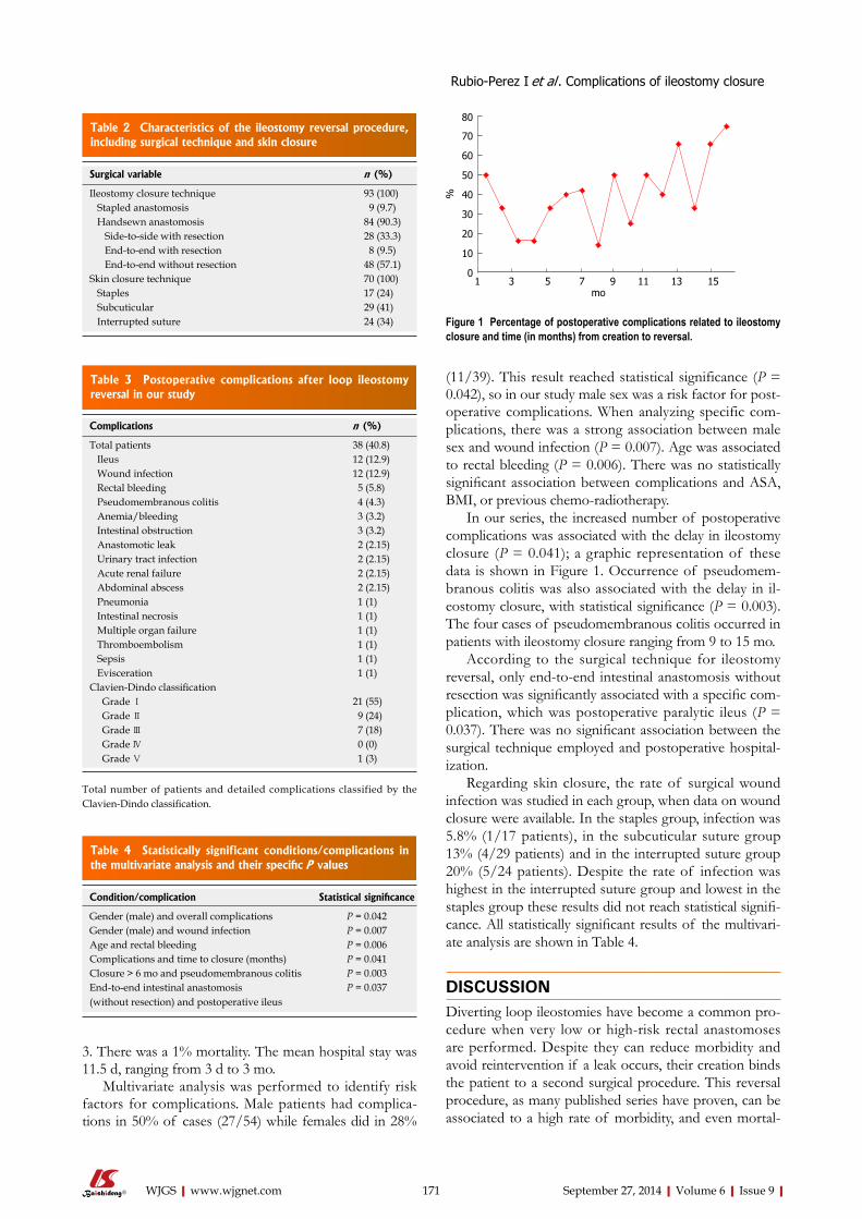
3. There was a 1% mortality. The mean hospital stay was 11.5 d, ranging from 3 d to 3 mo.
Multivariate analysis was performed to identify risk factors for complications. Male patients had complica-tions in 50% of cases (27/54) while females did in 28%
(11/39). This result reached statistical significance (P = 0.042), so in our study male sex was a risk factor for post-operative complications. When analyzing specific com-plications, there was a strong association between male sex and wound infection (P = 0.007). Age was associated to rectal bleeding (P = 0.006). There was no statistically significant association between complications and ASA, BMI, or previous chemo-radiotherapy.
In our series, the increased number of postoperative complications was associated with the delay in ileostomy closure (P = 0.041); a graphic representation of these data is shown in Figure 1. Occurrence of pseudomem-branous colitis was also associated with the delay in il-eostomy closure, with statistical significance (P = 0.003). The four cases of pseudomembranous colitis occurred in patients with ileostomy closure ranging from 9 to 15 mo.
According to the surgical technique for ileostomy reversal, only end-to-end intestinal anastomosis without resection was significantly associated with a specific com-plication, which was postoperative paralytic ileus (P = 0.037). There was no significant association between the surgical technique employed and postoperative hospital-ization.
Regarding skin closure, the rate of surgical wound infection was studied in each group, when data on wound closure were available. In the staples group, infection was 5.8% (1/17 patients), in the subcuticular suture group 13% (4/29 patients) and in the interrupted suture group 20% (5/24 patients). Despite the rate of infection was highest in the interrupted suture group and lowest in the staples group these results did not reach statistical signifi-cance. All statistically significant results of the multivari-ate analysis are shown in Table 4.
DISCUSSIONDiverting loop ileostomies have become a common pro-cedure when very low or high-risk rectal anastomoses are performed. Despite they can reduce morbidity and avoid reintervention if a leak occurs, their creation binds the patient to a second surgical procedure. This reversal procedure, as many published series have proven, can be associated to a high rate of morbidity, and even mortal-
171 September 27, 2014|Volume 6|Issue 9|WJGS|www.wjgnet.com
Table 2 Characteristics of the ileostomy reversal procedure, including surgical technique and skin closure
Surgical variable n (%)
Ileostomy closure technique 93 (100) Stapled anastomosis 9 (9.7) Handsewn anastomosis 84 (90.3) Side-to-side with resection 28 (33.3) End-to-end with resection 8 (9.5) End-to-end without resection 48 (57.1)Skin closure technique 70 (100) Staples 17 (24) Subcuticular 29 (41) Interrupted suture 24 (34)
Table 3 Postoperative complications after loop ileostomy reversal in our study
Complications n (%)
Total patients 38 (40.8) Ileus 12 (12.9) Wound infection 12 (12.9) Rectal bleeding 5 (5.8) Pseudomembranous colitis 4 (4.3) Anemia/bleeding 3 (3.2) Intestinal obstruction 3 (3.2) Anastomotic leak 2 (2.15) Urinary tract infection 2 (2.15) Acute renal failure 2 (2.15) Abdominal abscess 2 (2.15) Pneumonia 1 (1) Intestinal necrosis 1 (1) Multiple organ failure 1 (1) Thromboembolism 1 (1) Sepsis 1 (1) Evisceration 1 (1)Clavien-Dindo classification Grade Ⅰ 21 (55) Grade Ⅱ 9 (24) Grade Ⅲ 7 (18) Grade Ⅳ 0 (0) Grade Ⅴ 1 (3)
Total number of patients and detailed complications classified by the Clavien-Dindo classification.
%
80
70
60
50
40
30
20
10
01 3 5 7 9 11 13 15
mo
Figure 1 Percentage of postoperative complications related to ileostomy closure and time (in months) from creation to reversal.
Table 4 Statistically significant conditions/complications in the multivariate analysis and their specific P values
Condition/complication Statistical significance
Gender (male) and overall complications P = 0.042Gender (male) and wound infection P = 0.007Age and rectal bleeding P = 0.006Complications and time to closure (months) P = 0.041Closure > 6 mo and pseudomembranous colitis P = 0.003End-to-end intestinal anastomosis (without resection) and postoperative ileus
P = 0.037
Rubio-Perez I et al . Complications of ileostomy closure
them occur after discharge and are managed in the out-patient clinic or by General Practitioners. This has been taken into consideration, and some recent publications already study standardized 30-d complications[17], and use classifications such as Clavien-Dindo to report results[9].
Different efforts have been made to reduce the infec-tion rate of stoma-closure wounds. The technique em-ployed has shown statistically significant results in various RCTs, such as that from Camacho-Mauries et al[18], favor-ing purse-string closure vs conventional sutures. In our study, a limitation regarding the retrospective analysis of skin closure was that data were incomplete and some sur-gical reports did not state the specific closure technique. From those available, interrupted, non absorbable sutures were the most frequently used, followed by subcuticular and staples. The staples group had a lower infection rate, but these data were not significant. Studies on the subject show contradictory results. In the retrospective study by Kobayashi et al[19], wound infection rate was as high as 23.5%, and subcuticular sutures apparently showed a protective effect. However, very recent studies and RCTs on wound closure, report purse-string sutures to achieve a 0% infection rate compared to other methods, thus not recommending linear closure of stoma wounds[20,21]. Other attempts, such as subcutaneous antibiotic implants (Gentamycin) in the wound, have not shown a relevant reduction in surgical site infections[22].
Another controversy around the subject is the best timing for stoma reversal. Some groups defend very early closure, even during the first admission, such as Alves et al[23], who perform reversal on the 8th postoperative day if no complications of the first intervention have occurred. Nevertheless, it is widely accepted to delay closure, and different studies report mean times of 3-6 mo, with a low medical priority given to this procedure[24].
In our study, we demonstrate that the delay in il-eostomy closure (> 6 mo) is a risk factor for increased complications, and is associated with a higher incidence of pseudomembranous colitis, which was 4.3%. As can be seen in Figure 1, the incidence of complications in-creased with time (in months); there was an apparently ‘safer’ period around 3-6 mo, which could be considered optimal. From 9 mo onwards the rate of complications was > 30%.
Pseudomembranous colitis (PMC) is secondary to Clostridium difficile (C.diff) infection, and associated with substantial morbidity and mortality, increased duration of hospitalization, and a marked economic impact[25]. C.diff is a toxin-producing anaerobic bacterium responsible for antibiotic-associated colitis, and it is now the most com-mon infectious cause of nosocomial diarrhea. Risk fac-tors for PMC include advanced age, systemic antibiotic therapy, hospitalization, nursing homes or long-term care facilities, contact with active carriers, and presence of comorbidities[26]. It has been speculated that stoma clo-sure can be another risk factor for PMC, which associates all the previous to an excluded and defunctioned bowel with altered flora, that could be more susceptible to C.diff
ity[8]. In a recent systematic review[7], the high morbidity associated to ileostomy reversal raised concerns over the real indication of diverting stomas related to clinical outcomes, and if a better selection of patients should be made. Luglio et al[9], consider that if there is a > 5% risk of anastomotic leak in the primary operation, a protec-tive stoma must be created.
In our study, the rate of postoperative morbidity was high (40%), but still among published data. The most common complications in our study were postoperative ileus and wound infections. Complications were mostly minor, classified as Clavien-Dindo Ⅰ-Ⅱ, and only 18% were considered major complications needing reopera-tion or invasive interventions (Clavien-Dindo Ⅲ). In a national study by Mengual-Ballester et al[6] data were simi-lar to our own, with complications up to 45.9%, being ileus/obstruction the most frequent, followed by diarrhea and wound infection.
Ileus and bowel obstruction are still a concern after stoma reversal, and different studies have tried to eluci-date if technical issues can reduce these complications. The surgical technique employed for the ileal anasto-mosis has been widely studied, and various published randomized controlled trials (RCTs) compare handsewn vs stapled anastomoses[10,11]. In our study, when analyzing the surgical technique and related complications, we only found statistical significance between end-to-end anasto-mosis without resection and postoperative paralytic ileus. This coincides with published meta-analysis[12-14] which mention a significant reduction in surgical time and a lower incidence of bowel obstruction when stapled anas-tomoses are performed compared to handsewn. Other complications (including infections, leak, readmission and reoperation rates) are similar.
Surgical infections after stoma reversal have been a subject of debate. Although both ileostomies and co-lostomies can be safe, the latter present a higher rate of infection after reversal. Therefore many authors rec-ommend protective ileostomies for fecal diversion, if dehydration is not to be expected[15]. The rate of wound infection in our study (12.9%) was similar to other pub-lished data[16]. Wound infection is usually underestimated due to different definitions or considerations, and can be influenced by patient’s characteristics and comorbidi-ties. In some series of ileostomy closure for pouch-anal anastomoses such as that from van Westreenen et al[3], only a 1.4% rate of infection is reported. When analyz-ing patient’s characteristics we realize mean age is only 49 years, most patients are ASA Ⅰ-Ⅱ, and indications for ileostomy are polyposis or inflammatory bowel disease rather than cancer. Data are therefore not comparable if the population of study is older or weaker. In our series, mean age was 60 years and patients mainly ASA Ⅱ-Ⅲ, with the main indication for ileostomy creation being colorectal cancer (56%).
Another important factor is if the 30-d infection rate is reported, or just the rate of infection during hospital-ization. This can underestimate infections, as many of
172 September 27, 2014|Volume 6|Issue 9|WJGS|www.wjgnet.com
Rubio-Perez I et al . Complications of ileostomy closure
infection[27]. In a large series of 13245 United States pa-tients undergoing ileostomy closure, Wilson et al[28], report a 1.6% incidence of pseudomembranous colitis. This is an important factor to be considered, especially if an ear-lier closure can in fact reduce the risk.
This study was a retrospective analysis of institutional patients in order to identify risk factors for postopera-tive complications after ileostomy reversal and improve quality of care in our Colorectal Surgery Unit. Therefore, limitations are all those of an observational retrospective study, and in some cases (as in skin closure technique) data were missing from medical records. Due to the small number of patients some data may not reach statistical significance.
Although closure of a protective ileostomy is a fairly common surgical procedure, it has a high rate of compli-cations, and this must be taken into account when the in-dication is made. The delay in stoma closure can increase the rate of complications in general, and specifically wound infections and colitis.
COMMENTSBackground The creation of a defunctioning stoma after some colorectal procedures has demonstrated to highly reduce morbidity and mortality rates if a leak occurs, and is usually widely recommended. However, a stoma can be an issue for many patients both psychologically and due of stoma-related complications. As these stomas are supposed to be temporary, a planned second operation for reversal must be performed. In some cases, due to cancer-related complica-tions or comorbidities stomas are never reversed. In patients considered fit for surgery, the reversal of the stoma should be performed at the “safest” time pos-sible, to reduce complications. This timing is sometimes difficult to determine, as it depends on clinical factors, oncological follow-up and treatment, surgeon’s decision and institutional issues, such as “low-priority” consideration in surgical waiting lists.Research frontiersIn the field of Colorectal Surgery, the optimization of anastomoses and methods to reinforce or protect them to avoid leaks is a matter of active research. Even-tually, the creation of stomas would become obsolete if this could be achieved, improving surgical outcomes and reducing complications.Innovations and breakthroughsWhen revising the literature for the optimal timing for stoma reversal, recom-mendations usually suggest a 3 to 6 mo interval after the first intervention, always tailored to the specific risk factors and situation of the patient. When revising the real timing in our general practice, the authors realize there is a significant delay, and these recommendations are not followed. Complications related to prolonged bowel defunctioning (such as ileus, bleeding, diarrhea or Clostridium difficile colitis) and wound infections could be reduced if the time for closure is optimal. The best practice would be to guarantee an adequate healing from the first operation and close the stoma early enough to avoid the consequences of a prolonged defunctioning.ApplicationsThe study suggests that there is an optimal time frame to be considered when planning the ileostomy reversal that could reduce postoperative complications.TerminologyA protective ileostomy is an opening of a loop of small bowel (usually the termi-nal ileum) in the abdominal wall, so that a distal anastomosis performed in the colon or rectum is protected from fecal matter and can heal properly. The ileos-tomy reversal is the surgical intervention performed to close the loop of small bowel and restore normal intestinal transit.Peer reviewAbstract is concise, topic is interesting, methods are appropriate, a well-struc-tured discussion.
REFERENCES1 Bax TW, McNevin MS. The value of diverting loop ileosto-
my on the high-risk colon and rectal anastomosis. Am J Surg 2007; 193: 585-587; discussion 587-588 [PMID: 17434360]
2 Tan WS, Tang CL, Shi L, Eu KW. Meta-analysis of defunc-tioning stomas in low anterior resection for rectal cancer. Br J Surg 2009; 96: 462-472 [PMID: 19358171]
3 van Westreenen HL, Visser A, Tanis PJ, Bemelman WA. Morbidity related to defunctioning ileostomy closure after ileal pouch-anal anastomosis and low colonic anastomosis. Int J Colorectal Dis 2012; 27: 49-54 [PMID: 21761119]
4 Cottam J, Richards K, Hasted A, Blackman A. Results of a nationwide prospective audit of stoma complications within 3 weeks of surgery. Colorectal Dis 2007; 9: 834-838 [PMID: 17672873]
5 D’Haeninck A, Wolthuis AM, Penninckx F, D’Hondt M, D’Hoore A. Morbidity after closure of a defunctioning loop il-eostomy. Acta Chir Belg 2011; 111: 136-141 [PMID: 21780519]
6 Mengual-Ballester M, García-Marín JA, Pellicer-Franco E, Guillén-Paredes MP, García-García ML, Cases-Baldó MJ, Aguayo-Albasini JL. Protective ileostomy: complications and mortality associated with its closure. Rev Esp Enferm Dig 2012; 104: 350-354 [PMID: 22849495]
7 Chow A, Tilney HS, Paraskeva P, Jeyarajah S, Zacharakis E, Purkayastha S. The morbidity surrounding reversal of defunctioning ileostomies: a systematic review of 48 studies including 6,107 cases. Int J Colorectal Dis 2009; 24: 711-723 [PMID: 19221766 DOI: 10.1007/s00384-009-0660-z]
8 Thalheimer A, Bueter M, Kortuem M, Thiede A, Meyer D. Morbidity of temporary loop ileostomy in patients with colorectal cancer. Dis Colon Rectum 2006; 49: 1011-1017 [PMID: 16598401]
9 Luglio G, Pendlimari R, Holubar SD, Cima RR, Nelson H. Loop ileostomy reversal after colon and rectal surgery: a single institutional 5-year experience in 944 patients. Arch Surg 2011; 146: 1191-1196 [PMID: 22006879 DOI: 10.1001/archsurg.2011.234]
10 Löffler T, Rossion I, Bruckner T, Diener MK, Koch M, von Frankenberg M, Pochhammer J, Thomusch O, Kijak T, Si-mon T, Mihaljevic AL, Krüger M, Stein E, Prechtl G, Hodina R, Michal W, Strunk R, Henkel K, Bunse J, Jaschke G, Politt D, Heistermann HP, Fußer M, Lange C, Stamm A, Vosschulte A, Holzer R, Partecke LI, Burdzik E, Hug HM, Luntz SP, Kieser M, Büchler MW, Weitz J. HAnd Suture Versus STA-pling for Closure of Loop Ileostomy (HASTA Trial): results of a multicenter randomized trial (DRKS00000040). Ann Surg 2012; 256: 828-835; discussion 835-836 [PMID: 23095628 DOI: 10.1097/SLA.0b013e318272df97]
11 Hasegawa H, Radley S, Morton DG, Keighley MR. Stapled versus sutured closure of loop ileostomy: a randomized con-trolled trial. Ann Surg 2000; 231: 202-204 [PMID: 10674611]
12 Sajid MS, Craciunas L, Baig MK, Sains P. Systematic review and meta-analysis of published, randomized, controlled tri-als comparing suture anastomosis to stapled anastomosis for ileostomy closure. Tech Coloproctol 2013; 17: 631-639 [DOI 10.1007/s10151-013-1027-6]
13 Gong J, Guo Z, Li Y, Gu L, Zhu W, Li J, Li N. Stapled vs hand suture closure of loop ileostomy: a meta-analysis. Colorectal Dis 2013; 15: e561-e568 [PMID: 24033921 DOI: 10.1111/codi.12388]
14 Leung TT, MacLean AR, Buie WD, Dixon E. Comparison of stapled versus handsewn loop ileostomy closure: a meta-analysis. J Gastrointest Surg 2008; 12: 939-944 [PMID: 18071833]
15 Klink CD, Lioupis K, Binnebösel M, Kaemmer D, Kozubek I, Grommes J, Neumann UP, Jansen M, Willis S. Diversion stoma after colorectal surgery: loop colostomy or ileostomy? Int J Colorectal Dis 2011; 26: 431-436 [PMID: 21221605 DOI: 10.1007/s00384-010-1123-2]
16 Mirbagheri N, Dark J, Skinner S. Factors predicting stomal wound closure infection rates. Tech Coloproctol 2013; 17: 215-220 [PMID: 23076288 DOI: 10.1007/s10151-012-0908-4]
173 September 27, 2014|Volume 6|Issue 9|WJGS|www.wjgnet.com
COMMENTS
Rubio-Perez I et al . Complications of ileostomy closure
17 Sharma A, Deeb AP, Rickles AS, Iannuzzi JC, Monson JR, Fleming FJ. Closure of defunctioning loop ileostomy is as-sociated with considerable morbidity. Colorectal Dis 2013; 15: 458-462 [PMID: 22974343 DOI: 10.1111/codi.12029]
18 Camacho-Mauries D, Rodriguez-Díaz JL, Salgado-Nesme N, González QH, Vergara-Fernández O. Randomized clinical trial of intestinal ostomy takedown comparing pursestring wound closure vs conventional closure to eliminate the risk of wound infection. Dis Colon Rectum 2013; 56: 205-211 [PMID: 23303149 DOI: 10.1097/DCR.0b013e31827888f6]
19 Kobayashi S, Ito M, Sugito M, Kobayashi A, Nishizawa Y, Saito N. Association between incisional surgical site infection and the type of skin closure after stoma closure. Surg Today 2011; 41: 941-945 [PMID: 21748610 DOI: 10.1007/s00595-010-4405-y]
20 Klink CD, Wünschmann M, Binnebösel M, Alizai HP, Lambertz A, Boehm G, Neumann UP, Krones CJ. Influence of skin closure technique on surgical site infection after loop ileostomy reversal: retrospective cohort study. Int J Surg 2013; 11: 1123-1125 [PMID: 24035923 DOI: 10.1016/j.ijsu.2013.09.003]
21 Habbe N, Hannes S, Liese J, Woeste G, Bechstein WO, Strey C. The use of purse-string skin closure in loop ileostomy reversals leads to lower wound infection rates--a single high-volume centre experience. Int J Colorectal Dis 2014; 29: 709-714 [PMID: 24407267 DOI: 10.1007/s00384-013-1822-6]
22 Haase O, Raue W, Böhm B, Neuss H, Scharfenberg M,
Schwenk W. Subcutaneous gentamycin implant to reduce wound infections after loop-ileostomy closure: a random-ized, double-blind, placebo-controlled trial. Dis Colon Rectum 2005; 48: 2025-2031 [PMID: 16228839]
23 Alves A, Panis Y, Lelong B, Dousset B, Benoist S, Vicaut E. Randomized clinical trial of early versus delayed temporary stoma closure after proctectomy. Br J Surg 2008; 95: 693-698 [PMID: 18446781 DOI: 10.1002/bjs.6212]
24 Floodeen H, Lindgren R, Matthiessen P. When are defunc-tioning stomas in rectal cancer surgery really reversed? Results from a population-based single center experience. Scand J Surg 2013; 102: 246-250 [PMID: 24056133 DOI: 10.1177/1457496913489086]
25 Voelker R. Study: Vast majority of C. difficile infections occur in medical settings. JAMA 2012; 307: 1356 [PMID: 22474192 DOI: 10.1001/jama.2012.380]
26 Khanna S, Pardi DS. Clostridium difficile infection: new in-sights into management. Mayo Clin Proc 2012; 87: 1106-1117 [PMID: 23127735 DOI: 10.1016/j.mayocp.2012.07.016]
27 Randall JK, Young BC, Patel G, Fitzgerald A, George BD. Is Clostridium difficile infection a particular problem after reversal of ileostomy? Colorectal Dis 2011; 13: 308-311 [PMID: 19925492 DOI: 10.1111/j.1463-1318.2009.02139.x]
28 Wilson MZ, Hollenbeak CS, Stewart DB. Impact of Clos-tridium difficile colitis following closure of a diverting loop ileostomy: results of a matched cohort study. Colorectal Dis 2013; 15: 974-981 [PMID: 23336347 DOI: 10.1111/codi.12128]
P- Reviewer: Gorseta K, O’Dwyer P S- Editor: Ji FF L- Editor: A E- Editor: Liu SQ
174 September 27, 2014|Volume 6|Issue 9|WJGS|www.wjgnet.com
Rubio-Perez I et al . Complications of ileostomy closure

a mean age of 55.3 ± 14.3 years (range 30-84 years). The mean age of the male patients was 50.8 ± 13.7 years (range 30-84 years); that of the female patients was 59.9 ± 13.3 years (range 38-81 years). Tumor dimensions were obtained for 28 cases (mean 114.4 ± 78.6 mm; range 20-350 mm). Tumors were diagnosed incidentally in 23.3% of patients; abdominal discomfort and weight loss were the major complaints in symp-tomatic patients. Risk of aggressive behavior according to Fletcher criteria was determined in 25 of 30 patients (68%: high risk, 28%: intermediate risk, 4%: low risk). Histopathological examination revealed the pres-ence of spindle cells in 96.1% of cases; CD117 and CD34 were present immunohistochemically in 96.6% and 84% of patients, respectively. The most common surgical procedures were distal pancreatectomy with splenectomy (n = 9) and pancreaticoduodenectomy (n = 7). The total follow-up period for the 28 patients ranged from 3-66 mo, during which locoregional or distant metastases were diagnosed in six patients and two patients died.
CONCLUSION: Studies on EGISTs have only been published in the last decade. The lack of studies with large patient cohorts and long-term follow-up limits evidence-based commentary. In theory, each case should be assessed individually and further genetic and immunohistochemical studies are needed.
© 2014 Baishideng Publishing Group Inc. All rights reserved.
Key words: Gastrointestinal stromal tumor; Extra-gas-trointestinal stromal tumor; Pancreas; Imatinib mesyl-ate; CD117
Core tip: Gastrointestinal stromal tumors are the most common gastrointestinal (GI) tract tumors of mesen-chymal origin. Stromal tumors of extragastrointestinal origin are termed extragastrointestinal stromal tumors (EGISTs) and are not associated with the walls of GI
Pancreatic extragastrointestinal stromal tumor: A case report and comprehensive literature review
Sami Akbulut, Rıdvan Yavuz, Emrah Otan, Sinan Hatipoglu
Sami Akbulut, Rıdvan Yavuz, Department of Surgery, Diyarba-kir Education and Research Hospital, Uckuyular Mevki, Kayapi-nar, 21400 Diyarbakir, TurkeyEmrah Otan, Department of Surgery, Inonu University Faculty of Medicine, 44280 Malatya, TurkeySinan Hatipoglu, Department of Surgery, Adiyaman University Faculty of Medicine, 02040 Adiyaman, TurkeyAuthor contributions: Akbulut S and Hatipoglu S designed the report; Akbulut S and Yavuz R were attending doctors for the pa-tients; Akbulut S and Yavuz R performed the surgery; Akbulut S, Otan E and Hatipoglu S organized the report and wrote the paper.Correspondence to: Sami Akbulut, MD, FICS, FACS, Depart-ment of Surgery, Diyarbakir Education and Research Hospital, Uckuyular Mevki, Kayapinar, 21400 Diyarbakir, Turkey. [email protected]: +90-412-2580052 Fax: +90-412-2580050Received: May 26, 2014 Revised: June 21, 2014 Accepted: July 17, 2014Published online: September 27, 2014
AbstractAIM: To provide an overview of the literature on pan-creatic extragastrointestinal stromal tumors (EGISTs).
METHODS: We report a case of pancreatic EGIST and review published studies on pancreatic EGIST ac-cessed via the PubMed, MEDlInE, Google Scholar, and Google databases. The keywords used were “pancreas and GIST”, “pancreas and extra GIST”, “pancreas and gastrointestinal stromal tumor”, and “pancreas and ex-tragastrointestinal stromal tumor”. literature reviews and/or duplicate studies were excluded. The search included articles published in the English language be-tween January 1, 2000 and May 15, 2014.
RESULTS: From our literature survey, 30 manuscripts on pancreatic EGISTs were considered, of which 27 met the search criteria and three were excluded. The studies involved 30 patients (15 men, 15 women) with
SYSTEMATIC REVIEWS
Submit a Manuscript: http://www.wjgnet.com/esps/Help Desk: http://www.wjgnet.com/esps/helpdesk.aspxDOI: 10.4240/wjgs.v6.i9.175
World J Gastrointest Surg 2014 September 27; 6(9): 175-182ISSN 1948-9366 (online)
© 2014 Baishideng Publishing Group Inc. All rights reserved.
175 September 27, 2014|Volume 6|Issue 9|WJGS|www.wjgnet.com
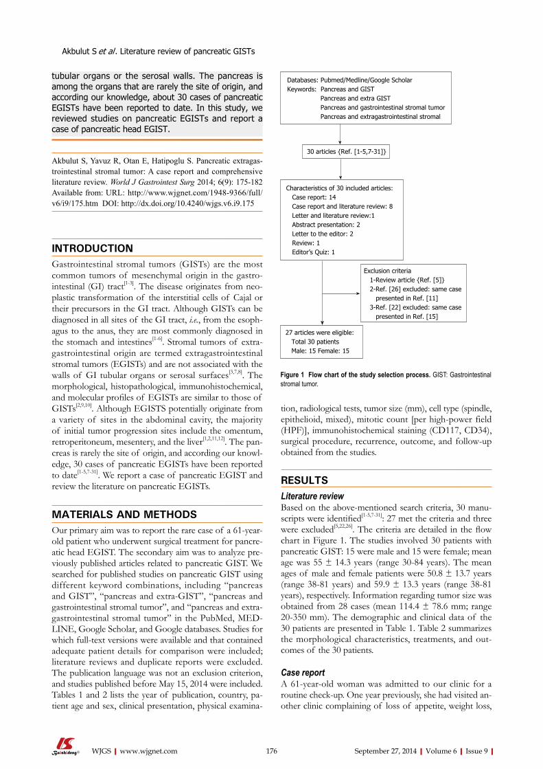
tubular organs or the serosal walls. The pancreas is among the organs that are rarely the site of origin, and according our knowledge, about 30 cases of pancreatic EGISTs have been reported to date. In this study, we reviewed studies on pancreatic EGISTs and report a case of pancreatic head EGIST.
Akbulut S, Yavuz R, Otan E, Hatipoglu S. Pancreatic extragas-trointestinal stromal tumor: A case report and comprehensive literature review. World J Gastrointest Surg 2014; 6(9): 175-182 Available from: URL: http://www.wjgnet.com/1948-9366/full/v6/i9/175.htm DOI: http://dx.doi.org/10.4240/wjgs.v6.i9.175
INTRODUCTIONGastrointestinal stromal tumors (GISTs) are the most common tumors of mesenchymal origin in the gastro-intestinal (GI) tract[1-3]. The disease originates from neo-plastic transformation of the interstitial cells of Cajal or their precursors in the GI tract. Although GISTs can be diagnosed in all sites of the GI tract, i.e., from the esoph-agus to the anus, they are most commonly diagnosed in the stomach and intestines[1-6]. Stromal tumors of extra-gastrointestinal origin are termed extragastrointestinal stromal tumors (EGISTs) and are not associated with the walls of GI tubular organs or serosal surfaces[3,7,8]. The morphological, histopathological, immunohistochemical, and molecular profiles of EGISTs are similar to those of GISTs[2,9,10]. Although EGISTS potentially originate from a variety of sites in the abdominal cavity, the majority of initial tumor progression sites include the omentum, retroperitoneum, mesentery, and the liver[1,2,11,12]. The pan-creas is rarely the site of origin, and according our knowl-edge, 30 cases of pancreatic EGISTs have been reported to date[1-5,7-31]. We report a case of pancreatic EGIST and review the literature on pancreatic EGISTs.
MATERIALS AND METHODSOur primary aim was to report the rare case of a 61-year-old patient who underwent surgical treatment for pancre-atic head EGIST. The secondary aim was to analyze pre-viously published articles related to pancreatic GIST. We searched for published studies on pancreatic GIST using different keyword combinations, including “pancreas and GIST”, “pancreas and extra-GIST”, “pancreas and gastrointestinal stromal tumor”, and “pancreas and extra-gastrointestinal stromal tumor” in the PubMed, MEd-lInE, Google Scholar, and Google databases. Studies for which full-text versions were available and that contained adequate patient details for comparison were included; literature reviews and duplicate reports were excluded. The publication language was not an exclusion criterion, and studies published before May 15, 2014 were included. Tables 1 and 2 lists the year of publication, country, pa-tient age and sex, clinical presentation, physical examina-
tion, radiological tests, tumor size (mm), cell type (spindle, epithelioid, mixed), mitotic count [per high-power field (HPF)], immunohistochemical staining (Cd117, Cd34), surgical procedure, recurrence, outcome, and follow-up obtained from the studies.
RESULTSLiterature reviewBased on the above-mentioned search criteria, 30 manu-scripts were identified[1-5,7-31]: 27 met the criteria and three were excluded[5,22,26]. The criteria are detailed in the flow chart in Figure 1. The studies involved 30 patients with pancreatic GIST: 15 were male and 15 were female; mean age was 55 ± 14.3 years (range 30-84 years). The mean ages of male and female patients were 50.8 ± 13.7 years (range 38-81 years) and 59.9 ± 13.3 years (range 38-81 years), respectively. Information regarding tumor size was obtained from 28 cases (mean 114.4 ± 78.6 mm; range 20-350 mm). The demographic and clinical data of the 30 patients are presented in Table 1. Table 2 summarizes the morphological characteristics, treatments, and out-comes of the 30 patients.
Case reportA 61-year-old woman was admitted to our clinic for a routine check-up. One year previously, she had visited an-other clinic complaining of loss of appetite, weight loss,
Akbulut S et al . literature review of pancreatic GISTs
176 September 27, 2014|Volume 6|Issue 9|WJGS|www.wjgnet.com
Databases: Pubmed/Medline/Google ScholarKeywords: Pancreas and GIST Pancreas and extra GIST Pancreas and gastrointestinal stromal tumor Pancreas and extragastrointestinal stromal
30 articles {Ref. [1-5,7-31]}
Characteristics of 30 included articles: Case report: 14 Case report and literature review: 8 letter and literature review:1 Abstract presentation: 2 letter to the editor: 2 Review: 1 Editor’s Quiz: 1
Exclusion criteria 1-Review article {Ref. [5]} 2-Ref. [26] excluded: same case presented in Ref. [11] 3-Ref. [22] excluded: same case presented in Ref. [15]
27 articles were eligible: Total 30 patients Male: 15 Female: 15
Figure 1 Flow chart of the study selection process. GIST: Gastrointestinal stromal tumor.
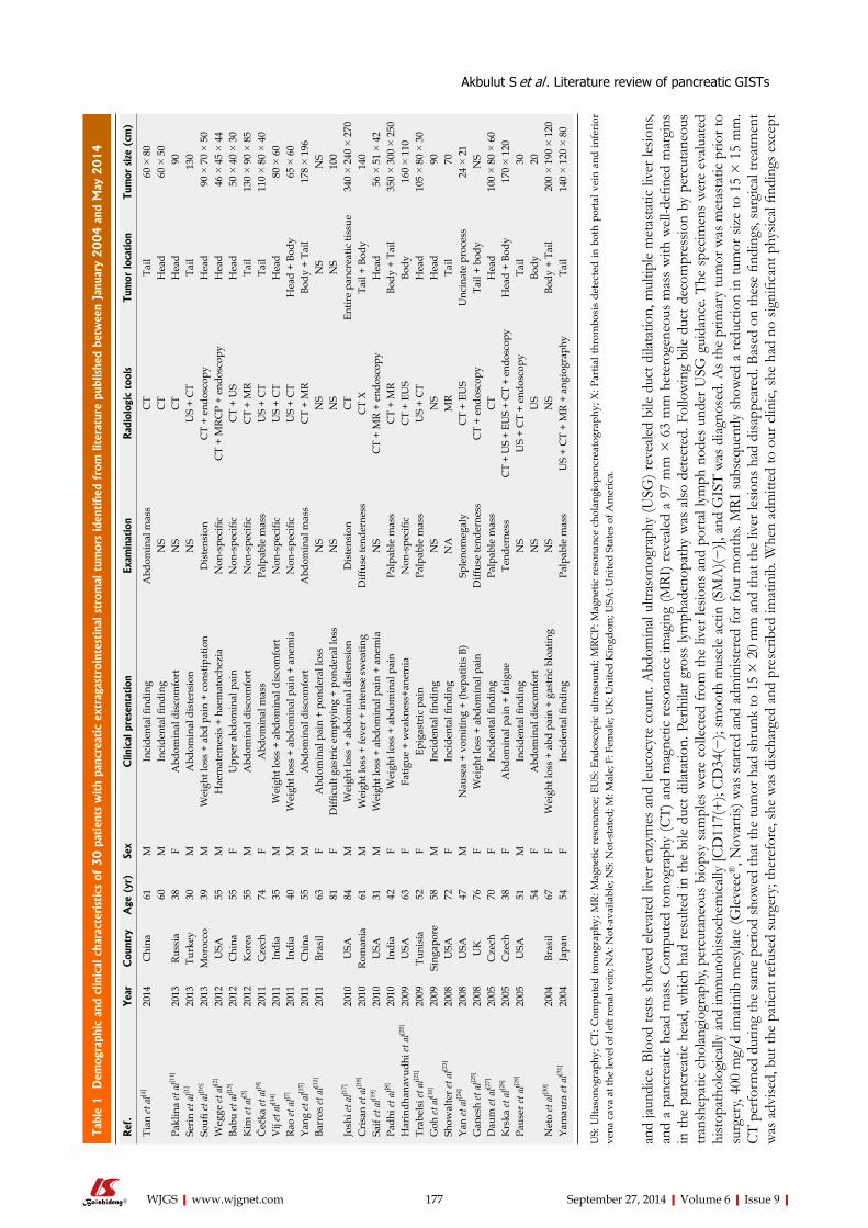
and
jaund
ice.
Bloo
d te
sts
show
ed e
leva
ted
liver
enz
ymes
and
leuc
ocyt
e co
unt.
Abd
omin
al ul
traso
nogr
aphy
(USG
) rev
eale
d bi
le d
uct d
ilata
tion,
mul
tiple
met
asta
tic li
ver l
esio
ns,
and
a pa
ncre
atic
hea
d m
ass.
Com
pute
d to
mog
raph
y (C
T) a
nd m
agne
tic re
sona
nce
imag
ing
(MRI
) rev
eale
d a
97 m
m ×
63
mm
het
erog
eneo
us m
ass
with
wel
l-defi
ned
mar
gins
in
the
panc
reat
ic h
ead,
whi
ch h
ad r
esul
ted
in th
e bi
le d
uct d
ilata
tion.
Per
ihila
r gr
oss
lym
phad
enop
athy
was
also
det
ecte
d. F
ollo
win
g bi
le d
uct d
ecom
pres
sion
by p
ercu
tane
ous
trans
hepa
tic c
holan
giog
raph
y, pe
rcut
aneo
us b
iops
y sa
mpl
es w
ere
colle
cted
from
the
liver
lesio
ns a
nd p
orta
l lym
ph n
odes
und
er U
SG g
uida
nce.
The
spec
imen
s w
ere
evalu
ated
hi
stop
atho
logi
cally
and
imm
unoh
istoc
hem
icall
y [C
D11
7(+
); C
D34
(−);
smoo
th m
uscl
e ac
tin (S
MA
)(−)],
and
GIS
T w
as d
iagno
sed.
As t
he p
rimar
y tu
mor
was
met
asta
tic p
rior t
o su
rger
y, 40
0 m
g/d
imat
inib
mes
ylat
e (G
leve
ec®, n
ovar
tis) w
as s
tart
ed a
nd a
dmin
ister
ed fo
r fou
r mon
ths.
MRI
sub
sequ
ently
sho
wed
a re
duct
ion
in tu
mor
siz
e to
15
× 1
5 m
m.
CT
perf
orm
ed d
urin
g th
e sa
me
perio
d sh
owed
that
the
tum
or h
ad sh
runk
to 1
5 ×
20
mm
and
that
the
liver
lesio
ns h
ad d
isapp
eare
d. B
ased
on
thes
e fin
ding
s, su
rgic
al tre
atm
ent
was
adv
ised,
but
the
patie
nt re
fuse
d su
rger
y; th
eref
ore,
she
was
disc
harg
ed a
nd p
resc
ribed
imat
inib
. Whe
n ad
mitt
ed to
our
clin
ic, sh
e ha
d no
sign
ifica
nt p
hysic
al fin
ding
s exc
ept
177 September 27, 2014|Volume 6|Issue 9|WJGS|www.wjgnet.com
Tabl
e 1 D
emog
raph
ic a
nd c
linic
al c
hara
cter
istics
of
30 p
atie
nts
with
panc
reat
ic e
xtra
gast
roin
test
inal
str
omal
tum
ors
iden
tifie
d fr
om li
tera
ture
pub
lishe
d be
twee
n Ja
nuar
y 2004 a
nd M
ay 2
014
Ref
.Yea
rC
ount
ryA
ge (
yr)
Sex
Clin
ical
pre
sent
atio
nEx
amin
atio
nR
adio
logi
c to
ols
Tum
or lo
cation
Tum
or s
ize
(cm
)
Tian
et a
l[4]
2014
Chi
na61
MIn
cide
ntal
find
ing
Abd
omin
al m
ass
CT
Tail
60 ×
80
60M
Inci
dent
al fi
ndin
gN
SC
TH
ead
60 ×
50
Pakl
ina
et a
l[11]
2013
Russ
ia38
FA
bdom
inal
dis
com
fort
NS
CT
Hea
d 9
0Se
rin
et a
l[1]
2013
Turk
ey30
MA
bdom
inal
dis
tens
ion
NS
US
+ C
TTa
il13
0So
ufi et
al[1
6]20
13M
oroc
co39
MW
eigh
t los
s +
abd
pain
+ c
onst
ipat
ion
Dis
tens
ion
CT
+ en
dosc
opy
Hea
d 9
0 ×
70
× 5
0W
egge
et a
l[2]
2012
USA
55M
Hae
mat
emes
is +
hae
mat
oche
zia
Non
-spe
cific
CT
+ M
RCP
+ en
dosc
opy
Hea
d 4
6 ×
45
× 4
4Ba
bu et
al[1
3]20
12C
hina
55F
Upp
er a
bdom
inal
pai
nN
on-s
peci
ficC
T +
US
Hea
d 5
0 ×
40
× 3
0K
im et
al[3
]20
12K
orea
55M
Abd
omin
al d
isco
mfo
rtN
on-s
peci
ficC
T +
MR
Tail
130
× 9
0 ×
85
Čeč
ka et
al[9
]20
11C
zech
74
FA
bdom
inal
mas
sPa
lpab
le m
ass
US
+ C
TTa
il11
0 ×
80
× 4
0V
ij et
al[1
4]20
11In
dia
35M
Wei
ght l
oss
+ ab
dom
inal
dis
com
fort
Non
-spe
cific
US
+ C
TH
ead
80 ×
60
Rao
et a
l[7]
2011
Indi
a40
MW
eigh
t los
s +
abdo
min
al p
ain
+ an
emia
Non
-spe
cific
US
+ C
TH
ead
+ Bo
dy65
× 6
0Ya
ng et
al[1
5]20
11C
hina
55M
Abd
omin
al d
isco
mfo
rtA
bdom
inal
mas
sC
T +
MR
Body
+ T
ail
178
× 1
96Ba
rros
et a
l[12]
2011
Bras
il63
FA
bdom
inal
pai
n +
pond
eral
loss
NS
NS
NS
NS
81F
Diffi
cult
gast
ric
empt
ying
+ p
onde
ral l
oss
NS
NS
NS
100
Josh
i et a
l[17]
2010
USA
84M
Wei
ght l
oss
+ ab
dom
inal
dis
tens
ion
Dis
tens
ion
CT
Entir
e pa
ncre
atic
tiss
ue
340
× 2
40 ×
270
Cri
san
et a
l[18]
2010
Rom
ania
61M
Wei
ght l
oss
+ fe
ver +
inte
nse
swea
ting
Diff
use
tend
erne
ssC
T X
Tail
+ Bo
dy14
0Sa
if et
al[1
9]20
10U
SA31
MW
eigh
t los
s +
abdo
min
al p
ain
+ an
emia
NS
CT
+ M
R +
endo
scop
yH
ead
56
× 5
1 ×
42
Padh
i et a
l[8]
2010
Indi
a42
FW
eigh
t los
s +
abdo
min
al p
ain
Palp
able
mas
sC
T +
MR
Body
+ T
ail
3
50 ×
300
× 2
50H
arin
dhan
avud
hi et
al[2
0]20
09U
SA63
FFa
tigue
+ w
eakn
ess+
anem
iaN
on-s
peci
ficC
T +
EUS
Body
160
× 1
10Tr
abel
si et
al[2
1]20
09Tu
nisi
a52
FEp
igas
tric
pai
nPa
lpab
le m
ass
US
+ C
TH
ead
105
× 8
0 ×
30
Goh
et a
l[10]
2009
Sing
apor
e58
MIn
cide
ntal
find
ing
NS
NS
Hea
d 9
0Sh
owal
ter e
t al[2
3]20
08U
SA72
FIn
cide
ntal
find
ing
NA
MR
Tail
70
Yan
et a
l[24]
2008
USA
47M
Nau
sea
+ vo
miti
ng +
(hep
atiti
s B)
Sple
nom
egal
yC
T +
EUS
Unc
inat
e pr
oces
s24
× 2
1G
anes
h et
al[2
5]20
08U
K76
FW
eigh
t los
s +
abdo
min
al p
ain
Diff
use
tend
erne
ssC
T +
endo
scop
yTa
il +
body
NS
Dau
m et
al[2
7]20
05C
zech
70F
Inci
dent
al fi
ndin
gPa
lpab
le m
ass
CT
Hea
d10
0 ×
80
× 6
0K
rska
et a
l[28]
2005
Cze
ch38
FA
bdom
inal
pai
n +
fatig
ueTe
nder
ness
CT
+ U
S +
EUS
+ C
T +
endo
scop
yH
ead
+ Bo
dy17
0 ×
120
Paus
er et
al[2
9]20
05U
SA51
MIn
cide
ntal
find
ing
NS
US
+ C
T +
endo
scop
yTa
il 3
054
FA
bdom
inal
dis
com
fort
NS
US
Body
20
Net
o et
al[3
0]20
04Br
asil
67F
Wei
ght l
oss
+ ab
d pa
in +
gas
tric
blo
atin
gN
SN
SBo
dy +
Tai
l
200
× 1
90 ×
120
Yam
aura
et a
l[31]
2004
Japa
n54
FIn
cide
ntal
find
ing
Palp
able
mas
sU
S +
CT
+ M
R +
angi
ogra
phy
Tail
140
× 1
20 ×
80
US:
Ulta
sono
grap
hy; C
T: C
ompu
ted
tom
ogra
phy;
MR:
Mag
netic
res
onan
ce; E
US:
End
osco
pic
ultr
asou
nd; M
RCP:
Mag
netic
res
onan
ce c
hola
ngio
panc
reat
ogra
phy;
X: P
artia
l thr
ombo
sis
dete
cted
in b
oth
port
al v
ein
and
infe
rior
ve
na c
ava
at th
e le
vel o
f lef
t ren
al v
ein;
NA
: Not
-ava
ilabl
e; N
S: N
ot-s
tate
d; M
: Mal
e; F
: Fem
ale;
UK
: Uni
ted
Kin
gdom
; USA
: Uni
ted
Stat
es o
f Am
eric
a.
Akbulut S et al . literature review of pancreatic GISTs
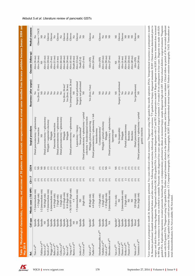
178 September 27, 2014|Volume 6|Issue 9|WJGS|www.wjgnet.com
Tabl
e 2
Mor
phol
ogic
al c
hara
cter
istics
, tr
eatm
ents
, an
d ou
tcom
es o
f 30 p
atie
nts
with
panc
reat
ic e
xtra
gast
roin
test
inal
str
omal
tum
or ide
ntifi
ed f
rom
liter
atur
e pu
blishe
d be
twee
n Ja
nuar
y 2004 a
nd
May
2014
Ref
.C
ell t
ype
Mitot
ic c
ount
(/5
0 H
PF)
CD
117
CD
34
Surg
ical
pro
cedu
res
Rec
urre
nce
(aft
er s
urge
ry)
Out
com
e (f
ollo
w-u
p)M
edic
al t
reat
men
t
Tian
et a
l[4]
Spin
dle
< 5
(inte
rmed
iate
risk
)(+
)(+
)D
ista
l pan
crea
tect
omy
+ sp
lene
ctom
yN
oA
live
(36
mo)
No
Spin
dle
> 5
(hig
h ri
sk)
(+)
NS
Tum
or re
sect
ion
Yes
(live
r, 12
mo)
Aliv
e (3
6 m
o)G
leev
ec +
TA
CE
Pakl
ina
et a
l[11]
Spin
dle
1-2
(inte
rmed
iate
risk
)(+
)N
SN
SN
SN
SN
SSe
rin
et a
l[1]
NS
NS
(hig
h ri
sk)
(+)
NS
Dis
tal p
ancr
eate
ctom
y +
sple
nect
omy
No
Aliv
e (2
1 m
o)N
oSo
ufi et
al[1
6]Sp
indl
e<
5 (in
term
edia
te ri
sk)
(+)
(+)
Whi
pple
+ s
egm
enta
l col
ecto
my
No
Aliv
e (2
4 m
o)G
leev
ecW
egge
et a
l[2]
Spin
dle
6 (in
term
edia
te ri
sk)
(+)
(+)
Whi
pple
N
oA
live
(5 m
o)G
leev
ecBa
bu et
al[1
3]Sp
indl
e6-
8 (h
igh
risk
)(+
)(+
)Pa
ncre
atic
hea
d re
sect
ion
No
Aliv
e (1
1 m
o)N
oK
im et
al[3
]Sp
indl
e7
(hig
h ri
sk)
(+)
(+)
Dis
tal p
ancr
eate
ctom
y +
sple
nect
omy
No
Aliv
e (4
mo)
Gle
evec
Čeč
ka et
al[9
]Sp
indl
e5
(hig
h ri
sk)
(+)
(+)
Dis
tal p
ancr
eate
ctom
y +
sple
nect
omy
No
Aliv
e (6
6 m
o)N
oV
ij et
al[1
4]Sp
indl
e12
-15
(hig
h ri
sk)
(+)
(-)
Whi
pple
Yes
(live
r, 24
mo)
aA
live
(48
mo)
Gle
evec
Rao
et a
l[7]
Spin
dleb
8-10
(hig
h ri
sk)
(+)
(+)
Whi
pple
Ye
s (li
ver,
24 m
o)A
live
(30
mo)
Gle
evec
Yang
et a
l[15]
Spin
dle
> 30
/10
HPF
(hig
h ri
sk)
(+)
(+)
Dis
tal p
ancr
eate
ctom
y +
sple
nect
omy
Yes
(intr
aper
itone
al, 2
4 m
o)c
Aliv
e (4
1 m
o)G
leev
ecBa
rros
et a
l[12]
NS
< 5
(+)
(+)
No
NS
Dea
th (8
mo)
No
NS
< 5
(inte
rmed
iate
risk
)(+
)(+
)La
paro
tom
y +
biop
sySu
rger
y no
t per
form
edA
live
(12
mo)
Gle
evec
Josh
i et a
l[17]
Spin
dle
NS
(+)
(+)
Non
e pe
rfor
med
dSu
rger
y no
t per
form
edD
eath
(5 d
)N
oC
risa
n et
al[1
8]Sp
indl
e(h
igh
risk
)(+
)(+
)D
ista
l pan
crea
tect
omy
+ sp
lene
ctom
y +
part
ial
cole
ctom
y +
duod
enoj
ejun
al re
sect
ion
NS
Aliv
e (3
mo)
NS
Saif
et a
l[19]
Spin
dlee
48 (h
igh
risk
) (+
) (-
)W
hipp
le, p
ylor
us p
rese
rvin
gYe
s (li
ver,
9 m
o)A
live
(NS)
Gle
evec
Padh
i et a
l[8]
Spin
dle
6-8
(hig
h ri
sk)
(+)
(+)
Dis
tal p
ancr
eate
ctom
y +
sple
nect
omy
+ le
ft he
mic
olec
tom
yN
oA
live
(10
mo)
No
Har
indh
anav
udhi
et a
l[20]
Spin
dle
< 5
(hig
h ri
sk)
(+)
(+)
Cys
toje
juno
stom
yfN
SA
live
(NS)
Gle
evec
Trab
elsi
et a
l[21]
Spin
dle
6 (h
igh
risk
)(+
)(+
)W
hipp
le +
par
tial c
olec
tom
yN
oA
live
(10
mo)
No
Goh
et a
l[10]
Spin
dle
> 10
(hig
h ri
sk)
(+)
NS
Whi
pple
No
Aliv
e (5
8 m
o)N
SSh
owal
ter e
t al[2
3]N
A3
(inte
rmed
iate
risk
)(+
) (-
)D
ista
l pan
crea
tect
omy
+ sp
lene
ctom
y -
lapa
rosc
opic
No
Aliv
e (2
7 m
o)N
S
Yan
et a
l[24]
Spin
dleg
3 (lo
w ri
sk)
(+)
NS
NS
NS
NS
NS
Gan
esh
et a
l[25]
Spin
dleh
NS
(+)
(+)
No
(inop
erab
le)
Surg
ery
no p
erfo
rmed
Aliv
e (3
0 m
o)G
leev
ecD
aum
et a
l[27]
Spin
dle
2 (in
term
edia
te ri
sk)
(+)
(-)W
hipp
leN
oA
live
(6 m
o)G
leev
ecK
rska
et a
l[28]
Spin
dlei
1 (h
igh
risk
) (-
)(+
)Pa
rtia
l pan
crea
tect
omy
No
Aliv
e (3
0 m
o)N
SPa
user
et a
l[29]
Spin
dle
NS
(+)
(+)
Rese
ctio
nN
oA
live
(24
mo)
NS
Spin
dle
NS
(+)
(+)
Rese
ctio
nN
oA
live
(48
mo)
NS
Net
o et
al[3
0]M
ixed
120
(hig
h ri
sk)
(+)
(+)
Dis
tal p
ancr
eate
ctom
yYe
s (p
erito
neum
)A
live
(NS)
Gle
evec
Yam
aura
et a
l[31]
Spin
dle
Few
(hig
h ri
sk)
(+)
(+)
Dis
tal p
ancr
eate
ctom
y +
sple
nect
omy
+ pa
rtia
l ga
stri
c re
sect
ion
NS
Aliv
e (3
0 m
o)N
S
a Live
r m
etas
tasi
s at
pos
tope
rativ
e m
onth
24.
Met
asta
sect
omy
perf
orm
ed. T
wo
year
s fo
llow
ed w
ithou
t rec
urre
nce;
b Dia
gnos
ed u
sing
USG
-gui
ded
fine
need
le a
spir
atio
n (F
NA
); c In
trap
erito
neal
rec
urre
nce
at p
osto
pera
tive
mon
th
24. R
esec
tion
perf
orm
ed. I
mat
inib
trea
tmen
t bot
h be
fore
and
afte
r res
ectio
n. F
ollo
win
g se
cond
rese
ctio
n, fo
llow
ed-u
p w
ithou
t rec
urre
nce;
d CT-
guid
ed li
ver b
iops
y di
agno
sed
met
asta
tic E
GIS
T. C
linic
al s
tatu
s de
teri
orat
ed p
rior
to
surg
ery
and
died
five
day
s fo
llow
ing
diag
nosi
s; e D
iagn
osed
with
End
o-U
SG (E
US)
-gui
ded
FNA
. Liv
er le
sion
dia
gnos
ed w
ith C
T an
d PE
T at
pos
tope
rativ
e m
onth
9. B
iops
y di
agno
sis
was
EG
IST.
Gle
evec
trea
tmen
t dos
e in
crea
sed
to 8
00 m
g. D
ue to
res
ista
nce
to tr
eatm
ent,
was
sw
itche
d to
sun
itini
b; f Pa
ncre
atic
mas
s di
agno
sed
four
yea
rs a
go, p
atie
nt r
efus
ed s
urgi
cal t
reat
men
t. C
T re
veal
ed 1
0-cm
enl
arge
men
t in
four
yea
rs. D
iagn
osis
was
mad
e w
ith E
US-
guid
ed F
NA
. Exp
lora
tive
lapa
roto
my
reve
aled
pan
crea
tic h
emor
rhag
ic c
yst;
cyst
ojej
unos
tom
y pe
rfor
med
to o
btai
n an
inci
sion
al b
iops
y sa
mpl
e di
agno
sed
high
-ris
k G
IST.
Pat
ient
ref
used
defi
nitiv
e su
rgic
al tr
eatm
ent;
g Dia
gnos
is
mad
e w
ith E
US-
guid
ed F
NA
; h Dia
gnos
ed u
sing
USG
-gui
ded
FNA
. Fur
ther
sur
gica
l tre
atm
ent a
bort
ed a
s th
e pa
tient
was
inop
erab
le, a
nd G
leev
ec tr
eatm
ent w
as in
itiat
ed. C
linic
al fo
llow
-up
peri
od o
f 30
mo
reve
aled
sig
nific
ant
tum
or re
duct
ion;
i USG
-gui
ded
biop
sy c
ould
not
pro
vide
dia
gnos
is. C
T: C
ompu
ted
tom
ogra
phy;
USG
: Ultr
ason
ogra
phy;
EG
IST:
Ext
raga
stro
inte
stin
al s
trom
al tu
mor
; PET
: Pos
itron
em
issi
on to
mog
raph
y; T
AC
E: T
rans
cath
eter
ar-
teri
al c
hem
oem
boliz
atio
n; N
A: N
ot-a
vaila
ble;
NS:
Not
-sta
ted.
Akbulut S et al . literature review of pancreatic GISTs
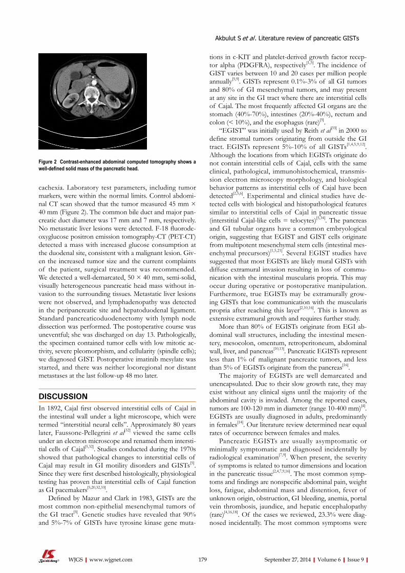
cachexia. laboratory test parameters, including tumor markers, were within the normal limits. Control abdomi-nal CT scan showed that the tumor measured 45 mm × 40 mm (Figure 2). The common bile duct and major pan-creatic duct diameter was 17 mm and 7 mm, respectively. No metastatic liver lesions were detected. F-18 fluorode-oxyglucose positron emission tomography-CT (PET-CT) detected a mass with increased glucose consumption at the duodenal site, consistent with a malignant lesion. Giv-en the increased tumor size and the current complaints of the patient, surgical treatment was recommended. We detected a well-demarcated, 50 × 40 mm, semi-solid, visually heterogeneous pancreatic head mass without in-vasion to the surrounding tissues. Metastatic liver lesions were not observed, and lymphadenopathy was detected in the peripancreatic site and hepatoduodenal ligament. Standard pancreaticoduodenectomy with lymph node dissection was performed. The postoperative course was uneventful; she was discharged on day 13. Pathologically, the specimen contained tumor cells with low mitotic ac-tivity, severe pleomorphism, and cellularity (spindle cells); we diagnosed GIST. Postoperative imatinib mesylate was started, and there was neither locoregional nor distant metastases at the last follow-up 48 mo later.
DISCUSSIONIn 1892, Cajal first observed interstitial cells of Cajal in the intestinal wall under a light microscope, which were termed “interstitial neural cells”. Approximately 80 years later, Faussone-Pellegrini et al[32] viewed the same cells under an electron microscope and renamed them intersti-tial cells of Cajal[5,32]. Studies conducted during the 1970s showed that pathological changes to interstitial cells of Cajal may result in GI motility disorders and GISTs[5]. Since they were first described histologically, physiological testing has proven that interstitial cells of Cajal function as GI pacemakers[5,20,32,33].
Defined by Mazur and Clark in 1983, GISTs are the most common non-epithelial mesenchymal tumors of the GI tract[5]. Genetic studies have revealed that 90% and 5%-7% of GISTs have tyrosine kinase gene muta-
tions in c-KIT and platelet-derived growth factor recep-tor alpha (PdGFRA), respectively[1,5]. The incidence of GIST varies between 10 and 20 cases per million people annually[5,9]. GISTs represent 0.1%-3% of all GI tumors and 80% of GI mesenchymal tumors, and may present at any site in the GI tract where there are interstitial cells of Cajal. The most frequently affected GI organs are the stomach (40%-70%), intestines (20%-40%), rectum and colon (< 10%), and the esophagus (rare)[5].
“EGIST” was initially used by Reith et al[33] in 2000 to define stromal tumors originating from outside the GI tract. EGISTs represent 5%-10% of all GISTs[1,4,5,9,12]. Although the locations from which EGISTs originate do not contain interstitial cells of Cajal, cells with the same clinical, pathological, immunohistochemical, transmis-sion electron microscopy morphology, and biological behavior patterns as interstitial cells of Cajal have been detected[2,5,6]. Experimental and clinical studies have de-tected cells with biological and histopathological features similar to interstitial cells of Cajal in pancreatic tissue (interstitial Cajal-like cells = telocytes)[5,34]. The pancreas and GI tubular organs have a common embryological origin, suggesting that EGIST and GIST cells originate from multipotent mesenchymal stem cells (intestinal mes-enchymal precursors)[1,5,21]. Several EGIST studies have suggested that most EGISTs are likely mural GISTs with diffuse extramural invasion resulting in loss of commu-nication with the intestinal muscularis propria. This may occur during operative or postoperative manipulation. Furthermore, true EGISTs may be extramurally grow-ing GISTs that lose communication with the muscularis propria after reaching this layer[2,10,16]. This is known as extensive extramural growth and requires further study.
More than 80% of EGISTs originate from EGI ab-dominal wall structures, including the intestinal mesen-tery, mesocolon, omentum, retroperitoneum, abdominal wall, liver, and pancreas[10,13]. Pancreatic EGISTs represent less than 1% of malignant pancreatic tumors, and less than 5% of EGISTs originate from the pancreas[16].
The majority of EGISTs are well demarcated and unencapsulated. due to their slow growth rate, they may exist without any clinical signs until the majority of the abdominal cavity is invaded. Among the reported cases, tumors are 100-120 mm in diameter (range 10-400 mm)[4]. EGISTs are usually diagnosed in adults, predominantly in females[14]. Our literature review determined near equal rates of occurrence between females and males.
Pancreatic EGISTs are usually asymptomatic or minimally symptomatic and diagnosed incidentally by radiological examination[7,9]. When present, the severity of symptoms is related to tumor dimensions and location in the pancreatic tissue[2,4,7,9,16]. The most common symp-toms and findings are nonspecific abdominal pain, weight loss, fatigue, abdominal mass and distention, fever of unknown origin, obstruction, GI bleeding, anemia, portal vein thrombosis, jaundice, and hepatic encephalopathy (rare)[4,16,18]. Of the cases we reviewed, 23.3% were diag-nosed incidentally. The most common symptoms were
179 September 27, 2014|Volume 6|Issue 9|WJGS|www.wjgnet.com
Figure 2 Contrast-enhanced abdominal computed tomography shows a well-defined solid mass of the pancreatic head.
Akbulut S et al . literature review of pancreatic GISTs

weight loss and abdominal discomfort.The most common diagnostic studies for pancreatic
masses involve biochemical [carbohydrate antigen 19-9, carcinoembryonic antigen (CEA)], radiological, histopath-ological, immunohistochemical, and genetic testing[3-5,21]. However, the diagnostic value of tumor markers such as CA 19-9 and CEA for pancreatic EGIST is limited, and are rarely used[4]. Abdominal CT, MRI, USG, endoscopic USG (Endo-USG), and PET-CT are the most frequently used radiological techniques, and aid in determining tu-mor localization, dimensions, margin irregularity, invasion of surrounding tissues, distant metastases, and resect-ability; however, most of them are non-diagnostic. USG and CT are often used in fine needle biopsies[5,7,17,20,24,25,28]. Endo-USG is a valuable diagnostic tool, allowing simul-taneous diagnosis and biopsy of solid or cystic pancreatic masses[4,5,16,19,20,24]. PET-CT is used more frequently for both diagnosing and monitoring GIST and is very effi-cient in cases where CT and MRI are inconclusive[35].
Histopathologically, GISTs are classified into spindle (70%), epithelioid (20%), or mixed (< 10%) type. Most pancreatic EGISTs consist of spindle cells[4]. Therefore, leiomyoma, leiomyosarcoma, liposarcoma, rhabdomyo-sarcoma, schwannoma, fibromatosis, inflammatory fibroid polyps, solitary fibrous tumor, and malignant fibrous histiocytoma should be considered in the differ-ential diagnoses[3,8,11,24,27]. Of the cases presented here, 26 had detailed histopathological data and 25 (96.1%) had spindle cells.
EGISTs have typical immunohistological staining features, among which Cd117 is the most well known. KIT is a transmembrane receptor for binding tyrosine ki-nase enzymes, and c-KIT is a newly discovered member of this receptor family, on whose receptor Cd117 is an epitope that can be stained immunohistochemically. The introduction of Cd117 staining in the 1990s changed the terminology for connective tissue tumors; since then, 95% of tumors defined as GIST or EGIST stain CD117-positive. For the 5% of tumors with negative staining, an-other tyrosine kinase receptor family member, PdGFRA, was investigated in immunohistochemical studies, with 33.3% positive staining[5]. Additionally, GISTs stain posi-tive for Cd34 (60%-70%), heavy caldesmon (80%), SMA (30%-40%), S100 (5%), and desmin (< 5%)[2-4,8,9]. Of the 30 cases presented, 29 (96.6%) stained Cd117-positive and 21 (84%) of 25 cases stained Cd34-positive.
Predicting GIST clinical and biological behavior is difficult. Fletcher defined the criteria of the National In-stitutes of Health (Fletcher criteria) to estimate the risks of GIST aggressive behavior and metastasis (locoregional and/or distant) using tumor dimensions (cm) and mitotic counts (per 50 HPF)[2,9]. According to the criteria, GISTs are classified based on their risk of aggressive behavior: very low (< 2 cm, < 5/50 HPF), low (2-5 cm, < 5/50 HPF), intermediate (< 5 cm, 6-10/50 HPF or 5-10 cm, < 5/50 HPF), and high (> 5 cm, > 5/50 HPF or > 10 cm, any mitotic count)[3,4,9,21]. This classification aids in surgi-cal treatment selection or neoadjuvant and/or adjuvant
treatment planning. The risk of aggressive behavior ac-cording to the Fletcher criteria was determined in 25 of the 30 patients in our literature review: risk of pancreatic EGIST aggressive behavior was high in 17 cases. The re-maining 8 cases were intermediate risk (n = 7; 28%) and low risk (n = 1; 4%).
The goal of surgical treatment, which is the most de-sirable treatment option for primary pancreatic EGISTs, is complete resection with microscopically clean (R0) margins[4,5,36]. Generally, primary surgery, surgical treat-ment following neoadjuvant chemotherapy, and debulk-ing surgery for metastatic and/or advanced disease are considered in the surgical treatment of GISTs[2,5]. Surgi-cal treatment selection depends on pancreatic EGIST localization. Standard or pylorus-preserving pancreati-coduodenectomy is the optimal treatment for pancreatic head tumors[4]. duodenum-preserving pancreatic head resection may be performed for small tumors, low-grade tumors, or patients who cannot tolerate the Whipple pro-cedure[4,36]. Conversely, radical surgical treatment may be the best option for preventing locoregional and/or dis-tant metastases[13,15]. Regional lymph node metastases are rare in pancreatic EGIST cases, and routine systematic regional lymph node dissection is not indicated[4,13,16,18]. In our patient, EGIST was diagnosed after lymph node bi-opsy. Therefore, we suggest lymphadenectomy for cases of pathological lymphadenopathy observed during surgi-cal exploration and for lymph node metastasis-positive cases based on intraoperative frozen section analysis. Depending on intraoperative findings and the surgeon’s experience, pancreaticoduodenectomy, distal pancreatec-tomy with splenectomy, or partial pancreatic resection may be used for treating tumors in the pancreatic tail and corpus[13]. nine and seven of the 30 patients underwent distal pancreatectomy with splenectomy, and the Whipple procedure, respectively.
The responses of GISTs to conventional chemother-apy and radiotherapy were very limited, being 10% and 5%, respectively[9,21]. These response rates changed when imatinib mesylate, an agent used for treating chronic myelogenous leukemia, was administered to a GIST case in the early 2000 s. Philadelphia chromosome-positive leukemia patients carry a mutation in the BCR-ABL gene, which is a KIT receptor family member. Additionally, the mutated c-KIT and PDGFRA genes seen in GISTs are members of the same family. Consequently, tyrosine kinase transmembrane receptors have been targeted in GIST treatment using two agents: imatinib mesylate and sunitinib malate. Imatinib was the first c-KIT tyrosine ki-nase inhibitor used for treating GISTs, specifically meta-static and unresectable GISTs, and was approved by the US Food and drug Administration. Sunitinib was sub-sequently introduced for patients who could not tolerate imatinib or who were imatinib-resistant[2,23]. Recently, new tyrosine kinase inhibitors, such as nilotinib, sorafenib, dovitinib, and dasatinib, were introduced[5]. despite the controversial approach of “which tyrosine kinase inhibi-tor, which patient and when”, there is consensus for
180 September 27, 2014|Volume 6|Issue 9|WJGS|www.wjgnet.com
Akbulut S et al . literature review of pancreatic GISTs

initiating imatinib treatment in patients with high mitotic activity, gross dimensions, necrosis, and locoregional and/or distant metastasis[2,15]. Imatinib may be used as a neoadjuvant agent to downstage gross tumor volume for R0 resection and contributes to good prognosis[4]. Imatinib may be used as adjuvant treatment in cases with R1 (positive microscopic margin) or R2 (residual gross visible tumor) resection, risk of aggressive behavior, or poor prognostic features[4,5]. Similarly, imatinib treatment may be used as a primary modality in metastatic or un-resectable cases to reduce tumor size, resulting in better prognosis[4]. Metastatic pancreatic EGIST cases benefit from debulking surgery, which increases the efficacy of imatinib[2]. The positive response to imatinib in patients with GISTs is 60%-70%, which can extend overall sur-vival up to 5 years[4].
In conclusion, the term EGIST was introduced into the literature in the last decade. debates on the similari-ties and differences between EGISTs and GISTs are ongoing. despite their behavioral similarities, the initial asymptomatic period accounts for the gross tumor size of EGISTs. The lack of comprehensive case reports on EGISTs, including pancreatic EGISTs, limited our evidence-based review. long-term follow-up studies of EGISTs are currently unavailable, limiting the amount of available information on tumor behavior. We are lim-ited to the case reports that have been published to date and further immunohistochemical and genetic studies regarding EGIST behavior and response to treatment are needed.
COMMENTSBackgroundGastrointestinal stromal tumors (GISTs) are the most common tumors of mes-enchymal origin in the gastrointestinal (GI) tract. The disease originates from neoplastic transformation of interstitial cells of Cajal or their precursors in the GI tract. Stromal tumors of extragastrointestinal origin are termed extragastro-intestinal stromal tumors (EGISTs) and are not associated with the walls of GI tubular organs or serosal surfaces. The morphological, histopathological, immu-nohistochemical and molecular profiles of EGISTs are similar to those of GISTs.Research frontiersThe primary aim was to report the rare case of a 61-year-old patient who un-derwent surgical treatment for pancreatic head EGIST. The secondary aim was to analyze previously published articles related to pancreatic GIST. To this end, the authors searched for studies on pancreatic GIST using different keyword combinations in the PubMed, MEdlInE, Google Scholar, and Google data-bases.TerminologyGISTs are the most common mesenchymal tumors of the GI tract. EGISTs are defined as tumors originating from outside the GI tract. Imatinib mesylate was the first c-KIT tyrosine kinase inhibitor used to treat GISTs. The Fletcher criteria are used to estimate the risk of GIST aggressive behavior and metastasis using tumor size and mitotic counts.Peer reviewThis paper comprises a case history, and a comprehensive review of the litera-ture on pancreas GIST. The strength of the paper is that the authors have tried to collect available literature of the limited articles published on this topic.
REFERENCES1 Serin KR, Keskin M, Gulluoglu M, Emre A. Atypical lo-
calisation of a gastrointestinal stromal tumor: A case report of pancreas gastrointestinal stromal tumor. Ulusal Cer Derg 2013; 29: 42-44 [DOI: 10.5152/UCD.2013.11]
2 Wegge J, Bartholomew DM, Burke LH, Miller LA. Pancre-atic extra-gastrointestinal stromal tumour masquerading as a bleeding duodenal mass. BMJ Case Rep 2012; 2012: [PMID: 23087281 DOI: 10.1136/bcr-2012-007040]
3 Kim HH, Koh YS, Park EK, Seoung JS, Hur YH, Kim JC, Cho CK, Kim HJ. Primary extragastrointestinal stromal tumor arising in the pancreas: report of a case. Surg Today 2012; 42: 386-390 [PMID: 22258729 DOI: 10.1007/s00595-011-0080-x]
4 Tian YT, Liu H, Shi SS, Xie YB, Xu Q, Zhang JW, Zhao DB, Wang CF, Chen YT. Malignant extra-gastrointestinal stromal tumor of the pancreas: report of two cases and review of the literature. World J Gastroenterol 2014; 20: 863-868 [PMID: 24574760 DOI: 10.3748/wjg.v20.i3.863]
5 Padhi S, Sarangi R, Mallick S. Pancreatic extragastrointestinal stromal tumors, interstitial Cajal like cells, and telocytes. JOP 2013; 14: 1-14 [PMID: 23306329 DOI: 10.6092/1590-8577/1293]
6 Miettinen M, Sobin LH, Sarlomo-Rikala M. Immunohisto-chemical spectrum of GISTs at different sites and their differ-ential diagnosis with a reference to CD117 (KIT). Mod Pathol 2000; 13: 1134-1142 [PMID: 11048809]
7 Rao RN, Vij M, Singla N, Kumar A. Malignant pancreatic extra-gastrointestinal stromal tumor diagnosed by ultra-sound guided fine needle aspiration cytology. A case report with a review of the literature. JOP 2011; 12: 283-286 [PMID: 21546710]
8 Padhi S, Kongara R, Uppin SG, Uppin MS, Prayaga AK, Challa S, Nagari B, Regulagadda SA. Extragastrointestinal stromal tumor arising in the pancreas: a case report with a review of the literature. JOP 2010; 11: 244-248 [PMID: 20442520]
9 Čečka F, Jon B, Ferko A, Šubrt Z, Nikolov DH, Tyčová V. Long-term survival of a patient after resection of a gastroin-testinal stromal tumor arising from the pancreas. Hepatobili-ary Pancreat Dis Int 2011; 10: 330-332 [PMID: 21669581 DOI: 10.1016/S1499-3872(11)60056-8]
10 Goh BK, Chow PK, Kesavan SM, Yap WM, Chung YF, Wong WK. A single-institution experience with eight CD117-positive primary extragastrointestinal stromal tumors: criti-cal appraisal and a comparison with their gastrointestinal counterparts. J Gastrointest Surg 2009; 13: 1094-1098 [PMID: 19238492 DOI: 10.1007/s11605-009-0828-4]
11 Paklina OV, Setdikova GR, Voskanyan SE. Extragastroin-testinal stromal tumor of the pancreas: A case report. 25 th European congress of pathology Lisbon. Poster No: 14, 2013
12 Barros A, Linhares E, Valadão M, Gonçalves R, Vilhena B, Gil C, Ramos C. Extragastrointestinal stromal tumors (EGIST): a series of case reports. Hepatogastroenterology 2011; 58: 865-868 [PMID: 21830406]
13 Babu SR, Kumari S, Zhang Y, Su A, Wang W, Tian B. Extra gastrointestinal stromal tumor arising in the pancreas: a case report and literature review. J GHR 2012; 1: 80-83
14 Vij M, Agrawal V, Pandey R. Malignant extra-gastrointesti-nal stromal tumor of the pancreas. A case report and review of literature. JOP 2011; 12: 200-204 [PMID: 21386653]
15 Yang F, Jin C, Fu D, Ni Q. Extra-gastrointestinal stromal tu-mor of the pancreas: clinical characteristics, diagnosis, treat-ment, and outcome. J Surg Oncol 2011; 103: 739-740 [PMID: 21240986 DOI: 10.1002/jso.21833]
16 Soufi M, Bouziane M, Massrouri R, Chad B. Pancreatic GIST with pancreas divisum: A new entity. Int J Surg Case Rep 2013; 4: 68-71 [PMID: 23123418 DOI: 10.1016/j.ijscr.2012.09.007]
17 Joshi J, Rustagi T. Pancreatic Extra-Gastrointestinal Stromal Tumor: An Unusual Presentation of a Rare Diagnosis. Gas-trointest Cancer Res 2010; (Suppl 1): S29-S30
18 Crisan A, Nicoara E, Cucui V, Cornea G, Laza R. Prolonged fever associated with gastrointestinal stromal tumor-case report. J Exp Med Surg Res 2010; 17: 219-224
181 September 27, 2014|Volume 6|Issue 9|WJGS|www.wjgnet.com
COMMENTS
Akbulut S et al . literature review of pancreatic GISTs

19 Saif MW, Hotchkiss S, Kaley K. Gastrointestinal stromal tu-mors of the pancreas. JOP 2010; 11: 405-406; author reply 412 [PMID: 20601822]
20 Harindhanavudhi T, Tanawuttiwat T, Pyle J, Silva R. Extra-gastrointestinal stromal tumor presenting as hemorrhagic pancreatic cyst diagnosed by EUS-FNA. JOP 2009; 10: 189-191 [PMID: 19287116]
21 Trabelsi A, Yacoub-Abid LB, Mtimet A, Abdelkrim SB, Ham-medi F, Ali AB, Mokni M. Gastrointestinal stromal tumor of the pancreas: A case report and review of the literature. N Am J Med Sci 2009; 1: 324-326 [PMID: 22666718]
22 Yang F, Long J, Di Y, Fu DL, Jin C, Ni QX, Zhu HG. A giant cystic lesion in the epigastric region. Pancreatic malignant gastrointestinal stromal tumour (GIST). Gut 2008; 57: 1494, 1636 [PMID: 18941004 DOI: 10.1136/gut.2008.159392]
23 Showalter SL, Lloyd JM, Glassman DT, Berger AC. Extra-gastrointestinal stromal tumor of the pancreas: case report and a review of the literature. Arch Surg 2008; 143: 305-308 [PMID: 18347279 DOI: 10.1001/archsurg.2007.68]
24 Yan BM, Pai RK, Van Dam J. Diagnosis of pancreatic gastro-intestinal stromal tumor by EUS guided FNA. JOP 2008; 9: 192-196 [PMID: 18326928]
25 Ganesh M, Kumar S, Krishnamoorthy R, Ang Y. Rare cause of pancreatic mass responding to imatinib treatment. Gastro-enterology Today 2008; 18: 50-51
26 Paklina OV, Setdikova GR, Voskanyan SE. Gastrointestinal Stromal Tumor of a Pancreas: Case Report and literature re-view. Медицинская визуализация 2013; 2: 122
27 Daum O, Klecka J, Ferda J, Treska V, Vanecek T, Sima R, Mukensnabl P, Michal M. Gastrointestinal stromal tumor of the pancreas: case report with documentation of KIT gene mutation. Virchows Arch 2005; 446: 470-472 [PMID: 15756592 DOI: 10.1007/s00428-004-1200-4]
28 Krska Z, Pesková M, Povýsil C, Horejs J, Sedlácková E,
Kudrnová Z. GIST of pancreas. Prague Med Rep 2005; 106: 201-208 [PMID: 16315768]
29 Pauser U, da Silva MT, Placke J, Klimstra DS, Klöppel G. Cellular hamartoma resembling gastrointestinal stromal tu-mor: a solid tumor of the pancreas expressing c-kit (CD117). Mod Pathol 2005; 18: 1211-1216 [PMID: 15803185 DOI: 10.1038/modpathol.3800406]
30 Neto MR, Machuca TN, Pinho RV, Yuasa LD, Bleggi-Torres LF. Gastrointestinal stromal tumor: report of two unusual cases. Virchows Arch 2004; 444: 594-596 [PMID: 15118853 DOI: 10.1007/s00428-004-1009-1]
31 Yamaura K, Kato K, Miyazawa M, Haba Y, Muramatsu A, Miyata K, Koide N. Stromal tumor of the pancreas with expression of c-kit protein: report of a case. J Gastroenterol Hepatol 2004; 19: 467-470 [PMID: 15012791 DOI: 10.1111/j.1440-1746.2003.02891.x]
32 Faussone-Pellegrini MS, Thuneberg L. Guide to the identi-fication of interstitial cells of Cajal. Microsc Res Tech 1999; 47: 248-266 [PMID: 10602286]
33 Reith JD, Goldblum JR, Lyles RH, Weiss SW. Extragastroin-testinal (soft tissue) stromal tumors: an analysis of 48 cases with emphasis on histologic predictors of outcome. Mod Pathol 2000; 13: 577-585 [PMID: 10824931]
34 Popescu LM, Hinescu ME, Ionescu N, Ciontea SM, Cretoiu D, Ardelean C. Interstitial cells of Cajal in pancreas. J Cell Mol Med 2005; 9: 169-190 [PMID: 15784175]
35 Williams A, Gutzeit A, Germer M, Pless M. PET-Negative Gastrointestinal Stromal Tumors. Case Rep Oncol 2013; 6: 508-513 [PMID: 24403895 DOI: 10.1159/000355432]
36 Yamashita S, Sakamoto Y, Saiura A, Yamamoto J, Kosuge T, Aoki T, Sugawara Y, Hasegawa K, Kokudo N. Pancreas-sparing duodenectomy for gastrointestinal stromal tumor. Am J Surg 2014; 207: 578-583 [PMID: 24119884 DOI: 10.1016/j.amjsurg.2013.05.009]
P- Reviewer: Fabre JM, Kapoor S, Soreide JA, Sumi S S- Editor: Ji FF L- Editor: Webster JR E- Editor: Liu SQ
182 September 27, 2014|Volume 6|Issue 9|WJGS|www.wjgnet.com
Akbulut S et al . literature review of pancreatic GISTs
Gastric necrosis: A late complication of nissen fundoplication
Javier Salinas, Tihomir Georgiev, Juan Antonio González-Sánchez, Elena López-Ruiz, José Antonio Rodríguez-Montes
Javier Salinas, Tihomir Georgiev, Juan Antonio González-Sánchez, José Antonio Rodríguez-Montes, Department of General Surgery, Hospital Universitario La Paz, Madrid 28046, SpainElena López-Ruiz, Department of Pathology, Hospital Universi-tario La Paz, Madrid 28046, SpainAuthor contributions: Salinas J, Georgiev T and González-Sánchez JA performed the surgery; López-Ruiz E performed the anatomopathological examination; Salinas J reviewed current literature and wrote the paper; González-Sánchez JA and Rodrí-guez-Montes JA coordinated the paper elaboration and revised the article.Correspondence to: Javier Salinas, MD, Department of Gen-eral Surgery, Hospital Universitario La Paz, Paseo de la Castel-lana, 261, Madrid 28046, Spain. [email protected]: +34-91-2071667 Fax: +34-91-2071064Received: April 8, 2014 Revised: July 4, 2014 Accepted: July 17, 2014Published online: September 27, 2014
AbstractGastric necrosis is a rare condition because of the rich blood supply and the extensive submucosal vascular network of the stomach. “Gas-bloat” syndrome is a well known Nissen fundoplication postoperative complica-tion. It may cause severe gastric dilatation, but very rarely an ischemic compromise of the organ. Other factors, such as gastric outlet obstruction, may concur to cause an intraluminal pressure enough to blockade venous return and ultimately arterial blood supply and oxygen deliver, leading to ischaemia. We report a case of a 63-year-old women, who presented a total gastric necrosis following laparoscopic Nissen fundoplication and a pyloric phytobezoar which was the trigger event. No preexisting gastric motility disorders were present by the time of surgery, as demonstrated in the preop-erative barium swallow, thus a poor mastication (patient needed no dentures) of a high fiber meal (cabbage) may have been predisposing factors for the develop-ment of a bezoar in an otherwise healthy women at the onset of old age. A total gastrectomy with esophagoje-
junostomy was performed and patient was discharged home after a 7-d hospital stay with no immediate com-plications. We also discuss some technical aspects of the procedure that might be important to reduce the incidence of this complication.
© 2014 Baishideng Publishing Group Inc. All rights reserved.
Key words: Gastric dilatation; Gastric outlet obstruc-tion; Necrosis; Fundoplication; Nissen operation
Core tip: Gastric necrosis is a rare condition because of the rich blood supply and the extensive submucosal vascular network of the stomach. “Gas-bloat” syndrome is a Nissen fundoplication postoperative complication that causes gastric dilatation, but very rarely an isch-emic compromise of the organ. We report a case of a 63-year-old women, who presented a total gastric ne-crosis following laparoscopic Nissen fundoplication and we discuss technical aspects of the procedure that are important to prevent this complication.
Salinas J, Georgiev T, González-Sánchez JA, López-Ruiz E, Ro-dríguez-Montes JA. Gastric necrosis: A late complication of nis-sen fundoplication. World J Gastrointest Surg 2014; 6(9): 183-186 Available from: URL: http://www.wjgnet.com/1948-9366/full/v6/i9/183.htm DOI: http://dx.doi.org/10.4240/wjgs.v6.i9.183
INTRODUCTIONThe rich blood supply of the stomach preserves this vis-cera from ischemic events, even after the ligation of all the major vessels[1] (a. gastrica dextra, a. gastrica sinistra, a. gastro-omentalis dextra, a. gastro-omentalis sinistra and aa. gastricae breves). Notwithstanding this fact, acute gas-tric dilatation accompanied with or without gastric outlet obstruction[2-7], eating disorders[8-10] or gas-bloat syndrome are recognized causes of ischemic gastric necrosis.
The gas-bloat syndrome is defined as a variable group
CASE REPORT
Submit a Manuscript: http://www.wjgnet.com/esps/Help Desk: http://www.wjgnet.com/esps/helpdesk.aspxDOI: 10.4240/wjgs.v6.i9.183
World J Gastrointest Surg 2014 September 27; 6(9): 183-186ISSN 1948-9366 (online)
© 2014 Baishideng Publishing Group Inc. All rights reserved.
183 September 27, 2014|Volume 6|Issue 9|WJGS|www.wjgnet.com
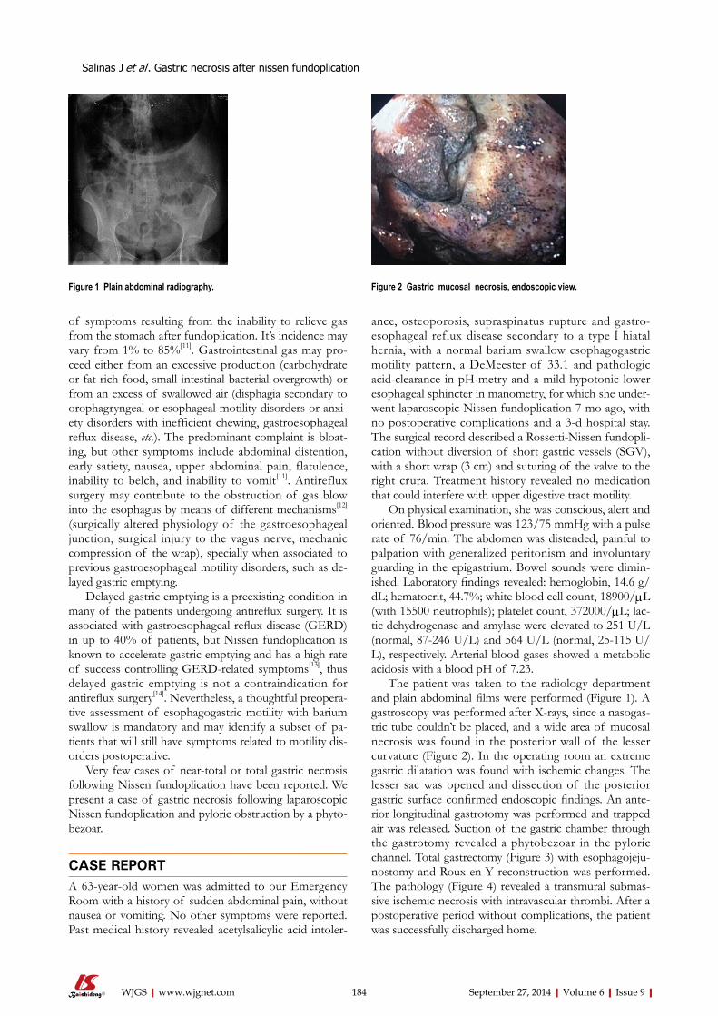
of symptoms resulting from the inability to relieve gas from the stomach after fundoplication. It’s incidence may vary from 1% to 85%[11]. Gastrointestinal gas may pro-ceed either from an excessive production (carbohydrate or fat rich food, small intestinal bacterial overgrowth) or from an excess of swallowed air (disphagia secondary to orophagryngeal or esophageal motility disorders or anxi-ety disorders with inefficient chewing, gastroesophageal reflux disease, etc.). The predominant complaint is bloat-ing, but other symptoms include abdominal distention, early satiety, nausea, upper abdominal pain, flatulence, inability to belch, and inability to vomit[11]. Antireflux surgery may contribute to the obstruction of gas blow into the esophagus by means of different mechanisms[12] (surgically altered physiology of the gastroesophageal junction, surgical injury to the vagus nerve, mechanic compression of the wrap), specially when associated to previous gastroesophageal motility disorders, such as de-layed gastric emptying.
Delayed gastric emptying is a preexisting condition in many of the patients undergoing antireflux surgery. It is associated with gastroesophageal reflux disease (GERD) in up to 40% of patients, but Nissen fundoplication is known to accelerate gastric emptying and has a high rate of success controlling GERD-related symptoms[13], thus delayed gastric emptying is not a contraindication for antireflux surgery[14]. Nevertheless, a thoughtful preopera-tive assessment of esophagogastric motility with barium swallow is mandatory and may identify a subset of pa-tients that will still have symptoms related to motility dis-orders postoperative.
Very few cases of near-total or total gastric necrosis following Nissen fundoplication have been reported. We present a case of gastric necrosis following laparoscopic Nissen fundoplication and pyloric obstruction by a phyto-bezoar.
CASE REPORTA 63-year-old women was admitted to our Emergency Room with a history of sudden abdominal pain, without nausea or vomiting. No other symptoms were reported. Past medical history revealed acetylsalicylic acid intoler-
ance, osteoporosis, supraspinatus rupture and gastro-esophageal reflux disease secondary to a type Ⅰ hiatal hernia, with a normal barium swallow esophagogastric motility pattern, a DeMeester of 33.1 and pathologic acid-clearance in pH-metry and a mild hypotonic lower esophageal sphincter in manometry, for which she under-went laparoscopic Nissen fundoplication 7 mo ago, with no postoperative complications and a 3-d hospital stay. The surgical record described a Rossetti-Nissen fundopli-cation without diversion of short gastric vessels (SGV), with a short wrap (3 cm) and suturing of the valve to the right crura. Treatment history revealed no medication that could interfere with upper digestive tract motility.
On physical examination, she was conscious, alert and oriented. Blood pressure was 123/75 mmHg with a pulse rate of 76/min. The abdomen was distended, painful to palpation with generalized peritonism and involuntary guarding in the epigastrium. Bowel sounds were dimin-ished. Laboratory findings revealed: hemoglobin, 14.6 g/dL; hematocrit, 44.7%; white blood cell count, 18900/μL (with 15500 neutrophils); platelet count, 372000/μL; lac-tic dehydrogenase and amylase were elevated to 251 U/L (normal, 87-246 U/L) and 564 U/L (normal, 25-115 U/L), respectively. Arterial blood gases showed a metabolic acidosis with a blood pH of 7.23.
The patient was taken to the radiology department and plain abdominal films were performed (Figure 1). A gastroscopy was performed after X-rays, since a nasogas-tric tube couldn’t be placed, and a wide area of mucosal necrosis was found in the posterior wall of the lesser curvature (Figure 2). In the operating room an extreme gastric dilatation was found with ischemic changes. The lesser sac was opened and dissection of the posterior gastric surface confirmed endoscopic findings. An ante-rior longitudinal gastrotomy was performed and trapped air was released. Suction of the gastric chamber through the gastrotomy revealed a phytobezoar in the pyloric channel. Total gastrectomy (Figure 3) with esophagojeju-nostomy and Roux-en-Y reconstruction was performed. The pathology (Figure 4) revealed a transmural submas-sive ischemic necrosis with intravascular thrombi. After a postoperative period without complications, the patient was successfully discharged home.
Salinas J et al . Gastric necrosis after nissen fundoplication
184 September 27, 2014|Volume 6|Issue 9|WJGS|www.wjgnet.com
Figure 1 Plain abdominal radiography. Figure 2 Gastric mucosal necrosis, endoscopic view.
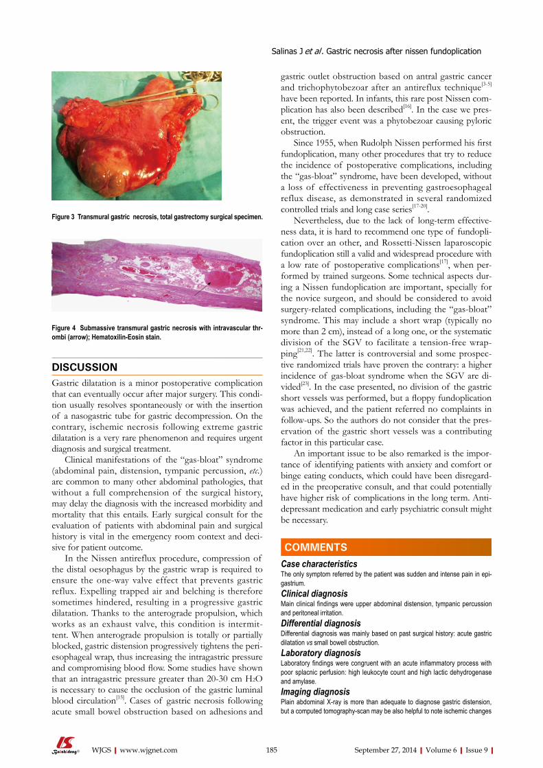
DISCUSSIONGastric dilatation is a minor postoperative complication that can eventually occur after major surgery. This condi-tion usually resolves spontaneously or with the insertion of a nasogastric tube for gastric decompression. On the contrary, ischemic necrosis following extreme gastric dilatation is a very rare phenomenon and requires urgent diagnosis and surgical treatment.
Clinical manifestations of the “gas-bloat” syndrome (abdominal pain, distension, tympanic percussion, etc.) are common to many other abdominal pathologies, that without a full comprehension of the surgical history, may delay the diagnosis with the increased morbidity and mortality that this entails. Early surgical consult for the evaluation of patients with abdominal pain and surgical history is vital in the emergency room context and deci-sive for patient outcome.
In the Nissen antireflux procedure, compression of the distal oesophagus by the gastric wrap is required to ensure the one-way valve effect that prevents gastric reflux. Expelling trapped air and belching is therefore sometimes hindered, resulting in a progressive gastric dilatation. Thanks to the anterograde propulsion, which works as an exhaust valve, this condition is intermit-tent. When anterograde propulsion is totally or partially blocked, gastric distension progressively tightens the peri-esophageal wrap, thus increasing the intragastric pressure and compromising blood flow. Some studies have shown that an intragastric pressure greater than 20-30 cm H2O is necessary to cause the occlusion of the gastric luminal blood circulation[15]. Cases of gastric necrosis following acute small bowel obstruction based on adhesions and
gastric outlet obstruction based on antral gastric cancer and trichophytobezoar after an antireflux technique[3-5]
have been reported. In infants, this rare post Nissen com-plication has also been described[16]. In the case we pres-ent, the trigger event was a phytobezoar causing pyloric obstruction.
Since 1955, when Rudolph Nissen performed his first fundoplication, many other procedures that try to reduce the incidence of postoperative complications, including the “gas-bloat” syndrome, have been developed, without a loss of effectiveness in preventing gastroesophageal reflux disease, as demonstrated in several randomized controlled trials and long case series[17-20].
Nevertheless, due to the lack of long-term effective-ness data, it is hard to recommend one type of fundopli-cation over an other, and Rossetti-Nissen laparoscopic fundoplication still a valid and widespread procedure with a low rate of postoperative complications[17], when per-formed by trained surgeons. Some technical aspects dur-ing a Nissen fundoplication are important, specially for the novice surgeon, and should be considered to avoid surgery-related complications, including the “gas-bloat” syndrome. This may include a short wrap (typically no more than 2 cm), instead of a long one, or the systematic division of the SGV to facilitate a tension-free wrap-ping[21,22]. The latter is controversial and some prospec-tive randomized trials have proven the contrary: a higher incidence of gas-bloat syndrome when the SGV are di-vided[23]. In the case presented, no division of the gastric short vessels was performed, but a floppy fundoplication was achieved, and the patient referred no complaints in follow-ups. So the authors do not consider that the pres-ervation of the gastric short vessels was a contributing factor in this particular case.
An important issue to be also remarked is the impor-tance of identifying patients with anxiety and comfort or binge eating conducts, which could have been disregard-ed in the preoperative consult, and that could potentially have higher risk of complications in the long term. Anti-depressant medication and early psychiatric consult might be necessary.
COMMENTSCase characteristicsThe only symptom referred by the patient was sudden and intense pain in epi-gastrium.Clinical diagnosisMain clinical findings were upper abdominal distension, tympanic percussion and peritoneal irritation.Differential diagnosisDifferential diagnosis was mainly based on past surgical history: acute gastric dilatation vs small bowell obstruction.Laboratory diagnosisLaboratory findings were congruent with an acute inflammatory process with poor splacnic perfusion: high leukocyte count and high lactic dehydrogenase and amylase.Imaging diagnosisPlain abdominal X-ray is more than adequate to diagnose gastric distension, but a computed tomography-scan may be also helpful to note ischemic changes
185 September 27, 2014|Volume 6|Issue 9|WJGS|www.wjgnet.com
Figure 3 Transmural gastric necrosis, total gastrectomy surgical specimen.
Figure 4 Submassive transmural gastric necrosis with intravascular thrombi (arrow); HematoxilinEosin stain.
COMMENTS
Salinas J et al . Gastric necrosis after nissen fundoplication

Tanaka N. Gastric perforation caused by a bulimic attack in an anorexia nervosa patient: report of a case. Surg Today 2000; 30: 435-437 [PMID: 10819480 DOI: 10.1007/s005950050618]
11 Richter JE. Gastroesophageal reflux disease treatment: side effects and complications of fundoplication. Clin Gastroenter-ol Hepatol 2013; 11: 465-471; quiz e39 [PMID: 23267868 DOI: 10.1016/j.cgh.2012.12.006]
12 Spechler SJ. The management of patients who have “failed” antireflux surgery. Am J Gastroenterol 2004; 99: 552-561 [PMID: 15056101 DOI: 10.1111/j.1572-0241.2004.04081.x]
13 Patti MG, Fisichella PM, Perretta S. Preoperative evaluation of patients with gastroesophageal reflux disease. J Laparoen-dosc Adv Surg Tech A 2001; 11: 327-331 [PMID: 11814122 DOI: 10.1089/10926420152761833]
14 Bais JE, Samsom M, Boudesteijn EA, van Rijk PP, Akker-mans LM, Gooszen HG. Impact of delayed gastric emptying on the outcome of antireflux surgery. Ann Surg 2001; 234: 139-146 [PMID: 11505058 DOI: 10.1097/00000658-200108000-00002]
15 Edlich RF, Borner JW, Kuphal J, Wangensteen OH. Gastric blood flow. I. Its distribution during gastric distention. Am J Surg 1970; 120: 35-37 [PMID: 5426860 DOI: 10.1016/S0002-9610(70)80139-8]
16 Bass KD, Meagher DP, Haase GM. Gastric necrosis after fundoplication: a novel approach for esophageal preserva-tion. J Pediatr Surg 1998; 33: 1720-1722 [PMID: 9856904 DOI: 10.1016/S0022-3468(98)90618-2]
17 O’Boyle CJ, Watson DI, Jamieson GG, Myers JC, Game PA, Devitt PG. Division of short gastric vessels at laparoscopic nissen fundoplication: a prospective double-blind random-ized trial with 5-year follow-up. Ann Surg 2002; 235: 165-170 [PMID: 11807353 DOI: 10.1097/00000658-200202000-00001]
18 Carlson MA, Frantzides CT. Complications and results of primary minimally invasive an-tireflux procedures: a review of 10,735 reported cases. J Am Coll Surg 2001; 193: 428-439 [DOI: 10.1016/S1072-7515(01)00992-9]
19 Anvari M, Allen C. Laparoscopic Nissen fundoplication: two-year comprehensive follow-up of a technique of mini-mal paraesophageal dissection. Ann Surg 1998; 227: 25-32 [PMID: 9445106 DOI: 10.1097/00000658-199801000-00004]
20 Watson DI, Pike GK, Baigrie RJ, Mathew G, Devitt PG, Britten-Jones R, Jamieson GG. Prospective double-blind randomized trial of laparoscopic Nissen fundoplication with division and without division of short gastric vessels. Ann Surg 1997; 226: 642-652 [PMID: 9389398 DOI: 10.1097/00000658-199711000-00009]
21 Society of American Gastrointestinal and Endoscopic Sur-geons (SAGES). Guidelines for Surgical Treatment of Gas-troesophageal Reflux Disease (GERD). 2010
22 Soper NJ. Fundoplication and the short gastric vessels: divide and conquer. Ann Surg 2002; 235: 171-173 [PMID: 11807354 DOI: 10.1097/00000658-200202000-00002]
23 Chrysos E, Tzortzinis A, Tsiaoussis J, Athanasakis H, Vass-silakis J, Xynos E. Prospective randomized trial comparing Nissen to Nissen-Rossetti technique for laparoscopic fundo-plication. Am J Surg 2001; 182: 215-221 [PMID: 11587680]
P- Reviewer: Beales ILP, Conzo G, Yin YW S- Editor: Ji FF L- Editor: A E- Editor: Liu SQ
and involvement.Pathological diagnosisRoutine Hematoxylin-Eosin stain was performed to find out ischemic tissue injuries.TreatmentSurgery was mandatory in this case based on clinical and analytical findings, and a total gastrectomy was performed.Experiences and lessonsAntireflux surgery, although it might not be of extreme complexity, it is not free of severe complications and common pitfalls such as long or tightened wraps should be avoided. Also, patients with antireflux surgery and anxiety symptoms or eating disorders should be recognized and properly treated with psychiatric consult if necessary.Peer reviewThe case report is useful as it serves to highlight that fundoplication can have serious complications.
REFERENCES1 Babkin BP, Armour JC, Webster DR. Restoration of the
Functional Capacity of the Stomach when Deprived of its Main Arterial Blood Supply. Can Med Assoc J 1943; 48: 1-10 [PMID: 20322656]
2 Aydin I, Pergel A, Yucel AF, Sahin DA, Ozer E. Gastric Ne-crosis due to Acute Massive Gastric Dilatation. Case Rep Med 2013; 2013: 847238 [DOI: 10.1155/2013/847238]
3 Powell JL, Payne J, Meyer CL, Moncla PR. Gastric necrosis associated with acute gastric dilatation and small bowel ob-struction. Gynecol Oncol 2003; 90: 200-203 [PMID: 12821365 DOI: 10.1016/S0090-8258(03)00204-X]
4 Patuto N, Acklin Y, Oertli D, Langer I. Gastric necrosis com-plicating lately a Nissen fundoplication. Report of a case. Langenbecks Arch Surg 2008; 393: 45-47 [PMID: 17690904 DOI: 10.1007/s00423-007-0216-z]
5 Lee D, Sung K, Lee JH. Acute gastric necrosis due to gastric outlet obstruction accompanied with gastric cancer and trichophytobezoar. J Gastric Cancer 2011; 11: 185-188 [PMID: 22076225 DOI: 10.5230/jgc.2011.11.3.185]
6 Glick PL, Harrison MR, Adzick NS, Webb HW, DeLorimier AA. Gastric infarction secondary to small bowel obstruction: a preventable complication after Nissen fundoplication. J Pediatr Surg 1987; 22: 941-943 [PMID: 3681628 DOI: 10.1016/S0022-3468(87)80595-X]
7 Barker JA, Burnett H, Carlson GL. Gastric necrosis compli-cating acute gastric dilatation after Nissen fundoplication. BMJ Case Rep 2011; 2011: [PMID: 22692484 DOI: 10.1136/bcr.02.2011.3811]
8 Patocskai EJ, Thomas JM. Gastric necrosis in a patient with bulimia. Eur J Surg 2002; 168: 302-304 [PMID: 12375613 DOI: 10.1002/ejs.50]
9 Abdu RA, Garritano D, Culver O. Acute gastric necrosis in anorexia nervosa and bulimia. Two case reports. Arch Surg 1987; 122: 830-832 [PMID: 3592974]
10 Nakao A, Isozaki H, Iwagaki H, Kanagawa T, Takakura N,
186 September 27, 2014|Volume 6|Issue 9|WJGS|www.wjgnet.com
Salinas J et al . Gastric necrosis after nissen fundoplication
Retroanastomotic hernia after Moynihan’s gastroenterostomy
Kerem Karaman, Omer Yalkin, Metin Ercan, Hakan Demir, Fatih Altintoprak, Ismail Zengin
Kerem Karaman, Metin Ercan, Fatih Altintoprak, Department of General Surgery, Faculty of Medicine, Sakarya University, Serdivan 54130, Sakarya, TurkeyOmer Yalkin, Hakan Demir, Ismail Zengin, Department of General Surgery, Sakarya Teaching and Research Hospital, Serdi-van 54130, Sakarya, TurkeyAuthor contributions: All the authors contributed equally to this work.Correspondence to: Kerem Karaman, MD, Department of General Surgery, Faculty of Medicine, Sakarya University, No: 76 Atioglu Sitesi B Blok Kapısı Girisi Daire: 4, Serdivan 54130, Sakarya, Turkey. [email protected]: +90-505-4926238Received: May 25, 2014 Revised: June 24, 2014Accepted: July 25, 2014Published online: September 27, 2014
AbstractRetroanastomotic hernias after gastroenterostomies-either antecolic or retrocolic-are extremely rare but are associated with high mortality rates due to delayed identification which precludes immediate surgical re-duction. In this report, we present a 77-year-old man with retroanastomotic herniation of the efferent loop segments that occurred 14 years after a Moynihan’s gastroenterostomy.
© 2014 Baishideng Publishing Group Inc. All rights reserved.
Key words: Retroanastomotic hernia; Moynihan’s gas-troenterostomy; Intestinal obstruction
Core tip: Retroanastomotic hernia is a rare but fatal condition. Preoperative diagnosis by ultrasound and/or computerized tomography is difficult and sometimes confusing. Early surgery is the key to decreasing mor-tality. The use of a short afferent loop and closure of the retroanastomotic space would decrease the inci-dence of these hernias.
Karaman K, Yalkin O, Ercan M, Demir H, Altintoprak F, Zengin
I. Retroanastomotic hernia after Moynihan’s gastroenterostomy. World J Gastrointest Surg 2014; 6(9): 187-189 Available from: URL: http://www.wjgnet.com/1948-9366/full/v6/i9/187.htm DOI: http://dx.doi.org/10.4240/wjgs.v6.i9.187
INTRODUCTIONRetroanastomotic hernias after gastroenterostomies-either antecolic or retrocolic-are extremely rare but are associated with high mortality rates due to a delay in iden-tification which precludes immediate surgical reduction[1]. Since Petersen[2] provided the first detailed description of a retroanastomotic hernia known as Petersen’s hernia in 1900, there have been few case reports or case series in the literature referring to this entity. In this report, we present a 77-year-old man with retroanastomotic hernia-tion of the efferent loop segments that occurred 14 years after a Moynihan’s gastroenterostomy.
CASE REPORTA 77-year-old man presented with a sudden onset of acute abdominal pain accompanied by nausea and vomit-ing. The physical examination revealed rebound tender-ness with abdominal distention. Abdominal computed tomography showed edematous bowel wall thickening in proximal small bowel segments and dense fluid collec-tion in the right upper quadrant which was considered an indication of visceral organ perforation (Figure 1A). The patient underwent a subtotal gastrectomy for duo-denal ulcer 14 years ago. During explorative laparotomy, a retroanastomotic hernia of the efferent loop segments, passing from right to left through the orifice between the transverse colon and the antecolic, antiperistaltic gastroje-junostomy anastomosis (Moynihan type), was found (Fig-ure 1B-D). The herniated bowel segments were reduced and the defect was closed with running sutures. Viability of the ischemic bowel segments improved after applica-tion of warm pads and the abdomen was closed without further intervention. The postoperative course was un-
CASE REPORT
Submit a Manuscript: http://www.wjgnet.com/esps/Help Desk: http://www.wjgnet.com/esps/helpdesk.aspxDOI: 10.4240/wjgs.v6.i9.187
World J Gastrointest Surg 2014 September 27; 6(9): 187-189ISSN 1948-9366 (online)
© 2014 Baishideng Publishing Group Inc. All rights reserved.
187 September 27, 2014|Volume 6|Issue 9|WJGS|www.wjgnet.com
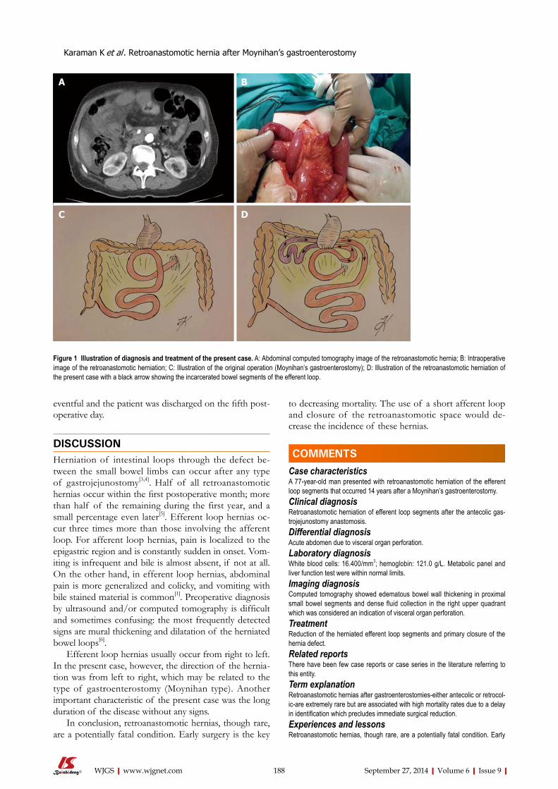
eventful and the patient was discharged on the fifth post-operative day.
DISCUSSIONHerniation of intestinal loops through the defect be-tween the small bowel limbs can occur after any type of gastrojejunostomy[3,4]. Half of all retroanastomotic hernias occur within the first postoperative month; more than half of the remaining during the first year, and a small percentage even later[5]. Efferent loop hernias oc-cur three times more than those involving the afferent loop. For afferent loop hernias, pain is localized to the epigastric region and is constantly sudden in onset. Vom-iting is infrequent and bile is almost absent, if not at all. On the other hand, in efferent loop hernias, abdominal pain is more generalized and colicky, and vomiting with bile stained material is common[1]. Preoperative diagnosis by ultrasound and/or computed tomography is difficult and sometimes confusing: the most frequently detected signs are mural thickening and dilatation of the herniated bowel loops[6].
Efferent loop hernias usually occur from right to left. In the present case, however, the direction of the hernia-tion was from left to right, which may be related to the type of gastroenterostomy (Moynihan type). Another important characteristic of the present case was the long duration of the disease without any signs.
In conclusion, retroanastomotic hernias, though rare, are a potentially fatal condition. Early surgery is the key
to decreasing mortality. The use of a short afferent loop and closure of the retroanastomotic space would de-crease the incidence of these hernias.
COMMENTSCase characteristicsA 77-year-old man presented with retroanastomotic herniation of the efferent loop segments that occurred 14 years after a Moynihan’s gastroenterostomy.Clinical diagnosisRetroanastomotic herniation of efferent loop segments after the antecolic gas-trojejunostomy anastomosis.Differential diagnosisAcute abdomen due to visceral organ perforation.Laboratory diagnosisWhite blood cells: 16.400/mm3; hemoglobin: 121.0 g/L. Metabolic panel and liver function test were within normal limits.Imaging diagnosisComputed tomography showed edematous bowel wall thickening in proximal small bowel segments and dense fluid collection in the right upper quadrant which was considered an indication of visceral organ perforation.TreatmentReduction of the herniated efferent loop segments and primary closure of the hernia defect.Related reportsThere have been few case reports or case series in the literature referring to this entity.Term explanation Retroanastomotic hernias after gastroenterostomies-either antecolic or retrocol-ic-are extremely rare but are associated with high mortality rates due to a delay in identification which precludes immediate surgical reduction.Experiences and lessonsRetroanastomotic hernias, though rare, are a potentially fatal condition. Early
Karaman K et al . Retroanastomotic hernia after Moynihan’s gastroenterostomy
188 September 27, 2014|Volume 6|Issue 9|WJGS|www.wjgnet.com
Figure 1 Illustration of diagnosis and treatment of the present case. A: Abdominal computed tomography image of the retroanastomotic hernia; B: Intraoperative image of the retroanastomotic herniation; C: Illustration of the original operation (Moynihan’s gastroenterostomy); D: Illustration of the retroanastomotic herniation of the present case with a black arrow showing the incarcerated bowel segments of the efferent loop.
COMMENTS
A B
C D
surgery is the key to decreasing mortality. The use of a short afferent loop and closure of the retroanastomotic space would decrease the incidence of these hernias.Peer reviewThis article is referring a rare complication of gastroenterostomy anastomosis and discusses the possible causes and preventive approaches.
REFERENCES1 Rutledge RH. Retroanastomotic hernias after gastrojejunal
anastomoses. Ann Surg 1973; 177: 547-553 [PMID: 4704039 DOI: 10.1097/00000658-197305000-00006]
2 Petersen W. Ueber darmverschlingung nach der gastroen-terostomie. Arch Klin Chir 1900; 62: 94-97
3 Faria G, Preto J, Oliveira M, Pimenta T, Baptista M, Costa-
Maia J. Petersen‘s space hernia: A rare but expanding diag-nosis. Int J Surg Case Rep 2011; 2: 141-143 [PMID: 22096708 DOI: 10.1016/j.ijscr.2011.03.004]
4 Kojima K, Inokuchi M, Kato K, Motoyama K, Sugihara K. Petersen‘s hernia after laparoscopic distal gastrectomy with Roux-en-Y reconstruction for gastric cancer. Gastric Cancer 2014; 17: 146-151 [PMID: 23558458 DOI: 10.1007/s10120-013-0256-8]
5 Bastable JR, Huddy PE. Retro-anastomotic hernia. Eight cases of internal hernia followSing gastrojejunal anastomo-sis, with a review of the literature. Br J Surg 1960; 48: 183-189 [PMID: 13687665 DOI: 10.1002/bjs.18004820821]
6 Kwon JH, Jang HY. Retroanastomotic hernia after gastrojeju-nostomy: US and CT findings with an emphasis on the whirl sign. Abdom Imaging 2005; 30: 656-664 [PMID: 16252151 DOI: 10.1007/s00261-005-0310-z]
P- Reviewer: Kate V, Piccolo G, Shrestha BM S- Editor: Song XX L- Editor: Wang TQ E- Editor: Liu SQ
189 September 27, 2014|Volume 6|Issue 9|WJGS|www.wjgnet.com
Karaman K et al . Retroanastomotic hernia after Moynihan’s gastroenterostomy

Published by Baishideng Publishing Group Inc
World Journal of Gastrointestinal SurgeryWorld J Gastrointest Surg 2014 October 27; 6(10): 190-207
ISSN 1948-9366 (online)

190 Hydatidcystofthepancreas:Reportofanundiagnosedcaseof
pancreatichydatidcystandbriefliteraturereview
Akbulut S, Yavuz R, Sogutcu N, Kaya B, Hatipoglu S, Senol A, Demircan F
201 Surgicalmanagementofcolonicperforationduetoulcerativecolitis
duringpregnancy:Reportofacase
Overbey D, Govekar H, Gajdos C
204 TorsionofMeckel'sdiverticulumasacauseofsmallbowelobstruction:A
casereport
Murruste M, Rajaste G, Kase K
Contents Monthly Volume 6 Number 10 October 27, 2014
WJGS|www.wjgnet.com I October 27, 2014|Volume 6|Issue 10|
ORIGINAL ARTICLE
CASE REPORT

Contents
APPENDIX
ABOUT COVER
World Journal of Gastrointestinal SurgeryVolume 6 Number 10 October 27, 2014
I-V Instructionstoauthors
EditorialBoardMemberofWorldJournalofGastrointestinalSurgery ,RobertGibson,Professor,DepartmentofRadiology,UniversityofMelbourne,RoyalMelbourneHospital,Victoria3050,Australia
World Journal of Gastrointestinal Surgery (World J Gastrointest Surg, WJGS, online ISSN 1948-9366, DOI: 10.4240) is a peer-reviewed open access academic journal that aims to guide clinical practice and improve diagnostic and therapeutic skills of clinicians.
WJGS covers topics concerning micro-invasive surgery; laparoscopy; hepatic, biliary, pancreatic and splenic surgery; surgical nutrition; portal hypertension, as well as associated subjects. The current columns of WJGS include editorial, frontier, diagnostic advances, therapeutics advances, field of vision, mini-reviews, review, topic highlight, medical ethics, original articles, case report, clinical case conference (Clinicopathological conference), and autobiography. Priority publication will be given to articles concerning diagnosis and treatment of gastrointestinal surgery diseases. The following aspects are covered: Clinical diagnosis, laboratory diagnosis, differential diagnosis, imaging tests, pathological diagnosis, molecular biological diagnosis, immunological diagnosis, genetic diagnosis, functional diagnostics, and physical diagnosis; and comprehensive therapy, drug therapy, surgical therapy, interventional treatment, minimally invasive therapy, and robot-assisted therapy.
We encourage authors to submit their manuscripts to WJGS. We will give priority to manuscripts that are supported by major national and international foundations and those that are of great basic and clinical significance.
World Journal of Gastrointestinal Surgery is now indexed in PubMed Central, PubMed, Digital Object Identifier, and Directory of Open Access Journals.
I-III EditorialBoard
Xiu-Xia Song, Vice DirectorWorld Journal of Gastrointestinal SurgeryRoom 903, Building D, Ocean International Center, No. 62 Dongsihuan Zhonglu, Chaoyang District, Beijing 100025, ChinaTelephone: +86-10-85381891Fax: +86-10-85381893E-mail: [email protected] Desk: http://www.wjgnet.com/esps/helpdesk.aspxhttp://www.wjgnet.com
PUBLISHERBaishideng Publishing Group Inc8226 Regency Drive, Pleasanton, CA 94588, USATelephone: +1-925-223-8242Fax: +1-925-223-8243E-mail: [email protected] Desk: http://www.wjgnet.com/esps/helpdesk.aspxhttp://www.wjgnet.com
PUBLICATIONDATEOctober 27, 2014
COPYRIGHT© 2014 Baishideng Publishing Group Inc. Articles pub-lished by this Open-Access journal are distributed under the terms of the Creative Commons Attribution Non-commercial License, which permits use, distribution, and reproduction in any medium, provided the original work is properly cited, the use is non commercial and is otherwise in compliance with the license.
SPECIALSTATEMENTAll articles published in journals owned by the Baishideng Publishing Group (BPG) represent the views and opin-ions of their authors, and not the views, opinions or policies of the BPG, except where otherwise explicitly indicated.
INSTRUCTIONSTOAUTHORSFull instructions are available online at http://www.wjgnet.com/1948-9366/g_info_20100305152206.htm
ONLINESUBMISSIONhttp://www.wjgnet.com/esps/
NAMEOFJOURNALWorld Journal of Gastrointestinal Surgery
ISSNISSN 1948-9366 (online)
LAUNCHDATENovember 30, 2009
FREQUENCYMonthly
EDITOR-IN-CHIEFTimothy M Pawlik, MD, MPH, FACS, Associate Professor of Surgery and Oncology, Hepatobiliary Surgery Program Director, Director, Johns Hopkins Medicine Liver Tumor Center Multi-Disciplinary Clinic, Co-Director of Center for Surgical Trials and Outcomes Research, Johns Hopkins Hospital, 600 N. Wolfe Street, Harvey 611, Baltimore, MD 21287, United States
EDITORIALOFFICEJin-Lei Wang, Director
EDITORS FOR THIS ISSUE
Responsible Assistant Editor: Xiang Li Responsible Science Editor: Xue-Mei GongResponsible Electronic Editor: Su-Qing Liu Proofing Editorial Office Director: Xiu-Xia SongProofing Editor-in-Chief: Lian-Sheng Ma
AIM AND SCOPE
FLYLEAF
INDEXING/ABSTRACTING
WJGS|www.wjgnet.com II October 27, 2014|Volume 6|Issue 10|

Hydatid cyst of the pancreas: Report of an undiagnosed case of pancreatic hydatid cyst and brief literature review
Sami Akbulut, Ridvan Yavuz, Nilgun Sogutcu, Bulent Kaya, Sinan Hatipoglu, Ayhan Senol, Firat Demircan
Sami Akbulut, Ridvan Yavuz, Firat Demircan, Department of Surgery, Diyarbakir Education and Research Hospital, Diyarbakir 21400, TurkeyNilgun Sogutcu, Department of Pathology, Diyarbakir Educa-tion and Research Hospital, Diyarbakir 21400, TurkeyBulent Kaya, Department of Surgery, Kanuni Sultan Suleyman Education and Research Hospital, Istanbul 34303, TurkeySinan Hatipoglu, Department of Surgery, Adiyaman University Faculty of Medicine, Adiyaman 02040, TurkeyAyhan Senol, Department of Radiology, Ergani State Hospital, Diyarbakir 21950, TurkeyAuthor contributions: Akbulut S, Kaya B and Hatipolgu S designed the report; Akbulut S and Yavuz R were the attending doctors for the patients; Akbulut S, Yavuz R and Demircan F per-formed the surgical operation; Akbulut S organized the report and wrote the paper; Senol A provided the radiological information; Sogutcu N provided the histopathological information. Correspondence to: Sami Akbulut, MD, FICS, FACS, Depart-ment of Surgery, Diyarbakir Education and Research Hospital, Uckuyular Mevki, Kayapinar, Diyarbakir 21400, Turkey. [email protected]: +90-412-2580052 Fax: +90-412-2580050Received: May 14, 2014 Revised: July 16, 2014Accepted: September 17, 2014Published online: October 27, 2014
AbstractAIM: To overview the literature on pancreatic hydatid cyst (PHC) disease, a disease frequently misdiagnosed during preoperative radiologic investigation.
METHODS: PubMed, Medline, Google Scholar, and Google databases were searched to identify articles related to PHC using the following keywords: hydatid cyst, hydatid disease, unusual location of hydatid cyst, hydatid cyst and pancreas, pancreatic hydatid cyst, and pancreatic echinococcosis. The search included let-ters to the editor, case reports, review articles, original articles, meeting presentations and abstracts that had been published between January 2010 and April 2014 without any restrictions on language, journal, or coun-
try. All articles identified and retrieved which contained adequate information on the study population (including patient age and sex) and disease and treatment related data (such as cyst size, cyst location, and clinical man-agement) were included in the study; articles with in-sufficient demographic and clinical data were excluded. In addition, we evaluated a case of a 48-year-old fe-male patient with PHC who was treated in our clinic.
RESULTS: A total of 58 patients, including our one new case, (age range: 4 to 70 years, mean ± SD: 31.4 ± 15.9 years) were included in the analysis. Twenty-nine of the patients were female, and 29 were male. The information about cyst location was available from studies involving 54 patients and indicated the follow-ing distribution of locations: pancreatic head (n = 21), pancreatic tail (n = 18), pancreatic body and tail (n = 8), pancreatic body (n = 5), pancreatic head and body (n = 1), and pancreatic neck (n = 1). Extra-pancreatic locations of hydatid cysts were reported in the studies involving 44 of the patients. Among these, no other focus than pancreas was detected in 32 of the patients (isolated cases) while 12 of the patients had hydatid cysts in extra-pancreatic sites (liver: n = 6, liver + spleen + peritoneum: n = 2, kidney: n = 1, liver + kidney: n = 1, kidney + peritoneum: n = 1 and liver + lung: n = 1). Serological information was available in the studies involving 40 patients, and 21 of those pa-tients were serologically positive and 15 were serologi-cally negative; the remaining 4 patients underwent no serological testing. Information about pancreatic cyst size was available in the studies involving 42 patients; the smallest cyst diameter reported was 26 mm and the largest cyst diameter reported was 180 mm (mean ± SD: 71.3 ± 36.1 mm). Complications were avail-able in the studies of 16 patients and showed the fol-lowing distribution: cystobiliary fistula (n = 4), cysto-pancreatic fistula (n = 4), pancreatitis (n = 6), and portal hypertension (n = 2). Postoperative follow-up data were available in the studies involving 48 patients and postoperative recurrence data in the studies of 51 patients; no cases of recurrence occurred in any
ORIGINAL ARTICLE
Submit a Manuscript: http://www.wjgnet.com/esps/Help Desk: http://www.wjgnet.com/esps/helpdesk.aspxDOI: 10.4240/wjgs.v6.i10.190
World J Gastrointest Surg 2014 October 27; 6(10): 190-200ISSN 1948-9366 (online)
© 2014 Baishideng Publishing Group Inc. All rights reserved.
190 October 27, 2014|Volume 6|Issue 10|WJGS|www.wjgnet.com
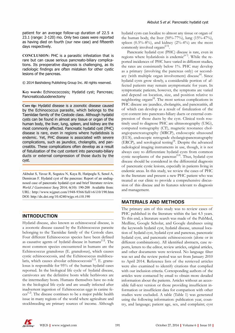
patient for an average follow-up duration of 22.5 ± 23.1 (range: 2-120) mo. Only two cases were reported as having died on fourth (our new case) and fifteenth days respectively.
CONCLUSION: PHC is a parasitic infestation that is rare but can cause serious pancreato-biliary complica-tions. Its preoperative diagnosis is challenging, as its radiologic findings are often mistaken for other cystic lesions of the pancreas.
© 2014 Baishideng Publishing Group Inc. All rights reserved.
Key words: Echinococcosis; Hydatid cyst; Pancreas; Pancreaticoduodenectomy
Core tip: Hydatid disease is a zoonotic disease caused by the Echinococcus parasite, which belongs to the Taeniidae family of the Cestode class. Although hydatid cysts can be found in almost any tissue or organ of the human body, the liver, lung, spleen, and kidney are the most commonly affected. Pancreatic hydatid cyst (PHC) disease is rare, even in regions where hydatidosis is endemic. Yet, PHC disease is associated with severe complications, such as jaundice, cholangitis, and pan-creatitis. These complications often develop as a result of fistulization of the cyst content into pancreato-biliary ducts or external compression of those ducts by the cyst.
Akbulut S, Yavuz R, Sogutcu N, Kaya B, Hatipoglu S, Senol A, Demircan F. Hydatid cyst of the pancreas: Report of an undiag-nosed case of pancreatic hydatid cyst and brief literature review. World J Gastrointest Surg 2014; 6(10): 190-200 Available from: URL: http://www.wjgnet.com/1948-9366/full/v6/i10/190.htm DOI: http://dx.doi.org/10.4240/wjgs.v6.i10.190
INTRODUCTIONHydatid disease, also known as echinococcal disease, is a zoonotic disease caused by the Echinococcus parasite belonging to the Taeniidae family of the Cestode class. Four different Echinococcus species have been defined as causative agents of hydatid disease in humans[1,2]. The most common species encountered in humans are the Echinococcus granulosus (E. granulosus), which causes cystic echinococcosis, and the Echinococcus multilocu-laris, which causes alveolar echinococcosis[1,2]. E. granu-losus is responsible for 95% of the human hydatid cases reported. In the biological life cycle of hydatid disease, carnivores are the definitive hosts while herbivores are the intermediary hosts. Humans themselves have no role in the biological life cycle and are usually infected after inadvertent ingestion of Echinococcus eggs in canine fe-ces[1,2]. The disease continues to be a major public health issue in many regions of the world where agriculture and stockbreeding are primary sources of income. Although
hydatid cysts can localize to almost any tissue or organ of the human body, the liver (50%-77%), lung (15%-47%), spleen (0.5%-8%), and kidney (2%-4%) are the most commonly involved organs[2-5].
Pancreatic hydatid cyst (PHC) disease is rare, even in regions where hydatidosis is endemic[4-7]. While the re-ported incidences of PHC have varied in different studies, the rates are consistently below 1%. PHC may develop as a primary (involving the pancreas only) or second-ary (with multiple organ involvement) disease[6]. Since hydatid cysts grow slowly, a considerable portion of af-fected patients may remain asymptomatic for years. In symptomatic patients, however, the symptoms are varied and depend on location, size, and position relative to neighboring organs[4]. The most serious complications in PHC disease are jaundice, cholangitis, and pancreatitis, all of which can develop as a result of fistulization of the cyst content into pancreato-biliary ducts or external com-pression of those ducts by the cyst. Clinical tools rou-tinely used to diagnose PHC are ultrasonography (USG), computed tomography (CT), magnetic resonance chol-angiopancreatography (MRCP), endoscopic ultasound (EUS), endoscopic retrograde cholangiopancreatography (ERCP), and serological testing[4]. Despite the advanced radiological imaging instruments in use, though, it is not always easy to differentiate hydatid cysts from common cystic neoplasms of the pancreas[4,8]. Thus, hydatid cyst disease should be considered in the differential diagnosis of pancreatic cystic lesions, especially in patients living in endemic areas. In this study, we review the cases of PHC in the literature and present a new PHC patient who was treated at our clinic to provide a comprehensive discus-sion of this disease and its features relevant to diagnosis and management.
MATERIALS AND METHODSThe primary aim of this study was to review cases of PHC published in the literature within the last 4.5 years. To this end, a literature search was made of the PubMed, Medline, Google Scholar, and Google databases using the keywords hydatid cyst, hydatid disease, unusual loca-tion of hydatid cyst, hydatid cyst and pancreas, pancreatic hydatid cyst, and pancreatic echinococcosis (alone or in different combinations). All identified abstracts, case re-ports, letters to the editor, review articles, original articles, and other documents were reviewed. No language filter was set and the review period was set from January 2010 to April 2014. Reference lists of the retrieved articles were also examined to identify citations that complied with our inclusion criteria. Corresponding authors of the articles were contacted by email to obtain more detailed information about the patients. Articles without an acces-sible full-text version or those providing insufficient in-formation or insufficient data for comparison with other studies were excluded. A table (Table 1) was generated using the following information: publication year, coun-try, and language; patient age, sex, and complaint; cyst
Akbulut S et al . Pancreatic hydatid cyst
191 October 27, 2014|Volume 6|Issue 10|WJGS|www.wjgnet.com
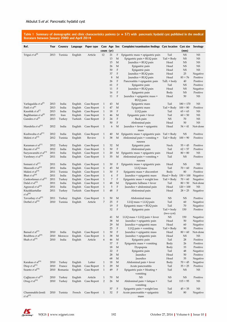
192 October 27, 2014|Volume 6|Issue 10|WJGS|www.wjgnet.com
Table 1 Summary of demographic and clinic characteristics patients (n = 57) with pancreatic hydatid cyst published in the medical literature between January 2000 and April 2014
Ref. Year Country Language Paper type Case Age Sex Complaint/examination findings Cyst location Cyst size Serology
count (yr) (mm)Trigui et al[4] 2013 Tunisia English Article 12 21 F Epigastric mass + epigastric pain Tail NS NS
13 M Epigastric pain + RUQ pain Tail + Body NS NS15 M Jaundice + RUQ pain Head NS NS26 M Epigastric pain Head NS NS50 F Epigastric pain Head NS NS37 F Jaundice + RUQ pain Head 25 Negative 8 M Jaundice + RUQ pain Head 83 × 76 Positive26 F Pancreatitis + epigastric pain TaİL + body 40 Positive61 F Epigastric pain Tail NS Positive11 F Jaundice + RUQ pain Head NS Negative16 F Epigastric pain Body NS Positive11 F Jaundice + epigastric mass +
RUQ painHead 50 NS
Yarlagadda et al[5] 2013 India English Case Report 1 43 M Epigastric mass Tail 180 × 170 NSPatil et al[6] 2013 India English Case Report 1 47 M Epigastric mass Tail + Body 100 × 80 PositiveKaushik et al[7] 2013 India English Case Report 1 18 F LUQ pain Tail 65 × 63 NSBaghbanian et al[8] 2013 Iran English Case Report 1 46 M Epigastric pain + fever Tail 60 × 50 NSGundes et al[9] 2013 Turkey Turkish Case Report 2 24 F Back pain NS 70 NS
50 F Abdominal pain Head 50 NSMandelia et al[10] 2012 India English Case Report 1 6 M Jaundice + fever + epigastric
massHead 54 × 41 Not-done
Kushwaha et al[11] 2012 India English Case Report 1 40 M Epigastric mass + epigastric pain Tail + Body NS PositiveMakni et al[12] 2012 Tunisia English Review 1 38 M Abdominal pain + vomiting +
nauseaTail + Body 100 × 90 Positive
Karaman et al[13] 2012 Turkey English Case Report 1 32 M Epigastric pain Neck 55 × 45 PositiveRayate et al[14] 2012 India English Case Report 1 30 F Abdominal pain Tail 62 × 57 PositiveSuryawanshi et al[15] 2011 India English Case Report 1 20 M Epigastric mass + epigastric pain Head 80 × 80 NSVarshney et al[16] 2011 India English Case Report 1 35 M Abdominal pain + vomiting +
nauseaTail NS Positive
Somani et al[17] 2011 India English Case Report 1 30 F Epigastric mass + epigastric pain Head NS NSMasoodi et al[18] 2011 India English Case Report 1 45 M LUQ mass Tail 70 × 60 PositiveMakni et al[19] 2011 Tunisia English Case Report 1 30 F Epigastric mass + discomfort Body 80 PositiveBhat et al[20] 2011 India English Case Report 1 4 F Jaundice + epigastric mass Head + Body 150 × 100 NegativeCankorkmaz et al[21] 2011 Turkey English Case Report 1 7 F Epigastric mass + weight loss Tail + Body 70 × 60 NegativeDalal et al[22] 2011 India English Case Report 1 48 M Epigastric fullness + fever Tail 80 × 50 Not-doneAgrawal et al[23] 2011 India English Case Report 1 5 F Jaundice + abdominal pain Head 120 × 100 NSKüçükkartallar et al[24]
2011 Turkey Turkish Case Report 1 48 F Abdominal pain Head 28 × 25 Negative
Tavusbay et al[25] 2011 Turkey English Case Report 1 50 M Abdominal mass NS NS PositiveDerbel et al[26] 2010 Tunisia English Article 7 25 F LUQ mass + LUQ pain Tail 60 Negative
19 F Epigastric mass + RUQ pain Tail 70 Negative32 F Epigastric pain Tail + body
(two cyst) 150 Positive
41 M LUQ mass + LUQ pain + fever NS 150 Negative38 M Jaundice + epigastric pain Head 50 Negative29 M Jaundice + epigastric mass Head 60 Negative25 F LUQ pain + vomiting Tail + Body 90 Positive
Bansal et al[27] 2010 India English Case Report 1 30 F Jaundice + epigastric mass Head 80 × 60 Not-doneBoubbou et al[28] 2010 Morocco English Case Report 1 38 M Jaundice + epigastric pain Head NS NSShah et al[29] 2010 India English Article 6 46 M Epigastric pain Tail 28 Positive
37 F Epigastric mass + vomiting Body 26 Positive18 M Dyspepsia Body 33 Positive22 F Epigastric pain Tail 48 Negative28 M Jaundice Head 50 Positive68 M Jaundice Head 35 Negative
Karakas et al[30] 2010 Turkey English Letter 1 18 M Abdominal pain + fever Body 70 × 45 NegativeDiop et al[31] 2010 France English Case Report 1 29 M Acute pancreatitis Tail 35 × 25 PositiveSzanto et al[32] 2010 Romania English Case Report 1 49 F Epigastric pain + bloating +
vomitingTail NS NS
Cağlayan et al[33] 2010 Turkey English Article 1 70 M NS NS NS PositiveOrug et al[34] 2010 Turkey English Case Report 2 26 M Abdominal pain + fatique +
vomitingTail 115 × 95 NS
57 F Epigastric pain + weight loss Tail 45 × 35 NSChammakhi-Jemli et al[35]
2010 Tunisia French Case Report 1 32 F Acute pancreatitis + epigastric mass
Tail 80 Negative
Akbulut S et al . Pancreatic hydatid cyst

15 respectively. Tables 1 and 2 provides detailed informa-tion regarding chief demographic data of the 57 patients included in the study.
A 48-year-old woman presented to our outpatient clinic with malaise, fatigue, pruritus, yellowish discolor-ation of the eyes, darkening of urine color, and acholic gaita. She explained that her complaints, except for jaun-dice, had started 2 mo previous and the jaundice devel-oped 15 d ago. On physical examination, her sclerae were icteric and whole body was jaundiced. Biochemical tests revealed the following results: aspartate aminotransferase (AST): 205 U/L; alanine aminotransferase (ALT): 673 U/L; total bilirubin: 11.6 mg/dL; direct bilirubin: 9.3 mg/dL; hemoglobin: 13 g/dL; platelet count: 244000/μL; white blood cell count: 5000/μL; carbohydrate anti-gen 19-9: 45 U/mL (normal range: 0-39). Ultrasonogra-phy showed that her gall bladder was hydropic and that the common bile duct and intrahepatic bile ducts were dilated. In addition, a 50 mm × 43 mm anechoic lesion consistent with choledococele was detected in the distal common bile duct. A MRCP was performed and showed a common bile duct diameter of 11 mm, dilated intrahe-patic bile ducts, and a 4.5 cm mass in the pancreatic head, which appeared hyperintense on T2A imaging and caused stenosis in the distal tip of the common bile duct (Figure 1). An ERCP showed no intraluminal mass lesion. The consensus of a gastroenterologist and a radiologist was that this lesion could be a choledococele or duodenal di-verticulum. Considering the above findings, a laparotomy was scheduled, in which the abdomen was entered via a midline incision followed by opening of the gastrocolic ligament and application of the Kocher maneuver. A mass lesion of 5 cm × 5 cm was observed in the pancre-atic head, and appeared to be malignant. The common bile duct was markedly dilated. Based on preoperative tests and the intraoperative appearance of the pancreatic head, the mass was regarded as a malignant lesion, and a pancreaticoduodenectomy with pyloric preservation was performed without any intraoperative complications. On post-surgery day 1, the patient’s liver function tests were abnormal and her blood pressure dropped. Yet, radio-logical tests revealed no abnormalities. Since her blood pressure and pulse continued to deteriorate substantially, the patient was taken back to the operating room. During laparotomy, it was observed that all intestinal segments were filled with abundant blood. A regional exploration revealed a pulsatile bleeding focus from a location close to the Wirsung canal in the intestinal lumen. The bleeding was stopped, and the patient was admitted to the inten-sive care unit. Unfortunately, the profound coagulopathy that developed in the patient could not be reverted and she died on postoperative day 4. A detailed examination of the pathology specimen demonstrated that the mass had characteristics consistent with a hydatid cyst (Figures
location and size; results of serologic tests and radiologic examinations; surgical approach, intraoperative complica-tions, postoperative medical management, recurrence, and follow-up (months). In addition, important notes from the studies were summarized in a single sentence.
We also present a case of a 48-year-old woman with PHC who was treated at our clinic and who ultimately died after follow-up. The aim of this case presentation is to emphasize the grave consequences of benign hydatid cyst disease when undiagnosed by preoperative radiologi-cal examinations or not considered by a radiologist in dif-ferential diagnosis.
RESULTSLiterature reviewA literature search using the above review criteria re-trieved a total of 33 articles containing 57 cases about PHC disease[4-36]. Of these, 15 articles were from India, 8 from Turkey, 5 from Tunisia, 2 from Morocco, and 1 each from Iran, France and Romania. Twenty-two cases were reported from Tunisia, 20 from India, 10 from Tur-key, 2 from Morocco, and 1 each from France, Iran and Romania. Twenty-nine articles were written in English, 2 in Turkish, and 2 in French. The current analysis, there-fore, included a total of 58 patients (including our one new case), represented by 29 (50%) females and 29 (50%) males, aged 4 to 70 (mean ± SD: 31.4 ± 15.9) years. The age range of the males was 6 to 70 (mean ± SD: 33.4 ± 16.2) years and that of the females was 4 to 61 (mean ± SD: 29.4 ± 15.4) years. Cyst location in pancreas was reported for 54 patients, wherein the cyst was localized to the pancreatic head in 21 (38.8%), the pancreatic tail in 18 (33.3%), the pancreatic body and tail in 8 (14.8%), the pancreatic body in 5 (9.2%), the pancreatic head and body in 1, and the pancreatic neck in 1. Extra-pancreatic location of hydatid cysts was reported for 44 patients. Among these, 32 (72.7%) had no other foci other than pancreas (isolated) while the remaining 12 patients had extra-PHC as follows: liver, n = 6; liver + spleen + peri-toneum, n = 2; kidney, n = 1; liver + kidney = 1; kidney + peritoneum = 1; and liver + lung, n = 1. Serological data were available from reports of 40 patients, of which 21 (54%) were serologically positive and 15 (38%) were serologically negative; the remaining 4 patients underwent no serological testing. Information about pancreatic cyst size was available for 42 patients; the smallest cyst diame-ter was 26 mm and the largest cyst diameter was 180 mm (mean ± SD: 71.3 ± 36.1 mm). Postoperative follow-up information was available for 48 patients and postopera-tive recurrence information for 51 patients. During the average follow-up duration of 22.5 ± 23.1 (range: 2-120) mo, none of the patients developed recurrence. Only two patients (our new case) died on postoperative day 4 and
193 October 27, 2014|Volume 6|Issue 10|WJGS|www.wjgnet.com
Elmadi et al[36] 2010 Morocco French Case Report 1 7 M Jaundice + fever + RUQ pain Head NS Negative
Akbulut S et al . Pancreatic hydatid cyst
RUQ: Right upper quadrant; LUQ: Left upper quadrant; NS: Not-stated.

194 October 27, 2014|Volume 6|Issue 10|WJGS|www.wjgnet.com
Tabl
e 2 D
etai
led
info
rmat
ion
rega
rdin
g ch
ief
dem
ogra
phic
dat
a of
the
57 p
atie
nts
incl
uded
in t
he s
tudy
Ref
.Rad
iolo
gySu
rgic
al a
ppro
ach
Com
plic
atio
nM
edic
al t
reat
men
tR
ecur
renc
eFo
llow
-up
(mo)
Oth
er h
ydat
id c
yst
focu
ses
Trig
ui et
al[4
]N
SD
ista
l pan
crea
tect
omy
No
NS
No
24N
SU
SG +
CT
Part
ial c
yste
ctom
y +
drai
nage
Hem
orrh
age
NS
No
36Li
ver +
spl
een
+ pe
rito
neum
USG
+ C
TPa
rtia
l cys
tect
omy
+ dr
aina
gePa
ncre
atic
fist
ula
NS
No
24Li
ver
USG
+ C
TPa
rtia
l cys
tect
omy
+ dr
aina
geC
avity
infe
ctio
nN
SN
oN
SLi
ver +
lung
USG
+ C
TPa
rtia
l cys
tect
omy
+ dr
aina
gePa
ncre
atic
fist
ula
NS
No
5N
oU
SG +
CT
Panc
reat
icod
uode
nect
omy
Hem
orrh
age
NS
No
9N
SU
SG +
CT
Part
ial c
yste
ctom
y +
duod
enal
fist
ula
trea
tmen
tN
oN
SN
o24
NS
USG
+ C
TD
ista
l pan
crea
tect
omy
+ sp
lene
ctom
yN
oN
SN
o36
Live
rU
SG +
CT
Dis
tal p
ancr
eate
ctom
y +
sple
nect
omy
No
NS
No
36N
SU
SG +
CT
Cys
to-d
uode
nal a
nast
omos
isN
oN
SN
oN
SLi
ver
USG
+ C
TPa
rtia
l cys
tect
omy
+ dr
aina
geN
oN
SN
o24
NS
USG
+ C
TC
ysto
-duo
dena
l ana
stom
osis
No
NS
No
NS
NS
Yarl
agad
da et
al[5
]U
SG +
CT
Dis
tal p
ancr
eate
ctom
y +
sple
nect
omy
No
Alb
enda
zole
-6 m
oN
o 6
No
Patil
et a
l[6]
USG
+ C
TD
ista
l pan
crea
tect
omy
No
Alb
enda
zole
-3 w
kN
o 6
No
Kau
shik
et a
l[7]
USG
+ C
TTo
tal c
yste
ctom
y +
mar
supi
alis
atio
nN
oA
lben
dazo
le-3
wk
No
6N
S1 Ba
ghba
nian
et a
l[8]
USG
+ C
TD
ista
l pan
crea
tect
omy
+ ne
croz
ecto
my
+ pa
rtia
l nep
hrec
tom
yD
ied
(PO
D15
)N
oD
ied
Die
dK
idne
yG
unde
s et
al[9
]N
SPa
rtia
l cys
tect
omy
+ om
ento
plas
tyW
ound
inf
ectio
nA
lben
dazo
le-4
mo
No
NS
NS
CT
Part
ial c
yste
ctom
y +
omen
topl
asty
Wou
nd i
nfec
tion
Alb
enda
zole
-4 m
oN
oN
SN
o2 M
ande
lia et
al[1
0]U
SG +
MRC
PEn
ucle
atio
n +
chol
angi
ogra
phy
No
Alb
enda
zole
-3 w
kN
o24
No
Kus
hwah
a et
al[1
1]U
SG +
CT
PAIR
+ e
piga
stri
c cy
st e
xcis
ion
Panc
reat
itis
Alb
enda
zole
-3 m
oN
o 6
Kid
ney
+ pe
rito
neum
Mak
ni et
al[1
2]C
TD
ista
l pan
crea
tect
omy
+ sp
lene
ctom
yN
oA
lben
dazo
le-3
mo
No
8N
o3 K
aram
an et
al[1
3]U
SG +
CT
Perc
utan
eous
dra
inag
eN
oA
lben
dazo
le-2
mo
No
18N
oRa
yate
et a
l[14]
USG
+ C
TD
ista
l pan
crea
tect
omy
No
Alb
enda
zole
-2 m
oN
o 7
No
4 Sury
awan
shi e
t al[1
5]U
SG +
CT
Cys
t eva
cuat
ion
+ om
ento
plas
ty-la
paro
scop
icN
oA
lben
dazo
le-3
mo
No
3N
o5 V
arsh
ney
et a
l[16]
CT
Dis
tal p
ancr
eate
ctom
yN
oA
lben
dazo
le-3
wk
No
10N
o6 So
man
i et a
l[17]
USG
+ C
T +
ERC
PPa
ncre
atic
oduo
dene
ctom
yN
oA
lben
dazo
le-6
mo
NS
6N
S7 M
asoo
di et
al[1
8]U
SG +
CT
Dis
tal p
ancr
eate
ctom
y +
sple
nect
omy
Hyp
ergl
isem
ia?
Alb
enda
zole
-1 m
oN
o 6
No
Mak
ni et
al[1
9]U
SGD
ista
l pan
crea
tect
omy
+ sp
lene
ctom
yPa
ncre
atic
fist
ula
Alb
enda
zole
-3 m
oN
o 8
No
Bhat
et a
l[20]
USG
+ C
TPa
rtia
l cys
tect
omy
+ dr
aina
geN
oA
lben
dazo
le-3
mo
No
24N
o8 C
anko
rkm
az et
al[2
1]U
SG +
CT
Part
ial c
yste
ctom
y +
drai
nage
Panc
reat
ic fi
stul
aN
SN
o24
No
9 Dal
al et
al[2
2]U
SG +
CT
Cys
t exc
isio
n al
ong
with
tail
of th
e pa
ncre
asN
oA
lben
dazo
le-6
mo
No
8N
o10
Agr
awal
et a
l[23]
USG
+ M
RCP
Enuc
leat
ion
+ ch
olan
giog
raph
y +
chol
ecys
tect
omy
+ cy
stog
raph
y N
oA
lben
dazo
le-2
mo
NS
NS
NS
11K
üçük
kart
alla
r et a
l[24]
USG
+ C
T +
MRC
P Pa
rtia
l cys
tect
omy
+ om
ento
plas
tyN
oA
lben
dazo
le-4
mo
No
10N
o12
Tavu
sbay
et a
l[25]
USG
+ C
TPa
rtia
l cys
tect
omy
+ om
ento
plas
ty +
sple
nect
omy
+ m
esen
teri
c cy
st e
xcis
ion
Wou
nd i
nfec
tion
Alb
enda
zole
-6 m
oN
o12
Live
r + s
plee
n +
peri
tone
um13
Der
bel e
t al[2
6]U
SG +
CT
Part
ial c
yste
ctom
yN
oN
oN
o35
No
USG
+ C
TPa
rtia
l cys
tect
omy
(for a
ll cy
sts)
No
Alb
enda
zole
-6 m
oN
o 9
Live
r (13
cys
t)U
SG +
CT
Part
ial c
yste
ctom
y fo
r bot
h pa
ncre
atic
cys
tsN
oN
oN
o 6
No
USG
+ C
TD
ista
l pan
crea
tect
omy
No
No
No
9N
oU
SG +
CT
Part
ial c
yste
ctom
y (fo
r all
cyst
)A
cute
pan
crea
titis
Alb
enda
zole
-6 m
oN
o14
Live
rU
SG +
CT
+ M
RIPa
rtia
l cys
tect
omy
No
No
No
16N
oU
SG +
CT
Dis
tal p
ancr
eate
ctom
y +
sple
nect
omy
No
No
No
2N
o14
Bans
al et
al[2
7]U
SG +
MRC
P +
EUS
Panc
reat
icod
uode
nect
omy
No
Alb
enda
zole
-6 w
kN
o 6
No
Boub
bou
et a
l[28]
USG
+ C
TPA
IR +
dra
inag
e +
cyst
ogas
tros
tom
y la
paro
scop
No
NS
NS
NS
NS
Shah
et a
l[29]
USG
+ C
TD
ista
l pan
crea
tect
omy
+ sp
lene
ctom
yN
oA
lben
dazo
le-6
wk
No
120
No
USG
+ C
TC
entr
al p
ancr
eate
ctom
y +
reco
nstr
uctio
nN
oA
lben
dazo
le-6
mo
No
96N
o
Akbulut S et al . Pancreatic hydatid cyst
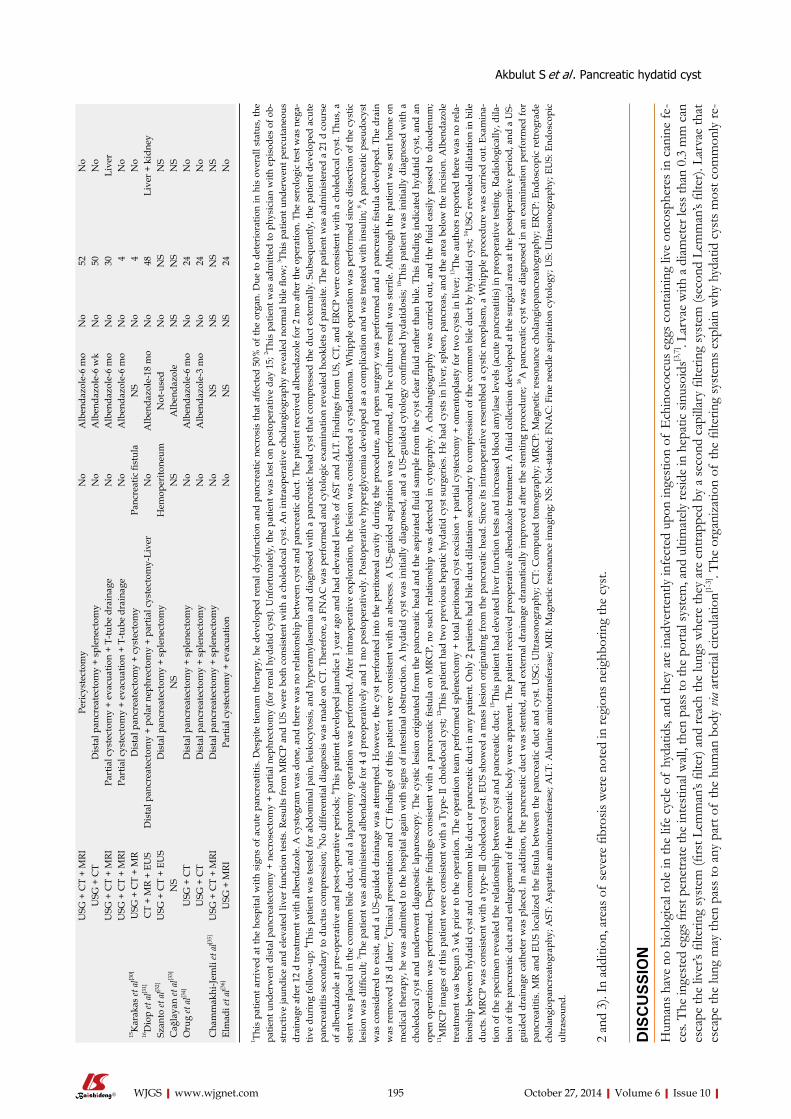
2 an
d 3)
. In
addi
tion,
are
as o
f se
vere
fibr
osis
wer
e no
ted
in re
gion
s nei
ghbo
ring
the
cyst
.
DIS
CU
SSIO
NH
uman
s hav
e no
bio
logi
cal r
ole
in th
e lif
e cy
cle
of h
ydat
ids,
and
they
are
inad
vert
ently
infe
cted
upo
n in
gest
ion
of E
chin
ococ
cus e
ggs c
onta
inin
g liv
e on
cosp
here
s in
cani
ne fe
-ce
s. Th
e in
gest
ed e
ggs fi
rst p
enet
rate
the
inte
stin
al w
all, t
hen
pass
to th
e po
rtal
syst
em, a
nd u
ltim
atel
y re
side
in h
epat
ic si
nuso
ids[3
,7] . L
arva
e w
ith a
diam
eter
less
than
0.3
mm
can
es
cape
the
liver
’s fil
terin
g sy
stem
(firs
t Lem
man
’s fil
ter)
and
reac
h th
e lu
ngs w
here
they
are
ent
rapp
ed b
y a
seco
nd c
apill
ary
filte
ring
syst
em (s
econ
d Le
mm
an’s
filte
r). L
arva
e th
at
esca
pe th
e lu
ng m
ay th
en p
ass t
o an
y pa
rt o
f th
e hu
man
bod
y via
art
erial
circ
ulat
ion[1
-3] . T
he o
rgan
izat
ion
of th
e fil
terin
g sy
stem
s exp
lain
why
hyd
atid
cys
ts m
ost c
omm
only
re-
195 October 27, 2014|Volume 6|Issue 10|WJGS|www.wjgnet.com
USG
+ C
T +
MRI
Peri
cyst
ecto
my
No
Alb
enda
zole
-6 m
oN
o52
No
USG
+ C
TD
ista
l pan
crea
tect
omy
+ sp
lene
ctom
yN
oA
lben
dazo
le-6
wk
No
50N
oU
SG +
CT
+ M
RIPa
rtia
l cys
tect
omy
+ ev
acua
tion
+ T-
tube
dra
inag
eN
oA
lben
dazo
le-6
mo
No
30Li
ver
USG
+ C
T +
MRI
Part
ial c
yste
ctom
y +
evac
uatio
n +
T-tu
be d
rain
age
No
Alb
enda
zole
-6 m
oN
o 4
No
15K
arak
as et
al[3
0]U
SG +
CT
+ M
RD
ista
l pan
crea
tect
omy
+ cy
stec
tom
yPa
ncre
atic
fist
ula
NS
No
4N
o16
Dio
p et
al[3
1]C
T +
MR
+ EU
SD
ista
l pan
crea
tect
omy
+ po
lar n
ephr
ecto
my
+ pa
rtia
l cys
tect
omy-
Live
rN
oA
lben
dazo
le-1
8 m
oN
o48
Live
r + k
idne
ySz
anto
et a
l[32]
USG
+ C
T +
EUS
Dis
tal p
ancr
eate
ctom
y +
sple
nect
omy
Hem
oper
itone
umN
ot-u
sed
No
NS
NS
Cağ
laya
n et
al[3
3]N
SN
SN
SA
lben
dazo
leN
SN
SN
SO
rug
et a
l[34]
USG
+ C
TD
ista
l pan
crea
tect
omy
+ sp
lene
ctom
yN
oA
lben
dazo
le-6
mo
No
24N
oU
SG +
CT
Dis
tal p
ancr
eate
ctom
y +
sple
nect
omy
No
Alb
enda
zole
-3 m
oN
o24
No
Cha
mm
akhi
-Jem
li et
al[3
5]U
SG +
CT
+ M
RID
ista
l pan
crea
tect
omy
+ sp
lene
ctom
yN
oN
SN
SN
SN
SEl
mad
i et a
l[36]
USG
+ M
RIPa
rtia
l cys
tect
omy
+ ev
acua
tion
No
NS
NS
24N
o
1 This
pat
ient
arr
ived
at t
he h
ospi
tal w
ith s
igns
of a
cute
pan
crea
titis
. Des
pite
tien
am th
erap
y, h
e de
velo
ped
rena
l dys
func
tion
and
panc
reat
ic n
ecro
sis
that
affe
cted
50%
of t
he o
rgan
. Due
to d
eter
iora
tion
in h
is o
vera
ll st
atus
, the
pa
tient
und
erw
ent d
ista
l pan
crea
tect
omy
+ ne
cros
ecto
my
+ pa
rtia
l nep
hrec
tom
y (fo
r ren
al h
ydat
id c
yst).
Unf
ortu
nate
ly, t
he p
atie
nt w
as lo
st o
n po
stop
erat
ive
day
15; 2 Th
is p
atie
nt w
as a
dmitt
ed to
phy
sici
an w
ith e
piso
des
of o
b-st
ruct
ive
jaun
dice
and
ele
vate
d liv
er fu
nctio
n te
sts.
Res
ults
from
MRC
P an
d U
S w
ere
both
con
sist
ent w
ith a
cho
ledo
cal c
yst.
An
intr
aope
rativ
e ch
olan
giog
raph
y re
veal
ed n
orm
al b
ile fl
ow; 3 Th
is p
atie
nt u
nder
wen
t per
cuta
neou
s dr
aina
ge a
fter 1
2 d
trea
tmen
t with
alb
enda
zole
. A c
ysto
gram
was
don
e, a
nd th
ere
was
no
rela
tions
hip
betw
een
cyst
and
pan
crea
tic d
uct.
The
patie
nt re
ceiv
ed a
lben
dazo
le fo
r 2 m
o af
ter t
he o
pera
tion.
The
ser
olog
ic te
st w
as n
ega-
tive
duri
ng fo
llow
-up;
4 This
pat
ient
was
test
ed fo
r ab
dom
inal
pai
n, le
ukoc
ytos
is, a
nd h
yper
amyl
asem
ia a
nd d
iagn
osed
with
a p
ancr
eatic
hea
d cy
st th
at c
ompr
esse
d th
e du
ct e
xter
nally
. Sub
sequ
ently
, the
pat
ient
dev
elop
ed a
cute
pa
ncre
atiti
s se
cond
ary
to d
uctu
s co
mpr
essi
on; 5 N
o di
ffere
ntia
l dia
gnos
is w
as m
ade
on C
T. T
here
fore
, a F
NA
C w
as p
erfo
rmed
and
cyt
olog
ic e
xam
inat
ion
reve
aled
hoo
klet
s of
par
asite
. The
pat
ient
was
adm
inis
tere
d a
21 d
cou
rse
of a
lben
dazo
le a
t pre
-ope
rativ
e an
d po
st-o
pera
tive
peri
ods;
6 This
pat
ient
dev
elop
ed ja
undi
ce 1
yea
r ago
and
had
ele
vate
d le
vels
of A
ST a
nd A
LT. F
indi
ngs
from
US,
CT,
and
ERC
P w
ere
cons
iste
nt w
ith a
cho
ledo
cal c
yst.
Thus
, a
sten
t was
pla
ced
in th
e co
mm
on b
ile d
uct,
and
a la
paro
tom
y op
erat
ion
was
per
form
ed. A
fter i
ntra
oper
ativ
e ex
plor
atio
n, th
e le
sion
was
con
side
red
a cy
stad
enom
a. W
hipp
le o
pera
tion
was
per
form
ed s
ince
dis
sect
ion
of th
e cy
stic
le
sion
was
diffi
cult;
7 The
patie
nt w
as a
dmin
iste
red
albe
ndaz
ole
for 4
d p
reop
erat
ivel
y an
d 1
mo
post
oper
ativ
ely.
Pos
tope
rativ
e hy
perg
lyce
mia
dev
elop
ed a
s a
com
plic
atio
n an
d w
as tr
eate
d w
ith in
sulin
; 8 A p
ancr
eatic
pse
udoc
yst
was
con
side
red
to e
xist
, and
a U
S-gu
ided
dra
inag
e w
as a
ttem
pted
. How
ever
, the
cys
t per
fora
ted
into
the
peri
tone
al c
avity
dur
ing
the
proc
edur
e, a
nd o
pen
surg
ery
was
per
form
ed a
nd a
pan
crea
tic fi
stul
a de
velo
ped.
The
dra
in
was
rem
oved
18
d la
ter;
9 Clin
ical
pre
sent
atio
n an
d C
T fin
ding
s of
this
pat
ient
wer
e co
nsis
tent
with
an
absc
ess.
A U
S-gu
ided
asp
irat
ion
was
per
form
ed, a
nd h
e cu
lture
res
ult w
as s
teri
le. A
lthou
gh th
e pa
tient
was
sen
t hom
e on
m
edic
al th
erap
y, h
e w
as a
dmitt
ed to
the
hosp
ital a
gain
with
sig
ns o
f int
estin
al o
bstr
uctio
n. A
hyd
atid
cys
t was
initi
ally
dia
gnos
ed, a
nd a
US-
guid
ed c
ytol
ogy
confi
rmed
hyd
atid
osis
; 10Th
is p
atie
nt w
as in
itial
ly d
iagn
osed
with
a
chol
edoc
al c
yst a
nd u
nder
wen
t dia
gnos
tic la
paro
scop
y. T
he c
ystic
lesi
on o
rigi
nate
d fr
om th
e pa
ncre
atic
hea
d an
d th
e as
pira
ted
fluid
sam
ple
from
the
cyst
cle
ar fl
uid
rath
er th
an b
ile. T
his
findi
ng in
dica
ted
hyda
tid c
yst,
and
an
open
ope
ratio
n w
as p
erfo
rmed
. Des
pite
find
ings
con
sist
ent w
ith a
pan
crea
tic fi
stul
a on
MRC
P, n
o su
ch r
elat
ions
hip
was
det
ecte
d in
cyt
ogra
phy.
A c
hola
ngio
grap
hy w
as c
arri
ed o
ut, a
nd th
e flu
id e
asily
pas
sed
to d
uode
num
; 11
MRC
P im
ages
of t
his
patie
nt w
ere
cons
iste
nt w
ith a
Typ
e-Ⅱ
cho
ledo
cal c
yst;
12Th
is p
atie
nt h
ad tw
o pr
evio
us h
epat
ic h
ydat
id c
yst s
urge
ries
. He
had
cyst
s in
live
r, sp
leen
, pan
crea
s, a
nd th
e ar
ea b
elow
the
inci
sion
. Alb
enda
zole
tr
eatm
ent w
as b
egun
3 w
k pr
ior
to th
e op
erat
ion.
The
ope
ratio
n te
am p
erfo
rmed
spl
enec
tom
y +
tota
l per
itone
al c
yst e
xcis
ion
+ pa
rtia
l cys
tect
omy
+ om
ento
plas
ty fo
r tw
o cy
sts
in li
ver;
13Th
e au
thor
s re
port
ed th
ere
was
no
rela
-tio
nshi
p be
twee
n hy
datid
cys
t and
com
mon
bile
duc
t or p
ancr
eatic
duc
t in
any
patie
nt. O
nly
2 pa
tient
s ha
d bi
le d
uct d
ilata
tion
seco
ndar
y to
com
pres
sion
of t
he c
omm
on b
ile d
uct b
y hy
datid
cys
t; 14
USG
reve
aled
dila
tatio
n in
bile
du
cts.
MRC
P w
as c
onsi
sten
t with
a ty
pe- Ⅲ
cho
ledo
cal c
yst.
EUS
show
ed a
mas
s le
sion
ori
gina
ting
from
the
panc
reat
ic h
ead.
Sin
ce it
s in
trao
pera
tive
rese
mbl
ed a
cys
tic n
eopl
asm
, a W
hipp
le p
roce
dure
was
car
ried
out
. Exa
min
a-tio
n of
the
spec
imen
reve
aled
the
rela
tions
hip
betw
een
cyst
and
pan
crea
tic d
uct;
15Th
is p
atie
nt h
ad e
leva
ted
liver
func
tion
test
s an
d in
crea
sed
bloo
d am
ylas
e le
vels
(acu
te p
ancr
eatit
is) i
n pr
eope
rativ
e te
stin
g. R
adio
logi
cally
, dila
-tio
n of
the
panc
reat
ic d
uct a
nd e
nlar
gem
ent o
f the
pan
crea
tic b
ody
wer
e ap
pare
nt. T
he p
atie
nt re
ceiv
ed p
reop
erat
ive
albe
ndaz
ole
trea
tmen
t. A
flui
d co
llect
ion
deve
lope
d at
the
surg
ical
are
a at
the
post
oper
ativ
e pe
riod
, and
a U
S-gu
ided
dra
inag
e ca
thet
er w
as p
lace
d. In
add
ition
, the
pan
crea
tic d
uct w
as s
tent
ed, a
nd e
xter
nal d
rain
age
dram
atic
ally
impr
oved
afte
r th
e st
entin
g pr
oced
ure;
16A
pan
crea
tic c
yst w
as d
iagn
osed
in a
n ex
amin
atio
n pe
rfor
med
for
panc
reat
itis.
MR
and
EUS
loca
lized
the
fistu
la b
etw
een
the
panc
reat
ic d
uct a
nd c
yst.
USG
: Ultr
ason
ogra
phy;
CT:
Com
pute
d to
mog
raph
y; M
RCP:
Mag
netic
res
onan
ce c
hola
ngio
panc
reat
ogra
phy;
ERC
P: E
ndos
copi
c re
trog
rade
ch
olan
giop
ancr
eato
grap
hy; A
ST: A
spar
tate
am
inot
rans
fera
se; A
LT: A
lani
ne a
min
otra
nsfe
rase
; MRI
: Mag
netic
res
onan
ce im
agin
g; N
S: N
ot-s
tate
d; F
NA
C: F
ine
need
le a
spir
atio
n cy
tolo
gy; U
S: U
ltras
onog
raph
y; E
US:
End
osco
pic
ultr
asou
nd.
Akbulut S et al . Pancreatic hydatid cyst
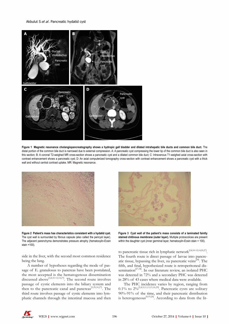
side in the liver, with the second most common residence being the lung.
A number of hypotheses regarding the mode of pas-sage of E. granulosus to pancreas have been postulated, the most accepted is the hematogenous dissemination discussed above[3,8,10-12,14,27]. The second route involves passage of cystic elements into the biliary system and then to the pancreatic canal and pancreas[3,10,14,27]. The third route involves passage of cystic elements into lym-phatic channels through the intestinal mucosa and then
to pancreatic tissue rich in lymphatic network[3,8,10-12,14,20,27]. The fourth route is direct passage of larvae into pancre-atic tissue, bypassing the liver, via pancreatic veins[20]. The fifth, and final, hypothesized route is retroperitoneal dis-semination[27,34]. In our literature review, an isolated PHC was detected in 72% and a secondary PHC was detected in 28% of 43 cases where medical data were available.
The PHC incidence varies by region, ranging from 0.1% to 2%[5,8,9,11,13,15,16,29]. Pancreatic cysts are solitary 90%-91% of the time, and their pancreatic distribution is heterogeneous[4,19,26]. According to data from the lit-
196 October 27, 2014|Volume 6|Issue 10|WJGS|www.wjgnet.com
Figure 2 Patient’s mass has characteristics consistent with a hydatid cyst. The cyst wall is surrounded by fibrous capsule (also called the pericyst layer). The adjacent parenchyma demonstrates pressure atrophy (hematoxylin-Eosin stain ×100).
Figure 3 Cyst wall of the patient’s mass consists of a laminated faintly stained chitinous membrane (outer layer). Multiple protoscolices are present within the daughter cyst (inner germinal layer, hematoxylin-Eosin stain × 100).
Figure 1 Magnetic resonance cholangiopancreatography shows a hydropic gall bladder and dilated intrahepatic bile ducts and common bile duct. The distal portion of the common bile duct is narrowed due to external compression. A: A pancreatic cyst compressing the lower tip of the common bile duct is also seen in this section; B: A coronal T2-weighed MR cross-section shows a pancreatic cyst and a dilated common bile duct; C: Intravenous T1-weighed axial cross-section with contrast enhancement shows a pancreatic cyst; D: An axial computerized tomography cross-section with contrast enhancement shows a pancreatic cyst with a thick wall and without central contrast uptake. MR: Magnetic resonance.
A B
C D
Ductus choledochus
Pancreatic cyst
Gallbladder
Ductus choledochus
Pancreatic cyst
Pancreatic cyst
Gallbladder
Pancreatic cyst
Gallbladder
Akbulut S et al . Pancreatic hydatid cyst
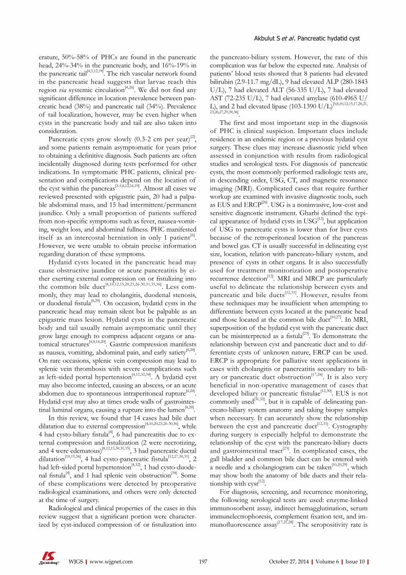
erature, 50%-58% of PHCs are found in the pancreatic head, 24%-34% in the pancreatic body, and 16%-19% in the pancreatic tail[4,5,12,19]. The rich vascular network found in the pancreatic head suggests that larvae reach this region via systemic circulation[4,26]. We did not find any significant difference in location prevalence between pan-creatic head (38%) and pancreatic tail (34%). Prevalence of tail localization, however, may be even higher when cysts in the pancreatic body and tail are also taken into consideration.
Pancreatic cysts grow slowly (0.3-2 cm per year)[2], and some patients remain asymptomatic for years prior to obtaining a definitive diagnosis. Such patients are often incidentally diagnosed during tests performed for other indications. In symptomatic PHC patients, clinical pre-sentation and complications depend on the location of the cyst within the pancreas[3-5,8,12,16,19]. Almost all cases we reviewed presented with epigastric pain, 20 had a palpa-ble abdominal mass, and 15 had intermittent/permanent jaundice. Only a small proportion of patients suffered from non-specific symptoms such as fever, nausea-vomit-ing, weight loss, and abdominal fullness. PHC manifested itself as an intercostal herniation in only 1 patient[6]. However, we were unable to obtain precise information regarding duration of these symptoms.
Hydatid cysts located in the pancreatic head may cause obstructive jaundice or acute pancreatitis by ei-ther exerting external compression on or fistulizing into the common bile duct[4,10,12,15,20,23,26-30,31,35,36]. Less com-monly, they may lead to cholangitis, duodenal stenosis, or duodenal fistula[4,20]. On occasion, hydatid cysts in the pancreatic head may remain silent but be palpable as an epigastric mass lesion. Hydatid cysts in the pancreatic body and tail usually remain asymptomatic until they grow large enough to compress adjacent organs or ana-tomical structures[4,8,16,20]. Gastric compression manifests as nausea, vomiting, abdominal pain, and early satiety[4,20]. On rare occasions, splenic vein compression may lead to splenic vein thrombosis with severe complications such as left-sided portal hypertension[4,12,32,34]. A hydatid cyst may also become infected, causing an abscess, or an acute abdomen due to spontaneous intraperitoneal rupture[4,20]. Hydatid cyst may also at times erode walls of gastrointes-tinal luminal organs, causing a rupture into the lumen[4,20].
In this review, we found that 14 cases had bile duct dilatation due to external compression[4,10,20,23,26-30,36], while 4 had cysto-biliary fistula[4], 6 had pancreatitis due to ex-ternal compression and fistulization (2 were necrotizing, and 4 were edematous)[8,12,15,30,31,35], 3 had pancreatic ductal dilatation[10,15,36], 4 had cysto-pancreatic fistula[12,27,30,31], 2 had left-sided portal hypertension[4,32], 1 had cysto-duode-nal fistula[4], and 1 had splenic vein obstruction[34]. Some of these complications were detected by preoperative radiological examinations, and others were only detected at the time of surgery.
Radiological and clinical properties of the cases in this review suggest that a significant portion were character-ized by cyst-induced compression of or fistulization into
the pancreato-biliary system. However, the rate of this complication was far below the expected rate. Analysis of patients’ blood tests showed that 8 patients had elevated bilirubin (2.9-11.7 mg/dL), 9 had elevated ALP (280-1843 U/L), 7 had elevated ALT (56-335 U/L), 7 had elevated AST (72-235 U/L), 7 had elevated amylase (610-4965 U/L), and 2 had elevated lipase (103-1390 U/L)[5,8,10,12,15,17,20,21,
23,26,27,29,30,36].The first and most important step in the diagnosis
of PHC is clinical suspicion. Important clues include residence in an endemic region or a previous hydatid cyst surgery. These clues may increase diasnostic yield when assessed in conjunction with results from radiological studies and serological tests. For diagnosis of pancreatic cysts, the most commonly performed radiologic tests are, in descending order, USG, CT, and magnetic resonance imaging (MRI). Complicated cases that require further workup are examined with invasive diagnostic tools, such as EUS and ERCP[26]. USG is a noninvasive, low-cost and sensitive diagnostic instrument. Gharbi defined the typi-cal appearance of hydatid cysts in USG[12], but application of USG to pancreatic cysts is lower than for liver cysts because of the retroperitoneal location of the pancreas and bowel gas. CT is usually successful in delineating cyst size, location, relation with pancreato-biliary system, and presence of cysts in other organs. It is also successfully used for treatment monitorization and postoperative recurrence detection[12]. MRI and MRCP are particularly useful to delineate the relationship between cysts and pancreatic and bile ducts[12,31]. However, results from these techniques may be insufficient when attempting to differentiate between cysts located at the pancreatic head and those located at the common bile duct[10,27]. In MRI, superposition of the hydatid cyst with the pancreatic duct can be misinterpreted as a fistula[23]. To demonstrate the relationship between cyst and pancreatic duct and to dif-ferentiate cysts of unknown nature, ERCP can be used. ERCP is appropriate for palliative stent applications in cases with cholangitis or pancreatitis secondary to bili-ary or pancreatic duct obstruction[17,26]. It is also very beneficial in non-operative management of cases that developed biliary or pancreatic fistulae[12,30]. EUS is not commonly used[31,32], but it is capable of delineating pan-creato-biliary system anatomy and taking biopsy samples when necessary. It can accurately show the relationship between the cyst and pancreatic duct[12,31]. Cystography during surgery is especially helpful to demonstrate the relationship of the cyst with the pancreato-biliary ducts and gastrointestinal tract[23]. In complicated cases, the gall bladder and common bile duct can be entered with a needle and a cholangiogram can be taken[10,20,29], which may show both the anatomy of bile ducts and their rela-tionship with cyst[12].
For diagnosis, screening, and recurrence monitoring, the following serological tests are used: enzyme-linked immunosorbent assay, indirect hemagglutination, serum immunelectrophoresis, complement fixation test, and im-munofluorescence assay[17,21,28]. The seropositivity rate is
197 October 27, 2014|Volume 6|Issue 10|WJGS|www.wjgnet.com
Akbulut S et al . Pancreatic hydatid cyst

higher in hepatic hydatid cysts than cysts in other organs. We calculated a rate of 54% for PHC cases. It should be noted, however, that seronegativity does not guarantee absence of hydatid disease[26].
The differential diagnosis of PHCs include neoplastic (cystadenoma, cystadenocarcinoma, gastroenteropancre-atic neuroendocrine tumors, vasculary tumors, metastatic cystic lesions) or non-neoplastic (congenital pancreatic cysts, pseudocysts) cystic lesions[16,19,27]. Diagnosis of cysts that cannot be made using noninvasive techniques can be made by taking either a biopsy from the lesion or an as-piration cytology sample from cyst fluid via percutaneous or endo-ultrasonographic techniques[4,18,26]. Using percu-taneous fine needle aspiration cytology (FNAC) for the differential diagnosis of cystic pancreatic lesion, Varshney et al[16] showed hooklets of hydatid cyst cytologically. In contrast, Dalal et al[22] had to perform FNAC twice in or-der to diagnose hydatid cyst. Anaphylaxis and pouring of cyst content into the abdominal cavity are potential com-plications of the FNAC procedure. Hence, prophylactic antihelminthic agents should be started when FNAC is contemplated in a patient with suspected cysts; otherwise, the procedure should be avoided[4].
All patients presented in this review underwent at least one preoperative radiological or serological test. Af-ter these tests, 20 patients were diagnosed with PHC, 14 with benign/neoplastic cystic lesion of pancreas, 8 with choledocal cyst, 4 with PHC/cystic neoplasm of pancre-as, 2 with hepatic hydatid cyst, and one with splenic hy-datid cyst. Minimally invasive surgery was contemplated. No presumptive diagnoses were made for the remaining patients. As seen, only 40%-49% of patients were diag-nosed with PHC at the preoperative period. This is true even for the most recent studies performed within the last 4.5 years. Diagnostically, the situation was even worse several decades ago, when the rate of preoperative PHC was far below 30%.
PHC can be treated with one or a combination of several therapies, including open or laparoscopic surgical approach, minimally invasive approach [puncture-aspira-tion-injection-reaspiration (PAIR) or direct percutaneous catheterization], and medical therapy[9]. As is the case for other organ hydatid cysts, open surgery is the gold stan-dard for the treatment of PHC disease. Selection of the appropriate management approach is affected by many factors, such as surgeon’s experience, patient age, pres-ence of comorbid conditions, pancreatic localization of cyst(s), cyst size, and relation of cyst to adjacent struc-tures or the pancreatic and common bile ducts[4,29,31].
Pancreatic head cysts with no communication with biliary or pancreatic ducts can be managed with partial cys-tectomy + external drainage, partial cystectomy + omen-topexy and pericystectomy, marsupialisation, and pancre-aticoduodenectomy procedures[4,29,34]. Each method has its own advantages and disadvantages. In order to avoid postoperative pancreatic fistula formation, cysts with communication with the pancreatic duct can be treated with cysto-jejunal, cysto-duodenal, or cysto-gastric anas-
tomosis techniques[4,26]. In cysts located in the pancreatic body or tail, the most appropriate approach is a spleen-preserving distal pancreatectomy[4,26]. In cases where the spleen cannot be preserved, pneumococcal and meningo-coccal vaccinations should be done immediately to avert postsplenectomy complications[5]. Central pancreatec-tomy may be preferable when cysts are localized to the pancreatic body or neck[29]. The main advantage of this method is the preservation of pancreatic tissue and the minimization of complications, such as diabetes or exo-crine pancreatic insufficiency[29]. Masoodi et al[18] reported in a patient that underwent a distal pancreatectomy hy-perglycemia high enough to require insulin injection. For management of hydatid cysts of the pancreatic head, the role of pancreaticoduodenectomy is very limited[20]. Pancreaticoduodenectomy was performed in only 3 of 19 pancreatic head cysts[4,17,27]. A whipple procedure was applied in all three of these cases since the results of preoperative radiological examination and/or intraopera-tive findings were consistent with a cystic lesion of the pancreatic head. In our case, we experienced similar diffi-culties. While the preoperative tests, including CT, MRCP, and ERCP, were consistent with a choledocal cyst, the intraoperative appearance was totally compatible with a mass in the pancreatic head. Unfortunately, our patient was lost to a misfortunate complication. In retrospect, we realize that patient outcome may have been improved if the diagnosis was made preoperatively and simple partial cystectomy and drainage was performed intraoperatively. Hence, our main objective for writing this manuscript was to heighten awareness about this topic.
Although rarely reported in the literature, there are some studies describing percutaneous drainage of pan-creatic hydatic cysts[10,13]. Percutaneous drainage can be accomplished by puncture, aspiration, injection of hy-pertonic saline solution, and re-aspiration of cyst content (PAIR) or direct catheterization of the cyst[13,18,28]. These procedures should be specifically carried out in Type Ⅰ and Ⅱ PHCs, cysts with a diameter less than 50 mm, patients who refuse surgery, and cases with a higher anesthesia risk[1]. The main advantage of PAIR is the ability to show scoleces in the aspirated cyst fluid cytopathologically with-in a short period of time. Another advantage is the ability to delineate the relative location of the cyst with the pan-creatic duct by contrast material administration during the procedure. However, an unconscious percutaneous drainage procedure or one that is performed without es-timating the possible presence of PHC may lead to cyst perforation and surgical complications[21]. The risk associ-ated with release of cyst contents into abdominal cavity is markedly lower with a PAIR procedure that is carried out by passing through parenchyma of solid organs like liver and spleen than it would be with pancreatic and other intraabdominal cysts[26]. In cases where minimally invasive surgical therapy has been contemplated, antihelminthic therapy should be administered before (≥ 4 d) and after (≥ 3-4 wk) the procedure in order to reduce intracystic pressure and prevent anaphylaxis[1].
198 October 27, 2014|Volume 6|Issue 10|WJGS|www.wjgnet.com
Akbulut S et al . Pancreatic hydatid cyst
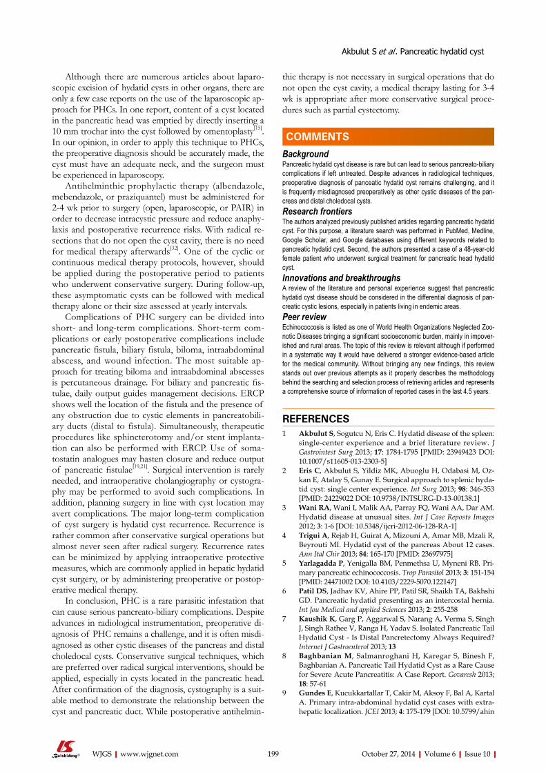
Although there are numerous articles about laparo-scopic excision of hydatid cysts in other organs, there are only a few case reports on the use of the laparoscopic ap-proach for PHCs. In one report, content of a cyst located in the pancreatic head was emptied by directly inserting a 10 mm trochar into the cyst followed by omentoplasty[15]. In our opinion, in order to apply this technique to PHCs, the preoperative diagnosis should be accurately made, the cyst must have an adequate neck, and the surgeon must be experienced in laparoscopy.
Antihelminthic prophylactic therapy (albendazole, mebendazole, or praziquantel) must be administered for 2-4 wk prior to surgery (open, laparoscopic, or PAIR) in order to decrease intracystic pressure and reduce anaphy-laxis and postoperative recurrence risks. With radical re-sections that do not open the cyst cavity, there is no need for medical therapy afterwards[32]. One of the cyclic or continuous medical therapy protocols, however, should be applied during the postoperative period to patients who underwent conservative surgery. During follow-up, these asymptomatic cysts can be followed with medical therapy alone or their size assessed at yearly intervals.
Complications of PHC surgery can be divided into short- and long-term complications. Short-term com-plications or early postoperative complications include pancreatic fistula, biliary fistula, biloma, intraabdominal abscess, and wound infection. The most suitable ap-proach for treating biloma and intraabdominal abscesses is percutaneous drainage. For biliary and pancreatic fis-tulae, daily output guides management decisions. ERCP shows well the location of the fistula and the presence of any obstruction due to cystic elements in pancreatobili-ary ducts (distal to fistula). Simultaneously, therapeutic procedures like sphincterotomy and/or stent implanta-tion can also be performed with ERCP. Use of soma-tostatin analogues may hasten closure and reduce output of pancreatic fistulae[19,21]. Surgical intervention is rarely needed, and intraoperative cholangiography or cystogra-phy may be performed to avoid such complications. In addition, planning surgery in line with cyst location may avert complications. The major long-term complication of cyst surgery is hydatid cyst recurrence. Recurrence is rather common after conservative surgical operations but almost never seen after radical surgery. Recurrence rates can be minimized by applying intraoperative protective measures, which are commonly applied in hepatic hydatid cyst surgery, or by administering preoperative or postop-erative medical therapy.
In conclusion, PHC is a rare parasitic infestation that can cause serious pancreato-biliary complications. Despite advances in radiological instrumentation, preoperative di-agnosis of PHC remains a challenge, and it is often misdi-agnosed as other cystic diseases of the pancreas and distal choledocal cysts. Conservative surgical techniques, which are preferred over radical surgical interventions, should be applied, especially in cysts located in the pancreatic head. After confirmation of the diagnosis, cystography is a suit-able method to demonstrate the relationship between the cyst and pancreatic duct. While postoperative antihelmin-
thic therapy is not necessary in surgical operations that do not open the cyst cavity, a medical therapy lasting for 3-4 wk is appropriate after more conservative surgical proce-dures such as partial cystectomy.
COMMENTSBackgroundPancreatic hydatid cyst disease is rare but can lead to serious pancreato-biliary complications if left untreated. Despite advances in radiological techniques, preoperative diagnosis of panceatic hydatid cyst remains challenging, and it is frequently misdiagnosed preoperatively as other cystic diseases of the pan-creas and distal choledocal cysts.Research frontiersThe authors analyzed previously published articles regarding pancreatic hydatid cyst. For this purpose, a literature search was performed in PubMed, Medline, Google Scholar, and Google databases using different keywords related to pancreatic hydatid cyst. Second, the authors presented a case of a 48-year-old female patient who underwent surgical treatment for pancreatic head hydatid cyst. Innovations and breakthroughsA review of the literature and personal experience suggest that pancreatic hydatid cyst disease should be considered in the differential diagnosis of pan-creatic cystic lesions, especially in patients living in endemic areas. Peer reviewEchinococcosis is listed as one of World Health Organizations Neglected Zoo-notic Diseases bringing a significant socioeconomic burden, mainly in impover-ished and rural areas. The topic of this review is relevant although if performed in a systematic way it would have delivered a stronger evidence-based article for the medical community. Without bringing any new findings, this review stands out over previous attempts as it properly describes the methodology behind the searching and selection process of retrieving articles and represents a comprehensive source of information of reported cases in the last 4.5 years.
REFERENCES1 Akbulut S, Sogutcu N, Eris C. Hydatid disease of the spleen:
single-center experience and a brief literature review. J Gastrointest Surg 2013; 17: 1784-1795 [PMID: 23949423 DOI: 10.1007/s11605-013-2303-5]
2 Eris C, Akbulut S, Yildiz MK, Abuoglu H, Odabasi M, Oz-kan E, Atalay S, Gunay E. Surgical approach to splenic hyda-tid cyst: single center experience. Int Surg 2013; 98: 346-353 [PMID: 24229022 DOI: 10.9738/INTSURG-D-13-00138.1]
3 Wani RA, Wani I, Malik AA, Parray FQ, Wani AA, Dar AM. Hydatid disease at unusual sites. Int J Case Reposts Images 2012; 3: 1-6 [DOI: 10.5348/ijcri-2012-06-128-RA-1]
4 Trigui A, Rejab H, Guirat A, Mizouni A, Amar MB, Mzali R, Beyrouti MI. Hydatid cyst of the pancreas About 12 cases. Ann Ital Chir 2013; 84: 165-170 [PMID: 23697975]
5 Yarlagadda P, Yenigalla BM, Penmethsa U, Myneni RB. Pri-mary pancreatic echinococcosis. Trop Parasitol 2013; 3: 151-154 [PMID: 24471002 DOI: 10.4103/2229-5070.122147]
6 Patil DS, Jadhav KV, Ahire PP, Patil SR, Shaikh TA, Bakhshi GD. Pancreatic hydatid presenting as an intercostal hernia. Int Jou Medical and applied Sciences 2013; 2: 255-258
7 Kaushik K, Garg P, Aggarwal S, Narang A, Verma S, Singh J, Singh Rathee V, Ranga H, Yadav S. Isolated Pancreatic Tail Hydatid Cyst - Is Distal Pancretectomy Always Required? Internet J Gastroenterol 2013; 13
8 Baghbanian M, Salmanroghani H, Karegar S, Binesh F, Baghbanian A. Pancreatic Tail Hydatid Cyst as a Rare Cause for Severe Acute Pancreatitis: A Case Report. Govaresh 2013; 18: 57-61
9 Gundes E, Kucukkartallar T, Cakir M, Aksoy F, Bal A, Kartal A. Primary intra-abdominal hydatid cyst cases with extra-hepatic localization. JCEI 2013; 4: 175-179 [DOI: 10.5799/ahin
199 October 27, 2014|Volume 6|Issue 10|WJGS|www.wjgnet.com
COMMENTS
Akbulut S et al . Pancreatic hydatid cyst
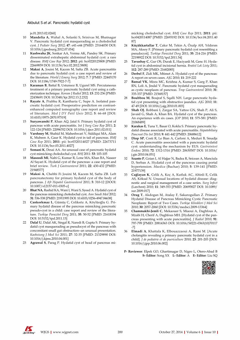
js.01.2013.02.0260]10 Mandelia A, Wahal A, Solanki S, Srinivas M, Bhatnagar
V. Pancreatic hydatid cyst masquerading as a choledochal cyst. J Pediatr Surg 2012; 47: e41-e44 [PMID: 23164030 DOI: 10.1016/j.jpedsurg.2012.07.054]
11 Kushwaha JK, Sonkar AA, Verma AK, Pandey SK. Primary disseminated extrahepatic abdominal hydatid cyst: a rare disease. BMJ Case Rep 2012; 2012: pii: bcr0220125808 [PMID: 22669859 DOI: 10.1136/bcr.02.2012.5808]
12 Makni A, Jouini M, Kacem M, Safta ZB. Acute pancreatitis due to pancreatic hydatid cyst: a case report and review of the literature. World J Emerg Surg 2012; 7: 7 [PMID: 22445170 DOI: 10.1186/1749-7922-7-7]
13 Karaman B, Battal B, Ustunsoz B, Ugurel MS. Percutaneous treatment of a primary pancreatic hydatid cyst using a cath-eterization technique. Korean J Radiol 2012; 13: 232-236 [PMID: 22438691 DOI: 10.3348/kjr.2012.13.2.232]
14 Rayate A, Prabhu R, Kantharia C, Supe A. Isolated pan-creatic hydatid cyst: Preoperative prediction on contrast-enhanced computed tomography case report and review of literature. Med J DY Patil Univ 2012; 5: 66-68 [DOI: 10.4103/0975-2870.97519]
15 Suryawanshi P, Khan AQ, Jatal S. Primary hydatid cyst of pancreas with acute pancreatitis. Int J Surg Case Rep 2011; 2: 122-124 [PMID: 22096702 DOI: 10.1016/j.ijscr.2011.02.011]
16 Varshney M, Shahid M, Maheshwari V, Siddiqui MA, Alam K, Mubeen A, Gaur K. Hydatid cyst in tail of pancreas. BMJ Case Rep 2011; 2011: pii: bcr0320114027 [PMID: 22673711 DOI: 10.1136/bcr.03.2011.4027]
17 Somani K, Desai AA. An unusual case of pancreatic hydatid cyst mimicking choledochal cyst. BHJ 2011: 53: 103-105
18 Masoodi MI, Nabi G, Kumar R, Lone MA, Khan BA, Naseer Al Sayari K. Hydatid cyst of the pancreas: a case report and brief review. Turk J Gastroenterol 2011; 22: 430-432 [PMID: 21948577]
19 Makni A, Chebbi Fi Jouini M, Kacem M, Safta ZB. Left pancreatectomy for primary hydatid cyst of the body of pancreas. J Afr Hepatol Gastroenterol 2011; 5: 310-12 [DOOI: 10.1007/s12157-011-0305-z]
20 Bhat NA, Rashid KA, Wani I, Wani S, Syeed A. Hydatid cyst of the pancreas mimicking choledochal cyst. Ann Saudi Med 2012; 31: 536-538 [PMID: 21911995 DOI: 10.4103/0256-4947.84638]
21 Cankorkmaz L, Gümüş C, Celiksöz A, Köylüoğlu G. Pri-mary hydatid disease of the pancreas mimicking pancreatic pseudo-cyst in a child: case report and review of the litera-ture. Turkiye Parazitol Derg 2011; 35: 50-52 [PMID: 21618194 DOI: 10.5152/tpd.2011.13]
22 Dalal U, Dalal AK, Singal R, Naredi B, Gupta S. Primary hy-datid cyst masquerading as pseudocyst of the pancreas with concomitant small gut obstruction--an unusual presentation. Kaohsiung J Med Sci 2011; 27: 32-35 [PMID: 21329890 DOI: 10.1016/j.kjms.2010.04.001]
23 Agrawal S, Parag P. Hydatid cyst of head of pancreas mi-
micking choledochal cyst. BMJ Case Rep 2011; 2011: pii: bcr0420114087 [PMID: 22693192 DOI: 10.1136/bcr.04.2011.4087]
24 Küçükkartallar T, Cakır M, Tekin A, Özalp AH, Yıldırım MA, Aksoy F. [Primary pancreatic hydatid cyst resembling a pseudocyst]. Turkiye Parazitol Derg 2011; 35: 214-216 [PMID: 22198922 DOI: 10.5152/tpd.2011.54]
25 Tavusbay C, Gur OS, Durak E, Haciyanli M, Genc H. Hyda-tid cyst in abdominal incisional hernia. Bratisl Lek Listy 2011; 112: 287-289 [PMID: 21682085]
26 Derbel F, Zidi MK, Mtimet A. Hydatid cyst of the pancreas: A report on seven cases. AJG 2010; 11: 219-222
27 Bansal VK, Misra MC, Krishna A, Kumar S, Garg P, Khan RN, Loli A, Jindal V. Pancreatic hydatid cyst masquerading as cystic neoplasm of pancreas. Trop Gastroenterol 2010; 31: 335-337 [PMID: 21568157]
28 Boubbou M, Boujraf S, Sqalli NH. Large pancreatic hyda-tid cyst presenting with obstructive jaundice. AJG 2010; 11: 47-49 [DOI: 10.1016/j.ajg.2010.01.003]
29 Shah OJ, Robbani I, Zargar SA, Yattoo GN, Shah P, Ali S, Javaid G, Shah A, Khan BA. Hydatid cyst of the pancreas. An experience with six cases. JOP 2010; 11: 575-581 [PMID: 21068489]
30 Karakas E, Tuna Y, Basar O, Koklu S. Primary pancreatic hy-datid disease associated with acute pancreatitis. Hepatobiliary Pancreat Dis Int 2010; 9: 441-442 [PMID: 20688612]
31 Diop SP, Costi R, Le Bian A, Carloni A, Meduri B, Smadja C. Acute pancreatitis associated with a pancreatic hydatid cyst: understanding the mechanism by EUS. Gastrointest Endosc 2010; 72: 1312-1314 [PMID: 20630507 DOI: 10.1016/j.gie.2010.04.051]
32 Szanto P, Goian I, Al Hajjar N, Badea R, Seicean A, Manciula D, Serban A. Hydatid cyst of the pancreas causing portal hypertension. Maedica (Buchar) 2010; 5: 139-141 [PMID: 21977139]
33 Cağlayan K, Celik A, Koç A, Kutluk AC, Altinli E, Celik AS, Köksal N. Unusual locations of hydatid disease: diag-nostic and surgical management of a case series. Surg Infect (Larchmt) 2010; 11: 349-353 [PMID: 20695827 DOI: 10.1089/sur.2009.017]
34 Orug T, Akdogan M, Atalay F, Sakarogulları Z. Primary Hydatid Disease of Pancreas Mimicking Cystic Pancreatic Neoplasm: Report of Two Cases. Turkiye Klinikleri J Med Sci 2010; 30: 2057-2060 [DOI: 10.5336/medsci.2009-13364]
35 Chammakhi-Jemli C, Mekaouer S, Miaoui A, Daghfous A, Mzabi H, Cherif A, Daghfous MH. [Hydatid cyst of the pan-creas presenting with acute pancreatitis]. J Radiol 2010; 91: 797-799 [PMID: 20814363 DOI: 10.1016/S0221-0363(10)70117-7]
36 Elmadi A, Khattala K, Elbouazzaoui A, Rami M. [Acute cholangitis revealing a primary pancreatic hydatid cyst in a child]. J de pediatrie et de puericulture 2010; 23: 201-203 [DOI: 10.1016/j.jpp.2010.06.002]
P- Reviewer: Elpek GO, Ghartimagar D, Nigro L, Otero-Abad B S- Editor: Song XX L- Editor: A E- Editor: Liu SQ
200 October 27, 2014|Volume 6|Issue 10|WJGS|www.wjgnet.com
Akbulut S et al . Pancreatic hydatid cyst

Surgical management of colonic perforation due to ulcerative colitis during pregnancy: Report of a case
Douglas Overbey, Henry Govekar, Csaba Gajdos
Douglas Overbey, Henry Govekar, Csaba Gajdos, Depart-ment of Surgery, University of Colorado, Aurora, CO 80045, United StatesAuthor contributions: All authors were involved in the case and contributed equally to writing and reviewing the manuscript.Correspondence to: Douglas Overbey, MD, Department of Surgery, University of Colorado, 12631 E 17th Ave, Mail Stop C302, Aurora, CO 80045, United States. [email protected]: +1-573-2257728 Fax: +1-303-7242682Received: May 12, 2014 Revised: July 30, 2014Accepted: September 17, 2014Published online: October 27, 2014
AbstractThis report describes a young female in her second trimester of pregnancy with known ulcerative colitis on maintenance medical therapy. She was admitted for abdominal pain, and workup revealed a colonic stric-ture and ulceration with contained perforation. After multidisciplinary discussion she was managed with colectomy and end ileostomy. She delivered a healthy newborn 18 wk after surgery. Only a few prior reports described surgical management of inflammatory bowel disease during pregnancy, with recent results indicating low risk of adverse outcomes.
© 2014 Baishideng Publishing Group Inc. All rights reserved.
Key words: Inflammatory bowel disease; Ulcerative colitis; Colonic stricture; Colon perforation; Pregnancy
Core tip: Surgical management of inflammatory bowel disease flare during pregnancy are rare and infrequent-ly reported in the literature. This case report summa-rizes the literature and describes a successful resection of a contained perforation and stricture secondary to ulcerative colitis flare.
Overbey D, Govekar H, Gajdos C. Surgical management of
colonic perforation due to ulcerative colitis during pregnancy: Report of a case. World J Gastrointest Surg 2014; 6(10): 201-203 Available from: URL: http://www.wjgnet.com/1948-9366/full/v6/i10/201.htm DOI: http://dx.doi.org/10.4240/wjgs.v6.i10.201
INTRODUCTIONInflammatory bowel disease (IBD) encompasses Crohn’s disease (CD) and Ulcerative Colitis (UC). These autoim-mune conditions involve mucosal inflammation of the entire gastrointestinal tract in CD and the colon and rec-tum in UC. While primary management of IBD is medi-cal, surgical indications are generally reserved for toxic colitis, perforation, bleeding, strictures, neoplasms, and failure of medical management.
Although the rate of IBD flare is similar in pregnant and nonpregnant patients (26%-34%), the primary deter-minant of disease outcome seems to involve quiescence vs active disease at the initiation of pregnancy[1]. Thus the goal in a planned pregnancy is remission before concep-tion[1]. The optimal drug regimen is patient dependent but the aminosalicylate (mesalamine, balsalazide, sul-fasalazine), thiopurine (AZA and 6-MP), and anti-TNF (INF, ADA, CZP) classes are generally considered safe in pregnancy. Active disease is a greater risk than active ther-apy and timing of dosages can avoid the later weeks of pregnancy to mediate placental transfer of these drugs[1,2].
Initial studies reported an association between IBD and adverse outcomes in pregnant patients, possibly mediated by the immunologic phases of pregnancy and specific interactions leading to preterm birth[3]. Surgical management of ulcerative colitis in an ongoing pregnan-cy is infrequently reported, with only one recent literature review published[4] and additional case reports. Results showed no mortality reported after 1974, and minimal morbidity[4,5]. Other more generalized reports of IBD relapse during pregnancy indicate a colectomy rate of up to 17%[6]. We report a case of a surgically treated compli-
CASE REPORT
Submit a Manuscript: http://www.wjgnet.com/esps/Help Desk: http://www.wjgnet.com/esps/helpdesk.aspxDOI: 10.4240/wjgs.v6.i10.201
World J Gastrointest Surg 2014 October 27; 6(10): 201-203ISSN 1948-9366 (online)
© 2014 Baishideng Publishing Group Inc. All rights reserved.
201 October 27, 2014|Volume 6|Issue 10|WJGS|www.wjgnet.com
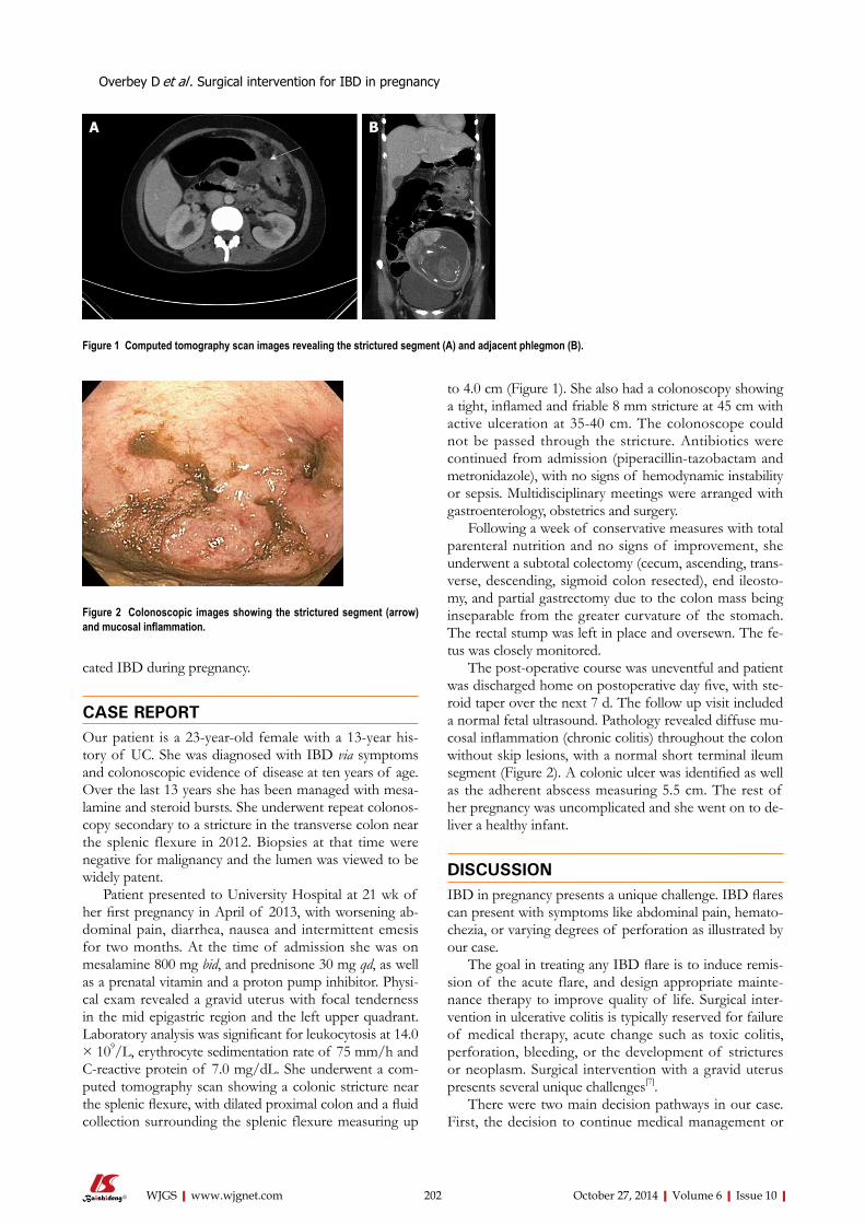
cated IBD during pregnancy.
CASE REPORTOur patient is a 23-year-old female with a 13-year his-tory of UC. She was diagnosed with IBD via symptoms and colonoscopic evidence of disease at ten years of age. Over the last 13 years she has been managed with mesa-lamine and steroid bursts. She underwent repeat colonos-copy secondary to a stricture in the transverse colon near the splenic flexure in 2012. Biopsies at that time were negative for malignancy and the lumen was viewed to be widely patent.
Patient presented to University Hospital at 21 wk of her first pregnancy in April of 2013, with worsening ab-dominal pain, diarrhea, nausea and intermittent emesis for two months. At the time of admission she was on mesalamine 800 mg bid, and prednisone 30 mg qd, as well as a prenatal vitamin and a proton pump inhibitor. Physi-cal exam revealed a gravid uterus with focal tenderness in the mid epigastric region and the left upper quadrant. Laboratory analysis was significant for leukocytosis at 14.0 × 10⁹/L, erythrocyte sedimentation rate of 75 mm/h and C-reactive protein of 7.0 mg/dL. She underwent a com-puted tomography scan showing a colonic stricture near the splenic flexure, with dilated proximal colon and a fluid collection surrounding the splenic flexure measuring up
to 4.0 cm (Figure 1). She also had a colonoscopy showing a tight, inflamed and friable 8 mm stricture at 45 cm with active ulceration at 35-40 cm. The colonoscope could not be passed through the stricture. Antibiotics were continued from admission (piperacillin-tazobactam and metronidazole), with no signs of hemodynamic instability or sepsis. Multidisciplinary meetings were arranged with gastroenterology, obstetrics and surgery.
Following a week of conservative measures with total parenteral nutrition and no signs of improvement, she underwent a subtotal colectomy (cecum, ascending, trans-verse, descending, sigmoid colon resected), end ileosto-my, and partial gastrectomy due to the colon mass being inseparable from the greater curvature of the stomach. The rectal stump was left in place and oversewn. The fe-tus was closely monitored.
The post-operative course was uneventful and patient was discharged home on postoperative day five, with ste-roid taper over the next 7 d. The follow up visit included a normal fetal ultrasound. Pathology revealed diffuse mu-cosal inflammation (chronic colitis) throughout the colon without skip lesions, with a normal short terminal ileum segment (Figure 2). A colonic ulcer was identified as well as the adherent abscess measuring 5.5 cm. The rest of her pregnancy was uncomplicated and she went on to de-liver a healthy infant.
DISCUSSIONIBD in pregnancy presents a unique challenge. IBD flares can present with symptoms like abdominal pain, hemato-chezia, or varying degrees of perforation as illustrated by our case.
The goal in treating any IBD flare is to induce remis-sion of the acute flare, and design appropriate mainte-nance therapy to improve quality of life. Surgical inter-vention in ulcerative colitis is typically reserved for failure of medical therapy, acute change such as toxic colitis, perforation, bleeding, or the development of strictures or neoplasm. Surgical intervention with a gravid uterus presents several unique challenges[7].
There were two main decision pathways in our case. First, the decision to continue medical management or
Overbey D et al . Surgical intervention for IBD in pregnancy
202 October 27, 2014|Volume 6|Issue 10|WJGS|www.wjgnet.com
Figure 1 Computed tomography scan images revealing the strictured segment (A) and adjacent phlegmon (B).
Figure 2 Colonoscopic images showing the strictured segment (arrow) and mucosal inflammation.
A B
pursue surgical intervention. With patient’s non-toxic state there was no urgent need for surgery, but due to the stricture and phlegmon, patient would have been unlikely to maintain adequate nutrition without repeat dilation or stenting of the strictured colonic segment. We also had concern for a developing malignancy, since UC may confer an increased risk for developing colon cancer of up to 30%[8]. Furthermore, increasing immu-nosuppression in the setting of a known perforation and abscess can be risky. This made surgical intervention the preferred approach.
The second decision is which operation is best suited to her case. Diversion only would allow nutritional intake and provide proximal decompression, but would leave a severely inflamed segment of colon in place thus re-quiring escalation of medical therapy and likely steroids throughout the rest of her pregnancy. The strictured segment of colon would ultimately require resection at a later date anyway. We elected to proceed with a subtotal colectomy. Primary anastomosis was considered too risky under the circumstances, and an end ileostomy elimi-nated the risk of a possible anastomotic leak. Ileostomy placement in a patient with a gravid uterus must also be given special consideration to avoid obstruction as the abdomen changes in girth[9]. A rectal stump was left in place to allow future reconstruction. Surveillance of the remaining rectal stump is recommended for malignancy concern.
A literature review in 2005 identified only five cases in 25 years at a large referral center, and 37 published cases in the literature for fulminant ulcerative colitis requiring an operation during pregnancy[4]. Although early cases noted a fetal mortality as high as 49%, more recent stud-ies have shown subtotal colectomy and Brooke ileostomy to be safe in the pregnant population[4].
In summary, we successfully managed a case of com-plicated UC in pregnancy via extended colectomy, partial gastrectomy and end ileostomy. Patient had an uneventful recovery and delivered a healthy newborn a few weeks later. Consideration should be given to surgical resection in cases of complicated UC in pregnant women in well selected cases following multidisciplinary evaluation.
COMMENTSCase characteristics Twenty-three years old pregnant female with known ulcerative colitis presents with abdominal pain, nausea, emesis, and diarrhea for two months. Clinical diagnosisPhysical exam revealed a gravid uterus with focal tenderness in the mid epi-gastric region and the left upper quadrant.Differential diagnosis Differential diagnosis included inflammatory bowel disease flare, gastroenteritis or infectious colitis, diverticulitis, and appendicitis-the next step was to discern via imaging and colonoscopic evaluation.
Laboratory diagnosis Laboratory analysis was significant for leukocytosis at 14.0 × 10⁹/L, erythrocyte sedimentation rate of 75 mm/h and C-reactive protein of 7.0 mg/dL. Imaging diagnosis Computerized tomography revealed a colonic stricture near the splenic flexure, with dilated proximal colon and a fluid collection surrounding the splenic flexure measuring up to 4.0 cm.Pathological diagnosis Pathology revealed diffuse mucosal inflammation limited to the colon and rec-tum, with a normal short terminal ileum segment. A colonic ulcer was identified as well as the adherent abscess measuring 5.5 cm. Treatment Treatment included antibiosis as well as surgical management including subto-tal colectomy and end ileostomy.Related reports A literature review in 2005 identified only five cases in 25 years at a large refer-ral center, and 37 published cases in the literature for fulminant ulcerative colitis requiring an operation during pregnancy. Although early cases noted a fetal mortality as high as 49%, more recent studies have shown subtotal colectomy and Brooke ileostomy to be safe in the pregnant population. Experiences and lessons An important lesson is that surgical management of complicated inflammatory bowel disease flare can be safe in pregnancy and should be considered in ap-propriate circumstances.Peer review Authors report a case of a surgically treated complicated during pregnancy. They successfully managed a case of complicated Ulcerative Colitis in preg-nancy and this information is important.
REFERENCES1 Beaulieu DB, Kane S. Inflammatory bowel disease in preg-
nancy. Gastroenterol Clin North Am 2011; 40: 399-413, ix [PMID: 21601787 DOI: 10.1016/j.gtc.2011.03.006]
2 Mahadevan U. Pregnancy and inflammatory bowel dis-ease. Gastroenterol Clin North Am 2009; 38: 629-649 [PMID: 19913206 DOI: 10.1016/j.gtc.2009.07.006]
3 Nasef NA, Ferguson LR. Inflammatory bowel disease and pregnancy: overlapping pathways. Transl Res 2012; 160: 65-83 [PMID: 22687963 DOI: 10.1016/j.trsl.2011.12.002]
4 Dozois EJ, Wolff BG, Tremaine WJ, Watson WJ, Drelichman ER, Carne PW, Bakken JL. Maternal and fetal outcome after colectomy for fulminant ulcerative colitis during pregnancy: case series and literature review. Dis Colon Rectum 2006; 49: 64-73 [PMID: 16320006 DOI: 10.1007/s10350-005-0210-x]
5 Wilson IA, Dench J, Garrett WV. Surgical management of ulcerative colitis in an ongoing pregnancy: report of a case and literature review. Int J Colorectal Dis 2014; 29: 271 [PMID: 24169963 DOI: 10.1007/s00384-013-1786-6]
6 Reddy D, Murphy SJ, Kane SV, Present DH, Kornbluth AA. Relapses of inflammatory bowel disease during pregnancy: in-hospital management and birth outcomes. Am J Gastro-enterol 2008; 103: 1203-1209 [PMID: 18422816 DOI: 10.1111/j.1572-0241.2007.01756.x]
7 Germain A, Brunaud L. Visceral surgery and pregnancy. J Visc Surg 2010; 147: e129-e135 [PMID: 20813621 DOI: 10.1016/j.jviscsurg.2010.07.005]
8 Rogler G. Chronic ulcerative colitis and colorectal cancer. Can-cer Lett 2013; 345: 230-234 [DOI: 10.1016/j.canlet.2013.07.032]
9 Spring A, Lee M, Patchett S, Deasy J, Wilson I, Cahill RA. Ileostomy obstruction in the third trimester of pregnancy. Colorectal Disease 2012; 14: e631-e632 [DOI: 10.1111/j.1463-1318.2012.02972.x]
P- Reviewer: Cologne KG, Ozkan OV, Pescatori M S- Editor: Song XX L- Editor: A E- Editor: Liu SQ
203 October 27, 2014|Volume 6|Issue 10|WJGS|www.wjgnet.com
COMMENTS
Overbey D et al . Surgical intervention for IBD in pregnancy
Torsion of Meckel's diverticulum as a cause of small bowel obstruction: A case report
Marko Murruste, Geidi Rajaste, Karri Kase
Marko Murruste, Geidi Rajaste, Department of General and Plastic Surgery, Surgery Clinic, Tartu University Hospital, Tartu 51014, Estonia Karri Kase, Surgery Clinic, Tartu University Hospital, Tartu 51014, Estonia Author contributions: Murruste M, Rajaste G and Kase K con-tributed to the study concept and design; Rajaste G and Kase K contributed to the acquisition and analysis of data; Murruste M and Kase K contributed to the drafting of the manuscript; Kase K critically revised the manuscript; Murruste M contributed to the study supervision; all authors have read and approved the final manuscript.Correspondence to: Marko Murruste, MD, Department of General and Plastic Surgery, Surgery Clinic, Tartu University Hospital, Puusepa str 8, Tartu 51014, Estonia. [email protected]: +372-73-18065 Fax: +372-73-18205Received: June 4, 2014 Revised: August 1, 2014 Accepted: September 6, 2014Published online: October 27, 2014
AbstractAxial torsion and necrosis of Meckel’s diverticulum causing simultaneous mechanical small bowel obstruc-tion are the rarest complications of this congenital anomaly. This kind of pathology has been reported only eleven times. Our case report presents this very unusual case of Meckel’s diverticulum. A 41-year-old man presented at the emergency department with complaints of crampy abdominal pain, nausea and re-tention of stool and gases. Clinical diagnosis was small bowel obstruction. Because the origin of obstruction was unknown, computer tomography was indicated. Computed tomography (CT)-scan revealed dilated small bowel loops with multiple air-fluid levels; the oral con-trast medium had reached the jejunum and proximal parts of the ileum but not the distal small bowel loops or the large bowel; in the right mid-abdomen there was a 11 cm × 6.4 cm × 7.8 cm fluid containing cavity with thickened wall, which was considered a dilated bowel-loop or cyst or diverticulum. Initially the patient was
treated conservatively. Because of persistent abdominal pain emergency laparotomy was indicated. Abdominal exploration revealed distended small bowel loops proxi-mal to the obstruction, and a large (12 cm × 14 cm) Meckel’s diverticulum at the site of obstruction. Meckel’s diverticulum was axially rotated by 720°, which caused small bowel obstruction and diverticular necrosis. About 20 cm of the small bowel with Meckel’s diverticulum was resected. The postoperative course was uneventful and the patient was discharged on the fifth postopera-tive day. We recommend CT-scan as the most useful diagnostic tool in bowel obstruction of unknown origin. In cases of Meckel’s diverticulum causing small bowel obstruction, prompt surgical treatment is indicated; de-lay in diagnosis and in adequate treatment may lead to bowel necrosis and peritonitis.
© 2014 Baishideng Publishing Group Inc. All rights reserved.
Key words: Meckel’s diverticulum; Axial torsion; Gan-grene; Bowel obstruction; Emergency surgery
Core tip: Axial torsion and necrosis of Meckel’s diver-ticulum causing simultaneous mechanical small bowel obstruction are the rarest complications of this congeni-tal anomaly. This kind of pathology has been reported only eleven times.
Murruste M, Rajaste G, Kase K. Torsion of Meckel's diverticu-lum as a cause of small bowel obstruction: A case report. World J Gastrointest Surg 2014; 6(10): 204-207 Available from: URL: http://www.wjgnet.com/1948-9366/full/v6/i10/204.htm DOI: http://dx.doi.org/10.4240/wjgs.v6.i10.204
INTRODUCTIONAlthough Meckel’s diverticulum was first described by Fabricus Heldanus in 1650[1], and reported thereafter by Levator in 1671[2] and by Ruysch in 1730[3], it was named
CASE REPORT
Submit a Manuscript: http://www.wjgnet.com/esps/Help Desk: http://www.wjgnet.com/esps/helpdesk.aspxDOI: 10.4240/wjgs.v6.i10.204
World J Gastrointest Surg 2014 October 27; 6(10): 204-207ISSN 1948-9366 (online)
© 2014 Baishideng Publishing Group Inc. All rights reserved.
204 October 27, 2014|Volume 6|Issue 10|WJGS|www.wjgnet.com
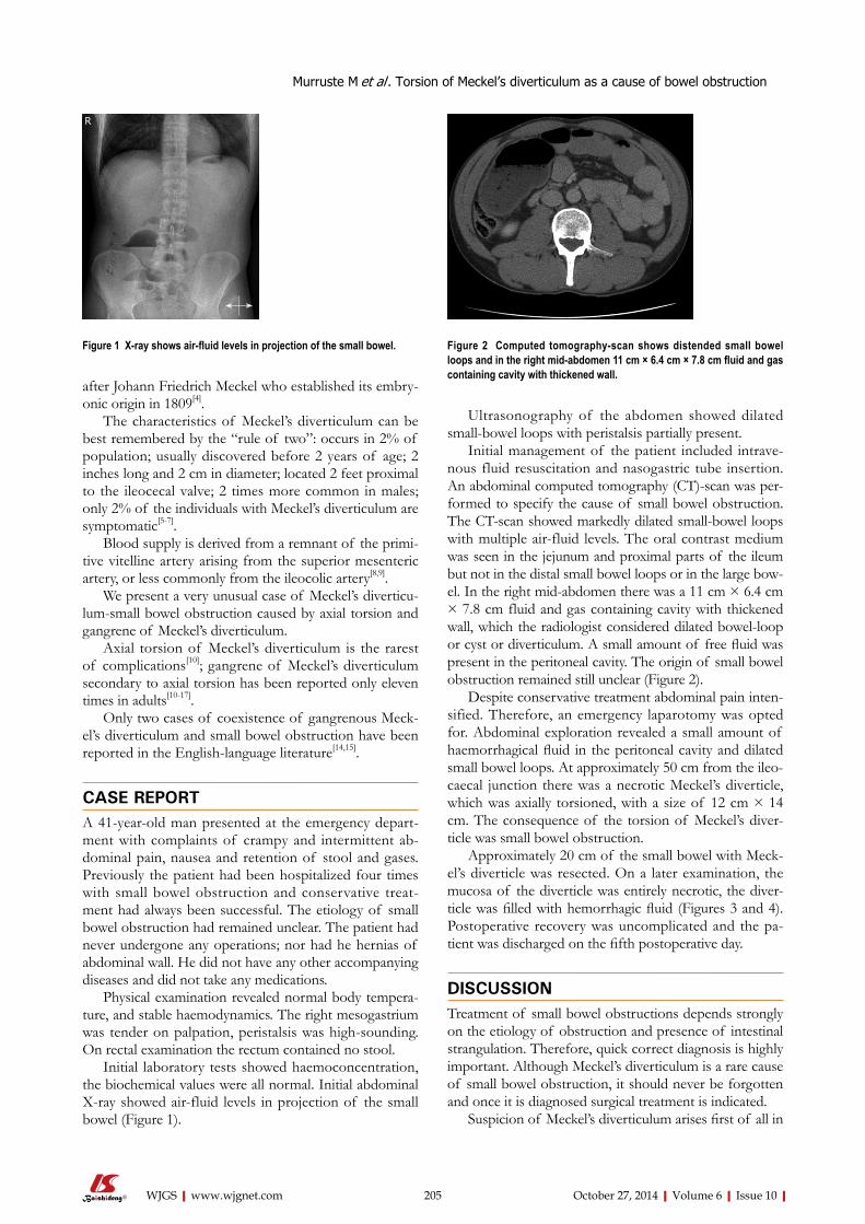
after Johann Friedrich Meckel who established its embry-onic origin in 1809[4].
The characteristics of Meckel’s diverticulum can be best remembered by the “rule of two”: occurs in 2% of population; usually discovered before 2 years of age; 2 inches long and 2 cm in diameter; located 2 feet proximal to the ileocecal valve; 2 times more common in males; only 2% of the individuals with Meckel’s diverticulum are symptomatic[5-7].
Blood supply is derived from a remnant of the primi-tive vitelline artery arising from the superior mesenteric artery, or less commonly from the ileocolic artery[8,9].
We present a very unusual case of Meckel’s diverticu-lum-small bowel obstruction caused by axial torsion and gangrene of Meckel’s diverticulum.
Axial torsion of Meckel’s diverticulum is the rarest of complications[10]; gangrene of Meckel’s diverticulum secondary to axial torsion has been reported only eleven times in adults[10-17].
Only two cases of coexistence of gangrenous Meck-el’s diverticulum and small bowel obstruction have been reported in the English-language literature[14,15].
CASE REPORTA 41-year-old man presented at the emergency depart-ment with complaints of crampy and intermittent ab-dominal pain, nausea and retention of stool and gases. Previously the patient had been hospitalized four times with small bowel obstruction and conservative treat-ment had always been successful. The etiology of small bowel obstruction had remained unclear. The patient had never undergone any operations; nor had he hernias of abdominal wall. He did not have any other accompanying diseases and did not take any medications.
Physical examination revealed normal body tempera-ture, and stable haemodynamics. The right mesogastrium was tender on palpation, peristalsis was high-sounding. On rectal examination the rectum contained no stool.
Initial laboratory tests showed haemoconcentration, the biochemical values were all normal. Initial abdominal X-ray showed air-fluid levels in projection of the small bowel (Figure 1).
Ultrasonography of the abdomen showed dilated small-bowel loops with peristalsis partially present.
Initial management of the patient included intrave-nous fluid resuscitation and nasogastric tube insertion. An abdominal computed tomography (CT)-scan was per-formed to specify the cause of small bowel obstruction. The CT-scan showed markedly dilated small-bowel loops with multiple air-fluid levels. The oral contrast medium was seen in the jejunum and proximal parts of the ileum but not in the distal small bowel loops or in the large bow-el. In the right mid-abdomen there was a 11 cm × 6.4 cm × 7.8 cm fluid and gas containing cavity with thickened wall, which the radiologist considered dilated bowel-loop or cyst or diverticulum. A small amount of free fluid was present in the peritoneal cavity. The origin of small bowel obstruction remained still unclear (Figure 2).
Despite conservative treatment abdominal pain inten-sified. Therefore, an emergency laparotomy was opted for. Abdominal exploration revealed a small amount of haemorrhagical fluid in the peritoneal cavity and dilated small bowel loops. At approximately 50 cm from the ileo-caecal junction there was a necrotic Meckel’s diverticle, which was axially torsioned, with a size of 12 cm × 14 cm. The consequence of the torsion of Meckel’s diver-ticle was small bowel obstruction.
Approximately 20 cm of the small bowel with Meck-el’s diverticle was resected. On a later examination, the mucosa of the diverticle was entirely necrotic, the diver-ticle was filled with hemorrhagic fluid (Figures 3 and 4). Postoperative recovery was uncomplicated and the pa-tient was discharged on the fifth postoperative day.
DISCUSSIONTreatment of small bowel obstructions depends strongly on the etiology of obstruction and presence of intestinal strangulation. Therefore, quick correct diagnosis is highly important. Although Meckel’s diverticulum is a rare cause of small bowel obstruction, it should never be forgotten and once it is diagnosed surgical treatment is indicated.
Suspicion of Meckel’s diverticulum arises first of all in
Murruste M et al . Torsion of Meckel’s diverticulum as a cause of bowel obstruction
205 October 27, 2014|Volume 6|Issue 10|WJGS|www.wjgnet.com
Figure 1 X-ray shows air-fluid levels in projection of the small bowel. Figure 2 Computed tomography-scan shows distended small bowel loops and in the right mid-abdomen 11 cm × 6.4 cm × 7.8 cm fluid and gas containing cavity with thickened wall.
R
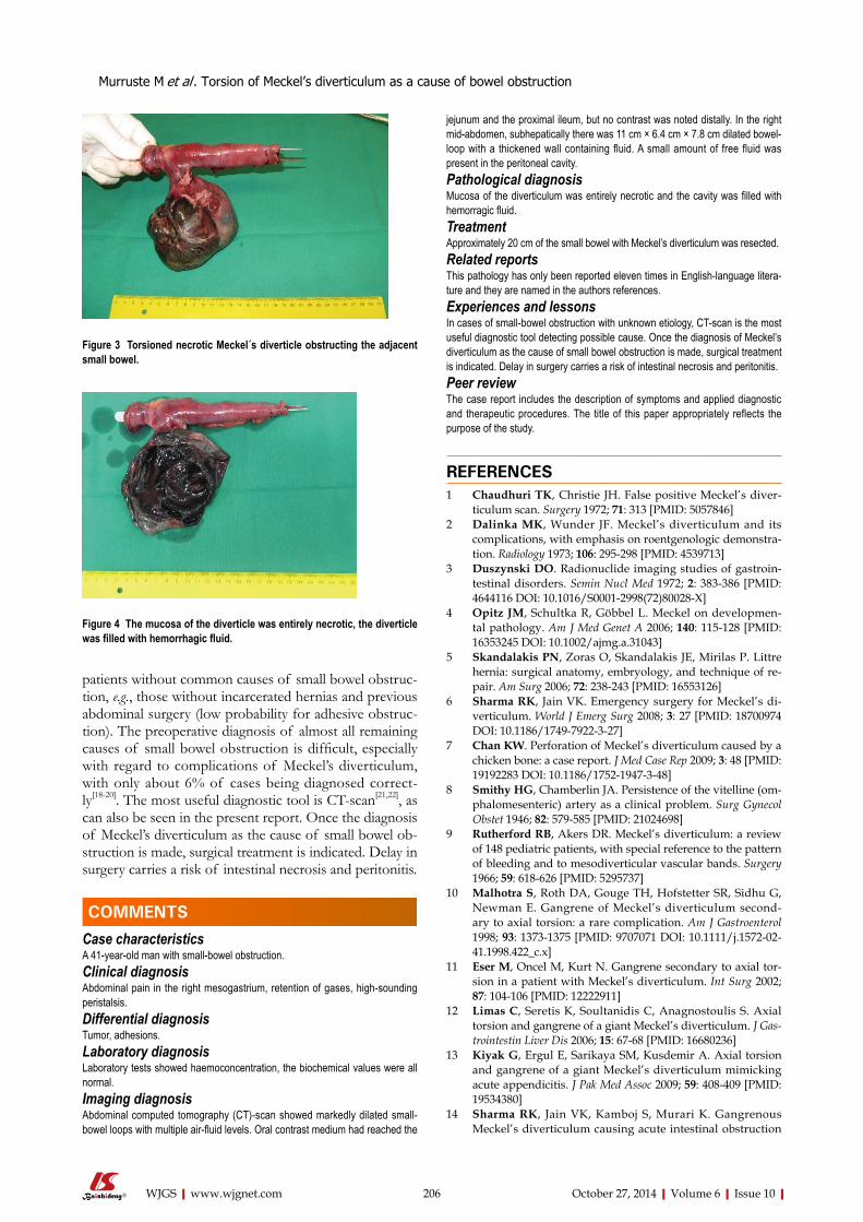
patients without common causes of small bowel obstruc-tion, e.g., those without incarcerated hernias and previous abdominal surgery (low probability for adhesive obstruc-tion). The preoperative diagnosis of almost all remaining causes of small bowel obstruction is difficult, especially with regard to complications of Meckel’s diverticulum, with only about 6% of cases being diagnosed correct-ly[18-20]. The most useful diagnostic tool is CT-scan[21,22], as can also be seen in the present report. Once the diagnosis of Meckel’s diverticulum as the cause of small bowel ob-struction is made, surgical treatment is indicated. Delay in surgery carries a risk of intestinal necrosis and peritonitis.
COMMENTSCase characteristicsA 41-year-old man with small-bowel obstruction.Clinical diagnosisAbdominal pain in the right mesogastrium, retention of gases, high-sounding peristalsis.Differential diagnosis Tumor, adhesions. Laboratory diagnosisLaboratory tests showed haemoconcentration, the biochemical values were all normal.Imaging diagnosisAbdominal computed tomography (CT)-scan showed markedly dilated small-bowel loops with multiple air-fluid levels. Oral contrast medium had reached the
jejunum and the proximal ileum, but no contrast was noted distally. In the right mid-abdomen, subhepatically there was 11 cm × 6.4 cm × 7.8 cm dilated bowel-loop with a thickened wall containing fluid. A small amount of free fluid was present in the peritoneal cavity. Pathological diagnosisMucosa of the diverticulum was entirely necrotic and the cavity was filled with hemorragic fluid. TreatmentApproximately 20 cm of the small bowel with Meckel’s diverticulum was resected.Related reportsThis pathology has only been reported eleven times in English-language litera-ture and they are named in the authors references. Experiences and lessonsIn cases of small-bowel obstruction with unknown etiology, CT-scan is the most useful diagnostic tool detecting possible cause. Once the diagnosis of Meckel’s diverticulum as the cause of small bowel obstruction is made, surgical treatment is indicated. Delay in surgery carries a risk of intestinal necrosis and peritonitis.Peer reviewThe case report includes the description of symptoms and applied diagnostic and therapeutic procedures. The title of this paper appropriately reflects the purpose of the study.
REFERENCES1 Chaudhuri TK, Christie JH. False positive Meckel’s diver-
ticulum scan. Surgery 1972; 71: 313 [PMID: 5057846]2 Dalinka MK, Wunder JF. Meckel’s diverticulum and its
complications, with emphasis on roentgenologic demonstra-tion. Radiology 1973; 106: 295-298 [PMID: 4539713]
3 Duszynski DO. Radionuclide imaging studies of gastroin-testinal disorders. Semin Nucl Med 1972; 2: 383-386 [PMID: 4644116 DOI: 10.1016/S0001-2998(72)80028-X]
4 Opitz JM, Schultka R, Göbbel L. Meckel on developmen-tal pathology. Am J Med Genet A 2006; 140: 115-128 [PMID: 16353245 DOI: 10.1002/ajmg.a.31043]
5 Skandalakis PN, Zoras O, Skandalakis JE, Mirilas P. Littre hernia: surgical anatomy, embryology, and technique of re-pair. Am Surg 2006; 72: 238-243 [PMID: 16553126]
6 Sharma RK, Jain VK. Emergency surgery for Meckel’s di-verticulum. World J Emerg Surg 2008; 3: 27 [PMID: 18700974 DOI: 10.1186/1749-7922-3-27]
7 Chan KW. Perforation of Meckel’s diverticulum caused by a chicken bone: a case report. J Med Case Rep 2009; 3: 48 [PMID: 19192283 DOI: 10.1186/1752-1947-3-48]
8 Smithy HG, Chamberlin JA. Persistence of the vitelline (om-phalomesenteric) artery as a clinical problem. Surg Gynecol Obstet 1946; 82: 579-585 [PMID: 21024698]
9 Rutherford RB, Akers DR. Meckel’s diverticulum: a review of 148 pediatric patients, with special reference to the pattern of bleeding and to mesodiverticular vascular bands. Surgery 1966; 59: 618-626 [PMID: 5295737]
10 Malhotra S, Roth DA, Gouge TH, Hofstetter SR, Sidhu G, Newman E. Gangrene of Meckel’s diverticulum second-ary to axial torsion: a rare complication. Am J Gastroenterol 1998; 93: 1373-1375 [PMID: 9707071 DOI: 10.1111/j.1572-02-41.1998.422_c.x]
11 Eser M, Oncel M, Kurt N. Gangrene secondary to axial tor-sion in a patient with Meckel’s diverticulum. Int Surg 2002; 87: 104-106 [PMID: 12222911]
12 Limas C, Seretis K, Soultanidis C, Anagnostoulis S. Axial torsion and gangrene of a giant Meckel’s diverticulum. J Gas-trointestin Liver Dis 2006; 15: 67-68 [PMID: 16680236]
13 Kiyak G, Ergul E, Sarikaya SM, Kusdemir A. Axial torsion and gangrene of a giant Meckel’s diverticulum mimicking acute appendicitis. J Pak Med Assoc 2009; 59: 408-409 [PMID: 19534380]
14 Sharma RK, Jain VK, Kamboj S, Murari K. Gangrenous Meckel’s diverticulum causing acute intestinal obstruction
206 October 27, 2014|Volume 6|Issue 10|WJGS|www.wjgnet.com
Figure 3 Torsioned necrotic Meckel´s diverticle obstructing the adjacent small bowel.
Figure 4 The mucosa of the diverticle was entirely necrotic, the diverticle was filled with hemorrhagic fluid.
COMMENTS
Murruste M et al . Torsion of Meckel’s diverticulum as a cause of bowel obstruction
2004; 28: 917-920 [PMID: 15593467 DOI: 10.1007/s00268-004-7512-3]
19 Lüdtke FE, Mende V, Köhler H, Lepsien G. Incidence and frequency or complications and management of Meckel’s diverticulum. Surg Gynecol Obstet 1989; 169: 537-542 [PMID: 2814770]
20 Rossi P, Gourtsoyiannis N, Bezzi M, Raptopoulos V, Massa R, Capanna G, Pedicini V, Coe M. Meckel’s diverticulum: imaging diagnosis. AJR Am J Roentgenol 1996; 166: 567-573 [PMID: 8623629 DOI: 10.2214/ajr.166.3.8623629]
21 Prall RT, Bannon MP, Bharucha AE. Meckel’s diverticulum causing intestinal obstruction. Am J Gastroenterol 2001; 96: 3426-3427 [PMID: 11774961 DOI: 10.1111/j.1572-0241.2001.05344.x]
22 Bennett GL, Birnbaum BA, Balthazar EJ. CT of Meckel’s diverticulitis in 11 patients. AJR Am J Roentgenol 2004; 182: 625-629 [PMID: 14975960 DOI: 10.2214/ajr.182.3.1820625]
P- Reviewer: Gurkan A, Katsoulis IE, Lorenzo-Zuniga V, Maric I S- Editor: Ji FF L- Editor: A E- Editor: Liu SQ
in an adult. ANZ J Surg 2008; 78: 1046-1047 [PMID: 18959721 DOI: 10.1111/j.1445-2197.2008.04739.x]
15 Cartanese C, Petitti T, Marinelli E, Pignatelli A, Martignetti D, Zuccarino M, Ferrozzi L. Intestinal obstruction caused by torsed gangrenous Meckel’s diverticulum encircling termi-nal ileum. World J Gastrointest Surg 2011; 3: 106-109 [PMID: 21860699 DOI: 10.4240/wjgs.v3.i7.106]
16 Ruiz LVA, Camacho OLA, Diaz TDA. Giant Meckel’s diver-ticula with necrosis due to axial torsion. Rev Col Gastroenterol 2010; 25: 398-400
17 Alvite Canosa M, Couselo Villanueva JM, Iglesias Porto E, González López R, Montoto Santomé P, Arija Val F. [Intesti-nal obstruction due to axial torsion and gangrene of a giant Meckel diverticulum]. Gastroenterol Hepatol 2012; 35: 452-453 [PMID: 22425353 DOI: 10.1016/j.gastrohep.2012.01.007]
18 Bani-Hani KE, Shatnawi NJ. Meckel’s diverticulum: com-parison of incidental and symptomatic cases. World J Surg
207 October 27, 2014|Volume 6|Issue 10|WJGS|www.wjgnet.com
Murruste M et al . Torsion of Meckel’s diverticulum as a cause of bowel obstruction

Published by Baishideng Publishing Group Inc
World Journal of Gastrointestinal SurgeryWorld J Gastrointest Surg 2014 November 27; 6(11): 208-234
ISSN 1948-9366 (online)
208 Diagnosisofinflammatoryboweldisease:Potentialroleofmolecular
biometrics
M’Koma AE
220 Medicalmanagementofpatientsafterbariatricsurgery:Principlesand
guidelines
Abd Elrazek MAA, Elbanna AEM, Bilasy SE
229 Factorsinfluencingthediagnosticaccuracyandmanagementinacute
surgicalpatients
Sajid MS, Hollingsworth T, McGlue M, Miles WFA
Contents Monthly Volume 6 Number 11 November 27, 2014
WJGS|www.wjgnet.com I November 27, 2014|Volume 6|Issue 11|
REVIEW
MINIREVIEWS
OBSERVATIONAL STUDY

Contents
APPENDIX
ABOUT COVER
World Journal of Gastrointestinal SurgeryVolume 6 Number 11 November 27, 2014
I-V Instructionstoauthors
EditorialBoardMemberofWorldJournalofGastrointestinalSurgery ,Marc-HDahlke,MD,PhD,DepartmentofSurgery,UniversityofRegensburgMedicalCenter,FranzJosefStraussAllee12,Regensburg93042,Germany
World Journal of Gastrointestinal Surgery (World J Gastrointest Surg, WJGS, online ISSN 1948-9366, DOI: 10.4240) is a peer-reviewed open access academic journal that aims to guide clinical practice and improve diagnostic and therapeutic skills of clinicians.
WJGS covers topics concerning micro-invasive surgery; laparoscopy; hepatic, biliary, pancreatic and splenic surgery; surgical nutrition; portal hypertension, as well as associated subjects. The current columns of WJGS include editorial, frontier, diagnostic advances, therapeutics advances, field of vision, mini-reviews, review, topic highlight, medical ethics, original articles, case report, clinical case conference (Clinicopathological conference), and autobiography. Priority publication will be given to articles concerning diagnosis and treatment of gastrointestinal surgery diseases. The following aspects are covered: Clinical diagnosis, laboratory diagnosis, differential diagnosis, imaging tests, pathological diagnosis, molecular biological diagnosis, immunological diagnosis, genetic diagnosis, functional diagnostics, and physical diagnosis; and comprehensive therapy, drug therapy, surgical therapy, interventional treatment, minimally invasive therapy, and robot-assisted therapy.
We encourage authors to submit their manuscripts to WJGS. We will give priority to manuscripts that are supported by major national and international foundations and those that are of great basic and clinical significance.
World Journal of Gastrointestinal Surgery is now indexed in PubMed Central, PubMed, Digital Object Identifier, and Directory of Open Access Journals.
I-III EditorialBoard
Xiu-Xia Song, Vice DirectorWorld Journal of Gastrointestinal SurgeryRoom 903, Building D, Ocean International Center, No. 62 Dongsihuan Zhonglu, Chaoyang District, Beijing 100025, ChinaTelephone: +86-10-85381891Fax: +86-10-85381893E-mail: [email protected] Desk: http://www.wjgnet.com/esps/helpdesk.aspxhttp://www.wjgnet.com
PUBLISHERBaishideng Publishing Group Inc8226 Regency Drive, Pleasanton, CA 94588, USATelephone: +1-925-223-8242Fax: +1-925-223-8243E-mail: [email protected] Desk: http://www.wjgnet.com/esps/helpdesk.aspxhttp://www.wjgnet.com
PUBLICATIONDATENovember 27, 2014
COPYRIGHT© 2014 Baishideng Publishing Group Inc. Articles pub-lished by this Open-Access journal are distributed under the terms of the Creative Commons Attribution Non-commercial License, which permits use, distribution, and reproduction in any medium, provided the original work is properly cited, the use is non commercial and is otherwise in compliance with the license.
SPECIALSTATEMENTAll articles published in journals owned by the Baishideng Publishing Group (BPG) represent the views and opin-ions of their authors, and not the views, opinions or policies of the BPG, except where otherwise explicitly indicated.
INSTRUCTIONSTOAUTHORSFull instructions are available online at http://www.wjgnet.com/1948-9366/g_info_20100305152206.htm
ONLINESUBMISSIONhttp://www.wjgnet.com/esps/
NAMEOFJOURNALWorld Journal of Gastrointestinal Surgery
ISSNISSN 1948-9366 (online)
LAUNCHDATENovember 30, 2009
FREQUENCYMonthly
EDITOR-IN-CHIEFTimothy M Pawlik, MD, MPH, FACS, Associate Professor of Surgery and Oncology, Hepatobiliary Surgery Program Director, Director, Johns Hopkins Medicine Liver Tumor Center Multi-Disciplinary Clinic, Co-Director of Center for Surgical Trials and Outcomes Research, Johns Hopkins Hospital, 600 N. Wolfe Street, Harvey 611, Baltimore, MD 21287, United States
EDITORIALOFFICEJin-Lei Wang, Director
EDITORS FOR THIS ISSUE
Responsible Assistant Editor: Xiang Li Responsible Science Editor: Xue-Mei GongResponsible Electronic Editor: Ya-Jing Lu Proofing Editorial Office Director: Xiu-Xia SongProofing Editor-in-Chief: Lian-Sheng Ma
AIM AND SCOPE
FLYLEAF
INDEXING/ABSTRACTING
WJGS|www.wjgnet.com II November 27, 2014|Volume 6|Issue 11|
Diagnosis of inflammatory bowel disease: Potential role of molecular biometrics
Amosy E M’Koma
Amosy E M’Koma, Department of Biochemistry and Cancer Biology, Meharry Medical College School of Medicine, Nash-ville, TN 37208-3599, United StatesAmosy E M’Koma, Department of Surgery, Vanderbilt Univer-sity School of Medicine, Nashville, TN 37232, United StatesAuthor contributions: M’Koma AE contributed not only to conception and design but also participated in the acquisition of data, analysis and interpretation of data and drafting the manu-script.Supported by NIH, No. R21DK095168, U54MD007593 and UL1TR000445Correspondence to: Amosy E M’Koma, MD, MS, PhD, As-sistant Professor of Surgery, Biochemistry and Cancer Biol-ogy, Department of Biochemistry and Cancer Biology, Meharry Medical College School of Medicine, 1005 Dr. D. B. Todd Jr. Blvd., Nashville, TN 37208-3599, United States. [email protected]: +1-615-3276796 Fax: +1-615-3276440Received: February 27, 2014 Revised: October 16, 2014Accepted: October 23, 2014Published online: November 27, 2014
AbstractAccurate diagnosis of predominantly colonic inflam-matory bowel disease (IBD) is not possible in 30% of patients. For decades, scientists have worked to find a solution to improve diagnostic accuracy for IBD, en-compassing Crohn’s colitis and ulcerative colitis. Evalu-ating protein patterns in surgical pathology colectomy specimens of colonic mucosal and submucosal compart-ments, individually, has potential for diagnostic medicine by identifying integrally independent, phenotype-specific cellular and molecular characteristics. Mass spectrom-etry (MS) and imaging (I) MS are analytical technolo-gies that directly measure molecular species in clinical specimens, contributing to the in-depth understanding of biological molecules. The biometric-system complex-ity and functional diversity is well suited to proteomic and diagnostic studies. The direct analysis of cells and tissues by Matrix-Assisted-Laser Desorption/Ionization
(MALDI) MS/IMS has relevant medical diagnostic po-tential. MALDI-MS/IMS detection generates molecular signatures obtained from specific cell types within tissue sections. Herein discussed is a perspective on the use of MALDI-MS/IMS and bioinformatics technologies for detection of molecular-biometric patterns and identifica-tion of differentiating proteins. I also discuss a perspec-tive on the global challenge of transferring technologies to clinical laboratories dealing with IBD issues. The significance of serologic-immunometric advances is also discussed.
© 2014 Baishideng Publishing Group Inc. All rights reserved.
Key words: Inflammatory bowel disease; Diagnosis; Advances and challenges; MALDI-MS/IMS; Molecular biometrics; Immunometrics
Core tip: Pouch surgery (the restorative proctocolec-tomy and ileal pouch-anal anastomosis for the cura-tive surgical treatment of ulcerative colitis and familial adenomatous polyposis) replaces the colon and rectum after proctocolectomy with a pouch constructed from the distal small bowel (ileum) and sutured to the anal canal above the dentate/pectinate line preserving the anal sphincters. The operation restores gut continuity, defecation, deferral, and discrimination, if the diagnosis is correct, which is unpredictable in 30% of the colonic-inflammatory bowel disease-patients. Mass spectrome-try and imaging mass spectrometry are groundbreaking, non-invasive analytical technologies with the ability to directly measure individual molecular species in complex clinical specimens. These technologies provide quanti-tative and qualitative analysis of cellular systems, and allow differentiation between disease and normal mol-ecules from the same organ. These characteristics offer diagnostic and prognostic value for clinical medicine.
M’Koma AE. Diagnosis of inflammatory bowel disease: Po-tential role of molecular biometrics. World J Gastrointest Surg
REVIEW
Submit a Manuscript: http://www.wjgnet.com/esps/Help Desk: http://www.wjgnet.com/esps/helpdesk.aspxDOI: 10.4240/wjgs.v6.i11.208
World J Gastrointest Surg 2014 November 27; 6(11): 208-219ISSN 1948-9366 (online)
© 2014 Baishideng Publishing Group Inc. All rights reserved.
208 November 27, 2014|Volume 6|Issue 11|WJGS|www.wjgnet.com

2014; 6(11): 208-219 Available from: URL: http://www.wjg-net.com/1948-9366/full/v6/i11/208.htm DOI: http://dx.doi.org/10.4240/wjgs.v6.i11.208
INTRODUCTIONInflammatory bowel disease Colonic inflammatory bowel disease (IBD) comprises Crohn’s colitis (CC) and ulcerative colitis (UC), a group of diseases of the gastrointestinal (GI) tract character-ized by chronic relapsing and remitting inflammation[1,2]. IBD affects as many as 1.6 million persons in the United States and 2.2 million in Europe. The incidence is in-creasing worldwide[1-5]. In spite of advances in IBD-ther-apy, IBD hospitalizations and surgery rates in the United States have increased significantly since 1990[6]. IBD is one of the five most prevalent GI disease burdens in the United States, with annual overall health care costs of more than $1.7 billion[7,8]. One to two of every 1000 people in developed countries are affected with IBD[9], and global rates seem to be increasing[1,10-12], attributable to the rapid modernization and Westernization of the population[1]. These chronic diseases result in significant morbidity and mortality, compromising quality of life and life expectancies. While there is no drug for cure for these diseases, the last three decades have seen major ad-vances in the molecular understanding intestinal immune responses and how they relate to IBD. This, in turn, has led to the development and refinement of several new treatments. Most significant has been the development of restorative proctocolectomy (RPC) with ileal pouch-anal anastomosis (IPAA). The pelvic pouch surgery allows for the removal of the entire colon while main-taining transanal fecal continence without a permanent diverting loop ileostomy. The success of RPC (judged by the entire removal of a diseased colon while preserving gastrointestinal continuity, bowel evacuation, continence and fertility) restores physiological function and greatly improves patient health quality of life. Successful RPC also frees the healthcare system from the immense bur-den of current lifelong, non-curative treatments. These outcomes are dependent on a correct diagnosis and me-ticulous surgical techniques available at well-established IBD centers[13-15].
The etiology of IBD poorly understood. The general consensus holds that IBD is an automatic dysfunction triangle of antigen and antibody reaction against mucosal response to commensal bacteria. The fundamental ques-tion is why the immune system responds aggressively to harmless, ever-present bacteria, releasing complex mixes of cytokines, chemokines and other substances that cause inflammation. One possible explanation is that the gut immune system is compromised because of defects in the barrier function of the gut luminal epithelium[16]. Al-though the etiology of IBD is at present not delineated, histopathologic and clinical assessments demonstrate that CD and UC, the two major classifications of IBD,
are indeed distinct entities and have different causes and discrete mechanisms of tissue damage and treatment[16-21]. UC results in inflammation and ulcerations in the muco-sal and to a lesser degree submucosal linings of the colon and rectum. CD differs in that it may result in inflamma-tion deeper within the intestinal wall (transmullary) and can occur in any parts of the digestive system (includ-ing the mouth, esophagus, stomach, duodenum, small intestine, colon and rectum). Further, Crohn’s may also involve other organs outside the GI system through fistu-lization[22,23]. Crohn’s is diagnosed in at least four patients per 100000 in the United States, and the incidence and prevalence is rising worldwide[1,10-12].
Diagnosis challenges in IBDThe current standard of care for IBD treatment is based on steroids and immunosuppressant agents, including glu-cocorticoids, aminosalicylates, cyclosporine, methotrexate and biologic agents such as anti-TNF-α and IL1-β. The correct IBD diagnosis is crucial for providing correct, evidence-based treatment, since treatment response and complications differ significantly among UC and CC pa-tients[24]. The absence of specific phenotypes indicating the particular disease condition challenges pathologist interpretation and categorization of tissue morphology, subsequently leading to difficulties in diagnosis and con-sistent standard of care[25]. However, despite advances in our understanding of the genetic[16,26], immunologic[26,27], and environmental[1,24,28] influences that may trigger com-plex IBD pathologies, to date there is no single indicator sensitive enough to accurately and consistently delineate CC and UC. The available data indicate that genetic fac-tors determine an individual’s susceptibility to developing IBD, and environmental factors elicit cellular responses that drive disease progression. Histological evaluation and interpretation of tissue provides insights that directly impact care[25]. Pathologists rely mainly on microscopic visual inspection and interpretation of stained and/or dyed tissue sections to identify the disease state of a pa-tient sample[29,30]. Inherently, these procedures possess a significant degree of subjectivity[31] and are fraught with problems[31,32]. Rigorous training in pathology subspecial-ties has attempted to improve the standard of care and avoid unnecessary mistakes[33]. Despite these extremely thorough standards, inevitable situations arise in which objectivity cannot be guaranteed and where significant disagreement occurs between specialists[34]. This chal-lenge is common for IBD patient populations[13,15,35,36] To date, there is no single, absolute diagnostic test[37,38]. A diagnosis should neither be based on nor excluded by any one variable or result[39]. The consensus statement on the diagnosis, management and surveillance of both CC[40]
and UC[41] recommend that “multiple” tissue biopsies from at list five sites around the colon and rectum should be collected for support of a reliable diagnosis. Of these six sites a minimum of two samples from each should be sampled[40,41]. Although the procedure is reliable, it is invasive and uncomfortable to the patients.
M’Koma AE. Advances and challenges in IBD diagnosis
209 November 27, 2014|Volume 6|Issue 11|WJGS|www.wjgnet.com

coming a global health concern. Distinguishing between CC and UC is critical to therapy. The clinical experience suggests that identifying patients with CC and positive outcomes after pouch surgery is arduous. Thus, RPC should be contraindicated for CC patients, whereas IPAA is standard acceptable care for patients with UC and IC who are predicted likely to develop UC. Inevitably, pouch complications are significantly higher in patients with CC (± 64%) and IC (± 43%) vs patients having UC (± 22%) (P < 0.05)[46,47,49]. This diagnostic dilemma and the potential morbidity from a wrong diagnosis and unnecessary and/or inappropriate surgical interventions underscore the importance of research strategy focused at improving di-agnosis of the colitides using molecular biometrics[42,50-52].
Clinico-histopathologic findings in Crohn’s colitisCrohn’s colitis is recognized to encompass a heteroge-neous group of disorders[38]. Usually CC is segmental with deep inflammation where the disease activity is transmural, with lymphoid composite extending to the sub-serosa. The Montreal classification[53] and the Paris pediatric modification[54] have brought consistency to definitions of subtypes of CC and of colitides. It is noteworthy that both the Montreal and Paris classifica-tions rely on the location of gross disease, i.e., visible lesions with more than a few aphthous ulcers. Patterns of macroscopic involvement, rather than microscopic, have been useful traditionally in predicting clinical course, as exemplified by the tendency of small bowel disease, particularly, to stricture over time. Despite the fact that microscopic involvement does not define subtypes of CC, the role of histology in the diagnosis of CC does differ according to the anatomic location of macroscopic disease[38].
Histologic features useful for the diagnosis of CC have been reviewed by Griffiths[38], (Table 1) but, according to Van Assche et al[40] presented at The second European evidence-based Consensus on the diagnosis and manage-ment of Crohn’s colitis, there are no data available as to how many of these features must be present to allow a firm diagnosis[40]. Focal (discontinuous) chronic (lympho-cytes and plasma cells) inflammation and patchy chronic inflammation, focal crypt irregularity (discontinuous crypt distortion) and granulomas (not related to crypt injury) are the generally accepted microscopic features which allow a diagnosis of CC[40]. Within one histologic section, inflam-mation may be immediately adjacent to an uninflamed microscopic “skip area”. Mucosal changes may resemble ulcerative or infectious colitis with infiltration of the crypts by polymorphonuclear leukocytes (cryptitis or crypt abscesses), and distortion of crypt architecture. Granu-lomas (collections of monocytes/macrophages) in the lamina propria (not associated with crypt injury) are a cor-roborating feature of suspected Crohn’s after exclusion of identifiable infectious etiology, but reported prevalence in mucosal biopsies at time of first diagnosis varies. The like-lihood of finding granuloma is a function of the number of specimens taken, the number of sections examined,
Inaccurate diagnosis in IBD and consequencesWhen IBD predominantly involves the colon, differen-tiation between CC from UC is often challenging. Inac-curate diagnoses are estimated to occur in 30% of IBD patients[42,43]. In most cases the diagnostic uncertainty arises from the overlap of clinical and histologic features, making CC appear like UC[44]. This scenario is particularly relevant to young children, a population in which IBD consists of up to 80%. The differentiation between UC and CC relies on a compilation of clinical, radiologic, en-doscopic, and histopathologic interpretations[40]; a com-pilation that is not always accurate. An estimated 15% of IBD patients are indistinguishable and are labeled as ‘‘in-determinate colitis’’ (IC)[45-47]. In addition, another 15% of the colonic IBD cases that undergo pouch surgery resulting from a definitive UC diagnosis (based on the pa-thologist’s initial designation of endoscopic biopsies and colectomy specimen) will have their original UC diagno-sis changed to CC based on the postoperative follow-up when clinical and histopathology changes indicate devel-opment of CC in the ileal pouch[15,35,36,48,49]. One-half of these patients will require pouch excision or diversion[49].
Because of the unpredictable nature of IBD, side ef-fects of medications, and potential complications, some of which may end in sudden incapacitation, IBD is be-
210 November 27, 2014|Volume 6|Issue 11|WJGS|www.wjgnet.com
M’Koma AE. Advances and challenges in IBD diagnosis
Table 1 Microscopic features used for the diagnosis of Crohn’s colitis
ColonArchitecture Crypt architectural irregularity Focal
Diffuse Reduces crypt numbers/mucosal atrophy Irregular surface
Chronic inflammation Distribution I Focal increased in intensity
Patchy increase Distribution II Diffuse increase
Superficial Granulomas Transmucosal Mucin granulomas Basal plasma cellsPolymorph inflammation Lamina propria Focal Crypt epithelial polymorphs Diffuse
Polymorph exudatesEpithelial changes Erosion/ulceration Mucin Depletion
Preservation Paneth cells distal to hepatic flexureEpithelial-associated changes Increased intraepithelial lymphocytes > 15Terminal ileum/Ileocecal /Cecum Architecture Villus irregularity
Crypt architecture Epithelial changes Irregularity
Pseudopyloric glandMetaplasia
Reproduced by permission of the publisher from ref. [38].
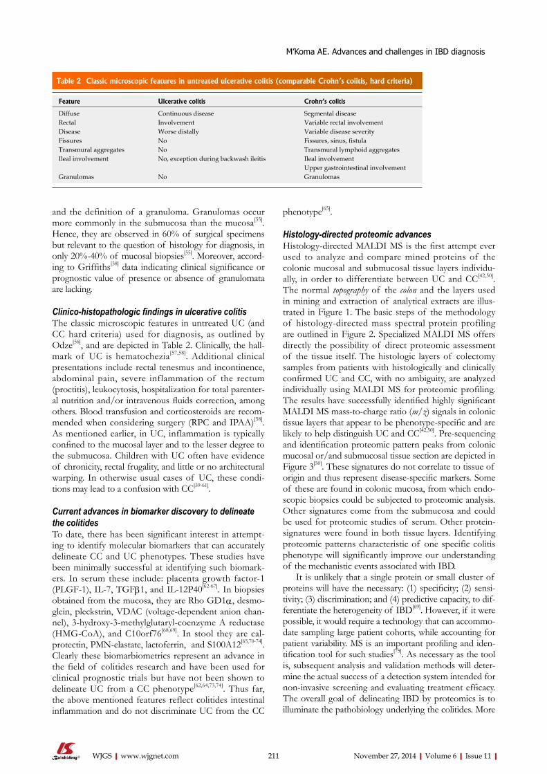
and the definition of a granuloma. Granulomas occur more commonly in the submucosa than the mucosa[55]. Hence, they are observed in 60% of surgical specimens but relevant to the question of histology for diagnosis, in only 20%-40% of mucosal biopsies[55]. Moreover, accord-ing to Griffiths[38] data indicating clinical significance or prognostic value of presence or absence of granulomata are lacking.
Clinico-histopathologic findings in ulcerative colitisThe classic microscopic features in untreated UC (and CC hard criteria) used for diagnosis, as outlined by Odze[56], and are depicted in Table 2. Clinically, the hall-mark of UC is hematochezia[57,58]. Additional clinical presentations include rectal tenesmus and incontinence, abdominal pain, severe inflammation of the rectum (proctitis), leukocytosis, hospitalization for total parenter-al nutrition and/or intravenous fluids correction, among others. Blood transfusion and corticosteroids are recom-mended when considering surgery (RPC and IPAA)[58]. As mentioned earlier, in UC, inflammation is typically confined to the mucosal layer and to the lesser degree to the submucosa. Children with UC often have evidence of chronicity, rectal frugality, and little or no architectural warping. In otherwise usual cases of UC, these condi-tions may lead to a confusion with CC[59-61].
Current advances in biomarker discovery to delineate the colitidesTo date, there has been significant interest in attempt-ing to identify molecular biomarkers that can accurately delineate CC and UC phenotypes. These studies have been minimally successful at identifying such biomark-ers. In serum these include: placenta growth factor-1 (PLGF-1), IL-7, TGFβ1, and IL-12P40[62-67]. In biopsies obtained from the mucosa, they are Rho GD1α, desmo-glein, pleckstrin, VDAC (voltage-dependent anion chan-nel), 3-hydroxy-3-methylglutaryl-coenzyme A reductase (HMG-CoA), and C10orf76[68,69]. In stool they are cal-protectin, PMN-elastate, lactoferrin, and S100A12[65,70-74]. Clearly these biomarbiometrics represent an advance in the field of colitides research and have been used for clinical prognostic trials but have not been shown to delineate UC from a CC phenotype[62,64,73,74]. Thus far, the above mentioned features reflect colitides intestinal inflammation and do not discriminate UC from the CC
phenotype[65].
Histology-directed proteomic advancesHistology-directed MALDI MS is the first attempt ever used to analyze and compare mined proteins of the colonic mucosal and submucosal tissue layers individu-ally, in order to differentiate between UC and CC[42,50]. The normal topography of the colon and the layers used in mining and extraction of analytical extracts are illus-trated in Figure 1. The basic steps of the methodology of histology-directed mass spectral protein profiling are outlined in Figure 2. Specialized MALDI MS offers directly the possibility of direct proteomic assessment of the tissue itself. The histologic layers of colectomy samples from patients with histologically and clinically confirmed UC and CC, with no ambiguity, are analyzed individually using MALDI MS for proteomic profiling. The results have successfully identified highly significant MALDI MS mass-to-charge ratio (m/z) signals in colonic tissue layers that appear to be phenotype-specific and are likely to help distinguish UC and CC[42,50]. Pre-sequencing and identification proteomic pattern peaks from colonic mucosal or/and submucosal tissue section are depicted in Figure 3[50]. These signatures do not correlate to tissue of origin and thus represent disease-specific markers. Some of these are found in colonic mucosa, from which endo-scopic biopsies could be subjected to proteomic analysis. Other signatures come from the submucosa and could be used for proteomic studies of serum. Other protein-signatures were found in both tissue layers. Identifying proteomic patterns characteristic of one specific colitis phenotype will significantly improve our understanding of the mechanistic events associated with IBD.
It is unlikely that a single protein or small cluster of proteins will have the necessary: (1) specificity; (2) sensi-tivity; (3) discrimination; and (4) predictive capacity, to dif-ferentiate the heterogeneity of IBD[69]. However, if it were possible, it would require a technology that can accommo-date sampling large patient cohorts, while accounting for patient variability. MS is an important profiling and iden-tification tool for such studies[75]. As necessary as the tool is, subsequent analysis and validation methods will deter-mine the actual success of a detection system intended for non-invasive screening and evaluating treatment efficacy. The overall goal of delineating IBD by proteomics is to illuminate the pathobiology underlying the colitides. More
211 November 27, 2014|Volume 6|Issue 11|WJGS|www.wjgnet.com
M’Koma AE. Advances and challenges in IBD diagnosis
Table 2 Classic microscopic features in untreated ulcerative colitis (comparable Crohn’s colitis, hard criteria)
Feature Ulcerative colitis Crohn’s colitis
Diffuse Continuous disease Segmental diseaseRectal Involvement Variable rectal involvementDisease Worse distally Variable disease severityFissures No Fissures, sinus, fistulaTransmural aggregates No Transmural lymphoid aggregatesIleal involvement No, exception during backwash ileitis Ileal involvement
Upper gastrointestinal involvementGranulomas No Granulomas
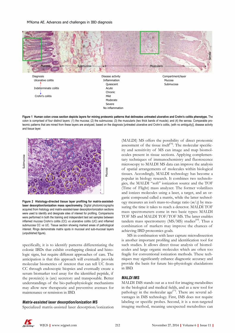
specifically, it is to identify patterns differentiating the colonic IBDs that exhibit overlapping clinical and histo-logic signs, but require different approaches of care. The anticipation is that this approach will eventually provide molecular biometrics of interest that can tell UC from CC through endoscopic biopsies and eventually create a serum biomarker tool assay for the identified peptide, if the protein(s) is (are) secretory and transposable. Better understandings of the bio-pathophysiologic mechanisms may allow new therapeutic and preventive avenues for maintenance or remission in IBD.
Matrix-assisted laser desorption/ionization MSSpecialized matrix-assisted laser desorption/ionization
(MALDI) MS offers the possibility of direct proteomic assessment of the tissue itself[76]. The molecular specific-ity and sensitivity of MS can image and map biomol-ecules present in tissue sections. Applying complemen-tary techniques of immunochemistry and fluorescence microscopy to MALDI MS data can improve the analysis of spatial arrangements of molecules within biological tissues. Accordingly, MALDI technology has become a popular in biology research. It combines two technolo-gies, the MALDI “soft” ionization source and the TOF (Time of Flight) mass analyzer. The former volatilizes and ionizes molecules using a laser, a target, and an or-ganic compound called a matrix, while the latter technol-ogy measures an ion’s mass-to-charge ratio (m/z) by mea-suring the time it takes to reach a detector. MALDI TOF mass spectrometers come in two basic types: MALDI TOF MS and MALDI TOF/TOF MS. The latter enables tandem mass spectrometry (MS/MS) studies[69]. Thus a combination of markers may improve the chances of achieving IBD proteomics goals.
MS in combination with laser capture microdissection is another important profiling and identification tool for such studies. It allows direct tissue analysis of biomol-ecules and large organic molecules which are often too fragile for conventional ionization methods. These tech-niques may significantly enhance diagnostic accuracy and provide the basis for future bio-physiologic elucidations in IBD.
MALDI IMS MALDI IMS stands out as a tool for imaging metabolites in the biological and medical fields, and as a new tool for pathology in the molecular age[77]. There are several ad-vantages in IMS technology. First, IMS does not require labeling or specific probes. Second, it is a non-targeted imaging method, meaning unexpected metabolites can
212 November 27, 2014|Volume 6|Issue 11|WJGS|www.wjgnet.com
M’Koma AE. Advances and challenges in IBD diagnosis
Diagnosis Ulcerative colitis
Indeterminate colitis
Crohn’s colitis
Disease activity Inflammation Quiescent Acute Chronic Mild Moderate Severe No inflammation
Compartment/layer Mucosa Submucosa
Figure 1 Human colon cross section depicts layers for mining proteomic patterns that delineates untreated ulcerative and Crohn’s colitis phenotype. The colon is comprised of four distinct layers: (1) the mucosa; (2) the submucosa; (3) the muscularis (two thick bands of muscle); and (4) the serosa. Comparable pro-teomic patterns that are mined from these layers are analyzed, based on the diagnosis [untreated ulcerative and Crohn’s colitis, (with no ambiguity)], disease activity and tissue layer.
Mucosa Submucosa Muscularis
Sero
sa
Subm
ucos
a
Muc
osa
Figure 2 Histology-directed tissue layer profiling for matrix-assisted-laser desorption/ionization mass spectrometry. Digital photomicrographs acquired from histology and matrix-assisted-laser desorption/ionization sections were used to identify and designate sites of interest for profiling. Comparisons were performed in both the training and independent test set samples between inflamed mucosa Crohn’s colitis (CC) vs ulcerative colitis (UC) and inflamed submucosa CC vs UC. Tissue section showing marked areas of pathological interest. Rings demonstrate matrix spots in mucosal and sub-mucosal layers (unpublished figure).
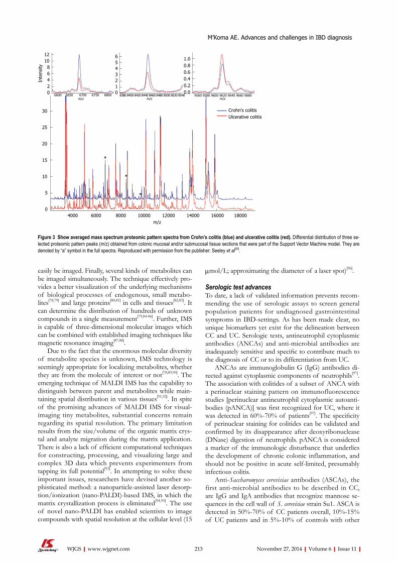
easily be imaged. Finally, several kinds of metabolites can be imaged simultaneously. The technique effectively pro-vides a better visualization of the underlying mechanisms of biological processes of endogenous, small metabo-lites[78,79] and large proteins[80,81] in cells and tissues[82,83]. It can determine the distribution of hundreds of unknown compounds in a single measurement[79,84-86]. Further, IMS is capable of three-dimensional molecular images which can be combined with established imaging techniques like magnetic resonance imaging[87,88].
Due to the fact that the enormous molecular diversity of metabolite species is unknown, IMS technology is seemingly appropriate for localizing metabolites, whether they are from the molecule of interest or not[78,89,90]. The emerging technique of MALDI IMS has the capability to distinguish between parent and metabolites while main-taining spatial distribution in various tissues[91,92]. In spite of the promising advances of MALDI IMS for visual-imaging tiny metabolites, substantial concerns remain regarding its spatial resolution. The primary limitation results from the size/volume of the organic matrix crys-tal and analyte migration during the matrix application. There is also a lack of efficient computational techniques for constructing, processing, and visualizing large and complex 3D data which prevents experimenters from tapping its full potential[93]. In attempting to solve these important issues, researchers have devised another so-phisticated method: a nanoparticle-assisted laser desorp-tion/ionization (nano-PALDI)-based IMS, in which the matrix crystallization process is eliminated[94,95]. The use of novel nano-PALDI has enabled scientists to image compounds with spatial resolution at the cellular level (15
μmol/L; approximating the diameter of a laser spot)[96].
Serologic test advancesTo date, a lack of validated information prevents recom-mending the use of serologic assays to screen general population patients for undiagnosed gastrointestinal symptoms in IBD-settings. As has been made clear, no unique biomarkers yet exist for the delineation between CC and UC. Serologic tests, antineutrophil cytoplasmic antibodies (ANCAs) and anti-microbial antibodies are inadequately sensitive and specific to contribute much to the diagnosis of CC or to its differentiation from UC.
ANCAs are immunoglobulin G (IgG) antibodies di-rected against cytoplasmic components of neutrophils[97]. The association with colitides of a subset of ANCA with a perinuclear staining pattern on immunofluorescence studies [perinuclear antineutrophil cytoplasmic autoanti-bodies (pANCA)] was first recognized for UC, where it was detected in 60%-70% of patients[97]. The specificity of perinuclear staining for colitides can be validated and confirmed by its disappearance after deoxyribonuclease (DNase) digestion of neutrophils. pANCA is considered a marker of the immunologic disturbance that underlies the development of chronic colonic inflammation, and should not be positive in acute self-limited, presumably infectious colitis.
Anti-Saccharomyces cerevisiae antibodies (ASCAs), the first anti-microbial antibodies to be described in CC, are IgG and IgA antibodies that recognize mannose se-quences in the cell wall of S. cerevisiae strain Su1. ASCA is detected in 50%-70% of CC patients overall, 10%-15% of UC patients and in 5%-10% of controls with other
213 November 27, 2014|Volume 6|Issue 11|WJGS|www.wjgnet.com
M’Koma AE. Advances and challenges in IBD diagnosis
Crohn's colitisUlcerative colitis
30
25
20
15
10
5
04000 6000 8000 10000 12000 14000 16000 18000
m/z
Inte
nsity
1210 8 6 4 2 0
6543210
1.00.80.60.40.20.0
6600 6650 6700 6750 6800m/z
8380 8400 8420 8440 8460 8480 8500 8520 8540m/z
9560 9580 9600 9620 9640 9660 9680m/z
Figure 3 Show averaged mass spectrum proteomic pattern spectra from Crohn’s colitis (blue) and ulcerative colitis (red). Differential distribution of three se-lected proteomic pattern peaks (m/z) obtained from colonic mucosal and/or submucosal tissue sections that were part of the Support Vector Machine model. They are denoted by “a” symbol in the full spectra. Reproduced with permission from the publisher: Seeley et al[50].
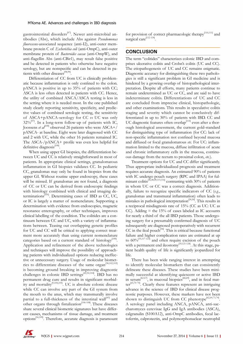
gastrointestinal disorders[97]. Newer anti-microbial an-tibodies (Abs), which include Abs against Pseudomonas fluorescens-associated sequence (anti-I2), anti-outer mem-brane protein C of Escherichia coli (anti-OmpC), anti-outer membrane protein of Bacteroides caccae (anti-OmpW), and anti-flagellin Abs (anti-CBir1), may result false positive and be detected in patients who otherwise have negative serology, but are nonspecific and can be detected in pa-tients with other diseases[98,99].
Differentiation of CC from UC is clinically problem-atic because inflammation is only confined to the colon. pANCA is positive in up to 35% of patients with CC; ASCA is less often detected in patients with CC. Hence, the utility of combined ANCA/ASCA testing is less in the setting where it is needed most. In the one published study clearly reporting sensitivity, specificity, and predic-tive values of combined serologic testing, the sensitivity of ASCA+pANCA-serology for CC vs UC was only 32%[97]. In a long-term follow-up of patients with IC, Joossens et al[100] observed 26 patients who were ASCA+/pANCA- at baseline. Eight were later diagnosed with CC and 2 with UC, while the other 16 patients remained IC. The ASCA-/pANCA+ profile was even less helpful for definitive diagnosis[100].
When using upper GI biopsies, the differentiation be-tween UC and CC is relatively straightforward in most of patients. In appropriate clinical settings, granulomatous inflammation in GI biopsies validates CC. In pediatric CC, granulomas may only be found in biopsies from the upper GI. Without routine upper endoscopy, these cases will be missed. If granulomas are not found, a diagnosis of CC or UC can be derived from endoscopic findings with histology combined with clinical and imaging de-terminations[101]. Determining cases of IBD as CC, UC, or IC is largely a matter of nomenclature. Supporting a determination with evidence from endoscopies, magnetic resonance enterography, or other techniques, improves clinical labelling of the condition. The colitides are a con-tinuum between CC and UC, with a variety of inflamma-tions between. Teasing out overlapping genetic profiles for UC and CC will be critical to applying correct treat-ment more accurately than using current nomenclature categories based on a current standard of histology[100]. Application and refinement of the above technologies and techniques will improve the possibility of approach-ing patients with individualized options reducing ineffec-tive or unnecessary surgery. Usage of molecular biomet-rics to differentiate diseases of the same organ[38,102,103] is becoming ground breaking in improving diagnostic challenges in colonic IBD settings[42,50,104]. IBD has no permanent drug cure and results in significant morbid-ity and mortality[9,104,105]. UC is absolute colonic disease while CC can involve any part of the GI system from the mouth to the anus, which may transmurally involve partial to a full-thickness of the intestinal wall[43] and other organs through fistulization[106-108]. These diseases share several clinical biometric signatures but have differ-ent causes, mechanisms of tissue damage, and treatment options[16,109]. Therefore, accurate diagnosis is paramount
for provision of correct pharmacologic therapy[110,111] and surgical care[112-114].
CONCLUSIONThe term “colitides” characterizes colonic IBD and com-prises ulcerative colitis and Crohn’s colitis (UC and CC). The etiopathogenesis of UC and CC remains enigmatic. Diagnostic accuracy for distinguishing these two patholo-gies is still a significant problem in GI medicine and is hindered by a growing overlap of histopathological inter-pretation. Despite all efforts, many patients continue to remain undetermined as UC or CC, and are said to have indeterminate colitis. Differentiations of UC and CC are concluded from imprecise clinical, histopathologic, and other examinations. This results in speculative colitis staging and severity which cannot be conclusively dif-ferentiated in up to 30% of patients with IBD. CC and UC diagnostic features often overlap[115] even after a thor-ough histological assessment, the current gold-standard for distinguishing type of inflammation (for CC: lack of non-specific inflammation not confined beyond mucosa and diffused or focal granulomatous etc. For UC: inflam-mation limited to the mucosa, diffuse infiltration of acute and chronic inflammatory cells in the mucosa, continu-ous damage from the rectum to proximal colon, etc.).
Treatment options for UC and CC differ significantly. Thus appropriate individualized prognosis and treatment requires accurate diagnosis. An estimated 90% of patients with IC undergo pouch surgery (RPC and IPAA) for ful-minant colitis[36,48,49,115,116] contrasting with 30% of patients in whom UC or CC was a correct diagnosis. Addition-ally, failure to recognize specific indicators of CC (e.g., granulomas and transmural inflammation) often leads to mistakes in pathological interpretation[24,36]. This results in a reciprocal misdiagnosis rate of 15% (CC as UC: UC as CC). Adding = the 15% of cases labeled as IC accounts for nearly a third of the all IBD patients. Those undergo-ing surgery for a presumably confirmed diagnosis of UC subsequently are diagnosed postoperatively with recurrent CC in the ileal pouch[36]. This is critical because functional failure and higher complication rates are estimated at up to 60%[35,117-123] and often require excision of the pouch with a permanent end ileostomy[35,121-124]. At this stage, pa-tient health quality of life is significantly jeopardized for life.
There has been wide ranging interest in attempting to identify molecular biomarkers that can consistently delineate these diseases. These studies have been mini-mally successful at identifying quiescent or active IBD in serum[62-67], in mucosal biopsies[68,69], and in fecal mat-ter[65,70-74]. Clearly these features represent an intriguing advance in the science of IBD for clinical disease prog-nostic purposes. However, these markers have not been shown to distinguish UC from CC phenotype[62,64,73,74]. A serology panel including ANCA, pANCA, anti-sac-charomyces cerevisae IgG and IgA antibodies (ASCA), calgranulin (S100A12), anti-OmpC antibodies, fecal lac-toferrin, calprotectin, and polymorphonuclear neutrophil
214 November 27, 2014|Volume 6|Issue 11|WJGS|www.wjgnet.com
M’Koma AE. Advances and challenges in IBD diagnosis
elastase (PMN-e)[65] is marketed as a promising approach to monitor disease activity and prognosis and may prove to be beneficial in the management of IBD. The specific-ity, sensitivity and diagnostic accuracy of these param-eters with reference to clinical disease indices and/or endoscopically measured inflammation in IBD setting remain unclear. What we have learned to date is that: (1) Although not yet commercially available as tests, patients with CC are more likely than healthy control and/or IBD patients to be positive for a range of biomarkers such as S100A12 (calgranulin), ASCA, OmpC, CBir1, pseudomonas fluorescens protein, and pANCA[125,126]. Significant increases of these proteins are noted during active intestinal inflammation. The greater the number of positive serologies and the higher the titer, the more aggressive the course. These biomarkers are also seen in an active UC[127]; (2) A combination of these biomarkers and a disease-specific activity index could promote the diagnostic accuracy in clinical medicine with reference to endoscopic inflammation but at present none are supe-rior in the ability to reflect endoscopic inflammation[70]; (3) These molecular biometrics significantly assist in pre-dicting relapses in patients with confirmed IBD (active or quiescent)[2-5,17,21,128] but are not discriminatory between UC/CC; (4) Patients who are pANCA+ and ASCA- are more likely to have UC than CC, while in pANCA- and ASCA+ patients the reverse may be true[67]. However, these biomarkers have not demonstrated clinical utility as predictors or monitoring tools of IBD activity[67].
At the present time there is insufficient biometric information to recommend use of serologic assays in screening for IBD in patients from the general popula-tion who have undiagnosed gastrointestinal symptoms. Further, no efficacy for the delineation of CC and UC clearly exist.
ACKNOWLEDGMENTSThe author is thankful to Jared Elzey, CRA, from the Me-harry Research Concierge Services (supported by NIH grants U54MD007593 and UL1TR000445) for com-ments, suggestions and for language editing.
REFERENCES1 M’Koma AE. Inflammatory bowel disease: an expanding
global health problem. Clin Med Insights Gastroenterol 2013; 6: 33-47 [PMID: 24833941]
2 Farrokhyar F, Swarbrick ET, Irvine EJ. A critical review of epidemiological studies in inflammatory bowel disease. Scand J Gastroenterol 2001; 36: 2-15 [PMID: 11218235 DOI: 10.1080/00365520150218002]
3 Bernstein CN, Wajda A, Svenson LW, MacKenzie A, Koehoo-rn M, Jackson M, Fedorak R, Israel D, Blanchard JF. The epi-demiology of inflammatory bowel disease in Canada: a pop-ulation-based study. Am J Gastroenterol 2006; 101: 1559-1568 [PMID: 16863561 DOI: 10.1111/j.1572-0241.2006.00603.x]
4 Heyman MB, Kirschner BS, Gold BD, Ferry G, Baldassano R, Cohen SA, Winter HS, Fain P, King C, Smith T, El-Serag HB. Children with early-onset inflammatory bowel disease (IBD): analysis of a pediatric IBD consortium registry. J
Pediatr 2005; 146: 35-40 [PMID: 15644819 DOI: 10.1016/j.jpeds.2004.08.043]
5 Loftus EV. Clinical epidemiology of inflammatory bowel disease: Incidence, prevalence, and environmental influ-ences. Gastroenterology 2004; 126: 1504-1517 [PMID: 15168363 DOI: 10.1053/j.gastro.2004.01.063]
6 Bewtra M, Su C, Lewis JD. Trends in hospitalization rates for inflammatory bowel disease in the United States. Clin Gastroenterol Hepatol 2007; 5: 597-601 [PMID: 17382602 DOI: 10.1016/j.cgh.2007.01.015]
7 Sandler RS, Everhart JE, Donowitz M, Adams E, Cronin K, Goodman C, Gemmen E, Shah S, Avdic A, Rubin R. The burden of selected digestive diseases in the United States. Gastroenterology 2002; 122: 1500-1511 [PMID: 11984534 DOI: 10.1053/gast.2002.32978]
8 Baldassano RN, Piccoli DA. Inflammatory bowel disease in pediatric and adolescent patients. Gastroenterol Clin North Am 1999; 28: 445-458 [PMID: 10372276 DOI: 10.1016/S0889-8553(05)70064-9]
9 Blumberg R, Cho J, Lewis J, Wu G. Inflammatory bowel disease: an update on the fundamental biology and clinical management. Gastroenterology 2011; 140: 1701-1703 [PMID: 21530735 DOI: 10.1053/j.gastro.2011.03.013]
10 Burisch J, Munkholm P. Inflammatory bowel disease epi-demiology. Curr Opin Gastroenterol 2013; 29: 357-362 [PMID: 23695429 DOI: 10.1097/MOG.0b013e32836229fb]
11 Molodecky NA, Soon IS, Rabi DM, Ghali WA, Ferris M, Chernoff G, Benchimol EI, Panaccione R, Ghosh S, Barkema HW, Kaplan GG. Increasing incidence and prevalence of the inflammatory bowel diseases with time, based on system-atic review. Gastroenterology 2012; 142: 46-54.e42; quiz e30 [PMID: 22001864]
12 Bernstein CN, Shanahan F. Disorders of a modern lifestyle: reconciling the epidemiology of inflammatory bowel diseas-es. Gut 2008; 57: 1185-1191 [PMID: 18515412 DOI: 10.1136/gut.2007.122143]
13 Shen B, Remzi FH, Brzezinski A, Lopez R, Bennett AE, Lav-ery IC, Queener E, Fazio VW. Risk factors for pouch failure in patients with different phenotypes of Crohn’s disease of the pouch. Inflamm Bowel Dis 2008; 14: 942-948 [PMID: 18300279 DOI: 10.1002/ibd.20409]
14 M‘Koma AE, Wise PE, Muldoon RL, Schwartz DA, Was-hington MK, Herline AJ. Evolution of the restorative proc-tocolectomy and its effects on gastrointestinal hormones. Int J Colorectal Dis 2007; 22: 1143-1163 [PMID: 17576578 DOI: 10.1007/s00384-007-0331-x]
15 Shen B. Crohn’s disease of the ileal pouch: reality, diagno-sis, and management. Inflamm Bowel Dis 2009; 15: 284-294 [PMID: 18816633 DOI: 10.1002/ibd.20661]
16 Podolsky DK. Inflammatory bowel disease. N Engl J Med 2002; 347: 417-429 [PMID: 12167685 DOI: 10.1056/NEJM-ra020831]
17 Cobrin GM, Abreu MT. Defects in mucosal immunity leading to Crohn’s disease. Immunol Rev 2005; 206: 277-295 [PMID: 16048555 DOI: 10.1111/j.0105-2896.2005.00293.x]
18 Marx G, Seidman EG, Martin SR, Deslandres C. Outcome of Crohn’s disease diagnosed before two years of age. J Pediatr 2002; 140: 470-473 [PMID: 12006965 DOI: 10.1067/mpd.2002.123281]
19 Targan SR, Karp LC. Defects in mucosal immunity leading to ulcerative colitis. Immunol Rev 2005; 206: 296-305 [PMID: 16048556 DOI: 10.1111/j.0105-2896.2005.00286.x]
20 Hyams JS. Crohn’s disease in children. Pediatr Clin North Am 1996; 43 : 255-277 [PMID: 8596683 DOI: 10.1016/S0031-3955(05)70405-3]
21 Hyams JS, Davis P, Grancher K, Lerer T, Justinich CJ, Mar-kowitz J. Clinical outcome of ulcerative colitis in children. J Pediatr 1996; 129: 81-88 [PMID: 8757566 DOI: 10.1016/S0022-3476(96)70193-2]
22 Nosti PA, Stahl TJ, Sokol AI. Surgical repair of rectovaginal
215 November 27, 2014|Volume 6|Issue 11|WJGS|www.wjgnet.com
M’Koma AE. Advances and challenges in IBD diagnosis
fistulas in patients with Crohn’s disease. Eur J Obstet Gy-necol Reprod Biol 2013; 171: 166-170 [PMID: 24011379 DOI: 10.1016/j.ejogrb.2013.08.011]
23 Nielsen OH, Rogler G, Hahnloser D, Thomsen OØ. Diagno-sis and management of fistulizing Crohn‘s disease. Nat Clin Pract Gastroenterol Hepatol 2009; 6: 92-106 [PMID: 19153563 DOI: 10.1038/ncpgasthep1340]
24 Farmer M, Petras RE, Hunt LE, Janosky JE, Galandiuk S. The importance of diagnostic accuracy in colonic inflamma-tory bowel disease. Am J Gastroenterol 2000; 95: 3184-3188 [PMID: 11095339 DOI: 10.1111/j.1572-0241.2000.03199.x]
25 Cotran RSKV, Collins T. Robbins pathologic basis of dis-ease. 6th ed. Philadelphia: Saunders Co., 1999
26 Pallone F, Blanco Gdel V, Vavassori P, Monteleone I, Fina D, Monteleone G. Genetic and pathogenetic insights into inflammatory bowel disease. Curr Gastroenterol Rep 2003; 5: 487-492 [PMID: 14602058 DOI: 10.1007/s11894-003-0038-2]
27 Heller F, Fuss IJ, Nieuwenhuis EE, Blumberg RS, Strober W. Oxazolone colitis, a Th2 colitis model resembling ulcer-ative colitis, is mediated by IL-13-producing NK-T cells. Immunity 2002; 17: 629-638 [PMID: 12433369 DOI: 10.1016/S1074-7613(02)00453-3]
28 Krishnan A, Korzenik JR. Inflammatory bowel disease and environmental influences. Gastroenterol Clin North Am 2002; 31: 21-39 [PMID: 12122733 DOI: 10.1016/S0889-8553(01)00003-6]
29 Seldenrijk CA, Morson BC, Meuwissen SG, Schipper NW, Lindeman J, Meijer CJ. Histopathological evaluation of colonic mucosal biopsy specimens in chronic inflamma-tory bowel disease: diagnostic implications. Gut 1991; 32: 1514-1520 [PMID: 1773958 DOI: 10.1136/gut.32.12.1514]
30 Theodossi A, Spiegelhalter DJ, Jass J, Firth J, Dixon M, Leader M, Levison DA, Lindley R, Filipe I, Price A. Ob-server variation and discriminatory value of biopsy features in inflammatory bowel disease. Gut 1994; 35: 961-968 [PMID: 8063225 DOI: 10.1136/gut.35.7.961]
31 Rizzardi AE, Johnson AT, Vogel RI, Pambuccian SE, Hen-riksen J, Skubitz AP, Metzger GJ, Schmechel SC. Quantita-tive comparison of immunohistochemical staining mea-sured by digital image analysis versus pathologist visual scoring. Diagn Pathol 2012; 7: 42 [PMID: 22515559 DOI: 10.1186/1746-1596-7-42]
32 Gavrielides MA, Gallas BD, Lenz P, Badano A, Hewitt SM. Observer variability in the interpretation of HER2/neu im-munohistochemical expression with unaided and computer-aided digital microscopy. Arch Pathol Lab Med 2011; 135: 233-242 [PMID: 21284444]
33 In: College of American Pathologists. Northfiield, IL, 2013. Available from: URL: http://www.cap.org
34 Staradub VL, Messenger KA, Hao N, Wiley EL, Morrow M. Changes in breast cancer therapy because of pathology second opinions. Ann Surg Oncol 2002; 9: 982-987 [PMID: 12464590 DOI: 10.1007/BF02574516]
35 Keighley MR. The final diagnosis in pouch patients for presumed ulcerative colitis may change to Crohn’s disease: patients should be warned of the consequences. Acta Chir Iugosl 2000; 47: 27-31 [PMID: 11432239]
36 Wagner-Bartak NA, Levine MS, Rubesin SE, Laufer I, Rombeau JL, Lichtenstein GR. Crohn‘s disease in the ileal pouch after total colectomy for ulcerative colitis: findings on pouch enemas in six patients. AJR Am J Roentgenol 2005; 184: 1843-1847 [PMID: 15908540 DOI: 10.2214/ajr.184.6.01841843]
37 Loginov AS, Parfenov AI, Sivash ES, Tsvetkov VF, Zinov’ev OI. [Crohn’s disease. The problem of early diagnosis]. Ter Arkh 1992; 64: 82-85 [PMID: 1440317]
38 Griffiths AM. Challenging question: can we diagnose Crohn’s disease without histology? Dig Dis 2013; 31: 202-206 [PMID: 24030226 DOI: 10.1159/000353368]
39 Baumgart DC, Sandborn WJ. Crohn’s disease. Lancet 2012; 380: 1590-1605 [PMID: 22914295 DOI: 10.1016/S0140-6736(12)60026-9]
40 Van Assche G, Dignass A, Reinisch W, van der Woude CJ,
Sturm A, De Vos M, Guslandi M, Oldenburg B, Dotan I, Mar-teau P, Ardizzone A, Baumgart DC, D’Haens G, Gionchetti P, Portela F, Vucelic B, Söderholm J, Escher J, Koletzko S, Kolho KL, Lukas M, Mottet C, Tilg H, Vermeire S, Carbonnel F, Cole A, Novacek G, Reinshagen M, Tsianos E, Herrlinger K, Old-enburg B, Bouhnik Y, Kiesslich R, Stange E, Travis S, Lindsay J. The second European evidence-based Consensus on the diagnosis and management of Crohn’s disease: Special situ-ations. J Crohns Colitis 2010; 4: 63-101 [PMID: 21122490 DOI: 10.1016/j.crohns.2009.12.003]
41 Stange EF, Travis SP, Vermeire S, Reinisch W, Geboes K, Barakauskiene A, Feakins R, Fléjou JF, Herfarth H, Hommes DW, Kupcinskas L, Lakatos PL, Mantzaris GJ, Schreiber S, Villanacci V, Warren BF. European evidence-based Consen-sus on the diagnosis and management of ulcerative colitis: Definitions and diagnosis. J Crohns Colitis 2008; 2: 1-23 [PMID: 21172194 DOI: 10.1016/j.crohns.2007.11.001]
42 M’Koma AE, Seeley EH, Washington MK, Schwartz DA, Muldoon RL, Herline AJ, Wise PE, Caprioli RM. Proteomic profiling of mucosal and submucosal colonic tissues yields protein signatures that differentiate the inflammatory coliti-des. Inflamm Bowel Dis 2011; 17: 875-883 [PMID: 20806340 DOI: 10.1002/ibd.21442]
43 Bousvaros A, Antonioli DA, Colletti RB, Dubinsky MC, Glickman JN, Gold BD, Griffiths AM, Jevon GP, Higuchi LM, Hyams JS, Kirschner BS, Kugathasan S, Baldassano RN, Rus-so PA. Differentiating ulcerative colitis from Crohn disease in children and young adults: report of a working group of the North American Society for Pediatric Gastroenterology, Hep-atology, and Nutrition and the Crohn’s and Colitis Founda-tion of America. J Pediatr Gastroenterol Nutr 2007; 44: 653-674 [PMID: 17460505 DOI: 10.1097/MPG.0b013e31805563f3]
44 Geboes K, Van Eyken P. Inflammatory bowel disease un-classified and indeterminate colitis: the role of the patholo-gist. J Clin Pathol 2009; 62: 201-205 [PMID: 18952692 DOI: 10.1136/jcp.2008.059311]
45 Burakoff R. Indeterminate colitis: clinical spectrum of dis-ease. J Clin Gastroenterol 2004; 38: S41-S43 [PMID: 15115931 DOI: 10.1097/01.mcg.0000123991.13937.7e]
46 Tremaine WJ. Is indeterminate colitis determinable? Curr Gastroenterol Rep 2012; 14: 162-165 [PMID: 22314810 DOI: 10.1007/s11894-012-0244-x]
47 Mitchell PJ, Rabau MY, Haboubi NY. Indeterminate coli-tis. Tech Coloproctol 2007; 11: 91-96 [PMID: 17510748 DOI: 10.1007/s10151-007-0337-y]
48 Marcello PW, Schoetz DJ, Roberts PL, Murray JJ, Coller JA, Rusin LC, Veidenheimer MC. Evolutionary changes in the pathologic diagnosis after the ileoanal pouch procedure. Dis Colon Rectum 1997; 40: 263-269 [PMID: 9118738 DOI: 10.1007/BF02050413]
49 Brown CJ, Maclean AR, Cohen Z, Macrae HM, O’Connor BI, McLeod RS. Crohn’s disease and indeterminate colitis and the ileal pouch-anal anastomosis: outcomes and pat-terns of failure. Dis Colon Rectum 2005; 48: 1542-1549 [PMID: 15937625 DOI: 10.1007/s10350-005-0059-z]
50 Seeley EH, Washington MK, Caprioli RM, M’Koma AE. Pro-teomic patterns of colonic mucosal tissues delineate Crohn’s colitis and ulcerative colitis. Proteomics Clin Appl 2013; 7: 541-549 [PMID: 23382084 DOI: 10.1002/prca.201200107]
51 M’Koma A, Wise PE, Schwartz DA, Washington MK, Muldoon RL, El-Rifai WM, Herline AJ. Gene Expression of Colonic Submucosa Differs Between the Inflammatory Colitides. Cancer Research 2011; 71 [DOI: 10.1158/1538-7445.AM2011-LB-450]
52 M’Koma AE, Seeley EH, Wise PE, Washington MK, Schwartz DA, Herline AJ, Muldoon RL, Caprioli RM. Proteomic analy-sis of colonic submucosa differentiates Crohn’s and ulcerative colitis. Annual Congress - Digestive Disease Week, Chicago, IL, M1096 P 600 2009. Available from: URL: http: //www.gastrojournal.org/article/S0016-5085(09)61599-7/abstract
216 November 27, 2014|Volume 6|Issue 11|WJGS|www.wjgnet.com
M’Koma AE. Advances and challenges in IBD diagnosis
53 Silverberg MS, Satsangi J, Ahmad T, Arnott ID, Bernstein CN, Brant SR, Caprilli R, Colombel JF, Gasche C, Geboes K, Jewell DP, Karban A, Loftus EV, Peña AS, Riddell RH, Sachar DB, Schreiber S, Steinhart AH, Targan SR, Vermeire S, Warren BF. Toward an integrated clinical, molecular and serological classification of inflammatory bowel disease: report of a Working Party of the 2005 Montreal World Con-gress of Gastroenterology. Can J Gastroenterol 2005; 19 Suppl A: 5A-36A [PMID: 16151544]
54 Levine A, Griffiths A, Markowitz J, Wilson DC, Turner D, Russell RK, Fell J, Ruemmele FM, Walters T, Sherlock M, Dubinsky M, Hyams JS. Pediatric modification of the Mon-treal classification for inflammatory bowel disease: the Paris classification. Inflamm Bowel Dis 2011; 17: 1314-1321 [PMID: 21560194 DOI: 10.1002/ibd.21493]
55 Rubio CA, Orrego A, Nesi G, Finkel Y. Frequency of epi-thelioid granulomas in colonoscopic biopsy specimens from paediatric and adult patients with Crohn’s colitis. J Clin Pathol 2007; 60: 1268-1272 [PMID: 17293387 DOI: 10.1136/jcp.2006.045336]
56 Odze R. Diagnostic problems and advances in inflamma-tory bowel disease. Mod Pathol 2003; 16: 347-358 [PMID: 12692200 DOI: 10.1097/01.MP.0000064746.82024.D1]
57 85 FS. Inflammatory Bowel Disease. In: GNJ Tytgat JBaSvD, editor. Proceedings of the Falk Symposium No 85; Den Haag, Netrhelands: Kluwer Academic Publishers, 1995
58 M’Koma AE. Follow-up results of hematology data before and after restorative proctocolectomy. Clinical outcome. Dis Colon Rectum 1994; 37: 932-937 [PMID: 8076494 DOI: 10.1007/BF02052601]
59 Glickman JN, Bousvaros A, Farraye FA, Zholudev A, Fried-man S, Wang HH, Leichtner AM, Odze RD. Pediatric pa-tients with untreated ulcerative colitis may present initially with unusual morphologic findings. Am J Surg Pathol 2004; 28: 190-197 [PMID: 15043308 DOI: 10.1097/00000478-200402000-00006]
60 Holmquist L, Ahrén C, Fällström SP. Clinical disease activ-ity and inflammatory activity in the rectum in relation to mucosal inflammation assessed by colonoscopy. A study of children and adolescents with chronic inflammatory bowel disease. Acta Paediatr Scand 1990; 79: 527-534 [PMID: 2386043 DOI: 10.1111/j.1651-2227.1990.tb11507.x]
61 Finkelstein SD, Sasatomi E, Regueiro M. Pathologic fea-tures of early inflammatory bowel disease. Gastroenterol Clin North Am 2002; 31: 133-145 [PMID: 12122728 DOI: 10.1016/S0889-8553(01)00009-7]
62 Kader HA, Tchernev VT, Satyaraj E, Lejnine S, Kotler G, Kingsmore SF, Patel DD. Protein microarray analysis of disease activity in pediatric inflammatory bowel disease demonstrates elevated serum PLGF, IL-7, TGF-beta1, and IL-12p40 levels in Crohn’s disease and ulcerative colitis patients in remission versus active disease. Am J Gastro-enterol 2005; 100: 414-423 [PMID: 15667502 DOI: 10.1111/j.1572-0241.2005.40819.x]
63 Shinzaki S, Iijima H, Nakagawa T, Egawa S, Nakajima S, Ishii S, Irie T, Kakiuchi Y, Nishida T, Yasumaru M, Kanto T, Tsujii M, Tsuji S, Mizushima T, Yoshihara H, Kondo A, Miyoshi E, Hayashi N. IgG oligosaccharide alterations are a novel diagnostic marker for disease activity and the clinical course of inflammatory bowel disease. Am J Gastro-enterol 2008; 103: 1173-1181 [PMID: 18177457 DOI: 10.1111/j.1572-0241.2007.01699.x]
64 Burczynski ME, Peterson RL, Twine NC, Zuberek KA, Bro-deur BJ, Casciotti L, Maganti V, Reddy PS, Strahs A, Immer-mann F, Spinelli W, Schwertschlag U, Slager AM, Cotreau MM, Dorner AJ. Molecular classification of Crohn’s disease and ulcerative colitis patients using transcriptional profiles in peripheral blood mononuclear cells. J Mol Diagn 2006; 8: 51-61 [PMID: 16436634 DOI: 10.2353/jmoldx.2006.050079]
65 Langhorst J, Elsenbruch S, Koelzer J, Rueffer A, Michalsen
A, Dobos GJ. Noninvasive markers in the assessment of intestinal inflammation in inflammatory bowel diseases: per-formance of fecal lactoferrin, calprotectin, and PMN-elastase, CRP, and clinical indices. Am J Gastroenterol 2008; 103: 162-169 [PMID: 17916108 DOI: 10.1111/j.1572-0241.2007.01556.x]
66 Anand V, Russell AS, Tsuyuki R, Fedorak R. Perinuclear antineutrophil cytoplasmic autoantibodies and anti-Saccharomyces cerevisiae antibodies as serological markers are not specific in the identification of Crohn’s disease and ulcerative colitis. Can J Gastroenterol 2008; 22: 33-36 [PMID: 18209778]
67 Sandborn WJ, Loftus EV, Colombel JF, Fleming KA, Seibold F, Homburger HA, Sendid B, Chapman RW, Tremaine WJ, Kaul DK, Wallace J, Harmsen WS, Zinsmeister AR, Targan SR. Evaluation of serologic disease markers in a population-based cohort of patients with ulcerative colitis and Crohn’s disease. Inflamm Bowel Dis 2001; 7: 192-201 [PMID: 11515844 DOI: 10.1097/00054725-200108000-00003]
68 Fukushima K, Yonezawa H, Fiocchi C. Inflammatory bowel disease-associated gene expression in intestinal epithelial cells by differential cDNA screening and mRNA display. Inflamm Bowel Dis 2003; 9: 290-301 [PMID: 14555912 DOI: 10.1097/00054725-200309000-00002]
69 Shkoda A, Werner T, Daniel H, Gunckel M, Rogler G, Haller D. Differential protein expression profile in the in-testinal epithelium from patients with inflammatory bowel disease. J Proteome Res 2007; 6: 1114-1125 [PMID: 17330946 DOI: 10.1021/pr060433m]
70 Walkiewicz D, Werlin SL, Fish D, Scanlon M, Hanaway P, Kugathasan S. Fecal calprotectin is useful in predicting disease relapse in pediatric inflammatory bowel disease. Inflamm Bowel Dis 2008; 14: 669-673 [PMID: 18240279 DOI: 10.1002/ibd.20376]
71 Costa F, Mumolo MG, Ceccarelli L, Bellini M, Romano MR, Sterpi C, Ricchiuti A, Marchi S, Bottai M. Calprotectin is a stronger predictive marker of relapse in ulcerative coli-tis than in Crohn’s disease. Gut 2005; 54: 364-368 [PMID: 15710984 DOI: 10.1136/gut.2004.043406]
72 Sidler MA, Leach ST, Day AS. Fecal S100A12 and fecal cal-protectin as noninvasive markers for inflammatory bowel disease in children. Inflamm Bowel Dis 2008; 14: 359-366 [PMID: 18050298 DOI: 10.1002/ibd.20336]
73 Felley-Bosco E, André M. Proteomics and chronic inflam-matory bowel diseases. Pathol Res Pract 2004; 200: 129-133 [PMID: 15237921 DOI: 10.1016/j.prp.2004.02.002]
74 Bossuyt X. Serologic markers in inflammatory bowel dis-ease. Clin Chem 2006; 52: 171-181 [PMID: 16339302 DOI: 10.1373/clinchem.2005.058560]
75 Norris JL, Caprioli RM. Analysis of tissue specimens by ma-trix-assisted laser desorption/ionization imaging mass spec-trometry in biological and clinical research. Chem Rev 2013; 113: 2309-2342 [PMID: 23394164 DOI: 10.1021/cr3004295]
76 Norris JL, Cornett DS, Mobley JA, Andersson M, Seeley EH, Chaurand P, Caprioli RM. Processing MALDI Mass Spectra to Improve Mass Spectral Direct Tissue Analysis. Int J Mass Spectrom 2007; 260: 212-221 [PMID: 17541451 DOI: 10.1016/j.ijms.2006.10.005]
77 Norris JL, Caprioli RM. Imaging mass spectrometry: a new tool for pathology in a molecular age. Proteomics Clin Appl 2013; 7: 733-738 [PMID: 24178781 DOI: 10.1002/prca.201300055]
78 Garrett TJ, Yost RA. Analysis of intact tissue by interme-diate-pressure MALDI on a linear ion trap mass spectrom-eter. Anal Chem 2006; 78: 2465-2469 [PMID: 16579637 DOI: 10.1021/ac0522761]
79 Khatib-Shahidi S, Andersson M, Herman JL, Gillespie TA, Caprioli RM. Direct molecular analysis of whole-body animal tissue sections by imaging MALDI mass spectrom-etry. Anal Chem 2006; 78: 6448-6456 [PMID: 16970320 DOI: 10.1021/ac060788p]
217 November 27, 2014|Volume 6|Issue 11|WJGS|www.wjgnet.com
M’Koma AE. Advances and challenges in IBD diagnosis
80 Stoeckli M, Staab D, Staufenbiel M, Wiederhold KH, Si-gnor L. Molecular imaging of amyloid beta peptides in mouse brain sections using mass spectrometry. Anal Bio-chem 2002; 311: 33-39 [PMID: 12441150 DOI: 10.1016/S0003-2697(02)00386-X]
81 Chaurand P, Norris JL, Cornett DS, Mobley JA, Caprioli RM. New developments in profiling and imaging of pro-teins from tissue sections by MALDI mass spectrometry. J Proteome Res 2006; 5: 2889-2900 [PMID: 17081040 DOI: 10.1021/pr060346u]
82 Cornett DS, Reyzer ML, Chaurand P, Caprioli RM. MALDI imaging mass spectrometry: molecular snapshots of bio-chemical systems. Nat Methods 2007; 4: 828-833 [PMID: 17901873 DOI: 10.1038/nmeth1094]
83 Grüner BM, Hahne H, Mazur PK, Trajkovic-Arsic M, Maier S, Esposito I, Kalideris E, Michalski CW, Kleeff J, Rauser S, Schmid RM, Küster B, Walch A, Siveke JT. MALDI imaging mass spectrometry for in situ proteomic analysis of preneo-plastic lesions in pancreatic cancer. PLoS One 2012; 7: e39424 [PMID: 22761793 DOI: 10.1371/journal.pone.0039424]
84 Caldwell RL, Caprioli RM. Tissue profiling by mass spec-trometry: a review of methodology and applications. Mol Cell Proteomics 2005; 4: 394-401 [PMID: 15677390 DOI: 10.1074/mcp.R500006-MCP200]
85 Reyzer ML, Caprioli RM. MALDI-MS-based imaging of small molecules and proteins in tissues. Curr Opin Chem Biol 2007; 11: 29-35 [PMID: 17185024 DOI: 10.1016/j.cbpa.2006.11.035]
86 Woods AS, Jackson SN. Brain tissue lipidomics: direct prob-ing using matrix-assisted laser desorption/ionization mass spectrometry. AAPS J 2006; 8: E391-E395 [PMID: 16796390 DOI: 10.1208/aapsj080244]
87 Sinha TK, Khatib-Shahidi S, Yankeelov TE, Mapara K, Ehtesham M, Cornett DS, Dawant BM, Caprioli RM, Gore JC. Integrating spatially resolved three-dimensional MALDI IMS with in vivo magnetic resonance imaging. Nat Methods 2008; 5: 57-59 [PMID: 18084298 DOI: 10.1038/nmeth1147]
88 Andersson M, Groseclose MR, Deutch AY, Caprioli RM. Imaging mass spectrometry of proteins and peptides: 3D volume reconstruction. Nat Methods 2008; 5: 101-108 [PMID: 18165806 DOI: 10.1038/nmeth1145]
89 Shimma S, Sugiura Y, Hayasaka T, Zaima N, Matsumoto M, Setou M. Mass imaging and identification of biomolecules with MALDI-QIT-TOF-based system. Anal Chem 2008; 80: 878-885 [PMID: 18166020 DOI: 10.1021/ac071301v]
90 Sugiura Y, Shimma S, Konishi Y, Yamada MK, Setou M. Imaging mass spectrometry technology and application on ganglioside study; visualization of age-dependent accumu-lation of C20-ganglioside molecular species in the mouse hippocampus. PLoS One 2008; 3: e3232 [PMID: 18800170 DOI: 10.1371/journal.pone.0003232]
91 McEwen AB, Henson CM, Wood SG. Quantitative whole-body autoradiography, LC-MS/MS and MALDI for drug-distribution studies in biological samples: the ultimate matrix trilogy. Bioanalysis 2014; 6: 377-391 [PMID: 24471957 DOI: 10.4155/bio.13.336]
92 Castellino S, Groseclose MR, Wagner D. MALDI imag-ing mass spectrometry: bridging biology and chemistry in drug development. Bioanalysis 2011; 3: 2427-2441 [PMID: 22074284 DOI: 10.4155/bio.11.232]
93 Trede D, Schiffler S, Becker M, Wirtz S, Steinhorst K, Stre-hlow J, Aichler M, Kobarg JH, Oetjen J, Dyatlov A, Heldma-nn S, Walch A, Thiele H, Maass P, Alexandrov T. Exploring three-dimensional matrix-assisted laser desorption/ioniza-tion imaging mass spectrometry data: three-dimensional spatial segmentation of mouse kidney. Anal Chem 2012; 84: 6079-6087 [PMID: 22720760 DOI: 10.1021/ac300673y]
94 Sugiura Y, Setou M. Matrix-assisted laser desorption/ion-ization and nanoparticle-based imaging mass spectrometry for small metabolites: a practical protocol. Methods Mol Biol 2010; 656: 173-195 [PMID: 20680591 DOI: 10.1007/978-1-607
61-746-4_10]95 Taira S, Sugiura Y, Moritake S, Shimma S, Ichiyanagi Y,
Setou M. Nanoparticle-assisted laser desorption/ioniza-tion based mass imaging with cellular resolution. Anal Chem 2008; 80: 4761-4766 [PMID: 18476721 DOI: 10.1021/ac800081z]
96 Moritake S, Taira S, Sugiura Y, Setou M, Ichiyanagi Y. Mag-netic nanoparticle-based mass spectrometry for the detec-tion of biomolecules in cultured cells. J Nanosci Nanotechnol 2009; 9: 169-176 [PMID: 19441292 DOI: 10.1166/jnn.2009.J012]
97 Quinton JF, Sendid B, Reumaux D, Duthilleul P, Cortot A, Grandbastien B, Charrier G, Targan SR, Colombel JF, Pou-lain D. Anti-Saccharomyces cerevisiae mannan antibodies combined with antineutrophil cytoplasmic autoantibodies in inflammatory bowel disease: prevalence and diagnostic role. Gut 1998; 42: 788-791 [PMID: 9691915 DOI: 10.1136/gut.42.6.788]
98 Davis MK, Andres JM, Jolley CD, Novak DA, Haafiz AB, González-Peralta RP. Antibodies to Escherichia coli outer membrane porin C in the absence of anti-Saccharomyces cerevisiae antibodies and anti-neutrophil cytoplasmic an-tibodies are an unreliable marker of Crohn disease and ul-cerative colitis. J Pediatr Gastroenterol Nutr 2007; 45: 409-413 [PMID: 18030205 DOI: 10.1097/MPG.0b013e31812f7f6e]
99 Ashorn S, Honkanen T, Kolho KL, Ashorn M, Välineva T, Wei B, Braun J, Rantala I, Luukkaala T, Iltanen S. Fecal cal-protectin levels and serological responses to microbial an-tigens among children and adolescents with inflammatory bowel disease. Inflamm Bowel Dis 2009; 15: 199-205 [PMID: 18618670 DOI: 10.1002/ibd.20535]
100 Joossens S, Reinisch W, Vermeire S, Sendid B, Poulain D, Peeters M, Geboes K, Bossuyt X, Vandewalle P, Oberhuber G, Vogelsang H, Rutgeerts P, Colombel JF. The value of sero-logic markers in indeterminate colitis: a prospective follow-up study. Gastroenterology 2002; 122: 1242-1247 [PMID: 11984510 DOI: 10.1053/gast.2002.32980]
101 Jevon GP, Madhur R. Endoscopic and histologic findings in pediatric inflammatory bowel disease. Gastroenterol Hepatol (N Y) 2010; 6: 174-180 [PMID: 20567564]
102 M’Koma AE, Blum DL, Norris JL, Koyama T, Billheimer D, Motley S, Ghiassi M, Ferdowsi N, Bhowmick I, Chang SS, Fowke JH, Caprioli RM, Bhowmick NA. Detection of pre-neoplastic and neoplastic prostate disease by MALDI profil-ing of urine. Biochem Biophys Res Commun 2007; 353: 829-834 [PMID: 17194448 DOI: 10.1016/j.bbrc.2006.12.111]
103 Blum DL, Koyama T, M’Koma AE, Iturregui JM, Martinez-Ferrer M, Uwamariya C, Smith JA, Clark PE, Bhowmick NA. Chemokine markers predict biochemical recurrence of pros-tate cancer following prostatectomy. Clin Cancer Res 2008; 14: 7790-7797 [PMID: 19047106 DOI: 10.1158/1078-0432.CCR-08-1716]
104 M’Koma AE, Moses HL, Adunyah SE. Inflammatory bowel disease-associated colorectal cancer: proctocolectomy and mucosectomy do not necessarily eliminate pouch-related cancer incidences. Int J Colorectal Dis 2011; 26: 533-552 [PMID: 21311893 DOI: 10.1007/s00384-011-1137-4]
105 Ullman TA, Itzkowitz SH. Intestinal inflammation and can-cer. Gastroenterology 2011; 140: 1807-1816 [PMID: 21530747 DOI: 10.1053/j.gastro.2011.01.057]
106 Cullis P, Mullassery D, Baillie C, Corbett H. Crohn’s disease presenting as enterovesical fistula. BMJ Case Rep 2013; 2013 [PMID: 24248323]
107 Rieder F, Fiocchi C. Mechanisms of tissue remodeling in in-flammatory bowel disease. Dig Dis 2013; 31: 186-193 [PMID: 24030223 DOI: 10.1159/000353364]
108 Zhang FM, Wang HG, Wang M, Cui BT, Fan ZN, Ji GZ. Fecal microbiota transplantation for severe enterocolonic fistulizing Crohn’s disease. World J Gastroenterol 2013; 19: 7213-7216 [PMID: 24222969 DOI: 10.3748/wjg.v19.i41.7213]
218 November 27, 2014|Volume 6|Issue 11|WJGS|www.wjgnet.com
M’Koma AE. Advances and challenges in IBD diagnosis
109 Podolsky DK, Fournier DA. Alterations in mucosal content of colonic glycoconjugates in inflammatory bowel disease defined by monoclonal antibodies. Gastroenterology 1988; 95: 379-387 [PMID: 3292335]
110 Lautenschläger C, Schmidt C, Fischer D, Stallmach A. Drug delivery strategies in the therapy of inflammatory bowel dis-ease. Adv Drug Deliv Rev 2014; 71: 58-76 [PMID: 24157534]
111 Danese S, Peyrin-Biroulet L. New mechanisms and targets for IBD Therapy: translational gastroenterology comes of age. Curr Drug Targets 2013; 14: 1377-1378 [PMID: 24060146 DOI: 10.2174/13894501113146660220]
112 Jan S, Slap G, Dai D, Rubin DM. Variation in surgical out-comes for adolescents and young adults with inflammatory bowel disease. Pediatrics 2013; 131 Suppl 1: S81-S89 [PMID: 23457154 DOI: 10.1542/peds.2012-1427j]
113 Sica GS, Biancone L. Surgery for inflammatory bowel dis-ease in the era of laparoscopy. World J Gastroenterol 2013; 19: 2445-2448 [PMID: 23674844 DOI: 10.3748/wjg.v19.i16.2445]
114 Buckley JP, Kappelman MD, Allen JK, Van Meter SA, Cook SF. The burden of comedication among patients with inflam-matory bowel disease. Inflamm Bowel Dis 2013; 19: 2725-2736 [PMID: 24216689 DOI: 10.1097/01.MIB.0000435442.07237.a4]
115 Price AB. Overlap in the spectrum of non-specific inflam-matory bowel disease--’colitis indeterminate’. J Clin Pathol 1978; 31: 567-577 [PMID: 670413 DOI: 10.1136/jcp.31.6.567]
116 Delaney CP, Remzi FH, Gramlich T, Dadvand B, Fazio VW. Equivalent function, quality of life and pouch survival rates after ileal pouch-anal anastomosis for indeterminate and ulcerative colitis. Ann Surg 2002; 236: 43-48 [PMID: 12131084 DOI: 10.1097/00000658-200207000-00008]
117 Reese GE, Lovegrove RE, Tilney HS, Yamamoto T, Heriot AG, Fazio VW, Tekkis PP. The effect of Crohn’s disease on outcomes after restorative proctocolectomy. Dis Colon Rectum 2007; 50: 239-250 [PMID: 17180251 DOI: 10.1007/s10350-006-0777-x]
118 Neilly P, Neill ME, Hill GL. Restorative proctocolectomy with ileal pouch-anal anastomosis in 203 patients: the Auck-land experience. Aust N Z J Surg 1999; 69: 22-27 [PMID: 9932915 DOI: 10.1046/j.1440-1622.1999.01464.x]
119 Tekkis PP, Heriot AG, Smith O, Smith JJ, Windsor AC, Nicholls RJ. Long-term outcomes of restorative proctocolec-tomy for Crohn’s disease and indeterminate colitis. Colorec-tal Dis 2005; 7: 218-223 [PMID: 15859957 DOI: 10.1111/
j.1463-1318.2005.00800.x]120 McLaughlin SD, Clark SK, Tekkis PP, Ciclitira PJ, Nicholls
RJ. Review article: restorative proctocolectomy, indications, management of complications and follow-up--a guide for gastroenterologists. Aliment Pharmacol Ther 2008; 27: 895-909 [PMID: 18266993 DOI: 10.1111/j.1365-2036.2008.03643.x]
121 Deutsch AA, McLeod RS, Cullen J, Cohen Z. Results of the pelvic-pouch procedure in patients with Crohn’s disease. Dis Colon Rectum 1991; 34: 475-477 [PMID: 2036927 DOI: 10.1007/BF02049932]
122 Hyman NH, Fazio VW, Tuckson WB, Lavery IC. Conse-quences of ileal pouch-anal anastomosis for Crohn’s colitis. Dis Colon Rectum 1991; 34: 653-657 [PMID: 1855421 DOI: 10.1007/BF02050345]
123 Grobler SP, Hosie KB, Affie E, Thompson H, Keighley MR. Outcome of restorative proctocolectomy when the diagnosis is suggestive of Crohn’s disease. Gut 1993; 34: 1384-1388 [PMID: 8244106 DOI: 10.1136/gut.34.10.1384]
124 Mylonakis E, Allan RN, Keighley MR. How does pouch construction for a final diagnosis of Crohn’s disease com-pare with ileoproctostomy for established Crohn’s proc-tocolitis? Dis Colon Rectum 2001; 44: 1137-1142; discussion 1137-1142 [PMID: 11535853 DOI: 10.1007/BF02234634]
125 Landers CJ, Cohavy O, Misra R, Yang H, Lin YC, Braun J, Targan SR. Selected loss of tolerance evidenced by Crohn’s disease-associated immune responses to auto- and mi-crobial antigens. Gastroenterology 2002; 123: 689-699 [PMID: 12198693 DOI: 10.1053/gast.2002.35379]
126 Kaiser T, Langhorst J, Wittkowski H, Becker K, Friedrich AW, Rueffer A, Dobos GJ, Roth J, Foell D. Faecal S100A12 as a non-invasive marker distinguishing inflammatory bowel disease from irritable bowel syndrome. Gut 2007; 56: 1706-1713 [PMID: 17675327 DOI: 10.1136/gut.2006.113431]
127 Foell D, Kucharzik T, Kraft M, Vogl T, Sorg C, Domschke W, Roth J. Neutrophil derived human S100A12 (EN-RAGE) is strongly expressed during chronic active inflammatory bowel disease. Gut 2003; 52: 847-853 [PMID: 12740341 DOI: 10.1136/gut.52.6.847]
128 Pardi DS, Sandborn WJ. Predicting relapse in patients with inflammatory bowel disease: what is the role of biomark-ers? Gut 2005; 54: 321-322 [PMID: 15710974 DOI: 10.1136/gut.2004.048850]
P- Reviewer: Albulescu R, Tanase CP, Wang HX S- Editor: Ji FF L- Editor: A E- Editor: Lu YJ
219 November 27, 2014|Volume 6|Issue 11|WJGS|www.wjgnet.com
M’Koma AE. Advances and challenges in IBD diagnosis

Medical management of patients after bariatric surgery: Principles and guidelines
Abd Elrazek Mohammad Ali Abd Elrazek, Abduh Elsayed Mohamed Elbanna, Shymaa E Bilasy
Abd Elrazek Mohammad Ali Abd Elrazek, Department of Gatsroenterology and Hepatology, Al-Azhar Faculty of Medi-cine, Al-Azhar University, Asiut Branch, Asuit 721572, EgyptAbduh Elsayed Mohamed Elbanna, Department of General, Laparoscopic and Bariatric Surgery-Head of Bariatric; unit (D) - Al Husain university Hospital, Al Azhar University, Darrasa-Cairo 16789, Egypt Shymaa E Bilasy, Department of Biochemistry, Faculty of Pharmacy, Suez Canal University, Ismailia 41522, EgyptAuthor contributions: Abd Elrazek MAA wrote, drafted the manuscript and designed the figures; Elbanna AEM wrote the manuscript and designed figures; Bilasy SE wrote and critical revised the manuscript; all authors approved the final version of this review.Correspondence to: Dr. Abd Elrazek Mohammad Ali Abd Elrazek, Department of Gatsroenterology and Hepatology, Al-Azhar Faculty of Medicine, King Faisal Area, Al-Azhar Univer-sity, Asiut 721572, Egypt. [email protected]: +2-88-2180445 Fax: +2-88-2181194 Received: August 7, 2014 Revised: September 6, 2014Accepted: October 28, 2014Published online: November 27, 2014
AbstractObesity is a major and growing health care concern. Large epidemiologic studies that evaluated the rela-tionship between obesity and mortality, observed that a higher body-mass index (BMI) is associated with increased rate of death from several causes, among them cardiovascular disease; which is particularly true for those with morbid obesity. Being overweight was also associated with decreased survival in several stud-ies. Unfortunately, obese subjects are often exposed to public disapproval because of their fatness which sig-nificantly affects their psychosocial behavior. All obese patients (BMI ≥ 30 kg/m2) should receive counseling on diet, lifestyle, exercise and goals for weight manage-ment. Individuals with BMI ≥ 40 kg/m2 and those with BMI > 35 kg/m2 with obesity-related comorbidities; who failed diet, exercise, and drug therapy, should be
considered for bariatric surgery. In current review ar-ticle, we will shed light on important medical principles that each surgeon/gastroenterologist needs to know about bariatric surgical procedure, with special concern to the early post operative period. Additionally, we will explain the common complications that usually follow bariatric surgery and elucidate medical guidelines in their management. For the first 24 h after the bariatric surgery, the postoperative priorities include pain man-agement, leakage, nausea and vomiting, intravenous fluid management, pulmonary hygiene, and ambula-tion. Patients maintain a low calorie liquid diet for the first few postoperative days that is gradually changed to soft solid food diet within two or three weeks fol-lowing the bariatric surgery. Later, patients should be monitored for postoperative complications. Hyperten-sion, diabetes, dumping syndrome, gastrointestinal and psychosomatic disorders are among the most important medical conditions discussed in this review.
© 2014 Baishideng Publishing Group Inc. All rights reserved.
Key words: Obesity; Bariatric surgery; Postoperative care; Body-mass index; El banna
Core tip: Obesity is a growing health concern world-wide that impacts the life of individuals both physically and psychologically. There are several well-established health hazards associated with obesity. Additionally, obese subjects are often exposed to public disapproval because of their fatness which significantly affects their psychosocial behavior. Bariatric surgery is one of the definite solutions for obesity. In this review, we will briefly discuss the general guidelines that should be considered before bariatric surgery. Also, we discuss the protocols of patients’ postoperative care and the management of medical disorders that must be consid-ered after bariatric surgery.
Abd Elrazek MAA, Elbanna AEM, Bilasy SE. Medical manage-ment of patients after bariatric surgery: Principles and guidelines.
MINIREVIEWS
Submit a Manuscript: http://www.wjgnet.com/esps/Help Desk: http://www.wjgnet.com/esps/helpdesk.aspxDOI: 10.4240/wjgs.v6.i11.220
World J Gastrointest Surg 2014 November 27; 6(11): 220-228ISSN 1948-9366 (online)
© 2014 Baishideng Publishing Group Inc. All rights reserved.
220 November 27, 2014|Volume 6|Issue 11|WJGS|www.wjgnet.com

World J Gastrointest Surg 2014; 6(11): 220-228 Available from: URL: http://www.wjgnet.com/1948-9366/full/v6/i11/220.htm DOI: http://dx.doi.org/10.4240/wjgs.v6.i11.220
INTRODUCTIONObesity is a chronic disease that impairs health-related quality of life in adolescents and children. In 2010, over-weight and obesity were estimated to cause 3.4 million deaths, 3.9% of years of life loss, and 3.8% of disability-adjusted life-years worldwide. Obesity is increasing in prevalence, currently, the proportion of adults with a body-mass index (BMI) of 25 kg/m2 or greater is 36.9% in men and 38.0% in women worldwide[1]. Attempts to explain the large increase in obesity in the past 30 years focused on several potential contributors including in-crease in caloric intake, changes in the composition of diet, decrease in the levels of physical activity and chang-es in the gut microbiome. More than 50% of the obese individuals in the world are located in ten countries (listed in order of number of obese individuals): United States, China, India, Russia, Brazil, Mexico, Egypt, Germany, Pakistan and Indonesia. Although age-standardized rates were lower in developing than in developed countries overall, 62% of the world’s obese individuals live in de-veloping countries. Recently, United States accounted for 13% of obese people worldwide, the prevalence of obe-sity was 31.7% and 33.9% among adult men and women, respectively. In Canada 21.9% of men and 20.5% of women are obese. Reported prevalence rates of obesity include: 27.5% of men and 29.8% of women in Austra-lia, 24.5% of men and 25.4% of women in the United Kingdom, in Germany 21.9% of men and 22.5% of women, in Mexico 20.6% of men and 32.7% of women, in South Africa 13.5% of men and 42% of women, in Egypt 26.4% of men and 48.4% of women, in Saudi Arabia 30% of men and 44.4% of women and in Kuwait 43.4% of men and 58.6% of women (Table 1, Figure 1)[2]. There are several well-established health hazards associat-ed with obesity, e.g., nonalcoholic steatohepatitis (NASH), type 2 diabetes, heart disease, chronic kidney disease, gastroesophageal reflux disease, gastrointestinal motility disorders, sexual disorders, cerebrovascular stroke, certain cancers, osteoarthritis, depression and others[3-10]. The risk of development of such complications rises with the increase of adiposity, while weight loss can reduce the risk. Bariatric surgery could be the definitive clue in many situations[11-15]. Bariatric surgery is one of the fast-est growing operative procedures performed worldwide, with an estimated > 340000 operations performed in 2011. While the absolute growth rate of bariatric surgery in Asia was 44.9% between 2005 and 2009, the numbers of procedures performed in the United States plateaued at approximately 200000 operations per year[16,17]. Starting in 2006, the Center for Medicare and Medicaid Services, United States, restricted the coverage of bariatric surgery to hospitals designated as “Centers of Excellence” by
two major professional organizations[18]. Medical man-agement and follow up of patients who have undergone bariatric surgery is a challenge due to post operative com-plications.
GENERAL GUIDELINES FOR SURGEONS/GASTROENTEROLOGISTSA well skilled physician or a surgeon has to consider the followings: (1) as the prevalence of obesity increases so does the prevalence of the comorbidities associated with obesity. Losing weight means overcoming illness at the present, complications in future and alleviating the eco-nomic burden in the present and future; (2) Overweight; BMI between 25 and 30, technically refers to excessive body weight, whereas “obesity” BMI ≥ 30 kg/m2 refers excessive body fat, “Severe obesity”, BMI ≥ 35 kg/m2,
or “morbid obesity” refers to individuals with obesity-related comorbidities. Furthermore, severe obesity and morbid obesity groups who failed dietary and medical regimens are candidates for bariatric surgery; (3) Children obesity; refers to children with BMI > 95th percentile for their age and sex and “overweight” refers to children with BMI between the 85th and 95th percentile for their age and sex; (4) Patients undergoing a bariatric operation should have a nutritional assessment for deficiencies in macro and micronutrients, also with no contraindication for such a major operation; (5) Most of bariatric procedures are performed in women (> 80%) and approximately half of these (> 40% of all bariatric procedures) are performed in reproductive aged women, accordingly, pregnancy plan-ning and contraception options should be discussed in de-tails with women who will undergo bariatric procedures. Fertility improves soon after bariatric surgery, particularly in middle-aged women, who were anovulatory. Addition-ally, oral contraceptives may be less effective in women who have undergone malabsorptive bariatric procedure. Therefore, it is better to delay pregnancy for 6-12 mo fol-lowing bariatric surgery. Risk of preeclampsia, gestational diabetes, and macrosomia significantly decrease post bar-iatric surgery, but the risk of intrauterine growth restric-tion/small infants for their gestational age may increase. Body contouring surgery is in high demand following bar-iatric surgery; (6) All bariatric operations are accompanied with restrictive and/or malabsorption maneuvers; less food intake and malabsorption concepts; (7) The most common types of bariatric surgeries performed world-wide are Sleeve gastrectomy (SG): This procedure involves the longitudinal excision of the stomach and thus shaping the remaining part of the stomach into a tube or a “sleeve” like structure. SG removes almost 85% of the stomach (Figure 2); Roux-en-Y gastric bypass (RYGB): It reduces the size of the stomach to the size of a small pouch that is directly surgically attached to the lower part of the small intestine. In this procedure, most of the stomach and the duodenum are surgically stapled and therefore, bypassed (Figure 3); The laparoscopic adjustable gastric band (AGB): This is one of the least invasive procedures,
Abd Elrazek MAA et al . Patient management after bariatric surgery
221 November 27, 2014|Volume 6|Issue 11|WJGS|www.wjgnet.com
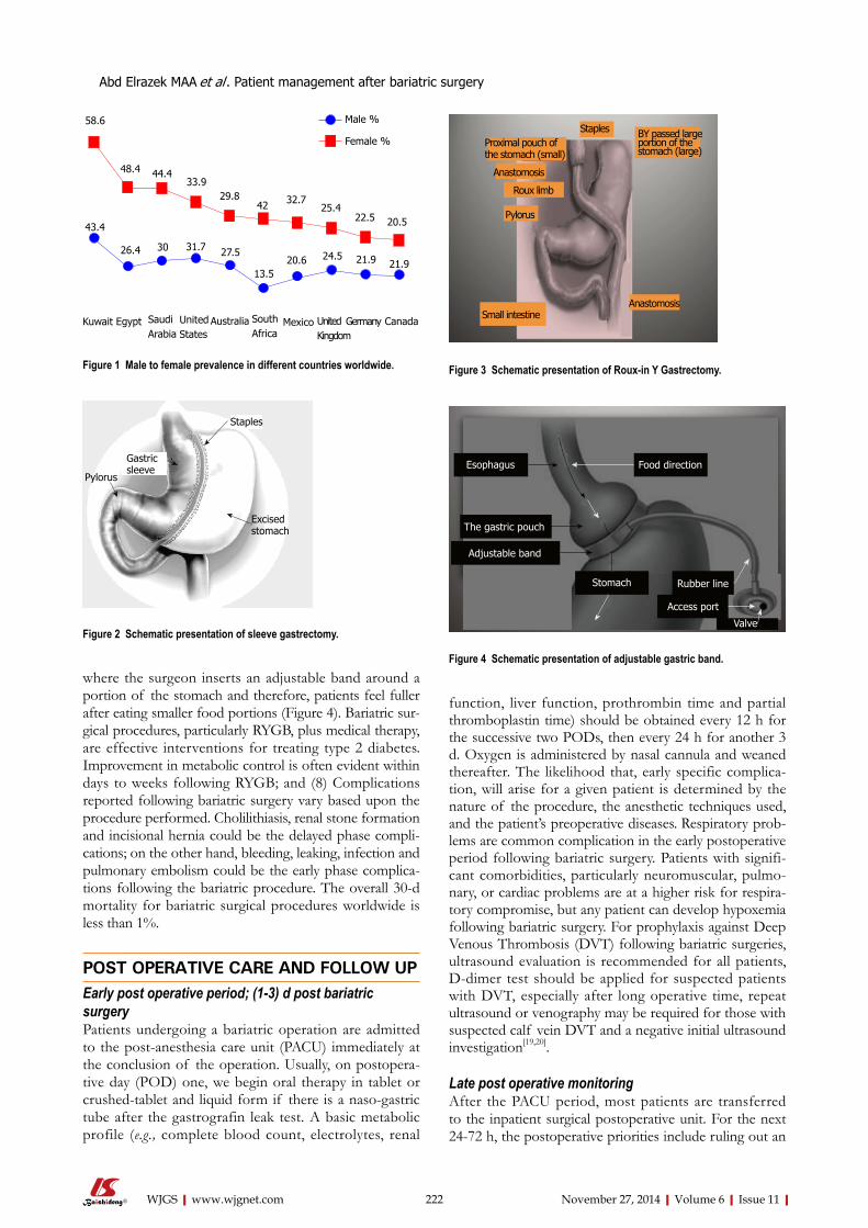
function, liver function, prothrombin time and partial thromboplastin time) should be obtained every 12 h for the successive two PODs, then every 24 h for another 3 d. Oxygen is administered by nasal cannula and weaned thereafter. The likelihood that, early specific complica-tion, will arise for a given patient is determined by the nature of the procedure, the anesthetic techniques used, and the patient’s preoperative diseases. Respiratory prob-lems are common complication in the early postoperative period following bariatric surgery. Patients with signifi-cant comorbidities, particularly neuromuscular, pulmo-nary, or cardiac problems are at a higher risk for respira-tory compromise, but any patient can develop hypoxemia following bariatric surgery. For prophylaxis against Deep Venous Thrombosis (DVT) following bariatric surgeries, ultrasound evaluation is recommended for all patients, D-dimer test should be applied for suspected patients with DVT, especially after long operative time, repeat ultrasound or venography may be required for those with suspected calf vein DVT and a negative initial ultrasound investigation[19,20].
Late post operative monitoringAfter the PACU period, most patients are transferred to the inpatient surgical postoperative unit. For the next 24-72 h, the postoperative priorities include ruling out an
where the surgeon inserts an adjustable band around a portion of the stomach and therefore, patients feel fuller after eating smaller food portions (Figure 4). Bariatric sur-gical procedures, particularly RYGB, plus medical therapy, are effective interventions for treating type 2 diabetes. Improvement in metabolic control is often evident within days to weeks following RYGB; and (8) Complications reported following bariatric surgery vary based upon the procedure performed. Cholilithiasis, renal stone formation and incisional hernia could be the delayed phase compli-cations; on the other hand, bleeding, leaking, infection and pulmonary embolism could be the early phase complica-tions following the bariatric procedure. The overall 30-d mortality for bariatric surgical procedures worldwide is less than 1%.
POST OPERATIVE CARE AND FOLLOW UPEarly post operative period; (1-3) d post bariatric surgeryPatients undergoing a bariatric operation are admitted to the post-anesthesia care unit (PACU) immediately at the conclusion of the operation. Usually, on postopera-tive day (POD) one, we begin oral therapy in tablet or crushed-tablet and liquid form if there is a naso-gastric tube after the gastrografin leak test. A basic metabolic profile (e.g., complete blood count, electrolytes, renal
222 November 27, 2014|Volume 6|Issue 11|WJGS|www.wjgnet.com
Abd Elrazek MAA et al . Patient management after bariatric surgery
58.6
48.4 44.433.9
29.842 32.7
25.422.5 20.543.4
26.4 30 31.7 27.5
13.520.6 24.5 21.9 21.9
Kuwait Egypt CanadaGermanyUnited States
Australia South Africa
Mexico United Kingdom
Saudi Arabia
Male %
Female %
Figure 1 Male to female prevalence in different countries worldwide.
Gastric sleeve
Staples
Excisedstomach
Pylorus
Figure 2 Schematic presentation of sleeve gastrectomy.
Proximal pouch of the stomach (small)
Staples BY passed large portion of the stomach (large)
Anastomosis
Roux limb
Pylorus
Small intestineAnastomosis
Figure 3 Schematic presentation of Roux-in Y Gastrectomy.
Esophagus
The gastric pouch
Food direction
Stomach Rubber line
Adjustable band
Access port
Valve
Figure 4 Schematic presentation of adjustable gastric band.
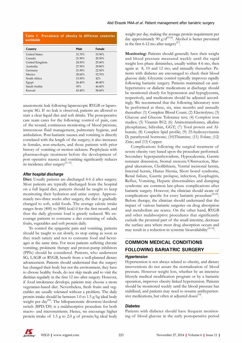
anastomotic leak following laparoscopic RYGB or laparo-scopic SG. If no leak is observed, patients are allowed to start a clear liquid diet and soft drinks. The postoperative care team cares for the following: control of pain, care of the wound, continuous monitoring of blood pressure, intravenous fluid management, pulmonary hygiene, and ambulation. Post-bariatric nausea and vomiting is directly correlated with the length of the surgery; it also increases in females, non-smokers, and those patients with prior history of vomiting or motion sickness. Prophylaxis with pharmacologic treatment before the development of post operative nausea and vomiting significantly reduces its incidence after surgery[21-23].
After hospital dischargeDiet: Usually patients are discharged 4-6 d after surgery. Most patients are typically discharged from the hospital on a full liquid diet, patients should be taught to keep monitoring their hydration and urine output. Approxi-mately two-three weeks after surgery, the diet is gradually changed to soft, solid foods. The average caloric intake ranges from (400) to (800) kcal/d for the first month, and thus the daily glycemic load is greatly reduced. We en-courage patients to consume a diet consisting of salads, fruits, vegetables and soft protein daily.
To control the epigastric pain and vomiting, patients should be taught to eat slowly, to stop eating as soon as they reach satiety and not to consume food and bever-ages at the same time. For most patients suffering chronic vomiting, prokinetic therapy and proton-pump inhibitors (PPIs) should be considered. Patients, who underwent SG, LAGB or RYGB, benefit from a well-planned dietary advancement. Patients should understand that the surgery has changed their body but not the environment, they have to choose healthy foods, do not skip meals and to visit the dietitian regularly in the first 12 mo after surgery. However, if food intolerance develops, patients may choose a more vegetarian-based diet. Nevertheless, fresh fruits and veg-etables are usually tolerated without a problem. The daily protein intake should be between 1.0 to 1.5 g/kg ideal body weight per day[24]. The biliopancreatic diversion/duodenal switch (BPD/DS) is a malabsorptive procedure for both macro- and micronutrients. Hence, we encourage higher protein intake of 1.5 g to 2.0 g of protein/kg ideal body
weight per day, making the average protein requirement per day approximately 90 g/d[25,26]. Alcohol is better prevented in the first 6-12 mo after surgery[27].
Monitoring: Patients should generally have their weight and blood pressure measured weekly until the rapid weight loss phase diminishes, usually within 4-6 mo, then again at 8, 10 and 12 mo, and annually thereafter. Pa-tients with diabetes are encouraged to check their blood glucose daily. Glycemic control typically improves rapidly following bariatric surgery. Patients maintained on anti-hypertensive or diabetic medications at discharge should be monitored closely for hypotension and hypoglycemia, respectively, and medications should be adjusted accord-ingly. We recommend that the following laboratory tests be performed at three, six, nine months and annually thereafter: (1) Complete Blood Count; (2) Electrolytes; (3) Glucose and Glucose Tolerance test; (4) Complete iron studies; (5) Vitamin B12; (6) Aminotransferases, alkaline phosphatase, bilirubin, GGT; (7) Total protein and Al-bumin; (8) Complete lipid profile; (9) 25-hydroxyvitamin D, parathyroid hormone; (10)Thiamine; (11) Folate; (12) Zinc; and (13) Copper.
Complications following the surgical treatment of severe obesity vary based upon the procedure performed. Secondary hyperparathyroidism, Hypocalcemia, Gastric remnant distension, Stomal stenosis/Obstruction, Mar-ginal ulcerations, Cholilithiasis, Ventral incisional hernia, Internal hernia, Hiatus Hernia, Short bowel syndrome, Renal failure, Gastric prolapse, infection, Esophagitis, Reflux, Vomiting, Hepatic abnormalities and dumping syndrome are common late-phase complications after bariatric surgery. However, the clinician should aware of complications specific for every bariatric procedure[28,29]. Before therapy, the clinician should understand that the impact of various bariatric surgeries on drug absorption and metabolism are scarce. On the other hand, RYGB and other malabsorptive procedures that significantly exclude the proximal part of the small intestine, decrease the surface area where most drug absorption occurs and may result in a reduction in systemic bioavailability[30-32].
COMMON MEDICAL CONDITIONS FOLLOWING BARIATRIC SURGERYHypertensionHypertension is not always related to obesity, and dietary interventions do not assure the normalization of blood pressure. However weight loss, whether by an intensive lifestyle medical modification program or by a bariatric operation, improves obesity-linked hypertension. Patients should be monitored weekly until the blood pressure has stabilized, and patients may need to resume antihyperten-sive medications, but often at adjusted doses[33].
DiabetesPatients with diabetes should have frequent monitor-ing of blood glucose in the early postoperative period
223 November 27, 2014|Volume 6|Issue 11|WJGS|www.wjgnet.com
Abd Elrazek MAA et al . Patient management after bariatric surgery
Table 1 Prevalence of obesity in different countries worldwide
Country Male Female
United States 31.70% 33.90%Canada 21.90% 20.50%United Kingdom 24.50% 25.40%Australia 27.50% 29.80%Germany 21.90% 22.50%Mexico 20.60% 32.70%South Africa 13.50% 42%Egypt 26.40% 48.40%Saudi Arabia 30% 44.40%Kuwait 43.40% 58.60%
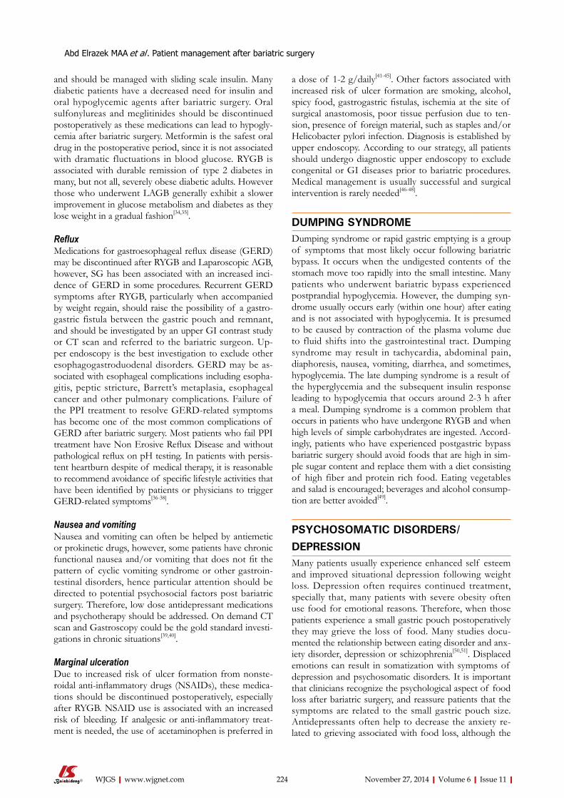
and should be managed with sliding scale insulin. Many diabetic patients have a decreased need for insulin and oral hypoglycemic agents after bariatric surgery. Oral sulfonylureas and meglitinides should be discontinued postoperatively as these medications can lead to hypogly-cemia after bariatric surgery. Metformin is the safest oral drug in the postoperative period, since it is not associated with dramatic fluctuations in blood glucose. RYGB is associated with durable remission of type 2 diabetes in many, but not all, severely obese diabetic adults. However those who underwent LAGB generally exhibit a slower improvement in glucose metabolism and diabetes as they lose weight in a gradual fashion[34,35].
RefluxMedications for gastroesophageal reflux disease (GERD) may be discontinued after RYGB and Laparoscopic AGB, however, SG has been associated with an increased inci-dence of GERD in some procedures. Recurrent GERD symptoms after RYGB, particularly when accompanied by weight regain, should raise the possibility of a gastro-gastric fistula between the gastric pouch and remnant, and should be investigated by an upper GI contrast study or CT scan and referred to the bariatric surgeon. Up-per endoscopy is the best investigation to exclude other esophagogastroduodenal disorders. GERD may be as-sociated with esophageal complications including esopha-gitis, peptic stricture, Barrett’s metaplasia, esophageal cancer and other pulmonary complications. Failure of the PPI treatment to resolve GERD-related symptoms has become one of the most common complications of GERD after bariatric surgery. Most patients who fail PPI treatment have Non Erosive Reflux Disease and without pathological reflux on pH testing. In patients with persis-tent heartburn despite of medical therapy, it is reasonable to recommend avoidance of specific lifestyle activities that have been identified by patients or physicians to trigger GERD-related symptoms[36-38].
Nausea and vomitingNausea and vomiting can often be helped by antiemetic or prokinetic drugs, however, some patients have chronic functional nausea and/or vomiting that does not fit the pattern of cyclic vomiting syndrome or other gastroin-testinal disorders, hence particular attention should be directed to potential psychosocial factors post bariatric surgery. Therefore, low dose antidepressant medications and psychotherapy should be addressed. On demand CT scan and Gastroscopy could be the gold standard investi-gations in chronic situations[39,40].
Marginal ulcerationDue to increased risk of ulcer formation from nonste-roidal anti-inflammatory drugs (NSAIDs), these medica-tions should be discontinued postoperatively, especially after RYGB. NSAID use is associated with an increased risk of bleeding. If analgesic or anti-inflammatory treat-ment is needed, the use of acetaminophen is preferred in
a dose of 1-2 g/daily[41-45]. Other factors associated with increased risk of ulcer formation are smoking, alcohol, spicy food, gastrogastric fistulas, ischemia at the site of surgical anastomosis, poor tissue perfusion due to ten-sion, presence of foreign material, such as staples and/or Helicobacter pylori infection. Diagnosis is established by upper endoscopy. According to our strategy, all patients should undergo diagnostic upper endoscopy to exclude congenital or GI diseases prior to bariatric procedures. Medical management is usually successful and surgical intervention is rarely needed[46-48].
DUMPING SYNDROMEDumping syndrome or rapid gastric emptying is a group of symptoms that most likely occur following bariatric bypass. It occurs when the undigested contents of the stomach move too rapidly into the small intestine. Many patients who underwent bariatric bypass experienced postprandial hypoglycemia. However, the dumping syn-drome usually occurs early (within one hour) after eating and is not associated with hypoglycemia. It is presumed to be caused by contraction of the plasma volume due to fluid shifts into the gastrointestinal tract. Dumping syndrome may result in tachycardia, abdominal pain, diaphoresis, nausea, vomiting, diarrhea, and sometimes, hypoglycemia. The late dumping syndrome is a result of the hyperglycemia and the subsequent insulin response leading to hypoglycemia that occurs around 2-3 h after a meal. Dumping syndrome is a common problem that occurs in patients who have undergone RYGB and when high levels of simple carbohydrates are ingested. Accord-ingly, patients who have experienced postgastric bypass bariatric surgery should avoid foods that are high in sim-ple sugar content and replace them with a diet consisting of high fiber and protein rich food. Eating vegetables and salad is encouraged; beverages and alcohol consump-tion are better avoided[49].
PSYCHOSOMATIC DISORDERS/DEPRESSIONMany patients usually experience enhanced self esteem and improved situational depression following weight loss. Depression often requires continued treatment, specially that, many patients with severe obesity often use food for emotional reasons. Therefore, when those patients experience a small gastric pouch postoperatively they may grieve the loss of food. Many studies docu-mented the relationship between eating disorder and anx-iety disorder, depression or schizophrenia[50,51]. Displaced emotions can result in somatization with symptoms of depression and psychosomatic disorders. It is important that clinicians recognize the psychological aspect of food loss after bariatric surgery, and reassure patients that the symptoms are related to the small gastric pouch size. Antidepressants often help to decrease the anxiety re-lated to grieving associated with food loss, although the
224 November 27, 2014|Volume 6|Issue 11|WJGS|www.wjgnet.com
Abd Elrazek MAA et al . Patient management after bariatric surgery
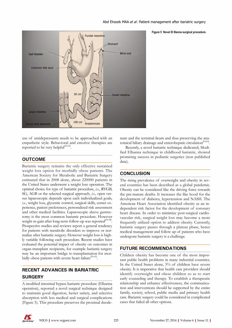
use of antidepressants needs to be approached with an empathetic style. Behavioral and emotive therapies are reported to be very helpful[52,53].
OUTCOMEBariatric surgery remains the only effective sustained weight loss option for morbidly obese patients. The American Society for Metabolic and Bariatric Surgery estimated that in 2008 alone, about 220000 patients in the United States underwent a weight loss operation. The optimal choice for type of bariatric procedure, i.e., RYGB, SG, AGB or the selected surgical approach, i.e., open ver-sus laparoscopic depends upon each individualized goals, i.e., weight loss, glycemic control, surgical skills, center ex-perience, patient preferences, personalized risk assessment and other medical facilities. Laparoscopic sleeve gastrec-tomy is the most common bariatric procedure. However weight re-gain after long-term follow-up was reported[54-58]. Prospective studies and reviews report a general tendency for patients with metabolic disorders to improve or nor-malize after bariatric surgery. However weight loss is high-ly variable following each procedure. Recent studies have evaluated the potential impact of obesity on outcomes in organ-transplant recipients, for example bariatric surgery may be an important bridge to transplantation for mor-bidly obese patients with severe heart failure[59-63].
RECENT ADVANCES IN BARIATRIC SURGERYA modified intestinal bypass bariatric procedure (Elbanna operation), reported a novel surgical technique designed to maintain good digestion, better satiety, and selective absorption with less medical and surgical complications (Figure 5). This procedure preserves the proximal duode-
num and the terminal ileum and thus preserving the ana-tomical biliary drainage and enterohepatic circulation[64,65].
Recently, a novel bariatric technique dedicated; Modi-fied Elbanna technique in childhood bariatric, showed promising success in pediatric surgeries (non published data).
CONCLUSIONThe rising prevalence of overweight and obesity in sev-eral countries has been described as a global pandemic. Obesity can be considered like the driving force towards the pre-mature deaths. It increases the like hood for the development of diabetes, hypertension and NASH. The American Heart Association identified obesity as an in-dependent risk factor for the development of coronary heart disease. In order to minimize post-surgical cardio-vascular risk, surgical weight loss may become a more frequently utilized option to address obesity. Currently, bariatric surgery passes through a plateau phase, hence medical management and follow up of patients who have undergone bariatric surgery is a challenge.
FUTURE RECOMMENDATIONSChildren obesity has become one of the most impor-tant public health problems in many industrial countries. In the United States alone, 5% of children have severe obesity. It is imperative that health care providers should identify overweight and obese children so as to start early counseling and therapy. To establish a therapeutic relationship and enhance effectiveness, the communica-tion and interventions should be supported by the entire family, society, school, public media and primary health care. Bariatric surgery could be considered in complicated cases that failed all other options.
225 November 27, 2014|Volume 6|Issue 11|WJGS|www.wjgnet.com
Abd Elrazek MAA et al . Patient management after bariatric surgery
Blind end
Stomach
Fundal resection
Small intestine50 cm
100 cm Anastomosis
Large intestine
Gall bladder
Common bile duct
Cecum and appendix
Figure 5 Novel El Banna surgical procedure.
REFERENCES1 Schauer PR, Kashyap SR, Wolski K, Brethauer SA, Kirwan
JP, Pothier CE, Thomas S, Abood B, Nissen SE, Bhatt DL. Bariatric surgery versus intensive medical therapy in obese patients with diabetes. N Engl J Med 2012; 366: 1567-1576 [PMID: 22449319 DOI: 10.1056/NEJMoa1200225]
2 Ng M, Fleming T, Robinson M, Thomson B, Graetz N, Mar-gono C, Mullany EC, Biryukov S, Abbafati C, Abera SF, Abraham JP, Abu-Rmeileh NM, Achoki T, AlBuhairan FS, Alemu ZA, Alfonso R, Ali MK, Ali R, Guzman NA, Ammar W, Anwari P, Banerjee A, Barquera S, Basu S, Bennett DA, Bhutta Z, Blore J, Cabral N, Nonato IC, Chang JC, Chowd-hury R, Courville KJ, Criqui MH, Cundiff DK, Dabhadkar KC, Dandona L, Davis A, Dayama A, Dharmaratne SD, Ding EL, Durrani AM, Esteghamati A, Farzadfar F, Fay DF, Feigin VL, Flaxman A, Forouzanfar MH, Goto A, Green MA, Gupta R, Hafezi-Nejad N, Hankey GJ, Harewood HC, Havmoeller R, Hay S, Hernandez L, Husseini A, Idrisov BT, Ikeda N, Islami F, Jahangir E, Jassal SK, Jee SH, Jeffreys M, Jonas JB, Kabagambe EK, Khalifa SE, Kengne AP, Khader YS, Khang YH, Kim D, Kimokoti RW, Kinge JM, Kokubo Y, Kosen S, Kwan G, Lai T, Leinsalu M, Li Y, Liang X, Liu S, Logroscino G, Lotufo PA, Lu Y, Ma J, Mainoo NK, Mensah GA, Merriman TR, Mokdad AH, Moschandreas J, Naghavi M, Naheed A, Nand D, Narayan KM, Nelson EL, Neuhous-er ML, Nisar MI, Ohkubo T, Oti SO, Pedroza A, Prabha-karan D, Roy N, Sampson U, Seo H, Sepanlou SG, Shibuya K, Shiri R, Shiue I, Singh GM, Singh JA, Skirbekk V, Stapelberg NJ, Sturua L, Sykes BL, Tobias M, Tran BX, Trasande L, Toy-oshima H, van de Vijver S, Vasankari TJ, Veerman JL, Velas-quez-Melendez G, Vlassov VV, Vollset SE, Vos T, Wang C, Wang X, Weiderpass E, Werdecker A, Wright JL, Yang YC, Yatsuya H, Yoon J, Yoon SJ, Zhao Y, Zhou M, Zhu S, Lopez AD, Murray CJ, Gakidou E. Global, regional, and national prevalence of overweight and obesity in children and adults during 1980-2013: a systematic analysis for the Global Bur-den of Disease Study 2013. Lancet 2014; 384: 766-781 [PMID: 24880830 DOI: 10.1016/S0140-6736(14)60460-8]
3 Hogan MC, Foreman KJ, Naghavi M, Ahn SY, Wang M, Makela SM, Lopez AD, Lozano R, Murray CJ. Maternal mortality for 181 countries, 1980-2008: a systematic analy-sis of progress towards Millennium Development Goal 5. Lancet 2010; 375: 1609-1623 [PMID: 20382417 DOI: 10.1016/S0140-6736(10)60518-1]
4 Rajaratnam JK, Marcus JR, Flaxman AD, Wang H, Levin-Rector A, Dwyer L, Costa M, Lopez AD, Murray CJ. Neonatal, postneonatal, childhood, and under-5 mortal-ity for 187 countries, 1970-2010: a systematic analysis of progress towards Millennium Development Goal 4. Lan-cet 2010; 375: 1988-2008 [PMID: 20546887 DOI: 10.1016/S0140-6736(10)60703-9]
5 Bleich S, Cutler D, Murray C, Adams A. Why is the de-veloped world obese? Annu Rev Public Health 2008; 29: 273-295 [PMID: 18173389 DOI: 10.1146/annurev.publ-health.29.020907.090954]
6 Food and Agriculture Organization Corporate Statisti-cal Database. Food balance sheets. Available from: URL: http://faostat3.fao.org/faostat-gateway/go/to/home/E
7 UN Department of Economic and Social Affairs, Popula-tion Division. World population prospects: the 2010 revi-sion. Volume 1: Comprehensive tables. New York: United Nations, 2011
8 Astrup A, Brand-Miller J. Diet composition and obesity. Lancet 2012; 379: 1100; author reply 1100-1101 [PMID: 22444397 DOI: 10.1016/S0140-6736(12)60456-5]
9 Drewnowski A, Popkin BM. The nutrition transition: new trends in the global diet. Nutr Rev 1997; 55: 31-43 [PMID: 9155216 DOI: 10.1111/j.1753-4887.1997.tb01593.x]
10 Briefel RR, Johnson CL. Secular trends in dietary intake in the United States. Annu Rev Nutr 2004; 24: 401-431 [PMID:
15189126 DOI: 10.1146/annurev.nutr.23.011702.073349]11 Swinburn B, Sacks G, Ravussin E. Increased food energy
supply is more than sufficient to explain the US epidemic of obesity. Am J Clin Nutr 2009; 90: 1453-1456 [PMID: 19828708 DOI: 10.3945/ajcn.2009.28595]
12 Popkin BM. The nutrition transition and obesity in the developing world. J Nutr 2001; 131: 871S-873S [PMID: 11238777]
13 Ng M, Freeman MK, Fleming TD, Robinson M, Dwyer-Lindgren L, Thomson B, Wollum A, Sanman E, Wulf S, Lopez AD, Murray CJ, Gakidou E. Smoking prevalence and cigarette consumption in 187 countries, 1980-2012. JAMA 2014; 311: 183-192 [PMID: 24399557 DOI: 10.1001/jama.2013.284692]
14 Ben-Menachem T. Risk factors for cholangiocarcinoma. Eur J Gastroenterol Hepatol 2007; 19: 615-617 [PMID: 17625428]
15 Younossi ZM, Stepanova M, Negro F, Hallaji S, Younossi Y, Lam B, Srishord M. Nonalcoholic fatty liver disease in lean individuals in the United States. Medicine (Balti-more) 2012; 91: 319-327 [PMID: 23117851 DOI: 10.1097/MD.0b013e3182779d49]
16 American Society for Metabolic and Bariatric Surgery. Fact Sheet: Metabolic and Bariatric Surgery. [updated 2009 January 28]. Available from: URL: http://www.asbs.org/ Newsite07/media/asbs_presskit.htm
17 Nguyen NT, Masoomi H, Magno CP, Nguyen XM, Lauge-nour K, Lane J. Trends in use of bariatric surgery, 2003-2008. J Am Coll Surg 2011; 213: 261-266 [PMID: 21624841 DOI: 10.1016/j.jamcollsurg.2011.04.030]
18 Dimick JB, Nicholas LH, Ryan AM, Thumma JR, Birkmeyer JD. Bariatric surgery complications before vs after imple-mentation of a national policy restricting coverage to cen-ters of excellence. JAMA 2013; 309: 792-799 [PMID: 23443442 DOI: 10.1001/jama.2013.755]
19 Chen KN. Managing complications I: leaks, strictures, emptying, reflux, chylothorax. J Thorac Dis 2014; 6 Suppl 3: S355-S363 [PMID: 24876942 DOI: 10.3978/j.issn.2072-1439.2014.03.36]
20 Mechanick JI, Youdim A, Jones DB, Garvey WT, Hurley DL, McMahon MM, Heinberg LJ, Kushner R, Adams TD, Shikora S, Dixon JB, Brethauer S. Clinical practice guidelines for the perioperative nutritional, metabolic, and nonsurgical support of the bariatric surgery patient--2013 update: co-sponsored by American Association of Clinical Endocrinol-ogists, The Obesity Society, and American Society for Meta-bolic & amp; Bariatric Surgery. Obesity (Silver Spring) 2013; 21 Suppl 1: S1-27 [PMID: 23529939 DOI: 10.1002/oby.20461]
21 Tucker ON, Szomstein S, Rosenthal RJ. Nutritional con-sequences of weight-loss surgery. Med Clin North Am 2007; 91: 499-514, xii [PMID: 17509392 DOI: 10.1016/j.mcna.2007.01.006]
22 Sjöström L, Peltonen M, Jacobson P, Sjöström CD, Karason K, Wedel H, Ahlin S, Anveden Å, Bengtsson C, Bergmark G, Bouchard C, Carlsson B, Dahlgren S, Karlsson J, Lindroos AK, Lönroth H, Narbro K, Näslund I, Olbers T, Svensson PA, Carlsson LM. Bariatric surgery and long-term cardio-vascular events. JAMA 2012; 307: 56-65 [PMID: 22215166 DOI: 10.1001/jama.2011.1914]
23 Bouldin MJ, Ross LA, Sumrall CD, Loustalot FV, Low AK, Land KK. The effect of obesity surgery on obesity comorbid-ity. Am J Med Sci 2006; 331: 183-193 [PMID: 16617233 DOI: 10.1097/00000441-200604000-00004]
24 Schweiger C, Weiss R, Keidar A. Effect of different bariatric operations on food tolerance and quality of eating. Obes Surg 2010; 20: 1393-1399 [PMID: 20680506 DOI: 10.1007/s11695-010-0233-9]
25 Ortega J, Ortega-Evangelio G, Cassinello N, Sebastia V. What are obese patients able to eat after Roux-en-Y gastric bypass? Obes Facts 2012; 5: 339-348 [PMID: 22722236 DOI: 10.1159/000339769]
26 Nelson WK, Fatima J, Houghton SG, Thompson GB, Ken-
226 November 27, 2014|Volume 6|Issue 11|WJGS|www.wjgnet.com
Abd Elrazek MAA et al . Patient management after bariatric surgery
drick ML, Mai JL, Kennel KA, Sarr MG. The malabsorp-tive very, very long limb Roux-en-Y gastric bypass for super obesity: results in 257 patients. Surgery 2006; 140: 517-522, discussion 522-523 [PMID: 17011898 DOI: 10.1016/j.surg.2006.06.020]
27 Shen Z, Li Y, Yu C, Shen Y, Xu L, Xu C, Xu G. A cohort study of the effect of alcohol consumption and obesity on serum liv-er enzyme levels. Eur J Gastroenterol Hepatol 2010; 22: 820-825 [PMID: 19829121 DOI: 10.1097/MEG.0b013e3283328b86]
28 Koenig SM. Pulmonary complications of obesity. Am J Med Sci 2001; 321: 249-279 [PMID: 11307867 DOI: 10.1097/00000441-200104000-00006]
29 Holes-Lewis KA, Malcolm R, O’Neil PM. Pharmacotherapy of obesity: clinical treatments and considerations. Am J Med Sci 2013; 345: 284-288 [PMID: 23531960 DOI: 10.1097/MAJ.0b013e31828abcfd]
30 Sakcak I, Avsar FM, Cosgun E, Yildiz BD. Management of concurrent cholelithiasis in gastric banding for morbid obesity. Eur J Gastroenterol Hepatol 2011; 23: 766-769 [PMID: 21712718 DOI: 10.1097/MEG.0b013e3283488adb]
31 Herrara MF, Lozano-Salazar RR, González-Barranco J, Rull JA. Diseases and problems secondary to massive obesity. Eur J Gastroenterol Hepatol 1999; 11: 63-67 [PMID: 10102212 DOI: 10.1097/00042737-199902000-00002]
32 Lassailly G, Caiazzo R, Hollebecque A, Buob D, Leteurtre E, Arnalsteen L, Louvet A, Pigeyre M, Raverdy V, Verkindt H, Six MF, Eberle C, Patrice A, Dharancy S, Romon M, Pat-tou F, Mathurin P. Validation of noninvasive biomarkers (FibroTest, SteatoTest, and NashTest) for prediction of liver injury in patients with morbid obesity. Eur J Gastroenterol Hepatol 2011; 23: 499-506 [PMID: 21499110 DOI: 10.1097/MEG.0b013e3283464111]
33 Hofsø D, Nordstrand N, Johnson LK, Karlsen TI, Hager H, Jenssen T, Bollerslev J, Godang K, Sandbu R, Røislien J, Hjelmesaeth J. Obesity-related cardiovascular risk factors after weight loss: a clinical trial comparing gastric bypass surgery and intensive lifestyle intervention. Eur J Endo-crinol 2010; 163: 735-745 [PMID: 20798226 DOI: 10.1530/EJE-10-0514]
34 Service GJ, Thompson GB, Service FJ, Andrews JC, Collazo-Clavell ML, Lloyd RV. Hyperinsulinemic hypoglycemia with nesidioblastosis after gastric-bypass surgery. N Engl J Med 2005; 353: 249-254 [PMID: 16034010 DOI: 10.1056/NEJ-Moa043690]
35 Arterburn DE, Bogart A, Sherwood NE, Sidney S, Coleman KJ, Haneuse S, O’Connor PJ, Theis MK, Campos GM, Mc-Culloch D, Selby J. A multisite study of long-term remission and relapse of type 2 diabetes mellitus following gastric bypass. Obes Surg 2013; 23: 93-102 [PMID: 23161525 DOI: 10.1007/s11695-012-0802-1]
36 Fass R, Shapiro M, Dekel R, Sewell J. Systematic review: proton-pump inhibitor failure in gastro-oesophageal reflux disease--where next? Aliment Pharmacol Ther 2005; 22: 79-94 [PMID: 16011666 DOI: 10.1111/j.1365-2036.2005.02531.x]
37 Löfdahl HE, Lane A, Lu Y, Lagergren P, Harvey RF, Bla-zeby JM, Lagergren J. Increased population prevalence of reflux and obesity in the United Kingdom compared with Sweden: a potential explanation for the difference in inci-dence of esophageal adenocarcinoma. Eur J Gastroenterol Hepatol 2011; 23: 128-132 [PMID: 21178778 DOI: 10.1097/MEG.0b013e3283424e25]
38 Fornari F, Madalosso CA, Farré R, Gurski RR, Thiesen V, Callegari-Jacques SM. The role of gastro-oesophageal pres-sure gradient and sliding hiatal hernia on pathological gastro-oesophageal reflux in severely obese patients. Eur J Gastroenterol Hepatol 2010; 22: 404-411 [PMID: 20110819 DOI: 10.1097/MEG.0b013e328332f7b8]
39 Aasheim ET. Wernicke encephalopathy after bariatric surgery: a systematic review. Ann Surg 2008; 248: 714-720 [PMID: 18948797 DOI: 10.1097/SLA.0b013e3181884308]
40 Salgado W, Modotti C, Nonino CB, Ceneviva R. Anemia and iron deficiency before and after bariatric surgery. Surg Obes Relat Dis 2014; 10: 49-54 [PMID: 24071485 DOI: 10.1016/j.soard.2013.06.012]
41 Klockhoff H, Näslund I, Jones AW. Faster absorption of ethanol and higher peak concentration in women after gastric bypass surgery. Br J Clin Pharmacol 2002; 54: 587-591 [PMID: 12492605 DOI: 10.1046/j.1365-2125.2002.01698.x]
42 Maluenda F, Csendes A, De Aretxabala X, Poniachik J, Sal-vo K, Delgado I, Rodriguez P. Alcohol absorption modifica-tion after a laparoscopic sleeve gastrectomy due to obesity. Obes Surg 2010; 20: 744-748 [PMID: 20358306 DOI: 10.1007/s11695-010-0136-9]
43 Woodard GA, Downey J, Hernandez-Boussard T, Morton JM. Impaired alcohol metabolism after gastric bypass sur-gery: a case-crossover trial. J Am Coll Surg 2011; 212: 209-214 [PMID: 21183366 DOI: 10.1016/j.jamcollsurg.2010.09.020]
44 King WC, Chen JY, Mitchell JE, Kalarchian MA, Steffen KJ, Engel SG, Courcoulas AP, Pories WJ, Yanovski SZ. Preva-lence of alcohol use disorders before and after bariatric surgery. JAMA 2012; 307: 2516-2525 [PMID: 22710289 DOI: 10.1001/jama.2012.6147]
45 Sasse KC, Ganser J, Kozar M, Watson RW, McGinley L, Lim D, Weede M, Smith CJ, Bovee V. Seven cases of gastric per-foration in Roux-en-Y gastric bypass patients: what lessons can we learn? Obes Surg 2008; 18: 530-534 [PMID: 18324450 DOI: 10.1007/s11695-007-9335-4]
46 Capella JF, Capella RF. Gastro-gastric fistulas and marginal ulcers in gastric bypass procedures for weight reduction. Obes Surg 1999; 9: 22-27; discussion 28 [PMID: 10065576 DOI: 10.1381/096089299765553674]
47 Abd Elrazek AE, Mahfouz HM, Metwally AM, El-Shamy AM. Mortality prediction of nonalcoholic patients present-ing with upper gastrointestinal bleeding using data mining. Eur J Gastroenterol Hepatol 2014; 26: 187-191 [PMID: 24088733 DOI: 10.1097/MEG.0b013e328365c3b0]
48 Abd Elrazek AE, Yoko N, Hiroki M, Afify M, Asar M, Is-mael B, Salah M. Endoscopic management of Dieulafoy’s lesion using Isoamyl-2-cyanoacrylate. World J Gastrointest Endosc 2013; 5: 417-419 [PMID: 23951399 DOI: 10.4253/wjge.v5.i8.417]
49 Ukleja A. Dumping syndrome: pathophysiology and treat-ment. Nutr Clin Pract 2005; 20: 517-525 [PMID: 16207692 DOI: 10.1177/0115426505020005517]
50 Flegal KM, Carroll MD, Kit BK, Ogden CL. Prevalence of obesity and trends in the distribution of body mass index among US adults, 1999-2010. JAMA 2012; 307: 491-497 [PMID: 22253363 DOI: 10.1001/jama.2012.39]
51 Flegal KM, Carroll MD, Ogden CL, Curtin LR. Prevalence and trends in obesity among US adults, 1999-2008. JAMA 2010; 303: 235-241 [PMID: 20071471 DOI: 10.1001/jama.2009.2014]
52 García-García ML, Martín-Lorenzo JG, Campillo-Soto A, Torralba-Martínez JA, Lirón-Ruiz R, Miguel-Perelló J, Mengual-Ballester M, Aguayo-Albasini JL. [Complications and level of satisfaction after dermolipectomy and abdomi-noplasty post-bariatric surgery]. Cir Esp 2014; 92: 254-260 [PMID: 24360407 DOI: 10.1016/j.ciresp.2013.04.024]
53 Wyatt SB, Winters KP, Dubbert PM. Overweight and obesi-ty: prevalence, consequences, and causes of a growing pub-lic health problem. Am J Med Sci 2006; 331: 166-174 [PMID: 16617231 DOI: 10.1097/00000441-200604000-00002]
54 Lamers F, van Oppen P, Comijs HC, Smit JH, Spinhoven P, van Balkom AJ, Nolen WA, Zitman FG, Beekman AT, Pen-ninx BW. Comorbidity patterns of anxiety and depressive disorders in a large cohort study: the Netherlands Study of Depression and Anxiety (NESDA). J Clin Psychiatry 2011; 72: 341-348 [PMID: 21294994 DOI: 10.4088/JCP.10m06176blu]
55 de Graaf R, Bijl RV, Smit F, Vollebergh WA, Spijker J. Risk factors for 12-month comorbidity of mood, anxiety, and substance use disorders: findings from the Netherlands
227 November 27, 2014|Volume 6|Issue 11|WJGS|www.wjgnet.com
Abd Elrazek AEMA et al . Patient management after bariatric surgery
Mental Health Survey and Incidence Study. Am J Psychia-try 2002; 159: 620-629 [PMID: 11925301 DOI: 10.1176/appi.ajp.159.4.620]
56 Cesana G, Uccelli M, Ciccarese F, Carrieri D, Castello G, Olmi S. Laparoscopic re-sleeve gastrectomy as a treatment of weight regain after sleeve gastrectomy. World J Gastrointest Surg 2014; 6: 101-106 [PMID: 24976903 DOI: 10.4240/wjgs.v6.i6.101]
57 Lee WJ, Ser KH, Chong K, Lee YC, Chen SC, Tsou JJ, Chen JC, Chen CM. Laparoscopic sleeve gastrectomy for diabe-tes treatment in nonmorbidly obese patients: efficacy and change of insulin secretion. Surgery 2010; 147: 664-669 [PMID: 20004451 DOI: 10.1016/j.surg.2009.10.059]
58 Mechanick JI, Youdim A, Jones DB, Timothy Garvey W, Hurley DL, Molly McMahon M, Heinberg LJ, Kushner R, Adams TD, Shikora S, Dixon JB, Brethauer S. Clini-cal practice guidelines for the perioperative nutritional, metabolic, and nonsurgical support of the bariatric surgery patient--2013 update: cosponsored by American Associa-tion of Clinical Endocrinologists, the Obesity Society, and American Society for Metabolic & amp; Bariatric Surgery. Surg Obes Relat Dis 2013; 9: 159-191 [PMID: 23537696 DOI: 10.1016/j.soard.2012.12.010]
59 Adams PL. Long-term patient survival: strategies to im-prove overall health. Am J Kidney Dis 2006; 47: S65-S85 [PMID: 16567242 DOI: 10.1053/j.ajkd.2005.12.043]
60 Gore JL, Pham PT, Danovitch GM, Wilkinson AH, Rosen-
thal JT, Lipshutz GS, Singer JS. Obesity and outcome follow-ing renal transplantation. Am J Transplant 2006; 6: 357-363 [PMID: 16426321 DOI: 10.1111/j.1600-6143.2005.01198.x]
61 Meier-Kriesche HU, Arndorfer JA, Kaplan B. The impact of body mass index on renal transplant outcomes: a significant independent risk factor for graft failure and patient death. Transplantation 2002; 73: 70-74 [PMID: 11792981 DOI: 10.1097/00007890-200201150-00013]
62 Wikiel KJ, McCloskey CA, Ramanathan RC. Bariatric sur-gery: a safe and effective conduit to cardiac transplantation. Surg Obes Relat Dis 2014; 10: 479-484 [PMID: 24462310 DOI: 10.1016/j.soard.2013.11.002]
63 DiCecco SR, Francisco-Ziller N. Obesity and organ trans-plantation: successes, failures, and opportunities. Nutr Clin Pract 2014; 29: 171-191 [PMID: 24503157 DOI: 10.1177/0884533613518585]
64 Elbanna A, Tawella N, Neff K, Abd Elfattah A, Bakr I. Ab-stracts from the 18th World Congress of the International Federation for the Surgery of Obesity & Metabolic Disorders (IFSO), Istanbul, Turkey 28-31 August 2013. Obes Surg 2013; 23: 1017-1243 [DOI: 10.1007/s11695-013-0986-z]
65 Elbanna A, Taweela NH, Gaber MB, Tag El-Din MM, La-bib MF, Emam MA, Khalil OO, Abdel Meguid MM, Abd Elrazek MAA. Medical Management of Patients with Modi-fied Intestinal Bypass: A New Promising Procedure for Morbid Obesity. GJMR 2014; 14: 8-19
P- Reviewer: Amiya E, Firstenberg MS, Narciso-Schiavon JL S- Editor: Tian YL L- Editor: A E- Editor: Lu YJ
228 November 27, 2014|Volume 6|Issue 11|WJGS|www.wjgnet.com
Abd Elrazek AEMA et al . Patient management after bariatric surgery

Factors influencing the diagnostic accuracy and management in acute surgical patients
Muhammad Shafique Sajid, Thaddeus Hollingsworth, Mike McGlue, William FA Miles
Muhammad Shafique Sajid, William FA Miles, Department of General, Laparoscopic and Endoscopic Colorectal Surgery, Western Sussex Hospitals NHS Foundation Trust, Worthing Hospital, Worthing, West Sussex BN11 2DH, United Kingdom Thaddeus Hollingsworth, Mike McGlue, The American Uni-versity of the Caribbean School of Medicine, FL 33134, United StatesAuthor contributions: All authors contributed to this manu-script.Correspondence to: Muhammad Shafique Sajid, Surgical Specialist Registrar, Department of General, Laparoscopic and Endoscopic Colorectal Surgery, Western Sussex Hospitals NHS Foundation Trust, Worthing Hospital, Washington Suite, North Wing, Worthing, West Sussex BN11 2DH, United Kingdom. [email protected]: +44-01-903205111 Fax: +44-01-903285010Received: July 28, 2014 Revised: September 16, 2014 Accepted: October 14, 2014Published online: November 27, 2014
AbstractAIM: To evaluate the diagnostic accuracy (DA) in acute surgical patients admitted to a District General Hospital.
METHODS: The case notes of all acute surgical pa-tients admitted under the surgical team for a period of two weeks were reviewed for the data pertaining to the admission diagnoses, relevant investigations and final diagnoses confirmed by either surgery or various other diagnostic modalities. The diagnostic pathway was re-corded from the source of referral [general practitioner (GP), A and E, in-patient] to the correct final diagnosis by the surgical team.
RESULTS: Forty-one patients (23 males) with acute surgical admissions during two weeks of study period were evaluated. The mean age of study group was 61.05 ± 23.24 years. There were 111 patient-doctor encounters. Final correct diagnosis was achieved in 85.4% patients. The DA was 46%, 44%, 50%, 33%,
61%, 61%, and 75% by GP, A and E, in-patient refer-ral, surgical foundation year-1, surgical senior house officer (SHO), surgical registrar, and surgical consultant respectively. The percentage of clinical consensus di-agnosis was 12%. Surgery was performed in 48.8% of patients. Sixty-seven percent of GP-referred patients, 31% of A and E-referred, and 25% of the in-patient re-ferrals underwent surgery. Surgical SHO made the most contributions to the primary diagnostic pathway.
CONCLUSION: Approximately 85% of acute surgical patients can be diagnosed accurately along the diag-nostic pathway. Patients referred by a GP are more likely to require surgery as compared to other referral sources. Surgical consultant was more likely to make correct surgical diagnosis, however it is the surgical SHO that contributes the most correct diagnoses along the diagnostic pathway.
© 2014 Baishideng Publishing Group Inc. All rights reserved.
Key words: Diagnostic accuracy; Diagnostic error; Mis-diagnosis; Premature closure
Core tip: Approximately 85% of acute surgical patients can be diagnosed accurately along the diagnostic path-way. One of the strategies to reduce diagnostic error is to develop pathways for feedback. It is particularly impor-tant to develop feedback pathways for the junior doctors, as it has been shown that less experienced doctors tend to most over-estimate their diagnostic accuracy. With anonymity removed, the basic design of this study seems well suited to enable feedback to each physician involved in the care of an acute surgical patient.
Sajid MS, Hollingsworth T, McGlue M, Miles WFA. Factors influencing the diagnostic accuracy and management in acute surgical patients. World J Gastrointest Surg 2014; 6(11): 229-234 Available from: URL: http://www.wjgnet.com/1948-9366/full/v6/i11/229.htm DOI: http://dx.doi.org/10.4240/wjgs.v6.i11.229
OBSERVATIONAL STUDY
Submit a Manuscript: http://www.wjgnet.com/esps/Help Desk: http://www.wjgnet.com/esps/helpdesk.aspxDOI: 10.4240/wjgs.v6.i11.229
World J Gastrointest Surg 2014 November 27; 6(11): 229-234ISSN 1948-9366 (online)
© 2014 Baishideng Publishing Group Inc. All rights reserved.
229 November 27, 2014|Volume 6|Issue 11|WJGS|www.wjgnet.com
INTRODUCTIONDiagnostic errors have recently begun to receive more attention as a preventable source of patient harm. Diag-nostic errors are estimated to account for 80000-160000 deaths per year[1]. Misdiagnosis has been the leading cause of medical malpractice payments over the last 25 years, making up 28.6% of claims and 35.2% of total payouts[1]. Missed, incorrect, or delayed diagnoses are estimated to occur in 15% of clinical cases, accounting for 8%-20% of adverse medical events[2]. Diagnosis is the most criti-cal of a physician’s skills. Nuland previously so perfectly stated, “It is every doctor’s measure of his abilities; it is the most important ingredient in his professional self image”[3], yet even with such a high regard for diagnostic accuracy, there remains an absence of ownership when it comes to quality and safety systems to reduce the diag-nostic errors[4]. Most of the studies attempting to quantify the diagnostic error have either been retrospective stud-ies examining adverse events, such as malpractice claims and autopsy reports, or have been experimental studies comparing multiple physician responses to the same di-agnostically challenging scenarios, which are often not reflective of the actual clinical environment[2]. Therefore, the true prevalence of diagnostic errors and inability to make right diagnosis along the acute surgical pathway has been notoriously difficult to evaluate[2,5].
Preventable diagnostic errors can result form the system-related factors and various cognitive factors. A published article on the prevalence of diagnostic error in 100 clinical cases revealed the system-related factors result in 65% diagnostic inaccuracies and cognitive factors in 74%[6] of the diagnostic inaccuracies. While many pro-grams have been initiated to address the system-related factors such as improved communication, enhancing the concept of an effective teamwork, and tackling the pro-cedural problems, clear pathways to reduce the diagnostic errors contributed by the cognitive factors have been more elusive and indefinable. A wide range of suggestions have been made about how to reduce cognitive errors in making the correct diagnosis. Graber et al[6] have described three distinct categories of interventions as those meant to: (1) improve knowledge and experience; (2) improve clinical reasoning and decision-making skills; and (3) provide cognitive “help”. Though many of these sugges-tions are well conceptualized and widely endorsed, a large portion remain untested or testing has been restricted to trainees in artificial settings, which does not necessarily reflect actual practice[7]. The diagnostic pathway for acute surgical patients involves GPs, Accident and Emergency, and surgeons. The need to investigate and quantify the impact of procedural and diagnostic accuracy at each level of medical contact is clear. Lack of a working diagnosis impacts patient care, outcome, and cost.
The objective of this observational study is to evalu-ate the diagnostic accuracy at each level of the primary diagnostic pathway in acute surgical patients admitted to a District General Hospital.
MATERIALS AND METHODSStudy conceptionIt was noted that ward rounds for on-call surgeons were often disorganized, with no clear roles defined, leading to inconsistent record keeping. Therefore, on call surgi-cal team decided to address this by running ward rounds using briefing and debriefing for each ward round, rotat-ing roles (each person present taking turns with patient presentation, record-keeping, and drug chart review), and asking each member of the team if they had anything to add before moving to the next patient. The surgical team was encouraged to clearly record 3 differential diagnoses for each doctor-patient encounter. It became apparent that the differential diagnoses listed would often change along the primary diagnostic pathway, and the idea to survey these changes emerged.
ProfromaA one-page profroma was designed and relevant variables reported in previous but similar publications were in-serted on it. Because this was an observational and pilot study examining the performance of the acute surgical team without any involvement of the patients, therefore, only an informal approval of the study was taken from the local Ethics and Research Committee with verbal discussion and electronic communication. The contents of the profroma were presented in internal clinical gov-ernance meeting and few additional variables were also included based upon the recommendations of clinical governance panel. All authors and local Ethics and Re-search Committee approved the profroma and its con-tents before starting the data collection.
Inclusion criteriaTo review the case notes and ward round entries of all acute surgical admissions during randomly selected two weeks.
Exclusion criteriaPatients whose notes were not available through secretar-ies or on the wards during data collection were excluded.
Data collectionPatient information including surname, hospital number, date of birth, and gender was collected onto a one page proforma, and each doctor/patient encounter was reviewed retrospectively to include up to 3 differential diagnoses listed in the patient notes. Doctors were anonymously recorded as general practitioner (GP), accident and emer-gency doctor (A and E), in-patient referrer (IP), surgical foundation year-1, surgical senior house officer (SHO), sur-gical registrar on-call (SROC), and the surgical consultant. Patient/doctor encounters were recorded along the primary diagnostic pathway, from GP referral, if available, up to the first surgical consultant review or definitive diagnosis by emergency surgery if that preceded consultant review. Final
Sajid MS et al . Diagnostic accuracy in acute surgical patients
230 November 27, 2014|Volume 6|Issue 11|WJGS|www.wjgnet.com
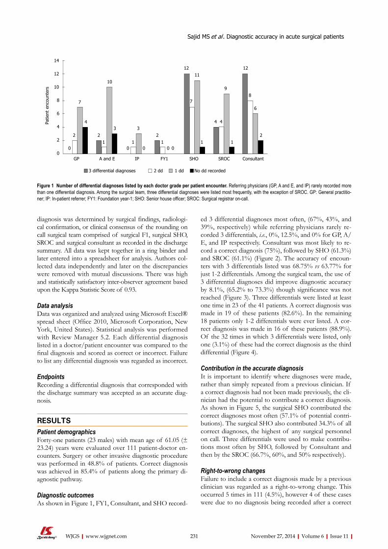
ed 3 differential diagnoses most often, (67%, 43%, and 39%, respectively) while referring physicians rarely re-corded 3 differentials, i.e., 0%, 12.5%, and 0% for GP, A/E, and IP respectively. Consultant was most likely to re-cord a correct diagnosis (75%), followed by SHO (61.3%) and SROC (61.1%) (Figure 2). The accuracy of encoun-ters with 3 differentials listed was 68.75% vs 63.77% for just 1-2 differentials. Among the surgical team, the use of 3 differential diagnoses did improve diagnostic accuracy by 8.1%, (65.2% to 73.3%) though significance was not reached (Figure 3). Three differentials were listed at least one time in 23 of the 41 patients. A correct diagnosis was made in 19 of these patients (82.6%). In the remaining 18 patients only 1-2 differentials were ever listed. A cor-rect diagnosis was made in 16 of these patients (88.9%). Of the 32 times in which 3 differentials were listed, only one (3.1%) of these had the correct diagnosis as the third differential (Figure 4).
Contribution in the accurate diagnosisIt is important to identify where diagnoses were made, rather than simply repeated from a previous clinician. If a correct diagnosis had not been made previously, the cli-nician had the potential to contribute a correct diagnosis. As shown in Figure 5, the surgical SHO contributed the correct diagnoses most often (57.1% of potential contri-butions). The surgical SHO also contributed 34.3% of all correct diagnoses, the highest of any surgical personnel on call. Three differentials were used to make contribu-tions most often by SHO, followed by Consultant and then by the SROC (66.7%, 60%, and 50% respectively).
Right-to-wrong changesFailure to include a correct diagnosis made by a previous clinician was regarded as a right-to-wrong change. This occurred 5 times in 111 (4.5%), however 4 of these cases were due to no diagnosis being recorded after a correct
diagnosis was determined by surgical findings, radiologi-cal confirmation, or clinical consensus of the rounding on call surgical team comprised of surgical F1, surgical SHO, SROC and surgical consultant as recorded in the discharge summary. All data was kept together in a ring binder and later entered into a spreadsheet for analysis. Authors col-lected data independently and later on the discrepancies were removed with mutual discussions. There was high and statistically satisfactory inter-observer agreement based upon the Kappa Statistic Score of 0.93.
Data analysisData was organized and analyzed using Microsoft Excel® spread sheet (Office 2010, Microsoft Corporation, New York, United States). Statistical analysis was performed with Review Manager 5.2. Each differential diagnosis listed in a doctor/patient encounter was compared to the final diagnosis and scored as correct or incorrect. Failure to list any differential diagnosis was regarded as incorrect.
EndpointsRecording a differential diagnosis that corresponded with the discharge summary was accepted as an accurate diag-nosis.
RESULTSPatient demographicsForty-one patients (23 males) with mean age of 61.05 (± 23.24) years were evaluated over 111 patient-doctor en-counters. Surgery or other invasive diagnostic procedure was performed in 48.8% of patients. Correct diagnosis was achieved in 85.4% of patients along the primary di-agnostic pathway.
Diagnostic outcomesAs shown in Figure 1, FY1, Consultant, and SHO record-
231 November 27, 2014|Volume 6|Issue 11|WJGS|www.wjgnet.com
Sajid MS et al . Diagnostic accuracy in acute surgical patients
0 0 0 0 011111
2 2 2 2
4433
4
7
10
7
1211
9
12
6
8
14
12
10
8
6
4
2
0
Patie
nt e
ncou
nter
s
GP A and E IP FY1 SHO SROC Consultant
3 differential diagnoses 2 dd 1 dd No dd recorded
Figure 1 Number of differential diagnoses listed by each doctor grade per patient encounter. Referring physicians (GP, A and E, and IP) rarely recorded more than one differential diagnosis. Among the surgical team, three differential diagnoses were listed most frequently, with the exception of SROC. GP: General practitio-ner; IP: In-patient referrer; FY1: Foundation year-1; SHO: Senior house officer; SROC: Surgical registrar on-call.
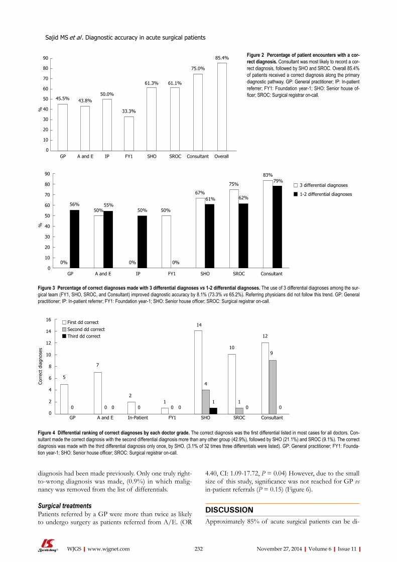
diagnosis had been made previously. Only one truly right-to-wrong diagnosis was made, (0.9%) in which malig-nancy was removed from the list of differentials.
Surgical treatmentsPatients referred by a GP were more than twice as likely to undergo surgery as patients referred from A/E. (OR
4.40, CI: 1.09-17.72, P = 0.04) However, due to the small size of this study, significance was not reached for GP vs in-patient referrals (P = 0.15) (Figure 6).
DISCUSSIONApproximately 85% of acute surgical patients can be di-
232 November 27, 2014|Volume 6|Issue 11|WJGS|www.wjgnet.com
Sajid MS et al . Diagnostic accuracy in acute surgical patients
45.5% 43.8%50.0%
33.3%
61.3% 61.1%
75.0%
85.4%90
80
70
60
50
40
30
20
10
0
%
GP A and E IP FY1 SHO SROC Consultant Overall
Figure 2 Percentage of patient encounters with a cor-rect diagnosis. Consultant was most likely to record a cor-rect diagnosis, followed by SHO and SROC. Overall 85.4% of patients received a correct diagnosis along the primary diagnostic pathway. GP: General practitioner; IP: In-patient referrer; FY1: Foundation year-1; SHO: Senior house of-ficer; SROC: Surgical registrar on-call.
0% 0% 0%
56%50%
55%50% 50%
67%61%
75%
62%
83%79%
GP A and E IP FY1 SHO SROC Consultant
90
80
70
60
50
40
30
20
10
0
%
3 differential diagnoses
1-2 differential diagnoses
Figure 3 Percentage of correct diagnoses made with 3 differential diagnoses vs 1-2 differential diagnoses. The use of 3 differential diagnoses among the sur-gical team (FY1, SHO, SROC, and Consultant) improved diagnostic accuracy by 8.1% (73.3% vs 65.2%). Referring physicians did not follow this trend. GP: General practitioner; IP: In-patient referrer; FY1: Foundation year-1; SHO: Senior house officer; SROC: Surgical registrar on-call.
0 0 0 0 0 0 0 0111
2
7
54
14
10
12
9
16
14
12
10
8
6
4
2
0GP A and E In-Patient FY1 SHO SROC Consultant
First dd correctSecond dd correctThird dd correct
Corr
ect
diag
nose
s
Figure 4 Differential ranking of correct diagnoses by each doctor grade. The correct diagnosis was the first differential listed in most cases for all doctors. Con-sultant made the correct diagnosis with the second differential diagnosis more than any other group (42.9%), followed by SHO (21.1%) and SROC (9.1%). The correct diagnosis was made with the third differential diagnosis only once, by SHO. (3.1% of 32 times three differentials were listed). GP: General practitioner; FY1: Founda-tion year-1; SHO: Senior house officer; SROC: Surgical registrar on-call.
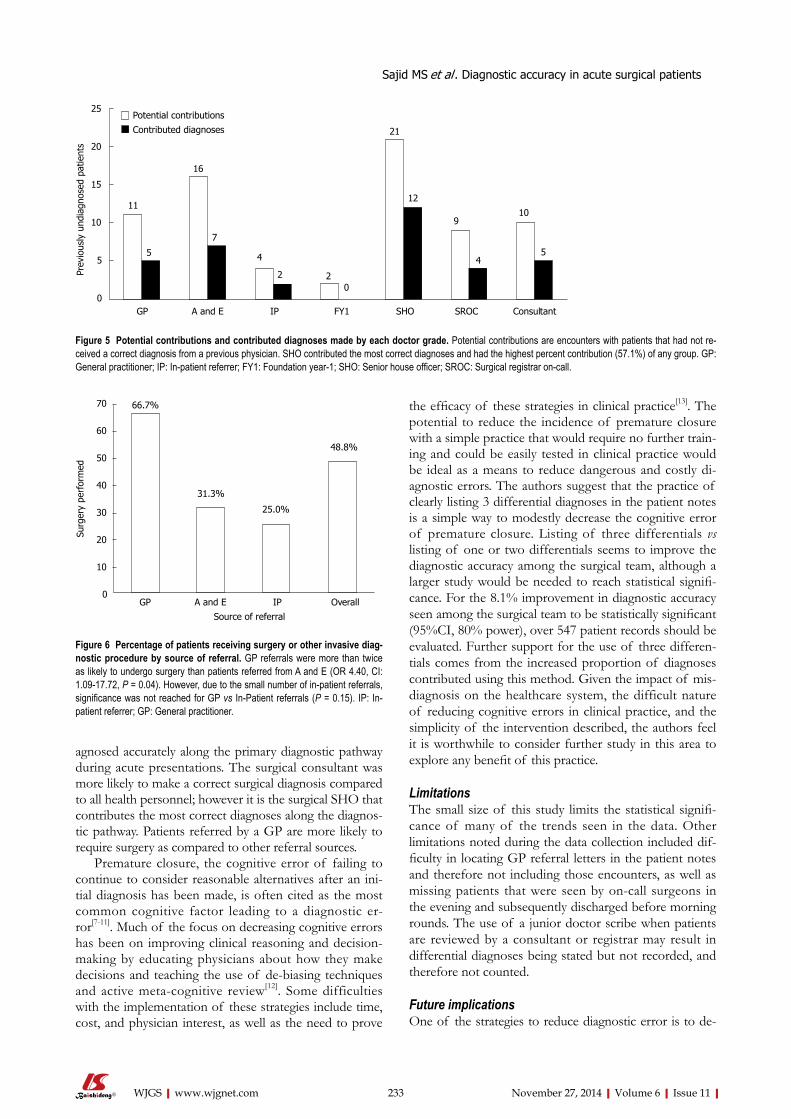
agnosed accurately along the primary diagnostic pathway during acute presentations. The surgical consultant was more likely to make a correct surgical diagnosis compared to all health personnel; however it is the surgical SHO that contributes the most correct diagnoses along the diagnos-tic pathway. Patients referred by a GP are more likely to require surgery as compared to other referral sources.
Premature closure, the cognitive error of failing to continue to consider reasonable alternatives after an ini-tial diagnosis has been made, is often cited as the most common cognitive factor leading to a diagnostic er-ror[7-11]. Much of the focus on decreasing cognitive errors has been on improving clinical reasoning and decision-making by educating physicians about how they make decisions and teaching the use of de-biasing techniques and active meta-cognitive review[12]. Some difficulties with the implementation of these strategies include time, cost, and physician interest, as well as the need to prove
the efficacy of these strategies in clinical practice[13]. The potential to reduce the incidence of premature closure with a simple practice that would require no further train-ing and could be easily tested in clinical practice would be ideal as a means to reduce dangerous and costly di-agnostic errors. The authors suggest that the practice of clearly listing 3 differential diagnoses in the patient notes is a simple way to modestly decrease the cognitive error of premature closure. Listing of three differentials vs listing of one or two differentials seems to improve the diagnostic accuracy among the surgical team, although a larger study would be needed to reach statistical signifi-cance. For the 8.1% improvement in diagnostic accuracy seen among the surgical team to be statistically significant (95%CI, 80% power), over 547 patient records should be evaluated. Further support for the use of three differen-tials comes from the increased proportion of diagnoses contributed using this method. Given the impact of mis-diagnosis on the healthcare system, the difficult nature of reducing cognitive errors in clinical practice, and the simplicity of the intervention described, the authors feel it is worthwhile to consider further study in this area to explore any benefit of this practice.
LimitationsThe small size of this study limits the statistical signifi-cance of many of the trends seen in the data. Other limitations noted during the data collection included dif-ficulty in locating GP referral letters in the patient notes and therefore not including those encounters, as well as missing patients that were seen by on-call surgeons in the evening and subsequently discharged before morning rounds. The use of a junior doctor scribe when patients are reviewed by a consultant or registrar may result in differential diagnoses being stated but not recorded, and therefore not counted.
Future implicationsOne of the strategies to reduce diagnostic error is to de-
233 November 27, 2014|Volume 6|Issue 11|WJGS|www.wjgnet.com
Sajid MS et al . Diagnostic accuracy in acute surgical patients
10
5
9
4
21
12
022
4
7
16
5
11
GP A and E IP FY1 SHO SROC Consultant
25
20
15
10
5
0
Prev
ious
ly u
ndia
gnos
ed p
atie
nts
Potential contributions
Contributed diagnoses
Figure 5 Potential contributions and contributed diagnoses made by each doctor grade. Potential contributions are encounters with patients that had not re-ceived a correct diagnosis from a previous physician. SHO contributed the most correct diagnoses and had the highest percent contribution (57.1%) of any group. GP: General practitioner; IP: In-patient referrer; FY1: Foundation year-1; SHO: Senior house officer; SROC: Surgical registrar on-call.
66.7%
31.3%
25.0%
48.8%
70
60
50
40
30
20
10
0
Surg
ery
perf
orm
ed
GP A and E IP OverallSource of referral
Figure 6 Percentage of patients receiving surgery or other invasive diag-nostic procedure by source of referral. GP referrals were more than twice as likely to undergo surgery than patients referred from A and E (OR 4.40, CI: 1.09-17.72, P = 0.04). However, due to the small number of in-patient referrals, significance was not reached for GP vs In-Patient referrals (P = 0.15). IP: In-patient referrer; GP: General practitioner.
velop pathways for feedback[2,14]. It is particularly important to develop feedback pathways for the junior doctors, as it has been shown that less experienced doctors tend to most over-estimate their diagnostic accuracy[2]. With anonymity removed, the basic design of this study seems well suited to enable feedback to each physician involved in the care of an acute surgical patient. In this way a simple score could be reported as a way to objectively evaluate diagnostic per-formance, with the ultimate goal of self-improvement and future decrease in diagnostic errors. This approach would allow feedback not just after a negative event, as is the case with many feedback systems in place, but would track per-formance in a simple, ongoing manner.
COMMENTSBackgroundAccurate clinical diagnosis in acutely admitted surgical patients is of immense importance because of the necessity of timely surgical interventions such as need of laparotomy, laparoscopy and or organ resection. Inaccurate diagnosis can lead to serious consequences in terms of delayed treatment, prolonged hospital stay, increased operative morbidity or mortality putting excessive strain on the health resources. Any measure which directly or indirectly may influence the diagnostic accuracy in acute surgical patients should be investigated and implemented in a timely manner to avoid these consequences. This article high-lights the value and shortcomings of a referral pathway through which acute surgical patients pass through and get accurately diagnosed for the optimum management.Research frontierVarious studies published on this topic, although reported the diagnostic ac-curacy of different grades of acute surgical team with variable accuracy. This is the first study which investigates the diagnostic accuracy of all sources of referral during the course of management of acutely ill patients such as general practitioners, A/E doctors, surgical juniors, surgical middle grade doctors and eventually surgical consultant on call.Innovations and breakthroughsThe potential to reduce the incidence of mis-diagnosis with a simple practice that would require no further training and could be easily tested in clinical practice would be ideal as a means to reduce dangerous and costly diagnostic errors. The authors suggest that the practice of clearly listing 3 differential diagnoses in the patient notes is a simple way to modestly decrease the cognitive error of premature closure. Listing of three differentials vs listing of one or two differentials seems to improve the diagnostic accuracy among the surgical team, although a larger study would be needed to reach statistical significance. For the 8.1% improvement in diagnostic accuracy seen among the surgical team to be statistically significant (95%CI, 80% power), over 547 patient records should be evaluated. Further support for the use of three dif-ferentials comes from the increased proportion of diagnoses contributed using this method. Given the impact of misdiagnosis on the healthcare system, the difficult nature of reducing cognitive errors in clinical practice, and the simplicity of the intervention described, the authors feel it is worthwhile to consider further study in this area to explore any benefit of this practice. ApplicationsOne of the strategies to reduce diagnostic error is to develop pathways for feedback. It is particularly important to develop feedback pathways for the ju-nior doctors, as it has been shown that less experienced doctors tend to most over-estimate their diagnostic accuracy. With anonymity removed, the basic design of this study seems well suited to enable feedback to each physician in-volved in the care of an acute surgical patient. In this way a simple score could be reported as a way to objectively evaluate diagnostic performance, with the ultimate goal of self-improvement and future decrease in diagnostic errors. This approach would allow feedback not just after a negative event, as is the case
with many feedback systems in place, but would track performance in a simple, ongoing manner.TerminologyFY1: It stands for foundation year 1. The group of junior most surgical doctors in the United Kingdom NHS Trust health system which start clinical practice just after finishing medical school. SHO: It stands for Senior House Officer which a surgical grade after finishing two years of foundation training (FY1 and FY2). SROC: Its stands for Surgical Registrar On Call. Surgical registrar is of variable experience depending upon the step of ladder on training pathway (year 1 to year 8). Peer reviewThis is an interesting article.
REFERENCES1 Saber Tehrani AS, Lee H, Mathews SC, Shore A, Makary
MA, Pronovost PJ, Newman-Toker DE. 25-Year summary of US malpractice claims for diagnostic errors 1986-2010: an analysis from the National Practitioner Data Bank. BMJ Qual Saf 2013; 22: 672-680 [PMID: 23610443 DOI: 10.1136/bmjqs-2012-001550]
2 Berner ES, Graber ML. Overconfidence as a cause of diag-nostic error in medicine. Am J Med 2008; 121: S2-23 [PMID: 18440350 DOI: 10.1016/j.amjmed.2008.01.001]
3 Nuland S. How We Die: Reflections on Life’s Final Chapter. New York, NY: Knopf, 1994: 248
4 Graber ML, Wachter RM, Cassel CK. Bringing diagnosis into the quality and safety equations. JAMA 2012; 308: 1211-1212 [PMID: 23011708]
5 Berner ES, Miller RA, Graber ML. Missed and delayed di-agnoses in the ambulatory setting. Ann Intern Med 2007; 146: 470; author reply 470-471 [PMID: 17371899]
6 Graber ML, Franklin N, Gordon R. Diagnostic error in in-ternal medicine. Arch Intern Med 2005; 165: 1493-1499 [PMID: 16009864]
7 Graber ML, Kissam S, Payne VL, Meyer AN, Sorensen A, Lenfestey N, Tant E, Henriksen K, Labresh K, Singh H. Cognitive interventions to reduce diagnostic error: a narra-tive review. BMJ Qual Saf 2012; 21: 535-557 [PMID: 22543420 DOI: 10.1136/bmjqs-2011-000149]
8 Ely JW, Graber ML, Croskerry P. Checklists to reduce diag-nostic errors. Acad Med 2011; 86: 307-313 [PMID: 21248608 DOI: 10.1097/ACM.0b013e31820824cd]
9 Vázquez-Costa M, Costa-Alcaraz AM. Premature diag-nostic closure: an avoidable type of error. Rev Clin Esp (Barc) 2013; 213: 158-162 [PMID: 22818221 DOI: 10.1016/j.rce.2012.05.012]
10 Eva KW, Link CL, Lutfey KE, McKinlay JB. Swapping horses midstream: factors related to physicians’ changing their minds about a diagnosis. Acad Med 2010; 85: 1112-1117 [PMID: 20592506 DOI: 10.1097/ACM.0b013e3181e16103]
11 Borrell-Carrió F, Epstein RM. Preventing errors in clinical practice: a call for self-awareness. Ann Fam Med 2004; 2: 310-316 [PMID: 15335129]
12 Croskerry P. The importance of cognitive errors in diag-nosis and strategies to minimize them. Acad Med 2003; 78: 775-780 [PMID: 12915363]
13 Nendaz M, Perrier A. Diagnostic errors and flaws in clinical reasoning: mechanisms and prevention in practice. Swiss Med Wkly 2012; 142: w13706 [PMID: 23135902 DOI: 10.4414/smw.2012.13706]
14 Anderson RE, Graber ML. The new kid on the patient safety block: Diagnostic Error in Medicine. NPSF Profes-sional Learning Series. [updated 2011 November 16]. Avail-able from: URL: http://www.npsf.org/wp-content/up-loads/2011/12/PLS_1111_RE.pdf
P- Reviewer: Bludovsky D, Buell JF, Paraskevas KI S- Editor: Ji FF L- Editor: A E- Editor: Lu YJ
234 November 27, 2014|Volume 6|Issue 11|WJGS|www.wjgnet.com
Sajid MS et al . Diagnostic accuracy in acute surgical patients
COMMENTS

Published by Baishideng Publishing Group Inc
World Journal of Gastrointestinal SurgeryWorld J Gastrointest Surg 2014 December 27; 6(12): 235-258
ISSN 1948-9366 (online)
Volume End
235 IntrathoracicesophagojejunostomyusingOrVil™forgastric
adenocarcinomainvolvingtheesophagus
Yajima K, Kanda T, Kosugi S, Kano Y, Ishikawa T, Ichikawa H, Hanyu T, Wakai T
241 Systematicreviewofabsorbablevsnon-absorbablesuturesusedforthe
closureofsurgicalincisions
Sajid MS, McFall MR, Whitehouse PA, Sains PS
248 Inflammatorypseudotumourofthespleenassociatedwithsplenic
tuberculosis
Prieto-Nieto MI, Pérez-Robledo JP, Díaz-San Andrés B, Nistal M,
Rodríguez-Montes JA
253 Caseofbronchoesophagealfistulawithgastricperforationduetomul-
tidrug-resistanttuberculosis
Park CS, Seo KW, Park CR, Nah YW, Suh JH
Contents Monthly Volume 6 Number 12 December 27, 2014
WJGS|www.wjgnet.com I December 27, 2014|Volume 6|Issue 12|
ORIGINAL ARTICLE
SYSTEMATIC REVIEWS
CASE REPORT

Contents
APPENDIX
ABOUT COVER
World Journal of Gastrointestinal SurgeryVolume 6 Number 12 December 27, 2014
I-V Instructionstoauthors
EditorialBoardMemberofWorldJournalofGastrointestinalSurgery ,HelenaMIsoniemi,MD,PhD,Professor,TransplantationandLiverSurgeryClinic,HelsinkiUniversityHospital,box263,Helsinki00029-HUCH,Finland
World Journal of Gastrointestinal Surgery (World J Gastrointest Surg, WJGS, online ISSN 1948-9366, DOI: 10.4240) is a peer-reviewed open access academic journal that aims to guide clinical practice and improve diagnostic and therapeutic skills of clinicians.
WJGS covers topics concerning micro-invasive surgery; laparoscopy; hepatic, biliary, pancreatic and splenic surgery; surgical nutrition; portal hypertension, as well as associated subjects. The current columns of WJGS include editorial, frontier, diagnostic advances, therapeutics advances, field of vision, mini-reviews, review, topic highlight, medical ethics, original articles, case report, clinical case conference (Clinicopathological conference), and autobiography. Priority publication will be given to articles concerning diagnosis and treatment of gastrointestinal surgery diseases. The following aspects are covered: Clinical diagnosis, laboratory diagnosis, differential diagnosis, imaging tests, pathological diagnosis, molecular biological diagnosis, immunological diagnosis, genetic diagnosis, functional diagnostics, and physical diagnosis; and comprehensive therapy, drug therapy, surgical therapy, interventional treatment, minimally invasive therapy, and robot-assisted therapy.
We encourage authors to submit their manuscripts to WJGS. We will give priority to manuscripts that are supported by major national and international foundations and those that are of great basic and clinical significance.
World Journal of Gastrointestinal Surgery is now indexed in PubMed Central, PubMed, Digital Object Identifier, and Directory of Open Access Journals.
I-III EditorialBoard
Xiu-Xia Song, Vice DirectorWorld Journal of Gastrointestinal SurgeryRoom 903, Building D, Ocean International Center, No. 62 Dongsihuan Zhonglu, Chaoyang District, Beijing 100025, ChinaTelephone: +86-10-85381891Fax: +86-10-85381893E-mail: [email protected] Desk: http://www.wjgnet.com/esps/helpdesk.aspxhttp://www.wjgnet.com
PUBLISHERBaishideng Publishing Group Inc8226 Regency Drive, Pleasanton, CA 94588, USATelephone: +1-925-223-8242Fax: +1-925-223-8243E-mail: [email protected] Desk: http://www.wjgnet.com/esps/helpdesk.aspxhttp://www.wjgnet.com
PUBLICATIONDATEDecember 27, 2014
COPYRIGHT© 2014 Baishideng Publishing Group Inc. Articles pub-lished by this Open-Access journal are distributed under the terms of the Creative Commons Attribution Non-commercial License, which permits use, distribution, and reproduction in any medium, provided the original work is properly cited, the use is non commercial and is otherwise in compliance with the license.
SPECIALSTATEMENTAll articles published in journals owned by the Baishideng Publishing Group (BPG) represent the views and opin-ions of their authors, and not the views, opinions or policies of the BPG, except where otherwise explicitly indicated.
INSTRUCTIONSTOAUTHORSFull instructions are available online at http://www.wjgnet.com/1948-9366/g_info_20100305152206.htm
ONLINESUBMISSIONhttp://www.wjgnet.com/esps/
NAMEOFJOURNALWorld Journal of Gastrointestinal Surgery
ISSNISSN 1948-9366 (online)
LAUNCHDATENovember 30, 2009
FREQUENCYMonthly
EDITOR-IN-CHIEFTimothy M Pawlik, MD, MPH, FACS, Associate Professor of Surgery and Oncology, Hepatobiliary Surgery Program Director, Director, Johns Hopkins Medicine Liver Tumor Center Multi-Disciplinary Clinic, Co-Director of Center for Surgical Trials and Outcomes Research, Johns Hopkins Hospital, 600 N. Wolfe Street, Harvey 611, Baltimore, MD 21287, United States
EDITORIALOFFICEJin-Lei Wang, Director
EDITORS FOR THIS ISSUE
Responsible Assistant Editor: Xiang Li Responsible Science Editor: Fang-Fang JiResponsible Electronic Editor: Su-Qing Liu Proofing Editorial Office Director: Xiu-Xia SongProofing Editor-in-Chief: Lian-Sheng Ma
AIM AND SCOPE
FLYLEAF
INDEXING/ABSTRACTING
WJGS|www.wjgnet.com II December 27, 2014|Volume 6|Issue 12|
Intrathoracic esophagojejunostomy using OrVil™ for gastric adenocarcinoma involving the esophagus
Kazuhito Yajima, Tatsuo Kanda, Shin-ichi Kosugi, Yosuke Kano, Takashi Ishikawa, Hiroshi Ichikawa, Takaaki Hanyu, Toshifumi Wakai
Kazuhito Yajima, Tatsuo Kanda, Shin-ichi Kosugi, Yosuke Kano, Takashi Ishikawa, Hiroshi Ichikawa, Takaaki Hanyu, Toshifumi Wakai, Division of Digestive and General Surgery, Niigata University Graduate School of Medical and Dental Sciences, Niigata 951-8510, JapanAuthor contributions: Yajima K performed this surgery and rote the paper; Kanda T and Kosugi S contributed assistant of this surgery; Kano Y, Ishikawa T, Ichikawa H and Hanyu T coordinated the surgery; Wakai T contributed the final coordination of this paper. Correspondence to: Kazuhito Yajima, MD, PhD, Division of Digestive and General Surgery, Niigata University Graduate School of Medical and Dental Sciences, 1-757 Asahimachi-dori, Niigata 951-8510, Japan. [email protected]: +81-25-2272228 Fax: +81-25-2270779Received: June 13, 2014 Revised: October 9, 2014Accepted: November 17, 2014Published online: December 27, 2014
AbstractAIM: To demonstrate a new surgical technique of lower mediastinal lymphadenectomy and intrathoracic anastomosis of esophagojejunostomy using OrVil™.
METHODS: After a total median phrenotomy, the supradiaphragmatic and lower thoracic paraesophageal lymph nodes were transhiatally dissected. The esophagus was cut off using a liner stapler and OrVil™was inserted. Finally, end-to-side esophagojejunostomy was created by using a circular stapler. From July 2009, we adopted this surgical technique for five patients with gastric cancer involving the lower esophagus.
RESULTS: The median operation time was 314 min (range; 210-367 min), and median blood loss was 210 mL (range; 100-838 mL). The median numbers of dissected lower mediastinal nodes were 3 (range; 1-10). None of the patients had postoperative complications including anastomotic leakage and stenosis. The
median hospital stay was 16 d (range: 15-20 d). The median length of esophageal involvement was 14 mm (range: 6-48 mm) and that of the resected esophagus was 40 mm (range: 35-55 mm); all resected specimens had tumor-free margins.
CONCLUSION: This surgical technique is easy and safe intrathoracic anastomosis for the patients with gastric adenocarcinoma involving the lower esophagus.
© 2014 Baishideng Publishing Group Inc. All rights reserved.
Key words: Gastric cancer; Esophageal invasion; Lower mediastinal lymphadenectomy; OrVil™; Intrathoracic anastomosis
Core tip: We report a new technique of lower in-trathoracic anastomosis through the transhiatal approach using OrVil™ for five patients with gastric adenocarcinoma involving the esophagus. This surgical technique is an easy and safe method to create lower intrathoracic anastomosis.
Yajima K, Kanda T, Kosugi S, Kano Y, Ishikawa T, Ichikawa H, Hanyu T, Wakai T. Intrathoracic esophagojejunostomy using OrVil™ for gastric adenocarcinoma involving the esophagus. World J Gastrointest Surg 2014; 6(12): 235-240 Available from: URL: http://www.wjgnet.com/1948-9366/full/v6/i12/235.htm DOI: http://dx.doi.org/10.4240/wjgs.v6.i12.235
INTRODUCTIONAdenocarcinoma of the esophagogastric junction (EGJ) is still uncommon in Japan although the number of patients with this disease has tended to increase[1-6]. For gastric cancer surgery, ensuring safe cancer-free margins and dedicated systematic lymphadenectomy are both
ORIGINAL ARTICLE
Submit a Manuscript: http://www.wjgnet.com/esps/Help Desk: http://www.wjgnet.com/esps/helpdesk.aspxDOI: 10.4240/wjgs.v6.i12.235
World J Gastrointest Surg 2014 December 27; 6(12): 235-240ISSN 1948-9366 (online)
© 2014 Baishideng Publishing Group Inc. All rights reserved.
235 December 27, 2014|Volume 6|Issue 12|WJGS|www.wjgnet.com
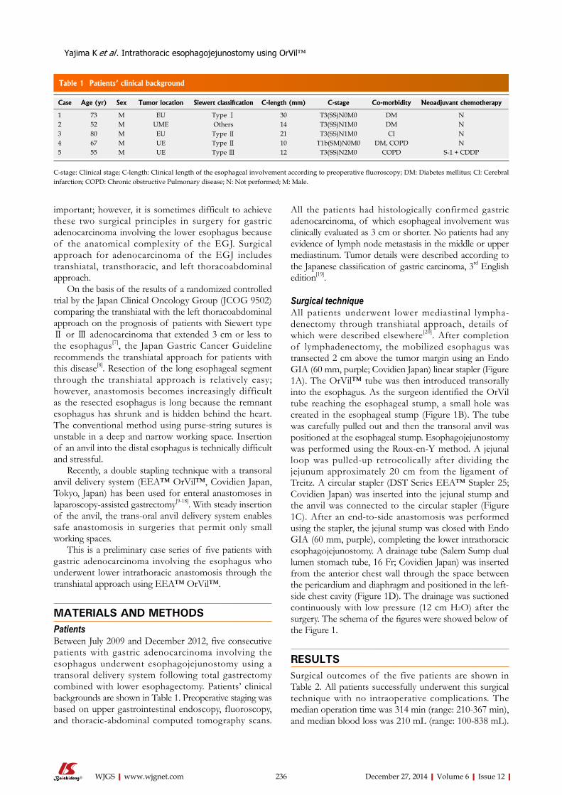
important; however, it is sometimes difficult to achieve these two surgical principles in surgery for gastric adenocarcinoma involving the lower esophagus because of the anatomical complexity of the EGJ. Surgical approach for adenocarcinoma of the EGJ includes transhiatal, transthoracic, and left thoracoabdominal approach.
On the basis of the results of a randomized controlled trial by the Japan Clinical Oncology Group (JCOG 9502) comparing the transhiatal with the left thoracoabdominal approach on the prognosis of patients with Siewert type Ⅱ or Ⅲ adenocarcinoma that extended 3 cm or less to the esophagus[7], the Japan Gastric Cancer Guideline recommends the transhiatal approach for patients with this disease[8]. Resection of the long esophageal segment through the transhiatal approach is relatively easy; however, anastomosis becomes increasingly difficult as the resected esophagus is long because the remnant esophagus has shrunk and is hidden behind the heart. The conventional method using purse-string sutures is unstable in a deep and narrow working space. Insertion of an anvil into the distal esophagus is technically difficult and stressful.
Recently, a double stapling technique with a transoral anvil delivery system (EEA™ OrVil™, Covidien Japan, Tokyo, Japan) has been used for enteral anastomoses in laparoscopy-assisted gastrectomy[9-18]. With steady insertion of the anvil, the trans-oral anvil delivery system enables safe anastomosis in surgeries that permit only small working spaces.
This is a preliminary case series of five patients with gastric adenocarcinoma involving the esophagus who underwent lower intrathoracic anastomosis through the transhiatal approach using EEA™ OrVil™.
MATERIALS AND METHODSPatientsBetween July 2009 and December 2012, five consecutive patients with gastric adenocarcinoma involving the esophagus underwent esophagojejunostomy using a transoral delivery system following total gastrectomy combined with lower esophagectomy. Patients’ clinical backgrounds are shown in Table 1. Preoperative staging was based on upper gastrointestinal endoscopy, fluoroscopy, and thoracic-abdominal computed tomography scans.
All the patients had histologically confirmed gastric adenocarcinoma, of which esophageal involvement was clinically evaluated as 3 cm or shorter. No patients had any evidence of lymph node metastasis in the middle or upper mediastinum. Tumor details were described according to the Japanese classification of gastric carcinoma, 3rd English edition[19].
Surgical techniqueAll patients underwent lower mediastinal lympha-denectomy through transhiatal approach, details of which were described elsewhere[20]. After completion of lymphadenectomy, the mobilized esophagus was transected 2 cm above the tumor margin using an Endo GIA (60 mm, purple; Covidien Japan) linear stapler (Figure 1A). The OrVil™ tube was then introduced transorally into the esophagus. As the surgeon identified the OrVil tube reaching the esophageal stump, a small hole was created in the esophageal stump (Figure 1B). The tube was carefully pulled out and then the transoral anvil was positioned at the esophageal stump. Esophagojejunostomy was performed using the Roux-en-Y method. A jejunal loop was pulled-up retrocolically after dividing the jejunum approximately 20 cm from the ligament of Treitz. A circular stapler (DST Series EEA™ Stapler 25; Covidien Japan) was inserted into the jejunal stump and the anvil was connected to the circular stapler (Figure 1C). After an end-to-side anastomosis was performed using the stapler, the jejunal stump was closed with Endo GIA (60 mm, purple), completing the lower intrathoracic esophagojejunostomy. A drainage tube (Salem Sump dual lumen stomach tube, 16 Fr; Covidien Japan) was inserted from the anterior chest wall through the space between the pericardium and diaphragm and positioned in the left-side chest cavity (Figure 1D). The drainage was suctioned continuously with low pressure (12 cm H2O) after the surgery. The schema of the figures were showed below of the Figure 1.
RESULTSSurgical outcomes of the five patients are shown in Table 2. All patients successfully underwent this surgical technique with no intraoperative complications. The median operation time was 314 min (range: 210-367 min), and median blood loss was 210 mL (range: 100-838 mL).
Yajima K et al . Intrathoracic esophagojejunostomy using OrVil™
236 December 27, 2014|Volume 6|Issue 12|WJGS|www.wjgnet.com
Table 1 Patients’ clinical background
Case Age (yr) Sex Tumor location Siewert classification C-length (mm) C-stage Co-morbidity Neoadjuvant chemotherapy
1 73 M EU Type Ⅰ 30 T3(SS)N0M0 DM N2 52 M UME Others 14 T3(SS)N1M0 DM N3 80 M EU Type Ⅱ 21 T3(SS)N1M0 CI N4 67 M UE Type Ⅱ 10 T1b(SM)N0M0 DM, COPD N5 55 M UE Type Ⅲ 12 T3(SS)N2M0 COPD S-1 + CDDP
C-stage: Clinical stage; C-length: Clinical length of the esophageal involvement according to preoperative fluoroscopy; DM: Diabetes mellitus; CI: Cerebral infarction; COPD: Chronic obstructive Pulmonary disease; N: Not performed; M: Male.

The median interval to water intake was 2 d (range: 1-3 d) and that to oral intake was 7 d (range: 6-10 d). None of the patients had postoperative surgical complications.
Postoperative fluorography demonstrated no leakage or stenosis of the esophagojejunal anastomosis in all five patients. Figure 2 shows representative fluorography of
237 December 27, 2014|Volume 6|Issue 12|WJGS|www.wjgnet.com
Figure 1 Intraabdominal findings of an esophagojejunostomy using a circular stapler. A: Transection of the esophagus was performed approximately 2 cm proximal to the tumor margin using an Endo GIA (60 purple; Covidien, Tokyo, Japan) linear stapler; B: The OrVil™ tube was then introduced transorally into the esophagus; C: A circular stapler (DST Series EEA Stapler 25; Covidien Japan) was inserted into the jejunal stump and the anvil was connected to the circular stapler; D: After the anastomosis, a Salem Sump Dual Lumen Stomach Tube (16 Fr; Covidien Japan) for the anastomotic site was inserted from the anterior chest wall through the space between the pericardium and diaphragm and positioned in the left-sided chest cavity (white arrow). Eso: Esophagus; DH: Diaphragm.
A
B
C
D
Liner stapler
Liver DH Eso
Stomach
DH
LiverDH
DH
OrVil system
Pericardia
Pericardia
DH
DH
Liver
JejunumCircular stapler
Anastomosis
Drain
Jejunum
DH
Liver
Jejunum
Yajima K et al . Intrathoracic esophagojejunostomy using OrVil™

anastomotic leakage and anastomotic stenosis occurred in one patient in their series of 21 patients undergoing laparoscopy-assisted total gastrectomy. From these reports, gastrojejunostomy using OrVil™ was acceptable in terms of fewer anastomotic complications. We successfully performed intrathoracic anastomosis with Roux-en-Y reconstruction in five patients without any anastomotic complications.
Complete resection with no residual tumor remains the only treatment that can lead to cure in gastric cancer[21]. Obtaining a safe free margin of the distal esophagus is one of the most important problems, especially in patients with junctional cancer invading the esophagus. On the other hand, the correlation between the length of esophageal resection and free surgical margin has not been well described. In the JCOG9502 trial, the median length of the resected esophagus was 4.2 cm in the transhiatal approach, with two having positive proximal margin[7]. In the present study, the median length of the resected esophagus was 40 mm and all patients had a negative proximal margin. In our technique exposing the lower mediastinum by dissection using a lower mediastinal lymphadenectomy, a wide and good view was obtained. Using this space, we could easily cut the esophagus and insert the anvil head transorally with a sufficient tumor-free margin.
Insertion of an anvil into the distal esophagus is technically difficult and stressful for the esophagoje-junostomy. Kunisaki et al[10] and Liao et al[11] reported that total operation time and time of reconstruction were significantly shortened when comparing mini-laparotomy and a transorally inserted anvil in laparoscopy-assisted total gastrectomy esophagojejunostomy using OrVil™. At the same way, OrVil™ might decrease the time of reconstruction also in conventional open gastrectomy when intrathoracic anastomosis. In our series, unfortunately, the time of anastomosis was not measured, there were no trouble concerning the insertion of the OrVil and anastomosis using EEA™ during surgery. In our experience, lower intrathoracic esophagojejunostomy using OrVil™ was less stressful technique.
In conclusion, we report the surgical techniques of the EEA™ OrVil™ stapler in end-to-side anastomosis for esophagojejunostomy. We believe that this technique is especially useful for patients with lower intrathoracic anastomosis, in whom conventional anastomosis is
the esophagojejunal anastomosis. The median hospital stay was 16 d (range: 15-20 d).
The median length of esophageal involvement was 14 mm (range: 6-48 mm) and that of the resected esophagus was 40 mm (range: 35-55 mm); all resected specimens had tumor-free margins.
DISCUSSIONWe have herein reported the use of the EEA™ OrVil™ stapler in end-to-side anastomosis for esophagojejunostomy for patients with gastric adenocarcinoma involving the esophagus. To the best of our knowledge, this is the first report of the use of a transoral anvil in lower intrathoracic esophagojejunostomy and our preliminary results showed that this technique was feasible and safe.
By literature review of PubMed database using the keywords “OrVil™”, “gastric cancer”, and “esophago-jejunostomy”, there were ten original reports describing the outcomes of esophagojejunostomy using a transoral anvil (EEA™ OrVil™) so far. However, all five reports addressed the issues of esophagojejunostomy after laparoscopic total gastrectomy for gastric cancer[9-18]. Jeong et al[9] reported that of the 16 patients who underwent esophagojejunostomy using OrVil™ in laparoscopy-assisted total gastrectomy, no patient had intraoperative complications or anastomotic complications, including leakage and stenosis. Liao et al[11] also supported the safety of esophagojejunostomy using a transoral anvil:
238 December 27, 2014|Volume 6|Issue 12|WJGS|www.wjgnet.com
Table 2 Surgical outcomes
Case Surgical Lymph- Residual Operation time Blood loss Start of foods Hospital stay E-length P-length
procedures adenectomy tumor (min) (mL) (d) (d) (mm) (mm)1 Total GR, RY D2 R0 367 838 10 20 55 482 Total GR, RY D2 R0 314 270 10 16 40 143 Total GR, RY D2 R0 210 210 7 15 52 224 Proximal GR, JI D1+ R0 314 100 7 18 36 65 Total GR, RY D2 R0 277 140 6 15 35 10
Total GR: Total gastrectomy; Proximal GR: Proximal gastrectomy; RY: Roux-en Y reconstruction; JI: Jejunal interposition; R0: No residual tumor; E-length: Length of resected esophagus; P-length: Pathological length of esophageal involvement.
Figure 2 Postoperative fluoroscopy. Anastomosis of the esophagojejunostomy was 6.4 cm above the diaphragm (black arrowhead) with no anastomotic leakage or stenosis.
Yajima K et al . Intrathoracic esophagojejunostomy using OrVil™
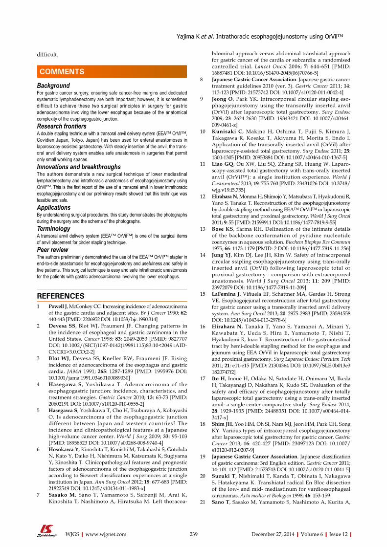
difficult.
COMMENTSBackgroundFor gastric cancer surgery, ensuring safe cancer-free margins and dedicated systematic lymphadenectomy are both important; however, it is sometimes difficult to achieve these two surgical principles in surgery for gastric adenocarcinoma involving the lower esophagus because of the anatomical complexity of the esophagogastric junction. Research frontiersA double stapling technique with a transoral anvil delivery system (EEA™ OrVil™, Covidien Japan, Tokyo, Japan) has been used for enteral anastomoses in laparoscopy-assisted gastrectomy. With steady insertion of the anvil, the trans-oral anvil delivery system enables safe anastomosis in surgeries that permit only small working spaces.Innovations and breakthroughsThe authors demonstrate a new surgical technique of lower mediastinal lymphadenectomy and intrathoracic anastomosis of esophagojejunostomy using OrVil™. This is the first report of the use of a transoral anvil in lower intrathoracic esophagojejunostomy and our preliminary results showed that this technique was feasible and safe.ApplicationsBy understanding surgical procedures, this study demonstrates the photographs during the surgery and the schema of the photographs.TerminologyA transoral anvil delivery system (EEA™ OrVil™) is one of the surgical items of anvil placement for circler stapling technique. Peer reviewThe authors preliminarily demonstrated the use of the EEA™ OrVil™ stapler in end-to-side anastomosis for esophagojejunostomy and usefulness and safely in five patients. This surgical technique is easy and safe intrathoracic anastomosis for the patients with gastric adenocarcinoma involving the lower esophagus.
REFERENCES1 Powell J, McConkey CC. Increasing incidence of adenocarcinoma
of the gastric cardia and adjacent sites. Br J Cancer 1990; 62: 440-443 [PMID: 2206952 DOI: 10.1038/bjc.1990.314]
2 Devesa SS, Blot WJ, Fraumeni JF. Changing patterns in the incidence of esophageal and gastric carcinoma in the United States. Cancer 1998; 83: 2049-2053 [PMID: 9827707 DOI: 10.1002/(SICI)1097-0142(19981115)83:10<2049::AID-CNCR1>3.0.CO;2-2]
3 Blot WJ, Devesa SS, Kneller RW, Fraumeni JF. Rising incidence of adenocarcinoma of the esophagus and gastric cardia. JAMA 1991; 265: 1287-1289 [PMID: 1995976 DOI: 10.1001/jama.1991.03460100089030]
4 Hasegawa S , Yoshikawa T. Adenocarcinoma of the esophagogastric junction: incidence, characteristics, and treatment strategies. Gastric Cancer 2010; 13: 63-73 [PMID: 20602191 DOI: 10.1007/s10120-010-0555-2]
5 Hasegawa S, Yoshikawa T, Cho H, Tsuburaya A, Kobayashi O. Is adenocarcinoma of the esophagogastric junction different between Japan and western countries? The incidence and clinicopathological features at a Japanese high-volume cancer center. World J Surg 2009; 33: 95-103 [PMID: 18958523 DOI: 10.1007/s00268-008-9740-4]
6 Hosokawa Y, Kinoshita T, Konishi M, Takahashi S, Gotohda N, Kato Y, Daiko H, Nishimura M, Katsumata K, Sugiyama Y, Kinoshita T. Clinicopathological features and prognostic factors of adenocarcinoma of the esophagogastric junction according to Siewert classification: experiences at a single institution in Japan. Ann Surg Oncol 2012; 19: 677-683 [PMID: 21822549 DOI: 10.1245/s10434-011-1983-x]
7 Sasako M, Sano T, Yamamoto S, Sairenji M, Arai K, Kinoshita T, Nashimoto A, Hiratsuka M. Left thoracoa-
bdominal approach versus abdominal-transhiatal approach for gastric cancer of the cardia or subcardia: a randomised controlled trial. Lancet Oncol 2006; 7: 644-651 [PMID: 16887481 DOI: 10.1016/S1470-2045(06)70766-5]
8 Japanese Gastric Cancer Association. Japanese gastric cancer treatment guidelines 2010 (ver. 3). Gastric Cancer 2011; 14: 113-123 [PMID: 21573742 DOI: 10.1007/s10120-011-0042-4]
9 Jeong O, Park YK. Intracorporeal circular stapling eso-phagojejunostomy using the transorally inserted anvil (OrVil) after laparoscopic total gastrectomy. Surg Endosc 2009; 23: 2624-2630 [PMID: 19343421 DOI: 10.1007/s00464-009-0461-z]
10 Kunisaki C, Makino H, Oshima T, Fujii S, Kimura J, Takagawa R, Kosaka T, Akiyama H, Morita S, Endo I. Application of the transorally inserted anvil (OrVil) after laparoscopy-assisted total gastrectomy. Surg Endosc 2011; 25: 1300-1305 [PMID: 20953884 DOI: 10.1007/s00464-010-1367-5]
11 Liao GQ, Ou XW, Liu SQ, Zhang SR, Huang W. Laparo-scopy-assisted total gastrectomy with trans-orally inserted anvil (OrVil™): a single institution experience. World J Gastroenterol 2013; 19: 755-760 [PMID: 23431026 DOI: 10.3748/wjg.v19.i5.755]
12 Hirahara N, Monma H, Shimojo Y, Matsubara T, Hyakudomi R, Yano S, Tanaka T. Reconstruction of the esophagojejunostomy by double stapling method using EEA™ OrVil™ in laparoscopic total gastrectomy and proximal gastrectomy. World J Surg Oncol 2011; 9: 55 [PMID: 21599911 DOI: 10.1186/1477-7819-9-55]
13 Bose KS, Sarma RH. Delineation of the intimate details of the backbone conformation of pyridine nucleotide coenzymes in aqueous solution. Biochem Biophys Res Commun 1975; 66: 1173-1179 [PMID: 2 DOI: 10.1186/1477-7819-11-256]
14 Jung YJ, Kim DJ, Lee JH, Kim W. Safety of intracorporeal circular stapling esophagojejunostomy using trans-orally inserted anvil (OrVil) following laparoscopic total or proximal gastrectomy - comparison with extracorporeal anastomosis. World J Surg Oncol 2013; 11: 209 [PMID: 23972079 DOI: 10.1186/1477-7819-11-209]
15 LaFemina J, Viñuela EF, Schattner MA, Gerdes H, Strong VE. Esophagojejunal reconstruction after total gastrectomy for gastric cancer using a transorally inserted anvil delivery system. Ann Surg Oncol 2013; 20: 2975-2983 [PMID: 23584558 DOI: 10.1245/s10434-013-2978-6]
16 Hirahara N, Tanaka T, Yano S, Yamanoi A, Minari Y, Kawabata Y, Ueda S, Hira E, Yamamoto T, Nishi T, Hyakudomi R, Inao T. Reconstruction of the gastrointestinal tract by hemi-double stapling method for the esophagus and jejunum using EEA OrVil in laparoscopic total gastrectomy and proximal gastrectomy. Surg Laparosc Endosc Percutan Tech 2011; 21: e11-e15 [PMID: 21304364 DOI: 10.1097/SLE.0b013e31820747f2]
17 Ito H, Inoue H, Odaka N, Satodate H, Onimaru M, Ikeda H, Takayanagi D, Nakahara K, Kudo SE. Evaluation of the safety and efficacy of esophagojejunostomy after totally laparoscopic total gastrectomy using a trans-orally inserted anvil: a single-center comparative study. Surg Endosc 2014; 28: 1929-1935 [PMID: 24488351 DOI: 10.1007/s00464-014-3417-x]
18 Shim JH, Yoo HM, Oh SI, Nam MJ, Jeon HM, Park CH, Song KY. Various types of intracorporeal esophagojejunostomy after laparoscopic total gastrectomy for gastric cancer. Gastric Cancer 2013; 16: 420-427 [PMID: 23097123 DOI: 10.1007/s10120-012-0207-9]
19 Japanese Gastric Cancer Association. Japanese classification of gastric carcinoma: 3rd English edition. Gastric Cancer 2011; 14: 101-112 [PMID: 21573743 DOI: 10.1007/s10120-011-0041-5]
20 Suzuki T, Nishimaki T, Kanda T, Obinata I, Nakagawa S, Hatakeyama K. Transhiatal radical En Bloc dissection of the low- and mid- mediastinum for vardioesophageal carcinomas. Acta medica et Biologica 1998; 46: 153-159
21 Sano T, Sasako M, Yamamoto S, Nashimoto A, Kurita A,
239 December 27, 2014|Volume 6|Issue 12|WJGS|www.wjgnet.com
COMMENTS
Yajima K et al . Intrathoracic esophagojejunostomy using OrVil™
Hiratsuka M, Tsujinaka T, Kinoshita T, Arai K, Yamamura Y, Okajima K. Gastric cancer surgery: morbidity and mortality results from a prospective randomized controlled trial
comparing D2 and extended para-aortic lymphadenectomy--Japan Clinical Oncology Group study 9501. J Clin Oncol 2004; 22: 2767-2773 [PMID: 15199090 DOI: 10.1200/JCO.2004.10.184]
P- Reviewer: Aoyagi K, Tiberio GAM S- Editor: Tian YL L- Editor: A E- Editor: Liu SQ
240 December 27, 2014|Volume 6|Issue 12|WJGS|www.wjgnet.com
Yajima K et al . Intrathoracic esophagojejunostomy using OrVil™
Systematic review of absorbable vs non-absorbable sutures used for the closure of surgical incisions
Muhammad S Sajid, Malcolm R McFall, Pauline A Whitehouse, Parv S Sains
Muhammad S Sajid, Western Sussex Hospitals NHS Trust, Washington Suite, North Wing, Worthing Hospital, BN11 2DH West Sussex, United KingdomMuhammad S Sajid, Malcolm R McFall, Pauline A Whitehouse, Parv S Sains, Department of General, Endoscopic and Laparoscopic Colorectal Surgery, Worthing Hospital, Worthing, BN11 2DH West Sussex, United KingdomAuthor contributions: All authors contributed to this paper.Correspondence to: Mr. Muhammad S Sajid, Western Sussex Hospitals NHS Trust, Washington Suite, North Wing, Worthing Hospital, BN11 2DH West Sussex, United Kingdom. [email protected]: +44-01903-205111 Fax: +44-01903-285010Received: August 7, 2014 Revised: October 22, 2014 Accepted: October 31, 2014Published online: December 27, 2014
AbstractAIM: To report a systematic review of published randomized controlled trials (RCTs) investigating the role of absorbable suture (AS) against non-AS (NAS) used for the closure of surgical incisions.
METHODS: RCTs investigating the use of AS vs NAS for the closure of surgical incisions were statistically analysed based upon the principles of meta-analysis and the summated outcomes were represented as OR.
RESULTS: The systematic search of medical literature yielded 10 RCTs on 1354 patients. Prevalence of wound infection (OR = 0.97; 95%CI: 0.56, 1.69; Z = 0.11; P = 0.92) and operative morbidity (P = 0.45) was comparable in both groups. Nonetheless, the use of AS lead to lower risk of wound break-down (OR = 0.12; 95%CI: 0.04, 0.39; Z = 3.52; P < 0.0004). CONCLUSION: This meta-analysis of 10 RCTs demon-strates that the use of AS is similar to NAS for skin closure for surgical site infection and other operative morbidities. AS do not increase the risk of skin wound dehiscence,
rather lead to a reduced risk of wound dehiscence compared to NAS.
© 2014 Baishideng Publishing Group Inc. All rights reserved.
Key words: Skin closure; Surgical site infection; Wound dehiscence; Absorbable sutures; Non-absorbable suture
Core tip: Based upon the meta-analysis of 10 controlled trials, the absorbable sutures (AS) are similar to non-AS (NAS) for skin closure in cases of wound infection and other complications. AS do not increase the risk of skin wound dehiscence, rather leads to a reduced risk of wound break-down compared to NAS.
Sajid MS, McFall MR, Whitehouse PA, Sains PS. Systematic review of absorbable vs non-absorbable sutures used for the closure of surgical incisions. World J Gastrointest Surg 2014; 6(12): 241-247 Available from: URL: http://www.wjgnet.com/1948-9366/full/v6/i12/241.htm DOI: http://dx.doi.org/10.4240/wjgs.v6.i12.241
INTRODUCTIONA number of studies have been reported in search of improving the skin closure related outcome measures following various surgical procedures, and due to this fact the skin closure techniques are evolving vastly and immensely, predominantly over the last few decades. Innumerable skin closure methods reported in medical literature include continuous stitch closure, interrupted stitch closure, full thickness closure, sub-cuticular closure, primary closure, secondary closure, vacuum assisted closure, glue assisted closure, skin clips or staples closure, simple suture vs mattress sutures, steri-strips closure, absorbable or non-absorbable suture (NAS) closure and other innovative methods[1-13]. These manifold practices of skin approximation after surgical procedures can
SYSTEMATIC REVIEWS
Submit a Manuscript: http://www.wjgnet.com/esps/Help Desk: http://www.wjgnet.com/esps/helpdesk.aspxDOI: 10.4240/wjgs.v6.i12.241
World J Gastrointest Surg 2014 December 27; 6(12): 241-247ISSN 1948-9366 (online)
© 2014 Baishideng Publishing Group Inc. All rights reserved.
241 December 27, 2014|Volume 6|Issue 12|WJGS|www.wjgnet.com
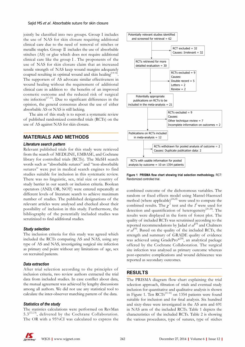
jointly be classified into two groups. Group I includes the use of NAS for skin closure requiring additional clinical care due to the need of removal of stitches or metallic staples. Group Ⅱ includes the use of absorbable stitches (AS) or glue which does not require additional clinical care like the group Ⅰ. The proponents of the use of NAS for skin closure claim that an increased tensile strength of NAS keep wound margins adequately coapted resulting in optimal wound and skin healing[14-16]. The supporters of AS advocate similar effectiveness in wound healing without the requirement of additional clinical care in addition to the benefits of an improved cosmetic outcome and the reduced risk of surgical site infection[17-21]. Due to significant differences in the opinion, the general consensus about the use of either absorbable AS or NAS is still lacking.
The aim of this study is to report a systematic review of published randomized controlled trials (RCTs) on the use of AS against NAS for skin closure.
MATERIALS AND METHODSLiterature search patternRelevant published trials for this study were retrieved from the search of MEDLINE, EMBASE, and Cochrane library for controlled trials (RCTs). The MeSH search words such as “absorbable sutures” and “non-absorbable sutures” were put in medical search engines to find studies suitable for inclusion in this systematic review. There was no linguistic, sex, trial size or country of study barrier in our search or inclusion criteria. Boolean operators (AND, OR, NOT) were entered repeatedly at different levels of literature search to achieve maximum number of studies. The published designations of the relevant articles were analysed and checked about their possibility of inclusion in this study. Furthermore, the bibliography of the potentially included studies was scrutinized to find additional studies.
Study selectionThe inclusion criteria for this study was agreed which included the RCTs comparing AS and NAS, using any type of AS and NAS, investigating surgical site infection as primary end point without any limitations of age, sex on recruited patients.
Data extractionAfter trial selection according to the principles of inclusion criteria, two review authors extracted the trial data from included studies. In case conflict about data, the mutual agreement was achieved by lengthy discussions among all authors. We did not use any statistical tool to calculate the inter-observer matching pattern of the data.
Statistics of the studyThe statistics calculations were performed on RevMan 5.3[22,23], delivered by the Cochrane Collaboration. The OR with a 95%CI was calculated to express the
combined outcome of the dichotomous variables. The random or fixed effects model using Mantel-Haenszel method (where applicable)[24,25] were used to compute the combined results. The χ 2 test and the I2 were used for detection and quantification of heterogeneity[26-28]. The results were displayed in the form of forest plot. The quality of included RCTs was scrutinised according to the reported recommendations by Jadad et al[29] and Chalmers et al[30]. Based on the quality of the included RCTs, the strength and summary of GRADE quality of evidence was achieved using GradePro®[28], an analytical package offered by the Cochrane Collaboration. The surgical site infection was analysed as primary outcome whereas post-operative complications and wound dehiscence was reported as secondary outcomes.
RESULTSThe PRISMA diagram flow chart explaining the trial selection approach, filtration of trials and eventual study inclusion for quantitative and qualitative analysis is shown in Figure 1. Ten RCTs[31-41] on 1354 patients were found suitable for inclusion and for final analysis. Six hundred and sixty-three were investigated in the AS arm and 691 in NAS arm of the included RCTs. Table 1 depicts the characteristics of the included RCTs. Table 2 is showing the various procedures, type of sutures, type of stiches
Sajid MS et al . Absorbable suture for skin closure
242 December 27, 2014|Volume 6|Issue 12|WJGS|www.wjgnet.com
Potentially relevant studies identified and screened for retrieval = 62
RCT excluded = 32Causes: Irrelevant = 32
RCTs retrieved for more detailed evaluation = 30
Potentially appropriate publications on RCTs to be
included in the meta-analysis = 21
RCTs excluded = 9Causes:Double record = 5Letters = 2Review = 2
RCTs excluded = 9Causes:Other technique review = 7Incomplete information on outcomes = 2
Publications on RCTs included in meta-analysis = 12
RCTs withdrawn for pooled analysis of outcome = 2Causes: Duplicate publication data: 2
RCTs with usable information for pooled analysis by outcome = 10 on 1354 patients
Figure 1 PRISMA flow chart showing trial selection methodology. RCT: Randomized controlled trial.

used in included RCTs.
Methodological quality of included studiesInadequate randomization approach, improper concealment in the process of allocation, absence of power calculations, lack of utilization of single or double blinding and lastly lack of reporting of IIT were major factors responsible for scoring the majority of included RTCs of poor quality (Table 3). GRADE[31] quality of evidence is shown in Figure 2.
Surgical site infectionThere was no heterogeneity [Tau2 = 0.23, χ 2 = 12.12, γ =
8, (P = 0.15); I2 = 34%] among RCTs that contributed to the combined calculation of this variable. In the random effects model (OR = 0.97; 95%CI: 0.56, 1.69; Z = 0.11; P = 0.92; Figure 3), the risk of surgical site infection was statistically similar in both groups. Although the AS lead to lower incidence of wound infection but it failed to reach at statistical significance.
Postoperative complicationsCombined analysis showed significant statistical heterogeneity [Tau2 = 0.61, χ 2 = 20.57, γ = 9, (P = 0.01); I2 = 56%] among included RCTs. Therefore, in the random effects model (OR = 0.77; 95%CI: 0.39, 1.52;
243 December 27, 2014|Volume 6|Issue 12|WJGS|www.wjgnet.com
Table 1 Characteristics of included trials
Ref. Year Country Age in years Male:female Duration of follow up Operative procedure
Dørflinger et al[32]
AS 1983 Denmark 64 (11-83) 27:2 6 mo Inguinal and femoral hernia repairNAS 64 (19-85) 21:8Foster et al[33]
AS 1977 United Kingdom NA NA 1 mo AppendicectomyNASGlough et al[34] LaparotomyAS 1975 United Kingdom NA Mixed groups of males
and females4 wk Inguinal and femoral hernia repair
NASHarimoto et al[35]
AS 2011 Japan 68 ± 10 37:25 30 d HepatectomyNAS 67 ± 12 41:22Kotaluoto et al[36]
AS 2012 Finland 40.6 (18-88) 45:45 3 wk AppendicectomyNAS 40.5 (18-83) 63:32Lundblad et al[37]
AS 1989 Norway NA NA NA AppendicectomyNAS Inguinal hernia repairPauniaho et al[38]
AS 2010 Finland 12.7 (4-17) 57:43 1 wk AppendicectomyNAS 12.7 (4-18) 54:44Ralphs et al[39]
AS 1982 United Kingdom NA NA 18 mo Inguinal hernia repairNASSzabó et al[40]
AS 2002 Hungary 64.7 (23-87) 23:2 3 mo Inguinal hernia repairNAS 66.3 (25-86) 21:4Tan et al[41]
AS 2008 Malaysia 30.8 ± 7.9 0:106 4 wk Transverse suprapubic for benign gynaecological surgery or c-section
NAS 31.6 ± 6.9 0:107
AS: Absorbable suture; NAS: Non-absorbable suture; NA: Not available.
Table 2 Treatment protocol adopted in included trials
Ref. Absorbable suture Non-absorbable suture
Dørflinger et al[32] Polyglycolic acid Dacron just for aponeurotic layerFoster et al[33] Subcuticular Polyglycolic acid Interrupted 00 nylon 1 cm apartGlough et al[34] Polyglycolic acid 3/0 straight needle Silk 2/0 straight needleHarimoto et al[35] Polyglactin SilkKotaluoto et al[36] 4/0 monofilament monocryl 4/0 interrupted EthilonLundblad et al[37] 3/0 polyglycolic 4/0 monofilament nylonPauniaho et al[38] 4/0 polyglactin 910/370 4/0 braided nylonRalphs et al[39] 3/0 Dexon 5/0 nylonSzabó et al[40] Polyglactin 910/370 Monofilament nylonTan et al[41] Monofilament poliglecaprone 25 Monofilament polypropylene
Sajid MS et al . Absorbable suture for skin closure

244 December 27, 2014|Volume 6|Issue 12|WJGS|www.wjgnet.com
Table 3 Quality assessment of included trials
Ref. Randomisation technique Power calculations Blinding Intention-to-treat analysis Concealment
Dørflinger et al[32] Consecutive patients No Yes No InadequateFoster et al[33] Consecutive patients No No No InadequateGlough et al[34] Consecutive patients No No No InadequateHarimoto et al[35] Sealed envelop Yes No No AdequateKotaluoto et al[36] Consecutive patients Yes No Yes InadequateLundblad et al[37] Consecutive patients No No No InadequatePauniaho et al[38] Consecutive patients Yes No No InadequateRalphs et al[39] Consecutive patients No No No InadequateSzabó et al[40] No No No No InadequateTan et al[41] Consecutive patients Yes No Yes Inadequate
Figure 2 Strength and summary of the evidence analysed on GradePro®.
Absorbable suture Non-absorbable suture Odds ratio Odds ratioStudy or subgroup Events Total Events Total Weight M-H, random, 95%CI M-H, random, 95%CIDorflinger 1983 0 29 0 29 Not estimableFoster 1977 21 62 9 65 18.5% 3.19 (1.32, 7.67)Glough 1975 7 76 8 76 15.1% 0.86 (0.30, 2.51)Harimoto 2011 7 62 10 63 15.6% 0.67 (0.24, 1.90)Kotaluoto 2012 3 90 7 95 10.9% 0.43 (0.11, 1.73)Lundblad 1989 8 78 4 78 12.6% 2.11 (0.61, 7.33)Pauniaho 2010 1 79 2 87 4.5% 0.54 (0.05, 6.13)Ralphs 1982 1 56 3 66 5.0% 0.38 (0.04, 3.78)Szabo 2002 1 25 1 25 3.4% 1.00 (0.06, 16.93)Tan 2008 5 106 9 107 14.2% 0.54 (0.17, 1.67)
Total (95%CI) 663 691 100.0% 0.97 (0.56, 1.69)Total events 54 53Heterogeneity: Tau2 = 0.23, χ 2 = 12.12, df = 8 (P = 0.15); I 2 = 34%Test for overall effect: Z = 0.11 (P = 0.92)
0.05 0.2 1 5 20Favours AS Favours NAS
Figure 3 Forest plot for surgical site infection following the use of absorbable suture and non-absorbable suture for skin closure. Odds ratios are shown with 95%CI. AS: Absorbable stitch; NAS: Non-absorbable stitch.
Sajid MS et al . Absorbable suture for skin closure

Z = 0.75; P = 0.45; Figure 4), the incidence of operative morbidity was statistically comparable in both arms of included RCTs. Although the AS was associated with the reduced risk of developing postoperative complications but statistically it was not significant.
Risk of wound dehiscenceThere was no heterogeneity [χ 2 = 3.64, γ = 4, (P = 0.46); I2 = 0%] among included RCTs. Six trials[32,36-40] contributed to the combined calculation of this variable. Therefore, in the random effects model (OR = 0.12; 95%CI: 0.04, 0.39; Z = 3.52; P < 0.0004; Figure 5), the use of AS was associated with the reduced risk of developing wound break-down.
Other variablesAuthors initially planned to analyse other outcome measures such as cosmetic outcomes, stitch granulomas, health-related quality of life measurement, and outcomes comparisons between contaminated and non-contaminated skin wound closures but unfortunately there was either insufficient data reporting or these variables were not investigated.
DISCUSSIONThe findings of this review article demonstrate that the use of AS is similar to NAS for skin closure for surgical site infection and other operative morbidities. AS do not increase the risk of skin wound dehiscence, rather lead to a reduced risk of wound dehiscence compared to NAS.
The conclusions of this study are consistent with the previously reported several RCTs[32,34-41] and comparative studies[32,34,35,37-41]. Majority of these studies compared the usage of AS against NAS by continuous skin closure stitches. Two trials[33,36] compared the use of AS with NAS by interrupted skin closure stiches. Their outcome was also in favour of AS as for as surgical site infection and postoperative complications are concerned. The comparison between continuous stitch vs interrupted stitch closure of skin by using absorbable or non-absorbable sutures could not be performed in this review due to scarcity of trials and number of patients. Therefore, it is difficult to analyse and conclude the superiority of any technique of skin closure.
Current study has many limitations. There were substantial variances in the inclusion criteria such as RCTs
245 December 27, 2014|Volume 6|Issue 12|WJGS|www.wjgnet.com
Figure 4 Forest plot for postoperative complications following the use of absorbable suture and non-absorbable suture for skin closure. Odds ratios are shown with 95%CI. AS: Absorbable stitch; NAS: Non-absorbable stitch.
Absorbable suture Non-absorbable suture Odds ratio Odds ratioStudy or subgroup Events Total Events Total Weight M-H, random, 95%CI M-H, random, 95%CIDorflinger 1983 1 29 1 29 4.5% 1.00 (0.06, 16.79)Foster 1977 21 62 9 65 14.9% 3.19 (1.32, 7.67)Glough 1975 7 76 8 76 13.3% 0.86 (0.30, 2.51)Harimoto 2011 7 62 10 63 13.6% 0.67 (0.24, 1.90)Kotaluoto 2012 3 90 18 95 11.8% 0.15 (0.04, 0.52)Lundblad 1989 8 78 4 78 11.9% 2.11 (0.61, 7.33)Pauniaho 2010 1 79 3 87 6.1% 0.36 (0.04, 3.52)Ralphs 1982 1 56 4 66 6.4% 0.28 (0.03, 2.60)Szabo 2002 1 25 1 25 4.5% 1.00 (0.06, 16.93)Tan 2008 5 106 9 107 12.9% 0.54 (0.17, 1.67)
Total (95%CI) 663 691 100.0% 0.77 (0.39, 1.52)Total events 55 67Heterogeneity: Tau2 = 0.61, χ 2 = 20.57, df = 9 (P = 0.01); I 2 = 56%Test for overall effect: Z = 0.75 (P = 0.45)
0.05 0.2 1 5 20Favours AS Favours NAS
Figure 5 Forest plot for the risk of wound dehiscence following the use of absorbable suture and non-absorbable suture for skin closure. Odds ratios are shown with 95%CI. AS: Absorbable stitch; NAS: Non-absorbable stitch.
Absorbable suture Non-absorbable suture Odds ratio Odds ratioStudy or subgroup Events Total Events Total Weight M-H, fixed, 95%CI M-H, fixed, 95%CIDorflinger 1983 0 29 0 29 Not estimableKotaluoto 2012 0 90 11 95 44.8% 0.04 (0.00, 0.70)Lundblad 1989 0 78 1 78 6.0% 0.33 (0.01, 8.20)Pauniaho 2010 0 79 9 87 36.2% 0.05 (0.00, 0.91)Ralphs 1982 0 56 2 66 9.2% 0.23 (0.01, 4.86)Szabo 2002 1 25 1 25 3.9% 1.00 (0.06, 16.93)
Total (95%CI) 357 380 100.0% 0.12 (0.04, 0.39)Total events 1 24Heterogeneity: χ 2 = 3.64, df = 4 (P = 0.46); I 2 = 0%Test for overall effect: Z = 3.52 (P = 0.0004) 0.002 0.1 1 10 500
Favours AS Favours NAS
Sajid MS et al . Absorbable suture for skin closure
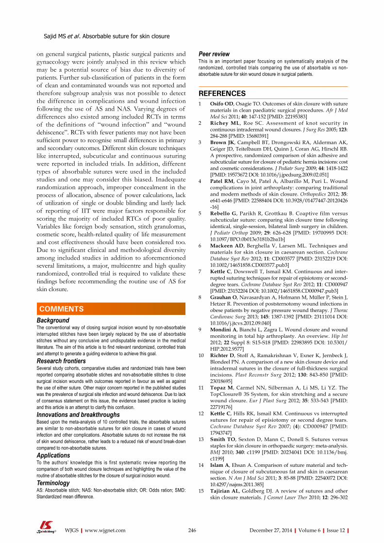
on general surgical patients, plastic surgical patients and gynaecology were jointly analysed in this review which may be a potential source of bias due to diversity of patients. Further sub-classification of patients in the form of clean and contaminated wounds was not reported and therefore subgroup analysis was not possible to detect the difference in complications and wound infection following the use of AS and NAS. Varying degrees of differences also existed among included RCTs in terms of the definitions of “wound infection” and “wound dehiscence”. RCTs with fewer patients may not have been sufficient power to recognise small differences in primary and secondary outcomes. Different skin closure techniques like interrupted, subcuticular and continuous suturing were reported in included trials. In addition, different types of absorbable sutures were used in the included studies and one may consider this biased. Inadequate randomization approach, improper concealment in the process of allocation, absence of power calculations, lack of utilization of single or double blinding and lastly lack of reporting of IIT were major factors responsible for scoring the majority of included RTCs of poor quality. Variables like foreign body sensation, stitch granulomas, cosmetic score, health-related quality of life measurement and cost effectiveness should have been considered too. Due to significant clinical and methodological diversity among included studies in addition to aforementioned several limitations, a major, multicentre and high quality randomized, controlled trial is required to validate these findings before recommending the routine use of AS for skin closure.
COMMENTSBackgroundThe conventional way of closing surgical incision wound by non-absorbable interrupted stitches have been largely replaced by the use of absorbable stitches without any conclusive and undisputable evidence in the medical literature. The aim of this article is to find relevant randomized, controlled trials and attempt to generate a guiding evidence to achieve this goal.Research frontiersSeveral study cohorts, comparative studies and randomized trials have been reported comparing absorbable stiches and non-absorbable stitches to close surgical incision wounds with outcomes reported in favour as well as against the use of either suture. Other major concern reported in the published studies was the prevalence of surgical site infection and wound dehiscence. Due to lack of consensus statement on this issue, the evidence based practice is lacking and this article is an attempt to clarify this confusion.Innovations and breakthroughsBased upon the meta-analysis of 10 controlled trials, the absorbable sutures are similar to non-absorbable sutures for skin closure in cases of wound infection and other complications. Absorbable sutures do not increase the risk of skin wound dehiscence, rather leads to a reduced risk of wound break-down compared to non-absorbable sutures.ApplicationsTo the authors’ knowledge this is first systematic review reporting the comparison of both wound closure techniques and highlighting the value of the routine of absorbable stitches for the closure of surgical incision wound.TerminologyAS: Absorbable stitch; NAS: Non-absorbable stitch; OR: Odds ration; SMD: Standardized mean difference.
Peer reviewThis is an important paper focusing on systematically analysis of the randomized, controlled trials comparing the use of absorbable vs non-absorbable suture for skin wound closure in surgical patients.
REFERENCES1 Osifo OD, Osagie TO. Outcomes of skin closure with suture
materials in clean paediatric surgical procedures. Afr J Med Med Sci 2011; 40: 147-152 [PMID: 22195383]
2 Richey ML, Roe SC. Assessment of knot security in continuous intradermal wound closures. J Surg Res 2005; 123: 284-288 [PMID: 15680391]
3 Brown JK, Campbell BT, Drongowski RA, Alderman AK, Geiger JD, Teitelbaum DH, Quinn J, Coran AG, Hirschl RB. A prospective, randomized comparison of skin adhesive and subcuticular suture for closure of pediatric hernia incisions: cost and cosmetic considerations. J Pediatr Surg 2009; 44: 1418-1422 [PMID: 19573672 DOI: 10.1016/j.jpedsurg.2009.02.051]
4 Patel RM, Cayo M, Patel A, Albarillo M, Puri L. Wound complications in joint arthroplasty: comparing traditional and modern methods of skin closure. Orthopedics 2012; 35: e641-e646 [PMID: 22588404 DOI: 10.3928/01477447-20120426-16]
5 Rebello G, Parikh R, Grottkau B. Coaptive film versus subcuticular suture: comparing skin closure time following identical, single-session, bilateral limb surgery in children. J Pediatr Orthop 2009; 29: 626-628 [PMID: 19700995 DOI: 10.1097/BPO.0b013e3181b2ba1b]
6 Mackeen AD, Berghella V, Larsen ML. Techniques and materials for skin closure in caesarean section. Cochrane Database Syst Rev 2012; 11: CD003577 [PMID: 23152219 DOI: 10.1002/14651858.CD003577.pub3]
7 Kettle C, Dowswell T, Ismail KM. Continuous and inter-rupted suturing techniques for repair of episiotomy or second-degree tears. Cochrane Database Syst Rev 2012; 11: CD000947 [PMID: 23152204 DOI: 10.1002/14651858.CD000947.pub3]
8 Grauhan O, Navasardyan A, Hofmann M, Müller P, Stein J, Hetzer R. Prevention of poststernotomy wound infections in obese patients by negative pressure wound therapy. J Thorac Cardiovasc Surg 2013; 145: 1387-1392 [PMID: 23111014 DOI: 10.1016/j.jtcvs.2012.09.040]
9 Mondini A, Bianchi L, Zagra L. Wound closure and wound monitoring in total hip arthroplasty. An overview. Hip Int 2012; 22 Suppl 8: S15-S18 [PMID: 22983895 DOI: 10.5301/HIP.2012.9577]
10 Richter D, Stoff A, Ramakrishnan V, Exner K, Jernbeck J, Blondeel PN. A comparison of a new skin closure device and intradermal sutures in the closure of full-thickness surgical incisions. Plast Reconstr Surg 2012; 130: 843-850 [PMID: 23018695]
11 Topaz M, Carmel NN, Silberman A, Li MS, Li YZ. The TopClosure® 3S System, for skin stretching and a secure wound closure. Eur J Plast Surg 2012; 35: 533-543 [PMID: 22719176]
12 Kettle C, Hills RK, Ismail KM. Continuous vs interrupted sutures for repair of episiotomy or second degree tears. Cochrane Database Syst Rev 2007; (4): CD000947 [PMID: 17943747]
13 Smith TO, Sexton D, Mann C, Donell S. Sutures versus staples for skin closure in orthopaedic surgery: meta-analysis. BMJ 2010; 340: c1199 [PMID: 20234041 DOI: 10.1136/bmj.c1199]
14 Islam A, Ehsan A. Comparison of suture material and tech-nique of closure of subcutaneous fat and skin in caesarean section. N Am J Med Sci 2011; 3: 85-88 [PMID: 22540072 DOI: 10.4297/najms.2011.385]
15 Tajirian AL, Goldberg DJ. A review of sutures and other skin closure materials. J Cosmet Laser Ther 2010; 12: 296-302
246 December 27, 2014|Volume 6|Issue 12|WJGS|www.wjgnet.com
COMMENTS
Sajid MS et al . Absorbable suture for skin closure
[PMID: 21142740 DOI: 10.3109/14764172.2010.538413]16 Papazoglou LG, Tsioli V, Papaioannou N, Georgiadis M,
Savvas I, Prassinos N, Kouti V, Bikiaris D, Hadzigiannakis C, Zavros N. Comparison of absorbable and nonabsorbable sutures for intradermal skin closure in cats. Can Vet J 2010; 51: 770-772 [PMID: 20885834]
17 Sughrue ME, Bloch OG, Manley GT, Stiver SI. Marked reduction in wound complication rates following decom-pressive hemicraniectomy with an improved operative closure technique. J Clin Neurosci 2011; 18: 1201-1205 [PMID: 21752652 DOI: 10.1016/j.jocn.2011.01.016]
18 Adams IW, Bell MS, Driver RM, Fry WG. A comparative trial of polyglycolic acid and silk as suture materials for accidental wounds. Lancet 1977; 2: 1216-1217 [PMID: 73912]
19 Rosenzweig LB, Abdelmalek M, Ho J, Hruza GJ. Equal cosmetic outcomes with 5-0 poliglecaprone-25 versus 6-0 polypropylene for superficial closures. Dermatol Surg 2010; 36: 1126-1129 [PMID: 20653727 DOI: 10.1111/j.1524-4725.2010.01594.x]
20 Kronborg O. Polyglycolic acid (Dexon) versus silk for fascial closure of abdominal incisions. Acta Chir Scand 1976; 142: 9-12 [PMID: 773068]
21 Parell GJ, Becker GD. Comparison of absorbable with nonabsorbable sutures in closure of facial skin wounds. Arch Facial Plast Surg 2003; 5: 488-490 [PMID: 14623686]
22 Higgins JPT, Green S (eds). Cochrane Handbook for Systematic Reviews of Interventions. [Accessed on 2014 July 28]. Available from: URL: http: //www.cochrane-handbook.org
23 Review Manager (RevMan). [Computer program]. Version 5, 2008. Copenhagen: The Nordic Cochrane Centre, The Cochrane Collaboration, 2008
24 DerSimonian R, Laird N. Meta-analysis in clinical trials. Control Clin Trials 1986; 7: 177-188 [PMID: 3802833]
25 Demets DL. Methods for combining randomized clinical trials: strengths and limitations. Stat Med 1987; 6: 341-350 [PMID: 3616287]
26 Higgins JP, Thompson SG. Quantifying heterogeneity in a meta-analysis. Stat Med 2002; 21: 1539-1558 [PMID: 12111919]
27 Egger M, Smith GD, Altman DG. Systematic reviews in healthcare. London: BMJ Publishing, 2006
28 Deeks JJ, Altman DG, Bradburn MJ. Statistical methods for examining heterogeneity and combining results from several studies in meta-analysis. Systemic reviews in health care: meta-analysis in context. Editor(s): Matthias Egger, George Davey Smith, Douglas G Altman. 2nd ed. London: BMJ
Publication group, 2001: 285-31229 Jadad AR, Moore RA, Carroll D, Jenkinson C, Reynolds DJ,
Gavaghan DJ, McQuay HJ. Assessing the quality of reports of randomized clinical trials: is blinding necessary? Control Clin Trials 1996; 17: 1-12 [PMID: 8721797]
30 Chalmers TC, Smith H, Blackburn B, Silverman B, Schroeder B, Reitman D, Ambroz A. A method for assessing the quality of a randomized control trial. Control Clin Trials 1981; 2: 31-49 [PMID: 7261638]
31 Available from: URL: http: //tech.cochrane.org/revman/other-resources/gradepro/download
32 Dørflinger T, Kiil J. Absorbable suture in hernia repair. Acta Chir Scand 1984; 150: 41-43 [PMID: 6322489]
33 Foster GE, Hardy EG, Hardcastle JD. Subcuticular suturing after appendicectomy. Lancet 1977; 1: 1128-1129 [PMID: 68225]
34 Glough JV, Alexander-Williams J. Surgical and economic advantages of polyglycolic-acid suture material in skin closure. Lancet 1975; 1: 194-195 [PMID: 47421]
35 Harimoto N, Shirabe K, Abe T, Yukaya T, Tsujita E, Gion T, Kajiyama K, Nagaie T. Prospective randomized controlled trial investigating the type of sutures used during hepatectomy. World J Gastroenterol 2011; 17: 2338-2342 [PMID: 21633600 DOI: 10.3748/wjg.v17.i18.2338]
36 Kotaluoto S, Pauniaho SL, Helminen M, Kuokkanen H, Rantanen T. Wound healing after open appendectomies in adult patients: a prospective, randomised trial comparing two methods of wound closure. World J Surg 2012; 36: 2305-2310 [PMID: 22669400 DOI: 10.1007/s00268-012-1664-3]
37 Lundblad R, Simensen HV, Wiig JN, Niels Grüner OP. [Skin closure. A prospective randomized study]. Tidsskr Nor Laegeforen 1989; 109: 1307-1309 [PMID: 2660322]
38 Pauniaho SL, Lahdes-Vasama T, Helminen MT, Iber T, Mäkelä E, Pajulo O. Non-absorbable interrupted versus absorbable continuous skin closure in pediatric appendectomies. Scand J Surg 2010; 99: 142-146 [PMID: 21044931]
39 Ralphs DN, Cannon SR, Bolton JP. Skin closure of inguinal herniorrhaphy wounds in short-stay patients. Br J Surg 1982; 69: 341-342 [PMID: 7044464]
40 Szabó S, István G. [Skin closure in inguinal hernia repair with rapidly absorbing Polyglactin 910/370 (Vicryl-Rapide) suture material]. Magy Seb 2002; 55: 77-80 [PMID: 12049012]
41 Tan PC, Mubarak S, Omar SZ. Absorbable versus non-absorbable sutures for subcuticular skin closure of a transverse suprapubic incision. Int J Gynaecol Obstet 2008; 103: 179-181 [PMID: 18639876 DOI: 10.1016/j.ijgo.2008.05.023]
P- Reviewer: Deng B, Wong KKY S- Editor: Ji FF L- Editor: A E- Editor: Liu SQ
247 December 27, 2014|Volume 6|Issue 12|WJGS|www.wjgnet.com
Sajid MS et al . Absorbable suture for skin closure
Inflammatory pseudotumour of the spleen associated with splenic tuberculosis
Maria Isabel Prieto-Nieto, Juan Pedro Pérez-Robledo, Beatriz Díaz-San Andrés, Manuel Nistal, José Antonio Rodríguez-Montes
Maria Isabel Prieto-Nieto, Juan Pedro Pérez-Robledo, Beatriz Díaz-San Andrés, José Antonio Rodríguez-Montes, Department of General and Digestive Surgery, La Paz University Hospital, Paseo de la Castellana, 28046 Madrid, SpainManuel Nistal, Department of Pathology, La Paz University Hospital, Paseo de la Castellana, 28046 Madrid, SpainAuthor contributions: All authors contributed to the work.Correspondence to: Maria Isabel Prieto-Nieto, MD, PhD, FACS, Department of General and Digestive Surgery, La Paz University Hospital, Paseo de la Castellana, 261, 28046 Madrid, Spain. [email protected]: +34-60-6839295 Fax: +34-60-6839295Received: March 29, 2014 Revised: May 18, 2014 Accepted: October 28, 2014Published online: December 27, 2014
AbstractInflammatory pseudotumor (IPT) of the spleen is an uncommon entity with an uncertain aetiology. Inflam-matory pseudotumors present diagnostic difficulties because the clinical and radiological findings tend to suggest a malignancy. The symptoms include weight loss, fever, and abdominal pain. Most cases of splenic IPT present solitary relatively large well circumscribed masses on imaging. The diagnosis in the majority of the cases is made after histopathologic study of splenectomy specimens. The IPTs that occur in the spleen and liver are typically associated with Epstein-Barr virus. Thirty-seven percent of all new cases of active tuberculosis infection are extrapulmonary tuberculosis and tuberculous lymphadenitis the most commonly occurring form of extrapulmonary tuberculosis. We report the case of an inflammatory pseudotumor of the spleen associated with splenic tuberculous lymphadenitis in a 50-year-old female patient who was preoperatively diagnosed with a malignant spleen tumour based on her history of breast of carcinoma.
© 2014 Baishideng Publishing Group Inc. All rights reserved.
Key words: Inflammatory pseudotumour; Spleen; Splenic lymphadenitis; Splenectomy; Splenic tuberculosis
Core tip: A rare benign; lesion inflammatory pseu-dotumours (IPT) are infrequently found in the spleen with only sporadic case reports and short case series reported in the literature. Here we report a case of a spleen IPT associated with splenic tuberculous lymphadenitis in a 50-year-old female patient who was preoperatively diagnosed with a malignant spleen neoplasm.
Prieto-Nieto MI, Pérez-Robledo JP, Díaz-San Andrés B, Nistal M, Rodríguez-Montes JA. Inflammatory pseudotumour of the spleen associated with splenic tuberculosis. World J Gastrointest Surg 2014; 6(12): 248-252 Available from: URL: http://www.wjgnet.com/1948-9366/full/v6/i12/248.htm DOI: http://dx.doi.org/10.4240/wjgs.v6.i12.248
INTRODUCTIONInflammatory pseudotumours (IPT) characterized microscopically by a proliferation of inflammatory cells, are infrequently found in the spleen and there are only sporadic case reports and short case series in the literature[1]. We report a case of a spleen IPT associated with splenic tuberculous lymphadenitis in a 50-year-old female patient who was preoperatively diagnosed with a malignant spleen neoplasm[1,2].
CASE REPORTA 50-year-old woman had been undergone a quadran-tectomy for an intraductal breast carcinoma intraductal. The sentinel ganglion was negative so the patient received radiotherapy followed by tamoxifen. Two years later her routine follow-up ultrasonography incidentally
CASE REPORT
Submit a Manuscript: http://www.wjgnet.com/esps/Help Desk: http://www.wjgnet.com/esps/helpdesk.aspxDOI: 10.4240/wjgs.v6.i12.248
World J Gastrointest Surg 2014 December 27; 6(12): 248-252ISSN 1948-9366 (online)
© 2014 Baishideng Publishing Group Inc. All rights reserved.
248 December 27, 2014|Volume 6|Issue 12|WJGS|www.wjgnet.com
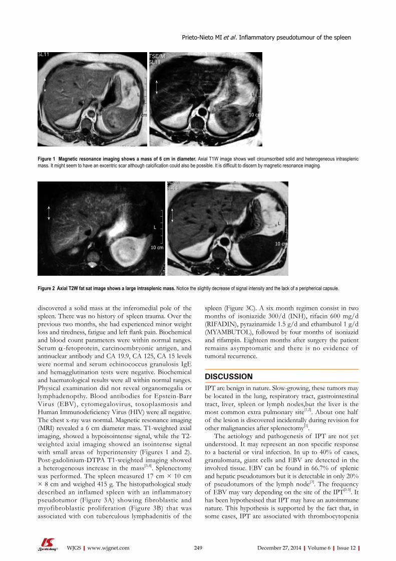
discovered a solid mass at the inferomedial pole of the spleen. There was no history of spleen trauma. Over the previous two months, she had experienced minor weight loss and tiredness, fatigue and left flank pain. Biochemical and blood count parameters were within normal ranges. Serum α-fetoprotein, carcinoembryonic antigen, and antinuclear antibody and CA 19.9, CA 125, CA 15 levels were normal and serum echinococcus granulosis IgE and hemagglutination tests were negative. Biochemical and haematological results were all within normal ranges. Physical examination did not reveal organomegalia or lymphadenopthy. Blood antibodies for Epstein-Barr Virus (EBV), cytomegalovirus, toxoplasmosis and Human Immunodeficiency Virus (HIV) were all negative. The chest x-ray was normal. Magnetic resonance imaging (MRI) revealed a 6 cm diameter mass. T1-weighted axial imaging, showed a hypoisointense signal, while the T2-weighted axial imaging showed an isointense signal with small areas of hyperintensity (Figures 1 and 2). Post-gadolinium-DTPA T1-weighted imaging showed a heterogeneous increase in the mass[3,4]. Splenectomy was performed. The spleen measured 17 cm × 10 cm × 8 cm and weighed 415 g. The histopathological study described an inflamed spleen with an inflammatory pseudotumor (Figure 3A) showing fibroblastic and myofibroblastic proliferation (Figure 3B) that was associated with con tuberculous lymphadenitis of the
spleen (Figure 3C). A six month regimen consist in two months of isoniazide 300/d (INH), rifacin 600 mg/d (RIFADIN), pyrazinamide 1.5 g/d and ethambutol 1 g/d (MYAMBUTOL), followed by four months of isoniazid and rifampin. Eighteen months after surgery the patient remains asymptomatic and there is no evidence of tumoral recurrence.
DISCUSSIONIPT are benign in nature. Slow-growing, these tumors may be located in the lung, respiratory tract, gastrointestinal tract, liver, spleen or lymph nodes,but the liver is the most common extra pulmonary site[1,2]. About one half of the lesion is discovered incidentally during revision for other malignancies after splenectomy[1].
The aetiology and pathogenesis of IPT are not yet understood. It may represent an non specific response to a bacterial or viral infection. In up to 40% of cases, granulomata, giant cells and EBV are detected in the involved tissue. EBV can be found in 66.7% of splenic and hepatic pseudotumors but it is detectable in only 20% of pseudotumors of the lymph node[3]. The frequency of EBV may vary depending on the site of the IPT[3-5]. It has been hypothesised that IPT may have an autoimmune nature. This hypothesis is supported by the fact that, in some cases, IPT are associated with thrombocytopenia
Prieto-Nieto MI et al . Inflammatory pseudotumour of the spleen
249 December 27, 2014|Volume 6|Issue 12|WJGS|www.wjgnet.com
Figure 1 Magnetic resonance imaging shows a mass of 6 cm in diameter. Axial T1W image shows well circumscribed solid and heterogeneous intrasplenic mass. It might seem to have an excentric scar although calcification could also be possible. It is difficult to discern by magnetic resonance imaging.
10 cm
L
10 cm
L
Figure 2 Axial T2W fat sat image shows a large intrasplenic mass. Notice the slightly decrease of signal intensity and the lack of a peripherical capsule.
10 cm
L
10 cm
L
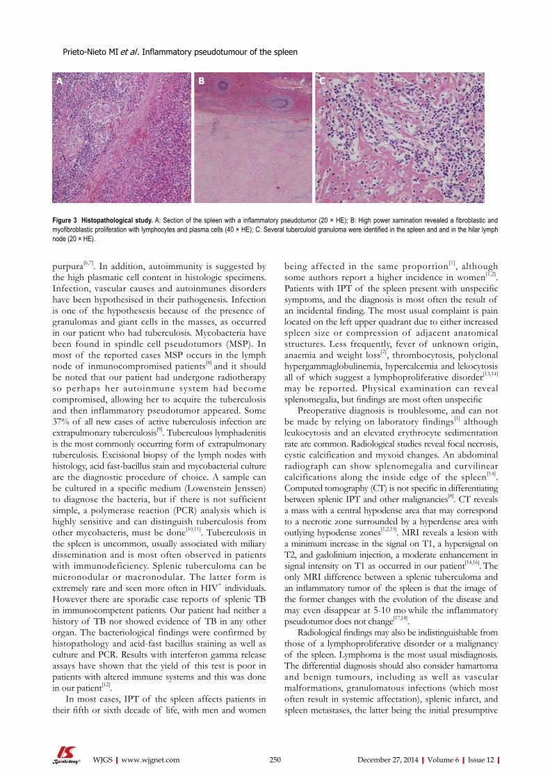
purpura[6,7]. In addition, autoimmunity is suggested by the high plasmatic cell content in histologic specimens. Infection, vascular causes and autoinmunes disorders have been hypothesised in their pathogenesis. Infection is one of the hypothesesis because of the presence of granulomas and giant cells in the masses, as occurred in our patient who had tuberculosis. Mycobacteria have been found in spindle cell pseudotumors (MSP). In most of the reported cases MSP occurs in the lymph node of inmunocompromised patients[8] and it should be noted that our patient had undergone radiotherapy so perhaps her autoinmune system had become compromised, allowing her to acquire the tuberculosis and then inflammatory pseudotumor appeared. Some 37% of all new cases of active tuberculosis infection are extrapulmonary tuberculosis[9]. Tuberculous lymphadenitis is the most commonly occurring form of extrapulmonary tuberculosis. Excisional biopsy of the lymph nodes with histology, acid fast-bacillus stain and mycobacterial culture are the diagnostic procedure of choice. A sample can be cultured in a specific medium (Lowenstein Jenssen) to diagnose the bacteria, but if there is not sufficient simple, a polymerase reaction (PCR) analysis which is highly sensitive and can distinguish tuberculosis from other mycobacteris, must be done[10,11]. Tuberculosis in the spleen is uncommon, usually associated with miliary dissemination and is most often observed in patients with immunodeficiency. Splenic tuberculoma can be micronodular or macronodular. The latter form is extremely rare and seen more often in HIV+ individuals. However there are sporadic case reports of splenic TB in immunocompetent patients. Our patient had neither a history of TB nor showed evidence of TB in any other organ. The bacteriological findings were confirmed by histopathology and acid-fast bacillus staining as well as culture and PCR. Results with interferon gamma release assays have shown that the yield of this test is poor in patients with altered immune systems and this was done in our patient[12].
In most cases, IPT of the spleen affects patients in their fifth or sixth decade of life, with men and women
being affected in the same proportion[1], although some authors report a higher incidence in women[1,2]. Patients with IPT of the spleen present with unspecific symptoms, and the diagnosis is most often the result of an incidental finding. The most usual complaint is pain located on the left upper quadrant due to either increased spleen size or compression of adjacent anatomical structures. Less frequently, fever of unknown origin, anaemia and weight loss[2], thrombocytosis, polyclonal hypergammaglobulinemia, hypercalcemia and lekocytosis all of which suggest a lymphoproliferative disorder[13,14]
may be reported. Physical examination can reveal splenomegalia, but findings are most often unspecific
Preoperative diagnosis is troublesome, and can not be made by relying on laboratory findings[1] although leukocytosis and an elevated erythrocyte sedimentation rate are common. Radiological studies reveal focal necrosis, cystic calcification and myxoid changes. An abdominal radiograph can show splenomegalia and curvilinear calcifications along the inside edge of the spleen[14]. Computed tomography (CT) is not specific in differentiating between splenic IPT and other malignancies[8]. CT reveals a mass with a central hypodense area that may correspond to a necrotic zone surrounded by a hyperdense area with outlying hypodense zones[1,2,15]. MRI reveals a lesion with a minimum increase in the signal on T1, a hypersignal on T2, and gadolinium injection, a moderate enhancement in signal intensity on T1 as occurred in our patient[14,16]. The only MRI difference between a splenic tuberculoma and an inflammatory tumor of the spleen is that the image of the former changes with the evolution of the disease and may even disappear at 5-10 mo while the inflammatory pseudotumor does not change[17,18].
Radiological findings may also be indistinguishable from those of a lymphoproliferative disorder or a malignancy of the spleen. Lymphoma is the most usual misdiagnosis. The differential diagnosis should also consider hamartoma and benign tumours, including as well as vascular malformations, granulomatous infections (which most often result in systemic affectation), splenic infarct, and spleen metastases, the latter being the initial presumptive
250 December 27, 2014|Volume 6|Issue 12|WJGS|www.wjgnet.com
A B C
Figure 3 Histopathological study. A: Section of the spleen with a inflammatory pseudotumor (20 × HE); B: High power xamination revealed a fibroblastic and myofibroblastic proliferation with lymphocytes and plasma cells (40 × HE); C: Several tuberculoid granuloma were identified in the spleen and and in the hilar lymph node (20 × HE).
Prieto-Nieto MI et al . Inflammatory pseudotumour of the spleen
the lymph node.Term explanationExcisional biopsy of the lymph nodes with histology, acid fast-bacillus stain and mycobacterial culture are the diagnostic procedure of choice. A sample can be cultured in a specific medium (Lowenstein Jenssen) to diagnose the bacteria, but if there is not sufficient simple, a polymerase reaction analysis which is highly sensitive and can distinguish tuberculosis from other mycobacteris, must be done. Tuberculosis in the spleen is uncommon, usually associated with miliary dissemination and is most often observed in patients with immunodeficiency. Splenic tuberculoma can be micronodular or macronodular. The latter form is extremely rare and seen more often in Human Immunodeficiency Virus+ individuals.Experiences and lessons The frequency of EBV may vary depending on the site of the IPT. It has been hypothesised that IPT may have an autoimmune nature. This hypothesis is supported by the fact that, in some cases, IPT are associated with thrombocytopenia purpura. In addition, autoimmunity is suggested by the high plasmatic cell content in histologic specimens. Infection, vascular causes and autoinmunes disorders have been hypothesised in their pathogenesis. Infection is one of the hypothesis because of the presence of granulomas and giant cells in the masses, as occurred in their patient who had tuberculosis. Mycobacteria have been found in spindle cell pseudotumors (MSP). In most of the reported cases MSP occurs in the lymph node of inmunocompromised patients and it should be noted that their patient had undergone radiotherapy so perhaps her autoinmune system had become compromised, allowing her to acquire the tuberculosis and then inflammatory pseudotumor appeared. Tuberculous lymphadenitis is the most commonly occurring form of extrapulmonary tuberculosis. Peer reviewIt is an interesting manuscript worthy of publication.
REFERENCES1 Ma ZH, Tian XF, Ma J, Zhao YF. Inflammatory pseudotumor
of the spleen: A case report and review of published cases. Oncol Lett 2013; 5: 1955-1957 [PMID: 23833674]
2 Ozkara SK, Gürbüz Y, Erçín C, Müezzínoğlu B, Türkmen M. Inflammatory pseudotumor of the spleen. Virchows Arch 2001; 438: 629-631 [PMID: 11469697 DOI: 10.1007/s004280100403]
3 Oz Puyan F, Bilgi S, Unlu E, Yalcin O, Altaner S, Demir M, Cakir B. Inflammatory pseudotumor of the spleen with EBV positivity: report of a case. Eur J Haematol 2004; 72: 285-291 [PMID: 15089768 DOI: 10.1111/j.0902-4441.2003.00208.x]
4 Lewis JT, Gaffney RL, Casey MB, Farrell MA, Morice WG, Macon WR. Inflammatory pseudotumor of the spleen associated with a clonal Epstein-Barr virus genome. Case report and review of the literature. Am J Clin Pathol 2003; 120: 56-61 [PMID: 12866373 DOI: 10.1309/BUWNMG5RV4D09YYH]
5 Rosenbaum L, Fekrazad MH, Rabinowitz I, Vasef MA. Epstein-Barr virus-associated inflammatory pseudotumor of the spleen: report of two cases and review of the literature. J Hematop 2009; 2: 127-131 [PMID: 19669195 DOI: 10.1007/s12308-009-0030-3]
6 Alimoglu O, Cevikbas U. Inflammatory pseudotumor of the spleen: report of a case. Surg Today 2003; 33: 960-964 [PMID: 14669094 DOI: 10.1007/s00595-003-2619-y]
7 Noguchi H, Kondo H, Kondo M, Shiraiwa M, Monobe Y. Inflammatory pseudotumor of the spleen: a case report. Jpn J Clin Oncol 2000; 30: 196-203 [PMID: 10830990 DOI: 10.1093/jjco/hyd048]
8 Philip J, Beasley MB, Dua S. Mycobacterial spindle cell pseudotumor of the lung. Chest 2012; 142: 783-784 [PMID: 22948583 DOI: 10.1378/chest.11-2503]
9 Golden MP, Vikram HR. Extrapulmonary tuberculosis: an overview. Am Fam Physician 2005; 72: 1761-1768 [PMID: 16300038]
10 Velásquez MJ, Szigethi QM, Panace VR, Morales IR, Márquez CS, Pefaur PJ, Mocarquer MA, Salinas CP, Beltrán
diagnosis in our patient[13,14,19]. Surgery remains the gold stantard for definitive diagnosis.
In most cases the pseudotumor consists of a single well-circumscribed mass of 2-15 cm in diameter, composed of inflammatory cells, particularly plasmatic cells, and a fibroblastic proliferation of spindle cells, such as smooth muscle antibody (+), myofibroblasts and follicular dendritic cells (+). The spindle cells in IPT are most often myofibroblasts[5,20]. (1) IPT of the spleen is classified into three groups according to the cellular characteristics of the mass, clinical presentation and course, potential aetiology and prognosis: IPT-like follicular dendritic cell tumour. Is the kind that most frequently affects women. It is associated with a spindle cells and EBV infection; (2) The presence of myofibroblasts in a splenic IPT has led to the designation of inflammatory myofibroblastic (IMT) tumor. A considerable proportion of these tumours are EBV (+) and they have a potential for malignancy; and (3) IPT of the spleen was described by Cottelingam and Jaffe as having a predominance of spindle cells. Some 33%-48% of cases are found incidentally. This is the group which best deserves the term IPT of the spleen[5,20]. The World
Health Organization classification places inflammatory myofibroblastic tumors in an intermediate category (rarely metastasizing, < 5%) between benign and malignant[14].
IPT of the spleen is a rare entity. It should be included in the differential diagnosis for splenic masses, especially when MRI discloses a single mass showing increased signal on T1 following gadolinium injection.
COMMENTSCase characteristicsA 50-year-old woman with a inflammatory pseudotumour of the spleen that present diagnostic difficulties because the clinical and radiological findings tend to suggest a malignancy.Clinical diagnosisUnspecific symptoms and the diagnosis is most often the result of an incidental finding.Differential diagnosisLymphoproliferative disorder or a malignancy of the spleen. Lymphoma, hamartoma, vascular malformations, splenic infarct and spleen metastases.Laboratory diagnosisBiochemical and blood count parameters were within normal ranges. Serum α-fetoprotein, carcinoembryonic antigen, and antinuclear antibody and CA 19.9, CA 125, CA 15 levels were normal and serum echinococcus granulosis IgE and hemagglutination tests were negative.Imaging diagnosisThe histopathological study described an inflamed spleen with an inflammatory pseudotumor showing fibroblastic and myofibroblastic proliferation that was associated with con tuberculous lymphadenitis of the spleen.Treatment A six month regimen consist in two months of isoniazide 300/d (INH), rifacin 600 mg/d (RIFADIN), pyrazinamide 1.5 g/d and ethambutol 1 g/d (MYAMBUTOL), followed by four months of isoniazid and rifampin. Splenectomy was performed.Related reports The aetiology and pathogenesis of inflammatory pseudotumours (IPT) are not yet understood. It may represent an non specific response to a bacterial or viral infection. In up to 40% of cases, granulomata, giant cells and Epstein-Barr virus (EBV) are detected in the involved tissue. EBV can be found in 66.7% of splenic and hepatic pseudotumors but it is detectable in only 20% of pseudotumors of
251 December 27, 2014|Volume 6|Issue 12|WJGS|www.wjgnet.com
COMMENTS
Prieto-Nieto MI et al . Inflammatory pseudotumour of the spleen

BC. [Hepatic-splenic micobacteriosis, unusual form of probable extrapulmonary tuberculosis. Case report and review]. Rev Chilena Infectol 2007; 24: 59-62 [PMID: 17369973]
11 Rodarte-Shade M, Diaz-Elizondo JA. Splenic tuberculosis. Surg Infect (Larchmt) 2012; 13: 420-421 [PMID: 23268618 DOI: 10.1089/sur.2012.007]
12 Oikonomou A, Mantatzis M, Ritis K, Kartalis G, Prassopoulos P. Multiple splenic macronodular tuberculomas: MRI characteristics under treatment. Int J Tuberc Lung Dis 2006; 10: 233-234 [PMID: 16499268]
13 Hsu CW, Lin CH, Yang TL, Chang HT. Splenic inflammatory pseudotumor mimicking angiosarcoma. World J Gastroenterol 2008; 14: 6421-6424 [PMID: 19009664 DOI: 10.3748/wjg.14.6421]
14 Rajabi P, Noorollahi H, Hani M, Bagheri M. Inflammatory pseudotumor of spleen. Adv Biomed Res 2014; 3: 29 [PMID: 24592376 DOI: 10.4103/2277-9175.124679]
15 Kapoor R, Jain AK, Chaturvedi U, Saha MM. Ultrasound detection of tuberculomas of the spleen. Clin Radiol 1991; 43: 128-129 [PMID: 2004510 DOI: 10.1016/S0009-9260(05)81593-4]
16 McHenry CR, Perzy-Gall HB, Mardini G, Chung-Park M. Inflammatory pseudotumor of the spleen: a rare entity that may mimic hematopoietic malignancy. Am Surg 1995; 61: 1067-1071 [PMID: 7486448]
17 Bastounis E, Pikoulis E, Varelas P, Cirochristos D, Aessopos A. Tuberculoma of the spleen: a rare but important clinical entity. Am Surg 1999; 65: 131-132 [PMID: 9926745]
18 Dede F, Doğan E, Demir M, Sener D, Kös M, Tad M, Eskioğlu E. Unusual presentation of tuberculosis as a splenic mass. Tohoku J Exp Med 2006; 210: 79-82 [PMID: 16960348 DOI: 10.1620/tjem.210.79]
19 Krishnan J, Frizzera G. Two splenic lesions in need of clarification: hamartoma and inflammatory pseudotumor. Semin Diagn Pathol 2003; 20: 94-104 [PMID: 12945933 DOI: 10.1016/S0740-2570(03)00014-5]
20 Oshiro H, Nomura M, Yamanaka S, Watanabe S, Inayama Y. Splenic inflammatory pseudotumor (inflammatory myofibroblastic tumor). J Clin Exp Hematop 2007; 47: 83-88 [PMID: 18040148]
P- Reviewer: Demonacos C, Garcia-Elorriaga G, Iso Y, Li ZF, Mastroianni CM
S- Editor: Ji FF L- Editor: A E- Editor: Liu SQ
252 December 27, 2014|Volume 6|Issue 12|WJGS|www.wjgnet.com
Prieto-Nieto MI et al . Inflammatory pseudotumour of the spleen
Case of bronchoesophageal fistula with gastric perforation due to multidrug-resistant tuberculosis
Chan Sung Park, Kwang won Seo, Chang Ryul Park, Yang Won Nah, Jae Hee Suh
Chan Sung Park, Kwang won Seo, Department of Internal medicine, Ulsan University Hospital, University of Ulsan College of Medicine, Ulsan 682-714, South KoreaChang Ryul Park, Department of Thoracic and Cardiovascular Surgery, Ulsan University Hospital, University of Ulsan College of Medicine, Ulsan 682-714, South KoreaYang Won Nah, Department of General Surgery, Ulsan University Hospital, University of Ulsan College of Medicine, Ulsan 682-714, South KoreaJae Hee Suh, Department of Pathology, Ulsan University Hospital, University of Ulsan College of Medicine, Ulsan 682-714, South KoreaAuthor contributions: Park CS, Seo KW, Park CR and Nah YW wrote the paper; Suh JH made the pathological diagnosis.Correspondence to: Kwang Won Seo, MD, Department of Internal Medicine, Ulsan University Hospital, University of Ulsan College of Medicine, 290-3 Jeonha-dong, Dong-gu, Ulsan 682-714, South Korea. [email protected]: +82-52-2508861 Fax: +82-52-2518235Received: April 2, 2012 Revised: August 8, 2012Accepted: March 20, 2013Published online: December 27, 2014
AbstractGastric perforation and tuberculous bronchoesophageal fistula (TBEF) are very rare complications of extrapulmonary tuberculosis (TB). We present a case of pulmonary TB with TBEF and gastric perforation caused by a multidrugresistant tuberculosis strain in a nonacquired immune deficiency syndrome male patient. The patient underwent total gastrectomy with RouxenY endtoside esophagojejunostomy and feeding jejunostomy during intravenous treatment with antiTB medication, and esophageal reconstruction with colonic interposition and jejunocolostomy were performed successfully after a full course of antiTB medication. Though recent therapies for TBEF have favored medication, patients with severe stenosis or perforation require surgery and medication with antiTB drugs based
upon adequate culture and drug susceptibility testing.
© 2014 Baishideng Publishing Group Inc. All rights reserved.
Key words: Bronchoesophageal fistula; Gastric perforation; Multidrugresistant tuberculosis; Extrapulmonary tuberculosis; Treatment
Core tip: A case of pulmonary extrapulmonary tuberculosis (TB) with TB and gastric perforation caused by a multidrugresistant tuberculosis strain in a nonacquired immune deficiency syndrome male patient.
Park CS, Seo KW, Park CR, Nah YW, Suh JH. Case of bronch-oesophageal fistula with gastric perforation due to multidrug-resistant tuberculosis. World J Gastrointest Surg 2014; 6(12): 253-258 Available from: URL: http://www.wjgnet.com/1948-9366/full/v6/i12/253.htm DOI: http://dx.doi.org/10.4240/wjgs.v6.i12.253
INTRODUCTIONTuberculous gastric perforation is a rare presentation of gastric tuberculosis (TB) with six previous cases reported in the literature[1,2]. However, to the best of our knowledge, there are no previous reports of gastric perforation due to multidrug-resistant TB (MDR-TB). Tuberculous bronchoesophageal fistula (TBEF) is also a very infrequent complication of extrapulmonary TB[3]. Although MDR-TB with TBEF has been reported in two cases, one case was in a human immunodeficiency virus (HIV) positive patient[4], and the other case was published in the Japanese language[5]. Moreover, these were cases of TBEF in patients with esophageal TB, but our case involved TBEF and gastric perforation in a patient with TB throughout the upper gastrointestinal (GI) tract. The patient with severe pulmonary TB, especially
CASE REPORT
Submit a Manuscript: http://www.wjgnet.com/esps/Help Desk: http://www.wjgnet.com/esps/helpdesk.aspxDOI: 10.4240/wjgs.v6.i12.253
World J Gastrointest Surg 2014 December 27; 6(12): 253-258ISSN 1948-9366 (online)
© 2014 Baishideng Publishing Group Inc. All rights reserved.
253 December 27, 2014|Volume 6|Issue 12|WJGS|www.wjgnet.com
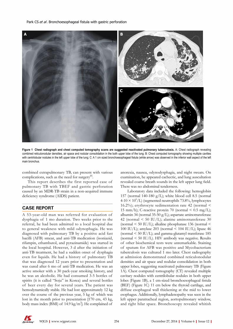
combined extrapulmonary TB, can present with various complications, such as the need for surgery[6].
This report describes the first reported case of pulmonary TB with TBEF and gastric perforation caused by an MDR-TB strain in a non-acquired immune deficiency syndrome (AIDS) patient.
CASE REPORTA 53-year-old man was referred for evaluation of dysphagia of 1 mo duration. Two weeks prior to the referral, he had been admitted to a local hospital due to general weakness with mild odynophagia. He was diagnosed with pulmonary TB by a positive acid fast bacilli (AFB) smear, and anti-TB medication (isoniazid, rifampin, ethambutol, and pyrazinamide) was started in the local hospital. However, 3 d after the initiation of anti-TB treatment, he had a sudden onset of dysphagia even for liquids. He had a history of pulmonary TB that was diagnosed 12 years prior to presentation and was cured after 6 mo of anti-TB medication. He was an active smoker with a 30 pack-year smoking history, and he was an alcoholic. He had consumed 3-5 bottles of spirits (it is called “Soju” in Korea) and several bottles of beer every day for several years. The patient was hemodynamically stable. He had lost approximately 12 kg over the course of the previous year, 5 kg of which was lost in the month prior to presentation [170 cm, 43 kg, body mass index (BMI) of 14.9 kg/m2]. He complained of
anorexia, nausea, odynodysphagia, and night sweats. On examination, he appeared cachectic, and lung auscultation revealed coarse breath sounds in the left upper lung field. There was no abdominal tenderness.
Laboratory data included the following: hemoglobin 157 (normal 140-180 g/L); white blood cell 8.5 (normal 4-10 × 106/L) (segmented neutrophils 73.8%, lymphocytes 16.2%); erythrocyte sedimentation rate 42 (normal < 15 mm/h); C-reactive protein 70 (normal < 0.5 mg/L); albumin 34 (normal 35-50 g/L); aspartate aminotransferase 42 (normal < 50 IU/L); alanine aminotransferase 30 (normal < 50 IU/L); alkaline phosphatase 136 (normal < 100 IU/L); amylase 203 (normal < 104 IU/L); lipase 46 (normal < 50 IU/L); and gamma-glutamyl transferase 185 (normal < 50 IU/L). HIV antibody was negative. Results of other biochemical tests were unremarkable. Staining of sputum for AFB was positive and Mycobacterium tuberculosis was cultured 1 mo later. Chest radiography at admission demonstrated combined reticulonodular densities and air space and nodular consolidation in both upper lobes, suggesting reactivated pulmonary TB (Figure 1A). Chest computed tomography (CT) revealed multiple cavitary nodules with centrilobular nodules in both upper lobes (Figure 1B), a 1 cm-sized bronchoesophageal fistula (BEF) (Figure 1C) 11 cm below the thyroid cartilage, and diffuse esophageal wall thickening at the mid to lower esophagus. Additionally, lymphadenopathy was seen in the left upper paratracheal region, aortopulmonary window, and right hilar space. Bronchoscopy revealed whitish
Park CS et al . Bronchoesophageal fistula with gastric perforation
254 December 27, 2014|Volume 6|Issue 12|WJGS|www.wjgnet.com
Figure 1 Chest radiograph and chest computed tomography scans are suggested reactivated pulmonary tuberculosis. A: Chest radiograph revealing combined reticulonodular densities, air space and nodular consolidation in the both upper lobe of the lung; B: Chest computed tomography showing multiple cavities with centrilobular nodules in the left upper lobe of the lung; C: A 1 cm sized bronchoesophageal fistula (white arrow) was observed in the inferior wall aspect of the left main bronchus.
A B
C
L
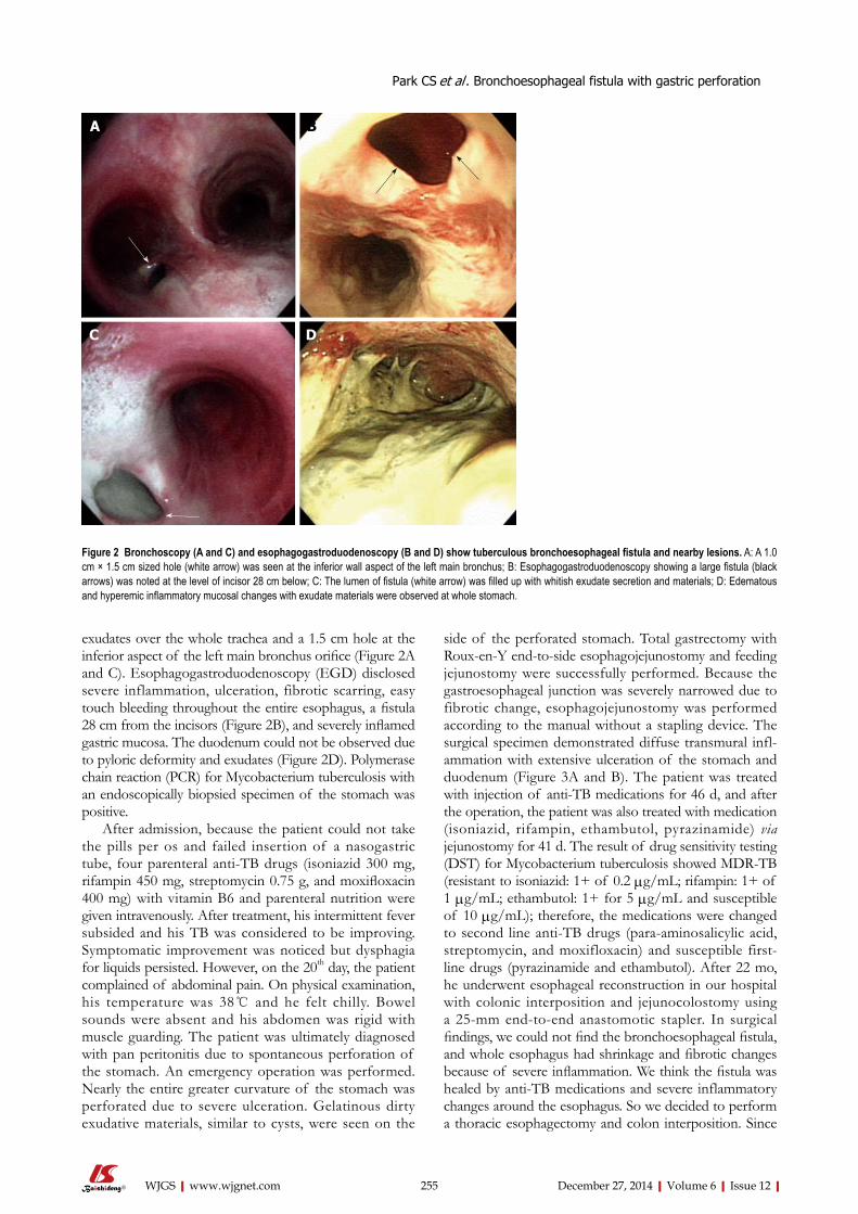
exudates over the whole trachea and a 1.5 cm hole at the inferior aspect of the left main bronchus orifice (Figure 2A and C). Esophagogastroduodenoscopy (EGD) disclosed severe inflammation, ulceration, fibrotic scarring, easy touch bleeding throughout the entire esophagus, a fistula 28 cm from the incisors (Figure 2B), and severely inflamed gastric mucosa. The duodenum could not be observed due to pyloric deformity and exudates (Figure 2D). Polymerase chain reaction (PCR) for Mycobacterium tuberculosis with an endoscopically biopsied specimen of the stomach was positive.
After admission, because the patient could not take the pills per os and failed insertion of a nasogastric tube, four parenteral anti-TB drugs (isoniazid 300 mg, rifampin 450 mg, streptomycin 0.75 g, and moxifloxacin 400 mg) with vitamin B6 and parenteral nutrition were given intravenously. After treatment, his intermittent fever subsided and his TB was considered to be improving. Symptomatic improvement was noticed but dysphagia for liquids persisted. However, on the 20th day, the patient complained of abdominal pain. On physical examination, his temperature was 38 ℃ and he felt chilly. Bowel sounds were absent and his abdomen was rigid with muscle guarding. The patient was ultimately diagnosed with pan peritonitis due to spontaneous perforation of the stomach. An emergency operation was performed. Nearly the entire greater curvature of the stomach was perforated due to severe ulceration. Gelatinous dirty exudative materials, similar to cysts, were seen on the
side of the perforated stomach. Total gastrectomy with Roux-en-Y end-to-side esophagojejunostomy and feeding jejunostomy were successfully performed. Because the gastroesophageal junction was severely narrowed due to fibrotic change, esophagojejunostomy was performed according to the manual without a stapling device. The surgical specimen demonstrated diffuse transmural infl-ammation with extensive ulceration of the stomach and duodenum (Figure 3A and B). The patient was treated with injection of anti-TB medications for 46 d, and after the operation, the patient was also treated with medication (isoniazid, rifampin, ethambutol, pyrazinamide) via jejunostomy for 41 d. The result of drug sensitivity testing (DST) for Mycobacterium tuberculosis showed MDR-TB (resistant to isoniazid: 1+ of 0.2 µg/mL; rifampin: 1+ of 1 µg/mL; ethambutol: 1+ for 5 µg/mL and susceptible of 10 µg/mL); therefore, the medications were changed to second line anti-TB drugs (para-aminosalicylic acid, streptomycin, and moxifloxacin) and susceptible first-line drugs (pyrazinamide and ethambutol). After 22 mo, he underwent esophageal reconstruction in our hospital with colonic interposition and jejunocolostomy using a 25-mm end-to-end anastomotic stapler. In surgical findings, we could not find the bronchoesophageal fistula, and whole esophagus had shrinkage and fibrotic changes because of severe inflammation. We think the fistula was healed by anti-TB medications and severe inflammatory changes around the esophagus. So we decided to perform a thoracic esophagectomy and colon interposition. Since
255 December 27, 2014|Volume 6|Issue 12|WJGS|www.wjgnet.com
Figure 2 Bronchoscopy (A and C) and esophagogastroduodenoscopy (B and D) show tuberculous bronchoesophageal fistula and nearby lesions. A: A 1.0 cm × 1.5 cm sized hole (white arrow) was seen at the inferior wall aspect of the left main bronchus; B: Esophagogastroduodenoscopy showing a large fistula (black arrows) was noted at the level of incisor 28 cm below; C: The lumen of fistula (white arrow) was filled up with whitish exudate secretion and materials; D: Edematous and hyperemic inflammatory mucosal changes with exudate materials were observed at whole stomach.
A B
C D
Park CS et al . Bronchoesophageal fistula with gastric perforation
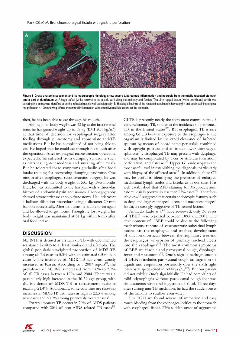
GI TB is presently nearly the sixth most common site of extrapulmonary TB, similar to the incidence of peritoneal TB, in the United States[10]. But esophageal TB is rare among GI TB because exposure of the esophagus to the organism is limited by the rapid clearance of infected sputum by means of coordinated peristalsis combined with upright posture and an intact lower esophageal sphincter[11]. Esophageal TB may present with dysphagia and may be complicated by ulcer or stricture formation, perforation, and fistulae[12]. Upper GI endoscopy is the most useful tool in establishing the diagnosis, particularly with biopsy of the affected area[13]. In addition, chest CT may be useful in identifying the presence of enlarged mediastinal lymph nodes and fistula, as in our case. It is well established that AFB staining for Mycobacterium tuberculosis is positive in less than 25% cases[14]. Therefore, Abid et al[15] suggested that certain endoscopic features, such as deep and large esophageal ulcers and tracheoesophageal fistula, are strongly suggestive of TB-related lesions.
As Lado Lado et al[3] have reviewed, only 36 cases of TBEF were reported between 1893 and 2001. The development of TBEF could be due to the following mechanisms: rupture of caseonecrotic subcarinal lymph nodes into the esophagus and trachea; development of traction diverticula between the respiratory tree and the esophagus; or erosion of primary tracheal ulcers into the esophagus[16]. The most common symptoms of BEF are chronic and paroxysmal cough, dysphagia, fever and pneumonia[3]. Ono’s sign is pathognomonic of BEF; it includes paroxysmal cough on ingestion of liquids and crepitation posteriorly over the sixth right intercostal space (cited in Alkhuja et al[17]). But our patient did not exhibit Ono’s sign initially. He had complaints of mild odynophagia without paroxysmal cough that was simultaneous with oral ingestion of food. Three days after starting anti-TB mediation, he had the sudden onset of the inability to swallow even water.
On EGD, we found severe inflammation and easy touch bleeding from the esophageal orifice to the stomach with esophageal fistula. This sudden onset of aggravated
then, he has been able to eat through his mouth.Although his body weight was 43 kg at the first referral
time, he has gained weight up to 58 kg (BMI 20.1 kg/m2) at that time of decision for esophageal surgery after feeding through jejunostomy and appropriate anti-TB medications. But he has complained of not being able to eat. He hoped that he could eat through his mouth after the operation. After esophageal reconstruction operation, expectedly, he suffered from dumping syndrome such as diarrhea, light-headedness and sweating after meals. But he tolerated these symptoms gradually after food intake training for preventing dumping syndrome. One month after esophageal reconstruction surgery, he was discharged with his body weight at 53.7 kg. Two months later, he was readmitted to the hospital with a three-day history of abdominal pain and nausea. Esophagography showed severe stricture at colojejunostomy. He underwent a balloon dilatation procedure using a diameter 20 mm balloon successfully. After that time, he is able to eat again and be allowed to go home. Though he lost weight, his body weight was maintained at 51 kg within 6 mo after oral food intake.
DISCUSSIONMDR-TB is defined as a strain of TB with documented resistance in vitro to at least isoniazid and rifampin. The global population weighted proportion of MDR-TB among all TB cases is 5.3% with an estimated 0.5 million cases[7]. The incidence of MDR-TB has continuously increased in Korea. According to a 2007 report[8], the prevalence of MDR-TB increased from 1.6% to 2.7% of all TB cases between 1994 and 2004. There was a particularly high increase in the 30-39 age group, with the incidence of MDR-TB in retreatment patients reaching 21.4%. Additionally, some countries are showing increases in MDR-TB with rates as high as 22.3% among new cases and 60.0% among previously treated cases[7].
Extrapulmonary TB occurs in 70% of AIDS patients compared with 20% of non-AIDS related TB cases[9].
256 December 27, 2014|Volume 6|Issue 12|WJGS|www.wjgnet.com
Figure 3 Gross anatomic specimen and its macroscopic histology show severe tuberculous inflammation and necrosis from the totally resected stomach and a part of duodenum. A: A huge defect (white arrows) in the gastric wall along the midbody and fundus. The dirty ragged tissue (white arrowhead) which was covering the defect was identified to be the infracted gastric wall pathologically; B: Histologic findings of the resected specimen in hematoxylin and eosin staining (original magnification × 100) showing diffuse transmural inflammation with extensive multiple ulcers on the stomach.
A B
Park CS et al . Bronchoesophageal fistula with gastric perforation
symptoms may represent a paradoxical response. Cheng et al[18] reviewed 122 episodes of paradoxical responses after anti-TB medication in non-HIV infected patients. Overall, 82.8% were associated with extrapulmonary TB and 95% of the Mycobacterium tuberculosis isolates were susceptible to first-line anti-TB therapy. Esophagography is highly helpful in visualizing the fistula[17]. However, we did not perform esophagography because the patient could not swallow. The role of bronchoscopy is diagnostic to visualize proximally located TBEF and to obtain specimens[4]. The reported overall yield of bronchoscopy in detecting TBEF is as high as 83%[19]. In addition to this diagnostic role, bronchoscopy performed in conjunction with esophagoscopy allows preoperative analysis and planning for surgical correction[20]. Though traditionally the treatment of TBEF has relied on surgery[16], recent therapies have favored medication, which has proven to be successful in anti-TB treatment[3]. However, our patient could not be treated by medication only and he ultimately underwent an esophageal reconstruction operation. Treatment for MDR-TB requires second-line drugs that are less effective. Multidrug regimens consist of a minimum of four or five drugs to which the infecting strain has documented susceptibility and the regimen should be used for a minimum of 18 to 24 mo[21]. Our patient received five drugs to which his MDT-TB was susceptible for 18 mo after DST.
The stomach is the sixth most common site in the GI tract to be affected by TB, following the ileocecal region, ascending colon, jejunum, appendix, and duodenum[22]. Possible causes for stomach sparing include high acidity, a paucity of lymphoid tissue, and rapid transit of food in the stomach[10]. Therefore, long-term therapy with H2 blockers increases the incidence of gastric TB due to decreased acidity[23]. The gross appearance of gastric TB has been described, and it is generally divided into three categories: tuberculous, ulcerative, and hypertrophic[24]. Definitive diagnosis of gastric TB requires the identification of AFB in biopsy material or microbial culture, which is not always possible. In the absence of AFB, the presence of a caseating granuloma may be considered diagnostic[22]. Our patient had severely inflamed gastric mucosa on EGD, and the PCR of the gastric biopsy specimen was positive. Gastric TB presents most commonly as an ulcer with or without associated gastric outlet obstruction. This kind of presentation is seen in approximately 80% of cases, with ulcers ranging from a few millimeters to as large as 20 cm, and manifests as non-specific chronic abdominal pain[22].
Tubercular gastric ulcers usually perforate or bleed because of their superficial location and associated endarteritis[1]. Patients with complications, such as pyloric mass, stenosis, bleeding, or perforation, require surgery[1]. Treatment with anti-TB drugs should be followed by surgical intervention[24].
In conclusion, this is the first reported case of successfully treated upper GI TB with TBEF and gastric perforation caused by an MDR-TB strain in a non-
AIDS patient. Although the incidence of MDR-TB is low in the West and developed countries, some countries are showing increases of MDR-TB among new cases and particularly among previously treated cases. The various manifestations observed in patients with TBEF, gastric perforation, or both suggest a need for careful monitoring and management including surgery and anti-TB medications based on adequate culture and DST.
REFERENCES1 Sharma D, Gupta A, Jain BK, Agrawal V, Dargan P, Upreti
L, Arora V. Tuberculous gastric perforation: report of a case. Surg Today 2004; 34: 537-541 [PMID: 15170553 DOI: 10.1007/s00595-004-2745-1]
2 Gill RS, Gill SS, Mangat H, Logsetty S. Gastric perforation associated with tuberculosis: a case report. Case Rep Med 2011; 2011: 392769 [PMID: 21629804 DOI: 10.1155/2011/392769]
3 Lado Lado FL, Golpe Gómez A, Cabarcos Ortíz de Barrón A, Antúnez López JR. Bronchoesophageal fistulae secondary to tuberculosis. Respiration 2002; 69: 362-365 [PMID: 12169754 DOI: 10.1159/000063264]
4 Asnis DS, Saltzman HP, Giron JA. Bronchoesophageal fistula due to multidrug-resistant tuberculosis in a patient infected with human immunodeficiency virus. Clin Infect Dis 1995; 21: 1061-1062 [PMID: 8645819]
5 Hatakeyama N, Okano Y, Miki M, Iwahara Y, Nakamura Y, Motoki T, Ogushi F. [Case of multi-drug resistant tuberculosis complicated with bronchoesophageal fistula]. Nihon Kokyuki Gakkai Zasshi 2004; 42: 755-759 [PMID: 15455950]
6 Mitchell RS, Bristol LJ. Intestinal tuberculosis: an analysis of 346 cases diagnosed by routine intestinal radiography on 5,529 admissions for pulmonary tuberculosis, 1924-49. Am J Med Sci 1954; 227: 241-249 [PMID: 13138589]
7 Anti-tuberculosis drug resistance in the world Fourth global report. 2008. Available from: URL: http: //www.who.int/tb/publications/2008/en/
8 Bai GH, Park YK, Choi YW, Bai JI, Kim HJ, Chang CL, Lee JK, Kim SJ. Trend of anti-tuberculosis drug resistance in Korea, 1994-2004. Int J Tuberc Lung Dis 2007; 11: 571-576 [PMID: 17439684]
9 Goldman KP. AIDS and tuberculosis. Tubercle 1988; 69: 71-72 [PMID: 3176184]
10 Marshall JB. Tuberculosis of the gastrointestinal tract and peritoneum. Am J Gastroenterol 1993; 88: 989-999 [PMID: 8317433]
11 Grubbs BC, Baldwin DR, Trenkner SW, McCabe RP, Maddaus MA. Distal esophageal perforation caused by tuberculosis. J Thorac Cardiovasc Surg 2001; 121: 1003-1004 [PMID: 11326253]
12 de Silva R, Stoopack PM, Raufman JP. Esophageal fistulas associated with mycobacterial infection in patients at risk for AIDS. Radiology 1990; 175: 449-453 [PMID: 2326472]
13 Rasheed S, Zinicola R, Watson D, Bajwa A, McDonald PJ. Intra-abdominal and gastrointestinal tuberculosis. Colorectal Dis 2007; 9: 773-783 [PMID: 17868413]
14 Damtew B, Frengley D, Wolinsky E, Spagnuolo PJ. Esophageal tuberculosis: mimicry of gastrointestinal malignancy. Rev Infect Dis 1987; 9: 140-146 [PMID: 3823717]
15 Abid S, Jafri W, Hamid S, Khan H, Hussainy A. Endoscopic features of esophageal tuberculosis. Gastrointest Endosc 2003; 57: 759-762 [PMID: 12739552]
16 Spalding AR, Burney DP, Richie RE. Acquired benign bronchoesophageal fistulas in the adult. Ann Thorac Surg 1979; 28: 378-383 [PMID: 507984]
17 Alkhuja S, Miller A. Tuberculous bronchoesophageal fistulae
257 December 27, 2014|Volume 6|Issue 12|WJGS|www.wjgnet.com
Park CS et al . Bronchoesophageal fistula with gastric perforation
in patients infected with the human immunodeficiency virus: a case report and review. Heart Lung 1998; 27: 143-145 [PMID: 9548070]
18 Cheng VC, Ho PL, Lee RA, Chan KS, Chan KK, Woo PC, Lau SK, Yuen KY. Clinical spectrum of paradoxical deterioration during antituberculosis therapy in non-HIV-infected patients. Eur J Clin Microbiol Infect Dis 2002; 21: 803-809 [PMID: 12461590 DOI: 10.1007/s10096-002-0821-2]
19 Campion JP, Bourdelat D, Launois B. Surgical treatment of malignant esophagotracheal fistulas. Am J Surg 1983; 146: 641-646 [PMID: 6195932]
20 Wang KP, Mehta AC, Turner JF. Flexible bronchoscopy. In: Turner JF, Wang KP, eds. Indications and contraindications in flexible bronchoscopy. 2nd ed. Massachessetts: Blackwell Publishing, 2005: 51-68
21 Furin J. The clinical management of drug-resistant tuberculosis. Curr Opin Pulm Med 2007; 13: 212-217 [PMID: 17414129 DOI: 10.1097/MCP.0b013e3280f3c0b2]
22 Talukdar R, Khanna S, Saikia N, Vij JC. Gastric tuberculosis presenting as linitis plastica: a case report and review of the literature. Eur J Gastroenterol Hepatol 2006; 18: 299-303 [PMID: 16462546]
23 Di Placido R, Pietroletti R, Leardi S, Simi M. Primary gastroduodenal tuberculous infection presenting as pyloric outlet obstruction. Am J Gastroenterol 1996; 91: 807-808 [PMID: 8677960]
24 Kim SH, Park JH, Kang KH, Lee JH, Park CK, Cho CM, Tak WY, Kweon YO, Kim SK, Choi YH, Yoo WS, Bae HI. Gastric tuberculosis presenting as a submucosal tumor. Gastrointest Endosc 2005; 61: 319-322 [PMID: 15729256]
P- Reviewer: Alvares-da-Silva MR S- Editor: Wen LL L- Editor: A E- Editor: Liu SQ
258 December 27, 2014|Volume 6|Issue 12|WJGS|www.wjgnet.com
Park CS et al . Bronchoesophageal fistula with gastric perforation

© 2014 Baishideng Publishing Group Inc. All rights reserved.
Published by Baishideng Publishing Group Inc8226 Regency Drive, Pleasanton, CA 94588, USA
Telephone: +1-925-223-8242Fax: +1-925-223-8243
E-mail: [email protected] Desk: http://www.wjgnet.com/esps/helpdesk.aspx
http://www.wjgnet.com