THE SRI LANKA JOURNAL OF SURGERY
76
March 2021 Volume 39, No.1 ISSN 1391-491X THE SRI LANKA JOURNAL OF SURGERY The College of Surgeons of Sri Lanka In this issue Ÿ Cholangiocarcinoma in Sri Lanka - experience of a tertiary referral centre Ÿ Job satisfaction among general surgeons and standards of surgical care Ÿ Alvarado score in predicting acute appendicitis – a new cut-off value Ÿ Open tibia fracture management : are we following current guidelines? Ÿ Outcome of partial cystectomy for non-urothelial bladder malignancies
-
Upload
khangminh22 -
Category
Documents
-
view
11 -
download
0
Transcript of THE SRI LANKA JOURNAL OF SURGERY

March 2021 Volume 39, No.1 ISSN 1391-491X
THE SRI LANKAJOURNAL OF SURGERY
The College of Surgeons of Sri Lanka
In this issue
Ÿ Cholangiocarcinoma in Sri Lanka - experience of a tertiary referral centre
Ÿ Job satisfaction among general surgeons and standards of surgical care
Ÿ Alvarado score in predicting acute appendicitis – a new cut-off value
Ÿ Open tibia fracture management : are we following current guidelines?
Ÿ Outcome of partial cystectomy for non-urothelial bladder malignancies

March 2021 Volume 39, No. 1 ISSN 1391-491X e - journal ISSN 2279 2201
Mission: “To reach the highest standard of scientific surgical practice by dissemination of high quality scientific information and to foster and promote the growth of scientific surgery in Sri Lanka and in the region”
Nethishika S.Fenando (Editorial Assistant)
The College of Surgeons of Sri Lanka Phone : 0094- 11 - 2682290No.6, Independence Avenue Fax : 0094- 11 - 2695080Colombo 07 Email : [email protected]
Published byThe College of Surgeons of Sri Lanka
No.6, Independence Avenue, Colombo 07,
Tel : +94112682290 Fax : +94112695080 Email : [email protected]; [email protected]
ISSN 1391-49X
EDITORIAL BOARD
EDITORIAL OFFICE
Journal of
The College of Surgeons
of Sri Lanka.
Ajith P. Malalasekera (Editor-in-Chief) Kemal I. Deen Sivasuriya Sivaganesh
Ruvini Abeygunaratne Naomal Perera Hiran Amarasekara
Dulantha De Silva Sanjeewa Seneviratne Pramodh Chandrasinghe
Dileepa Ediriweera Dakshitha Wickramasinghe Rasika Jayatillake
Kesara Ratnatunga Gayan Ekanayake
ASSOCIATE EDITORSShalini Sri Ranganathan Varuni De Silva
INTERNATIONAL ADVISORY BOARD
Ian Pearce (UK) Tom R DeMeester (USA) Peter Hutchinson (UK)
Konstantina Karabatsou (UK) Vinay Kumar Kapoor (India) Anil Mandhani (India)
Michael Silva (UK) Nimalan Pathmanathan (Australia) Carolynne Vaizey (UK)
Janindra Warusavitarne (UK)
EMERITUS EDITORS
Serozsha A. S. Goonewardena Suren C. Paul E. D. Rodrigo C. S. Sinnatamby

Contents
March 2021 Volume 39, No. 1

Contents
March 2021 Volume 39, No. 1

March 2021 Volume 39, No. 1
Contents

SCIENTIFIC ARTICLE
Cholangiocarcinoma in Sri Lanka :experience of a tertiary referral centre
1 1 2 1 1 1H S Perera , R G M S Nandasena , M D Jayawardene , B D Chandragupta , S Piyarathne , A A Pathirana1Department of Surgery, Faculty of Medical Sciences, University of Sri Jayewardenepura
2Professorial Surgical Unit, Colombo South Teaching Hospital, Kalubowila
Keywords: Cholangiocarcinoma; CA19-9; resectability;
operability
Abstract
Introduction
Cholangiocarcinoma is a malignancy arising from the
epithelial lining of the biliary tree, which is associated with a
poor outcome.
Objectives
To describe the relative incidence of each type of
cholangiocarcinoma, gender distribution, common
presenting symptoms, the prevalence of metastatic disease
and assess the resectability rates and to assess the relationship
of different types of cholangiocarcinoma with CA19-9 levels.
Methods: A descriptive cross-sectional study, of patients
diagnosed with cholangiocarcinoma, whose details were
obtained from the hepato-pancreato-biliary database that is
maintained in a tertiary care unit, in Sri Lanka. Quantitative
variables expressed as the mean and standard deviation (SD)
and qualitative variables expressed in percentages. The
statistical analyses were carried out using SPSS version 25
with statistical significance defined as P<0.05.
Results
One hundred and twenty patients (n=120) were studied.
There was a higher incidence of cholangiocarcinoma among
males. Average age of presentation was 61.8 years, with males
presenting at a slightly older age. Hilar cholangiocarcinomas
were the most common type, followed by distal and
intrahepatic. Jaundice was the most common presenting
symptom among distal and hilar cholangiocarcinomas. CA
19-9 levels were found to be elevated in 71.9% of the patients.
Patients with hilar and intrahepatic cholangiocarcinomas had
a higher incidence of advanced disease at the time of
presentation with relatively low resectability rates observed
among these patients.
Correspondence: H S PereraE-mail: [email protected] https://orcid.org/0000-0003-3973-1292Received: 11-01-2021 Accepted: 30-03-2021DOI: http://doi.org/10.4038/sljs.v39i1.8792
The Sri Lanka Journal of Surgery 2021; 39(1): 01-04 01
Discussion
The demographic characteristics and the relative incidence of
each type of cholangiocarcinoma was similar to those
observed in the western world. However, we noted that the
resectability rates in our study to be significantly lower in
comparison.
Introduction
Cholangiocarcinoma is a malignancy arising from the
epithelial lining of the biliary tree. Traditionally
cholangiocarcinomas were classified as intrahepatic and
extrahepatic tumours, with those arising proximal to the
second order bile ducts being denoted as intrahepatic
tumours. However, contemporary studies have shown that
perihilar and distal cholangiocarcinomas which are separated
at the point of insertion of the cystic duct, are unique in
tumour biology [1], which has led to them being identified as
distinct entities with individual staging, management
protocols and prognostic parameters. Majority of the
cholangiocarcinomas are adenocarcinomas, other
histological types are also rarely seen.
While surgery remains the only potentially curative therapy,
most patients are deemed to be unresectable due to advanced
stage of the disease at the time of presentation [1], thus
making cholangiocarcinoma a disease with poor outcome and
high mortality. Traditionally a higher incidence has been
noted among Asian populations as opposed to Europeans and
North Americans [1]. Although the overall incidence is low in
comparison to other malignancies, studies have shown that
the incidence of cholangiocarcinoma, especially intrahepatic
cholangiocarcinoma is steadily rising [2][3]. Intrahepatic
cholangiocarcinoma is the second commonest primary
hepatic malignancy following hepatocellular carcinoma [4],
thus making it an important differential diagnosis which
needs to be excluded in patients presenting with a malignant
liver lesion.
Primary sclerosing cholangitis, inflammatory bowel disease,
hepatolithiasis, choledochal cysts and biliary parasites are
some of the recognized risk factors of cholangiocarcinoma,
while smoking and alcohol are up and coming risk factors
[5][6]. Recent studies have suggested hepatitis B & C,
diabetes mellitus and obesity as potential risk factors [5][6].

However, there is insufficient evidence to support this.
To date there has not been any studies describing the
demographic and clinical characteristics of cholangio-
carcinoma in Sri Lanka. While such a study is long overdue, it
will not only provide as a source for surgical units in Sri Lanka
to better understand cholangiocarcinoma, but also would
provide a platform to build on and carry out further extensive
studies, thus uplifting the standard of health care provision
and benefiting the patients with cholangiocarcinoma in turn.
Materials and Method
The aim of this study was to describe the relative incidence of
each type of cholangiocarcinoma, gender distribution,
common presenting symptoms, the prevalence of metastatic
disease and assess the resectability rates. The relationship
between the CA19-9 levels with each subset of
cholangiocarcinoma was also evaluated. CA19-9 level >100
U/L were more likely to have malignant aetiology and were
considered as elevated for this study[7]. However, not all
patients with extrahepatic cholangiocarcinomas had their
biliary system decompressed prior to CA19-9 measurement,
which was identified as a limitation.
Patients with cholangiocarcinoma, almost all of whom were
diagnosed from January 2016 to March 2020, at a hepato-
pancreato-biliary multidisciplinary team meeting in the
presence of surgeons, radiologists, and an oncologist.
Histological diagnosis was available only in those who
underwent surgery and those who needed biopsy due to
diagnostic doubt or when histology was needed prior to
palliative chemotherapy. In majority the diagnosis was made
radiologically with the aid of CECT and MRI/MRCP. This
was identified as a limitation in the study. The characteristic
CECT findings of intrahepatic Cholangiocarcinomas,
capsular retraction, dilated bile ducts distal to the mass and
delayed tumoral enhancement were used to distinguish it from
hepatocellularcarcinoma. Details were obtained from the
hepato-pancreato-biliary database that is maintained in the
unit. While our study was restricted to patients presenting to
and are followed up at our unit, it is important to note that our
database comprised of patients from 13 districts (out of 25) in
the country with majority from the Colombo (42%) and
Kalutara (19%) districts.
Most of our study is descriptive in nature, with quantitative
variables expressed as the mean and standard deviation (SD)
and qualitative variables expressed in percentages. The
statistical analyses were carried out using SPSS version 25
with statistical significance defined as P<0.05.
02
Results
There were 120 patients. Males accounted for 55% of the
study population. The average age of presentation of
cholangiocarcinoma was 61.8 years, ranging from 29 to 87.
The average age of the male patients was 63 years, while that
of the female patients was 60 years. The average age of
presentation of distal, hilar and intra hepatic cholangio-
carcinomas were 65.2, 61.4 and 57.2 years respectively.
Male patients accounted for 52.6%, 60.3% and 48% of distal,
hilar and intrahepatic cholangiocarcinomas respectively.
The relative incidence of distal, hilar and intrahepatic
cholangiocarcinoma was 32.5%, 45.8% and 21.7%
respectively. Of the hilar cholangiocarcinomas Bismuth
Corlette type 2 accounted for 50% of the cases with types 3a,
3b and 4 accounting for the rest.
Table 1. Patient characteristics
Figure 1. Incidence of each type of cholangiocarcinoma
Jaundice was the presenting symptom of majority of distal
and hilar cholangiocarcinoma patients, 85% and 72%
respectively. Less than 20% of the patients with intrahepatic
cholangiocarcinomas were jaundiced at the time of
presentation. Among patients with intra hepatic cholangio-
carcinoma the most common presenting symptom was
abdominal pain seen in 34.6% of the patients.
Table 2. Presenting symptoms
The Sri Lanka Journal of Surgery 2021; 39(1): 01-04

CA 19-9 levels were found to be elevated in 71.9% of the
patients with cholangiocacinoma. Elevated CA 19-9 levels
were seen in 75%, 71% and 66.6% of the patients with
intrahepatic, hilar and distal cholangiocarcinomas
respectively.
The proportion of patients found to have metastatic disease at
the time of presentation was 21%, 33% and 35% for distal,
hilar and intrahepatic cholangiocarcinomas respectively.
Table 3. Metastatic disease
Of the patients with distal cholangiocarcinoma (n=39), 61.5%
had resectable disease (n=24) at the time of presentation.
However, only 50% (n=12) of these patients were deemed fit
for surgery. Twelve patients underwent pancreato-
duodenectomy and twenty-five patients underwent palliative
metal stenting during this period.
Among the patients with hilar cholangiocarcinoma only 20%
had resectable disease at the time of presentation. Only 27%
of the patients with resectable disease were fit enough for
surgery with two left and one right hepatectomy being
performed in this period. Thirty-seven patients underwent
palliative metal stenting.
Thirty-two percent (32%) of the patients with intrahepatic
cholangiocarcinoma had disease amenable to resection at the
time of presentation. All of them were deemed fit for curative
therapy, with 2 left and 2 right hepatectomy, one non-
anatomical liver resection and two locoregional therapy (1
TACE and 1 MWA) being performed. Eight patients were
referred for palliative chemotherapy.
Table 4. Resectability and Operability
Discussion
Males were the predominant gender among cholangio-
carcinoma patients and they were found to present at an older
age than females. Patients with distal and hilar
cholangiocarcinomas were more likely to present at an older
age in comparison to intra cholangiocarcinomas and this was
statistically significant (p=0.02). While the gender
distribution of distal cholangiocarcinomas (male-52%) and
intrahepatic cholangiocarcinomas (male-48%) were
relatively similar, males were the predominant gender in hilar
cholangiocarcinomas (60%). However, this was not
statistically significant (p=0.3). Hilar cholangiocarcinoma
was the most common type of cholangiocarcinoma (45.8%),
followed by distal (32.5%) and intrahepatic (21.7%)
cholangiocarcinomas.
Patients with distal and hilar cholangiocarcinomas were more
likely to present with jaundice, while those with intrahepatic
cholangiocarcinomas were more likely to present with
abdominal pain. Both these associations were found to be
statistically significant (p<0.05). In a study by Forner et. Al, it
was described that the most characteristic and common
symptom of extrahepatic cholangiocarcinomas is jaundice. In
the case of intrahepatic cholangiocarcinomas, jaundice is the
initial symptom only in around 10%15% of the cases, when
biliary obstruction would mainly be related to obstruction of
the liver hilum by lymph nodes or migration of detritus and
subsequent failure of the correct drainage of the biliary ducts.
When Cholangiocarcinoma patients present with symptoms
others than jaundice, they most frequently include abdominal
pain, malaise, night sweats, asthenia, nausea and weight loss.
CA 19-9 tumour marker levels were elevated in 71.9% of
patients with cholangiocarcinoma. It was noted that the more
proximal the malignancy the higher the probability of an
elevated CA 19-9 levels however no statistical correlation
was found for this association. In our study 46% of IHCC,
25% distal and 30% hilar cholangiocarcinoma had CA19-9
values more than 1000.
Patients with hilar and intrahepatic cholangiocarcinomas
were more likely to have advanced disease at the time of
presentation and this is reflected in the relatively low
resectability rates of these patients in comparison to distal
cholangiocarcinoma.
As studies on cholangiocarcinoma are sparse in Sri Lanka and
within the South Asian region, we compared the results of our
study with that of a similar study carried out by Nakeeb A, et al
at The Johns Hopkins Medical Institutions, Baltimore,
Maryland [8]. The demographic characteristics and the
relative incidence of each type of cholangiocarcinoma is
similar in both studies. However, we noted that the
03The Sri Lanka Journal of Surgery 2021; 39(1): 01-04

All authors disclose no conflict of interest. The study was conducted
in accordance with the ethical standards of the relevant institutional
or national ethics committee and the Helsinki Declaration of 1975, as
revised in 2000.
References
1. Boris Blechacz. Cholangiocarcinoma: Current Knowledge and
New Developments. Gut and Liver, Vol. 11, No. 1, January 2017,
pp. 13-26
2. Yasser Shaib, Hashem B. El-Serag. The Epidemiology of
Cholangiocarcinoma. Seminars in liver disease/volume 24,
number 2 2004
3. Kim Y, Moris DP, Zhang X-F, et al. Evaluation of the 8th edition
American Joint Commission on Cancer (AJCC) staging system
for patients with intrahepatic cholangiocarcinoma: A
surveillance, epidemiology, and end results (SEER) analysis. J
Surg Oncol. 2017;9999:1–8. https://doi.org/10.1002/jso.24720
4. Han Zhang, Tian Yang, Mengchao Wu, Feng Shen. Intrahepatic
cholangiocarcinoma: epidemiology, risk factors, diagnosis and
surgical management. Cancer Letters (2015) .
http://dx.doi.org/doi: 10.1016/j.canlet.2015.09.008.
5. Chinchilla-López P, Aguilar-Olivos NE, García-Gómez J, et al..
Prevalence, Risk Factors, and Survival of Patients with
Intrahepatic Cholangiocarcinoma. Ann Hepatol. 2017 Jul-
Aug;16(4):565-568
6. Rizvi S, Gores GJ. Pathogenesis, diagnosis, and management of
cholangiocarcinoma. Gastroenterology. 2013; 145:1215–1229.
[PubMed: 24140396]
7. Saluja SS, Sharma R, Pal S, Sahni P, Chattopadhyay TK.
Differentiation between benign and malignant hilar obstructions
using laboratory and radiological investigations: a prospective
s t u d y. H P B ( O x f o r d ) . 2 0 0 7 ; 9 ( 5 ) : 3 7 3 - 8 2 . d o i :
10.1080/13651820701504207. PMID: 18345322; PMCID:
PMC2225516.
8. Attila Nakeeb, Henry A. Pitt, Taylor A. Sohn, et al. Cholangio-
carcinoma A Spectrum of Intrahepatic, Perihilar, and Distal
Tumors. ANNALS OF SURGERY Vol. 224, No. 4.463-475
resectability rates in our study to be significantly lower in
comparison.
While it is difficult to point out the exact reason for the low
resectability rates in Sri Lanka in comparison to the western
world, one can speculate that poor patient awareness of the
disease, late presentation, inadequate resources (hence longer
periods spent arriving at the diagnosis) and longer waiting
lists as probable contributory factors.
Conclusions
Cholangiocarcinoma is a malignancy with poor outcomes and
further studies need to be conducted at national level to
ascertain its risk factors. There is a need to assess the survival
rates of these patients and to determine the overall incidence
of cholangiocarcinomas in comparison to other
gastrointestinal malignancies. This will aid in broadening the
knowledge of cholangiocarcinoma among the medical
practitioners of Sri Lanka and will undoubtedly benefit the
patients with this disease.
Table 5. Our study compared to western published literature
04The Sri Lanka Journal of Surgery 2021; 39(1): 01-04

SCIENTIFIC ARTICLE
Job satisfaction among general surgeons and their perception of standards of surgical care provided within the health ministry structure in Sri Lanka
1 2 2 3S A Hewage , M A C Lakmal , E M D N K Ekanayake , J A S B Jayasundara1National Program for Tuberculosis Control and Chest Diseases, Ministry of Health and Indigenous Medical
Services, Sri Lanka2Colombo South Teaching Hospital, Kalubowila, Sri Lanka3District General Hospital, Nuwaraeliya
Keywords: Job satisfaction; trauma care; Andrew-Whithey
scale
Abstract
Introduction
A surgical career is challenging but provides opportunity for
tremendous professional satisfaction. Research has shown
that improved surgical infrastructure and human resources to
be associated with better clinical outcomes, patient
satisfaction and surgeons' job satisfaction. In Sri Lanka,
general surgical services are predominantly delivered by
surgeons employed by Ministry of Health (MoH).
Objectives
To assess the job satisfaction, perceptions on available
facilities and quality of provided surgical services among
general surgeons affiliated to MoH.
Method
A self-administered questionnaire based cross-sectional
study evaluated the surgeons' opinion on adequacy of human
resources and infrastructure at working institutions and the
quality of provided care on elective general surgery,
emergency trauma and emergency non-trauma surgical care.
Job satisfaction was assessed using Andrew-Withey scale.
Results
The response rate was 49.4%(n=78). Majority of general
surgeons were 'satisfied' (n=49,62.8%) or 'extremely
satisfied' (n=7,9.0%) with their job while 15(19.2%) and
7(9%) were 'neutral' and 'unsatisfied' respectively.
Twenty-seven of 29(93.1%) Teaching Hospital(TH)-
surgeons, 12/18(66.7%) of Provincial/District General
Hospital(PGH/DGH)-surgeons and 17/31(71.8%) Base
Hospital(BH)-surgeons were 'extremely satisfied' or
'satisfied' (p-value=0.004). Only 14 of 25(56%) domains
assessing the adequacy of infrastructure and manpower had a
positive response rate over 50% by participants. Many
Correspondence: Bingumal Jayasundara E-mail: [email protected] https://orcid.org/0000-0002-6435-0734Received: 16-12-2020 Accepted: 26-03-2021DOI: http://doi.org/10.4038/sljs.v39i1.8773
The Sri Lanka Journal of Surgery 2021; 39(1): 05-12 05
believed the quality of elective general surgical care
(n=67,89.3%) to be satisfactory than emergency non-trauma
surgical care (n=55,70.5%) and trauma care (n=48,61.5%).
Distribution of surgeons satisfied on trauma care within
THs(n=22,75.9%) over PGH/DGHs(n=9, 50%) and
BHs(n=17, 54.8%) was significant (p=0.015) in contrast to
other service domains.
Conclusions
Overall majority of general surgeons were satisfied with their
job with a significant proportion affiliated to THs. Many
believed that available manpower and infrastructure to be
suboptimal for ideal surgical care especially at BHs. Surgeons
were satisfied on provided elective services than emergency
and trauma care.
Introduction
Job satisfaction is a simple personal perception about ones'
employment, but its' conceptual foundation has been
complexly studied using various instruments [1]. Positive job
satisfaction retains qualified workforce, achieves better
productivity, delivers better customer-care and result in well-
functioning organizations. Higher job satisfaction buffers
against negative influences like employment-related stress
[1]. A surgeons' career is demanding with many physical,
psychological and spiritual challenges; yet it provides great
rewards with tremendous personal and professional
satisfaction [2]. However, the challenging nature of a surgical
life may lead to substantial personal distresses to individuals
and their families [3]. Professional stresses of a surgical life
may contribute negatively, thus surgeons have a higher
prevalence of burnout, psychiatric morbidity, and depression
rates than the general population [4]. Studies have
demonstrated strong associations between better working
conditions for surgeons and patient satisfaction [4, 5].
Improved service delivery for surgical patients through
improved infrastructure and human resources have led to
better clinical outcomes, patient satisfaction and surgeons' job
satisfaction [5-7]. Majority of studies on surgeons' job
satisfaction are from developed countries.
Despite being a low-middle income country in the World
Bank classification [8], Sri Lanka has a well-established state
driven health care system. Ministry of Health and Indigenous

Medical Services(MoH) is the main health care service
provider. General surgeons are positioned-in from Type-B
Base Hospitals to National Hospitals in MOH organizational
structure. Due to ill-planned human resource management
governed by non-updated decades-old government circulars
[9], general surgeons working in the Base Hospital (BH)s and
several District General Hospital (DGH)s are expected to
cover both elective and emergency 'surgical' services that
should ideally be provided by surgical sub/finer-specialties.
This added burden has a potency to create a negative impact
not only on quality of overall patient care, but also on general
surgeon's job satisfaction. To the best of authors' knowledge,
general surgeons' job satisfaction has not been studied in Sri
Lanka before. In such a background, this study aims to assess
the job satisfaction, perceptions on available facilities and
quality of provided surgical care among general surgeons
affiliated to MoH.
Method
This cross-sectional study was conducted among the general
surgeons working at different hospital categories affiliated to
the MoH, throughout Sri Lanka. A self-administered
questionnaire containing five domains assessed the job
satisfaction and the perception on delivered care. First domain
evaluated the institutional availability of human resources and
infrastructure required for proper functioning of a general
surgical unit compared to the position paper developed by the
Association of General Surgeons of Sri Lanka (AGSSL),
summarized in Table-1. Second, third and fourth domains
evaluated the General Surgeons' perception about the
provided surgical care at his/her institution on elective general
surgery, emergency trauma care and emergency non-trauma
surgical care respectively. Fifth assessed the individuals' job
satisfaction objectively using the Andrew-Withey(A-W)
scale [1, 10]. Within the questionnaire, opportunity was
provided to the respondents to reason out the answers to
achieve qualitative assessment. The pre-tested questionnaire
was posted to all Specialist General Surgeons affiliated to
MoH to be returned anonymously. Data analysis was done
using SPSS (version 21). Levels of job satisfaction, basic
socio-demographic details and availability of infrastructure
facilities and human resource were presented as frequency
distributions. Associations between the surgeon's hospital
category with the levels of overall job satisfaction and
perception on provided services in elective surgeries,
emergency trauma care and emergency non-trauma care were
assessed using Chi square test.
Ethical clearance was obtained from the Ethical Review
Committee of Sri Lanka Medical Association.
06
Results
Seventy-eight out of 158 board certified consultant general
surgeons responded with a response rate of 49.4%. Twenty-
nine of 48 (60.4%) TH surgeons, 18 of 48 (37.5%) PGH/DGH
surgeons and 31 of 62 (50%) BH surgeons responded to the
survey. Out of those responded, approximately similar
percentages were employed at BHs (39.7%, n=31) and THs
(37.2%, n=29). The remainder of 18 (23.1%) were employed
at a PGH/DGHs. Majority of 47(60.3%) were between 45-55
years of age, while 17 (21.8%) and 14 (17.9%) were above 55
years and below 45 years respectively (Figure 1).
Among all participants, 7(9%) and 49(62.8%) had responded
as 'extremely-satisfied' and 'satisfied' on their overall job
satisfaction according to the A-W scale. Fifteen (19.2%) were
'neutral' and 7(9%) were 'unsatisfied'. Stratification of the
overall satisfaction by the hospital type showed,
93.1%(n=27) of TH, 66.7%(n=12) of PGH/DGH and
71.8%(n=17) of BH general surgeons were to be either
'extremely satisfied' or 'satisfied' on their job. Distribution of
'extremely satisfied' or 'satisfied' general surgeons within the
hospital categories were significant(p=0.004). Of the 7
'unsatisfied' general surgeons, one (14.3%) was employed at a
TH and three (42.8%) each were placed at the other types of
hospital categories (Figure 2).
Majority of participants, 66.7%(n=52) were confident to
recommend his/her working position to a colleague. The
percentage was significantly higher (p=0.029) among
surgeons employed at THs-82.8%(n=24) compared to ones at
PGH/DGHs (n=10, 55.6%) and BHs (n=18, 58.1%).
Perception on available facilities at the current working
station
Table-2 and table-3 summarize the frequency distribution of
the general surgeons who agreed on adequacy/availability of
human resources and infrastructural facilities in their
institutions respectively. Out of the 25 points questioned,
14(56.0%) had a positive response rate of 50% or more in
overall evaluation. Positive response rate was highest among
those at THs (96.0%), followed by PGH/DGHs (60.0%) and
BHs (52.0%). Adequacy of junior surgical/anaesthetic staff,
availability of specialist anaesthetic/radiological and surgical
subspecialty support during weekends and adequacy of
nursing and other support staff were the important points that
had less than 50% positive responses in overall evaluation. In
addition, availability of critical care and advanced radiology
units and instruments were the vital points that had less than
50% positive responses. Percentage positive responses were
higher in THs than BHs and PGH/DGHs.
The Sri Lanka Journal of Surgery 2021; 39(1): 05-12

Table 1. Minimum requirements need to be established to start a general surgical unit in a hospital
Table 2. Frequency distribution of positive responses on the adequacy/availability of human resources by the type of hospital.
(TH- Teaching Hospital, PGH/DGH- Provincial/District General Hospital, BH- Base Hospital, ICU/HDU – Intensive Care Unit/
High Dependency Unit, FAST-Focused Abdominal Sonography in Trauma, CT- Computed Tomography)
07The Sri Lanka Journal of Surgery 2021; 39(1): 05-12

Table 3. Frequency distribution of positive responses on the adequacy/availability of infrastructural facilities and resources by the
type of hospital.
(TH- Teaching Hospital, PGH/DGH- Provincial/District General Hospital, BH- Base Hospital, ICU/HDU – Intensive Care Unit/
High Dependency Unit, FAST-Focused Abdominal Sonography in Trauma, CT- Computed Tomography)
Figure 1. Distribution of the study sample according to the
type of hospital employed, by age category (PGH/DGH –
Provincial/District General Hospital)Figure 2. Distribution of levels of overall satisfaction among
the specialist general surgeons by the type of hospital
(PGH/DGH – Provincial/District General Hospital)
08The Sri Lanka Journal of Surgery 2021; 39(1): 05-12

Perception on elective general surgical care
Sixty-seven (89.3%) general surgeons believed to provide a
satisfactory care for their patients during management of
elective general surgical problems. The positive feedback
percentage was higher among surgeons at THs (86.2%, n=25)
and PGH/DGHs (100.0%, n=18) than surgeons at BHs
(77.4%, n=24). Only 28(35.9%) general surgeons were
satisfied with the available human resources for elective
general surgery. Majority of these satisfied surgeons were
from THs (58.6%, n=17), with similar proportions from
PGH/DGHs (22.2%, n=4) and BHs (22.6%, n=7). On
availability of surgical sub-specialists' (Orthopaedic,
Urology, Neurosurgery etc.) support, TH general surgeons
had higher percentage positive response (62.1%, n=18) than
PGH/DGH (11.1%, n=2) or BH-surgeons (9.7%, n=3) (p
<0.0001). In the view of available non-surgical specialists'
(Anaesthesia, Radiology, Pathology etc.) support in elective
surgical care, 37(47.4%) respondents were satisfied. Among
them, TH-surgeons (n=19, 65.5%) and PGH/DGH-
surgeons(n=10,55.6%) were higher in percentage than BH-
surgeons 8(25.8%) (p=0.006).
Only 30(38.5%) participants were satisfied with the available
infrastructure facilities for elective surgery. TH-surgeons
formed a significant majority of 17(58.6%) compared to
surgeons at PGH/DGHs (n=4,22.2%) and BH (n=9,29.0%)
(p=0.017). Majority believed that a better elective general
surgical care could be provided at their institutions with
improved human resources(n=59,75.6%) and upgraded
infrastructure (n=60,76.9%). Further, 49(62.8%) were in the
opinion that additional surgical and non-surgical specialists
support would improve elective surgical care. Interestingly,
39(50.0%) responding general surgeons were happy to
recommend the own station for a major elective surgery to a
close relative, purely considering the available facilities.
Distribution of this response across the THs(n=21,72.4%),
PGH/DGHs (n=10,55.6%) and BHs(n=8,25.8%) was
statistically significant(p=0.001).
Perception on emergency trauma care
Considering quality of emergency trauma care delivered,
48(61.5%) general surgeons were satisfied over the standard
of service provided. Surgeons at THs(n=22,75.9%) formed a
significant majority of positive respondents than surgeons at
PGH/DGHs(n=9,50.0%) and BHs(n=17,54.8%) (P-
value=0.015). Only 24(30.8%) agreed on adequacy of human
resources to manage major trauma at their institutions and
again TH-surgeons(n=15,51.7%) formed the significant
majority of positive respondents over PGH/DGH
(n=3,16.7%) and BH(n=6,19.4%) surgeons (P-value=0.008).
A minority 21(26.9%) agreed that the availability of surgical
sub-specialists' (Orthopaedic, Neurosurgery etc.) support for
emergency trauma care is adequate, with a distribution among
THs, PGH/DGHs and BHs of 11(37.9%), 5(27.8%) and
5(16.1%) respectively(p=0.005). Nine (11.5%) surgeons
specifically pointed out their unpleasant experiences on the
support from centralized neurosurgical units in the
managements of cases with severe head injuries. A little more
than half of the study sample (n=43,55.1%) with almost equal
percentage distribution between the hospital types, were
satisfied over the available non-surgical specialists'
(Anaesthesia/Radiology) support in trauma care at their
hospital.
Only 21(26.9%) respondents were satisfied with the
infrastructure for emergency major trauma care in their
institutions. Surgeons at THs formed a significant majority
(n=15,51.7%) compared to PGH/DGH(n=2,11.1%)-surgeons
and BH(n=4,12.9%)-surgeons(p=0.001). Majority of the
general surgeons believed that they could provide a better
trauma care at their institutions with improved human
resources (n=58,74.4%) and upgraded infrastructure
(n=67,85.9%). Further, 57(73.1%) were in the opinion that
additional surgical and non-surgical specialists support would
enhance the quality of trauma management. Only 24(30.8%)
general surgeons felt assured for a close relative to have major
trauma care at his/her hospital, purely considering the
available facilities. This included 13(44.8%) employed at
THs, 5(27.7%) at PGH/DGHs and 6(19.4%) employed at
BHs (p=0.007).
Perception on emergency non-trauma surgical care
Fifty-five (70.5%) surgeons were satisfied with the quality of
emergency non-trauma surgical care provided at their
institution. This comprised of 21(72.4%) TH-surgeons,
13(72.2%) PGH/DGH-surgeons and 21(67.7%) BH-
surgeons. Number of general surgeons who felt contented
with the adequacy of available human resources for surgical
emergencies was 33(42.3%). Almost similar to previous two
surgical care categories, only 20(25.6%) surgeons were
satisfied about the available surgical specialists' support
during the management of non-trauma surgical emergencies.
Fifteen (51.7%) TH surgeons were the substantial majority
over 2(11.1%) PGH/DGH surgeons and 3(9.7%) BH
surgeons (p<0.0001). Only 20 (25.6%) were satisfied about
the available hospital infrastructure for such management.
Many believed that emergency non-trauma surgical care at
their hospital can be enhanced by improved human
resources(n=54,69.2%), better infrastructure(n=61,78.2%)
and by additional specialists' support (n=46, 59%). Less than
half of the study sample (n=36,46.2%) agreed they would
recommend their working station for a close relative to have
major non-trauma emergency surgical care considering the
available facilities.
09The Sri Lanka Journal of Surgery 2021; 39(1): 05-12

Discussion
MoH has a guidance over the hospital classification and
expected level of care at each type of hospital [11, 12].
However, the minimum standard of infrastructure and human
resources defined to each type of hospital has never been
specified. Thus, in many instances, specialists including
general surgeons are expected to deliver level of care
comparable with world-expected norms with limited,
insufficient and inadequate human resources and structural
facilities. Such deficiencies in the working environment had
shown to create a negative impact on ones' overall job
satisfaction [3, 7].
Furthermore, in absence/limitation of trauma and other
surgically treated disease registries, Sri Lanka does not have a
validated island-wide system to retrieve patient outcome data.
This has voided national indicators for surgical care in Sri
Lanka. Additionally, due to lack of proper patient reported
outcome data for surgical diseases, there is no quality
assurance about the provided surgical care. With these
factors, space for general surgeons to be satisfied and
appreciated for the care they provide is limited. Present study
details the service providers' perception on overall
specialized general surgical care and job satisfaction within
the MoH set up in Sri Lanka.
More than half the respondents expressed the inadequacy of
human resources including surgical, anesthetic and nursing
staff members. Inadequate supportive specialist categories
required to provide surgical care (anesthesia/radiology) and
surgical sub-specialties (orthopaedics/urology/ neuro-
surgery) were raised as concerns in many institutions. These
inadequacies were prominent during weekends and
pronounced in BHs than the THs. Lack of elective theatre
time was raised as a limitation by the TH- surgeons than the
rest. Lack of critical care facilities was a prominent deficiency
affecting BHs. In-house unavailability or lack of access of
advanced imaging modalities (Computed Tomography/
Mammography) was another factor that disturb work
performance of Bhs.
Understanding the work place deficiencies raised by the
membership, AGSSL formed a minimum standard of
requirements (human resources and infrastructure) needed
for well-functioning of a general surgical unit in 2017 which
was communicated to the MoH. This study evaluated the
number/percentage of general surgeons satisfied with the
adequacy of 25 domains of identified requirements, in
accordance with that position paper. Unfortunately, there
were 11 domains with less than 50% positive responses,
suggesting the disappointment of general surgeons scattered
around the country, on the available facilities. Insufficiencies
were seen at every type of hospitals and were prominent in
BHs than PGH/DGHs and THs. Many BH-general surgeons
pointed out, that the organizational misalignment between the
line ministry and provincial government administrative
structure as an important factor for this longstanding and
ongoing problem.
Almost 90% of general surgeons were satisfied with the
delivered elective general surgical services at their
institutions. This was higher than the percentage of surgeons
satisfied with the provided emergency trauma care (61.5%)
and emergency non-trauma surgical services (70.5%). Lack
of advanced radiological imaging for diagnosing and staging
of diseases, lack of theatre facilities and critical care services
were the main infrastructural deficiencies pointed by
respondents that hamper elective surgical care. Irrespective of
the deficiencies impeding the smooth delivery of elective
general surgical care, many surgeons felt that they provide the
maximum possible care. A few mentioned about the lack of
commitment of the available staff and deficiency of team
work as factors that need attention in order to improve the
standard of elective surgical care. General surgeons'
perception on the quality of provided emergency non-trauma
care was similar to their perception on elective surgical
services. The important additional consideration raised was
the pronounced human resource (specialist and other staff)
deficiencies during weekends.
According to the responding general surgeons, quality of
delivered emergency trauma care was the least satisfied area
of the surgical services. As detailed by a number of BH-
general surgeons, their institutions lacked a dedicated
'accident and emergency' units. Lack of emergency X-ray/
trauma sonography facilities, deficiency of fulltime dedicated
blood bank services and unavailability of intensive care units
were the main physical deficiencies and the BHs were
prominently affected. Almost all BH-surgeons had to manage
orthopaedic trauma fulltime and several DGH-surgeons had
to manage orthopaedic trauma during weekends. Many
surgeons at BHs were in the opinion that the lack of specialist
anesthetic support as a critical factor that compromise trauma
care. Many general surgeons, mainly affiliated to BHs and
DGHs raised the concerns over the management of patients
with major head injury at their settings. According to them,
lack of in-house advanced imaging facilities and transport-
ation inadequacies have been delaying the anatomical
diagnosis of head injuries. Number of general surgeons were
pointing out on instances where centralized neurosurgical
units not accepting cases with severe head injury. These
events would have happened most probably due to
infrastructural limitations at such units. As a result, there have
been occasions where BH-general surgeons having to manage
patients with severe head injury in ward settings. Lack of a
proper trauma system with escalation criteria was identified
10The Sri Lanka Journal of Surgery 2021; 39(1): 05-12

as an important island wide problem impeding a higher level
of trauma care.
To the best of the authors' knowledge there are no previous
studies on the job satisfaction of state sector general surgeons
and their perception on the quality of delivered care in Sri
Lanka. Further, there are no outcome studies on surgical
diseases (including malignancies, other non-malignant
diseases and trauma) at national or regional level, due to lack
of centralized databases/registries in the country. Almost all
available surgical outcome studies in Sri Lanka are from
isolated research groups/units. Therefore, extrapolation of
such results to the national level is not realistic and this
information gap makes it difficult to corelate the outcomes of
service provision with the available facilities. Couple of
studies on major trauma outcome from the neighboring India
have shown low overall survival outcome, when compared to
the expected models of survival from western world [13, 14].
Insufficiencies in manpower and infrastructure have been
postulated as important reasons for the suboptimal end
results. Unavailability of national trauma registries to assess
the magnitude of the problem leading to poor policy planning
has been contributory to the meagre outcomes of major
trauma care in India [14]. Similar comparisons of Sri Lankan
trauma outcomes against the world accepted reference
standards are not available. It is likely to be similar to the
outcomes from the subcont inent due to s imilar
socioeconomic background, infrastructural and human
resource insufficiencies.
Conclusions and recommendations
The present study provides the perspective of the service
provider on the quality of general surgical care delivered in
Sri Lanka. Despite a higher proportion of responded general
surgeons employed in the MoH being satisfied with their job,
they have highlighted many deficiencies hindering the quality
of delivered care. Low response rate among the general
surgeons especially affiliated to the PGH/DGHs and BHs is a
limitation of this study. Whether this lower response rate
despite reminders itself, is an indirect measure of job
dissatisfaction among the surgeons is unclear. Deficiencies
were raised in emergency major trauma care than elective
general surgery by the respondents. It is not practical to rectify
all the said human resource and infrastructural deficiencies in
each and every hospital at once. However, stakeholders need
to have a long-term plan to develop the surgical facilities in
already functioning hospitals, specially BHs and ill-equipped
DGHs while implementing short/ intermediate-term plans to
counteract the main shortages affecting the practice of safe
general surgical care. In view of improving the service
delivery to patients, following recommendations were
suggested by the study participants for consideration of
relevant authorities.
Ÿ Cadre expansions in order to provide adequate specialist
and non-specialist medical cover, increasing the nursing
and other supportive staff etc. needs to be performed with
identified end points.
Ÿ Advanced radiological facilities and intensive care
facilities needs to be expanded at least to the level of
DGHs with regional BHs getting dedicated slots.
Ÿ There is a need to implement a proper trauma care system
with dedicated regional level I trauma centers, thus BHs
could function as level II trauma centers.
Ÿ Cluster on-call system may be arranged within nearby
hospital groups, specially to cover weekends and to
provide better work-life balance for the clinicians.
As suggested resolutions to overcome the service provision
barriers are multidimensional, the authors recommend the
MoH to liaise with all concerned parties to find out a long-
lasting solution. Also, the MoH need to publish a minimum
standard for surgical care at each level of hospitals and
establish a system to monitor and ensure the said minimum
standards are available at all times. In addition, professional
bodies like College of Surgeons of Sri Lanka and the other
surgical associations including AGSSL need to play an active
role in the whole process to achieve a sustainable long-term
solution to these complex and complicated problems
affecting overall patient care.
All authors disclose no conflict of interest. The study was conducted
in accordance with the ethical standards of the relevant institutional
or national ethics committee and the Helsinki Declaration of 1975, as
revised in 2000.
References
1. van Saane N, Sluiter JK, Verbeek JHAM, Frings-Dresen MHW.
Reliability and validity of instruments measuring job
satisfaction—a systematic review. Occupational Medicine 2003;
53: 191–200. DOI: 10.1093/occmed/kqg038
2. Brandt ML. Sustaining a career in surgery. American Journal of
Surgery 2017; 214: 707-14.
DOI: 10.1016/j.amjsurg.2017.06.022
3. Balch CM, Shanafelt T. Combating stress and burnout in surgical
practice: a review. Advances in Surgery 2010; 44: 29-47.
DOI: 10.1016/j.yasu.2010.05.018
4. Oskrochi Y, Maruthappu M, Henriksson M, Davies AH, Shalhoub
J. Beyond the body: A systematic review of the nonphysical
effects of a surgical career. Surgery. 2016; 159: 650-64
DOI: 10.1016/j.surg.2015.08.017
5. Mache S, Vitzthum K, Klapp BF, Groneberg DA. Improving
quality of medical treatment and care: are surgeons' working
conditions and job satisfaction associated to patient satisfaction?
Langenbecks Archives of Surgery 2012; 397: 973-82.
DOI: 10.1007/s00423-012-0963-3
6. Stupart DA, Watters DA, Guest GD, Cuthbert V, Ryan S.
Dedicated emergency theatres improve service delivery and
11The Sri Lanka Journal of Surgery 2020; 39(1): 05-12

surgeons' job satisfaction. ANZ Journal of Surgery. 2013; 83:
549-53. DOI: 10.1111/ans.12001
7. Helewa RM, Kholdebarin R, Hochman DJ. Attending surgeon
burnout and satisfaction with the establishment of a regional
acute care surgical service. Canadian Journal Surgery 2012; 55:
312-16. DOI: 10.1503/cjs.000611
8.The World Bank/Data/GDP per capita (current US$)
https://data.worldbank.org/indicator/ny.gdp.pcap.cd. Accessed
on 25th June 2019
9. Ministry of Health - Health Circulars; Transfer of patients from
one institution to another; Circular Number 01-09/2002.
Accessed and downloaded on 29th March 2020 at
http://www.health.gov.lk/CMS/cmsmoh1/viewcircular.php?cno
=01-09/2002&med=english
10. Rentsch JR, Steel RP. Construct and concurrent validation of the
Andrews and Withey Job Satisfaction Questionnaire.
Educational and Psychological Measurement 1992; 52: 357–67.
11.Hospital Re-categorization – 2005 (General Circular 02-61/05).
Available at
http://www.health.gov.lk/CMS/cmsmoh1/viewcircular.php?cno
=02-61/2005&med=english Accessed on 6th March 2020
12.Facilities offered at different categories of Medical care
Institutions – 2020 (General Circular 01-18/2020). Available at
http://www.health.gov.lk/CMS/cmsmoh1/viewcircular.php?cno
=01-18/2020&med=english Accessed on 6th June 2020
13.Goel A, Kumar S, Bagga MK. Epidemiological and trauma injury
and severity score (TRISS) analysis of trauma patients at a
tertiary care centre in India. National Medical Journal of India
2004; 17:186–9. PMID: 15372759
14.Deshmukh VU, Ketkar MN, Bharucha EK. Analysis of Trauma
Outcome Using the TRISS Method at a Tertiary Care Centre in
Pune. Indian Journal of Surgery 2012; 74: 440-4
DOI: 10.1007/s12262-011-0404-5
12The Sri Lanka Journal of Surgery 2020; 39(1): 05-12

SCIENTIFIC ARTICLE
Alvarado score in predicting acute appendicitis among patients presenting to a secondary care unit in Sri Lanka: a new cut-off value
1 1,2 1 1 1Abeysinghe G.B , Tennakoon S. U. B , Fahim H.M , Bandara R.M.S.N , Dassanayake G.K , 1
Dissanayake D.M.A.H1District General Hospital, Matale, Sri Lanka2Department of community medicine, Faculty of Medicine, Peradeniya, Sri Lanka
Keywords: Alvarado score; appendicitis; cutoff; sensitivity;
specificity
Abstract
Introduction
The commonest abdominal emergency in high and low-
income countries is acute appendicitis. The lifetime risk is
about 7%. The young age group is more susceptible, but none
of the age groups is immune. As symptoms of acute
appendicitis overlap with a few other conditions, accurate
diagnosis is difficult. The objectives were to evaluate the
sensitivity and specificity of the Alvarado score in the
diagnosis of acute appendicitis among patients presenting
with abdominal pain suggestive of acute appendicitis among
Sri Lankan patients.
Materials and Methods
This was a validation study to determine the use of the
Alvarado score for predicting the diagnosis of acute
appendicitis at a General Hospital in the Central Province of
Sri Lanka. All patients who were admitted to surgical units of
General Hospital Matale with suspected acute appendicitis
and undergoing appendicectomy were the study population.
Results
A total of 178 patients were recruited for the study of which 83
were histologically confirmed cases and while 89 were not
confirmed. The recommended Alvarado score cutoff of 7
returns a sensitivity of 62.5% and a specificity of 91%. On the
other hand, a cut-off value of 4.5 provides a sensitivity of
89.2% and a specificity of 86.5%. This cutoff value increased
the Negative Predictive Value to 89.5% from 72% whereas
the Positive Predictive Value did not change.
Conclusions
An Alvarado Score cut off value of 4.5 provides a sensitivity
of 89.2% and a specificity of 86.5% compared to 62% and
91% respectively at the recommended cutoff value of 7. Since
calculating decimals is not practical with the score we suggest
Correspondence: Gamini AbeysingheE-mail: [email protected] https://orcid.org/0000-0001-9335-0015Received: 20-11-2020 Accepted: 30-03-2021DOI: http://doi.org/10.4038/sljs.v39i1.8767
The Sri Lanka Journal of Surgery 2021; 39(1): 13-17 13
lowering the cutoff of the Alvarado score to 5 for patients in
Sri Lanka.
Introduction
The commonest abdominal emergency in high and low-
income countries is acute appendicitis [1]. The lifetime risk is
about 7% [2]. The young age is more susceptible but none of
the age groups is immune. As symptoms of acute appendicitis
overlap with a few other conditions, an accurate diagnosis of
the condition is difficult. Various other factors such as late
presentation and partial treatment make the diagnosis more
challenging. Hence investigations may be needed to support
the clinical diagnosis [3]. None of the available
investigations is 100% diagnostic [4].
Apart from C-reactive protein and white blood cell count the
other commonly used investigation modality is ultrasound
scan; which is freely available but highly operator dependent
in the diagnosis of acute appendicitis [5]. Contrast-enhanced,
thin-section computed tomography scanning is the imaging
modality of choice in achieving a diagnosis and detecting
complications in acute appendicitis, with high specificity. But
it is not freely available in our set up. Due to the above facts,
many clinicians have proposed the use of clinical predictive
rules [CPR] to enhance the clinical diagnosis of acute
appendicitis. [6,7]. This CPRs utilise important symptoms,
signs and test results in an attempt to quantify the probability
of the disease being present [8]. CPR would allow junior
medical officers to decide to either transfer or not transfer the
patient to a better-equipped centre when they receive a patient
with abdominal pain of which the aetiology is not clear. The
Alvarado Score is the most used CPR in the diagnosis of
Acute Appendicitis. Alvarado score, which was suggested by
Alford Alvarado in 1986, consists of eight predictive factors
to help in the early diagnosis of acute appendicitis [9]. The
score is based on four symptoms, two signs and one
laboratory investigation which translate to a total score of 10
points [Table 1]. Based on this score, three groups of patients
are identified [10, 11].
I score more than seven - appendicitis confirmed
ii score of five and six – to be observed
iii score less than four- acute appendicitis is unlikely and

other causes of abdominal pain to be conceded
The Alvarado score was developed in 1986 as a diagnostic
tool [12. 13. 14]. Surgeons have found that this score is a
sensitive diagnostic tool for the diagnosis of acute
appendicitis, but many researchers have found that the
Alvarado score has poor accuracy in Asian populations [13,
14]. The score is well calibrated in men but tends to be over
predictive in females [14]. This was a study to assess the
sensitivity and specificity of the current recommended
ALVARDO score cutoff and to determine an appropriate cut
off point of Alvarado score for predicting the diagnosis of
Acute Appendicitis for Sri Lankan patients seeking health
care at Matale General Hospital.
Materials and methods
This was a validation study conducted at General Hospital
Matale Sri Lanka. The study population was all consecutive
patients who were admitted with suspected acute appendicitis
to the surgical units of the General Hospital Matale Sri Lanka
for appendicectomy. The study was carried out between the
1st of October 2016 to 31st of March 2017.
The target was 178 cases and controls [89 histologically
confirmed cases of acute appendicitis and 89 histologically
confirmed non-appendicitis controls] that had undergone
appendicectomy to detect a 90% sensitivity and a specificity,
assuming 50% confirmed cases among the suspected, at a
power of 80% [15]. All consecutive patients, meeting
inclusion and exclusion criteria. Exclusion criteria were
patients presenting with predominant urological and
gynaecological symptoms and right iliac fossa mass. Data
were collected by an interviewer-administered questionnaire
and a data sheet by medical officers of the surgical unit. Data
on socio-demographics of the patient and symptoms, signs
and investigations about eight factors of Alvarado score were
recorded. Relevant investigation results and post-operative
histological findings were obtained from the bed head tickets.
Alvarado score was calculated for each patient. The final
diagnosis of “appendicitis” was based on histology [gold
standard] for this study. Data were entered into SPSS version
16 which was used to analyze data. Clinical features and
demographics were compared between the cases and controls
and Cronbach's alpha was calculated to assess the internal
consistency of the tool. The sensitivity and specificity of the
Alvarado score were calculated. ROC curves were used to
assess criterion validity where the “cases/controls” variable
was the state variable, and the Alvarado score was the test
variable. Coordinate points of the ROC were generated
together with the curve. Written informed consent was
obtained from the individuals before recruitment after
explaining the purpose and the procedures of the study. For
patients between 13 to 16 years, proxy consent was obtained.
14
Ethical clearance for the study was obtained from the Ethical
Clearance Committee of the Faculty of Medicine,
Peradeniya, Sri Lanka, a body which is recognized by the
Forum of Ethical Clearance Committees of Asia and the
Pacific.
The Alvarado score did not influence the management of the
patient. The diagnosis of acute appendicitis was made on the
decision of the Consultant Surgeon for management
purposes. Confidentiality of data was ensured, and no
individually identifiable data was exposed to a third party.
Results
A total of 178 consecutive patients were recruited for the
study. 6 were dropped due to incomplete data. Of the 172
participants with complete data who were included in the
sensitivity and specificity analysis, 83 were histologically
confirmed [cases] and 89 were negative for appendicitis
[controls] [Table 2]. No statistically significant difference in
the mean age or the sex composition between the cases and the
controls were observed [Table 2]. A higher proportion of cases
were found to be positive for all symptoms, signs, and
laboratory investigations except fever. The Cronbach's alpha
value was 0.63 and deleting items would not improve the
value significantly [Table 5].
Sensitivity and specificity at a cut-off of 7: The cross-
tabulation and the sensitivity and specificity calculation
indicate that a cut-off of 7 provides a sensitivity of 62.5% and
a specificity of 91% [Table 3]. Although the specificity is
highly appropriate a sensitivity of 62.5% is not acceptable for
a screening test. Apart from a low sensitivity the Negative
Predictive Value [NPV] too was found to be low [72%].
Therefore, the sensitivity and the specificity of the test were
calculated at different cutoff values of the Alvarado score
[Table 4]. A cut off value of 4.5 provides a sensitivity of 89.2%
and a specificity of 86.5%. This cutoff value increased the
NPV to 89.5% [data are not shown] whereas the Positive
Predictive Value was the same as for a cutoff of 7 which was
an acceptable 86%. The ROC curve calculations indicated an
area under the curve of 0.85 which was significant at the 95%
confidence level [Figure 1].
Discussion
This study was conducted to evaluate the sensitivity and
specificity of the Alvarado score in detecting acute
appendicitis cases presenting to the surgical unit at a
secondary care centre in central Sri Lanka. The tool returned a
Cronbach's alpha value of 0.63 which is considered an
acceptable level indicating the adequacy of internal
consistency [16]. It was found that at a cut-off of 7 [9] as
recommended by the developers of the tool, the sensitivity
was unacceptably low at 62.7% but the specificity was high at
The Sri Lanka Journal of Surgery 2021; 39(1): 13-17

Table 1. Alvarado score calculation: symptoms, signs and
laboratory investigations and the weights allocated to each if
positive
Table 2. . Age, sex, symptoms, signs and laboratory investigations comparison between cases and controls
Figure 1. ROC curve of Alvarado score
Area under the curve 0.85, P value <0.001
15The Sri Lanka Journal of Surgery 2021; 39(1): 13-17
** students' t test # chi-square WBC- white blood cell count UFR- urine full report
CRP- c-reactive protein $ invalid due to more than 25% cells with expected count less than 5

Table 3. Appendicitis diagnosis based on the recommended cutoff value of Alvarado Score of 7 against the gold standard
[Histological diagnosis]
Table 5. Cronbach's alpha value of the Alvarado score
and the item wise analysis of the value if item deleted
Table 4. Sensitivity and 1-specificity of the Alvarado score at
different Cut off values for screening of acute appendicitis
A cutoff of 4.5 provides a Sensitivity 89.2%, specificity
86.5%, aPositive Predictive value 86% and bNegative
Preditive Value 89.5 for diagnosis of Appendicitis
91.0%, for the patients evaluated. The PPV too was
acceptable at 86.0%. In contrast, for Iraqi patients in Bagdad,
the cutoff value of 7 provided a sensitivity of 89.6% and
92.3% with a positive predictive value was 98.7% and a
negative predictive value of 57.4% in both males and females
respectively [17]. One other study from the Asian region too
reports high sensitivity and positive predictive values but low
specificity at a cutoff of 7 [18]. Khan and Rehman have
reported a Positive Predictive Value of 84% at a cutoff of 7
[19].
Although surgeons have found that this score is a sensitive
screening tool for acute appendicitis [17–20] researchers
have reported that the Alvarado score has poor accuracy
[11–13,21]. The score was found to be well-calibrated in men
but tended to be over predictive in females [13]. Some studies
have shown that a cutoff of 7 does not suit all demographics
and specific cutoff points may be required for males and
females, adults and children, as well as for different ethnic
groups [7,13,20]. Some studies have indicated that a cutoff of
6 to be appropriate for all patients and both girls and boys
under 16 years as well [20,22]. On the other hand, this finding
reinforces the fact that the place of Alvarado score in the
clinical management of acute appendicitis cases may not be
clear [11]. The unsuitability of the Alvarado score for
Bangladeshis has been demonstrated as well [23]. In the
current study, a cutoff of 4.5 returned the best sensitivity and
specificity [89.2 and 86.5 respectively]. This finding is closer
to values that were mentioned in the systematic review by
Ohle and others where cutoff values of 5 and 7 were evaluated
and 5 was reported as acceptable [11]. An area under the curve
of 0.85 of the ROC indicates a test that is acceptable as values
above 0.7 are considered appropriate [17].
Conclusions
The Alvarado score is a reliable and valid CPR. A cutoff score
of 4.5 is suggested as the best for the participants of this study.
Since 4.5 is not a practically possible score due to the nature of
it we suggest 5 as the cutoff for Sri Lankan patients.
Multicenter studies would identify if the score can be
generalized to Sri Lankans.
16The Sri Lanka Journal of Surgery 2021; 39(1): 13-17

Doi:10.1016/s0196-0644[86]80993-3
12.Ohle R, O'Reilly F, O'Brien KK, Fahey T, Dimitrov BD. The
Alvarado score for predicting acute appendicitis: a systematic
review. BMC Medicine.2011;9[1];139-146 doi:10.1186/1741-
7015-9-139
13.Chong CF, Thein A, Ahamed MAJ, Tin A, Tripathi S, Ahmad MA.
Evaluation of the RIPASA score: a new scoring system for the
diagnosis of acute appendicitis. Brueni. Int Med J.2010;6[1]:17-
26.
14.Kalan M, Talbot D, Cunliffe WJ, Rich AJ. Evaluation of the
modified Alvarado score in the diagnosis of acute appendicitis: a
prospective study. Ann R Coll Surg. 1994;76[6]11:418–419.
15.Bujang, M. A., Adnan, T. H., Requirements for Minimum Sample
Size for Sensitivity and Specificity Analysis, Journal of Clinical
and Diagnostic Research. 2016 Oct, Vol-10[10]: YE01-YE06,
DOI: 10.7860/JCDR/2016/18129.8744
16.Cortina, J. M. [1993]. What is coefficient alpha? An examination
of theory and applications. Journal of applied psychology, 78[1],
98
17.Thomas GT. Interpreting diagnostic test [Internet]. [University of
Nebraka Medical Center]: Available from:
http://gim.unmc.edu/dxtests/Default.htm.
18.Mallick KK, Yin EYN. Alvarado score in Diagnosing Acute
Appendicitis.IOSR-JDMS.2014 Jan;13[1]:71-74
19.Kong V, Van Der Linde S, Aldous C, Handley J, Clarke, D. The
accuracy of the Alvarado score in predicting acute appendicitis in
the black South African population needs to be validated. Can J
Sur.2014; 57[4]:121-125. Doi:10.1503/cjs.023013
20.Khan I, ur Rehman A. Application of Alvarado scoring system in
diagnosis of acute appendicitis. J Ayub Med CollAbbottabad
2005;17[3:41-44.]
21.Ohmann C, Yang Q, Franke C. Diagnostic scores for acute
append ic i t i s . Abdomina l Pa in S tudy Group .Eur J
Surg.1995;161[4]:273-281.
22.Gilmore OJA, Jones D, Yang Q. Appendicitis and mimicking
conditions. Lancet.1975;11:421–424. DOI: 10.1016/S0140-
6736[75]90841-7.
23.Thabit MF, Al An sari HM, Kamoona BR. Evaluation of modified
Alvarado score in the diagnosis of acute appendicitis at Baghdad
teaching hospital. The IPM Jur.2012;11:675-683.
All authors disclose no conflict of interest. The study was conducted
in accordance with the ethical standards of the relevant institutional
or national ethics committee and the Helsinki Declaration of 1975, as
revised in 2000.
References
1. Okobia MN, Osime U, Aligbe JU. Acute appendicitis: review of
the rate of negative appendectomy in Benin city. Nig J
Surg.1999;6:1–5.
2. Ergul E. Importance of family history and genetics for the
prediction of acute appendicitis. Internet J Surg.2007;10:2.
3. Hoffmann J, Rasmussen, OO. Aids in the diagnosis of acute
appendicitis. Br J Surg.1989;76[8]:774-79.
Doi:10.1002/bjs.1800760803
4. Field S, Morrison I.The Acute Abdomen. In: Sutton D [eds]:
Textbook of Radiology and Imaging.7th edition. Churchill
Livingstone,2003;685.
5. Jahn H, Mathiesen FK, Neckelmann K, Hovendal CP, Bellstrøm
T, Gottrup F. Comparison of Clinical Judgment and Diagnostic
Ultrasonography in the Diagnosis of Acute Appendicitis:
experience with a score-aids diagnosis. Eur J Surg
1997;163[6]:433-443. https://doi.org/10.1186/isrctn56471730
6. Kulik DM, Uleryk EM, Maguire JL. Does this child have
appendicitis? A systematic review of clinical prediction rules for
children with acute abdominal pain. J ClinEpidermiol.
2013;66[1]:95-104. DOI: 10.1016/j.jclinepi.2012.09.004
7. Andersson RE. Meta-analysis of the clinical and laboratory
diagnosis of appendicitis. Br J Surg.2004;91[1]:28-37. Doi:
10.1002/bjs.4464
8. Reilly BM, Evans AT. Translating clinical research into clinical
practice: impact of using prediction rules to make decisions. Ann
Intern Med.2006; 144[3], 201-209. Doi: 10.7326/0003-4819-
144-3-200602070-00009
9. Alvarado AMD. A practical score for the early diagnosis of acute
appendicitis. Ann Emerg Med 1986;15 [5]:557-564. Doi;
10.1016/S0196-0644[86]80993-3
10.Spiegel DA, Gosselin RA. Surgical services in low-income and
middle-income countries. Lancet.2007;370:1013–1015.
Doi:10.1016/s0140-6736[07]61457-3
11.Alvarado A. A practical score for the early diagnosis of acute
appendici t is . Ann Emerg Med.1986;15[5]:557–564.
17The Sri Lanka Journal of Surgery 2021; 39(1): 13-17

SCIENTIFIC ARTICLE
An audit on open tibia fracture management at a tertiary level ortho-plastic center in Sri Lanka: are we following current guidelines?
1,2 3 2Kuruwitaarachchi DKT , Mathangasinghe Y , Munidasa D1 Postgraduate Institute of Medicine, University of Colombo, Sri Lanka
2 National Hospital of Sri Lanka 3Department of Anatomy, Faculty of Medicine, University of Colombo, Sri Lanka
Keywords: Tibia; open fracture; orthopaedic; plastic surgery
Abstract
Introduction
Despite the availability of guidelines for the surgical
management of open tibia fractures set out by developed
countries, the adherence of the surgical teams to current
recommendations is explored sparsely in resource-poor
surgical settings. Here, we report current practice and ortho-
plastic care gaps at managing open tibia fractures at the
National Hospital of Sri Lanka [NHSL].
Methods
An audit was conducted on patients with open tibia fractures
presenting to the NHSL. We explored if the ortho-plastic
management practices adhere to the recommendations of the
standard international guidelines.
Results
Thirty patients with compound fractures of the tibia were
analyzed. The majority [n=12, 40%] had Gustilo-Anderson
type 2 injuries. The median time of presentation from the
injury was 2h [IQR=2.5h]. Only 50% of the patients received
the first dose of antibiotics within three hours of injury. Initial
wound debridement was conducted after a median time of
4.5h [IQR=2.2h] after admission. Only 16.6% of procedures
liaised with the plastic surgical team. The median flap cover
time was 10 [IQR=4] days. None of the patients underwent
simultaneous internal fixation and soft tissue cover by
orthopaedic interventions.
Conclusions
Timely antibiotic prophylaxis for open tibia fractures was
achieved only in 50% of the patients due to late presentation.
There were divergences from the current recommendations in
the timing of soft tissue debridement and simultaneous ortho-
plastic interventions, probably due to limited facilities,
theatre time and relatively high patient load. We recommend
strengthening ortho-plastic approaches and developing
Correspondence: Kasun Kuruwitaarachchi E-mail: [email protected] https://orcid.org/0000-0002-9326-1407Received: 21-02-2021 Accepted: 30-03-2021DOI: http://doi.org/10.4038/sljs.v39i1.8781
18
national guidelines for open tibia fracture management which
could help improve surgical outcomes.
Introduction
Open tibia fractures represent the commonest long bone
fractures [1] with a reported global incidence ranging from
8.1 to 37.0 per 100 000 patients [2, 3]. Significant mortality
and long-term morbidity are associated with open fractures of
the tibia in patients with polytrauma [4], making surgical
management challenging.
Several professional bodies in the developed countries have
set forth guidelines for the management of open fractures of
the tibia to optimize surgical management and improve
postoperative outcomes. The British Orthopaedic Association
[BOA] and the British Association of Plastic, Reconstructive
and Aesthetic Surgeons [BAPRAS] published joint standards
for the management of open fractures of the lower limb in
2009 [5, 6] which emphasized the importance of timely
multidisciplinary management of open fractures to enable
optimum recovery and minimize the risk of infection. In 2016
the national institute for health and care excellence [NICE]
published 'newer NICE Complex Fractures: Assessment and
Management guidelines', which aimed to minimize mortality
and long-term morbidity by improving the quality of open
fracture management at emergency units [7].
Despite widespread awareness of these guidelines and
emphasis of their importance in orthopaedic and plastic
surgery, the level of adherence to these protocols in
developing countries is reported to be highly variable, mostly
related to fracture stabilization, antibiotic prophylaxis and
flap coverage [8]. The deviations from the recommended
practice could be partly attributed to a lack of awareness
among surgeons [8], hence regular audits to evaluate patterns
of orthopaedic management in local settings are
recommended. To our knowledge, there are no published
audits on the management of open tibia fractures in Sri Lanka
to date. Here, we report the results of an audit in a designated
trauma centre in Sri Lanka with established orthopaedic and
plastic surgical units, to provide insight into the current
practice and ortho-plastic care gaps at managing open tibia
fractures.
The Sri Lanka Journal of Surgery 2021; 39(1): 18-21

Methods
This audit was conducted on patients with open fractures of
the tibia admitted to accidents and orthopaedic service,
National Hospital of Sri Lanka [NHSL] between 2019 July to
2020 July. Patients with multiple life-threatening injuries
which affected the timing of open fracture management were
excluded from the analysis. Details on patient demographics,
type of injury, Gustilo-Anderson classification, initial
assessment and treatments offered, the timing of initial
debridement, involvement of the plastic surgical team, timing
to soft tissue cover and antibiotic administration were
obtained from medical notes and theatre records. Data from
the initial survey were critically analysed against the
published BOA/BAPRAS guidelines for the management of
open fracture in view of identifying any deficiencies in
management and possible areas of improvement.
Statistical analysis
Categorical data were presented as frequency and percentages
while continuous data were reported as median and
interquartile range unless otherwise specified. Mann Whitney
U test was used to compare continuous data. Data analysis
was conducted using SPSS version 27 at a significant level of
0.05.
Results
Thirty patients were analysed in this study. Of them, 19 were
direct admissions to the NHSL while 11 were transferred from
other hospitals. The male to female ratio was 10:3. The
median age was 38 [IQR=30] years. All had compound
fractures of the tibia and fibula. Additionally, one patella, one
calcaneal and two closed femur fractures were present. The
majority had right-sided fractures [n=21, 70%]. The
frequencies of Gustilo-Anderson types 1, 2 and 3 injuries
were 11 [37%], 12 [40%] and 7 [23%] respectively.
The visual inspections and normality tests of main variables
showed that the data were not normally distributed
[Supplementary Table 1]. The median time of presentation
from the injury was 2h [IQR=2.5h]. This was significantly
higher for those who were transferred [median=3h, IQR=2.5
h] as opposed to direct admissions to the NHSL [median=1h,
IQR=1.3 h] [U=176.5, p=0.002]. The initial assessment was
done by the attending registrar in 28 cases while the rest [n=2]
were done by a medical officer. Initial photographs were taken
in 8 [27%] patients on admission. Tetanus toxoid and
intravenous cefuroxime were administered to everyone. Also,
gentamycin was started on 9 patients. Fifty percent [n=15] of
the patients received the first dose of antibiotic within three
hours of injury while 11 [16.7%] received antibiotics within
one hour of injury. Initial wound toilet with or without
definitive surgery was conducted after a median time of 4.5h
[IQR=2.2h] after admission, with a median duration of 7h
19
[IQR=4h] from injury to the initial treatment. The duration of
injury to treatment was significantly higher in patients who
were transferred from regional hospitals [median=7h,
IQR=4.5 h] compared to direct admissions [median=6h,
IQR=3.5 h] [U=160.5, p=0.014]. Nonetheless, there was no
statistically significant difference in the duration between
presentation and initial treatment of those who were admitted
directly or transferred from regional hospitals [U=128.5,
p=0.307]. However, all patients [100%] had initial surgical
intervention done within the first 24 hours of injury. During
the initial debridement external fixation, internal fixation and
plaster of Paris [POP] cast application was performed in 13, 8
and 9 patients, respectively. None of the patients underwent
simultaneous internal fixation and soft tissue cover by ortho-
plastic interventions. The median time of definitive surgery
after the initial injury for flaps was 10 [IQR=4] days. The
median duration for exchange external fixation or POP to
reamed tibia nails was 8.5 [IQR=4] days. Overall, ortho-
plastic interventions were done in 5 [16.67%] patients. Seven
patients were decided to be managed conservatively.
The median duration of hospital stay of the patients who
underwent definitive surgery or plastic interventions [n=23]
during initial admission was 16 [IQR=5] days.
Discussion
Sri Lankan primary healthcare system has been interna-
tionally recognized as a leading and highly cost-effective
model among the developing countries [9]. Nonetheless, the
state sector faces substantial difficulties in providing
specialized healthcare facilities due to the lack of resources
and heavy patient load. This audit reports current practices in
the management of open tibia fractures in the National
Hospital of Sri Lanka with an emphasis on potential avenues
to improve ortho-plastic healthcare delivery.
Compared to developed countries, we report a considerable
delay in the presentation of the patients after compound
fracture of the tibia in Sri Lanka [median=2h, IQR=2.5h].
Therefore, only 50% of patients received prophylactic
antibiotics within 3 hours of injury. Although there was a
significant delay in the presentation of transferred patients
compared to direct admissions, all received the initial dose of
prophylactic antibiotics before the transfer. Antibiotic
prophylaxis is considered the single most important
intervention to prevent secondary infections at the injury site,
thus guidelines recommend administering broad-spectrum
antibiotics as soon as possible following the injury, at least
within 3 h [6]. Current recommendations are to use either
intravenous co-amoxiclav 1.2g 8-hourly or a cephalosporin
such as cefuroxime 1.5g 8-hourly as prophylaxis, gentamicin
1.5 mg/kg at the time of debridement and gentamicin 1.5
mg/kg and either vancomycin 1 g or teicoplanin 800 mg on
The Sri Lanka Journal of Surgery 2021; 39(1): 18-21

circumvent the difficulties in strictly adhering to the
guidelines set out by developed countries, which has been
successful in improving orthopaedic care in many low and
middle-income countries [18]. Moreover, attention should be
paid to improve ortho-plastic approaches for limb
reconstruction which could improve surgical outcomes of
patients with open tibia fractures in Sri Lanka [6].
Conclusion
Timely antibiotic prophylaxis for open tibia fractures was
achieved only in 50% of the patients. There were divergences
from the current recommendations in the timing of soft tissue
debridement and simultaneous ortho-plastic interventions,
probably due to limited facilities, theatre time and relatively
high patient load. We recommend strengthening ortho-plastic
approaches and developing national guidelines for open tibia
fracture management which could help improve surgical
outcomes.
All authors disclose no conflict of interest. The study was conducted
in accordance with the ethical standards of the relevant institutional
or national ethics committee and the Helsinki Declaration of 1975, as
revised in 2000.
References
1. Weber CD, Hildebrand F, Kobbe P, Lefering R, Sellei RM, Pape
H-C, et al. Epidemiology of open tibia fractures in a population-
based database: update on current risk factors and clinical
implications. European Journal of Trauma and Emergency
Surgery. 2019;45[3]:445-53.
https://doi.org/10.1007/s00068-018-0916-9
2. Larsen P, Elsoe R, Hansen SH, Graven-Nielsen T, Laessoe U,
Rasmussen S. Incidence and epidemiology of tibial shaft
fractures. Injury. 2015;46[4]:746-50.
https://doi.org/10.1016/j.injury.2014.12.027
3. Wani N, Baba A, Kangoo K, Mir M. Role of early Ilizarov ring
fixator in the definitive management of type II, IIIA and IIIB open
tibial shaft fractures. International orthopaedics. 2011;35[6]:915-
23. https://doi.org/10.1007/s00264-010-1023-7
4. Connelly C, Bucknall V, Jenkins P, Court-Brown C, McQueen M,
Biant L. Outcome at 12 to 22 years of 1502 tibial shaft fractures.
The bone & joint journal. 2014;96[10]:1370-7.
https://doi.org/10.1302/0301-620X.96B10.32914
5. Association BO. British Association of Plastic, Reconstructive
and Aesthetic Surgeons. BOAST 4: The Management of Severe
Open Lower Limb Fractures. 2009.
6. BAPRAS. Standards for the management of open fractures of
the lower limb. London; 2009.
7. Glen J, Constanti M, Brohi K. Assessment and initial management
of major trauma: summary of NICE guidance. Bmj.
2016;353:i3051. https://doi.org/10.1136/bmj.i3051
8. Albright PD, MacKechnie MC, Roberts HJ, Shearer DW, Rojas
LGP, Segovia J, et al. Open Tibial Shaft Fractures: Treatment
Patterns in Latin America. JBJS. 2020;102[22]:e126.
https://doi.org/10.2106/JBJS.20.00292
induction of anaesthesia at the time of skeletal stabilization
with or without soft tissue cover. [6] In line with these
standards, all patients in our study sample received
cefuroxime prophylaxis. Also, some patients who had highly
contaminated wounds received a single dose of gentamycin
during the initial assessment. Rupp et al. advocated the use of
additional gram-negative antibiotic coverage for Gustilo-
Anderson type III fractures and high dose penicillin
prophylaxis if faecal or clostridia contamination is suspected
[10]. However, prophylaxis with piperacillin/tazobactam
showed no inferior results compared to the combined use of
cephalosporin and gentamicin for Gustilo-Anderson type III
open fractures with regard to surgical site infections after 30
days, fracture non-union, re-hospitalization rates or deaths at
one year [11].
The majority of the patients underwent initial debridement
within 12 hours irrespective of the mechanism of injury or
time of the admission to the hospital. Guidelines advocate to
practice early but proper and extensive initial debridement in
an elective trauma list once experienced orthopaedic and
plastic surgeons are available, because of avoiding repeated
debridement as it found to increase risk of infections [12]. We
found that the majority of the initial wound debridement was
performed by a speciality training orthopaedic registrar or a
medical officer while only 16.6% of procedures liaised with
the plastic surgical team. This practice is suboptimal as
current guidelines encourage performing initial debridement
with the involvement of both orthopaedic and plastic
surgeons [6]. This, most likely, could have been a result of
heavy patient burden and limited theatre time which is
universally observed in developing countries. Nonetheless,
this practice is consistent with the findings of other similar
research conducted in the developed countries [13, 14].
The optimal timing of soft tissue cover has been debated over
the last few decades [15, 16]. Performing flap cover for open
tibia fractures early as possible, at least within 7 days, may
help minimize complications [17]. In contrast, we report an
average flap cover time of 12.5 days after the injury. None of
the patients underwent simultaneous internal fixation and soft
tissue cover by ortho-plastic intervention, instead, internal
fixation was planned after achieving a desirable soft tissue
status. This too is a divergence from current recommen-
dations for performing definitive fracture stabilization with
soft tissue cover at the same time as it is associated with
significantly low rates of infections [6].
This audit has certain limitations owing to the small sample
size and since it was based on a single centre. We recommend
regular audits to capture the current practices in orthopaedic
care delivery in multiple stations. Developing national
guidelines, considering the facilities available, could
20The Sri Lanka Journal of Surgery 2021; 39(1): 18-21

9. Perera S, Nieveras O, de Silva P, Wijesundara C, Pendse R.
Accelerating reforms of primary health care towards universal
health coverage in Sri Lanka. WHO South-East Asia Journal of
Public Health. 2019;8[1]:21-5.
https://doi.org/10.4103/2224-3151.255345
10. Rupp M, Popp D, Alt V. Prevention of infection in open fractures:
Where are the pendulums now? Injury. 2020;51:S57-S63.
https://doi.org/10.1016/j.injury.2019.10.074
11. Redfern J, Wasilko SM, Groth ME, McMillian WD, Bartlett III
CS. Surgical site infections in patients with type 3 open fractures:
comparing antibiotic prophylaxis with cefazolin plus gentamicin
versus piperacillin/tazobactam. Journal of orthopaedic trauma.
2016;30[8]:415-9.
https://doi.org/10.1097/BOT.0000000000000554
12.Reuss BL, Cole JD. Effect of delayed treatment on open tibial
shaf t f rac tures . American Journal of Orthopedics .
2007;36[4]:215.
13. Townley W, Nguyen D, Rooker J, Dickson J, Goroszeniuk D,
Khan M, et al. Management of open tibial fractures-a regional
experience. The Annals of The Royal College of Surgeons of
England. 2010;92[8]:693-6.
https://doi.org/10.1308/003588410X12699663904592
14.Allison K, Wong M, Bolland B, Peart F, Porter K. The
management of compound leg injuries in the West Midlands
[UK]: are we meeting current guidelines? British journal of
plastic surgery. 2005;58[5]:640-5.
https://doi.org/10.1016/j.bjps.2005.02.006
15. Hertel R, Lambert S, Müller S, Ballmer F, Ganz R. On the timing
of soft-tissue reconstruction for open fractures of the lower leg.
Archives of orthopaedic and trauma surgery. 1999;119[1-2]:7-
12. https://doi.org/10.1007/s004020050346
16. Breugem CC, Strackee SD. Is there evidence-based guidance for
timing of soft tissue coverage of grade III B tibia fractures? The
international journal of lower extremity wounds. 2006;5[4]:261-
70. https://doi.org/10.1177/1534734606295030
17.Pincus D, Byrne JP, Nathens AB, Miller AN, Wolinsky PR,
Wasserstein D, et al. Delay in flap coverage past 7 days increases
complications for open tibia fractures: a cohort study of 140 north
American trauma Centers. Journal of orthopaedic trauma.
2019;33[4]:161-8.
https://doi.org/10.1097/BOT.0000000000001434
18. Miclau T, Hoogervorst P, Shearer DW, El Naga AN, Working
ZM, Martin C, et al. Current status of musculoskeletal trauma
care systems worldwide. Journal of orthopaedic trauma.
2018;32:S64-S70.
21The Sri Lanka Journal of Surgery 2021; 39(1): 18-21

SCIENTIFIC ARTICLE
Outcome of partial cystectomy for non-urothelial urinary bladder malignancies in a tertiary care urology unit in Sri Lanka
Umesh Jayarajah, Kasun Herath, Manoj Hilary Fernando, Serozsha Goonewardena
Department of Urology, National Hospital of Sri Lanka, Colombo, Sri Lanka
Keywords: Partial cystectomy; non-urothelial urinary
bladder malignancies; outcomes
Abstract
Introduction
Non-urothelial bladder cancers (NUBC) are very aggressive
form of tumors and the standard treatment is radical
cystectomy which is associated with high morbidity. This
study was aimed to describe the outcome of partial
cystectomy for selected patients with NUBC in a tertiary care
urology unit in Sri Lanka.
Methods
A retrospective analysis of all patients (n=7, males=4) who
underwent partial cystectomy for NUBC in a tertiary care
urology unit in National Hospital of Sri Lanka over a period of
16 years was done (From 2001 to 2016). All underwent
preliminary transurethral resection of bladder tumour
(TURBT), histological confirmation and imaging before
partial cystectomy. Lymphadenectomy was performed in
patients with macroscopic involvement.
Results
The median age was 59 years (range:40-71). Six patients had
haematuria at presentation. Four patients had primary
a d e n o c a r c i n o m a o f t h e b l a d d e r. O t h e r s w e r e
leiomyosarcoma, small cell cancer and goblet cell carcinoid
tumour. The majority had muscle invasive disease as
confirmed by histopathology. The patients with signet ring
cell carcinoma, leiomyosarcoma and well differentiated
adenocarcinoma had favorable outcomes, while patients with
papillary carcinoma, small cell carcinoma and goblet cell
carcinoid tumour showed poor outcomes.
Conclusion
Partial cystectomy was associated with variable outcomes.
Further analyses are necessary to understand the factors
Correspondence: Umesh JayarajahE-mail: [email protected] https://orcid.org/0000-0002-0398-5197Received: 14-11-2020 Accepted: 09-03-2021DOI: http://doi.org/10.4038/sljs.v39i1.8740
The Sri Lanka Journal of Surgery 2021; 39(1): 22-25 22
associated with favorable outcomes which would be helpful
in patient selection.
Introduction
Non-urothelial bladder cancers [NUBC] are a rare and
generally aggressive form of tumours accounting for less than
5% of all bladder tumours [1, 2]. Due to the rarity, the
understanding of tumour behaviour and effective
management strategies are poor. At present, the accepted
surgical treatment is radical cystectomy which is associated
with high morbidity and poor quality of life [1, 2].
Furthermore, this may be unsuitable for elderly patients with
significant comorbidities. Some patients refuse to undergo
the surgery in our setting due to the associated functional
morbidity. Therefore, such patients with NUBC with
potentially resectable tumours were offered partial
cystectomy. We report the outcome of partial cystectomy for
selected patients with NUBC in a tertiary care urology unit in
Sri Lanka.
Methods
Seven patients including 4 males and 3 females, who
underwent partial cystectomy for non-urothelial bladder
cancers in a tertiary care urology unit in National Hospital of
Sri Lanka over a period of 16 years (From 2001 to 2016) were
described. All patients underwent transurethral resection of
the bladder tumour (TURBT) which was the standard
procedure in the initial diagnosis and treatment of bladder
cancer. Furthermore, histological confirmation and
ultrasonography/ contrast enhanced computed tomography
(CT) imaging was performed before partial cystectomy.
Lymphadenectomy was performed in patients with
macroscopic involvement. Details of patients were collected
from hospital and clinic records. Ethical approval was
obtained from the Ethics Review Committee of the National
Hospital of Sri Lanka.
Results
Table 1 gives the summary of the 7 cases. The median age was
59 years [range: 34-71]. Six patients had haematuria at
presentation. The majority of tumours were seen in the
anterior wall and dome of the bladder. Four patients had
primary adenocarcinoma of the bladder, of which one patient

Table 1. Clinicopathological characteristics and outcome of patients who underwent partial cystectomy for non-urothelial bladder
cancer
23The Sri Lanka Journal of Surgery 2021; 39(1): 22-25

had signet ring cell adenocarcinoma. Others had
leiomyosarcoma, small cell cancer with squamous
differentiation and goblet cell carcinoid tumour of the bladder.
The majority [n=6] had muscle-invasive disease at surgery as
confirmed by histopathology. The patients with signet ring
cell carcinoma, leiomyosarcoma and well-differentiated
adenocarcinoma had favourable outcomes [Table: 1], while
patients with papillary carcinoma, small cell carcinoma and
goblet cell carcinoid tumour showed poor outcomes. One
patient with pT3b moderately differentiated adenocarcinoma
had no CT evidence of recurrence at 4 months and was
asymptomatic at nine months but thereafter, she was lost to
follow up.
Discussion
Partial cystectomy is considered for selected urothelial
carcinoma due to its function preservation and lesser surgical
morbidity [3]. Recently, more cases of young patients with
NUBC have been treated with partial cystectomy. However,
they are restricted to a few case reports and series [4]. We
reported our experience on partial cystectomy for NUBC
which showed variable outcomes.
In our series, 4 patients had adenocarcinoma of the bladder.
The adenocarcinoma of the bladder accounts for 0.5-2% of
bladder cancers with the majority having a muscle-invasive
disease. These are generally treated with radical cystectomy
and pelvic lymph node dissection [5]. The 5-year overall
survival rates are 10% to 60% for bladder adenocarcinomas.
Of the different histological variants of adenocarcinoma,
signet ring cell carcinoma is considered to have the worst
outcomes [5]. In our series, 2 patients had a good outcome
with no recurrence and of which, one patient had signet-ring
cell carcinoma which is known to be associated with the worst
prognosis. One patient was lost to follow up but the computed
tomography [CT] at 4 months post-op did not show any
recurrences. Papillary adenocarcinoma had the worst
prognosis with overall survival of only 6 months.
Sarcomas are the commonest mesenchymal cancers of the
bladder and leiomyosarcoma is the commonest histology
variant. Although initial evidence suggested aggressive
tumour biology, a recent larger case series of 35 patients,
showed relatively favourable outcomes [recurrence rate-34%,
5-year disease-specific survival-62%] [6]. In our series, one
patient had leiomyosarcoma of the bladder and had an
excellent outcome with no detectable recurrence at 5 years.
Neuroendocrine carcinoma of the urinary bladder is very rare,
accounting for only 0.35- 0.70% of all bladder cancers and is
further subdivided into small cell and large cell carcinoma [7].
To date, there is no standard treatment for the disease because
of a lack of data due to the rarity of the disease. In a multi-
institutional review of 64 patients in the United States 5-year
disease-specific survival rates among patients who
underwent radical cystectomy was only 16%. Furthermore,
the place for cystectomy is doubtful as there is no difference in
survival between patients who underwent cystectomy and
patients without surgery [7]. Our patient with muscle-
invasive small cell carcinoma had a poor outcome with
overall survival of only 6 months. Another patient with a
goblet cell carcinoid tumour of the bladder also had a poor
outcome.
There are no large scale prospective studies analyzing the
effectiveness of partial cystectomy for NUBC and current
evidence is only restricted to few case reports and case series.
We reported our experience which showed variable outcomes
following partial cystectomy for NUBC. In our series, some
tumours which are believed to have a worse prognosis such as
signet ring cell carcinoma and leiomyosarcoma had good
outcomes following partial cystectomy. Radical cystectomy
is associated with high morbidity due to surgery and poor
quality of life and may not be suitable for patients with
significant comorbidities. In future, with advances in early
detection and accurate staging, partial cystectomy may be
offered for selected patients with early NUBC. However,
further studies will be required to study the role of partial
cystectomy for NUBC in terms of factors associated with
outcome and patient selection.
Conclusion
In this series of NUBC, partial cystectomy was associated
with variable outcomes. Careful patient selection with
specific emphasis on the tumour type, location and staging
with prior patient counselling is mandatory. Further analyses
are necessary to understand the factors associated with good
outcomes following partial cystectomy which would be
helpful in patient selection.
All authors disclose no conflict of interest. The study was conducted
in accordance with the ethical standards of the relevant institutional
or national ethics committee and the Helsinki Declaration of 1975, as
revised in 2000.
References
1. Jayara jah U, Herath K, Fernando M, Kuruppu S,
Wickramanayaka U, Fernando I, et al. A clinico-pathological
study of non-urothelial bladder cancers in a cohort of patients
from a tertiary care urology unit in Sri Lanka. Ceylon Medical
Journal. 2018;63[2]:92-4.
https://doi.org/10.4038/cmj.v63i2.8680
2. Fernando MH, Jayarajah U, Herath KB, de Silva MVC,
Goonewardena SAS. Aggressive squamous cell carcinoma of the
bladder associated with a history of large bladder stone–a case
report. Clinical case reports. 2017;5[10]:1616.
24The Sri Lanka Journal of Surgery 2021; 39(1): 22-25

https://doi.org/10.1002/ccr3.1133
3. Bazzi W M, Kopp RP, Donahue TF, Bernstein M, Russo P,
Bochner BH, et al. Partial cystectomy after neoadjuvant
chemotherapy: Memorial Sloan Kettering Cancer Center
contemporary experience. International scholarly research
notices. 2014;2014. https://doi.org/10.1155/2014/702653
4. Jayarajah U, Fernando MH, Herath KB, de Silva VC,
Goonewardena SAS. Partial cystectomy for a primary locally
advanced leiomyosarcoma of the bladder: a case report and
review of the literature. Clinical Case Reports. 2018;6[5]:883.
https://doi.org/10.1002/ccr3.1476
5. Dadhania V, Czerniak B, Guo CC. Adenocarcinoma of the urinary
bladder. American Journal of Clinical and Experimental Urology.
2015;3[2]:51-63.
https://www.ncbi.nlm.nih.gov/pmc/articles/PMC4539107/
6. Rosser CJ, Slaton JW, Izawa JI, Levy LB, Dinney CP. Clinical
presentation and outcome of high-grade urinary bladder
leiomyosarcoma in adults. Urology. 2003;61[6]:1151-5.
https://doi.org/10.1016/s0090-4295[03]00021-9
7. Cheng L, Pan CX, Yang XJ, LopezBeltran A, MacLennan GT,
Lin H, et al. Small cell carcinoma of the urinary bladder. Cancer.
2004;101[5]:957-62. https://doi.org/10.1002/cncr.20456
25The Sri Lanka Journal of Surgery 2021; 39(1): 22-25

SCIENTIFIC ARTICLE
Study on the variations of the ventral abdominal aortic branches : a computed tomography based study
Joel Arudchelvam
Teaching Hospital Anuradhapura, Sri Lanka
Keywords: Aortic branching; Sri Lankan population; coelio
mesenteric trunk; coeliac axis variations
Abstract
Introduction
The abdominal aorta [AA] begins at the aortic hiatus [at the
12th thoracic vertebra [T12]] and ends at the fourth lumbar
vertebra [L4]. The main ventral branches of the AA are
Coeliac Axis [CA], Superior Mesenteric Artery [SMA] and
Inferior Mesenteric Artery [IMA]. Variations occur in
branching pattern and the level of origin.
Methods
This is a cross-sectional, descriptive Computed Tomographic
imaging [CT] based study done at the Teaching Hospital
Anuradhapura, Sri Lanka from November 2018 to March
2019. Consecutive patients undergoing CT abdomen at the
radiology department were selected. All images were
analysed by the author in conjunction with the radiologist.
Images of patients less than 25 years, incomplete records,
non-clear images and images with the non-identifiable origin
of the arteries were also excluded.
Results
102 were included. 53.9% were males. The mean age was
58.0 years. CA originated at the level of T12 in 68.3%.SMA
originated at the L1 level in 90.2%. IMA originated at the L3
level in 89.3%. AA divided at the level of L4 in 81.8%. Three
[2.9%] had variations including coelio mesenteric trunk,
splenic artery/left gastric artery directly arising from the
aorta.
Discussion and Conclusions
Visceral branches of AA develop from omphalo-mesenteric
arteries [vitelline arteries] and their ventral anastomosis. The
regression pattern of these arteries results in variations. In this
study, SMA had the most consistent level of origin at L1.
Variations of CA and SMA occurred in 2.9%. Awareness of
these is important to avoid complications, especially in
Correspondence: Joel Arudchelvam E-mail: [email protected] https://orcid.org/0000-0002-4371-4527Received: 09-03-2021 Accepted: 30-03-2021DOI: http://doi.org/10.4038/sljs.v39i1.8751
26
emergency surgeries.
Introduction
The abdominal aorta [AA] begins at the level of the twelfth
thoracic vertebra [T12] at the aortic hiatus of the diaphragm
and ends by dividing into common iliac arteries [CIA] at the
level of the fourth lumbar vertebra [L4]. The main anterior
visceral branches of the AA are coeliac axis [CA], superior
mesenteric artery [SMA] and inferior mesenteric artery
[IMA]. Generally, CA originates at the level of T12, SMA
arises at the level of L1 approximately 1.5 cms below the CA
and the IMA originates at the level of L3. CA divides into
common hepatic artery [CHA], splenic artery [SA] and left
gastric artery [LGA]. These branches supply the liver,
stomach, duodenum, pancreas and spleen. The absence of
LGA results in the hepatosplenic trunk [HST] and the absence
of SA results in the hepato gastric trunk [HST]. SMA supplies
the entire small intestine other than the first part of the
duodenum and it also supplies part of the large intestine [up to
the left 1/3 of the transverse colon]. IMA supplies left [distal]
1/3 of the transverse colon, descending colon and the upper
part of the rectum.
Variations of the level of origin and branching patterns of
these arteries are known to occur due to the changes which
take place during embryonic development [1]. The reported
incidence of variations of the CA and the SMA are between
3% to 7.2% [2] [3]. Awareness of these variations is important
to avoid disasters during interventions and surgeries. Such an
instance is the absence of a superior mesenteric artery which
is associated with dilatation of the inferior mesenteric artery.
In such a situation ligation of the IMA, when repairing
abdominal aortic aneurysm, results in lower intestinal
ischaemia. Awareness of variations in CA and SMA is also
important in liver transplantation [also in organ retrieval],
liver resection, surgeries of the stomach and chemoe-
mbolization of liver tumours. Also, awareness of these
variations in anatomy will prevent unnecessary dissection
and associated morbidity during surgery and will prevent
prolongation of surgeries.
This study aimed to describe the prevalence of variations of
the level of origin and branching pattern of the anterior
visceral branches of the aorta among patients presenting to the
The Sri Lanka Journal of Surgery 2021; 39(1): 26-29

Teaching Hospital of Anuradhapura [THA] Sri Lanka.
Methods
This was a cross-sectional descriptive study. Consecutive
patients undergoing CT abdomen at the radiology department
were selected. The study was done at the THA, Sri Lanka,
from November 2018 to March 2019. Contrast-enhanced
computed tomographic angiography [CTA] images in an
arterial phase were analysed. All CT scans were performed by
160 detectors Toshiba Aquilion scanner machine. All images
were transferred to the console and 3D reconstructions were
made before analysis. The CTA images were analysed by both
the author and the radiologist in the CT scan working station
[console room]. Data on the patient's age, gender, details on
branching level [Vertebral level] variations, branching pattern
variations and the vertebral level of aortic division were
obtained. Patients younger than 25 years were excluded.
Because the vertebral level of origin is known to vary with the
growth of the individuals [4]. Besides incomplete records,
non-clear images and images with the non-identifiable origin
of the arteries were also excluded from the analysis.
Results
A series of images from 102 patients were included. There
were 55 [53.9%] males and 47 females. The mean age was
58.0 years [30 -88]. CA originated at the level of T12 in 68.3%
and L1 level in 31.7% of patients. In females, CA was arising
at T12 in 66.0% of individuals whereas it was arising at T12 in
70.4% of males. But this difference was not statistically
significant [P-0.30]. SMA originated at the L1 level in 92
[90.2%] and T12 level in 9 [8.8%]. Of the 9 patients on whom
the SMA originated at T12, 6 [66.7%] were females.
This difference was not statistically significant [p-0.20]. IMA
originated at the L3 level in 89.3% and L2 level in 8.9%. IMA
was not visualised in 29.1%. This happened in elderly patients
27
due to atherosclerotic occlusion of the Ostia of the IMA. AA
divided at the level of L4 in 81.8% and L3 in 18.2%. These
differences of the level of origin were not significantly
different between males and females. Three [2.9%] had
variations, those were;
Coeliomesenteric trunk [CMT] in one patient [0.98%]
[Figure 2]– In this variation, the CA and the SMA have a
common origin.
Splenic artery [SA] arising directly from the aorta [this is also
called the Hepato Gastric Trunk - HGT] – This was found in
one [0.98%]. Left gastric artery [LG] arising directly from
the aorta [in this situation the CHA and splenic arteries have a
common origin i.e. hepatosplenic trunk - HST] – found in one
[0.98%].
Discussion and conclusions
CA and SMA develop by a series of ventral branches
[omphalomesenteric arteries -OMA or vitelline or ventral
splanchnic arteries] from the dorsal aorta which appears
during the embryological period [Figure 1]. These branches
supply the yolk sac in the embryo [5]. A ventral longitudinal
vessel connects the OMA [ventral longitudinal anastomosis]
[1]. The first OMA develops into CA while the fourth
develops into SMA. The common hepatic artery [CHA], LG
and SA develop from this longitudinal anastomosis. The part
of the longitudinal anastomosis distal to these branches and
the second, third OMA disappear thus separating CA and
SMA. If the First or the fourth OMA disappear common
coelio mesenteric trunk develops. Other forms of regression
of anastomosis and regression of OMA result in other
variations.
The reported incidence of CMT is about 2.7% to 5.4% [1].
While the reported incidence of HGT is between 0.08% to
Figure 1. Coelio Mesenteric Trunk development
The Sri Lanka Journal of Surgery 2021; 39(1): 26-29

Therefore as discussed above branching variations occur in
the anterior visceral branches of the abdominal aorta
especially about the level of origin and the branching pattern
especially in CA and its branches [2.9%] as in this series. The
awareness of these variations in the branches of the aorta is
important especially during emergency surgeries. The
practice of pre interventional imaging and determination of
the variations is important to prevent unwanted complic-
ations.
All authors disclose no conflict of interest. The study was conducted
in accordance with the ethical standards of the relevant institutional
or national ethics committee and the Helsinki Declaration of 1975, as
revised in 2000.
References
1. Coeliomesenteric trunk - a rare variation to be aware of.
Arudchelvam, J. 3, 2019, Sri Lanka Journal of Surgery, Vol. 37,
pp. 8-9. DOI: http://doi.org/10.4038/sljs.v37i3.8621
2. Variations in the anatomy of the celiac trunk: A systematic review
and clinical implications. Panagouli, E, Venieratos, D and Lolis
E, Skandalakis, P. 6, 2013, Ann Anat, Vol. 195, pp. 501-511.
doi: 10.1016/j.aanat.2013.06.003.
3. Analysis of anatomical variations of the main arteries branching
from the abdominal aorta, with 64-detector computed
tomography. Kornafel, O., Baran, B., Pawlikowska, I.,
Laszczyński, P., Guziński, M., & Sąsiadek, M. 2, 2010, PMID:
22802775; PMCID: PMC3389861.
4. Variation in the vertebral levels of the origins of the abdominal
aorta branches: a retrospective imaging study. Fataftah, J.,
Amarin, J. Z., Suradi, H. H., Hadidi, M. T., Shatarat, A. T.,
Manasra, A., Shahin, S., Badran, D. H. 3, 2020, Anatomy & cell
biology, Vol. 53, pp. 279–283. doi: 10.5115/acb.20.048.
5. The abdominal aorta and its branches: anatomical variations and
clinical implications. Prakash, Mokhasi V, Rajini T, Shashirekha
M. 4, Nov 2011, Folia Morphologica, Vol. 70, pp. 282-286.
PMID: 22117246
6. A composite study of the celiac axis artery. Lipshutz, B. 1917,
Ann. Surg, Vol. 65, pp. 159–169. doi: 10.1097/00000658-
191702000-00006.
7. Celiac axis and common hepatic artery variations in 5002 patients:
systematic analysis with spiral CT and DSA. Song, S.Y., Chung,
J.W. and Yin, Y.H. 2010, Radiology, Vol. 255, pp. 278–288.
doi: 10.1148/radiol.09090389.
8. A study of morphology of the proximal part of ventral branches of
abdominal aorta in cadavers. Kruti Bharatkumar Tapiyawala, M.
Natarajan. 24, 2015, International Journal of Current Research
and Review, Vol. 7, pp. 01-05. DOI 10.31782/2231-2196
9. Inferior mesenteric arterial patterns. An angiographic study. Kahn
P, Abrams HL. 1964, Radiology, Vol. 82, pp. 429–42.
doi.org/10.1148/82.3.429
10. Multiple variations in the branching pattern of abdominal aorta.
DV. Prabhakar, SM. Santram, KP. Raghunath, GS. Hanumant.
2014, Int J Anat Var [IJAV], Vol. 7, pp. 86–88.
11. Absence of the superior mesenteric artery in an adult and a new
classification method for superior-inferior mesenteric arterial
7.2% [2] [6]. HST occurs at a rate of 0.02% [7]. In this series,
CA originated at the level of T12 in 68.3% of individuals.
Other similar series also report a similar level of origin in
about 38% - 64% of cases [5] [8]. The SMA is fairly constant
in respect to the level of origin i.e. 90.2% at the L1 level. The
reported level of origin of IMA is at L3 in 47 - 66% of cases
[9]. Knowing the level of origin of the CA and its branches
variation is extremely important especially in upper
abdominal and liver surgeries. Accidental ligation of variant
arteries e.g in the case of Celio mesenteric trunk can result in
disastrous complications [10]. Similarly in the case of
bleeding pre-operative knowledge of the variations is
important to prevent unnecessary complications. Besides in
endovascular aneurysm repair, knowing the exact level of
origin and the variations is important to prevent inadvertent
occlusion of the origin and post-procedure organ ischemia.
Another example is the absence of a superior mesenteric
artery which is associated with dilatation of the inferior
mesenteric artery. In such a situation ligation of the IMA,
when repairing abdominal aortic aneurysm, results in lower
intestinal ischaemia [11].
Division of aorta occurs above the level of L4 vertebral level
in 18.2% [as in this series] to as high as 53.84% in some series
[10]. Knowing this level is also important in clinical practice.
When radiotherapy is utilised to treat carcinoma of the cervix,
usually the upper limit is determined as L4-L5 intervertebral
disc. This level is chosen to cover the iliac lymph nodes. If
there is a higher division of the aorta [i.e. at L3 level in 18.2%
as in this series], a significant length of CIA and therefore a
significant number of iliac lymph nodes would be missed if
the radiotherapy field is not shifted upwards. Therefore
knowing the level of aortic division is important.
Figure 2. Coelio mesenteric trunk
28The Sri Lanka Journal of Surgery 2021; 39(1): 26-29

variations. Wu Y, Peng W, Wu H, Chen G, Zhu J, Xing C. 5, 2014,
Surg Radiol Anat, Vol. 36, pp. 511-5.
DOI: https://doi.org/10.1007/s00276-013-1183-4
12. Implication of Bifurcation of Abdominal Aorta for Radiotherapy
Planning for Cervical Cancers. Thiru Raju Arul Ponni,
Handanakere Udayashankara Avinash, Manur Gururajachar
Janaki, Agrahara Srinivas Kirthi Koushik , Mohan Kumar
Somashekar. 12, 2015, J Clin Diagn Res, Vol. 9, pp. XC01–XC03.
doi: 10.7860/JCDR/2015/15051.6902
29The Sri Lanka Journal of Surgery 2021; 39(1): 26-29

SCIENTIFIC ARTICLE
A prospective study of a cohort of patients with necrotizing fasciitis at a level 2 hospital in Sri Lanka
Janaka Perera, Piyal Kurukulasooriya
Surgical Department, District General Hospital Polonnaruwa
Keywords: Necrotizing fasciitis; atypical presentations
Abstract
Necrotizing fasciitis is a rapidly progressive inflammation
and necrosis of soft tissues and fascia, accompanied by severe
systemic toxicity. In some cases early diagnosis is
challenging. Objective of this study is to identify basic
investigations that will aid in early diagnosis in such
equivocal cases.
This is a prospective study. Patients who underwent wound
debridement for clinically diagnosed necrotizing fasciitis and
suspected necrotizing fasciitis at a level 2 hospital in Sri
Lanka were included in the study. All together there were 24
patients who were diagnosed to have necrotizing fasciitis with
intraoperative findings. In all patients we did full blood count
[FBC], C-reactive protein [CRP], random blood sugar [RBS],
Arterial blood gas analysis [ABG], plain x-ray films of
affected area and wound swab culture and ABST before
surgical intervention. Mean age was 67.2 [range: 38-85]
years. Diabetes mellitus was a comorbidity in 67% of
patients. Commonest affected area was the lower limbs. Out
of all patients 62% of patients had a history of muddy water
contact. All the patients had WBC more than 15,000 where as
58% of patients had WBC more than 25,000. CRP was more
than 150 in 83% patients. Among all 54% of patients' plain x-
rays were positive for soft tissue gas. Majority of wound swab
cultures were poly microbial. Commonest organisms were
Streptococcus species, Staphylococcus and enterobac-
teriaceae.
Overall death rate was 42% in this study. Among 14 patients
who were diagnosed on admission, 3 patients died. Death rate
was 21%. Among 10 patients whose diagnoses got delayed, 7
patients died. Death rate was 70%.
A high clinical suspicion of necrotizing fasciitis is needed in
an elderly patient presenting with cutaneous infection causing
swelling, pain and erythema, with comorbidity of diabetes.
Correspondence: Janaka PereraE-mail: [email protected] https://orcid.org/0000-0002-0764-9959Received: 20-12-2020 Accepted: 30-03-2021DOI: http://doi.org/10.4038/sljs.v39i1.8783
30
High WBC, High CRP and presence of soft tissue gas in plain
x-rays are highly suggestive for underline necrotizing
fasciitis. Diagnostic score for Necrotising fasciitis can be
made using these basic investigations and x-ray findings
which need to be validated in further studies.
Introduction
Necrotizing fasciitis is a rapidly progressive inflammation
and necrosis of fascia, underlying muscle, subcutaneous fat
and overlying skin which is often accompanied by severe
systemic toxicity[1]. Infection typically starts and spreads
along the muscle fascia due to its relatively poor blood
supply; muscle tissue is frequently spared because of its
generous blood supply. Initially, the overlying tissue can
appear unaffected; therefore, necrotizing fasciitis is difficult
to diagnose without direct visualization of the fascia. It is first
described by Hippocrates in the fifth century B.C. Although
the incidences of necrotizing fasciitis are ranging between 0.3
to 15 per 100,000 population all over the world, mortality
rates are very high. Irrespective of the advances of care case
fatality rate is approximately 30% and over many years it has
remained almost unchanged[1].
Necrotizing infection most commonly involves the
extremities [lower extremity more commonly than upper
extremity][2]. Other presentations are necrotizing fasciitis of
the perineum [Fournier's gangrene], head and neck region.
Usually there is preceding event like major penetrating
trauma, minor laceration or recent surgery.
Necrotizing infection usually presents acutely [over hours];
rarely, it may present sub acutely [over days]. Rapid
progression can occur, leading to systemic toxicity, limb loss,
and death.[1] Common clinical manifestations include
erythema, oedema, severe pain, fever, crepitus, skin bullae,
necrosis, or ecchymosis.Conditions which can presents in the
same way are cellulitis, deep vein thrombosis etc.
Laboratory findings are generally nonspecific. Abnormalities
may include leucocytosis, acidosis, coagulopathy,
hyponatremia, elevated inflammatory markers [C-reactive
protein and/or erythrocyte sedimentation rate], and elevations
in serum creatinine, lactate, creatine kinase [CK], and
aspartate aminotransferase [AST][1]. Risk Indicator for
The Sri Lanka Journal of Surgery 2021; 39(1): 30-33

Necrotizing Fasciitis [LRINEC] score has been described; it
is based on laboratory indicators including white cell count,
haemoglobin, sodium, glucose, creatinine, and C-reactive
protein. The tool was initially noted to have high specificity
and negative predictive value; however, subsequent studies
have demonstrated limited sensitivity[3].
Although classically a clinical diagnosis, imaging is a
powerful adjunct to facilitate early diagnosis in equivocal
cases. The findings of dissecting gas along fascial planes in
the absence of trauma in plain x-rays is a specific sign, but is
only seen in 24.8–55.0% of patients[2]. Compared to plain
radiography, CT provides higher sensitivity [80%]. But
compared to CT, plain x-rays are freely available and easily
accessible in level 2 hospitals here.
The diagnosis of necrotizing infection is established via
surgical exploration of the soft tissues in the operating
room[1]. Intraoperative specimens should be sent for Gram
stain and culture. Multiple bacteria are often involved. Based
on the etiology it is divided into two main types which are
polymicrobial and monomicrobial. In polymicrobial type
anaerobic species [most commonly Bacteroides, Clostridium,
or Peptostreptococcus] is isolated in combination with
Enterobacteriaceae [Escherichia coli, Enterobacter,
Klebsiella, Proteus etc] and one or more facultative anaerobic
streptococci [other than group A Streptococcus [GAS]]. In
monomicrobial type necrotizing infection is usually caused
by GAS or other beta-hemolytic streptococci[1].
Diabetes, Immunosuppression, advanced age, Chronic
systemic diseases [renal failure, atherosclerosis, and
hypertension] are considered as predisposing factors[5].
Necrotizing fasciitis is a surgical emergency. The degree of
suspicion should be high since the clinical presentation is
variable and prompt intervention is critical. The lay press has
referred to organisms that cause NSTI as "flesh-eating
bacteria”[1].Treatment includes early diagnosis, prompt
surgical debridement and parenteral antibiotics[6].
In some atypical cases early diagnosis is challenging and
result a delay in diagnosis and treatment. Basic laboratory
investigations [FBC, CRP, RBS, and Serum Lactate] might
helpful in diagnosis of atypical presentations. Although not
routinely performed presence of soft tissue gas and
inflammatory changes in plain x-ray might help in early
diagnosis of such atypical presentations in resource limited
settings. To correlate the possibilities if there are any, between
those freely available and cost effective investigations which
are available even in a resource poor setting towards early
diagnosis of necrotizing fasciitis is the purpose of this study.
31
Objectives
Objectives of this study are to correlate basic laboratory
investigations, importance of positive plain x-ray findings to
formulate a combined diagnostic score with basic laboratory
tests and plain x-rays towards early diagnosis of atypical
presentations and to identify common causative organisms
for necrotizing fasciitis.
Methodology
This was a prospective study. Patients who underwent wound
debridement for clinically diagnosed necrotizing fasciitis in
the extremities and suspected necrotizing fasciitis in the
extremities at a level 2 hospital in Sri Lanka from August
2019 to March 2020 were included in the study. In all patients
we did full blood count [FBC], C-reactive protein [CRP],
random blood sugar [RBS], arterial blood gas analysis
[ABG], plain x-ray films of affected area and wound swab
culture and ABST before surgical intervention. Laboratory
data, x-ray findings, comorbidities and outcomes were
collected for each patient and extracted for analysis.
Results
A total of 26 patients who were clinically diagnosed to have
necrotizing fasciitis underwent surgical intervention. Out of
that 24 patients were diagnosed to have necrotizing fasciitis
by intraoperative findings of soft tissue necrosis and
infection. Only those with confirmed intraoperative diagnosis
were included in the study. Among those diagnosed patients
14 patients were diagnosed on admission by clinical
presentation while rests of 10 patients' diagnoses were
delayed until septic features were developed because of their
atypical initial presentations.
There was no delay in treatments for clinically apparent
necrotizing fasciitis whose first surgical debridement were
done in the same day in few hours of admission. Delay in
offering initial debridement for certain cases were only
because delay in diagnosis due to atypical presentations.
Subsequent wound debridements were done without any
delay depending on patients' clinical condition and wound
status.
Out of them 17 were males and 7 were females. Mean age was
67.2 [range: 38-85] years. Diabetes mellitus was a
comorbidity in 67% of patients. Commonest affected site
was the lower limbs which was 21 out of 24. Other 3 patients
had disease involvement in their upper limbs. Among those
21 patients 15 patents had minor skin breach. None of patients
had major blunt or penetrating trauma or any surgical
procedures in that affected limb in the recent past. Among
those 21 patients 62% had history of muddy water contact.
The Sri Lanka Journal of Surgery 2021; 39(1): 30-33

It appears from the data in this study, the disease is commoner
in males than females with the mean age 67.2 years. Similar
studies also reported that the majority of patients were male
and the mean age was between 50 to 60 years in those
studies[8]. In this study most commonly affected site was the
lower extremities probably due to most of patients in this
agricultural area were farmers with a history of muddy water
contact. A systemic review of similar studies which has done
previously also shows lower extremity is commoner than the
upper extremity in regard necrotizing fasciitis[2].
Out of 24 patients 14 patients had necrotizing fasciitis which
we could diagnosed clinically on admission. 10 patients had
necrotizing fasciitis which were difficult to differentiate from
cellulitis like soft tissue infection whose clinical diagnoses
were delayed until features of sepsis develop.
Case fatality rate reported in the literature varies widely. A
systemic review shows it's up to 34%[8]. Mortality rate in our
study was 42% [n=10. Among 14 patients who were
diagnosed on admission, 3 patients died. Death rate was 21%.
Among 10 patients whose diagnoses got delayed, 7 patients
died. Death rate was 70%. Mortality rate in patients who were
diagnosed on admission is significantly low compared to the
patients whose diagnosis was delayed [p<0.01].
Plain x-rays are not routinely used with a diagnostic intention
in necrotising fasciitis. But presence of soft tissue gas in a
radiograph with clinical features suggestive of soft tissue
infection is highly suggestive for necrotizing fasciitis[9].
Although CT is much more sensitive than plain radiographs in
identifying soft tissue gas, it is not freely available, especially
in a level 2 hospital in Sri Lanka. Conversely plain
radiographs are freely available, less time consuming and
result in less radiation exposure. In several previous studies
with similar interest we identified presence of soft tissue gas
All the patients had WBC more than 15,000 where as 58% of
patients had WBC more than 25,000. CRP was more than 150
in 83% patients.
Among 14 patients who were clinically diagnosed to have
necrotizing fasciitis on admission 7 patients had soft tissue
gas in their plain x-rays. Among patients whose diagnosis got
delayed 6 out of 10 patients had soft tissue gas in their plain x-
ray films. Among all, 54% of patients' plain x-ray films were
positive for soft tissue gas [Figure 1].
Mortality in our study was 42% [n=10]. Among 14 patients
who were diagnosed on admission, 3 patients died. Among 10
patients whose diagnoses got delayed, 7 patients died.
Majority of wound swab cultures were poly microbial.
Commonest organisms identified were Streptococcus
species, Staphylococcus aureus, Enterobacteriaceae,
Pseudomonas aeruginosa and Clostridium species.
Discussion
Necrotizing fasciitis is a rare but very destructive invasive
infection of skin, subcutaneous tissues and deep fascia[1]. It is
often polymicrobial[7]. Diabetes, Immunosuppression,
advanced age, Chronic systemic diseases [renal failure,
atherosclerosis, and hypertension] are considered as
predisposing factors[1]. Early diagnosis is lifesaving in
managing necrotizing fasciitis patients. Treatment includes
prompt surgical debridement, parenteral antibiotics and
nutritional support.
Figure 1. AP view of right foot showing soft tissue gas
[indicated by arrow]
32The Sri Lanka Journal of Surgery 2021; 39(1): 30-33

in plain x-rays were ranging from 20% to 50%[5]. Percentage
of patients in our study who had soft tissue gas in the plain x-
rays [54%] was similar to the previous studies. Out of 10
patients whose diagnoses were delayed due to atypical
presentations, 6 patients' plain x-rays were positive for soft
tissue gas. Here in our study we did plain x-rays only after
clinical diagnosis of necrotizing fasciitis. But if we could have
done those x-rays as a diagnostic aid in those atypical cases,
we could have diagnosed and intervened early in a certain
number of patients.
Our study and similar previous studies show most of
necrotizing fasciitis patients have high WBC count which is
more than 15,000 and high CRP value which is more than
150[3]. As in other previous studies, our study also showed
accompanying diabetes in this infection at a higher
percentage.
In 9 cases the cause was monobacterial, with a predominance
of Pseudomonas aeruginosa in 5 cases, where as in 15 cases
cause was polybacterial with a predominance of Strepto-
coccus, staphylococcus and enterobactericae. Similar results
were found in other studies also.[7]
With our study we recommend to use basic laboratory
investigations including high WBC count, high CRP value,
risk factors such as presence of diabetes, advanced age
together with presence of soft tissue gas in plain x-rays as a
diagnostic aid for atypical presentations of necrotizing
fasciitis. And it is possible to develop a cost effective
diagnostic score for resource poor settings, which need to be
validated in future studies for the early diagnosis of
necrotizing fasciitis in difficult cases.
Limitations
The study was conducted as a retrospective observational
study. We included here only the patients who were diagnosed
to have necrotizing fasciitis and analysed common findings
among them. But to analyse the results in a much more
objective way, a control group of people who has similar
presentation but not diagnosed to have necrotizing fasciitis is
needed. Additionally the sample size was small. Therefore
future studies with case control design are necessary to
confirm the findings and to formulate a score for early
diagnosis of necrotizing fasciitis in atypical presentations.
Conclusion
A high clinical suspicion of necrotizing fasciitis is needed in
an elderly patient presenting with soft tissue infection causing
swelling, pain and erythema, with comorbidity of diabetes.
High WBC, High CRP and presence of soft tissue gas in plain
x-rays are highly suggestive for underline necrotizing
fasciitis. Combining all those findings together will aid in
early diagnosis and to formulate a better diagnostic score for
the diagnosis of necrotising fasciitis which need to be
validated in further studies. Early surgical debridement is the
mainstay of treatment.
All authors disclose no conflict of interest. The study was conducted
in accordance with the ethical standards of the relevant institutional
or national ethics committee and the Helsinki Declaration of 1975, as
revised in 2000.
References
1. Necrotizing soft tissue infections - UpToDate [Internet]. [cited
2020 Nov 23]. Available from:
https://www.uptodate.com/contents/necrotizing-soft-tissue-
infections
2. Tso DK, Singh AK. Necrotizing fasciitis of the lower extremity:
Imaging pearls and pitfalls [Internet]. Vol. 91, British Journal of
Radiology. British Institute of Radiology; 2018 [cited 2020 Nov
24]. Available from:
/pmc/articles/PMC6209465/?report=abstract
DOI: 10.1259/bjr.20180093
3. Chao WN, Tsai SJ, Tsai CF, Su CH, Chan KS, Lee YT, et al. The
Laboratory Risk Indicator for Necrotizing Fasciitis score for
discernment of necrotizing fasciitis originated from Vibrio
vulnificus infections. J Trauma Acute Care Surg. 2012
Dec;73[6]:1576–82. DOI: 10.1097/TA.0b013e318270d761
4. Necrotizing fasciitis | Radiology Reference Article |
Radiopaedia.org [Internet]. [cited 2020 Nov 25]. Available from:
https://radiopaedia.org/articles/necrotising-fasciitis
5. Goh T, Goh LG, Ang CH, Wong CH. Early diagnosis of
necrotizing fasciitis. [cited 2020 Nov 25]; Available from:
www.bjs.co.uk DOI: 10.1002/bjs.9371
6. Misiakos EP, Bagias G, Patapis P, Sotiropoulos D, Kanavidis P,
Machairas A. Current concepts in the management of necrotizing
fasciitis. Front Surg [Internet]. 2014 [cited 2019 Dec 6];1:36.
Available from:
http://www.ncbi.nlm.nih.gov/pubmed/25593960
7. Arifi HM, Duci SB, Zatriqi VK, Ahmeti HR, Ismajli VH, Gashi
MM, et al. A retrospective study of 22 patients with necrotising
fasciitis treated at the University Clinical Center of Kosovo
[2005-2010]. Int Wound J [Internet]. 2013 Aug [cited 2019 Dec
6];10[4]:461–5. Available from:
http://www.ncbi.nlm.nih.gov/pubmed/22747529
DOI: 10.1111/j.1742-481X.2012.01005.x
8. Kobayashi T, Morisawa K, Yanai M, Takamatsu Y, Taira Y, Beppu
M . 7 9 5 : R E T R O S P E C T I V E E VA L U AT I O N O F
NECROTIZING FASCIITIS FOR 5 YEARS. Crit Care Med.
2013;41:A198. DOI: 10.1097/01.ccm.0000440033.83571.4a
9. Leichtle SW, Tung L, Khan M, Inaba K, Demetriades D. The role
of radiologic evaluation in necrotizing soft tissue infections. J
Trauma Acute Care Surg. 2016 Nov 1;81[5]:921–4.
DOI: 10.1097/TA.0000000000001244
33The Sri Lanka Journal of Surgery 2021; 39(1): 30-33

SCIENTIFIC ARTICLE
A retrospective analysis of early stoma complications1 1 2 3 2
Valbona Bilali , Ilirjana Zekja , Ibrahim Bruka , Helidon Nina , Sokol Bilali1 Faculty of Technical Medical Science, University of Medicine, Tirana, Albania2 Department of General Surgery, University Hospital Center “Mother Teresa”, Tirana, Albania3 Department of Oncology, University Hospital Center “Mother Teresa”, Tirana, Albania
Keywords: Stoma complication; malignancies; colostomy;
ileostomy; stoma care
Abstract
Introduction
Colorectal cancer is among the most commonly occurring
malignancies and it is often associated with a high morbidity
and mortality rate. Although significant advances have been
made in ostomy surgery since the late 1800s, early
postoperative complications may still occur. This study aimed
to determine the prevalence of early stoma complication in
our surgery clinic.
Methods
In this paper, we studied 314 consecutive patients who were
admitted to the hospital for stoma creation, as emergency or
elective procedures.
Results
The most common indication for stoma creation was
colorectal carcinoma. The more frequently used surgical
interventions were low anterior resection with diverting
(loop) ileostomy (161 patients, 51.2%) followed by proximal
defunctioning colostomy (54 patients, 17.2%) and abdominal
perineal resection with end colostomy (52 patients, 16.6%).
Ostomy-related complications developed in 121 patients
(38.5%).
Conclusion
Our findings indicated that early stoma complication rates
were higher in patients with malignancies and permanent
stomas.
Introduction
A stoma is a Greek word meaning “mouth or opening” [1].
The stoma is a surgically-created opening of the intestine
(colostomy and ileostomy) or urinary tract (urostomy) on the
front wall of the abdomen that allows the expulsion of faeces
or urine from the body into a pouch or another collection
Correspondence: Sokol Bilali E-mail: [email protected] https://orcid.org/0000-0001-6877-8378Received: 16-01-2020 Accepted: 26-03-2021DOI: http://doi.org/10.4038/sljs.v39i1.8693
34
device [2]. Colorectal cancer is among the most commonly
occurring malignancies and it is often associated with a high
morbidity and mortality rate. Unfortunately, colostomy
continues to be the necessary treatment option in a large
number of patients.
Although surgical techniques have evolved a lot, there is still
a considerable occurrence of stoma complications. It's
estimated that 25-60% of patients still suffer from
complications. Such complications affect patient quality of
life and increase the health care cost, as shown in prospective
research and audits [3-7]. Stoma performance is a health-
saving or life-saving surgery most frequently performed
between the 5th and the 7th decades of life.
The postoperative period can manifest many problems for
stoma patients, including not only health problems but also
psycho-emotional changes due to stress, financial costs,
lifestyle changes, stoma bag adaptation etc.
Usually, these complications include superficial or deep
stoma necrosis, dermatitis, mucocutaneous separation,
retraction, bleeding, and parastomal abscess [8-11].
One of the immediate actions of the early postoperative
period is the evaluation of the stoma. This assessment should
focus on two aspects; first the possible complications of the
stoma such as stomal necrosis, superficial and deep
mucocutaneous separation, retraction and dermatitis and
second the psycho-emotional evaluations of the patient and
his adaptation to the new health situation.
Expected outcomes in the preoperative period included well-
informed patient, marked stoma site and a manageable level
of anxiety.
The goal of our study was to analyze the prevalence of early
stoma complications and classify the risk factors because only
in this way we will be able to prevent them.
Materials and Methods
We collected and retrospectively analyzed the medical data of
314 consecutive patients who were admitted to the General
Surgery Clinic at the University Hospital Center “Mother
Teresa”, in the period from January 2014 to December 2018.
The Sri Lanka Journal of Surgery 2021; 39(1): 34-37

For all the patients included in the study are analyzed both, the
data collections (gender, age, BMI, lifestyle, profession,
heredity) and the history of comorbidities, history of the
treatment of patients with chemotherapeutic preparations or
use of radiotherapy sessions. Also considered type of stoma
and if the stoma was temporary or permanent.
Before leaving the hospital, patients and their families are
given a necessary instruction for home care, including diet,
personal hygiene, the way of changing stoma bag, reducing
strenuous physical activity by encouraging more walks in the
fresh air to minimize the stress.
Despite the advice given, patients were usually seen at least
two times during the first month following surgery, assessing
and recording any expected changes or not of their health
condition.
The follow-up of patients in this period was carried out also by
a nurse stoma therapist.
Data were collected on a spreadsheet and entered into a
software program (SPSS Inc., Chicago, IL). Data analysis was
performed using SPSS 18 for Windows (SPSS Inc., Chicago,
IL). All statistical tests were considered significant at P < 0.05.
Results
Our study included 314 patients, of whom 178 were female
(56.7%) and the remaining 136 patients were male (43.3%).
The mean age of patients taken in the study varies 53.5 ± 15.4
years. Of all the patients only 45(14.3%) had BMI <18.5 while
113 patients (36%) had BMI 18.5 to ≤29.9 and 156 patients
(49.7%) had a BMI ≥of 30. Table 1 presents the diagnoses of
all the patients taken in our study. As can be seen from the
table, the dominant diagnosis in patients who had a stoma,
were colorectal carcinoma (249 patients, 79.3%).
35
Table 1. Diagnosis of patients
Table 2 presents the types of surgical interventions for all
patients included in our study. As seen from the table, the
more surgical procedure used were Anterior / low anterior
resection + loop ileostomy in 161 patients (51.2%).
Meanwhile, the least surgical procedure used were Right
hemicolectomy + end ileostomy in only 6 patients (1.9%).
Table 2. Types of surgical intervention
Table 3. Distribution of early stoma complications
# Separation of the stoma from skin up to fascia
Table 3 gives a more detailed overview of stoma
complications assessed in the first 30 days after surgery.
Based on the evaluation of the data, it is noted that stoma
complications were found in 121 patients (38.5%). The most
common complication was Superficial mucocutaneous
separation in 64 patients (20.4%) and the least common was
Deep mucocutaneous separation in only 5 patients (1.6%).
Table 4 shows the distribution of complications in the patients
with respect to age, gender, BMI, comorbidity, and
undergoing neoadjuvant chemo/radiotherapy. As shown in
table 4, stoma complications were observed more in
malignant diseases (33.8%) than in benign diseases (4.7%).
Meanwhile, it was noticed that cardiac pathologies (38.2%)
and diabetes mellitus (34%) had a significant impact on the
occurrence of stoma complications. Patients who underwent
radiotherapy (32.7%) were more at risk for stoma
complications than those who underwent chemotherapy
(8.3%).
Also, stoma located in the right lower abdominal quadrant
(24.8%) were more significantly higher to that of those with
stoma in the left lower abdominal quadrant (13.7%).
The Sri Lanka Journal of Surgery 2021; 39(1): 34-37

Discussion
Patients with colorectal cancer who undergo surgery and are
subsequently left with a permanent ostomy deserve special
attention. Besides the psychological impact of the
malignancy, the stoma itself affects the patient in several
ways.
The patients care in the preoperative period, according to the
International Ostomy Association, should focus not only on
physical preparation by clearly defining the side and location
of the stoma but also on psychological preparation for what
awaits the patients in the postoperative period [12,13].
Another aspect impacting the quality of life in stoma patients
involves skin issues, such as skin irritation (due to the close
contact with stoma effluent) and the use of skin care products
under the adhesive. However, if skin irritation is experienced,
regular visits to the stoma care clinic could ensure optimal
stoma skin management [14,15]. Studies focusing on the
complications after ostomy surgery have shown complication
rates between 21-70% and peristomal dermatitis was
observed in several cases by Shabbir et al. [8]. Dermatitis is
one of the stoma complications from frequent appliance
changes were encountered in 29 patients (9.3%). Park et al.
reported that peristomal skin irritation ranges from 3 to 42%
[16].
In our study, a significant number of early stoma
complications were observed in 121 patients (38.5%). In the
study of Duchesne et al. [17] were reported 25% of 164 stoma
patients while in another study, Robertson et al. [18] stoma
complications were encountered in 23.5% of 408 stoma
patients.
In terms of gender, women were more predisposed (18.8%) to
stoma complications than men (13%). Ponczek et al. in their
prospective study involving 3970 stoma patients, noticed a
greater tendency of females for stoma complication than
males [13].
Our study revealed that stoma complications were observed
more in malignant diseases (33.8%) than in benign diseases
(4.7%). Also, Nastro et al. had reported similar findings in
their study [19].
In our study, it was found that the largest number of
complications were in ileostomy than in colostomy, in 92
patients (29.3%) vs 74 patients (23.6%). These results can be
attributed to a higher number of patients treated with an
ileostomy than colostomy. While some retrospective and
prospective studies have revealed reverse results, noticing a
greater number of complications in colostomy than in
ileostomy [20-22]. Among the types of ileostomy, it was
noticed that complications in loop ileostomy were more
frequent than end ileostomy, respectively 78 patients (24.8%)
vs 14 patients (4.5%). The same findings have been observed
in the study of Williams et.al.[23].
Previous studies have shown that the stoma site is another
factor that affects stoma complications. In these studies, it is
emphasized that the appropriate marking of the stoma site in
the preoperative period is a necessary action that affects the
minimization of stoma complications [16,24].
Conclusion
Our findings indicated that early stoma complication rates
were higher in patients with malignancies, permanent stomas,
and loop ileostomies. Radiotherapy conferred a higher risk
for stoma complications than chemotherapy Meanwhile, the
history of comorbidities like cardiac pathologies and diabetes
mellitus had a significant impact on the occurrence of stoma
complications. Prospective, randomized controlled studies
are warranted to further elucidate the factors that influence
stoma complication rates.
All authors disclose no conflict of interest. The study was conducted
in accordance with the ethical standards of the relevant institutional
or national ethics committee and the Helsinki Declaration of 1975, as
revised in 2000.
References
1. Burch J. The pre- and postoperative nursing care for patients with
a stoma. Br J Nurs 2005;14: 310-318
https://doi.org/10.12968/bjon.2005.14.6.17799
Table 4. Patient, surgery and stoma characteristics
36The Sri Lanka Journal of Surgery 2021; 39(1): 34-37

2. Szewczyk J, Bajon A. Opieka pielegniarska w okresie
okolooperacyjnym nad pacjentem z wyloniona stomia jelitowa.
Pol Merkuriuz Lek 2009; 26; 575-8
3. Saghir JH, McKenzie FD, Leckie DM, et al. Factors that predict
complications after construction of a stoma: a retrospective study.
Eur J Surg. 2001;167(7):531-534
https://doi.org/10.1080/110241501316914911
4. Parmar KL, Zammit M, Smith A, Kenyon D, Lees NP. A
prospective audit of early stoma complications in colorectal
cancer treatment throughout the Greater Manchester and
Cheshire colorectal cancer network. Colorectal Dis. 2011
;13(8):935-938.
https://doi.org/10.1111/j.1463-1318.2010.02325.x
5. Pittman J, Rawl S M, Schmidt C M, et al. Demographic and clin-
ical factors related to ostomy complications and quality of life in
veterans with an ostomy. J Wound Ostomy Continence Nurs.
2008;35(5):493-503
https://doi.org/10.1097/01.WON.0000335961.68113.cb
6. de Miguel Velasco M, Jiménez Escovar F, Parajó Calvo A. Current
status of the prevention and treatment of stoma complications. A
narrative review. [in Spanish] Cir Esp. 2014;92(3):149-156
https://doi.org/10.1016/j.ciresp.2013.09.011
7. Salvadalena GD. The incidence of stoma and peristomal
complications during the first 3 months after ostomy creation. J
Wound Ostomy Continence Nurs. 2013;40(4):400-406
https://doi.org/10.1097/WON.0b013e318295a12b
8. Shabbir J, Britton D C. Stoma complications: a literature
overview. Colorectal Dis. 2010;12(10):958-964
https://doi.org/10.1111/j.1463-1318.2009.02006.x
9. Formijne Jonkers HA, Draaisma WA, Roskott AM, van
Overbeeke AJ, Broeders IA, Consten EC. Early complications
after stoma formation: a prospective cohort study in 100 patients
with 1-year follow-up. Int J Colorectal Dis. 2012;27(8):1095-
1099. https://doi.org/10.1007/s00384-012-1413-y
10.Lindholm E, Persson E, Carlsson E, Hallén AM, Fingren J,
Berndtsson I. Ostomy-related complications after emergent
abdominal surgery: a 2-year follow-up study. J Wound Ostomy
Continence Nurs. 2013;40(6):603-610
https://doi.org/10.1097/WON.0b013e3182a9a7d9
11.Hendren S, Hammond K, Glasgow SC, et al. Clinical practice
guidelines for ostomy surgery. Dis Colon Rectum. 2015;
58(4):375-387.
https://doi.org/10.1097/DCR.0000000000000347
12. Charter of Ostomates Rights. United Ostomy Associations of
America, Inc., Web site. http://www.uoaa.org/ostomy_info/
bill_of_rights.shtml. Published 2004. Accessed June 27, 2008.
13.Ponczek D, Nowicki A, Zegarski W. Evaluation of the quality of
life of patients with rectal cancer resection or low anterior
ventrolateral perineal excision without preoperative
radiotherapy. Aspect Onkol 2005; 9:359-364
14.Ratliff CR, Scarano KA, Donovan AM, Colwell JC. Descriptive
study of peristomal complications. J Wound Ostomy Cont Nurs
2005;32(1):33-37
https://doi.org/10.1097/00152192-200501000-00008
15.Liao C, Qin Y. Factors associated with stoma quality of life
among stoma patients. International Journal of Nursing Sciences
2014;1(2):196-201. https://doi.org/10.1016/j.ijnss.2014.05.007
16. Park J J, Del Pino A, Orsay C P, et al. Stoma complications: The
Cook County Hospital experience. Dis Colon Rectum. 1999;
42:1575-1580. https://doi.org/10.1007/BF02236210
17. Duchesne J C, Wang Y, Weintraub S L, Boyle M, Hunt J P. Stoma
complications: a multivariate analysis. Am Surg. 2002; 68:961-
966
18.Robertson I, Leung E, Hughes D, et al. Prospective analysis of
stoma-related complications. Colorectal Dis. 2005; 7:279-285
https://doi.org/10.1111/j.1463-1318.2005.00785.x
19.Nastro P, Knowles CH, McGrath A, Heyman B, Porrett TR,
Lunniss PJ. Complications of intestinal stomas. Br J Surg.
2010;97(12):1885-1889. https://doi.org/10.1002/bjs.7259
20.Pokorny H, Herkner H, Jakesz R, Herbst F. Predictors for
complications after loop stoma closure in patients with rectal
cancer. World J Surg. 2006;30(8):1488-1493
https://doi.org/10.1007/s00268-005-0734-1
21.Rullier E, Le Toux N, Laurent C, Garrelon JL, Parneix M, Saric J.
Loop ileostomy versus loop colostomy for defunctioning low
anastomoses during rectal cancer surgery. World J Surg.
2001;25(3):274-277. https://doi.org/10.1007/s002680020091
22.Pine J, Stevenson L. Ileostomy and colostomy. Surgery (Oxford).
2014;32(4):212-217.
https://doi.org/10.1016/j.mpsur.2014.01.007
23.Williams NS, Nasmyth DG, Jones D, Smith AH. De-functioning
stomas: a prospective controlled trial comparing loop ileostomy
with loop transverse colostomy. Br J Surg. 1986;73(7):566-570
https://doi.org/10.1002/bjs.1800730717
24.Standards Development Committee of the United Ostomy
Association National guidelines for enterostomal patient
education. Dis Colon Rectum. 1994; 37:559-563
https://doi.org/10.1007/BF02050990
37The Sri Lanka Journal of Surgery 2021; 39(1): 34-37

REVIEW ARTICLE
Perioperative pulmonary complications: can they be predicted and minimized?
1 2Yamuna Rajapakse , Lakmali Amarasiri1Department of Anatomy, Faculty of Medicine, University of Colombo
2Department of Physiology, Faculty of Medicine, University of Colombo
Keywords: Perioperative; pulmonary; COVID-19
Abstract
Patients undergo surgery for various reasons. A main concern
for the surgeon and anaesthetist is the occurrence of
pulmonary complications during and after surgery.
Perioperative pulmonary complications are more frequent in
patients with compromised pulmonary function. If there is a
possibility to predict such complications, necessary
precautions could be taken, and the patients could be
optimized to ensure that such complications are minimized. A
better understanding of the physiological changes and risk
factors predisposing to perioperative pulmonary compli-
cations is essential to develop strategies for reducing these
complications. The resulting reduction in morbidity and
mortality would lead to improvement of quality of life and
reduced of costs.
This article aims to make the surgeon aware of the possible
surgery and anaesthesia related physiological derangements,
and human and environmental factors in the operating theatre
that can contribute to perioperative respiratory complications
in patients undergoing surgery that is not directly related to
the lungs. It stresses on the importance of actively looking for
unseen and underlying physiological, pathological, patient
and drug related factors to minimize perioperative pulmonary
complications, highlighting important considerations in
patients with underlying pulmonary disease.
Introduction
Annually approximately 300 million people undergo surgery
worldwide [1]. Perioperative pulmonary complications are a
cause of significant morbidity and mortality with such
patients having longer duration of hospital stay. They occur
more frequently than cardiac complications [2]. Such
complications arise due to patient factors and effects of
surgical procedures, anaesthesia, the surgical environment or
pharmacological interventions during the procedure [3].
Poor pain relief, sedation and increased ventilatory demands
Correspondence: Yamuna RajapakseE-mail: [email protected] https://orcid.org/0000-0002-5583-2977Received: 25-03-2021 Accepted: 31-03-2021DOI: http://doi.org/10.4038/sljs.v39i1.8819
38
in the cold environment exacerbate these effects [4, 5]. When
the load on the respiratory system exceeds capacity, failure
will occur.
The risk for perioperative pulmonary complications depends
on the site of surgery and whether it is elective or emergency
surgery. The risk increases if the patient has underlying
comorbidities and pre-existing pulmonary diseases such as
asthma, interstitial lung disease (ILD), chronic obstructive
pulmonary disease (COPD) and obstructive sleep apnoea
(OSA) and, if the surgical process is directly related to the
pulmonary system [5, 6]. These risks are more in extremes of
age, obesity and smokers [5]. Other commonly overlooked
risk factors in the 'apparently healthy' are atopy [7] and gastro-
oesophageal reflux disease (GORD) [8]. The risk of
postoperative complications can be reduced by postponement
of surgery in patients who have reaspiratory infections or
have incompletely treated pulmonary disease or
comorbidities [6].
This article provides answers to frequently asked questions
regarding perioperative pulmonary complications and also
discusses special considerations in patients with respiratory
infections including COVID-19 and tuberculosis, OSA and
those on long term steroid use.
What are the commonly occurring perioperative pulmonary
complications?
These include hypoxaemia, atelectasis, aspiration pneumo-
nitis, bronchospasm, pleural effusion, pneumothorax,
pneumonia, exacerbation of pre-existing pulmonary
conditions, respiratory infections, pulmonary embolism,
acute respiratory distress syndrome and respiratory failure [5,
9].
How does surgery affect pulmonary function?
Direct or indirect effects of surgery can lead to respiratory
muscle dysfunction. Location of surgical incisions, length of
the surgery, post-surgical deconditioning, procedures causing
neurological damage or dysfunction can lead to respiratory
muscle dysfunction, reducing tidal volume (TV), vital
capacity (VC), and total lung capacity (TLC) [4, 10].
Insufficient cough and insufficient inspiratory effort due to
pain following surgery can lead to atelectasis, reduce
The Sri Lanka Journal of Surgery 2021; 39(1): 38-47

functional residual capacity and lead to ventilation-perfusion
mismatch. Atelectasis is a factor that predisposes to
pulmonary infections [5, 11]. However, procedures such as
lung transplantation and lung volume reduction may actually
improve the function of the respiratory muscles and
oxygenation [12].
Surgeries that are considered as high risk to the patient include
thoracic surgery, abdominal surgery (upper more than lower),
aortic aneurysm repair, neurological surgery, vascular and
head and neck surgeries. Surgery where the duration exceeds
two hours and emergency procedures also increase the risk of
pulmonary adverse events [5, 13].
What are the physiological derangements in pulmonary
function that may occur during anaesthesia?
Physiological derangements are more common during
general anaesthesia (compared to regional anesthesia) and
with use of long-acting muscle relaxants. Inspiratory capacity
and expiratory reserve are reduced and cough efficiency
temporarily decreased during high lumbar and thoracic
segment blockage [14]. Physiological derangements that arise
as a consequence of general anaesthesia are given in table 1.
These include effects of intubation, anaesthetic agents and
effects attributable to the patient's position during anaesthesia
[5, 11, 14].
How can perioperative pulmonary complications be
predicted?
A thorough preoperative medical history and physical
examination combined with investigations are important
steps that help identify risk factors associated with
perioperative pulmonary complications [13]. Preoperative
risk stratification using risk prediction models takes into
consideration non-modifiable and modifiable risk factors
(based on history and examination), type of surgery and
preoperative investigations. ARISCAT (Assess Respiratory
Risk In Surgical patients in Catalonia) and PERISCOPE
(Prospective Evaluation of a Risk Score for postoperative
pulmonary complications in Europe) are such models [5].
However, first time drug allergy or errors in drug
administration can very rarely occur during surgery despite
thorough preoperative assessment [15].
What are the patient-related factors that increase risk of
perioperative pulmonary complications?
Important information to be elicited by a thorough medical
history is given in table 2, including demographic parameters,
symptoms, exposure and habits, exercise capacity, and
presence of comorbidities and allergies. Often overlooked
conditions such as atopy, wheezing (in childhood, in response
to infections or cold) and GORD need to be specifically
sought [5, 7, 8, 13, 14].
39
What are the important features that should be looked for
during physical examination?
A thorough examination of general appearance and the
cardiorespiratory system is essential, including measurement
of height and weight [5,13]. Important features to note are
mentioned in table 3.
What are the investigations that assist in predicting
perioperative pulmonary complications? [5, 12, 14]
Table 4 lists investigations that are routinely performed to
predict risk of perioperative complications in high risk
surgeries and in those with compromised pulmonary sustems
[5, 13]. Prior to performing such investigations, factors such
as whether the outcome would influence the decision to
postpone or cancel surgery, the risk versus benefit and the cost
of the test are taken into consideration.
Do pulmonary function tests (PFTs) need to be performed
routinely before surgery?
Routine spirometry assessment prior to surgery is generally
not recommended in non-thoracic surgery or in individuals
with no known or suspected pulmonary disease [16]. PFTs in
these situations are unlikely to influence the decision whether
the surgery should be performed or to alter perioperative
management. The guidelines states that even patients who
undergo major surgery and who are of American Society of
Anesthesiologists (ASA) grade 3 or 4 because of known or
suspected pulmonary disease should be tested only after
seeking advice from a senior anaesthetist. Such patients are
likely to have already undergone investigation or treatment by
respiratory specialists. If they have not been assessed, then
PFTs should be performed [16].
In surgeries where pulmonary parenchymal resection is
planned, PFTs are of value. In pneumonectomy, preoperative
spirometry, diffusing capacity (DLCO) and lung perfusion
scintigraphy are used to calculate percent predicted
postoperative values [12].
What PFTs are commonly performed to predict
perioperative pulmonary complications?
Arterial blood gas analysis and oxygen saturation (SPO2)
estimation on room air, and spirometry are the most common
PFTs to be performed prior to cardiothoracic surgery [12].
Spirometry which measures inhaled and exhaled lung
volumes and flow over time, is used to diagnose airway
obstruction and restriction and to grade severity in those with
suspected pulmonary disease. A postbronchodilator
FEV1/FVC ratio of < 70% on spirometry confirms COPD in a
patient with airway obstruction (GOLD criteria) and
reversibility indicates asthma [17]. Spirometry is also
combined with static lung volumes and DLCO values in risk
assessment (table 5) [12]. An abnormal preoperative
The Sri Lanka Journal of Surgery 2021; 39(1): 38-47

40The Sri Lanka Journal of Surgery 2021; 39(1): 38-47
Surgery

41The Sri Lanka Journal of Surgery 2021; 39(1): 38-47

42The Sri Lanka Journal of Surgery 2021; 39(1): 38-47

spirometry result alone should not deter patient from
undergoing planned surgery. In such an event, assessment of
risks versus benefits in consultation with relevant specialists
should be done [16].
What are the other PFTs that are useful in pre-operative
assessment in a patient with known or suspected
pulmonary disease?
Static lung volumes are useful to detect hyperinflation of the
chest in patients with COPD and DLCO helps to differentiate
intrinsic from extrinsic restrictive (neuromuscular, chest wall
and pleural) disease. Exercise tests such as six-minute walk
test (6MWT), incremental shuttle test and cardiopulmonary
exercise testing (CPET) assess the exercise capacity of an
individual and indicate the physiological cardiorespiratory
reserve that may be available after surgery. Maximal
inspiratory and expiratory pressures (MIPs and MEPs) and
sniff nasal inspiratory pressure (SNIP) are useful in
assessment of respiratory muscle function. Bronchial
challenge tests such as bronchoprovocation and exercise
challenges are useful to detect compromised lung function in
those with normal PFTs, yet with high clinical suspicion. For
those who require air travel for surgery, it would seem prudent
to assess fitness to fly [5, 6, 12].
How can perioperative pulmonary complications be
minimized?
The following perioperative care interventions are carried out
in clinical practice [3,5, 6, 7, 8, 14, 18], though some lack best
quality evidence regarding reducing complications [18].
after treatment.
Perioperative steroid management
Smoking cessation (at least 8 weeks prior to elective surgery)
Correction of anaemia
Preoperative respiratory muscle training
At the time of surgery
Adequate analgesia
Appropriate management of neuromuscular blocking drugs
Routine use of warming techniques
Intraoperative PEEP, ventilation regimes
Extubation in a sitting up posture (where possible)
Intraoperative use of antiemetics and prokinetics
After surgery
Early commencement of chest physiotherapy and steam
inhalation
Oral hygiene
Early mobilization
Adequate pain relief
Appropriate fluid resuscitation and fluid balance
Continuation of patient's usual inhaler medication with added
nebulizer as needed
Postoperative respiratory support with CPAP and nasal high-
flow oxygen where required.
Is preoperative respiratory muscle training useful in
prevention of postoperative pulmonary complications?
Improving respiratory muscle strength in patients undergoing
thoracic surgery is beneficial, though there is limited effect on
sustained ventilation and maximal oxygen consumption.
Intermittent positive pressure breathing, incentive spirometry
and deep breathing exercises are commonly utilized to
prevent pulmonary complications after thoracic surgery [5,
6,].
How soon after respiratory infections can surgery be
planned?
A patient with recent or ongoing upper or lower respiratory
tract infection poses a dilemma in terms of timing of elective
and emergency surgery. An upper respiratory tract infection
(URI) in adults in the month before surgery was found to be
have negative impact on postoperative outcome [19, 20],
through either local or systemic effects.
Respiratory tract infections may make even normal
individuals susceptible to laryngospasm, bronchial hyper-
reactivity and bronchospasm, on exposure to anaesthetic
gases or produce increased amounts of respiratory secretions
for as long as 4-6 weeks after the infectious episode [19].
These patients are more prone to hypoxaemia, atelectasis,
Before surgery
Identification of the “high-risk” patient
Preoperative optimization of comorbidities and respiratory
infections
Optimal treatment for COPD, asthma, bronchiectasis and
interstitial lung disease
Preoperative bronchodilator therapy in those with a FVC < 1
L or a FEV1< 500 cm3, especially if improvement occurs
43The Sri Lanka Journal of Surgery 2021; 39(1): 38-47
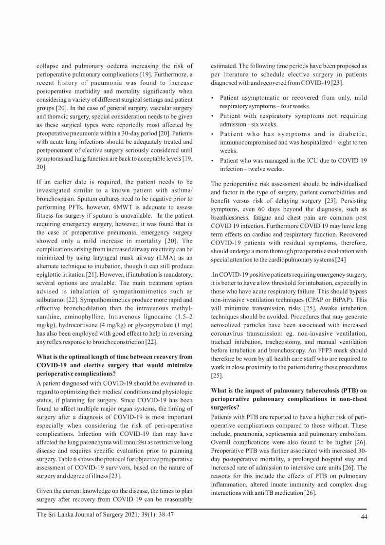
estimated. The following time periods have been proposed as
per literature to schedule elective surgery in patients
diagnosed with and recovered from COVID-19 [23].
Ÿ Patient asymptomatic or recovered from only, mild
respiratory symptoms – four weeks.
Ÿ Patient with respiratory symptoms not requiring
admission – six weeks.
Ÿ Pa t i en t who has symptoms and i s d i abe t i c ,
immunocompromised and was hospitalized – eight to ten
weeks.
Ÿ Patient who was managed in the ICU due to COVID 19
infection – twelve weeks.
The perioperative risk assessment should be individualised
and factor in the type of surgery, patient comorbidities and
benefit versus risk of delaying surgery [23]. Persisting
symptoms, even 60 days beyond the diagnosis, such as
breathlessness, fatigue and chest pain are common post
COVID 19 infection. Furthermore COVID 19 may have long
term effects on cardiac and respiratory function. Recovered
COVID-19 patients with residual symptoms, therefore,
should undergo a more thorough preoperative evaluation with
special attention to the cardiopulmonary systems [24]
.In COVID-19 positive patients requiring emergency surgery,
it is better to have a low threshold for intubation, especially in
those who have acute respiratory failure. This should bypass
non-invasive ventilation techniques (CPAP or BiPAP). This
will minimize transmission risks [25]. Awake intubation
techniques should be avoided. Procedures that may generate
aerosolized particles have been associated with increased
coronavirus transmission: eg. non-invasive ventilation,
tracheal intubation, tracheostomy, and manual ventilation
before intubation and bronchoscopy. An FFP3 mask should
therefore be worn by all health care staff who are required to
work in close proximity to the patient during these procedures
[25].
What is the impact of pulmonary tuberculosis (PTB) on
perioperative pulmonary complications in non-chest
surgeries?
Patients with PTB are reported to have a higher risk of peri-
operative complications compared to those without. These
include, pneumonia, septicaemia and pulmonary embolism.
Overall complications were also found to be higher [26].
Preoperative PTB was further associated with increased 30-
day postoperative mortality, a prolonged hospital stay and
increased rate of admission to intensive care units [26]. The
reasons for this include the effects of PTB on pulmonary
inflammation, altered innate immunity and complex drug
interactions with anti TB medication [26].
collapse and pulmonary oedema increasing the risk of
perioperative pulmonary complications [19]. Furthermore, a
recent history of pneumonia was found to increase
postoperative morbidity and mortality significantly when
considering a variety of different surgical settings and patient
groups [20]. In the case of general surgery, vascular surgery
and thoracic surgery, special consideration needs to be given
as these surgical types were reportedly most affected by
preoperative pneumonia within a 30-day period [20]. Patients
with acute lung infections should be adequately treated and
postponement of elective surgery seriously considered until
symptoms and lung function are back to acceptable levels [19,
20].
If an earlier date is required, the patient needs to be
investigated similar to a known patient with asthma/
bronchospasm. Sputum cultures need to be negative prior to
performing PFTs, however, 6MWT is adequate to assess
fitness for surgery if sputum is unavailable. In the patient
requiring emergency surgery, however, it was found that in
the case of preoperative pneumonia, emergency surgery
showed only a mild increase in mortality [20]. The
complications arising from increased airway reactivity can be
minimized by using laryngeal mask airway (LMA) as an
alternate technique to intubation, though it can still produce
epiglottic irritation [21]. However, if intubation is mandatory,
several options are available. The main treatment option
advised is inhalation of sympathomimetics such as
salbutamol [22]. Sympathomimetics produce more rapid and
effective bronchodilation than the intravenous methyl-
xanthine, aminophylline. Intravenous lignocaine (1.5–2
mg/kg), hydrocortisone (4 mg/kg) or glycopyrrolate (1 mg)
has also been employed with good effect to help in reversing
any reflex response to bronchoconstriction [22].
What is the optimal length of time between recovery from
COVID-19 and elective surgery that would minimize
perioperative complications?
A patient diagnosed with COVID-19 should be evaluated in
regard to optimizing their medical conditions and physiologic
status, if planning for surgery. Since COVID-19 has been
found to affect multiple major organ systems, the timing of
surgery after a diagnosis of COVID-19 is most important
especially when considering the risk of peri-operative
complications. Infection with COVID-19 that may have
affected the lung parenchyma will manifest as restrictive lung
disease and requires specific evaluation prior to planning
surgery. Table 6 shows the protocol for objective preoperative
assessment of COVID-19 survivors, based on the nature of
surgery and degree of illness [23].
Given the current knowledge on the disease, the times to plan
surgery after recovery from COVID-19 can be reasonably
44The Sri Lanka Journal of Surgery 2021; 39(1): 38-47

When planning surgery for patients with PTB, a respiratory
physician's opinion is invaluable. Patients with PTB become
non-infectious at two weeks of anti-tuberculous treatment
(ATT). After commencing treatment for PTB, sputum is
examined for acid fast bacilli (AFB) and Mycobacterium
tuberculosis (MTB) culture at two months when sputum
smear conversion is expected [27]. Ideally, non-chest surgery,
if elective, should be planned for after two months of
commencing ATT [26, 27].
In the case of emergency surgery, a preoperative 6MWT in
patients who are able, or oxygen saturation and ABG in
patients who cannot perform 6MWT, is ideal as PFTs are
contraindicated owing to PTB infection. Risk to the operating
staff can be minimized by performing sputum AFB and rapid
MTB culture as soon as surgery is planned, and by using
personal protective equipment (PPE) and negative pressure
environment for surgery [26, 27].
What is the effect of OSA on perioperative pulmonary
complications?
Patients with OSA have a higher risk of perioperative
pulmonary complications. Anaesthetic medication, opioids
and other sedatives increase airway obstruction and
hypoventilation, which in turn lead to hypercapnic
respiratory failure [28]. Patients with OSA have an increased
risk of being intubated postoperatively and a several-fold
higher risk of being mechanically ventilated than those
without OSA [28, 29]. There is also a higher risk of
postoperative complications and admission to ICU as well as
longer hospital stays than in those without OSA [29, 30].
OSA is generally undiagnosed, therefore, the use of the
STOP-BANG questionnaire has been advised for pre-
assessment screening. Depending on the results, the patient
may either be referred for sleep studies and started on
definitive management or recommended an alternative
anaesthetic regime, for example, by using opioid sparing
anaesthetic techniques and enhanced monitoring. This
screening is recommended by the association of anaesthetists
in the UK for all obese patients undergoing surgery [31].
Studies on obese OSA patients undergoing bariatric surgery
have shown that such screening and management has
improved post-surgical outcome including reduction in post-
operative intensive care [28]. Screening also can predict
susceptible patients who are either undiagnosed or who are
resistant to continuous positive airway pressure treatment,
thus enabling more intensive perioperative monitoring [28].
What is the perioperative steroid management in a
patient on high doses of steroids for interstitial lung
disease or sarcoidosis, requiring surgery?
Hypothalamo-pituitary adrenal axis (HPAA) suppression has
been seen in patients who take doses of prednisolone that
exceed 5 mg/d long term, 7.5-10 mg/d for 1 month or 20 mg/d
for 3 weeks It is also seen in patients taking high doses of other
inhaled corticosteroids for a variety of conditions [32].
Patients on such high doses of steroids at the time of planning
for surgery require supplementation of corticosteroids in
doses that should reflect the physiologic response of the
normal adrenal gland to surgical stress. This supplementation
should be for only a very short duration. Table 7 describes the
perioperative steroid dosing recommendations based on the
type of surgery [33, 34].
Conclusion
Perioperative pulmonary complications are common and
more evident in those with compromised pulmonary function
or undergoing surgery related to the pulmonary system.
Knowledge of physiological derangements during surgery
and anaesthesia, awareness and anticipation of human and
environmental risk factors coupled with risk prediction and
correction of modifiable risk factors could help minimize
such complications.
All authors disclose no conflict of interest. The study was conducted
in accordance with the ethical standards of the relevant institutional
or national ethics committee and the Helsinki Declaration of 1975, as
revised in 2000.
References
1. Weiser TG, Haynes AB, Molina G, Lipsitz SR, Esquivel MM,
Uribe-Leitz T, Fu R, Azad T, Chao TE, Berry WR, Gawande AA.
Estimate of the global volume of surgery in 2012: an assessment
supporting improved health outcomes. Lancet. 2015 Apr 27;385
Suppl 2:S11. doi: 10.1016/S0140-6736(15)60806-6.
2. Gupta S, Fernandes RJ, Rao JS, Dhanpal R. Perioperative risk
factors for pulmonary complications after non-cardiac surgery. J
Anaesthesiol Clin Pharmacol. 2020 Jan-Mar;36(1):88-93.
doi: 10.4103/joacp.
3. Abbott TEF, Fowler AJ, Pelosi P, Gama de Abreu M, Møller AM,
Canet J, Creagh-Brown B, Mythen M, Gin T, Lalu MM, Futier E,
Grocott MP, Schultz MJ, Pearse RM; StEP-COMPAC Group. A
systematic review and consensus definitions for standardised
end-points in perioperative medicine: pulmonary complications.
Br J Anaesth. 2018 May;120(5):1066-1079.
doi: 10.1016/j.bja.2018.02.007.
4. Sasaki N, Meyer MJ, Eikermann M. Postoperative respiratory
muscle dysfunction: pathophysiology and preventive strategies.
Anesthesiology. 2013 Apr;118(4):961-78.
doi: 10.1097/ALN.0b013e318288834f. PMID: 23429163.
5. Miskovic A, Lumb AB. Postoperative pulmonary complications.
Br J Anaesth. 2017 Mar 1;118(3):317-334.
doi: 10.1093/bja/aex002.
6. Lumb AB. Pre-operative respiratory optimisation: an expert
review. Anaesthesia. 2019 Jan;74 Suppl 1:43-48.
doi: 10.1111/anae.14508.
45The Sri Lanka Journal of Surgery 2021; 39(1): 38-47

7. Velicković J, Ladjević N, Milaković B, Likić-Ladjević I,
Ugrinović D, Kalezić N. Preoperative evaluation of patients with
history of allergy. Acta Chir Iugosl. 2011;58(2):177-83.
doi: 10.2298/aci1102177v.
8. Tilak A, Ramirez AG, Turrentine FE, Sohn MW, Jones RS.
Preoperative Gastroesophageal Reflux Disorder Is Associated
With Increased Morbidity in Patients Undergoing Abdominal
Surgery. J Surg Res. 2018 Dec;232:587-594.
doi: 10.1016/j.jss.2018.07.051.
9. Jammer I, Wickboldt N, Sander M, Smith A, Schultz MJ, Pelosi P,
Leva B, Rhodes A, Hoeft A, Walder B, Chew MS, Pearse RM;
European Society of Anaesthesiology (ESA) and the European
Society of Intensive Care Medicine (ESICM); European Society
of Anaesthesiology; European Society of Intensive Care
Medicine. Standards for definitions and use of outcome measures
for clinical effectiveness research in perioperative medicine:
European Perioperative Clinical Outcome (EPCO) definitions: a
statement from the ESA-ESICM joint taskforce on perioperative
outcome measures. Eur J Anaesthesiol. 2015 Feb;32(2):88-105.
doi: 10.1097/EJA.0000000000000118.
10. Özkan M, Kırkıl G, Dilektaşlı AG, Söğüt A, Sertoğullarından B,
Çetinkaya E, Coşkun F, Ulubay G, Yüksel H, Sezer M, Özbudak
Ö, Ulaşlı SS, Arslan S, Kovan T. Summary of Consensus Report
on Preoperative Evaluation. Turk Thorac J. 2015 Jan;16(1):43-
52. doi: 10.5152/ttd.2014.4505. Epub 2015 Jan 1.
11.Patwa A, Shah A. Anatomy and physiology of respiratory system
relevant to anaesthesia. Indian J Anaesth. 2015 Sep;59(9):533-
41. doi: 10.4103/0019-5049.165849.
12.Ntima NO, Lumb AB. Pulmonary function tests in anaesthetic
prac t ice . BJA Educ. 2019 Jul ;19(7) :206-211. doi :
10.1016/j.bjae.2019.02.001. Epub 2019 Mar 26. PMID:
33456892; PMCID: PMC7807994.
13.Bevacqua BK. Pre-operative pulmonary evaluation in the patient
with suspected respiratory disease. Indian J Anaesth. 2015
Sep;59(9):542-9. doi: 10.4103/0019-5049.165854.
14.Degani-Costa LH, Faresin SM, dos Reis Falcão LF. Preoperative
evaluation of the patient with pulmonary disease. Braz J
Anesthesiol. 2014 Jan-Feb;64(1):22-34.
doi: 10.1016/j.bjane.2012.11.002.
15.Wahr JA, Abernathy JH 3rd, Lazarra EH, Keebler JR, Wall MH,
Lynch I, Wolfe R, Cooper RL. Medication safety in the operating
room: literature and expert-based recommendations. Br J
Anaesth. 2017 Jan;118(1):32-43. doi: 10.1093/bja/aew379.
16.National Guideline Centre (UK). Preoperative Tests (Update):
Routine Preoperative Tests for Elective Surgery. London:
National Institute for Health and Care Excellence (UK); 2016
Apr. PMID: 27077168.
17.Global Initiative for Chronic Obstructive Lung Disease. Global
Strategy for the Diagnosis, Management, and Prevention of
C h r o n i c O b s t r u c t i v e P u l m o n a r y D i s e a s e . 2 0 1 9 .
https://goldcopd.org/wp-content/uploads/2018/11/GOLD-2019-
v1.7-FINAL-14Nov2018-WMS.pdf Date last accessed: March
25, 2019.
18.Odor PM, Bampoe S, Gilhooly D, Creagh-Brown B,
Moonesinghe SR. Perioperative interventions for prevention of
postoperative pulmonary complications: systematic review and
meta-analysis. BMJ. 2020 Mar 11;368:m540.
doi: 10.1136/bmj.m540.
19.Canet J, Gallart L, Gomar C, Paluzie G, Vallès J, Castillo J, Sabaté
S, Mazo V, Briones Z, Sanchis J; ARISCAT Group. Prediction of
postoperative pulmonary complications in a population-based
surgical cohort. Anesthesiology. 2010 Dec;113(6):1338-50.
doi: 10.1097/ALN.0b013e3181fc6e0a.
20.Jamali, S., Dagher, M., Bilani, N., Mailhac, A., Habbal, M.,
Zeineldine, S., & Tamim, H. (2018). The Effect of Preoperative
Pneumonia on Postsurgical Mortality and Morbidity: A NSQIP
Analysis. World Journal of Surgery, 42(9), 2763–2772.
doi:10.1007/s00268-018-4539-4
21.Jonathan L. Benumof; Laryngeal Mask Airway and the ASA
Difficult Airway Algorithm. Anesthesiology 1996; 84:686–699.
https://doi.org/10.1097/00000542-199603000-00024
22.Lawal I, Bakari AG. Reactive airway and anaesthesia: challenge
to the anaesthetist and the way forward. Afr Health Sci. 2009
Sep;9(3):167-9. PMID: 20589145; PMCID: PMC2887028.
23.Bui N, Coetzer M, Schenning KJ, O'Glasser AY. Preparing
previously COVID-19-positive patients for elective surgery: a
framework for preoperative evaluation. Perioper Med (Lond).
2021 Jan 7;10(1):1. doi: 10.1186/s13741-020-00172-2.
24.American Society of Anesthesiologists and Anesthesia Patient
Safety Foundation Joint Statement on Elective Surgery and
Anesthesia for Patients after COVID-19 Infection [updated 2020
December 8, 2020; cited 2020 March 16]. Available from:
https://www.apsf.org/news-updates/asa-and-apsf-joint-
statement-on-perioperative-testing-for-the-covid-19-virus/
25.Coccolini, F., Perrone, G., Chiarugi, M. et al. Surgery in COVID-
19 patients: operational directives. World J Emerg Surg 15, 25
(2020). doi: 10.1186/s13017-020-00307-2
26.Ke CC, Lin CS, Yeh CC, Chung CL, Hung CJ, Liao CC, Chen TL.
Adverse Outcomes after Non-Chest Surgeries in Patients with
Pulmonary Tuberculosis: A Nationwide Study. PLoS One. 2015
Jul 14;10(7):e0133064. doi: 10.1371/journal.pone.0133064.
27.National manual for Tuberculosis Control. 3rd ed. Health
information Management Unit, National Programme for
Tuberculosis Control and Chest Diseases. 2016. ISBN 978-955-
0742-07-3
28.Weingarten TN, Kor DJ, Gali B, Sprung J. Predicting
postoperative pulmonary complications in high-risk populations.
Curr Opin Anaesthesiol. 2013;26(2):116-125.
doi:10.1097/ACO.0b013e32835e21d2
29.Memtsoudis S, Liu SS, Ma Y, Chiu YL, Walz JM, Gaber-Baylis
LK, Mazumdar M. Perioperative pulmonary outcomes in patients
with sleep apnea after noncardiac surgery. Anesth Analg. 2011
Jan;112(1):113-21. doi: 10.1213/ANE.0b013e3182009abf.
30.Kaw R, Pasupuleti V, Walker E, Ramaswamy A, Foldvary-
Schafer N. Postoperative complications in patients with
obstructive sleep apnea. Chest. 2012 Feb;141(2):436-441.
doi: 10.1378/chest.11-0283.
31.Members of the Working Party, Nightingale CE, Margarson MP,
Shearer E, Redman JW, Lucas DN, Cousins JM, Fox WT,
Kennedy NJ, Venn PJ, Skues M, Gabbott D, Misra U, Pandit JJ,
Popat MT, Griffiths R; Association of Anaesthetists of Great
Britain; Ireland Society for Obesity and Bariatric Anaesthesia.
Peri-operative management of the obese surgical patient 2015:
Association of Anaesthetists of Great Britain and Ireland Society
46The Sri Lanka Journal of Surgery 2021; 39(1): 38-47

for Obesity and Bariatric Anaesthesia. Anaesthesia. 2015
Jul;70(7):859-76. doi: 10.1111/anae.13101.
32.Jabbour SA. Steroids and the surgical patient. Med Clin North
Am. 2001 Sep;85(5) :1311-7 . do i : 10 .1016/s0025-
7125(05)70379-5.
33.Liu MM, Reidy AB, Saatee S, Collard CD. Perioperative Steroid
Management: Approaches Based on Current Evidence.
Anesthesiology. 2017 Jul;127(1):166-172.
doi: 10.1097/ALN.0000000000001659.
34.Woodcock T, Barker P, Daniel S, Fletcher S, Wass JAH,
Tomlinson JW, Misra U, Dattani M, Arlt W, Vercueil A.
Guidelines for the management of glucocorticoids during the
peri-operative period for patients with adrenal insufficiency:
Guidelines from the Association of Anaesthetists, the Royal
College of Physicians and the Society for Endocrinology UK.
A n a e s t h e s i a . 2 0 2 0 M a y ; 7 5 ( 5 ) : 6 5 4 - 6 6 3 . d o i :
10.1111/anae.14963. Epub 2020 Feb 3. Erratum in: Anaesthesia.
2020 Sep;75(9):1252. PMID: 32017012.
47The Sri Lanka Journal of Surgery 2021; 39(1): 38-47

REVIEW ARTICLE
Anaesthetic implications during the COVID-19 pandemic: an appraisal of the literature
1 2 2 1 1Ravindri Jayasinghe , Nadeeshya Welikala ,Ravindi Gunaratne , Umesh Jayarajah ,Vihara Dassanayake ,
1Sanjeewa Seneviratne1Department of Surgery and Department of Anaesthesiology & Critical Care, Faculty of Medicine,
University of Colombo, Sri Lanka2Department of Anaesthesiology & Critical Care, National Hospital Colombo, Sri Lanka
Keywords: Anaesthesia; anaesthesiology; COVID-19;
pandemic; SARS-CoV-2
Abstract
The coronavirus disease-2019 (COVID-19) pandemic has
raised many concerns regarding the safety of patients and
healthcare workers. Anaesthetic implications and the impact
of this pandemic on anaesthesiologists have been discussed
widely in the recent past. Operating theatres are high-risk
areas where anaesthesiologists perform various aerosol-
generating procedures (AGPs).
Prolong patient contact predisposes them to the risk of
exposure. Scarcity of resources and limitations in testing pose
an additional risk to the anesthesiologist. Although guidance
from various professional bodies is in abundance, there is a
general lack of consensus. This review summarises current
evidence and guidelines related to anaesthesiology during the
COVID-19 pandemic. Ensuring the provision of unhindered
care, ensuring patient safety and protecting oneself with
optimum utilization of available resources remain a challenge
to the anesthesiologist.
Introduction
SARS-CoV-2, a novel strain of coronavirus was first isolated
in January 2020 while investigating an outbreak of
pneumonia in Wuhan, China [1]. Since its origin, millions of
cases have been confirmed positive and more than 2.5 million
deaths have been reported worldwide [2]. The pandemic has
had a significant impact on the healthcare system, economy
and the general public [3, 4].
The impact of COVID-19 on the anaesthesiologist is
considerably higher when compared to other healthcare
workers due to their involvement in airway management in a
high-risk environment [5]. Hospitals worldwide have taken
measures to reduce the number of procedures to minimise
disease transmission and to preserve both consumables and
human resources [6]. Rational use of personal protective
Correspondence: Umesh JayarajahE-mail: [email protected] https://orcid.org/0000-0002-0398-5197Received: 21-03-2021 Accepted: 30-03-2021DOI: http://doi.org/10.4038/sljs.v39i1.8814
48
equipment (PPE) is essential to containing the spread of
infection. The objective of this review is to focus on the
current practice of anaesthesiology during the pandemic and
to summarise key recommendations based on consensus
guidelines for anaesthesiologists.
Mode of anaesthesia: General anaesthesia (GA) vs.
Regional anaesthesia (RA)
Anaesthesia for patients with COVID-19 mandates a balance
between the provision of care and safety for both the patient
and the anesthesiologist. However, during the process of
shared decision- making, the priority is to provide the safest
and the most appropriate mode of anaesthesia for a surgical
procedure to the patient. Whenever there is a doubt, patient
comfort and safety should be considered as a priority. The
consenting process should not be biased towards any
particular modality and all information based on evidence
should be provided to the patient. It is also prudent to
consider the risks of disease transmission related to the mode
of anaesthesia and the urgency of surgery [7].
Many potential advantages have been described concerning
various modes of anaesthesia. Regional anaesthesia reduces
the number of AGPs thereby saving time and cost spent on
PPEs, preservation of immune function when compared with
general anaesthesia, improved postoperative analgesia
minimising direct contact with caregivers, and earlier hospital
discharge [8]. Additionally, it has been identified that patients
with acute COVID-19 infection were at an increased risk of
developing postoperative pulmonary complications
including ARDS [9, 10]. Overall, patients who underwent
neuraxial anaesthesia had fewer perioperative pulmonary
complications compared to general anaesthesia [11].
To date, no published randomized controlled trials are
evaluating the reduction of risk for disease transmission
brought on by regional anaesthesia when compared to general
anaesthesia. Therefore, it is reasonable to consider that all the
recommendations on the above have been based on logical
thinking and understanding. Interestingly, a recent death
review of health care workers carried out in the United
Kingdom revealed that there were no reported deaths among
anaesthesiologists or staff involved in critical care practices,
The Sri Lanka Journal of Surgery 2021; 39(1): 48-52

where the highest number of AGPs had been carried out [8].
The reason for this is unclear but may well be attributed to the
rigorous utilization of PPEs [8].
Based on the above, regional anaesthesia may be considered a
suitable option where appropriate. However, the risk of
conversion to GA midway during the procedure should be
thoroughly evaluated beforehand and appropriate PPEs
should be kept ready. It should be remembered that droplet
and aerosol generation are possible while breathing, speaking,
coughing and sneezing which are all possible in the awake
patient under regional anaesthesia. In such patients, donning
in only droplet PPEs may be sufficient. Such a situation may
not arise or be minimal in a patient who is under general
anaesthesia with a definitive airway in situ, which might
appear to be a safer option as opposed to regional anaesthesia.
Other general standards should be given due consideration
such as limiting proximity to the patient and duration of
exposure, minimum of five air changes/hour, placement of a
surgical mask on the patient and minimal manipulation of
oxygen therapy devices. Avoidance of high oxygen flow rates
will minimize the generation of aerosols.
Furthermore, with more cases being performed under
regional anaesthesia there are advantages to an institution,
such as efficient utilization of drugs for critical care services,
reducing the time consumed per case and bypassing the time
spent in the recovery area.
Due to a lack of reliable evidence, the adverse effects of
regional anaesthesia on COVID-19 patients have not been
fully demonstrated. Literature suggests there are minimal
effects on clinical outcomes in patients undergoing regional
anaesthesia [11].
Despite being commonly associated with hypercoagulability,
COVID-19 is also associated with thrombocytopenia [12].
Therefore, a full blood count is mandated before providing
neuraxial anaesthesia. Care needs to be taken during the
insertion and removal of indwelling epidural catheters with
pre-existing coagulopathy.
In patients with neurological disease, central neuraxial blocks
(CNB) were thought to be safe as the coronavirus had not been
isolated in cerebrospinal fluid [8]. However, with a more
recently reported case of COVID-19 meningitis and
detectable virus within the CSF, such evidence should be
revisited [8].
During placement of peripheral nerve blocks, several key
recommendations have been made by various bodies to
improve quality and safety related to anaesthesia. Use of
ultrasonography to perform regional blocks, use of wound
infiltration catheters in case of prolonged blocks or to provide
49
post-operative pain relief using adjuncts to peripheral nerve
blocks (PNB) along with local anaesthetics without dose
modifications, can all be incorporated into current practise
during the pandemic [8].
Accredited authorities of regional anaesthesia had issued
guidance on neuraxial anaesthesia and peripheral nerve
blocks during the COVID-19 pandemic recommending
regional anaesthesia over general anaesthesia [13]. However,
the possibility of a “failed block” and conversion to general
anaesthesia may increase the risk of viral transmission.
Therefore, careful preoperative assessment to avoid any
unplanned conversions to general anaesthesia is imperative.
Pre-operative assessment and Preparation
During the preoperative assessment, it is important to weigh
the risk of disease transmission and the benefit of surgery to
the patient. Identifying the high-risk patient is of paramount
importance [14]. It is also important to balance resource
consumption and the risk of transmission of the disease to
healthcare workers. Factors such as the need for critical care
beds, transfusions, number of personnel required in theatre,
risks associated with various modes of anaesthesia and type of
surgery should all be taken into consideration [15, 16]. It is
recommended to have a designated operating room for
COVID-19 suspected patients and to do with the appropriate
level of PPEs. Limited resources including consumables is a
universal problem. The World Health Organization (WHO)
recommends using N95, Filtering Face Piece-2 (FFP2)
respirator or an equivalent during aerosol-generating
procedures [17]. Despite guidance on PPEs, there is limited
consensus related to their usage. A three-tiered approach to
PPE has been recommended for airway managers. Their
recommendation is to use airborne, droplet and contact
precautions in combination for high-risk aerosol-generating
procedures which comprise of a head cover, eye protection,
N95 respirator, AAMI (Association for the advancement of
medical instrumentation) level-2 or higher gown and a pair of
single gloves overlapping the sleeve of the waterproof gown
[17]. Droplet and contact precautions are considered adequate
for most routine procedures. High-risk precautions should be
used by personnel directly involved in procedures such as
airway management. Limiting the number of personnel in the
operating room (OR) is critical during high-risk procedures
which minimises exposure [17].
It is essential to have a pre-planned team led by a senior
anaesthesiologist for patients suspected or confirmed with
COVID-19. Roles should be allocated preoperatively, and all
members should maintain closed-loop communication.
During emergency surgery, a member of the team should
perform a rapid pre-anaesthetic check-in in a predesigned
room within the theatre [18]. During an elective procedure,
The Sri Lanka Journal of Surgery 2021; 39(1): 48-52

To minimize the risk of airborne exposure, it is best to manage
the patient in a negative pressure room with rates of air
exchange amounting to more than 12 exchanges per hour.
Monitoring should be established by adhering to the
Association of Anaesthetists standards including the use of
continuous waveform capnography.
Intubation should be performed by the most experienced
member of the team to ensure success at the first attempt,
safety, accuracy and swiftness in securing the airway. A
tracheal intubation trolley must be prepared and be checked
daily. An airway strategy including rescue plans should be
briefed to the airway team before intubation. The team should
comprise of an “intubator”, an assistant and a third staff
member to administer drugs and to monitor the patient. A
runner or buddy should be present outside the intubating bay
who can call for help if required and assist in donning of team
members and observe doffing once the procedure is over.
Rapid sequence induction (RSI) with cricoid pressure is
recommended to secure the airway. The procedure begins
with pre-oxygenation using a well-fitting mask for 3–5 min
with a closed circuit. A heat and moisture exchange (HME)
filter should be connected between the catheter mount and the
circuit. Non-invasive ventilation and high flow nasal oxygen
should be avoided. Ketamine can be used as an induction
agent to avoid cardiovascular collapse provided that there is
no contraindication. Rocuronium (if sugammadex is
available) or succinylcholine is used as neuromuscular
blockers and complete paralysis must be confirmed before
attempting intubation. Face mask ventilation should be
avoided during apnoea. Continuous positive airway pressure
(CPAP) may be applied. If mask ventilation is necessary to
prevent hypoxemia, 2-hand, 2-person technique with a VE
grip should be used.
The use of a video-laryngoscope is advocated as the operator
can maintain sufficient distance from the patient's airway
during tracheal intubation and achieve prompt first-pass
tracheal intubation. The tracheal tube has to be placed without
losing sight of it on the screen and the cuff is passed 1– 2 cm
below the cords for optimal positioning. The cuff is inflated to
20-30 cmH2O. Mechanical ventilation is commenced only
after cuff inflation. Correct placement of the tracheal tube is
then confirmed with continuous waveform capnography.
Auscultation to confirm tube placement is not recommended.
Visual observation of equal bilateral chest wall expansion,
lung ultrasound or chest X-ray can also be done to confirm the
correct placement of the tube.
A ventilator dedicated for COVID-19 positive patients in the
OR has also been recommended which would minimize
cross-contamination [21]. The use of barrier-enclosure
pre-anaesthetic patient assessment could be done via
telemedicine as an alternative to face-to-face consultation.
Video and audio devices can be used to perform a visual
examination and provide medical advice. Most routine
surgeries including cardiac, vascular and thoracic surgery can
potentially be performed following a telemedicine
evaluation. However, there are limitations especially in
patients with multiple comorbidities and technological
constraints related to internet access and the level of literacy.
This is a hindrance and a constant challenge to the
anesthesiologist and patients seeking perioperative care in a
low-middle income country, such as Sri Lanka.
Airway management
Consensus guidelines for managing the airway in patients
with COVID-19 was published by the Difficult Airway
Society, the Association of Anaesthetists, the Intensive Care
Society, the Faculty of Intensive Care Medicine, and the
Royal College of Anaesthetists [19].
Airway establishment and management may be required for
asymptomatic or minimally symptomatic patients with
COVID 19 in the community requiring emergency surgery for
unrelated conditions [19] and controlled ventilation for
patients diagnosed with viral pneumonia in the critical care
unit.
Several AGPs have been identified as high-risk of
dissemination of the virus during airway manipulation. These
include tracheal intubation, tracheostomy, emergency front-
of neck airway (FONA), non-invasive ventilation (NIV), and
bag-mask ventilation. Other possible AGPs includes tracheal
extubation, disconnection of ventilatory circuits during use,
performing chest compressions during cardiopulmonary
resuscitation without tracheal intubation (minimal risk during
defibrillation), bronchoscopy, and tracheal suctioning.
Recent studies have demonstrated that in practice, tracheal
intubation and facemask ventilation produced only very low
quantities of aerosolised particles. Tracheal extubation when
associated with coughing produced 15 times more aerosols
than intubation. Volitional coughing is associated with a very
high aerosol generation [20].
To ensure safety for the anesthesiologist and other health care
workers, correct use of PPEs, decontamination of surfaces
and equipment, minimizing unnecessary patient and surface
contact and careful waste disposal are essential elements
during patient care. Double gloving may protect against
fomite contamination. Training and regular simulation
sessions in the use of PPEs (both donning and doffing) before
patient management is beneficial [19].
50The Sri Lanka Journal of Surgery 2021; 39(1): 48-52

systems, such as aerosol boxes and plastic drapes have been
developed to act as a shield during endotracheal intubation
while allowing enough mobility during intubation. Its use
may complicate airway management with potential adverse
events, particularly during airway emergencies. Concerns
include the limited ability for interventions, limited access to
support during intubation by the assistant, damage to the PPE
of the “intubator”, injuries to the patient and secondary
aerosolization during barrier removal. There is no clear
evidence to suggest additional protection for HCWs and there
is no standard for cleaning such equipment. Enclosure
barriers cannot substitute adequate use of PPEs and further
studies are required for validation [22].
Perioperative management and extubation
Adherence to the prevention of infections is mandatory
during the intraoperative period. As a policy for infection
control, it is recommended that only one anesthesiologist
should use the workstation and it should be covered with
disposable transparent drapes. Frequent intraoperative
suctioning should be avoided and low-flow anaesthesia
should be used for maintenance to reduce viral transmission
[18].
Tracheal extubation should be performed inside the operating
theatre with limited HCWs and efforts taken to minimize
coughing due to increased aerosol generation. Use of
supraglottic airway as a bridging technique or use of
medications such as dexmedetomidine, lidocaine and opioids
which suppresses cough may be considered to achieve
smooth extubation. However, the safety of these procedures
in the setting of the COVID-19 pandemic has not been
proven. Tracheal suctioning through a closed suction
apparatus and oral suctioning can be performed as usual.
There should be equipment available to provide low flow
nasal oxygen (< 5 litres/ min) or face mask oxygen before
extubation to ensure the immediate connection of the patient
to their oxygen mask or nasal cannula [19]. If extubation is
deferred, the patient transfer should take place using a
transport ventilator with an HME (heat and moisture
exchanger) filter connected to the patient end [18].
Recovery and post anaesthesia care
Patients should be taken to a designated recovery room
following extubation. The patients should wear surgical
masks before leaving the theatre. Congestion in the recovery
area should be avoided and experts recommend maintaining
at least a one-meter distance between patients [23]. After
leaving the theatre, it is recommended to keep the patient in an
isolated room. However, due to overwhelmed resources, this
may be impractical in many centres.
All used PPE s should be disposed into biohazard bags
keeping with the local infection control policy. It is mandatory
to avoid contamination during the doffing of PPEs. This could
be achieved by having an observer/buddy to guide safe
doffing [24]. Decontamination of the theatre and equipment
should follow hospital policy. All clinical waste should be
clearly labelled, double bagged and sealed. It is then sprayed
with chlorinated disinfectant and covered with an additional
bag. Single-use laryngoscopy blades, breathing circuits,
masks, HME filters, tracheal tubes, gas sampling lines and
soda-lime needs to be discarded [25]. Agents that are used for
decontamination include, vaporized Glutaraldehyde
(ANIOS) or hydrogen peroxide generator (VHPG), 1%
sodium hypochlorite solution, and 75% alcohol. Metallic
equipment must be kept immersed in 1% sodium hypochlorite
for 30 minutes and then washed and wiped [25]. The
operating theatre should be cleaned and disinfected between
surgeries. The disinfecting personnel must enter the theatre
only after adequate air changes have been completed to
remove infectious particles. Staff must be aware of the
COVID status of the patient and must handle equipment only
after wearing full PPE [26]. All floors and walls should be
cleaned with 1% sodium hypochlorite solution [25]. The
theatre can be disinfected with UV light after chemical
disinfection [26].
Conclusion
The ongoing pandemic is causing a dramatic shift in the role
of healthcare workers in their diverse specialities.
Anaesthesiologists are placed at high risk due to manipulation
of the airway and dealing with AGPs. Hospitals should
develop a detailed pandemic preparedness plan addressing
major concerns such as limitation of resources, staff well-
being and safety, and implementation of strict measures of
infection control. Current guidance is mostly based on expert
opinion. High-quality multicentre research is deemed
essential to formulate evidence-based healthcare policy
changes both locally and internationally.
All authors disclose no conflict of interest. The study was conducted
in accordance with the ethical standards of the relevant institutional
or national ethics committee and the Helsinki Declaration of 1975, as
revised in 2000.
References
1. Borges do Nascimento IJ, Cacic N, Abdulazeem HM: Novel
Coronavirus Infection (COVID-19) in Humans: A Scoping
Review and Meta-Analysis. 2020, 9(4):941
https://doi.org/10.3390/jcm9040941
2. Borges do Nascimento IJ, von Groote TC, O'Mathúna DP et al.
Clinical, laboratory and radiological characteristics and
outcomes of novel coronavirus (SARS-CoV-2) infection in
humans: A systematic review and series of meta-analyses. PloS
51The Sri Lanka Journal of Surgery 2021; 39(1): 48-52

one 2020, 15(9):e0239235.
https://doi.org/10.1371/journal.pone.0239235
3. Jayasinghe R, Ranasinghe S, Jayarajah U, Seneviratne S: Quality
of online information for the general public on COVID-19.
Pa t ien t Educa t ion and Counse l ing 2020 in press .
https://doi.org/10.1016/j.pec.2020.08.001
4. Wijerathne PK, Nanayakkara K, Basnayake O, Gunapala N,
Jayarajah U: Impact of COVID-19 on postgraduate surgical
training: the trainees' perspective. Sri Lanka Journal of Surgery
2020, 38(3):53-55. http://doi.org/10.4038/sljs.v38i3.8769
5. Preventing infection of patients and healthcare workers should be
the new normal in the era of novel coronavirus epidemics
[https://anesthesiology.pubs.asahq.org/article.aspx?articleid=27
63452]
6. Collaborative C: Delaying surgery for patients with a previous
SARSCoV2 infection. The British journal of surgery 2020,
107:e601-e602. https://dx.doi.org/10.1002%2Fbjs.12050
7. Glasbey JC, Bhangu A, Collaborative C: Elective cancer surgery
in COVID-19–free surgical pathways during the SARS-CoV-2
pandemic: an international, multicenter, comparative cohort
study. Journal of Clinical Oncology 2020:JCO. 20.01933. doi:
https://doi.org/10.1101/2020.12.15.20248195
8. Macfarlane AJ, Harrop-Griffiths W, Pawa A: Regional anaesthe-
sia and COVID-19: first choice at last? British journal of
anaesthesia 2020, 125(3):243-247.
https://doi.org/10.1016/j.bja.2020.05.016
9. Myles PS, Maswime S: Mitigating the risks of surgery during the
COVID-19 pandemic. The Lancet 2020, 396(10243):2-3.
https://doi.org/10.1016/S0140-6736(20)31256-3
10.Collaborative C: Preoperative nasopharyngeal swab testing and
postoperative pulmonary complications in patients undergoing
elective surgery during the SARS-CoV-2 pandemic. British
Journal of Surgery. https://doi.org/10.1093/bjs/znaa051
11.Rodgers A, Walker N, Schug S, McKee A, Kehlet H, Van Zundert
A, Sage D, Futter M, Saville G, Clark T: Reduction of
postoperative mortality and morbidity with epidural or spinal
anaesthesia: results from overview of randomised trials. Bmj
2000, 321(7275):1493.
https://doi.org/10.1136/bmj.321.7275.1493
12.Rahman A, Niloofa R et al. Hematological Abnormalities in
COVID-19: A Narrative Review. The American journal of
tropical medicine and hygiene:tpmd201536.
https://doi.org/10.4269/ajtmh.20-1536
13.Practice Recommendations on Neuraxial Anesthesia and
Peripheral Nerve Blocks during the COVID-19 Pandemic
[https://www.asra.com/page/2905/practice-recommendations-
on-neuraxial-anesthesia-and-peripheral-nerve-blocks-dur]
14.Prachand VN, Milner R, Angelos P, Posner MC, Fung JJ, Agrawal
N, Jeevanandam V, Matthews JB: Medically-necessary, time-
sensitive procedures: a scoring system to ethically and efficiently
manage resource scarcity and provider risk during the COVID-19
pandemic. Journal of the American College of Surgeons 2020.
https://doi.org/10.1016/j.jamcollsurg.2020.04.011
15.Jayasinghe R, Jayarajah U, Seneviratne S: Consensus on peri-
operative surgical practice during the COVID-19 pandemic: an
appraisal of the literature. Sri Lanka Journal of Surgery 2020, 38
(2):57-61.
https://doi.org/10.4038/sljs.v38i2.8738
16.Jayasinghe R, Jayarajah U, Seneviratne S: Consensus in Surgical
Practice during the COVID-19 Pandemic: An Appraisal of the
Literature. 2020.
17.Rational use of personal protective equipment for coronavirus
disease 2019 (COVID-19)
[https://apps.who.int/iris/bitstream/handle/10665/331215/WHO-
2019-nCov-IPCPPE_use-2020.1-eng.pdf.]
18.Kumar S, Palta S, Saroa R, Mitra S: Anesthesiologist and
COVID-19—current perspective. Journal of Anaesthesiology
Clinical Pharmacology 2020, 36(5):50.
https://doi.org/10.4103/joacp.joacp_205_20
19.Cook TM, El-Boghdadly K, McGuire B, McNarry AF, Patel A,
Higgs A: Consensus guidelines for managing the airway in
patients with COVID-19: Guidelines from the Difficult Airway
Society, the Association of Anaesthetists the Intensive Care
Society, the Faculty of Intensive Care Medicine and the Royal
College of Anaesthetists. Anaesthesia 2020, 75(6):785-799.
https://doi.org/10.1111/anae.15054
20.Brown J, Gregson FKA, Shrimpton A, Cook TM, Bzdek BR,
Reid JP, Pickering AE: A quantitative evaluation of aerosol
generation during tracheal intubation and extubation.
Anaesthesia 2021, 76(2):174-181.
https://doi.org/10.1111/anae.15292
21.College of Anaesthesiologists and Intensivists of Sri Lanka,
Interim guidelines on management of critically ill patients with
COVID-19 [https://anaesthesia.lk/sdm_downloads/interim-
guidelines-on-management-of-critically-ill-patients-with-
covid-19/]
22.Sorbello M, Rosenblatt W, Hofmeyr R, Greif R, Urdaneta F:
Aerosol boxes and barrier enclosures for airway management in
COVID-19 patients: a scoping review and narrative synthesis.
BJA: British Journal of Anaesthesia 2020, 125(6):880.
https://doi.org/10.1016/j.bja.2020.08.038
23.Velly L, Gayat E, Quintard H, Weiss E, De Jong A, Cuvillon P,
Audibert G, Amour J, Beaussier M, Biais M: Guidelines:
Anaesthesia in the context of COVID-19 pandemic. Anaesthesia
Critical Care & Pain Medicine 2020, 39(3):395-415.
https://doi.org/10.1016/j.accpm.2020.05.012
24.Interim Infection Prevention and Control Recommendations for
Patients with Suspected or Confirmed Coronavirus Disease 2019
(COVID-19) in Healthcare Settings
[https://www.cdc.gov/coronavirus/2019-ncov/infection-
control/control-recommendations.html?CDC_AA_refVal=
https%3A%2F%2Fwww.cdc.gov%2Fcoronavirus%2F2019-
ncov%2Fhcp%2Finfection-control.html]
25.Malhotra N, Bajwa SJS, Joshi M, Mehdiratta L, Trikha A:
COVID operation theatre-advisory and position statement of
Indian Society of Anaesthesiologists (ISA National). Indian
Journal of Anaesthesia 2020, 64(5):355.
https://doi.org/10.4103/ija.ija_288_20
26.Prakash L, Dhar SA, Mushtaq M: COVID-19 in the operating
room: a review of evolving safety protocols. Patient Safety in
Surgery 2020, 14(1):30.
https://doi.org/10.1186/s13037-020-00254-6
52The Sri Lanka Journal of Surgery 2021; 39(1): 48-52

PERSPECTIVE
The need for trainee-led surgical research collaborative in Sri Lanka
Umesh Jayarajah, Oshan Basnayake
Postgraduate Institute of Medicine, University of Colombo, Sri Lanka
Keywords: Research collaborative; trainee-led research;
surgical research
Introduction
The value of medical research in the undergraduate and
postgraduate medical curriculum is increasingly recognised
[1] . Many professional medical bodies promote
undergraduate and post-graduate trainees to gain some
experience in research, not only to assist in carrying out
original studies but also to cultivate skills to become
accomplished research consumers. This will enable them to
be informed about current evidence and enhance their
professional practice [1].
However, there have been many obstacles to fulfil this aim.
Post-graduate trainees are occupied with their clinical
practice, especially in a field like surgery where necessary
practical skills must be learned in addition to the core clinical
knowledge [1]. Furthermore, partly due to inadequate
guidance, many trainees feel discouraged about research
altogether or commence impractical or over-ambitious
studies often ending up in failures discouraging further
attempts in research. Outputs from such attempts are unlikely
to contribute to science and are often not disseminated. This is
because of too small sample size, lack of novelty, single-
centre, lack of proper follow-up and lack generalizability [2].
Furthermore, there is a general lack of formal training in
research methodology.
In general, there are several disadvantages related to small
scale single centre studies. Underpowered studies with a
small sample size and broad confidence intervals result in
findings that contribute minimally to science and unlikely to
change clinical practice [3]. Many such studies failed to get
published in peer-reviewed journals. However, there is an
increasing trend to conduct scientific work by collaborative
networks [4]. Such collaborative networks enable a group of
geographically scattered researchers of common interests to
communicate and contribute towards a common goal. This is
Correspondence: Umesh JayarajahE-mail: [email protected] https://orcid.org/0000-0002-0398-5197Received: 14-11-2020 Accepted: 09-03-2021DOI: http://doi.org/10.4038/sljs.v39i1.8753
53
feasibly achieved through online platforms using modern
information technology [5].
Several difficulties about working across geographical
distance have been identified. These include issues related to
ownership, authorship, funding, intellectual property, trust
and communication [1]. The establishment of well organised
collaborative networks with well documented and transparent
protocols have been helpful to overcome these shortcomings
[1]. A functional model for a collaborative research network is
shown in figure 1.
In the UK, over the past decade, several trainee-led regional
collaborative in general surgery and other specialities have
been established [1]. This approach enables to include a
sufficiently large sample in a short duration and prevents
duplication of data. Moreover, this enables to conduct of
multicentre pragmatic studies and allows for greater
generalizability in contrast to single centre short term studies
[6]. Trainees follow clinical rotation in multiple hospitals
nationwide and are in regular contact with each other and
therefore, are ideal to implement this model. As trainees
become independent practitioners, a culture of surgical
research and trials could be distilled in the country's surgical
practice [6]. So far, several surgical research collaborative
networks have been initiated in the UK with considerable
success with large scale trainee-driven cohort studies and
clinical trials [6].
There is increasing evidence that research experiences of
trainee doctors have long term benefits in their subsequent
careers and may also enhance the opportunities for overseas
training [7]. This may positively influence lifelong academic
inquisitiveness and contributions to teaching, ultimately
improving patient care. Due to the above reasons, a research
forum led by trainees with the support and guidance of
experienced researchers will be useful. Such an initiative
should allow all trainees within a region or country to team up
irrespective of the geographical location, academic schedule
and long term academic interest. In conclusion, a trainer-led
collaborative platform may be an effective model to enhance
and promote high-quality surgical research in Sri Lanka.
The Sri Lanka Journal of Surgery 2021; 39(1): 53-54

All authors disclose no conflict of interest. The study was conducted
in accordance with the ethical standards of the relevant institutional
or national ethics committee and the Helsinki Declaration of 1975, as
revised in 2000.
References
1. Dowswell G, Bartlett D C, Futaba K, Whisker L, Pinkney T D.
How to set up and manage a trainee-led research collaborative.
BMC Medical Education 2014; 14: 94.
https://doi.org/10.1186/1472-6920-14-94
2. Brubaker L, Kenton K. Clinical research education study teams: a
research curriculum for obstetric and gynecology residents.
Obstetrics & Gynecology 2011; 117: 1403-7.
https://doi.org/10.1097/AOG.0b013e3182179705
3. Bhandari M, Sprague S, Schemitsch EH. Resolving controversies
in hip fracture care: the need for large collaborative trials in hip
fractures. Journal of orthopaedic trauma 2009; 23: 479.
https://doi.org/10.1097/BOT.0b013e3181a772e3
4. Newman ME. The structure of scientific collaboration networks.
Proceedings of the national academy of sciences 2001; 98: 404-9.
https://doi.org/10.1073/pnas.98.2.404
5. Bos N, Zimmerman A, Olson J et al. From shared databases to
communities of practice: A taxonomy of collaboratories. Journal
of Computer-Mediated Communication 2007; 12: 652-72.
https://doi.org/10.1111/j.1083-6101.2007.00343.x
6. Bhangu A, Kolias AG, Pinkney T, Hall NJ, Fitzgerald JE. Surgical
research collaboratives in the UK. The Lancet 2013; 382: 1091-2.
https://doi.org/10.1016/S0140-6736(13)62013-9.
7. Bhattacharya SD, Williams JB, de la Fuente SG, Kuo PC, Seigler
HF. Does protected research time during general surgery training
contribute to graduates' career choice? The American surgeon
2011; 77: 907-10.
https://www.ncbi.nlm.nih.gov/pmc/articles/PMC3720679/
5
Figure 1. Functional model for a collaborative research network
54The Sri Lanka Journal of Surgery 2021; 39(1): 53-54

TECHNICAL NOTE
Favourable anatomical variation for an extended hepatectomy with portal vein resection in a hilar cholangiocarcinoma involving second order ducts
1 1 2 3Prabath Kumarasinghe , Hasith Fernando , Chathurika Sagara , Aloka Pathirana1National Hospital of Sri Lanka2Colombo South Teaching Hospital
3Faculty of Medical Sciences, University of Sri Jayewardenepura
Keywords: Hilar cholangiocarcinoma; portal vein; hepatic
artery
Introduction
A fifty-seven-year-old female presented with recent onset
painless obstructive jaundice. She had no other co-
morbidities. Her total bilirubin was elevated with a
predominant direct fraction (Total bilirubin-330mmol/l,
Direct.bilirubin-165.66 mmol/l). CECT findings were
consistent with hilar cholangiocarcinoma (Bismuth-Corlettte
type 3A) without distant metastasis. An external biliary drain
was placed in the right hepatic duct, to relieve the obstruction.
The percutaneous transhepatic cholangiogram confirmed the
involvement of the hepatic duct confluence. CT-angiogram
revealed a replaced right hepatic artery originating from the
superior mesenteric artery
Procedure
A modified Makuuchi incision was made to open into the
peritoneal cavity. The peritoneal survey confirmed a localised
disease. The liver showed extensive cholestatic changes.
Hilar dissection was performed and the right hepatic artery
arising from the superior mesenteric artery was dissected
away from the tumour. Tumour infiltration was noted at the
portal vein confluence. The right hepatic duct (RHD) was
involved near the confluence and tumour extension noted in
the left hepatic duct (LHD). Left hemi-liver with caudate lobe
was mobilized off the inferior vena cava. Parenchymal
transection was carried out along the inter-sectoral line
(between the right anterior and posterior sector).
Anterior sector vessels were divided between ligatures and
inflow to the posterior sector was carefully preserved. The
right posterior sectoral hepatic duct was divided from the
lesion with a margin. Imprint cytology was done to confirm
negative margins. Parenchymal transection was completed
using CUSA®. The left hepatic artery was divided close to the
gastroduodenal artery between ligatures. The right portal vein
(RPV) was slung distal to the tumour and the main portal vein
Correspondence: Prabath KumarasingheE-mail: [email protected] https://orcid.org/0000-0002-5078-0944Received: 26-02-2021 Accepted: 30-03-2021DOI: http://doi.org/10.4038/sljs.v39i1.8795
55
(MPV) slung below the confluence and vascular clamps
applied.
The portal vein was divided at MPV and RPV well away from
the tumour. Left and middle hepatic veins were divided by
vascular staplers and an extended Left Hepatectomy
specimen was removed en-bloc with the common bile duct,
right anterior and left hepatic duct and the portal vein segment
with perihilar lymphatics. MPV was anastomosed to the distal
RPV with 5.o polypropylene. Limited mobilization of the
right lobe of the liver and plication of the right diaphragm was
done to relieve the tension at the PV anastomosis.
Roux-en-Y hepaticojejunostomy was done to the right
posterior sectoral bile duct. Postoperative doppler confirmed
good doppler flow of the RHA and RPV. The patient had an
uneventful recovery and was referred for adjuvant
chemotherapy. Histology confirmed a moderately
differentiated cholangiocarcinoma with a negative RHD
margin and PV resection margin.
Discussion
Hillar cholangiocarcinoma has a dismal prognosis without
complete surgical resection (with negative resection margins)
[1, 2, and 3]. En-bloc resection of the tumour with PV and
perihilar lymphatics provides improved survival in carefully
selected patients with HCCA. Routine resection of the PV
when there is evidence of tumour abutment may minimize the
tumour dissemination during dissection [4]. Especially right-
sided tumours frequently invade the RHA due to its close
relationship to the biliary confluence [5]. In instances where
RHA originates from SMA, a more lateral course of the artery
makes it less likely to be involved by the tumour.
Given the lack of an alternative curative method for hillar
cholangiocarcinoma, aggressive resection with involved
portal vein provides the greatest survival benefit and it can be
performed safely as in our given patient.
The Sri Lanka Journal of Surgery 2021; 39(1): 55-56

All authors disclose no conflict of interest. The study was conducted
in accordance with the ethical standards of the relevant institutional
or national ethics committee and the Helsinki Declaration of 1975, as
revised in 2000.
References
1. Cho MS, Kim SH, Park SW, Lim JH, Choi GH, Park JS, Chung
JB, Kim KS. Surgical outcomes and predicting factors of curative
resection in patients with hilar cholangiocarcinoma: 10-year
single-institution experience. J Gastrointest Surg. 2012
Sep;16(9):1672-9.
doi: 10.1007/s11605-012-1960-0. Epub 2012 Jul 14.
2. Cannon RM, Brock G, Buell JF. Surgical resection for hilar
cholangiocarcinoma: experience improves resectability. HPB
(Oxford). 2012 Feb;14(2):142-9.
doi : 10.1111/j.1477-2574.2011.00419.x. Epub 2011 Dec 12.
56
3. Nishio H, Nagino M, Nimura Y. Surgical management of hilar
cholangiocarcinoma: the Nagoya experience. HPB (Oxford).
2005;7(4):259-62. doi: 10.1080/13651820500373010.
4. Tamoto E, Hirano S, Tsuchikawa T, Tanaka E, Miyamoto M,
Matsumoto J, Kato K, Shichinohe T. Portal vein resection using
the no-touch technique with a hepatectomy for hilar
cholangiocarcinoma. HPB (Oxford). 2014 Jan;16(1):56-61.
doi: 10.1111/hpb.12067. Epub 2013 Mar 6.
5. Nagino M, Nimura Y, Nishio H, Ebata T, Igami T, Matsushita M,
et al. Hepatectomy with simultaneous resection of the portal vein
and hepatic artery for advanced perihilar cholangiocarcinoma:
an audit of 50 consecutive cases. Ann Surg. 2010;252:115–23.
Figure 1. Liver parenchyma split along the intersectional line
of right anterior and posterior sectors, portal vein confluence
infiltrated by the tumour.
RPV- Right portal vein
MPV- Main portal Vein network
Figure 2. Preserved right posterior sector of the liver with
intact inflow structures
HD-RP - Hepatic duct right posterior
HA-RP- Hepatic artery right posterior sector
The Sri Lanka Journal of Surgery 2021; 39(1): 55-56

CASE SERIES
Isolated jejunal loop hepaticoduodenostomy following excision of type VI-A choledochal cyst
1 1 2 1 2A. D Dharmapala , B.K Dassananyake , P. G Athanospolous , K.B Galketiya , M. Malago1 Department of Surgery, Faculty of Medicine, University of Peradeniya, Sri Lanka2 Academic Department of Surgery, University College London/Royal Free NHS trust, Hampstead,London
Keywords: Hepaticoduodenostomy; choledochal cyst;
MRCP; jejunal loop
Introduction
Choledochal cyst is a congenital dilatation of the intra hepatic
or extra hepatic bile ducts or both. This is classified by Todani
classification, which was modified by Alfonso. Depending on
the involvement of the biliary tree and the morphological
appearance, it is classified into 5 types [1, 2] (Figure1). Type I
consists of a solitary extrahepatic cyst, type II is an
extrahepatic supraduodenal diverticulum, type III is an
intraduodenal cyst(choledochocele), type IV consists of both
extrahepatic and intrahepatic cysts; and type V consists of
multiple intrahepatic cysts (Caroli's disease).
The etiology for the development of this dilatation is still not
clearly established but the abnormalous pancreatico biliary
junction (APBJ) is so far the most accepted explanation [3].
Due to the APBJ the pancreatic juice is refluxed back into the
bile, weakening the wall and resulting in a dilatation of the
ducts [4].
The dilated bile ducts allow stasis of bile causing a chronic
irritation leading to dysplastic changes in the mucosa and
causing cholangio carcinoma [5].
Therefore, the treatment of these cysts consists of complete
excision of the cyst and reconstruction with a roux and y
hepatico jejunostomy. The simple drainage procedure
without excising the cyst will not eliminate the risk of
malignancy in these group of patients [6,7].
There are various forms of reconstruction techniques and this
is a case report where an isolated jejunal graft with its vascular
pedicle is used to reconstruct the bile duct.
Case presentation
A 21-year-old female presented with repeated attacks of
cholangitis and acute pancreatitis. The investigations with an
MRCP scan (Figure 2) showed a type IVA choledochal cyst as
Correspondence: Arinda DharmapalaE-mail: [email protected] https://orcid.org/0000-0002-7414-9241Received: 08-10-2020 Accepted: 28-03-2021DOI: http://doi.org/10.4038/sljs.v39i1.8710
57
the aetiological factor Her cholangitic attacks and pancreatitis
were managed successfully and she was scheduled for an
elective choledochal cyst excision. The surgery was
performed with an upper midline incision.
A careful adhesiolysis and a complete dissection of the cyst
were done. It demonstrated that the right anterior, right
posterior, left, segment 4 and caudate ducts opening up
separately to the hepatic hilum thus confirming the
preoperative MRCP scan findings. The distal end was
cannulated and an operative cholangiogram was performed to
confirm the complete dissection of the cyst right down to the
ampulla including the intrapancreatic portion of the common
bile duct (Figure 3). Once the extension of the cyst delineated
and satisfied with the dissection, the cyst was completely
excised with the gallbladder.
A 15cm long-isolated jejunal loop with its vascular arcade
taken as the conduit to perform the hepaticoduodenostomy.
The divided 15cm graft was brought up through the transverse
mesocolon into the supra colic compartment. The proximal
end was then sutured to the opened proximal end of the
hepatic duct at the porta hepatis in an end-to-side fashion with
a continuous 3/0 Poly dioxone stitch. The distal end of the
jejunal graft was anastomosed to the convex or lateral aspect
of the second part of the duodenum (D2) similarly with 3/0
poydiaxone. The graft was kept in an isoperistaltic manner
(Figure 4). The graft vascularity was good and there was no
bile leak from the anastomoses. The proximal and distal ends
of the jejunum were anastomosed with a 3/0 polydioxanone
stitch in a double-layered fashion to keep the continuity of the
small intestine. The abdomen was closed by routine closure
leaving a size FR 24 drain in the right subhepatic space. A
single dose of teicoplanin, gentamycin and metronidazole
was given as prophylactic antibiotics.
The patient had an uneventful recovery. She had a follow-up
MRCP 6 months and 1 year later which did not show any
evidence of anastomotic strictures. She is asymptomatic two
years following surgery. The histology of the cyst wall
showed chronic inflammatory changes only and there was no
evidence of metaplasia or malignancy
The Sri Lanka Journal of Surgery 2021; 39(1): 57-60

Figure1. Appearance of 5 types and the subtypes of biliary tree depending on the involvement and the morphological
appearance
Figure1. The hepatico jejunostomy and the preparation for the hepatico duodenostomy
Figure 2. Demonstrate the hilum with the opening of the
ducts
Figure 3. The intra operative cholangiogram demonstrating
the distal end of the bileduct and the cannula tip lies just at the
junction of the CBD to the PD. The contrast can be seeing
going in to the PD.
58The Sri Lanka Journal of Surgery 2021; 39(1): 57-60

Discussion
The management of type IV-A choledochal cyst involves the
complete excision of all the dilated ducts as much as possible.
Some advocate dissection into the parenchyma to remove all
dilated intrahepatic ducts or remove the segment of the liver
which is involved. Since it was bilobar disease we were
unable to proceed further. The further dissection into the liver
would have done more harm than a benefit.
The wide opening of the hilum and creating a wider
anastomosis is essential in preventing anastomotic strictures,
which is a major concern in hepaticojejunostomy. This is also
a key factor in preventing bile stasis and recurrent cholangitis.
In this patient also we were able to create a wider anastomosis
and it demonstrated that after a follow up of two years, that
there is good drainage of bile without any stasis or cholangitis.
The hepaticoduodenostomy which is commonly done in the
paediatric group has shown some increased risk of cholangitis
and has gone out of favour. This is usually direct anastomosis
of the duodenum into the liver hilum. But the chances are
minimal for translocation of bacteria when there is an
interposition graft and the possibility of recurrent cholangitis
is very rare [9].
Hepticoduodenostomy is more physiological and this gives
easy endoscopic access in follow up of these patients.
Stricture formation is one of the commonest and difficult
complications to manage. Endoscopic access is advantageous
in this situation as a therapeutic option. The endoscopic access
gives an additional advantage of a more detailed inspection
with direct visualization by cholangiospcopic usage.
Other advantage of endoscopic surveillance is that biopsies
can be taken from suspicious areas. These factors favour the
jejunal interposition heaptico duodenostomy than a roux –en
–Y reconstruction.
The adequate length of the interposition graft is also important
in preventing ascending cholangitis. The duodenogastric
reflux should be minimal in the interposition hepatico
duodenostomy as there is minimal disruption of the pylorus
and the duodenum [10].
59
It is demonstrated that jejunal interposition hepatico-
duodenostomy will be a more physiological reconstruction
following excision of choledochal cyst than other commonly
used reconstruction methods. Moreover, this gives the
additional benefit of having endoscopic access for close
monitoring as well as in managing post-operative
complications. Therefore, this can be recommended as a safe
procedure in managing patients with choledochal cysts who
require intense and long term follow up.
All authors disclose no conflict of interest. The study was conducted
in accordance with the ethical standards of the relevant institutional
or national ethics committee and the Helsinki Declaration of 1975, as
revised in 2000.
References
1. Todani T, Watanabe Y, Narusue M, Tabuchi K, Okajima K.
Congenital bile duct cysts. The American Journal of Surgery.
1977;134(2):263-9.
https://doi.org/10.1016/0002-9610(77)90359-2
2. Alonso-Lej F, editor Congenital choledochal cyst, with a report of
two and analysis of 94 cases. Int Abstr Surg; 1959.
3. Babbitt D, editor Congenital choledochal cysts: new etiological
concept based on anomalous relationships of the common bile
duct and pancreatic bulb. Annales de radiologie; 1969.
4. Kimura K, Ohto M, Ono T, Tsuchiya Y, Saisho H, Kawamura K, et
al. Congenital cystic dilatation of the common bile duct:
relationship to anomalous pancreaticobiliary ductal union.
American Journal of Roentgenology. 1977;128(4):571-7.
https://doi.org/10.2214/ajr.128.4.571
5. Hasumi A, Matsui H, Sugioka A, Uyama I, Komori Y, Fujita J, et
al. Precancerous conditions of biliary tract cancer in patients with
pancreaticobiliary maljunction: reappraisal of nationwide survey
in Japan. Journal of hepato-biliary-pancreatic surgery.
2000;7(6):551-5.https://doi.org/10.1007/s005340070003
7. Ohashi T, Wakai T, Kubota M, Matsuda Y, Arai Y, Ohyama T, et al.
Risk of subsequent biliary malignancy in patients undergoing
cyst excision for congenital choledochal cysts. Journal of
gastroenterology and hepatology. 2013;28(2):243-7.
https://doi.org/10.1111/j.1440-1746.2012.07260.x
8. Shimotakahara A, Yamataka A, Yanai T, Kobayashi H, Okazaki T,
Lane GJ, et al . Roux-en-Y hepaticojejunostomy or
hepaticoduodenostomy for biliary reconstruction during the
surgical treatment of choledochal cyst: which is better? Pediatric
surgery international. 2005;21(1):5-7.
https://doi.org/10.1007/s00383-004-1252-1
The Sri Lanka Journal of Surgery 2021; 39(1): 57-60

9. Chijiiwa K, Komura M, & Kameoka, N. Postoperative follow-up
of patients with type IVA choledochal cysts after excision of
extrahepatic cyst. Journal of the American College of
Surgeons.1994; 179(6):641-645
10.Xia, Hong-Tian, Tao Yang, Bin Liang, Jian-Ping Zeng, and Jia-
Hong Dong. Treatment and Outcomes of Adults with Remnant
Intrapancreatic Choledochal Cysts. Surgery 2016; 159(2):418-
25. https://doi.org/10.1016/j.surg.2015.04.042
Learning Points:
Ÿ Isolated jejunal loop hepaticoduodenostomy following excision of type VI-A choledochal cyst is a more physiological
reconstruction.
Ÿ The ability for endoscopic access is the main advantage in dealing with complications and long term follow up.
Ÿ It is technically not difficult to perform
60The Sri Lanka Journal of Surgery 2021; 39(1): 57-60

CASE REPORT
Primary squamous cell cancer of the liver- a rare complex cystic liver lesion1 1 2
Kachchakadugen Jude Dilshan Fernando , Hiroshi Jayathilake , Buddhika Uragoda , Suchintha 1 1
Tillakerathne , Rohan Chaminda Siriwardana1Faculty of medicine, University of Kelaniya, Sri Lanka2Colombo north teaching hospital, Sri Lanka
Keywords: Squamous cell; cancer of the liver; complex cyst
Introduction
Cystic liver lesions are commonly diagnosed due to the
widespread use of ultrasound scans. A large majority of these
are benign. Occasionally, patients present with doubtful
radiological features, which require further evaluation and
intervention. This case describes an extremely rare case of
cystic neoplasm of the liver.
Case presentation
A 49-year-old male underwent an ultrasound scan for right-
sided abdominal pain and revealed a large cystic lesion in the
right lobe of the liver. Further imaging with a triple-phase CT
scan revealed a complex cyst. Initial diagnosis of biliary
cystadenoma was considered. As apart of the workup, a
diagnostic aspiration was done. Tumour markers and
cytological evaluation were normal. Considering the
radiological appearance of a complex cyst, the patient was
offered the right hepatectomy. During laparotomy, multiple
small peritoneal nodules were noted in the right upper
abdomen. Imprint cytology from the lesions revealed
malignant cells. Surgery was abandoned after taking a biopsy
from the cyst and deposits. Subsequent histology revealed a
moderately differentiated invasive squamous cell carcinoma
(SCC) of the liver. Later, the patient was further evaluated to
exclude possible primary focus, which became eventually
negative.
Discussion
Primary SCC of the liver is extremely rare with less than 50
reported cases globally [1]. Skin, head and neck, respiratory
tract, oesophagus, cervix, and rectum are common. Though
the exact origin is unclear, a common hypothesis is
inflammation of a longstanding cyst leading to squamous
metaplasia and subsequent SCC [2,3]. In a few rare cases,
SCC was attributed to hepatic teratomas and hepatolithiasis
[4]. Localized disease to the liver is managed by radical resection.
Alcohol ablation and transcatheter arterial chemoe-mboliza-
tion were other treatment options offered in literature [5].
However primary SCC of the liver has aggressive behaviour
and a poor prognosis. Reported overall survival is less than
one year [3, 4].
Correspondence: U. A Buddhika E-mail: [email protected] https://orcid.org/0000-0002-9118-6722Received: 27-11-2020 Accepted: 20-03-2021DOI: http://doi.org/10.4038/sljs.v39i1.8745
61
Figure 1. Contrast enhanced computed tomography showing
complex liver cyst.
Figure 1. Liver tissue (*) with a cyst wall lined by dysplastic
stratified squamous epithelium(--->). The discontinuous
basement membrane in focal areas raises the possibility of
invasion (^).
The Sri Lanka Journal of Surgery 2021; 39(1): 61-62

All authors disclose no conflict of interest. The study was conducted
in accordance with the ethical standards of the relevant institutional
or national ethics committee and the Helsinki Declaration of 1975, as
revised in 2000.
References
1. Lantinga MA, Gevers TJG, Drenth JPH. Evaluation of hepatic
cystic lesions. World J Gastroenterol. 2013;19(23):3543-54.
https://doi.org/10.3748/wjg.v19.i23.3543
2. Moghtaderi M, Sivandzadeh GR, Geramizadeh B, Dehghan AR,
Anushirvani A. Primary squamous cell carcinoma of the liver: A
case report. Govaresh. 2018;23(1):58-62.
3. Zhang XF, Du ZQ, Liu XM, Lv Y. Primary squamous cell
carcinoma of liver: Case series and review of literatures. Med
(United States). 2015 Jul 1;94(28).
https://doi.org/10.1097/MD.0000000000000868
4. Marrero JA, Ahn J, Rajender Reddy K, Americal College of
Gastroenterology. ACG clinical guideline: the diagnosis and
management of focal liver lesions. Am J Gastroenterol.
2014;109(9):1328-47.
https://doi.org/10.1038/ajg.2014.213
5. Del Poggio P, Buonocore M. Cystic tumors of the liver: A practical
approach. World J Gastroenterol. 2008;14(23):3616-20.
https://doi.org/10.3748/wjg.14.3616
62
Learning Points:
Ÿ Primary squamous cell cancer of the liver is very rare.
Ÿ It can arise from a pre existing cyst.
Ÿ Generally has a poor prognosis.
Figure 3. Figure 2: Peritoneal biopsy showing an Infiltrating
tumor composed of anastomosing cords, nests, and trabeculae
(^). The constituent squamoid cells bear moderately
pleomorphic nuclei with coarse chromatin and eosinophilic
cytoplasm. The stroma is desmoplastic (--> )
The Sri Lanka Journal of Surgery 2021; 39(1): 61-62

CASE REPORT
Distal pancreatectomy with coeliac axis resection (Appleby procedure) 1 1 2 3Prabath Kumarasinghe , Ruwan Dissanayaka , Buddika Uragoda , Dulani Benaragama ,
3Deepaka Weerasekara1National Hospital of Sri Lanka2Professorial unit, Colombo North Teaching Hospital
3Faculty of Medical Sciences, University of Sri Jayewardenepura
Keywords: Coeliac axis; distal pancreatectomy; aorta;
complications; liver
Introduction
Pancreatic neck and body cancer are frequently diagnosed at a
locally advanced stage. According to current recommenda-
tions, involvement of superior mesenteric artery (SMA) or
coeliac axis (CA) is no longer a contraindication for surgery
[1]. In a selected subset of patients' coeliac axis resection with
distal pancreatectomy (DP-CAR) can be done safely without
vascular reconstruction.
Case presentation
A sixty-four-year-old woman presented with persistent
epigastric pain with significant loss of appetite and loss of
weight of 6 kg in 4 months duration. On further evaluation
with MDCT pancreatic neck and body tumour was detected
with tumour encasement (>180 degrees) of the coeliac axis.
She had no evidence of distant metastasis on cross-sectional
imaging and CT mesenteric angiography confirmed patent
pancreatoduodenal vascular arcade between a gastro-
duodenal artery (GDA) and SMA. She was planned for distal
pancreatectomy with a coeliac axis resection (DP -CAR).
At the peritoneal survey, there were no distant metastases.
There was a solid tumour involving the body and tail of the
pancreas with the involvement of the coeliac axis (>180
degrees) and its branches. Common hepatic artery (CHA) was
involved by the tumour up to the GDA junction. Clamp
placed at CHA and liver doppler was done to confirm
retrograde hepatic perfusion.
The pancreas was tunnelled anterior to the portal vein. SMA
was explored and it was uninvolved. Pancreas transected at
the neck away from the tumour preserving the GDA. CHA has
transected 1mm away from the GDA junction and closed the
distal end with 5.0 prolene. The left gastric artery was divided
close to the stomach. Further dissection was carried along the
anterior surface of SMA to reach the anterior surface of the
aorta. Aorta was cleared on the sides to place an aortic clamp.
CA was excised after side clamping the aorta and the defect
was sutured with 3.0 prolene. A distal pancreatectomy with
splenectomy was performed. The posterior resection margin
was cleared to achieve complete exposure of the left renal
vein. The proximal pancreatic margin was closed with 5.0
prolene. The drain was kept at the pancreatic transection site.
The patient had an uneventful recovery. On day 0 serum
lactate was recorded highest 3.5(mmol/L) and by day1 it was
reduced to 1.5(mmol/L) while the recorded highest AST was
on Day 2 (192U/L). An ultrasound scan was done in 2 weeks
reconfirmed retrograde hepatic perfusion without any
collections in the pancreatic bed.
Histology confirmed a moderately differentiated ductal
adenocarcinoma on a background of chronic pancreatitis with
negative resection margins at the root of CA and distal CHA.
There were perineural invasion and infiltration of coeliac
ganglions by the tumour. Four lymph nodes (peripancreatic)
out of eighteen (Peripancreatic 7, perisplenic 11) were
positive for tumour deposits. Tumour staging was pT4N2.
The patient was referred for adjuvant chemotherapy.
Correspondence: Prabath KumarasingheE-mail: [email protected] https://orcid.org/0000-0002-5078-0944Received: 26-02-2021 Accepted: 20-03-2021DOI: http://doi.org/10.4038/sljs.v39i1.8796
63
Figure 1. Post DP-CAR
The Sri Lanka Journal of Surgery 2021; 39(1): 63-64

Discussion
Appleby procedure was performed initially in 1953 for a
locally advanced gastric cancer [2]. Since then it gained
popularity and has been adopted to treat locally advanced
pancreatic cancer. Careful patient selection is essential to
delineate patients who may benefit from this extensive
procedure. A crucial aspect of this procedure is to re-establish
the blood supply to the liver after coeliac axis resection. There
is a constantly existent pancreatoduodenal vascular arcade
from the Superior mesenteric artery (SMA) connecting to the
Gastroduodenal artery (GDA). If this arcade is patent CA
resection can be done without vascular reconstruction.
Detailed imaging including MDCT with CA and SMA
angiography is essential to delineate the local extent of the
lesion and to assess the collaterals. The tumour must be
confined to the pancreatic neck and body and the pancreatic
head should be spared. Extension of the tumour must be
confined to CA without the involvement of the aorta.
Gastroduodenal artery, proper hepatic artery or SMA should
not be involved by the tumour.
DP-CAR has mortality around 3% even though it has high
morbidi ty (49%) [3, 4] . Common postoperat ive
complications are pancreatic fistula, delayed gastric emptying
and chylous ascites. Hepatic infarction is the most dreaded
complication after DP- CAR.
In carefully selected patients DP-CAR offers a survival
benefit over unresected locally advanced pancreatic ductal
adenocarcinoma involving the coeliac axis [5]. Median
overall survival after DP-CAR is 15-17 months and 1-year
survival is 60 -87%.
64
Learning Points:
Ÿ Surgical resection with curative intent is possible in locally advanced carcinoma of the pancreas.
Ÿ Detailed radiological assessment with coeliac axis and superior mesenteric artery angiography is essential in planning DP-
CAR.
Ÿ Common complications of DP-CAR includes pancreatic fistula, gastric ischemia and rarely hepatic necrosis.
Conclusion
DP CAR is a complex surgical procedure for patients with
locally advanced pancreatic neck and body carcinoma
involving the coeliac axis. Favourable outcomes can be
achieved with careful selection of patients before the
procedure. A specially detailed evaluation of vascular
anatomy and collateral circulation is essential to overcome
serious complications after this procedure.
All authors disclose no conflict of interest. The study was conducted
in accordance with the ethical standards of the relevant institutional
or national ethics committee and the Helsinki Declaration of 1975, as
revised in 2000.
References
1. Pancreatic Adenocarcinoma Version 1.2020 NCCN Clinical
Practice Guidelines in Oncology (NCCN Guidelines ®). Fort
Washington, PA: National Comprehensive Cancer
Network,Inc., 2020
2. APPLEBY LH. The coeliac axis in the expansion of the operation
for gastric carcinoma. Cancer. 1953 Jul;6(4):704-7. doi:
10 .1002 /1097-0142(195307)6 :4<704 : : a id -cnc r282
0060410>3.0.co;2-p. PMID: 13059764.
3. Gong H, Ma R, Gong J, Cai C, Song Z, Xu B. Distal
Pancreatectomy With En Bloc Celiac Axis Resection for Locally
Advanced Pancreatic Cancer: A Systematic Review and Meta-
Analysis. Medicine (Baltimore). 2016 Mar;95(10):e3061.
doi: 10.1097/MD.0000000000003061.
4. Zhou YM, Zhang XF, Li XD, Liu XB, Wu LP, Li B. Distal
pancreatectomy with en bloc celiac axis resection for pancreatic
body-tail cancer: Is it justified? Med Sci Monit. 2014 Jan 2;20:1-
5. doi: 10.12659/MSM.889847. PMID: 24382572
5. Yamamoto Y, Sakamoto Y, Ban D, Shimada K, Esaki M, Nara S,
Kosuge T. Is celiac axis resection justified for T4 pancreatic body
cancer? Surgery. 2012 Jan;151(1):61-9.
doi: 10.1016/j.surg.2011.06.030. Epub 2011 Nov 14.
The Sri Lanka Journal of Surgery 2021; 39(1): 63-64

CASE STUDY
Hybrid endo-laparoscopic management of sigmoid colon perforation due to an accidentally ingested denture
1 1 1 1 1S. H. R. Sanjeewa , V. Sanjeev , G. M. D. Kariyawasam ,M. A. C. Lakmal ,G. P. U. P. de Silva ,
2B. D Gamage 1University Surgical Unit, Colombo South Teaching Hospital
Keywords: Hybrid; endoscopy; laparoscopy; ingested
denture; bowel perforation
Introduction
Denture ingestion is one of the commonest ingested foreign
bodies we encounter in surgical casualty. The concern is that
most dentures are radiolucent and difficult to identify on
conventional imaging which may delay diagnosis. Here we
present a case of a 46-year-old female who was admitted with
clinical and radiological signs of bowel perforation. X-ray
supine abdomen and the non-contrast CT imaging failed to
provide a conclusive diagnosis. Diagnostic laparoscopy
revealed a mid-sigmoid perforation with partial protrusion of
the denture. Colonoscopic retrieval of the denture and intra-
corporeal suturing of the sigmoid perforation were
performed. The patient had an uneventful recovery. We
suggest minimal acess interventions are both diagnostic and
therapeutic in the management of complications of ingested
foreign bodies.
Case presentation
A 46-year-old female presented with lower abdominal pain
for 4 hours duration. She recalled swallowing her loose
denture while taking pills 3 days before the admission. She
was tachycardic. Abdominal examination revealed
tenderness over the lower abdomen. Erect chest X-ray
showed air under the diaphragm. Though the NCCT abdomen
confirmed pneumoperitoneum it couldn't conclusively
identify the foreign body.
The decision was made to proceed with diagnostic
laparoscopy. The pointed end of the denture was seen through
a perforation of the mid sigmoid colon [Figure 1]. A moderate
amount of purulent fluid was present in the peritoneal cavity.
No gross faecal contamination was seen. The denture was
carefully pushed back into the sigmoid colon and the decision
was taken to attempt endoscopic retrieval of the denture and
laparoscopic repair of the perforation. The sigmoid colon
was clamped just distal to the perforation with a non-
Correspondence: Chamila Lakmal E-mail: [email protected] https://orcid.org/0000 0003 3867 7861Received: 08-01-2021 Accepted: 30-03-2021DOI: http://doi.org/10.4038/sljs.v39i1.8793
65
traumatic intestinal grasper to avoid unnecessary bowel
distension during colonoscopy. The denture was grasped with
snare forceps [Boston Scientific Microvasive ®, rotatable
snare forceps] and withdrawn carefully under direct vision
without damaging the colonic wall Figure 2]. Perforation was
repaired intracorporeally with 3/0 polydioxanone [PDS]
interrupted sutures. A thorough washout of the peritoneal
cavity was done with normal saline.
The patient had an uneventful recovery and discharged on the
third postoperative day.
Figure 1. Part of the denture is seen through the perforation of
the sigmoid colon
Figure 2. Endoscopic retrieval of the denture
The Sri Lanka Journal of Surgery 2021; 39(1): 65-66

Discussion
Ingestion of foreign bodies is a common presentation to
surgical casualty. Dentures are among the commonest such
ingested items [1]. Studies estimated that the majority [80-
90%] of these will pass through the digestive tract without
complications. However, necrosis, perforation, migration to
close-by organs, haemorrhage and bowel obstruction have
been reported. Gastrointestinal perforation is a rare complica-
tion, occurring in less than 1% of the cases of foreign body
ingestion [2]. Perforation frequently occurs at sites of acute
angulation and narrowing. Ileo-caecal junction and sigmoid
colon are the commonest sites where perforation occurs.
Computed Topography [CT] is considered the best method to
accurately locate the site of foreign bodies [2]. In the event of
perforation, CT has shown the perforation and the cause of
perforation with 86 % accuracy [2] Most of the dentures are
made from polymethylmethacrylate [PMMA] which is
radiolucent. Therefore it is difficult to visualize on
conventional imaging [3]. In our case non-contrast, CT
imaging failed to localize the denture conclusively.
Management options following ingestion of dentures are
close observation, endoscopic extraction and surgery. When
there are no complications at presentation, the denture is small
in size, its shape does not risk causing injury to the bowel wall
[e.g. not pointed; no sharp edges] and when the item is distal to
the ligament of Trietz at presentation, observation can be
considered [4].
Approximately 1-14% of ingested foreign bodies will
necessitate surgical intervention. Indications for surgery are
failed endoscopic retrieval and the presence of complications
[4]. Our patient required surgery as she presented with
evidence of visceral perforation.
Complicated foreign bodies particularly with bowel
perforation require surgery. Most perforations need open
surgery. Combined laparoscopy and trans-anal approach for
66
retrieval of a foreign body in the distal colon has been reported
[5]. The foreign body has been pushed down from the distal
colon into the rectum and retrieved per anally using an
anoscope.
Not many cases of hybrid endo-laparoscopic management of
foreign bodies with visceral perforation have been reported. It
avoids a major laparotomy, gives early recovery with less
wound-related complications and good cosmesis as well. As
we demonstrated, this method is technically feasible and safe.
All authors disclose no conflict of interest. The study was conducted
in accordance with the ethical standards of the relevant institutional
or national ethics committee and the Helsinki Declaration of 1975, as
revised in 2000.
References
1. Sarmast AH, Showkat HI, Patloo AM et al. Gastrointestinal Tract
Perforations Due to Ingested Foreign Bodies; A review of 21
cases. Brit Jour of Medi Practi. 2012 Sep 1;5[3].
2. Kim SH, Shin SS, Jeong YY et al. Gastrointestinal tract
perforation: MDCT findings according to the perforation sites.
Kore Jour of Radio. 2009 Feb 1;10[1]:63-70.
https://doi.org/10.3348/kjr.2009.10.1.63.
3. Borofsky S, Taffel M, Khati N et al. The emergency room
diagnosis of gastrointestinal tract perforation: the role of CT.
Emer Radio. 2015 Jun 1;22[3]:315-27.
https://doi.org/10.1007/s10140-014-1283-4
4. Chia DK, Wijaya R, Wong A et al. Laparoscopic management of
complicated foreign body ingestion: a case series. Inter sur. 2015
May;100[5]:849-53.
https://doi.org/10.9738/INTSURG-D-14-00238.1
5. Rispoli G, Esposito C, Monachese D et al. Removal of a foreign
body from the distal colon using a combined laparoscopic and
endoanal approach. Dise of the colo & rect. 2000 Nov
1;43[11]:1632-4.
https://doi.org/10.1007/BF02236754
Learning Points:
Ÿ Approximately 1-14% of ingested foreign bodies will necessitate surgical intervention.
Ÿ Laparoscopy has a diagnostic and therapeutic value in management of ingested foreign bodies with complications.
Ÿ Hybrid endo-laparoscopic management of foreign bodies with visceral perforation avoids a major laparotomy, gives early
recovery with less wound-related complications and good cosmesis, and is technically feasible and safe.
The Sri Lanka Journal of Surgery 2021; 39(1): 65-66

CASE REPORT
A case of an accidental ingestion of a toothbrush
G. P. U. P. de Silva, R. M. S. S. B. Rathnayake, M.S. E. Karunadasa
Surgical Unit, Base hospital Mahiyangana
Keywords: Upper gastrointestinal; foreign body; toothbrush
Introduction
Approximately 20% of upper gastrointestinal foreign bodies
are reported among adults. The ingestion could be accidental
or intentional to achieve secondary gain. Accidental
ingestions of non-food objects are rare and reported in
patients with neurological, psychiatric illnesses or alcohol
intoxication [1, 2].
Approximately 80% of upper gastrointestinal foreign bodies
pass without any intervention. However, stiff objects longer
than 5-10cm are less likely to pass through the lower
oesophagal sphincter and have the tendency to impact one of
the physiological narrowings of the oesophagus [3, 5].
Foreign bodies impacted in the oesophagus for more than 24
hours have the risk of complications including pressure
necrosis, perforation, mediastinitis, retropharyngeal abscess
and aspiration. Therefore an urgent endoscopic intervention is
required.
Case Presentation
We present a case of a 67 years old man who had an accidental
ingestion of a toothbrush while brushing teeth. He admitted to
the emergency treatment unit of Base Hospital Mahiyangana
1 hour after the incident. The patient could not withstand the
strong involuntary drag applied to the toothbrush while
brushing the posterior part of the tongue. He complained of
retrosternal chest pain, absolute dysphagia and spoke with
difficulty. He was not inebriated and denied a background
history of psychiatric or neurological illness.
He looked anxious and there was drooling of saliva. His vital
signs were stable except for tachycardia. His neck extension
was restricted. There was no oedema or crepitus noted at the
neck or chest region. Auscultation of the lungs did not reveal
any added sounds. The chest radiograph showed the head of
the toothbrush lying at the level of the left bronchus (Figure
1).
Correspondence: G P U P de SilvaE-mail: [email protected] https://orcid.org/0000-0003-2508-3765Received: 26-02-2020 Accepted: 28-03-2021DOI: http://doi.org/10.4038/sljs.v39i1.8762
67
Figure 1. Chest radiograph showing head of the toothbrush
Figure 2. Removal of toothbrush with a snare loop
Emergency upper gastrointestinal endoscopy was carried out
under deep sedation. The handle of the brush was seen just
distal to the upper oesophageal sphincter. It was removed
using a snare loop successfully (Figure 2). The recovery was
uneventful and the patient has been discharged on the second
post-procedure day.
Discussion
Upper gastrointestinal foreign bodies are reported commonly
among children between 6 months to 3 years. Among the
The Sri Lanka Journal of Surgery 2021; 39(1): 67-69

adults, fish bones and dentures are the commonly reported
foreign bodies [2]. However, accidental ingestion of non-food
objects in adults are rare and precipitated by neurological,
psychiatric illnesses or alcohol intoxication [1].
Approximately 80-90% out of upper gastrointestinal foreign
bodies pass without any intervention, 10-20% require
endoscopic removal and about 1% necessitate surgical
removal. Impaction generally occurs at physiological
narrowing and angulations. There are 4 narrowings at the
oesophagus namely, upper oesophageal sphincter, level of the
aortic arch, level of the left main bronchus (seen in this
patient), and lower oesophageal sphincter. Pylorus, ileocecal
valve and anus are the other physiological narrowing of the
gastrointestinal tract. The duodenal sweep is the typical site
for physiological angulation [3]. Foreign bodies longer than
5-10cm are less likely to pass through the lower oesophagal
sphincter and difficult to remove through the upper
oesophagal sphincter [5].
Foreign bodies impacted in the oesophagus have the risk of
complications including pressure necrosis, perforation,
mediastinitis, retropharyngeal abscess and aspiration. The
risk of developing a complication is increased by 14.1 times
when the foreign body is impacted in the oesophagus for more
than 24 hours. Also, the feasibility of successful removal is
reduced with the delay [4].
Therefore therapeutic endoscopy should be arranged within
24 hours. However, the presence of features of complete
oesophageal obstruction denoted by the inability to swallow
saliva as in our patient warrants emergency endoscopic
removal at least within 6 hours (preferably within 2 hours)
since the higher risk for aspiration and aforementioned
complications [2]. Our patient had an uneventful, early
recovery and had been discharged on the following day, as we
were able to perform the therapeutic endoscopy within few
hours of the admission.
Radiological evaluation in suspected foreign body ingestion
includes the x-rays of the neck, chest and abdomen to
determine the size, site of impaction and configuration. This
will further detect the presence of complications including
pneumomediastinum, pneumoperitoneum, subcutaneous
emphysema and aspiration [2]. Even though plastic objects
are not regularly identified on X-rays due to their radiolucent
nature, we were able to identify the head of the toothbrush on
the X-ray chest posteroanterior view (Figure 1). Lateral view
of chest X-ray aids to locate the position of radio-opaque
foreign body and to identify the presence of extraluminal air
suggestive of oesophageal perforation [3].
68
Airway protection was a major concern as the high risk of
aspiration due to inadequate fasting and proximal
oesophageal location of the foreign body [2]. However,
endotracheal intubation was difficult in our patient owing to
apprehended position with limited neck extension.
The method of retrieval is decided by the size and
configuration of the ingested foreign body. Polypectomy
snare or Dormia basket is recommended for the removal of
lengthy foreign bodies. The object must be \ grasped at the end
to facilitate successful removal since grasping near the centre
would turn the object transversely hindering it from being
pulled through the oesophagus. Before performing the
therapeutic endoscopy, the most appropriate retrieval method
can be determined by practising grasping an object similar to
the ingested foreign body using different accessories [2, 5].
Conclusions
Oesophageal impaction of accidentally ingested non-food
objects is rare among adults with intact sensorium. The
spontaneous passage of long and stiff foreign objects similar
to toothbrushes is unlikely and necessitate urgent therapeutic
endoscopy to prevent complications including necrosis and
perforation of oesophagus, mediastinitis, retropharyngeal
abscess and aspiration.
All authors disclose no conflict of interest. The study was conducted
in accordance with the ethical standards of the relevant institutional
or national ethics committee and the Helsinki Declaration of 1975, as
revised in 2000.
References
1. Wyllie R. Foreign bodies in the gastrointestinal tract. Curr Opin
Pediatr. 2006 Oct;18(5):563-4.
doi: 10.1097/01.mop.0000245359.13949.1c. PMID: 16969173.
2. Birk M , Bauerfeind P , Deprez P H , Häfner M , Hartmann D ,
Hassan C, Hucl T, Lesur G, Aabakken L, Meining A. Removal of
foreign bodies in the upper gastrointestinal tract in adults:
European Society of Gastrointestinal Endoscopy (ESGE)
Clinical Guideline. Endoscopy. 2016 May;48(5):489-96.
d o i : 1 0 . 1 0 5 5 / s - 0 0 4 2 - 1 0 0 4 5 6 . E p u b 2 0 1 6 F e b 1 0 .
PMID: 26862844.
3. Ambe P, Weber SA, Schauer M, Knoefel WT. Swallowed foreign
bodies in adults. Dtsch Arztebl Int. 2012 Dec;109(50):869-75.
doi: 10.3238/arztebl.2012.0869. Epub 2012 Dec 14.
PMID: 23293675; PMCID: PMC3536040.
4. Loh KS, Tan LK, Smith JD et al. Complications of foreign bodies
in the esophagus. Otolaryngol Head Neck Surg 2000; 123:
613–616.
5. Pfau PR.Removal and management of esophageal foreign bodies.
Tech Gastrointest Endosc 2014; 16: 32–39
The Sri Lanka Journal of Surgery 2021; 39(1): 67-69

Learning Points:
Ÿ Accidental ingestion of a toothbrush is rare.
Ÿ Therapeutic endoscopy is warranted within 24 hours for oesophageal foreign bodies to prevent sinister complications.
Ÿ Emergency endoscopic removal at least within 6 hours (preferably within 2 hours) if the features of complete oesophageal
obstruction are present.
The Sri Lanka Journal of Surgery 2021; 39(1): 67-69 69

CASE REPORT
True leiomyoma of prostate
Gupta Kartik Chandrakant
Mimsr Medical College, Latur, Maharashtra, India
Keywords: Leiomyoma; prostate; radical prostatectomy;
smooth muscle
Introduction
Leiomyoma of prostate occurs in two patterns. The most
common presentation is leiomiomatosis associated with
benign adenomatous hyperplasia with multiple small
nodules. True leiomyoma of the prostate, which is devoid of
glandular portion, is another type that is extremely rare [1].
Histopathological examination is the only method by which
differentiation between leiomyoma and leiomyosarcoma can
be done [2].
We report a case of true prostatic leiomyoma associated with
favourable outcome after complete removal [3].
Case presentation
A 68-year-old male presented to casualty with acute urinary
retention for which patient was catheterized. Patient had
history of chronic retention of urine associated with
occasional haematuria. Patient had no associated medical
history. Physical examination was normal. On digital rectal
exam, a uniformly enlarged prostate gland with smooth
surface and elastic consistency noted.
Blood assessment was also normal. Serum PSA was 7.4.
Urinalysis revealed E. coli with no other abnormality. USG
showed gross prostatomegaly with volume 175 cc and median
lobe projecting into urinary bladder. Oval mass arising from
postero-inferior region of bladder with size 8 cm & uniformly
dense relative to muscle was seen on CT abdomen.
Prostate biopsy was suggestive of benign spindle cell
proliferation of prostatic stroma. Patient then underwent open
radical prostatectomy. On gross inspection the growth was
well confined but devoid of capsule and was nodular in
consistency. Cut surface showed a solid, firm, grey white
mass with whorled appearance. Microscopy section shows a
tumour arranged in fascicles and bundles. Individual cells are
elongated, spindle-shaped with hyperchromatic nuclei,
Correspondence: Kartik Chandrakant GuptaE-mail: [email protected] https://orcid.org/0000-0002-3462-0959Received: 29-11-2020 Accepted: 19-03-2021DOI: http://doi.org/10.4038/sljs.v39i1.8744
70
Figure 1. CT abdomen and pelvis showed an 8 cm size,
confined oval growth arising postero-inferiorly to bladder
Figure 2. HE staining 10 x spindle-shaped cells arranged in
fascicles and bundles. Individual cells are elongated,
abundant eosinophilic cytoplasm and absence of normal
prostatic glands.
abundant eosinophilic cytoplasm and inconspicuous nucleoli
with absence of normal prostatic glands.
A diagnosis of true prostatic leiomyoma was made, confirmed
on immunohistochemistry. Post-operative period was
uneventful, urinary catheter removed on post-operative day 8.
After 12 month of follow-up patient is asymptomatic.
The Sri Lanka Journal of Surgery 2021; 39(1): 70-71
Discussion
Leiomyoma can arise from any organ containing smooth
muscles but mostly arise from gastrointestinal and female
genital source. It can also be seen in the genitourinary system
but is rare [1].
Lebec first reported leiomyoma of prostate in 1876.
Following which 64 cases have been reported world-wide [5].
Various theories had been proposed but none firmly
established the pathogenesis of prostatic leiomyoma. Most
probable cause may be from repeated infections and
inflammation, leading to transformation of glandular tissue to
smooth muscle [3].
Size and location of leiomyoma determines variety of
symptom. Patients presents with complaints similar to benign
adenomatous hyperplasia such as acute urinary retention
associated with lower urinary tract symptoms and few
reported macroscopic hematuria [3, 4, 6].
Ultrasound shows congruent mass with a typical whorled
appearance and smooth wall [2]. Computed tomography
demonstrate a well circumscribed nodules or mass in the
pelvis arising from the prostate with intra-vesical protrusion
or compression of the bladder [3]. On MRI, T1-weighted
images display intermediate signal strength, while T2-
weighted images show medium to intermediate signal
intensity which is similar to muscle. Extension of mass and its
infiltration into surrounding tissues can also be seen [2].
Spindle shaped smooth muscle cells with ample cytoplasm,
combined with absence of nuclear atypia or mitosis on
microscopy differentiate leiomyoma with leiomyosarcoma
[2, 3]. Atypical nuclei and high mitotic activity indicate
malignant pathology which can be ruled out on histological
examination [3, 6].
On immunohistochemistry, leiomyoma is positive for
myogenic marker such as desmin and smooth muscle actin.
Leiomyoma's are negative for c-kit (CD 117) and should be
differentiated from GIST (Gastro intestinal stromal tumour)
[7]. Complete resection of the tumour is preferred as there is
risk for malignant change, particularly when atypical cells are
found [2, 3, 5]. Being a benign lesion, complete resection of
71
leiomyoma is associated with excellent prognosis and very
low recurrence rate [2, 4, 6].
Conclusion
True prostatic leiomyoma is a rare, benign lesion which
resembles benign hypertrophy of prostate but devoid of
glandular tissue. Confirmatory diagnosis can be made with
histological examination. Complete removal of tumour
benefits with symptom free survival and very low recurrence
rate.
All authors disclose no conflict of interest. The study was conducted
in accordance with the ethical standards of the relevant institutional
or national ethics committee and the Helsinki Declaration of 1975, as
revised in 2000.
References
1. Rubio AM, Berné JM, Bono AA, Sanz JV, Esclarín MD, Marigil
MG. Pure prostatic leiomyoma. Actas urologicas espanolas. 2000
Jan;24(1):40-2. https://doi.org/10.1016/S0210-4806(00)72403X
2. Oderda M, Mondaini N, Bartoletti R, Vigna D, Fiorito C, Marson
F, Peraldo F, Bosio A, Maletta F, Pacchioni D, Gontero P.
Leiomyomata of the genitourinary tract: a case series from the
"rare urological neoplasm" registry. Scandinavian journal of
urology. 2013 Apr 1;47(2):158-62.
https://doi.org/10.3109/00365599.2012.727466
3. Barba J, Tolosa E, Panizo AF, Berian JM. Prostatic leiomyoma.
Case report . Archivos espanoles de urologia. 2011
Sep;64(7):631-5.
4. Kaufman JJ, Berneike RR. Leiomyoma of the prostate. The
Journal of urology. 1951 Feb;65(2):297-310.
https://doi.org/10.1016/S0022-5347(17)68484-2
5. Nakamoto T, Fujiwara S, Ishino T, Fukushige M. A case of pure
leiomyoma of the prostate. Hinyokika kiyo. Acta urologica
Japonica. 1985 Dec;31(12):2261-4.
6. Mellas S, Bouchikhi AA, Tazi MF, Khallouk A, Elammari JE, El
Fassi MJ, Farih MH. Prostatic leiomyoma: a case report. Case
reports in urology. 2012;2012.
https://doi.org/10.1155/2012/702762
7. Paal E, Miettinen M. Retroperitoneal leiomyomas: a
clinicopathologic and immunohistochemical study of 56 cases
with a comparison to retroperitoneal leiomyosarcomas. The
American journal of surgical pathology. 2001 Nov
1;25(11):1355-63.
https://doi.org/10.1097/00000478-200111000-00002
Learning Points:
Ÿ A very rare disorder involving prostate which presents with symptoms similar to benign enlargement of prostate.
Ÿ Must be differentiated with leiomyosarcoma using biopsy.
Ÿ Complete resection is associated with excellent prognosis.
The Sri Lanka Journal of Surgery 2021; 39(1): 70-71