European Society of Gastrointestinal Endoscopy (ESGE) Guideli
13
Endoscopic management of superficial nonampullary duodenal tumors: European Society of Gastrointestinal Endoscopy (ESGE) Guideline Authors Geoffroy Vanbiervliet 1 , Alan Moss 2,3 , Marianna Arvanitakis 4 , Urban Arnelo 5 , Torsten Beyna 6 , Olivier Busch 7 , Pierre H. Deprez 8 , Lumir Kunovsky 9, 10 , Alberto Larghi 11 , Gianpiero Manes 12 , Bertrand Napoleon 13 , Kumanan Nalankilli 2,3 , Manu Nayar 14 , Enrique Pérez-Cuadrado-Robles 15 , Stefan Seewald 16 , Marin Strijker 7 , Marc Barthet 17 , Jeanin E. van Hooft 18 Institutions 1 Department of Digestive Endoscopy, Centre Hospitalier Universitaire de Nice, Nice, France 2 Department of Endoscopic Services, Western Health, Melbourne, Australia 3 Department of Medicine - Western Health, Melbourne Medical School, The University of Melbourne, Victoria, Australia 4 Gastroenterology, Hepatopancreatology, and Digestive Oncology, Erasme Hospital, Université Libre de Bruxelles, Brussels, Belgium 5 Department of Surgery, Centre for Digestive Diseases, Karolinska University Hospital, Stockholm, Sweden 6 Department of Gastroenterology, Evangelisches Krankenhaus Düsseldorf, Düsseldorf, Nordrhein- Westfalen, Germany 7 Department of Surgery, Cancer Center Amsterdam, Amsterdam UMC, University of Amsterdam, Amsterdam, The Netherlands 8 Gastroenterology and Hepatology Department, Cliniques universitaires Saint-Luc, Université Catholique de Louvain, Brussels, Belgium 9 Department of Gastroenterology and Internal Medicine, University Hospital Brno, Faculty of Medicine, Masaryk University, Brno, Czech Republic 10 Department of Surgery, University Hospital Brno, Faculty of Medicine, Masaryk University, Brno, Czech Republic 11 Digestive Endoscopy Unit, Fondazione Policlinico Universitario A. Gemelli IRCCS, Università Cattolica del Sacro Cuore, Rome, Italy 12 Aziende Socio Sanitaria Territoriale Rhodense, Gastroenterology, Garbagnate Milanese, Italy 13 Service de Gastroentérologie, Hôpital Privé Jean Mermoz, Ramsay Générale de Santé, Lyon, France 14 Department of Gastroenterology, Freeman Hospital, Newcastle upon Tyne, UK 15 Department of Gastroenterology, Georges-Pompidou European Hospital, AP-HP Centre - Université de Paris, Paris, France 16 Center of Gastroenterology Centre, Klinik Hirslanden, Zurich, Switzerland 17 Department of Gastroenterology, Hôpital Nord, Assistance publique des hôpitaux de Marseille, Marseille, France 18 Department of Gastroenterology and Hepatology, Leiden University Medical Center, The Netherlands published online 1.4.2021 Bibliography Endoscopy 2021; 53: 522–534 DOI 10.1055/a-1442-2395 ISSN 0013-726X © 2021. European Society of Gastrointestinal Endoscopy All rights reserved. This article is published by Thieme. Georg Thieme Verlag KG, Rüdigerstraße 14, 70469 Stuttgart, Germany Corresponding author Geoffroy Vanbiervliet, MD PhD, Endoscopie Digestive, Hôpital L ’ Archet 2, Centre Hospitalier Universitaire de Nice, 151 Route de Saint Antoine de Ginestière, CS 23079, 06202 Nice Cedex 3, France [email protected] Appendix 1s Supplementary material is available at https://doi.org/10.1055/a-1442-2395 Guideline 522 Vanbiervliet Geoffroy et al. Endoscopic management of … Endoscopy 2021; 53: 522–534 | © 2021. European Society of Gastrointestinal Endoscopy. All rights reserved. This document was downloaded for personal use only. Unauthorized distribution is strictly prohibited. Published online: 2021-04-01
-
Upload
khangminh22 -
Category
Documents
-
view
0 -
download
0
Transcript of European Society of Gastrointestinal Endoscopy (ESGE) Guideli

Endoscopic management of superficial nonampullary duodenaltumors: European Society of Gastrointestinal Endoscopy (ESGE)Guideline
Authors
Geoffroy Vanbiervliet1, Alan Moss2,3, Marianna Arvanitakis4, Urban Arnelo5, Torsten Beyna6 , Olivier Busch7, Pierre
H. Deprez8 , Lumir Kunovsky9,10 , Alberto Larghi11, Gianpiero Manes12 , Bertrand Napoleon13, Kumanan
Nalankilli2, 3, Manu Nayar14, Enrique Pérez-Cuadrado-Robles15 , Stefan Seewald16, Marin Strijker7, Marc Barthet17 ,
Jeanin E. van Hooft18
Institutions
1 Department of Digestive Endoscopy, Centre
Hospitalier Universitaire de Nice, Nice, France
2 Department of Endoscopic Services, Western Health,
Melbourne, Australia
3 Department of Medicine - Western Health, Melbourne
Medical School, The University of Melbourne, Victoria,
Australia
4 Gastroenterology, Hepatopancreatology, and
Digestive Oncology, Erasme Hospital, Université Libre
de Bruxelles, Brussels, Belgium
5 Department of Surgery, Centre for Digestive Diseases,
Karolinska University Hospital, Stockholm, Sweden
6 Department of Gastroenterology, Evangelisches
Krankenhaus Düsseldorf, Düsseldorf, Nordrhein-
Westfalen, Germany
7 Department of Surgery, Cancer Center Amsterdam,
Amsterdam UMC, University of Amsterdam,
Amsterdam, The Netherlands
8 Gastroenterology and Hepatology Department,
Cliniques universitaires Saint-Luc, Université
Catholique de Louvain, Brussels, Belgium
9 Department of Gastroenterology and Internal
Medicine, University Hospital Brno, Faculty of
Medicine, Masaryk University, Brno, Czech Republic
10 Department of Surgery, University Hospital Brno,
Faculty of Medicine, Masaryk University, Brno, Czech
Republic
11 Digestive Endoscopy Unit, Fondazione Policlinico
Universitario A. Gemelli IRCCS, Università Cattolica del
Sacro Cuore, Rome, Italy
12 Aziende Socio Sanitaria Territoriale Rhodense,
Gastroenterology, Garbagnate Milanese, Italy
13 Service de Gastroentérologie, Hôpital Privé Jean
Mermoz, Ramsay Générale de Santé, Lyon, France
14 Department of Gastroenterology, Freeman Hospital,
Newcastle upon Tyne, UK
15 Department of Gastroenterology, Georges-Pompidou
European Hospital, AP-HP Centre - Université de Paris,
Paris, France
16 Center of Gastroenterology Centre, Klinik Hirslanden,
Zurich, Switzerland
17 Department of Gastroenterology, Hôpital Nord,
Assistance publique des hôpitaux de Marseille,
Marseille, France
18 Department of Gastroenterology and Hepatology,
Leiden University Medical Center, The Netherlands
published online 1.4.2021
Bibliography
Endoscopy 2021; 53: 522–534
DOI 10.1055/a-1442-2395
ISSN 0013-726X
© 2021. European Society of Gastrointestinal Endoscopy
All rights reserved.
This article is published by Thieme.
Georg Thieme Verlag KG, Rüdigerstraße 14,
70469 Stuttgart, Germany
Corresponding author
Geoffroy Vanbiervliet, MD PhD, Endoscopie Digestive,
Hôpital L’Archet 2, Centre Hospitalier Universitaire de Nice,
151 Route de Saint Antoine de Ginestière, CS 23079, 06202
Nice Cedex 3, France
Appendix 1s
Supplementary material is available at
https://doi.org/10.1055/a-1442-2395
Guideline
522 Vanbiervliet Geoffroy et al. Endoscopic management of… Endoscopy 2021; 53: 522–534 | © 2021. European Society of Gastrointestinal Endoscopy. All rights reserved.
Thi
s do
cum
ent w
as d
ownl
oade
d fo
r pe
rson
al u
se o
nly.
Una
utho
rized
dis
trib
utio
n is
str
ictly
pro
hibi
ted.
Published online: 2021-04-01
1 IntroductionSuperficial nonampullary duodenal tumors (SNADTs) are lessfrequently observed compared with adenomas in the otherareas of the gastrointestinal (GI) tract but recent studies haveshown a gradual increase in incidence of these lesions [1]. Thisincrease could be explained by some environmental factors butalso by better accuracy of gastroscopy and new endoscopic de-tection technologies. Endoscopy has taken the main role inmanagement of these lesions, particularly in a curative setting.Nevertheless, diagnostic and therapeutic strategies need to beclearly defined.
Lesions associated with predisposing genetic syndromes,including familial adenomatous polyposis (FAP), or of submu-cosal or neuroendocrine origin, will not be discussed here asthey are considered in another Guideline from the EuropeanSociety of Gastrointestinal Endoscopy (ESGE) [2]. While the in-dications for endoscopic treatment and follow-up may be dif-ferent between the sporadic and polyposis-related forms, the
statements regarding diagnosis, evaluation, technical modali-ties of SNADT treatment, and management of complicationsare similar.
2 MethodsESGE commissioned this Guideline (Guideline Committee Chair,J.v.H) and appointed a guideline leader (G.V.) who invited thelisted authors to participate in the project development. Thekey questions were prepared by the guideline leader on twotopics (endoscopic management of ampullary tumors and ofpreneoplastic duodenal lesions) and then approved by theother project members. The coordinating team establishedtask force subgroups, each with its own leader, that were as-signed key questions (see Appendix 1s, online-only Supple-mentary Material).
Each task force performed a systematic literature search toprepare evidence-based and well-balanced statements on theirassigned key questions. The literature search was performed forEnglish-language articles in MEDLINE, Embase, and theCochrane database, focusing on meta-analyses and fully pub-lished prospective studies, particularly randomized controlledtrials (RCTs), performed in humans. Retrospective analyses andpilot studies were also included if they addressed topics notcovered in the prospective studies. The Grading of Recommen-dations Assessment, Development and Evaluation (GRADE)system was adopted to define the strength of recommendationand quality of evidence. Each task force proposed statements
SOURCE AND SCOPE
This is the second part of a two-part guideline from theEuropean Society of Gastrointestinal Endoscopy (ESGE)that covers the endoscopic management of superficialnonampullary tumors of the duodenum. The companionguideline gives guidance on ampullary tumors.
MAIN RECOMMENDATIONS
1 ESGE recommends that all duodenal adenomas should be
considered for endoscopic resection as progression to
invasive carcinoma is highly likely.
Strong recommendation, low quality evidence.
2 ESGE recommends performance of a colonoscopy, if that
has not yet been done, in cases of duodenal adenoma.
Strong recommendation, low quality evidence.
3 ESGE recommends the use of the cap-assisted method
when the location of the minor and/or major papilla and
their relationship to a duodenal adenoma is not clearly es-
tablished during forward-viewing endoscopy.
Strong recommendation, moderate quality evidence.
4 ESGE recommends the routine use of a side-viewing
endoscope when a laterally spreading adenoma with exten-
sion to the minor and/or major papilla is suspected.
Strong recommendation, low quality evidence.
5 ESGE suggests cold snare polypectomy for small (< 6 mm
in size) nonmalignant duodenal adenomas.
Weak recommendation, low quality evidence.
6 ESGE recommends endoscopic mucosal resection (EMR)
as the first-line endoscopic resection technique for nonma-
lignant large nonampullary duodenal adenomas.
Strong recommendation, moderate quality evidence.
7 ESGE recommends that endoscopic submucosal dissec-
tion (ESD) for duodenal adenomas is an effective resection
technique only in expert hands.
Strong recommendation, low quality evidence.
8 ESGE recommends using techniques that minimize
adverse events such as immediate or delayed bleeding or
perforation. These may include piecemeal resection, defect
closure techniques, noncontact hemostasis, and other
emerging techniques, and these should be considered on a
case-by-case basis.
Strong recommendation, low quality evidence.
9 ESGE recommends endoscopic surveillance 3 months
after the index treatment. In cases of no recurrence, a fur-
ther follow-up endoscopy should be done 1 year later.
Thereafter, surveillance intervals should be adapted to the
lesion site, en bloc resection status, and initial histological
result.
Strong recommendation, low quality evidence.
Vanbiervliet Geoffroy et al. Endoscopic management of… Endoscopy 2021; 53: 522–534 | © 2021. European Society of Gastrointestinal Endoscopy. All rights reserved. 523
Thi
s do
cum
ent w
as d
ownl
oade
d fo
r pe
rson
al u
se o
nly.
Una
utho
rized
dis
trib
utio
n is
str
ictly
pro
hibi
ted.

on their assigned key questions which were discussed during aweb meeting in July 2020. Literature searches were re-run inSeptember 2020. This time-point should be the starting pointin the search for new evidence for future updates to this Guide-line.
In September 2020, a draft prepared by G.V. was sent to allgroup members for review. The draft was also reviewed by twoexternal reviewers and then sent for further comments to theESGE national societies and individual members. After agree-ment on a final version, the manuscript was submitted to thejournal Endoscopy for publication. All authors agreed on the fi-nal revised version.
This Guideline was issued in 2021 and will be considered forreview in 2025, or sooner if new and relevant evidence be-comes available. Any updates to the Guideline in the interimperiod will be noted on the ESGE website: http://www.esge.com/ esge-guidelines.html.
3 Diagnosis of superficial nonampullaryduodenal tumors3.1 Epidemiology, histology, presentation, andpredictive factors
The prevalence of SNADTs is relatively low, reported as be-tween 1.0% to 1.5% in retrospective series [3, 4] and 4.6% in aprospective one [5]. Among these and another retrospectiveseries, the overall prevalence of adenoma ranged only from0.03 to 0.4% [3, 5, 6].
In contrast to ampullary tumors, duodenal adenomas areless often sporadic, being associated in 60% of cases with FAP[7]. The other predisposing genetic syndrome is MUTYH-asso-ciated polyposis in which the prevalence of duodenal adenomasis estimated to be 17%–25% of patients [8]. Some independentpredictive factors for sporadic duodenal adenomas haverecently been determined, including current smoking (odds ra-tio [OR] 3.35, 95%CI 1.79–6.30), Barrett’s esophagus (OR 4.23,95%CI 2.17–8.25), fundic gland polyp (OR 2.29, 95%CI 1.29–4.06), and malignant disease (OR 2.84, 95%CI 1.57–5.15) [9].When the patient presents with predictive factors, a carefulgastroscopic examination of the whole duodenum must be car-ried out with fulfilment of the appropriate quality criteria [10].
In addition, a meta-analysis of several case–control studies(24 studies, 37152 participants) has suggested an associationbetween sporadic duodenal adenoma and colorectal adenoma[11]. The largest case–control study on the subject, publishedafter the above meta-analysis, included 203277 patients (537with duodenal adenomas) who underwent upper and lowergastrointestinal endoscopy. Patients with duodenal adenomashowed a significantly higher prevalence of all types of colonicadenomas (OR 2.65, 95%CI 2.16–3.25), advanced colonic ade-nomas (OR 4.30, 95%CI 3.24–5.70), and colorectal cancer (OR3.13, 95%CI 1.38–7.12), without location preference betweenleft and right colon [12].
Most of the lesions are diagnosed incidentally during a gas-troscopy, with initial histopathological findings of low gradedysplasia [13]. After a follow-up of 6 months, 20.9% (9/43) oflow grade dysplasia adenomas showed progression to highgrade dysplasia, including 4.7% in situ carcinomas [13]. Highgrade dysplasia diagnosis at first biopsy and a lesion diameter
RECOMMENDATION
ESGE recommends that all duodenal adenomas should beconsidered for endoscopic resection as progression to in-vasive carcinoma is highly likely.Strong recommendation, low quality evidence.
RECOMMENDATION
ESGE recommends performance of a colonoscopy, if thathas not yet been done, in cases of duodenal adenoma.Strong recommendation, low quality evidence.
ABBREVIATIONS
APC argon plasma coagulationCA-EGD cap-assisted esophagogastroduodenoscopyCT computed tomographyEMR endoscopic mucosal resectionERCP endoscopic retrograde cholangiopancreato-
graphyESD endoscopic submucosal dissectionESGE European Society of Gastrointestinal Endos-
copyEUS endoscopic ultrasoundEUS-BD endoscopic ultrasound-guided biliary drain-
ageEUS-FNA/B endoscopic ultrasound fine-needle aspira-
tion/biopsyFAP familial adenomatous polyposisFTRD full-thickness resection deviceGI gastrointestinalGRADE Grading of Recommendations Assessment,
Development and EvaluationIDUS intraductal ultrasoundIHC immunohistochemistryMRCP magnetic resonance cholangiopancreato-
graphyNBI narrow band imagingOR odds ratioOTS over-the-scopeRCT randomized controlled trialRFA radiofrequency ablationSEMS self-expandable metal stentSNADT superficial nonampullary duodenal tumorU-EMR underwater endoscopic mucosal resection
524 Vanbiervliet Geoffroy et al. Endoscopic management of… Endoscopy 2021; 53: 522–534 | © 2021. European Society of Gastrointestinal Endoscopy. All rights reserved.
Guideline
Thi
s do
cum
ent w
as d
ownl
oade
d fo
r pe
rson
al u
se o
nly.
Una
utho
rized
dis
trib
utio
n is
str
ictly
pro
hibi
ted.

of ≥20 mm are significantly predictive of progression to adeno-carcinoma [13].
The progression from adenoma to adenocarcinoma is of twotypes [14]. In the first, intestinal-type lesions in proximal anddistal duodenum follow the adenoma–carcinoma sequence,similarly to carcinogenesis in the colon. Secondly, progression ofa de novo gastric type, including gastric foveolar-type or pyloricgland adenoma, is independent of the usual Wnt/β-cateninpathway and is associated with gastric duodenal metaplasia inthe proximal segment (bulb). The gastric-type lesion is morefrequently diagnosed as carcinoma, with a tendency to poorerprognosis [15]. The intestinal-type progression is associatedwith classic adenomas, most of which are located in the secondpart of the duodenum and are the most common form ofpresentation.
3.2 Endoscopic assessment, biopsy, and staging
The macroscopic presentation for sporadic duodenal adeno-ma is mainly milk-white or reddish mucosa (▶Fig. 1) [16, 17],and the morphology of the lesion is usually 0-IIa in the Parisclassification [17–19]. The associations between the macro-scopic type or the tumor size and malignancy have been ana-lyzed with conflicting results [1, 13, 20, 21]. Nevertheless, Paris0-IIc or III lesions with ulcerated forms and loss of superficial pitpattern remain potentially significantly more likely to have anunfavorable outcome and to be invasive and therefore morelikely to lead to a definitive histological finding.
Evaluation of the extent of the SNADT may require sometechnical adaptations. The use of a transparent cap on the tipof a forward-viewing endoscope (cap-assisted esophagogastro-duodenoscopy [CA-EGD]), for the duodenal folds and the areaof the ampulla, enhances visualization and targeting of lesionsespecially at the genu superius [22]. It also has been shown toeffectively visualize the ampulla, with failure rates of only 3%–9% [23–26]. Although CA-EGD appears significantly betterthan standard gastroscopy to explore the papilla, comparativestudies of CA-EGD versus side-viewing duodenoscopy had con-flicting results [25–27]. CA-EGD can therefore be used whenthe location of the papilla and its relationship to the duodenaladenoma have not been definitively established. However, theuse of a side-viewing endoscope remains essential when exten-sion of the lesion to the papilla is suspected.
Indigo carmine chromoendoscopy has consistently beenshown to increase detection rates especially in high risk popula-tions [28–31]. The use of narrow-band imaging (NBI) also im-proved the detection capability for duodenal adenomas in a
▶ Fig. 1 Sporadic duodenal adenoma: different macroscopic presentations. a Type 0-Is with milk-white mucosa; b Type 0-IIa with reddish mu-cosa; c Type 0-IIa with milk-white mucosa.
RECOMMENDATION
ESGE recommends the routine use of a side-viewingendoscope when a laterally spreading adenoma withextension to the minor and/or major papilla is suspected.Strong recommendation, low quality evidence.
RECOMMENDATION
ESGE recommends the use of the cap-assisted methodwhen the location of the minor and/or major papilla andtheir relationship to a duodenal adenoma is not clearlyestablished during forward-viewing endoscopy.Strong recommendation, moderate quality evidence.
RECOMMENDATION
ESGE suggests the use of magnifying chromoendoscopyfor endoscopic diagnosis and staging of duodenal lesions.Weak recommendation, low quality evidence.
RECOMMENDATION
ESGE suggests that if endoscopic features are suggestiveof superficial duodenal adenoma, the use of biopsy forhistological assessment should be limited prior to endo-scopic resection, since its additional diagnostic yieldmight be limited and resection might be compromised.Weak recommendation, low quality evidence.
Vanbiervliet Geoffroy et al. Endoscopic management of… Endoscopy 2021; 53: 522–534 | © 2021. European Society of Gastrointestinal Endoscopy. All rights reserved. 525
Thi
s do
cum
ent w
as d
ownl
oade
d fo
r pe
rson
al u
se o
nly.
Una
utho
rized
dis
trib
utio
n is
str
ictly
pro
hibi
ted.

prospective study in patients with FAP [21]. The magnifying NBIcriteria for microsurface structures and microvessel patternswere reported to be useful to distinguish neoplastic from non-neoplastic lesions [32, 33]. Considering the pit and vascularpatterns in the largest retrospective study of 107 patients(114 lesions), and using a propensity score-matching analysis,NBI showed sensitivity of 92% (95%CI 86–98), specificity 79%(95%CI 67–91), positive predictive value 87% (95%CI 80–95),negative predictive value 87% (95%CI 77–97), and accuracy87% (95%CI 81–94), with good interobserver agreement (κcoefficient 0.60–0.76) [33]. NBI was also useful for distinguish-ing between low and high grade dysplasia and adenocarcinoma[34–36]). Crystal violet staining appears more accurate for dif-ferentiating adenoma with low grade dysplasia from high gradedysplasia and adenocarcinoma when compared to white lightendoscopy [37]. Nevertheless, it failed to show any significantsuperiority in a comparative retrospective study with NBI chro-moendoscopy; the latter may be preferable because it is a sim-ple, less time-consuming procedure [38].
Recent studies have reported limited diagnostic perform-ance for endoscopic duodenal biopsy sampling [1, 32, 33, 39,40]. A multicenter case series of 364 patients with histological-ly proven adenoma found significantly higher diagnostic per-formance for preoperative endoscopic assessment (with highresolution endoscopy) compared to biopsies, for sensitivity(77% vs. 58%, P <0.01) and accuracy (75% vs. 68%, P=0.03)[1]. In a retrospective analysis of 95 resected duodenal adeno-mas, the sensitivity of biopsies was only 37.5% (95%CI 18.8–59.4) for prediction of final histologic diagnosis of carcinoma[40]. Furthermore, preoperative biopsies can induce submuco-sal fibrosis that makes endoscopic resection more difficult andincreases the risk of adverse events. Thus, Kinoshita et al. [40]noted a conversion from endoscopic mucosal resection (EMR)to endoscopic submucosal dissection (ESD) because of the non-lifting sign in 24.6% of cases, to which prior biopsies may havecontributed.
4 Endoscopic treatment of small (< 6mm)duodenal adenomas
Traditionally, duodenal adenomas were removed by hotsnare polypectomy. However, hot snare polypectomy has asso-ciated risks of delayed bleeding, post-polypectomy syndrome,and perforation that are higher compared with those of thestomach and colon, because of the thin and vascular walls ofthe duodenum [41, 42].
Cold snare polypectomy is the preferred technique for re-moval of small duodenal adenomas <6mm in size. The evi-dence for this was initially extrapolation from studies on small
colonic polyps [41, 43]. Recently, increasing evidence is sup-porting the use of cold snare polypectomy for small polyps inthe duodenum, even in polyposis syndromes such as FAP [44–46]. In a prospective study of 30 patients, 39 lesions (mean[SD] size 3.9 [1.2 ]mm, range 2–6mm) were removed via coldforceps polypectomy (9 lesions in 8 patients) or cold snare po-lypectomy (30 lesions in 22 patients) [47]. The en bloc resec-tion rate was 77.8% for cold forceps polypectomy and 96.7%for cold snare polypectomy. No delayed bleeding or perforationoccurred, and the recurrence rate was 0% at 3 months [47].
5 Endoscopic treatment of large duodenaladenomas5.1 Duodenal EMR in management of largeadenomas
The largest prospective study on EMR of duodenal adenomasincluded 110 patients with 118 lesions (mean size 15mm,range 4–70) and showed a complete resection rate of 94.1%of lesions [48]. Adverse events were noted in 22.9% (mainly de-layed bleeding in 18.6% of lesions) and major adverse eventsoccurred in 15.3% of all lesions with a procedure-related mor-tality of 1.7% (n =2 patients) [48]. Nearly all other studies ofduodenal EMR are retrospective and, when compared to EMRfor similar-sized lesions elsewhere in the gastrointestinal tract,show higher rates of complications such as intraproceduralbleeding, post-procedural bleeding, and perforation [19, 42,49–51]. In a systematic review and meta-analysis that included440 patients with 485 duodenal nonampullary adenomas from14 retrospective studies published up to May 2015, the meanpolyp size ranged from 13 to 35mm and complete endoscopicresection by polypectomy or EMR was achieved in 93% oflesions [52]. The overall bleeding rate including intra- andpost-procedural bleeding was 16% and the pooled delayedbleeding rate was 5%. The rate of perforation was 1% and therate of surgical intervention because of noncurative EMR or ad-verse events was 2%. There was no procedure-related mortality[52].
In more recent retrospective studies, high rates of completeendoscopic resection (90.5%–96.1%) have been obtained withEMR, whereas the adverse event rates ranged from 2% to 24.4%[17, 19, 53–61]. Increasing lesion size was associated withreduced rates of en bloc resection as well as increased rate ofadverse events [50, 53, 55–57, 62]. However, the majority ofduodenal EMR adverse events can be safely managed endo-scopically [54, 55,60].
RECOMMENDATION
ESGE suggests cold snare polypectomy for small ( < 6mmin size) nonmalignant duodenal adenomas.Weak recommendation, low quality evidence.
RECOMMENDATION
ESGE recommends EMR as the first-line endoscopic resec-tion technique for nonmalignant large nonampullaryduodenal adenomas.Strong recommendation, moderate quality evidence.
526 Vanbiervliet Geoffroy et al. Endoscopic management of… Endoscopy 2021; 53: 522–534 | © 2021. European Society of Gastrointestinal Endoscopy. All rights reserved.
Guideline
Thi
s do
cum
ent w
as d
ownl
oade
d fo
r pe
rson
al u
se o
nly.
Una
utho
rized
dis
trib
utio
n is
str
ictly
pro
hibi
ted.

▶Table 1 summarizes the outcomes from recent EMR stud-ies and the findings of the abovementioned systematic review[52].
5.2 Emerging and alternative EMR techniques
Underwater EMR (U-EMR) may improve duodenal EMR out-comes [59, 64, 65]. The filling of the lumen with water in U-EMR would theoretically limit the risk of ensnaring the muscu-laris propria layer. In a recent retrospective Japanese study, 104patients underwent U-EMR for duodenal nonampullary adeno-mas of size ≤20mm [59]. The complete resection rate withoutconversion to ESD was higher with U-EMR (87%) compared withconventional EMR (70%) (P<0.01). There was no difference inadverse event rates between the two techniques [59].
Recently, the efficacy and safety of piecemeal cold snareEMR for large duodenal adenomas were evaluated in smallretrospective series [66, 67]. In a study of 15 patients with le-sions ranging from 10 to 60mm in size, the technical successrate was 100% with no cases of perforation and with only onecase of delayed bleeding in a patient who was on warfarin [67].
5.3 Duodenal ESD in management of largeadenomas
ESD for adenomas in the duodenum is more challengingthan in other locations such as the esophagus stomach, or rec-tum. In expert Asian centers, larger lesions (> 20mm) are oftenconsidered for ESD at the outset [68], whereas in Western
▶Table 1 Outcomes of endoscopic mucosal resection (EMR) for superficial nonampullary duodenal lesions in recent literature.
First author, year Participants, n
(Lesions, n),
Study design
En bloc resec-
tion, n/N (%)
Complete
resection1,
n/N (%)
Overall morbid-
ity, n/N (%)
Residual
adenoma2,
n/N (%)
Recurrence,
n/N (%)
Probst, 2020 [48] 110 (118),Prospective
46/118 (39.0%) 111/118 (94.1%) 27/118 (22.9%) 19/93 (20.4%) NA
Kuroki, 2020 [17] 163 (171),Retrospective3
152/157 (93%) 141/157 (90%) 9/157 (5.7%) NA 2/157 (1%)
Na, 2020 [61] 92 (95),Retrospective4
49/59 (83.1%) 48/59 (81.4%) 7/59 (11.9%) NA 0/59 (0)
Zou, 2019 [58] 54 (54),Retrospective5
8/21 (38.1%) NA 2/21 (9.6%) NA 4/21 (19%)
Tomizawa, 2018[55]
142 (166),Retrospective
88/166 (53%) 130/142 (92%) 18/166 (11%) NA 32/142 (23%)
Valerii, 2018 [54] 68 (75),Retrospective
42/75 (56%) 75/75 (100%) 16/75 (21.3%) 9/68 (14.5%) 6/68 (10.9%)
Klein, 2017 [63] 102 (102),Retrospective
NA 95/102 (93.1%) 19/102 (18.6%) 14/79 (17.7%) 6/55 (10%)
Valli, 2017 [56] 78 (78),Retrospective
28/78 (35.9%) 71/78 (91%) 9/78 (11.6%) 7/78 (9%) 0/78 (0)
Jamil, 2017 [53] 42 (49),Retrospective
10/49 (20.4%) 38/42 (90.5%) 10/59 (16.9%) 4/42 (9.5%) 0/32 (0)
Navaneethan,2016 [52]
440 (485),Systematic review
–45%
–93%(95%CI 89–97%)
–Delayed bleeding:5%(95%CI 2%–7%)Perforation: 1%(95%CI 1%–3%)
NA –15%(95%CI7%–23%)
NA, Not available; 95%CI, 95% confidence interval.1 Complete resection was defined as a complete removal of the lesion after the first endoscopic treatment session.2 Residual adenoma was defined when tumoral tissue was confirmed by histology at the first endoscopic follow-up.3 157 lesions only treated by EMR.4 59 lesions only treated by EMR.5 21 patients only treated by EMR.
RECOMMENDATION
ESGE recommends that ESD for duodenal adenomas is aneffective resection technique only in expert hands.Strong recommendation, low quality evidence.
RECOMMENDATION
ESGE recommends that duodenal ESD should be reservedfor select indications at expert ESD centers.Strong recommendation, low quality evidence.
Vanbiervliet Geoffroy et al. Endoscopic management of… Endoscopy 2021; 53: 522–534 | © 2021. European Society of Gastrointestinal Endoscopy. All rights reserved. 527
Thi
s do
cum
ent w
as d
ownl
oade
d fo
r pe
rson
al u
se o
nly.
Una
utho
rized
dis
trib
utio
n is
str
ictly
pro
hibi
ted.
centers this technique is usually reserved for cases of suspectedsuperficial submucosal invasion, or for nonmalignant lesionsthat are nonlifting due to de novo submucosal fibrosis or sec-ondary to previous biopsy or incomplete resection. However,duodenal ESD is associated with a high incidence of adverseevents, even in experienced centers [69–72]. Perforation inci-dences of 13%–50% have been reported [39, 73–79].
Since a previous ESGE Guideline that recommended againstroutine use of ESD in the duodenum because of its high risk ofperforation [80], further series have been published, mainlyfrom expert Asian centers. En bloc resection rates of higherthan 90% have been reported, even in lesions larger than20mm [74, 75]. Nevertheless, more limited duodenal ESD datafrom Europe are available [20, 81, 82], and the largest series re-ported a disappointing en bloc resection rate of 29.7%, with a14.7% recurrence rate [20]. Furthermore, comparative dataanalysis between EMR and ESD showed better R0 rates for largelesions with ESD but no differences in long-term outcomes andsurvival [20, 61, 74, 76, 82]. However, intraprocedural perfora-tion (up to 30%) and delayed perforation was significantly asso-ciated with ESD [20, 61, 75, 77, 83]. Therefore, in most cases, thefocus of duodenal endoscopic resection should primarily be onsafety, rather than on achieving en bloc or R0 resection. The su-perior safety profile of EMR compared to ESD lends greaterweight to EMR’s being the first-line technique for duodenal ade-nomas in most cases, despite the higher recurrence rate withEMR, that may require further endoscopic therapy.
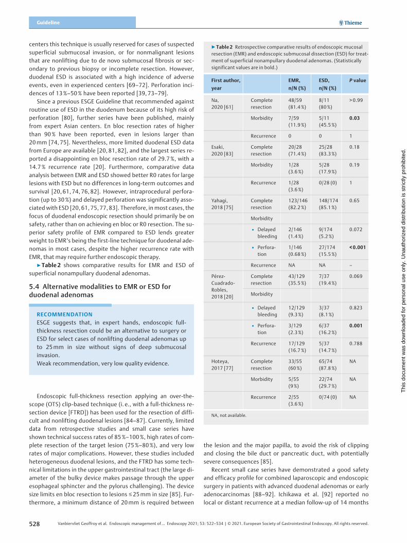
▶Table 2 shows comparative results for EMR and ESD ofsuperficial nonampullary duodenal adenomas.
5.4 Alternative modalities to EMR or ESD forduodenal adenomas
Endoscopic full-thickness resection applying an over-the-scope (OTS) clip-based technique (i. e., with a full-thickness re-section device [FTRD]) has been used for the resection of diffi-cult and nonlifting duodenal lesions [84–87]. Currently, limiteddata from retrospective studies and small case series haveshown technical success rates of 85%–100%, high rates of com-plete resection of the target lesion (75%–80%), and very lowrates of major complications. However, these studies includedheterogeneous duodenal lesions, and the FTRD has some tech-nical limitations in the upper gastrointestinal tract (the large di-ameter of the bulky device makes passage through the upperesophageal sphincter and the pylorus challenging). The devicesize limits en bloc resection to lesions ≤25mm in size [85]. Fur-thermore, a minimum distance of 20mm is required between
the lesion and the major papilla, to avoid the risk of clippingand closing the bile duct or pancreatic duct, with potentiallysevere consequences [85].
Recent small case series have demonstrated a good safetyand efficacy profile for combined laparoscopic and endoscopicsurgery in patients with advanced duodenal adenomas or earlyadenocarcinomas [88–92]. Ichikawa et al. [92] reported nolocal or distant recurrence at a median follow-up of 14 months
RECOMMENDATION
ESGE suggests that, in expert hands, endoscopic full-thickness resection could be an alternative to surgery orESD for select cases of nonlifting duodenal adenomas upto 25mm in size without signs of deep submucosalinvasion.Weak recommendation, very low quality evidence.
▶Table 2 Retrospective comparative results of endoscopic mucosalresection (EMR) and endoscopic submucosal dissection (ESD) for treat-ment of superficial nonampullary duodenal adenomas. (Statisticallysignificant values are in bold.)
First author,
year
EMR,
n/N (%)
ESD,
n/N (%)
P value
Na,2020 [61]
Completeresection
48/59(81.4%)
8/11(80%)
> 0.99
Morbidity 7/59(11.9%)
5/11(45.5%)
0.03
Recurrence 0 0 1
Esaki,2020 [83]
Completeresection
20/28(71.4%)
25/28(83.3%)
0.18
Morbidity 1/28(3.6%)
5/28(17.9%)
0.19
Recurrence 1/28(3.6%)
0/28 (0) 1
Yahagi,2018 [75]
Completeresection
123/146(82.2%)
148/174(85.1%)
0.65
Morbidity
▪ Delayedbleeding
2/146(1.4%)
9/174(5.2 %)
0.072
▪ Perfora-tion
1/146(0.68%)
27/174(15.5%)
<0.001
Recurrence NA NA –
Pérez-Cuadrado-Robles,2018 [20]
Completeresection
43/129(35.5%)
7/37(19.4%)
0.069
Morbidity
▪ Delayedbleeding
12/129(9.3%)
3/37(8.1 %)
0.823
▪ Perfora-tion
3/129(2.3%)
6/37(16.2%)
0.001
Recurrence 17/129(16.7%)
5/37(14.7%)
0.788
Hoteya,2017 [77]
Completeresection
33/55(60%)
65/74(87.8%)
NA
Morbidity 5/55(9%)
22/74(29.7%)
NA
Recurrence 2/55(3.6%)
0/74 (0) NA
NA, not available.
528 Vanbiervliet Geoffroy et al. Endoscopic management of… Endoscopy 2021; 53: 522–534 | © 2021. European Society of Gastrointestinal Endoscopy. All rights reserved.
Guideline
Thi
s do
cum
ent w
as d
ownl
oade
d fo
r pe
rson
al u
se o
nly.
Una
utho
rized
dis
trib
utio
n is
str
ictly
pro
hibi
ted.

in 10 patients with mucosal adenocarcinoma treated with com-bination laparoscopic and endoscopic surgery. In a retrospec-tive observational study by Ojima et al. [91], this techniqueshowed no adverse events (0%) compared to ESD (28%). How-ever, larger prospective studies are needed to confirm theseresults.
5.5 Role of tumor-destruction techniques
Historically, several complementary tissue destruction tech-niques had been used for nonampullary duodenal lesions.These included monopolar/bipolar coagulation, lasers such asthe Nd-YAG [93–95], photodynamic therapy [96], and cryo-therapy [97, 98]. However, most of these have been abandonedbecause of lack of efficacy or unacceptable adverse events [95,96].
Argon plasma coagulation (APC) is still used [60, 99–101],although it is not effective as a primary therapy, showing ade-noma recurrence rates of 39%–75% [7, 102–104]. APC hasbeen used as an adjunctive technique to eliminate residual ade-noma when technical difficulties resulted in incomplete endo-scopic resection [99, 100,104–111]. However, in one suchstudy, the reported recurrence rate was high at 25.7% [105].In another study by Apel et al. [99], the use of APC for residualduodenal adenoma did not lead to eradication in most of thelesions. However, a study by Alexander et al. [100] showedcomplete eradication of residual adenoma using APC in all 5patients reported in the study. Given these findings, a carefulendoscopic surveillance is required.
5.6 Prevention of delayed adverse events afterduodenal endoscopic resection
The evidence for routine prophylactic clip closure followingduodenal EMR is limited. Prophylactic through-the-scope clip-ping was associated with a significant reduction in delayedbleeding (0% vs. 22%, P=0.044) when compared to no prophy-laxis, in a retrospective study involving 43 duodenal EMR ses-sions [50]. In a prospective study using U-EMR for 31 duodenaladenomas of size≤20 mm, clip closure of the defect was per-formed for all lesions with no procedure-related adverse eventsbeing reported [64]. However, the risk of perforation due to clipapplication and large resection sites that cannot be fully closedare limiting factors, and therefore clips should be applied care-fully and their use considered on a case-by-case basis.
Noncontact hemostatic products have been successfullyused to minimize bleeding following duodenal EMR; howeverthe evidence is still limited [112, 113].
In the multivariate analysis of a recent case series of duo-denal ESDs, lesion location in the duodenal flexure, lesion size> 40mm, and occupied duodenal circumference of > 50% wereassociated with increased adverse events [114]. In a recentlarge retrospective Japanese study involving 168 patients, therate of delayed adverse events after duodenal ESD was signifi-cantly reduced when the mucosal defect was completelyclosed, compared with only partial closure or no closure (1.7%vs. 25% vs. 15.6%, respectively, P <0.01) [115]. These data wereconfirmed by two more studies where delayed bleeding was ef-fectively prevented by prophylactic endoscopic closure of thedefect [18, 116]. Recently, closure of the defect by OTS clippinghas also been shown to be effective in reducing delayed adverseevents after ESD [117]. Furthermore, the additional use of con-ventional through-the-scope clips, to cover the inverted sub-mucosa after defect closure with OTS clipping, was found tosignificantly reduce the risk of delayed bleeding [118].
6 Follow-up, risk and management of recur-rence after endoscopic duodenal resection
RECOMMENDATION
ESGE recommends that the high adverse event rate withduodenal resection may be reduced by mucosal defectclosure techniques such as endoscopic clipping or OTSclipping, and by noncontact hemostatic measures.Strong recommendation, low quality evidence.
RECOMMENDATION
ESGE recommends using techniques that minimizeadverse events such as immediate or delayed bleedingor perforation. These may include piecemeal resection,defect closure techniques, noncontact hemostasis, andother emerging techniques, and these should be consid-ered on a case-by-case basis.Strong recommendation, low quality evidence.
RECOMMENDATION
ESGE suggests that an additive role for ablative or othertumor-destruction techniques is minimal because of lackof efficacy.Weak recommendation, low quality evidence.
RECOMMENDATION
ESGE recommends that recurrences after endoscopictreatment for superficial nonampullary duodenal lesionscan be managed endoscopically, if this is deemedtechnically feasible and in the absence of suspectedmalignancy.Strong recommendation, low quality evidence.
Vanbiervliet Geoffroy et al. Endoscopic management of… Endoscopy 2021; 53: 522–534 | © 2021. European Society of Gastrointestinal Endoscopy. All rights reserved. 529
Thi
s do
cum
ent w
as d
ownl
oade
d fo
r pe
rson
al u
se o
nly.
Una
utho
rized
dis
trib
utio
n is
str
ictly
pro
hibi
ted.

Over a median follow-up period ranging from 6 to 72months, the local recurrence rate after EMR was 15% (95%CI7%–23%) in the largest review of the literature [52]. Advancedhistopathology, defined as the presence of villous changes (OR4.86, 95%CI 1.62–14.63) or high grade dysplasia, was shown toincrease the risk of local recurrence [52, 56, 62]. Similarly, in-creasing lesion size was associated with a higher recurrencerate [19, 52, 55, 63, 106]. With regard to the techniques origi-nally used to remove the lesion, no significant difference in re-currence rate was observed between EMR versus ESD or EMRversus hybrid ESD [18, 81]. After a median follow-up of6.5 months (2–125), Pérez-Cuadrado-Robles et al. [20] showed5/37 recurrences (14.7%) after ESD and 17/129 (16.7%) afterEMR (P=0.788). Furthermore, there were no demonstrable dif-ferences in recurrence rates between en bloc or piecemeal EMRin the largest review of duodenal EMR studies [52].
The available studies have shown that recurrent lesions areusually small in size and can be successfully treated endoscopi-cally in most cases by an expert endoscopist [19, 119]. In thereview from Navaneethan et al., six studies reported the out-comes of managing recurrent adenoma, and further endo-scopic therapy was successful in 62% (95%CI 37%–87%) [52].In the absence of relevant comparative data, no specific endo-scopic technique could be preferentially recommended tomanage adenoma recurrence.
Regarding the surveillance interval after index endoscopy,expert opinions are in favor of a first endoscopy at 3–6 months.The evidence for this approach is limited, but it has been re-cently supported by a prospective study showing that at3 months, residual or recurrent adenoma was noted in 20.4%of patients who then had endoscopic re-treatment [48]. Valeriiet al. [54] retrospectively reported 15 recurrences in 62 lesions,with 9 of them (60%) being found at the first follow-up endos-copy performed 3 months after the initial endoscopic treat-ment. A second surveillance endoscopy 1 year later seems tobe advisable, if no residual or recurrent adenoma has been de-tected during the first surveillance endoscopy [41, 48, 52, 120].
Subsequent surveillance intervals should then be individua-lized, taking into account lesion factors (size, high grade dys-plasia, or villous component) and patient factors (age, comor-bidities) [52]. Late recurrences are uncommon, but possible.
7 Role of surgery for nonmalignant sporadicduodenal adenomasThe literature on duodenal surgery for SNADT is limited andmainly consists of studies on patients with FAP, as detailed inthe ESGE Guideline for FAP [2]. For sporadic nonmalignant duo-denal lesions, less invasive options such as transduodenal exci-sion and segmental duodenal resection are preferred comparedwith pancreaticoduodenectomy or pancreas-sparing duode-nectomy, as the less invasive approaches demonstrate lowermorbidity rates [121–124]. A retrospective study of 86 patientsshowed morbidity rates of 17% after transduodenal excisioncompared with 40% and 45% after pancreaticoduodenectomyand pancreas sparing duodenectomy, respectively [121]. Inthree other retrospective studies, the morbidity rate forpancreaticoduodenectomy was significantly higher comparedto the less invasive transduodenal excision or segmental duode-nal resection [7, 122, 125]. However, a 5-year recurrence rate of32% after transduodenal excision for villous adenomas is re-ported [121]. Therefore, postoperative endoscopic surveillanceis mandatory after surgery, and endoscopic resection of recur-rences following surgery is still possible [125].
In a cohort of 121 patients with nonampullary duodenal le-sions, 91 were treated by EMR, as opposed to surgical therapywith pancreas-sparing duodenectomy [125]. The recurrencerate during follow-up was significantly higher in the EMR thanin the surgical group (32% vs. 0%, P <0.001). However, therewas a trend towards higher adverse event rates in the surgicalgroup than in the EMR group (26% vs. 15%), although it shouldbe noted that larger and more advanced lesions had been treat-ed in the surgical group [125]. Other studies have also shownlower mortality and morbidity, shorter procedural time andshorter hospital stay in endoscopically treated patients [7,118, 122, 124]. Therefore, while adenoma recurrence is low fol-lowing pancreas-sparing duodenectomies, the high morbidityand mortality associated with these procedures make themoptions of last resort for most cases of sporadic duodenaladenoma [7, 125].
Finally, in cases of confirmed duodenal malignancy, an onco-logical resection including lymph node dissection, such asachieved by pancreaticoduodenectomy is required, whereaspancreas-sparing duodenectomy and transduodenal excisionare not oncological resections and are reserved for premalig-nant lesions only.
DisclaimerThe legal disclaimer for ESGE guidelines [126] applies to thisGuideline.
RECOMMENDATION
ESGE suggests that the choice of endoscopic techniqueto manage recurrent adenoma should be left to the dis-cretion of the endoscopist, according to the morphologyof the lesion and patient characteristics.Weak recommendation, very low quality evidence.
RECOMMENDATION
ESGE recommends endoscopic surveillance 3 monthsafter the index treatment. In cases of no recurrence, afurther follow-up endoscopy should be done 1 year later.Thereafter, surveillance intervals should be adapted tothe lesion site, en bloc resection status, and initial histo-logical result.Strong recommendation, low quality evidence.
530 Vanbiervliet Geoffroy et al. Endoscopic management of… Endoscopy 2021; 53: 522–534 | © 2021. European Society of Gastrointestinal Endoscopy. All rights reserved.
Guideline
Thi
s do
cum
ent w
as d
ownl
oade
d fo
r pe
rson
al u
se o
nly.
Una
utho
rized
dis
trib
utio
n is
str
ictly
pro
hibi
ted.

AcknowledgmentsESGE wishes to thank, for the added value they have brought tothe final manuscript, the two external reviewers, ProfessorSchalk van der Merwe of the University Hospital-Gasthuisberg,University of Leuven, Belgium, and Dr. Udayakumar Navanee-than, Digestive Health Institute, Orlando, Florida, USA, andalso Drs. Khalil Bedran, St George Hospital University MedicalCenter, Beirut, Lebanon, Marco Bustamente-Balén, La Fe Uni-versity Hospital, Valencia, Spain, Gertran Rasschaert, Universi-tair Ziekenhuis Brussel, Belgium, and Suzane Ribeiro, GhentUniversity Hospital, Belgium, for their comments.
Competing interests
M. Arvanitakis has received lecture fees from Olympus. T. Beyna pro-vides consultancy to and gives lectures for Boston Scientific and CookMedical (ongoing). J.E. van Hooft’s department has received researchgrants from Cook Medical (from 2014 to 2019) and Abbott (from2014 to 2017); she has received lecture fees from Medtronics (from2014 to 2015, 2019) and Cook Medical (from 2019); and she hasreceived consultancy fees from Boston Scientific (from 2014 to2017). G. Vanbiervliet has provided consultancy to Boston Scientificand Cook Medical (both from 2019 to present). U. Arnelo, M. Barthet,O. Busch, P. Deprez, A. Larghi, G. Manes, A. Moss, K. Nalankilli, B.Napoleon, M. Nayar, E. Pérez-Cuadrado-Robles, L. Kunovsky, S. See-wald, and M. Strijker, declare that they have no conflicts of interest.
References
[1] Goda K, Kikuchi D, Yamamoto Y et al. Endoscopic diagnosis of su-perficial non-ampullary duodenal epithelial tumors in Japan: Multi-center case series. Dig Endosc 2014; 26: (Suppl. 02): 23–29
[2] van Leerdam ME, Roos VH, van Hooft JE et al. Endoscopic manage-ment of polyposis syndromes: European Society of GastrointestinalEndoscopy (ESGE) Guideline. Endoscopy 2019; 51: 877–895
[3] Jung SH, Chung WC, Kim EJ et al. Evaluation of non-ampullary duo-denal polyps: comparison of non-neoplastic and neoplastic lesions.World J Gastroenterol 2010; 16: 5474–5480
[4] Höchter W, Weingart J, Seib HJ et al. [Duodenal polyps. Incidence,histologic substrate and significance] [Article in German]. DtschMed Wochenschr 1984; 109: 1183–1186
[5] Jepsen JM, Persson M, Jakobsen NO et al. Prospective study of prev-alence and endoscopic and histopathologic characteristics of duo-denal polyps in patients submitted to upper endoscopy. Scand JGastroenterol 1994; 29: 483–487
[6] Alkhatib AA. Sporadic nonampullary tubular adenoma of the duo-denum: Prevalence and patients’ characteristics. Turk J Gastroen-terol 2019; 30: 112–113
[7] Johnson MD, Mackey R, Brown N et al. Outcome based on manage-ment for duodenal adenomas: sporadic versus familial disease. JGastrointest Surg 2010; 14: 229–235
[8] Syngal S, Brand RE, Church JM et al. ACG clinical guideline: Genetictesting and management of hereditary gastrointestinal cancer syn-dromes. Am J Gastroenterol 2015; 110: 223–262; quiz 263
[9] Matsuzaki J, Suzuki H, Shimoda M et al. Clinical and endoscopicfindings to assist the early detection of duodenal adenoma andadenocarcinoma. United Eur Gastroenterol J 2019; 7: 250–260
[10] Bisschops R, Areia M, Coron E et al. Performance measures for uppergastrointestinal endoscopy: a European Society of GastrointestinalEndoscopy (ESGE) Quality Improvement Initiative. Endoscopy 2016;48: 843–864
[11] Wu Z-J, Lin Y, Xiao J et al. Clinical significance of colonoscopy inpatients with upper gastrointestinal polyps and neoplasms: a meta-analysis. PloS One 2014; 9: e91810
[12] Genta RM, Hurrell JM, Sonnenberg A. Duodenal adenomas coincidewith colorectal neoplasia. Dig Dis Sci 2014; 59: 2249–2254
[13] Okada K, Fujisaki J, Kasuga A et al. Sporadic nonampullary duodenaladenoma in the natural history of duodenal cancer: a study of fol-low-up surveillance. Am J Gastroenterol 2011; 106: 357–364
[14] Niwa A, Kuwano S, Tomita H et al. The different pathogeneses ofsporadic adenoma and adenocarcinoma in non-ampullary lesions ofthe proximal and distal duodenum. Oncotarget 2017; 8: 41078–41090
[15] Toya Y, Endo M, Akasaka R et al. Clinicopathological features andmagnifying chromoendoscopic findings of non-ampullary duodenalepithelial tumors. Digestion 2018; 97: 219–227
[16] Tsuji S, Doyama H, Tsuji K et al. Preoperative endoscopic diagnosis ofsuperficial non-ampullary duodenal epithelial tumors, includingmagnifying endoscopy. World J Gastroenterol 2015; 21: 11832–11841
[17] Kuroki K, Sanomura Y, Oka S et al. Clinical outcomes of endoscopicresection for superficial non-ampullary duodenal tumors. Endosc IntOpen 2020; 8: E354–E359
[18] Pérez-Cuadrado-Robles E, Quénéhervé L, Margos W et al. ESD versusEMR in non-ampullary superficial duodenal tumors: a systematic re-view and meta-analysis. Endosc Int Open 2018; 6: E998–E1007
[19] Klein A, Nayyar D, Bahin FF et al. Endoscopic mucosal resection oflarge and giant lateral spreading lesions of the duodenum: success,adverse events, and long-term outcomes. Gastrointest Endosc2016; 84: 688–696
[20] Pérez-Cuadrado-Robles E, Quénéhervé L, Margos W et al. Compara-tive analysis of ESD versus EMR in a large European series of non-ampullary superficial duodenal tumors. Endosc Int Open 2018; 6:E1008–E1014
[21] Lopez-Ceron M, van den Broek FJC, Mathus-Vliegen EM et al. The roleof high-resolution endoscopy and narrow-band imaging in the eval-uation of upper GI neoplasia in familial adenomatous polyposis.Gastrointest Endosc 2013; 77: 542–550
[22] Yap CK, Ng HS. Cap-fitted gastroscopy improves visualization andtargeting of lesions. Gastrointest Endosc 2001; 53: 93–95
[23] Choi YR, Han J-H, Cho YS et al. Efficacy of cap-assisted endoscopy forroutine examining the ampulla of Vater. World J Gastroenterol 2013;19: 2037–2043
[24] Kallenberg FGJ, Bastiaansen BAJ, Dekker E. Cap-assisted forward-viewing endoscopy to visualize the ampulla of Vater and the duode-num in patients with familial adenomatous polyposis. Endoscopy2017; 49: 181–185
[25] Abdelhafez M, Phillip V, Hapfelmeier A et al. Cap assisted upperendoscopy for examination of the major duodenal papilla: a ran-domized, blinded, controlled crossover study (CAPPA Study). Am JGastroenterol 2017; 112: 725–733
[26] Abdelhafez M, Phillip V, Hapfelmeier A et al. Comparison of cap-as-sisted endoscopy vs. side-viewing endoscopy for examination of themajor duodenal papilla: a randomized, controlled, non-inferioritycrossover study. . Endoscopy 2019; 51: 419–426
[27] Shi X, Luo H, Ning B et al. Effect of cap-assisted esophagogastro-duodenoscopy on examination of the major duodenal papilla: a non-inferior, randomized controlled trial. Endoscopy 2019; 51: 427–435
Vanbiervliet Geoffroy et al. Endoscopic management of… Endoscopy 2021; 53: 522–534 | © 2021. European Society of Gastrointestinal Endoscopy. All rights reserved. 531
Thi
s do
cum
ent w
as d
ownl
oade
d fo
r pe
rson
al u
se o
nly.
Una
utho
rized
dis
trib
utio
n is
str
ictly
pro
hibi
ted.

[28] Kiesslich R, Mergener K, Naumann C et al. Value of chromoendos-copy and magnification endoscopy in the evaluation of duodenalabnormalities: a prospective, randomized comparison. Endoscopy2003; 35: 559–563
[29] Picasso M, Filiberti R, Blanchi S et al. The role of chromoendoscopy inthe surveillance of the duodenum of patients with familial adeno-matous polyposis. Dig Dis Sci 2007; 52: 1906–1909
[30] Dekker E, Boparai KS, Poley JW et al. High resolution endoscopy andthe additional value of chromoendoscopy in the evaluation of duo-denal adenomatosis in patients with familial adenomatous polypo-sis. Endoscopy 2009; 41: 666–669
[31] Hurley JJ, Thomas LE, Walton S-J et al. The impact of chromoendos-copy for surveillance of the duodenum in patients with MUTYH-associated polyposis and familial adenomatous polyposis. Gastroin-test Endosc 2018; 88: 665–673
[32] Shahid MW, Buchner A, Gomez V et al. Diagnostic accuracy ofprobe-based confocal laser endomicroscopy and narrow band ima-ging in detection of dysplasia in duodenal polyps. J Clin Gastroen-terol 2012; 46: 382–389
[33] Yamasaki Y, Takeuchi Y, Kanesaka T et al. Differentiation betweenduodenal neoplasms and non-neoplasms using magnifying narrow-band imaging – Do we still need biopsies for duodenal lesions? DigEndosc 2020; 32: 84–95
[34] Yoshimura N, Goda K, Tajiri H et al. Endoscopic features of nonam-pullary duodenal tumors with narrow-band imaging. Hepatogas-troenterology 2010; 57: 462–467
[35] Kikuchi D, Hoteya S, Iizuka T et al. Diagnostic algorithm of magnify-ing endoscopy with narrow band imaging for superficial non-am-pullary duodenal epithelial tumors. Dig Endosc 2014; 26: (Suppl.02): 16–22
[36] Kakushima N, Yoshida M, Yamaguchi Y et al. Magnified endoscopywith narrow-band imaging for the differential diagnosis of superfi-cial non-ampullary duodenal epithelial tumors. Scand J Gastroen-terol 2019; 54: 128–134
[37] Toya Y, Endo M, Oizumi T et al. Diagnostic algorithm of magnifyingendoscopy with crystal violet staining for non-ampullary duodenalepithelial tumors. Dig Endosc 2020; 32: 1066–1073
[38] Mizumoto T, Sanomura Y, Tanaka S et al. Clinical usefulness of mag-nifying endoscopy for non-ampullary duodenal tumors. Endosc IntOpen 2017; 5: E297–E302
[39] Kakushima N, Kanemoto H, Sasaki K et al. Endoscopic and biopsydiagnoses of superficial, nonampullary, duodenal adenocarcinomas.World J Gastroenterol 2015; 21: 5560–5567
[40] Kinoshita S, Nishizawa T, Ochiai Y et al. Accuracy of biopsy for thepreoperative diagnosis of superficial nonampullary duodenal ade-nocarcinoma. Gastrointest Endosc 2017; 86: 329–332
[41] Ma MX, Bourke MJ. Management of duodenal polyps. Best Pract ResClin Gastroenterol 2017; 31: 389–399
[42] Chen W-C, Wallace MB. Endoscopic management of mucosal lesionsin the gastrointestinal tract. Expert Rev Gastroenterol Hepatol 2016;10: 481–495
[43] Ferlitsch M, Moss A, Hassan C et al. Colorectal polypectomy andendoscopic mucosal resection (EMR): European Society of Gastroin-testinal Endoscopy (ESGE) Clinical Guideline. Endoscopy 2017; 49:270–297
[44] Hamada K, Takeuchi Y, Ishikawa H et al. Feasibility of cold snarepolypectomy for multiple duodenal adenomas in patients withfamilial adenomatous polyposis: a pilot study. Dig Dis Sci 2016; 61:2755–2759
[45] Patel NJ, Ponugoti PL, Rex DK. Cold snare polypectomy effectivelyreduces polyp burden in familial adenomatous polyposis. Endosc IntOpen 2016; 4: E472–474
[46] Hamada K, Takeuchi Y, Ishikawa H et al. Safety of cold snare poly-pectomy for duodenal adenomas in familial adenomatous polyposis:a prospective exploratory study. Endoscopy 2018; 50: 511–517
[47] Maruoka D, Matsumura T, Kasamatsu S et al. Cold polypectomy forduodenal adenomas: a prospective clinical trial. Endoscopy 2017;49: 776–783
[48] Probst A, Freund S, Neuhaus L et al. Complication risk despite pre-ventive endoscopic measures in patients undergoing endoscopicmucosal resection of large duodenal adenomas. Endoscopy 2020;52: 847–855
[49] Fanning SB, Bourke MJ, Williams SJ et al. Giant laterally spreadingtumors of the duodenum: endoscopic resection outcomes, limita-tions, and caveats. Gastrointest Endosc 2012; 75: 805–812
[50] Lépilliez V, Chemaly M, Ponchon T et al. Endoscopic resection ofsporadic duodenal adenomas: an efficient technique with a sub-stantial risk of delayed bleeding. Endoscopy 2008; 40: 806–810
[51] Bourke MJ. Endoscopic resection in the duodenum: current limita-tions and future directions. Endoscopy 2013; 45: 127–132
[52] Navaneethan U, Hasan MK, Lourdusamy V et al. Efficacy and safety ofendoscopic mucosal resection of non-ampullary duodenal polyps: asystematic review. Endosc Int Open 2016; 4: E699–708
[53] Jamil LH, Kashani A, Peter N et al. Safety and efficacy of cap-assistedEMR for sporadic nonampullary duodenal adenomas. GastrointestEndosc 2017; 86: 666–672
[54] Valerii G, Tringali A, Landi R et al. Endoscopic mucosal resection ofnon-ampullary sporadic duodenal adenomas: a retrospective analy-sis with long-term follow-up. Scand J Gastroenterol 2018; 53: 490–494
[55] Tomizawa Y, Ginsberg GG. Clinical outcome of EMR of sporadic, non-ampullary, duodenal adenomas: a 10-year retrospective. Gastroin-test Endosc 2018; 87: 1270–1278
[56] Valli PV, Mertens JC, Sonnenberg A et al. Nonampullary duodenaladenomas rarely recur after complete endoscopic resection: a Swissexperience including a literature review. Digestion 2017; 96: 149–157
[57] Singh A, Siddiqui UD, Konda VJ et al. Safety and efficacy of EMR forsporadic, nonampullary duodenal adenomas: a single U.S. centerexperience (with video). Gastrointest Endosc 2016; 84: 700–8
[58] Zou J, Chai N, Linghu E et al. Clinical outcomes of endoscopic resec-tion for non-ampullary duodenal laterally spreading tumors. SurgEndosc 2019; 33: 4048–4056
[59] Kiguchi Y, Kato M, Nakayama A et al. Feasibility study comparingunderwater endoscopic mucosal resection and conventional endo-scopic mucosal resection for superficial non-ampullary duodenalepithelial tumor < 20 mm. Dig Endosc 2020; 32: 753–760
[60] Cosgrove N, Siddiqui AA, Kistler CA et al. Endoscopic mucosalresection of large non-ampullary duodenal polyps: technical aspectsand long-term therapeutic outcomes. Minerva Gastroenterol Dietol2016; 62: 131–137
[61] Na HK, Kim DH, Ahn JY et al. Clinical outcomes following endoscopictreatment for sporadic nonampullary duodenal adenoma. Dig Dis2020; 38: 364–372
[62] Cassani LS, Lanke G, Chen H-C et al. Comparison of nonampullaryduodenal adenomas in patients with familial adenomatous polyposisversus patients with sporadic adenomas. Gastrointest Endosc 2017;85: 803–812
[63] Klein A, Ahlenstiel G, Tate DJ et al. Endoscopic resection of largeduodenal and papillary lateral spreading lesions is clinically andeconomically advantageous compared with surgery. Endoscopy2017; 49: 659–667
[64] Yamasaki Y, Uedo N, Takeuchi Y et al. Underwater endoscopic mu-cosal resection for superficial nonampullary duodenal adenomas.Endoscopy 2018; 50: 154–158
532 Vanbiervliet Geoffroy et al. Endoscopic management of… Endoscopy 2021; 53: 522–534 | © 2021. European Society of Gastrointestinal Endoscopy. All rights reserved.
Guideline
Thi
s do
cum
ent w
as d
ownl
oade
d fo
r pe
rson
al u
se o
nly.
Una
utho
rized
dis
trib
utio
n is
str
ictly
pro
hibi
ted.

[65] Shibukawa G, Irisawa A, Sato A et al. Endoscopic mucosal resectionperformed underwater for nonampullary duodenal epithelial tumor:evaluation of feasibility and safety. Gastroenterol Res Pract 2018:doi:10.1155/2018/7490961
[66] Javia SB, Chathadi K. Cold snare piecemeal endoscopic mucosalresection of a very large duodenal adenoma. Endoscopy 2019; 51:E217–E218
[67] Choksi N, Elmunzer BJ, Stidham RW et al. Cold snare piecemealresection of colonic and duodenal polyps ≥1 cm. Endosc Int Open2015; 3: E508–E513
[68] Ochiai Y, Kato M, Kiguchi Y et al. Current status and challenges ofendoscopic treatments for duodenal tumors. Digestion 2019; 99:21–26
[69] Shibagaki K, Ishimura N, Kinoshita Y. Endoscopic submucosal dis-section for duodenal tumors. Ann Transl Med 2017; 5: 188
[70] Yamamoto Y, Yoshizawa N, Tomida H et al. Therapeutic outcomes ofendoscopic resection for superficial non-ampullary duodenal tumor.Dig Endosc 2014; 26: (Suppl. 02): 50–56
[71] Maple JT, Abu Dayyeh BK, Chauhan SS et al. Endoscopic submucosaldissection. Gastrointest Endosc 2015; 81: 1311–1325
[72] Deprez PH, Bergman JJ, Meisner S et al. Current practice with endo-scopic submucosal dissection in Europe: position statement from apanel of experts. Endoscopy 2010; 42: 853–858
[73] Jung JH, Choi KD, Ahn JY et al. Endoscopic submucosal dissection forsessile, non-ampullary duodenal adenomas. Endoscopy 2013; 45:133–135
[74] Matsumoto S, Yoshida Y. Selection of appropriate endoscopictherapies for duodenal tumors: an open-label study, single-centerexperience. World J Gastroenterol 2014; 20: 8624–8630
[75] Yahagi N, Kato M, Ochiai Y et al. Outcomes of endoscopic resectionfor superficial duodenal epithelial neoplasia. Gastrointest Endosc2018; 88: 676–682
[76] Honda T, Yamamoto H, Osawa H et al. Endoscopic submucosal dis-section for superficial duodenal neoplasms. Dig Endosc 2009; 21:270–274
[77] Hoteya S, Furuhata T, Takahito T et al. Endoscopic submucosal dis-section and endoscopic mucosal resection for non-ampullary su-perficial duodenal tumor. Digestion 2017; 95: 36–42
[78] Hoteya S, Yahagi N, Iizuka T et al. Endoscopic submucosal dissectionfor non-ampullary large superficial adenocarcinoma/adenoma ofthe duodenum: feasibility and long-term outcomes. Endosc IntOpen 2013; 1: 2–7
[79] Nonaka S, Oda I, Tada K et al. Clinical outcome of endoscopic resec-tion for nonampullary duodenal tumors. Endoscopy 2015; 47: 129–135
[80] Pimentel-Nunes P, Dinis-Ribeiro M, Ponchon T et al. Endoscopicsubmucosal dissection: European Society of Gastrointestinal Endos-copy (ESGE) Guideline. Endoscopy 2015; 47: 829–854
[81] Basford PJ, George R, Nixon E et al. Endoscopic resection of sporadicduodenal adenomas: comparison of endoscopic mucosal resection(EMR) with hybrid endoscopic submucosal dissection (ESD) tech-niques and the risks of late delayed bleeding. Surg Endosc 2014; 28:1594–1600
[82] Santos-Antunes J, Baldaque-Silva F, Marques M et al. Real-life evalu-ation of the safety, efficacy and therapeutic outcomes of endoscopicsubmucosal dissection in a Western tertiary centre. United Eur Gas-troenterol J 2018; 6: 702–709
[83] Esaki M, Haraguchi K, Akahoshi K et al. Endoscopic mucosal resec-tion vs endoscopic submucosal dissection for superficial non-am-pullary duodenal tumors. World J Gastrointest Oncol 2020; 12: 918–930
[84] Andrisani G, Di Matteo FM. Endoscopic full-thickness resection ofduodenal lesions (with video). Surg Endosc 2020; 34: 1876–1881
[85] Bauder M, Schmidt A, Caca K. Endoscopic full-thickness resection ofduodenal lesions-a retrospective analysis of 20 FTRD cases. UnitedEur Gastroenterol J 2018; 6: 1015–1021
[86] Schmidt A, Meier B, Cahyadi O et al. Duodenal endoscopic full-thickness resection (with video). Gastrointest Endosc 2015; 82:728–733
[87] Schmidt A, Beyna T, Schumacher B et al. Colonoscopic full-thicknessresection using an over-the-scope device: a prospective multicentrestudy in various indications. Gut 2018; 67: 1280–1289
[88] Ohata K, Nonaka K, Sakai E et al. Novel technique of endoscopic full-thickness resection for superficial nonampullary duodenal neo-plasms to avoid intraperitoneal tumor dissemination. Endosc IntOpen 2016; 4: E784–E787
[89] Irino T, Nunobe S, Hiki N et al. Laparoscopic-endoscopic cooperativesurgery for duodenal tumors: a unique procedure that helps ensurethe safety of endoscopic submucosal dissection. Endoscopy 2015;47: 349–351
[90] Kakushima N, Yoshida M, Yabuuchi Y et al. Present status of endo-scopic submucosal dissection for non-ampullary duodenal epithelialtumors. Clin Endosc 2020; 53: 652–658
[91] Ojima T, Nakamori M, Nakamura M et al. Laparoscopic and endo-scopic cooperative surgery versus endoscopic submucosal dissec-tion for the treatment of low-risk tumors of the duodenum. J Gas-trointest Surg 2018; 22: 935–940
[92] Ichikawa D, Komatsu S, Dohi O et al. Laparoscopic and endoscopicco-operative surgery for non-ampullary duodenal tumors. World JGastroenterol 2016; 22: 10424–10431
[93] Ghilain JM, Dive C. Endoscopic laser therapy for small villous adeno-mas of the duodenum. Endoscopy 1994; 26: 308–310
[94] Paraf F, Naveau S, Zourabichvili O et al. Endoscopic laser therapy forduodenal villous adenoma. Dig Dis Sci 1989; 34: 1466–1467
[95] Norton ID, Geller A, Petersen BT et al. Endoscopic surveillance andablative therapy for periampullary adenomas. Am J Gastroenterol2001; 96: 101–106
[96] Mlkvy P, Messmann H, Debinski H et al. Photodynamic therapy forpolyps in familial adenomatous polyposis – a pilot study. Eur J Cancer1995; 31A: 1160–1165
[97] Ooka K, Celli R, Farrell JJ. Cryoablation of a duodenal adenoma withintramucosal carcinoma. VideoGIE 2017; 2: 244–246
[98] Raphael KL, Benias PC, Trindade AJ. Salvage CryoBalloon cryotherapyablation for a duodenal adenoma. Endoscopy 2020; 52: E189–E190
[99] Apel D, Jakobs R, Spiethoff A et al. Follow-up after endoscopic snareresection of duodenal adenomas. Endoscopy 2005; 37: 444–448
[100] Alexander S, Bourke MJ, Williams SJ et al. EMR of large, sessile,sporadic non-ampullary duodenal adenomas: technical aspects andlong-term outcome (with videos). Gastrointest Endosc 2009; 69:66–73
[101] Koritala T, Zolotarevsky E, Bartley AN et al. Efficacy and safety of theband and slough technique for endoscopic therapy of non-ampul-lary duodenal adenomas: a case series. Gastrointest Endosc 2015;81: 985–988
[102] Jaganmohan S, Lynch PM, Raju RP et al. Endoscopic management ofduodenal adenomas in familial adenomatous polyposis–a single-center experience. Dig Dis Sci 2012; 57: 732–737
[103] Fawal H, Gambiez L, Raad A et al. Management of duodenal adeno-matosis in patients with familial adenomatous polyposis. Ann Chir2003; 128: 594–598
[104] Min YW, Min B-H, Kim ER et al. Efficacy and safety of endoscopictreatment for nonampullary sporadic duodenal adenomas. Dig DisSci 2013; 58: 2926–2932
[105] Navaneethan U, Lourdusamy D, Mehta D et al. Endoscopic resectionof large sporadic non-ampullary duodenal polyps: efficacy and long-term recurrence. Surg Endosc 2014; 28: 2616–2622
Vanbiervliet Geoffroy et al. Endoscopic management of… Endoscopy 2021; 53: 522–534 | © 2021. European Society of Gastrointestinal Endoscopy. All rights reserved. 533
Thi
s do
cum
ent w
as d
ownl
oade
d fo
r pe
rson
al u
se o
nly.
Una
utho
rized
dis
trib
utio
n is
str
ictly
pro
hibi
ted.

[106] Abbass R, Rigaux J, Al-Kawas FH. Non-ampullary duodenal polyps:characteristics and endoscopic management. Gastrointest Endosc2010; 71: 754–759
[107] Aschmoneit-Messer I, Richl J, Pohl J et al. Prospective study of acutecomplication rates and associated risk factors in endoscopic therapyfor duodenal adenomas. Surg Endosc 2015; 29: 1823–1830
[108] Tsiamoulos ZP, Peake ST, Bourikas LA et al. Endoscopic mucosal ab-lation: a novel technique for a giant non-ampullary duodenal ade-noma. Endoscopy 2013; 45: (Suppl. 02): E12–E13
[109] Kedia P, Brensinger C, Ginsberg G. Endoscopic predictors of suc-cessful endoluminal eradication in sporadic duodenal adenomas andits acute complications. Gastrointest Endosc 2010; 72: 1297–1301
[110] Manner H, May A, Rabenstein T et al. Prospective evaluation of a newhigh-power argon plasma coagulation system (hp-APC) in thera-peutic gastrointestinal endoscopy. Scand J Gastroenterol 2007; 42:397–405
[111] Eswaran SL, Sanders M, Bernadino KP et al. Success and complica-tions of endoscopic removal of giant duodenal and ampullarypolyps: a comparative series. Gastrointest Endosc 2006; 64: 925–932
[112] Leblanc S, Vienne A, Dhooge M et al. Early experience with a novelhemostatic powder used to treat upper GI bleeding related to ma-lignancies or after therapeutic interventions (with videos). Gastro-intest Endosc 2013; 78: 169–175
[113] Naseer M, Lambert K, Hamed A et al. Endoscopic advances in themanagement of non-variceal upper gastrointestinal bleeding: Areview. World J Gastrointest Endosc 2020; 12: 1–16
[114] Kato M, Sasaki M, Mizutani M et al. Predictors of technical difficultywith duodenal ESD. Endosc Int Open 2019; 7: E1755–E1760
[115] Kato M, Ochiai Y, Fukuhara S et al. Clinical impact of closure of themucosal defect after duodenal endoscopic submucosal dissection.Gastrointest Endosc 2019; 89: 87–93
[116] Hoteya S, Kaise M, Iizuka T et al. Delayed bleeding after endoscopicsubmucosal dissection for non-ampullary superficial duodenal neo-plasias might be prevented by prophylactic endoscopic closure:analysis of risk factors. Dig Endosc 2015; 27: 323–330
[117] Tashima T, Ohata K, Sakai E et al. Efficacy of an over-the-scope clipfor preventing adverse events after duodenal endoscopic submuco-sal dissection: a prospective interventional study. Endoscopy 2018;50: 487–496
[118] Ohata K, Sakai E, Suzuki Y et al. Risk factors of delayed bleeding afterendoscopic resection of superficial non-ampullary duodenal epithe-lial tumors and prevention by over-the-scope and conventional clip-ping. Dig Endosc 20.05 2020: doi:10.1111/den.13729
[119] Kakushima N, Kanemoto H, Tanaka M et al. Treatment for superficialnon-ampullary duodenal epithelial tumors. World J Gastroenterol2014; 20: 12501–12508
[120] Gaspar JP, Stelow EB, Wang AY. Approach to the endoscopic resec-tion of duodenal lesions. World J Gastroenterol 2016; 22: 600–617
[121] Farnell MB, Sakorafas GH, Sarr MG et al. Villous tumors of the duo-denum: reappraisal of local vs. extended resection. . J GastrointestSurg 2000; 4: 13–21, discussion 22-23
[122] Perez A, Saltzman JR, Carr-Locke DL et al. Benign non-ampullaryduodenal neoplasms. J Gastrointest Surg 2003; 7: 536–541
[123] Bjork KJ, Davis CJ, Nagorney DM et al. Duodenal villous tumors. ArchSurg 1990; 125: 961–965
[124] Xu M, Wu J, Yu L et al. Surgical versus endoscopic resection of largesessile duodenal and papillary lesions. Eur J Gastroenterol Hepatol2020; 32: 48–53
[125] Bartel MJ, Puri R, Brahmbhatt B et al. Endoscopic and surgical man-agement of nonampullary duodenal neoplasms. Surg Endosc 2018;32: 2859–2869
[126] Hassan C, Ponchon T, Bisschops R et al. European Society of Gastro-intestinal Endoscopy (ESGE) Publications Policy – Update 2020.Endoscopy 2020; 52: 123–126
534 Vanbiervliet Geoffroy et al. Endoscopic management of… Endoscopy 2021; 53: 522–534 | © 2021. European Society of Gastrointestinal Endoscopy. All rights reserved.
Guideline
Thi
s do
cum
ent w
as d
ownl
oade
d fo
r pe
rson
al u
se o
nly.
Una
utho
rized
dis
trib
utio
n is
str
ictly
pro
hibi
ted.