Bisphosphonate derivatized polyurethanes resist calcification
Upload
independentCategory
view
4download
0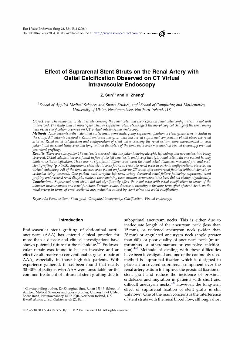
* CorrespondApplied MedShore Road, NE-mail address
1078–5884/00
Effect of Suprarenal Stent Struts on the Renal Artery withOstial Calcification Observed on CT Virtual
Intravascular Endoscopy
Z. Sun1* and H. Zheng2
1School of Applied Medical Sciences and Sports Studies, and 2School of Computing and Mathematics,University of Ulster, Newtownabbey, Northern Ireland, UK
Objectives. The behaviour of stent struts crossing the renal ostia and their effect on renal ostia configuration is not wellunderstood. The study aims to investigate whether suprarenal stent struts affect the morphological change of the renal arterywith ostial calcification observed on CT virtual intravascular endoscopy.Methods. Nine patients with abdominal aortic aneurysms undergoing suprarenal fixation of stent grafts were included inthe study. All patients received a Zenith endovascular graft with uncovered suprarenal components placed above the renalarteries. Renal ostial calcification and configuration of stent wires crossing the renal ostium were characterized in eachpatient and maximal transverse and longitudinal diameters of the renal ostia were measured on virtual endoscopy pre- andpost-stent grafting.Results. There were altogether 17 renal ostia assessed with one patient having atrophic left kidney and no renal ostium beingobserved. Ostial calcification was found in five of the left renal ostia and five of the right renal ostia with one patient havingbilateral ostial calcification. There was no significant difference between the renal ostial diameters measured pre- and post-stent grafting (pO0.05). Suprarenal stent struts were found to cross the renal ostia in various configurations observed onvirtual endoscopy. All of the renal arteries were patent on follow-up CT scans after suprarenal fixation without stenosis orocclusion being observed. One patient with atrophic left renal artery developed renal failure following suprarenal stentgrafting and received renal dialysis, while in the remaining cases median serum creatinine level did not change significantly.Conclusions. Suprarenal stent struts did not significantly affect the renal ostia with ostial calcification in terms of thediameter measurements and renal function. Further studies deserve to investigate the long-term effect of stent struts on therenal artery in terms of cross-sectional area reduction caused by stent wires and ostial calcification.
Keywords: Renal ostium; Stent graft; Computed tomography; Calcification; Virtual endoscopy.
Introduction
Endovascular stent grafting of abdominal aorticaneurysm (AAA) has entered clinical practice formore than a decade and clinical investigations haveshown potential future for the technique.1–3 Endovas-cular repair was found to be less invasive and aneffective alternative to conventional surgical repair ofAAA, especially in those high-risk patients. Withexperience gathered, it has been found that nearly30–40% of patients with AAA were unsuitable for thecommon treatment of infrarenal stent grafting due to
ing author. Dr Zhonghua Sun, Room 15J 13, School ofical Sciences and Sports Studies, University of Ulster,ewtownabbey BT37 0QB, Northern Ireland, UK
: [email protected] (Z. Sun).
0534 + 09 $35.00/0 q 2004 Elsevier Ltd. All rights reser
suboptimal aneurysm necks. This is either due toinadequate length of the aneurysm neck (less than15 mm), or widened aneurysm neck (wider than28 mm) or angulated aneurysm neck (angle greaterthan 608), or poor quality of aneurysm neck (muralthrombus or atheromatous or extensive calcifica-tion).4–6 Methods of dealing with these difficultieshave been investigated and one of the commonly usedmethod is suprarenal fixation which is designed toplace an uncovered suprarenal component over therenal artery ostium to improve the proximal fixation ofstent graft and reduce the incidence of proximalendoleaks and migration in patients with short anddifficult aneurysm necks.7,8 However, the long-termeffect of suprarenal fixation of stent grafts is stillunknown. One of the main concerns is the interferenceof stent struts with the renal blood flow, although short
Eur J Vasc Endovasc Surg 28, 534–542 (2004)
doi:10.1016/j.ejvs.2004.08.005, available online at http://www.sciencedirect.com on
ved.
Effect of Suprarenal Stent Struts on the Renal Ostium 535
to medium-term reports did not show any significantrenal dysfunction.8–12 Another possible effect of stentstruts on the renal arteries is the morphologicalinterference of renal ostia after the suprarenal fixation.This seems particularly important in patients withrenal ostial calcification as stent struts were reported tocover some areas of the renal ostia.9,13,14 The patencyof stent-covered renal arteries is of utmost importancefollowing suprarenal fixation and helical CT angio-graphy (CTA) is the preferred method to follow-uppatients treated with aortic stent grafting.15,16 CTA-generated virtual intravascular endoscopy (VIE) is arecently developed technique and capable of provid-ing intraluminal views of both normal anatomicstructures (the aorta, its branches and stent struts),configuration between stent struts and aortic ostiumand pathological changes such as intraluminal calci-fication.17–19 Virtual endoscopic imaging allows theprecise position of stent struts to be assessed and thishas been previously validated using a stent graftphantom.19 Moreover, VIE was recently reported to bea valuable technique in the visualisation of theencroachment of stent struts to the renal ostiumwhen compared to axial CT and other 3D post-processing methods in a cohort of patients withAAA treated with suprarenal stent grafts.17 However,interference of morphological configuration to therenal ostium by suprarenal stent struts has not beenaddressed in the study. To our knowledge, there hasbeen no report regarding the effect of stent struts onthe renal ostium in terms of morphological change,particularly in those patients with ostial calcification.The calcified plaque may be displaced by the presenceof stent wires and subsequent renal perfusion or renalblood flow will be affected, although this has neverbeen investigated before. Therefore, the purpose ofthis study was to investigate whether the suprarenalstent struts would have any effect on the morphologi-cal change of renal ostium, especially in those withostial calcification based on our preliminary experi-ence of VIE findings.
Materials and Methods
Of 47 patients with AAA undergoing suprarenalfixation of stent grafts, nine were found to havecalcification in the renal ostia and, therefore,included in the study. There were seven men andtwo women with age ranging from 76 to 83 years(mean age 78 years). All patients were treated witha Zenithe/AAA (Cook Europe, Denmark) stentgraft with uncovered suprarenal component placedabove the renal arteries for proximal fixation. All
patients were given an intravenous contrastenhancement with a total volume of 100 ml admi-nistered at an injection rate of 2 ml/s and a scandelay of 30 s. Helical CTA was performed pre- andpost-stent grafting at a single slice CT scanner(Philips AV-E1) with the scanning protocol ofcollimation 5 mm, pitch 1.0 and reconstructioninterval of 2 mm. Post-stent grafting CTA datasetswere acquired within 1 week following stent graftimplantation. Virtual endoscopic images of the renalostium and aortic stent struts were post-processedin a workstation equipped with a commercialsoftware Analyze V 4.0 (www.analyzedirect.com,Mayo Clinic USA). Generation of VIE images wasdetermined by a CT number thresholding.20 A CTnumber range applied to generate optimal VIEimages with fewer artefacts was identified bymeasuring the region of interest in the level ofrenal arteries (Fig. 1). The threshold range wasmeasured in each patient as the degree of contrastenhancement was different individually.
Calcification around the renal ostium was charac-terised into the following types as shown in Fig. 2(a)–(e): focal calcification within the renal ostium (A),circular calcification outside the renal ostium (outercircular—B), circular calcification inside the renalostium (inner circular—C), semi-circular calcificationoutside the renal ostium (D), semicircular calcificationinside the renal ostium (E). Fig. 2(f) showed that thered circle indicated the outer edge of the renal ostiumand the short straight line demonstrated the area ofrenal ostium covered by the calcification. Configur-ation of encroachment to the renal ostium by a stentwire was characterised into the following types on thebasis of previous experiences:14,20 a stent wire placedacross the centre of the ostium (A), an off-centre wirepositioned at one quarter of the ostial diameter (B),a V-shaped wire with its vertex at the centre (C) and2 stent wires spaced at one third of the ostialdiameter (D).
Maximal transverse and longitudinal diameters ofthe renal ostium were measured on VIE images.Comparison was performed to determine if there isany significant difference between diametersmeasured pre- and post-stent grafting in terms of themorphological change. 3D relationship of stent strutsrelative to the renal ostia was also assessed on VIE,which includes the number and configuration of stentwires crossing the renal ostia. Renal function wasassessed using serum creatinine levels, which weremeasured pre-operatively and prior to discharge. AStudent T test was used to analyse the results and a pvalue less than 0.05 was considered statisticallysignificant difference.
Eur J Vasc Endovasc Surg Vol 28, November 2004
Fig. 1. A threshold range was determined by measuring theregion of interest in the level of renal artery (a) and applied toremove the contrast-enhanced blood from the aorta. Avirtual endoscopic image (b) of the left renal ostium wasclearly visualised after applying the threshold (230 HU).
Fig. 2. Characterisation of calcification in the renal ostium.(a)–(e) Show these different types of calcification in thefollowing order: focal calcification within the renal ostium(a); outer circular calcification indicates that calcificationlocates outside of the ostium (b); while inner circularcalcification indicates calcification to be inside of the ostium(c); semicircular calcification outside the renal ostium butpartially covers the ostium (d); semicircular calcificationwithin the renal ostium (e); (f) shows that measurement ofthe renal ostial diameter is underestimated due to theoverlapping of calcification in type C and E.
Z. Sun and H. Zheng536
Results
VIE images of the renal ostia and stent struts weresuccessfully generated in all patients. There werealtogether 17 renal ostia assessed in the study. Onepatient had an atrophic left kidney and no filling of theleft renal artery was observed. Calcification was foundin five of the left renal ostia and five of the right renalostia. Calcification was present in bilateral renal ostiain one patient. Calcification was not found to bepresent in seven renal ostia. The median CT attenu-ation of the calcified plaque measured 471 HU (range:303–1079 HU). The range, median CT attenuation andthreshold range selected for generation of VIE imageswere shown in Table 1. It was found that the lowest CT
Eur J Vasc Endovasc Surg Vol 28, November 2004
attenuation of calcified plaque is greater than thehighest threshold value applied to generate VIEimages. Therefore, after applying the threshold calci-fied plaques were removed from the aorta togetherwith contrast-enhanced blood and demonstrated assigns of protrusion in the aortic wall. Renal ostialcalcification was visualised on VIE as various types,which was shown in Table 2. Type A–E calcificationwas found in 3/10, 2/10, 3/10, 1/10 and 1/10,respectively, (Fig. 3). For the type B and D calcificationas shown in Fig. 2(f), part of the renal ostium wascovered by the presence of calcification resulting inunderestimation of the ostial diameter, which wasnoticed in three of 10 renal ostia. Table 2 also showed
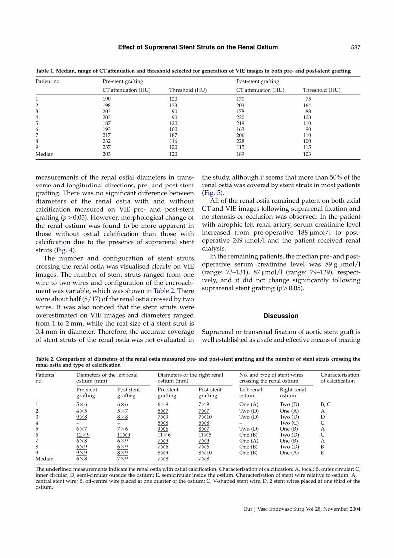
Table 1. Median, range of CT attenuation and threshold selected for generation of VIE images in both pre- and post-stent grafting
Patient no. Pre-stent grafting Post-stent grafting
CT attenuation (HU) Threshold (HU) CT attenuation (HU) Threshold (HU)
1 190 120 170 752 198 133 203 1643 203 90 178 884 203 90 220 1035 187 120 219 1106 193 100 163 907 217 187 206 1108 232 116 228 1009 237 120 115 115
Median 203 120 189 103
Effect of Suprarenal Stent Struts on the Renal Ostium 537
measurements of the renal ostial diameters in trans-verse and longitudinal directions, pre- and post-stentgrafting. There was no significant difference betweendiameters of the renal ostia with and withoutcalcification measured on VIE pre- and post-stentgrafting (pO0.05). However, morphological change ofthe renal ostium was found to be more apparent inthose without ostial calcification than those withcalcification due to the presence of suprarenal stentstruts (Fig. 4).
The number and configuration of stent strutscrossing the renal ostia was visualised clearly on VIEimages. The number of stent struts ranged from onewire to two wires and configuration of the encroach-ment was variable, which was shown in Table 2. Therewere about half (8/17) of the renal ostia crossed by twowires. It was also noticed that the stent struts wereoverestimated on VIE images and diameters rangedfrom 1 to 2 mm, while the real size of a stent strut is0.4 mm in diameter. Therefore, the accurate coverageof stent struts of the renal ostia was not evaluated in
Table 2. Comparison of diameters of the renal ostia measured pre- arenal ostia and type of calcification
Patientsno.
Diameters of the left renalostium (mm)
Diameters of the rostium (mm)
Pre-stentgrafting
Post-stentgrafting
Pre-stentgrafting
Pg
1 5!6 6!6 6!9 72 4!5 5!7 5!7 73 9!8 8!8 7!9 74 – – 5!8 55 6!7 7!6 9!6 86 12!9 11!9 11!6 17 6!8 6!9 7!9 78 6!9 6!9 7!6 79 9!9 8!9 8!9 8Median 6!8 7!9 7!8 7
The underlined measurements indicate the renal ostia with ostial calcifiinner circular; D, semi-circular outside the ostium; E, semicircular insicentral stent wire; B, off-centre wire placed at one quarter of the ostiumostium.
the study, although it seems that more than 50% of therenal ostia was covered by stent struts in most patients(Fig. 5).
All of the renal ostia remained patent on both axialCT and VIE images following suprarenal fixation andno stenosis or occlusion was observed. In the patientwith atrophic left renal artery, serum creatinine levelincreased from pre-operative 188 mmol/l to post-operative 249 mmol/l and the patient received renaldialysis.
In the remaining patients, the median pre- and post-operative serum creatinine level was 89 g mmol/l(range: 73–131), 87 mmol/l (range: 79–129), respect-ively, and it did not change significantly followingsuprarenal stent grafting (pO0.05).
Discussion
Suprarenal or transrenal fixation of aortic stent graft iswell established as a safe and effective means of treating
nd post-stent grafting and the number of stent struts crossing the
ight renal No. and type of stent wirescrossing the renal ostium
Characterisationof calcification
ost-stentrafting
Left renalostium
Right renalostium
!9 One (A) Two (D) B, C!7 Two (D) One (A) A!10 Two (D) Two (D) D!8 – Two (C) C!7 Two (D) One (B) A1!5 One (B) Two (D) C!9 One (A) One (B) A!6 One (B) Two (D) B!10 One (B) One (A) E!8
cation. Characterisation of calcification: A, focal; B, outer circular; C,de the ostium. Characterisation of stent wire relative to ostium: A,
; C, V-shaped stent wire; D, 2 stent wires placed at one third of the
Eur J Vasc Endovasc Surg Vol 28, November 2004
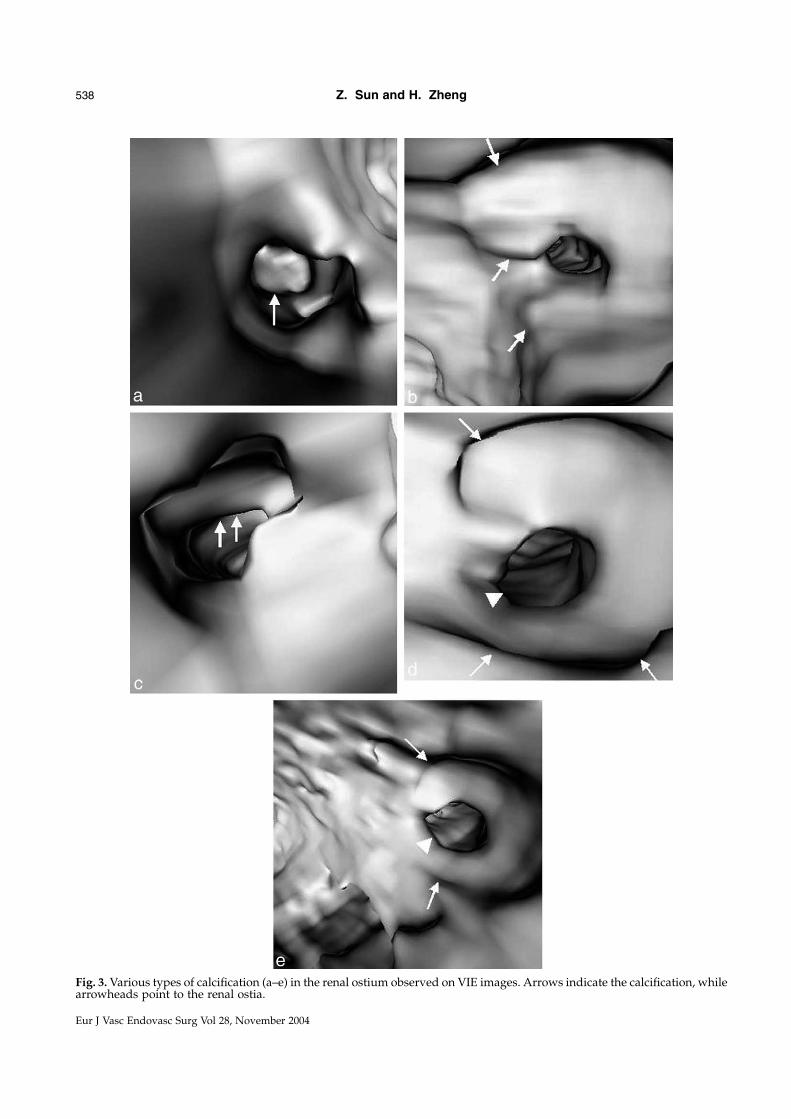
Fig. 3. Various types of calcification (a–e) in the renal ostium observed on VIE images. Arrows indicate the calcification, whilearrowheads point to the renal ostia.
Z. Sun and H. Zheng538
Eur J Vasc Endovasc Surg Vol 28, November 2004
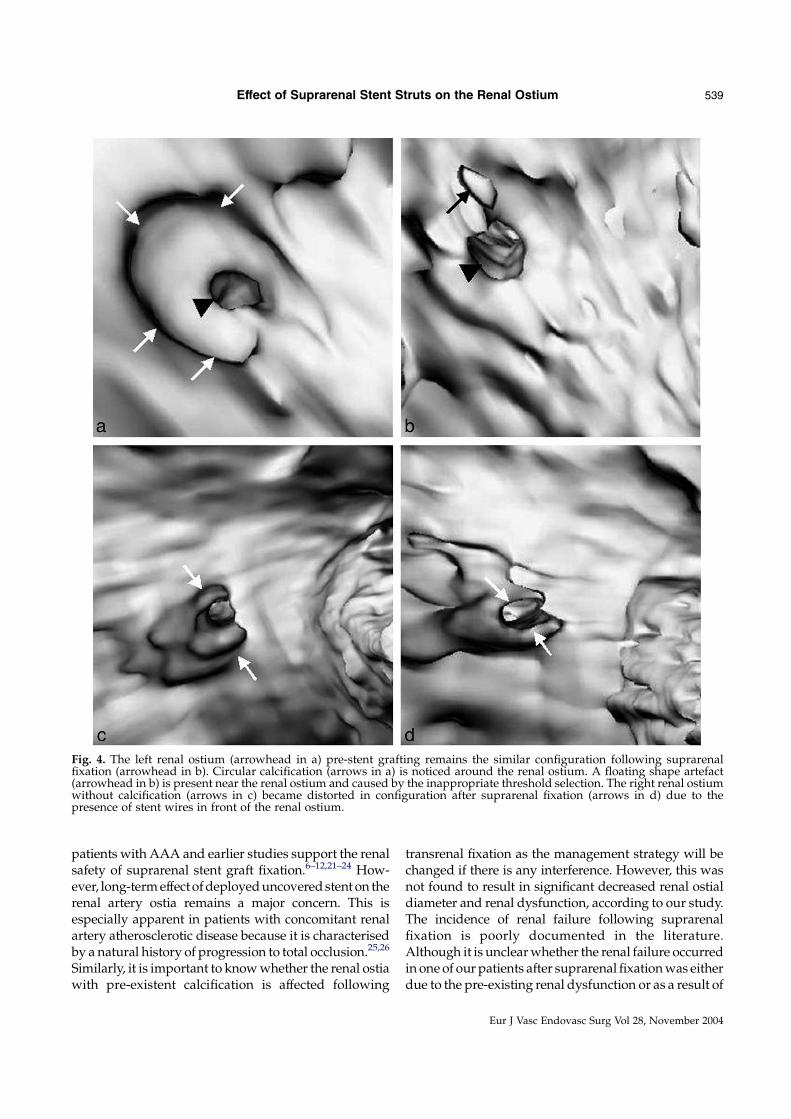
Fig. 4. The left renal ostium (arrowhead in a) pre-stent grafting remains the similar configuration following suprarenalfixation (arrowhead in b). Circular calcification (arrows in a) is noticed around the renal ostium. A floating shape artefact(arrowhead in b) is present near the renal ostium and caused by the inappropriate threshold selection. The right renal ostiumwithout calcification (arrows in c) became distorted in configuration after suprarenal fixation (arrows in d) due to thepresence of stent wires in front of the renal ostium.
Effect of Suprarenal Stent Struts on the Renal Ostium 539
patients with AAA and earlier studies support the renalsafety of suprarenal stent graft fixation.6–12,21–24 How-ever, long-term effect of deployed uncovered stent on therenal artery ostia remains a major concern. This isespecially apparent in patients with concomitant renalartery atherosclerotic disease because it is characterisedby a natural history of progression to total occlusion.25,26
Similarly, it is important to know whether the renal ostiawith pre-existent calcification is affected following
transrenal fixation as the management strategy will bechanged if there is any interference. However, this wasnot found to result in significant decreased renal ostialdiameter and renal dysfunction, according to our study.The incidence of renal failure following suprarenalfixation is poorly documented in the literature.Although it is unclear whether the renal failure occurredin one of our patients after suprarenal fixation was eitherdue to the pre-existing renal dysfunction or as a result of
Eur J Vasc Endovasc Surg Vol 28, November 2004
Fig. 5. Central stent wire crossing the right renal ostium wasobserved in an 83-year-old man with an infrarenal AAA. Itwas demonstrated that more than 50% of the renal ostiumwas covered by the stent wire due to the overestimatedthickness of metal wire. Arrows indicate suprarenal stentstruts (wires), while arrowheads point to the renal ostia.
Z. Sun and H. Zheng540
the effect of suprarenal stent wires or calcification, ourpreliminary results should arise clinical awareness inpatients with atherosclerotic disease such as renal ostialcalcification. It is possible to physically displace plaque(especially type D and E) in the vicinity of the renalostium during deployment of the suprarenal stent graft,supposedly that the stent wire was always deployedadjacent to the opening of the renal ostium. Althoughthis was not observed in our study due to a smallnumber of cases and no conclusion can be drawn, futurestudies in this area deserves to be investigated. VIE willbe a valuable technique in this observation throughvisualising the change of configuration in both renalostia and calcified plaque.
It is difficult to evaluate the morphological appear-ance and effect of stent struts on renal ostia onconventional axial CT and other 3D imagingmethods.18 This is especially apparent in post-stentgrafting as high density metal wires will obscure thevisualisation of pre-existent renal ostial calcification.Intravascular ultrasound was reported to be a reliabletool for intravascular visualisation when comparedwith angiography,27 however, it is still an invasive andtechnically demanding method. The limitation waseasily overcome by VIE as it allows non-invasive directvisualisation of intravascular views of the renal ostia,intraluminal calcification and stent wires from various
Eur J Vasc Endovasc Surg Vol 28, November 2004
angles. Moreover, the 3D relationship between stentstruts and renal ostia was clearly demonstrated in ourstudy.
Presence of stent wires in front of the renal ostium,especially 2-wire configuration has been investigatedexperimentally and reported to have the largestobstructive surface area.14 The presence of ostialcalcification will undoubtedly further reduce thecross-sectional area of the renal ostium, although it isdifficult to measure the percentage of area reductioncaused by the calcification. Despite a small number ofcases were included in our study, our results showedthat nearly half of the renal ostia were covered by twostent wires. Therefore, special attention should be paidto these patients while monitoring the renal blood flowor renal function. We consider that VIE findingspresented in this study will aid vascular surgeons orinterventional radiologists to accurately assess theeffect of suprarenal fixation and therapeuticmanagement.
Distortion of the renal ostium by the presence ofa stent wire could occur following suprarenalfixation, although this has not been addressedbefore. One could imagine that those renal arterieswith ostial calcification are less likely to bedistorted by the stent wires than those withoutostial calcification, as shown in our patient group(Fig. 4). Our study showed that renal ostia withoutcalcification were more easily distorted than thosewith ostial calcification by the presence of stentwires, although the results were based on a shortperiod of follow-up. Comparison of these twogroups is currently under investigation in ourinstitute over regular periods of follow-up.
One of the disadvantages of helical CTA in theimaging of stent grafts is the blurring effect that resultsin overestimation of the thickness of stent wires asdisplayed in both axial CT and VIE images accordingto recent studies.18,28 However, measurements of therenal ostia diameter were not affected in our study asmetal wires were removed after applying the appro-priate threshold to only visualise the renal ostia onVIE. Therefore, we know only a small proportion ofthe renal ostium was covered in reality. Althoughdiameters of the renal ostia were not found to changesignificantly in our study, nearly half of the renal ostialmeasurements were affected by the presence ofadjacent calcification (type C or D). We consider thata more accurate method would be measuring thecross-sectional area reduction of the renal ostium bypresence of stent wires when a feasible methodologybecomes available. As mentioned earlier, VIE imageswere generated from the CT data acquired from arelative wide collimation (5 mm) on a single slice CT
Effect of Suprarenal Stent Struts on the Renal Ostium 541
scanner, which limits image resolution in our cases.Future studies in aortic stent grafting should beperformed on a multislice CT scanner as it allowsthinner slice thickness and improves image qualitywhen compared to a single slice CT.29 It isexpected that stent wires as well as 3D relationshipbetween stent wires and the renal ostia will bebetter imaged on a multislice CT, includingcorresponding VIE images.
Conclusion
Our preliminary study demonstrated that suprarenalstent struts did not significantly affect the morpho-logical change of the renal ostia. Stent-covered renalarteries remained patent and no renal dysfunctionoccurred in most of the cases following transrenalfixation, although renal ostia were crossed by stentstruts to various extents. VIE images of the renal ostiaand stent wires as well as their 3D relationshipenhanced our understanding of the effect of transrenalfixation on the renal ostia, especially in those withostial calcification. VIE findings will play an importantrole in clinical-decision making such as closelymonitor patients with renal ostial calcification crossedby multiple stent wires. Further studies deserve toinvestigate the long-term effect of stent struts on therenal artery and renal blood flow in terms of the cross-sectional area reduction caused by stent wires andostial calcification.
Acknowledgements
The authors would like to thank Mr John Winder for histechnical assistance and Dr Peter Ellis for his clinical co-operation. Many thanks also go to Mr Mark O’Donnell forhis clinical assistance in the evaluation of renal function.
References
1 Parodi JC, Blum U, Voshage G, Lammer J et al. Two-centreGerman experience with aortic endografting. J Endovasc Surg1997;4:137–146.
2 Parodi JC, Palmaz JC, Barone HD. Transfemoral intraluminalgraft implantation for abdominal aortic aneurysms. Ann VascSurg 1991;5:491–499.
3 Mialhe C, Amicabile C, Becquemin JP et al. Endovasculartreatment of infrarenal abdominal aortic aneurysms by theStentor system: preliminary results of 79 cases. J Vasc Surg 1997;26:199–209.
4 Parodi JC, Barone A, Piraino R et al. Endovascular treatment ofabdominal aortic aneurysms: lessons learned. J Endovasc Surg1997;4:102–110.
5 Lawrence-Brown M, Hartley D, MacSweeney ST et al. ThePerth endoluminal bifurcated graft system: development andearly experience. Cardiovasc Surg 1996;4:706–712.
6 Malina M, Lindh M, Ivancev K et al. The effects of endovascularaortic stents placed across the renal arteries. Eur J Vasc EndovascSurg 1997;13:207–213.
7 Ferko A, Krajina A, Jon B et al. Juxtarenal aortic aneurysm:endoluminal transfemoral repair? Eur Radiol 1997;7:703–707.
8 Birch PC, Start RD, Whitebread T et al. The effect of crossingporcine renal artery ostia with various endovascular stents. EurJ Vasc Endovasc Surg 1999;17:185–190.
9 Alric P, Hinchliffe RJ, Picot MC et al. Long-term renal functionfollowing endovascular aneurysm repair with infrarenal andsuprarenal aortic stent-grafts. J Endovasc Ther 2003;10:397–405.
10 Lau LL, Hakaim AG, Oldenburg WA et al. Effect of suprarenalversus infrarenal aortic endograft fixation on renal function andrenal artery patency: a comparative study with intermediatefollow-up. J Vasc Surg 2003;37:1162–1168.
11 Burks JA, Faries PL, Gravereaux EC et al. Endovascular repairof abdominal aortic aneurysms: stent-graft fixation across thevisceral arteries. J Vasc Surg 2002;35:109–113.
12 Cayne NS, Rhee SJ, Veith FJ et al. Does transrenal fixation ofaortic endografts impair renal function? J Vasc Surg 2003;38:639–644.
13 Desgranges P, Hutin E, Kedzia C et al. Aortic stents coveringthe renal arteries ostia: an animal study. JVIR 1997;8:77–82.
14 Lilffamn K, Lawrence-Brown MMD, Semmens JB et al.Suprarenal fixation: effect on blood flow of an endoluminalstent wire across an arterial orifice. J Endovasc Ther 2003;10(2):260–274.
15 Armerding MD, Rubin GD, Beaulieu CF et al. Aortic aneur-ysmal disease: assessment of stent-grafted treatment-CT versusconventional angiography. Radiology 2000;215:138–146.
16 Dorffner R, Thurnher S, Youssesfzadeh S et al. Spiral CTangiography in the assessment of abdominal aortic aneurysmsafter stent grafting: value of maximum intensity projections.J Comput Assist Tomogr 1997;21:472–477.
17 Neri E, Bonanomi C, Vignali R et al. Spiral CT virtualendoscopy of abdominal arteries: clinical applications. AbdomImaging 2000;25:59–61.
18 Sun Z, Winder RJ, Kelly B et al. Diagnostic value of CT virtualintravascular endoscopy in aortic stent grafting. J Endovasc Ther2004;11:13–25.
19 Winder J, Sun Z, Kelly B et al. Abdominal aortic aneurysm andstent graft phantom manufactured by medical rapid prototyping.J Med Eng Technol 2002;26(2):75–78.
20 Sun Z, Winder J, Kelly B et al. CT virtual intravascularendoscopy of abdominal aortic aneurysms treated with suprar-enal endovascular stent grafting. Abdom Imaging 2003;28(4):580–587.
21 Sonesson B, Malina M, Ivancev K et al. Dilatation of theinfrarenal aneurysm neck after endovascular exclusion ofabdominal aortic aneurysm. J Endovasc Surg 1998;5:195–200.
22 Malina M, Brunkwall J, Lindblad B et al. Endovascularmanagement of the juxtarenal aortic aneurysm: can uncoveredstents safely cross the renal arteries? Semin Vasc Surg 1999;12:182–191.
23 Bove PG, Long GW, Zelenock GB et al. Transrenal fixation ofaortic stent-grafts for the treatment of infrarenal aortic aneurysmdisease. J Vasc Surg 2000;32:697–703.
24 Lobato AC, Quick RC, Vaughn PL et al. Transrenal fixation ofaortic endografts: intermediate follow-up of a single-centreexperience. J Endovasc Ther 2000;7:273–278.
25 Caps MT, Perissinotto C, Zierler E et al. Prospective study ofatherosclerotic disease progression in the renal artery. Circulation1998;98:2866–2872.
26 Caps MT, Zierler RE, Polissar NL et al. Risk of atrophy inkidneys with atherosclerotic renal artery stenosis. Kidney Int 1998;53:735–742.
27 von Segesser LK, Marty B, Ruchat P et al. Routine use ofintravascular ultrasound for endovascular aneurysm repair:
Eur J Vasc Endovasc Surg Vol 28, November 2004
Z. Sun and H. Zheng542
angiography is not necessary. Eur J Vasc Endovasc Surg 2002;23:537–542.
28 Sun Z, Winder J, Kelly B et al. Assessment of VIE image qualityusing helical CT angiography: in vitro phantom study. ComputMed Imaging Graph 2004;28:3–12.
Eur J Vasc Endovasc Surg Vol 28, November 2004
29 Hu H, He HD, Foley WD et al. Four multidetector-row helical CT:image quality and volume coverage speed. Radiology 2000;215:55–62.
Accepted 26 August 2004
Copyright © 2022 FDOKUMEN