Optimization of intravascular shear stress assessment in vivo
19
Optimization of Intravascular Shear Stress Assessment in Vivo Lisong Ai, Department of Biomedical Engineering and Cardiovascular Medicine, University of Southern California, Los Angeles, California 90089-1111 Hongyu Yu, School of Earth and Space Exploration and the Department of Electrical Engineering, Arizona State University, Tempe, AZ 85287 Wakako Takabe, Department of Biomedical Engineering and Cardiovascular Medicine, University of Southern California, Los Angeles, California 90089-1111 Anna Paraboschi, Department of Biomedical Engineering and Cardiovascular Medicine, University of Southern California, Los Angeles, California 90089-1111 Fei Yu, Department of Biomedical Engineering and Cardiovascular Medicine, University of Southern California, Los Angeles, California 90089-1111 E. S. Kim, Department of Electrical Engineering and Electrophysics, University of Southern California, Los Angeles, California 90089-1111 Rongsong Li, and Department of Biomedical Engineering and Cardiovascular Medicine, University of Southern California, Los Angeles, California 90089-1111 Tzung K. Hsiai Department of Biomedical Engineering and Cardiovascular Medicine, University of Southern California, Los Angeles, California 90089-1111 Introduction Hemodynamic forces, namely, fluid shear stress, regulate the biological activities of the vascular endothelial cells (EC) (14) and vessel wall remodeling in the resistant arteries and aorta (9,37). Sedentary lifestyle promotes atherogenic hemodynamics in arterial bifurcations where oscillatory and low shear stress predisposes the development of atherosclerosis (8). The advent of Microelectromecahnical Systems (MEMS) provides an entry point to characterize the spatial and temporal variations of the WSS in various arterial configurations(12,15,23– 25). Previously, silicon nitride was applied as an insulating layer on the diaphragm to enhance © 2009 Elsevier Ltd. All rights reserved. Tzung K. Hsiai, MD, PhD, Department of Biomedical Engineering and Division of Cardiovascular Medicine, University of Southern California, Los Angeles, Ca 90089, [email protected]. Publisher's Disclaimer: This is a PDF file of an unedited manuscript that has been accepted for publication. As a service to our customers we are providing this early version of the manuscript. The manuscript will undergo copyediting, typesetting, and review of the resulting proof before it is published in its final citable form. Please note that during the production process errors may be discovered which could affect the content, and all legal disclaimers that apply to the journal pertain. NIH Public Access Author Manuscript J Biomech. Author manuscript; available in PMC 2010 July 22. Published in final edited form as: J Biomech. 2009 July 22; 42(10): 1429–1437. doi:10.1016/j.jbiomech.2009.04.021. NIH-PA Author Manuscript NIH-PA Author Manuscript NIH-PA Author Manuscript
Transcript of Optimization of intravascular shear stress assessment in vivo

Optimization of Intravascular Shear Stress Assessment in Vivo
Lisong Ai,Department of Biomedical Engineering and Cardiovascular Medicine, University of SouthernCalifornia, Los Angeles, California 90089-1111
Hongyu Yu,School of Earth and Space Exploration and the Department of Electrical Engineering, Arizona StateUniversity, Tempe, AZ 85287
Wakako Takabe,Department of Biomedical Engineering and Cardiovascular Medicine, University of SouthernCalifornia, Los Angeles, California 90089-1111
Anna Paraboschi,Department of Biomedical Engineering and Cardiovascular Medicine, University of SouthernCalifornia, Los Angeles, California 90089-1111
Fei Yu,Department of Biomedical Engineering and Cardiovascular Medicine, University of SouthernCalifornia, Los Angeles, California 90089-1111
E. S. Kim,Department of Electrical Engineering and Electrophysics, University of Southern California, LosAngeles, California 90089-1111
Rongsong Li, andDepartment of Biomedical Engineering and Cardiovascular Medicine, University of SouthernCalifornia, Los Angeles, California 90089-1111
Tzung K. HsiaiDepartment of Biomedical Engineering and Cardiovascular Medicine, University of SouthernCalifornia, Los Angeles, California 90089-1111
IntroductionHemodynamic forces, namely, fluid shear stress, regulate the biological activities of thevascular endothelial cells (EC) (14) and vessel wall remodeling in the resistant arteries andaorta (9,37). Sedentary lifestyle promotes atherogenic hemodynamics in arterial bifurcationswhere oscillatory and low shear stress predisposes the development of atherosclerosis (8). Theadvent of Microelectromecahnical Systems (MEMS) provides an entry point to characterizethe spatial and temporal variations of the WSS in various arterial configurations(12,15,23–25). Previously, silicon nitride was applied as an insulating layer on the diaphragm to enhance
© 2009 Elsevier Ltd. All rights reserved.Tzung K. Hsiai, MD, PhD, Department of Biomedical Engineering and Division of Cardiovascular Medicine, University of SouthernCalifornia, Los Angeles, Ca 90089, [email protected]'s Disclaimer: This is a PDF file of an unedited manuscript that has been accepted for publication. As a service to our customerswe are providing this early version of the manuscript. The manuscript will undergo copyediting, typesetting, and review of the resultingproof before it is published in its final citable form. Please note that during the production process errors may be discovered which couldaffect the content, and all legal disclaimers that apply to the journal pertain.
NIH Public AccessAuthor ManuscriptJ Biomech. Author manuscript; available in PMC 2010 July 22.
Published in final edited form as:J Biomech. 2009 July 22; 42(10): 1429–1437. doi:10.1016/j.jbiomech.2009.04.021.
NIH
-PA Author Manuscript
NIH
-PA Author Manuscript
NIH
-PA Author Manuscript

unidirectional heat transfer (40). In the new generation of MEMS sensors, biocompatibleparylene C coating not only insulated the microelectronics but also provided flexibility forpackaging the sensors to the catheter/coaxial wire.
Numerous CFD codes have been developed to assess WSS with relevance to cardiovasculardisease (20–22). Steinman and Ethier investigated the effects of wall distensibility in a 2-Dend-to-side anastomosis (32). Lei et al. further described the applications of CFD to designend-to-side anastomoses (16). Taylor et al. developed a stabilized FEM to simulate blood flowin the abdominal aorta under resting and exercise-induced pulsatile flow conditions (33–35).Hence, CFD codes predicted arterial regions where elevated temporal oscillations in shearstress are prone to vascular oxidative stress and inflammatory responses (1).
Despite numerous CFD solutions for WSS, there has been a paucity of experimental data.Conventionally, shear stress has been assessed by the direct and indirect methods (28). Theformer is based on both piezoresistive (30) and capacitive readout schemes in a floating elementsensing (28). The latter is based on convective heat transfer by correlating convective coolingof a heated element to shear stress. Indirect methods also include ultrasonic Doppler andMagnetic Resonance Imaging (MRI). Both methods require a known geometry to correlateflow rates with shear stress. While ultrasonic Doppler and MRI are non-invasive, the spatialresolution near the wall is difficult to establish.
To assess shear stress in the complicated arterial geometry, we developed the polymer-basedsensors that are flexible, biocompatible, and deployable into the arterial system. A Titanium(Ti) and Platinum (Pt) layer embedded in the flexible polymer was used as the sensing elements.Based on heat transfer principle (42), shear stress can be inferred from the heat loss from theheated sensing element to the flow in terms of the measured changes in voltage. The sensorwas fabricated via surface micromachining technique utilizing parylene C as electricalinsulation layer. The resistance of the sensing element was measured at approximately 1.0kOhm and the signal-to-noise ratio was 4.8 (42). Direct measurement of wall shear stress inthe aorta thereby enables us to predict the arterial regions exposed to low or oscillatory shearstress that is linked with the development of plaque formation.
To optimize intravascular shear stress (ISS) assessment, we performed both theoretical andCFD analyses. These analyses predicted the extent to which experimental parameters governedthe deployment of the catheter/coaxial wire-based MEMS sensors into the aorta of NZWrabbits; namely, the diameter ratios of the aorta to the coaxial wires, position of sensors, andentrance length (Le) in relation to the Reynolds numbers.
Methods1. Packaging and Deployment of MEMS Thermal Sensors
We have developed biocompatible and flexible intravascular MEMS sensors to assess real-time ISS (Figs. 1a and 1b). The detailed operational principle and fabrication process of theMEMS sensor were previously described (41). The diameter of the coaxial wire was 0.4 mmand the length of the sensor was 4 cm (Fig. 1b). The biocompatible epoxy (EPO-TEK 301:Epoxy Technology, Billerica, MA, USA) was used to anchor the sensor to the coaxial wire(Fig. 1c). The sensor was packaged to the coaxial wire, which was deployed into the abdominalaorta of adult NZW rabbits under fluoroscopic guidance.
The sensors were first tested in the tube model and the shear stress measurements were validatedby CFD simulation (Fig. 2a). A range of steady flow rates were generated by a flow systemconsisting of a digital modular drive (Master Flex L/S 77300-80, Cole-Palmer) and a pumpdrive (Master Flex L/S 7518-12, Cole-Palmer). The pump setting was calibrated using an
Ai et al. Page 2
J Biomech. Author manuscript; available in PMC 2010 July 22.
NIH
-PA Author Manuscript
NIH
-PA Author Manuscript
NIH
-PA Author Manuscript

electromagnetic flow meter (MAGFLO®, Danfoss A/S, DK). Once validated in the in vitromodel, the MEMS sensors were deployed into the aorta of the rabbits through the co-axial wirevia femoral cut-down procedure. Using the 3-D fluoroscopic guidance (Phillips BV-22HQ C-arm), the guide wire and MEMS sensors were visualized in the abdominal aorta. Thefluoroscope could be rotated to acquire the images in various planes. By injecting the contrastdye from the carotid artery, the operators were able to delineate the geometry of the rabbit aortaand the position of the coaxial wire in relation to the inner diameter of the aorta (Fig. 2b).
2. Theoretical and Computational Analyses of Intravascular Shear Stress2.1. Theoretical Formulation to Predict WSS in the Presence of a Catheter—Analogous to intravascular ultrasound (IVUS), the deployment of coaxial wires wouldinevitably influence the flow field(4,5,13,18,19,25,38). To optimize the MEMS sensormeasurement, we formulated Navier-Stokes equations analogous to flow in an annular duct topredict the flow disturbance when the catheter was positioned at the center (Fig. 3a). Theviscosity, μ, was treated as a constant. The velocity profiles obtained for annulus and circularpipe flow were expressed as (27):
(1)
(2)
where U and u represent the velocity in the annulus and circular pipe, respectively. R and Riare the radii of the pipe and coaxial wire, respectively. n is the radius or diameter ratio betweenthe vessel and the catheter , n=R/Ri, and dP/dx and dp/dx are the pressure gradient in the annulusand the circular pipe, respectively. The flow rate, Q, and WSS, τw, in the absence of coaxialwire were expressed as:
(3)
and
(4)
2.2. Flow Rates and Wall Shear Stress—Assuming the effect of the coaxial wire on theflow was negligible (10,26), the flow rate in the presence of coaxial wire would be defined as:
(5)
From Eq. (5), the following relation was obtained:
Ai et al. Page 3
J Biomech. Author manuscript; available in PMC 2010 July 22.
NIH
-PA Author Manuscript
NIH
-PA Author Manuscript
NIH
-PA Author Manuscript

(6)
where Ep is defined as the pressure elevation factor (PEF):
(7)
From the velocity profile, U , the WSS in the presence of coaxial wire, τw_wire, was expressedas:
(8)
where Ewss is the WSS elevation factor (WEF):
(9)
Similarly, the shear stress on the MEMS sensor, τi_wire, was evaluated at r=Ri:
(10)
where τw is the WSS in the absence of coaxial wire and Eiss is defined as the ISS elevationfactor (IEF):
(11)
2.3. Computational Analyses—CFD analyses were performed to investigate the effectsof the flow disturbance and the entrance length on the coaxial wire required to mount the MEMSsensors. The governing equations were solved for laminar, incompressible, and non-Newtonianflow. In our CFD model, the shear rate-dependent dynamic viscosity was implemented for theshear-thinning behavior of the rabbit blood (6,7). The simulations were performed for thegeometry as shown in figure 3b.
In general, entrance length, Le, is the distance required from the terminal end of the coaxialwire to the point where fully developed flow develops. The definition of the Le (31) was alsobased on the length required to achieve a local incremental pressure drop of ~ 98% of the fullydeveloped region. In our study, shear stress was used to predict Le. The flow disturbance in theLe region influenced heat transfer from the sensor to the flow stream (17). In this context,predicting the entrance length in relation to the Reynolds numbers guided the packaging ofMEMS sensors to the coaxial wire.
The inlet Reynolds number, Re, was defined by the mean velocity:
Ai et al. Page 4
J Biomech. Author manuscript; available in PMC 2010 July 22.
NIH
-PA Author Manuscript
NIH
-PA Author Manuscript
NIH
-PA Author Manuscript

(12)
where Umean is the mean flow velocity at the inlet.
3.1 Generation of 3-D Geometries and MeshesThe geometries of the tube model and the rabbit abdominal aorta model in the presence ofcoaxial wire or catheter were reconstructed in Pro Engineer Wildfire V.3.0 (ParametricTechnology, Needham, MA) using boundary conditions from experimental measurements.The models were imported into GAMBIT for mesh generation (Fluent Inc., Gambit 2.3.16,Lebanon, NH, USA). The meshed models were imported into the main CFD solver (FluentInc., Fluent 6.2.16, Lebanon, NH, USA) for further flow simulation. The CFD model wasconstructed with 78,868 cells, which were primarily the Tet/Hybrid elements. Since our focuswas on the shear stress on the sensor, fine elements immediately adjacent to the coaxial wirewere constructed to obtain sufficient information to characterize the large fluid velocitygradients near the wall.
In the abdominal aorta model, the flow field was solved under three catheter positioningschemes as described previously (2). The vessel inner diameter, Daorta, was measured to be2.4 mm from the angiogram (Fig. 3). The MEMS sensor was located 4.0 cm from the terminalend of the catheter/coaxial wire. Three different diameters (Dcatheter) namely, 0.25 mm (mousecatheter 0.010”OD), 0.4 mm (coaxial wire), and 0.97 mm (dog catheter 0.038”OD), werestudied in the CFD simulations. The geometry of the computational model of rabbit abdominalaorta is shown in figure 3b, where u and v represent the axial and radial velocities, andDcatheter represents the catheter diameter.
3.2. Boundary conditionsThe steady flow rates from the in vitro experiment were applied as the inlet flow boundarycondition for the corresponding 3-D computational model. The ultrasound transducer (PhilipsSONOS 5500 at 12 MHz) was used to obtain the pulsatile velocity waveform. The recordedprofile of the center-line velocity, Uc, as shown in figure 4a, was reconstructed by applyingthe Fourier analysis using 12 harmonics (Fig. 4b). The peak and time-averaged velocity were57.7 cm·s−1 and 14.6 cm·s−1, respectively. The period of one cardiac cycle, T, was 0.33 secondscorresponding to 180 beats·min−1. From Eq. (12), the mass flux at the inlet in the axial direction(nx) was obtained as:
(13)
The calculated mass flux was applied as the inlet boundary condition and implemented by auser defined C++ code in FLUENT.
The traction-free condition was assumed at flow outlet. No-slip boundary condition wasapplied to the inner walls and the surface of the coaxial wire.
The spatial WSS, τw, was calculated for incompressible fluids as:
(14)
Ai et al. Page 5
J Biomech. Author manuscript; available in PMC 2010 July 22.
NIH
-PA Author Manuscript
NIH
-PA Author Manuscript
NIH
-PA Author Manuscript

where ut is the velocity tangential to the wall and n is the unit vector perpendicular to the wall.
A range of steady flow rates were also studied in order to establish the relationship betweenthe entrance length and flow rate. The mean flow velocity was maintained at 0.158, 0.288,0.625, 0.92654, and 1.567m·sec−1, yielding Reynolds numbers of 116, 210, 330, 439, 680, 910,1150, 1350 and 1550. The Reynolds numbers of 116 and 439 represented the time-averagedvalue and the maximum value in the rabbit thoracic aorta, respectively. Comparatively, theReynolds number of 1150 represented the maximal value in the human descending aorta.
Reynolds numbers were determined in the presence or absence of coaxial wire. The hydraulicdiameter, Dh = D − Di, was used to normalize the Reynolds number and the entrance length,Le. In the absence of a catheter, the Reynolds number was expressed in terms of the flow rate,Q:
(15)
Given that in the presence of catheter,
(16)
the Reynolds number, Rewire, was calculated from the mean velocity, Umean_wire:
(17)
We assumed the effect of the catheter on the flow to be negligible (10,26):
(18)
(19)
The normalized entrance length, Le_wire as a function of the normalized Reynolds number,Rewire, was derived using the Curve Fitting Toolbox of MATLAB 7.1 (Math-works, Natick,USA).
Results3.1. Theoretical Prediction of WSS in the Presence of a Coaxial Wire
The diameter ratio influenced the extent to which the coaxial wire increased the pressure andWSS measurements in a 3-D tube. This change in velocity profile reversed the direction ofshear stress, leading to an increase in shear stress on the coaxial wire by an elevation factor(Eiss). Figure 5a illustrated an increase in pressure by an elevation factor (Ep) as a function ofthe diameter ratio. The magnitude of Ep decreased significantly from 24.2 to 2.5 as the ratioincreased from 1.5 to 4.5 with an asymptotic value of ~1.1 at a diameter ratio approaching 100.The pressure was predicted to increase by 20% at a diameter ratio of 100.
Ai et al. Page 6
J Biomech. Author manuscript; available in PMC 2010 July 22.
NIH
-PA Author Manuscript
NIH
-PA Author Manuscript
NIH
-PA Author Manuscript

Wall shear stress and WSS elevation factor (Ewss) were also dependent on the diameter ratio(Fig. 5b). Ewss decreased from 7.6 to 1.7 as the diameter ratio increased from 1.5 to 4.5. Theelevation factor for ISS on the coaxial wire (Eiss) was reduced from 8.747 to its minimum of3.024 as the diameter ratio increased from 2 to 4.5 (Fig. 5c). In humans, the large diameterratio would minimize the WSS elevation to ~10%. Taken together, a large catheter (n < 2)would introduce significant elevations in pressure and shear stress whereas n > 4.5 wouldminimize these effects (3).
3.2. Shear Stress Assessment in a 3-D Tube ModelCFD simulation was performed to analyze both velocity profiles and shear stress distributionon the catheter/coaxial wire in response to three distinct Reynolds numbers; namely, 23, 91,and 133 (Fig. 6). The effects of flow disturbance on the MEMS sensors were negligible in thepresence of a coaxial wire until the Reynolds number reached to above 133 (Fig. 6a and 6b).At a Reynolds number of 23, shear stress on the coaxial wire was 0.59 dyn·cm−2; at a Reynoldsnumber of 91, shear stress was 3.13 dyn·cm−2; and at 133, shear stress was 5.61 dyn·cm−2.Next, real-time shear stress assessment by the MEMS sensor was compared with that of CFDsolutions (Fig. 7). The relationship between the voltage changes and flow rates (Fig. 7a) wereconverted to shear stress using the calibration curve (Fig. 7b) (41). Real-time shear stressmeasurement was in good agreement with that of CFD at flow rates greater than 6 ml·s−1.
3.3. Entrance Length and Reynolds NumbersThe normalized entrance length, Le_wire, and its corresponding normalized Reynolds number,Rewire, were determined based on the CFD results (Fig. 8). A relation between Rewire andLe_wire derived from CFD was expressed as:
(20)
Le_wire increased in a relatively linear fashion as a function of Rewire. This relation enabled usto mount the MEMS sensors downstream from the terminal end of coaxial wire to optimizereal-time shear stress assessment.
3. 4. Shear Stress Assessment in the Abdominal Aorta of NZW RabbitsThe entrance lengths were determined as a function of flow rates by the computational results.The MEMS sensor was positioned at 4 cm from the terminal end of the coaxial wire. The inflowboundary conditions obtained from the Doppler ultrasound were incorporated into the CFDsimulations.
The position of the coaxial wire and the orientation of the sensors facing the flow fieldsignificantly influenced ISS assessment. At the diameter ratio of 6, Le of 1.18 cm, and positionof the coaxial wire at the center, the time-averaged shear stress (τave) on the coaxial wire wascomputed to be 31.2 dyn·cm−2 with a systolic peak at 102.8 dyn·cm−2 (corresponding to amaximum Reynolds number of 212). Despite the identical afore mentioned physicalparameters, a shift in the coaxial position at 0.2 mm off the center resulted in an asymmetricdistribution of shear stress on the coaxial wire from τ 0 degree = 97.8 dyn·cm−2 at 0°,τ 90 degree = 107.6 dyn·cm−2 at 90°, to τ 180 degree = 112.8 dyn·cm−2 at 180° corresponding toa maximum Reynolds number of 212. Similarly, positioning the coaxial wire at 0.1 mm awayfrom the vessel wall, the shear stress at the peak of systole was τ 0 degree = 6.9 dyn·cm−2 at 0°,τ 90 degree = 53.6 dyn·cm−2 at 90°, and τ 180 degree = 98.3 dyn·cm−2 at 180°.
CFD analysis revealed that the presence of a coaxial wire (Daorta / Dcoaxial wire = 6 and Le =1.18 cm) increased the time-averaged WSS (τave) from 10.06 dyn·cm−2 to 15.5 dyn·cm−2 and
Ai et al. Page 7
J Biomech. Author manuscript; available in PMC 2010 July 22.
NIH
-PA Author Manuscript
NIH
-PA Author Manuscript
NIH
-PA Author Manuscript

the systolic peak from 33.2 dyn·cm−2 to 51.3 dyn·cm−2 (Fig. 9). Experimentally, underfluoroscopic visualization, real-time ISS assessment by the coaxial wire near the wall with theMEMS sensor facing the flow field revealed τave value of 11.92 dyn·cm−2 with a systolic peakat 47.04 dyn·cm−2. The difference between CFD and experimental τave was 18.5%. Takentogether, our findings demonstrated that deploying the coaxial wire using a steerable catheterunder 3-D fluoroscopic guidance along with the angiogram would optimize real-time shearstress assessment.
DiscussionIn this study, we performed theoretical and CFD analyses to optimize real-time intravascularassessment in the aorta of NZW rabbits. CFD analyses revealed that an entrance length of 2.9mm and the diameter ratio (n = 4.5) would minimize the pressure and shear stress elevation inthe rabbit aorta. When the catheter was positioned off the center of vessel, Velusamy and Garg(36) reported that an eccentricity of the velocity profiles developed, and shear stress on thecatheter and the vessel wall varied circumferentially (2) (Fig 3b). In humans, the diameter ratiocould approach to 100 for an aortic inner diameter of 2.5cm with a catheter outer diameter of0.254 mm. Hence, ISS assessment could be further optimized by positioning the catheter nearthe vessel wall using a steerable catheter with a large diameter ratio, such as a large animalmodel.
Several equations for estimating entrance length had been proposed (29,39). Among these wasthe one proposed by Wojtkowiak and Popiel (39) for flow in an annular duct. The slope of theirlinear equation, 0.0161, was about twice as our estimation at 0.008184. One reason for thisdeviation was the criteria used to define the fully developed flow. We adopted the notion thatthe wall shear rate converges to its fully developed value at about half the length at which thecenterline velocity converges to its fully developed value (14).
Despite numerous CFD solutions for wall shear stress, there has been a paucity of experimentaldata. A 30% distortion is commonly encountered for experimental intravascular measurements(14). The boundary conditions and dimensions implemented in the CFD model might notprecisely reflect the experimental boundary conditions. Therefore, the center-positioningscheme and the CFD results provided a basis to compare with direct sensor measurement.
Our computational results indicated that the entrance length in response to the maximal inletReynolds numbers in the rabbits (Re max_rabbit=459) and human abdominal aorta(Re max_human=1150) were Le rabbit =1.244 cm and Le human=1.985 cm, respectively. For aWomersley number greater than 1, the above Le values could be overestimated. Sufficient timewas allowed for the flow to develop towards a parabolic velocity profile during individualcardiac cycles (2) and the pulsatile flow behaved in a quasi-steady manner (11). The MEMSsensing element were mounted at 4.0 cm downstream from the terminal end of the coaxialwire; thereby, optimizing intravascular measurements.
In conclusion, ISS assessment was validated in vitro by deploying the catheter-based MEMSsensors in a 3-D model. Theoretical and computational models further provided physicalparameters to optimize real-time shear stress assessment. These fundamental analyses provideda basis to further investigate the application of MEMS devices (24) for in vivo studies.
AcknowledgmentsThis work was supported by American Heart Association GIA 0655051Y (TKH), NIH R01 HL083015 (TKH), NIHK08 HL068689 (TKH), and American Heart Association Post-Doctoral Fellowship 0725016Y (HY). The authorswould like to thank Ping Sun for assistance with signal processing.
Ai et al. Page 8
J Biomech. Author manuscript; available in PMC 2010 July 22.
NIH
-PA Author Manuscript
NIH
-PA Author Manuscript
NIH
-PA Author Manuscript

References1. Ai L, Rouhanizadeh M, Wu JC, Takabe W, Yu H, Alavi M, Li R, Chu Y, Miller J, Heistad D, Hsiai
TK. Shear stress influences spatial variations in vascular Mn-SOD expression: implication for LDLnitration. Am. J. Physiol. Cell Physiol 2008:C1576–C1585. [PubMed: 18434620]
2. Ai L, Yu, Dai WH, Hale S, Kloner R, Hsiai TK. Real-time Intravascular Shear Stress in the RabbitAbdominal Aorta. IEEE Transactions on Biomedical Engineering (submitted). 2008
3. Anderson HV, Zaatari GS, Roubin GS, Leimgruber PP, Gruentzig AR. Steerable fiberoptic catheterdelivery of laser energy in atherosclerotic rabbits. Am Heart J 1986;111:1065–1072. [PubMed:3716979]
4. Back LH. Estimated Mean Flow Resistance Increase during Coronary-Artery Catheterization. Journalof Biomechanics 1994;27:169–175. [PubMed: 8132684]
5. Banerjee RK, Back LH, Back MR, Cho YI. Catheter obstruction effect on pulsatile flow rate-pressuredrop during coronary angioplasty. Journal of Biomechanical Engineering-Transactions of the ASME1999;121:281–289.
6. Bird, RB.; ARC; Hassager, O. Fluid Mechanics. Vol. Vol.1. New York: Wiley; 1977. Dynamics ofPolymeric Liquids; p. 470
7. Boger DV. Viscoelastic Flows through Contractions. Annual Review of Fluid Mechanics 1987;19:157–182.
8. Chatzizisis YS, Coskun AU, Jonas M, Edelman ER, Feldman CL, Stone PH. Role of endothelial shearstress in the natural history of coronary atherosclerosis and vascular remodeling - Molecular, cellular,and vascular behavior. Journal of the American College of Cardiology 2007;49:2379–2393. [PubMed:17599600]
9. Chatzizisis YS, Jonas M, Coskun AU, Beigel R, Stone BV, et al. Prediction of the localization of high-risk coronary atherosclerotic plaques on the basis of low endothelial shear stress - An intravascularultrasound and histopathology natural history study. Circulation 2008;117:993–1002. [PubMed:18250270]
10. Doucette JW, Corl PD, Payne HM, Flynn AE, Goto M, et al. Validation of a Doppler Guide Wire forIntravascular Measurement of Coronary-Artery Flow Velocity. Circulation 1992;85:1899–1911.[PubMed: 1572046]
11. Fung, YC. Biomechanics:Circulation. Springer; 1997. p. 130-132.12. Hsiai TK, Cho SK, Wang PK, Ing MH, Salazar A, et al. Micro Sensors: Linking Vascular
Inflammatory Responses with Real-Time Oscillatory Shear Stress. Ann Biomed Eng 2004;32:189–201. [PubMed: 15008367]
13. Karahalios GT. Some Possible Effects of a Catheter on the Arterial-Wall. Medical Physics1990;17:922–925. [PubMed: 2233580]
14. Ku DN. Blood Flow in Arteries. Annu. Rev. Fluid Mech 1997;29:399–434.15. Lasheras JC. The biomechanics of arterial aneurysms. Annual Review of Fluid Mechanics
2007;39:293–319.16. Lei M, Archie JP, Kleinstreuer C. Computational design of a bypass graft that minimizes wall shear
stress gradients in the region of the distal anastomosis. Journal of Vascular Surgery 1997;25:637–646. [PubMed: 9129618]
17. Liu C, Huang JB, Zhu ZJ, Jiang FK, Tung S, et al. Micromachined flow shear-stress sensor based onthermal transfer principles. Journal of Microelectromechanical Systems 1999;8:90–99.
18. Macdonald DA. Fully-Developed Incompressible-Flow between Non-Coaxial Circular-Cylinders.Zeitschrift Fur Angewandte Mathematik Und Physik 1982;33:737–751.
19. Macdonald DA. Pulsatile Flow in a Catheterized Artery. Journal of Biomechanics 1986;19:239–249.[PubMed: 3700436]
20. Moore JA, Steinman DA, Ethier CR. Computational blood flow modelling: Errors associated withreconstructing finite element models from magnetic resonance images. Journal of Biomechanics1998;31:179–184. [PubMed: 9593213]
21. Pedersen EM, Agerbaek M, Kristensen IB, Yoganathan AP. Wall shear stress and early atheroscleroticlesions in the abdominal aorta in young adults. European Journal of Vascular and EndovascularSurgery 1997;13:443–451. [PubMed: 9166266]
Ai et al. Page 9
J Biomech. Author manuscript; available in PMC 2010 July 22.
NIH
-PA Author Manuscript
NIH
-PA Author Manuscript
NIH
-PA Author Manuscript

22. Perktold K, Resch M, Peter RO. 3-Dimensional Numerical-Analysis of Pulsatile Flow and Wall Shear-Stress in the Carotid-Artery Bifurcation. Journal of Biomechanics 1991;24:409–420. [PubMed:1856241]
23. Rouhanizadeh M, Lin TC, Arcas D, Hwang J, Hsiai TH. Spatial variations in shear stress in a 3-Dbifurcation model at low Reynolds numbers. Annals of Biomedical Engineering 2005;33:1360–1374.[PubMed: 16240085]
24. Rouhanizadeh M, Soundararajan G, Ascara D, Lo R, Browand F, Hsiai T. MEMS sensors to resolvespatial variations in shear stress in a 3-D bifurcation model. IEEE Sensors 2006;6:78–88.
25. Roy AS, Back LH, Banerjee RK. Guidewire flow obstruction effect on pressure drop-flow relationshipin moderate coronary artery stenosis. Journal of Biomechanics 2006;39:853–864. [PubMed:16488224]
26. Roy AS, Banerjee RK, Back LH, Back MR, Khoury S, Millard RW. Delineating the guide-wire flowobstruction effect in assessment of fractional flow reserve and coronary flow reserve measurements.Am J Physiol Heart Circ Physiol 2005;289:H392–H397. [PubMed: 15734887]
27. Schetz, JA.; Fuhs, AE., editors. Fundamentals of Fluid Mechanics. New York: John Wiley & Sons;1999.
28. Schmidt MA, Howe RT, Senturia SD, Haritonidis JH. Design and Calibration of a MicrofabricatedFloating-Element Shear-Stress Sensor. Ieee Transactions on Electron Devices 1988;35:750–757.
29. Shah RK. Correlation for Laminar Hydrodynamic Entry Length Solutions for Circular andNoncircular Ducts. Journal of Fluids Engineering-Transactions of the Asme 1978;100:177–179.
30. Shajii J, Ng KY, Schmidt MA. A microfabricated floating element shear stress sensor using wafer-bonding technology. J. Microelectromech. Syst 1992:89–94.
31. Sparrow EM, Lin SH, Lundgren TS. Flow Development in the Hydrodynamic Entrance Region ofTubes and Ducts. Physics of Fluids 1964;7:338–347.
32. Steinman DA, Ethier CR. The Effect of Wall Distensibility on Flow in a 2-Dimensional End-to-SideAnastomosis. Journal of Biomechanical Engineering-Transactions of the Asme 1994;116:294–301.
33. Taylor CA, Hughes TJR, Zarins CK. Finite element modeling of blood flow in arteries. ComputerMethods in Applied Mechanics and Engineering 1998;158:155–196.
34. Taylor CA, Hughes TJR, Zarins CK. Finite element modeling of three-dimensional pulsatile flow inthe abdominal aorta: Relevance to atherosclerosis. Annals of Biomedical Engineering 1998;26:975–987. [PubMed: 9846936]
35. Taylor CA, Hughes TJR, Zarins CK. Effect of exercise on hemodynamic conditions in the abdominalaorta. Journal of Vascular Surgery 1999;29:1077–1089. [PubMed: 10359942]
36. Velusamy K, Garg VK. Entrance Flow in Eccentric Annular Ducts. International Journal forNumerical Methods in Fluids 1994;19:493–512.
37. Volokh KY, Vorp DA. A model of growth and rupture of abdominal aortic aneurysm. J Biomech2008;41:1015–1021. [PubMed: 18255074]
38. Wentzel JJ, Krams R, van der Steen AFW, Li W, Cespedes EI, Bom N, Slager CJ. Disturbance of 3-D velocity profiles induced by IVUS catheter, evaluation with computational fluid dynamics. ComputCardiol IEEE 1997:597–599.
39. Wojtkowiak J, Popiel CO. Inherently linear annular-duct-type laminar flowmeter. Journal of FluidsEngineering-Transactions of the Asme 2006;128:196–198.
40. Xu Y, Lin Q, Lin GY, Katragadda RB, Jiang FK, et al. Micromachined thermal shear-stress sensorfor underwater applications. Journal of Microelectromechanical Systems 2005;14:1023–1030.
41. Yu H, Ai L, Rouhanizadeh M, Patel D, Kim ES, Hsiai TK. Flexible Polymer Sensors for in VivoIntravascular Shear Stress Analysis. IEEE/ASME J MEMS. 2008in press
42. Yu H, Ai L, Rouhanizadeh M, Patel D, Kim ES, Hsiai TK. Flexible Polymer Sensors for In VivoIntravascular Shear Stress Analysis. IEEE/ASME J. MEMS 2008;17:1178–1186.
Ai et al. Page 10
J Biomech. Author manuscript; available in PMC 2010 July 22.
NIH
-PA Author Manuscript
NIH
-PA Author Manuscript
NIH
-PA Author Manuscript

Fig. 1.Biocompatible and flexible intravascular sensors. (a) The sensor array on the wafer prior torelease into individual devices. (b) The sensor could be bended or folded in a zigzag fashionwithout structural or functional damage. The Ti/Pt sensing element is located at the tip of thesensor. Located at the other end of the sensor are the wire-bonding sites for packaging to theterminal end of the electrical coaxial wire. (c) The biocompatible epoxy is used to anchor thesensor body onto the coaxial wire surface, and to insulate the electronic parts.
Ai et al. Page 11
J Biomech. Author manuscript; available in PMC 2010 July 22.
NIH
-PA Author Manuscript
NIH
-PA Author Manuscript
NIH
-PA Author Manuscript

Fig. 2.The sensors were tested in a 3-D fluidic tube model. (a) The measurements were validated byCFD simulation with the geometry reconstructed from photos taken by a digital camera (b)Using fluoroscopic guidance in the animal angiographic lab, the operator was able to visualizeand steer the sensor wire in the aorta of the NZW rabbits to the anatomic regions of interest;namely, the aortic arch and abdominal aorta. Contrast dye was injected to delineate the positionof the wire in relation to the inner diameter of the aorta and the arterial branches.
Ai et al. Page 12
J Biomech. Author manuscript; available in PMC 2010 July 22.
NIH
-PA Author Manuscript
NIH
-PA Author Manuscript
NIH
-PA Author Manuscript
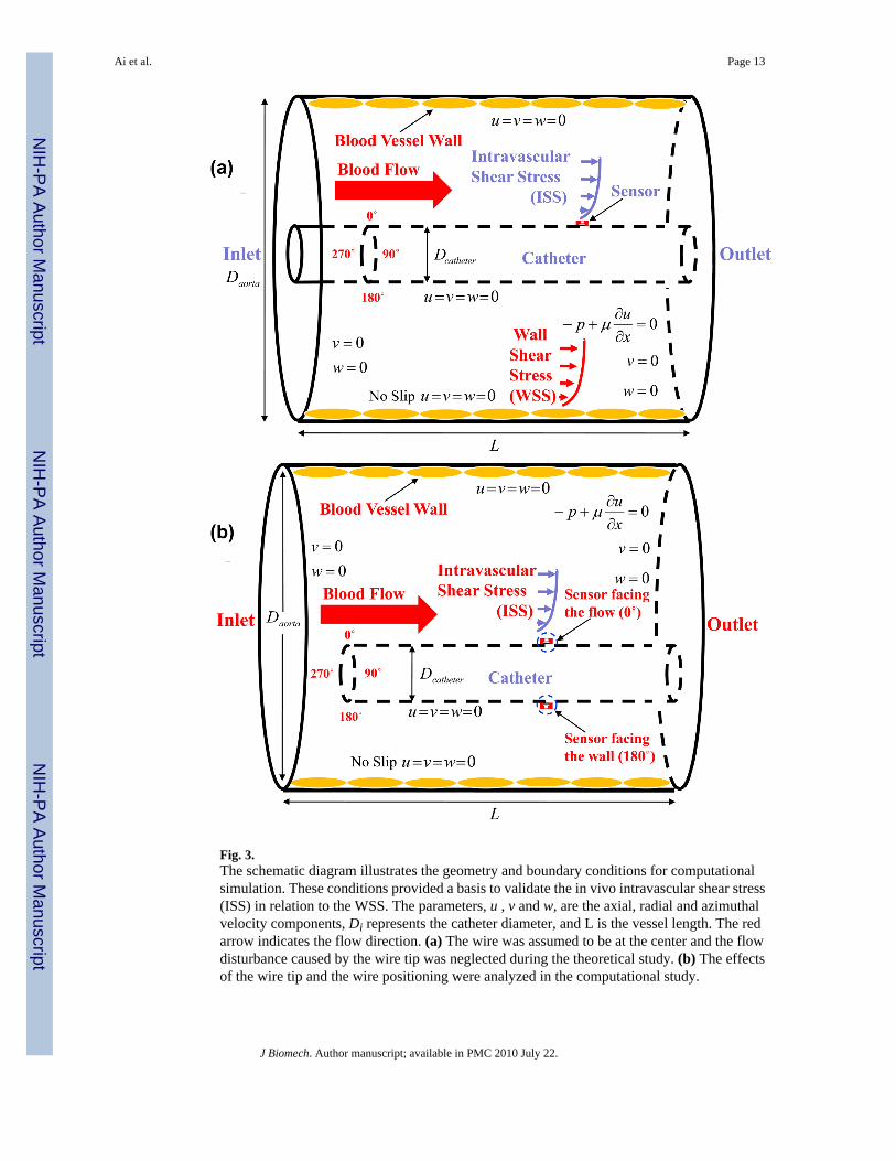
Fig. 3.The schematic diagram illustrates the geometry and boundary conditions for computationalsimulation. These conditions provided a basis to validate the in vivo intravascular shear stress(ISS) in relation to the WSS. The parameters, u , v and w, are the axial, radial and azimuthalvelocity components, Di represents the catheter diameter, and L is the vessel length. The redarrow indicates the flow direction. (a) The wire was assumed to be at the center and the flowdisturbance caused by the wire tip was neglected during the theoretical study. (b) The effectsof the wire tip and the wire positioning were analyzed in the computational study.
Ai et al. Page 13
J Biomech. Author manuscript; available in PMC 2010 July 22.
NIH
-PA Author Manuscript
NIH
-PA Author Manuscript
NIH
-PA Author Manuscript

Fig. 4.The pulsatile velocity waveform was derived from Doppler ultrasound measurement andreconstructed using Fourier analysis with 12 harmonics for computational simulation. (a) Thecenter-line velocity profile, including the peak velocity, was recorded. (b) The recordedvelocity profile was reconstructed with 12 harmonics by applying Fourier analysis.
Ai et al. Page 14
J Biomech. Author manuscript; available in PMC 2010 July 22.
NIH
-PA Author Manuscript
NIH
-PA Author Manuscript
NIH
-PA Author Manuscript

Fig. 5.Variations in elevation factors as a function of the diameter ratio, n. (a) Pressure elevationfactor, Ep , as a function of n. (b) WSS elevation factor, Ewss , as a function of n. (c) ISSelevation factor, Eiss , as a function of n.
Ai et al. Page 15
J Biomech. Author manuscript; available in PMC 2010 July 22.
NIH
-PA Author Manuscript
NIH
-PA Author Manuscript
NIH
-PA Author Manuscript

Fig. 6.CFD simulation was performed at various flow rates to analyze both velocity profiles and shearstress distributions on the coaxial wire. (a) The velocity profiles in response to three distinctflow rates (Re = 23, 91, and 133, respectively). (b) The shear stress distributions on the coaxialwire and catheter in response to the three flow rates in figure 6a.
Ai et al. Page 16
J Biomech. Author manuscript; available in PMC 2010 July 22.
NIH
-PA Author Manuscript
NIH
-PA Author Manuscript
NIH
-PA Author Manuscript

Fig. 7.The experimental ISS as detected by the MEMS sensor was compared with the computationalsolutions. (a) The relationship between the voltage changes and flow rates. (b) The convertedshear stress using our previously reported calibration curve was compared with thecomputational solutions.
Ai et al. Page 17
J Biomech. Author manuscript; available in PMC 2010 July 22.
NIH
-PA Author Manuscript
NIH
-PA Author Manuscript
NIH
-PA Author Manuscript

Fig. 8.The normalized entrance length, e*, and its corresponding normalized Reynolds number, Re*,were determined based on the CFD results.
Ai et al. Page 18
J Biomech. Author manuscript; available in PMC 2010 July 22.
NIH
-PA Author Manuscript
NIH
-PA Author Manuscript
NIH
-PA Author Manuscript

Fig. 9.Measured shear stress tracing in one cardiac cycle was compared with CFD solutions. (a)Superimposed on the real-time ISS profile (red dashed line) are the CFD solutions of WSSbefore (blue) and after (magenta) the coaxial wire was positioned at the center. The waveformof the ISS measurement overlapped with those of the computed ones despite the differencesin magnitude. (b) Comparison between the real-time ISS profile (red dashed line) and computedISS values on the coaxial wire positioned at the center.
Ai et al. Page 19
J Biomech. Author manuscript; available in PMC 2010 July 22.
NIH
-PA Author Manuscript
NIH
-PA Author Manuscript
NIH
-PA Author Manuscript