Intravascular cell delivery device for therapeutic VEGF-induced angiogenesis in chronic vascular...
11
Intravascular cell delivery device for therapeutic VEGF-induced angiogenesis in chronic vascular occlusion Arun H.S. Kumar a, 1 , Kenneth Martin a, 1 , Brendan Doyle b , Chien-Ling Huang a , Gopala-Krishnan M. Pillai a , Mohammed T. Ali a , Kimberly A. Skelding b , Shaohua Wang b , Birgitta M. Gleeson a , Saleem Jahangeer c , Erik L. Ritman d , Stephen J. Russell e , Noel M. Caplice a, b, * a Centre for Research in Vascular Biology (CRVB), Biosciences Institute, University College Cork, Cork, Ireland b Division of Cardiovascular Diseases, Molecular Medicine Program, Mayo Clinic, Rochester, MN, USA c Cork Cancer Research Centre, Biosciences Institute, University College Cork, Cork, Ireland d Department of Physiology and Biomedical Engineering, Mayo Clinic, Rochester, MN, USA e Division of Hematology, Molecular Medicine Program, Mayo Clinic, Rochester, MN, USA article info Article history: Received 16 May 2014 Accepted 10 July 2014 Available online 2 August 2014 Keywords: Angiogenesis Gene therapy Intravascular stent Smooth muscle cells Blood flow abstract Site specific targeting remains elusive for gene and stem cell therapies in the cardiovascular field. One promising option involves use of devices that deliver larger and more sustained cell/gene payloads to specific disease sites using the versatility of percutaneous vascular access technology. Smooth muscle cells (SMCs) engineered to deliver high local concentrations of an angiogenic molecule (VEGF) were placed in an intravascular cell delivery device (ICDD) in a porcine model of chronic total occlusion (CTO) involving ameroid placement on the proximal left circumflex (LCx) artery. Implanted SMC were retained within the ICDD and were competent for VEGF production in vitro and in vivo. Following implantation, micro-CT analyses revealed that ICDD-VEGF significantly enhanced vasa vasora microvessel density with a concomitant increase in tissue VEGF protein levels and formation of endothelial cell colonies sug- gesting increased angiogenic potential. ICDD-VEGF markedly enhanced regional blood flow determined by microsphere and contrast CT analysis translating to a functional improvement in regional wall motion and global left ventricular (LV) systolic and diastolic function. Our data indicate robust, clinically relevant angiogenesis can be achieved in a human scale porcine chronic vascular occlusion model following ICDD- VEGF-based delivery of angiogenic cells. This may have implications for percutaneous delivery of numerous therapeutic factors promoting creation of microvascular bypass networks in chronic vaso- occlusive diseases. © 2014 The Authors. Published by Elsevier Ltd. This is an open access article under the CC BY license (http://creativecommons.org/licenses/by/3.0/). 1. Introduction Chronic vaso-occlusive disease remains the Achilles heel of cardiovascular medicine and surgery [1]. Percutaneous revascu- larization of chronic occlusion is hampered by the risk of arterial perforation, reduced primary success and high restenosis rates [2]. Moreover, conventional surgical strategies aimed at bypass- ing these occlusive lesions are frequently not feasible because of the poor condition of distal blood vessels [3] or co-morbidities in the target population [4]. The need for alternative revasculari- zation strategies is great and for more than two decades, gene and cell therapy have offered tantalizing prospects for alleviation of such no-option occlusive vasculopathies. However, despite initial promise, significant barriers have emerged to both vector delivery and exogenous cell retention at the site of vascular disease [5]. * Corresponding author. Rm. 2.40, CRVB, Biosciences Institute, University College Cork, Cork, Ireland. Tel.: þ353 21 490 1329. E-mail address: [email protected] (N.M. Caplice). 1 These are co-first authors of the manuscript. Contents lists available at ScienceDirect Biomaterials journal homepage: www.elsevier.com/locate/biomaterials http://dx.doi.org/10.1016/j.biomaterials.2014.07.016 0142-9612/© 2014 The Authors. Published by Elsevier Ltd. This is an open access article under the CC BY license (http://creativecommons.org/licenses/by/3.0/). Biomaterials 35 (2014) 9012e9022
-
Upload
independent -
Category
Documents
-
view
1 -
download
0
Transcript of Intravascular cell delivery device for therapeutic VEGF-induced angiogenesis in chronic vascular...
lable at ScienceDirect
Biomaterials 35 (2014) 9012e9022
Contents lists avai
Biomaterials
journal homepage: www.elsevier .com/locate/biomateria ls
Intravascular cell delivery device for therapeutic VEGF-inducedangiogenesis in chronic vascular occlusion
Arun H.S. Kumar a, 1, Kenneth Martin a, 1, Brendan Doyle b, Chien-Ling Huang a,Gopala-Krishnan M. Pillai a, Mohammed T. Ali a, Kimberly A. Skelding b, Shaohua Wang b,Birgitta M. Gleeson a, Saleem Jahangeer c, Erik L. Ritman d, Stephen J. Russell e,Noel M. Caplice a, b, *
a Centre for Research in Vascular Biology (CRVB), Biosciences Institute, University College Cork, Cork, Irelandb Division of Cardiovascular Diseases, Molecular Medicine Program, Mayo Clinic, Rochester, MN, USAc Cork Cancer Research Centre, Biosciences Institute, University College Cork, Cork, Irelandd Department of Physiology and Biomedical Engineering, Mayo Clinic, Rochester, MN, USAe Division of Hematology, Molecular Medicine Program, Mayo Clinic, Rochester, MN, USA
a r t i c l e i n f o
Article history:Received 16 May 2014Accepted 10 July 2014Available online 2 August 2014
Keywords:AngiogenesisGene therapyIntravascular stentSmooth muscle cellsBlood flow
* Corresponding author. Rm. 2.40, CRVB, BioscienceCork, Cork, Ireland. Tel.: þ353 21 490 1329.
E-mail address: [email protected] (N.M. Caplice).1 These are co-first authors of the manuscript.
http://dx.doi.org/10.1016/j.biomaterials.2014.07.0160142-9612/© 2014 The Authors. Published by Elsevie
a b s t r a c t
Site specific targeting remains elusive for gene and stem cell therapies in the cardiovascular field. Onepromising option involves use of devices that deliver larger and more sustained cell/gene payloads tospecific disease sites using the versatility of percutaneous vascular access technology. Smooth musclecells (SMCs) engineered to deliver high local concentrations of an angiogenic molecule (VEGF) wereplaced in an intravascular cell delivery device (ICDD) in a porcine model of chronic total occlusion (CTO)involving ameroid placement on the proximal left circumflex (LCx) artery. Implanted SMC were retainedwithin the ICDD and were competent for VEGF production in vitro and in vivo. Following implantation,micro-CT analyses revealed that ICDD-VEGF significantly enhanced vasa vasora microvessel density witha concomitant increase in tissue VEGF protein levels and formation of endothelial cell colonies sug-gesting increased angiogenic potential. ICDD-VEGF markedly enhanced regional blood flow determinedby microsphere and contrast CT analysis translating to a functional improvement in regional wall motionand global left ventricular (LV) systolic and diastolic function. Our data indicate robust, clinically relevantangiogenesis can be achieved in a human scale porcine chronic vascular occlusion model following ICDD-VEGF-based delivery of angiogenic cells. This may have implications for percutaneous delivery ofnumerous therapeutic factors promoting creation of microvascular bypass networks in chronic vaso-occlusive diseases.© 2014 The Authors. Published by Elsevier Ltd. This is an open access article under the CC BY license
(http://creativecommons.org/licenses/by/3.0/).
1. Introduction
Chronic vaso-occlusive disease remains the Achilles heel ofcardiovascular medicine and surgery [1]. Percutaneous revascu-larization of chronic occlusion is hampered by the risk of arterial
s Institute, University College
r Ltd. This is an open access article
perforation, reduced primary success and high restenosis rates[2]. Moreover, conventional surgical strategies aimed at bypass-ing these occlusive lesions are frequently not feasible because ofthe poor condition of distal blood vessels [3] or co-morbidities inthe target population [4]. The need for alternative revasculari-zation strategies is great and for more than two decades, geneand cell therapy have offered tantalizing prospects for alleviationof such no-option occlusive vasculopathies. However, despiteinitial promise, significant barriers have emerged to both vectordelivery and exogenous cell retention at the site of vasculardisease [5].
under the CC BY license (http://creativecommons.org/licenses/by/3.0/).

A.H.S. Kumar et al. / Biomaterials 35 (2014) 9012e9022 9013
Previous animal studies employing local VEGF-based genetherapy strategies have been limited by the amount and durationof gene delivery that can be achieved in the vessel wall [6] aswell as safety concerns including angioma formation [7]. Toachieve sustained delivery at a safe local dose, we adopted astrategy of placing therapeutic cells, engineered with an angio-genic gene, into a percutaneously deliverable intravascular celldelivery device (ICDD) incorporating a non-woven matrix thatallowed a 3D cell mass to be stably conditioned in culture (Fig. 1).The approach of utilizing an expanded autologous cell source asan angiogenic gene/protein delivery platform is considered toobviate some of the reliability, dosing and technical challengesassociated with direct in vivo viral vector administration [8]. We
Fig. 1. Overview of ICDD strategy and chronic total occlusion (CTO) study protocol. Aaccomodate adherent Dil-labeled SMC. B. CTO was induced by ameroid constrictor placemtransduced with a retroviral VEGF expression vector and loaded onto an ICDD prior to deploanalyses were performed at the time of ICDD-VEGF placement and one month later (sacrifi
hypothesized that such an ICDD would elicit a more robust,stable and reliable local biologic response than cells or genesalone and that this would facilitate targeted, sustained andtherapeutic angiogenesis in the myocardium supplied by theocclusive artery.
We selected autologous SMCs as a conduit for VEGF delivery andexpression for several reasons: 1) we hypothesized that these cellswould be amore stable source for gene transfer than vector alone 2)in previous experiments, we had shown that SMC could be deliv-ered on a stent-based platform in 100-fold greater numbers thanendothelial cells [9]. 3) SMC grow in multilayers within the meshallowing development of a stable organoid that does not embolize4) autologous SMC are also a clinically translatable cell as they can
. SMC within ICDD Inset showing non-woven mesh topography with the ability toent on the proximal LCx. SMCs simultaneously harvested from the jugular vein wereyment four weeks after ameroid placement. CT, microsphere and conductance catheterce) along with further tissue analyses.
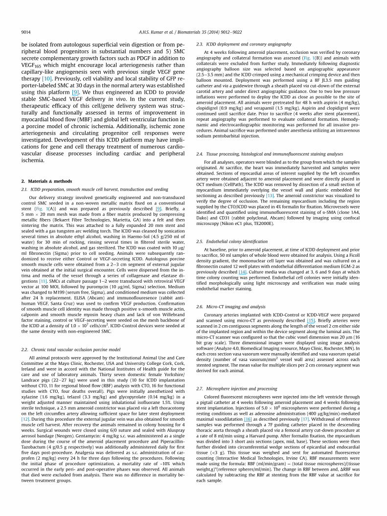
A.H.S. Kumar et al. / Biomaterials 35 (2014) 9012e90229014
be isolated from autologous superficial vein digestion or from pe-ripheral blood progenitors in substantial numbers and 5) SMCsecrete complementary growth factors such as PDGF in addition toVEGF165 which might encourage local arteriogenesis rather thancapillary-like angiogenesis seen with previous single VEGF genetherapy [10]. Previously, cell viability and local stability of GFP re-porter-labeled SMC at 30 days in the normal artery was establishedusing this platform [9]. We thus engineered an ICDD to providestable SMC-based VEGF delivery in vivo. In the current study,therapeutic efficacy of this cell/gene delivery system was struc-turally and functionally assessed in terms of improvement inmyocardial blood flow (MBF) and global left ventricular function ina porcine model of chronic ischemia. Additionally, ischemic zonearteriogenesis and circulating progenitor cell responses wereinvestigated. Development of this ICDD platform may have impli-cations for gene and cell therapy treatment of numerous cardio-vascular disease processes including cardiac and peripheralischemia.
2. Materials & methods
2.1. ICDD preparation, smooth muscle cell harvest, transduction and seeding
Our delivery strategy involved genetically engineered and non-transducedcontrol SMC seeded in a non-woven metallic matrix fixed on a conventionalstent (Fig. 1(A)) and was prepared as previously described [9]. Briefly, a5 mm � 20 mm mesh was made from a fiber matrix produced by compressingmetallic fibers (Bekaert Fibre Technologies, Marietta, GA) into a felt and thensintering the matrix. This was attached to a fully expanded 20 mm stent andsealed with a gas tungsten arc welding torch. The ICDD was cleaned by sonicationseveral times in absolute ethyl alcohol, washing in Haemo-Sol (4.5 g/250 ml ofwater) for 30 min of rocking, rinsing several times in filtered sterile water,washing in absolute alcohol, and gas sterilized. The ICDD was coated with 10 mg/ml fibronectin (Sigma) prior to cell seeding. Animals were subsequently ran-domized to receive either Control or VEGF-secreting ICDD. Autologous porcinesmooth muscle cells were obtained from a 2e3 cm segment of external jugularvein obtained at the initial surgical encounter. Cells were dispersed from the in-tima and media of the vessel through a series of collagenase and elastase di-gestions [11]. SMCs at culture passage 1e2 were transduced with retroviral VEGFvector at 100 MOI, followed by puromycin (10 mg/ml, Sigma) selection. Mediumwas changed to M199 (serum free, Sigma), and conditioned medium was collectedafter 24 h replacement. ELISA (Abcam) and immunoflouresence (rabbit anti-human VEGF, Santa Cruz) was used to confirm VEGF production. Confirmationof smooth muscle cell identity was made through positive a-smooth muscle actin,calponin and smooth muscle myosin heavy chain and lack of von Willebrandfactor staining, control or VEGF-secreting were seeded on the mesh backbone ofthe ICDD at a density of 1.0 � 107 cells/cm2. ICDD-Control devices were seeded atthe same density with non-engineered SMC.
2.2. Chronic total vascular occlusion porcine model
All animal protocols were approved by the Institutional Animal Use and CareCommittee at the Mayo Clinic, Rochester, USA and University College Cork, Cork,Ireland and were in accord with the National Institutes of Health guide for thecare and use of laboratory animals. Thirty seven domestic female Yorkshire/Landrace pigs (22e27 kg) were used in this study (10 for ICDD implantationwithout CTO, 11 for regional blood flow (RBF) analysis with CTO, 16 for functionalstudies with CTO, four deaths overall). Pigs were initially anesthetized withxylazine (1.6 mg/kg), telazol (3.3 mg/kg) and glycopyrolate (0.14 mg/kg) in aweight adjusted manner maintained using inhalational isofluorane 1.5%. Usingsterile technique, a 2.5 mm ameroid constrictor was placed via a left thoracotomyon the left circumflex artery allowing sufficient space for later stent deployment[12]. During this procedure the external jugular vein was also obtained for smoothmuscle cell harvest. After recovery the animals remained in colony housing for 4weeks. Surgical wounds were closed using 6/0 suture and sealed with Alusprayaerosol bandage (Neogen). Gentamycin: 4 mg/kg s.c. was administered as a singledose during the course of the ameroid placement procedure and Piperacillin-Tazobactum (4 g/0.5 g respectively) was additionally administered daily for firstfive days post-procedure. Analgesia was delivered as s.c. administration of car-profen (2 mg/kg) every 24 h for three days following the procedures. Followingthe initial phase of procedure optimization, a mortality rate of ~10% whichoccurred in the early peri- and post-operative phases was observed. All animalsthat died were excluded from analysis. There was no difference in mortality be-tween treatment groups.
2.3. ICDD deployment and coronary angiography
At 4 weeks following ameroid placement, occlusion was verified by coronaryangiography and collateral formation was assessed (Fig. 1(B)) and animals withcollaterals were excluded from further study. Immediately following diagnosticangiography balloon size was selected based on angiographic appearance(2.5e3.5 mm) and the ICDD crimped using a mechanical crimping device and thenballoon mounted. Deployment was performed using a 8F JL3.5 mm guidingcatheter and via a guidewire through a sheath placed via cut-down of the externalcarotid artery and under direct angiographic guidance. One to two low pressureinflations were performed to deploy the ICDD as close as possible to the site ofameroid placement. All animals were pretreated for 48 h with aspirin (4 mg/kg),clopidigrel (0.9 mg/kg) and verapamil (1.5 mg/kg). Aspirin and clopidigrel werecontinued until sacrifice date. Prior to sacrifice (4 weeks after stent placement),repeat angiography was performed to evaluate collateral formation. Hemody-namic and electrocardiographic monitoring was performed for all invasive pro-cedures. Animal sacrifice was performed under anesthesia utilizing an intravenoussodium pentobarbital injection.
2.4. Tissue processing, histological and immunofluorescent staining analyses
For all analyses, operators were blinded as to the group fromwhich the samplesoriginated. At sacrifice, the heart was immediately harvested and samples wereobtained. Sections of myocardial areas of interest supplied by the left circumflexartery were obtained adjacent to ameroid placement and were directly placed inOCT medium (CellPath). The ICDD was removed by dissection of a small section ofmyocardium immediately overlying the vessel wall and plastic embedded forsectioning as described previously [13]. The ameroid constrictor was visualized toverify the degree of occlusion. The remaining myocardium including the regionsupplied by the CTO/ICDD was placed in 4% formalin for fixation. Microvessels wereidentified and quantified using immunofluorescent staining of a-SMA (clone 1A4,Dako) and CD31 (rabbit polyclonal, Abcam) followed by imaging using confocalmicroscopy (Nikon eC1 plus, TE2000E).
2.5. Endothelial colony identification
At baseline, prior to ameroid placement, at time of ICDD deployment and priorto sacrifice, 50 ml samples of whole blood were obtained for analysis. Using a Ficolldensity gradient, the mononuclear cell layer was obtained and was cultured on afibronectin coated 12 well plates with endothelial differentiation medium EGM-2 aspreviously described [14]. Culture media was changed at 3, 6 and 9 days at whichtime colony counting was performed. Endothelial cell colonies were initially iden-tified morphologically using light microscopy and verification was made usingendothelial marker staining.
2.6. Micro-CT imaging and analysis
Coronary arteries implanted with ICDD-Control or ICDD-VEGF were preparedand scanned using micro-CT as previously described [15]. Briefly arteries werescanned in 2 cm contiguous segments along the length of the vessel 2 cm either sideof the implanted region and within the device segment along the luminal axis. Themicro-CT scanner was configured so that the cubic voxel dimension was 20 mm (16bit gray scale). Three dimensional images were displayed using image analysissoftware (Analyze 4.0, Biomedical Imaging resource, Mayo Clinic, Rochester, MN). Ineach cross section vasa vasorumwere manually identified and vasa vasorum spatialdensity (number of vasa vasorum/mm2 vessel wall area) assessed across eachstented segment. The mean value for multiple slices per 2 cm coronary segment wasderived for each animal.
2.7. Microsphere injection and processing
Colored fluorescent microspheres were injected into the left ventricle througha pigtail catheter at 4 weeks following ameroid placement and 4 weeks followingstent implantation. Injections of 5.0 � 106 microspheres were performed during aresting conditions as well as adenosine administration (400 mg/kg/min)-mediatedmaximal vasodilatation [16] as described previously [17]. Withdrawal of referencesamples was performed through a 7F guiding catheter placed in the descendingthoracic aorta through a sheath placed via a femoral artery cut-down procedure ata rate of 8 ml/min using a Harvard pump. After formalin fixation, the myocardiumwas divided into 3 short axis sections (apex, mid, base). These sections were thenfurther divided into circumferential wedge sections of epicardial and endocardialtissue (<3 g). This tissue was weighed and sent for automated fluorescencecounting (Interactive Medical Technologies, Irvine CA). RBF measurements weremade using the formula: RBF (ml/min/gram) ¼ (total tissue microspheres)/(tissueweight,g)*(reference spheres/ml/min). The change in RBF between and, DRBF wascalculated by subtracting the RBF at stenting from the RBF value at sacrifice foreach sample.
A.H.S. Kumar et al. / Biomaterials 35 (2014) 9012e9022 9015
2.8. Tube formation and SMC-EC association assays
Tube formation for in vitro angiogenesis analysis was performed as previouslydescribed [18]. Briefly, a 96-well plate was coated with 50 ml of Matrigel (BectonDickinson Labware, Bedford, MA), which was allowed to solidify at 37 �C for 1 h.Human endothelial cells (1.5 � 104 cells per well) labeled with CellTracker Green(Molecular Probes) were treated with conditioned media from control or VEGF-secreting SMC, co-seeded with smooth muscle cells (1.5 � 103 cells per well)labeled with CellTracker Orange on the Matrigel and cultured in M199 conditionedmedium, respectively. The cells were incubated for 20 h, and complete tubes fromrandomly chosen fields were captured using Nikon EZC1-3.30 software on aconfocal microscope system (Nikon eC1 plus, TE2000E). Tube length, network areaand SMC-EC coverage analysis were performed using ImageJ software (NIH,Bethesda, MD).
2.9. Sixty-four-slice CT imaging, image reconstruction and data analysis
Cardiac CT imaging was performed using a 64-slice scanner (GE Discovery VCTRX) with Iodixanol (Visipaque 320; Amersham Health) contrast agent. All gated CTimages were reconstructed at a 1.25 mm slice thickness, and phase data on allaxial slices were reconstructed from 0% to 99% of the cardiac cycle in 9% in-crements for assessment of LV function parameters and ejection fraction (EF), ascalculated on an online workstation (AW 4.4; GE Healthcare). Sixty-four-sliceimages were analyzed with CardIQ software (AW 4.4) to generate polar maps ofregional wall motion and quantified as mm2/g myocardial mass/cardiac cycle.Myocardial perfusion was evaluated by contrast intensity 5 s after bolus injectionas described previously [17]. Relative Hounsfield Unit (HU) measurement wascalculated as the mean HU in the LCx-related region compared to mean HU in theLV chamber and normalized to the initial scan at the time of ameroid constrictorplacement.
2.10. Statistical analyses
For in vivo experiments, significance was calculated using the Kruskal Wallistest followed by Dunn's Multiple Comparison test. For in vitro analyses, compar-isons between treatment groups were made using ANOVA and Mann Whitneytests.
3. Results
3.1. ICDD-VEGF and local angiogenic indices in vivo
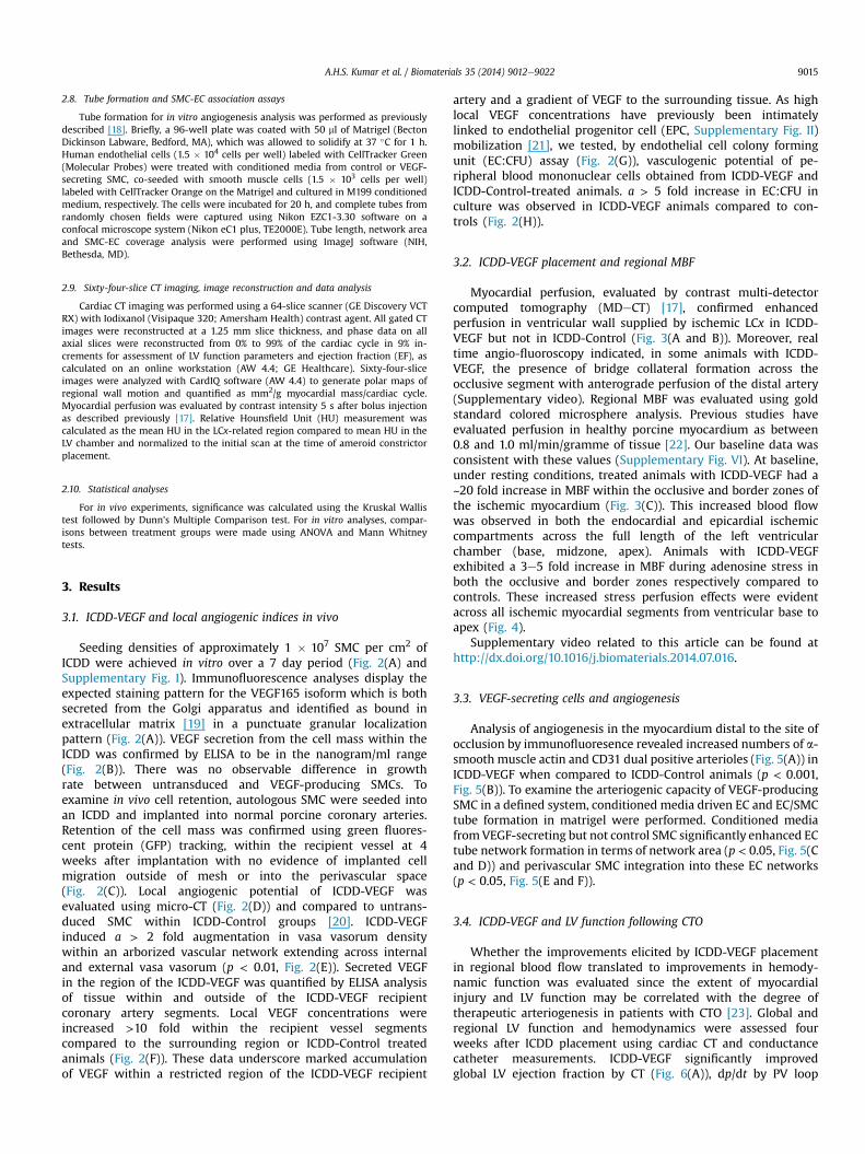
Seeding densities of approximately 1 � 107 SMC per cm2 ofICDD were achieved in vitro over a 7 day period (Fig. 2(A) andSupplementary Fig. I). Immunofluorescence analyses display theexpected staining pattern for the VEGF165 isoform which is bothsecreted from the Golgi apparatus and identified as bound inextracellular matrix [19] in a punctuate granular localizationpattern (Fig. 2(A)). VEGF secretion from the cell mass within theICDD was confirmed by ELISA to be in the nanogram/ml range(Fig. 2(B)). There was no observable difference in growthrate between untransduced and VEGF-producing SMCs. Toexamine in vivo cell retention, autologous SMC were seeded intoan ICDD and implanted into normal porcine coronary arteries.Retention of the cell mass was confirmed using green fluores-cent protein (GFP) tracking, within the recipient vessel at 4weeks after implantation with no evidence of implanted cellmigration outside of mesh or into the perivascular space(Fig. 2(C)). Local angiogenic potential of ICDD-VEGF wasevaluated using micro-CT (Fig. 2(D)) and compared to untrans-duced SMC within ICDD-Control groups [20]. ICDD-VEGFinduced a > 2 fold augmentation in vasa vasorum densitywithin an arborized vascular network extending across internaland external vasa vasorum (p < 0.01, Fig. 2(E)). Secreted VEGFin the region of the ICDD-VEGF was quantified by ELISA analysisof tissue within and outside of the ICDD-VEGF recipientcoronary artery segments. Local VEGF concentrations wereincreased >10 fold within the recipient vessel segmentscompared to the surrounding region or ICDD-Control treatedanimals (Fig. 2(F)). These data underscore marked accumulationof VEGF within a restricted region of the ICDD-VEGF recipient
artery and a gradient of VEGF to the surrounding tissue. As highlocal VEGF concentrations have previously been intimatelylinked to endothelial progenitor cell (EPC, Supplementary Fig. II)mobilization [21], we tested, by endothelial cell colony formingunit (EC:CFU) assay (Fig. 2(G)), vasculogenic potential of pe-ripheral blood mononuclear cells obtained from ICDD-VEGF andICDD-Control-treated animals. a > 5 fold increase in EC:CFU inculture was observed in ICDD-VEGF animals compared to con-trols (Fig. 2(H)).
3.2. ICDD-VEGF placement and regional MBF
Myocardial perfusion, evaluated by contrast multi-detectorcomputed tomography (MDeCT) [17], confirmed enhancedperfusion in ventricular wall supplied by ischemic LCx in ICDD-VEGF but not in ICDD-Control (Fig. 3(A and B)). Moreover, realtime angio-fluoroscopy indicated, in some animals with ICDD-VEGF, the presence of bridge collateral formation across theocclusive segment with anterograde perfusion of the distal artery(Supplementary video). Regional MBF was evaluated using goldstandard colored microsphere analysis. Previous studies haveevaluated perfusion in healthy porcine myocardium as between0.8 and 1.0 ml/min/gramme of tissue [22]. Our baseline data wasconsistent with these values (Supplementary Fig. VI). At baseline,under resting conditions, treated animals with ICDD-VEGF had a~20 fold increase in MBF within the occlusive and border zones ofthe ischemic myocardium (Fig. 3(C)). This increased blood flowwas observed in both the endocardial and epicardial ischemiccompartments across the full length of the left ventricularchamber (base, midzone, apex). Animals with ICDD-VEGFexhibited a 3e5 fold increase in MBF during adenosine stress inboth the occlusive and border zones respectively compared tocontrols. These increased stress perfusion effects were evidentacross all ischemic myocardial segments from ventricular base toapex (Fig. 4).
Supplementary video related to this article can be found athttp://dx.doi.org/10.1016/j.biomaterials.2014.07.016.
3.3. VEGF-secreting cells and angiogenesis
Analysis of angiogenesis in the myocardium distal to the site ofocclusion by immunofluoresence revealed increased numbers of a-smooth muscle actin and CD31 dual positive arterioles (Fig. 5(A)) inICDD-VEGF when compared to ICDD-Control animals (p < 0.001,Fig. 5(B)). To examine the arteriogenic capacity of VEGF-producingSMC in a defined system, conditioned media driven EC and EC/SMCtube formation in matrigel were performed. Conditioned mediafromVEGF-secreting but not control SMC significantly enhanced ECtube network formation in terms of network area (p < 0.05, Fig. 5(Cand D)) and perivascular SMC integration into these EC networks(p < 0.05, Fig. 5(E and F)).
3.4. ICDD-VEGF and LV function following CTO
Whether the improvements elicited by ICDD-VEGF placementin regional blood flow translated to improvements in hemody-namic function was evaluated since the extent of myocardialinjury and LV function may be correlated with the degree oftherapeutic arteriogenesis in patients with CTO [23]. Global andregional LV function and hemodynamics were assessed fourweeks after ICDD placement using cardiac CT and conductancecatheter measurements. ICDD-VEGF significantly improvedglobal LV ejection fraction by CT (Fig. 6(A)), dp/dt by PV loop
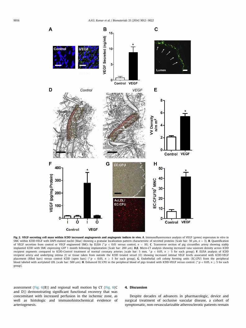
Fig. 2. VEGF-secreting cell mass within ICDD increased angiogenesis and angiogenic indices in vivo. A. Immunofluorescence analysis of VEGF (green) expression in vitro inSMC within ICDD-VEGF with DAPI-stained nuclei (blue) showing a granular localization pattern characteristic of secreted proteins (Scale bar: 50 mm, n ¼ 3). B. Quantificationof VEGF secretion from control or VEGF engineered SMCs by ELISA (*:p < 0.01 versus control, n ¼ 10). C. Transverse section of pig circumflex artery showing stablyimplanted ICDD with SMC expressing GFP 1 month following implantation (Scale bar: 200 mm). D,E. Micro-CT analysis showing increased vasa vasorum density across ICDDrecipient segments compared to ICDD-Control treatment of normal coronary arteries (scale bar: 5 mm, *:p < 0.05, n � 5 for each group). F. ELISA analysis of ICDDrecipient artery and underlying intima (I) or tissue taken from outside the ICDD treated vessel (O) showing increased intimal VEGF levels associated with ICDD-VEGFplacement (filled bars) versus control ICDD (open bars) (*:p < 0.05, n � 5 for each group). G. Endothelial cell colony forming units (EC:CFU) from the peripheralblood labeled with acetylated LDL (scale bar: 500 mm). H. Enhanced EC:CFU in the peripheral blood of pigs treated with ICDD-VEGF versus control. (*:p < 0.05, n � 5 for eachgroup).
A.H.S. Kumar et al. / Biomaterials 35 (2014) 9012e90229016
assessment (Fig. 6(B)) and regional wall motion by CT (Fig. 6(Cand D)) demonstrating significant functional recovery that wasconcomitant with increased perfusion in the ischemic zone, aswell as histologic and immunohistochemical evidence ofarteriogenesis.
4. Discussion
Despite decades of advances in pharmacologic, device andsurgical treatment of occlusive vascular disease, a cohort ofsymptomatic, non-revascularizable atherosclerotic patients remain
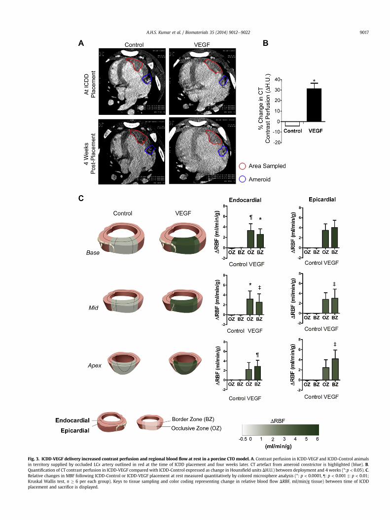
Fig. 3. ICDD-VEGF delivery increased contrast perfusion and regional blood flow at rest in a porcine CTO model. A. Contrast perfusion in ICDD-VEGF and ICDD-Control animalsin territory supplied by occluded LCx artery outlined in red at the time of ICDD placement and four weeks later. CT artefact from ameroid constrictor is highlighted (blue). B.Quantification of CT contrast perfusion in ICDD-VEGF compared with ICDD-Control expressed as change in Hounsfield units DH.U.) between deployment and 4 weeks (*:p < 0.05). C.Relative changes in MBF following ICDD-Control or ICDD-VEGF placement at rest measured quantitatively by colored microsphere analysis (*: p < 0.0001, ¶: p < 0.001 z: p < 0.01;Kruskal Wallis test, n � 6 per each group). Keys to tissue sampling and color coding representing change in relative blood flow DRBF, ml/min/g tissue) between time of ICDDplacement and sacrifice is displayed.
A.H.S. Kumar et al. / Biomaterials 35 (2014) 9012e9022 9017
Fig. 4. ICDD-VEGF increased regional blood flow during adenosine cardiac stress in porcine CTO model. Relative changes in MBF four weeks following ICDD-Control or ICDD-VEGF placement during adenosine-induced stress measured quantitatively by colored microsphere analysis (*: p < 0.0001, ¶: p < 0.001 z: p < 0.01; Kruskal Wallis test, n ¼ 10). Keysto tissue sampling and color coding representing change in relative blood flow DRBF, ml/min/g tissue) between time of stent placement and sacrifice is displayed.
A.H.S. Kumar et al. / Biomaterials 35 (2014) 9012e90229018
[24]. These patients with chronic vascular occlusion are frequentlyunsuitable for percutaneous coronary intervention (PCI) due toprocedural risk and high restenosis rates, and may not be idealcandidates for surgical revascularization due to poor distal vesseltargets, unfavorable co-morbidities or lack of suitable bypassconduits [5]. Gene and more recently cell therapy have beentouted as a therapeutic answer for these no-option patients [25]but these approaches still lack sufficient site specific targeting,and are hampered by dissipation of therapeutic effect throughdilution and dispersion of gene/cell product within the widervascular system [26].
The aim of this study was to synergize cell and gene therapybenefits using a tissue engineered ICDD to enhance site specificangiogenesis in a model of chronic myocardial ischemia. Weposited that an ICDD secreting VEGF transgene product could bedelivered safely to a site of chronic vascular occlusion and wouldpromote increased myocardial blood flow and improved cardiacfunction in the ischemic zone of a porcine ameroid constrictor CTO
model. In the current study, we demonstrate that ICDD-VEGFpromotes significant increases in regional ischemic zone MBF bygold standard color microsphere detection, regional cardiac func-tion by CT contrast imaging and regional arteriogenesis by immu-nohistochemical microvessel analysis. Moreover, we show thatincreased blood flow is likely driven by both vasculogenic andarteriogenic processes incorporating endothelial and SMC-pericyteinvestment of nascent microvessels. Finally, the functional conse-quences of this augmented angiogenesis includes a > 50 fold in-crease in ischemic zone MBF in VEGF compared to control-treatment under both rest and adenosine-induced stress condi-tions, with attendant improvement in regional wall motion andglobal LV functional parameters.
VEGF has been widely used in the past for stand-alone genetherapy but limitations were observed in terms of plasmid vectordispersion and lack of site specificity when delivered via theintravascular delivery route, ineffective therapeutic dosing andlack of transgene persistence in the case of adenoviral vectors
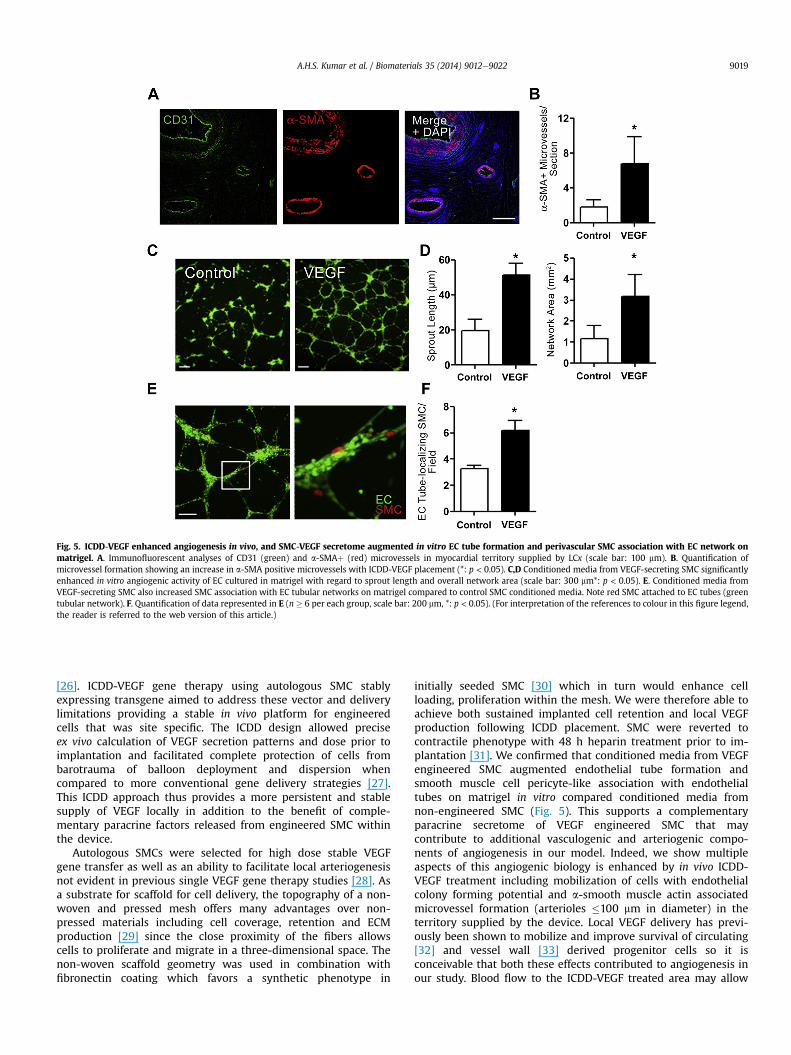
Fig. 5. ICDD-VEGF enhanced angiogenesis in vivo, and SMC-VEGF secretome augmented in vitro EC tube formation and perivascular SMC association with EC network onmatrigel. A. Immunofluorescent analyses of CD31 (green) and a-SMAþ (red) microvessels in myocardial territory supplied by LCx (scale bar: 100 mm). B. Quantification ofmicrovessel formation showing an increase in a-SMA positive microvessels with ICDD-VEGF placement (*: p < 0.05). C,D Conditioned media from VEGF-secreting SMC significantlyenhanced in vitro angiogenic activity of EC cultured in matrigel with regard to sprout length and overall network area (scale bar: 300 mm*: p < 0.05). E. Conditioned media fromVEGF-secreting SMC also increased SMC association with EC tubular networks on matrigel compared to control SMC conditioned media. Note red SMC attached to EC tubes (greentubular network). F. Quantification of data represented in E (n � 6 per each group, scale bar: 200 mm, *: p < 0.05). (For interpretation of the references to colour in this figure legend,the reader is referred to the web version of this article.)
A.H.S. Kumar et al. / Biomaterials 35 (2014) 9012e9022 9019
[26]. ICDD-VEGF gene therapy using autologous SMC stablyexpressing transgene aimed to address these vector and deliverylimitations providing a stable in vivo platform for engineeredcells that was site specific. The ICDD design allowed preciseex vivo calculation of VEGF secretion patterns and dose prior toimplantation and facilitated complete protection of cells frombarotrauma of balloon deployment and dispersion whencompared to more conventional gene delivery strategies [27].This ICDD approach thus provides a more persistent and stablesupply of VEGF locally in addition to the benefit of comple-mentary paracrine factors released from engineered SMC withinthe device.
Autologous SMCs were selected for high dose stable VEGFgene transfer as well as an ability to facilitate local arteriogenesisnot evident in previous single VEGF gene therapy studies [28]. Asa substrate for scaffold for cell delivery, the topography of a non-woven and pressed mesh offers many advantages over non-pressed materials including cell coverage, retention and ECMproduction [29] since the close proximity of the fibers allowscells to proliferate and migrate in a three-dimensional space. Thenon-woven scaffold geometry was used in combination withfibronectin coating which favors a synthetic phenotype in
initially seeded SMC [30] which in turn would enhance cellloading, proliferation within the mesh. We were therefore able toachieve both sustained implanted cell retention and local VEGFproduction following ICDD placement. SMC were reverted tocontractile phenotype with 48 h heparin treatment prior to im-plantation [31]. We confirmed that conditioned media from VEGFengineered SMC augmented endothelial tube formation andsmooth muscle cell pericyte-like association with endothelialtubes on matrigel in vitro compared conditioned media fromnon-engineered SMC (Fig. 5). This supports a complementaryparacrine secretome of VEGF engineered SMC that maycontribute to additional vasculogenic and arteriogenic compo-nents of angiogenesis in our model. Indeed, we show multipleaspects of this angiogenic biology is enhanced by in vivo ICDD-VEGF treatment including mobilization of cells with endothelialcolony forming potential and a-smooth muscle actin associatedmicrovessel formation (arterioles �100 mm in diameter) in theterritory supplied by the device. Local VEGF delivery has previ-ously been shown to mobilize and improve survival of circulating[32] and vessel wall [33] derived progenitor cells so it isconceivable that both these effects contributed to angiogenesis inour study. Blood flow to the ICDD-VEGF treated area may allow
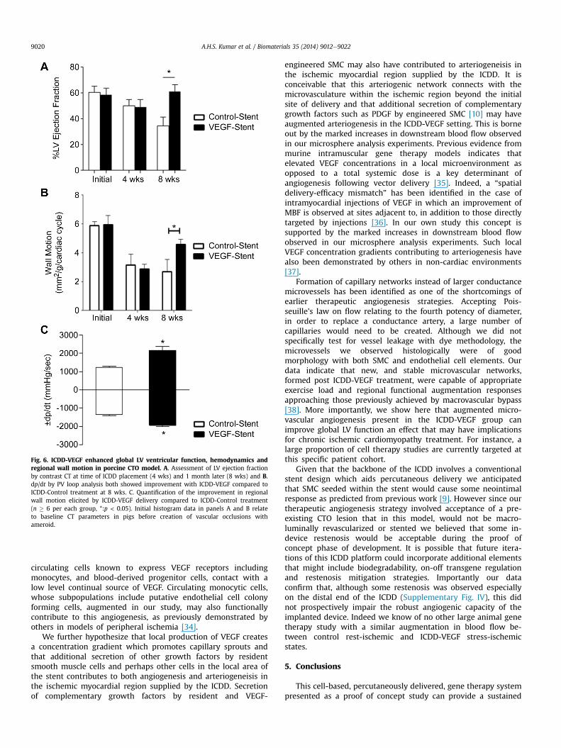
Fig. 6. ICDD-VEGF enhanced global LV ventricular function, hemodynamics andregional wall motion in porcine CTO model. A. Assessment of LV ejection fractionby contrast CT at time of ICDD placement (4 wks) and 1 month later (8 wks) and B.dp/dt by PV loop analysis both showed improvement with ICDD-VEGF compared toICDD-Control treatment at 8 wks. C. Quantification of the improvement in regionalwall motion elicited by ICDD-VEGF delivery compared to ICDD-Control treatment(n � 6 per each group, *:p < 0.05). Initial histogram data in panels A and B relateto baseline CT parameters in pigs before creation of vascular occlusions withameroid.
A.H.S. Kumar et al. / Biomaterials 35 (2014) 9012e90229020
circulating cells known to express VEGF receptors includingmonocytes, and blood-derived progenitor cells, contact with alow level continual source of VEGF. Circulating monocytic cells,whose subpopulations include putative endothelial cell colonyforming cells, augmented in our study, may also functionallycontribute to this angiogenesis, as previously demonstrated byothers in models of peripheral ischemia [34].
We further hypothesize that local production of VEGF createsa concentration gradient which promotes capillary sprouts andthat additional secretion of other growth factors by residentsmooth muscle cells and perhaps other cells in the local area ofthe stent contributes to both angiogenesis and arteriogeneisis inthe ischemic myocardial region supplied by the ICDD. Secretionof complementary growth factors by resident and VEGF-
engineered SMC may also have contributed to arteriogeneisis inthe ischemic myocardial region supplied by the ICDD. It isconceivable that this arteriogenic network connects with themicrovasculature within the ischemic region beyond the initialsite of delivery and that additional secretion of complementarygrowth factors such as PDGF by engineered SMC [10] may haveaugmented arteriogenesis in the ICDD-VEGF setting. This is borneout by the marked increases in downstream blood flow observedin our microsphere analysis experiments. Previous evidence frommurine intramuscular gene therapy models indicates thatelevated VEGF concentrations in a local microenvironment asopposed to a total systemic dose is a key determinant ofangiogenesis following vector delivery [35]. Indeed, a “spatialdelivery-efficacy mismatch” has been identified in the case ofintramyocardial injections of VEGF in which an improvement ofMBF is observed at sites adjacent to, in addition to those directlytargeted by injections [36]. In our own study this concept issupported by the marked increases in downstream blood flowobserved in our microsphere analysis experiments. Such localVEGF concentration gradients contributing to arteriogenesis havealso been demonstrated by others in non-cardiac environments[37].
Formation of capillary networks instead of larger conductancemicrovessels has been identified as one of the shortcomings ofearlier therapeutic angiogenesis strategies. Accepting Pois-seuille's law on flow relating to the fourth potency of diameter,in order to replace a conductance artery, a large number ofcapillaries would need to be created. Although we did notspecifically test for vessel leakage with dye methodology, themicrovessels we observed histologically were of goodmorphology with both SMC and endothelial cell elements. Ourdata indicate that new, and stable microvascular networks,formed post ICDD-VEGF treatment, were capable of appropriateexercise load and regional functional augmentation responsesapproaching those previously achieved by macrovascular bypass[38]. More importantly, we show here that augmented micro-vascular angiogenesis present in the ICDD-VEGF group canimprove global LV function an effect that may have implicationsfor chronic ischemic cardiomyopathy treatment. For instance, alarge proportion of cell therapy studies are currently targeted atthis specific patient cohort.
Given that the backbone of the ICDD involves a conventionalstent design which aids percutaneous delivery we anticipatedthat SMC seeded within the stent would cause some neointimalresponse as predicted from previous work [9]. However since ourtherapeutic angiogenesis strategy involved acceptance of a pre-existing CTO lesion that in this model, would not be macro-luminally revascularized or stented we believed that some in-device restenosis would be acceptable during the proof ofconcept phase of development. It is possible that future itera-tions of this ICDD platform could incorporate additional elementsthat might include biodegradability, on-off transgene regulationand restenosis mitigation strategies. Importantly our dataconfirm that, although some restenosis was observed especiallyon the distal end of the ICDD (Supplementary Fig. IV), this didnot prospectively impair the robust angiogenic capacity of theimplanted device. Indeed we know of no other large animal genetherapy study with a similar augmentation in blood flow be-tween control rest-ischemic and ICDD-VEGF stress-ischemicstates.
5. Conclusions
This cell-based, percutaneously delivered, gene therapy systempresented as a proof of concept study can provide a sustained
A.H.S. Kumar et al. / Biomaterials 35 (2014) 9012e9022 9021
therapeutic transgene product that significantly improves bloodflow to ischemic myocardium, translating into regional and globalleft ventricular functional recovery. This system may have impli-cations for non-revascularizable no-option patients or may beadapted to other vascular beds such as in the peripheralcirculation.
Funding sources
This workwas supported by grants from the National Institute ofHealth (NIH-HL66958) and Science Foundation Ireland (NMC e 07/RFP/BIMF816, 10/IN.1/B3034). This work was also supported by anequipment grant from the National Biophotonics Imaging Platformwithin the Programme for Research at Third Level Institutions Cycle(RCSI/NBIP/CEMP11).
Acknowledgments
We wish to thank Sanja Trinki for graphic design, Cindy Reed,and Jill Allen for technical assistance. The authors also wish tothank Mr Cormac O'Brien of the National Centre for Laser Appli-cations (NCLA) at the National University of Ireland, Galway forassistance in ICDD preparation.
Appendix A. Supplementary data
Supplementary data related to this article can be found at http://dx.doi.org/10.1016/j.biomaterials.2014.07.016.
References
[1] Simons M, Annex BH, Laham RJ, Kleiman N, Henry T, Dauerman H, et al.Pharmacological treatment of coronary artery disease with recombinantfibroblast growth factor-2: double-blind, randomized, controlled clinical trial.Circulation 2002;105:788e93.
[2] Prasad A, Rihal CS, Lennon RJ, Wiste HJ, Singh M, Holmes Jr DR. Trends inoutcomes after percutaneous coronary intervention for chronic total occlu-sions: a 25-year experience from the Mayo Clinic. J Am Coll Cardiol 2007;49:1611e8.
[3] Suero JA, Marso SP, Jones PG, Laster SB, Huber KC, Giorgi LV, et al. Pro-cedural outcomes and long-term survival among patients undergoingpercutaneous coronary intervention of a chronic total occlusion in nativecoronary arteries: a 20-year experience. J Am Coll Cardiol 2001;38:409e14.
[4] Roques F, Nashef SA, Michel P, Gauducheau E, de Vincentiis C, Baudet E, et al.Risk factors and outcome in European cardiac surgery: analysis of the Euro-SCORE multinational database of 19030 patients. Eur J Cardiothorac Surg1999;15:816e22. discussion 22e23.
[5] Caplice NM, Gersh BJ, Alegria JR. Cell therapy for cardiovascular disease: whatcells, what diseases and for whom? Nat Clin Pract Cardiovasc Med 2005;2:37e43.
[6] Klugherz BD, Meneveau NF, Kolansky DM, Herrmann HC, Schiele F,Matthai Jr WH, et al. Predictors of clinical outcome followingpercutaneous intervention for in-stent restenosis. Am J Cardiol 2000;85:1427e31.
[7] Carmeliet P. VEGF gene therapy: stimulating angiogenesis or angioma-gene-sis? Nat Med 2000;6:1102e3.
[8] Von Degenfeld G, Banfi A, Springer ML, Blau HM. Myoblast-mediated genetransfer for therapeutic angiogenesis and arteriogenesis. Br J Pharmacol2003;140:620e6.
[9] Panetta CJ, Miyauchi K, Berry D, Simari RD, Holmes DR, Schwartz RS, et al.A tissue-engineered stent for cell-based vascular gene transfer. Hum GeneTher 2002;13:433e41.
[10] Cao R, Brakenhielm E, Pawliuk R, Wariaro D, Post MJ, Wahlberg E, et al.Angiogenic synergism, vascular stability and improvement of hind-limbischemia by a combination of PDGF-BB and FGF-2. Nat Med 2003;9:604e13.
[11] Ross R. The smooth muscle cell. II. Growth of smooth muscle in culture andformation of elastic fibers. J Cell Biol 1971;50:172e86.
[12] Roth DM, White FC, Mathieu-Costello O, Guth BD, Heusch G, Bloor CM,et al. Effects of left circumflex ameroid constrictor placement on adrenergicinnervation of myocardium. Am J Physiol 1987;253:H1425e34.
[13] Kumar AH, McCauley SD, Hynes BG, O'Dea J, Caplice NM. Improved protocolfor processing stented porcine coronary arteries for immunostaining. J MolHistol 2011;42:187e93.
[14] Simper D, Stalboerger PG, Panetta CJ, Wang S, Caplice NM.Smooth muscle progenitor cells in human blood. Circulation 2002;106:1199e204.
[15] Gossl M, Rosol M, Malyar NM, Fitzpatrick LA, Beighley PE, Zamir M, et al.Functional anatomy and hemodynamic characteristics of vasa vasorum in thewalls of porcine coronary arteries. Anat Rec A Discov Mol Cell Evol Biol2003;272:526e37.
[16] Mannheim D, Versari D, Daghini E, Gossl M, Galili O, Chade A, et al. Impairedmyocardial perfusion reserve in experimental hypercholesterolemia is inde-pendent of myocardial neovascularization. Am J Physiol Heart Circ Physiol2007;292:H2449e58.
[17] O'Sullivan JF, Leblond AL, O'Dea J, Hristova I, Kumar S, Martin K, et al. Mul-tidetector computed tomography accurately defines infarct size, but notmicrovascular obstruction after myocardial infarction. J Am Coll Cardiol2013;61:208e10.
[18] Arnaoutova I, Kleinman HK. In vitro angiogenesis: endothelial cell tubeformation on gelled basement membrane extract. Nat Protoc 2010;5:628e35.
[19] Ferrara N. Binding to the extracellular matrix and proteolytic processing: twokey mechanisms regulating vascular endothelial growth factor action. MolBiol Cell 2010;21:687e90.
[20] Langheinrich AC, Michniewicz A, Sedding DG, Walker G, Beighley PE,Rau WS, et al. Correlation of vasa vasorum neovascularization andplaque progression in aortas of apolipoprotein E(-/-)/low-density lipo-protein(-/-) double knockout mice. Arterioscler Thromb Vasc Biol 2006;26:347e52.
[21] Kalka C, Masuda H, Takahashi T, Gordon R, Tepper O, Gravereaux E, et al.Vascular endothelial growth factor(165) gene transfer augments circu-lating endothelial progenitor cells in human subjects. Circ Res 2000;86:1198e202.
[22] M€ohlenkamp S, Beighley PE, Pfeifer EA, Behrenbeck TR, Sheedy PF,Ritman EL. Intramyocardial blood volume, perfusion and transit time inresponse to embolization of different sized microvessels. Cardiovasc Res2003;57:843e52.
[23] Choi JH, Chang SA, Choi JO, Song YB, Hahn JY, Choi SH, et al. Frequency ofmyocardial infarction and its relationship to angiographic collateral flow interritories supplied by chronically occluded coronary arteries. Circulation2013;127:703e9.
[24] Svorkdal N. Treatment of inoperable coronary disease and refractoryangina: spinal stimulators, epidurals, gene therapy, transmyocardiallaser, and counterpulsation. Semin Cardiothorac Vasc Anesth 2004;8:43e58.
[25] Losordo DW, Vale PR, Symes JF, Dunnington CH, Esakof DD, Maysky M, et al.Gene therapy for myocardial angiogenesis: initial clinical results with directmyocardial injection of phVEGF165 as sole therapy for myocardial ischemia.Circulation 1998;98:2800e4.
[26] Leiden JM. Human gene therapy: the good, the bad, and the ugly. Circ Res2000;86:923e5.
[27] Flugelman MY, Virmani R, Leon MB, Bowman RL, Dichek DA. Geneticallyengineered endothelial cells remain adherent and viable after stent deploy-ment and exposure to flow in vitro. Circ Res 1992;70:348e54.
[28] Le Ricousse-Roussanne S, Barateau V, Contreres JO, Boval B, Kraus-Berthier L,Tobelem G. Ex vivo differentiated endothelial and smooth muscle cells fromhuman cord blood progenitors home to the angiogenic tumor vasculature.Cardiovasc Res 2004;62:176e84.
[29] Turner NJ, Kielty CM, Walker MG, Canfield AE. A novel hyaluronan-basedbiomaterial (Hyaff-11®) as a scaffold for endothelial cells in tissue engi-neered vascular grafts. Biomaterials 2004;25:5955e64.
[30] Stegemann JP, Hong H, Nerem RM. Mechanical, biochemical, and extracellularmatrix effects on vascular smooth muscle cell phenotype. J Appl Physiol2005;98:2321e7.
[31] Caplice NM, West MJ, Campbell GR, Campbell J. Inhibition of human vascularsmooth muscle cell growth by heparin. Lancet 1994;344:97e8.
[32] Hattori K, Dias S, Heissig B, Hackett NR, Lyden D, Tateno M, et al. Vascularendothelial growth factor and angiopoietin-1 stimulate postnatal hemato-poiesis by recruitment of vasculogenic and hematopoietic stem cells. J ExpMed 2001;193:1005e14.
[33] Hu Y, Zhang Z, Torsney E, Afzal AR, Davison F, Metzler B, et al. Abundantprogenitor cells in the adventitia contribute to atherosclerosis of vein grafts inApoE-deficient mice. J Clin Invest 2004;113:1258e65.
[34] Heil M, Ziegelhoeffer T, Pipp F, Kostin S, Martin S, Clauss M, et al. Bloodmonocyte concentration is critical for enhancement of collateral arterygrowth. Am J Physiol Heart Circ Physiol 2002;283:H2411e9.
[35] Ozawa CR, Banfi A, Glazer NL, Thurston G, Springer ML, Kraft PE, et al.Microenvironmental VEGF concentration, not total dose, determines athreshold between normal and aberrant angiogenesis. J Clin Invest2004;113:516e27.

A.H.S. Kumar et al. / Biomaterials 35 (2014) 9012e90229022
[36] Radke PW, Heinl-Green A, Frass OM, Griesenbach U, Ferrari S, Geddes DM,et al. Effects of intramyocardial pVEGF165 delivery on regional myocardialblood flow: evidence for a spatial ‘delivery-efficacy’ mismatch. Gene Ther2004;11:1249e55.
[37] Webber MJ, Tongers J, Newcomb CJ, Marquardt KT, Bauersachs J,Losordo DW, et al. Supramolecular nanostructures that mimic VEGF as a
strategy for ischemic tissue repair. Proc Natl Acad Sci U S A 2011;108:13438e43.
[38] Gunning MG, Chua TP, Harrington D, Knight CJ, Burman E, Pennell DJ, et al.Hibernating myocardium: clinical and functional response to revascularisa-tion. Eur J Cardiothorac Surg 1997;11:1105e12.