Conduite à tenir face à une occlusion inversée antérieure
17
Management of anterior dental crossbite MOTS CLÉS / KEYWORDS Interception Classe III Proglissement mandibulaire Croissance maxillaire Interception Class III Mandibular prognathism Maxillary growth C. DURAND, L. BEN HAOUIA Conduite à tenir face à une occlusion inversée antérieure CHARLOTTE DURAND. Docteur en Chirurgie Dentaire. Spécialiste Qualifiée en Orthopédie Dento-Faciale. LASSAAD BEN HAOUIA. Docteur en Chirurgie Dentaire. Spécialiste Qualifié en Orthopédie Dento-Faciale. Ancien interne des hôpitaux de Paris. ROS – SEPTEMBRE 2018 241 Rev Odont Stomat 2018;47:241-257 SEPTEMBER 2018 RÉSUMÉ Dans les cas d’inversé antérieur, l’interception orthodontique prend tout son sens, car face à des tableaux cliniques divers, la prise en charge précoce doit permettre une réhabilitation du guide antérieur en redonnant aux bases osseuses un contexte favorable à une croissance harmonieuse. Cet article se propose de détailler ces tableaux cliniques et les thérapeutiques qui peuvent être employées, en insistant sur la finesse du diagnostic, sur les éléments décisionnels à prendre en considération et enfin sur les perspectives permises par les ancrages osseux. ABSTRACT In cases of anterior crossbite, orthodontic interception can play its full role: considering the various clinical pictures, early treatment must rehabilitate the anterior guidance by allowing osseous structures to grow harmoniously. This article aims at reviewing these clinical pictures and the therapeutics that may be used, while insisting on the accuracy of diagnosis, on the elements that need to be considered in the decision-making process and finally on the perspectives provided by bone anchoring. N U M É R O S P É C I A L O R T H O D O N T I E Demande de tirés-à-part : [email protected]
-
Upload
khangminh22 -
Category
Documents
-
view
0 -
download
0
Transcript of Conduite à tenir face à une occlusion inversée antérieure

Management of anterior dental crossbite
MOTS CLÉS / KEYWORDS
Interception Classe III Proglissement mandibulaire Croissance maxillaire
Interception Class III Mandibular prognathismMaxillary growth
C. DURAND, L. BEN HAOUIA
Conduite à tenir face à uneocclusion inversée antérieure
CHARLOTTE DURAND. Docteur en Chirurgie Dentaire. Spécialiste Qualifiée en Orthopédie Dento-Faciale. LASSAAD BEN HAOUIA. Docteur en Chirurgie Dentaire. Spécialiste Qualifié enOrthopédie Dento-Faciale. Ancien interne des hôpitaux de Paris.
ROS – SEPTEMBRE 2018241 Rev Odont Stomat 2018;47:241-257 SEPTEMBER 2018
RÉSUMÉDans les cas d’inversé antérieur, l’interception orthodontique prend tout son sens, car face à des tableaux cliniques divers,la prise en charge précoce doit permettre une réhabilitation du guide antérieur en redonnant aux bases osseuses un contextefavorable à une croissance harmonieuse. Cet article se propose de détailler ces tableaux cliniques et les thérapeutiques qui peuvent être employées, en insistant surla finesse du diagnostic, sur les éléments décisionnels à prendre en considération et enfin sur les perspectives permises parles ancrages osseux.
ABSTRACTIn cases of anterior crossbite, orthodontic interception can play its full role: considering the various clinical pictures, earlytreatment must rehabilitate the anterior guidance by allowing osseous structures to grow harmoniously. This article aims at reviewing these clinical pictures and the therapeutics that may be used, while insisting on the accuracyof diagnosis, on the elements that need to be considered in the decision-making process and finally on the perspectivesprovided by bone anchoring.
N U M É R O S P É C I A L O R T H O D O N T I E
Demande de tirés-à-part : [email protected]

N U M É R O S P É C I A L O R T H O D O N T I E
INTRODUCTION
Un inversé d’articulé antérieur est un signe clinique qui peut être d’originesdiverses. Il faut distinguer les classes III squelettique, des classes IIId’origine cinétique, d’un simple inversé antérieur qui n’est pas associé àune classe III. Du fait de la diversité des formes cliniques, il est très important de poser undiagnostic précis pour réaliser un traitement adapté. En effet, les classesIII squelettiques et dentaires sont le plus souvent associées, mais on peutrencontrer des relations occlusales de classe III sur un schéma squelettiquede classe I et inversement.L’interception dans notre discipline se définit par la correction précoced’une malocclusion qui doit empêcher son aggravation et faciliter uneintervention future. Dans les cas d’inversé antérieur l’interception peut sefaire en denture temporaire, mixte et même adulte jeune.
On différenciera le traitement purement orthodontique dans le cas où l’onjuge qu’une intervention uniquement dentaire est adaptée, des traitementsorthopédiques lorsqu’une action squelettique est nécessaire.
Le chirurgien-dentiste a un rôle central dans le diagnostic de ces patientscar du diagnostic peut découler différentes prises en charge de la plussimple à la plus complexe. Dans le cas difficile des classes III, il est primordialde réaliser le traitement au moment idéal afin que le traitement interceptifsoit le plus efficient.
DIAGNOSTIC DES INVERSES ANTÉRIEURS
QUESTIONNAIRE MÉDICALDans les cas d’inversés antérieurs, des problèmes fonctionnels sontsouvent associés. Il faut interroger le patient notamment sur les éventuelsantécédents ORL (rhumes à répétition, retrait des amygdales ou desvégétations) et sur la qualité de la respiration. La respiration buccale et leronflement sont des facteurs importants à noter lors du questionnairemédical car ces dysfonctions ont une influence sur l’évolution de lamalocclusion.Le questionnaire doit déterminer si dans la famille proche quelqu’unprésente un inversé d’occlusion. La notion d’hérédité peut avoir un impactsur le pronostic, même si les notions d’acquis et d’inné concernant lacroissance maxillo-faciale demeurent un sujet de controversé. L’exemplebien connu de la dynastie des Habsbourg est souvent cité en illustration.
EXAMEN EXOBUCCALL’examen exo buccal va orienter le traitement car l’un des objectifs est decorriger le déficit esthétique facial souvent présent dans les cas de classeIII. Généralement plus le déficit esthétique est important plus le décalagesquelettique est marqué.La concavité sous nasale est systématique dans les cas d’inversé antérieur,les signes cliniques qui indiqueront un réel décalage squelettique sont
INTRODUCTION
Anterior dental crossbite is a clinical sign which mayhave several causes. It is necessary to distinguishskeletal class III and class III due to a kinetic cause froma simple anterior crossbite which is not associated toclass III. Considering the great number of clinical forms, it isessential to make an accurate diagnosis in order tochoose a suitable treatment. Indeed, skeletal and dentalclass III are usually associated but it is possible toencounter class III occlusal relationships with a skeletalclass I pattern and vice versa.In our field, interception can be defined as the earlycorrection of malocclusion aiming at preventing itsaggravation and facilitating future intervention. In casesof anterior crossbite, interceptive treatments can beperformed in temporary, mixed and even young adultdentition.We will make a difference between purely orthodontictreatments in cases where an adapted dental procedureis enough, and orthopedic treatments when skeletalaction is required. Dental surgeons play a key role in the diagnosis of thesepatients because it leads to a wide range of treatments,from the simplest to the most elaborate. In the difficultcases of class III, it is essential to start the treatment atthe right moment to make sure the interceptiveprocedure will be efficient.
DIAGNOSIS OF ANTERIOR CROSSBITES
MEDICAL QUESTIONNAIREIn cases of anterior crossbite, functional problems areoften associated. It is necessary to ask the patientabout possible ENT medical history (chronic colds,tonsillectomy or adenoidectomy) and on his/herbreathing quality. Mouth breathing and snoring areimportant factors to note during the medicalquestionnaire because these dysfunctions have animpact on the evolution of malocclusion.The questionnaire must determine if there are othercases of anterior crossbite among the closest familymembers. The notion of heredity can influence theprognosis, even if the ideas of innate and acquired, asfar as maxillofacial growth is concerned, remain acontroversial topic. The well-known example of theHapsburg dynasty is often cited.
EXO-ORAL EXAMINATIONIt will provide indications for the adequate treatmentbecause one of the objectives is to correct the frequentfacial aesthetic deficiency in cases of class III. Generally,the more the aesthetic deficiency is important, the morethe skeletal discrepancy is marked.Subnasal concavity is systematic in the cases ofanterior crossbite; clinical signs which will indicate an
ROS – SEPTEMBRE/SEPTEMBER 2018242
CONDUITE À TENIR FACE À UNE OCCLUSION INVERSÉE ANTÉRIEURE

N U M É R O S P É C I A L O R T H O D O N T I E
principalement l’effacement des pommettes, des régions naso-geniennescreuses et une progénie. Il faudra également aussi évaluer la dimension verticale : hauteur de l’étageinférieur de la face augmentée ou diminuée.
EXAMEN ENDOBUCCALEXAMEN STATIQUE
– Inversé d’occlusion antérieur L’inversé d’occlusion antérieur est le signe le plus évident et souvent celuiqui va motiver le patient à consulter. Mais parfois l’inversé d’occlusion oubien le bout à bout n’est pas toujours présent par exemple en coursd’établissement de la denture mixte lorsque les incisives n’ont pascomplètement évolué ou bien dans les cas de béance. Il faut donc êtreattentif aux autres signes dentaires.– Relations occlusales de classe IIID’après la classification d’Angle la classe III est déterminé par unemésioclusion de l’arcade mandibulaire par rapport à l’arcade maxillaire. Elleest mesurée au niveau des canines et des molaires. – Orientation des procès alvéolaire L’orientation linguale ou vestibulaire des procès alvéolaires maxillaire etmandibulaire permet de déterminer le degré de compensations alvéolairepar rapport au décalage squelettique.Ainsi une proalvéolie maxillaire masque une partie du décalage squelettiqueet inversement une rétroalvéolie maxillaire peut entrainer un inverséd’occlusion sans qu’il y ait de décalage squelettique associé.À titre d’exemple, la chute prématurée d’une canine temporaire peutentrainer une rétroposition des incisives maxillaires et donc un inverséd’occlusion antérieur, sans décalage squelettique associé (fig. 1a - c) et(fig. 2a - b).
actual skeletal discrepancy are mainly flat cheekbones,pronounced nasolabial folds and prognathism. Vertical dimension also needs to be assessed: increasedor reduced lower facial height.
ENDO-ORAL EXAMINATIONSTATIC EXAMINATION
– Anterior crossbite It is the most obvious sign and often the one that willincite the patient to consult. However, anterior crossbiteor end-to-end occlusion are not always present -forexample in mixed dentition when incisors are notentirely developed or in cases of open bite. Attentionmust thus be paid to the other dental signs.
– Class III occlusal relationships According to Angle classification, class III refers to amesiocclusion of the mandibular arch in relation to themaxillary arch. It is measured in canine and molar sector.
– Alveolar process orientationThe lingual or vestibular orientation of the maxillary andmandibular alveolar process allows to determine thealveolar degree of compensation for skeletal discrepancy.Maxillary prognathism can partly hide skeletal discrepancyand maxillary retrognathism can generate anteriorcrossbite with no associated skeletal discrepancy.
As an example, the premature loss of a temporarycanine can generate a retroposition of maxillary incisorsand thus an anterior crossbite with no associatedskeletal discrepancy (fig. 1a - c) and (fig. 2a - b).
ROS – SEPTEMBRE/SEPTEMBER 2018243
CONDUITE À TENIR FACE À UNE OCCLUSION INVERSÉE ANTÉRIEURE
1a 1c1b
Fig. 1a - c. Patiente âgée de 9 ans, en inversé d’occlusionantérieur en relation avec la perte de 63 et 64.
Fig. 1a - c. 9-year old patient with anterior crossbitedue to loss of 63 and 64.

N U M É R O S P É C I A L O R T H O D O N T I E
– Anomalies associées• Dans certain cas l’inversé d’occlusion est localisé à une dent et peut êtrel’expression d’une DDM, c’est à dire un manque de place sur l’arcade, quientraine une évolution palatine d’une incisive maxillaire (fig. 3).
• Une insuffisance du sens transversal maxillaire peut être associée. Dansle cas d’une classe III, un inversé d’occlusion latéral peut être simplementlié au décalage sagittal, il faut mesurer la dimension transversale maxillaireet mandibulaire pour établir le diagnostic.• Dans le sens vertical, l’excès ou l’insuffisance de développement verticalpeut se traduire par une supraclusion ou une infraclusion antérieureassociée au décalage sagittal.
– Associated anomalies· In certain cases, anterior crossbite is located on onetooth and may highlight a dento-maxillary disharmony,namely a lack of space on the arch, forcing a maxillaryincisor to develop palatally (fig. 3).
· A maxillary transverse deficiency can be associated.In case of class III, anterior crossbite may only be due tosagittal discrepancy; it is necessary to measure themaxillary and mandibular transverse dimension to makethe diagnosis.· In the vertical direction, excessive or insufficientvertical development can entail anterior supraclusion orinfraclusion associated with sagittal discrepancy.
ROS – SEPTEMBRE/SEPTEMBER 2018244
CONDUITE À TENIR FACE À UNE OCCLUSION INVERSÉE ANTÉRIEURE
2a
3
2b
Fig. 2a - b. Radiographie panoramique et téléradiographie de profil de la patiente.
Fig. 2a - b. Panoramic X-ray and profile teleradiography of the patient.
Fig. 3. Inversé d’occlusion antérieur lié à l’évolution palatine de11.
Fig. 3. Anterior crossbite due to palatal development of 11.

N U M É R O S P É C I A L O R T H O D O N T I E
EXAMEN DYNAMIQUEIl est important de manipuler le patient en relation centrée pour rechercherune interférence qui peut entrainer un proglissement mandibulaire et doncun inversé d’occlusion antérieure, dans ce cas on parle de pseudo classe IIIou de classe III cinétique. Un degré de proglissement peut être égalementretrouvé dans une vraie classe 3 squelettique. Le diagnostic positif d’un proglissment mandibulaire se fait grâce à lamanœuvre de Nevrezé (Le Gall et coll., 2009) qui consiste à guider lamandibule pour recentrer les condyles dans leurs cavités glénoïdes. Ellecomprend trois temps :– 1er temps : abaissement de la mandibule pour échapper aux blocagesocclusaux.– 2e temps : rétropulsion de la mandibule guidée par l’opérateur vers lehaut et l’arrière, bouche ouverte.– 3e temps : fermeture en relation centrée.
En cas de proglissement, les positions d’OIM et de RC ne correspondentpas. Il existe une déviation du chemin de fermeture d’arrière en avant. Leplus souvent le patient présentant un inversé occlusion en OIM (fig. 4a - b)se retrouve en bout à bout en RC (fig. 5a - b). Cette interférence peut êtredue à des canines temporaires non abrasées ou par une malpositiondentaire (linguo-position d’une incisive maxillaire ou vestibulo-positiond’une incisive mandibulaire).
DYNAMIC EXAMINATIONIt is important to manipulate the patient in centricrelation to be able to detect an interference which cangenerate mandibular prognathism and thus anteriorcrossbite; this is what we call pseudo-class IIImalocclusion or kinetic class III. A certain degree ofprognathism can also be found in actual skeletal classIII. Positive diagnosis of mandibular prognathism can bemade with the de Nevrezé procedure (Gall et al., 2009)which consists in guiding the mandible to put condylesback in a proper position within glenoid cavities. This isa three-step procedure:– 1st step: putting the mandible in a lower position toeliminate occlusal blockage.– 2nd step: the operator pushes the mandible up- andback-wards when mouth is open.– 3rd step: closure in centric relation.
In case of prognathism, MIO and CR positions do not relate.Closure path is deviated from back to front. Most of thetime, patients presenting crossbite in MIO (fig. 4a - b)are in end-to-end occlusion in CR (fig. 5a - b). Thisinterference can be due to unabraded temporarycanines or by a dental malposition (lingual position ofmaxillary incisor or vestibular position of mandibularincisor).
ROS – SEPTEMBRE/SEPTEMBER 2018245
CONDUITE À TENIR FACE À UNE OCCLUSION INVERSÉE ANTÉRIEURE
4a 4b
5a 5b
Fig. 4a - b. Inversé d’occlusion en OIM.
Fig. 4a - b. Crossbite in MIO.
Fig. 5a - b. Bout à bout incisif en RC.
Fig. 5a - b. Incisal end-to-end occlusion in CR.

N U M É R O S P É C I A L O R T H O D O N T I E
EXAMEN FONCTIONNELL’aspect fonctionnel occupe une place centrale car une dysfonction peutaggraver voire provoquer le décalage antéro-postérieur (Delaire 2003).Une ventilation buccale et une posture linguale basse sont deux signestrès fréquemment retrouvés chez les patients en classe 3. La ventilationbuccale joue un rôle étiologique majeur dans les classes III en imposant uneposture linguale basse. La langue joue un rôle indiscutable dans lamorphogenèse du complexe maxillo-faciale.Il faut donc :– Observer l’enfant au repos pour évaluer son type de respiration.– Examiner les amygdales à la recherche d’une hypertrophie.– Évaluer une éventuelle brièveté du frein lingual empêchant une positionhaute de la langue. – Noter les signes d’une ventilation buccale qui peut être associé à unSAHOS (Syndrome d'Apnées Hypopnées Obstructives du Sommeil) : facièsadénoïdien, cernes, nez pincé, lèvres sèches.
EXAMEN RADIOLOGIQUELa classe III squelettique est définit par Ballard, au niveau des basesosseuses c’est l’analyse céphalométrique qui va permettre de quantifier ledécalage squelettique notamment par la mesure de l’angle ANB. Si l’ANB estinférieur à 0° il s’agit d’une classe 3 squelettique qui peut avoir pour origineune retrognathie maxillaire et/ou une prognathie mandibulaire. Le Wits de Jacobson (Jacobson 1975) (mesure entre les projections despoints A et B sur le plan d’occlusion) est une autre mesure intéressantevenant compléter la première.
THÉRAPEUTIQUES
THÉRAPEUTIQUES FONCTIONNELLESEn ce qui concerne l’environnement fonctionnel, l’objectif est toujoursidentique : normaliser les fonctions dès que possible.
Solliciter un ORL en cas de respiration buccale, et demander un avis sur lanécessité d’intervention sur les végétations ou les amygdales afin derétablir des conditions idéales pour une bonne respiration nasale est unacte de prévention.La prise en compte du problème fonctionnel a une importance dansl’efficacité du traitement orthodontique et orthopédique et surtout danssa stabilité. Elle est souvent complémentaire du traitement orthodontiqueet orthopédique.Généralement une prise en charge des dysfonctions sera réalisée après lacorrection de la malocclusion, car un inversé d’occlusion antérieur ne donnepas un contexte favorable à une rééducation.Il s’agit le plus souvent de rééducation linguale et de la respiration baséesur des exercices chez un kinésithérapeute maxillo-facial ou unorthophoniste. Ces exercices peuvent être complétés par le port d’unappareil fonctionnel comme une enveloppe linguale nocturne ou bien unéducateur fonctionnel aidant l’enfant dans ses exercices le jour et demanière inconsciente la nuit.
FUNCTIONAL EXAMINATIONThe functional aspect plays a major role because adysfunction can aggravate and even provoke ananteroposterior discrepancy (Delaire, 2003).Mouth breathing and low lingual posture are twofrequent signs in class III patients. Mouth breathingplays a major etiologic role in class III by inducing a lowtongue posture. The tongue is a key factor in themorphogenesis of the maxillofacial structure.It is thus necessary to:– Observe the child in rest position to see how he/shebreathes.– Examine tonsils, in search of possible hypertrophy.– Check if lingual frenulum is not too short, which wouldprevent a high lingual position.– Notice the signs of oral breathing which could beassociated with OSAS (Obstructive Sleep ApneaSyndrome): adenoid facies, rings under the eyes,pinched nose, dry lips
RADIOLOGICAL EXAMINATIONSkeletal class III is described by Ballard. Concerningosseous structures, a cephalometric analysis will allowto quantify the skeletal discrepancy, particularly bymeasuring ANB angle. If ANB is lower than 0°, theskeletal class III can be due to maxillary retrognathismand/or mandibular prognathism. Jacobson Wits appraisal (Jacobson,1975) (measurementbetween projections of points A and B onto the occlusalplane) is also an interesting additional tool.
THERAPEUTIC OPTIONS
FUNCTIONAL THERAPEUTICSAs far as the functional sphere is concerned, theobjective is always the same: normalizing the functionsas soon as possible.
Sending the patient to an ENT specialist in case of oralbreathing, and asking for an opinion on the necessityof adenoidectomy or tonsillectomy to restore idealconditions for proper nasal breathing is a measure ofprevention.Taking into account the functional problem has animpact on the efficiency of the orthodontic andorthopedic treatment and especially on its stability. Itis often complementary to the orthodontic and orthopedictreatment.Generally, dysfunctions will be treated after malocclusionhas been corrected, since anterior crossbite does notprovide a favorable context to reeducation. Lingual and breathing reeducation sessions with specificexercises under the supervision of a maxillofacialphysiotherapist or a speech therapist are generallyprescribed. These exercises can be completed with afunctional appliance such as an “Enveloppe lingualenocturne” (tongue positioner worn at night) or a functionaleducation device helping the child doing his/herexercises during the day and unconsciously at night.
ROS – SEPTEMBRE/SEPTEMBER 2018246
CONDUITE À TENIR FACE À UNE OCCLUSION INVERSÉE ANTÉRIEURE

N U M É R O S P É C I A L O R T H O D O N T I E
THÉRAPEUTIQUES ORTHODONTIQUESPRINCIPE
L’objectif des traitements d’orthodontie sera de lever les freins à lacroissance maxillaire, en supprimant l’inversé antérieur lorsqu’il n’y a pasde décalage squelettique, et dans certaines situations de compenser undécalage squelettique par une correction dento-alvéolaire.
MEULAGE SÉLECTIF
En cas de proglissement mandibulaire, on pourra réaliser un meulagesélectif des interférences si cela est possible, il s’agit le plus souvent d’uneinterférence avec des canines temporaires non abrasées.
DISPOSITIFS ORTHODONTIQUES
• Appareil amovibleSi l’interférence est liée à une dent permanente il faudra mettre en placeun appareil permettant de déplacer la ou les dents entrainant leproglissement. On utilisera le plus souvent une plaque palatine avec unplan de surélévation et un ressort au niveau de la dent à repositionner.Dans les situations ou l’inversé d’occlusion est localisé à une dent,généralement lié à l’évolution palatine d’une incisive lié à une DDM leprincipe de traitement sera le même, comme dans le cas clinique ci-dessous(fig. 6a - f).
ORTHODONTIC THERAPEUTICSCONCEPT
The purpose of orthodontic treatments is to liftconstraints to maxillary growth by eliminating theanterior crossbite when there is no skeletal gap, and insome cases, to compensate for skeletal discrepancywith a dento-alveolar correction.
SELECTIVE GRINDING
In case of mandibular prognathism, grinding interferencesis sometimes possible. The interference is generally dueto unabraded temporary canines.
ORTHODONTIC APPLIANCES
· Removable device When the interference is due to a permanent tooth, itis necessary to place an appliance which will move thetooth or the teeth causing prognathism. We generallyuse a palatal plate fitted with an elevation plaque and awire on the tooth to be repositioned.In cases where anterior crossbite is located on onetooth, generally due to the palatal development of anincisor related to dento-maxillary disharmony, thetreatment plan will be the same, like in the clinical casedescribed below (fig. 6a - f).
ROS – SEPTEMBRE/SEPTEMBER 2018247
CONDUITE À TENIR FACE À UNE OCCLUSION INVERSÉE ANTÉRIEURE
6a 6b
6c 6d

N U M É R O S P É C I A L O R T H O D O N T I E
• Appareil multi-attaches partielCertains cas se prêtent à l’utilisation d’un appareil multi-attaches partielafin de réaliser une vestibulo-version des incisives maxillaires, de réouvrirles espaces pour les dents permanentes et de recentrer le milieu interincisif, à l’aide notamment de ressorts (fig. 7a - d).
· Partial multiple-attachment applianceIn some cases, the use of a partial multiple-attachmentappliance is enough to generate a vestibular version ofmaxillary incisors, to re-open spaces for permanentteeth and re-position the interincisal midline,particularly with the help of wires (fig. 7a - d).
ROS – SEPTEMBRE/SEPTEMBER 2018248
CONDUITE À TENIR FACE À UNE OCCLUSION INVERSÉE ANTÉRIEURE
6e 6f
Fig. 6a - f. Photographie avant-après de la correction d’un inversé d’occlusionantérieur lié à la position palatine d’incisive, par une plaque maxillaire.
Fig. 6a - f. Before/After photographs of correction of anterior crossbite due toincisor palatal position, with a maxillary plate.
7a 7b
7c 7d
Fig. 7a - d. Cas clinique de correction de l’inversé antérieur par un système multi-attaches partiel.
Fig. 7a - d. Clinical case of anterior crossbite correction with partial multiple-attachment appliance.

N U M É R O S P É C I A L O R T H O D O N T I E
• Appareil multi-attaches completD’autres cas nécessitent une thérapeutique multi attache bi-maxillaireassocié à des élastiques de classe III, soit en 1ère intention, soit pourcompléter une première phase à visée orthopédique. Le cas détaillé propose de schématiser ce qu’il est possible de faire face àun cas de classe III : le surplomb est positif en fin de traitement, mais avecde fortes compensations, car le décalage squelettique n’a pu être corrigé(fig. 8a - e) (fig. 9a - e) (fig. 10a - b) (fig. 11a - b).
· Bimaxillary multiple-attachment applianceOther cases require bimaxillary multiple-attachmentappliances associated with class III elastic bands, eitherin first intention, or to complete a first orthopedic phase. The detailed presentation shows what it is possible todo in a class III case: the overjet is still positive at theend of the treatment, albeit with strong compensations,because the skeletal discrepancy could not be corrected(fig. 8a - e) (fig. 9a - e) (fig. 10a - b) (fig. 11a - b).
ROS – SEPTEMBRE/SEPTEMBER 2018249
CONDUITE À TENIR FACE À UNE OCCLUSION INVERSÉE ANTÉRIEURE
8a 8c8b
8d 8e
9a 9c9b
9d 9e
Fig. 8a - e. Bilan photographique initial d’un casclinique géré à l’aide d’un système multi-attaches bimaxillaire avec tractions intermaxillaires, après unephase de Delaire de 10 mois.
Fig. 8a - e. Initial photographic assessment of aclinical case treated with bi-maxillary multiple-attachment appliance fitted with intermaxillarypulling devices, after a 10-month Delaire phase.
Fig. 9a - e. Bilan en fin de traitement.
Fig. 9a - e. Assessment at the end of treatmen.

N U M É R O S P É C I A L O R T H O D O N T I E
THÉRAPEUTIQUES ORTHOPÉDIQUESTHÉRAPEUTIQUES CONVENTIONNELLESLe but de l’orthopédie est d’influer sur la croissance : la modifier, la freinerou la stimuler. Ainsi, l’objectif de ces thérapeutiques est d’agir plus au niveau des basesosseuses que sur la denture. Il est possible de différencier deux types dethérapeutiques :– Une orthopédie fonctionnelle où l’objectif est de rééduquer les fonctionset de se servir des forces musculaires pour atteindre notre objectif (LeFränkel type III pour stimuler la croissance maxillaire, le Bionator type III oul’activateur de classe III pour freiner la croissance mandibulaire). – Une orthopédie mécaniste où une force extérieure (mesurable) estappliquée directement sur les sites de croissances (Masque facial de Delairepour stimuler la croissance maxillaire, fronde occipito-mentonnière pourfreiner la croissance mandibulaire). L’idée du Pr Delaire d’appliquer des forces postéro-antérieures sur desmaxillaires de jeunes enfants en vue « d’étirer » les sutures péri-maxillaires
ORTHOPEDIC THERAPEUTICSCONVENTIONAL THERAPEUTICSThe purpose of orthopedics is to influence growth, i.eto modify it, to slow it down or stimulate it. Consequently, the objective is to act more on osseousstructures than on dentition. There are two types oftherapeutics:– Functional orthopedics aiming at re-educatingfunctions and using muscular strengths to achieve ourgoal (Fränkel type III to stimulate maxillary growth,Bionator type III or class III activator to slow downmandibular growth). – Mechanistic orthopedics where (measurable) outsidestrength is directly applied on growth sites (Delaire facemask to stimulate maxillary growth, occipital pull chincup to curb mandibular growth).
In 1969, Pr Delaire decided to apply anterior-posteriorstrengths on young children's maxillaries "to stretch "perimaxillary sutures and this idea revolutionized themanagement of class III patients. He explained how to
ROS – SEPTEMBRE/SEPTEMBER 2018250
CONDUITE À TENIR FACE À UNE OCCLUSION INVERSÉE ANTÉRIEURE
10a 10b
11a 11b
Fig. 10a - b. Profils avant et après traitement.
Fig. 10a - b. Profiles before and after treatment.
Fig. 11a - b. Téléradiographies de début et de fin de traitement.
Fig. 11a - b. Teleradiographies at the beginning and at the endof treatment.
N U M É R O S P É C I A L O R T H O D O N T I E
dès 1969 a révolutionné la prise en charge des patients de classe III. Il décritl’utilisation de son masque en 1971 (Delaire 1971) et cette technique seraensuite codifiée par Verdon (Verdon et coll., 1971) et Salagnac (1983). Le masque de Delaire et ses variantes donnent de très bons résultatsorthopédiques lorsqu’ils sont utilisés précocement (en denture temporaireou en denture mixte stable précoce) (Cha 2003 ; Graber et coll., 1092 ;Kapust et coll., 1998 ; Kim et coll., 1999 ; Proffit et coll., 2012).Lorsque le traitement est débuté à partir du stade de denture mixte tardif,l’augmentation des interdigitations suturales provoquent une résistanceà la traction et donc une « perte d’ancrage » qui se manifeste par descompensations dentaires, qui semblent inévitables mais surtoutsupérieures aux modifications squelettiques (Baik 1995 ; Cha 2003 ;Gallagher et coll. 1998 ; Turley 2002).
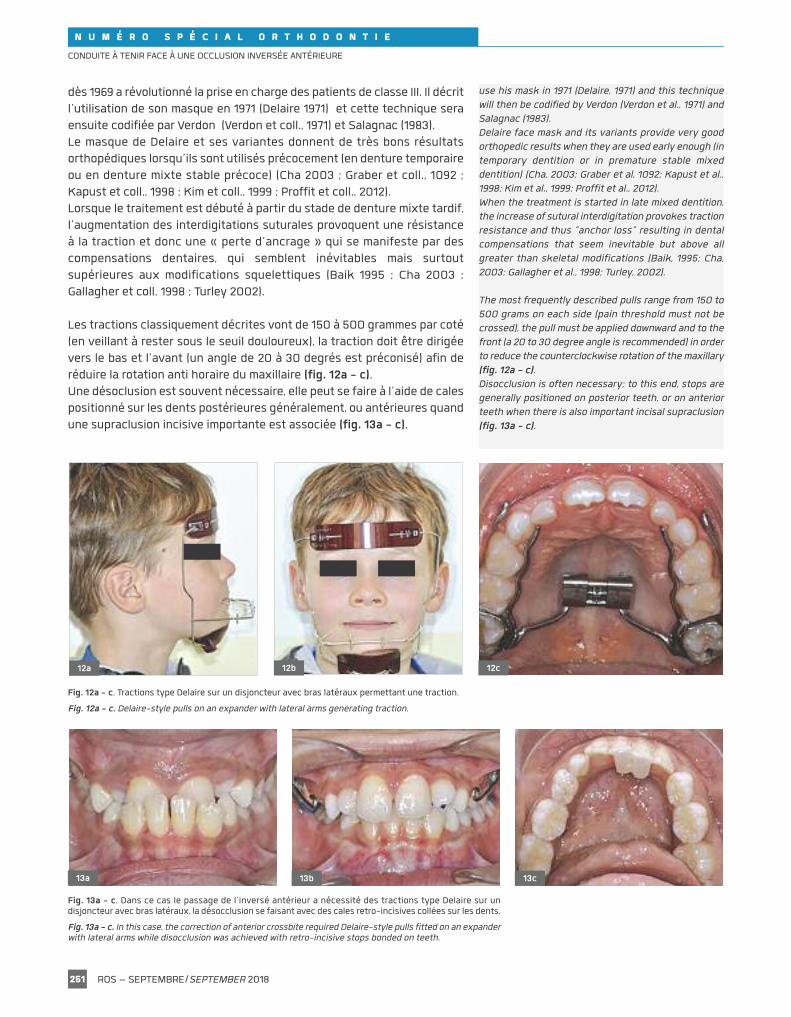
Les tractions classiquement décrites vont de 150 à 500 grammes par coté(en veillant à rester sous le seuil douloureux), la traction doit être dirigéevers le bas et l’avant (un angle de 20 à 30 degrés est préconisé) afin deréduire la rotation anti horaire du maxillaire (fig. 12a - c).Une désoclusion est souvent nécessaire, elle peut se faire à l’aide de calespositionné sur les dents postérieures généralement, ou antérieures quandune supraclusion incisive importante est associée (fig. 13a - c).
use his mask in 1971 (Delaire, 1971) and this techniquewill then be codified by Verdon (Verdon et al., 1971) andSalagnac (1983). Delaire face mask and its variants provide very goodorthopedic results when they are used early enough (intemporary dentition or in premature stable mixeddentition) (Cha, 2003; Graber et al, 1092; Kapust et al.,1998; Kim et al., 1999; Proffit et al., 2012).When the treatment is started in late mixed dentition,the increase of sutural interdigitation provokes tractionresistance and thus “anchor loss” resulting in dentalcompensations that seem inevitable but above allgreater than skeletal modifications (Baik, 1995; Cha,2003; Gallagher et al., 1998; Turley, 2002).
The most frequently described pulls range from 150 to500 grams on each side (pain threshold must not becrossed), the pull must be applied downward and to thefront (a 20 to 30 degree angle is recommended) in orderto reduce the counterclockwise rotation of the maxillary(fig. 12a - c).Disocclusion is often necessary; to this end, stops aregenerally positioned on posterior teeth, or on anteriorteeth when there is also important incisal supraclusion(fig. 13a - c).
ROS – SEPTEMBRE/SEPTEMBER 2018251
CONDUITE À TENIR FACE À UNE OCCLUSION INVERSÉE ANTÉRIEURE
12a 12b 12c
13a 13b 13c
Fig. 12a - c. Tractions type Delaire sur un disjoncteur avec bras latéraux permettant une traction.
Fig. 12a - c. Delaire-style pulls on an expander with lateral arms generating traction.
Fig. 13a - c. Dans ce cas le passage de l’inversé antérieur a nécessité des tractions type Delaire sur undisjoncteur avec bras latéraux, la désocclusion se faisant avec des cales retro-incisives collées sur les dents.
Fig. 13a - c. In this case, the correction of anterior crossbite required Delaire-style pulls fitted on an expanderwith lateral arms while disocclusion was achieved with retro-incisive stops bonded on teeth.

N U M É R O S P É C I A L O R T H O D O N T I E
UTILISATION D’ANCRAGES SQUELETTIQUES
Beaucoup d’auteurs ont cherché un moyen de protracter le maxillaire enlimitant les effets dentoalvéolaires (Enacar et coll., 2003 ; Hong et coll.,2005 ; Kokich et coll., 1985 ; Singer et coll., 2000 ; Smalley et coll., 1988), unpas a réellement été franchi en 2006 par l’équipe du Dr Kircelli qui réalisedes tractions orthopédiques type Delaire sur des plaques d’ancrage aumaxillaire, qui permettent sur ce cas d’obtenir une avancée du maxillaire de8mm (Kircelli et coll., 2006) (fig. 14a - d).
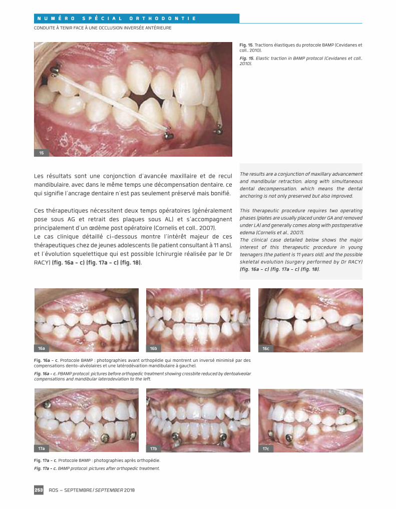
L’équipe du Dr De Clerck ira plus loin avec le protocole BAMP (Bone-Anchored Maxillary Protraction) en se passant d’appareillage extra-oral :des élastiques de classe III relient directement l’os maxillaire à la mandibule(Cevidanes et coll., 2010 ; De Clerck et coll., 2009 ; 2010 ; 2012 ; Heymannet coll., 2010 ; Nguyen et coll., 1971) (fig. 15).
SKELETAL ANCHORS
Many authors have tried to find a technique to protractthe maxillary by hindering dentoalveolar effects (Enacaret al., 2003; Hong et al., 2005; Kokich et al., 1985; Ape etal., 2000; Smalley et al.,1988). A step forward was takenin 2006 by Dr Kircelli’s team who performed orthopedicpulls inspired from Delaire on anchor plates placed atthe maxillary; in this specific case, this procedureresulted in a 8-millimeter maxillary advancement (Kircelliet al., 2006) (fig. 14a - d).
Dr De Clerck’s team still went further with BAMPprotocol (Bone-Anchored Maxillary Protraction) bygetting rid of extra-oral equipment: class III elasticbands directly connect the maxillary bone with themandible (Cevidanes et al., 2010; De Clerck et al., 2009;2010; 2012; Heymann et al., 2010; Nguyen et al., 1971)(fig. 15).
ROS – SEPTEMBRE/SEPTEMBER 2018252
CONDUITE À TENIR FACE À UNE OCCLUSION INVERSÉE ANTÉRIEURE
14a 14b
14c 14d
Fig. 14a - d. Téléradiographies de profil et photos de profilavant/après phase orthopédique (Kircelli et coll., 2006).
Fig. 14a - d. Profile teleradiographies and pictures before/afterorthopedic phase (Kircelli et al., 2006).
N U M É R O S P É C I A L O R T H O D O N T I E
Les résultats sont une conjonction d’avancée maxillaire et de reculmandibulaire, avec dans le même temps une décompensation dentaire, cequi signifie l’ancrage dentaire n’est pas seulement préservé mais bonifié.
Ces thérapeutiques nécessitent deux temps opératoires (généralementpose sous AG et retrait des plaques sous AL) et s’accompagnentprincipalement d’un œdème post opératoire (Cornelis et coll., 2007).Le cas clinique détaillé ci-dessous montre l’intérêt majeur de cesthérapeutiques chez de jeunes adolescents (le patient consultant à 11 ans),et l’évolution squelettique qui est possible (chirurgie réalisée par le DrRACY) (fig. 16a - c) (fig. 17a - c) (fig. 18).
The results are a conjunction of maxillary advancementand mandibular retraction, along with simultaneousdental decompensation, which means the dentalanchoring is not only preserved but also improved.
This therapeutic procedure requires two operatingphases (plates are usually placed under GA and removedunder LA) and generally comes along with postoperativeedema (Cornelis et al., 2007).The clinical case detailed below shows the majorinterest of this therapeutic procedure in youngteenagers (the patient is 11 years old), and the possibleskeletal evolution (surgery performed by Dr RACY)(fig. 16a - c) (fig. 17a - c) (fig. 18).
ROS – SEPTEMBRE/SEPTEMBER 2018253
CONDUITE À TENIR FACE À UNE OCCLUSION INVERSÉE ANTÉRIEURE
15
Fig. 15. Tractions élastiques du protocole BAMP (Cevidanes etcoll., 2010).
Fig. 15. Elastic traction in BAMP protocol (Cevidanes et coll.,2010).
16a 16c16b
17a 17c17b
Fig. 16a - c. Protocole BAMP : photographies avant orthopédie qui montrent un inversé minimisé par descompensations dento-alvéolaires et une latérodévaition mandibulaire à gauche).
Fig. 16a - c. PBAMP protocol: pictures before orthopedic treatment showing crossbite reduced by dentoalveolarcompensations and mandibular laterodeviation to the left.
Fig. 17a - c. Protocole BAMP : photographies après orthopédie.
Fig. 17a - c. BAMP protocol: pictures after orthopedic treatment.

N U M É R O S P É C I A L O R T H O D O N T I E
ÉLÉMENTS DÉCISIONNELS
ÂGE ET POTENTIEL DE CROISSANCEUn traitement précoce est à privilégier, quel qu’il soit, car l’inversé d’articuléest traumatogène pour les articulations (Kobayashi et coll., 1999), délétèrepour le parodonte (Eismann et Prusas, 1990), sa correction est plus aiséejeune (Cornelis et coll., 2007), et permet une rééducation fonctionnellegarante de stabilité (Talmant et Deniaud, 2005 ; Vesse 2007).
L’os mandibulaire terminant sa croissance après celle du maxillaire, il estfondamental de corriger l’anomalie le plus précocement possible, carl’inversé antérieur représente un frein majeur à la croissance maxillaire.
Si la méthode CVM (Cervical Vertebral Maturation) est retenue afin dedéterminer l’âge osseux du patient, on ne peut espérer avec des techniquesconventionnelles un effet sur le maxillaire qu’aux stades CS1 et CS2, à partirdu stade CS3, seul une limitation de la croissance mandibulaire est possible. Si le patient consulte en denture adulte jeune, un protocole de tractionssur plaques peut être proposé, l’âge d’indication allant de 11 à 13-14 ans.
ESTHÉTIQUE ET ÉQUILIBRE FACIAL L’équilibre labial est souvent retrouvé après correction de l’inverséd’articulé antérieur.
Certains patients avec une hypomaxillie sévère, présentent un « creux » dela région para nasale (Verdon et Castel, 1971), une simple correction« dentaire » de l’inversé d’articulé ne va pas suffire et une actionréellement orthopédique sera nécessaire pour permettre une avancée del’étage moyen de la face (fig. 19).
Chez ces patients il faudra privilégier le masque de Delaire jusqu’en denturemixte stable, et privilégier une traction orthopédique sur plaques ensuite(Hino et coll., 2013).
DECISION-MAKING ELEMENTS
AGE AND GROWTH POTENTIALEarly treatment, whatever it is, is recommended becausecrossbite is noxious for joints (Kobayashi et al., 1999),noxious for the periodontium (Eismann and Prusas,1990). Correcting this anomaly is easier when thepatient is young (Cornelis et al., 2007) and allowsfunctional reeducation providing stability (Talmant andDeniaud, 2005; Vesse, 2007).Since mandibular growth stops after maxillary growth, itis essential to correct the anomaly as soon as possible,because anterior crossbite can significantly hindermaxillary growth.
If the CVM method (Cervical Vertebral Maturation) isheld to determine the patient’s osseous age, we cannotexpect, with conventional techniques, to have an impacton the maxillary in stages CS1 and CS2, from stage CS3– only a limitation of the mandibular growth is possible. When the patient consults with young adult dentition, aprotocol of pulls on plates can be proposed- the age forindication ranges from 11 to 13-14 years old.
AESTHETICS AND FACIAL BALANCELabial balance is often restored after correction of theanterior crossbite.
Some patients suffering from severe hypomaxillia, havea “hollow” in the paranasal region (Verdon and Castel,1971); in this case, a simple "dental" correction of thecrossbite will not be enough and an actual orthopedicaction will be necessary to generate the advancementof the median part of the face (fig. 19).
For these patients, Delaire’s facemask is recommendeduntil stable mixed dentition and then, an orthopedic pullon plates procedure will be prescribed (Hino et al., 2013).
ROS – SEPTEMBRE/SEPTEMBER 2018254
CONDUITE À TENIR FACE À UNE OCCLUSION INVERSÉE ANTÉRIEURE
18
Fig. 18. Téléradiographies de profil avant et après orthopédie :diminution des compensations dentaires, l’évolution des voiesaériennes et la posture céphalique du patient.
Fig. 18. Profile teleradiographies before/after orthopedictreatment: decreased dental compensation, airway progressionand patient’s cephalic posture.

N U M É R O S P É C I A L O R T H O D O N T I E
OBJECTIFS PARODONTAUX ET OCCLUSAUXCes notions sont à corréler avec les objectifs du traitement.
OBJECTIFS PARODONTAUXDans certains cas, une compensation dentaire peut être tout à faitacceptable d’un point de vue parodontal et fonctionnel. Dans d’autres cas, un biotype parodontal fin peut interdire d’accentuer descompensations dentaires, ce qui signifie que l’action devra principalementêtre orthopédique.
OBJECTIFS OCCLUSAUXD’un point de vue occlusal, Raymond (2003) en s’appuyant sur les travauxde Planas, insiste sur la nécessité de ne pas aggraver l’inclinaison anti-horaire du plan d’occlusion. En effet, un traitement d’orthodontie avec élastiques de classe III, ou unetraction orthopédique de Delaire sur ancrage dentaire provoquent trèssouvent une égression des secteurs postérieurs maxillaires et ainsi unebascule anti horaire du plan d’occlusion.Cette orientation serait à l’origine d’une cinétique mandibulaire qui favo -riserait la croissance mandibulaire, en aggravant le décalage de classe III.
ANOMALIES ASSOCIÉESTrès souvent le sens transversal est à traiter via une expansion maxillairechez les patients en classe III, il semble cependant admis aujourd’hui quela disjonction maxillaire n’augmente pas l’avancée maxillaire (Kim et coll.,1999 ; Vaughn et coll., 2005).Le sens vertical est à prendre en compte car l’hyperdivergence représenteune contre-indication à toute thérapeutique augmentant le sens vertical.Le SAHOS, enfin, représente un élément à prendre en considération, cartrès souvent les patients en classe III présentent une lumière pharyngéediminuée soit par la position postérieure du maxillaire, soit par une posture
PERIODONTAL AND OCCLUSAL OBJECTIVESThese notions need to be correlated with the treatmentobjectives.
PERIODONTAL OBJECTIVESIn certain cases, dental compensation can be bothperiodontally and functionally acceptable. In other cases where periodontal biotype is thin, dentalcompensations cannot be accentuated, which meansthe action needs to be mainly orthopedic.
OCCLUSAL OBJECTIVESFrom an occlusal point of view, Raymond (2003), relyingon Planas’ work, insists on the necessity of notaggravating the counterclockwise inclination of theocclusal plane. Indeed, a class III orthodontic treatment with elasticbands or a Delaire orthopedic pull on dental anchoringcan very often provoke a drift of the maxillary posteriorsectors and thus a counterclockwise tilting of theocclusal plane.This orientation could generate mandibular kineticmovements strengthening mandibular growth byaggravating class III discrepancy.
ASSOCIATED ANOMALIESVery often, transverse deficiency needs to be treatedwith maxillary expansion in class III patients, it seemshowever accepted today that maxillary disjunction doesnot increase maxillary advancement (Kim et al., 1999;Vaughn et al., 2005).Vertical direction needs to be taken into accountbecause hyperdivergency is a contraindication to anytherapeutics increasing the vertical direction.Finally, OSAS also needs to be taken into account: veryoften indeed, class III patients present a reducedpharyngeal airway space either due to the posterior
ROS – SEPTEMBRE/SEPTEMBER 2018255
CONDUITE À TENIR FACE À UNE OCCLUSION INVERSÉE ANTÉRIEURE
19
Fig. 19. Profils avant, pendant et après phase orthopédique surplaques, l’harmonisation faciale ne concerne pas que la zonelabiale mais aussi l’étage moyen de la face.
Fig. 19. Profiles before, during and after orthopedic phase onplates, the facial harmonization does not concern only thelabial zone but also the average stage of the face.
N U M É R O S P É C I A L O R T H O D O N T I E
linguale basse et postérieure. Dans ce contexte une thérapeutiqueorthopédique avançant le maxillaire permet d’augmenter la lumièrepharyngée et d’améliorer la posture linguale (Lee W.C. et coll., 2018 ; Vesse2007), alors que les thérapeutiques entrainant un recul mandibulaireaugmentent le risque de SAHOS (Irani et coll., 2018).
PRONOSTIC ET SOLUTION CHIRURGICALELa question que se pose chaque praticien devant un cas d’inversé d’articuléantérieur : ce cas est-il « chirurgical » ? En effet, en traitant un cas de classe III par compensations dentaires, lepraticien se retrouve en cas de récidive devant la nécessité de proposer àson patient un protocole ortho-chirurgical quand le potentiel de croissancen’est plus suffisant et donc de faire l’exact inverse de sa première phaseen « décompensant » les arcades pour permettre une chirurgie decorrection des bases osseuses. La problématique se complexifie encore quand on sait qu’une chirurgie derecul mandibulaire présente elle-même un risque important de récidive(Proffit et coll., 2007), et comme précisé plus tôt un risque de réduire lalumière pharyngée (Irani et coll., 2018). Mais élément majeur, le pronosticd’échec est d’autant plus important que le décalage squelettique estinitialement élevé. On peut ainsi conclure que toute interception permettant une réduction dudécalage squelettique va dans le bon sens, même si une chirurgie estinévitable en fin de croissance, car le pronostic de cette chirurgie en seraamélioré. Si l’interception s’accompagne de faibles compensations, voirede compensations spontanées, cela est d’autant plus intéressant.
CONCLUSION
L’inversé d’articulé antérieur est le symptôme de pathologies diverses, lediagnostic devra être précis car il influera sur la prise en charge.
La thérapeutique devra être la plus précoce possible pour lever ce frein àla croissance maxillaire et permettre une harmonisation fonctionnelle. L’apport des ancrages osseux offrent de nouvelles perspectives, etaméliorent radicalement le pronostic de patients plus âgés, mais ne doitpas faire oublier l’importance des thérapeutiques interceptives précoces. C’est le sens des recommandations de la Haute autorité de santé « Pour lescas spécifiques des malocclusions de classes III, (…) de traiter le plus tôtpossible les proglissements mandibulaires (…) le traitement des classes IIId’origine fonctionnelle doit être mis en œuvre le plus précocement possible(rééducation des fonctions orofaciales et avancée maxillaire en utilisant lemasque de Delaire) ».
Méconnaître la thérapeutique interceptive engage la responsabilitéprofessionnelle du praticien pour manquement au devoir d’informationquant aux différentes possibilités thérapeutiques et pour perte de chancecar il aura fait perdre à son patient le choix d’un traitement orthodontiqueprécoce (Bery 2013).
position of the maxillary, or by the low and posteriortongue posture. In this context, orthopedic therapeuticsaiming at moving forward the maxillary allows toincrease the pharyngeal airway space and to improvethe tongue posture (Lee W.C et al., 2018; Vesse, 2007),whereas treatments generating a mandibular backwardmovement increase the risk of OSAS (Irani et al., 2018).
PROGNOSIS AND SURGICAL SOLUTION Dealing with a case of anterior crossbite, every practitionernecessarily wonders whether surgery is needed or not. Indeed, when treating a class III with dental compensations,the practitioner, in case of relapse, is forced to proposeto his/her patient an orthosurgical procedure whengrowth potential is no longer sufficient. In this case,he/she will have to do the opposite of what he/she’sdone during the first phase by “decompensating” thearches to allow corrective osseous surgery. The problem gets even more complicated when we knowthat mandibular setback surgery also induces aconsiderable risk of relapse (Proffit et al., 2007), and asmentioned above, a risk of reducing the pharyngealairway space (Irani et al., 2018). However, the importantfact is that the prognosis of failure is all the more highwhen the skeletal discrepancy is initially significant. We can then conclude that any interception aiming atreducing the skeletal discrepancy goes in the rightdirection, even if surgery is inevitable at the end ofgrowth, since the prognosis of this surgical procedurewill be improved. When interception involves smallcompensations, and even spontaneous compensations,the process is even more effective.
CONCLUSION
Anterior crossbite can be the symptom of variouspathologies. The diagnosis thus needs to be accuratebecause it will influence the treatment. Treatment should be started as soon as possible toeliminate this obstacle to maxillary growth and allowfunctional harmonization. The contribution of osseous anchors provides newperspectives and radically improves the prognosis forolder patients, although it must not hide the relevanceof early interceptive treatment. This is also what HAS (French National Authority forhealth) has recommended: “For specific class IIImalocclusion cases (…) treating mandibular prognathismas soon as possible (…) Treatment of class IIImalocclusion due to functional anomalies must beperformed as soon as possible (reeducation of orofacialfunctions and maxillary advancement with Delairefacemask)”.Underestimating the action of interceptive treatmentsinvolves the practitioner’s professional liability forfailure to supply information on the various therapeuticoptions and for loss of opportunity since because ofhim/her, the patient could not choose early orthodontictreatment (Bery, 2013).
Traduction : Marie Chabin
ROS – SEPTEMBRE/SEPTEMBER 2018256
CONDUITE À TENIR FACE À UNE OCCLUSION INVERSÉE ANTÉRIEURE

N U M É R O S P É C I A L O R T H O D O N T I E
ROS – SEPTEMBRE/SEPTEMBER 2018257
CONDUITE À TENIR FACE À UNE OCCLUSION INVERSÉE ANTÉRIEURE
BAIK H.S. – Clinical results of the maxillary protraction inKorean children. Am J Orthod Dentofacial Orthop 1995;108:583-592.
BÉRY A. – Le préjudice réparable : la perte de chance. OrthodFr 2013;84:15-27.
COZZA P., MARINO A., MUCEDERO M. – An orthopaedicapproach to the treatment of Class III malocclusions in theearly mixed dentition. Eur J Orthod. 2004;26(2):191-199.
CEVIDANES L.H., BACCETTI T., FRANCHI L., MC NAMARA J.A.JR, DE CLERCK H. – Comparison of two protocols for maxillaryprotraction: bone anchors versus face mask with rapidmaxillary expansion. Angle Orthod. 2010;80(5):799-806.
CHA B.K. – Skeletal changes of maxillary protraction inpatients exhibiting skeletal Class III malocclusion: a comparisonof three skeletal maturation groups. Angle Orthod 2003;73:26-35.
CORNELIS M.A., SCHEFFLER N.R., DE CLERCK H.J., TULLOCHJ.F., BEHETS C.N. – Systematic review of the experimentaluse of temporary skeletal anchorage devices in orthodontics.Am J Orthod Dentofacial Orthop 2007;131(4 Suppl):S52-58.
COZZA P., MARINO A., MUCEDERO M. – An orthopaedicapproach to the treatment of Class III malocclusions in theearly mixed dentition. Eur J Orthod. 2004;26(2):191-199.
DE CLERCK H.J., CORNELIS M.A., CEVIDANES L.H., HEYMANNG.C., TULLOCH C.J. – Orthopedic Traction of the Maxilla WithMiniplates: A New Perspective for Treatment of MidfaceDeficiency. J Oral Maxillofac Surg. 2009;67:2123-2129.
DE CLERCK H.J., CEVIDANES L.H., BACCETTI T. – Dentofacialeffects of bone-anchored maxillary protraction: A controlledstudy of consecutively treated Class III patients. Am J OrthodDentofacial Orthop 2010;138:577-581.
DE CLERCK H.J., NGUYEN T., DE PAULA L.K., CEVIDANES L. –Three-dimensional assessment of mandibular and glenoidfossa changes after bone-anchored Class III intermaxillarytraction. Am J Orthod Dentofacial Orthop 2012;142:25-31.
DELAIRE J. – Confection du masque orthopédique. RevStomatol. 1971;72(5):579-584.
DELAIRE J. – Maxillary development revisited: relevance tothe orthopaedic treatment of class III malocclusions. Eur JOrthod 1997;19:289-311.
DELAIRE J. – La croissance maxillaire : déductionsthérapeutiques. Trans Eur Orthod Soc 1971:81-102. DeshayesMJ. La morphogenèse cranio-faciale. Rev Orthop DentoFaciale 1998;32:299-310.
DELAIRE J. – Le développement « adaptatif » de la base ducrâne. Justification du traitement précoce des dysmor -phoses de classe III. Rev Orthop Dentofac 2003;37:243-265.
EISMANN D., PRUSAS R. – Periodontal findings before andafter orthodontic therapy in cases of incisor crossbite. Eur JOrthod 1990;12:281-283.
ENACAR A., GIRAY B., PEHLIVANOGLU M., IPLIKCIOGLU H. –Facemask therapy with rigid anchorage in a patient withmaxillary hypoplasia and severe oligodontia. Am J OrthodDentofacial Orthop.2003;123:571-577.
GALLAGHER R.W., MIRANDA F., BUSCHANG P.H. – Maxillaryprotraction: treatment and posttreatment effects. Am JOrthod Dentofacial Orthop 1998;113:612-619.
BIBLIOGRAPHIE
PROFFIT W.R., FIELDS H.W., SARVER D.M. – ContemporaryOrthodontics, 5th Edition. Elsevier Mosby editions, 2012.
RAYMOND J.L. – Finalité fonctionnelle et occlusale dutraitement orthopédique de classe III. Rev Orthop DentoFaciale 2003;37:285-303.
SALAGNAC – Études téléradiographiques des effets destractions postéro-antérieures sur masque orthodontiquedans le traitement des classes III (séquelles des fentes labio-maxillaires exclues). Bilan des résultats obtenus dans 60 cas.In: Ve congrès de stomatologie et de chirurgie maxillofaciale.Paris, Masson, 1983.
SINGER S.L., HENRY P.J., ROSENBERG I. – Osseointegratedimplants as an adjunct to face mask therapy: a case report.Angle Orthod. 2000;70:253-262.
SMALLEY W.M., SHAPIRO P.A., HOHL T.H., KOKICH V.G.,BRANEMARK P.I. – Osseointegrated titanium implants formaxillofacial protraction in monkeys. Am J Orthod DentofacialOrthop 1988;94:285-295.
TALMANT J., DENIAUD J. – Du role des incisives maxillairesdans le développement de la base du nez. Applicationsorthopédiques. Rev Orthop Dentofac 2005;39:297-336.
TURLEY P.K. – Managing the developing Class IIImalocclusion with palatal expansion and facemask therapy.AmJ Orthod Dentofacial Orthop 2002;122:349-352.
VAUGHN G.A., MASON B., MOON H.B., TURLEY P.K. – Theeffects of maxillary protraction therapy with or without rapidpalatal expansion: a prospective, randomized clinical trial.Am J Orthod Dentofacial Orthop. 2005;128(3):299-309.
VERDON P., CASTEL C.H. – Réalisation pratique et résultatscliniques de cas traités par forces extra-orales sur “masqueorthopédique”. Orthod Fr. 1971; 42:568.
VESSE M. – Classes III squelettiques. EMC, Odontologie/Orthopédie dentofaciale 2007;23-472-G-10.
WOON S.C., THIRUVENKATACHARI B. – Early orthodontictreatment for Class III malocclusion: A systematic review andmeta-analysis. Am J Orthod Dentofacial Orthop. 2017;151(1):28-52.
GRABER L.W., VANARSDALL ROBERT L., VIG KATHERINEW.L. – Orthodontics: Current Principles and Techniques, 5thEdition. Elsevier Mosby editions, 2011.
HEYMANN G.C., CEVIDANES L., CORNELIS M., DE CLERCK H.J.,TULLOCH J.F. – Three-dimensional analysis of maxillaryprotraction with intermaxillary elastics to miniplates. Am JOrthod Dentofacial Orthop 2010;137:274-284.
HINO CT., CEVIDANES L.H., NGUYEN T.T., DE CLERCK H.J.,FRANCHI L., MCNAMARA J.A. JR. – Three-dimensionalanalysis of maxillary changes associated with facemask andrapid maxillary expansion compared with bone anchoredmaxillary protraction. Am J Orthod Dentofacial Orthop. 2013;144(5):705–714.
HONG H., NGAN P., LI H.G., QI L.G., WEI SH. – Use of onplantsas stable anchorage for facemask treatment: a case report.Angle Orthod 2005;75:453-460.
IRANI S.K., OLIVER D.R., MOVAHED R., KIM Y.I., THIESEN G.,KIM K.B. – Pharyngeal airway evaluation after isolatedmandibular setback surgery using cone-beam computedtomography. Am J Orthod Dentofacial Orthop. 2018;153(1):46-53.
JACOBSON A. – The “Wits” appraisal of jaw disharmony. AmJ Orthod. 1975;67(2):125-138.
KAPUST A.J., TURLEY P.K., RUDOLPH D.J., SINCLAIR P.M. –Cephalometric effects of facemask/ expansion therapy inClass III children: a comparison of three age groups. Am JOrthod Dentofacial Orthop 1998;113:204-212.
KIM J.H., VIANA M.A., GRABER T.M., OMERZA F.F., BEGOLEE.A. – The effectiveness of protraction face mask therapy: ameta-analysis. Am J Orthod Dentofacial Orthop 1999;115:675-685.
KIRCELLI B.H., PEKTAS Z.O., UCKAN S. –Orthopedic protractionwith skeletal anchorage in a patient with maxillaryhypoplasia and hypodontia. Angle Orthod 2006;76:156-163.
Kobayashi T., Honma K., Izumi K., Hayashi T., Shingaki S.,Nakadjim T. : Temporomandibular joint symptoms and discdisplacement in patients with mandibular prognathism. Br JOral Maxillofac Surg 1999;37:455-458.
Kokich V.G., Shapiro P.A., Oswald R., Koskinen-Moffett L.,Clarren S.K. – Ankylosed teeth as abutments for maxillaryprotraction: a case report. Am J Orthod 1985;88:303-307.
LEE W.C. TU Y.K., HUANG C.S., CHEN R. FU M.W., FU E. –Pharyngeal airway changes following maxillary expansion orprotraction: A meta-analysis. Orthod Craniofac Res. 2018;21(1):4-11.
LE GALL M., PHILIP C., BANDON D. – The functionalmandibular prognathism. Arch Pediatr. 2009 Jan;16(1):77-83.
NGUYEN T. CEVIDANES L., CORNELIS M.A., HEYMANN G., DEPAULA L.K., DE CLERCK H. – Three-dimensional assessmentof maxillary changes associated with bone anchoredmaxillary protraction. Am J Orthod Dentofacial Orthop2011;140:790-798.
Verdon P., Castel C.H. – Réalisation pratique et résultatscliniques de cas traités par forces extra-orales sur “masqueorthopédique”. Orthod Fr 1971;42:568.
PROFFIT W.R., TURVEY T.A., PHILLIPS C. – The hierarchy ofstability and predictability in orthognathic surgery with rigidfixation: an update and extension. Head Face Med. 2007;3:21.