Vascular Smooth Muscle Cell Calcification is Mediated by Regulated Exosome Secretion
Upload
independentCategory
view
1download
0
Calcif Tissue Int (1996) 58:177-185
Tissue h~Lemafiol~ �9 1996 Springer-Verlag New York Inc.
Laboratory Investigations
Viable Cells Are a Requirement for In Vitro Cartilage Calcification
A. L. Boskey, S. B. Doty, D. Stiner, I. Binderman
The Hospital for Special Surgery, affiliated with Cornell University Medical College, New York, New York 10021
Received: 12 January 1995 / Accepted: 30 September 1995
Abstract. It is a common belief that chondrocyte death must precede calcification in the growth plate. To challenge this dogma, cell devitalization was induced in an in vitro model that mimics in situ cartilage calcification. Chick limb-bud mesenchymal cells, plated in micromass culture, differentiate to form a cartilaginous matrix which mineral- izes in the presence of inorganic or organic phosphate. The mineral formed resembles physiologic mineral in crystal size, composition, and distribution. Killing cells by water lysis, ethanol fixation, freeze-thawing, trypsinization, or im- pairing their function by oligomycin treatment prior to the time at which mineralization commenced, prevented min- eral deposition. In contrast, devitalizing cells by any of these techniques after mineralization commenced resulted in dystrophic calcification (excessive, randomly distributed mineral of larger than physiologic crystal size). Based on analyses of 45Ca uptake, FT-IR microscopy, X-ray diffrac- tion, and transmission electron microscopy, it is concluded that the presence of viable cells is obligatory for physiologic cartilage calcification in the differentiating chick limb-bud mesenchymal cell culture system.
Key words: Calcification - - Cell death - - Cell culture - - Chondrocyte - - Hydroxyapatite - - FT-IR microscopy.
In the epiphyseal growth plate, extracellular matrix calcifi- cation occurs around the chondrocytes in the lowermost hypertrophic cell region. These cells lose mitochondrial granules, and exist in an anoxic environment [1, 2]. The concept thus developed that cell death is a prerequisite for cartilage calcification [3]. This concept persists in the or- thopedic literature [4]. Studies using the differentiating chick limb-bud mesenchymal cell culture system suggested that cell death prevents calcification [5]. To challenge the hypothesis that cell death is required for physiologic carti- lage calcification [3, 4], cell death was induced in an in vitro model which mimics this process. This model previously was used to characterize the initially formed mineral [6], and to determine the effects of vitamin C [5], [3-glycero- phosphate [7], and ATP [8] on in vitro cartilage calcifica- tion.
Correspondence to: A. L. Boskey
Cell function in micromass cultures of differentiating mesenchymal cells was disrupted by a variety of indepen- dent techniques: physical (water lysis, freeze-thawing), chemical (trypsinization, ethanol fixation), and metabolic (inhibition of oxidative phosphorylation with oligomycin). Although the precise effect seen depended on both the ac- tivity of the cells at the time of treatment and the method of treatment, in general, cell death during matrix synthesis pre- vented mineralization; cell death after the initial mineral was formed resulted in dystrophic calcification.
Materials and Methods
Culture Conditions
The culture system previously shown to produce a mineralized cartilaginous matrix resembling that of chick calcified cartilage in ovo [6, 91 was used. In brief, fertilized White Leg Horn eggs (Truslow Farms, Chestertown, MD) were maintained in a humid- ified incubator at 37 ~ . At 4.5 days, when the embryos corre- sponded to stages 21-24 according to the classification of Ham- burger and Hamilton [10], the embryos were sterilely withdrawn from the eggs and their limb buds removed into 0.9% USP grade saline (Abbott Laboratories, N Chicago, IL). Cells were released from the limb buds by digestion for 7 minutes in 5 ml 0.25 wt % trypsin-0.53 mM EDTA (GIBCO, Grand Island, NY). The reaction was quenched by the addition of 5 ml low calcium (0.3 raM) Dulbecco's Modified Eagle Medium (DMEM; GIBCO Formula 80-0303 AJ). Following vortexing, the cells were separated from debris by passage through two layers of 20 gm Nitex membrane (Tetko Inc, Ardsley, NY), counted with a hemocytometer, checked for viability by trypma blue dye exclusion, and pelleted in the cold at 2300 rpm. For all experiments, viability at plating exceeded 98%. Cells were resuspended in DMEM supplemented with 50 U/ml penicillin, 25 ~tg/ml streptomycin, and 10% fetal calf serum (GIBCO), containing 1.1 mM Ca, and plated, using the micromass technique [11], at a density of 0.5 million cells per 20 gl drop in 35 x 10 mm Falcon dishes. After 2 hours in a humidified atmo- sphere of 5% CO 2 at 37 ~ 2 ml of medium was added. Medium was changed every 48 hours, with 25 ~tg/ml ascorbic acid and 0.3 mg/ml glutamine added from day 2. For mineralizing cultures, the inorganic phosphate content was adjusted to 4 mM by adding KH~PO 4 from day 2 onward. In some experiments, either 2.5 or 5 mM ~-glycerophosphate was used as the phosphate supplement. Nonmineralizing controls which had a basal inorganic phosphate content of 1 mM were not further supplemented.
Cell Devitalization
Cells were devitalized on days 3, 5, or 7 (before mineralization), day 11 (at the start of mineralization), or day 14 (during mineral-

178
40
3O
2O v
z Q
10
Day 7
Day 14
Day 11
Day of Treatment
18
15
-'= 12
�9 o
Viable
+ EtOH
+ Trypsin
+ Water
+Freeze/ Thaw
Oligo-c
Oligo lx
100
80
g ~ 60
< 40
20
A. L. Boskey et al.: Viable Cells and Calcification
Day 18 m Viable
Day 7 Day 11
Day of Treatment
b ~ + EtOH
+ Trypsin
+ Water
+Freeze/ Thaw
Oligo-c
Oligo Ix
3/ 0
Day 11 Day 14
Day of Assay
Fig. 1. (a,b) DNA content of cultures maintained in the presence of 4 mM inorganic phosphate (a) from day 2 to 14 and (b) from day 2 to 18. Cultures were devitalized by treatment with ethanol (+EtOH), trypsin, water lysis (+water), by freeze thawing, or con- tinuous addition of 25 gM oligomycin (oligo) or a single addition (oligo lx) on day 7 or 11. Dotted lines show mean DNA content of viable cells at day 7 or 11. Values are mean + SD. *Significantly different from viable cultures at day of treatment, ANOVA
Viable
EtOH d7
EtOH dl 1
Oligo-c d7
Oligo lx d7
Day 18
Bonferonni P ~< 0.05. (c) Protein synthesis (3H-leucine incorpo- ration) in cultures maintained in the presence of 4 mM inorganic phosphate from day 3 and left untreated (viable) or treated with ethanol or oligomycin (25 gM) once on day 7 (1 x day 7) or continuously from day 7 (oligo). (Values are mean _+ SD.) *Sig- nificantly different from viable cultures at time of assay, Bonfer- onni P ~< 0.05.
ization) using five different treatments. Unless otherwise noted, the medium was removed before treating the cells, and half that medium and an equal volume of fresh medium (mixed medium) were added after treatment. Medium changes were continued until the indicated time points. Controls for each experiment included viable cells from the same culture batch maintained in mineraliz- ing and in nonmineralizing (low phosphate) media. Devitalization was done by ethanol fixation, freeze thawing, trypsin treatment, water lysis, or oligomycin treatment.
For ethanol fixation, after removing the medium, 90% ethanol was added to the culture dish for 5 minutes, the ethanol was removed, cultures were rinsed with fresh DMEM, and mixed me- dium was added. During freeze thawing, the plates were dipped three times into liquid nitrogen and thawed for 5 minutes between each immersion. The dishes were then placed in the incubator for 5 minutes prior to adding mixed medium. (In several instances, the culture dishes cracked and the cultures were discarded.) To lyse cells with trypsin, the cultures were treated with a 0.5% solution of TPCK-trypsin (Sigma Chemical, Co. St. Louis, MO) in DMEM for 5 minutes, the cultures washed three times in DMEM to remove any released cells, and mixed medium was added. During water lysis, cultures were covered with 2 ml sterile water for 3 minutes. (With longer exposure, cultures were released from the surface of
the dish.) After treatment, water was removed, cultures were rinsed with fresh DMEM, and mixed medium was added. Oligomycin treatment involved either a single addition of 10, 25, or 50 gM oligomycin (Sigma) in DMEM for 30 minutes, followed by re- placement with fresh oligomycin-free mixed media, or constant treatment, in which the oligomycin was kept in the cultures throughout the entire experimental period.
Verification of Cell Death
The efficacy of the cell devitalization processes was monitored in several ways. During the first 9 days, cells released by trypsiniza- tion were monitored for viability by trypan blue dye exclusion. After day 11, fewer than 20% of the cells could be released from the micromass cultures by mild trypsinization, and other assay methods ,were used. To determine if cells had stopped replicating after devitalization, the DNA content of parallel cultures was mon- itored at the time of treatment, and at successive times thereafter, and compared to the DNA contents of the viable cultures at these time points. DNA content was measured using a fluorescence as- say [12]. The cultures were washed briefly with isotonic saline, transferred to sterile polystyrene tubes, and digested in papain to

A.L. Boskey et al.: Viable Cells and Calcification 179
Fig. 2. Electron micrograph demonstrating that ethanol fixation results in disruption of all cells in the different mesenchymal cell cultures. The cartilage cells shown here were fixed on day 11 and examined on day 14. The cells are obviously disrupted and contain few recognizable structures. The matrix is more dense than the day 11 cultures described in Figure 3. The cells have dramatically shrunk in size within their lacunae, creating an unusually large pericellular space (arrows). Magnification x6000.
release the cells from the matrix prior to mixing with Hoechst dye 33258 (Sigma).
To demonstrate that the devitalization techniques also stopped cell function, protein synthesis was monitored in viable and devi- talized cultures. 3H-leucine (0.1 gCi/ml) was added to the cultures along with the medium from day 7 onward. Total label accumu- lation was monitored at days 11, 14, and 18, following washing 3 x with cold DMEM to remove unincorporated label, hydrolysis in 0.2 ml 6 N HC1 (1 hour, 60~ and scintillation counting.
The appearance of the cells in the devitalized and viable min- eralizing and non-mineralizing cultures was studied by electron microscopy. Cultures collected at days 7, 11, 14, 18 and 21 were washed with 0.05 M cacodylate buffer, and maintained in EM fixative (0.5 glutaraldehyde, 2% paraformaldehyde, pH 7.2 0.05 M cacodylate buffer) in the culture dish for 12-18 hours at 4~ They were then stored at 4~ in 0.05 M cacodylate buffer containing 7% sucrose. After being removed from the dish, they were postfixed with 2% aqueous osmium, dehydrated in a graded series of alco- hols, and embedded in Spurr's resin. Thin sections were collected on ammoniated water containing bromthymol blue as an indicator for pH above 8.0. Lead citrate and alcoholic uranyl acetate-stained sections were examined on a Phillips CM12 electron microscope. For a limited number of experiments, cultures fixed in the dish with ethanol were stained using the von Kossa technique [13], and prepared for electron microscopy as described above.
Mineralization Assays
Mineralization was monitored based on 45Ca uptake, X-ray dif- fraction, FT-IR microscopy, electron microscopy, and in a limited number of cases (for reasons described below), using the von Kossa stain. To monitor the rate of calcium uptake, 1 gCi/m145Ca (Amersham, Arlington Heights, IL) was added to mineralizing and similarly treated nonmineralizing control cultures on alternate days from day 4 onward. At the indicated time points, the medium was removed from labeled cultures, cultures were washed with fresh, cold medium, and the matrix was scraped into scintillation vials and hydrolyzed (1 hour, 60 ~ in 0.2 ml 4 N HC1. The 45Ca incor- poration, determined by scintillation counting, was expressed per micromass culture. For cultures with ~3-glycerophosphate, to de- termine whether 45Ca uptake in cultures with nonviable cells could be attributable to residual alkaline phosphatase activity in the me- dia, 100 gM levamisole (a specific alkaline phosphatase inhibitor [ 14]) was added to 50% of the cultures after cell devitalization and 50% of the viable cultures at the same time point.
At selected time points, three mineralizing micromass spot cul-
tures from each treatment group (devitalized or viable) were washed with pH 8 ammonium hydroxide, lyophilized, and used for X-ray diffraction analyses. The dried material was ground in a mortar and pestle, placed in a 0.7-mm-diameter capillary, and subjected to wide-angle X-ray diffraction analysis using a Debye- Schen'er camera and Ni filtered Cu K-~ radiation. The presence of poorly crystalline and well-crystallized hydroxyapatite was deter- mined based on comparison with bovine bone (poorly crystalline) and commercial (Fisher Chemical, Springfield, NJ) synthetic (well crystallized) apatites.
Viable and devitalized cultures fixed with absolute ethanol at the time of sampling were placed on barium fluoride windows and examined using a FT-IR microscope (Mattsen Industries, Madison, WI), at 20 gm resolution [6]. Spectra were collected at 4 cm -1 resolution; 256 scans per spectrum at sites in the center of the cartilage nodule, and 20, 40, and 60 gm from the center. A min- imum of three nodules per culture were examined. Qualitative comparisons of mineral phases were based on standards previously characterized by wide-angle X-ray diffraction. Relative mineral: matrix ratio and crystallite size and perfection were estimated from the spectra as described in detail elsewhere [6].
Data Analysis
For all data, comparisons to viable cells or other culture conditions were based on analysis of variance (ANOVA); statistical signifi- cance was assumed for a Bonferroni P ~< 0.05 (Student's t-test).
Results
Trypan blue exclusion demonstrated that more than 98% of the cells not subject to devitalization were viable at days 3, 5, and 7. Conversely, the devitalization affected 100% of the cells at these time points. After day 7, this method could not be used as gentle t ryps in- -EDTA extraction did not release all the cells encased in the micromass matrix, and more stringent extraction destroyed cell membranes, preventing use of the dye exclusion technique.
The DNA contents measured on day 14 (Fig. la) and day 18 (Fig. lb) in viable and devitalized cultures are compared with those in the viable cultures (dashed lines) on the day of devitalization. The DNA contents of all but the cultures exposed to oligomycin one time on day 11 remained invari-

180 A.L. Boskey et al.: Viable Cells and Calcification
Fig. 3. Electron micrographs showing the variation in effect of oligomycin on differentiating mesenchymal cell cultures at day 11. Oligomycin (10 p.M) added at day 7 had little effect on some cells (a) but was toxic to others (b,c). The appearance of the chondrocyte in Figure a was indistinguishable from those of cells in cultures that were not treated. (a) This cartilage cell appears normal with Golgi saccules, mitochondria, and strands of endoplasmic reticulum scattered through the cytoplasm. A small portion of the nucleus (N) is seen in this section. (b) This is a badly disrupted cartilage cell from the same culture as (a). The mitochondria are enlarged (arrows) and the cytoplasm is filled with lipid droplets (L) and two noncharacterized structures. The cell membrane is disrupted in several places. (e) The matrix from this culture appears normal in spite of the poor morphology of the adjacent cartilage cells. Magnification x16000.
ant after devitalization. All cultures devitalized on day 14, except those treated with oligomycin, similarly failed to show any increase in DNA content by day 18 (not shown).
Figure lc presents data on protein synthesis (3H-leucine incorporation) in viable cultures and in those devitalized with ethanol and oligomycin. Cultures devitalized by water lysis, trypsin, and freeze thawing (not shown), similar to the ethanol-fixed cultures, had 3H-leucine uptake on day 14 and 18 not significantly different from that of viable cultures on the day they were devitalized. Cultures treated continuously or with a single dose of oligomycin, continued protein syn- thesis but to a significantly lesser extent than in viable cul- tures.
The electron micrographs showed that those devitaliza- tion techniques which totally blocked replication and pro- tein synthesis, such as ethanol fixation, affected all cells (Fig. 2), whereas oligomycin treatment affected only some cells (Fig. 3). The assessment of viability was based on the presence of typical chondrocyte morphology, with a well- formed Golgi complex, normal sized and shaped mitochon-
dria, segments of rough endoplasmic reticulum, and an in- tact cell membrane (Fig. 3a). Nonviable cells had enlarged mitochondria, a cytoplasm filled with disrupted remanents of cytoplasm and organelles, and a disrupted external cell membrane (Fig. 3b). Even when cells were dead, the extra- cellular cartilage matrix often appeared to be normal (Fig. 3c) and indistinguishable from that in viable cultures.
Devitalization affected mineralization in variable ways, depending on the time at which the cultures were killed and the method of treatment. Figure 4 describes typical data for 45Ca uptake on days 14, 18, and 21 in viable cultures and cultures fixed with ethanol on days 7, 11, or 14. Data for devitalization by freeze-thawing, trypsin treatment, or water lysis showed similar trends (not shown). Results are pre- sented for cultures supplemented with inorganic phosphate (Fig. 4a) and ~-glycerophosphate (Fig. 4b). Ethanol fixation on day 5 (not shown) or day 7 prevented calcium uptake. Treatment on day 11 tended to decrease 45Ca uptake, how- ever, in ethanol-fixed cultures with 4P, the decrease was only statistically significant at day 21. Since total protein

A. L. Boskey et al.: Viable Cells and Calcification
50 a 4O
3o
E = ~ 20
O lO
Day 14 Day 18
Day of Assay
Day 21
1 i I VIABLE
ETOH D7
ETOH D l l
ETOH D14
*p < 0.05 compared to viable cells 20 C 16
E~ o_~ o ~ - g
O 4 O
Day 18
* p<0.01 compared to V IABLE
** p~O.01 compared to ETOH D l l
Fig. 4. 45Ca uptake in cultures maintained in the presence of 4 mM inorganic phosphate (a) of 5 mM [~-glycerophosphate (b,e) and devitalized by ethanol treatment. In the experiments shown in (e), 100gM levamisole (+LEV) was added from day 9to viable
=
E o. o v
1.10 1.01 0.93 0.84 0.75
25
Day 14
2O
@. lo
181
Day 18
Day of Assay
Day 21
I VIABLE
ETOH D7
ETOH D l l
ETOH D14
p < 0.05 compared to v iable cel ls
VIABLE
*LEV
ETOH D l l
ETOH D11+LEV
cultures and cultures treated with ethanol. All values are mean + SD. Statistical comparisons are based on ANOVA. Bonferroni P for multiple comparisons.
Table 1. X-ray diffraction analysis (d21) of mineralizing cultures ~ with viable b and nonviable cells
Day of treatment
Method of treatment: Day 3 Day 5 Day 7 Day 11 Day 14 HzO lysis ND ND ND xHA xHA Freeze thawing ND ND ND xHA xHA EtOH ND ND ND trHA xHA Trypsin . . . . . . ND ... xHA Oligomycin (50 gM) constant . . . . . . . . . HA xHA
a All cultures with 4 mM Pi added from day 2 b All viable cultures contained poorly crystalline hydroxyapatite at day 21 ND = not detectable; HA = poorly crystalline HA; xHA = well-crystallized HA; trHA = HA seen in some but not all samples analyzed . . . . not analyzed
synthesis m similarly treated cultures also did not increase after devitalization, this implies that the ratio of Ca to matrix content did not increase with time. In contrast, devitaliza- tion on day 14 caused 45Ca uptake to exceed that of viable cultures immediately after treatment and on days 18 and 21, both in the case of cultures supplemented with ~-glycero- phosphate and inorganic phosphate. Here, since protein syn- thesis in the devitalized cultures ceased at day 14, the ratio of Ca to matrix would exceed values for viable cells. Inhi- bition of alkaline phosphatase activity by levamisole in ~3GP-containing viable cultures significantly reduced 45Ca uptake (Fig. 4c), but levamisole addition to cultures devi- talized at day 11 did not cause a significant decrease in 45Ca
uptake relative to the devitalized cultures. Treatment with 25 or 50 g M oligomycin caused less extensive, but similar changes in 45Ca uptake in cultures with 4 mM P and [3-glyc- erophosphate (not shown). Continuous treatment from day 7 or 11 significantly reduced, but did not prevent, 45Ca up- take. Single treatments with ol igomycin on each of these days reduced 45Ca uptake to an even lesser extent. Results of continuous treatment from day 14 were variable.
X-ray diffraction analysis (Table 1) similarly revealed differences in mineral characteristics depending on when cultures were devitalized and the method of treatment. Min- eralizing cultures devitalized at days 3, 5, and 7 never formed mineral. Cultures devi tal ized by water lysis or
182
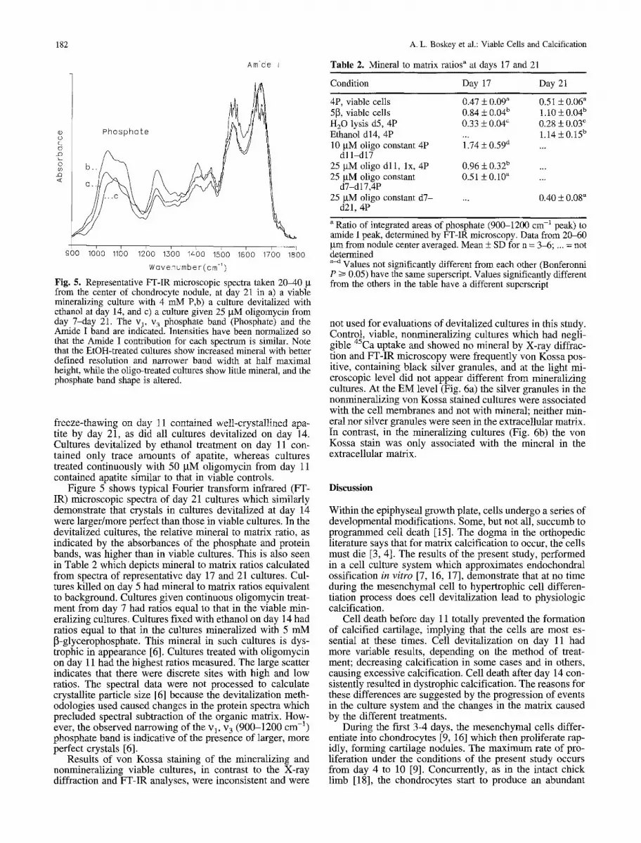
Amide I
Phosphate o
~ !
900 10C10 11130 12'00 15'00 1r 15'00 16'00 1700 18'00 Wovenumber(cm -~)
Fig. 5. Representative FT-IR microscopic spectra taken 2040 g from the center of chondrocyte nodule, at day 21 in a) a viable mineralizing culture with 4 mM P,b) a culture devitalized with ethanol at day 14, and c) a culture given 25 gM oligomycin from day 7-day 21. The v 1, v 3 phosphate band (Phosphate) and the Amide I baud are indicated. Intensities have been normalized so that the Amide I contribution for each spectrum is similar. Note that the EtOH-treated cultures show increased mineral with better defined resolution and narrower band width at half maximal height, while the oligo-treated cultures show little mineral, and the phosphate band shape is altered.
freeze-thawing on day 11 contained well-crystallined apa- tire by day 21, as did all cultures devitalized on day 14. Cultures devitalized by ethanol treatment on day 11 con- tained only trace amounts of apatite, whereas cultures treated continuously with 50 BM oligomycin from day 11 contained apatite similar to that in viable controls.
Figure 5 shows typical Fourier transform infrared (FT- IR) microscopic spectra of day 21 cultures which similarly demonstrate that crystals in cultures devitalized at day 14 were larger/more perfect than those in viable cultures. In the devitalized cultures, the relative mineral to matrix ratio, as indicated by the absorbances of the phosphate and protein bands, was higher than in viable cultures. This is also seen in Table 2 which depicts mineral to matrix ratios calculated from spectra of representative day 17 and 21 cultures. Cul- tures killed on day 5 had mineral to matrix ratios equivalent to background. Cultures given continuous oligomycin treat- ment from day 7 had ratios equal to that in the viable min- eralizing cultures. Cultures fixed with ethanol on day 14 had ratios equal to that in the cultures mineralized with 5 mM [3-glycerophosphate. This mineral in such cultures is dys- trophic in appearance [6]. Cultures treated with oligomycin on day 11 had the highest ratios measured. The large scatter indicates that there were discrete sites with high and low ratios. The spectral data were not processed to calculate crystallite particle size [6] because the devitalization meth- odologies used caused changes in the protein spectra which precluded spectral subtraction of the organic matrix. How- ever, the observed narrowing of the vl, v 3 (900-1200 cm -1) phosphate band is indicative of the presence of larger, more perfect crystals [6].
Results of von Kossa staining of the mineralizing and nonmineralizing viable cultures, in contrast to the X-ray diffraction and FT-IR analyses, were inconsistent and were
A. L. Boskey et al.: Viable Cells and Calcification
T a b l e 2. Minera l to mat r ix rat ios a at days 17 and 21
Condition Day 17 Day 21
4P, viable cells 0.47 _+ 0.09 ~ 5[3, viable cells 0.84 _-+ 0 .04 b
H20 lysis d5, 4P 0.33 _+ 0.04 c Ethanol d14, 4P 10 gM oligo constant 4P 1174 _ 0.59 d
dl l -dl7 25 ~M oligo dll, lx, 4P 0.96 + 0.32 b
25 p.M oligo constant 0.51 _+ 0.10 a d7-d17,4P
25 gM oligo constant d7- ... d21, 4P
0.51 + 0.06 a 1.10 + 0.04 b
0.28 +_ 0.03 c 1.14 + 0.15 b
0.40 + 0.08 a
a Ratio of integrated areas of phosphate (900-1200 cm -1 peak) to amide I peak, determined by FT-IR microscopy. Data from 20-60 gm from nodule center averaged. Mean _+ SD for n = 3-6; . . . . not determined a-d Values not significantly different from each other (Bonferonni P 1> 0.05) have the same superscript. Values significantly different from the others in the table have a different superscript
not used for evaluations of devitalized cultures in this study. Control, viable, nonmineralizing cultures which had negli- gible 45Ca uptake and showed no mineral by X-ray diffrac- tion and FT-IR microscopy were frequently yon Kossa pos- itive, containing black silver granules, and at the light mi- croscopic level did not appear different from mineralizing cultures. At the EM level (Fig. 6a) the silver granules in the nonmineralizing von Kossa stained cultures were associated with the cell membranes and not with mineral; neither min- eral nor silver granules were seen in the extracellular matrix. In contrast, in the mineralizing cultures (Fig. 6b) the von Kossa stain was only associated with the mineral in the extracellular matrix.
Discussion
Within the epiphyseal growth plate, cells undergo a series of developmental modifications. Some, but not all, succumb to programmed cell death [15]. The dogma in the orthopedic literature says that for matrix calcification to occur, the cells must die [3, 4]. The results of the present study, performed in a cell culture system which approximates endochondral ossification in vitro [7, 16, 17], demonstrate that at no time during the mesenchymal cell to hypertrophic cell differen- tiation process does cell devitalization lead to physiologic calcification.
Cell death before day 11 totally prevented the formation of calcified cartilage, implying that the cells are most es- sential at these times. Cell devitalization on day 11 had more variable results, depending on the method of treat- ment; decreasing calcification in some cases and in others, causing excessive calcification. Cell death after day 14 con- sistently resulted in dystrophic calcification. The reasons for these differences are suggested by the progression of events in the culture system and the changes in the matrix caused by the different treatments.
During the first 3-4 days, the mesenchymal cells differ- entiate into chondrocytes [9, 16] which then proliferate rap- idly, forming cartilage nodules. The maximum rate of pro- liferation under the conditions of the present study occurs from day 4 to 10 [9]. Concurrently, as in the intact chick limb [18], the chondrocytes start to produce an abundant

A. L. Boskey et al.: Viable Cells and Calcification 183
Fig. 6. (a) Electron micrograph of a viable nonmineralizing culture at day 14 stained by the von Kossa technique. The culture stained von Kossa positive despite the absence of high phosphate concentrations. The silver reaction product lines the cell membranes and is largely absent from the matrix. No mineral crystals are detectable. (b) Electron micrograph of a mineralizing culture with 2.5 mM ~-glycero-phosphate at day 17. The mineral is deposited within the extracellular matrix but not along the cell membrane. Magnification x7600.
three-dimensional extracellular matrix. The rate of protein synthesis is maximal from day 7 to 12 [9]. Since the rate of ~Hrotein synthesis decreases after day 11, the changes in
-leucine incorporation observed with cell death at day 11 or 14 would be expected to be less significant. When phos- phate supplements are present, initial mineral deposition in the extracellular matrix begins by day 12 [9]. Alkaline phos- phatase activity, detectable in the cultures by day 8, reaches maximal levels between day 12 and 14 [19], and would be available to hydrolyze organic phosphates.
Cell death after the cartilage matrix is present may affect the calcification process in several ways. First, it essentially freezes the culture matrix at the time point of treatment. There is no further matrix synthesis, and cell-mediated ex- tracellular matrix modifications that would occur at a later time point are prevented. Thus, calcification is mediated only by physicochemistry, i.e., the CaxPO4xOH product of the media, and the presence or absence of mineralization nucleators and inhibitors in that "frozen" matrix. Alkaline phosphatase activity, as shown by the levamisole experi-
merits, is no longer available to increase local phosphate concentrations. However, because cell membranes and cel- lular organelles are lysed, there may be a release of both lysosomal enzymes which can degrade the "frozen" matrix and of cell calcium.
In general, when cells were devitalized on day 11, little or no mineral deposition was detected despite the abundant matrix. This suggests that cellular activity is still required, perhaps for modifying the matrix or for regulating ionic flux. The variable results in the different experiments with cells killed on day 11 reflect the effects of the treatment on the matrix. Ethanol fixation may have precipitated certain lipids, changed the conformation of matrix proteins, or most importantly, prevented lysosomal protease degradation of proteins that regulate mineralization. Thus, little mineral- ization occurred. In contrast, trypsinization and water lysis may have removed such regulatory matrix proteins and freeze thawing may have dehydrated the matrix, facilitating the mineralization process.
When mineralizing cultures were devitalized after rain-

184
eral deposition had commenced (day 14), the mineral accu- mulated appeared dystrophic. The dystrophic crystals were of comparable size to those in cultures with high (5-10 mM) concentrations of [3-glycerophosphate [6]. Such larger, more abundant crystals were previously shown [5] not to differ in Ca:P molar ratio from the expected stoichiometric ratio for apatite of 1.7:1. Such crystals are most likely formed by rapid growth of existing crystals in an environment of ele- vated calcium (from release of cell calcium) and/or phos- phate concentrations.
The oligomycin data provide insight into a different as- pect of cell function. Oligomycin blocks mitochondrial ATP synthesis, depriving the cells of energy. In the growth plate [2, 20, 21], mitochondria supply calcium to extracellular matrix vesicles, initial sites of mineralization. The release of ATP from hypertrophic chondrocytes [1] also may provide the small increase in extracellular phosphate required to initiate mineralization [22]. Blocking ATP syntheses with oligomycin would thus be predicted to have less of an effect after the mature matrix was synthesized than before that time. This suggests why a single treatment with oligomycin at day 7 significantly reduced mineral accumulation as in-
4s dicated by Ca uptake, whereas constant treatment after day 14 had a lesser effect. Since not all the cells died in oligomycin-treated cultures, new matrix synthesis and mod- ification of the matrix by cell-derived enzymes may have continued throughout the study, resulting in less extensive changes. Further, alterations in ATP production may affect the distribution of mineral in the matrix, since ATP is re- quired for kinase-mediated phosphorylation of matrix pro- teins that act as nucleators. This, along with the efflux of calcium from dying cells, may account for the significantly increased mineral to matrix ratio in some oligomycin- treated cultures.
The data obtained during this study revealed a potential problem in the commonly used yon Kossa positive cultures as proof of the presence of mineral [e.g., 23-25], and is presented here as a caveat. The electron microscopic results indicate that the von Kossa reaction product need not be associated with calcium phosphate mineral, a fact noted originally by yon Kossa who observed that nonmineral components within the organic matrix could produce the black silver granules [13]. Similarly, the alizarin red stain may be associated with proteoglycans, anionic macromole- cules with an appreciable affinity for divalent cations [26]. 45Ca uptake alone also does not indicate the presence of mineral, but when combined with X-ray diffraction, FT-IR, or EM methods can provide confirmation of mineral accu- mulation.
In summary, the studies reported here reveal the need for viable cells for the formation of physiologic mineral in the differentiating mesenchymal cell cultures. The need for vi- able cells in cartilage calcification was previously suggested by elegant stereologic studies of cryo-preserved rat tibial growth plates showing that the lower-most hypertrophic cells of the growth plate were still viable, and that cell degeneration was not associated with physiologic cartilage calcification [20]. The need for viable cells to regulate min- eral deposition and prevent dystrophic calcification does not only apply to cartilage calcification. Recent studies have shown necrotic osteoblasts in tissues retrieved from patients with heterotopic ossification adjacent to total joint arthro- plasties and similar necrotic cells without a mineralized ma- trix at the implant surface [27], suggesting that viable os- teoblasts may also be required to regulate matrix synthesis, mineral deposition, and prevent dystrophic calcification.
A. L. Boskey et al.: Viable Cells and Calcification
Acknowledgments. This study was supported by NIH grant AR037661. The authors appreciate the assistance of Ms. Kitty Bock in preparing the sections for histologic evaluation, and of Dr. R. Mendelsohn for his assistance with the FT-IR microscopy.
References
1. Brighton CT, Heppenstall RB (1971) Oxygen tension in zones of the epiphyseal plate, the metaphysis and diaphysis. J Bone Jt Surg 53A:719-728
2. Matsumoto H, DeBolt K, Shapiro IM (1982) Adenine, gua- nine, and inosine nucleotides of chick growth cartilage: rela- tionship between energy status and the mineralization process. J Bone Miner Res 3:347-352
3. Cruess RL (1982) The musculoskeletal system: embryology, biochemistry and physiology. Churchill Livingston, NY p 197
4. Gamble JG (1988) The musculoskeletal system: physiological basics. Raven Press, New York
5. Boskey AL, Stiner D, Doty S, Binderman I (1991) Require- ment of vitamin C for cartilage calcification in a differentiat- ing chick limb-bud mesenchymal cell culture. Bone 12:277- 282
6. Boskey AL, Pleshko N, Binderman I, Mendelsohn R (1992) Mineralization during in vitro calcification: an FT-IR micro- scopic investigation, Calcif Tissue Int 51:443-448
7. Boskey AL, Doty SB, Binderman I, Leboy P (1993) Insight into the effect of ~3-glycerophosphate on in vitro mineraliza- tion. 39th Annual Meeting, Orthopaedic Research Society, Feb 15-18
8. Boskey AL, Doty SB, Binderman I (1994) ATP promotes mineralization in differentiating chick limb-bud mesenchymal cell cultures. Microsc Res Technique 28:492-504
9. Boskey AL, Stiner D, Leboy P, Doty S, Binderman I (1992) Optimal conditions for cartilage calcification in differentiating chick limb-bud mesenchymal cells. Bone Miner 16:11-36
10. Hamburger V, Hamilton HL (1951) A series of normal stages in the development of the chick embryo. J Morphol 88:49-92
11. Ahrens PBA, Solursh M, Reiter RS (1977) Stage-related ca- pacity for limb chondrogenesis in cell culture. Dev Biol 60: 69-82
12. Kim YJ, Sah RJY, Doong JYH, Grodzinsky AJ (1988) Fluo- rometric assay of DNA in cartilage explants using Hoechst 33258. Anal Biochem 174:158-165
13. Meloan SN, Puchtler H (1985) Chemical mechanisms of stain- ing methods: yon Kossa's technique. What von Kossa really wrote and a modified reaction for selective demonstration of inorganic phosphates. J Histotech 8:11-13
14. Fallon MD, Whyte MP, Teitelbaum SL (1980) Stereospecific inhibition of alkaline phosphatase by L-tetramisole prevents in vitro cartilage calcification. Lab Invest 43:489-494
15. Roach HJ (1992) Trans-differentiation of hypertrophic chon- drocytes into cells capable of producing a mineralized bone matrix. Bone Miner 19:1-20
16. Paulsen D, Solursh M (1988) Microtiter micromass cultures of limb-bud mesenchymal cells. In Vitro Cell Dev Biol 24:138- 147
17. Tuan RS (1991) Ionic regulation of chondrogenesis In: Car- tilage: molecular aspects. Hall BK Newman SA (eds) CRC Press, Boca Raton, FL
18. Janners MY, Searls RL (1970) Changes in the rate of cellular proliferation during the differentiation of cartilage and muscle in the mesenchyme of the embryonic chick wing. Dev Biol 23:136-165
19. Boskey AL, Ziecheck W, Guidon P, Doty SB (1993) Gallium nitrate inhibits alkaline phosphatase activity in differentiating mesenchymal cell cultures. Bone Miner 20:179-192

A. L. Boskey et al.: Viable Cells and Calcification 185
20. Hunziger EB, Schenk RK, Cruz-Orvive L-M (1987) Quanti- tation of chondrocyte performance in growth-plate cartilage during longitudinal bone growth. J Bone Jt Surg 69A: 162-173
21. Lee NH, Shapiro IM (1974) Oxidative phosphorylation by chonch'ocyte mitochondria. Calcif Tissue Res 16:277-282
22. Shapiro [M, Boyde A (1984) Microdissection - - elemental analysis of the mineralizing growth cartilage of the normal and rachitic rat. Metab Bone Dis Rel Res 5:317-326
23. Bellows CG, Aubin JE, Heersche JNM, Antosz ME (1986) Mineralized bone nodules formed in vitro from enzymatically released rat calvarial cell populations. Calcif Tissue Int 38: 143-154
24. Gerstenfeld LC, Chipman SD, Glowacki J, Lian JB (1987) Expression of differentiated function by mineralizing cultures of chicken osteoblasts. Dev Biol 122:49-60
25. Tacchetti C, Quarto R, Campanile G, Cancedda R (1989) Cal- cification of in vitro developed hypertrophic cartilage. Dev BioI 132:442-447
26. Dunstone JR (1959) Ion-exchange reactions between cartilage and various cations. Biochem J 77:164-170
27. Ralis ZA (1991) Structural and cellular reactions of bone tis- sue to orthopaedic implants. Interfaces medicine and mechan- ics 2. Williams KR, Toni A, Middleton J, Palloti G (eds) Elsevier, Applied Science, London, pp 204-225
Copyright © 2022 FDOKUMEN