anatomy & physiology coloring workbook
416
-
Upload
khangminh22 -
Category
Documents
-
view
16 -
download
0
Transcript of anatomy & physiology coloring workbook
ANATOMY & PHYSIOLOGY COLORING WORKBOOK
A Complete Study Guide
TWELFTH EDITION
Elaine N. Marieb, R.N., Ph.D.
Holyoke Community College
Simone Brito, M.S.
Fresno City College
330 Hudson Street, NY, NY 10013
Courseware Portfolio Manager: Lauren Harp
Content Producer: Susan Malloy
Managing Producer: Nancy Tabor
Courseware Director, Content Development: Barbara Yien
Courseware Editorial Assistant: Nicky Montalvo Romano
Full-Service Vendor: iEnergizer Aptara, Ltd
Senior Project Manager: Kelly Ricci
Copyeditor: Jenifer F. Walker
Compositor: iEnergizer Aptara, Ltd
Design Manager: Mark Ong
Cover Designer: Gary Hespenheide Design
Cover Photo Credit: Rueangwit Sawangkaew/iStock/Getty Images
Illustrators: ImagineeringArt.com Inc.
Manufacturing Buyer: Stacey Weinberger
Executive Marketing Manager: Allison Rona
Copyright © 2018, 2015, 2012, 2009 Pearson Education, Inc. All Rights Reserved. Printed in the United States of America. This publication is protected by copyright, and permission should be obtained from the publisher prior to any prohibited reproduction, storage in a retrieval system, or transmission in any form or by any means, electronic, mechanical, photocopying, recording, or otherwise. For information regarding permissions, request forms and the appropriate contacts within the Pearson Education Global Rights & Permissions department, please visit www.pearsoned.com/permissions/.
ISBN 10: 0-134-45936-9 (Student edition)ISBN 13: 978-0-134-45936-3 (Student edition)
1 17
www.pearsonhighered.com
iii
PREFACE
Although never a simple task, the study of the human body is always fascinating. Over the years, thousands of students have benefited in their studies and enjoyed the process of working through this book. Whether you are taking a one- or two-semester course, you will find this book invaluable to the study of anatomy and physiology.
What’s New to This Edition?
The twelfth edition of the Anatomy & Physiology Coloring Workbook continues to serve as a review and reinforcement tool to help health professional and life-science students master the basic concepts of human anatomy and physiology. We have helped students by making the following revisions:
• New crossword puzzle exercises have been added to every chapter.
• New streamlined presentation of exercises has been created.
• Updated terminology has been added throughout the book.
• Seventeen figures have been revised.
• New figure illustrating the skeletal muscle has been added.
• New exercise and figure illustrating the blood flow through the heart have been added.
• New groupings of terms have been added to the elimination-type exercises.• Direct instructions for coloring exercises were introduced, replacing “as you
wish” coloring sections.
Scope
Although this book reviews the human body from microscopic to macroscopic levels (that is, topics range from simple chemistry and cells to body organ systems), it is not intended to be encyclopedic. In fact, to facilitate learning, this workbook covers only the most important and useful aspects of human anatomy and physiology. Pathophysiology is briefly introduced with each system so that students can apply their learning. Where relevant, clinical aspects (for example, muscles used for injection sites, the role of ciliated cells in protection of the res-piratory tract, and reasons for skin ulcer formation) are covered. To encourage a view of the human body as a dynamic and continually changing organism, devel-opmental aspects of youth, adulthood, and old age are included.
Learning Aids
As in previous editions, multiple pedagogical devices are used throughout the book to test comprehension of key concepts. The integration of a traditional study guide approach with visualization and coloring exercises is unique. The variety of exercises demands learning on several levels, avoids rote memoriza-tion, and helps maintain a high level of interest.
The exercises include completion from a selection of key choices, matching terms or descriptions, and labeling diagrams. Elimination questions require the student to discover the similarities or dissimilarities among a number of structures or objects and to select the one that is not appropriate. Correctable true/false ques-tions add a new dimension to the more traditional form of this exercise. Also, students are asked to provide important definitions. In the completion sections, the answer lines are long enough so that the student can write in either the key letter or the appropriate term. Both responses are provided in the answer section.
Coloring exercises are a proven motivating, effective approach to learning. Each illustration has been carefully prepared to show sufficient detail for learning with-out students becoming bored with coloring. There are more than 120 coloring exercises distributed throughout the text that should prove valuable to all students. Students who are visually oriented will find these exercises particularly beneficial. When completed, the color diagrams provide an ideal reference and review tool.
At least one crossword puzzle is found within each chapter of this book. These crossword puzzle exercises were created to increase student learning in a new and fun way.
Visualization exercises are a truly unique feature of this book. With the exception of the introductory chapter on terminology, each chapter contains an “ Incredible Journey.” Students are asked to imagine themselves in miniature, traveling within the body through various organs and systems. These visualization exercises are optional, but they often summarize chapter content, allowing students to assimi- late what they have learned in unusual and amusing ways.
Thought-provoking “At the Clinic” ques tions challenge students to apply their newly acquired knowledge to clinical situations. Additionally, the twelfth edition features a finale to each chapter with challenging multiple-choice questions.
Acknowledgments
To those educators, colleagues, and students who have provided feedback and suggestions during the preparation of all twelve editions of this workbook, we are sincerely grateful. In particular, we want to thank the following reviewers for their valuable comments and suggestions: Laura Bianco (Delaware Technical Commu-nity College), Allen Crooker (Hartwick College), Jackie Hedgpeth (Everett Com-munity College), Sara Kalifa (Northern Virginia Community College), Karen Martin (Fulton Montgomery Community College), Kathy Monroe (Blue Ridge Community and Technical College), Laura Ritt (Burlington County College), Trish Sevene (CSU Monterey Bay), and Laura Sweet (Eastern Michigan University). For this edition, special thanks to Joshua Parker, Fresno City College; and Patricia Mote and Janna Blum, Georgia State University—Perimeter College.
The staff at Pearson Education has continuously supported our efforts to turn out a study tool that will be well-received and beneficial to both educator and student audiences. For this edition, Kelly Ricci at Aptara and Susan Malloy, Brooke Suchomel, and Tiffany Mok at Pearson Education deserve special mention.
iv Anatomy & Physiology Coloring Workbook
v
INSTRUCTIONS FOR THE STUDENT— HOW TO USE THIS BOOK
Dear Student,
The Anatomy & Physiology Coloring Workbook has been created particularly for you. It is the outcome of years of personal attempts to find and create exercises helpful to our own students when they study and review for a lecture test or lab- oratory quiz.
We never cease to be amazed by how remarkable the human body is, but we would never try to convince you that studying it is easy and, like learning a new language, it requires a lot of dedication. The study of human anatomy and physi-ology has its own special terminology. It also requires that you become familiar with the basic concepts of chemistry to understand physiology, and often (sadly) it requires rote memorization of facts. It is our hope that this workbook will help simplify your task. To make the most of the exercises, read these instructions carefully before starting work.
Labeling and Coloring. Some of these questions ask you only to label a diagram, but most also ask that you do some coloring of the figure. You can usu-ally choose whichever colors you prefer. Soft-colored pencils are recommended so that the underlying diagram shows through. Most figures have several parts to color, so you will need a variety of colors—18 should be sufficient. In the color-ing exercises, you are asked to choose a particular color for each structure to be colored. That color is then used to fill in both a color-coding circle found next to the name of the structure or organ, and the structure or organ on the figure. This allows you to identify the colored structure quickly and by name in cases where the diagram is not labeled. In a few cases, you are given specific coloring instruc-tions to follow.
Matching. Here, you are asked to match a key term denoting a structure or physiological process with a descriptive phrase or sentence. Because you must write the chosen term in the appropriate answer blank, the learning is more enduring.
Completion. You select the correct term to answer a specific question, or you fill in blanks to complete a sentence. In many exercises, some terms are used more than once and others are not used at all.
Definitions. You are asked to provide a brief definition of a particular structure or process.
True or False. One word or phrase is underlined in a sentence. You decide if the sentence is true as it is written. If not, you correct the underlined word or phrase.
Elimination. Here, you are asked to find the term that does not “belong” in a particular grouping of related terms. You will also have to identify a key word, or in some cases a phrase, that the remaining terms have in common and that defines them as a group. In this type of exercise, you must analyze how the vari-ous terms are similar to or different from the others.
Crossword Puzzle. Here, you fill in the crossword puzzle with one or two words from the key choices that answer each clue. In some exercises, more choices than clues are provided. When the answer to a puzzle is composed of two words, the words are used in the puzzle without a space.
Visualization. The “Incredible Journey” is a special type of completion exercise, found in every chapter except the first one. For this exercise, you are asked to imagine that you have been miniaturized and injected into the body of a human being (your host). Anatomical landmarks and physiological events are described from your miniaturized viewpoint, and you are then asked to identify your observations. Although this exercise is optional, our students have found them fun to complete and we hope you will too.
At the Clinic. “At the Clinic” sections ask you to apply your newly acquired knowledge to clinical situations.
The Finale: Multiple Choice. The multiple-choice questions test you from several vantage points, and 1, 2, 3, or all of the answers may be correct—an approach that really tests your understanding of what you have studied.
Each exercise has complete instructions, which you should read carefully before beginning the exercise. When there are multiple instructions, complete them in the order given.
At times, it may appear that information is duplicated in the different types of exercises. Although there is some overlap, the understandings being tested are different in the different exercises. Remember, when you understand a concept from several different perspectives, you have mastered that concept.
We sincerely hope that the Anatomy & Physiology Coloring Workbook challenges you to increase your knowledge, comprehension, retention, and appreciation of the structure and function of the human body.
Good luck!
Elaine Marieb Simone BritoPearson Education Pearson Education1301 Sansome Street 1301 Sansome StreetSan Francisco, CA 94111 San Francisco, CA 94111
vi Anatomy & Physiology Coloring Workbook

vii
CONTENTS
Chapter 1 THE HUMAN BODY: AN ORIENTATION 1An Overview of Anatomy and Physiology 1Levels of Structural Organization 2Maintaining Life 7Homeostasis 8The Language of Anatomy 8At the Clinic 13The Finale: Multiple Choice 15
Chapter 2 BASIC CHEMISTRY 17Concepts of Matter and Energy 17Composition of Matter 18Molecules, Chemical Bonds, and Chemical
Reactions 20Biochemistry: The Composition of Living Matter 23Incredible Journey: A Visualization Exercise
for Biochemistry 28At the Clinic 30The Finale: Multiple Choice 31
Chapter 3 CELLS AND TISSUES 33Cells 33
Overview 33Anatomy of a Generalized Cell 34Cell Physiology 38
Body Tissues 46Tissue Repair 51
Developmental Aspects of Cells and Tissues 52Incredible Journey: A Visualization Exercise
for the Cell 53At the Clinic 54The Finale: Multiple Choice 56
Chapter 4 SKIN AND BODY MEMBRANES 59Classification of Body Membranes 59Integumentary System (Skin) 61
Basic Functions of the Skin 61Basic Structure of the Skin 62Appendages of the Skin 64Homeostatic Imbalances of the Skin 67
Developmental Aspects of the Skin and Body Membranes 68
Incredible Journey: A Visualization Exercise for the Skin 68
At the Clinic 70The Finale: Multiple Choice 72
Chapter 5 THE SKELETAL SYSTEM 75Bones—An Overview 75Axial Skeleton 79
Skull 79Vertebral Column 83Thoracic Cage 86
Appendicular Skeleton 87Bone Fractures 96Joints 97
Homeostatic Imbalances of Bones and Joints 99Developmental Aspects of the Skeleton 99Incredible Journey: A Visualization Exercise
for the Skeletal System 100At the Clinic 101The Finale: Multiple Choice 103
Chapter 6 THE MUSCULAR SYSTEM 105Overview of Muscle Tissues 105Microscopic Anatomy of Skeletal Muscle 107Skeletal Muscle Activity 109Muscle Movements, Types, and Names 112Gross Anatomy of the Skeletal Muscles 114
Muscles of the Head 114Muscles of the Trunk 116Muscles of the Hip, Thigh, and Leg 120Muscles of the Arm and Forearm 122General Body Muscle Review 123
Developmental Aspects of the Muscular System 128Incredible Journey: A Visualization Exercise
for the Muscular System 128At the Clinic 129The Finale: Multiple Choice 131
Chapter 7 THE NERVOUS SYSTEM 133Organization of the Nervous System 134Nervous Tissue—Structure and Function 134Central Nervous System 141
Brain 141Protection of the CNS 147Brain Dysfunctions 148Spinal Cord 149
Peripheral Nervous System 152Structure of a Nerve 152Cranial Nerves 153

Spinal Nerves and Nerve Plexuses 154Autonomic Nervous System (ANS) 156
Developmental Aspects of the Nervous System 158
Incredible Journey: A Visualization Exercise for the Nervous System 158
At the Clinic 160The Finale: Multiple Choice 163
Chapter 8 SPECIAL SENSES 165The Eye and Vision 165The Ear: Hearing and Balance 170Chemical Senses: Smell and Taste 174Developmental Aspects of the Special
Senses 177Incredible Journey: A Visualization Exercise
for the Special Senses 178At the Clinic 179The Finale: Multiple Choice 181
Chapter 9 THE ENDOCRINE SYSTEM 183The Endocrine System and Hormone Function—
An Overview 183The Major Endocrine Organs 185Other Hormone-Producing Tissues
and Organs 190Developmental Aspects of the Endocrine
System 191Incredible Journey: A Visualization Exercise for
the Endocrine System 191At the Clinic 192The Finale: Multiple Choice 193
Chapter 10 BLOOD 195Composition and Functions of Blood 195Hemostasis 200Blood Groups and Transfusions 201Developmental Aspects of Blood 201Incredible Journey: A Visualization Exercise
for the Blood 202At the Clinic 203The Finale: Multiple Choice 205
Chapter 11 THE CARDIOVASCULAR SYSTEM 207The Heart 207
Anatomy of the Heart 207Physiology of the Heart 212
Blood Vessels 214Microscopic Anatomy of Blood Vessels 214Gross Anatomy of Blood Vessels 215Physiology of Circulation 225
Developmental Aspects of the Cardiovascular System 229
Incredible Journey: A Visualization Exercise for the Cardiovascular System 230
At the Clinic 231The Finale: Multiple Choice 234
Chapter 12 THE LYMPHATIC SYSTEM AND BODY DEFENSES 237The Lymphatic System 237
Lymphatic Vessels 237 Lymph Nodes and Other Lymphoid Organs 239
Body Defenses 242 Nonspecific (Innate) Body Defenses 242 Specific (Adaptive) Body Defenses: The Immune System 245Disorders of Immunity 254
Developmental Aspects of the Lymphatic System and Body Defenses 254
Incredible Journey: A Visualization Exercise for the Immune System 255
At the Clinic 257The Finale: Multiple Choice 259
Chapter 13 THE RESPIRATORY SYSTEM 261Functional Anatomy of the Respiratory
System 261Respiratory Physiology 269Respiratory Disorders 273Developmental Aspects of the Respiratory
System 274Incredible Journey: A Visualization Exercise for
the Respiratory System 274At the Clinic 276The Finale: Multiple Choice 277
Chapter 14 THE DIGESTIVE SYSTEM AND BODY METABOLISM 279Anatomy of the Digestive System 279Physiology of the Digestive System 291Nutrition and Metabolism 295
Nutrients Used by Body Cells 295Metabolic Processes 296
Developmental Aspects of the Digestive System 300
Incredible Journey: A Visualization Exercise for the Digestive System 301
At the Clinic 302The Finale: Multiple Choice 304
Chapter 15 THE URINARY SYSTEM 307Kidneys 308
Location and Structure 308Nephrons, Urine Formation, and Control of Blood Composition 310
Ureters, Urinary Bladder, and Urethra 315Fluid, Electrolyte, and Acid-Base Balance 317Developmental Aspects of the Urinary System 320
viii Anatomy & Physiology Coloring Workbook
Incredible Journey: A Visualization Exercise for the Urinary System 320
At the Clinic 322The Finale: Multiple Choice 323
Chapter 16 THE REPRODUCTIVE SYSTEM 327Anatomy of the Male Reproductive System 327Male Reproductive Functions 330Anatomy of the Female Reproductive System 333Female Reproductive Functions and Cycles 335
Mammary Glands 340Survey of Pregnancy and Embryonic
Development 340Developmental Aspects of the Reproductive
System 345Incredible Journey: A Visualization Exercise for
the Reproductive System 346At the Clinic 348The Finale: Multiple Choice 350
Answers 353
Anatomy & Physiology Coloring Workbook ix

This page intentionally left blank
Most of us have a natural curiosity about our bodies, and a study of anatomy and physiology elaborates on this interest. Anatomists have developed a universally acceptable set of reference terms that allows body structures to be located and identified with a high degree of clarity. Initially, students might have difficulties with the language used to describe anatomy and physiology, but without such a special vocabulary, confusion is bound to occur.
The topics in this chapter enable students to test their mastery of terminology commonly used to describe the body and its various parts, and concepts concerning functions vital for life and homeostasis. Body organization from simple to complex levels and an introduction to the organ systems forming the body as a whole are also covered.
AN OVERVIEW OF ANATOMY AND PHYSIOLOGY
1. Match the terms in Column B to the appropriate descriptions provided in Column A. Enter the correct letter or its corresponding term in the answer blanks.
Column A
_________________________ 1. The branch of biological science that studies and describes how body parts work or function
_________________________ 2. The study of the shape and structure of body parts
_________________________ 3. The tendency of the body’s systems to maintain a relatively constant or balanced internal environment
_________________________ 4. The term that indicates all chemical reactions occurring in the body
THE HUMAN BODY: AN ORIENTATION
1
Column B
A. Anatomy
B. Homeostasis
C. Metabolism
D. Physiology
1

2 Anatomy & Physiology Coloring Workbook
2. Use a highlighter to identify the terms or phrases that correctly relate to the study of physi ol ogy. Use a different color highlighter to identify those terms or phrases that relate to the study of anatomy. Color the coding circles.
○ Physiology ○ Anatomy
A. Measuring an organ’s size, shape, and weight H. Dynamic
B. Can be studied in dead specimens I. Dissection
C. Often studied in living subjects J. Experimentation
D. Chemistry principles K. Observation
E. Measuring the acid content of the stomach L. Directional terms
F. Principles of physics M. Static
G. Observing a heart in action
LEVELS OF STRUCTURAL ORGANIZATION
3. The structures of the body are organized into successively larger and more complex structures. Fill in the answer blanks with the correct terms for these increasingly larger structures.
Chemicals
Organism
4. Circle the term that does not belong in each of the following groupings. Then, fill in the answer blanks with the correct group name. Follow the example below.
E.g. Atom Cell Tissue Alive Organ Group: Levels of structural organization
1. Brain Stomach Heart Liver Epithelium Group: _________
2. Epithelium Heart Muscle tissue Nervous tissue Connective tissue Group: ________
3. Human Digestive system Horse Pine tree Amoeba Group: _________
5. Using the key choices, complete the crossword puzzle by naming the organ system that correctly answers each of the clues provided.
Key Choices
Cardiovascular Integumentary Nervous SkeletalDigestive Lymphatic (Immune) Reproductive UrinaryEndocrine Muscular Respiratory

Chapter 1 The Human Body: An Orientation 3
Across 1. Protects the body; destroys bacteria and tumor cells. 4. Removes carbon dioxide from the blood. 6. Rids the body of nitrogencontaining wastes; conserves body water or
eliminates excesses. 7. Includes the brain, nerves, and sensory receptors. 8. Moves the limbs; allows facial expression. 9. Provides support and levers on which the muscular system can act. 10. Is affected by the removal of the thyroid gland.
Down 2. Delivers oxygen and nutrients to the body tissues. 3. Protects underlying organs from drying out and from mechanical damage. 4. Includes the testis, vas deferens, and urethra. 5. Includes the esophagus, large intestine, and rectum.
1 2
3 4
5 6
7
8
9
10
6. Figures 1–1 to 1–6, on pages 4–6, represent the various body organ systems. Complete the following:
(A) Identify and name each organ system by labeling the organ system under each illustration.
(B) Select a different color for each organ and use it to color the coding circles and corresponding structures in the illustrations.

4 Anatomy & Physiology Coloring Workbook
○ Blood vessels ○ Nasal cavity
○ Heart ○ Lungs
○ Trachea
Figure 1–1 Figure 1–2
Organ System: ______________________ Organ System: ______________________

Chapter 1 The Human Body: An Orientation 5
○ Brain ○ Kidneys
○ Spinal cord ○ Ureters
○ Nerves ○ Bladder
Figure 1–3 Figure 1–4
Organ System: ______________________ Organ System: ______________________

○ Stomach ○ Esophagus ○ Ovaries
○ Intestines ○ Oral cavity ○ Uterus
6 Anatomy & Physiology Coloring Workbook
Figure 1–5 Figure 1–6
Organ System: ______________________ Organ System: ______________________

MAINTAINING LIFE
7. Match the terms that relate to functional characteristics of organisms in Column B with the appropriate descriptions in Column A. Fill in the answer blanks with the appropriate letter or term.
Column A
_________________________ 1. Keeps the body’s internal environment distinct from the external environment
_________________________ 2. Provides new cells for growth and repair at a cellular level
_________________________ 3. Occurs when constructive activities occur at a faster rate than destructive activities
_________________________ 4. The tuna sandwich you have just eaten is broken down to its chemical building blocks
_________________________ 5. Elimination of carbon dioxide by the lungs and elimination of nitrogenous wastes by the kidneys
_________________________ 6. Ability to react to stimuli; a major role of the nervous system
_________________________ 7. Walking, throwing a ball, riding a bicycle
_________________________ 8. All chemical reactions occurring in the body
_________________________ 9. At the cellular level, membranes; for the whole organism, the skin
8. Using the key choices, correctly identify the survival needs that correspond to the following descriptions. Insert the correct letter or term in the answer blanks. Letters or terms can be used more than once.
Key Choices
A. Appropriate body temperature C. Nutrients E. WaterB. Atmospheric pressure D. Oxygen
_________________________ 1. Includes carbohydrates, proteins, fats, and minerals
_________________________ 2. Essential for normal operation of the respiratory system and breathing
_________________________ 3. Single substance accounting for more than 60% of body weight
_________________________ 4. Required for the release of energy from foodstuffs
_________________________ 5. Provides the basis for body fluids of all types
_________________________ 6. When too high or too low, physiological activities cease, primarily because molecules are destroyed or become nonfunctional
Chapter 1 The Human Body: An Orientation 7
Column B
A. Digestion
B. Excretion
C. Growth
D. Maintenance of boundaries
E. Metabolism
F. Movement
G. Responsiveness
H. Reproduction

HOMEOSTASIS
9. The following statements refer to homeostatic control systems. Complete each statement by inserting your answers in the answer blanks.
_________________________ 1.
_________________________ 2.
_________________________ 3.
_________________________ 4.
_________________________ 5.
_________________________ 6.
_________________________ 7.
_________________________ 8.
_________________________ 9.
THE LANGUAGE OF ANATOMY
10. Complete the following statements by filling in the answer blanks with the correct term.
_________________________ 1.
_________________________ 2.
_________________________ 3.
11. Circle the term or phrase that does not belong in each of the following groupings. Then, fill in the answer blanks with the correct group name.
1. Transverse Distal Frontal Sagittal Group: _________
2. Pelvic Thoracic Antecubital Abdominal Group: _________
3. Sural Brachial Femoral Popliteal Group: _________
4. Epigastric Hypogastric Right iliac Left upper quadrant Group: _________
5. Orbital cavity Nasal cavity Ventral cavity Oral cavity Group: _________
8 Anatomy & Physiology Coloring Workbook
There are three essential components of all homeostatic control mechanisms: control center, receptor, and effector. The
(1) senses changes in the environment and responds by sending information (input) to the (2) along the (3) pathway. The (4) analyzes the input, determines the appropriate response, and activates the (5) by sending information along the (6) pathway. When the response causes the initial stimulus to decline, the homeostatic mechanism is referred to as a (7) feedback mechanism. When the response enhances the initial stimulus, the mechanism is called a (8) feedback mechanism. (9) feedback mechan isms are much more common in the body.
The abdominopelvic and thoracic cavities are subdivisions of the (1) body cavity; the cranial and spinal cavities are parts of the (2) body cavity. The (3) body cavity is totally sur rounded by bone and provides very good protection to the structures it contains.

12. Select different colors for the dorsal and ventral body cavities and color the coding circles below. Complete the following in Figure 1–7:
(A) Color the corresponding cavities in figure A. (B) Label the body cavity subdivisions that have a leader line in figure A. (C) Label each of the abdominal regions indicated by a leader line in figure B.
○ Dorsal body cavity ○ Ventral body cavity
Chapter 1 The Human Body: An Orientation 9
BA
Figure 1–7
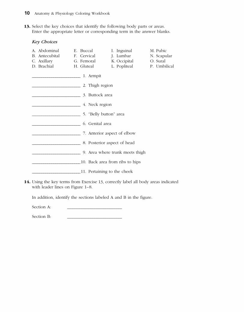
13. Select the key choices that identify the following body parts or areas. Enter the appropriate letter or corresponding term in the answer blanks.
Key Choices
A. Abdominal E. Buccal I. Inguinal M. PubicB. Antecubital F. Cervical J. Lumbar N. ScapularC. Axillary G. Femoral K. Occipital O. SuralD. Brachial H. Gluteal L. Popliteal P. Umbilical
_________________________ 1. Armpit
_________________________ 2. Thigh region
_________________________ 3. Buttock area
_________________________ 4. Neck region
_________________________ 5. “Belly button” area
_________________________ 6. Genital area
_________________________ 7. Anterior aspect of elbow
_________________________ 8. Posterior aspect of head
_________________________ 9. Area where trunk meets thigh
_________________________ 10. Back area from ribs to hips
_________________________ 11. Pertaining to the cheek
14. Using the key terms from Exercise 13, correctly label all body areas indicated with leader lines on Figure 1–8.
In addition, identify the sections labeled A and B in the figure.
Section A: _________________________
Section B: _________________________
10 Anatomy & Physiology Coloring Workbook

Chapter 1 The Human Body: An Orientation 11
A
A
BB
Figure 1–8
15. From the key choices, select the body cavities and the cavity subdivision where the following surgical procedures would occur. Insert the correct letter(s) or term(s) in the answer blanks. Be precise. Items may have more than one answer.
Key Choices
A. Abdominal C. Dorsal E. Spinal G. VentralB. Cranial D. Pelvic F. Thoracic
_________________________ 1. Removal of the uterus, or womb
_________________________ 2. Coronary bypass surgery (heart surgery)
_________________________ 3. Removal of a serious brain tumor
_________________________ 4. Removal of a “hot” appendix
_________________________ 5. A stomach ulcer operation
16. Complete the following statements by choosing an anatomical term from the key choices. Enter the appropriate letter or term in the answer blanks.
Key Choices
A. Anterior D. Inferior G. Posterior J. SuperiorB. Distal E. Lateral H. Proximal K. TransverseC. Frontal F. Medial I. Sagittal
_________________________ 1.
_________________________ 2.
_________________________ 3.
_________________________ 4.
_________________________ 5.
_________________________ 6.
_________________________ 7.
_________________________ 8.
_________________________ 9.
_________________________ 10.
_________________________ 11.
In the anatomical position, the face and palms are on the (1) body surface, the buttocks and shoulder blades are on
the (2) body surface, and the top of the head is the most (3) part of the body. The ears are (4) to the shoulders
and (5) to the nose. The heart is (6) to the spine and (7) to the lungs. The elbow is (8) to the fingers but (9) to the shoulder. In humans, the dorsal surface can also
be called the (10) surface; however, in fourlegged animals, the dorsal surface is the (11) surface.
12 Anatomy & Physiology Coloring Workbook
_________________________ 12.
_________________________ 13.
_________________________ 14.
_________________________ 15.
17. Using the key choices, identify the body cavities where the following body organs are located. Enter the appropriate letter or term in the answer blanks. Letters or terms can be used more than once.
Key Choices
A. Abdominopelvic B. Cranial C. Spinal D. Thoracic
_________________________ 1. Stomach _________________________ 7. Bladder
_________________________ 2. Small intestine _________________________ 8. Trachea
_________________________ 3. Large intestine _________________________ 9. Lungs
_________________________ 4. Spleen _________________________ 10. Pituitary gland
_________________________ 5. Liver _________________________ 11. Rectum
_________________________ 6. Spinal cord _________________________ 12. Ovaries
18. Number the following structures, from darkest (black) to lightest (white), as they would appear on an Xray. Number the darkest one 1, the next darkest 2, etc. (Hint: Denser structures appear lighter).
_________________________ A. Soft tissue
_________________________ B. Femur (bone of the thigh)
_________________________ C. Air in lungs
_________________________ D. Gold (metal) filling in a tooth
19. A jogger has stepped in a pothole and sprained his ankle. What organ systems have suffered damage?
AT THE CLINIC
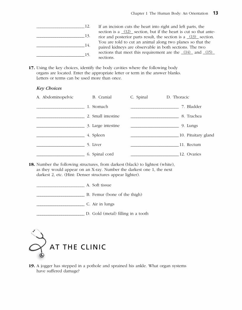
Chapter 1 The Human Body: An Orientation 13
If an incision cuts the heart into right and left parts, the section is a (12) section, but if the heart is cut so that anterior and posterior parts result, the section is a (13) section. You are told to cut an animal along two planes so that the paired kidneys are observable in both sections. The two sections that meet this requirement are the (14) and (15) sections.

20. A newborn baby is unable to hold down any milk. Examination reveals a developmental disorder in which the esophagus fails to connect to the stomach. What survival needs are most immediately threatened?
21. The Chan family was traveling in their van and had a minor accident. The children in the backseat were wearing lap belts, but they still sustained bruises around the abdomen and had some internal organ injuries. Why is this area more vulnerable to damage than others?
22. John, a patient at Jones City Hospital, is in tough shape. He has a hernia in his inguinal region, pain from an infected kidney in his lumbar region, and severe bruises and swelling in his pubic region. Explain where each of these regions is located.
23. The hormone thyroxine is released in response to a pituitary hormone called TSH. As thyroxine levels increase in the blood, they exert negative feedback on the release of TSH by the pituitary gland. What effect will this have on the release of TSH?
24. In congestive heart failure, the weakened heart is unable to pump with sufficient strength to empty its own chambers. As a result, blood backs up in the veins, blood pressure rises, and circulation is impaired. Describe what will happen as this situation worsens owing to positive feedback. Then, predict how a heartstrengthening medication will reverse the positive feedback.
14 Anatomy & Physiology Coloring Workbook

25. The following advanced imaging techniques are discussed in the text: CT, DSA, PET, and MRI. Which of these techniques uses Xray? Which uses radio waves and magnetic fields? Which uses radioisotopes? Which displays body regions in sections? (You may have more than one answer for each question.)
26. A patient reports stabbing pains in the right hypochondriac region. The medical staff suspects gallstones. What region of the body will be examined?
27. Mr. Harvey, a computer programmer, has been complaining of numbness and pain in his right hand. His nurse practitioner diagnoses his problem as carpal tunnel syndrome and prescribes use of a splint. Where will Mr. Harvey apply the splint?
28. Mrs. Gallo's physician suspects that she is showing the initial signs of multiple sclerosis, a disease characterized by the formation of hardened plaques in the insulating sheaths surrounding nerve fibers. What medical imaging technique will the physician probably order to determine if such plaques are present?
Chapter 1 The Human Body: An Orientation 15
THE FINALE: MULTIPLE CHOICE
1. Which of the following activities would not represent an anatomical study?
A. Making a section through the heart to observe its interior
B. Drawing blood from recently fed laboratory animals at timed intervals to determine their blood sugar levels
C. Examining the surface of a bone
D. Viewing muscle tissue through a microscope
2. The process that increases the size of the body or its number of cells is:
A. metabolism. C. growth.
B. responsiveness. D. digestion.
3. Which of the following is (are) involved in maintaining homeostasis?
A. Effector D. Feedback
B. Control center E. Lack of change
C. Receptor
29. Select the best answer or answers from the choices given.

16 Anatomy & Physiology Coloring Workbook
4. When a capillary is damaged, a platelet plug is formed. The process involves platelets sticking to each other. The more platelets that stick together, the more the plug attracts additional platelets. This is an example of:
A. negative feedback.
B. positive feedback.
5. A coronal plane through the head:
A. could pass through both the nose and the occiput.
B. could pass through both ears.
C. must pass through the mouth.
D. could lie in a horizontal plane.
6. Which of the following statements is (are)correct?
A. The brachium is proximal to the antebrachium.
B. The femoral region is superior to the tarsal region.
C. The orbital region is inferior to the buccal region.
D. The axillary region is lateral to the sternal region.
E. The crural region is posterior to the sural region.
7. Which of the following body regions is (are) found on the torso?
A. Gluteal D. Acromial
B. Inguinal E. Olecranal
C. Popliteal
8. A neurosurgeon orders a spinal tap for a patient. Into what body cavity will the needle be inserted?
A. Ventral D. Cranial
B. Thoracic E. Pelvic
C. Dorsal
9. An accident victim has a collapsed lung. Which cavity has been entered?
A. Mediastinal D. Vertebral
B. Pericardial E. Ventral
C. Pleural
10. Which body system would be affected by degenerative cartilage?
A. Muscular D. Skeletal
B. Nervous E. Lymphatic
C. Cardiovascular
11. The position of the heart relative to the structures around it would be described accurately as:
A. deep to the sternum (breast bone).
B. lateral to the lungs.
C. superior to the diaphragm.
D. inferior to the ribs.
E. anterior to the vertebral column.
12. What term(s) could be used to describe the position of the nose?
A. Intermediate to the eyes
B. Inferior to the brain
C. Superior to the mouth
D. Medial to the ears
E. Anterior to the ears
13. The radiographic technique used to provide information about blood flow is:
A. DSR. D. ultrasonography.
B. CT. E. any Xray technique.
C. PET.
14. A patient complains of pain in the lower right quadrant. Which system is most likely to be involved?
A. Respiratory D. Skeletal
B. Digestive E. Muscular
C. Urinary
15. Harry was sweating profusely as he ran in the 10K race. The sweat glands producing the sweat would be considered which part of a feedback system?
A. Stimulus C. Control center
B. Effectors D. Receptors
Everything in the universe is composed of one or more elements, the unique building blocks of all matter. Although more than 100 elemental substances exist, only four of these (carbon, hydrogen, oxygen, and nitrogen) make up more than 96% of all living material.
The student activities in this chapter consider basic concepts of both inorganic and organic chemistry. Chemistry is the science that studies the composition of matter. Inorganic chemistry studies the chemical composition of nonliving substances that (generally) do not contain carbon. Organic chemistry studies the carbon-based chemistry (or biochemistry) of living organisms, whether they are maple trees, fish, or humans.
Understanding of atomic structure, bonding behavior of elements, and the structure and activities of the most abundant biological molecules (proteins, fats, carbohy-drates, and nucleic acids) is tested in various ways. Mastering these concepts is necessary to understand how the body functions.
CONCEPTS OF MATTER AND ENERGY
1. Select all phrases that apply to each of the following statements and insert the letters in the answer blanks.
______ 1. The energy located in the bonds of food molecules: A. is called thermal energy. C. causes molecular movement. B. is a form of potential energy. D. can be transformed to the
bonds of ATP (adenosine triphosphate).
______ 2. Heat is: A. thermal energy. C. kinetic energy. B. infrared radiation. D. molecular movement.
______ 3. Whenever energy is transformed: A. the amount of useful energy decreases. C. some energy is created. B. some energy is lost as heat. D. some energy is destroyed.
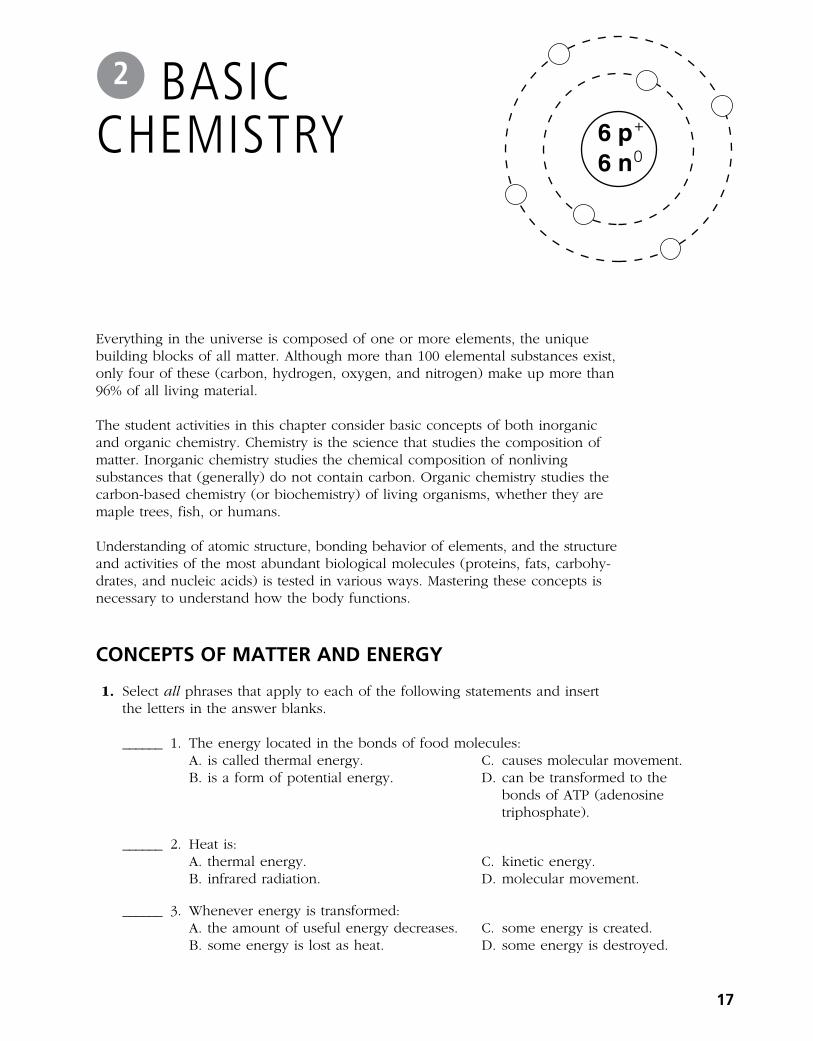
6 p6 n
+
0
BASIC CHEMISTRY
2
17

2. Use choices from the key to identify the energy form in use in each of the following examples. Items may have more than one answer.
Key Choices
A. Chemical B. Electrical C. Mechanical D. Radiant
_________________________ 1. Chewing food
_________________________ 2. Vision (two types of energy, please—think!)
_________________________ 3. Bending your fingers to make a fist
_________________________ 4. Breaking the bonds of ATP molecules to energize your muscle cells to make that fist
_________________________ 5. Getting a tan on the beach
COMPOSITION OF MATTER
3. Complete the following table by inserting the missing words.
Particle Location Electrical charge Mass (amu)
+ 1
Neutron
Orbitals
4. Insert the chemical symbol (the chemist’s shorthand) in the answer blank for each of the following elements.
______ 1. Oxygen ______ 4. Iodine ______ 7. Calcium ______ 10. Magnesium
______ 2. Carbon ______ 5. Hydrogen ______ 8. Sodium ______ 11. Chlorine
______ 3. Potassium ______ 6. Nitrogen ______ 9. Phosphorus ______ 12. Iron
5. Using the key choices, complete the crossword puzzle by answering each of the clues provided.
Key Choices
Atom Element Ion Molecule ProtonsElectrons Energy Matter Neutrons Valence
Across 4. The smallest particle of an element that retains the properties of the element. 7. Formed when atoms combine chemically. 8. Uncharged subatomic particles, forming part of an atom. 9. Subatomic particles that determine an atom’s chemical behavior or
bonding ability.
18 Anatomy & Physiology Coloring Workbook
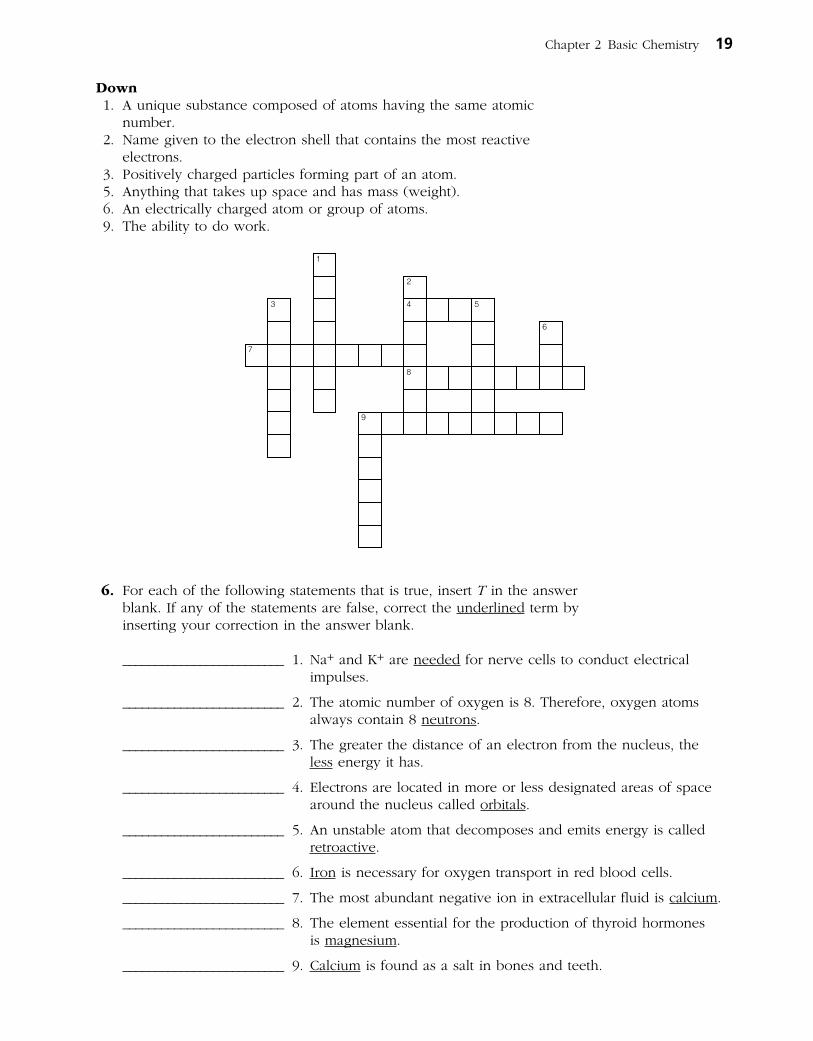
Down 1. A unique substance composed of atoms having the same atomic
number. 2. Name given to the electron shell that contains the most reactive
electrons. 3. Positively charged particles forming part of an atom. 5. Anything that takes up space and has mass (weight). 6. An electrically charged atom or group of atoms. 9. The ability to do work.
1
2
3 4 5
6
7
8
9
6. For each of the following statements that is true, insert T in the answer blank. If any of the statements are false, correct the underlined term by inserting your correction in the answer blank.
_________________________ 1. Na+ and K+ are needed for nerve cells to conduct electrical impulses.
_________________________ 2. The atomic number of oxygen is 8. Therefore, oxygen atoms always contain 8 neutrons.
_________________________ 3. The greater the distance of an electron from the nucleus, the less energy it has.
_________________________ 4. Electrons are located in more or less designated areas of space around the nucleus called orbitals.
_________________________ 5. An unstable atom that decomposes and emits energy is called retroactive.
_________________________ 6. Iron is necessary for oxygen transport in red blood cells.
_________________________ 7. The most abundant negative ion in extracellular fluid is calcium.
_________________________ 8. The element essential for the production of thyroid hormones is magnesium.
_________________________ 9. Calcium is found as a salt in bones and teeth.
Chapter 2 Basic Chemistry 19

MOLECULES, CHEMICAL BONDS, AND CHEMICAL REACTIONS
7. Match the terms in Column B to the chemical equations listed in Column A. Enter the correct letter or term in the answer blanks.
Column A Column B
_________________________ 1. A + B AB A. Decomposition
_________________________ 2. AB + CD AD + CB B. Exchange
_________________________ 3. XY X + Y C. Synthesis
8. Figure 2–1 is a diagram of an atom. Select two different colors and use them to color the coding circles and corresponding structures on the figure. Com plete this exercise by responding to the questions that follow, referring to the atom in this figure. Insert your answers in the answer blanks provided.
○ Nucleus
○ Electrons
20 Anatomy & Physiology Coloring Workbook
6 p6 n
+
0
Figure 2–1
1. What is the atomic number of this atom? _________________________
2. What is its atomic mass? _________________________
3. Which atom is this? _________________________
4. If this atom had one additional neutron but the other subatomic particles remained the same as shown, this slightly different atom (of the same element) would be called a(n) _________________________
5. Is this atom chemically active or inert? _________________________
6. How many electrons would be needed to fill its outer (valence) shell? _____________________
7. Would this atom most likely take part in forming ionic or
covalent bonds? _______________________ Why? _______________________
_____________________________________________________________________
9. Both H2O2 and 2OH– are compound molecules composed of the same two hydrogen atoms and two oxygen atoms. Briefly explain how these molecules are different:
__________________________________________________________________________
__________________________________________________________________________
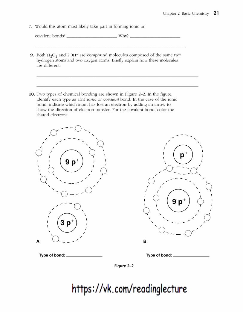
10. Two types of chemical bonding are shown in Figure 2–2. In the figure, identify each type as a(n) ionic or covalent bond. In the case of the ionic bond, indicate which atom has lost an electron by adding an arrow to show the direction of electron transfer. For the covalent bond, color the shared electrons.
Chapter 2 Basic Chemistry 21
p9 p+
9 p+
+
3 p+
Type of bond: Type of bond:
A B
Figure 2–2

11. Figure 2–3 illustrates five water molecules held together by hydrogen bonds.
(A) Select three different colors and use them to color the coding circles and corresponding structures on the figure.
○ Oxygen
○ Hydrogen
○ Hydrogen bonds
(B) Label the positive and negative poles (ends) in one of the water molecules.
12. Circle each structural formula that is likely to be a polar covalent compound.
13. Respond to the instructions following the equation:
H2CO3
11. Figure 2–3 illustrates five water molecules held together by hydrogenbonds. First, correctly identify the oxygen and hydrogen atoms both bycolor and by inserting their atomic symbols on the appropriate circles(atoms). Then label the following structures in the figure:
o Oxygen
o Hydrogen
o Positive pole (end)
o Negative pole (end)
o Hydrogen bonds
12. Circle each structural formula that is likely to be a polar covalent compound.
13. Respond to the instructions following the equation:
H2CO3 � H+ + HCO3–
1. In the space provided, list the chemical formula(s) of compounds. _______________________
2. In the space provided, list the chemical formula(s) of ions. _______________________
3. Circle the product(s) of the reaction.
4. Modify the equation by adding a colored arrow in the proper place to indicate that the reaction is reversible.
22 Anatomy & Physiology Coloring Workbook
A Cl C Cl
Cl
Cl
B H Cl C N
H
H
H
D Cl Cl E O
H
H
Figure 2–3
H+ + HCO3–
1. In the space provided, list the chemical formula(s) of compounds. _______________________
2. In the space provided, list the chemical formula(s) of ions. _______________________
3. Circle the product(s) of the reaction.
4. Modify the equation by adding a colored arrow in the proper place to indicate that the reaction is reversible.
22 Anatomy & Physiology Coloring Workbook
A Cl C Cl
Cl
Cl
B H Cl C N
H
H
H
D Cl Cl E O
H
H
Figure 2–3
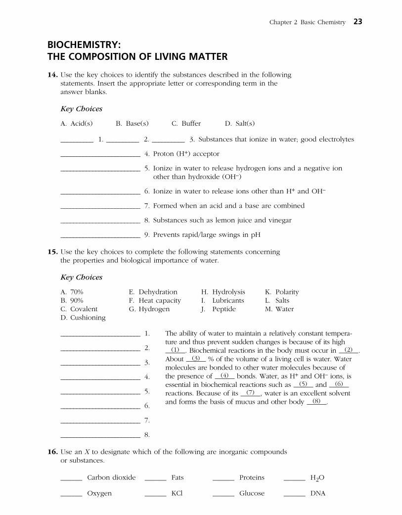
BIOCHEMISTRY: THE COMPOSITION OF LIVING MATTER
14. Use the key choices to identify the substances described in the following statements. Insert the appropriate letter or corresponding term in the answer blanks.
Key Choices
A. Acid(s) B. Base(s) C. Buffer D. Salt(s)
__________ 1. __________ 2. __________ 3. Substances that ionize in water; good electrolytes
_________________________ 4. Proton (H+) acceptor
_________________________ 5. Ionize in water to release hydrogen ions and a negative ion other than hydroxide (OH–)
_________________________ 6. Ionize in water to release ions other than H+ and OH–
_________________________ 7. Formed when an acid and a base are combined
_________________________ 8. Substances such as lemon juice and vinegar
_________________________ 9. Prevents rapid/large swings in pH
15. Use the key choices to complete the following statements concerning the properties and biological importance of water.
Key Choices
A. 70% E. Dehydration H. Hydrolysis K. PolarityB. 90% F. Heat capacity I. Lubricants L. SaltsC. Covalent G. Hydrogen J. Peptide M. WaterD. Cushioning
_________________________ 1.
_________________________ 2.
_________________________ 3.
_________________________ 4.
_________________________ 5.
_________________________ 6.
_________________________ 7.
_________________________ 8.
16. Use an X to designate which of the following are inorganic compounds or substances.
______ Carbon dioxide ______ Fats ______ Proteins ______ H2O
______ Oxygen ______ KCl ______ Glucose ______ DNA
Chapter 2 Basic Chemistry 23
The ability of water to maintain a relatively constant tempera- ture and thus prevent sudden changes is because of its high
(1) . Biochemical reactions in the body must occur in (2) . About (3) % of the volume of a living cell is water. Water molecules are bonded to other water molecules because of the presence of (4) bonds. Water, as H+ and OH– ions, is essential in biochemical reactions such as (5) and (6) reactions. Because of its (7) , water is an excellent solvent and forms the basis of mucus and other body (8) .

17. Using the key choices, fully characterize weak and strong acids.
Key Choices
A. Act as part of a buffer system E. Ionize at low pH B. Ionize completely in water F. Ionize at pH 7 C. Ionize incompletely in water G. When placed in water, always act to change the pH D. Ionize at high pH
Weak acid: _____________________ Strong acid: _____________________
18. Match the terms in Column B to the descriptions provided in Column A. Enter the correct letter(s) or term(s) in the answer blanks. Items may have more than one answer.
Column A
_________________________ 1. Building blocks of carbohydrates
_________________________ 2. Building blocks of fat
_________________________ 3. Building blocks of protein
_________________________ 4. Building blocks of nucleic acids
_________________________ 5. Cellular cytoplasm is primarily composed of this substance
_________________________ 6. The single most important fuel source for body cells
_________________________ 7. Not soluble in water
_________________________ 8. Contain C, H, and O in the ratio CH2O
_________________________ 9. Contain C, H, and O, but have relatively small amounts of oxygen
_________________________ 10. _________________________ 11. These building blocks contain N in addition to C, H, and O
_________________________ 12. Contain P in addition to C, H, O, and N
_________________________ 13. Used to insulate the body and found in all cell membranes
_________________________ 14. Primary components of meat
_________________________ 15. Primary components of bread and lollipops
_________________________ 16. Primary components of egg yolk and peanut oil
_________________________ 17. Include collagen and hemoglobin
_________________________ 18. Class that usually includes cholesterol
_________________________ 19. The alpha helix and beta pleated sheet are both examples of the secondary structure of these molecules.
Column B
A. Amino acids
B. Carbohydrates
C. Lipids (fats)
D. Fatty acids
E. Glycerol
F. Nucleotides
G. Monosaccharides
H. Proteins
24 Anatomy & Physiology Coloring Workbook

19. Using the key choices, correctly select all terms that correspond to the following descriptions. Insert the correct letter(s) or their corresponding term(s) in the answer blanks. Items may have more than one answer.
Key Choices
A. Cholesterol D. Enzyme G. Hormones J. MaltoseB. Collagen E. Glycogen H. Keratin K. RNAC. DNA F. Hemoglobin I. Lactose L. Starch
_________________________ 1. Example(s) of fibrous (structural) proteins
_________________________ 2. Example(s) of globular (functional) proteins
_________________________ 3. Biological catalyst
_________________________ 4. Plant storage carbohydrate
_________________________ 5. Animal storage carbohydrate
_________________________ 6. The material of the genes
_________________________ 7. A steroid
_________________________ 8. Double sugars, or disaccharides
20. Five simplified diagrams of biological molecules are represented in Figure 2–4.
(A) Iden tify the molecules and insert the correct names in the answer blanks on the figure.
(B) Select a different color for each molecule listed below and use them to color the coding circles and the corresponding molecules on the illustration.
○ Fat ○ Nucleotide ○ Monosaccharide
○ Globular protein ○ Polysaccharide
Chapter 2 Basic Chemistry 25
Figure 2–4
A
D E
B C

21. Circle the term that does not belong in each of the following groupings. Then, fill in the answer blanks with the correct group name.
1. Adenine Guanine Glucose Thymine Group: _________
2. DNA Ribose Phosphate Deoxyribose Group: _________
3. Galactose Glycogen Fructose Glucose Group: _________
4. Amino acid Polypeptide Glycerol Hemoglobin Group: _________
5. Glucose Sucrose Lactose Maltose Group: _________
22. For each true statement, insert T in the answer blank. If any are false, correct the underlined term and insert your correction in the answer blank.
_________________________ 1. Phospholipids are polarized molecules.
_________________________ 2. Steroids are the major form in which body fat is stored.
_________________________ 3. Water is the most abundant compound in the body.
_________________________ 4. Nonpolar molecules are generally soluble in water.
_________________________ 5. The bases of RNA are A, G, C, and U.
_________________________ 6. The universal energy currency of living cells is RNA.
_________________________ 7. RNA is single stranded.
_________________________ 8. The four elements that make up more than 90% of living matter are C, H, N, and Na.
23. Figure 2–5 shows the molecular structure of DNA, a nucleic acid.
A. First, identify the two unnamed nitrogen (N) bases and insert their names and symbols in the two blanks beside the color-coding circles.
B. Complete the identification of the bases on the diagram by inserting the correct symbols in the appropriate spaces on the right side of the diagram.
C. Select different colors and color the coding circles and the corresponding parts of the diagram.
○ Deoxyribose sugar (d-R) ○ Adenine (A) ○ ( )
○ Phosphate (P) ○ Cytosine (C) ○ ( )
D. Label one deoxyribose (d-R) sugar unit and one phosphate (P) unit of the “backbones” of the DNA structure by inserting leader lines and labels on the diagram, then circle the associated nucleotide.
E. Answer the questions following Figure 2–5 by writing your answers in the answer blanks.
26 Anatomy & Physiology Coloring Workbook

Chapter 2 Basic Chemistry 27
1. Name the bonds that help to hold the two DNA strands together. __________________________
2. Name the three-dimensional shape of the DNA molecule. _________________________________
3. How many base pairs are present in this segment of a DNA model? _______________________
4. What is the term that describes the pattern or base-pairing? ________________________________
Figure 2–5
C
G
A
A
G
G
A
T
A
T
C
T

24. The biochemical reaction shown in Figure 2–6 represents the complete digestion of a polymer (a large molecule as consumed in food) down to its con stituent monomers, or building blocks.
(A) Select two colors and color the coding circles and the molecules. (B) Select the one correct answer for each statement below and insert your
answer in the answer blank.
○ Monomer ○ Polymer
_________________________ 1. If starch is the polymer, the monomer is:
A. glycogen. B. amino acid. C. glucose. D. maltose.
_________________________ 2. During polymer digestion, water as H+ and OH– ions would:
A. be a product of the reaction.
B. act as a catalyst.
C. enter between monomers, bond to them, and keep them separated.
D. not be involved in this reaction.
_________________________ 3. Another name for the chemical digestion of polymers is:
A. dehydration. B. hydrolysis. C. synthesis. D. displacement.
_________________________ 4. If the monomers are amino acids, they may differ from each other by their:
A. R group. B. amino group. C. acid group. D. peptide bond.
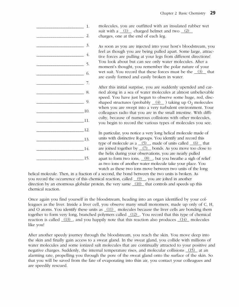
A Visualization Exercise for Biochemistry
. . . you are suddenly upended and are carried along in a sea of water molecules at almost unbelievable speed.
25. Complete the narrative by inserting the missing words in the answer blanks.
For this journey, you are miniaturized to the size of a very small molecule by colleagues who will remain in contact with you by radio. Your instructions are to play the role of a water molecule and to record any reactions that involve water molecules. Considering water molecules are polar
28 Anatomy & Physiology Coloring Workbook
Figure 2–6
INCREDIBLE JOURNEY
Chapter 2 Basic Chemistry 29
molecules, you are outfitted with an insulated rubber wet suit with a (1) charged helmet and two (2) charges, one at the end of each leg.
As soon as you are injected into your host’s bloodstream, you feel as though you are being pulled apart. Some large, attrac-tive forces are pulling at your legs from different directions! You look about but can see only water molecules. After a moment’s thought, you remember the polar nature of your wet suit. You record that these forces must be the (3) that are easily formed and easily broken in water.
After this initial surprise, you are suddenly upended and car-ried along in a sea of water molecules at almost unbelievable speed. You have just begun to observe some huge, red, disc-shaped structures (probably (4) ) taking up O2 molecules when you are swept into a very turbulent environment. Your colleagues radio that you are in the small intestine. With diffi-culty, because of numerous collisions with other molecules, you begin to record the various types of molecules you see.
In particular, you notice a very long helical molecule made of units with distinctive R-groups. You identify and record this type of molecule as a (5) , made of units called (6) that are joined together by (7) bonds. As you move too close to the helix during your observations, you are nearly pulled apart to form two ions, (8) , but you breathe a sigh of relief as two ions of another water molecule take your place. You watch as these two ions move between two units of the long
helical molecule. Then, in a fraction of a second, the bond between the two units is broken. As you record the occurrence of this chemical reaction, called (9) , you are jolted in another direction by an enormous globular protein, the very same (10) that controls and speeds up this chemical reaction.
Once again you find yourself in the bloodstream, heading into an organ identified by your col-leagues as the liver. Inside a liver cell, you observe many small monomers, made up only of C, H, and O atoms. You identify these units as (11) molecules because the liver cells are bonding them together to form very long, branched polymers called (12) . You record that this type of chemical reaction is called (13) , and you happily note that this reaction also produces (14) molecules like you!
After another speedy journey through the bloodstream, you reach the skin. You move deep into the skin and finally gain access to a sweat gland. In the sweat gland, you collide with millions of water molecules and some ionized salt molecules that are continually attracted to your positive and negative charges. Suddenly, the internal temperature rises, and molecular collisions (15) at an alarming rate, propelling you through the pore of the sweat gland onto the surface of the skin. So that you will be saved from the fate of evaporat ing into thin air, you contact your colleagues and are speedily rescued.
_________________________ 1.
_________________________ 2.
_________________________ 3.
_________________________ 4.
_________________________ 5.
_________________________ 6.
_________________________ 7.
_________________________ 8.
_________________________ 9.
_________________________ 10.
_________________________ 11.
_________________________ 12.
_________________________ 13.
_________________________ 14.
_________________________ 15.
26. It is determined that a patient is in acidosis. What does this mean, and would you treat the condition with a chemical that would raise or lower the pH?
27. A newborn is diagnosed with sickle cell anemia, a genetic disease in which substitution of one amino acid results in abnormal hemoglobin. Explain to the parents how the substitution can have such a drastic effect on the structure of the protein.
28. Johnny’s body temperature is spiking upward. When it reaches 104°F, his mother puts in a call to the pediatrician. She is advised to give Johnny children’s acetaminophen or ibuprofen and sponge his body with cool to tepid water to prevent a further rise in temperature. How might a fever (excessively high body temperature) be detrimental to Johnny’s welfare?
29. Stanley has indigestion and is doubled over with pain. How could an antacid reduce his stomach discomfort?
30. Explain why the formation of ATP from ADP (adenosine diphosphate) and Pi requires more energy than the amount released for cellular use when ATP is broken down.
30 Anatomy & Physiology Coloring Workbook
AT THE CLINIC

31. Select the best answer or answers from the choices given.
Chapter 2 Basic Chemistry 31
THE FINALE: MULTIPLE CHOICE
1. Which of the following is (are) true concern-ing the atomic nucleus?
A. Contains the mass of the atom
B. The negatively charged particles are here
C. Particles can be ejected
D. Contains particles that determine atomic number
E. Contains particles that interact with other atoms
2. Organic compounds include:
A. water. D. carbonic acid.
B. carbon dioxide. E. glycerol.
C. oxygen.
3. Important functions of water include:
A. cushioning.
B. transport medium.
C. participation in chemical reactions.
D. solvent for sugars, salts, and other solutes.
E. reducing temperature fluctuations.
4. Which of the elements listed is the most abundant extracellular anion?
A. Phosphorus D. Chloride
B. Sulfur E. Calcium
C. Potassium
5. The element essential for normal thyroid function is:
A. iodine.
B. iron.
C. copper.
D. selenium.
E. zinc.
6. Alkaline substances include:
A. gastric juice. D. orange juice.
B. water. E. ammonia.
C. blood.
7. Which of the following is (are) not a monosaccharide?
A. Glucose D. Glycogen
B. Fructose E. Deoxyribose
C. Sucrose
8. Which is a building block of neutral fats?
A. Ribose D. Glycine
B. Guanine E. Glucose
C. Glycerol
9. Which of the following is primarily responsi-ble for the helical structure of a polypeptide chain?
A. Hydrogen bonding
B. Tertiary folding
C. Peptide bonding
D. Quaternary associations
E. Complementary base pairing
10. Which of the following is (are) not true of RNA?
A. Double stranded
B. Contains cytosine
C. Directs protein synthesis
D. Found primarily in the nucleus
E. Can act as an enzyme
11. DNA:
A. contains uracil. C. is the “genes”.
B. is a helix. D. contains ribose.

12. Glucose is to starch as:
A. a steroid is to a lipid.
B. a nucleotide is to nucleic acid.
C. an amino acid is to a protein.
D. a polypeptide is to an amino acid.
13. An organic sample is analyzed and shown to have C, H, O, N, and P as its constituents. The organic molecule is identified as a:
A. carbohydrate. C. lipid.
B. protein. D. nucleic acid.
14. Which of the following forms of energy is the stimulus for vision?
A. Mechanical C. Electrical
B. Light D. Chemical
15. Which of the following describe energy?
A. Has mass
B. Massless
C. Occupies space
D. Puts matter into motion
16. Which of the following is (are) a synthetic reaction?
A. Glucose to glycogen
B. Glucose and fructose to sucrose
C. Starch to glucose
D. Amino acids to dipeptide
32 Anatomy & Physiology Coloring Workbook
The basic unit of structure and function in the human body is the cell. Each of a cell’s parts, or organelles, as well as the entire cell, is organized to perform a specific function. Cells have the ability to metabolize, grow and reproduce, move, and respond to stimuli. The cells of the body differ in shape, size, and in specific roles in the body. Cells that are similar in structure and function form tissues, which, in turn, construct the various body organs.
Student activities in this chapter include questions relating to the structure and function of the generalized animal cell and to the general arrangement of tissues and their contribution to the activities of the various body organs.
CELLS
Overview
1. Answer the following questions by inserting your responses in the answer blanks.
_________________________ 1.
_________________________ 2.
_________________________ 3.
_________________________ 4.
_________________________ 5.
_________________________ 6.
_________________________ 7.
_________________________ 8.
_________________________ 9.
_________________________ 10.
33
CELLS AND TISSUES
3
1–4. Name the four elements that make up the bulk of living matter.
5. Name the single most abundant molecule in living matter.
6. Name the trace element most important for making bones hard.
7. Name the element, found in small amounts in the body, that is needed to make hemoglobin for oxygen transport.
8–12. Although there are many specific “jobs” that certain cells are able to do, name five functions common to all cells.
_________________________ 11.
_________________________ 12.
_________________________ 13.
_________________________ 14.
_________________________ 15.
_________________________ 16.
_________________________ 17.
Anatomy of a Generalized Cell
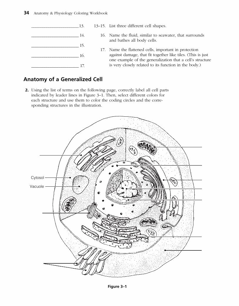
2. Using the list of terms on the following page, correctly label all cell parts indicated by leader lines in Figure 3–1. Then, select different colors for each structure and use them to color the coding circles and the corre-sponding structures in the illustration.
34 Anatomy & Physiology Coloring Workbook
13–15. List three different cell shapes.
16. Name the fluid, similar to seawater, that surrounds and bathes all body cells.
17. Name the flattened cells, important in protection against damage, that fit together like tiles. (This is just one example of the generalization that a cell’s structure is very closely related to its function in the body.)
Cytosol
Vacuole
Figure 3–1
○ Plasma membrane ○ Mitochondrion
○ Centriole(s) ○ Nuclear membrane
○ Chromatin thread(s) ○ Nucleolus
○ Golgi apparatus ○ Rough endoplasmic reticulum (Rough ER)
○ Microvilli ○ Smooth endoplasmic reticulum (Smooth ER)
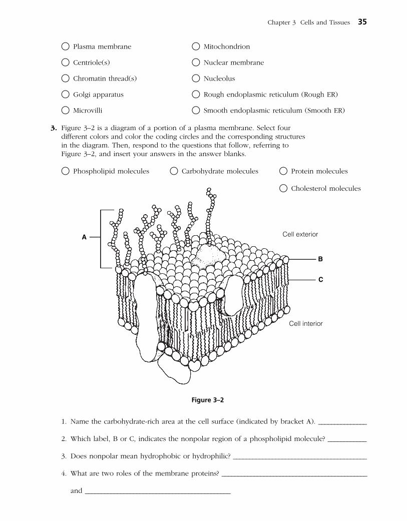
3. Figure 3–2 is a diagram of a portion of a plasma membrane. Select four different colors and color the coding circles and the corresponding structures in the diagram. Then, respond to the questions that follow, referring to Figure 3–2, and insert your answers in the answer blanks.
○ Phospholipid molecules ○ Carbohydrate molecules ○ Protein molecules
1. Name the carbohydrate-rich area at the cell surface (indicated by bracket A). _______________
2. Which label, B or C, indicates the nonpolar region of a phospholipid molecule? ____________
3. Does nonpolar mean hydrophobic or hydrophilic? _________________________________________
4. What are two roles of the membrane proteins? _____________________________________________
and _____________________________________________
Chapter 3 Cells and Tissues 35
A
C
B
Cell exterior
Cell interior
Figure 3–2
○ Cholesterol molecules

4. Based on Figure 3–3, answer the following:(A) Label the specializations of the plasma membrane.(B) Color the coding circles and the corresponding cell parts.(C) Answer the questions provided below.
○ Nucleus ○ Nucleolus ○ Mitochondrion ○ ER
1. What type of cell function(s) does the presence of microvilli typically
indicate? __________________________________________________________________________________
2. Which cell junction forms an impermeable barrier? _________________________________________
3. Which cell junction is an anchoring junction? _______________________________________________
4. Which junction has linker proteins spanning the intercellular space? ________________________
5. Which cell junction is not illustrated, and what is its function? ______________________________
______________________________________________________________________________________________
6. Which two types of membrane junctions would you expect to find between cells of the heart?
_______________________________________________ and ____________________________________________
36 Anatomy & Physiology Coloring Workbook
A
B
C
Figure 3–3

5. Relative to cellular organelles, circle the term or phrase that does not belong in each of the following groupings. Then, fill in the answer blanks with the correct group name.
1. Peroxisomes Enzymatic breakdown Centrioles Lysosomes Group: _________
2. Microtubules Intermediate filaments Microfilaments Cilia Group: _________
3. Ribosomes Smooth ER Rough ER Amino acids Group: _________
4. Double membrane Cristae ATP production Vitamin A storage Group: _________
5. Centrioles Mitochondria Cilia Flagella Group: _________
6. ER Ribosomes Transport vesicles Golgi apparatus Group: ________
7. Nuclear pores DNA Lysosomes Chromatin Nucleolus Group: _________
6. Name the cytoskeletal element (microtubules, microfilaments, or intermediate filaments) described by each of the following phrases.
_________________________ 1. Give the cell its shape
_________________________ 2. Resist tension placed on a cell
_________________________ 3. Radiate from the cell center
_________________________ 4. Involved in moving intracellular structures
_________________________ 5. Are the most stable
_________________________ 6. Have the thickest diameter
7. Different organelles are abundant in different cell types. Match the cell types with their abundant organelles by selecting a letter or letters from the key choices. Items may have more than one answer.
Key Choices
A. Golgi apparatus C. Lysosomes E. Mitochondria G. Rough ERB. Intermediate filaments D. Microfilaments F. Peroxisomes H. Smooth ER
______ 1. Cell lining the small intestine (assembles fats)
______ 2. White blood cell; a phagocyte
______ 3. Liver cell that detoxifies carcinogens
______ 4. Muscle cell (contractile cell)
______ 5. Mucus-secreting cell (secretes a protein product)
______ 6. Cell at external skin surface (withstands friction and tension)
______ 7. Kidney tubule cell (makes and uses large amounts of ATP)
Chapter 3 Cells and Tissues 37

Cell Physiology
Membrane Transport
8. Figure 3–4 shows a semipermeable sac, containing 4% NaCl, 9% glucose, and 10% albumin, suspended in a solution with the following composition: 10% NaCl, 10% glucose, and 40% albumin. Assume the sac is permeable to all substances except albumin. Using the key choices, insert the letter indicating the correct event in the answer blanks.
Key Choices
A. Moves into the sac B. Moves out of the sac C. Does not move
_________________________ 1. Glucose _________________________ 3. Albumin
_________________________ 2. Water _________________________ 4. NaCl
9. Figure 3–5 shows three microscopic fields (A–C) containing red blood cells. Arrows indicate the direction of net osmosis. Respond to the following questions, referring to Figure 3–5, by inserting your responses in the spaces provided.
1. Which microscopic field contains a hypertonic solution? ___________________________________
The cells in this field are said to be ________________________________________________________
2. Which microscopic field contains an isotonic bathing solution? _____________________________
What does isotonic mean? _________________________________________________________________
___________________________________________________________________________________________
3. Which microscopic field contains a hypotonic solution? ____________________________________
What is happening to the cells in this field and why? ______________________________________
38 Anatomy & Physiology Coloring Workbook
Solution contains: 10% NaCl 10% Glucose 40% Albumin
Sac contains: ��4% NaCl ��9% Glucose 10% Albumin
Figure 3–4

Chapter 3 Cells and Tissues 39
A B C
Figure 3–5
10. Figure 3–6 is a simplified diagram of the plasma membrane. Structure A represents channel proteins constructing a pore, structure B represents an ATP- energized solute pump, and structure C is a transport protein that does not depend on energy from ATP.
(A) Identify these structures and the membrane phospholipids by color.
○ Channel ○ Solute pump ○ Passive transport protein carrier ○ Phospholipids
(B) For each substance that moves through the plasma membrane, draw an arrow indicating its (most likely) direction of movement (into or out of the cell). If it is moved actively, use a red arrow; if it is moved passively, use a blue arrow. Color the coding arrows.
Active Passive
Figure 3–6
Cellexterior
Cellinterior
Steroid
Amino acid
Amino acid
Fat
H2OCO2
CO2
O2
O2
Cl–Na+
Na+
Glucose
Glucose
K+
K+
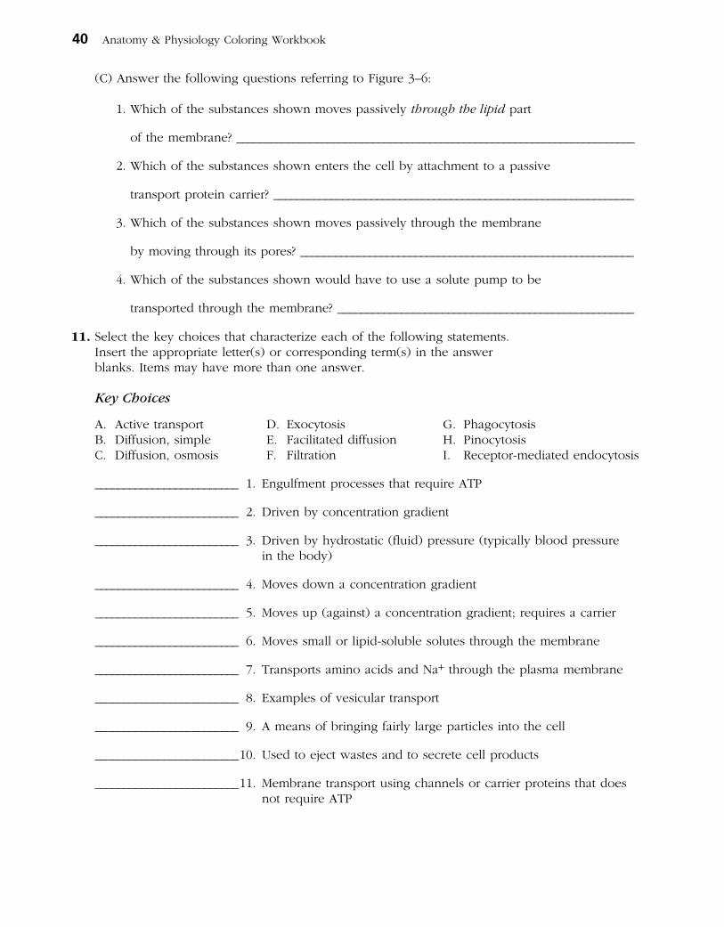
(C) Answer the following questions referring to Figure 3–6:
1. Which of the substances shown moves passively through the lipid part
of the membrane? ______________________________________________________________________
2. Which of the substances shown enters the cell by attachment to a passive
transport protein carrier? _______________________________________________________________
3. Which of the substances shown moves passively through the membrane
by moving through its pores? __________________________________________________________
4. Which of the substances shown would have to use a solute pump to be
transported through the membrane? ___________________________________________________
11. Select the key choices that characterize each of the following statements. Insert the appropriate letter(s) or corresponding term(s) in the answer blanks. Items may have more than one answer.
Key Choices
A. Active transport D. Exocytosis G. PhagocytosisB. Diffusion, simple E. Facilitated diffusion H. PinocytosisC. Diffusion, osmosis F. Filtration I. Receptor-mediated endocytosis
_________________________ 1. Engulfment processes that require ATP
_________________________ 2. Driven by concentration gradient
_________________________ 3. Driven by hydrostatic (fluid) pressure (typically blood pressure in the body)
_________________________ 4. Moves down a concentration gradient
_________________________ 5. Moves up (against) a concentration gradient; requires a carrier
_________________________ 6. Moves small or lipid-soluble solutes through the membrane
_________________________ 7. Transports amino acids and Na+ through the plasma membrane
_________________________ 8. Examples of vesicular transport
_________________________ 9. A means of bringing fairly large particles into the cell
_________________________ 10. Used to eject wastes and to secrete cell products
_________________________ 11. Membrane transport using channels or carrier proteins that does not require ATP
40 Anatomy & Physiology Coloring Workbook

Cell Division
12. The following statements provide an overview of the structure of DNA (genetic material) and its role in the body. Choose responses from the key choices that complete the statements. Insert the appropriate answers in the answer blanks.
Key Choices
A. Adenine G. Enzymes M. Nucleotides S. RibosomeB. Amino acids H. Genes N. Old T. Sugar (deoxyribose)C. Bases I. Growth O. Phosphate U. Template, or modelD. Codons J. Guanine P. Proteins V. ThymineE. Complementary K. Helix Q. Replication W. TranscriptionF. Cytosine L. New R. Repair X. Uracil
DNA molecules contain information for building specific (1) . In a three-dimensional view, a DNA molecule looks
like a spiral staircase; this is correctly called a (2) . The con- stant parts of DNA molecules are the (3) and (4) mole-cules, forming the DNA-ladder uprights, or backbones. The information of DNA is actually coded in the sequence of nitrogen-containing (5) , which are bound together to form the “rungs” of the DNA ladder. When the four DNA bases are combined in different three-base sequences, called triplets, different (6) of the protein are called for. It is said that the N-containing bases of DNA are (7) , which means that only certain bases can fit or interact together. Specifically, this means that (8) can bind with guanine, and adenine binds with (9) .
The production of proteins involves the cooperation of DNA and RNA. RNA is another type of nucleic acid that serves as a “molecular slave” to DNA. That is, it leaves the nucleus and carries out the instructions of the DNA for the building of a protein on a cytoplasmic structure called a (10) . When a cell is preparing to divide, in order for its daughter cells to have all its information, it must oversee the (11) of its DNA so that a “double dose” of genes is present for a brief period. For DNA synthesis to occur, the DNA must uncoil, and the bonds between the N bases must be broken. Then the two single strands of (12) each act as a (13) for the building of a whole DNA molecule. When completed, each DNA mole- cule formed is half (14) and half (15) . DNA replicates before a cell divides, ensuring that each daughter cell has a complete set of (16) . Cell division, which then fol lows, provides new cells so that (17) and (18) can occur.
Chapter 3 Cells and Tissues 41
_________________________ 1.
_________________________ 2.
_________________________ 3.
_________________________ 4.
_________________________ 5.
_________________________ 6.
_________________________ 7.
_________________________ 8.
_________________________ 9.
_________________________ 10.
_________________________ 11.
_________________________ 12.
_________________________ 13.
_________________________ 14.
_________________________ 15.
_________________________ 16.
_________________________ 17.
_________________________ 18.

13. Identify the phases of mitosis depicted in Figure 3–7 by inserting the correct name in the blank under the appropriate diagram. Then, select different colors to represent the structures listed below and use them to color in the coding circles and the corresponding structures in the illustration.
○ Nuclear membrane(s), if present ○ Centrioles
○ Nucleoli, if present ○ Spindle fibers
○ Chromosomes
42 Anatomy & Physiology Coloring Workbook
A
C
B
D
Figure 3–7

14. The following statements describe events that occur during the different phases of mitosis. Identify the phase by choosing the correct response(s) from the key choices and inserting the letter(s) or term(s) in the answer blanks. Items may have more than one answer.
Key Choices
A. Anaphase C. Prophase E. None of theseB. Metaphase D. Telophase
_________________________ 1. Chromatin coils and condenses to form deeply staining bodies.
_________________________ 2. Centromeres break, and chromosomes begin migration toward opposite poles of the cell.
_________________________ 3. The nuclear membrane and nucleoli reappear.
_________________________ 4. When chromosomes cease their poleward movement, this phase begins.
_________________________ 5. Chromosomes align on the equator of the spindle.
_________________________ 6. The nucleoli and nuclear membrane disappear.
_________________________ 7. The spindle forms through the migration of the centrioles.
_________________________ 8. Chromosomal material replicates.
_________________________ 9. Chromosomes first appear to be duplex structures.
_________________________ 10. Chromosomes attach to the spindle fibers.
_________________________ 11. A cleavage furrow forms during this phase.
_________________________ 12. The nuclear membrane is absent during the entire phase.
_________________________ 13. A cell carries out its usual metabolic activities.
15. Using the key choices, complete the crossword puzzle by answering each of the clues provided.
Key Choices
Anucleate Centromeres Cytoplasm NucleusAster Centrosomes Interphase ProphaseBinucleate Coiled Loose Spindle
Across 3. The structure that acts as a scaffolding for chromosomal attachment and
movement is called the _______. 4. If a cell undergoes nuclear division but not cytoplasmic division, the
product is a _________ cell. 7. Chromosomes attach to the spindle fibers by undivided structures
called ______.
Chapter 3 Cells and Tissues 43

Down 1. Cytokinesis is the division of the ______. 2. Division of the _____ is referred to as mitosis. 5. ______ is the period of cell life when the cell is not involved in division. 6. The major structural difference between chromatin and chromosomes is
that the latter are _____.
1 2
3
4 5 6
7
Protein Synthesis
16. Figure 3–8 is a diagram illustrating protein synthesis. (A) Select four different colors, and use them to color the coding circles
and the corresponding struc tures in the diagram. (B) Using the letters of the genetic code, label the nitrogen bases on
strand 2 of the DNA double helix, on the mRNA strands, and on the tRNA molecules.
(C) Answer the questions that follow referring to Figure 3–8, inserting your answers in the answer blanks.
○ Backbones of the DNA double helix ○ tRNA molecules
○ Backbone of the mRNA strands ○ Amino acid molecules
44 Anatomy & Physiology Coloring Workbook

1. Transfer of the genetic message from DNA to mRNA is called _________________________.
2. Assembly of amino acids according to the genetic information carried by mRNA is called
_________________________.
3. The set of three nitrogen bases on tRNA that is complementary to an mRNA codon is called
a _________________________. The complementary three-base sequence on DNA is called a
_________________________.
Chapter 3 Cells and Tissues 45
Nucleus Nuclearmembrane
Ribosome
12
A
G
C
G
A
A
C
T
T
A
T
A
Same strand
Figure 3–8
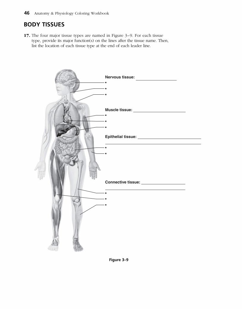
46 Anatomy & Physiology Coloring Workbook
BODY TISSUES
17. The four major tissue types are named in Figure 3–9. For each tissue type, provide its major function(s) on the lines after the tissue name. Then, list the location of each tissue type at the end of each leader line.
Nervous tissue:•
•
•
Muscle tissue: •
•
•
Epithelial tissue:
•
•
Connective tissue:
•
•
•
Figure 3–9
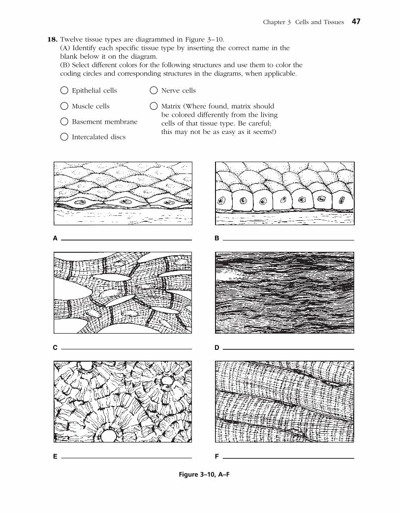
Chapter 3 Cells and Tissues 47
18. Twelve tissue types are diagrammed in Figure 3–10. (A) Identify each specific tissue type by inserting the correct name in the
blank below it on the diagram. (B) Select different colors for the following structures and use them to color the
coding circles and corresponding structures in the diagrams, when applicable.
○ Epithelial cells ○ Nerve cells
○ Muscle cells ○ Matrix (Where found, matrix should be colored differently from the living cells of that tissue type. Be careful; this may not be as easy as it seems!)
○ Basement membrane
○ Intercalated discs
A
C
B
D
E F
Figure 3–10, A–F
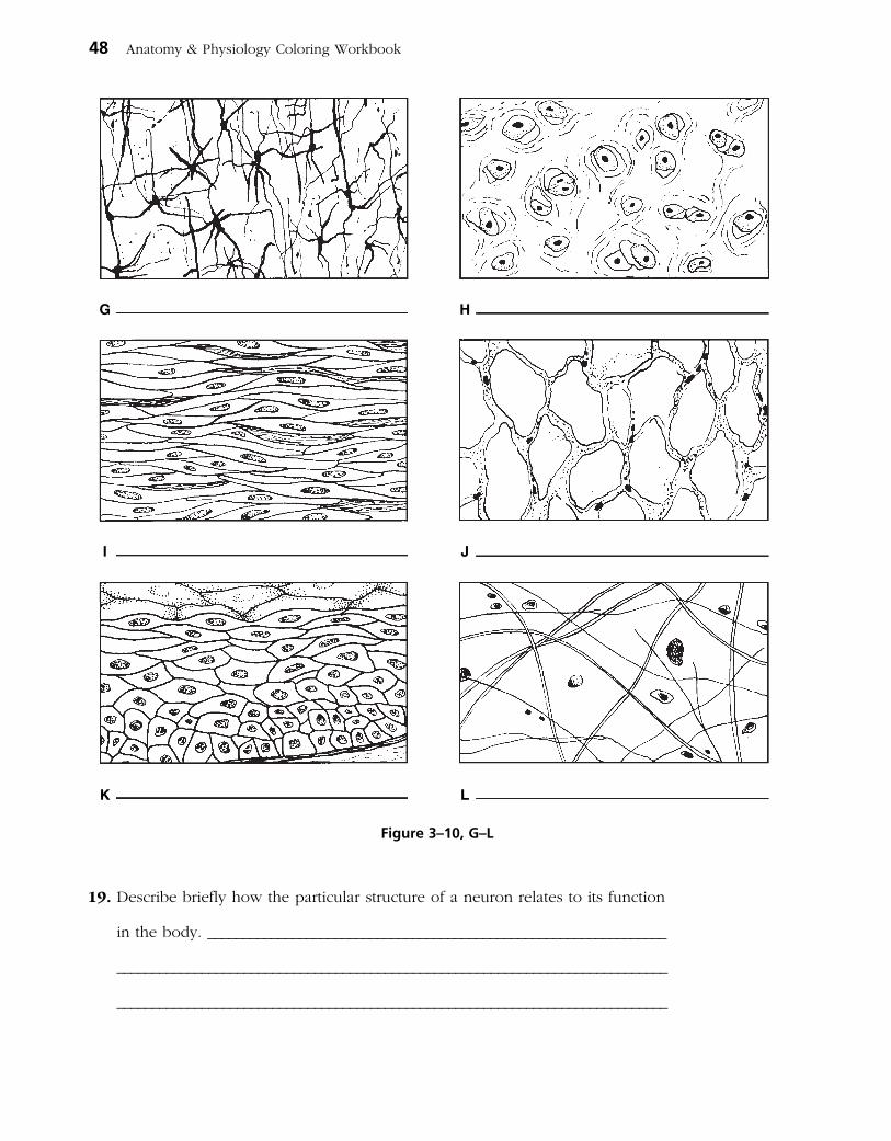
48 Anatomy & Physiology Coloring Workbook
19. Describe briefly how the particular structure of a neuron relates to its function
in the body. _________________________________________________________________
______________________________________________________________________________
______________________________________________________________________________
G
I
H
J
K L
Figure 3–10, G–L

20. Using the key choices, correctly identify the major tissue types described. Enter the appropriate letter or tissue type term in the answer blanks.
Key Choices
A. Connective B. Epithelium C. Muscle D. Nervous
_________________________ 1. Forms mucous, serous, and epidermal membranes
_________________________ 2. Allows for organ movements within the body
_________________________ 3. Transmits electrochemical impulses
_________________________ 4. Supports body organs
_________________________ 5. Cells of this tissue may absorb and/or secrete substances
_________________________ 6. Basis of the major controlling system of the body
_________________________ 7. Cells of this tissue shorten to exert force
_________________________ 8. Forms hormones
_________________________ 9. Packages and protects body organs
_________________________ 10. Characterized by having large amounts of nonliving matrix
_________________________ 11. Allows you to smile, grasp, swim, ski, and shoot an arrow
_________________________ 12. Most widely distributed tissue type in the body
_________________________ 13. Forms the brain and spinal cord
21. Using the key choices, identify the following specific type(s) of epithelial tissue. Enter the appropriate letter or classification term in the answer blanks.
Key Choices
A. Pseudostratified columnar (ciliated) C. Simple cuboidal E. Stratified squamousB. Simple columnar D. Simple squamous F. Transitional
_________________________ 1. Lines the esophagus and forms the skin epidermis
_________________________ 2. Forms the lining of the stomach and small intestine
_________________________ 3. Best suited for areas subjected to friction
_________________________ 4. Lines much of the respiratory tract
_________________________ 5. Propels substances (e.g., mucus) across its surface
_________________________ 6. Found in the bladder lining; peculiar cells that slide over one another
_________________________ 7. Forms thin serous membranes; a single layer of flattened cells
Chapter 3 Cells and Tissues 49

50 Anatomy & Physiology Coloring Workbook
22. The three types of muscle tissue exhibit certain similarities and differences. Check (✓) the appropriate spaces in the following table to indicate which muscle types exhibit each characteristic.
Characteristic Skeletal Cardiac Smooth
1. Voluntarily controlled
2. Involuntarily controlled
3. Banded appearance
4. Single nucleus in each cell
5. Multinucleate
6. Found attached to bones
7. Allows you to direct your eyeballs
8. Found in the walls of stomach, uterus, and arteries
9. Contains spindle-shaped cells
10. Contains cylindrical cells with branching ends
11. Contains long, nonbranching cylindrical cells
12. Displays intercalated discs
13. Concerned with locomotion of the body as a whole
14. Changes the internal volume of an organ as it contracts
15. Tissue of the circulatory pump
23. Circle the term that does not belong in each of the following groupings. Then, fill in the answer blanks with the correct group name.
1. Collagen Cell Matrix Cell product Group: _________
2. Cilia Flagellum Microvilli Elastic fibers Group: _________
3. Glands Bones Epidermis Mucosae Group: _________
4. Adipose Hyaline Osseous Nervous Group: _________
5. Blood Smooth Cardiac Skeletal Group: _________

24. Using the key choices, identify the following connective tissue types. Insert the appropriate letter or corresponding term in the answer blanks.
Key Choices
A. Adipose connective tissue C. Dense fibrous connective tissue E. Osseous tissueB. Areolar connective tissue D. Hyaline cartilage F. Reticular connective tissue
_________________________ 1. Provides great strength through parallel bundles of collagenic fibers; found in tendons
_________________________ 2. Acts as a storage depot for fat
_________________________ 3. Composes the majority of the dermis of the skin
_________________________ 4. Forms the bony skeleton
_________________________ 5. Composes the lamina propria and packages organs; includes a gel-like matrix with all categories of fibers and many cell types
_________________________ 6. Forms the embryonic skeleton and the surfaces of bones at the joints; reinforces the trachea
_________________________ 7. Provides insulation for the body
_________________________ 8. Matrix with no specific shape, heavily invaded with fibers; appears glassy and smooth
_________________________ 9. Contains cells arranged concentrically around a nutrient canal; matrix is hard due to calcium salts
_________________________ 10. Forms the stroma or internal “skeleton” of lymph nodes, the spleen, and other lymphoid organs
Tissue Repair
25. For each of the following statements about tissue repair that is true, enter T in the answer blank. For each false statement, correct the underlined words by writing the correct words in the answer blank.
_________________________ 1. The nonspecific response of the body to injury is called regeneration.
_________________________ 2. Intact capillaries near an injury dilate, leaking plasma, blood cells, and antibodies, which cause the blood to clot. The clot at the surface dries to form a scab.
_________________________ 3. During the first phase of tissue repair, capillary buds invade the clot, forming a delicate pink tissue called endodermal tissue.
_________________________ 4. When damage is not too severe, the surface epithelium migrates beneath the dry scab and across the surface of the granulation tissue. This repair process is called proliferation.
Chapter 3 Cells and Tissues 51
_________________________ 5. If tissue damage is very severe, tissue repair is more likely to occur by fibrosis, or scarring.
_________________________ 6. During fibrosis, fibroblasts in the granulation tissue lay down keratin fibers, which form a strong, compact, but inflexible mass.
_________________________ 7. The repair of cardiac muscle and nervous tissue occurs mainly by fibrosis.
DEVELOPMENTAL ASPECTS OF CELLS AND TISSUES
26. Correctly complete each statement by inserting your responses in the answer blanks.
_________________________ 1.
_________________________ 2.
_________________________ 3.
_________________________ 4.
_________________________ 5.
_________________________ 6.
_________________________ 7.
_________________________ 8.
_________________________ 9.
_________________________ 10.
_________________________ 11.
_________________________ 12.
_________________________ 13.
_________________________ 14.
_________________________ 15.
_________________________ 16.
_________________________ 17.
_________________________ 18.
_________________________ 19.
_________________________ 20.
52 Anatomy & Physiology Coloring Workbook
During embryonic development, cells specialize to form (1) . Mitotic cell division is very important for overall body (2) . All tissues except (3) tissue continue to undergo cell division until the end of adolescence. After this time, (4) tissue also becomes amitotic. When amitotic tissues are dam-aged, they are replaced by (5) tissue, which does not func- tion in the same way as the original tissue. This is a serious problem when heart cells are damaged.
Aging begins almost as soon as we are born. Three explana-tions of the aging process have been offered. One states that
(6) insults, such as the presence of toxic substances in the blood, are important. Another theory states that external
(7) factors, such as X-rays, help to cause aging. A third theory suggests that aging is programmed in our (8) . Three examples of aging processes seen in all people are (9) ,
(10) , and (11) .
Neoplasms occur when cells “go wild” and the normal con-trols of cell (12) are lost. The two types of neoplasms are
(13) and (14) . The (15) type tends to stay localized and have a capsule. The (16) type is likely to invade other body tissues and spread to other (distant) parts of the body. To cor-rectly diagnose the type of neoplasm, a microscopic examina-tion of the tissue called a (17) is usually done. Whenever possible, (18) is the treatment of choice for neoplasms.
An overgrowth of tissue that is not considered to be a neo-plasm is referred to as (19) . Conversely, a decrease in the size of an organ or tissue, resulting from loss of normal stimu-lation, is called (20) .

A Visualization Exercise for the Cell
A long, meandering membrane with dark globules clinging to its outer surface now comes into sight.
27. Where necessary, complete statements by inserting the missing words in the answer blanks.
For your second journey, you will be miniaturized to the size of a small protein molecule and will travel in a microsubma-rine specially designed to enable you to pass easily through living membranes. You are injected into the intercellular space between two epithelial cells and are instructed to observe one of these cells firsthand and to identify as many of its struc-tures as possible.
You struggle briefly with the controls and then maneuver your microsub into one of these cells. Once inside the cell, you find yourself in a kind of “sea.” This salty fluid that surrounds you is the (1) of the cell.
Far below looms a large, dark, oval structure, much larger than anything else you can see. You conclude that it is the
(2) . As you move downward, you pass a cigar-shaped structure with strange-looking folds on its inner surface. Although you have a pretty good idea that it must be a (3) , you decide to investigate more thoroughly. After passing through the external membrane of the structure, you are con-fronted with yet another membrane. Once past this mem-
brane, you are inside the strange-looking structure. You activate the analyzer switch in your micro-sub for a readout indicating which molecules are in your immediate vicinity. As suspected, there is an abundance of energy-rich (4) molecules. Having satisfied your curiosity, you leave this struc-ture to continue the investigation.
A long, meandering membrane with dark globules clinging to its outer surface now comes into sight. You maneuver closer and sit back to watch the activity. As you watch, amino acids are joined together, and a long, threadlike protein molecule is built. The globules must be (5) , and the mem brane, therefore, is the (6) . Once again, you head toward the large dark structure seen and tentatively identified earlier. On approach, you observe that this huge structure has very large openings in its outer wall; these openings must be the (7) . Passing through one of these open-ings, you discover that from the inside, the color of this structure is a result of dark, coiled, inter-twined masses of (8) , which your analyzer confirms contain genetic material, or (9) mole-cules. Making your way through this tangled mass, you pass a round, dense structure that appears to be full of the same type of globules you saw outside. This round structure is the (10) . All this information confirms your earlier identification of this cellular structure, so now you move to its exterior to continue observations.
Chapter 3 Cells and Tissues 53
INCREDIBLE JOURNEY
_________________________ 1.
_________________________ 2.
_________________________ 3.
_________________________ 4.
_________________________ 5.
_________________________ 6.
_________________________ 7.
_________________________ 8.
_________________________ 9.
_________________________ 10.

Just ahead, you see what appears to be a mountain of flat-tened sacs with hundreds of small saclike vesicles at its edges. The vesicles seem to be migrating away from this area and heading toward the outer edges of the cell. The mountain of sacs must be the (11) . Eventually you come upon a
rather simple-looking membrane-bound sac. Although it doesn’t look too exciting and has few distinguishing marks, it does not resemble anything else you have seen so far. Deciding to obtain a chemical analysis before entering this sac, you activate the analyzer and on the screen you see “Enzymes — Enzymes — Hydrolases — Hydrolases — Danger — Danger.” There is little doubt that this innocent-appearing structure is actually a (12) .
Completing your journey, you count the number of organelles identified so far. Satisfied that you have observed most of them, you request retrieval from the intercellular space.
28. Johnny lacerated his arm and rushed home to Mom so she could “fix it.” His mother poured hydrogen peroxide over the area, and it bubbled vigorously where it came in contact with the wound. Because you can expect that cells were ruptured in the injured area, what do you think was happening here?
29. The epidermis (epithelium of the cutaneous membrane or skin) is a keratinized stratified squamous epithelium. Explain why that epithelium is much better suited for protecting the body’s external surface than a mucosa consisting of a simple columnar epithelium would be.
30. Streptomycin (an antibiotic) binds to the small ribosomal subunit of bacteria (but not to the ribosomes of the host cells infected by bacteria). The result is the misreading of bacteria mRNA and the breakup of polysomes (mRNA and two or more ribosomes). What process is being affected, and how does this kill the bacterial cells?
54 Anatomy & Physiology Coloring Workbook
_________________________ 11.
_________________________ 12.
AT THE CLINIC
Chapter 3 Cells and Tissues 55
31. Systemic lupus erythematosus (often simply called lupus) is a condition that primarily affects young women. It is a chronic (persistent) inflammation that affects all or most of the connective tissue proper in the body. Suzy is told by her doctor that she has lupus, and she asks if it will have widespread or merely localized effects within the body. What would the physician answer? Explain.
32. Mrs. Linsey sees her gynecologist because she is unable to become pregnant. The doctor discovers granulation tissue in her vaginal canal and explains that sperm are susceptible to some of the same chemicals as bacteria. What is inhibiting the sperm?
33. Sarah, a trainee of the electron microscopist at the local hospital, is reviewing some micrographs of muscle cells and macrophages (phagocytic cells). She notices that the muscle cells are loaded with mitochondria while the macrophages have abundant lysosomes. Why is this so?
34. Bradley tripped and tore one of the tendons surrounding his ankle. In anguish with pain, he asked his doctor how quickly he could expect it to heal. What do you think the doctor’s response was and why?
35. In normally circulating blood, the plasma proteins cannot leave the bloodstream easily and thus tend to remain in the blood. But if stasis (blood flow stoppage) occurs, the proteins will begin to leak out into the interstitial fluid. Explain why this leads to edema (water buildup in the tissues).

56 Anatomy & Physiology Coloring Workbook
36. Phagocytes gather in the air sacs of the lungs, especially in the lungs of smokers. What is the connection?
THE FINALE: MULTIPLE CHOICE
37. Select the best answer or answers from the choices given.
1. A cell’s plasma membrane would not contain:
A. phospholipid. D. cholesterol.
B. nucleic acid. E. glycolipid.
C. protein.
2. Which of the following would you expect to find in or on cells whose main function is absorption?
A. Microvilli D. Gap junctions
B. Cilia E. Secretory vesicles
C. Desmosomes
3. Which cytoskeletal element interacts with myosin to produce contractile force in mus-cle cells?
A. Microtubules
B. Microfilaments
C. Intermediate filaments
D. None of the above
4. If a 10% sucrose solution within a semiper-meable sac causes the fluid volume in the sac to increase a given amount when the sac is immersed in water, what would be the effect of replacing the sac solution with a 20% sucrose solution?
A. The sac would lose fluid.
B. The sac would gain the same amount of fluid.
C. The sac would gain more fluid.
D. There would be no effect.
5. Which of the following are possible func tions of the glycocalyx?
A. Determination of blood groups
B. Binding sites for toxins
C. Aiding the binding of sperm to egg
D. Guiding embryonic development
E. Increasing the efficiency of absorption
6. A cell stimulated to increase steroid produc-tion will have:
A. abundant ribosomes.
B. a rough ER.
C. a smooth ER.
D. a Golgi apparatus.
E. abundant secretory vesicles.
7. A cell’s ability to replenish its ATP stores has been diminished by a metabolic poison. What organelle is most likely to be affected?
A. Nucleus D. Microtubule
B. Plasma membrane E. Mitochondrion
C. Centriole
8. The fundamental structure of the plasma mem brane is determined almost exclusively by:
A. phospholipid molecules.
B. peripheral proteins.
C. cholesterol molecules.
D. integral proteins.
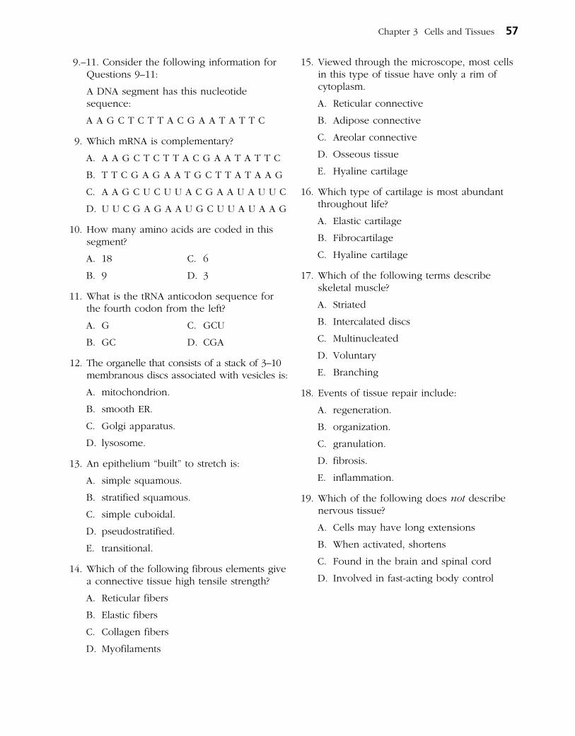
Chapter 3 Cells and Tissues 57
9.–11. Consider the following information for Questions 9–11:
A DNA segment has this nucleotide sequence:
A A G C T C T T A C G A A T A T T C
9. Which mRNA is complementary?
A. A A G C T C T T A C G A A T A T T C
B. T T C G A G A A T G C T T A T A A G
C. A A G C U C U U A C G A A U A U U C
D. U U C G A G A A U G C U U A U A A G
10. How many amino acids are coded in this segment?
A. 18 C. 6
B. 9 D. 3
11. What is the tRNA anticodon sequence for the fourth codon from the left?
A. G C. GCU
B. GC D. CGA
12. The organelle that consists of a stack of 3–10 membranous discs associated with vesicles is:
A. mitochondrion.
B. smooth ER.
C. Golgi apparatus.
D. lysosome.
13. An epithelium “built” to stretch is:
A. simple squamous.
B. stratified squamous.
C. simple cuboidal.
D. pseudostratified.
E. transitional.
14. Which of the following fibrous elements give a connective tissue high tensile strength?
A. Reticular fibers
B. Elastic fibers
C. Collagen fibers
D. Myofilaments
15. Viewed through the microscope, most cells in this type of tissue have only a rim of cytoplasm.
A. Reticular connective
B. Adipose connective
C. Areolar connective
D. Osseous tissue
E. Hyaline cartilage
16. Which type of cartilage is most abundant throughout life?
A. Elastic cartilage
B. Fibrocartilage
C. Hyaline cartilage
17. Which of the following terms describe skeletal muscle?
A. Striated
B. Intercalated discs
C. Multinucleated
D. Voluntary
E. Branching
18. Events of tissue repair include:
A. regeneration.
B. organization.
C. granulation.
D. fibrosis.
E. inflammation.
19. Which of the following does not describe nervous tissue?
A. Cells may have long extensions
B. When activated, shortens
C. Found in the brain and spinal cord
D. Involved in fast-acting body control

This page intentionally left blank

Body membranes, which cover body surfaces, line its cavities, and form protective sheets around organs, fall into two major categories. These are epithelial membranes (skin epidermis, mucosae, and serosae) and the connective tissue synovial membranes.
Topics for review in this chapter include a comparison of structure and function of various membranes, anatomical characteristics of the skin (com posed of the connective tissue dermis and the epidermis) and its derivatives, and the manner in which the skin responds to both internal and external stimuli to protect the body.
CLASSIFICATION OF BODY MEMBRANES
1. Complete the following table relating to body membranes. Enter your responses in the areas left blank.
Tissue type Membrane (epithelial/connective) Common locations Functions
Mucous Epithelial sheet with underlying connective tissue (lamina propria)
Serous Lines internal ventral body cavities and covers their organs
Cutaneous Protection from external insults and water loss
Synovial Lines cavities of synovial joints
59
SKIN AND BODY MEMBRANES
4

2. Four simplified diagrams are shown in Figure 4–1. Select different colors for the membranes listed below and use them to color the coding circles and the corresponding structures.
○ Cutaneous membrane ○ Parietal pleura (serosa) ○ Synovial membrane
○ Mucosae ○ Visceral pericardium (serosa)
○ Visceral pleura (serosa) ○ Parietal pericardium (serosa)
60 Anatomy & Physiology Coloring Workbook
Figure 4–1
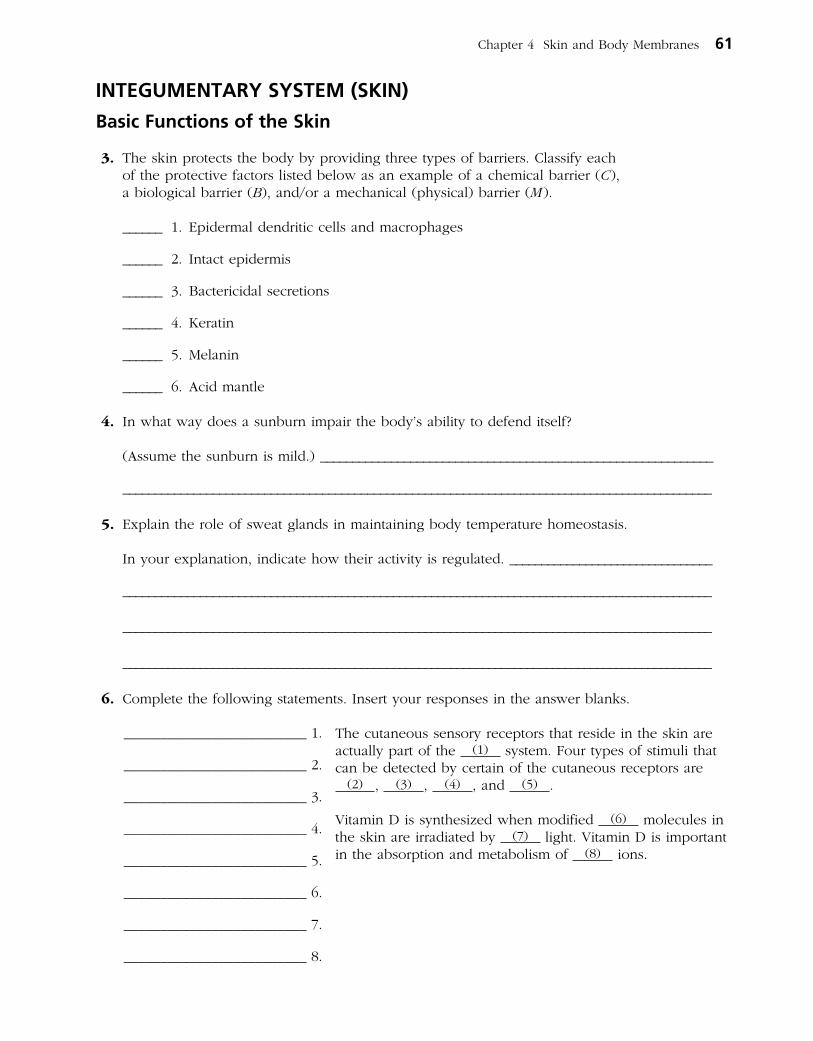
INTEGUMENTARY SYSTEM (SKIN)
Basic Functions of the Skin
3. The skin protects the body by providing three types of barriers. Classify each of the protective factors listed below as an example of a chemical barrier (C), a biological barrier (B), and/or a mechanical (physical) barrier (M).
______ 1. Epidermal dendritic cells and macrophages
______ 2. Intact epidermis
______ 3. Bactericidal secretions
______ 4. Keratin
______ 5. Melanin
______ 6. Acid mantle
4. In what way does a sunburn impair the body’s ability to defend itself?
(Assume the sunburn is mild.) _____________________________________________________________
____________________________________________________________________________________________
5. Explain the role of sweat glands in maintaining body temperature homeo stasis.
In your explanation, indicate how their activity is regulated. ________________________________
____________________________________________________________________________________________
____________________________________________________________________________________________
____________________________________________________________________________________________
6. Complete the following statements. Insert your responses in the answer blanks.
The cutaneous sensory receptors that reside in the skin are actually part of the (1) system. Four types of stimuli that can be detected by certain of the cutaneous receptors are
(2) , (3) , (4) , and (5) .
Vitamin D is synthesized when modified (6) molecules in the skin are irradiated by (7) light. Vitamin D is important in the absorption and metabolism of (8) ions.
Chapter 4 Skin and Body Membranes 61
_________________________ 1.
_________________________ 2.
_________________________ 3.
_________________________ 4.
_________________________ 5.
_________________________ 6.
_________________________ 7.
_________________________ 8.

Basic Structure of the Skin
7. Figure 4–2 depicts a longitudinal section of the skin.
(A) Label the skin structures and areas indicated by leader lines and brackets on the figure.
(B) Select different colors for the structures below and color the coding circles and the corresponding structures on the figure.
○ Arrector pili muscle
○ Adipose tissue
○ Hair follicle
○ Nerve fibers
○ Sweat (sudoriferous) gland
○ Sebaceous gland
(C) Which bracket(s) compose(s) the cutaneous membrane? ______________.
8. The more superficial cells of the epidermis become less viable and ultimately die. Which two factors account for this natural demise of the epidermal cells?
1. ___________________________________________________________________________________________
2. ___________________________________________________________________________________________
___________________________________________________________________________________________
62 Anatomy & Physiology Coloring Workbook
Figure 4–2
9. Using the key choices, complete the crossword puzzle by answering each of the clues provided.
Key Choices
Dermis (as a whole) Reticular layer Stratum granulosumEpidermis (as a whole) Stratum basale Stratum lucidumPapillary layer Stratum corneum Stratum spinosum
Across 4. Epidermal layer containing the oldest cells. 5. Major skin area from which the derivatives (hair, nails) arise. 6. Vascular region; site of elastic and collagen fibers.
Down 1. Dermis layer responsible for fingerprints. 2. Translucent cells containing keratin. 3. Epidermal region involved in rapid cell division and melanin formation.
1
2
3
4
5
6
10. Circle the term that does not belong in each of the following groupings. Then, fill in the answer blanks with the correct group name.
1. Reticular layer Keratin Dermal papillae Meissner’s corpuscles Group: ______
2. Mole Freckle Wart Malignant melanoma Group: ______
3. Prickle cells Stratum basale Stratum spinosum Cell shrinkage Group: ______
4. Meissner’s corpuscles Lamellar corpuscles Merkel’s cells Arrector pili Group: ______
Chapter 4 Skin and Body Membranes 63

11. This exercise examines the relative importance of three pigments in determining skin color. Indicate which pigment is identified by the following descrip tions by inserting the appropriate answer from the key choices in the answer blanks.
Key Choices
A. Carotene B. Hemoglobin C. Melanin
_________________________ 1. Most responsible for the skin color of darkskinned people
_________________________ 2. Provides an orange cast to the skin
_________________________ 3. Provides a natural sunscreen
_________________________ 4. Most responsible for the skin color of lightskinned (Caucasian) people
_________________________ 5. Phagocytized by keratinocytes
_________________________ 6. Found predominantly in the stratum corneum
_________________________ 7. Found within red blood cells in the blood vessels
12. Complete the following statements in the blanks provided.
_________________________ 1. Radiation from the skin surface and evaporation of sweat are two ways in which the skin helps to get rid of body (1) .
_________________________ 2. Fat in the (2) tissue layer beneath the dermis helps to insulate the body.
_________________________ 3. A vitamin that is manufactured in the skin is (3) .
_________________________ 4. Wrinkling of the skin is caused by loss of the (4) of the skin.
_________________________ 5. A decubitus ulcer results when skin cells are deprived of (5) .
_________________________ 6. (6) is a bluish cast of the skin resulting from inadequate oxygenation of the blood.
Appendages of the Skin
13. For each true statement, write T. For each false statement, correct the underlined word(s) and insert your correction in the answer blank.
_________________________ 1. A saltwater solution is secreted by sebaceous glands.
_________________________ 2. The most abundant protein in dead epidermal structures such as hair and nails is melanin.
_________________________ 3. Sebum is an oily mixture of lipids, cholesterol, and cell fragments.
_________________________ 4. The externally observable part of a hair is called the root.
_________________________ 5. The epidermis provides mechanical strength to the skin.
64 Anatomy & Physiology Coloring Workbook
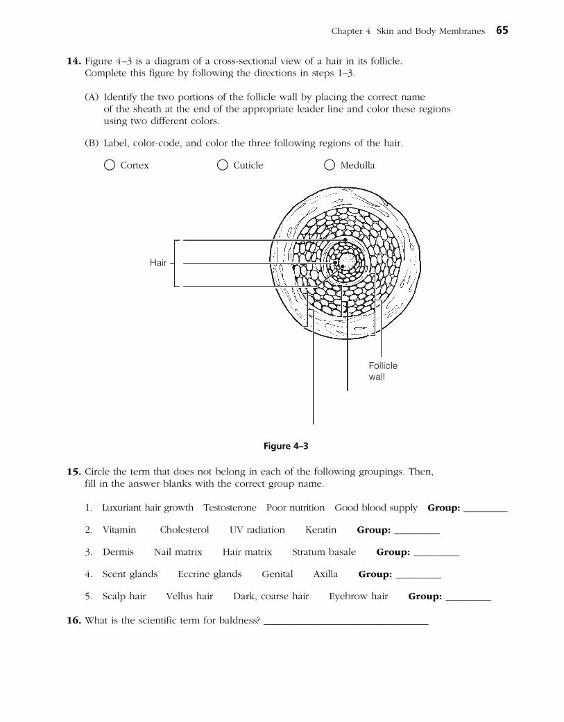
14. Figure 4–3 is a diagram of a crosssectional view of a hair in its follicle. Complete this figure by following the directions in steps 1–3.
(A) Identify the two portions of the follicle wall by placing the correct name of the sheath at the end of the appropriate leader line and color these regions using two different colors.
(B) Label, colorcode, and color the three following regions of the hair.
○ Cortex ○ Cuticle ○ Medulla
15. Circle the term that does not belong in each of the following groupings. Then, fill in the answer blanks with the correct group name.
1. Luxuriant hair growth Testosterone Poor nutrition Good blood supply Group: _________
2. Vitamin Cholesterol UV radiation Keratin Group: _________
3. Dermis Nail matrix Hair matrix Stratum basale Group: _________
4. Scent glands Eccrine glands Genital Axilla Group: _________
5. Scalp hair Vellus hair Dark, coarse hair Eyebrow hair Group: _________
16. What is the scientific term for baldness? ____________________________________
Chapter 4 Skin and Body Membranes 65
Folliclewall
Hair
Figure 4–3

17. Using the key choices, complete the following statements. Insert the appropriate letter(s) or term(s) in the answer blanks. Items may have more than one answer.
Key Choices
A. Arrector pili C. Hair E. Sebaceous glands G. Sweat gland (eccrine)B. Cutaneous receptors D. Hair follicle(s) F. Sweat gland (apocrine)
_________________________ 1. A blackhead is an accumulation of oily material produced by (1) .
_________________________ 2. Tiny muscles attached to hair follicles that pull the hair upright during fright or cold are called (2) .
_________________________ 3. The most numerous variety of perspiration gland is the (3) .
_________________________ 4. A sheath formed of both epithelial and connective tissues is the (4) .
_________________________ 5. A less numerous variety of perspiration gland is the (5) . Its secretion (often milky in appearance) contains proteins and other substances that favor bacterial growth.
_________________________ 6. (6) is found everywhere on the body except the palms of the hands, soles of the feet, and lips, and primarily consists of dead keratinized cells.
_________________________ 7. (7) are specialized nerve endings that respond to temperature and touch, for example.
_________________________ 8. (8) become more active at puberty.
_________________________ 9. Part of the heatliberating apparatus of the body is the (9) .
_________________________ 10. (10) secretion contains bacteria killing substances.
18. Circle the term that does not belong in each of the following groupings. Then, fill in the answer blanks with the correct group name.
1. Sebaceous gland Hair Arrector pili Epidermis Group: _________
2. Radiation Absorption Conduction Evaporation Group: _________
3. Cortex Medulla Cuticle Epithelial sheath Group: _________
4. Epidermis Dermis Hypodermis Papillary layer Group: _________
5. Cyanosis Erythema Wrinkles Pallor Group: _________
66 Anatomy & Physiology Coloring Workbook
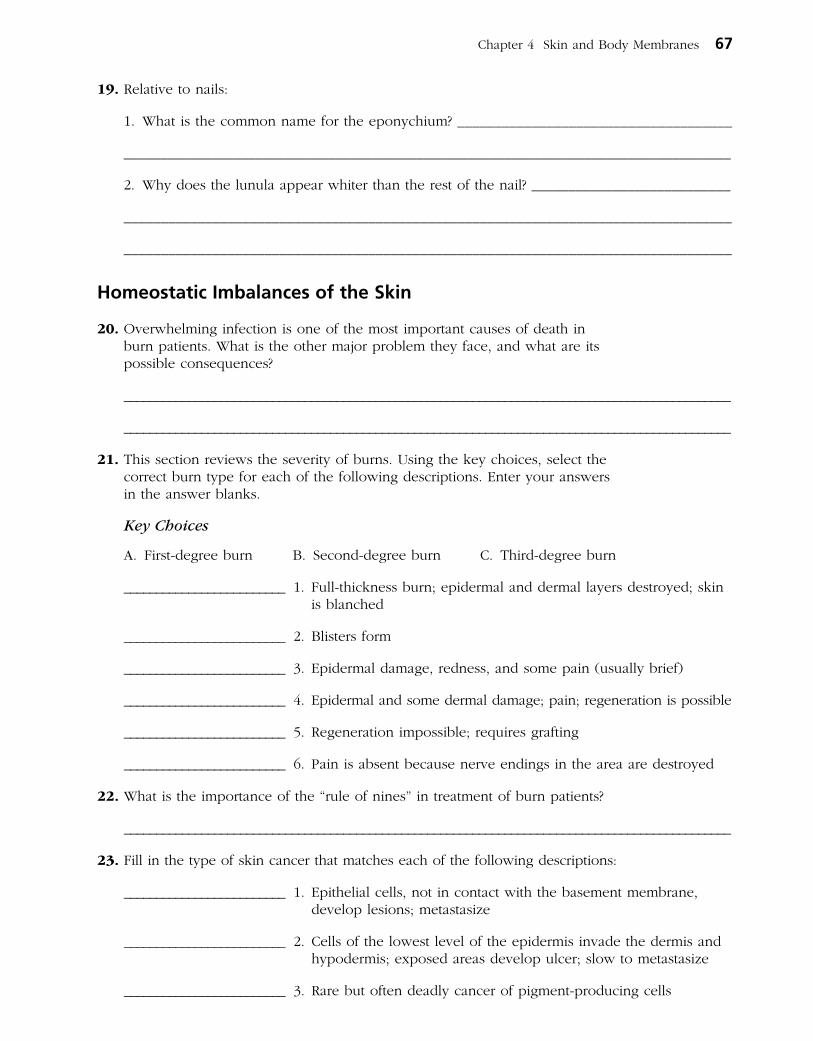
19. Relative to nails:
1. What is the common name for the eponychium? _____________________________________
___________________________________________________________________________________
2. Why does the lunula appear whiter than the rest of the nail? ___________________________
__________________________________________________________________________________
__________________________________________________________________________________
Homeostatic Imbalances of the Skin
20. Overwhelming infection is one of the most important causes of death in burn patients. What is the other major problem they face, and what are its possible consequences?
______________________________________________________________________________________________
______________________________________________________________________________________________
21. This section reviews the severity of burns. Using the key choices, select the correct burn type for each of the following descriptions. Enter your answers in the answer blanks.
Key Choices
A. Firstdegree burn B. Seconddegree burn C. Thirddegree burn
_________________________ 1. Fullthickness burn; epidermal and dermal layers destroyed; skin is blanched
_________________________ 2. Blisters form
_________________________ 3. Epidermal damage, redness, and some pain (usually brief)
_________________________ 4. Epidermal and some dermal damage; pain; regeneration is possible
_________________________ 5. Regeneration impossible; requires grafting
_________________________ 6. Pain is absent because nerve endings in the area are destroyed
22. What is the importance of the “rule of nines” in treatment of burn patients?
______________________________________________________________________________________________
23. Fill in the type of skin cancer that matches each of the following descriptions:
_________________________ 1. Epithelial cells, not in contact with the basement membrane, develop lesions; metastasize
_________________________ 2. Cells of the lowest level of the epidermis invade the dermis and hypodermis; exposed areas develop ulcer; slow to metastasize
_________________________ 3. Rare but often deadly cancer of pigmentproducing cells
Chapter 4 Skin and Body Membranes 67

24. What does ABCD mean in reference to examination of pigmented areas? _____________________
______________________________________________________________________________________________
______________________________________________________________________________________________
DEVELOPMENTAL ASPECTS OF THE SKIN AND BODY MEMBRANES
25. Match the choices (letters or terms) in Column B with the appropriate descriptions in Column A.
Column A
_________________________ 1. Skin inflammations that increase in frequency with age
_________________________ 2. Cause of graying hair
_________________________ 3. Small white bumps on the skin of newborn babies, resulting from accumulations of sebaceous gland material
_________________________ 4. Reflects the loss of insulating subcu taneous tissue with age
_________________________ 5. A common consequence of accelerated sebaceous gland activity during adolescence
_________________________ 6. Oily substance produced by the fetus’s sebaceous glands
_________________________ 7. The hairy “cloak” of the fetus
A Visualization Exercise for the Skin
Your immediate surroundings resemble huge grotesquely twisted vines . . . you begin to climb upward.
26. Where necessary, complete statements by inserting the missing words in the answer blanks.
For this trip, you are miniaturized for injection into your host’s skin. Your journey begins when you are deposited in a soft gellike substance. Your immediate surroundings resemble huge grotesquely twisted vines. But when you peer carefully at the closest “vine,” you realize you are actually seeing
68 Anatomy & Physiology Coloring Workbook
Column B
A. Acne
B. Cold intolerance
C. Dermatitis
D. Delayedaction gene
E. Lanugo
F. Milia
G. Vernix caseosa
INCREDIBLE JOURNEY

connective tissue fibers. Although tangled together, most of the fibers are fairly straight and look like strong cables. You identify these as the (1) fibers. Here and there are fibers that resemble coiled springs. These must be the (2) fibers that help give skin its springiness. At this point, there is little question that you are in the (3) region of the skin, particularly considering that you can also see blood vessels and nerve fibers around you.
Carefully, using the fibers as steps, you begin to climb upward. After climbing for some time and finding that you still haven’t reached the upper regions of the skin, you stop for a rest. As you sit, a strangelooking cell approaches, moving slowly with parts alternately flowing forward and then receding. Suddenly you realize that this must be a (4) that is about to dispose of an intruder (you) unless you move in a hurry! You scramble to your feet and resume your upward climb. On your right is a large fibrous structure that looks like a tree trunk anchored in place by muscle fibers. By scurrying up this (5) sheath, you are able to escape from the cell. Once safely out of harm’s way, you again scan your
surroundings. Directly overhead are tall cubelike cells, forming a continuous sheet. In your rush to escape, you have reached the (6) layer of the skin. As you watch the activity of the cells in this layer, you notice that many of the cells are pinching in two, and the daughter cells are being forced upward. Obviously, this is the layer that continually replaces cells that rub off the skin surface, and these cells are the (7) cells.
Looking through the transparent cell membrane of one of the basal cells, you see a dark mass hanging over its nucleus. You wonder if this cell could have a tumor; but then, looking through the membranes of the neighboring cells, you find that they also have dark umbrellalike masses hanging over their nuclei. As you consider this matter, a black cell with long tentacles begins to pick its way carefully between the other cells. As you watch with interest, one of the transparent cells engulfs the tip of one of the black cell’s tentacles. Within seconds a black substance appears above the transparent cell’s nucleus. Suddenly, you remember that one of the skin’s functions is to pro tect the deeper layers from sun damage; the black substance must be the protective pigment (8) .
Once again you begin your upward climb and notice that the cells are becoming shorter and harder and are full of a waxylooking substance. This substance has to be (9) , which would account for the increasing hardness of the cells. Climbing still higher, the cells become flattened like huge shingles. The only material apparent in the cells is the waxy substance—there is no nucleus and there appears to be no activity in these cells. Considering the clues—shinglelike cells, no nuclei, full of the waxy substance, no activity—these cells are obviously (10) and therefore very close to the skin surface.
Suddenly, you feel a strong agitation in your immediate area. The pressure is tremendous. Looking upward through the transparent cell layers, you see your host’s fingertips vigorously scratching the area directly overhead. You wonder if you are causing his skin to sting or tickle. Then, within seconds, the cells around you begin to separate and fall apart, and you are catapulted out into the sunlight. Because the scratching fingers might descend once again, you quickly advise your host of your whereabouts.
Chapter 4 Skin and Body Membranes 69
_________________________ 1.
_________________________ 2.
_________________________ 3.
_________________________ 4.
_________________________ 5.
_________________________ 6.
_________________________ 7.
_________________________ 8.
_________________________ 9.
_________________________ 10.
27. Mrs. Ibañez volunteered to help at a hospital for children with cancer. When she first entered the cancer ward, she was upset by the fact that most of the children had no hair. What is the explanation for their baldness?
28. Linda, a new mother, brings her infant to the clinic, worried about a yellowish, scummy deposit that has built up on the baby’s scalp. What is this condition called, and is it serious?
29. Patients in hospital beds are rotated every 2 hours to prevent bedsores. Exactly why is this effective?
30. Eric and his wife are of northern European descent. Eric is a proud new father who was in the delivery room during his daughter’s birth. He tells you that when she was born, her skin was purple and covered with a cream cheese–like substance. Shortly after birth, her skin turned pink. Can you explain his observations?
31. Would you expect to find the highest rate of skin cancer among the blacks of tropical Africa, research scientists in the Arctic, Norwegians in the southern United States, or blacks in the United States? Explain your choice.
70 Anatomy & Physiology Coloring Workbook
AT THE CLINIC

32. After studying the skin in anatomy class, Toby grabbed the large “love handles” at his waist and said, “I have too thick a hypodermis, but that’s okay because this layer performs some valuable functions!” What are the functions of the hypodermis?
33. A man got his finger caught in a machine at the factory. The damage was less serious than expected, but nonetheless, the entire nail was torn from his right index finger. The parts lost were the body, root, bed, matrix, and cuticle of the nail. First, define each of these parts. Then, tell if this nail is likely to grow back.
34. In cases of a ruptured appendix, what serous membrane is likely to become infected? Why can this be lifethreatening?
35. Mrs. Gaucher received seconddegree burns on her abdomen when she dropped a kettle of boiling water. She asked the clinic physician (worriedly) if she would have to have a skin graft. What do you think he told her?
36. Which two factors in the treatment of critical thirddegree burn patients are absolutely essential?
37. Both newborn and aged individuals have very little subcutaneous tissue. How does this affect their sensitivity to cold?
Chapter 4 Skin and Body Membranes 71

38. Select the best answer or answers from the choices given.
72 Anatomy & Physiology Coloring Workbook
1. Which is not part of the skin?
A. Epidermis C. Dermis
B. Hypodermis D. Superficial fascia
2. Which of the following is not a tissue type found in the skin?
A. Stratified squamous epithelium
B. Loose connective tissue
C. Dense irregular connective tissue
D. Ciliated columnar epithelium
E. Vascular tissue
3. Epidermal cells that aid in the immune response include:
A. Merkel’s cells. C. melanocytes.
B. dendritic cells. D. spinosum cells.
4. Which epidermal layer has a high concentration of Langerhans’ cells and has numerous desmosomes and thick bundles of keratin filaments?
A. Stratum corneum
B. Stratum lucidum
C. Stratum granulosum
D. Stratum spinosum
5. Fingerprints are caused by:
A. the genetically determined arrangement of dermal papillae.
B. the conspicuous epidermal ridges.
C. the sweat pores.
D. all of these.
6. Some infants are born with a fuzzy skin; this is due to:
A. vellus hairs. C. lanugo.
B. terminal hairs. D. hirsutism.
7. What is the major factor accounting for the waterproof nature of the skin?
A. Desmosomes in stratum corneum
B. Glycolipid between stratum corneum cells
C. The thick insulating fat of the hypodermis
D. The leathery nature of the dermis
8. Which of the following is true concerning oil production in the skin?
A. Oil is produced by sudoriferous glands.
B. Secretion of oil is the job of the apocrine glands.
C. The secretion is called sebum.
D. Oil is usually secreted into hair follicles.
9. Contraction of the arrector pili would be “sensed” by:
A. Merkel’s discs.
B. tactile corpuscles.
C. hair follicle receptors.
D. lamellated corpuscles.
10. A dermatologist examines a patient with lesions on the face. Some of the lesions appear as shiny, raised spots; others are ulcerated with beaded edges. What is the diagnosis?
A. Melanoma
B. Squamous cell carcinoma
C. Basal cell carcinoma
D. Either squamous or basal cell carcinoma
11. Components of sweat include:
A. water. D. ammonia.
B. sodium chloride. E. sebum.
C. vitamin D.
THE FINALE: MULTIPLE CHOICE
12. A burn patient reports that the burns on her hands and face are not painful, but she has blisters on her neck and forearms and the skin on her arms is very red. This burn would be classified as:
A. firstdegree only.
B. seconddegree only.
C. thirddegree only.
D. critical.
13. The reticular layer of the dermis is most important in providing:
A. strength and elasticity to the skin.
B. toughness to the skin.
C. insulation to prevent heat loss.
D. the dermal papilla, which produce fingerprints.
14. Which of the following is not associated with sweat?
A. Sweat glands
B. Holocrine glands
C. Eccrine glands
D. Apocrine glands
Chapter 4 Skin and Body Membranes 73

This page intentionally left blank

The skeleton is constructed of two of the most supportive tissues found in the human body—cartilage and bone. Besides supporting and protecting the body as an internal framework, the skeleton provides a system of levers that the skele tal muscles use to move the body. In addition, the bones provide a sto rage depot for substances such as lipids and calcium, and blood cell formation goes on within the red marrow cavities of bones.
The skeleton consists of bones connected at joints, or articulations, and is subdivided into two divisions. The axial skeleton includes those bones that lie around the body’s center of gravity. The appendicular skeleton includes the bones of the limbs and girdles.
Topics for student review include structure and function of long bones, loca tion and naming of specific bones in the skeleton, fracture types, and a classification of joint types in the body.
BONES—AN OVERVIEW
1. Classify each of the following terms as a projection (P) or a depression (or opening) (D). Enter the appropriate letter in the answer blanks.
____ 1. Condyle ____ 4. Foramen ____ 7. Ramus
____ 2. Crest ____ 5. Head ____ 8. Spine
____ 3. Fissure ____ 6. Meatus ____ 9. Tuberosity
2. Group each of the following bones into one of the four major bone categories. Use L for long bone, S for short bone, F for flat bone, and I for irregular bone. Enter the appropriate letter in the space provided.
____ 1. Calcaneus ____ 4. Humerus ____ 7. Radius
____ 2. Frontal ____ 5. Mandible ____ 8. Sternum
____ 3. Femur ____ 6. Metacarpal ____ 9. Vertebra
75
THE SKELETAL SYSTEM
5
3. Using the key choices, characterize the following statements relating to long bones. Enter the appropriate term(s) or letter(s) in the answer blanks. Items may have more than one answer.
Key Choices
A. Diaphysis C. Epiphysis E. Yellow marrow cavityB. Epiphyseal plate D. Red marrow
_________________________ 1. Site of spongy bone in the adult
_________________________ 2. Site of compact bone in the adult
_________________________ 3. Site of hematopoiesis in the adult
_________________________ 4. Scientific name for bone shaft
_________________________ 5. Site of fat storage in the adult
_________________________ 6. Site of longitudinal growth in a child
4. Complete the following statements concerning bone formation and destruction, using the terms provided in the key choices. Insert the key letter or corresponding term in the answer blanks.
Key Choices
A. Atrophy C. Gravity E. Osteoclasts G. Parathyroid hormoneB. Calcitonin D. Osteoblasts F. Osteocytes H. Stress and/or tension
_________________________ 1. When blood calcium levels begin to drop below homeostatic levels, (1) is released, causing calcium to be released from bones.
_________________________ 2. Mature bone cells, called (2) , maintain bone in a viable state.
_________________________ 3. Disuse such as that caused by paralysis or severe lack of exercise results in muscle and bone (3) .
_________________________ 4. Large tubercles and/or increased deposit of bony matrix occur at sites of (4) .
_________________________ 5. Immature, or matrixdepositing, bone cells are referred to as (5) .
_________________________ 6. (6) causes blood calcium to be deposited in bones as calcium salts.
_________________________ 7. Bone cells that liquefy bone matrix and release calcium to the blood are called (7) .
_________________________ 8. Our astronauts must do isometric exercises when in space because bones atrophy under conditions of weightlessness or lack of (8) .
76 Anatomy & Physiology Coloring Workbook

5. Five descriptions of bone structure are provided in Column A.
(A) Identify the structure by choosing the appropriate term from Column B and placing the corresponding answer in the answer blank.
(B) Select different colors for the structures and bone areas in Column B and use them to color the coding circles and corresponding structures on Figure 51, diagrams A and B.
(C) Identify one lamella on diagram A by using a bracket and label (the concentric lamellae would be difficult to color without confusing other structures).
Column A
_________________________ 1. Layers of calcified matrix
_________________________ 2. “Residences” of osteocytes
_________________________ 3. Longitudinal canal, carrying blood vessels and nerves
_________________________ 4. Nonliving, structural part of bone
_________________________ 5. Tiny canals, connecting lacunae
6. Circle the term that does not belong in each of the following groupings. Then, fill in the answer blanks with the correct group name.
1. Hematopoiesis Red marrow Yellow marrow Spongy bone Group: _________
2. Lamellae Canaliculi Circulation Osteoblasts Group: _________
3. Osteocyte Marrow cavity Central canal Canaliculi Group: _________
4. Spongy bone Articular cartilage Periosteum Hyaline cartilage Group: _________
Chapter 5 The Skeletal System 77
Column B
A. Central (Haversian) canal ○
B. Concentric lamellae
C. Lacunae ○
D. Canaliculi ○
E. Bone matrix ○
F. Osteocyte ○
A (diagrammatic view of a cross section of bone) B (higher magnification view of compact bone tissue)
Figure 5–1

7. Figure 5–2A is a midlevel, crosssectional view of the diaphysis of the femur. (A) Label the membrane that lines the cavity and the membrane that covers the outside surface.
Figure 5–2B is a drawing of a longitudinal section of the femur. (B) Color the bone tissue gold. Do not color the articular cartilage; leave it white. (C) Select different colors for the bone regions listed at the coding circles
below. Color the coding circles and the corresponding regions on the drawing (Figure 5–2B only).
(D) Complete Figure 5–2B by labeling compact bone and spongy bone.
○ Diaphysis ○ Area where red marrow is found
○ Epiphyseal plate ○ Area where yellow marrow is found
8. The following events apply to the endochondral ossification process as it occurs in the primary ossification center. Put these events in their proper order by assigning each a number (1–6).
______ 1. Cavity formation occurs within the hyaline cartilage.
______ 2. Collar of bone is laid down around the hyaline cartilage model just beneath the periosteum.
______ 3. Periosteal bud invades the marrow cavity.
______ 4. Perichondrium becomes vascularized to a greater degree and becomes a periosteum.
______ 5. Osteoblasts lay down bone around the cartilage spicules in the bone’s interior.
______ 6. Osteoclasts remove the cancellous bone from the shaft interior, leaving a marrow cavity that then houses fat.
78 Anatomy & Physiology Coloring Workbook
A B
Cavity
Compact bone
Figure 5–2

AXIAL SKELETON
Skull
9. Using the key choices, identify the bones indicated by the following descriptions. Enter the appropriate term or letter in the answer blanks.
Key Choices
A. Ethmoid E. Mandible I. Palatines M. VomerB. Frontal F. Maxillae J. Parietals N. ZygomaticC. Hyoid G. Nasals K. SphenoidD. Lacrimals H. Occipital L. Temporals
_________________________ 1. Forehead bone
_________________________ 2. Cheek bone
_________________________ 3. Lower jaw
_________________________ 4. Bridge of nose
_________________________ 5. Posterior part of hard palate
_________________________ 6. Much of the lateral and superior cranium
_________________________ 7. Most posterior part of cranium
_________________________ 8. Single, irregular, batshaped bone, forming part of the cranial floor
_________________________ 9. Tiny bones, bearing tear ducts
_________________________ 10. Anterior part of hard palate
_________________________ 11. Superior and middle nasal conchae formed from its projections
_________________________ 12. Site of mastoid process
_________________________ 13. Site of sella turcica
_________________________ 14. Site of cribriform plate
_________________________ 15. Site of mental foramen
_________________________ 16. Site of styloid process
_________________________ 17. _________________________ 18. Four bones, containing paranasal sinuses
_________________________ 19. _________________________ 20.
_________________________ 21. Its condyles articulate with the atlas
_________________________ 22. Foramen magnum contained here
_________________________ 23. Middle ear found here
_________________________ 24. Nasal septum
_________________________ 25. Bears an upward protrusion, the “cock’s comb,” or crista galli
_________________________ 26. Site of external acoustic meatus
Chapter 5 The Skeletal System 79

10. For each statement that is true, insert T in the answer blank. For false statements, correct the underlined words by inserting the correct words in the answer blanks.
_________________________ 1. When a bone forms from a fibrous membrane, the process is called endochondral ossification.
_________________________ 2. When trapped in lacunae, osteoblasts change into osteocytes.
_________________________ 3. Large numbers of osteocytes are found in the inner periosteum layer.
_________________________ 4. Primary ossification centers appear in the epiphyses of a long bone.
_________________________ 5. Epiphyseal plates are made of spongy bone.
_________________________ 6. In appositional growth, bone reabsorption occurs on the periosteal surface.
_________________________ 7. “Maturation” of newly formed (noncalcified) bone matrix takes about 10 days.
11. Figure 5–3, A–C, shows lateral, inferior, and anterior views of the skull. Select different colors for the bones listed below and color the coding circles and corresponding bones in the figure. Complete the figure by labeling the bone markings indicated by leader lines.
○ Frontal
○ Parietal
○ Mandible
○ Maxilla
○ Sphenoid
○ Ethmoid
○ Temporal
○ Zygomatic
○ Palatine
○ Occipital
○ Nasal
○ Lacrimal
○ Vomer
80 Anatomy & Physiology Coloring Workbook
A (skull lateral view)
Figure 5–3, A–C

Chapter 5 The Skeletal System 81
B (skull inferior view)
C (skull anterior view)

12. An anterior view of the skull, showing the positions of the sinuses, is provided in Figure 5–4. Select different colors for each of the sinuses and use them to color the coding circles and the corresponding structures on the figure. Then, briefly answer the following questions concerning the sinuses.
○ Sphenoid sinus ○ Ethmoid sinuses
○ Frontal sinus ○ Maxillary sinus
1. What are sinuses? _________________________________________________________________________
2. What purpose do they serve in the skull? __________________________________________________
_______________________________________________________________________________________________
_______________________________________________________________________________________________
3. Why are they so susceptible to infection? __________________________________________________
_______________________________________________________________________________________________
_______________________________________________________________________________________________
82 Anatomy & Physiology Coloring Workbook
Figure 5–4

Vertebral Column
13. Using the key choices, correctly identify the vertebral parts/areas described as follows. Enter the appropriate term(s) or letter(s) in the spaces provided. Items may have more than one answer.
Key Choices
A. Body C. Spinous process E. Transverse processB. Intervertebral foramina D. Superior articular process F. Vertebral arch
_________________________ 1. Structure that encloses the nerve cord
_________________________ 2. Weightbearing part of the vertebra
_________________________ 3. Provide(s) levers for the muscles to pull against
_________________________ 4. Provide(s) an articulation point for the ribs
_________________________ 5. Openings allowing spinal nerves to pass
14. The following statements provide distinguishing characteristics of the verte brae composing the vertebral column. Using the key choices, identify each described structure or region by inserting the appropriate term(s) or letter(s) in the spaces provided. Items may have more than one answer.
Key Choices
A. Atlas D. Coccyx F. SacrumB. Axis E. Lumbar vertebra G. Thoracic vertebraC. Cervical vertebra—typical
_________________________ 1. Type of vertebra(e) containing foramina in the transverse processes, through which the vertebral arteries ascend to reach the brain
_________________________ 2. Dens provides a pivot for rotation of the first cervical vertebra
_________________________ 3. Transverse processes have facets for articulation with ribs; spinous process points sharply downward
_________________________ 4. Composite bone; articulates with the coxal bone (hip bone) laterally
_________________________ 5. Massive vertebrae; weightsustaining
_________________________ 6. Tail bone; vestigal fused vertebrae
_________________________ 7. Supports the head; allows the rocking motion of the occipital condyles
_________________________ 8. Seven components; unfused
_________________________ 9. Twelve components; unfused
Chapter 5 The Skeletal System 83

15. Complete the following statements by inserting your answers in the answer blanks.
_________________________ 1.
_________________________ 2.
_________________________ 3.
_________________________ 4.
16. Figure 5–5, A–D, shows superior views of four types of vertebrae. In the spaces provided below each vertebra, indicate in which region of the spinal column it would be found. In addition, specifically identify Figure 5–5A. Where indicated by leader lines, identify the vertebral body, spinous and transverse processes, superior articular processes, and vertebral foramen.
84 Anatomy & Physiology Coloring Workbook
In describing abnormal curvatures, it could be said that (1) is an exaggerated thoracic curvature, and in (2) , the vertebral column is displaced laterally.
Invertebral discs are made of (3) tissue. The discs provide (4) to the spinal column.
A
C D
B
Figure 5–5
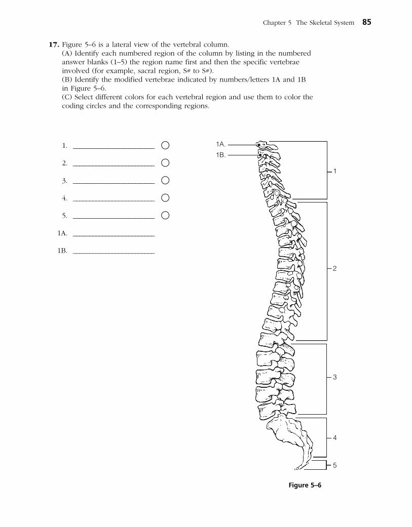
17. Figure 5–6 is a lateral view of the vertebral column. (A) Identify each numbered region of the column by listing in the numbered
answer blanks (1–5) the region name first and then the specific vertebrae involved (for example, sacral region, S# to S#).
(B) Identify the modified vertebrae indicated by numbers/letters 1A and 1B in Figure 5–6.
(C) Select different colors for each vertebral region and use them to color the coding circles and the corresponding regions.
1. _________________________ ○
2. _________________________ ○
3. _________________________ ○
4. _________________________ ○
5. _________________________ ○
1A. _________________________
1B. _________________________
Chapter 5 The Skeletal System 85
1A.
1B.
1
2
3
4
5
Figure 5–6
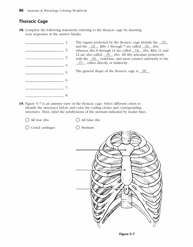
Thoracic Cage
18. Complete the following statements referring to the thoracic cage by inserting your responses in the answer blanks.
_________________________ 1.
_________________________ 2.
_________________________ 3.
_________________________ 4.
_________________________ 5.
_________________________ 6.
_________________________ 7.
_________________________ 8.
19. Figure 5–7 is an anterior view of the thoracic cage. Select different colors to identify the structures below and color the coding circles and corresponding structures. Then, label the subdivisions of the sternum indicated by leader lines.
○ All true ribs ○ All false ribs
○ Costal cartilages ○ Sternum
86 Anatomy & Physiology Coloring Workbook
The organs protected by the thoracic cage include the (1) and the (2) . Ribs 1 through 7 are called (3) ribs, whereas ribs 8 through 12 are called (4) ribs. Ribs 11 and 12 are also called (5) ribs. All ribs articulate posteriorly with the (6) vertebrae, and most connect anteriorly to the
(7) , either directly or indirectly.
The general shape of the thoracic cage is (8) .
Figure 5–7

APPENDICULAR SKELETON
Several bones forming part of the upper limb and/or shoulder girdle are shown in Figures 5–8 to 5–11. Follow the specific directions for each figure.
20. Identify the bone in Figure 5–8. Insert your answer in the blank below the illustration. Select different colors for each structure listed below and use them to color the coding circles and the corresponding structures in the diagram. Then, label the angles indicated by leader lines.
○ Spine ○ Glenoid cavity ○ Coracoid process ○ Acromion
Chapter 5 The Skeletal System 87
Superior border
Lateral border
Medial border
Bone:
Figure 5–8
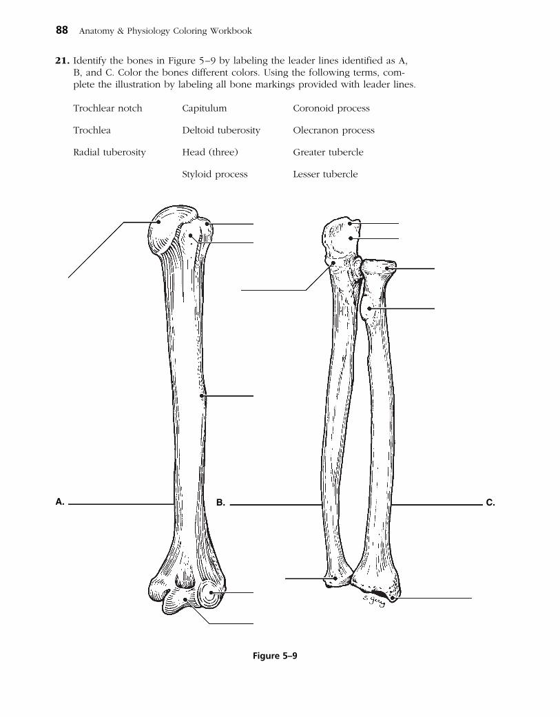
21. Identify the bones in Figure 5–9 by labeling the leader lines identified as A, B, and C. Color the bones different colors. Using the following terms, complete the illustration by labeling all bone markings provided with leader lines.
Trochlear notch Capitulum Coronoid process
Trochlea Deltoid tuberosity Olecranon process
Radial tuberosity Head (three) Greater tubercle
Styloid process Lesser tubercle
88 Anatomy & Physiology Coloring Workbook
A. B. C.
Figure 5–9

22. Figure 5–10 is a diagram of the hand. Select different colors for the following structures and use them to color the coding circles and the corresponding structures in the diagram.
○ Carpals ○ Metacarpals ○ Phalanges
23. Compare the pectoral and pelvic girdles by choosing descriptive terms from the key choices. Insert the appropriate key letters in the answer blanks.
Key Choices
A. Flexibility D. Shallow socket for limb attachmentB. Massive E. Deep, secure socket for limb attachmentC. Lightweight F. Weightbearing
Pectoral: _______, _______, _______ Pelvic: _______, _______, _______
24. Using the key choices, complete the crossword puzzle by answering each of the clues provided.
Key Choices
Acromion Coronoid fossa Olecranon fossa ScapulaCapitulum Deltoid tuberosity Olecranon process Sternum
Chapter 5 The Skeletal System 89
Radius
Ulna
Figure 5–10
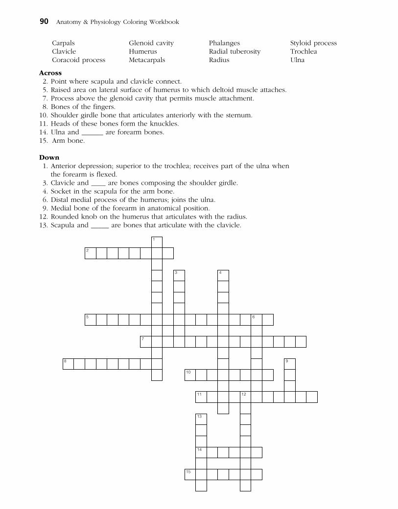
Carpals Glenoid cavity Phalanges Styloid processClavicle Humerus Radial tuberosity TrochleaCoracoid process Metacarpals Radius Ulna
Across 2. Point where scapula and clavicle connect. 5. Raised area on lateral surface of humerus to which deltoid muscle attaches. 7. Process above the glenoid cavity that permits muscle attachment. 8. Bones of the fingers. 10. Shoulder girdle bone that articulates anteriorly with the sternum. 11. Heads of these bones form the knuckles. 14. Ulna and ______ are forearm bones. 15. Arm bone.
Down 1. Anterior depression; superior to the trochlea; receives part of the ulna when
the forearm is flexed. 3. Clavicle and ____ are bones composing the shoulder girdle. 4. Socket in the scapula for the arm bone. 6. Distal medial process of the humerus; joins the ulna. 9. Medial bone of the forearm in anatomical position. 12. Rounded knob on the humerus that articulates with the radius. 13. Scapula and _____ are bones that articulate with the clavicle.
1
2
3 4
5 6
7
8 9
10
11 12
13
14
15
90 Anatomy & Physiology Coloring Workbook

25. Figure 5–11 is a diagram of the articulated pelvis. (A) Identify the bones and bone markings indicated by leader lines on
the figure. (B) Select different colors for the structures listed below and use them to
color the coding circles and the corresponding structures in the figure. (C) Label the dashed line show ing the dimensions of the true pelvis and that
showing the diameter of the false pelvis. (D) Complete the illustration by labeling the following bone mark ings:
obturator foramen, iliac crest, anterior superior iliac spine, ischial spine, pubic ramus, and pelvic brim.
(E) List three ways in which the female pelvis differs from the male pelvis and insert your answers in the answer blanks.
○ Coxal bone (hip bone) ○ Pubic symphysis
○ Sacrum ○ Acetabulum
1. ____________________________________________________________________________________________
2. ____________________________________________________________________________________________
3. ____________________________________________________________________________________________
26. Circle the term that does not belong in each of the following groupings. Then, fill in the answer blanks with the correct group name.
1. Tibia Ulna Fibula Femur Group: _________
2. Skull Rib cage Vertebral column Pelvis Group: _________
3. Ischium Scapula Ilium Pubis Group: _________
4. Mandible Frontal bone Temporal bone Occipital bone Group: _________
5. Calcaneus Tarsals Carpals Talus Group: _________
Chapter 5 The Skeletal System 91
Figure 5–11

27. Using the key choices, identify the bone names and markings, according to the descriptions that follow. Insert the appropriate key term(s) or letter(s) in the answer blanks. Items may have more than one answer.
Key Choices
A. Acetabulum I. Ilium Q. PatellaB. Calcaneus J. Ischial tuberosity R. Pubic symphysisC. Femur K. Ischium S. PubisD. Fibula L. Lateral malleolus T. Sacroiliac jointE. Gluteal tuberosity M. Lesser sciatic notch U. TalusF. Greater sciatic notch N. Medial malleolus V. TarsalsG. Greater and lesser trochanters O. Metatarsals W. TibiaH. Iliac crest P. Obturator foramen X. Tibial tuberosity
_________________________ 1. Fuse to form the coxal bone (hip bone)
_________________________ 2. Receives the weight of the body when sitting
_________________________ 3. Point where the coxal bones join anteriorly
_________________________ 4. Upper margin of iliac bones
_________________________ 5. Deep socket in the coxal bone (hip bone) that receives the head of the thigh bone
_________________________ 6. Point where the axial skeleton attaches to the pelvic girdle
_________________________ 7. Longest bone in body; articulates with the coxal bone
_________________________ 8. Lateral bone of the leg
_________________________ 9. Medial bone of the leg
_________________________ 10. Bones forming the knee joint
_________________________ 11. Point where the patellar ligament attaches
_________________________ 12. Kneecap
_________________________ 13. Shinbone
_________________________ 14. Distal process on medial tibial surface
_________________________ 15. Process forming the outer ankle
_________________________ 16. Heel bone
92 Anatomy & Physiology Coloring Workbook

_________________________ 17. Bones of the ankle
_________________________ 18. Bones forming the instep of the foot
_________________________ 19. Opening in a coxal bone (hip bone) formed by the pubic and ischial rami
_________________________ 20. Sites of muscle attachment on the proximal end of the femur
_________________________ 21. Tarsal bone that articulates with the tibia
28. For each of the following statements that is true, insert T in the answer blank. If any of the statements are false, correct the underlined term by inserting the correct term in the answer blank.
_________________________ 1. The pectoral girdle is formed by the articulation of the coxal bones (hip bones) and the sacrum.
_________________________ 2. Bones present in both the hand and the foot are carpals.
_________________________ 3. The tough, fibrous connective tissue covering of a bone is the periosteum.
_________________________ 4. The point of fusion of the three bones forming a coxal bone is the glenoid cavity.
_________________________ 5. The large nerve that must be avoided when giving injections into the buttock muscles is the femoral nerve.
_________________________ 6. The long bones of a fetus are constructed of hyaline cartilage.
_________________________ 7. Bones that provide the most protection to the abdominal viscera are the ribs.
_________________________ 8. The largest foramen in the skull is the foramen magnum.
_________________________ 9. The intercondylar fossa, greater trochanter, and gluteal tuberosity are all bone markings of the humerus.
_________________________ 10. The first major event of fracture healing is hematoma formation.
_________________________ 11. An exaggerated thoracic curvature known as “dowager’s hump” is an abnormal condition called scoliosis.
Chapter 5 The Skeletal System 93

29. The bones of the thigh and the leg are shown in Figure 5–12. (A) Select different colors for the lower limb bones listed below and
use them to color in the coding circles and corresponding bones on the diagram.
(B) Complete the illustration by inserting the terms indicating bone markings at the ends of the appropriate leader lines in the figure.
○ Femur ○ Tibia ○ Fibula
Head of femur Anterior border of tibia Head of fibula
Lesser trochanter Intercondylar eminence Lateral malleolus
Greater trochanter Medial malleolus
Tibial tuberosity
94 Anatomy & Physiology Coloring Workbook
Lateralcondyle
NeckLateral
condyle
Medial condyle
Figure 5–12

30. Figure 5–13 is a diagram of the articulated skeleton in anatomical position. Identify all bones or groups of bones by writing the correct labels at the end of the leader lines. Then, select two different colors for the bones of the axial and appendicular skeletons and use them to color in the coding circles and corresponding structures in the diagram.
○ Axial skeleton ○ Appendicular skeleton
Chapter 5 The Skeletal System 95
Figure 5–13

BONE FRACTURES
31. Using the key choices, identify the fracture (fx) types shown in Figure 5–14 and the fracture types and treatments described below. Enter the appropriate key letter or term in each answer blank.
Key Choices
A. Closed reduction D. Depressed fracture G. Simple fractureB. Compression fracture E. Greenstick fracture H. Spiral fractureC. Compound fracture F. Open reduction
_________________________ 1. Bone broken cleanly; ends do not penetrate the skin
_________________________ 2. Nonsurgical realignment of broken bone ends and splinting of bone
_________________________ 3. A break common in children; bone splinters, but break is incomplete
_________________________ 4. A fracture in which the bone is crushed; common in the vertebral column
_________________________ 5. A fracture in which the bone ends penetrate through the skin surface
_________________________ 6. Surgical realignment of broken bone ends
_________________________ 7. A result of twisting forces
96 Anatomy & Physiology Coloring Workbook
Figure 5–14

32. For each of the following statements that is true about bone breakage and the repair process, insert T in the answer blank. For false statements, correct the underlined terms by inserting the correct term in the answer blank.
_________________________ 1. A hematoma usually forms at a fracture site.
_________________________ 2. Deprived of nutrition, osteocytes at the fracture site die.
_________________________ 3. Nonbony debris at the fracture site is removed by osteoclasts.
_________________________ 4. Growth of a new capillary supply into the region produces granulation tissue.
_________________________ 5. Osteoblasts from the medullary cavity migrate to the fracture site.
_________________________ 6. The fibrocartilage callus is the first repair mass to splint the broken bone.
_________________________ 7. The bony callus is initially composed of compact bone.
JOINTS
33. Figure 5–15 shows the structure of a typical diarthrotic joint. Select different colors to identify each of the following areas and use them to color the coding circles and the corresponding structures on the figure. Then, complete the statements below the figure.
○ Articular cartilage of bone ends
○ Fibrous capsule
○ Synovial membrane
○ Joint cavity
1. _________________________ The lubricant that minimizes friction and abrasion of joint surfaces is (1) .
2. _________________________ The resilient substance that keeps bone ends from crushing when compressed is (2) .
3. _________________________ (3) , which reinforce the fibrous capsule, help to prevent dislocation of the joint.
Chapter 5 The Skeletal System 97
Figure 5–15

34. For each joint described below, select an answer from Key A. Then, classify the joint further by making a choice from Key B when applicable. Items may have more than one answer.
Key Choices
Key A: A. Cartilaginous Key B: 1. Synchondrosis (epiphyseal disc) B. Fibrous 2. Suture C. Synovial 3. Symphysis
_________________________ 1. Has amphiarthrotic and synarthrotic examples
_________________________ 2. Fibrous capsule lined with synovial membrane surrounding a joint cavity
_________________________ 3. Bone regions united by fibrous connective tissue
_________________________ 4. Joints between skull bones
_________________________ 5. Joint between the atlas and axis
_________________________ 6. Hip, elbow, and knee
_________________________ 7. All examples are diarthroses
_________________________ 8. Pubic symphysis
_________________________ 9. All reinforced by ligaments
_________________________ 10. Joint providing the most protection to underlying structures
_________________________ 11. Often contains a fluidfilled cushion
_________________________ 12. Child’s longbone growth plate made of hyaline cartilage
_________________________ 13. Most joints of the limbs
_________________________ 14. Often associated with bursae
_________________________ 15. Have the greatest mobility
35. Which structural joint type is not commonly found in the axial skeleton and why not?
_______________________________________________________________________________________________
_______________________________________________________________________________________________
_______________________________________________________________________________________________
98 Anatomy & Physiology Coloring Workbook
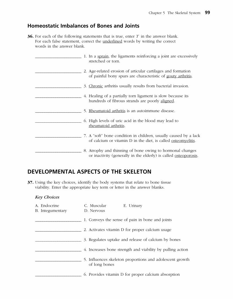
Homeostatic Imbalances of Bones and Joints
36. For each of the following statements that is true, enter T in the answer blank. For each false statement, correct the underlined words by writing the correct words in the answer blank.
_________________________ 1. In a sprain, the ligaments reinforcing a joint are excessively stretched or torn.
_________________________ 2. Agerelated erosion of articular cartilages and formation of painful bony spurs are characteristic of gouty arthritis.
_________________________ 3. Chronic arthritis usually results from bacterial invasion.
_________________________ 4. Healing of a partially torn ligament is slow because its hundreds of fibrous strands are poorly aligned.
_________________________ 5. Rheumatoid arthritis is an autoimmune disease.
_________________________ 6. High levels of uric acid in the blood may lead to rheumatoid arthritis.
_________________________ 7. A “soft” bone condition in children, usually caused by a lack of calcium or vitamin D in the diet, is called osteomyelitis.
_________________________ 8. Atrophy and thinning of bone owing to hormonal changes or inactivity (generally in the elderly) is called osteoporosis.
DEVELOPMENTAL ASPECTS OF THE SKELETON
37. Using the key choices, identify the body systems that relate to bone tissue viability. Enter the appropriate key term or letter in the answer blanks.
Key Choices
A. Endocrine C. Muscular E. UrinaryB. Integumentary D. Nervous
_________________________ 1. Conveys the sense of pain in bone and joints
_________________________ 2. Activates vitamin D for proper calcium usage
_________________________ 3. Regulates uptake and release of calcium by bones
_________________________ 4. Increases bone strength and viability by pulling action
_________________________ 5. Influences skeleton proportions and adolescent growth of long bones
_________________________ 6. Provides vitamin D for proper calcium absorption
Chapter 5 The Skeletal System 99

38. Complete the following statements concerning fetal and infant skeletal development. Insert the missing words in the answer blanks.
_________________________ 1.
_________________________ 2.
_________________________ 3.
_________________________ 4.
_________________________ 5.
_________________________ 6.
_________________________ 7.
_________________________ 8.
_________________________ 9.
A Visualization Exercise for the Skeletal System
. . . stalagmite- and stalactite-like structures that surround you. . . . Because the texture is so full of holes. . . .
39. Where necessary, complete statements by inserting the missing words in the answer blanks.
For this journey, you are miniaturized and injected into the interior of the largest and longest bone of your host’s body, the (1) . Once inside this bone, you look around and find yourself examining the stalagmite and stalactitelike structures that surround you. Although you feel as if you are in an under ground cavern, you know that it has to be bone. Because the texture is so full of holes, it obviously is (2) bone. Although the arrangement of these bony spars seems to be haphazard, as if someone randomly dropped straws, they are precisely arranged to resist points of (3) . All about you is frantic, hurried activity. Cells are dividing rapidly, nuclei are being ejected, and disclike cells are appearing. You decide that these disclike cells are (4) and that this is the (5)
cavity. As you explore further, strolling along the edge of the cavity, you spot many tunnels leading into the solid bony area on which you are walking. Walking into one of these drainpipelike openings, you notice that it contains a glistening white ropelike structure (a (6) , no doubt) and blood vessels running the length of the tube. You eventually come to a point in the channel where the
100 Anatomy & Physiology Coloring Workbook
“Soft spots,” or membranous joints called (1) in the fetal skull, allow the skull to be (2) slightly when in the birth canal. They also allow for continued brain (3) during the later months of fetal development and early infancy. Eventually these soft spots are replaced by immovable joints called (4) .
The two spinal curvatures welldeveloped at birth are the (5) and (6) curvatures. Because they are present at
birth, they are called (7) curvatures. The secondary curva tures develop as the baby matures. The (8) curvature develops as the baby begins to lift his or her head. The (9) curvature matures when the baby begins to walk or assume the upright posture.
_________________________ 1.
_________________________ 2.
_________________________ 3.
_________________________ 4.
_________________________ 5.
_________________________ 6.
INCREDIBLE JOURNEY

horizontal passageway joins with a vertical passage that runs with the longitudinal axis of the bone. This is obviously a
(7) canal. Because you would like to see how nutrients are brought into (8) bone, you decide to follow this channel. Reasoning that there is no way you can possibly scale the slick walls of the channel, you leap and grab onto a white cord hanging down its length. Because it is easier to slide down than to try to climb up the cord, you begin to lower yourself, handoverhand. During your descent, you notice small openings in the wall, which are barely large enough for you to wriggle through. You conclude that these are the
(9) that connect all the (10) to the nutrient supply in the central canal. You decide to investigate one of these tiny
openings and begin to swing on your cord, trying to get a foothold on one of the openings. After managing to anchor yourself and squeezing into an opening, you use a flashlight to illuminate the passage way in front of you. You are startled by a giant cell with many dark nuclei. It appears to be plastered around the entire lumen directly ahead of you. As you watch this cell, the bony material beneath it, the (11) , begins to liquefy. The cell apparently is a bonedigesting cell, or (12) , and because you are unsure whether or not its enzymes can also liquefy you, you slither backwards hurriedly and begin your trek back to your retrieval site.
40. Antonio is hit in the face with a football during practice. An Xray reveals multiple fractures of the bones around an orbit. Name the bones that form margins of the orbit.
41. Mrs. Bruso, a woman in her 80s, is brought to the clinic with a fractured hip. Xrays reveal compression fractures in her lower vertebral column and extremely low bone density in her vertebrae, coxal bones (hip bones), and femurs. What are the condition, cause, and treatment?
42. Jack, a young man, is treated at the clinic for an accident in which he hit his forehead. When he returns for a checkup, he complains that he can’t smell anything. A hurried Xray of his head reveals a fracture. What part of which bone was fractured to cause his loss of smell?
Chapter 5 The Skeletal System 101
_________________________ 7.
_________________________ 8.
_________________________ 9.
_________________________ 10.
_________________________ 11.
_________________________ 12.
AT THE CLINIC

43. A middleaged woman comes to the clinic complaining of stiff, painful joints and increasing immobility of her finger joints. A glance at her hands reveals knobby, deformed knuckles. For what condition will she be tested?
44. At his 94th birthday party, James was complimented on how good he looked and was asked about his health. He replied, “I feel good most of the time, but some of my joints ache and are stiff, especially my knees, hips, and lower back, and especially in the morning when I wake up.” A series of Xrays and an MRI scan taken a few weeks earlier had revealed that the articular cartilages of these joints were rough and flaking off, and bone spurs (overgrowths) were present at the ends of some of James’s bones. What is James’s probable condition?
45. Janet, a 10yearold girl, is brought to the clinic after falling out of a tree. An Xray shows she has small fractures of the transverse processes of T3 to T5 on the right side. Janet will be watched for what abnormal spinal curvature over the next several years?
46. The serving arm of many tennis players is often significantly larger (thicker) than the other arm. Explain this phenomenon.
47. Jerry is giving cardiopulmonary resuscitation (CPR) to Ms. Jackson, an elderly woman who has just been rescued from the waters of Cape Cod Bay. What bone is he compressing?
102 Anatomy & Physiology Coloring Workbook

49. Select the best answer or answers from the choices given.
Chapter 5 The Skeletal System 103
1. Important bone functions include:
A. support of the pelvic organs.
B. protection of the brain.
C. provision of levers for movement of the limbs.
D. protection of the skin and limb musculature.
E. storage of water.
2. A passageway connecting neighboring osteocytes in an osteon is a:
A. central canal. D. canaliculus.
B. lamella. E. perforating canal.
C. lacuna.
3. What is the earliest event (of those listed) in endochondral ossification?
A. Ossification of proximal epiphysis
B. Appearance of the epiphyseal plate
C. Invasion of the shaft by the periosteal bud
D. Cavitation of the cartilage shaft
E. Formation of secondary ossification centers
4. The growth spurt of puberty is triggered by:
A. high levels of sex hormones.
B. the initial, low levels of sex hormones.
C. growth hormone.
D. parathyroid hormone.
E. calcitonin.
5. Deficiency of which of the following hormones will cause dwarfism?
A. Growth hormone
B. Sex hormones
C. Thyroid hormones
D. Calcitonin
E. Parathyroid hormone
6. Women suffering from osteoporosis are frequent victims of _________________ fractures of the vertebrae.
A. compound D. compression
B. spiral E. depression
C. comminuted
7. Which of the following bones are part of the axial skeleton?
A. Vomer D. Parietal
B. Clavicle E. Coxal bone (hip
C. Sternum bone)
8. A blow to the cheek is most likely to break what superficial bone or bone part?
A. Superciliary arches
B. Zygomatic process
C. Mandibular ramus
D. Styloid process
9. Which of the following are part of the sphenoid?
A. Crista galli D. Pterygoid process
B. Sella turcica E. Lesser wings
C. Petrous portion
THE FINALE: MULTIPLE CHOICE
48. Rita’s bone density scan revealed she has osteoporosis. Her physician prescribed a drug that inhibits osteoclast activity. Explain this treatment.

10. Structural characteristics of all cervical vertebrae are:
A. small body.
B. bifid spinous process.
C. transverse foramina.
D. small vertebral foramen.
E. costal facets.
11. Which of the following bones exhibit a styloid process?
A. Hyoid D. Radius
B. Temporal E. Ulna
C. Humerus
12. Coxal bone (hip bone) markings include:
A. ala. D. pubic ramus.
B. sacral hiatus. E. fovea capitis.
C. gluteal surface.
13. Cartilaginous joints include:
A. syndesmoses. C. synostoses.
B. symphyses. D. synchondroses.
14. Considered to be part of a synovial joint are:
A. bursae. C. tendon sheath.
B. articular cartilage. D. capsular ligaments.
15. Abduction is:
A. moving the right arm out to the right.
B. spreading out the fingers.
C. wiggling the toes.
D. moving the sole of the foot laterally.
16. In comparing two joints of the same type, what characteristic(s) would you use to determine strength and flexibility?
A. Depth of the depression of the concave bone of the joint
B. Snugness of fit of the bones
C. Size of bone projections for muscle attachments
D. Presence of menisci
17. Which of the following joints has the greatest freedom of movement?
A. Interphalangeal
B. Saddle joint of thumb
C. Distal tibiofibular
D. Coxal (hip)
18. Which specific joint does the following description identify? “Articular surfaces are deep and secure, multiaxial; capsule heavily reinforced by ligaments; labrum helps prevent dislocation; the first joint to be built artificially; very stable.”
A. Elbow C. Knee
B. Hip D. Shoulder
19. An autoimmune disease resulting in inflammation and eventual fusion of diarthrotic joints is:
A. gout.
B. rheumatoid arthritis.
C. degenerative joint disease.
D. pannus.
20. Plane joints allow:
A. pronation. C. rotation.
B. flexion. D. gliding.
21. Movements made in chewing food are:
A. flexion. D. depression.
B. extension. E. opposition.
C. elevation.
22. Which of the following bones are not paired?
A. Parietal D. Pubis
B. Frontal E. Calcaneus
C. Sternum
104 Anatomy & Physiology Coloring Workbook

Muscles, the specialized tissues that facilitate body movement, make up about 40% of body weight. Most body muscle is the voluntary type, called skeletal muscle because it is attached to the bony skeleton. Skeletal muscles contrib-ute to body contours and shape. These muscles allow you to grin, run, shake hands, and to otherwise manipulate your environment. The balance of body muscle consists of smooth and cardiac muscles, which form the bulk of the walls of hollow organs and the heart. Smooth and cardiac muscles are involved in the transport of materials within the body.
Study activities in this chapter deal with microscopic and gross structure of muscle, identification of voluntary muscles, body movements, and important understandings of muscle physiology.
OVERVIEW OF MUSCLE TISSUES
1. Six characteristics of muscle tissue are listed below. Identify the muscle tissue type described by choosing the correct response(s) from the key choices. Enter the appropriate term(s) or letter(s) of the key choice in the answer blank.
Key Choices
A. Cardiac B. Smooth C. Skeletal
_________________________ 1. Involuntary
_________________________ 2. Banded appearance
_________________________ 3. Dense connective tissue packaging
_________________________ 4. Coordinated activity to act as a pump
_________________________ 5. Moves bones and the facial skin
_________________________ 6. Referred to as the muscular system
105
THE MUSCULAR SYSTEM
6

2. Identify the type of muscle in each of the illustrations in Figure 6–1. Select different colors for each structure listed below and use them to color the coding circles and the corresponding structures in the diagrams, when applicable.
○ Nucleus ○ Nucleolus ○ Striations ○ Intercalated discs
3. Circle the term that does not belong in each of the following groupings. Then, fill in the answer blanks with the correct group name.
1. Urine Foodstuffs Bones Smooth muscle Group: _______
2. Pacemaker Cardiac muscle Blood pump Promotes labor during birth Group: _______
3. Excitability Response to a stimulus Contractility Action potential Group: _______
4. Ability to shorten Contractility Pulls on bones Stretchability Group: _______
5. Maintains posture Movement Promotes growth Generates heat Group: _______
106 Anatomy & Physiology Coloring Workbook
A
B
Figure 6–1
C
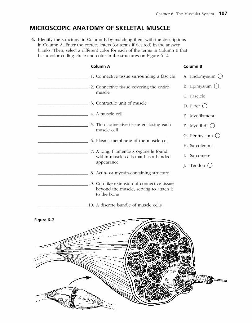
MICROSCOPIC ANATOMY OF SKELETAL MUSCLE
4. Identify the structures in Column B by matching them with the descrip tions in Column A. Enter the correct letters (or terms if desired) in the answer blanks. Then, select a different color for each of the terms in Column B that has a color-coding circle and color in the structures on Figure 6–2.
Column A
_________________________ 1. Connective tissue surrounding a fascicle
_________________________ 2. Connective tissue covering the entire muscle
_________________________ 3. Contractile unit of muscle
_________________________ 4. A muscle cell
_________________________ 5. Thin connective tissue enclosing each muscle cell
_________________________ 6. Plasma membrane of the muscle cell
_________________________ 7. A long, filamentous organelle found within muscle cells that has a banded appearance
_________________________ 8. Actin- or myosin-containing structure
_________________________ 9. Cordlike extension of connective tissue beyond the muscle, serving to attach it to the bone
_________________________ 10. A discrete bundle of muscle cells
Chapter 6 The Muscular System 107
Column B
A. Endomysium ○
B. Epimysium ○
C. Fascicle
D. Fiber ○
E. Myofilament
F. Myofibril ○
G. Perimysium ○
H. Sarcolemma
I. Sarcomere
J. Tendon ○
Figure 6–2
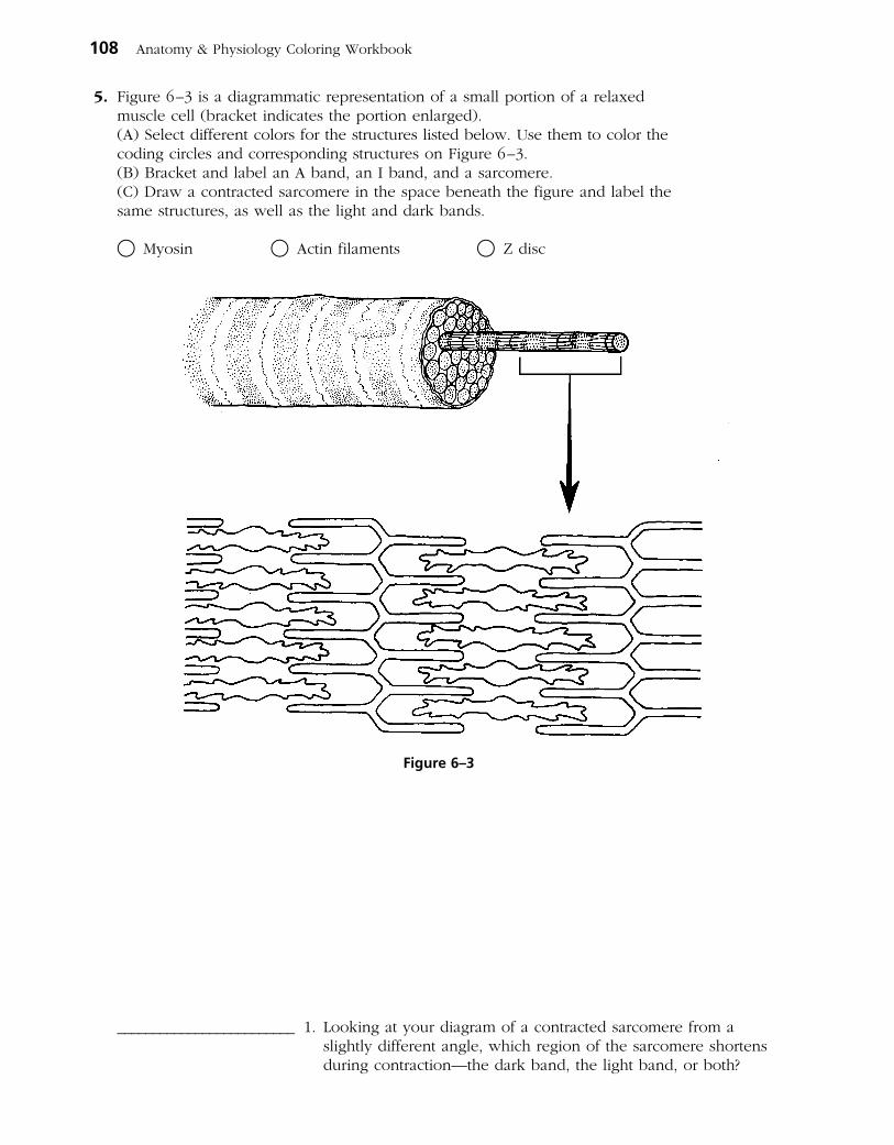
5. Figure 6–3 is a diagrammatic representation of a small portion of a relaxed muscle cell (bracket indicates the portion enlarged).
(A) Select different colors for the structures listed below. Use them to color the coding circles and corresponding structures on Figure 6–3.
(B) Bracket and label an A band, an I band, and a sarcomere. (C) Draw a contracted sarcomere in the space beneath the figure and label the
same structures, as well as the light and dark bands.
○ Myosin ○ Actin filaments ○ Z disc
_________________________ 1. Looking at your diagram of a contracted sarcomere from a slightly different angle, which region of the sarcomere shortens during contraction—the dark band, the light band, or both?
108 Anatomy & Physiology Coloring Workbook
Figure 6–3

SKELETAL MUSCLE ACTIVITY
6. Complete the following statements relating to the neuromuscular junction. Insert the correct answers in the numbered answer blanks.
_________________________ 1.
_________________________ 2.
_________________________ 3.
_________________________ 4.
_________________________ 5.
_________________________ 6.
7. Figure 6–4 shows the components of a neuromuscular junction. (A) Identify the parts by coloring the coding circles and the corresponding
structures in the diagram. (B) Add small arrows to indicate the location of the acetylcholine (ACh)
receptors and label appropriately.
○ Mitochondrion ○ T tubule ○ Sarcomere
○ Synaptic vesicles ○ Synaptic cleft ○ Junctional folds
Chapter 6 The Muscular System 109
A motor neuron and all of the skeletal muscle cells it stimulates is called a (1) . The axon of each motor neuron has numerous endings called (2) . The actual gap between an axonal ending and the muscle cell is called a (3) . Within the axonal endings are many small vesicles containing a neurotransmitter substance called (4) .
When the (5) reaches the ends of the axon, the neurotrans- mitter is released, and it diffuses to the muscle cell membrane to combine with receptors there. Binding of the neurotrans-mitters with muscle membrane receptors causes the membrane to become permeable to sodium, resulting in the influx of sodium ions and (6) of the membrane. Then contraction of the muscle cell occurs.
Figure 6–4
8. Number the following statements in their proper sequence to describe the contraction mechanism in a skeletal muscle cell. The first step has already been identified as number 1.
___1___ 1. ACh is released into the neuromuscular junction by the axonal terminal.
______ 2. The action potential, carried deep into the cell, causes the sarcoplasmic reticulum to release calcium ions.
______ 3. The muscle cell relaxes and lengthens.
______ 4. ACh diffuses across the neuromuscular junction and binds to receptors on the sarcolemma.
______ 5. The calcium ion concentration at the myofilaments increases; the myofilaments slide past one another, and the cell shortens.
______ 6. Depolarization occurs, and the action potential is generated.
______ 7. As calcium is actively reabsorbed into the sarcoplasmic reticulum, its concentration at the myofilaments decreases.
9. The following incomplete statements refer to a muscle cell in the resting, or polarized, state just before stimulation. Complete each statement by choosing the correct response from the key choices and entering the appropriate letter in the answer blanks.
Key Choices
A. Na+ diffuses out of the cell
B. K+ diffuses out of the cell
C. Na+ diffuses into the cell
D. K+ diffuses into the cell
E. Inside the cell
F. Outside the cell
_________________________ 1.
_________________________ 2.
_________________________ 3.
_________________________ 4.
_________________________ 5.
_________________________ 6. _________________________ 7.
There is a greater concentration of Na+ (1) , and there is a greater concentration of K+ (2) . When the stimulus is deliv-ered, the permeability of the membrane is changed, and
(3) , initiating the depolarization of the membrane. Almost as soon as the depolarization wave begins, a repolarization wave follows it across the membrane. This occurs as (4) . Repolarization restores the (5) of the resting cell membrane. The (6) is (are) reestablished by (7) .
110 Anatomy & Physiology Coloring Workbook
G. Relative ionic concentrations on the two sides of the membrane during rest
H. Electrical conditions
I. Activation of the sodium-potassium pump, which moves K+ into the cell and Na+ out of the cell
J. Activation of the sodium-potassium pump, which moves Na+ into the cell and K+ out of the cell

10. Complete the following statements by choosing the correct response from the key choices and entering the appropriate letter or term in the answer blanks.
Key Choices
A. Fatigue E. Isometric contraction I. Many motor unitsB. Isotonic contraction F. Whole muscle J. RepolarizationC. Muscle cell G. Fused tetanus K. DepolarizationD. Muscle tone H. Few motor units L. Unfused tetanus
_________________________ 1. ______ is a continuous contraction that shows no evidence of relaxation.
_________________________ 2. A(n) ______ is a contraction in which the muscle shortens and work is done.
_________________________ 3. To accomplish a strong contraction, ______ are stimulated at a rapid rate.
_________________________ 4. When a weak but smooth muscle contraction is desired, ______ are stimulated at a rapid rate.
_________________________ 5. When a muscle is being stimulated but is not able to respond because of “oxygen deficit,” the condition is called ______.
_________________________ 6. A(n) ______ is a contraction in which the muscle does not shorten, but tension in the muscle keeps increasing.
11. The terms in the key choices refer to the three ways that muscle cells replenish their ATP supplies. Select the term(s) that best apply to the conditions described and insert the correct key letter(s) in the answer blanks. Items may have more than one answer.
Key Choices
A. Coupled reaction of creatine phosphate (CP) and ADPB. Anaerobic glycolysis C. Aerobic respiration
______ 1. Accompanied by lactic acid formation
______ 2. Supplies the highest ATP yield per glucose molecule
______ 3. Involves the simple transfer of a phosphate group
______ 4. Requires no oxygen
______ 5. The slowest ATP regeneration process
______ 6. Produces carbon dioxide and water
______ 7. The energy mechanism used in the second hour of running in a marathon
______ 8. Used when the oxygen supply is inadequate over time
______ 9. Good for a sprint
Chapter 6 The Muscular System 111

12. Briefly describe how you can tell when you are repaying the oxygen deficit.
______________________________________________________________________________________________
______________________________________________________________________________________________
13. Which of the following occur within a muscle cell during oxygen deficit? Place a check (✓) by the correct choices.
______ 1. Decreased ATP ______ 5. Increased oxygen
______ 2. Increased ATP ______ 6. Decreased carbon dioxide
______ 3. Increased lactic acid ______ 7. Increased carbon dioxide
______ 4. Decreased oxygen ______ 8. Increased glucose
MUSCLE MOVEMENTS, TYPES, AND NAMES
14. Relative to general terminology concerning muscle activity, label the following structures on Figure 6–5: insertion, origin, tendon, resting muscle, and contracting muscle. Then, identify the two structures named below by choosing different colors for the coding circles and the corre-sponding structures in the figure.
○ Movable bone
○ Immovable bone
112 Anatomy & Physiology Coloring Workbook
Figure 6–5

15. Complete the following statements. Insert your answers in the answer blanks.
_________________________ 1.
_________________________ 2.
_________________________ 3.
_________________________ 4.
_________________________ 5.
_________________________ 6.
_________________________ 7.
_________________________ 8.
_________________________ 9.
_________________________ 10.
_________________________ 11.
_________________________ 12.
_________________________ 13.
_________________________ 14.
16. The terms provided in the key choices are often used to describe the manner in which muscles interact with other muscles. Select the key terms that apply to the following definitions and insert the correct letter or term in the answer blanks.
Key Choices
A. Antagonist B. Fixator C. Prime mover D. Synergist
_________________________ 1. Agonist
_________________________ 2. Postural muscles for the most part
_________________________ 3. Stabilizes a joint so that the prime mover can act at more distal joints
_________________________ 4. Performs the same movement as the prime mover
_________________________ 5. Reverses and/or opposes the action of a prime mover
_________________________ 6. Immobilizes the origin of a prime mover
Standing on your toes as in ballet is (1) of the foot. Walk-ing on your heels is (2) .
Winding up for a pitch (as in baseball) can properly be called (3) . To keep your seat when riding a horse, the tendency
is to (4) your thighs.
In running, the action at the hip joint is (5) in reference to the leg moving forward and (6) in reference to the leg in the posterior position. When kicking a football, the action at the knee is (7) . In climbing stairs, the hip and knee of the forward leg are both (8) . You have just touched your chin to your chest; this is (9) of the neck.
Using a screwdriver with a straight arm requires (10) of the arm. Consider all the movements of which the arm is capable. One often used for strengthening all the upper arm and shoulder muscles is (11) .
Moving the head to signify “no” is (12) . Action that moves the distal end of the radius across the ulna is (13) . Raising the arms laterally away from the body is called (14) of the arms.
Chapter 6 The Muscular System 113

17. Several criteria are applied to the naming of muscles. These are provided in Column B. Identify which criteria pertain to the muscles listed in Column A and enter the correct letter(s) in the answer blank. Items may have more than one answer.
GROSS ANATOMY OF THE SKELETAL MUSCLES
Muscles of the Head
18. Identify the major muscles described in Column A by choosing a response from Column B. Enter the correct letter in the answer blank. Select a different color for each muscle described and color in the coding circle and corresponding muscle on Figure 6–6.
Column A
○ ______ 1. Used to show you’re happy
○ ______ 2. Used to suck in your cheeks
○ ______ 3. Used in winking
○ ______ 4. Wrinkles the forehead horizontally
○ ______ 5. The “kissing” muscle
○ ______ 6. Prime mover of jaw closure
○ ______ 7. Synergist muscle for jaw closure
○ ______ 8. Prime mover of head flexion; a two-headed muscle
Column B
A. Action of the muscle
B. Shape of the muscle
C. Location of the muscle’s origin and/or insertion
D. Number of origins
E. Location of muscle relative to a bone or body region
F. Direction in which the muscle fibers run relative to some imaginary line
G. Relative size of the muscle
Column A
______ 1. Gluteus maximus
______ 2. Adductor magnus
______ 3. Biceps femoris
______ 4. Transversus abdominis
______ 5. Extensor carpi ulnaris
______ 6. Trapezius
______ 7. Rectus femoris
______ 8. External oblique
Column B
A. Buccinator
B. Frontalis
C. Masseter
D. Orbicularis oculi
E. Orbicularis oris
F. Sternocleidomastoid
G. Temporalis
H. Trapezius
I. Zygomaticus
114 Anatomy & Physiology Coloring Workbook
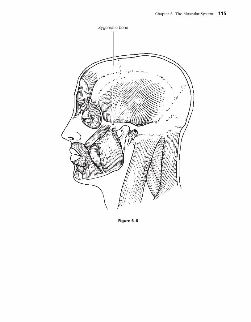
Chapter 6 The Muscular System 115
Zygomatic bone
Figure 6–6
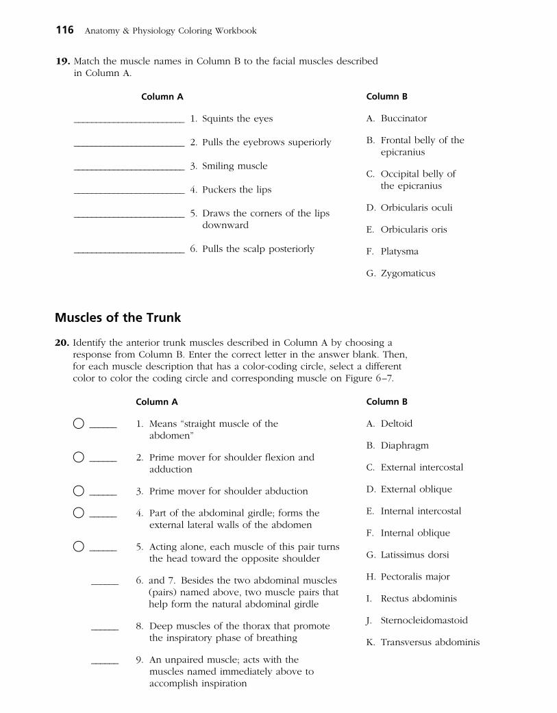
Muscles of the Trunk
20. Identify the anterior trunk muscles described in Column A by choosing a response from Column B. Enter the correct letter in the answer blank. Then, for each muscle description that has a color-coding circle, select a different color to color the coding circle and corresponding muscle on Figure 6–7.
Column A
○ ______ 1. Means “straight muscle of the abdomen”
○ ______ 2. Prime mover for shoulder flexion and adduction
○ ______ 3. Prime mover for shoulder abduction
○ ______ 4. Part of the abdominal girdle; forms the external lateral walls of the abdomen
○ ______ 5. Acting alone, each muscle of this pair turns the head toward the opposite shoulder
______ 6. and 7. Besides the two abdominal muscles (pairs) named above, two muscle pairs that help form the natural abdominal girdle
______ 8. Deep muscles of the thorax that promote the inspiratory phase of breathing
______ 9. An unpaired muscle; acts with the muscles named immediately above to accomplish inspiration
116 Anatomy & Physiology Coloring Workbook
Column B
A. Deltoid
B. Diaphragm
C. External intercostal
D. External oblique
E. Internal intercostal
F. Internal oblique
G. Latissimus dorsi
H. Pectoralis major
I. Rectus abdominis
J. Sternocleidomastoid
K. Transversus abdominis
19. Match the muscle names in Column B to the facial muscles described in Column A.
Column A
_________________________ 1. Squints the eyes
_________________________ 2. Pulls the eyebrows superiorly
_________________________ 3. Smiling muscle
_________________________ 4. Puckers the lips
_________________________ 5. Draws the corners of the lips downward
_________________________ 6. Pulls the scalp posteriorly
Column B
A. Buccinator
B. Frontal belly of the epicranius
C. Occipital belly of the epicranius
D. Orbicularis oculi
E. Orbicularis oris
F. Platysma
G. Zygomaticus
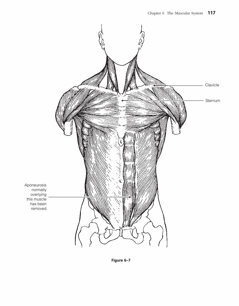
Chapter 6 The Muscular System 117
Aponeurosisnormallyoverlying
this musclehas beenremoved
Clavicle
Sternum
Figure 6–7

21. Identify the posterior trunk muscles described in Column A by choosing a response from Column B. Enter the correct letter in the answer blank. Select a different color for each muscle description with a coding circle and color the coding circles and corresponding muscles on Figure 6–8.
Column A
○ ______ 1. Muscle that allows you to shrug your shoulders or extend your head
○ ______ 2. Muscle that adducts the shoulder and causes extension of the shoulder joint
○ ______ 3. Shoulder muscle that is the antagonist of the muscle just described
______ 4. Prime mover of back extension; a deep composite muscle consisting of three columns
______ 5. Large paired superficial muscle of the lower back
○ ______ 6. Fleshy muscle forming part of the posterior abdominal wall that helps maintain upright posture
118 Anatomy & Physiology Coloring Workbook
Column B
A. Deltoid
B. Erector spinae
C. External oblique
D. Gluteus maximus
E. Latissimus dorsi
F. Quadratus lumborum
G. Trapezius
Chapter 6 The Muscular System 119
Figure 6–8
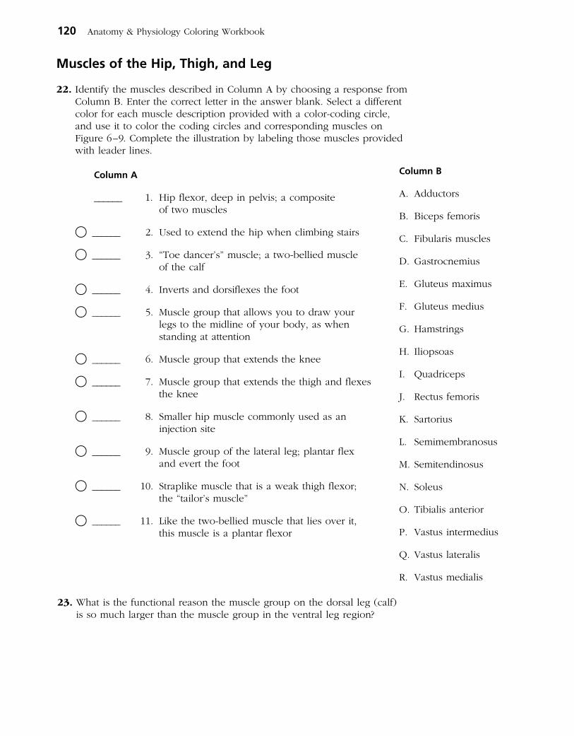
Muscles of the Hip, Thigh, and Leg
22. Identify the muscles described in Column A by choosing a response from Column B. Enter the correct letter in the answer blank. Select a different color for each muscle description provided with a color-coding circle, and use it to color the coding circles and corresponding muscles on Figure 6–9. Complete the illustration by labeling those muscles provided with leader lines.
Column A
______ 1. Hip flexor, deep in pelvis; a composite of two muscles
○ ______ 2. Used to extend the hip when climbing stairs
○ ______ 3. “Toe dancer’s” muscle; a two-bellied muscle of the calf
○ ______ 4. Inverts and dorsiflexes the foot
○ ______ 5. Muscle group that allows you to draw your legs to the midline of your body, as when standing at attention
○ ______ 6. Muscle group that extends the knee
○ ______ 7. Muscle group that extends the thigh and flexes the knee
○ ______ 8. Smaller hip muscle commonly used as an injection site
○ ______ 9. Muscle group of the lateral leg; plantar flex and evert the foot
○ ______ 10. Straplike muscle that is a weak thigh flexor; the “tailor’s muscle”
○ ______ 11. Like the two-bellied muscle that lies over it, this muscle is a plantar flexor
120 Anatomy & Physiology Coloring Workbook
Column B
A. Adductors
B. Biceps femoris
C. Fibularis muscles
D. Gastrocnemius
E. Gluteus maximus
F. Gluteus medius
G. Hamstrings
H. Iliopsoas
I. Quadriceps
J. Rectus femoris
K. Sartorius
L. Semimembranosus
M. Semitendinosus
N. Soleus
O. Tibialis anterior
P. Vastus intermedius
Q. Vastus lateralis
R. Vastus medialis
23. What is the functional reason the muscle group on the dorsal leg (calf) is so much larger than the muscle group in the ventral leg region?

Chapter 6 The Muscular System 121
A B Figure 6–9

Muscles of the Arm and Forearm
24. Identify the muscles described in Column A by choos-ing a response from Column B. Enter the corre ct letter in the answer blank. Select different colors for each muscle description provided with a color-coding circle and use them to color in the coding circles and corres-ponding muscles on Figure 6–10.
Column A
○ ______ 1. Wrist flexor that follows the ulna
○ ______ 2. Muscle that extends the fingers
______ 3. Muscle that flexes the fingers
○ ______ 4. Muscle that allows you to bend (flex) the elbow
○ ______ 5. Muscle that extends the elbow
○ ______ 6. Powerful shoulder abductor, used to raise the arm overhead
Column B
A. Biceps brachii
B. Deltoid
C. Extensor carpi radialis
D. Extensor digitorum
E. Flexor carpi ulnaris
F. Flexor digitorum superficialis
G. Triceps brachii
122 Anatomy & Physiology Coloring Workbook
Figure 6–10

General Body Muscle Review
25. Using the key choices, complete the crossword puzzle by answering each of the clues provided.
Key Choices
Anterior Distal Forearm ProximalCalcaneal Elbow Knee QuadricepsDeltoid Flex Posterior
Across 4. Three muscles, the gluteus maximus, gluteus medius, and ____, are
commonly used for intramuscular injections in adults. 6. Most flexor muscles are located on the _____ aspect of the body. 7. The extrinsic muscles of the hand originate on the _____. 8. The pectoralis major and deltoid muscles act synergistically to ______
the arm.
Down 1. The triceps surae (gastrocnemius and soleus) insert in common into
the _____ tendon. 2. The insertion tendon of the _______ group contains a large sesamoid
bone, the patella. 3. Most extensors are located on the _____ aspect of the body. 5. The bulk of the tissue of a muscle tends to lie _____ to the part of the
body it causes to move.
1 2
3
4
5 6
7
8
26. Circle the term that does not belong in each of the following groupings. Then, fill in the answer blanks with the correct group name.
1. Vastus lateralis Vastus medialis Knee extension Biceps femoris Group: _______
2. Latissimus dorsi Pectoralis major Adduction Antagonists Group: _______
3. Buccinator Frontalis Masseter Temporalis Group: _______
4. Vastus medialis Rectus femoris Iliacus Gluteus medius Group: _______
Chapter 6 The Muscular System 123

27. When kicking a football, at least three major actions of the lower limb are involved. Name the major muscles (or muscle groups) responsible for the following:
1. Flexing the hip joint: ______________________________________________________________________
2. Extending the knee: ______________________________________________________________
3. Dorsiflexing the foot: _____________________________________________________________
_________________________________________________________________________________
28. Identify the numbered muscles in Figure 6–11 by placing the numbers in the blanks next to the following muscle names. Select a different color for each muscle provided with a color-coding circle and color the coding circle and corresponding muscle in Figure 6–11.
○ ______ 1. Orbicularis oris
○ ______ 2. Pectoralis major
○ ______ 3. External oblique
○ ______ 4. Sternocleidomastoid
○ ______ 5. Biceps brachii
○ ______ 6. Deltoid
○ ______ 7. Vastus lateralis
○ ______ 8. Frontalis
○ ______ 9. Rectus femoris
○ ______ 10. Sartorius
○ ______ 11. Gracilis
○ ______ 12. Adductor group
○ ______ 13. Fibularis longus
○ ______ 14. Temporalis
○ ______ 15. Orbicularis oculi
○ ______ 16. Zygomaticus
○ ______ 17. Masseter
○ ______ 18. Vastus medialis
○ ______ 19. Tibialis anterior
○ ______ 20. Transversus abdominis
○ ______ 21. Rectus abdominis
124 Anatomy & Physiology Coloring Workbook

Chapter 6 The Muscular System 125
13
1211
10
9
8
7
6
5
4321
21
20
19
18
17
16
15
14
Figure 6–11

29. Identify each of the numbered muscles in Figure 6–12 by placing the numbers in the blanks next to the following muscle names. Select different colors for each muscle and color the coding circles and corre-sponding muscles on Figure 6–12.
○ ______ 1. Adductor muscle
○ ______ 2. Gluteus maximus
○ ______ 3. Gastrocnemius
○ ______ 4. Latissimus dorsi
○ ______ 5. Deltoid
○ ______ 6. Semitendinosus
○ ______ 7. Soleus
○ ______ 8. Biceps femoris
○ ______ 9. Triceps brachii
○ ______ 10. External oblique
○ ______ 11. Gluteus medius
○ ______ 12. Trapezius
126 Anatomy & Physiology Coloring Workbook
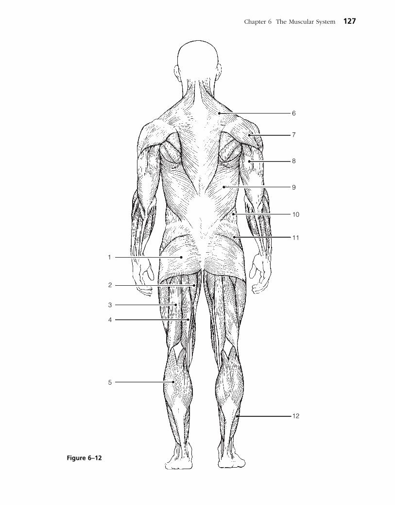
Chapter 6 The Muscular System 127
5
4
3
2
1
11
10
9
8
7
6
12
Figure 6–12

DEVELOPMENTAL ASPECTS OF THE MUSCULAR SYSTEM
30. Complete the following statements concerning the embryonic development of muscles and their functioning throughout life. Insert your answers in the answer blanks.
_________________________ 1.
_________________________ 2.
_________________________ 3.
_________________________ 4.
_________________________ 5.
_________________________ 6.
_________________________ 7.
_________________________ 8.
_________________________ 9.
_________________________ 10.
_________________________ 11.
_________________________ 12.
_________________________ 13.
A Visualization Exercise for the Muscular System
As you straddle this structure, you wonder what is happening.
31. Where necessary, complete statements by inserting the missing words in the numbered spaces.
On this incredible journey, you will be miniaturized and enter a skeletal muscle cell to observe the events that occur during muscle contraction. You prepare yourself by donning a wet
suit and charging your ion detector. Then you climb into a syringe to prepare for injection. Your journey will begin when you see the gleaming connective tissue covering, the (1) of a single muscle cell. Once injected, you monitor your descent through the epidermis and subcutaneous tis-sue. When you reach the muscle cell surface, you see that it is punctuated with pits at relatively
The first movement of the baby detected by the mother-to-be is called the (1) .
An important congenital muscular disease that results in the degeneration of the skeletal muscles by young adulthood is called (2) .
A baby’s control over muscles progresses in a (3) direction as well as a (4) direction. In addition, (5) muscular con-trol (that is, waving of the arms) occurs before (6) control (pincer grasp) does.
Muscles will ordinarily stay healthy if they are (7) regularly; without normal stimulation they (8) .
(9) is a disease of the muscles, which results from some problem with the stimulation of muscles by ACh. The muscles become progressively weaker in this disease.
With age, our skeletal muscles decrease in mass; this leads to a decrease in body (10) and in muscle (11) and (12) . Muscle tissue that is lost is replaced by noncontractile (13) tissue.
128 Anatomy & Physiology Coloring Workbook
_________________________ 1.
INCREDIBLE JOURNEY

regular intervals. Looking into the darkness and off in the dis-tance, you can see a group of fibers ending close to a number of muscle cells. Considering that all of these fibers must be from the same motor neuron, this functional unit is obviously a (2) . You approach the fiber ending on your muscle cell and scrutinize the (3) junction there. As you examine the junction, minute fluid droplets leave the nerve ending and attach to doughnut-shaped receptors on the muscle cell mem-brane. This substance released by the nerve ending must be
(4) . Then, as a glow falls over the landscape, your ion detector indicates ions are disappearing from the muscle cell exterior and entering the muscle pits. The needle drops from high to low as the (5) ions enter the pits from the watery fluid outside. You should have expected this, because these ions must enter to depolarize the muscle cells and start the
(6) .
Next, you begin to explore one of the surface pits. As the muscle jerks into action, you topple deep into the pit.
Sparkling electricity lights up the wall on all sides. You grasp for a handhold. Finally successful, you pull yourself laterally into the interior of the muscle cell and walk carefully along what seems to be a log. Then, once again, you notice an eerie glow as your ion detector reports that (7) ions are entering the cytoplasm rapidly. The “log” you are walking on “comes to life” and begins to slide briskly in one direction. Unable to keep your balance, you fall. As you straddle this structure, you wonder what is happening. On all sides, cylindrical structures—such as the one you are astride—are moving past other similar but larger structures. Suddenly you remember, these are the
(8) myofilaments that slide past the (9) myofilaments during muscle contraction.
Seconds later, the forward movement ends, and you begin to journey smoothly in the opposite direction. The ion detector now indicates low (10) ion levels. Because you cannot ascend the smooth walls of one of the entry pits, you climb from one myofilament to another to reach the underside of the sarcolemma. Then you travel laterally to enter a pit close to the surface and climb out onto the cell surface. Your journey is completed, and you prepare to leave your host once again.
32. Pete, who has been moving furniture all day, arrives at the clinic complaining of painful spasms in his back. He reports having picked up a heavy table by stooping over. What muscle group has Pete probably strained, and why are these muscles at risk when one lifts objects improperly?
Chapter 6 The Muscular System 129
_________________________ 2.
_________________________ 3.
_________________________ 4.
_________________________ 5.
_________________________ 6.
_________________________ 7.
_________________________ 8.
_________________________ 9.
_________________________ 10.
AT THE CLINIC

33. During an overambitious workout, a high school athlete pulled some muscles by forcing his knee into extension when his hip was already fully flexed. What muscles did he pull?
34. An emergency appendectomy is performed on Mr. Geiger. The incision was made at the lateral edge of the right iliac abdominopelvic region. Was his rec-tus abdominis cut?
35. Susan, a massage therapist, was giving Mr. Graves a back rub. Which two broad superficial muscles of the back were receiving most of her attention?
36. Mrs. Sanchez says that her 6-year-old son seems to be unusually clumsy and tires easily. The doctor notices that his calf muscles appear to be normal in size. If anything, they seem a bit enlarged rather than wasted. For what con-dition must the boy be checked? What is the prognosis?
37. People with chronic back pain occasionally get relief from a tummy tuck. How does this help?
38. Gregor, who works at a pesticide factory, comes to the clinic complaining of muscle spasms that interfere with his movement and breathing. A blood test shows that he has been contaminated with organophosphate pesticide, which is an acetylcholinesterase inhibitor. How would you explain to Gregor what this means?
130 Anatomy & Physiology Coloring Workbook
41. Select the best answer or answers from the choices given.
Chapter 6 The Muscular System 131
1. Select the type of muscle tissue that fits the following description: self-excitable, pacemaker cells, gap junctions, limited sar-coplasmic reticulum.
A. Skeletal muscle C. Smooth muscle
B. Cardiac muscle D. Voluntary muscle
2. Skeletal muscle is not involved in:
A. movement of skin.
B. propulsion of a substance through a body tube.
C. heat production.
D. inhibition of body movement.
3. Which of the following are part of a thin myofilament?
A. ATP-binding site C. Globular actin
B. Regulatory proteins D. Calcium
4. The movement of thin filaments toward the center of the sarcomere is called:
A. cocking of the myosin heads.
B. repolarization of the T tubules.
C. the power stroke.
D. the action potential.
5. Transmission of the stimulus at the neuromuscular junction involves:
A. synaptic vesicles. C. ACh.
B. sarcolemma. D. axon terminal.
6. Your ability to lift that heavy couch would be increased by which type of exercise?
A. Aerobic C. Resistance
B. Endurance D. Swimming
7. Which of the following activities depends most on anaerobic metabolism?
A. Jogging
B. Swimming a race
C. Sprinting
D. Running a marathon
8. The first energy source used to regenerate ATP when muscles are extremely active is:
A. fatty acids. C. creatine phosphate.
B. glucose. D. pyruvic acid.
9. Head muscles that insert on a bone include the:
A. zygomaticus. C. buccinator.
B. masseter. D. temporalis.
10. Lateral flexion of the torso involves:
A. erector spinae.
B. rectus abdominis.
C. quadratus lumborum.
D. external oblique.
11. Muscles attached to the vertebral column include the:
A. quadratus lumborum.
B. external oblique.
C. diaphragm.
D. latissimus dorsi.
12. Muscles that help stabilize the scapula and shoulder joint include the:
A. triceps brachii. C. trapezius.
B. biceps brachii. D. pectoralis major.
THE FINALE: MULTIPLE CHOICE
39. While riding an unusually large horse, Chao Jung had to spread her thighs wide to span its back, and she pulled the muscles in her medial thighs. Which muscles were these?
40. Do all muscles attach to bone? If not, what else do they attach to?

13. Which of these thigh muscles causes move-ment at the hip joint?
A. Rectus femoris
B. Biceps femoris
C. Vastus lateralis
D. Semitendinosus
14. Leg muscles that can cause movement at the knee joint include the:
A. tibialis anterior.
B. fibularis longus.
C. gastrocnemius.
D. soleus.
15. The main muscles used when doing chin-ups are the:
A. triceps brachii and pectoralis major.
B. infraspinatus and biceps brachii.
C. serratus anterior and external oblique.
D. latissimus dorsi and brachialis.
16. The major muscles used in doing push-ups are the:
A. biceps brachii and brachialis.
B. supraspinatus and subscapularis.
C. coracobrachialis and latissimus dorsi.
D. triceps brachii and pectoralis major.
17. Arm and leg muscles are arranged in antag-onistic pairs. How does this affect their functioning?
A. It provides a backup if one of the mus-cles is injured.
B. One muscle of the pair pushes while the other pulls.
C. A single neuron controls both of them.
D. It allows the muscles to produce oppos-ing movements.
18. Muscle A and muscle B are the same size, but muscle A is capable of much finer con-trol than muscle B. Which of the following is likely to be true of muscle A?
A. It is controlled by more neurons than muscle B.
B. It contains fewer motor units than muscle B.
C. It is controlled by fewer neurons than muscle B.
D. Each of its motor units consists of more cells than the motor units of muscle B.
19. Binding sites for calcium are found on:
A. thin filaments.
B. thick filaments.
C. myosin filaments.
D. actin filaments.
132 Anatomy & Physiology Coloring Workbook

133
THE NERVOUS SYSTEM
7
The nervous system is the master coordinating system of the body. Every thought, action, and sensation reflects its activity. The structures of the nervous system are described in terms of two principal divisions—the central nervous system (CNS) and the peripheral nervous system (PNS). The CNS (brain and spinal cord) interprets incoming sensory information and issues instructions based on experience. The PNS (cranial and spinal nerves and ganglia) provides the communica tion lines between the CNS and the body’s muscles, glands, and sensory receptors. The nervous system is also divided functionally in terms of motor activities into the somatic and autonomic divisions. It is important, however, to recognize that these classifications are made for the sake of convenience and that the nervous system acts in an integrated manner both structurally and functionally.
Student activities provided in this chapter review neuron anatomy and physiol-ogy, identify the various structures of the CNS and PNS, consider reflex and sensory physiology, and summarize autonomic nervous system anatomy and physiology. Because every body system is controlled, at least in part, by the nervous system, these concepts are extremely important to understanding how the body functions as a whole.
1. List the three major functions of the nervous system.
1. ___________________________________________________________________________________________
___________________________________________________________________________________________
2. ___________________________________________________________________________________________
___________________________________________________________________________________________
3. ___________________________________________________________________________________________
___________________________________________________________________________________________

ORGANIZATION OF THE NERVOUS SYSTEM
2. Choose the key responses that best correspond to the descriptions provided in the following statements. Insert the appropriate letter or term in the answer blanks.
Key Choices
A. Autonomic nervous system C. Peripheral nervous systemB. Central nervous system D. Somatic nervous system
_________________________ 1. Nervous system subdivision that is composed of the brain and spinal cord.
_________________________ 2. Subdivision of the PNS that controls voluntary activities such as the activation of skeletal muscles.
_________________________ 3. Nervous system subdivision that is composed of the cranial and spinal nerves and ganglia.
_________________________ 4. Subdivision of the PNS that regulates the activities of the heart and smooth muscle, and of glands; it is also called the involuntary nervous system.
_________________________ 5. A major subdivision of the nervous system that interprets incoming information and issues orders.
_________________________ 6. A major subdivision of the nervous system that serves as communication lines, linking all parts of the body to the CNS.
NERVOUS TISSUE—STRUCTURE AND FUNCTION
3. This exercise emphasizes the difference between neurons and neuroglia. Indicate which cell type is identified by the following descriptions. Insert the appropriate letter or term in the answer blanks.
Key Choices
A. Neurons B. Neuroglia
_________________________ 1. Support, insulate, and protect cells
_________________________ 2. Demonstrate irritability and conductivity, and thus transmit electrical messages from one area of the body to another area
_________________________ 3. Release neurotransmitters
_________________________ 4. Are amitotic
_________________________ 5. Able to divide; therefore are responsible for most brain neoplasms
134 Anatomy & Physiology Coloring Workbook
4. Relative to neuron anatomy, match the anatomical terms given in Column B with the appropriate descriptions of functions provided in Column A. Place the correct term or letter response in the answer blanks.
Column A
_________________________ 1. Releases neurotransmitters
_________________________ 2. Conducts local electrical currents toward the cell body
_________________________ 3. Increases the speed of impulse transmission
_________________________ 4. Location of the nucleus
_________________________ 5. Generally conducts impulses away from the cell body
_________________________ 6. Clustered ribosomes and rough ER (endoplasmic reticulum)
5. Certain activities or sensations are listed below. Using the key choices, select the specific receptor type that would be activated by the activity or sensation described. Insert the correct term(s) or letter response(s) in the answer blanks. Note that more than one receptor type may be activated in some cases.
Key Choices
A. Bare nerve endings (pain) C. Meissner’s (tactile) corpuscle E. Lamellated corpuscleB. Golgi tendon organ D. Muscle spindle
Activity or sensation Receptor type
Walking on hot pavement 1. _____________________________________________
and
Feeling a pinch 2. _____________________________________________
and
Leaning on a shovel 3. ______________________________________________
Muscle sensations when rowing a boat 4. _____________________________________________
and __________________________________________
Feeling a caress 5. ______________________________________________
Chapter 7 The Nervous System 135
Column B
A. Axon
B. Axon terminal
C. Dendrite
D. Myelin sheath
E. Neuron cell body
F. Nissl bodies

6. Using the key choices, select the terms identified in the following descriptions by inserting the appropriate letter or term in the spaces provided.
Key Choices
A. Afferent neuron F. Neuroglia K. ProprioceptorsB. Association neuron (or G. Neurotransmitters L. Schwann cells
interneuron) H. Nerve M. SynapseC. Cutaneous sense organs I. Nodes of Ranvier N. StimuliD. Efferent neuron J. Nuclei O. TractE. Ganglion
_________________________ 1. Sensory receptors found in the skin, which are specialized to detect temperature, pressure changes, and pain
_________________________ 2. Specialized cells; myelinate the fibers of neurons found in the PNS
_________________________ 3. Junction or point of close contact between neurons
_________________________ 4. Bundle of nerve processes inside the CNS
_________________________ 5. Neuron, serving as part of the conduction pathway between sensory and motor neurons
_________________________ 6. Gaps in a myelin sheath
_________________________ 7. Collection of nerve cell bodies found outside the CNS
_________________________ 8. Neuron that conducts impulses away from the CNS to muscles and glands
_________________________ 9. Sensory receptors found in muscle and tendons that detect their degree of stretch
_________________________ 10. Changes, occurring within or outside the body, that affect nervous system functioning
_________________________ 11. Neuron that conducts impulses toward the CNS from the body periphery
_________________________ 12. Chemicals released by neurons that stimulate other neurons, muscles, or glands
136 Anatomy & Physiology Coloring Workbook

7. Figure 7–1 is a diagram of a PNS neuron. (A) Label the parts indicated on the illustration by leader lines. (B) Choose different colors for each of the structures listed below and use
them to color in the coding circles and corre sponding structures in the illustration.
(C) Circle the term in the list of three terms to the left of the diagram that best describes this neuron’s structural class. (D) Draw arrows on the figure to indicate the direction of impulse transmission along the neuron’s membrane.
○ Axon
○ Dendrites
○ Cell body
○ Myelin sheath
8. List in order the minimum elements in a reflex arc from the stimulus to the activity of the effector. Place your responses in the answer blanks.
1. Stimulus 4. ________________________________________
2. ________________________________________ 5. Effector organ
3. ________________________________________
Chapter 7 The Nervous System 137
Figure 7–1

138 Anatomy & Physiology Coloring Workbook
9. Figure 7–2 is a diagram of a synapse. (A) Identify by coloring the following structures, which are typically part
of a chemical synapse. (B) Bracket the synaptic cleft. (C) Identify the arrows showing (1) the direction of the presynaptic
impulse and (2) the direction of net neurotransmitter movements.
○ Axon terminal ○ Postsynaptic membrane ○ Presynaptic membrane
○ Mitochondria ○ Na+ ions ○ Ca2+ ions
○ K+ ions ○ Chemically gated channels ○ Synaptic vesicles
○ Postsynaptic neurotransmitter receptors ○ Neurotransmitter molecules
Figure 7–2

10. Using the key choices, identify the terms defined in the following statements. Place the correct term or letter response in the answer blanks.
Key Choices
A. Action potential D. Potassium ions G. Resting periodB. Depolarization E. Refractory period H. Sodium ionsC. Polarized F. Repolarization I. Sodium-potassium pump
_________________________ 1. Period of repolarization of the neuron during which it cannot respond to a second stimulus
_________________________ 2. State in which the resting potential is reversed as sodium ions rush into the neuron
_________________________ 3. Electrical condition of the plasma membrane of a resting neuron
_________________________ 4. Period during which potassium ions diffuse out of the neuron
_________________________ 5. Transmission of the depolarization wave along the neuron’s membrane
_________________________ 6. The chief positive intracellular ion in a resting neuron
_________________________ 7. Process by which ATP is used to move sodium ions out of the cell and potassium ions back into the cell; completely restores the resting conditions of the neuron
_________________________ 8. State in which all voltage-gated Na+ and K+ channels are closed
11. Using the key choices, identify the types of reflexes involved in each of the following situations.
Key Choices
A. Somatic reflex(es) B. Autonomic reflex(es)
_________________________ 1. Patellar (knee-jerk) reflex
_________________________ 2. Pupillary light reflex
_________________________ 3. Effectors are skeletal muscles
_________________________ 4. Effectors are smooth muscle and glands
_________________________ 5. Flexor reflex
_________________________ 6. Regulation of blood pressure
_________________________ 7. Salivary reflex
Chapter 7 The Nervous System 139

140 Anatomy & Physiology Coloring Workbook
12. Refer to Figure 7–3, showing a reflex arc, as you complete this exercise. (A) Briefly answer the following questions by inserting your responses
in the spaces provided.
1. What is the stimulus? _____________________________________________________________________
2. Which specific type of tissue is the effector? _______________________________________________
3. How many synapses occur in this reflex arc? ______________________________________________
(B) Select different colors for each of the following structures and use them to color in the coding circles and corresponding structures in the diagram.
(C) Draw arrows on the figure indicating the direction of impulse transmission through this reflex pathway.
○ Receptor region ○ Interneuron
○ Afferent neuron ○ Efferent neuron
○ Effector
Figure 7–3
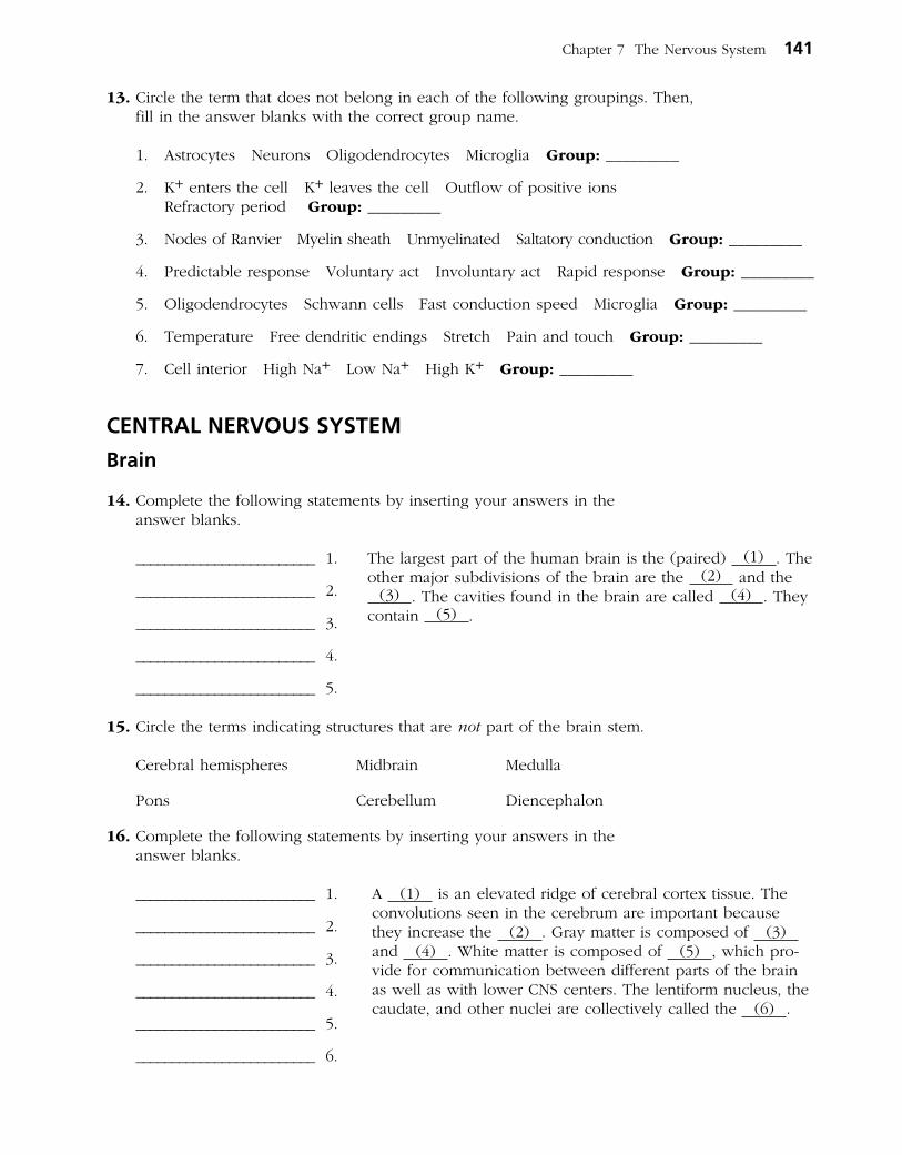
13. Circle the term that does not belong in each of the following groupings. Then, fill in the answer blanks with the correct group name.
1. Astrocytes Neurons Oligodendrocytes Microglia Group: _________
2. K+ enters the cell K+ leaves the cell Outflow of positive ions Refractory period Group: _________
3. Nodes of Ranvier Myelin sheath Unmyelinated Saltatory conduction Group: _________
4. Predictable response Voluntary act Involuntary act Rapid response Group: _________
5. Oligodendrocytes Schwann cells Fast conduction speed Microglia Group: _________
6. Temperature Free dendritic endings Stretch Pain and touch Group: _________
7. Cell interior High Na+ Low Na+ High K+ Group: _________
CENTRAL NERVOUS SYSTEM
Brain
14. Complete the following statements by inserting your answers in the answer blanks.
_________________________ 1.
_________________________ 2.
_________________________ 3.
_________________________ 4.
_________________________ 5.
15. Circle the terms indicating structures that are not part of the brain stem.
Cerebral hemispheres Midbrain Medulla
Pons Cerebellum Diencephalon
16. Complete the following statements by inserting your answers in the answer blanks.
_________________________ 1.
_________________________ 2.
_________________________ 3.
_________________________ 4.
_________________________ 5.
_________________________ 6.
The largest part of the human brain is the (paired) (1) . The other major subdivisions of the brain are the (2) and the
(3) . The cavities found in the brain are called (4) . They contain (5) .
A (1) is an elevated ridge of cerebral cortex tissue. The convolutions seen in the cerebrum are important because they increase the (2) . Gray matter is composed of (3) and (4) . White matter is composed of (5) , which pro-vide for com munication between different parts of the brain as well as with lower CNS centers. The lentiform nucleus, the caudate, and other nuclei are collectively called the (6) .
Chapter 7 The Nervous System 141

142 Anatomy & Physiology Coloring Workbook
17. Figure 7–4 is a diagram of the right lateral view of the human brain. (A) Match the letters on the diagram with the following list of terms and
insert the appropriate letters in the answer blanks. (B) Select different colors for each of the areas of the brain provided with
a color-coding circle and use them to color in the coding circles and cor-responding structures in the diagram. If an identified area is part of a lobe, use the color you selected for the lobe but use stripes for that area.
______ 1. ○ Frontal lobe
______ 2. ○ Parietal lobe
______ 3. ○ Temporal lobe
______ 4. ○ Precentral gyrus
______ 5. Parieto-occipital fissure
______ 6. ○ Postcentral gyrus
______ 7. Lateral sulcus
______ 8. Central sulcus
______ 9. ○ Cerebellum
______ 10. ○ Medulla
______ 11. ○ Occipital lobe
______ 12. ○ Pons
G
L
K
J
I
H
A B
C
D
F
E
Figure 7–4
18. Figure 7–5 is a diagram of the sagittal view of the human brain. (A) Match the letters on the diagram with the following list of terms and
insert the appropriate letter in each answer blank. (B) Color the brainstem areas blue and the areas where cerebrospinal fluid
is found yellow.
______ 1. Cerebellum
______ 2. Cerebral aqueduct
______ 3. Cerebral hemisphere
______ 4. Cerebral peduncle
______ 5. Choroid plexus
______ 6. Corpora quadrigemina
______ 7. Corpus callosum
______ 8. Fourth ventricle
Chapter 7 The Nervous System 143
______ 9. Mammillary body
______ 10. Medulla oblongata
______ 11. Optic chiasma
______ 12. Pineal gland
______ 13. Pituitary gland
______ 14. Pons
______ 15. Thalamus
G
LM
N
O
K
J I H
A B C
D
F
E
Figure 7–5

144 Anatomy & Physiology Coloring Workbook
19. Using the key choices, complete the crossword puzzle by answering each of the clues provided.
Key Choices
Cerebellum Corpus callosum Optic chiasmaCerebral aqueduct Fourth ventricle Pineal glandCerebral hemisphere Hypothalamus Pituitary glandCerebral peduncle Mammillary body PonsChoroid plexus Medulla oblongata ThalamusCorpora quadrigemina
Across 3. Responsible for the regulation of posture and coordination of skeletal muscle
movements. 6. Reflex center involved in olfaction; posterior to pituitary gland. 7. Site of regulation of water balance and body temperature. 8. Forms the cerebrospinal fluid. 9. Contains reflex centers involved in regulating respiratory rhythm in conjunc-
tion with lower brainstem centers.
Down 1. Large fiber tract connecting the cerebral hemispheres. 2. Connects the third and fourth ventricles. 4. Contains autonomic centers that regulate blood pressure and respiratory
rhythm as well as coughing and sneezing centers. 5. Midbrain area that is largely fiber tracts; bulges anteriorly.
1
2
3 4 5
6
7
8
9

20. Some of the following brain structures consist of gray matter; others are white matter. Write G (for gray) or W (for white) as appropriate.
______ 1. Cortex of cerebellum ______ 5. Pyramids
______ 2. Basal nuclei ______ 6. Thalamic nuclei
______ 3. Anterior commisure ______ 7. Cerebellar peduncle
______ 4. Corpus callosum
21. Figure 7–6 illustrates a “see-through” brain showing the positioning of the ventricles and connecting canals or apertures. Correctly identify all structures having leader lines by using the key choices provided below. One of the lateral ventricles has already been identified. Color the spaces filled with cerebrospinal fluid blue.
Key Choices
A. Anterior horn D. Fourth ventricle G. Lateral apertureB. Central canal E. Inferior horn H. Third ventricleC. Cerebral aqueduct F. Interventricular foramen
Chapter 7 The Nervous System 145
Figure 7–6
Lateralventricle
146 Anatomy & Physiology Coloring Workbook
22. If a statement is true, write the letter T in the answer blank. If a statement is false, correct the underlined word(s) and write the correct word(s) in the answer blank.
_________________________ 1. The primary somatosensory area of the cerebral hemisphere(s) is found in the precentral gyrus.
_________________________ 2. Cortical areas involved in audition are found in the occipital lobe.
_________________________ 3. The primary motor area in the temporal lobe is involved in the initiation of voluntary movements.
_________________________ 4. The specialized motor speech area is located at the base of the precentral gyrus in an area called Wernicke’s area.
_________________________ 5. The right cerebral hemisphere receives sensory input from the right side of the body.
_________________________ 6. The pyramidal tract is the major descending voluntary motor tract.
_________________________ 7. The primary motor cortex is located in the postcentral gyrus.
_________________________ 8. Centers for control of repetitious or stereotyped motor skills are found in the primary motor cortex.
_________________________ 9. The largest parts of the motor homunculi are the lips, tongue, and toes.
_________________________ 10. Sensations such as touch and pain are integrated in the primary sensory cortex.
_________________________ 11. The primary visual cortex is in the frontal lobe of each cerebral hemisphere.
_________________________ 12. In most humans, the area that controls the comprehension of language is located in the left cerebral hemisphere.
_________________________ 13. A flat electroencephalogram (EEG) is evidence of clinical death.
_________________________ 14. Beta waves are recorded when an individual is awake and relaxed.
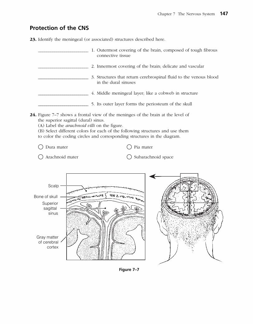
Protection of the CNS
23. Identify the meningeal (or associated) structures described here.
_________________________ 1. Outermost covering of the brain, composed of tough fibrous connective tissue
_________________________ 2. Innermost covering of the brain; delicate and vascular
_________________________ 3. Structures that return cerebrospinal fluid to the venous blood in the dural sinuses
_________________________ 4. Middle meningeal layer; like a cobweb in structure
_________________________ 5. Its outer layer forms the periosteum of the skull
24. Figure 7–7 shows a frontal view of the meninges of the brain at the level of the superior sagittal (dural) sinus.
(A) Label the arachnoid villi on the figure. (B) Select different colors for each of the following structures and use them
to color the coding circles and corresponding structures in the diagram.
○ Dura mater ○ Pia mater
○ Arachnoid mater ○ Subarachnoid space
Chapter 7 The Nervous System 147
Superiorsagittal
sinus
Bone of skull
Scalp
Gray matterof cerebral
cortex
Figure 7–7

148 Anatomy & Physiology Coloring Workbook
25. Complete the following statements by inserting your answers in the answer blanks.
_________________________ 1.
_________________________ 2.
_________________________ 3.
_________________________ 4.
_________________________ 5.
_________________________ 6.
_________________________ 7.
Brain Dysfunctions
26. Match the brain disorders listed in Column B with the conditions described in Column A. Place the correct answers in the answer blanks.
Column A
_________________________ 1. Slight and transient brain injury
_________________________ 2. Traumatic injury that destroys brain tissue
_________________________ 3. Total nonresponsiveness to stimulation
_________________________ 4. May cause medulla oblongata to be wedged into foramen magnum by pressure of blood
_________________________ 5. After head injury, retention of water by brain
_________________________ 6. Results when a brain region is deprived of blood or exposed to prolonged ischemia
_________________________ 7. Progressive degeneration of the brain with abnormal protein deposits
_________________________ 8. Autoimmune disorder with extensive demyelination
_________________________ 9. A ministroke; fleeting symptoms of a CVA
Cerebrospinal fluid is formed by capillary knots called (1) , which hang into the (2) of the brain. Ordinarily, cerebro-spinal fluid flows from the lateral ventricles to the third ventricle and then through the (3) to the fourth ventricle. Some of the fluid continues down the (4) of the spinal cord, but most of it circulates into the (5) by passing through three tiny openings in the walls of the (6) . As a rule, cerebro spinal fluid is formed and drained back into the venous blood at the same rate. If its drainage is blocked, a condition called (7) occurs, which results in increased pressure on the brain.
Column B
A. Alzheimer’s disease
B. Cerebral edema
C. Cerebrovascular accident (CVA)
D. Coma
E. Concussion
F. Contusion
G. Intracranial hemorrhage
H. Multiple sclerosis
I. Transient ischemic attack (TIA)

Spinal Cord
27. Complete the following statements by inserting your responses in the answer blanks.
_________________________ 1.
_________________________ 2.
_________________________ 3.
_________________________ 4.
_________________________ 5.
_________________________ 6.
_________________________ 7.
_________________________ 8.
_________________________ 9.
28. Using the key choices, select the appropriate terms to respond to the following descriptions referring to spinal cord anatomy. Place the correct term or letter in the answer blanks.
Key Choices
A. Afferent C. Both afferent and efferentB. Efferent D. Association neurons (interneurons)
_________________________ 1. Neuron type found in the dorsal horn
_________________________ 2. Neuron type found in the ventral horn
_________________________ 3. Neuron type in a dorsal root ganglion
_________________________ 4. Fiber type in the ventral root
_________________________ 5. Fiber type in the dorsal root
_________________________ 6. Fiber type in a spinal nerve
_________________________ 7. Fiber type in the anterior ramus
_________________________ 8. Damage to this fiber type would lead to a loss of sensory function
_________________________ 9. Damage to this fiber type results in a loss of motor function
The spinal cord extends from the (1) of the skull to the (2) region of the vertebral column. The meninges, which
cover the spinal cord, extend more inferiorly to form a sac from which cerebrospinal fluid can be withdrawn without damage to the spinal cord. This procedure is called a (3) .
(4) pairs of spinal nerves arise from the cord. Of these, (5) pairs are cervical nerves, (6) pairs are thoracic
nerves, (7) pairs are lumbar nerves, and (8) pairs are sacral nerves. The tail-like collection of spinal nerves at the inferior end of the spinal cord is called the (9) .
Chapter 7 The Nervous System 149

150 Anatomy & Physiology Coloring Workbook
29. Figure 7–8 is a cross-sectional view of the spinal cord. (A) Identify the areas listed in the key choices by inserting the correct letters
next to the appropriate leader lines on parts A and B of the figure.
Key Choices
A. Central canal E. Dorsal root I. Ventral hornB. Columns of white matter F. Dorsal root ganglion J. Ventral rootC. Conus medullaris G. Filum terminaleD. Dorsal horn H. Spinal nerve
(B) Color the lumbar bones of the vertebral column in figure B gold.(C) On figure A, color the butterfly-shaped gray matter gray, and color the spinal nerves and roots yellow. (D) Select different colors to identify the following structures and use them to color the figure.
○ Pia mater ○ Dura mater ○ Arachnoid mater
A
Spinalcord
L1
L2
L3
L4
L5
BFigure 7–8
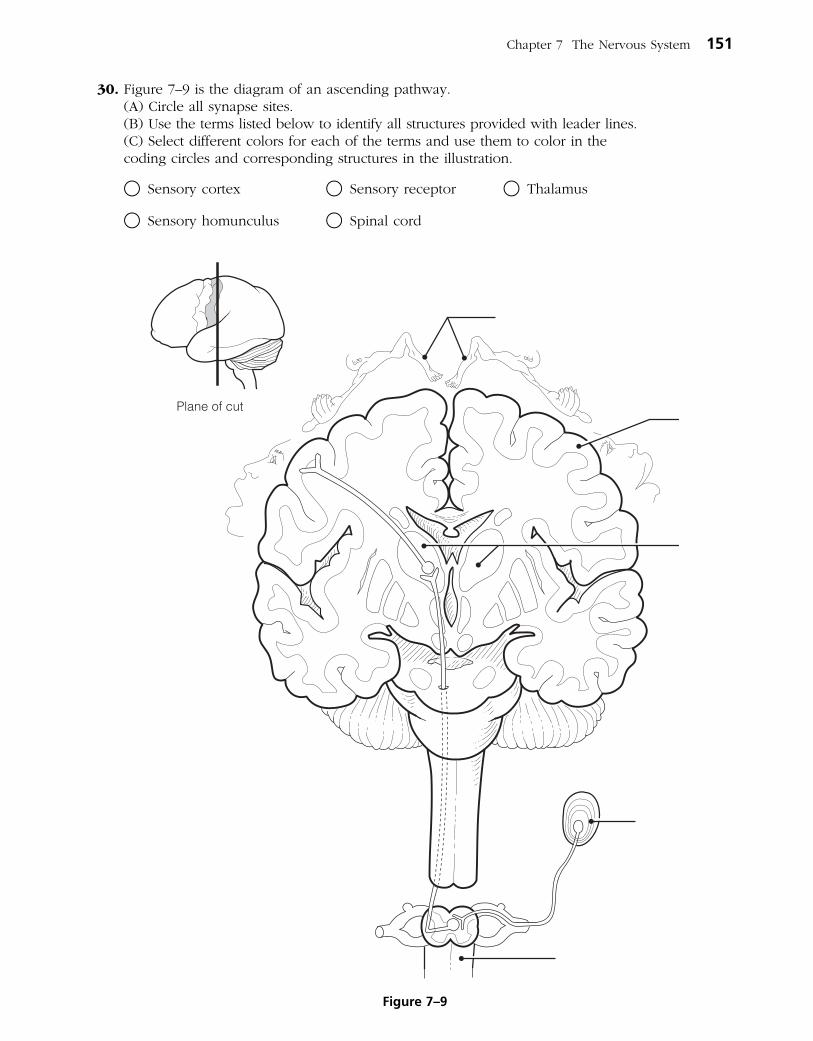
Chapter 7 The Nervous System 151
30. Figure 7–9 is the diagram of an ascending pathway. (A) Circle all synapse sites. (B) Use the terms listed below to identify all structures provided with leader lines. (C) Select different colors for each of the terms and use them to color in the
coding circles and corresponding structures in the illustration.
○ Sensory cortex ○ Sensory receptor ○ Thalamus
○ Sensory homunculus ○ Spinal cord
Figure 7–9

152 Anatomy & Physiology Coloring Workbook
PERIPHERAL NERVOUS SYSTEM
Structure of a Nerve
31. Figure 7– 10 is a diagrammatic view of a nerve wrapped in its connective tissue coverings.
(A) Select different colors to identify the following structures and use them to color the coding circles and corresponding structures in the figure.
(B) Label each of the sheaths indicated by leader lines on the figure.
○ Endoneurium ○ Perineurium ○ Epineurium
32. Complete the following statements by inserting your responses in the answer blanks.
_________________________ 1.
_________________________ 2.
_________________________ 3.
Axon
Bloodvessel
Fascicle
Myelin sheath
Figure 7–10
Another name for a bundle of nerve fibers is a (1) . Nerves carrying both sensory and motor fibers are called (2) nerves, whereas those carrying just sensory fibers are referred to as sensory, or (3) , nerves.

Chapter 7 The Nervous System 153
Cranial Nerves
33. The 12 pairs of cranial nerves are indicated by leader lines in Figure 7–11. (A) Label each by name and Roman numeral on the figure.
(B) Color each nerve with a different color.
Cerebrum
Medullaoblongata
Cerebellum
Pons
Spinal nerveSpinal cord
Figure 7–11
154 Anatomy & Physiology Coloring Workbook
34. Provide the name and number of the cranial nerves involved in each of the following activities, sensations, or disorders. Insert your response in the answer blanks.
_________________________ 1. Shrugging the shoulders
_________________________ 2. Smelling a flower
_________________________ 3. Raising the eyelids and focusing the lens of the eye for accommodation; constriction of the eye pupils
_________________________ 4. Slows the heart; increases the mobility of the digestive tract
_________________________ 5. Involved in smiling
_________________________ 6. Involved in chewing gum
_________________________ 7. Listening to music; seasickness
_________________________ 8. Secretion of saliva; tasting well-seasoned food
_________________________ 9. Involved in “rolling” the eyes (three nerves—provide numbers only)
_________________________ 10. Feeling a toothache
_________________________ 11. Reading this study guide
_________________________ 12. Purely sensory (three nerves—provide numbers only)
Spinal Nerves and Nerve Plexuses
35. Complete the following statements by inserting your responses in the answer blanks.
_________________________ 1.
_________________________ 2.
_________________________ 3.
_________________________ 4.
The ventral rami of spinal nerves C1 through T1 and L1 through S4 take part in forming (1) , which serve the (2) of the body. The ventral rami of T1 through T12 run between the ribs to serve the (3) . The posterior rami of the spinal nerves serve the (4) .

Chapter 7 The Nervous System 155
36. Figure 7–12 is an anterior view of the principal nerves arising from the brachial plexus. Select five different colors and color the coding circles and the nerves listed below. Also, label each nerve by inserting its name at the appropriate leader line.
○ Axillary nerve
○ Musculocutaneous nerve
○ Median nerve
○ Radial nerve
○ Ulnar nerve
C4
C5
C6
C7
C8
T1
Manubrium
Ulna
Radius
Humerus
Figure 7–12

156 Anatomy & Physiology Coloring Workbook
37. Name the major nerves that serve the following body areas. Insert your responses in the answer blanks.
_________________________ 1. Neck and shoulders (plexus only)
_________________________ 2. Abdominal wall (plexus only)
_________________________ 3. Anterior thigh
_________________________ 4. Diaphragm
_________________________ 5. Posterior thigh
_________________________ 6. Leg and foot (two)
Autonomic Nervous System (ANS)
38. Identify, by color coding and coloring, the following structures in Figure 7–13, which depicts the major anatomical differences between the somatic and autonomic motor divisions of the PNS. Also identify by labeling all structures provided with leader lines.
○ Somatic motor neuron ○ Effector of the somatic motor neuron
○ ANS preganglionic neuron ○ Effector of the autonomic motor neuron
○ ANS ganglionic neuron ○ Myelin sheath
○ Autonomic ganglion ○ White matter of spinal cord (CNS)
○ Gray matter of spinal cord (CNS)
CNS(spinal cord)
Skeletal muscle
Smallintestine(or othervisceralorgan)
Figure 7–13

39. The following table indicates a number of conditions. Use a check (✓) to show which division of the autonomic nervous system is involved in each condition. Then, respond to the true-to-life situation below the chart.
Condition Sympathetic Parasympathetic
1. Postganglionic axons secrete norepinephrine; adrenergic fibers
2. Postganglionic axons secrete acetylcholine; cholinergic fibers
3. Long preganglionic axon, short postganglionic axon
4. Short preganglionic axon, long postganglionic axon
5. Arises from cranial and sacral nerves
6. Arises from spinal nerves T1 to L3
7. Normally in control
8. Fight-or-flight system
9. Has more specific control
10. Causes a dry mouth, dilates bronchioles
11. Constricts eye pupils, decreases heart rate
You are alone in your home late in the evening, and you hear an unfamiliar sound in your backyard. In the spaces provided, list four physiological events promoted by the sympathetic nervous system that would help you to cope with this rather frightening situation.
1. ___________________________________________________________________________________________
2. ___________________________________________________________________________________________
3. ___________________________________________________________________________________________
4. ___________________________________________________________________________________________
Chapter 7 The Nervous System 157

158 Anatomy & Physiology Coloring Workbook
DEVELOPMENTAL ASPECTS OF THE NERVOUS SYSTEM
40. Complete the following statements by inserting your responses in the answer blanks.
_________________________ 1.
_________________________ 2.
_________________________ 3.
_________________________ 4.
_________________________ 5.
_________________________ 6.
_________________________ 7.
_________________________ 8.
A Visualization Exercise for the Nervous System
You climb on the first cranial nerve you see. . . .
41. Where necessary, complete statements by inserting the missing words in the answer blanks.
Nervous tissue is quite densely packed, and it is difficult to envision strolling through its various regions. Imagine instead that each of the various functional regions of the brain has a
computerized room where you can observe what occurs in that particular area. Your assignment is to determine where you are at any given time during your journey through the nervous system.
You begin your journey after being miniaturized and injected into the warm pool of cerebrospinal fluid in your host’s fourth ventricle. As you begin your stroll through the nervous tissue, you notice a huge area of branching white matter overhead. As you enter the first computer room, you hear an announcement through the loudspeaker: “The pelvis is tipping too far posteriorly. Please correct. We are beginning to fall backward and will soon lose our balance.” The computer responds imme-diately, decreasing impulses to the posterior hip muscles and increasing impulses to the anterior thigh muscles. “How is that, proprioceptor 1?” From this informa tion, you determine that your first stop is the (1) .
Body temperature regulation is a problem in premature infants because the (1) is not yet fully functional. Cerebral palsy involves crippling neuromuscular problems. It usually is a result of a lack of (2) to the infant’s brain during deliv-ery. Normal maturation of the nervous system occurs in a
(3) direction, and fine control occurs much later than (4) muscle control.
The sympathetic nervous system becomes less efficient as aging occurs, resulting in an inability to prevent sudden changes in (5) when abrupt changes in position are made. The usual cause of decreasing efficiency of the nervous system as a whole is (6) . A change in intellect caused by a gradual decrease in oxygen delivery to brain cells is called
(7) . Death of brain neurons, which results from a sudden cessation of oxygen delivery, is called a (8) .
_________________________ 1.
INCREDIBLE JOURNEY
Chapter 7 The Nervous System 159
At the next computer room, you hear, “Blood pressure to head is falling; increase sympathetic nervous system stimulation of the blood vessels.” Then, as it becomes apparent that your host has not only stood up but is going to run, you hear, “Increase rate of impulses to the heart and respiratory muscles. We are going to need more oxygen and a faster blood flow to the skeletal muscles of the legs.” You recognize that this second stop must be the (2) .
Computer room 3 presents a problem. There is no loudspeaker here. Instead, incoming messages keep flashing across the wall, giving only bits and pieces of information. “Four hours since last meal: stimulate appetite center. Slight decrease in body temperature: initiate skin vasoconstriction. Mouth dry: stimulate thirst center. Oh, a stroke on the arm: stimulate pleasure center.” Looking at what has been recorded here—appetite, temperature, thirst, and pleasure—you conclude that this has to be the (3) .
Continuing your journey upward toward the higher brain cen-ters, finally you are certain that you have reached the cerebral cortex. The first center you visit is quiet, like a library with millions of “encyclopedias” of facts and recordings of past
input. You conclude that this must be the area where (4) are stored and that you are in the (5) lobe. The next stop is close by. As you enter the computer center, you once again hear a
loudspeaker: “Let’s have the motor instructions to say ‘tintinnabulation.’ Hurry, we don’t want them to think we’re tongue-tied.” This area is obviously (6) . Your final stop in the cerebral cortex is a very hectic center. Electrical impulses are traveling back and forth between giant neurons, sometimes in different directions and sometimes back and forth between a small number of neurons. Watching intently, you try to make some sense out of these interactions and suddenly realize that this is what is happening here. The neurons are trying to make some sense out of something, and this helps you decide that this must be the brain area where (7) occurs in the
(8) lobe.
You hurry out of this center and retrace your steps back to the cerebrospinal fluid, deciding en route to observe a cranial nerve. You decide to pick one randomly and follow it to the organ it serves. You climb on to the first cranial nerve you see and slide down past the throat. Picking up speed, you quickly pass the heart and lungs and see the stomach and small intestine coming up fast. A moment later, you land on the stomach and now you know that this wandering nerve has to be the (9) . As you look upward, you see that the nerve is traveling almost straight up and that you’ll have to find an alternative route back to the cerebrospinal fluid. You begin to walk posteri- orly until you find a spinal nerve, which you follow until you reach the vertebral column. You squeeze between two adjacent vertebrae to follow the nerve to the spinal cord. With your pocket knife you cut away the tough connective tissue covering the cord. Thinking that the (10) cover-ing deserves its name, you finally manage to cut an opening large enough to get through, and you return to the warm bath of cerebrospinal fluid that it encloses. At this point, you are in the (11) , and from here you swim upward until you get to the lower brainstem. Once there, it should be an easy task to find the holes leading into the (12) ventricle, where your journey began.
_________________________ 2.
_________________________ 3.
_________________________ 4.
_________________________ 5.
_________________________ 6.
_________________________ 7.
_________________________ 8.
_________________________ 9.
_________________________ 10.
_________________________ 11.
_________________________ 12.
160 Anatomy & Physiology Coloring Workbook
42. After surgery, patients are often temporarily unable to urinate, and bowel sounds are absent. Identify the division of the autonomic nervous system that is affected by anesthesia.
43. A brain tumor is found in a CT scan of Mr. Childs’s head. The physician is assuming that it is not a secondary tumor (i.e., it did not spread from another part of the body) because an exhaustive workup has revealed no signs of cancer elsewhere in Mr. Childs’s body. Is the brain tumor more likely to have developed from nerve tissue or from neuroglia? Why?
44. Amy, a high-strung teenager, was suddenly startled by a loud bang that sounded like a gunshot. Her heartbeat accelerated rapidly. When she realized that the noise was only a car backfiring, she felt greatly relieved but her heart kept beating heavily for several minutes more. Why does it take a long time to calm down after we are scared?
45. You have been told that the superior and medial part of the right precentral gyrus of your patient’s brain has been destroyed by a stroke. What part of the body is the patient unable to move? On which side, right or left?
AT THE CLINIC

Chapter 7 The Nervous System 161
46. Application of knowledge: You have been given all of the information needed to identify the brain regions involved in the following situations. See how well your nervous system has integrated this information, and name the brain region (or condition) most likely to be involved in each situation. Place your responses in the answer blanks.
1. Following a train accident, a man with an obvious head injury was observed stumbling about the scene. An inability to walk properly and a loss of balance were quite obvious. Which brain region was injured?
____________________________________________________________________________________________
2. An elderly woman is admitted to the hospital to have a gallbladder operation. While she is being cared for, the nurse notices that she has trouble initiating movement and has a strange “pill-rolling” tremor of her hands. What cerebral area is most likely involved?
____________________________________________________________________________________________
3. A child is brought to the hospital with a high temperature. The doctor states that the child’s meninges are inflamed. What name is given to this condition?
____________________________________________________________________________________________
4. A young woman is brought into the emergency room with extremely dilated pupils. Her friends state that she has overdosed on cocaine. What cranial nerve is stimulated by the drug?
____________________________________________________________________________________________
5. A young man has just received serious burns resulting from standing with his back too close to a bonfire. He is muttering that he never felt the pain. Otherwise, he would have smothered the flames by rolling on the ground. What part of his CNS might be malfunctioning?
____________________________________________________________________________________________
6. An elderly gentleman has just suffered a stroke. He is able to understand verbal and written language, but when he tries to respond, his words are garbled. What cortical region has been damaged by the stroke?
____________________________________________________________________________________________
7. A 12-year-old boy suddenly falls to the ground, having an epileptic seizure. He is rushed to the emergency room of the local hospital for medication. His follow-up care includes a recording of his brain waves to try to determine the area of the lesion. What is this procedure called?
____________________________________________________________________________________________

162 Anatomy & Physiology Coloring Workbook
47. Marie Nolin exhibits slow, tentative movements and a very unstable gait. Examination reveals she cannot touch her finger to her nose with eyes closed. What is the name of this condition and what part of her brain is damaged?
48. Which would be the more likely result of injury to the posterior side of the spinal cord only—paralysis or paresthesia (loss of sensory input)? Explain your answer.
49. While jogging in Riverside Park, Susan was confronted by an angry dog. What division of her ANS was activated as she turned tail and ran from the dog?
50. During action potential transmission, many ions cross the neuronal membrane at right angles to the membrane. What is it that travels along the membrane and acts as the signal?
51. Suppose you cut the little finger of your left hand. Would you expect that the cut might interfere with motor function, sensory function, or both? Explain your choice.

Chapter 7 The Nervous System 163
54. Select the best answer or answers from the choices given.
THE FINALE: MULTIPLE CHOICE
1. Bipolar neurons:
A. are found in the head.
B. are always part of an afferent pathway.
C. have two dendrites.
D. have two axons.
2. Which of the following skin cells would form a junction with a motor neuron?
A. Keratinocyte
B. Sudoriferous glandular epithelial cell
C. Arrector pili muscle cell
D. Fibroblast
3. A synapse between an axon terminal and a neuron cell body is called:
A. axodendritic. C. axosomatic.
B. axoaxonic. D. axoneuronic.
4. Which is an incorrect association of brain region and ventricle?
A. Mesencephalon—third ventricle
B. Cerebral hemispheres—lateral ventricles
C. Pons—fourth ventricle
D. Medulla—fourth ventricle
5. The pineal gland is located in the:
A. hypophysis cerebri.
B. mesencephalon.
C. epithalamus.
D. corpus callosum.
6. Which of the following is not part of the brainstem?
A. Medulla C. Pons
B. Cerebellum D. Midbrain
7. When neurons in Wernicke’s area send impulses to neurons in Broca’s area, the white matter tracts utilized are:
A. commissural fibers.
B. projection fibers.
C. association fibers.
D. anterior funiculus.
52. Bill’s femoral nerve was crushed while clinicians tried to control bleeding from his femoral artery. This resulted in loss of function and sensation in his leg, which gradually returned over the course of a year. Which cells were important in his recovery?
53. As Melanie woke up, she stretched and quickly did 20 sit-ups before get-ting out of bed. As she brushed her teeth, the aroma of coffee stimulated her smell receptors and her stomach began to gurgle. Indicate the division of the nervous system involved in each of these activities or events.

164 Anatomy & Physiology Coloring Workbook
8. Functions that are at least partially overseen by the medulla are:
A. regulation of the heart.
B. maintaining of equilibrium.
C. regulation of respiration.
D. visceral motor function.
9. Which structures are directly involved with formation, circulation, and drainage of CSF?
A. Ependymal cilia
B. Ventricular choroid plexuses
C. Arachnoid villi
D. Serous layers of the dura mater
10. In an earthquake, which type of sensory receptor is most likely to sound the first alarm?
A. Exteroceptor C. Mechanoreceptor
B. Visceroceptor D. Proprioceptor
11. Cranial nerves that have some function in vision include the:
A. trochlear. C. abducens.
B. trigeminal. D. facial.
12. Eating difficulties would result from damage to the:
A. mandibular division of trigeminal nerve.
B. facial nerve.
C. glossopharyngeal nerve.
D. vagus nerve.
13. If the right trapezius and sternocleido mas-toid muscles were atrophied, you would suspect damage to the:
A. vagus nerve.
B. motor branches of the cervical plexus.
C. facial nerve.
D. accessory nerve.
14. Which nerve stimulates muscles that flex the forearm?
A. Ulnar C. Radial
B. Musculocutaneous D. Median
15. Motor functions of arm, forearm, and fingers would be affected by damage to which one of these nerves?
A. Radial C. Ulnar
B. Axillary D. Median
16. An inability to extend the leg would result from a loss of function of the:
A. lateral femoral cutaneous nerve.
B. ilioinguinal nerve.
C. saphenous branch of femoral nerve.
D. femoral nerve.
Use the following choices to respond to questions 17–28:
A. sympathetic division
B. parasympathetic division
C. both sympathetic and parasympathetic
D. neither sympathetic nor parasympathetic
______ 17. Typically has long preganglionic and short postganglionic fibers
______ 18. Some fibers utilize gray rami communicantes
______ 19. Courses through spinal nerves
______ 20. Has splanchnic nerves
______ 21. Courses through cranial nerves
______ 22. Originates in cranial nerves
______ 23. Effects enhanced by direct stim-ulation of a hormonal mechanism
______ 24. Includes otic ganglion
______ 25. Includes celiac ganglion
______ 26. Hypoactivity of this division would lead to decrease in meta-bolic rate
______ 27. Has widespread, long-lasting effects
______ 28. Sets the tone for the heart
29. Which contains only motor fibers?
A. Dorsal root C. Ventral root
B. Dorsal ramus D. Ventral ramus

165
SPECIAL SENSES8
The body’s sensory receptors react to stimuli or changes occurring both within the body and in the external environment. When triggered, these receptors send nerve impulses along afferent pathways to the brain for interpretation, thus allowing the body to assess and adjust to changing conditions so that homeostasis may be maintained.
The minute receptors of general sensation that react to touch—pressure, pain, temperature changes, and muscle tension—are widely distributed in the body. These are considered in Chapter 7. In contrast, receptors of the special senses—sight, hearing, equilibrium, smell, and taste—tend to be localized and in many cases are quite complex. The structure and function of the special sense organs are the subjects of the student activities in this chapter.
THE EYE AND VISION
1. Complete the following statements by inserting your responses in the answer blanks.
_________________________ 1.
_________________________ 2.
_________________________ 3.
_________________________ 4.
2. Trace the pathway that the secretion of the lacrimal glands takes from the surface of the eye by assigning a number to each structure. (Note that #1 will be closest to the lacrimal gland.)
______ 1. Lacrimal sac ______ 3. Nasolacrimal duct
______ 2. Nasal cavity ______ 4. Lacrimal canals
Attached to the eyes are the (1) muscles that allow us to direct our eyes toward a moving object. The anterior aspect of each eye is protected by the (2) , which have eyelashes projecting from their edges. Closely associated with the lashes are oil-secreting glands called (3) that help to lubricate the eyes. Inflammation of the mucosa lining the eyelids and cov-ering the anterior part of the eyeball is called (4) .
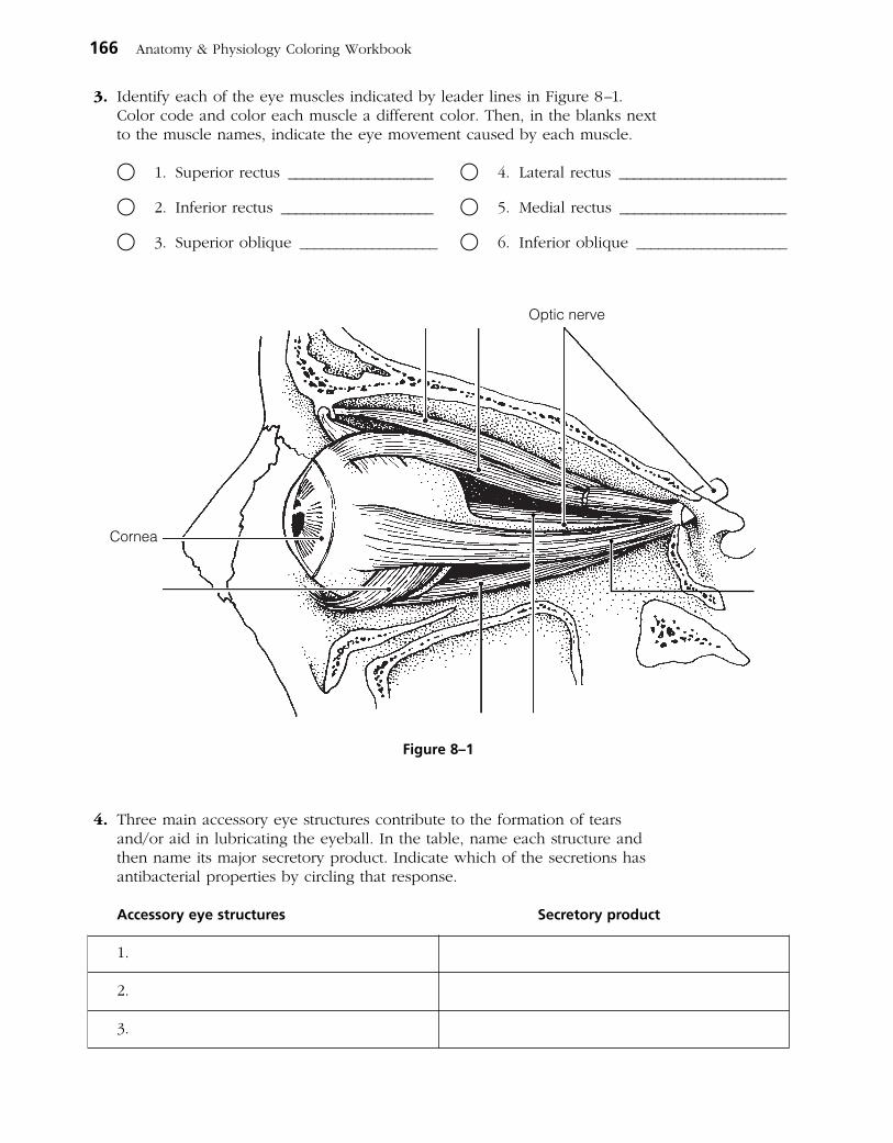
3. Identify each of the eye muscles indicated by leader lines in Figure 8–1. Color code and color each muscle a different color. Then, in the blanks next to the muscle names, indicate the eye movement caused by each muscle.
○ 1. Superior rectus ____________________ ○ 4. Lateral rectus _______________________
○ 2. Inferior rectus _____________________ ○ 5. Medial rectus _______________________
○ 3. Superior oblique ___________________ ○ 6. Inferior oblique _____________________
4. Three main accessory eye structures contribute to the formation of tears and/or aid in lubricating the eyeball. In the table, name each structure and then name its major secretory product. Indicate which of the secretions has antibacterial properties by circling that response.
Accessory eye structures Secretory product
1.
2.
3.
166 Anatomy & Physiology Coloring Workbook
Cornea
Optic nerve
Figure 8–1

5. Using the key choices, complete the crossword puzzle by answering each of the clues provided.
Key Choices
Accommodation Emmetropia Night blindnessAstigmatism Glaucoma Photopupillary (reflex)Cataract Hyperopia RefractionConvergence Myopia
Across 2. Condition of increasing pressure inside the eye, resulting from blocked
drainage of aqueous humor. 6. Light bending. 7. Normal vision. 9. Clouding of the lens, resulting in loss of sight. 10. Inability to see well in the dark; often a result of vitamin A deficiency.
Down 1. Ability to focus for close vision (less than 20 feet). 3. Inability to focus well on close objects; farsightedness. 4. Reflex constriction of pupils when they are exposed to bright light. 5. Blurred vision, resulting from unequal curvatures of the lens or cornea. 8. Nearsightedness. 9. Medial movement of the eyes during focusing on close objects.
1
2
3
4 5
6
7 8
9
10
Chapter 8 Special Senses 167

6. The intrinsic eye muscles are under the control of which division of the nervous system? Circle the correct response.
1. Autonomic nervous system 2. Somatic nervous system
7. Complete the following statements by inserting your responses in the answer blanks.
_________________________ 1.
_________________________ 2.
_________________________ 3.
_________________________ 4.
_________________________ 5. _________________________ 6.
8. Using the key choices, identify the parts of the eye described in the following statements. Insert the correct term or letter response in the answer blanks.
Key Choices
A. ○ Aqueous humor F. ○ Cornea K. ○ Retina
B. Canal of Schlemm G. ○ Fovea centralis L. ○ Sclera
C. ○ Choroid H. ○ Iris M. ○ Vitreous humor
D. ○ Ciliary body I. ○ Lens
E. ○ Ciliary zonule J. ○ Optic disk
_________________________ 1. Attaches the lens to the ciliary body
_________________________ 2. Fluid in the anterior segment that provides nutrients to the lens and cornea
_________________________ 3. The “white” of the eye
_________________________ 4. Area of retina that lacks photoreceptors
_________________________ 5. Contains muscle that controls the shape of the lens
_________________________ 6. Nutritive (vascular) layer of the eye
_________________________ 7. Drains the aqueous humor of the eye
_________________________ 8. Layer containing the rods and cones
_________________________ 9. Gel-like substance that helps to reinforce the eyeball
_________________________ 10. Heavily pigmented layer that prevents light scattering within the eye
_________________________ 11. _________________________ 12. Smooth muscle structures (intrinsic eye muscles)
_________________________ 13. Area of acute or discriminatory vision
_________________________ 14. _________________________ 15. Refractory media of the eye (#14–17)
_________________________ 16. _________________________ 17.
168 Anatomy & Physiology Coloring Workbook
A (1) lens, like that of the eye, produces an image that is upside down and reversed from left to right. Such an image is called a (2) image. In farsightedness, the light is focused
(3) the retina. The lens used to treat farsightedness is a (4) lens. In nearsightedness, the light is focused (5) the
retina; it is corrected with a (6) lens.

_________________________ 18. Most anterior part of the sclera—your “window on the world”
_________________________ 19. Pigmented “diaphragm” of the eye
9. Using the key choice terms given in Exercise 8, identify the structures indicated by leader lines on the diagram of the eye in Figure 8–2. Select different colors for all structures provided with a color-coding circle in Exercise 8, and then use them to color the coding circles and corresponding structures in the figure.
Chapter 8 Special Senses 169
Figure 8–2
10. In the following table, circle the correct word under the vertical headings that describes events occurring within the eye during close and distant vision.
Vision Ciliary muscle Lens convexity Degree of light refraction
1. Distant Relaxed Contracted Increased Decreased Increased Decreased
2. Close Relaxed Contracted Increased Decreased Increased Decreased
11. Name in sequence the neural elements of the visual pathway, beginning with the retina and ending with the optic cortex.
Retina
Synapse in thalamus
Optic cortex
12. Complete the following statements by inserting your responses in the answer blanks.
_________________________ 1.
_________________________ 2.
_________________________ 3.
_________________________ 4.
_________________________ 5.
_________________________ 6. _________________________ 7. _________________________ 8.
13. Circle the term that does not belong in each of the following groupings. Then, fill in the answer blanks with the correct group name.
1. Choroid Sclera Vitreous humor Retina Group: ____________
2. Ciliary body Iris Superior rectus Choroid Group: ____________
3. Pupil constriction Far vision Accommodation Bright light Group: ____________
4. Proprioceptors Rods Cones Respond to light Group: ____________
5. Ciliary body Iris Suspensory ligaments Ciliary zonule Group: ____________
6. Inferior oblique Iris Superior rectus Inferior rectus Group: ____________
7. Retina Pigmented layer Photoreceptors Rods and cones Group: ____________
14. Complete the statements concerning rod photopigment and physiology by writing your responses in the answer blanks.
_________________________ 1.
_________________________ 2.
_________________________ 3.
_________________________ 4.
_________________________ 5. _________________________ 6.
THE EAR: HEARING AND BALANCE
15. Using the key choices, select the terms that apply to the following descrip-tions. Place the correct letter in the answer blanks.
Key Choices
A. Anvil (incus) E. External acoustic I. Pinna M. Tympanic meatus membrane
B. Pharyngotympanic F. Hammer (malleus) J. Round window N. Vestibule tube (auditory tube)
C. Cochlea G. Oval window K. Semicircular canalsD. Endolymph H. Perilymph L. Stirrup (stapes)
There are (1) varieties of cones. One type responds most vigorously to (2) light, another to (3) light, and still another to (4) light. The ability to see intermediate colors such as purple results from the fact that more than one cone type is being stimulated (5) . Lack of all color receptors results in (6) . Because this condition is sex-linked, it occurs more commonly in (7) . Black and white, or dim light, vision is a function of the (8) .
The bent or kinked form of retinal is combined with a protein called (1) to form the visual pigment called (2) . When light strikes the visual pigment, it straightens out and breaks down into its two components. This event is called (3) because the purple color of the visual pigment changes to
(4) and finally becomes (5) as retinal is converted all the way back to vitamin (6) .
170 Anatomy & Physiology Coloring Workbook

_____ 1. _____ 2. _____ 3. Structures composing the outer ear
_____ 4. _____ 5. _____ 6. Structures composing the bony or osseous labyrinth
_____ 7. _____ 8. _____ 9. Collectively called the ossicles
_____ 10. _____ 11. Ear structures not involved with hearing
_____ 12. Allows pressure in the middle ear to be equalized with the atmospheric pressure
_____ 13. Vibrates as sound waves hit it; transmits the vibrations to the ossicles
_____ 14. Contains the organ of Corti
_____ 15. Connects the nasopharynx and the middle ear
_____ 16. _____ 17. Contain receptors for the sense of equilibrium
_____ 18. Transmits the vibrations from the stirrup to the fluid in the inner ear
_____ 19. Fluid that bathes the sensory receptors of the inner ear
_____ 20. Fluid contained within the osseous labyrinth, which bathes the membranous labyrinth
16. Figure 8–3 is a diagram of the ear. (A) Use anatomical terms (as needed) from the key choices in Exercise 15 to
correctly identify all structures in the figure provided with leader lines. (B) Color: External ear structures yellow; ossicles red; equilibrium areas of
the inner ear green; and internal ear structures involved with hearing blue.
Chapter 8 Special Senses 171
Figure 8–3

17. Sound waves hitting the eardrum set it into vibration. Trace the pathway through which vibrations and fluid currents travel to finally stimulate the hair cells in the organ of Corti. Name the appropriate ear structures in their correct sequence and insert your responses in the answer blanks.
Eardrum
Oval window
Hair cells
18. Figure 8–4 is a view of the structures of the membranous labyrinth. (A) Correctly identify the following major areas of the labyrinth indicated by
dashed leader lines: Membranous semicircular canals, saccule and utricle, and the cochlear duct.
(B) Correctly identify each of the receptor types shown in enlarged views (organ of Corti, crista ampullaris, and macula) and insert their names into the rectangles provided below.
(C) Using terms from the key choices below, identify all receptor structures provided with leader lines. (Some of these terms may need to be used more than once.)
Key Choices
A. Basilar membrane E. Hair cellsB. Cochlear nerve F. OtolithsC. Cupula G. Tectorial membraneD. Gel H. Vestibular nerve
172 Anatomy & Physiology Coloring Workbook
Oval window
Round window
Figure 8–4
19. Complete the following statements on the functioning of the static and dynamic equilibrium receptors by inserting the letter or term from the key choices in the answer blanks.
Key Choices
A. Angular/rotatory E. Gravity I. Semicircular canalsB. Cupula F. Perilymph J. StaticC. Dynamic G. Proprioception K. UtricleD. Endolymph H. Saccule L. Vision
The receptors for (1) equilibrium are found in the crista ampullaris of the (2) . These receptors respond to changes in (3) motion. When motion begins, the (4) fluid lags behind and the (5) is bent, which excites the hair cells. When the motion stops suddenly, the fluid flows in the oppo-site direction and again stimulates the hair cells. The receptors for (6) equilibrium are found in the maculae of the (7) and (8) . These receptors report the position of the head in space. Tiny stones found in a gel overlying the hair cells roll in response to the pull of (9) . As they roll, the gel moves and tugs on the hair cells, exciting them. Besides the equilib- rium receptors of the inner ear, the senses of (10) and
(11) are also important in helping to maintain equilibrium.
_________________________ 10. _________________________ 11.
20. Indicate whether the following conditions relate to conduction deafness (C) or sensorineural (central) deafness (S). Place the correct letter choice in each answer blank.
_________________________ 1. Can result from a bug wedged in the external auditory meatus
_________________________ 2. Can result from damage to the cochlear nerve
_________________________ 3. Sound is heard in one ear but not in the other during both bone and air conduction
_________________________ 4. Often improved by a hearing aid
_________________________ 5. Can result from otitis media
_________________________ 6. Can result from otosclerosis, excessive earwax, or a perforated eardrum
_________________________ 7. Can result from a blood clot in the auditory cortex of the brain
Chapter 8 Special Senses 173
_________________________ 1.
_________________________ 2.
_________________________ 3.
_________________________ 4.
_________________________ 5.
_________________________ 6.
_________________________ 7.
_________________________ 8.
_________________________ 9.

21. List three things about which a person with equilibrium problems might complain. Place your responses in the answer blanks.
_________________________ , _________________________ , and _________________________
22. Circle the term that does not belong in each of the following groupings. Then, fill in the answer blanks with the correct group name.
1. Hammer Anvil Pinna Stirrup Group: ________
2. Tectorial membrane Crista ampullaris Semicircular canals Cupula Group: ________
3. Gravity Angular motion Sound waves Rotation Group: ________
4. Utricle Saccule Auditory tube Maculae Group: ________
5. Vestibular nerve Optic nerve Cochlear nerve Vestibulocochlear nerve Group: ________
CHEMICAL SENSES: SMELL AND TASTE
23. Complete the following statements by inserting your responses in the answer blanks.
Three cranial nerves involved in transmitting impulses for the sense of taste are the (1) , (2) , and (3) . Impulses for the sense of smell are transmitted by the (4) nerve. The receptors for smell are located in the (5) of the nasal pas-sages; the act of (6) increases the sensation because it brings more air into contact with the receptors. The receptors for taste are found in cluster-like areas called (7) , most of which are located on the sides of (8) or (9) papillae. The five basic taste sensations are (10) , (11) , (12) ,
(13) , and (14) . The most protective receptors are thought to be those that respond to (15) substances. When nasal passages are congested, the sense of taste is decreased. This indicates that much of what is considered taste actually depends on the sense of (16) . It is impossible to taste sub-stances with a (17) tongue because foods must be dissolved (or in solution) to excite the taste receptors. The sense of smell is closely tied to the emotional centers of the brain (limbic region), and many odors bring back (18) .
_________________________ 15. _________________________ 17.
_________________________ 16. _________________________ 18.
174 Anatomy & Physiology Coloring Workbook
_________________________ 1.
_________________________ 2.
_________________________ 3.
_________________________ 4.
_________________________ 5.
_________________________ 6.
_________________________ 7.
_________________________ 8.
_________________________ 9.
_________________________ 10.
_________________________ 11.
_________________________ 12.
_________________________ 13.
_________________________ 14.

24. Figure 8–5 is a view of the location and structure of taste buds. (A) On Figure A, label the two types of tongue papillae containing taste buds. (B) On Figure B, color the taste buds green. (C) On Figure C, color the gustatory cells red, the basal cells blue, and the
cranial nerve fibers yellow. Add appropriate labels to the leader lines provided to identify the taste pore and microvilli of the gustatory cells.
Chapter 8 Special Senses 175
Connective tissue
Stratified squamous epithelium
A
B
C
Figure 8–5

25. Figure 8–6 illustrates the site of the olfactory epithelium in the nasal cavity (part A is an enlarged view of the olfactory receptor area).
(A) Select different colors to identify the structures listed below and use them to color the coding circles and corresponding structures in the illustration.
(B) Add a label and leader line to identify the olfactory “hairs.” (C) Add arrows to indicate the direction of impulse transmission. (D) Respond to the questions following the diagram.
○ Olfactory neurons (receptor cells) ○ Cribriform plate of the ethmoid bone
○ Olfactory bulb ○ Olfactory nerve filaments
○ Fibers of the olfactory tract
176 Anatomy & Physiology Coloring Workbook
Olfactoryepithelium
BA
Figure 8–6
1. What substance “captures” airborne odors (that is, acts as a solvent)? ____________________
2. How are olfactory neurons classified structurally? __________________________________________

26. Circle the term that does not belong in each of the following groupings. Then, fill in the answer blanks with the correct group name.
1. Sweet Musky Sour Bitter Salty Group: __________
2. Bipolar neuron Epithelial cell Olfactory filaments Ciliated Group: __________
3. Gustatory hair Taste pore Papillae Neuron Group: __________
4. Vagus nerve Facial nerve Glossopharyngeal nerve Olfactory nerve Group: __________
5. Fast adaptation High sensitivity Variety of stimuli Four receptor types Group: __________
6. Sugars Sweet Saccharine Metal ions Amino acids Group: __________
DEVELOPMENTAL ASPECTS OF THE SPECIAL SENSES
27. Complete the following statements by inserting your responses in the answer blanks.
The special sense organs are actually part of the (1) and are formed very early in the embryo. Maternal infections, par-ticularly (2) , may cause both deafness and (3) in the developing child. Of the special senses, the sense of (4) requires the most learning or takes longest to mature. All infants are (5) , but generally by school age emmetropic vision has been established. Beginning sometime after the age of 40, the eye lenses start to become less (6) and cannot bend properly to refract the light. As a result, a condition of farsightedness, called (7) , begins to occur. (8) , a condi-tion in which the lens becomes hazy or discolored, is a fre-quent cause of blindness. In old age, a gradual hearing loss, called (9) , occurs. A declining efficiency of the chemical senses is also common in the elderly.
Chapter 8 Special Senses 177
_________________________ 1.
_________________________ 2.
_________________________ 3.
_________________________ 4.
_________________________ 5.
_________________________ 6.
_________________________ 7.
_________________________ 8.
_________________________ 9.

A Visualization Exercise for the Special Senses
You . . . see a discontinuous sea of glistening, white rock slabs. . . .
28. Where necessary, complete statements by inserting the missing words in the answer blanks.
Your present journey will take you through your host’s inner ear to observe and document what you have learned about how hearing and equilibrium receptors work.
This is a very tightly planned excursion. Your host has been instructed to move his head at specific intervals and will be exposed to various sounds so that you can make certain observations. For this journey, you are miniaturized and injected into the bony cavity of the inner ear, the (1) laby-rinth, and are to make your way through its various chambers in a limited amount of time.
Your first observation is that you are in a warm sea of (2) in the vestibule. To your right are two large sacs, the (3) and (4) . You swim over to one of these membranous sacs, cut a small semicircular opening in the wall, and wiggle through. Because you are able to see very little in the dim light, you set out to explore this area more fully. As you try to move, however, you find that your feet are embedded in a thick, gluelike substance. The best you can manage is slow-motion movements through this (5) .
It is now time for your host’s first scheduled head movement. Suddenly, your world tips sharply sideways. You hear a roar (rather like an avalanche) and look up to see a discontinuous
sea of glistening, white rock slabs sliding toward you. You protect yourself from these (6) by ducking down between the hair cells that are bending vigorously with the motion of the rocks. Now that you have seen and can document the operation of a (7) , a sense organ of (8) equilibrium, you quickly back out through the hole you made.
Keeping in mind the schedule and that it is nearly time for your host to be exposed to tuning forks, you swim quickly to the right, where you see what looks like the opening of a cave with tall seaweed waving gently in the current. Abruptly, as you enter the cave, you find that you are no longer in control of your movements but instead are swept along in a smooth undulating pat-tern through the winding passageway of the cave, which you now know is the cavity of the
(9) duct. As you move up and down with the waves, you see hair cells of the (10) , the sense organ for (11) , being vigorously disturbed below you. Flattening yourself against the chamber wall to prevent being carried further by the waves, you wait for the stimulus to stop. Meanwhile, you are delighted by the electrical activity of the hair cells below you. As they depolarize to send impulses along the (12) , the landscape appears to be alive with fireflies.
178 Anatomy & Physiology Coloring Workbook
_________________________ 1.
_________________________ 2.
_________________________ 3.
_________________________ 4.
_________________________ 5.
_________________________ 6.
_________________________ 7.
_________________________ 8.
_________________________ 9.
_________________________ 10.
_________________________ 11.
_________________________ 12.
INCREDIBLE JOURNEY

Now that you have witnessed the events for this particular sense receptor, you swim back through the vestibule toward your final observation area at the other end of the bony chambers. You recognize that your host is being stimulated again because of the change in fluid currents, but because you are not close to any of the sensory receptors, you are not sure just what the stimulus is. Then, just before you, three dark openings appear, the (13) . You swim into the middle opening and see a strange structure that looks like the
brush end of an artist’s paintbrush; you swim upward and establish yourself on the soft brushy top portion. This must be the (14) of the (15) , the sensory receptor for (16) equilibrium. As you rock back and forth in the gentle currents, a sudden wave of fluid hits you. Clinging to the hairs as the fluid thunders past you, you realize that there will soon be another such wave in the opposite direction. You decide that you have seen enough of the special senses and head back for the vestibule to leave your host once again.
29. An infant girl with strabismus is brought to the clinic. Tests show that she can control both eyes independently. What therapy will be tried before surgery?
30. A man in his early 60s comes to the clinic complaining of fuzzy vision. An eye examination reveals clouding of his lenses. What is his problem and what factors might have contributed to it?
31. Albinism is a condition in which melanin pigment is not made. How do you think vision is affected by albinism?
32. A man claiming to have difficulty seeing at night seeks help at the clinic. What is the technical name for this disorder? What dietary supplement will be recommended? If the condition has progressed too far, which retinal structures will degenerate?
Chapter 8 Special Senses 179
_________________________ 13.
_________________________ 14.
_________________________ 15.
_________________________ 16.
AT THE CLINIC

33. A child is brought to the speech therapist because she does not pronounce high-pitched sounds (like “s”). If it is determined that the spiral organ of Corti is the source of the problem, which region of the organ would be defective? Is this conduction or sensorineural deafness?
34. Little Biff’s uncle tells the physician that 3-year-old Biff has frequent earaches and that a neighbor claims that Biff needs to have “ear tubes” put in. Upon questioning, the uncle reveals that Biff is taking swimming lessons and he can’t remember the last time the boy had a sore throat. Does Biff have otitis media or otitis externa? Does he need ear tubes? Explain your reasoning.
35. Harry fell off a tall ladder and fractured the anterior cranial fossa of his skull. On arrival at the hospital, a watery, blood-tinged fluid was dripping from his right nostril. Several days later, Harry complained that he could no longer smell. What nerve was damaged in his fall?
36. Brian is brought to the clinic by his parents, who noticed that his right eye does not rotate laterally very well. The doctor explains that the nerve serving the lat-eral rectus muscle is not functioning properly. To which nerve is he referring?
37. When Mrs. Martinez visits her ophthalmologist, she complains of pain in her right eye. The intraocular pressure of that eye is found to be abnormally elevated. What is the name of Mrs. Martinez’s probable condition? What causes it? What might be the outcome if the problem is not corrected?
38. Henri, a chef in a five-star French restaurant, has been diagnosed with leukemia. He is about to undergo chemotherapy, which will kill rapidly dividing cells in his body. He needs to continue working between bouts of chemotherapy. What consequences of chemotherapy would you predict that might affect his job as a chef?
180 Anatomy & Physiology Coloring Workbook

39. Select the best answer or answers from the choices given.
Chapter 8 Special Senses 181
THE FINALE: MULTIPLE CHOICE
1. Gustatory cells are:
A. bipolar neurons.
B. multipolar neurons.
C. unipolar neurons.
D. specialized epithelial cells.
2. Alkaloids excite gustatory hairs mostly at the:
A. tip of the tongue.
B. back of the tongue.
C. circumvallate papillae.
D. fungiform papillae.
3. Cranial nerves that are part of the gustatory pathway include:
A. trigeminal. C. hypoglossal.
B. facial. D. glossopharyngeal.
4. The receptors for olfaction are:
A. the ends of dendrites of bipolar neurons.
B. cilia.
C. specialized nonneural receptor cells.
D. olfactory hairs.
5. Which cranial nerve controls contraction of the circular smooth muscle of the iris?
A. Trigeminal C. Oculomotor
B. Facial D. Abducens
6. Which of the following would be found in the fovea centralis?
A. Ganglion neurons C. Cones
B. Bipolar neurons D. Rhodopsin
7. The vitreous humor:
A. helps support the lens.
B. holds the retina in place.
C. contributes to intraocular pressure.
D. is constantly replenished.
8. Blockage of which of the following is suspected in glaucoma?
A. Ciliary processes
B. Retinal blood vessels
C. Choroid vessels
D. Scleral venous sinus
9. Refraction can be altered for near or far vision by the:
A. cornea.
B. ciliary muscles.
C. vitreous humor.
D. neural layer of the retina.
10. Convergence:
A. requires contraction of the medial rectus muscles of both eyes.
B. is needed for near vision.
C. involves transmission of impulses along the abducens nerves.
D. can promote eye strain.
11. Objects in the periphery of the visual field:
A. stimulate cones.
B. cannot have their color determined.
C. can be seen in low light intensity.
D. appear fuzzy.
12. Depth perception is caused by all of the following factors except which one(s)?
A. The eyes are frontally located.
B. There is total crossover of the optic nerve fibers at the optic chiasma.
C. There is partial crossover of the optic nerve fibers at the optic chiasma.
D. Each visual cortex receives input from both eyes.

13. Which structures are contained within the petrous portion of the temporal bone?
A. Tympanic cavity
B. Mastoid air cells
C. External auditory meatus
D. Stapedius muscle
14. Movement of the _______________ membrane triggers bending of hairs of the hair cells in the spiral organ of Corti.
A. tympanic C. basilar
B. tectorial D. vestibular
15. Sounds entering the external auditory meatus are eventually converted to nerve impulses via a chain of events, including:
A. vibration of the eardrum.
B. vibratory motion of the ossicles against the round window.
C. stimulation of hair cells in the organ of Corti.
D. resonance of the basilar membrane.
16. Which of the following structures is involved in static equilibrium?
A. Maculae C. Crista ampullaris
B. Saccule D. Otoliths
17. Which of the following are paired incorrectly?
A. Cochlear duct—cupula
B. Saccule—macula
C. Ampulla—otoliths
D. Semicircular duct—ampulla
18. Taste receptor cells are stimulated by:
A. chemicals binding to the nerve fibers supplying them.
B. chemicals binding to their microvilli.
C. stretching of their microvilli.
D. impulses from the sensory nerves supplying them.
182 Anatomy & Physiology Coloring Workbook
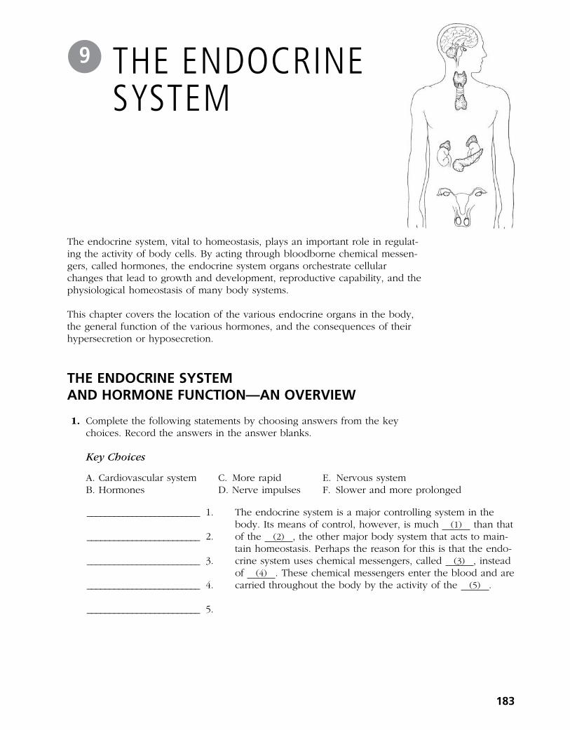
The endocrine system, vital to homeostasis, plays an important role in regulat-ing the activity of body cells. By acting through bloodborne chemical messen- gers, called hormones, the endocrine system organs orchestrate cellular changes that lead to growth and development, reproductive capability, and the physiological homeostasis of many body systems.
This chapter covers the location of the various endocrine organs in the body, the general function of the various hormones, and the consequences of their hypersecretion or hyposecretion.
THE ENDOCRINE SYSTEM AND HORMONE FUNCTION—AN OVERVIEW
1. Complete the following statements by choosing answers from the key choices. Record the answers in the answer blanks.
Key Choices
A. Cardiovascular system C. More rapid E. Nervous systemB. Hormones D. Nerve impulses F. Slower and more prolonged
The endocrine system is a major controlling system in the body. Its means of control, however, is much (1) than that of the (2) , the other major body system that acts to main-tain homeostasis. Perhaps the reason for this is that the endo-crine system uses chemical messengers, called (3) , instead of (4) . These chemical messengers enter the blood and are carried throughout the body by the activity of the (5) .
183
THE ENDOCRINE SYSTEM
9
_________________________ 1.
_________________________ 2.
_________________________ 3.
_________________________ 4.
_________________________ 5.

2. Complete the following statements by choosing answers from the key choices. Record the answers in the answer blanks.
Key Choices
A. Altering activity F. Negative feedback K. Steroid or amino acid–basedB. Anterior pituitary G. Neural L. Stimulating new or unusual activitiesC. Hormonal H. Neuroendocrine M. Sugar or proteinD. Humoral I. Receptors N. Target cell(s)E. Hypothalamus J. Releasing/inhibiting hormones
_________________________ 1.
_________________________ 2.
_________________________ 3.
_________________________ 4.
_________________________ 5.
_________________________ 6.
_________________________ 7.
_________________________ 8.
_________________________ 9.
_________________________ 10.
_________________________ 11. _________________________ 12. _________________________ 13.
3. For each key phrase, decide whether it better describes the mode of action of a steroid or amino acid–based hormone, and insert its key letter in the appropriate answer blank.
Key Choices
A. Binds to a plasma membrane receptor D. Activates a gene to transcribe messenger RNAB. Binds to a receptor in the cell’s E. Acts through a second messenger such as
nucleus cyclic AMP (cyclic adenine monophosphate)C. Is lipid soluble
Steroid hormones: ___________________ Amino acid–based hormones: ___________________
All cells do not respond to endocrine system stimulation. Only those that have the proper (1) on their cell mem branes are activated by the chemical messengers. These responsive cells are called the (2) of the various endocrine glands. Hor mones promote homeostasis by (3) of body cells rather than by
(4) . Most hormones are (5) mole cules.
The various endocrine glands are prodded to release their hormones by nerve fibers (a (6) stimulus), by other hor-mones (a (7) stimulus), or by the presence of increased or decreased levels of various other substances in the blood (a
(8) stimulus). The secretion of most hormones is regulated by a (9) system, in which increasing levels of that particu-lar hormone “turn off” its stimulus. The (10) is called the master endocrine gland because it regulates so many other endocrine organs. However, it is in turn controlled by (11) secreted by the (12) . The structure identified as #12 is also part of the brain, so it is appropriately called a (13) organ.
184 Anatomy & Physiology Coloring Workbook

THE MAJOR ENDOCRINE ORGANS
4. Figure 9–1 depicts the anatomical relationships between the hypothalamus and the anterior and posterior lobes of the pituitary in a highly simplified way.
(A) Identify each of the structures listed below by color coding and coloring them on the diagram.
(B) On the appropriate lines, write in the names of the hormones that influ-ence each of the target organs shown at the bottom of the diagram.
(C) Color the target organs that are affected by tropic hormones.
○ Hypothalamus ○ Anterior pituitary
○ Turk’s saddle of the sphenoid bone ○ Posterior pituitary
Chapter 9 The Endocrine System 185
Bones and muscles
Mammary glands
Testes or ovaries
andThyroid
Adrenal cortex
Releasing hormonesin portal circulation
Figure 9–1

5. Figure 9–2 is a diagram of the various endocrine organs of the body. (A) Next to each letter on the diagram, write the name of the endocrine-
producing organ (or area). (B) Select different colors for each and color the corresponding organs in the
illustration. (C) To complete your identification of the hormone-producing organs, name the
organs (not illustrated) described in items K and L.
K. Small glands that ride “horseback” on the thyroid
____________________________
L. Endocrine-producing organ present only in pregnant women
____________________________
186 Anatomy & Physiology Coloring Workbook
B
A
C
D
E
H
I
J
F
G
Figure 9–2

6. For each of the following hormones, indicate the organ (or organ part) producing or releasing the hormone by inserting the appropriate letters from Figure 9–2 in the answer blanks. Items may have more than one answer.
7. Name the hormone that would be produced in inadequate amounts in the following conditions. Place your responses in the answer blanks.
_________________________ 1. Sexual immaturity
_________________________ 2. Tetany
_________________________ 3. Excessive urination without high blood glucose levels; causes dehydration and tremendous thirst
_________________________ 4. Goiter
_________________________ 5. Cretinism; a type of dwarfism in which the individual retains childlike proportions and is mentally retarded
_________________________ 6. Excessive thirst, high blood glucose levels, acidosis
_________________________ 7. Abnormally small stature, normal proportions
_________________________ 8. Miscarriage
_________________________ 9. Lethargy, hair loss, low basal metabolic rate, obesity (myxedema in the adult)
8. Name the hormone that would be produced in excessive amounts in the following conditions. Place your responses in the answer blanks.
_________________________ 1. Lantern jaw; large hands and feet (acromegaly in the adult)
_________________________ 2. Bulging eyeballs, nervousness, increased pulse rate, weight loss (Graves’ disease)
_________________________ 3. Demineralization of bones; spontaneous fractures
_________________________ 4. Cushing’s syndrome—moon face, depression of the immune system
_________________________ 5. Abnormally large stature, relatively normal body proportions
_________________________ 6. Abnormal hairiness; masculinization
Chapter 9 The Endocrine System 187
______ 1. ACTH
______ 2. ADH
______ 3. Aldosterone
______ 4. Cortisol
______ 5. Epinephrine
______ 6. Estrogen
______ 7. FSH
______ 8. Glucagon
______ 9. Insulin
______ 10. LH
______ 11. Melatonin
______ 12. Oxytocin
______ 13. Progesterone
______ 14. Prolactin
______ 15. PTH
______ 16. Growth hormone
______ 17. Testosterone
______ 18. Thymosins
______ 19. Thyroxine
______ 20. TSH
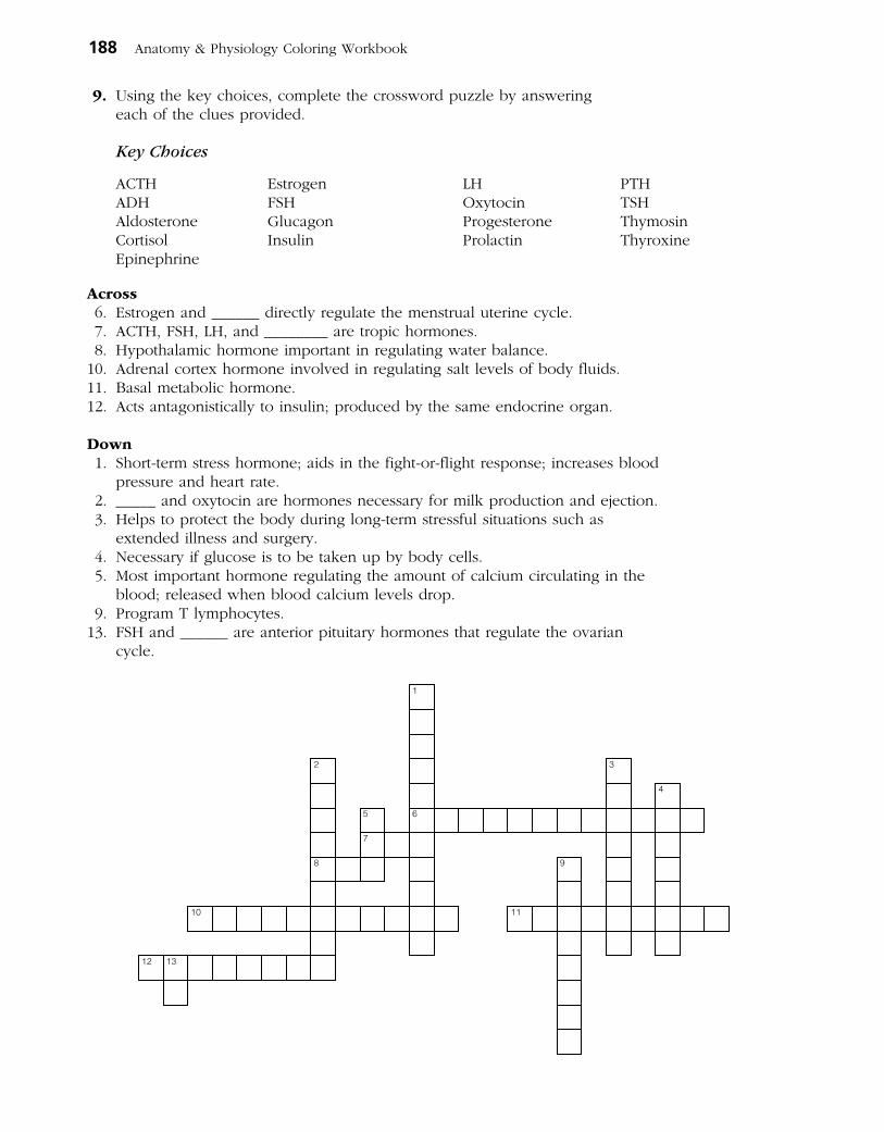
9. Using the key choices, complete the crossword puzzle by answering each of the clues provided.
Key Choices
ACTH Estrogen LH PTHADH FSH Oxytocin TSHAldosterone Glucagon Progesterone ThymosinCortisol Insulin Prolactin ThyroxineEpinephrine
Across 6. Estrogen and ______ directly regulate the menstrual uterine cycle. 7. ACTH, FSH, LH, and ________ are tropic hormones. 8. Hypothalamic hormone important in regulating water balance. 10. Adrenal cortex hormone involved in regulating salt levels of body fluids. 11. Basal metabolic hormone. 12. Acts antagonistically to insulin; produced by the same endocrine organ.
Down 1. Short-term stress hormone; aids in the fight-or-flight response; increases blood
pressure and heart rate. 2. _____ and oxytocin are hormones necessary for milk production and ejection. 3. Helps to protect the body during long-term stressful situations such as
extended illness and surgery. 4. Necessary if glucose is to be taken up by body cells. 5. Most important hormone regulating the amount of calcium circulating in the
blood; released when blood calcium levels drop. 9. Program T lymphocytes. 13. FSH and ______ are anterior pituitary hormones that regulate the ovarian
cycle.
1
2 3
4
5 6
7
8 9
10 11
12 13
188 Anatomy & Physiology Coloring Workbook
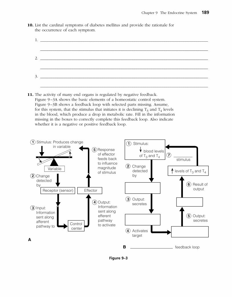
10. List the cardinal symptoms of diabetes mellitus and provide the rationale for the occurrence of each symptom.
1. _________________________________________________________________________________________
_________________________________________________________________________________________
2. _________________________________________________________________________________________
_________________________________________________________________________________________
3. _________________________________________________________________________________________
_________________________________________________________________________________________
11. The activity of many end organs is regulated by negative feedback. Figure 9 –3A shows the basic elements of a homeostatic control system. Figure 9–3B shows a feedback loop with selected parts missing. Assume, for this system, that the stimulus that initiates it is declining T3 and T4 levels in the blood, which produce a drop in metabolic rate. Fill in the information missing in the boxes to correctly complete this feedback loop. Also indicate whether it is a negative or positive feedback loop.
Chapter 9 The Endocrine System 189
Figure 9–3

12. Circle the term that does not belong in each of the following groupings. Then, fill in the answer blanks with the correct group name.
1. ADH Hormone storage Nervous tissue Anterior lobe Group: ____________
2. Steroid hormone Protein hormone Second messenger Membrane receptors Group: ____________
3. Catecholamines Norepinephrine Epinephrine Cortisol Group: ____________
4. Decreases blood Ca2+ Increases blood Ca2+ Thyroid gland Enhances Ca2+ deposit Group: ____________
5. Glucocorticoids Steroids Aldosterone Growth hormone Group: ____________
6. Thyroid follicles T3 and T4 Glucose metabolism Parafollicular cells Group: ___________
OTHER HORMONE-PRODUCING TISSUES AND ORGANS
13. Besides the major endocrine organs, isolated clusters of cells produce hor-mones within body organs that are usually not associated with the endocrine system. A number of these hormones are listed in the table below. Fill in the missing information (blank spaces) on these hormones in the table.
Chemical Hormone makeup Source Effects
Gastrin Peptide
Secretin Duodenum
Cholecystokinin Peptide
Erythropoietin Kidney in response to hypoxia
Active vitamin D3 Skin; activated by kidneys
Atrial natriuretic Peptide peptide (ANP)
Human chorionic Protein gonadotropin (hCG)
Leptin Adipose tissue
190 Anatomy & Physiology Coloring Workbook

A Visualization Exercise for the Endocrine System
. . . you notice charged particles shooting pell-mell out of the bone matrix. . . .
15. Where necessary, complete statements by inserting the missing words in the answer blanks.
For this journey, you will be miniaturized and injected into a vein of your host. Throughout the journey, you will be travel-ing in the bloodstream. Your instructions are to record changes in blood composition as you float along and to form some conclusions as to why they are occurring (that is, which hor-mone is being released).
Bobbing gently along in the slowly moving blood, you realize that there is a sugary taste to your environment; however, the sweetness begins to decrease quite rapidly. Because the glucose levels of the blood have just decreased, obviously (1) has been released by the (2) so that the cells can take up glucose.
A short while later you notice that the depth of the blood in the vein in which you are traveling has dropped sub-stantially. To remedy this potentially serious situation, the
(3) will have to release more (4) so the kidney tubules will reabsorb more water. Within a few minutes, the blood becomes much deeper; you wonder if the body is psychic as well as wise.
Chapter 9 The Endocrine System 191
_________________________ 1.
_________________________ 2.
_________________________ 3.
_________________________ 4.
_________________________ 5.
_________________________ 6.
_________________________ 7.
_________________________ 8.
_________________________ 9.
INCREDIBLE JOURNEY
DEVELOPMENTAL ASPECTS OF THE ENDOCRINE SYSTEM
14. Complete the following statements by inserting your responses in the answer blanks.
Under ordinary conditions, the endocrine organs operate smoothly until old age. However, a (1) in an endocrine organ may lead to (2) of its hormones. A lack of (3) in the diet may result in undersecretion of thyroxine. Later in life, a woman experiences a number of symptoms such as hot flashes and mood changes, which result from decreasing lev-els of (4) in her system. This period of a woman’s life is referred to as (5) , and it results in a loss of her ability to bear (6) . Because (7) tolerance tends to decrease in an aging person (due to declining sensitivity to insulin), adult-onset diabetes is common.
_________________________ 1.
_________________________ 2.
_________________________ 3.
_________________________ 4.
_________________________ 5.
_________________________ 6.
_________________________ 7.

16. Pete is very short for his chronological age of 8 years. Which physical features will allow you to determine quickly whether to check GH or thyroxine levels?
17. A young girl is brought to the clinic by her father. The girl fatigues easily and seems mentally sluggish. You notice a slight swelling in the anterior neck. What condition do you suspect? What are some possible causes and their treatments?
18. A 2-year-old boy is brought to the clinic by his anguished parents. He is develop-ing sexually and shows an obsessive craving for salt. Blood tests reveal hyperglycemia. What endocrine gland is hypersecreting?
19. When the carnival came to a small town, the local health professionals and con s umer groups joined forces to enforce truth-in-advertising laws to protect selected employees of the carnival. They demanded that the fat man, the dwarf, the giant, and the bearded lady be billed as “people with endocrine system problems” (which of course removed all the sensationalism usually associated with these attractions). Identify the endocrine disorder in each case and explain how (or why) the disorder produced the characteristic features of these four show people.
192 Anatomy & Physiology Coloring Workbook
AT THE CLINIC
As you circulate past the bones, you notice charged particles shooting everywhere out of the bone matrix and jumping into the blood. You conclude that the (5) glands have just released PTH because the (6) levels have increased in the blood. As you continue to move in the blood-stream, the blood suddenly becomes sticky sweet, indicating that your host must be nervous about something. Obviously, his (7) has released (8) to cause this sudden increase in blood glucose.
Sometime later, you become conscious of a humming activity around you, and you sense that the cells are very busy. Your host’s (9) levels appear to be sufficient because his cells are certainly not sluggish in their metabolic activities. You record this observation and prepare to end this journey.

22. Select the best answer or answers from the choices given.
Chapter 9 The Endocrine System 193
1. The major endocrine organs of the body:
A. tend to be very large organs.
B. are closely connected with each other.
C. all contribute to the same function (digestion).
D. tend to lie near the midline of the body.
2. Of the following endocrine structures, which develops from the brain?
A. Posterior pituitary C. Thyroid gland
B. Anterior pituitary D. Thymus gland
3. Which is generally true of hormones?
A. Exocrine glands produce them.
B. They travel throughout the body in the blood.
C. They affect only non-hormone-producing organs.
D. All steroid hormones produce very simi-lar physiological effects in the body.
4. Which of the following are tropic hormones secreted by the anterior pituitary gland?
A. LH C. TSH
B. ACTH D. FSH
5. Smooth muscle contractions are stimulated by:
A. testosterone. C. prolactin.
B. FSH. D. oxytocin.
6. Relative to the cyclic AMP second-messen ger system, which of the following is not accurate?
A. The activating hormone interacts with a receptor site on the plasma membrane.
B. Binding of the hormone directly produces the second messenger.
C. Activated enzymes catalyze the transfor-mation of AMP to cyclic AMP.
D. Cyclic AMP acts within the cell to alter cell function as is characteristic for that specific hormone.
7. Nerve input regulates the release of:
A. oxytocin. C. melatonin.
B. epinephrine. D. cortisol.
8. ANP, the hormone secreted by the heart, has exactly the opposite function of which hormone secreted by the adrenal cortex?
A. Epinephrine C. Aldosterone
B. Cortisol D. Testosterone
THE FINALE: MULTIPLE CHOICE
20. The brain is “informed” when we are stressed, and the hypothalamus responds by secreting a releasing hormone called corticotropin-releasing hormone (CRH) that helps the body deal with the stressors. Outline this entire sequence, starting with CRH and ending with the release of cortisol. (Be sure to trace the hormone through the hypophyseal portal system and out of the pituitary gland.)
21. Mrs. Jackson claims she is not menstruating and reports that her breasts are producing milk, although she has never been pregnant. What hormone is being hypersecreted?

9. Hormones that act directly or indirectly to elevate blood glucose include:
A. GH. C. insulin.
B. cortisol. D. ACTH.
10. Hormones secreted by females include:
A. estrogens. C. prolactin.
B. progesterone. D. testosterone.
11. Which of the following are direct or indirect effects of growth hormone?
A. Stimulates cells to take in amino acids and form proteins
B. Important in determining final body size
C. Increases blood levels of fatty acids
D. Decreases utilization of glucose by most body cells
12. Hypothyroidism can cause:
A. myxedema. C. cretinism.
B. Cushing’s syndrome. D. exophthalmos.
13. Which of the following is given as a drug to reduce inflammation?
A. Epinephrine C. Aldosterone
B. Cortisol D. ADH
14. Which of the following hormones is (are) released by neurons?
A. Oxytocin C. ADH
B. Insulin D. Cortisol
15. The major stimulus for release of thyroid hormone is:
A. hormonal.
B. humoral.
C. neural.
16. A hormone not involved in glucose metabolism is:
A. glucagon.
B. cortisone.
C. aldosterone.
D. insulin.
17. Parathyroid hormone:
A. increases bone formation and lowers blood calcium levels.
B. increases calcium excretion from the body.
C. decreases calcium absorption from the gut.
D. demineralizes bone and raises blood calcium levels.
18. The word root referring to body fluids is:
A. mell. C. humor.
B. hormon. D. gen.
19. Most hormones are made and released as needed. The exception to this generalization is:
A. catecholamines. C. insulin.
B. thyroxine. D. aldosterone.
194 Anatomy & Physiology Coloring Workbook

195
BLOOD10
Blood, the “life fluid” that courses through the body’s blood vessels, provides the means for the body’s cells to receive vital nutrients and oxygen and dispose of their metabolic wastes. As blood flows past the tissue cells, exchanges contin- ually occur between the blood and the tissue cells so that vital activities can go on continuously.
This chapter provides an opportunity to review the general characteristics of whole blood and plasma, to identify the various formed elements (blood cells), and to recall their functions. Blood groups, transfusion reactions, clotting, and various types of blood abnormalities are also considered.
COMPOSITION AND FUNCTIONS OF BLOOD
1. Complete the following description of the components of blood by writing the missing words in the answer blanks.
In terms of its tissue classification, blood is classified as a (1) tissue because it has living blood cells, called (2) ,
sus pended in a nonliving fluid matrix called (3) . The “fibers” of blood only become visible during (4) .
If a blood sample is centrifuged, the heavier blood cells become packed at the bottom of the tube. Most of this com-pacted cell mass is composed of (5) , and the volume of blood accounted for by these cells is referred to as the (6) . The less dense (7) rises to the top and constitutes about 45% of the blood volume. The so-called “buffy coat,” com-posed of (8) and (9) , is found at the junction between the other two blood elements. The buffy coat accounts for less than (10) % of blood volume.
Blood is scarlet red in color when it is loaded with (11) ; otherwise, it tends to be dark red.
_________________________ 1.
_________________________ 2.
_________________________ 3.
_________________________ 4.
_________________________ 5.
_________________________ 6.
_________________________ 7.
_________________________ 8.
_________________________ 9.
_________________________ 10. _________________________ 11.

2. Using the key choices, complete the crossword puzzle by answering each of the clues provided.
Key Choices
Basophil Formed Megakaryocyte PlasmaEosinophil Leukocyte Monocyte PlateletErythrocyte Lymphocyte Neutrophil
Across 6. Primarily water, noncellular; the fluid matrix of blood. 7. Lymphocyte and _________ are agranular leukocytes. 8. Increases during parasite attacks. 9. Blood is composed by plasma and cells collectively known as ________
elements.
Down 1. Most numerous leukocyte. 2. Fragments to form platelets. 3. Also called red blood cell, anucleate. 4. Releases histamine during inflammatory reactions. 5. After originating in bone marrow, may be formed in lymphoid tissue.
1 2
3 4
5 6
7
8
9
3. Figure 10–1 depicts (in incomplete form) the erythropoietin mechanism for regulating the rate of erythropoiesis.
(A) Complete the statements that have answer blanks, and then choose col-ors (other than yellow) for the color-coding circles and corresponding struc-tures on the diagram.
(B) Color all arrows on the diagram yellow. (C) Indicate the normal life span of erythrocytes.
○ Kidney ○ Red bone marrow ○ Red blood cells (RBCs)
196 Anatomy & Physiology Coloring Workbook

Chapter 10 Blood 197
Figure 10–1

4. Four leukocytes are diagrammed in Figure 10–2. (A) Identify each leukocyte type by writing in the correct name in the blank
below the illustration. (B) Follow directions (given under each figure) for coloring each leukocyte as
it appears when stained with Wright’s stain.
198 Anatomy & Physiology Coloring Workbook
A
C D
BColor the granules pale violet, the cytoplasm pink, and the nucleusdark purple.
Color the nucleus deep blue and the cytoplasm pale blue.
Color the granules bright red, the cytoplasm pale pink, and the nucleus red/purple.
For this smallest white blood cell,color the nucleus deep purple/blueand the sparse cytoplasm pale blue.
Figure 10–2
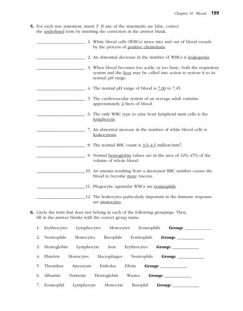
5. For each true statement, insert T. If any of the statements are false, correct the underlined term by inserting the correction in the answer blank.
_________________________ 1. White blood cells (WBCs) move into and out of blood vessels by the process of positive chemotaxis.
_________________________ 2. An abnormal decrease in the number of WBCs is leukopenia.
_________________________ 3. When blood becomes too acidic or too basic, both the respiratory system and the liver may be called into action to restore it to its normal pH range.
_________________________ 4. The normal pH range of blood is 7.00 to 7.45.
_________________________ 5. The cardiovascular system of an average adult contains approximately 4 liters of blood.
_________________________ 6. The only WBC type to arise from lymphoid stem cells is the lymphocyte.
_________________________ 7. An abnormal increase in the number of white blood cells is leukocytosis.
_________________________ 8. The normal RBC count is 3.5–4.5 million/mm3.
_________________________ 9. Normal hemoglobin values are in the area of 42%–47% of the volume of whole blood.
_________________________ 10. An anemia resulting from a decreased RBC number causes the blood to become more viscous.
_________________________ 11. Phagocytic agranular WBCs are eosinophils.
_________________________ 12. The leukocytes particularly important in the immune response are monocytes.
6. Circle the term that does not belong in each of the following groupings. Then, fill in the answer blanks with the correct group name.
1. Erythrocytes Lymphocytes Monocytes Eosinophils Group: ____________
2. Neutrophils Monocytes Basophils Eosinophils Group: ____________
3. Hemoglobin Lymphocyte Iron Erythrocytes Group: ____________
4. Platelets Monocytes Macrophages Neutrophils Group: ____________
5. Thrombus Aneurysm Embolus Fibrin Group: ____________
6. Albumin Nutrients Hemoglobin Wastes Group: ____________
7. Eosinophil Lymphocyte Monocyte Basophil Group: ____________
Chapter 10 Blood 199

7. Rank the following lymphocytes from 1 (most abundant) to 5 (least abun-dant) relative to their abundance in the blood of a healthy person.
______ 1. Lymphocyte ______ 3. Neutrophil ______ 5. Monocyte
______ 2. Basophil ______ 4. Eosinophil
8. Check (✓) all the factors that would serve as stimuli for erythropoiesis.
______ 1. Hemorrhage ______ 3. Living at a high altitude
______ 2. Aerobic exercise ______ 4. Breathing pure oxygen
HEMOSTASIS
9. Using the key choices, correctly complete the following description of the blood-clotting process. Insert the key term or letter in the answer blanks.
Key Choices
A. Break D. Fibrinogen G. Prothrombin activator J. ThrombinB. Erythrocytes E. Platelets H. PF3 K. Tissue factorC. Fibrin F. Prothrombin I. Serotonin
_________________________ 1.
_________________________ 2.
_________________________ 3.
_________________________ 4.
_________________________ 5.
_________________________ 6.
_________________________ 7.
_________________________ 8.
_________________________ 9. _________________________ 10. _________________________ 11.
10. For each true statement, write T. If any statements are false, correct the underlined term by inserting the correction in the answer blank.
_________________________ 1. Normally, blood clots within 5–10 minutes.
_________________________ 2. The most important natural body anticoagulant is histamine.
_________________________ 3. Hemostasis means stoppage of blood flow.
Clotting begins when a (1) occurs in a blood vessel wall. Almost immediately, (2) cling to the blood vessel wall and release (3) , which helps to decrease blood loss by helping to con strict the vessel. (4) , released by damaged cells in the area, interacts with (5) on the platelet surfaces and other clotting factors to form (6) . This chemical sub-stance causes (7) to be con verted to (8) . Once present, molecule #8 acts as an enzyme to attach (9) molecules together to form long, threadlike strands of (10) , which then traps (11) flowing by in the blood.
200 Anatomy & Physiology Coloring Workbook

BLOOD GROUPS AND TRANSFUSIONS
11. Correctly complete the following table concerning ABO blood groups.
Agglutinogens Agglutinins Can receive or antigens or antibodies Can donate blood Blood type on RBC surface in plasma blood to type from type
1. Type A A
2. Type B Anti-A
3. Type AB AB
4. Type O None
12. What blood type is the universal donor ? _________________________
The universal recipient ? _________________________
13. When a person is given a transfusion of mismatched blood, a transfusion reaction occurs. Define the term “transfusion reaction” in the blanks provided here.
___________________________________________________________________________________________
___________________________________________________________________________________________
DEVELOPMENTAL ASPECTS OF BLOOD
14. Complete the following statements by inserting your responses in the answer blanks.
A fetus has a special type of hemoglobin, hemoglobin (1) , that has a particularly high affinity for oxygen. After birth, the infant’s fetal RBCs are rapidly destroyed and replaced by hemoglobin A–containing RBCs. When the immature infant liver cannot keep pace with the demands to rid the body of hemoglobin breakdown products, the infant’s tissues become yellowed, or (2) .
Genetic factors lead to several congenital diseases concerning the blood. An anemia in which RBCs become sharp and “logjam” in the blood vessels under conditions of low-oxygen tension in the blood is (3) anemia. Bleeder’s disease, or
(4) , is a result of a deficiency of certain clotting factors.
Chapter 10 Blood 201
_________________________ 1.
_________________________ 2.
_________________________ 3.
_________________________ 4.

Diet is important to normal blood formation. Women are par-ticularly prone to (5) -deficiency anemia because of their monthly menses. A decreased efficiency of the gastric mucosa makes elderly individuals particularly susceptible to (6) anemia as a result of a lack of intrinsic factor, which is neces-sary for vitamin (7) absorption. An important problem in aged individuals is their tendency to form undesirable clots, or (8) . Both the young and the elderly are at risk for can-cer of the blood, or (9) .
A Visualization Exercise for the Blood
Once inside, you quickly make a slash in the vessel lining. . . .
15. Where necessary, complete statements by inserting the missing words in the answer blanks.
For this journey, you will be miniaturized and injected into the external iliac artery and will be guided by a fluorescent monitor into the bone marrow of the iliac bone. You will observe and report events of blood cell formation, also called
(1) , seen there; then you will move out of the bone into the circulation to initiate and observe the process of blood clotting, also called (2) . Once in the bone marrow, you watch as several large dark-nucleated stem cells, or (3) , begin to divide and produce daughter cells. To your right, the daughter cells eventually formed have tiny cytoplasmic gran-ules and very peculiarly shaped nuclei that look like small masses of nuclear material connected by thin strands of nucleoplasm. You have just witnessed the formation of a type of white blood cell called the (4) . You describe its appear-ance and make a mental note to try to observe its activity later. Meanwhile, you can tentatively report that this cell type functions as a (5) to protect the body.
At another site, daughter cells arising from the division of a stem cell are initially difficult to identify. As you continue to observe the cells, you see that they, in turn, divide. Eventually some of their daughter cells eject their nuclei and flatten out to assume a disc shape. You assume that the kidneys must have released (6) because those cells are (7) . That dark material filling their interior must be (8) because those cells function to transport
(9) in the blood.
202 Anatomy & Physiology Coloring Workbook
_________________________ 5.
_________________________ 6.
_________________________ 7.
_________________________ 8.
_________________________ 9.
_________________________ 1.
_________________________ 2.
_________________________ 3.
_________________________ 4.
_________________________ 5.
_________________________ 6.
_________________________ 7.
_________________________ 8.
_________________________ 9.
INCREDIBLE JOURNEY

Now you turn your attention to the daughter cells being formed by the division of another stem cell. They are small, round cells with relatively large round nuclei. In fact, their cytoplasm is very sparse. You record your observation of the formation of (10) . They do not remain in the marrow very long after formation but seem to enter the circulation almost as soon as they are produced. Some of those cells produce
(11) or act in other ways in the immune response. At this point, although you have yet to see the formation of (12) ,
(13) , (14) , or (15) , you decide to proceed into the cir-culation to make the blood-clotting observations.
You maneuver yourself into a small venule to enter the gen-eral circulation. Once inside, you quickly make a slash in the vessel lining, or (16) . Almost immediately, what appear to be hundreds of jagged cell fragments swoop into the area and plaster themselves over the freshly made incision. You record that (17) have just adhered to the damaged site. As you are writing, your chemical monitor flashes the message, “vasocon-strictor substance released.” You record that (18) has been released based on your observation that the vessel wall seems to be closing in. Peering out at the damaged site, you see that long ropelike strands are being formed at a rapid rate and are clinging to the site. You report that the (19) mesh is forming and is beginning to trap RBCs to form the basis of the (20) . Even though you do not have the equipment to monitor the intermediate steps of this process, you know that the interaction of platelet PF3 and other clotting factors must have generated (21) , which then converted (22) to (23) . This second enzyme then joined the soluble (24) molecules together to form the network of strands you can see.
You carefully back away from the newly formed clot. You do not want to disturb the area because you realize that if the clot detaches, it might become a life-threatening (25) . Your mission here is completed, and you return to the entrance site.
16. Correctly respond to five questions (#1–5) referring to the following situation. Mrs. Carlyle is pregnant for the first time. Her blood type is Rh negative, her husband is Rh positive, and their first child has been determined to be Rh positive. Ordinarily, the first such pregnancy causes no major problems, but baby Carlyle is born blue and cyanotic.
1. What is this condition, a result of Rh incompatibility, called?
Chapter 10 Blood 203
_________________________ 10.
_________________________ 11.
_________________________ 12.
_________________________ 13.
_________________________ 14.
_________________________ 15.
_________________________ 16.
_________________________ 17.
_________________________ 18.
_________________________ 19.
_________________________ 20.
_________________________ 21.
_________________________ 22.
_________________________ 23.
_________________________ 24.
_________________________ 25.
AT THE CLINIC

2. Why is the baby cyanotic?
3. Because this is Mrs. Carlyle’s first pregnancy, how can you account for the baby’s problem?
4. Assume that baby Carlyle was born pink and healthy. What measures should be taken to prevent the previously described situation from happening in a second pregnancy with an Rh-positive baby?
5. Mrs. Carlyle’s sister has had two miscarriages before seeking medical help with her third pregnancy. Blood typing shows that she, like her sister, is Rh negative; her husband is Rh positive. What course of treatment will be followed?
17. Ms. Pratt is claiming that Mr. X is the father of her child. Ms. Pratt’s blood type is O negative. Her baby boy has type A positive blood. Mr. X’s blood is typed and found to be B positive. Could he be the father of her child? ____________ If not, what blood type would the father be expected to have? ______________
18. Cancer patients being treated with chemotherapy drugs designed to destroy rapidly dividing cells are monitored closely for changes in their RBC and WBC counts. Why?
19. A red marrow biopsy is ordered for two patients—a young child and an adult. The specimen is taken from the tibia of the child but from the iliac crest of the adult. Explain why different sites are used to obtain marrow samples in adults and children. (You might want to check Chapter 5 for this one.)
20. Mrs. Graves has just donated a pint of blood. Shortly thereafter, her bone mar-row has started gearing up to replace the loss. Which of the formed elements will be produced in the greatest quantities?
21. Mr. Rudd, who has just had surgery for stomach cancer, has been receiving weekly injections of vitamin B12. Why is he receiving the vitamin injections? Why can’t the vitamin be delivered in tablet form? What will be the result if he refuses to continue the injections?
204 Anatomy & Physiology Coloring Workbook
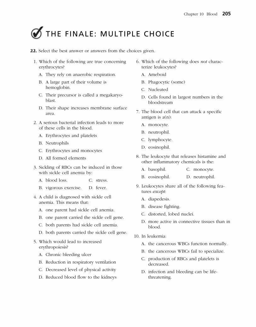
22. Select the best answer or answers from the choices given.
Chapter 10 Blood 205
THE FINALE: MULTIPLE CHOICE
1. Which of the following are true concerning erythrocytes?
A. They rely on anaerobic respiration.
B. A large part of their volume is hemoglobin.
C. Their precursor is called a mega karyo-blast.
D. Their shape increases membrane surface area.
2. A serious bacterial infection leads to more of these cells in the blood.
A. Erythrocytes and platelets
B. Neutrophils
C. Erythrocytes and monocytes
D. All formed elements
3. Sickling of RBCs can be induced in those with sickle cell anemia by:
A. blood loss. C. stress.
B. vigorous exercise. D. fever.
4. A child is diagnosed with sickle cell anemia. This means that:
A. one parent had sickle cell anemia.
B. one parent carried the sickle cell gene.
C. both parents had sickle cell anemia.
D. both parents carried the sickle cell gene.
5. Which would lead to increased erythropoiesis?
A. Chronic bleeding ulcer
B. Reduction in respiratory ventilation
C. Decreased level of physical activity
D. Reduced blood flow to the kidneys
6. Which of the following does not charac-terize leukocytes?
A. Ameboid
B. Phagocytic (some)
C. Nucleated
D. Cells found in largest numbers in the bloodstream
7. The blood cell that can attack a specific antigen is a(n):
A. monocyte.
B. neutrophil.
C. lymphocyte.
D. eosinophil.
8. The leukocyte that releases histamine and other inflammatory chemicals is the:
A. basophil. C. monocyte.
B. eosinophil. D. neutrophil.
9. Leukocytes share all of the following fea-tures except:
A. diapedesis.
B. disease fighting.
C. distorted, lobed nuclei.
D. more active in connective tissues than in blood.
10. In leukemia:
A. the cancerous WBCs function normally.
B. the cancerous WBCs fail to specialize.
C. production of RBCs and platelets is decreased.
D. infection and bleeding can be life- threatening.

11. A condition resulting from thrombocytopenia is:
A. thrombus formation.
B. embolus formation.
C. petechiae.
D. hemophilia.
12. Which of the following can cause problems in a transfusion reaction?
A. Donor antibodies attacking recipient RBCs
B. Clogging of small vessels by agglutinated clumps of RBCs
C. Lysis of donated RBCs
D. Blockage of kidney tubules
13. If an Rh– mother becomes pregnant, when can hemolytic disease of the newborn not possibly occur in the child?
A. If the child is Rh–.
B. If the child is Rh+.
C. If the father is Rh+.
D. If the father is Rh–.
14. What is the difference between a thrombus and an embolus?
A. One occurs in the bloodstream, whereas the other occurs outside the bloodstream.
B. One occurs in arteries, the other in veins.
C. One is a blood clot, whereas the other is a parasitic worm.
D. A thrombus must travel to become an embolus.
15. The plasma component that forms the fibrous skeleton of a clot consists of:
A. platelets.
B. fibrinogen.
C. thromboplastin.
D. thrombin.
16. The normal pH of blood is:
A. 8.4. C. 7.4.
B. 7.8. D. 4.7.
206 Anatomy & Physiology Coloring Workbook
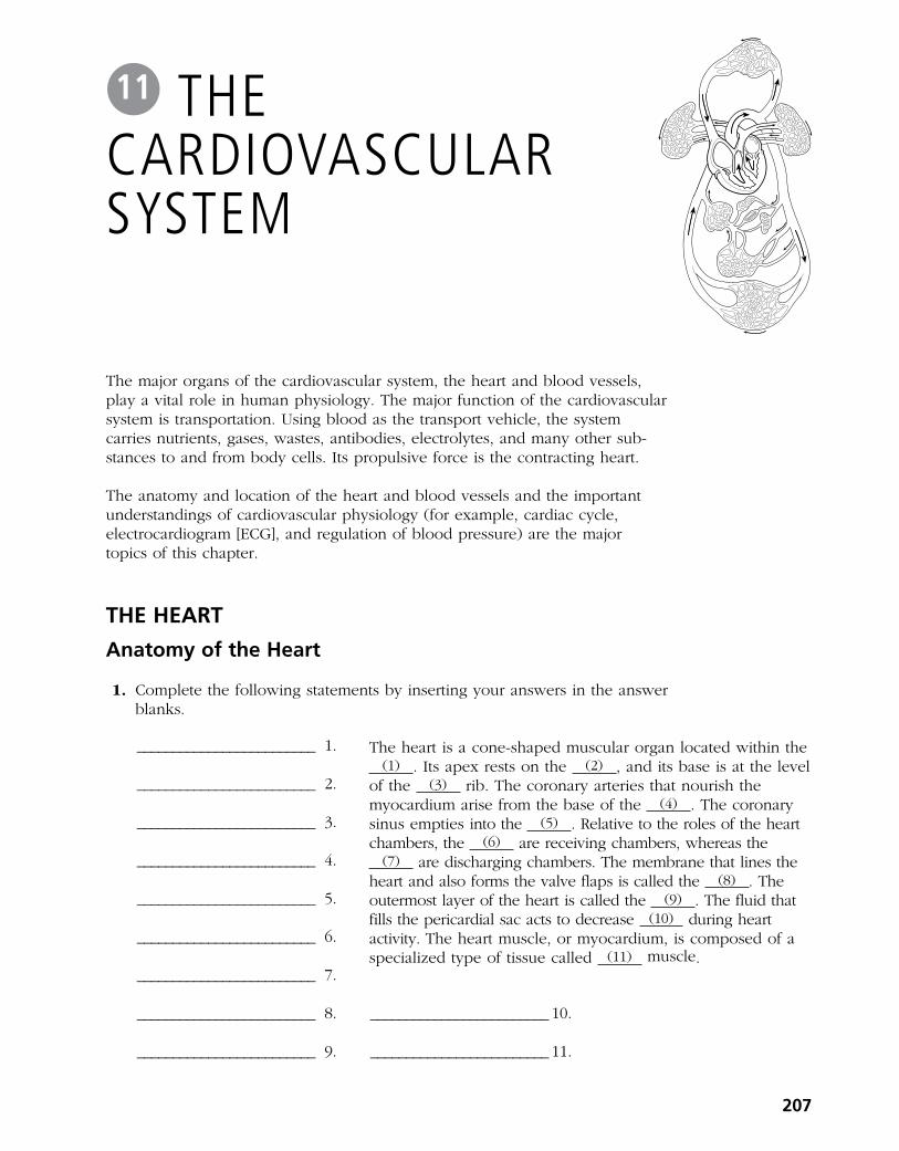
The major organs of the cardiovascular system, the heart and blood vessels, play a vital role in human physiology. The major function of the cardiovascular system is transportation. Using blood as the transport vehicle, the system carries nutrients, gases, wastes, antibodies, electrolytes, and many other substances to and from body cells. Its propulsive force is the contracting heart.
The anatomy and location of the heart and blood vessels and the important understandings of cardiovascular physiology (for example, cardiac cycle, electrocardiogram [ECG], and regulation of blood pressure) are the major topics of this chapter.
The hearT
anatomy of the heart
1. Complete the following statements by inserting your answers in the answer blanks.
The heart is a coneshaped muscular organ located within the (1) . Its apex rests on the (2) , and its base is at the level
of the (3) rib. The coronary arteries that nourish the myocardium arise from the base of the (4) . The coronary sinus empties into the (5) . Relative to the roles of the heart chambers, the (6) are receiving chambers, whereas the
(7) are discharging chambers. The membrane that lines the heart and also forms the valve flaps is called the (8) . The outermost layer of the heart is called the (9) . The fluid that fills the pericardial sac acts to decrease (10) during heart activity. The heart muscle, or myocardium, is composed of a specialized type of tissue called (11) muscle.
207
The cardiovascular sysTem
11
_________________________ 1.
_________________________ 2.
_________________________ 3.
_________________________ 4.
_________________________ 5.
_________________________ 6.
_________________________ 7.
_________________________ 8. _________________________ 10.
_________________________ 9. _________________________ 11.

2. The heart is called a double pump because it serves two circulations. (A) Trace the flow of blood through the pulmonary and systemic circu la
tions by writing the missing terms in the answer blanks (1–13). (B) Color regions trans porting O2poor blood blue and regions transport
ing O2rich blood red on Figure 11–1. (C) Identify the various regions of the circulation shown in Figure 11–1 by
labeling them using the key choices (A–F).
_________________________ 1.
_________________________ 2.
_________________________ 3.
_________________________ 4.
_________________________ 5.
_________________________ 6.
_________________________ 7.
_________________________ 8.
_________________________ 9.
_________________________ 10.
_________________________ 11.
_________________________ 12.
_________________________ 13.
Key Choices
A. Vessels serving head and upper limbs
B. Vessels serving body trunk and lower limbs
C. Vessels serving the visceraD. Pulmonary circulationE. Pulmonary “pump”F. Systemic “pump”
From the right atrium through the tricuspid valve to the (1) , through the (2) valve to the pulmonary trunk to the right and left (3) , to the capillary beds of the (4) , to the right and left (5) , to the (6) of the heart through the (7) valve, to the (8) through the (9) semilunar valve, to the
(10) , to the systemic arteries, to the (11) of the body tissues, to the systemic veins, to the (12) and (13) , which enter the right atrium of the heart.
208 Anatomy & Physiology Coloring Workbook
Figure 11–1

3. Figure 11–2 is an anterior view of the heart. (A) Identify each numbered structure and write its name in the corresponding
numbered answer blank. (B) Select different colors for each structure provided with a colorcoding
circle and use them to color the coding circles and corresponding structures on the figure.
○ ____________________ 1. ○ ____________________ 6. ○ ____________________ 11.
○ ____________________ 2. ○ ____________________ 7. ____________________ 12.
○ ____________________ 3. ○ ____________________ 8. ____________________ 13.
○ ____________________ 4. ____________________ 9. ____________________ 14.
____________________ 5. ____________________ 10. ○ ____________________ 15.
Chapter 11 The Cardiovascular System 209
15
9
12
10
5
7
811
136
3
4
14
1
2
(region)
Figure 11–2

4. Figure 11–3 is a schematic drawing of the microscopic structure of cardiac muscle. Using different colors, color the coding circles of the structures listed below and the corresponding structures on the figure.
○ Nuclei (with nucleoli) ○ Muscle fibers
○ Intercalated discs ○ Striations
5. The events of one complete heartbeat are referred to as the cardiac cycle. Complete the following statements that describe these events. Insert your answers in the answer blanks.
_________________________ 1.
_________________________ 2.
_________________________ 3.
_________________________ 4.
_________________________ 5.
_________________________ 6.
_________________________ 7. _________________________ 9.
_________________________ 8. _________________________ 10.
6. Figure 11–4 is a diagram of the interior frontal section of the heart. (A) Draw arrows to indicate the direction of blood flow through the heart and great vessels. (B) Color the heart chambers and the vessels transporting O2poor blood blue and chambers and vessels transporting O2rich blood red. (C) In the space provided, write down the pathway of blood as it enters the heart from the venae cavae and leaves the heart through the aorta. Include heart chambers, valves, and great vessels.
Superior and inferior venae cavae tricuspid valve pulmonary semilunar valve pulmonary trunk right and left lungs right and left valve
valve aorta.
The contraction of the ventricles is referred to as (1) , and the period of ventricular relaxation is called (2) . The mono syllables describing heart sounds during the cardiac cycle are (3) . The first heart sound is a result of closure of the (4) valves; the second heart sound is caused by closure of the (5) valves. The heart chambers that have just been filled when you hear the first heart sound are the (6) , and the chambers that have just emptied are the (7) . Immediately after the second heart sound, the (8) are filling with blood, and the (9) are empty. Abnormal heart sounds, or
(10) , usually indicate valve problems.
210 Anatomy & Physiology Coloring Workbook
Figure 11–3

7. Figure 11– 5 is a diagram of the frontal section of the heart. Follow the instruc tions below to complete this exercise.
(A) Identify each of the elements of the intrinsic conduction system (numbers 1–5 on the figure) by inserting the appropriate terms in the blanks left of the figure. Then, indicate with green arrows the pathway that impulses take through this system.
(B) Correctly identify each of the heart valves (numbers 6–9 on the figure) by inserting the appropriate terms in the blanks left of the figure.
(C) Draw in and identify by name the cordlike structures that anchor the flaps of the atrioventricular (AV) valves.
(D) Use the numbers from the figure to identify the structures described below. Place the numbers in the lettered answer blanks.
______ A. ______ B. Prevent backflow into the ventricles when the heart is relaxed
______ C. ______ D. Prevent backflow into the atria when the ventricles are contracting
______ E. AV valve with three flaps
______ F. AV valve with two flaps
______ G. The pacemaker of the intrinsic conduction system
______ H. The point in the intrinsic conduction system where the impulse is temporarily delayed
Chapter 11 The Cardiovascular System 211
Figure 11–4

_________________________ 1.
_________________________ 2.
_________________________ 3.
_________________________ 4.
_________________________ 5.
_________________________ 6.
_________________________ 7.
_________________________ 8.
_________________________ 9.
Physiology of the heart
8. Match the terms provided in Column B with the statements given in Column A. Place the correct term or letter response in the answer blanks.
Column a
_________________________ 1. A recording of the electrical activity of the heart
_________________________ 2. The period when the atria are depolarizing
_________________________ 3. The period when the ventricles are repolarizing
_________________________ 4. The period during which the ventricles are depolarizing, which precedes their contraction
_________________________ 5. An abnormally slow heartbeat, that is, slower than 60 beats per minute
_________________________ 6. A condition in which the heart is uncoordinated and useless as a pump
_________________________ 7. An abnormally rapid heartbeat, that is, faster than 100 beats per minute
_________________________ 8. Damage to the AV node, totally or partially releasing the ventricles from the control of the sinoatrial (SA) node
_________________________ 9. Chest pain, resulting from ischemia of the myocardium
Column B
A. Angina pectoris
B. Bradycardia
C. Electrocardiogram
D. Fibrillation
E. Heart block
F. P wave
G. QRS wave
H. T wave
I. Tachycardia
212 Anatomy & Physiology Coloring Workbook
Superiorvena cava
1
2
3
9
4
5
Inferiorvena cava
Wall of leftventricle
8
7
6Left atrium
Aorta
Pulmonarytrunk
Figure 11–5

9. A portion of an ECG is shown in Figure 11–6. (A) On the figure identify the QRS complex, the P wave, and the T wave. (B) Using a red pencil, bracket a portion of the recording equivalent to the
length of one cardiac cycle. (C) Using a blue pencil, bracket a portion of the recording in which the
ventricles would be in diastole.
Figure 11–6
10. Complete the following statements relating to cardiac output by writing the missing terms in the answer blanks.
In the relationship CO = HR . SV, CO stands for (1) , HR stands for (2) , and SV stands for (3) . For the normal resting heart, the value of HR is (4) beats per minute, and the value of SV is (5) mL per beat. The normal average adult cardiac output, therefore, is (6) mL per minute. The time for the entire blood supply to pass through the body is once each (7) .
According to Starling’s law of the heart, the critical factor that determines force of heartbeat, or (8) , is the degree of
(9) of the cardiac muscle just before it contracts. Consequently, the force of heartbeat can be increased by increasing the amount of (10) returned to the heart.
_________________________ 9. _________________________ 10.
11. Check (✓) all factors that lead to an increase in cardiac output by influencing either heart rate or stroke volume.
______ 1. Epinephrine ______ 6. Activation of the sympathetic nervous system
______ 2. Thyroxine ______ 7. Activation of the vagus nerves
______ 3. Hemorrhage ______ 8. Low blood pressure
______ 4. Fear ______ 9. High blood pressure
______ 5. Exercise ______ 10. Fever
_________________________ 1.
_________________________ 2.
_________________________ 3.
_________________________ 4.
_________________________ 5.
_________________________ 6.
_________________________ 7.
_________________________ 8.
Chapter 11 The Cardiovascular System 213

12. For each of the following statements that is true, write T in the answer blank. For any false statements, correct the underlined term by writing the correct term in the answer blank.
_________________________ 1. The resting heart rate is fastest in adult life.
_________________________ 2. Because the heart of the highly trained athlete hypertrophies, its stroke volume decreases.
_________________________ 3. If the right side of the heart fails, pulmonary congestion occurs.
_________________________ 4. In peripheral congestion, the feet, ankles, and fingers swell.
_________________________ 5. The pumping action of the healthy heart ordinarily maintains a balance between cardiac output and venous return.
13. Circle the term that does not belong in each of the following groupings. Then, fill in the answer blanks with the correct group name.
1. Pulmonary trunk Vena cava SA node Left side of heart Group: ____________
2. QRS wave T wave P wave Group: ____________
3. AV valves closed AV valves opened First heart sound Semilunar valves open Group: ____________
4. Papillary muscles Aortic semilunar valve Tricuspid valve Chordae tendineae Group: ____________
5. Tricuspid valve Mitral valve Bicuspid valve Group: ____________
6. Ischemia Infarct Scar tissue repair Heart block Group: ____________
Blood Vessels
Microscopic anatomy of Blood Vessels
14. Complete the following statements concerning blood vessels.
The central cavity of a blood vessel is called the (1) . Reduction of the diameter of this cavity is called (2) , and enlargement of the vessel diameter is called (3) . Blood is carried to the heart by (4) and away from the heart by (5) . Capillary beds are supplied by (6) and drained by (7) .
15. Briefly explain in the space provided the need for valves in veins but not in arteries.
___________________________________________________________________________________________
___________________________________________________________________________________________
16. Name two events occurring within the body that aid venous return. Place your responses in the blanks that follow.
___________________________________ and ___________________________________
214 Anatomy & Physiology Coloring Workbook
_________________________ 1.
_________________________ 2.
_________________________ 3.
_________________________ 4. _________________________ 6.
_________________________ 5. _________________________ 7.

17. Figure 11–7 is a diagram of the structure of the three types of blood vessels. (A) Select different colors for each of the three blood vessel tunics listed in
the key choices. Then, color the colorcoding circles and the corresponding structures in the three diagrams.
(B) Using the key choices, identify the blood vessel tunics described in each of the following descriptions. Insert the term or letter of the key choice in the answer blanks.
(C) In the blanks beneath the illustrations, correctly identify each vessel type. (D) In the additional spaces provided, list the structural details that allowed you
to make the identifications.
Key Choices
A. ○ Tunica intima B. ○ Tunica media C. ○ Tunica externa
_________________________ 1. Single thin layer of endothelium
_________________________ 2. Bulky middle coat, containing smooth muscle and elastin
_________________________ 3. Provides a smooth surface to decrease resistance to blood flow
_________________________ 4. The only tunic of capillaries
_________________________ 5. Also called the adventitia
_________________________ 6. The only tunic that plays an active role in blood pressure regulation
_________________________ 7. Supporting, protective coat
Vessel 1 _________________ Vessel 2 _________________ Vessel 3 __________________
Characteristics: ___________ Characteristics: __________ Characteristics: ____________
_______________________ ______________________ ________________________
Gross anatomy of Blood Vessels
18. Figures 11–8 and 11–9 on pages 216 and 217 illustrate the location of the most important arteries and veins of the body. The veins are shown in Figure 11–8 and the arteries are shown in Figure 11–9.
(A) Color the veins blue and then identify each vein provided with a leader line on the figure.
(B) Color the arteries red and then identify those indicated by leader lines on the figure.
(C) Indicate with arrows the direction of blood flow toward or away from the heart in Figures 11–8 and 11–9. Place arrows next to the blood vessels.
Note: If desired, the vessels identified may be colored differently to aid you in their later identification.
Chapter 11 The Cardiovascular System 215
Figure 11–7
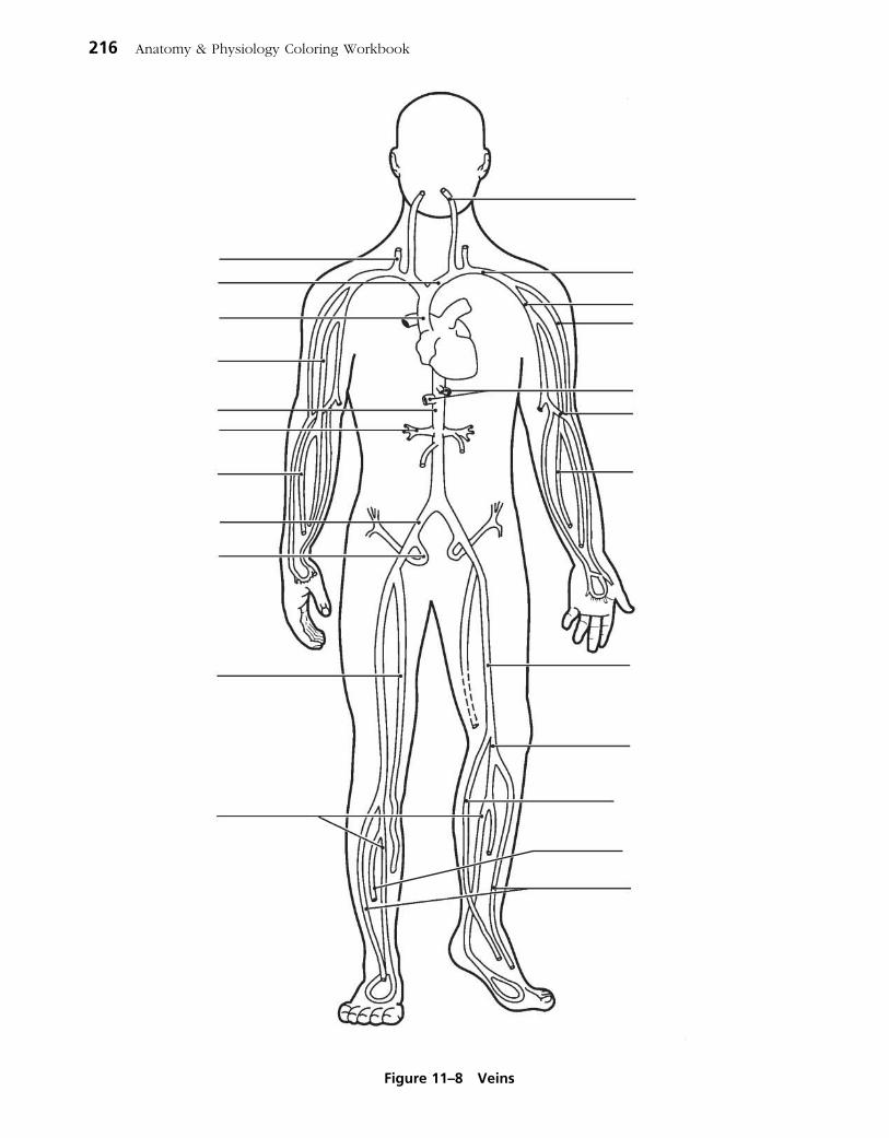
216 Anatomy & Physiology Coloring Workbook
Figure 11–8 Veins

Chapter 11 The Cardiovascular System 217
Figure 11–9 arteries

19. Using the key choices, complete the crossword puzzle by identifying the veins described in each of the clues provided.
Key Choices
Anterior tibial Cephalic Great saphenous Internal jugularAzygos Common iliac Hepatic RenalBasilic Femoral Hepatic portal SubclavianBrachiocephalic Gastric Inferior vena cava Superior vena cavaCardiac Gonadal Internal iliac Ulnar
Across 5. Veins that drain venous blood from the myocardium of the heart into the
coronary sinus. 6. Vein that drains the dural sinuses of the brain. 9. Vein that receives blood from the arm via the axillary vein. 10. Radial and ____ are deep veins, draining the forearm. 11. Vein that drains the kidney. 13. Two veins that join to become the superior vena cava. 15. Large vein that carries nutrientrich blood from the digestive organs to the
liver for processing.
Down 1. Vein that is formed by the union of the external and internal iliac veins. 2. Posterior tibial and ______ are veins that drain the leg and foot. 3. Deep vein of the thigh. 4. Largest vein below the thorax. 7. Longest superficial vein of the body; found in the leg. 8. Inferior mesenteric, superior mesenteric, and ______ are the three veins that
form/empty into the hepatic portal vein. 12. Vein that drains the ovaries or testes. 14. Vein that drains the thorax, empties into the superior vena cava.
1
2
3
4
5
6 7
8
9
10
11
12
13 14
15
218 Anatomy & Physiology Coloring Workbook

20. Figure 11–10 shows the pulmonary circuit. (A) Identify all vessels that have leader lines. (B) Color the vessels (and heart chambers) transporting oxygenrich
blood red; color those transporting carbon dioxiderich blood blue. Note: Indicate right or left when applicable.
Chapter 11 The Cardiovascular System 219
Figure 11–10
21. Using the key choices, identify the special circulations described below. Letters or terms may be used more than once.
Key Choices
A. Cerebral C. Hepatic E. Skeletal muscleB. Coronary D. Pulmonary F. Skin
_________________________ 1. The blood flow increases markedly when the body temperature rises.
_________________________ 2. These arteries characteristically have thin walls and large lumens.
_________________________ 3. These vessels do not constrict but are compressed during systole.
_________________________ 4. This circulation receives constant blood flow whether the body is at rest or strenuously exercising.
_________________________ 5. This circulation has a much lower arterial pressure than that in systemic circulation.
_________________________ 6. This circulation is indicated by impermeable tight junctions in capillary endothelium.
_________________________ 7. During vigorous physical activity, this circulation receives up to twothirds of blood flow.

22. The abdominal vasculature is depicted in Figure 11–11. (A) Using the key choices, identify the following vessels by selecting
the correct terms or letters. (B) Color the arteries red and veins blue.
Key Choices
A. Aorta F. Inferior mesenteric artery K. Renal arteriesB. Celiac trunk G. Inferior vena cava L. Renal veinsC. Common iliac arteries H. Lumbar arteries M. Left gonadal veinD. Gonadal arteries I. External iliac artery N. Right gonadal veinE. Hepatic veins J. Superior mesenteric artery O. Internal iliac artery
220 Anatomy & Physiology Coloring Workbook
Figure 11–11

23. Figure 11–12 is a diagram of the hepatic portal circulation. Select different colors for the structures listed below and use them to color the colorcoding circles and corresponding structures on the illustration.
○ Inferior mesenteric vein ○ Splenic vein ○ Hepatic portal vein
○ Superior mesenteric vein ○ Gastric vein
Chapter 11 The Cardiovascular System 221
Liver
Gallbladder
Hepaticportal
vein
Ascendingcolon
Descendingcolon
Small intestine
Spleen
Pancreas
Rectum
Stomach
Figure 11–12

24. Using the key choices, complete the crossword puzzle by identifying the arteries described in each of the clues provided.
Key Choices
Anterior tibial Coronary Inferior mesenteric RadialAorta Deep artery of thigh Intercostals RenalBrachial Dorsalis pedis Internal iliac SubclavianBrachiocephalic External carotid Peroneal (fibular) Superior mesentericCeliac trunk Femoral Phrenic UlnarCommon iliac Hepatic Posterior tibial Vertebral
Across 1. Arterial network on the dorsum of the foot. 3. Arterial trunk that has three major branches, which serve the liver, spleen,
and stomach. 5. External iliac becomes this artery on entering the thigh. 6. Common carotid and _____ are two arteries formed by the division of the
brachiocephalic trunk. 8. Artery that supplies the diaphragm. 9. Damage to the left semilunar valve would interfere with blood flow into this
vessel. 10. Artery that splits to form the radial and ulnar arteries. 11. Anterior tibial, posterior tibial, and _____ are the three arteries serving the leg
inferior to the knee. 12. Artery generally used to feel the pulse at the wrist. 13. First artery that branches off the ascending aorta; serves the heart.
Down 2. Major artery, serving the tissues external to the skull. 3. The terminal branches of the dorsal, or descending, aorta. 4. Artery that serves the pelvis. 7. Internal carotid and _____ are two paired arteries serving the brain.
1 2
3
4 5
6 7
8
9
10
11
12
13
222 Anatomy & Physiology Coloring Workbook

25. Figure 11–13 illustrates the arterial circulation of the brain. Select different colors for the following structures and use them to color the coding circles and corresponding structures in the diagram.
○ Basilar artery ○ Communicating branches
○ Anterior cerebral arteries ○ Middle cerebral arteries
○ Posterior cerebral arteries
Chapter 11 The Cardiovascular System 223
Pituitary gland
Frontal lobeof cerebral
hemisphere
Internalcarotidartery
Cerebellum
Vertebral artery
Pons
Figure 11–13

26. Figure 11–14 illustrates the special fetal structures listed below. Select different colors for each and use them to color coding circles and corresponding structures in the diagram.
○ Foramen ovale ○ Ductus arteriosus ○ Ductus venosus
○ Umbilical arteries ○ Umbilical cord ○ Umbilical vein
224 Anatomy & Physiology Coloring Workbook
Liver
Superiorvenacava
Hepaticportal vein
Internaliliacartery
Commoniliacartery
Aorta
Inferiorvenacava
Umbilicus
Fetal bladder
Figure 11–14
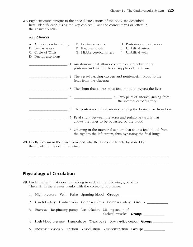
27. Eight structures unique to the special circulations of the body are described here. Identify each, using the key choices. Place the correct terms or letters in the answer blanks.
Key Choices
A. Anterior cerebral artery E. Ductus venosus H. Posterior cerebral arteryB. Basilar artery F. Foramen ovale I. Umbilical arteryC. Circle of Willis G. Middle cerebral artery J. Umbilical veinD. Ductus arteriosus
_________________________ 1. Anastomosis that allows communication between the posterior and anterior blood supplies of the brain
_________________________ 2. The vessel carrying oxygen and nutrientrich blood to the fetus from the placenta
_________________________ 3. The shunt that allows most fetal blood to bypass the liver
_________________________ 4. _________________________ 5. Two pairs of arteries, arising from the internal carotid artery
_________________________ 6. The posterior cerebral arteries, serving the brain, arise from here
_________________________ 7. Fetal shunt between the aorta and pulmonary trunk that allows the lungs to be bypassed by the blood
_________________________ 8. Opening in the interatrial septum that shunts fetal blood from the right to the left atrium, thus bypassing the fetal lungs
28. Briefly explain in the space provided why the lungs are largely bypassed by the circulating blood in the fetus.
___________________________________________________________________________________________
___________________________________________________________________________________________
Physiology of Circulation
29. Circle the term that does not belong in each of the following groupings. Then, fill in the answer blanks with the correct group name.
1. High pressure Vein Pulse Spurting blood Group: ____________
2. Carotid artery Cardiac vein Coronary sinus Coronary artery Group: ____________
3. Exercise Respiratory pump Vasodilation Milking action of skeletal muscles Group: ____________
4. High blood pressure Hemorrhage Weak pulse Low cardiac output Group: ____________
5. Increased viscosity Friction Vasodilation Vasoconstriction Group: ____________
Chapter 11 The Cardiovascular System 225

30. The following section relates to understandings concerning blood pressure and pulse. Match the items given in Column B with the appropriate descriptions provided in Column A. Place the correct term or letter response in the answer blanks.
Column a
_________________________ 1. Expansion and recoil of an artery during heart activity
_________________________ 2. Pressure exerted by the blood against the blood vessel walls
_________________________ 3. _________________________ 4. Factors related to blood pressure
_________________________ 5. Event primarily responsible for peripheral resistance
_________________________ 6. Blood pressure during heart contraction
_________________________ 7. Blood pressure during heart relaxation
_________________________ 8. Site where blood pressure determinations are normally made
_________________________ 9. Points at the body surface where the pulse may be felt
_________________________ 10. Sounds heard over a blood vessel when the vessel is partially compressed
31. Complete the following statements about capillary functions by placing an swers from the key in the answer blanks. Use terms or letters from the key.
Key Choices
A. Blood E. Fat soluble H. Osmotic pressureB. Capillary clefts F. Hydrostatic pressure I. VesiclesC. Diffusion G. Interstitial fluid J. Water solubleD. Fenestrations
_________________________ 1.
_________________________ 2.
_________________________ 3.
226 Anatomy & Physiology Coloring Workbook
Column B
A. Over arteries
B. Blood pressure
C. Cardiac output
D. Constriction of arterioles
E. Diastolic blood pressure
F. Peripheral resistance
G. Pressure points
H. Pulse
I. Sounds of Korotkoff
J. Systolic blood pressure
K. Over veins
All exchanges to and from the blood and tissue cells occur through the (1) . Generally speaking, substances tend to move according to their concentration gradients by the pro cess of (2) . Substances that are (3) pass directly through the plasma membranes of the capillary endothelial cells; other

substances pass by means of or via (4) , (5) , or (6) . The most permeable capillaries are those exhibiting (7) . Capillaries that have (8) and (9) tend to be leaky, and forces acting at capillary beds cause fluid flows.
Chapter 11 The Cardiovascular System 227
_________________________ 4.
_________________________ 5.
_________________________ 6.
_________________________ 7.
_________________________ 8. _________________________ 9.
32. Indicate what effect the following factors have on blood pressure. Indicate an increase in pressure by I and a decrease in pressure by D. Place the correct letter response in the answer blanks.
______ 1. Increased diameter of the arterioles ______ 8. Physical exercise
______ 2. Increased blood viscosity ______ 9. Physical training
______ 3. Increased cardiac output ______ 10. Alcohol
______ 4. Increased pulse rate ______ 11. Hemorrhage
______ 5. Anxiety, fear ______ 12. Nicotine
______ 6. Increased urine output ______ 13. Arteriosclerosis
______ 7. Sudden change in position from reclining to standing
33. For each of the following statements that is true, insert T in the answer blank. If any of the statements are false, correct the underlined term by inserting the correct word in the answer blank.
_________________________ 1. Renin, released by the kidneys, causes a decrease in blood pressure.
_________________________ 2. The decreasing efficiency of the sympathetic nervous system vasoconstrictor functioning, due to aging, leads to a type of hypotension called sympathetic hypotension.
_________________________ 3. Two body organs in which vasoconstriction rarely occurs are the heart and the kidneys.
_________________________ 4. A sphygmomanometer is used to take the apical pulse.
_________________________ 5. The pulmonary circulation is a highpressure circulation.
_________________________ 6. The fetal equivalent of (functional) lungs and liver is the placenta.
_________________________ 7. Cold has a vasodilating effect.
_________________________ 8. Thrombophlebitis is called the silent killer.

228 Anatomy & Physiology Coloring Workbook
34. Figure 11–15 is a diagram of a capillary bed. Arrows indicate the direction of blood flow.
(A) Select five different colors and color the coding circles and their structures on the figure.
(B) Answer the questions that follow by referring to Figure 11–15. Note: Questions 1–9 concern fluid flows at capillary beds and the forces
(hydrostatic and osmotic pressures) that promote such fluid shifts.
○ Arteriole ○ Vascular shunt ○ Postcapillary venule
○ Precapillary sphincters ○ True capillaries
1. If the precapillary sphincters are contracted, by which route will the blood flow?
___________________________________________________________________________________________
2. Under normal conditions, in which area does hydrostatic pressure predominate: A, B, or C?
___________________________________________________________________________________________
3. Which area has the highest osmotic pressure? _____________________________________________
4. Which pressure is in excess and causes fluids to move from A to C? (Be specific as to whether the force exists in the capillary or the interstitial space.)
_____________________________________________________________________________________________
5. Which pressure causes fluid to move from A to B? ________________________________________
AB
C
CC
Figure 11–15

Chapter 11 The Cardiovascular System 229
6. Which pressure causes fluid to move from C to B? ________________________________________
7. Which blood protein is most responsible for osmotic pressure? ____________________________
8. Where does the greater net flow of water out of the capillary occur? ______________________
9. If excess fluid does not return to the capillary, where does it go? __________________________
___________________________________________________________________________________________
35. Respond to the following exercise by placing brief answers in the spaces provided. Assume someone has been injured in an automobile accident and is bleeding profusely. What pressure point (artery) could you compress to help stop the bleeding from the following areas?
_________________________ 1. Thigh _________________________ 4. Lower jaw
_________________________ 2. Forearm _________________________ 5. Thumb
_________________________ 3. Back of the knee _________________________ 6. Temple
deVeloPMenTal asPeCTs oF The CardioVasCular sysTeM
36. Complete the following statements by inserting your responses in the answer blanks.
The cardiovascular system forms early, and the heart is acting as a functional pump by the (1) week of development. The ductus arteriosus and foramen ovale allow the blood to bypass the nonfunctioning fetal (2) . Another fetal structure, the (3) , allows most of the blood to bypass the liver. The fetus is supplied with oxygen and nutrients via the (4) , which carries blood from the (5) to the
(6) . Metabolic wastes and carbon dioxide are removed from the fetus in blood carried by the (7) . These special bypass structures that exist to bypass the fetal lungs and liver become (8) shortly after birth. Congenital heart defects (some resulting from the failure of the bypass structures to close) account for half of all infant (9) resulting from congenital defects.
(10) is a degenerative process that begins in youth but may take its toll in later life by promoting a myocardial infarct or stroke. Generally, women have less of this degenerative process than men until after (11) , when estrogen production ends.
_________________________ 1.
_________________________ 2.
_________________________ 3.
_________________________ 4.
_________________________ 5.
_________________________ 6.
_________________________ 7.
_________________________ 8.
_________________________ 9.
_________________________ 10.
_________________________ 11.
Regular (12) increases the efficiency of the cardiovascular system and helps to slow the progress of (13) . A vascular problem that affects many in “standing professions” is (14) . In this condition, the valves become incompetent, and the veins become twisted and enlarged, particularly in the (15) and (16) .
A Visualization Exercise for the Cardiovascular System
All about you are huge white cords, hanging limply from two flaps of endothelial tissue. . . .
37. Where necessary, complete the statements by inserting the missing word(s) in the answer blanks.
Your journey starts in the pulmonary vein and includes a trip to part of the systemic circulation and a special circulation. You ready your equipment and prepare to be miniaturized and injected into your host.
Almost immediately after injection, you find yourself swept into a goodsized chamber, the (1) . However, you do not stop in this chamber, but continue to plunge downward into a larger chamber below. You land with a big splash and examine your surroundings. All about you are huge white cords, hanging limply from two flaps of endothelial tissue far above you. You report that you are sitting in the (2) (chamber) of the heart, seeing the flaps of the (3) valve above you. The valve is open, and its anchor ing cords, the (4) , are lax. Because this valve is open, you conclude that the heart is in the (5) phase of the cardiac cycle.
Gradually, you notice that the chamber walls seem to be closing in. You hear a thundering boom, and the whole chamber vibrates as the valve slams shut above you. The cords, now rigid and strained, form a cage about you, and you feel extreme external pressure. Obviously, the heart is in a fullfledged (6) . Then, high above on the right, the “roof ” opens, and you are forced through this (7) valve. A fraction of a second later, you hear another tremendous boom that sends shock waves through the whole area. Out of the corner of your eye, you see that the valve below you is closed, and it looks rather like a pie cut into three wedges.
230 Anatomy & Physiology Coloring Workbook
_________________________ 12.
_________________________ 13.
_________________________ 14.
_________________________ 15.
_________________________ 16.
_________________________ 1.
_________________________ 2.
_________________________ 3.
_________________________ 4.
_________________________ 5.
_________________________ 6.
_________________________ 7.
INCREDIBLE JOURNEY

As you are swept along in this huge artery, the (8) , you pass several branchoff points but continue to careen along, straight down at a dizzying speed until you approach the
(9) artery, feeding the small intestine. After entering this artery and passing through successively smaller and smaller subdivisions of it, you finally reach the capillary bed of the small intestine. You watch with fascination as nutrient molecules move into the blood through the single layer of
(10) cells forming the capillary wall. As you move to the opposite shore of the capillary bed, you enter a venule and begin to move superiorly once again. The venules draining the small intestine combine to form the (11) vein, which in turn combines with the (12) vein to form the hepatic portal vein that carries you into the liver. As you enter the liver, you are amazed at the activity there. Sixsided hepatic cells, responsible for storing glucose and making blood proteins, are literally grabbing (13) out of the blood as it percolates slowly past them. Protective (14) cells are removing bacteria from the slowly moving blood. Leaving the liver through the
(15 vein, you almost immediately enter the huge (16) , which returns blood from the lower part of the body to the
(17) of the heart. From here, you move consecutively through the right chambers of the heart into the (18) . Soon, that vessel splits and you are carried into a (19) artery, which carries you to the capillary beds of the (20) and then back to the left side of the heart once again. After traveling through the left side of the heart, you leave your host when you are aspirated out of the (21) artery, which extends from the aorta to the axillary artery of the armpit.
38. A man, en route to the hospital emergency room by ambulance, is in fibrillation. What is his cardiac output likely to be? He arrives at the emergency entrance DOA (dead on arrival). His autopsy reveals a blockage of the posterior interventricular artery. What is the cause of death?
Chapter 11 The Cardiovascular System 231
_________________________ 8.
_________________________ 9.
_________________________ 10.
_________________________ 11.
_________________________ 12.
_________________________ 13.
_________________________ 14.
_________________________ 15.
_________________________ 16.
_________________________ 17.
_________________________ 18.
_________________________ 19.
_________________________ 20.
_________________________ 21.
AT THE CLINIC

39. Excessive vagal stimulation can be caused by severe depression. How would this be reflected in a routine physical examination?
40. Mrs. Suffriti has swollen ankles and signs of degenerating organ functions. What is a likely diagnosis?
41. A routine scan of an elderly man reveals partial occlusion of the right internal carotid artery, yet blood supply to his cerebrum is unimpaired. What are two possible causes of the occlusion? What anastomosis is maintaining blood supply to the brain and by what (probable) route(s)?
42. Mr. Abdul, a patient with a bone marrow cancer, is polycythemic. Will his blood pressure be high or low? Why?
43. After a bout with bacterial endocarditis, scar tissue often stiffens the edges of the heart valves. How would this be picked up in a routine examination?
44. Len, an elderly man, is bedridden after a hip fracture. He complains of pain in his legs, and thrombophlebitis is diagnosed. What is thrombophlebitis, and what lifethreatening complication can develop?
232 Anatomy & Physiology Coloring Workbook

45. Mr. Langley is telling his friend about his recent visit to his doctor for a checkup. During his story, he mentions that the ECG revealed that he had a defective mitral valve and a heart murmur. Mr. Langley apparently misunderstood some of what the doctor explained to him about the diagnostic process. What has he misunderstood?
46. A lessthanrespectable news tabloid announced that “Doctors show that exercise shortens life. Life expectancy is programmed into a set number of heartbeats; the faster your heart beats, the sooner you die.” Even if this hypothesis were true, what is wrong with the conclusion concerning exercise?
47. Mrs. Tuney says that when she stands up after lying down in the afternoon she gets very dizzy. Her husband grumbles, “It’s because she keeps the danged house too warm.” He’s right (in this particular case). Explain how this might cause her dizziness.
48. Mary Anne is taking a calcium channel blocking drug. What effect on her stroke volume (SV) would you expect this medication to have?
49. You are conducting animal research at Hampshire University. You have just chemically stimulated the ACh receptors on the rat’s heart. How would you expect this to affect that heart’s stroke volume?
Chapter 11 The Cardiovascular System 233
51. Select the best answer or answers from the choices given.
234 Anatomy & Physiology Coloring Workbook
1. The innermost layer of the pericardial sac is the:
A. epicardium.
B. fibrous pericardium.
C. parietal layer of the serous pericardium.
D. visceral layer of the serous pericardium.
2. The thickest layer of the heart wall is:
A. endocardium. C. epicardium.
B. myocardium. D. fibrous pericardium.
3. Atrioventricular valves are held closed by:
A. papillary muscles.
B. trabeculae carneae.
C. pectinate muscles.
D. chordae tendineae.
4. The fibrous skeleton of the heart:
A. supports valves.
B. anchors vessels.
C. provides electrical insulation to separate the atrial mass from the ventricular mass.
D. anchors cardiac muscle fibers.
5. Freshly oxygenated blood is first received by the:
A. right ventricle. C. right atrium.
B. left ventricle. D. left atrium.
6. Atrial repolarization coincides in time with the:
A. P wave. C. QRS wave.
B. T wave. D. PQ interval.
7. Soon after the onset of ventricular systole the:
A. AV valves close.
B. semilunar valves open.
C. first heart sound is heard.
D. aortic pressure increases.
8. Which of the following depolarizes next after the AV node?
A. Atrial myocardium
B. Ventricular myocardium
C. Bundle branches
D. Purkinje fibers
9. Which of the regulatory chemicals listed involve or target the kidneys?
A. Angiotensin C. ADH
B. Aldosterone D. ANP
10. Cardiovascular conditioning results in:
A. ventricular hypertrophy.
B. bradycardia.
C. increase in SV.
D. increase in CO.
THE FINALE: MULTIPLE CHOICE
50. How does the pulsating blood pressure in the largest arteries relate to their structures?

11. Which of the following is (are) part of the tunica intima?
A. Simple squamous epithelium
B. Basement membrane
C. Loose connective tissue
D. Smooth muscle
12. In comparing a parallel artery and vein, you would find that:
A. the artery wall is thicker.
B. the artery diameter is greater.
C. the artery lumen is smaller.
D. the artery endothelium is thicker.
13. Fenestrated capillaries occur in the:
A. liver.
B. kidney.
C. cerebrum.
D. intestinal mucosa.
14. Which of the following is (are) part of a capillary bed?
A. Precapillary sphincter
B. Vascular shunt
C. True capillaries
D. Terminal arteriole
15. Which of the following can function as a blood reservoir?
A. Brachiocephalic artery
B. Cerebral capillaries
C. Dural sinuses
D. Inferior vena cava
16. An increase in which of the following results in increased filtration from capillaries to the interstitial space?
A. Capillary hydrostatic pressure
B. Interstitial fluid hydrostatic pressure
C. Capillary osmotic pressure
D. Duration of precapillary sphincter contraction
17. Vessels involved in the circulatory pathway to and from the brain are the:
A. brachiocephalic artery.
B. subclavian artery.
C. internal jugular vein.
D. internal carotid artery.
18. Which of the following are associated with aging?
A. Increasing blood pressure
B. Weakening of venous valves
C. Arteriosclerosis
D. Stenosis of the ductus arteriosus
19. Which layer of the artery wall thickens most in atherosclerosis?
A. Tunica media
B. Tunica intima
C. Tunica adventitia
D. Tunica externa
20. Based on the vessels named pulmonary trunk, thyrocervical trunk, and celiac trunk, the term trunk must refer to:
A. a vessel in the heart wall.
B. a vein.
C. a capillary.
D. a large artery from which other arteries branch.
21. Which of these vessels is bilaterally symmet rical (i.e., one vessel of the pair occurs on each side of the body)?
A. Internal carotid artery
B. Brachiocephalic artery
C. Azygos vein
D. Superior mesenteric vein
22. A stroke that occludes a posterior cerebral artery will most likely affect:
A. hearing. C. smell.
B. vision. D. higher thought processes.
Chapter 11 The Cardiovascular System 235
23. Tracing the drainage of the superficial venous blood from the leg, we find that blood enters the great saphenous vein, femoral vein, inferior vena cava, and right atrium. Which veins are missing from that sequence?
A. Coronary sinus and superior vena cava
B. Posterior tibial and popliteal
C. Fibular (peroneal) and popliteal
D. External and common iliacs
24. Tracing the drainage of venous blood from the small intestine, we find that blood enters the superior mesenteric vein, hepatic vein, inferior vena cava, and right atrium. Which vessels are missing from that sequence?
A. Coronary sinus and left atrium
B. Celiac and common hepatic veins
C. Internal and common iliac veins
D. Hepatic portal vein and liver sinusoids
236 Anatomy & Physiology Coloring Workbook
The lymphatic system, with its many lymphoid organs and vessels derived from veins of the cardiovascular system, is a rather strange system. Although both types of organs help to maintain homeostasis, these two elements of the lym phatic system have substantially different roles. The lymphatic vessels help keep the cardiovascular system functional by maintaining blood volume. The lymphoid organs help defend the body from patho g ens by providing operating sites for phagocytes and cells of the immune system.
The immune system, which serves as the body’s specific defense system, is a unique func tional system made up of billions of individual cells, most of which are lympho-cytes. The sole function of this defensive system is to protect the body against an incredible array of pathogens. In general, these “enemies” fall into three major camps: (1) micro organisms (bacteria, viruses, and fungi) that have gained entry into the body, (2) foreign tissue cells that have been transplanted (or, in the case of red blood cells, infused) into the body, and (3) the body’s own cells that have become cancerous. The result of the immune system’s activities is immunity, or specific resistance to disease.
The body is also protected by a number of nonspecific defenses provided by intact sur-face membranes such as skin and mucosae, and by a variety of cells and chemicals that can quickly mount an attack against foreign substances. The specific and nonspecific defenses enhance each other’s effectiveness.
Chapter 12 tests your understanding of the functional roles of the various lymphatic sys-tem elements and both the nonspecific and specific body defenses.
THE LYMPHATIC SYSTEM
Lymphatic Vessels 1. Complete the following statements by writing the missing terms in the answer blanks.
Together, the cardiovascular and lymphatic systems make up the circulatory system. Although the cardiovascular system has a pump (the heart) and arteries, veins, and capillaries, the lymphatic system lacks two of these structures: the (1) and
(2) . Like the (3) of the cardiovascular system, the vessels of the lymphatic system are equipped with (4) to prevent backflow. The lymphatic vessels act primarily to pick up leaked fluid, now called (5) , and return it to the bloodstream. About (6) liters of fluid is returned every 24 hours.
_________________________ 1.
_________________________ 2.
_________________________ 3.
_________________________ 4.
_________________________ 5.
_________________________ 6.237
THE LYMPHATIC SYSTEM AND BODY DEFENSES
12

2. Figure 12–1 provides an overview of the lymphatic vessels. In part A, the relationship between lymphatic vessels and the blood vessels of the cardiovascular system is depicted schematically. Part B shows the different types of lymphatic vessels in a simple way.
(A) Color-code and color the following structures in Figure 12–1 (parts A and B).
○ Heart ○ Veins ○ Lymphatic vessels/lymph node
○ Arteries ○ Blood capillaries ○ Loose connective tissue around blood and lymph capillaries
(B) Label the following structures in part B:
A. Lymph capillaries C. Lymphatic collecting vessels E. Valves
B. Lymph duct D. Lymph node F. Vein
3. Circle the term that does not belong in each of the following groupings. Then, fill in the answer blanks with the correct group name.
1. Blood capillary Remarkably permeable Blind-ended Permeable to proteins Group: ____________
2. Edema Blockage of lymphatics Elephantiasis Inflammation Abundant supply of lymphatics Group: ____________
3. Skeletal muscle pump Respiratory pump High-pressure gradient Action of smooth muscle cells in walls of lymph vessels Group: ____________
4. Minivalves Endothelial cell overlap Impermeable Low-pressure pumpless system Group: ____________
238 Anatomy & Physiology Coloring Workbook
Tissue fluid(becomes lymph)
A B
Figure 12–1

Lymph Nodes and Other Lymphoid Organs
4. Match the terms in Column B with the appropriate descriptions in Column A. Items may have more than one answer.
Column A
_________________________ 1. A blood reservoir
_________________________ 2. Monitor composition of lymph
_________________________ 3. Located between the lungs at the base of the throat
_________________________ 4. Collectively called MALT
_________________________ 5. Prevents bacteria from breaching the intestinal wall
5. Figure 12–2 depicts several different lymphoid organs.
(A) Label all lymphoid organs indicated by a leader line.
(B) Add labels to identify the sites where the axillary, cervical, and inguinal lymph nodes would be located.
(C) Shade in light green the portion of the body that is drained by the right lymphatic duct.
Figure 12–2
Chapter 12 The Lymphatic System and Body Defenses 239
Column B
A. Lymph nodes
B. Peyer’s patches
C. Spleen
D. Thymus
E. Tonsils

6. Figure 12–3 is a diagram of a lymph node. (A) Using the terms with color-coding circles, label all structures on the
diagram that have leader lines. (B) Select different colors for each and use them to color coding circles
and corresponding structures in the diagram. (C) Add arrows to the diagram to show the direction of lymph flow
through the organ. (D) Circle the region that would approximately correspond to the medulla
of the organ. (E) Answer the questions that follow.
○ Germinal centers of follicles ○ Hilum
○ Cortex (other than germinal centers) ○ Afferent lymphatics
○ Medullary cords ○ Efferent lymphatics
○ Capsule and trabeculae ○ Sinuses (subcapsular and medullary)
240 Anatomy & Physiology Coloring Workbook
Figure 12–3

1. Which cell type is found in greatest abundance in the germinal centers?
___________________________________________________________________________________________
2. What is the function of their daughter cells, the plasma cells?
___________________________________________________________________________________________
3. What is the major cell type in cortical areas other than the germinal centers?
___________________________________________________________________________________________
4. The third important cell type in lymph nodes (usually found clustered around
the medullary sinuses) are the _____________________________________________ .
These cells act as ______________________________________________ .
5. Of what importance is the fact that there are fewer efferent than afferent lymphatics associated with lymph nodes?
___________________________________________________________________________________________
___________________________________________________________________________________________
___________________________________________________________________________________________
6. What structures ensure the one-way flow of lymph through the node?
___________________________________________________________________________________________
7. The largest collections of lymph nodes are found in what three body regions?
___________________________________________________________________________________________
7. Match the terms in Column B with the appropriate descriptions in Column A.
Column A
_________________________ 1. The largest lymphatic organ
_________________________ 2. Filter lymph
_________________________ 3. Particularly large and important during youth; helps to program T cells of the immune system
_________________________ 4. Found in the wall of the gastrointestinal tract
_________________________ 5. Removes aged and defective red blood cells
Column B
A. Lymph nodes
B. Peyer’s patches
C. Spleen
D. Thymus
E. Tonsils
Chapter 12 The Lymphatic System and Body Defenses 241

BODY DEFENSES
Nonspecific (Innate) Body Defenses
8. Complete the following statements by writing the missing terms in the answer blanks.
The three major elements of the body’s nonspecific defense system are: the
(1) __________________________ , consisting of the skin and _________________________ ;
defensive cells, such as (2) __________________________ and phagocytes; and a whole
deluge of (3) __________________________ .
9. Indicate the sites of activity or the secretions of the nonspecific defenses by writing the correct terms in the answer blanks.
1. Lysozyme is found in the body secretions called __________________ and __________________ .
2. Fluids with an acid pH are found in the __________________ and __________________ .
3. Sebum is a product of the __________________________ glands and acts at the surface
of the __________________________ .
4. Mucus is produced by mucus-secreting glands found in the respiratory and
__________________________ system mucosae.
10. Figure 12–4 diagrams the events involved in the inflammatory response. Assume the following events have already occurred: tissue injury and invasion of microbes, and release of inflammatory chemicals by mast cells. Each subsequent event is represented by a square with one or more arrows.
(A) From the list below, write the correct number in each event square in the figure.
(B) Color-code and color the structures that appear below the numbered list.
1. White blood cells are drawn to the injured area by the release of inflammatory chemicals.
2. Tissue repair occurs.
3. Local blood vessels dilate, and the capillaries become engorged with blood.
4. Phagocytosis of microbes occurs.
5. Fluid containing clotting proteins is lost from the bloodstream and enters the injured tissue area.
6. Diapedesis occurs.
○ Monocyte ○ Neutrophil(s) ○ Endothelium of capillary
○ Epithelium ○ Macrophage ○ Microorganisms
○ Erythrocyte(s) ○ Subcutaneous tissue ○ Fibrous repair tissue
242 Anatomy & Physiology Coloring Workbook
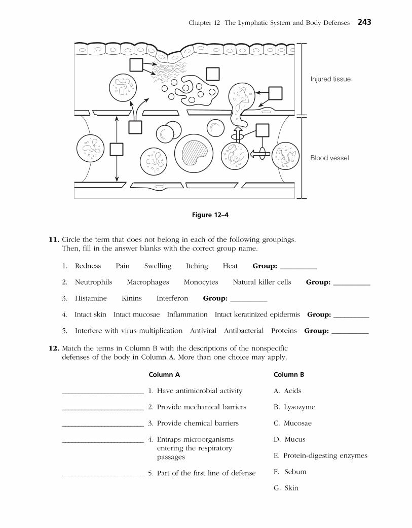
11. Circle the term that does not belong in each of the following groupings. Then, fill in the answer blanks with the correct group name.
1. Redness Pain Swelling Itching Heat Group: __________
2. Neutrophils Macrophages Monocytes Natural killer cells Group: __________
3. Histamine Kinins Interferon Group: __________
4. Intact skin Intact mucosae Inflammation Intact keratinized epidermis Group: __________
5. Interfere with virus multiplication Antiviral Antibacterial Proteins Group: __________
12. Match the terms in Column B with the descriptions of the nonspecific defenses of the body in Column A. More than one choice may apply.
Column A
_________________________ 1. Have antimicrobial activity
_________________________ 2. Provide mechanical barriers
_________________________ 3. Provide chemical barriers
_________________________ 4. Entraps microorganisms entering the respiratory passages
_________________________ 5. Part of the first line of defense
Column B
A. Acids
B. Lysozyme
C. Mucosae
D. Mucus
E. Protein-digesting enzymes
F. Sebum
G. Skin
Chapter 12 The Lymphatic System and Body Defenses 243
Injured tissue
Blood vessel
Figure 12–4

13. Describe the protective role of cilia in the respiratory tract. ________________________________
___________________________________________________________________________________________
___________________________________________________________________________________________
14. Define phagocytosis. _____________________________________________________________________
___________________________________________________________________________________________
___________________________________________________________________________________________
15. Check (✓) all phrases that correctly describe the role of fever in body protection.
______ 1. Is a normal response to pyrogens
______ 2. Protects by denaturing tissue proteins
______ 3. Reduces the availability of iron and zinc required for bacterial proliferation
______ 4. Increases metabolic rate
16. Using the key choices, complete the crossword puzzle by answering each of the clues provided.
Key Choices
Chemotaxis Eosinophils Inflammatory chemicalsDiapedesis Fibrin mesh MacrophagesEdema Histamine Neutrophils
Across 2. Results from accumulation of fluid leaked from the bloodstream. 5. First phagocytes to migrate into the injured area. 6. Cellular migration directed by a chemical gradient. 7. Leukocytes pass through the wall of a capillary.
Down 1. Walls off the area of injury. 3. Phagocytic offspring of monocytes. 4. Inflammatory chemical released by injured cells.
1
2 3
4
5
6
7
244 Anatomy & Physiology Coloring Workbook

17. Complete the following description of the activation and activity of complement by writing the missing terms in the answer blanks.
Complement is a system of plasma (1) that circulate in the blood in an inactive form. Complement is (2) when it becomes attached to the surface of foreign cells (bacteria, fungi, red blood cells). One result of this complement fixation is that (3) appear in the membrane of the foreign cell. This allows (4) to rush in, which causes (5) of the for-eign cell. Some of the chemicals released during complement fixation enhance phagocytosis. This is called (6) . Others amplify the inflammatory response.
18. Describe the event that leads to the synthesis of interferon and the result of its synthesis.
___________________________________________________________________________________________
___________________________________________________________________________________________
Specific (Adaptive) Body Defenses: The Immune System
Antigens
19. What are three important characteristics of the adaptive immune
response? __________________________, __________________________,
and __________________________.
20. Complete the following statements relating to antigens by writing the missing terms in the answer blanks.
Antigens are substances capable of mobilizing the (1) . Of all the foreign molecules that act as complete antigens,
(2) are the most potent. Small molecules are not usually antigenic, but when they bind to self-cell surface proteins, they may act as (3) , and then the complex is recognized as foreign, or (4) .
_________________________ 1.
_________________________ 2.
_________________________ 3.
_________________________ 4.
_________________________ 5.
_________________________ 6.
Chapter 12 The Lymphatic System and Body Defenses 245
_________________________ 1.
_________________________ 2.
_________________________ 3.
_________________________ 4.

Cells of the Immune System: An Overview
21. Using the key choices, select the term that correctly completes each statement. Insert the appropriate term or letter in the answer blanks.
Key Choices
A. Antigen(s) D. Cellular G. Lymph nodesB. B cells E. Humoral H. MacrophagesC. Blood F. Lymph I. T cells
Immunity is resistance to disease resulting from the presence of foreign substances or (1) in the body. When this resistance is provided by antibodies released to body fluids, the immunity is called (2) immunity. When living cells provide the protection, the immunity is referred to as (3) immunity. The major actors in the immune response are two lymphocyte populations, the (4) and the (5) . Phagocytic cells that act as accessory cells in the immune response are the (6) . Because pathogens are likely to use both (7) and (8) as a means of getting around the body, (9) and other lymphatic tissues (which house the immune cells) are in an excellent position to detect their presence.
22. A schematic of the life cycle of the lymphocytes involved in immunity is shown in Figure 12–5.
(A) Select different colors for the areas listed below and use them to color the coding circles and the corresponding regions in the figure. If there is overlap, use stripes of a second color to indicate the second identification.
(B) Respond to the statements following the figure, which relate to the two-phase differentiation process of B and T cells.
○ Area where immature lymphocytes arise
○ Area seeded by immunocompetent B and T cells
○ Area where T cells become immunocompetent
○ Area where the antigen challenge and clonal selection are likely to occur
○ Area where B cells become immunocompetent
_________________________ 1.
_________________________ 2.
_________________________ 3.
_________________________ 4.
_________________________ 5.
_________________________ 6.
_________________________ 7.
_________________________ 8.
_________________________ 9.
246 Anatomy & Physiology Coloring Workbook
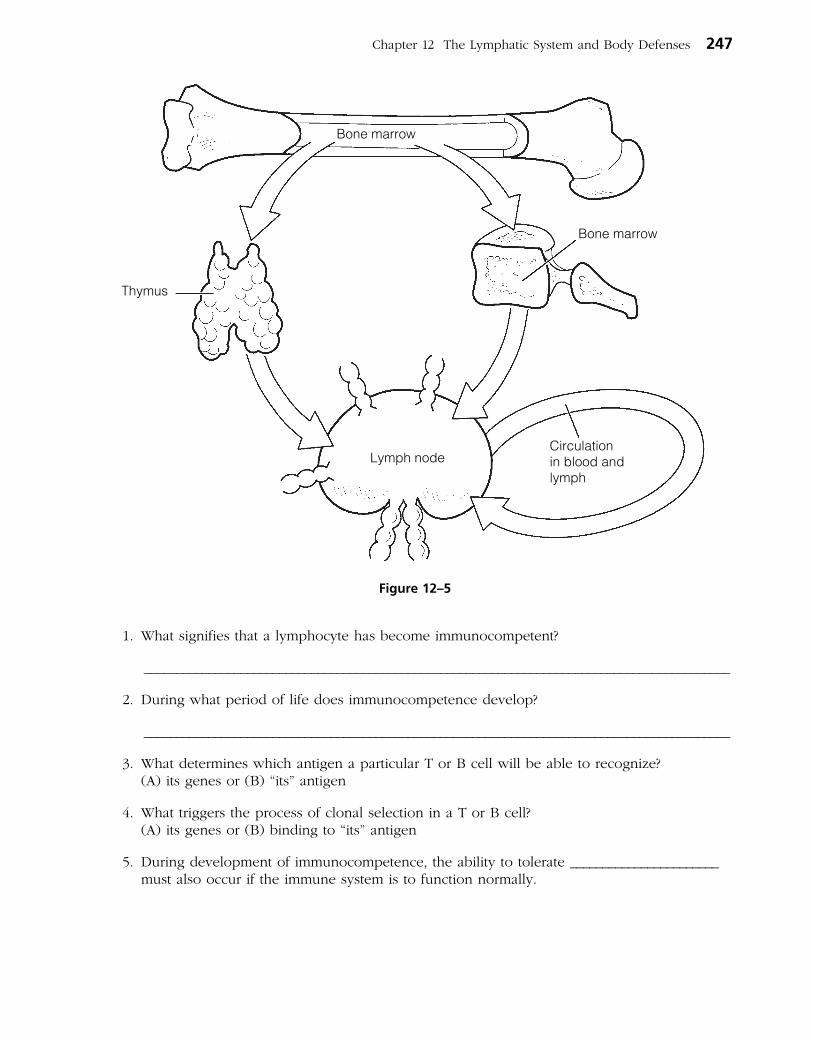
1. What signifies that a lymphocyte has become immunocompetent?
___________________________________________________________________________________________
2. During what period of life does immunocompetence develop?
___________________________________________________________________________________________
3. What determines which antigen a particular T or B cell will be able to recognize? (A) its genes or (B) “its” antigen
4. What triggers the process of clonal selection in a T or B cell? (A) its genes or (B) binding to “its” antigen
5. During development of immunocompetence, the ability to tolerate _______________________ must also occur if the immune system is to function normally.
Chapter 12 The Lymphatic System and Body Defenses 247
Thymus
Bone marrow
Circulationin blood andlymph
Lymph node
Bone marrow
Figure 12–5

23. T cells and B cells exhibit certain similarities and differences. Check (✓) the appropriate spaces in the table below to indicate the lymphocyte type that exhibits each characteristic.
Characteristic T cell B cell
Originates in bone marrow from stem cells called hemocytoblasts
Progeny are plasma cells
Progeny include regulatory, helper, and cytotoxic cells
Progeny include memory cells
Is responsible for directly attacking foreign cells or virus-infected cells
Produces antibodies that are released to body fluids
Bears a cell-surface receptor capable of recognizing a specific antigen
Forms clones upon stimulation
Accounts for most of the lymphocytes in the circulation
24. Circle the term that does not belong in each of the following groupings. Then, fill in the answer blanks with the correct group name.
1. Immunoglobulin G Gamma globulin Cytokines Immunoglobulins Group: ____________
2. Protein Large carbohydrates Nucleic acid Hapten Group: ____________
3. Lymph nodes Liver Spleen Thymus Group: ____________
Humoral (Antibody-Mediated) Immune Response
25. The basic structure of an antibody molecule is diagrammed in Figure 12–6. (A) Select different colors, and color in the coding circles below and the corresponding areas on the diagram.
○ heavy chains ○ light chains
(B) Add labels to the diagram to correctly identify the type of bonds holding the polypeptide chains together (arrows illustrated in diagram).
(C) Label the constant (C) and variable (V ) regions of the antibody, and add “polka dots” to the variable portions.
(D) Answer the two questions following the figure.
248 Anatomy & Physiology Coloring Workbook
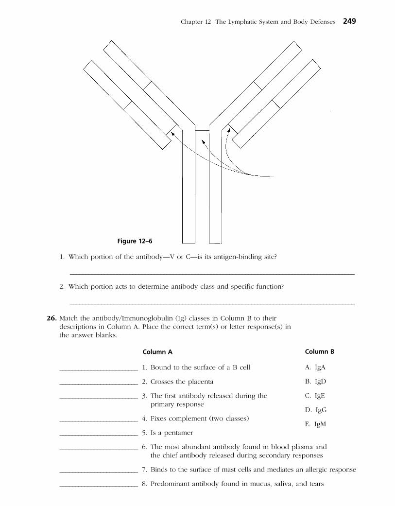
1. Which portion of the antibody—V or C—is its antigen-binding site?
___________________________________________________________________________________________
2. Which portion acts to determine antibody class and specific function?
___________________________________________________________________________________________
26. Match the antibody/Immunoglobulin (Ig) classes in Column B to their descriptions in Column A. Place the correct term(s) or letter response(s) in the answer blanks.
Column A
_________________________ 1. Bound to the surface of a B cell
_________________________ 2. Crosses the placenta
_________________________ 3. The first antibody released during the primary response
_________________________ 4. Fixes complement (two classes)
_________________________ 5. Is a pentamer
_________________________ 6. The most abundant antibody found in blood plasma and the chief antibody released during secondary responses
_________________________ 7. Binds to the surface of mast cells and mediates an allergic response
_________________________ 8. Predominant antibody found in mucus, saliva, and tears
Column B
A. IgA
B. IgD
C. IgE
D. IgG
E. IgM
Chapter 12 The Lymphatic System and Body Defenses 249
Figure 12–6

27. Complete the following descriptions of antibody function by writing the missing terms in the answer blanks.
Antibodies can inactivate antigens in various ways, depending on the nature of the (1) . Complement activation and (2) are the chief ammunitions used against cellular antigens such as bacteria and mis matched red blood cells. The binding of antibodies to sites on bacterial exotoxins or viruses that can cause cell injury is called (3) . The cross-linking of cellular antigens into large lattices by antibodies is called (4) ; Ig
(5) , with its 10 antigen binding sites, is particularly efficient in this mechanism. When molecules are cross-linked into lattices by antibodies, the mechanism is more properly called (6) . In virtually all these cases, the protective mechanism mounted by the anti bodies serves to disarm and/or immobilize the antigens until they can be disposed of by (7) .
28. Determine whether each of the following situations provides, or is an example of, active or passive immunity. If passive, write P in the blank; if active, write A in the blank.
______ 1. An individual receives Sabin polio vaccine.
______ 2. Antibodies migrate through a pregnant woman’s placenta into the vascular system of her fetus.
______ 3. A student nurse receives an injection of gamma globulin (containing antibodies to the hepatitis virus) after she has been exposed to viral hepatitis.
______ 4. This is a “borrowed” immunity.
______ 5. Immunological memory is provided.
______ 6. An individual suffers through chickenpox.
29. There are several important differences between primary and secondary immune response(s). If the following statements best describe a primary response, write P in the blank; if a secondary response, write S in the blank.
______ 1. The initial response to an antigen; gearing-up stage
______ 2. Lag period of several days before antibodies specific to the antigen appear in the bloodstream
______ 3. Antibody levels increase rapidly and remain high for an extended period
______ 4. Immunological memory established
______ 5. The second, third, and subsequent responses to the same antigen
250 Anatomy & Physiology Coloring Workbook
_________________________ 1.
_________________________ 2.
_________________________ 3.
_________________________ 4.
_________________________ 5.
_________________________ 6.
_________________________ 7.

Cellular (Cell-Mediated) Immune Response
30. Several populations of T cells exist. Match the terms in Column B to the descriptions in Column A. Place the correct term or letter response in the answer blanks.
Column A
_________________________ 1. Binds with and releases chemicals that activate B cells, T cells, and macrophages
_________________________ 2. Activated by recognizing both its antigen and a self-protein presented on the surface of a macrophage
_________________________ 3. Turns off the immune response when the “enemy” has been routed
_________________________ 4. Directly attacks and lyses cellular pathogens
_________________________ 5. Initiates secondary response to a recognized antigen
31. Using the key choices, select the terms that correspond to the descriptions of substances or events by inserting the appropriate term or letter in the answer blanks.
Key Choices
A. Anaphylactic shock D. Complement F. InflammationB. Antibodies E. Cytokines G. InterferonC. Chemotaxis factors
_________________________ 1. A protein released by macrophages and activated T cells that helps to protect other body cells from viral multiplication
_________________________ 2. Any types of molecules that attract neutrophils and other pro- tective cells into a region where an immune response is ongoing
_________________________ 3. Proteins released by plasma cells that mark antigens for destruc- tion by phagocytes or complement
_________________________ 4. A consequence of the release of histamine and of complement activation
_________________________ 5. C and G are examples of this class of molecules
_________________________ 6. A group of plasma proteins that amplifies the immune response by causing lysis of cellular pathogens once it has been “fixed” to their surface
_________________________ 7. Class of chemicals released by macrophages
Chapter 12 The Lymphatic System and Body Defenses 251
Column B
A. Helper T cell
B. Cytotoxic T cell
C. Regulatory T cell
D. Memory T cell

32. Organ transplants are often unsuccessful because self-proteins vary in differ-ent individuals. However, chances of success increase if certain important procedures are followed. The following questions refer to this important area of clinical medicine.
1. Assuming that autografts and isografts are not possible, what is the next most successful
graft type and what is its source? __________________________________________________________
2. What two cell types are important in rejection phenomena?
___________________________________________________________________________________________
3. Why are immunosuppressive drugs (or therapy) provided after transplant surgery, and what is the major shortcoming of this therapy?
___________________________________________________________________________________________
___________________________________________________________________________________________
___________________________________________________________________________________________
33. Figure 12–7 is a flowchart of the immune response that tests your understand-ing of the interrelationships of that process. Several terms have been omitted from this schematic.
Complete the figure by inserting appropriate terms from the key choices below. (Note that oval blanks indicate that the required term identifies a cell type and rectangular blanks represent the names of chemical molecules.)
Key Choices
Cell types: Molecules:
B cell AntibodiesHelper T cell Chemotactic factorsCytotoxic T cell ComplementMacrophage CytokinesMemory B cell InterferonMemory T cell PerforinNeutrophils Plasma cell
252 Anatomy & Physiology Coloring Workbook
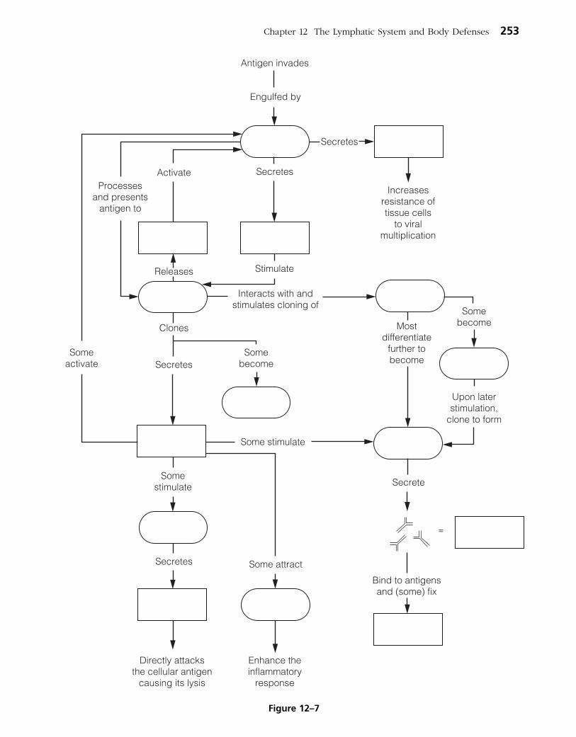
Chapter 12 The Lymphatic System and Body Defenses 253
Figure 12–7

Disorders of Immunity
34. Using the key choices, identify the type of immunity disorder described. Insert the appropriate term or letter in the answer blank.
Key Choices
A. Allergy/Hypersensitivity B. Autoimmune disease C. Immunodeficiency
_________________________ 1. AIDS and SCID
_________________________ 2. The immune system mounts an extraordinarily vigorous response to an otherwise harmless antigen
_________________________ 3. A hypersensitivity reaction
_________________________ 4. Occurs when the production or activity of immune cells or complement is abnormal
_________________________ 5. The body’s own immune system produces the disorder; a breakdown of self-tolerance
_________________________ 6. Affected individuals unable to combat infections that would present no problem for normally healthy people
_________________________ 7. Multiple sclerosis and rheumatic fever
_________________________ 8. Hay fever and contact dermatitis
_________________________ 9. Typical symptoms of the acute response: tearing, runny nose, and itching skin
DEVELOPMENTAL ASPECTS OF THE LYMPHATIC SYSTEM AND BODY DEFENSES
35. Complete the following statements concerning the development and operation of the immune system during the life span by inserting your answers in the answer blanks.
Lymphatic vessels that “bud” from developing (1) are visible by the fifth week of development. The first lymphoid organs to appear are the (2) and the (3) . Most other lymphoid organs are poorly formed before birth; their devel-opment is believed to be controlled by (4) hormones.
The earliest lymphocyte stem cells that can be identified appear during the first month of development in the fetal
(5) . Shortly thereafter, bone marrow becomes the lympho- cyte origin site; but after birth, lymphocyte proliferation occurs in the (6) . The development of immunocompetence has usually been accomplished by (7) .
254 Anatomy & Physiology Coloring Workbook
_________________________ 1.
_________________________ 2.
_________________________ 3.
_________________________ 4.
_________________________ 5.
_________________________ 6.
_________________________ 7.

During old age, the effectiveness of the immune system (8) , and elders are more at risk for (9) , (10) , and (11) . Part of the declining defenses may reflect the fact that (12) anti- bodies are unable to get to the mucosal surfaces where they carry out their normal protective role.
A Visualization Exercise for the Immune System
Something quite enormous and looking much like an octopus is nearly blocking the narrow tunnel just ahead.
36. Where necessary, complete statements by inserting the missing word(s) in the answer blanks.
For this journey, you are equipped with scuba gear before you are miniaturized and injected into one of your host’s lym-phatic vessels. He has been suffering with a red, raw “strep throat” and has swollen cervical lymph nodes. Your assign-ment is to travel into a cervical lymph node and observe the activities going on there that reveal that your host’s immune system is doing its best to combat the infection.
On injection, you enter the lymph with a “WHOOSH” and then bob gently in the warm yellow fluid. As you travel
along, you see what seem to be thousands of spherical bacteria and a few large globular (1) molecules that, no doubt, have been picked up by the tiny lymphatic capillaries. Shortly thereafter, a large dark mass, shaped like a kidney bean, looms just ahead. This has to be a (2) , you con-clude, and you dig in your wet suit pocket to find the waterproof pen and recording tablet.
As you enter the gloomy mass, the lymphatic stream becomes shallow and begins to flow sluggishly. So that you can explore this little organ fully, you haul yourself to your feet and begin to wade through the slowly moving stream. On each bank you see a huge ball of cells that have large nuclei and such a scant amount of cytoplasm that you can barely make it out. You write, “Sighted the spherical germinal centers composed of (3) .” As you again study one of the cell masses, you spot one cell that looks quite different and reminds you of a nest of angry hornets because it is furiously spewing out what seems to be a horde of tiny Y-shaped “bees.” “Ah ha,” you think, “another valuable piece of information.” You record, “Spotted a (4) making and releasing (5) .”
Chapter 12 The Lymphatic System and Body Defenses 255
_________________________ 8.
_________________________ 9.
_________________________ 10.
_________________________ 11.
_________________________ 12.
_________________________ 1.
_________________________ 2.
_________________________ 3.
_________________________ 4.
_________________________ 5.
INCREDIBLE JOURNEYThe basic unit of structure and function in the human body is the cell. Each ofa cell’s parts, or organelles, as well as the entire cell, is organized to perform aspecific function. Cells have the ability to metabolize, grow and reproduce,move, and respond to stimuli. The cells of the body differ in shape, size, andin specific roles in the body. Cells that are similar in structure and functionform tissues, which, in turn, construct the various body organs.
Student activities in this chapter include questions relating to the structure andfunction of the generalized animal cell and to the general arrangement oftissues and their contribution to the activities of the various body organs.
CELLS
Overview
1. Answer the following questions by inserting your responses in theanswer blanks.
_________________________ 1.
_________________________ 2.
_________________________ 3.
_________________________ 4.
_________________________ 5.
_________________________ 6.
_________________________ 7.
_________________________ 8.
_________________________ 9.
_________________________10.
33
CELLS ANDTISSUES3
1–4. Name the four elements that make up the bulk ofliving matter.
5. Name the single most abundant material or substancein living matter.
6. Name the trace element most important for makingbones hard.
7. Name the element, found in small amounts in thebody, that is needed to make hemoglobin for oxygentransport.
8–12. Although there are many specific “jobs” that certain cellsare able to do, name five functions common to all cells.
_________________________11.
_________________________12. �

That done, you turn your attention to scanning the rest of the landscape. Suddenly you let out an involuntary yelp. Some-thing quite enormous and looking much like an octopus is nearly blocking the narrow tunnel just ahead. Your mind whirls as it tries to figure out the nature of this cellular “beast” that appears to be guarding the channel. Then it hits you—this has to be a (6) on the alert for foreign invaders (more properly called (7) ), which it “eats” when it catches them. The giant cell roars, “Halt, stranger, and be recognized,” and you dig frantically in your pocket for your identification pass. As you drift toward the huge cell, you hold the pass in front of you, hands trembling because you know this cell could
liquefy you as quick as the blink of an eye. Again the cell bellows at you, “Is this some kind of a security check? I’m on the job, as you can see!” Frantically you shake your head “NO,” and the cell lifts one long tentacle and allows you to pass. As you squeeze by, the cell says, “Being inside, I’ve never seen my body’s outside. I must say, humans are a rather strange-looking lot!” Still shaking, you decide that you are in no mood for a chat and hurry along to put some distance between yourself and this guard cell.
Immediately ahead are what appear to be hundreds of the same type of cell sitting on every ledge and in every nook and cranny. Some are busily snagging and engulfing unfortunate strep bacteria that float too close. The slurping sound is nearly deafening. Then something grabs your attention: The surface of one of these cells is becoming dotted with some of the same donut-shaped chemi-cals that you see on the strep bacteria membranes; a round cell, similar, but not identical, to those you earlier saw in the germinal centers, is starting to bind to one of these “doorknobs.” You smile smugly because you know you have properly identified the octopus-like cells. You then record your observations as follows: “Cells like the giant cell just identified act as (8) presenters. I have just observed one in this role during its interaction with a helper (9) cell.”
You decide to linger a bit to see if the round cell becomes activated. You lean against the tunnel walls and watch quietly, but your wait is brief. Within minutes, the cell that was binding to the octopus-like cell begins to divide, and then its daughter cells divide again and again at a head-spinning pace. You write, “I have just witnessed the formation of a (10) of like cells.” Most of the daughter cells enter the lymph stream, but a few of them settle back and seem to go into a light sleep. You decide that the “napping cells” don’t have any role to play in helping get rid of your host’s present strep infection but instead will provide for immunologic (11) and become active at a later date.
You glance at your watch and wince as you realize that it is already 5 minutes past the time for your retrieval. You have already concluded that this is a dangerous place for those who don’t “belong” and are far from sure about how long your pass is good, so you swim hurriedly from the organ into the lymphatic stream to reach your pickup spot.
256 Anatomy & Physiology Coloring Workbook
_________________________ 6.
_________________________ 7.
_________________________ 8.
_________________________ 9.
_________________________ 10.
_________________________ 11.

37. A young man is rushed to the emergency room after fainting. His blood pressure is alarmingly low, and his companion reports the man collapsed shortly after being stung by a wasp. What has caused his hypotension? What treatment will be given immediately?
38. Patty Hourihan is a strict environmentalist and a new mother. Although she is very much against using disposable diapers, she is frustrated by the fact that her infant breaks out in a diaper rash when she uses cloth diapers. Considering that new cloth diapers do not cause the rash, but washed ones do, what do you think the problem is?
39. James, a 36-year-old engineer, appeared at the clinic in an extremely debil-itated condition. He had purple-brown lesions on his skin and a persistent cough. A physical examination revealed swollen lymph nodes. Laboratory tests revealed a low lymphocyte count. Information taken during the personal history revealed that James is homosexual. The skin lesions proved to be evi-dence of Kaposi’s sarcoma. What is James’s problem?
40. About 6 months after an automobile accident in which her neck was severely lacerated, a young woman comes to the clinic for a routine checkup. Visual examination shows a slight swelling just inferior to her larynx; her skin is dry and her face is puffy. When questioned, the woman reports that she fatigues easily, has been gaining weight, and her hair is falling out. What do you think is wrong?
Chapter 12 The Lymphatic System and Body Defenses 257
AT THE CLINIC
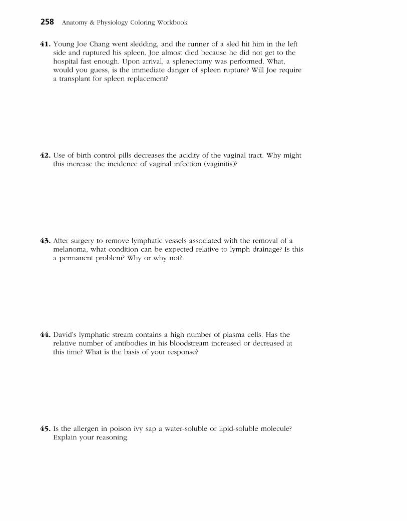
41. Young Joe Chang went sledding, and the runner of a sled hit him in the left side and ruptured his spleen. Joe almost died because he did not get to the hospital fast enough. Upon arrival, a splenectomy was performed. What, would you guess, is the immediate danger of spleen rupture? Will Joe require a transplant for spleen replacement?
42. Use of birth control pills decreases the acidity of the vaginal tract. Why might this increase the incidence of vaginal infection (vaginitis)?
43. After surgery to remove lymphatic vessels associated with the removal of a melanoma, what condition can be expected relative to lymph drainage? Is this a permanent problem? Why or why not?
44. David’s lymphatic stream contains a high number of plasma cells. Has the relative number of antibodies in his bloodstream increased or decreased at this time? What is the basis of your response?
45. Is the allergen in poison ivy sap a water-soluble or lipid-soluble molecule? Explain your reasoning.
258 Anatomy & Physiology Coloring Workbook
1. Statements that apply to lymphatic capillar-ies include the following:
A. The endothelial cells have continuous tight junctions.
B. They are open ended like straws.
C. Minivalves prevent the backflow of fluid into the interstitial spaces.
D. The endothelial cells are anchored by filaments to the surrounding structures.
2. Chyle flows into the:
A. lacteals.
B. intestinal lymph nodes.
C. intestinal trunk.
D. cisterna chyli.
3. Which parts of the lymph node show increased activity when antibody production is high?
A. Germinal centers
B. Outer follicles
C. Medullary cords
D. Sinuses
4. The classification lymphoid tissues includes:
A. the adenoids.
B. the spleen.
C. bone marrow.
D. the thyroid gland.
5. The spleen functions to:
A. remove aged red blood cells (RBCs).
B. house lymphocytes.
C. filter lymph.
D. store some blood components.
6. Which characteristics are associated with the thymus?
A. Providing immunocompetence
B. Hormone secretion
C. Hypertrophy in later life
D. Atrophy in later life
7. The tonsils:
A. have a complete epithelial capsule.
B. have crypts to trap bacteria.
C. filter lymph.
D. contain germinal centers.
8. Possible antigen-presenting cells (APCs) include:
A. dendritic cells.
B. Langerhans’ cells.
C. macrophages.
D. neutrophils.
9. Effector T cells secrete:
A. tumor necrosis factor.
B. histamine.
C. perforin.
D. interleukin 2.
10. Neutrophils die in the line of duty because:
A. they ingest infectious organisms.
B. their membranes become sticky and they are attacked by macrophages.
C. they secrete cellular toxins, which affect them in the same way they affect pathogens.
D. the buildup of tissue fluid pressure causes them to lyse.
Chapter 12 The Lymphatic System and Body Defenses 259
THE FINALE: MULTIPLE CHOICE
46. Select the best answer or answers from the choices given.

11. Macrophages:
A. form exudate.
B. present antigens.
C. secrete interleukin 1.
D. activate helper T cells.
12. Antibodies secreted in mother’s milk:
A. are IgG antibodies.
B. are IgA antibodies.
C. provide natural active immunity.
D. provide natural passive immunity.
13. Conditions for which passive artificial immunity is the treatment of choice include:
A. measles.
B. botulism.
C. rabies.
D. venomous snakebite.
14. Which of these antibody classes is often arranged as a dimer?
A. IgG
B. IgM
C. IgA
D. IgD
15. Which of the following antibody capabilities causes a transfusion reaction with A or B blood cell antigens?
A. Neutralization
B. Precipitation
C. Complement fixation
D. Agglutination
16. Which of the following terms is applicable to the use of part of the patient’s great saphenous vein in coronary bypass surgery?
A. Isograft
B. Xenograft
C. Allograft
D. Autograft
17. “Who” or “what” does the selecting that initiates clonal selection?
A. Antigen C. T cell
B. Antibody D. B cell
18. The cell type most often invaded by the HIV virus is:
A. helper T cell.
B. plasma cell.
C. cytotoxic T cell.
D. B cell.
260 Anatomy & Physiology Coloring Workbook

THE RESPIRATORY SYSTEM13
Body cells require an abundant and continuous supply of oxygen to carry out their activities. As cells use oxygen, they release carbon dioxide, a waste product that must be eliminated from the body. The circulatory and respiratory systems are intimately involved in obtaining and delivering oxygen to body cells and in eliminating carbon dioxide from the body. The respiratory system is responsible for gas exchange between the pulmonary blood and the external environment (that is, external respiration). The respiratory system also plays an important role in maintaining the acid-base balance of the blood.
Questions and activities in this chapter consider both the anatomy and physiology of the respiratory system structures.
FUNCTIONAL ANATOMY OF THE RESPIRATORY SYSTEM
1. The respiratory system is divisible into conducting zone and respiratory zone structures.
1. Name the conducting zone structures. ______________________________________________
____________________________________________________________________________________________
2. What is their common function? ____________________________________________________
____________________________________________________________________________________________
3. Name the respiratory zone structures. _______________________________________________
____________________________________________________________________________________________
261

2. The following questions refer to the main bronchi. In the spaces provided, insert the letter R to indicate the right main bronchus and the letter L to indi-cate the left main bronchus.
1. Which of the main bronchi is larger in diameter? ____________________________________
2. Which of the main bronchi is more horizontal? ____________________________________
3. Which of the main bronchi is the most common site for lodging of a foreign object
that has entered the respiratory passageways? ____________________________________
3. Complete the following statements by inserting your answers in the answer blanks.
Air enters the nasal cavity of the respiratory system through the (1) . The nasal cavity is divided by the midline nasal
(2) . The nasal cavity mucosa has several functions. Its major functions are to (3) , (4) , and (5) the incoming air. Mucous membrane–lined cavities called (6) are found in several bones surrounding the nasal cavities. They make the skull less heavy and probably act as resonance chambers for
(7) . The passageway common to the digestive and respira- tory systems, the (8) , is often referred to as the throat; it connects the nasal cavity with the (9) below. Clusters of lymphatic tissue, (10) , are part of the defensive system of the body. Reinforcement of the trachea with (11) rings prevents its collapse during (12) changes that occur during breathing. The fact that the rings are incomplete posteriorly allows a food bolus to bulge (13) during its transport to the stomach. The larynx or voice box is built from many carti-lages, but the largest is the (14) cartilage. Within the larynx are the (15) , which vibrate with exhaled air and allow an individual to (16) .
_________________________ 14.
_________________________ 15.
_________________________ 16.
4. Circle the term that does not belong in each of the following groupings. Then, fill in the answer blanks with the correct group name.
1. Sphenoidal Maxillary Mandibular Ethmoidal Frontal Group: ___________
2. Nasal cavity Trachea Alveolus Larynx Bronchus Group: ___________
3. Apex Base Hilum Larynx Pleura Group: ___________
4. Sinusitis Peritonitis Pleurisy Tonsillitis Laryngitis Group: ___________
5. Laryngopharynx Oropharynx Transports air and food Nasopharynx Group: ___________
6. Alveoli Respiratory zone Alveolar sac Main bronchus Group: ___________
262 Anatomy & Physiology Coloring Workbook
_________________________ 1.
_________________________ 2.
_________________________ 3.
_________________________ 4.
_________________________ 5.
_________________________ 6.
_________________________ 7.
_________________________ 8.
_________________________ 9.
_________________________ 10.
_________________________ 11.
_________________________ 12.
_________________________ 13.

5. Figure 13–1 is a sagittal view of the upper respiratory structures. (A) Correctly identify all structures provided with leader lines on the figure. (B) Select different colors for the structures listed below and use them to
color in the coding circles and the corresponding structures on the figure.
○ Nasal cavity ○ Larynx ○ Thyroid cartilage
○ Pharynx ○ Paranasal sinuses ○ Cricoid cartilage
○ Trachea
Chapter 13 The Respiratory System 263
Figure 13–1

6. Using the key choices, select the terms identified in the following descriptions by inserting the appropriate term or letter in the answer blanks. Not all terms will be used.
Key Choices
A. Alveoli D. Epiglottis G. Palate J. Main bronchi M. Vocal cordsB. Bronchioles E. Esophagus H. Parietal pleura K. TracheaC. Conchae F. Glottis I. Phrenic L. Visceral pleura
_________________________ 1. Smallest conducting respiratory passageways
_________________________ 2. Separates the oral and nasal cavities
_________________________ 3. Major nerve, stimulating the diaphragm
_________________________ 4. Food passageway posterior to the trachea
_________________________ 5. Closes off the larynx during swallowing
_________________________ 6. Windpipe
_________________________ 7. Actual site of gas exchanges
_________________________ 8. Pleural layer covering the thorax walls
_________________________ 9. Pleural layer covering the lungs
_________________________ 10. Opening between vocal folds
_________________________ 11. Fleshy lobes in the nasal cavity which increase its surface area
_________________________ 12. Vibrate with expired air
7. Complete the following paragraph concerning the alveolar cells and their roles by writing the missing terms in the answer blanks.
With the exception of the stroma of the lungs, which is (1) connective tissue, the lungs are mostly air spaces, of which the alveoli compose the greatest part. The bulk of the alveolar walls are made up of squamous epithelial cells, which are well-suited for their (2) exchange function. Much less numerous cuboidal cells produce a fluid that coats the air-exposed surface of the alveolus and contains a lipid-based molecule called (3) that functions to (4) the surface tension of the alveolar fluid.
264 Anatomy & Physiology Coloring Workbook
_________________________ 1.
_________________________ 2.
_________________________ 3.
_________________________ 4.

8. Figure 13–2 is a diagram of the larynx and associated structures. (A) Select a different color for each of the structures listed below and use it
to color in the coding circles and the corresponding structures on the figure. (B) Answer the questions following the diagram.
○ Hyoid bone ○ Tracheal cartilages ○ Cricoid cartilage
○ Thyroid cartilage ○ Epiglottis
1. What are three functions of the larynx? ____________________________________________________
___________________________________________________________________________________________
___________________________________________________________________________________________
2. What type of cartilage forms the epiglottis? ________________________________________________
3. What type of cartilage forms the other eight laryngeal cartilages? ___________________________
4. Explain this difference. ____________________________________________________________________
___________________________________________________________________________________________
___________________________________________________________________________________________
5. What is the common name for the anterior protrusion of the thyroid cartilage? _____________
Chapter 13 The Respiratory System 265
Ligaments
Figure 13–2

9. Figure 13–3 shows a cross section through the trachea. (A) Label the layers indicated by the leader lines. (B) Color the following:
•Mucosa(includingthecilia,epithelium,laminapropria):lightpink. •Areacontainingthesubmucosalseromucousglands:purple. •Hyalinecartilagering:blue. •Trachealismuscle:orange. •Adventitia:yellow. (C) Respond to the questions following the figure.
1. Which important role is played by the cartilage rings that reinforce the trachea?
___________________________________________________________________________________________
2. Of what importance is the fact that the cartilage rings are incomplete posteriorly?
___________________________________________________________________________________________
3. What occurs when the trachealis muscle contracts, and in what activities might this action
be very helpful? ___________________________________________________________________________
___________________________________________________________________________________________
266 Anatomy & Physiology Coloring Workbook
Figure 13–3
Esophagus
Lumen oftrachea
Posterior
Anterior
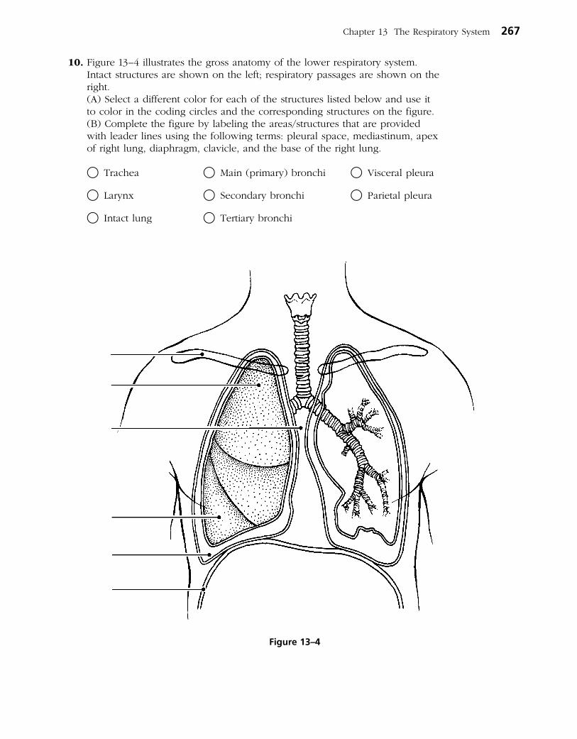
10. Figure 13–4 illustrates the gross anatomy of the lower respiratory system. Intact structures are shown on the left; respiratory passages are shown on the right.
(A) Select a different color for each of the structures listed below and use it to color in the coding circles and the corresponding structures on the figure.
(B) Complete the figure by labeling the areas/structures that are provided with leader lines using the following terms: pleural space, mediastinum, apex of right lung, diaphragm, clavicle, and the base of the right lung.
○ Trachea ○ Main (primary) bronchi ○ Visceral pleura
○ Larynx ○ Secondary bronchi ○ Parietal pleura
○ Intact lung ○ Tertiary bronchi
Chapter 13 The Respiratory System 267
Figure 13–4

11. Complete the items below: (A) Figure 13–5 illustrates the microscopic structure of the respiratory unit of
lung tissue. The external anatomy is shown in Figure 13–5A. Color the following: •Intactalveoli:yellow. •Pulmonarycapillaries:red. •Respiratorybronchioles:green. (B) A cross-section through an alveolus is shown on Figure 13–5B, and a
blowup of the respiratory membrane is shown in Figure 13–5C. On these illustrations, color the following:
•Alveolarepithelium:yellow. •Capillaryendothelium:pink. •Redbloodcellsinthecapillary:red. •Alveolarchamber:paleblue. (C) Label the alveolar chamber in Figure 13-5B. (D) In Figure 13–5C: •Labeltheregionofthefusedbasementmembranes. •Addthesymbolsforoxygengas(O2) and carbon dioxide gas (CO2) in the
sites where they would be in higher concentration and arrows correctly showing their direction of movement through the respiratory membrane.
•Namethecomponentsoftherespiratorymembraneinthespaceprovided.
268 Anatomy & Physiology Coloring Workbook
B
C
A
Respiratory membrane
Figure 13–5

RESPIRATORY PHYSIOLOGY
12. Using the key choices, select the terms identified in the following descrip -tions by inserting the appropriate term or letter in the answer blanks.
Key Choices
A. Atmospheric pressure B. Intrapulmonary pressure C. Intrapleural pressure
_________________________ 1. In healthy lungs, it is always lower than atmospheric pressure (that is, it is negative pressure)
_________________________ 2. Pressure of air outside the body
_________________________ 3. As it decreases, air flows into the passageways of the lungs
_________________________ 4. As it increases over atmospheric pressure, air flows out of the lungs
_________________________ 5. If this pressure becomes equal to the atmospheric pressure, the lungs collapse
_________________________ 6. Rises well over atmospheric pressure during a forceful cough
_________________________ 7. Also known as intra-alveolar pressure
13. Many changes occur within the lungs as the diaphragm (and external inter-costal muscles) contract and then relax. These changes lead to the flow of air into and out of the lungs. The activity of the diaphragm is given in the left column of the following table. Several changes in condition are listed in the column heads to the right. Complete the table by checking (✓) the appropri-ate column to correctly identify the change that would be occur ring relative to the diaphragm’s activity in each case.
Activity of Changes diaphragm in
Internal Internal volume pressure Size Direction of thorax in thorax of lungs of airflow
(↑ = increased) ↑ ↓ ↑ ↓ ↑ ↓ Into Out of (↓ = decreased) lung lung
Contracted, moves downward
Relaxed, moves superiorly
Chapter 13 The Respiratory System 269

14. Use the key choices to respond to the following descriptions. Insert the cor-rect term or letter in the answer blanks.
Key Choices
A. External respiration C. Inspiration E. Ventilation (breathing)B. Expiration D. Internal respiration
_________________________ 1. Period of breathing when air enters the lungs
_________________________ 2. Exchange of gases between the systemic capillary blood and body cells
_________________________ 3. Alternate flushing of air into and out of the lungs
_________________________ 4. Exchange of gases between alveolar air and pulmonary capillary blood
_________________________ 5. Period of breathing when air leaves the lungs
15. Although normal quiet expiration is largely passive because of lung recoil, when expiration must be more forceful (or the lungs are diseased), muscles that increase the abdominal pressure or depress the rib cage are enlisted.
1. Provide two examples of muscles that cause abdominal pressure to rise.
___________________________________________ and ___________________________________________
2. Provide two examples of muscles that depress the rib cage.
___________________________________________ and ___________________________________________
16. Four nonrespiratory movements are described here. Identify each by insert -ing your answers in the spaces provided.
1. Sudden inspiration, resulting from spasms of the diaphragm. _______________________________
2. A deep breath is taken, the glottis is closed, and air is forced out of the lungs against
the glottis; clears the lower respiratory passageways. ______________________________________
3. As just described, but it clears the upper respiratory passageways. __________________________
4. Very deep inspiration, taken with jaws open; increases ventilation of the lungs. _____________
270 Anatomy & Physiology Coloring Workbook

17. The following section concerns respiratory volume measurements. Using the key choices, select the terms identified in the following descriptions by insert-ing the appropriate term or letter in the answer blanks. Not all terms will be used.
Key Choices
A. Dead space volume C. Inspiratory reserve volume (IRV) E. Tidal volume (TV)B. Expiratory reserve D. Residual volume (RV) F. Vital capacity (VC)
volume (ERV)
_________________________ 1. Respiratory volume inhaled or exhaled during normal breathing
_________________________ 2. Air in respiratory passages that does not contribute to gas exchange
_________________________ 3. Total amount of exchangeable air
_________________________ 4. Gas volume that allows gas exchange to go on continuously
_________________________ 5. Amount of air that can still be exhaled (forcibly) after a normal exhalation
18. Figure 13–6 is a diagram showing respiratory volumes. Complete the figure by making the following additions.
1. Bracket the volume representing the vital capacity and color the area yellow; label it VC.
2. Add green stripes to the area representing the inspiratory reserve volume and label it IRV.
3. Add red stripes to the area representing the expiratory reserve volume and label it ERV.
4. Identify and label the tidal volume, which is now just yellow.
5. Color the residual volume (RV) blue and label it appropriately on the figure.
6. Bracket and label the inspiratory capacity (IC).
Chapter 13 The Respiratory System 271
Figure 13–6

19. Use the key choices to correctly complete the following statements, which refer to gas exchanges in the body. Insert the correct letter response in the answer blanks.
Key Choices
A. Active transport F. DiffusionB. Air of alveoli to capillary blood G. Higher concentrationC. Carbon dioxide–poor and oxygen-rich H. Lower concentrationD. Capillary blood to alveolar air I. Oxygen-poor and carbon dioxide–richE. Capillary blood to tissue cells J. Tissue cells to capillary blood
______ 1.
______ 2.
______ 3.
______ 4.
______ 5.
______ 6. ______ 7. ______ 8. ______ 9.
20. Complete the following statements by inserting your answers in the answer blanks.
Most oxygen is transported bound to (1) inside the red blood cells. Conversely, most carbon dioxide is carried in the form of (2) in the (3) . Carbon monoxide poisoning is lethal because carbon monoxide competes with (4) for binding sites.
21. Circle the term that does not belong in each of the following groupings. Then, fill in the answer blanks with the correct group name.
1. ↑Respiratory rate ↓In blood CO2 Alkalosis Acidosis Group: ___________
2. ↑H+ ↑Carbonic acid ↓pH ↑pH Group: ___________
3. Acidosis Hyperventilation Hypoventilation CO2 buildup Group: ___________
4. Apnea Cyanosis ↑Oxygen ↓Oxygen Group: ___________
5. ↑Respiratory rate ↑Exercise Anger ↑CO2 in blood Group: ___________
6. High altitude ↑PCO2 ↓Atmospheric pressure Group: ___________
All gas exchanges are made by (1) . When substances pass in this manner, they move from areas of their (2) to areas of their (3) . Thus, oxygen con-tinually passes from the (4) and then from the (5) . Conversely, carbon dioxide moves from the (6) and from (7) . From there, it passes out of the body during expiration. As a result of such exchanges, arterial blood tends to be (8) whereas venous blood is (9) .
_________________________ 1.
_________________________ 2.
_________________________ 3.
_________________________ 4.
272 Anatomy & Physiology Coloring Workbook
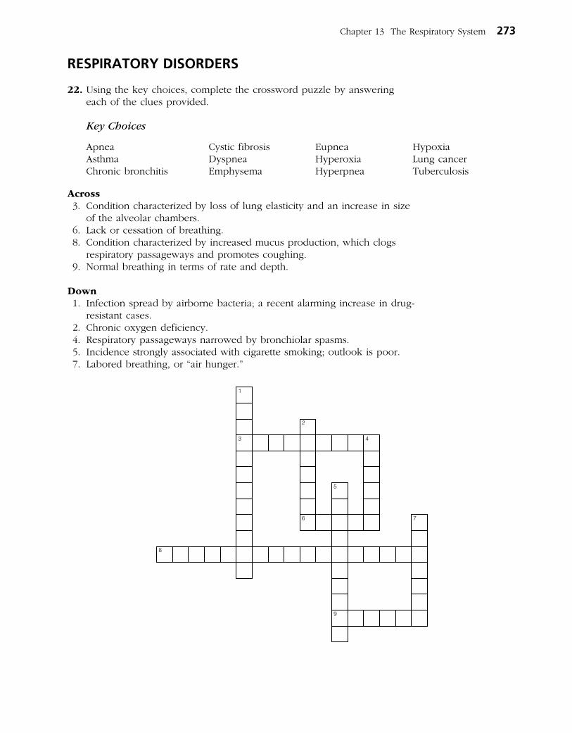
RESPIRATORY DISORDERS
22. Using the key choices, complete the crossword puzzle by answering each of the clues provided.
Key Choices
Apnea Cystic fibrosis Eupnea HypoxiaAsthma Dyspnea Hyperoxia Lung cancerChronic bronchitis Emphysema Hyperpnea Tuberculosis
Across 3. Condition characterized by loss of lung elasticity and an increase in size
of the alveolar chambers. 6. Lack or cessation of breathing. 8. Condition characterized by increased mucus production, which clogs
respiratory passageways and promotes coughing. 9. Normal breathing in terms of rate and depth.
Down 1. Infection spread by airborne bacteria; a recent alarming increase in drug-
resistant cases. 2. Chronic oxygen deficiency. 4. Respiratory passageways narrowed by bronchiolar spasms. 5. Incidence strongly associated with cigarette smoking; outlook is poor. 7. Labored breathing, or “air hunger.”
1
2
3 4
5
6 7
8
9
Chapter 13 The Respiratory System 273

DEVELOPMENTAL ASPECTS OF THE RESPIRATORY SYSTEM
23. Mrs. Jones gave birth prematurely to her first child. At birth, the baby weighed 2 lb 8 oz. Within a few hours, the baby had developed severe dyspnea and was becoming cyanotic. Therapy with a positive pressure ventilator was prescribed. Answer the following questions related to the situation just described. Place your responses in the answer blanks.
1. The infant’s condition is referred to as ____________________________________________________
2. It occurs because of a relative lack of ____________________________________________________
3. The function of the deficient substance is to ______________________________________________
___________________________________________________________________________________________
4. Explain what the positive pressure apparatus accomplishes. _______________________________
___________________________________________________________________________________________
24. Complete the following statements by inserting your answers in the answer blanks.
The respiratory rate of a newborn baby is approximately (1) respirations per minute. In a healthy adult, the respira-
tory range is (2) respirations per minute. Most problems that interfere with the operation of the respiratory system fall into one of the following categories: infections such as pneumonia, obstructive conditions such as (3) and (4) , and/or conditions that destroy lung tissue, such as (5) . With age, the lungs lose their (6) , and the (7) of the lungs decreases. Protective mechanisms also become less efficient, causing elderly individuals to be more susceptible to respiratory infections, particularly (8) .
_________________________ 1.
_________________________ 2.
_________________________ 3.
_________________________ 4.
_________________________ 5.
_________________________ 6.
_________________________ 7.
_________________________ 8.
274 Anatomy & Physiology Coloring Workbook
INCREDIBLE JOURNEY
A Visualization Exercise for the Respiratory System
You carefully begin to pick your way down, using cartilages as steps.
25. Where necessary, complete statements by inserting the missing word(s) in the answer blanks.
Your journey through the respiratory system is to be on foot. To begin, you simply will walk into your host’s external nares. You are miniaturized, and your host is sedated lightly to prevent sneez-ing during your initial observations in the nasal cavity and subsequent descent.
You begin your exploration of the nasal cavity in the right nostril. One of the first things you notice is that the chamber is very warm and humid. High above, you see three large, round lobes,

the nasal (1) , which provide a large mucosal surface area for warming and moistening the entering air. As you walk toward the rear of this chamber, you see a large lumpy mass of lymphatic tissue, the pharyngeal (2) in the (3) , or first portion of the pharynx. As you peer down the pharynx, you realize that it will be next to impossible to maintain your footing during the next part of your journey. It is nearly straight down, and the (4) secretions are like grease. You sit down and dig your heels in to get started. After a quick slide, you land abruptly on one of a pair of flat, sheetlike structures that begin to vibrate rapidly, bouncing you up and down helplessly. You are also conscious of a rhythmic hum during this jostling, and you realize that you have landed on a (5) . You pick yourself up and look over the superior edge of the (6) , down into the seemingly endless esopha-gus behind. You chastise yourself for not remembering that the (7) and respiratory pathways separate at this point. Hanging directly over your head is the leaflike (8) carti-lage. Normally, you would not have been able to get this far because it would have closed off this portion of the respira-tory tract. With your host sedated, however, that protective reflex does not work.
You carefully begin to pick your way down, using the cartilages as steps. When you reach the next respiratory organ, the (9) , your descent becomes much easier, because the structure’s C-shaped cartilages form a ladder-like support-ing structure. As you climb down the cartilages, your face is stroked rhythmically by soft cellular extensions, or (10) . You remember that their function is to move mucus laden with bacteria or dust and other debris toward the (11) .
You finally reach a point where the descending passageway splits into two (12) , and because you want to control your progress (rather than slide downward), you choose the more horizontal (13) branch. If you remain in the superior por-tion of the lungs, your return trip will be less difficult because the passageways will be more horizontal than steeply verti-cal. The passage ways get smaller and smaller, slowing your progress. As you are squeezing into one of the smallest of the respiratory passageways, a (14) , you see a bright spheri-
cal chamber ahead. You scramble into this (15) , pick yourself up, and survey the area. Scattered here and there are lumps of a substance that look suspiciously like coal, reminding you that your host is a smoker. As you stand there, a soft rustling wind seems to flow in and out of the chamber. You press your face against the transparent chamber wall and see disclike cells, (16) , passing by in the capillaries on the other side. As you watch, they change from a somewhat bluish color to a bright (17) color as they pick up (18) and unload (19) .
You record your observations and then contact headquarters to let them know you are ready to begin your ascent. You begin your return trek, slipping and sliding as you travel. By the time you reach the inferior edge of the trachea, you are ready for a short break. As you rest on the mucosa, you begin to notice that the air is becoming close and very heavy. You pick yourself up quickly and begin to scramble up the trachea. Suddenly and without warning, you are hit by a huge wad of mucus and catapulted upward and out onto your host’s freshly pressed handkerchief! Your host has assisted your exit with a (20) .
Chapter 13 The Respiratory System 275
_________________________ 1.
_________________________ 2.
_________________________ 3.
_________________________ 4.
_________________________ 5.
_________________________ 6.
_________________________ 7.
_________________________ 8.
_________________________ 9.
_________________________ 10.
_________________________ 11.
_________________________ 12.
_________________________ 13.
_________________________ 14.
_________________________ 15.
_________________________ 16.
_________________________ 17.
_________________________ 18.
_________________________ 19.
_________________________ 20.

276 Anatomy & Physiology Coloring Workbook
26. After a long bout of bronchitis, Ms. Dupee complains of a stabbing pain in her side with each breath. What is her probable condition?
27. The Kozloski family is taking a long auto trip. Michael, who has been riding in the back of the station wagon, complains of a throbbing headache. A little later, he seems confused and his face is flushed. What is your diagnosis of Michael’s problem?
28. A new mother checks on her sleeping infant son, only to find that he has stopped breathing and is turning blue. The mother quickly picks up the baby and pats his back until he starts to breathe. What tragedy has been averted?
29. Joanne Willis, a long-time smoker, is complaining that she has developed a persistent cough. What is your first guess as to her condition? What has happened to her bronchial cilia?
30. Barbara is rushed to the emergency room after an auto accident. The 8th through 10th ribs on her left side have been fractured and have punctured the lung. What term is used to indicate lung collapse? Will both lungs collapse? Why or why not?
31. A young boy is diagnosed with cystic fibrosis. What effect will this have on his respiratory system?
AT THE CLINIC
32. Mr. and Ms. Rao took their sick 5-year-old daughter to the doctor. The girl was breathing entirely through her mouth, her voice sounded odd and whiny, and a puslike fluid was dripping from her nose. Which one of the tonsils was most likely infected in this child?
33. Assume you are a second-year nursing student. As your assignment, you are asked to explain how a history of heavy smoking might interfere with a patient’s gas exchange.
34. Why does an emergency medical technician (EMT) administering a Breathalyzer test for alcohol ask the person being tested to expel one deep breath instead of several shallow ones?
35. The cilia lining the respiratory passageways superior to the larynx beat inferiorly whereas those lining the larynx and below beat superiorly. What is the functional “reason” for this difference?
Chapter 13 The Respiratory System 277
THE FINALE: MULTIPLE CHOICE
1. Structures that are part of the respiratory zone include:
A. terminal bronchioles.
B. respiratory bronchioles.
C. tertiary bronchi.
D. alveolar ducts.
2. Which structures are associated with the production of speech?
A. Cricoid cartilage
B. Arytenoid cartilage
C. Glottis
D. Pharynx
3. The skeleton of the external nose consists of:
A. cartilage and bone.
B. bone only.
C. hyaline cartilage only.
D. elastic cartilage only.
4. Which of the following is not part of the conducting zone of the respiratory system?
A. Pharynx D. Lobar bronchi
B. Alveolar sac E. Larynx
C. Trachea
36. Select the best answer or answers from the choices given.
278 Anatomy & Physiology Coloring Workbook
5. Select the single false statement about the true vocal cords.
A. They are the same as the vocal folds.
B. They attach to the arytenoid cartilages via the vocal ligaments.
C. Exhaled air flowing through the glottis vibrates them to produce sound.
D. They are also called the vestibular folds.
6. The function of the cuboid cells of the alveolar walls is:
A. to produce surfactant.
B. to propel mucous sheets.
C. phagocytosis of dust particles.
D. to allow rapid diffusion of respiratory gases.
7. An examination of a lobe of the lung reveals many branches off the main pass age way. These branches are:
A. main bronchi. C. tertiary bronchi.
B. lobar bronchi. D. segmental bronchi.
8. An alveolar sac:
A. is an alveolus.
B. relates to an alveolus as a bunch of grapes relates to one grape.
C. is a huge, saclike alveolus in an emphysema patient.
D. is the same as an alveolar duct.
9. The respiratory membrane (air-blood barrier) consists of:
A. squamous cells, basal membranes, endo-thelial cells.
B. air, connective tissue, lung.
C. squamous and cuboidal epithelial cells and macrophages.
D. pseudostratified epithelium, lamina pro-pria, capillaries.
10. Oxygen and carbon dioxide are exchanged in the lungs and through all cell membranes by:
A. active transportation.
B. diffusion.
C. filtration.
D. osmosis.
11. Which of the following are characteristic of a bronchopulmonary segment?
A. Removal causes collapse of adjacent segments
B. Fed by a tertiary bronchus
C. Supplied by its own branches of the pulmonary artery and vein
D. Separated from other segments by its septum
12. During inspiration, intrapulmonary pressure is:
A. greater than atmospheric pressure.
B. less than atmospheric pressure.
C. greater than intrapleural pressure.
D. less than intrapleural pressure.
13. When the inspiratory muscles contract,
A. the size of the thoracic cavity increases in diameter.
B. the size of the thoracic cavity increases in length.
C. the volume of the thoracic cavity decreases.
D. the size of the thoracic cavity increases in both length and diameter.
14. Lung collapse is prevented by:
A. high surface tension of alveolar fluid.
B. high surface tension of pleural fluid.
C. high pressure in the pleural cavities.
D. high elasticity of lung tissue.
15. Resistance is increased by:
A. epinephrine.
B. parasympathetic stimulation.
C. inflammatory chemicals.
D. contraction of the trachealis muscle.
16. Which of the following changes accom panies the loss of elasticity associated with aging?
A. Increase in tidal volume
B. Increase in inspiratory reserve volume
C. Increase in residual volume
D. Increase in vital capacity

THE DIGESTIVE SYSTEM AND BODY METABOLISM
14
The digestive system processes food so that it can be absorbed and used by the body’s cells. The digestive organs are responsible for food ingestion, digestion, absorption, and elimination of undigested remains from the body. In one sense, the digestive tract can be viewed as a disassembly line in which food is carried from one stage of its breakdown process to the next by muscular activity, and its nutrients are made available en route to the cells of the body. In addition, the digestive system provides for one of life’s greatest pleasures—eating.
The anatomy of both alimentary canal and accessory digestive organs, mechanical and enzymatic breakdown, and absorption mechanisms are covered in this chapter. An introduction to nutrition and some important understandings about cellular metabolism (utilization of foodstuffs by body cells) are also considered in this chapter review.
ANATOMY OF THE DIGESTIVE SYSTEM
1. Complete the following statements by inserting your answers in the answer blanks.
The digestive system is responsible for many body processes. Its functions begin when food is taken into the mouth, or
(1) . The process called (2) occurs as food is broken down both chemically and mechanically. For the brokendown foods to be made available to the body cells, they must be absorbed through the digestive system walls into the (3) . Undigestible food remains are removed, or (4) , from the body in (5) . The organs forming a continuous tube from the mouth to the anus are collectively called the (6) . Organs located outside the digestive tract proper, which secrete their products into the digestive tract, are referred to as (7) digestive system organs.
_________________________ 1.
_________________________ 2.
_________________________ 3.
_________________________ 4.
_________________________ 5.
_________________________ 6.
_________________________ 7.
279

2. Figure 14–1 is a frontal view of the digestive system. (A) Correctly identify all structures provided with leader lines. (B) Select different colors for the following organs and color the
coding circles and the corresponding struc tures of the figure.
○ Esophagus ○ Pancreas ○ Tongue
○ Liver ○ Salivary glands ○ Uvula
○ Large intestine ○ Small intestine ○ Stomach
280 Anatomy & Physiology Coloring Workbook
Trachea
Diaphragm
Figure 14–1
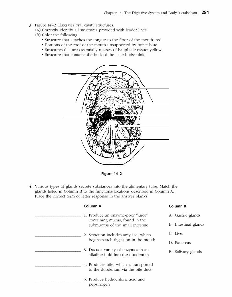
3. Figure 14–2 illustrates oral cavity structures. (A) Correctly identify all struc tures provided with leader lines. (B) Color the following: •Structurethatattachesthetonguetothefloorofthemouth:red. •Portionsoftheroofofthemouthunsupportedbybone:blue. •Structuresthatareessentiallymassesoflymphatictissue:yellow. •Structurethatcontainsthebulkofthetastebuds:pink.
4. Various types of glands secrete substances into the alimentary tube. Match the glands listed in Column B to the functions/locations described in Column A. Place the correct term or letter response in the answer blanks.
Column A
_________________________ 1. Produce an enzymepoor “juice” containing mucus; found in the submucosa of the small intestine
_________________________ 2. Secretion includes amylase, which begins starch digestion in the mouth
_________________________ 3. Ducts a variety of enzymes in an alkaline fluid into the duodenum
_________________________ 4. Produces bile, which is transported to the duodenum via the bile duct
_________________________ 5. Produce hydrochloric acid and pepsinogen
Column B
A. Gastric glands
B. Intestinal glands
C. Liver
D. Pancreas
E. Salivary glands
Chapter 14 The Digestive System and Body Metabolism 281
Figure 14–2

5. Using the key choices, complete the crossword puzzle by answering each of the clues provided.
Key Choices
Anal canal Lesser omentum RugaeAppendix Mesentery Small intestineColon Microvilli Soft palateEsophagus Oral cavity StomachGreater omentum Parietal peritoneum TongueHard palate Pharynx VestibuleHaustra Plicae circulares VilliIleocecal valve Pyloric sphincter (valve) Visceral peritoneum
Across 7. Anterosuperior boundary of the oral cavity; supported by bone. 8. Region, containing two sphincters, through which feces are expelled
from the body. 10. Stomach and ______ are anatomical regions involved in the mechanical
breakdown of food. 11. Membrane attached to the lesser curvature of the stomach. 13. Projections of the plasma membrane of a cell that increase the cell’s
surface area. 14. Area between the teeth and lips/cheeks. 15. Prevents food from moving back into the small intestine once it has
entered the large intestine. 16. Saclike outpocketings of the large intestine wall. 18. Serosa of the abdominal cavity wall. 21. Folds of the stomach mucosa. 23. Common passage for food and air.
Down 1. Blind sac hanging from the initial part of the colon. 2. Uvula hangs from its posterior edge. 3. Mesentery, lesser omentum, and _______ are three extensions/modifications
of the peritoneum. 4. Muscular tube; has no digestive or absorptive role. 5. Organ primarily involved in water absorption and feces formation. 6. Folds of the small intestine wall. 9. Sphincter controlling the movement of food from the stomach into the
duodenum. 12. Organ responsible for most food and water absorption. 14. Serous membrane forming part of the wall of the small intestine. 17. Fingerlike extensions of the intestinal mucosa that increase the surface area. 19. Organ that mixes food in the mouth. 20. Structure that suspends the small intestine from the posterior body wall. 22. Organ in which protein digestion begins.
282 Anatomy & Physiology Coloring Workbook

1 2 3
4
5
6 7
8
9
10
11 12
13
14
15
16
17
18 19 20
21
22
23
Chapter 14 The Digestive System and Body Metabolism 283

6. Circle the term that does not belong in each of the following groupings. Then, fill in the answer blanks with the correct group name.
1. Nasopharynx Esophagus Laryngopharynx Oropharynx Group: ___________
2. Villi Plicae circulares Rugae Microvilli Group: ___________
3. Salivary glands Pancreas Liver Gallbladder Group: ___________
4. Duodenum Cecum Jejunum Ileum Group: ___________
5. Ascending colon Haustra Circular folds Cecum Group: ___________
6. Mesentery Frenulum Greater omentum Parietal peritoneum Group: ___________
7. Parotid Sublingual Submandibular Palatine Group: ___________
8. Proteindigesting enzymes Saliva Intrinsic factor HCl Group: ___________
9. Colon Water absorption Protein absorption Vitamin B absorption Group: ___________
7. Figure 14–3A is a longitudinal section of the stomach. (A) Use the following terms to identify the regions provided with leader lines
on the figure.
Body Pyloric region Greater curvature Cardioesophageal sphincter
Fundus Pyloric valve Lesser curvature
(B) Select different colors for each of the following structures/areas and use them to color the coding circles and corresponding structures/areas on the figure.
○ Oblique muscle layer ○ Longitudinal muscle layer ○ Circular muscle layer
○ Area where rugae are visible ○ Serosa
(C) Figure 14–3B shows two types of secretory cells found in gastric glands. Identify the third type called chief cells by choosing a few cells deep in the glands and labeling them.
(D) Color the following: •Hydrochloricacid–secretingcells:red. •Mucus-secretingcells:yellow. •Cellsthatproduceprotein-digestingenzymes:blue.
284 Anatomy & Physiology Coloring Workbook

Chapter 14 The Digestive System and Body Metabolism 285
Parietalcells
Surfaceepithelialcells
Mucousneck cells
Gastric pit
Duodenum
Esophagus
B
A
Figure 14–3

8. Figure 14–4 illustrates the relationship between the pancreas, liver, and small intestine. Identify each structure provided with a leader line by selecting a response from the key choices.
Key Choices
A. Bile duct and sphincter F. GallbladderB. Common hepatic duct G. Hepatic ducts from liverC. Cystic duct H. Hepatopancreatic ampulla and sphincterD. Duodenal papilla I. Main pancreatic duct and sphincterE. Duodenum J. Pancreas
286 Anatomy & Physiology Coloring Workbook
Figure 14–4
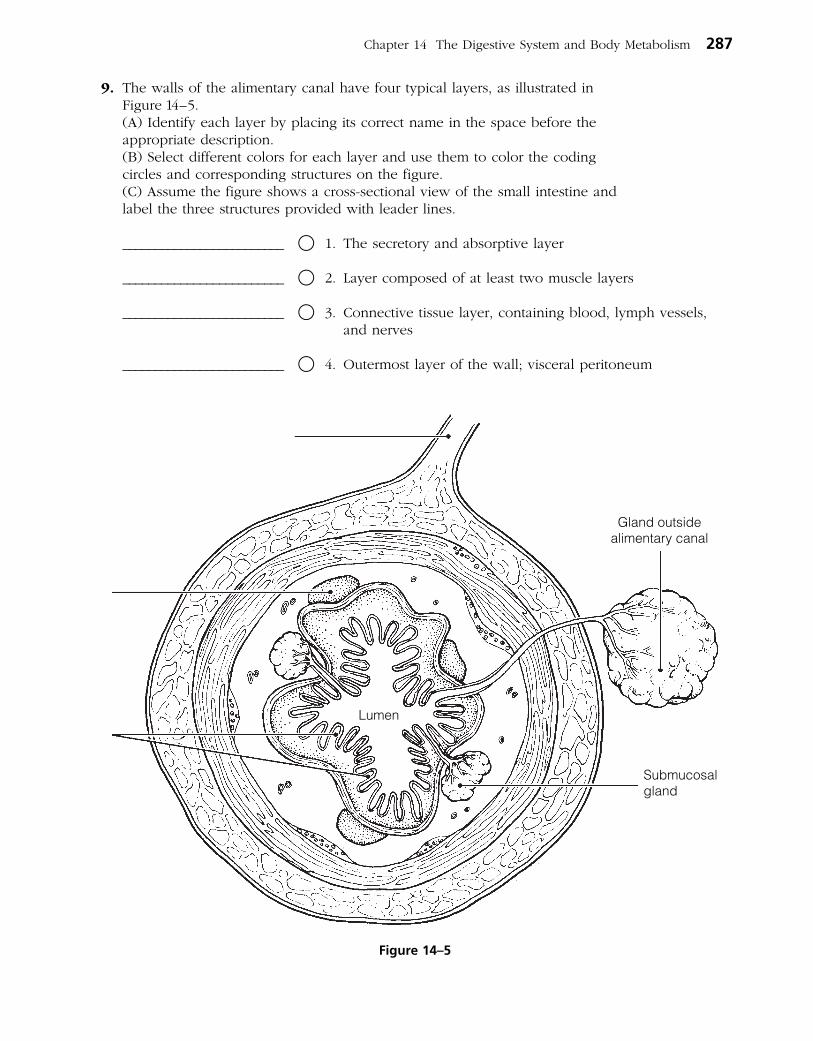
Chapter 14 The Digestive System and Body Metabolism 287
9. The walls of the alimentary canal have four typical layers, as illustrated in Figure 14–5.
(A) Identify each layer by placing its correct name in the space before the appropriate description.
(B) Select different colors for each layer and use them to color the coding circles and corresponding structures on the figure.
(C) Assume the figure shows a crosssectional view of the small intestine and label the three structures provided with leader lines.
_________________________ ○ 1. The secretory and absorptive layer
_________________________ ○ 2. Layer composed of at least two muscle layers
_________________________ ○ 3. Connective tissue layer, containing blood, lymph vessels, and nerves
_________________________ ○ 4. Outermost layer of the wall; visceral peritoneum
Submucosalgland
Gland outsidealimentary canal
Lumen
Figure 14–5
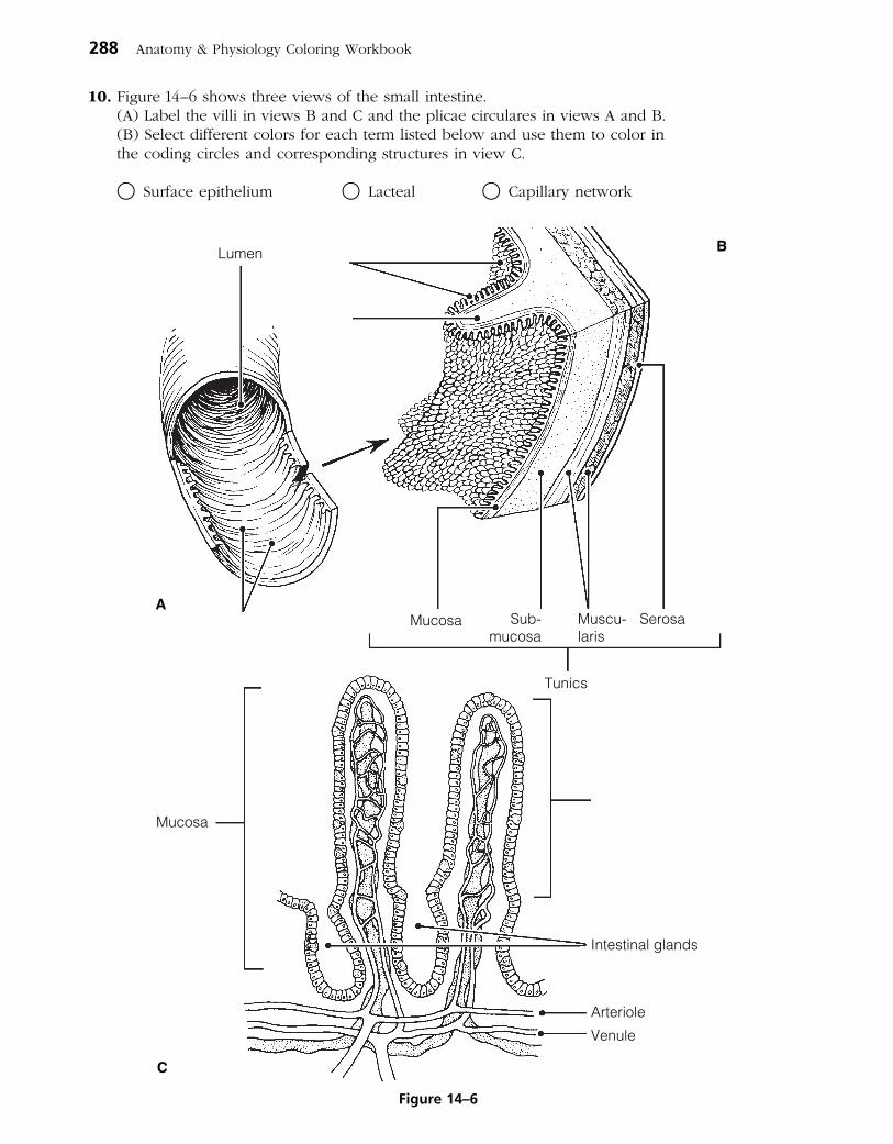
10. Figure 14–6 shows three views of the small intestine. (A) Label the villi in views B and C and the plicae circulares in views A and B. (B) Select different colors for each term listed below and use them to color in
the coding circles and corresponding structures in view C.
○ Surface epithelium ○ Lacteal ○ Capillary network
288 Anatomy & Physiology Coloring Workbook
Lumen
Mucosa Sub-mucosa
Muscu-laris
Serosa
Tunics
Mucosa
Intestinal glands
Arteriole
Venule
A
B
C
Figure 14–6

Chapter 14 The Digestive System and Body Metabolism 289
11. Three accessory organs are illustrated in Figure 14–7. (A) Identify each of the three organs and the ligament provided with leader
lines on the figure. (B) Select different colors for the following structures and use them to
color the coding circles and the corresponding structures on the figure.
○ Common hepatic duct ○ Bile duct ○ Cystic duct ○ Pancreatic duct
12. Complete the following statements referring to human dentition by inserting your answers in the answer blanks.
The first set of teeth, called the (1) teeth, begin to appear around the age of (2) and usually have begun to be replaced by the age of (3) . The (4) teeth are more numerous; that is, there are (5) teeth in the second set as opposed to a total of (6) teeth in the first set. If an adult has a full set of teeth, you can expect to find two (7) , one
(8) , two (9) , and three (10) in one side of each jaw. The most posterior molars in each jaw are commonly called
(11) teeth.
______________________ 7. ______________________ 8.
______________________ 10. ______________________ 11.
_________________________ 1.
_________________________ 2.
_________________________ 3.
_________________________ 4.
_________________________ 5.
_________________________ 6.
_________________________ 9.
Duodenum
Figure 14–7

13. Figure 14–8 illustrates the longitudinal section of a tooth. (A) Identify the crown, gingiva, and root of the tooth (leader lines). (B) Add leader lines to the figure and use the key choices to label them. (C) Select different colors to represent the key choices and use them to
color in the coding circles and corresponding structures in the figure. (D) Choose terms from the key choices to match the descriptions below
the figure.
Key Choices
○ A. Cement ○ C. Enamel ○ E. Pulp
○ B. Dentin ○ D. Periodontal membrane (ligament)
_______ 1. Material covering the tooth root
_______ 2. Forms the bulk of tooth structure
_______ 3. A collection of blood vessels, lymphatics, and nerve fibers
_______ 4. Cells that produce this substance degenerate after tooth eruption
290 Anatomy & Physiology Coloring Workbook
Figure 14–8

PHYSIOLOGY OF THE DIGESTIVE SYSTEM
14. Match the descriptions in Column B with the appropriate terms referring to digestive processes in Column A. Letters or terms may be used more than once.
Column A
_________________________ 1. Ingestion
_________________________ 2. Propulsion
_________________________ 3. Mechanical digestion
_________________________ 4. Chemical digestion
_________________________ 5. Absorption
_________________________ 6. Defecation
15. Identify the pathological conditions described below by using terms from the key choices. Insert the correct term or letter in the answer blanks.
Key Choices
A. Appendicitis C. Diarrhea E. Heartburn G. PeritonitisB. Constipation D. Gallstones F. Jaundice H. Ulcer
_________________________ 1. Inflammation of the abdominal serosa
_________________________ 2. Condition resulting from the reflux of acidic gastric juice into the esophagus
_________________________ 3. Usually indicates liver problems or blockage of the biliary ducts
_________________________ 4. An erosion of the stomach or duodenal mucosa
_________________________ 5. Passage of watery stools
_________________________ 6. Causes severe epigastric pain; associated with prolonged storage of bile in the gallbladder
_________________________ 7. Inability to pass feces; often a result of poor bowel habits
_________________________ 8. Inflammation of the wormlike extension of the large intestine.
Column B
A. Transport of nutrients from lumen to blood
B. Enzymatic breakdown
C. Elimination of feces
D. Eating
E. Chewing
F. Churning
G. Includes swallowing
H. Segmentation and peristalsis
Chapter 14 The Digestive System and Body Metabolism 291
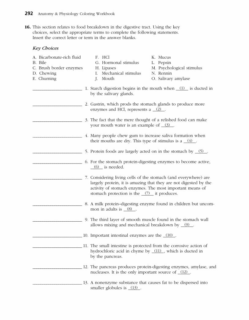
292 Anatomy & Physiology Coloring Workbook
16. This section relates to food breakdown in the digestive tract. Using the key choices, select the appropriate terms to complete the following statements. Insert the correct letter or term in the answer blanks.
Key Choices
A. Bicarbonaterich fluid F. HCl K. MucusB. Bile G. Hormonal stimulus L. PepsinC. Brush border enzymes H. Lipases M. Psychological stimulusD. Chewing I. Mechanical stimulus N. RenninE. Churning J. Mouth O. Salivary amylase
_________________________ 1. Starch digestion begins in the mouth when (1) is ducted in by the salivary glands.
_________________________ 2. Gastrin, which prods the stomach glands to produce more enzymes and HCl, represents a (2) .
_________________________ 3. The fact that the mere thought of a relished food can make your mouth water is an example of (3) .
_________________________ 4. Many people chew gum to increase saliva formation when their mouths are dry. This type of stimulus is a (4) .
_________________________ 5. Protein foods are largely acted on in the stomach by (5) .
_________________________ 6. For the stomach proteindigesting enzymes to become active, (6) is needed.
_________________________ 7. Considering living cells of the stomach (and everywhere) are largely protein, it is amazing that they are not digested by the activity of stomach enzymes. The most important means of stomach protection is the (7) it produces.
_________________________ 8. A milk protein–digesting enzyme found in children but uncom mon in adults is (8) .
_________________________ 9. The third layer of smooth muscle found in the stomach wall allows mixing and mechanical breakdown by (9) .
_________________________ 10. Important intestinal enzymes are the (10) .
_________________________ 11. The small intestine is protected from the corrosive action of hydro chloric acid in chyme by (11) , which is ducted in by the pancreas.
_________________________ 12. The pancreas produces proteindigesting enzymes, amylase, and nucleases. It is the only important source of (12) .
_________________________ 13. A nonenzyme substance that causes fat to be dispersed into smaller globules is (13) .
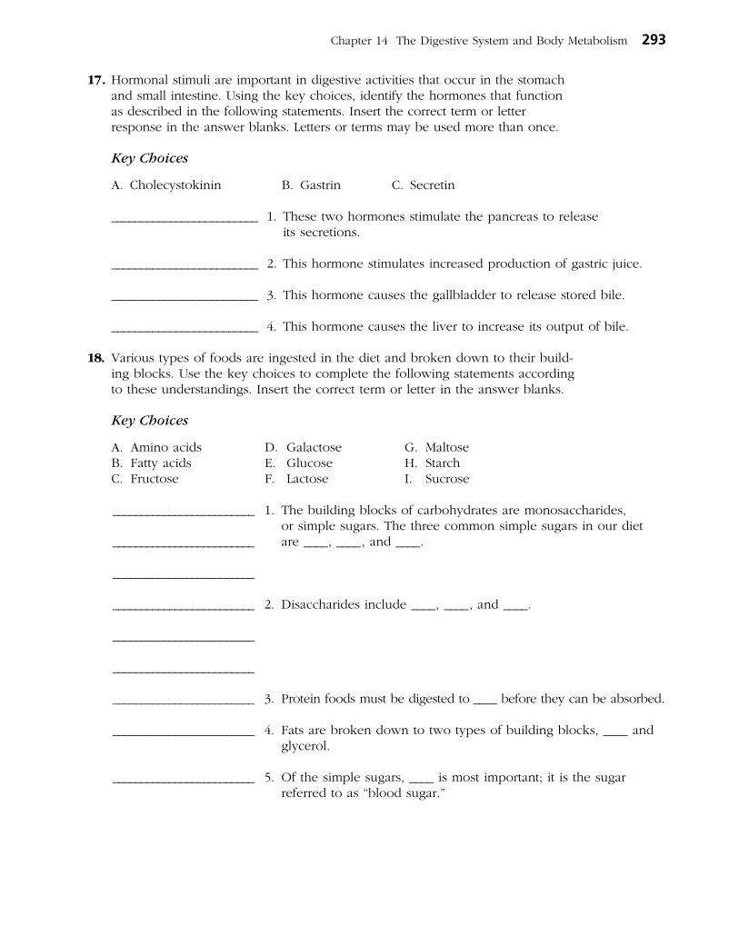
Chapter 14 The Digestive System and Body Metabolism 293
17. Hormonal stimuli are important in digestive activities that occur in the stomach and small intestine. Using the key choices, identify the hormones that function as described in the following statements. Insert the correct term or letter response in the answer blanks. Letters or terms may be used more than once.
Key Choices
A. Cholecystokinin B. Gastrin C. Secretin
_________________________ 1. These two hormones stimulate the pancreas to release its secretions.
_________________________ 2. This hormone stimulates increased production of gastric juice.
_________________________ 3. This hormone causes the gallbladder to release stored bile.
_________________________ 4. This hormone causes the liver to increase its output of bile.
18. Various types of foods are ingested in the diet and broken down to their building blocks. Use the key choices to complete the following statements according to these understandings. Insert the correct term or letter in the answer blanks.
Key Choices
A. Amino acids D. Galactose G. MaltoseB. Fatty acids E. Glucose H. StarchC. Fructose F. Lactose I. Sucrose
The building blocks of carbohydrates are monosaccharides, or simple sugars. The three common simple sugars in our diet are ____, ____, and ____.
Disaccharides include ____, ____, and ____.
Protein foods must be digested to ____ before they can be absorbed.
Fats are broken down to two types of building blocks, ____ and glycerol.
Of the simple sugars, ____ is most important; it is the sugar referred to as “blood sugar.”
_________________________ 1.
_________________________
_________________________
_________________________ 2.
_________________________
_________________________
_________________________ 3.
_________________________ 4.
_________________________ 5.
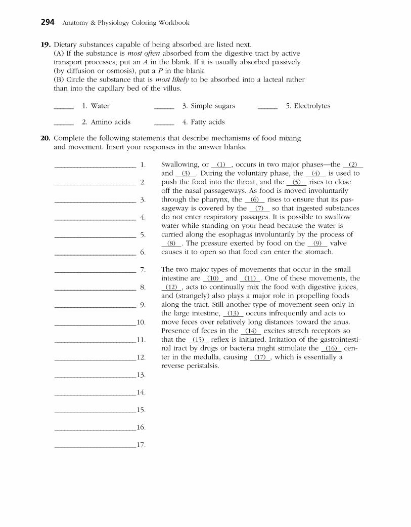
294 Anatomy & Physiology Coloring Workbook
19. Dietary substances capable of being absorbed are listed next. (A) If the substance is most often absorbed from the digestive tract by active
transport processes, put an A in the blank. If it is usually absorbed passively (by diffusion or osmosis), put a P in the blank.
(B) Circle the substance that is most likely to be absorbed into a lacteal rather than into the capillary bed of the villus.
______ 1. Water ______ 3. Simple sugars ______ 5. Electrolytes
______ 2. Amino acids ______ 4. Fatty acids
20. Complete the following statements that describe mechanisms of food mixing and movement. Insert your responses in the answer blanks.
Swallowing, or (1) , occurs in two major phases—the (2) and (3) . During the voluntary phase, the (4) is used to push the food into the throat, and the (5) rises to close off the nasal passageways. As food is moved involuntarily through the pharynx, the (6) rises to ensure that its passageway is covered by the (7) so that ingested substances do not enter respiratory passages. It is possible to swallow water while standing on your head because the water is carried along the esophagus involuntarily by the process of
(8) . The pressure exerted by food on the (9) valve causes it to open so that food can enter the stomach.
The two major types of movements that occur in the small intestine are (10) and (11) . One of these movements, the
(12) , acts to continually mix the food with digestive juices, and (strangely) also plays a major role in propelling foods along the tract. Still another type of movement seen only in the large intestine, (13) occurs infrequently and acts to move feces over relatively long distances toward the anus. Presence of feces in the (14) excites stretch receptors so that the (15) reflex is initiated. Irritation of the gastrointesti nal tract by drugs or bacteria might stimulate the (16) cen ter in the medulla, causing (17) , which is essentially a reverse peristalsis.
_________________________ 1.
_________________________ 2.
_________________________ 3.
_________________________ 4.
_________________________ 5.
_________________________ 6.
_________________________ 7.
_________________________ 8.
_________________________ 9.
_________________________ 10.
_________________________ 11.
_________________________ 12.
_________________________ 13.
_________________________ 14.
_________________________ 15.
_________________________ 16.
_________________________ 17.

Chapter 14 The Digestive System and Body Metabolism 295
NUTRITION AND METABOLISM
Nutrients Used by Body Cells
21. Using the key choices, identify the foodstuffs used by cells in the cellular functions described below. Insert the correct term or key letter in the answer blanks.
Key Choices
A. Amino acids B. Carbohydrates C. Fats
_________________________ 1. The most used substance for producing the energyrich ATP
_________________________ 2. Important in building myelin sheaths and cell membranes
_________________________ 3. Tend to be conserved by cells
_________________________ 4. The second most important food source for making cellular energy
_________________________ 5. Form insulating deposits around body organs and beneath the skin
_________________________ 6. Used to make the bulk of cell structure and functional substances such as enzymes
22. Identify the nutrients described by using the key choices. Insert the correct letter(s) in the answer blanks. Items may have more than one answer.
Key Choices
A. Bread/pasta D. Fruits G. StarchB. Cheese/cream E. Meat/fish H. VegetablesC. Cellulose F. Minerals I. Vitamins
_________________________ 1. Examples of carbohydrate-rich foods in the diet.
_________________________ 2. Fatty foods ingested in the normal diet include _____.
_________________________ 3. The only important digestible polysaccharide.
_________________________ 4. An indigestible polysaccharide that aids elimination because it adds bulk to the diet is _____.
_________________________ 5. Protein-rich foods include _____ and _____.
_________________________ 6. Most examples of these nutrients, which are found largely in vegetables and fruits, are used as coenzymes.
_________________________ 7. Include copper, iron, and sodium.

296 Anatomy & Physiology Coloring Workbook
Metabolic Processes
23. Figure 14–9 depicts the three stages of cellular respiration. (A) Label the figure by placing the following terms on the appropriate
answer blanks. (B) Answer the questions below the figure. Note: Terms may be used more than once.
ATP Glucose Mitochondrion
Carbon dioxide Glycolysis Pyruvic acid
Chemical energy Electron transport chain Water
Cytosol Krebs cycle
1. Which of the oxidative phases does not require oxygen?
___________________________________________________________________________________________
2. Which phases do require oxygen? _________________________________________________________
___________________________________________________________________________________________
3. In what form is chemical energy transferred from the first two phases to the third phase?
___________________________________________________________________________________________
4. Which of the phases produces the largest amount of ATP?
___________________________________________________________________________________________
5. Which phase combines energetic H atoms with molecular oxygen?
___________________________________________________________________________________________
1.
7.
9.
2.
5. 4.
8. 11.
10.
14. 13. 12.
3. (phase)
(phase)
(high-energy electron)
(high-energyelectron)
(cellular site)
(product) (product) (product)
(product)
(product)
(substrate) (product)
(cellular site)
6.
(phase)
Figure 14–9
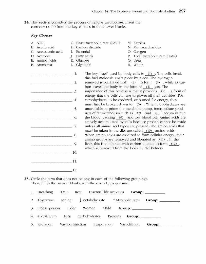
Chapter 14 The Digestive System and Body Metabolism 297
24. This section considers the process of cellular metabolism. Insert the correct word(s) from the key choices in the answer blanks.
Key Choices
A. ATP G. Basal metabolic rate (BMR) M. KetosisB. Acetic acid H. Carbon dioxide N. MonosaccharidesC. Acetoacetic acid I. Essential O. OxygenD. Acetone J. Fatty acids P. Total metabolic rate (TMR)E. Amino acids K. Glucose Q. UreaF. Ammonia L. Glycogen R. Water
The key “fuel” used by body cells is (1) . The cells break this fuel molecule apart piece by piece. The hydrogen removed is combined with (2) to form (3) , while its carbon leaves the body in the form of (4) gas. The importance of this process is that it provides (5) , a form of energy that the cells can use to power all their activities. For carbohydrates to be oxidized, or burned for energy, they must first be broken down to (6) . When carbohydrates are unavailable to prime the metabolic pump, intermediate products of fat metabolism such as (7) and (8) accumulate in the blood, causing (9) and low blood pH. Amino acids are actively accumulated by cells because protein cannot be made unless all amino acid types are present. The amino acids that must be taken in the diet are called (10) amino acids. When amino acids are oxidized to form cellular energy, their amino groups are removed and liberated as (11) . In the liver, this is combined with carbon dioxide to form (12) , which is removed from the body by the kidneys.
25. Circle the term that does not belong in each of the following groupings. Then, fill in the answer blanks with the correct group name.
1. Breathing TMR Rest Essential life activities Group: ___________
2. Thyroxine Iodine ↓Metabolic rate ↑Metabolic rate Group: ___________
3. Obese person Elder Women Child Group: ___________
4. 4 kcal/gram Fats Carbohydrates Proteins Group: ___________
5. Radiation Vasoconstriction Evaporation Vasodilation Group: ___________
_________________________ 1.
_________________________ 2.
_________________________ 3.
_________________________ 4.
_________________________ 5.
_________________________ 6.
_________________________ 7.
_________________________ 8.
_________________________ 9.
_________________________ 10.
_________________________ 11.
_________________________ 12.
298 Anatomy & Physiology Coloring Workbook
26. The liver has many functions in addition to its digestive function. Complete the following statements that elaborate on the liver’s function by inserting the correct terms in the answer blanks.
The liver is the most important metabolic organ in the body. In its metabolic role, the liver uses amino acids from the nutrientrich hepatic portal blood to make many blood proteins such as (1) , which helps to hold water in the bloodstream, and (2) , which prevent blood loss when blood vessels are damaged. The liver also makes a steroid substance that is released to the blood. This steroid, (3) , has been implicated in high blood pressure and heart disease. Addition ally, the liver acts to maintain homeostatic blood glucose levels. It removes glucose from the blood when blood glucose levels are high, a condition called (4) , and stores it as
(5) . Then, when blood glucose levels are low, a condition called (6) , liver cells break down the stored carbohydrate and release glucose to the blood once again. This latter process is termed (7) . When the liver makes glucose from noncarbohydrate substances such as fats or proteins, the process is termed (8) . In addition to its processing of amino acids and sugars, the liver plays an important role in the processing of fats. Other functions of the liver include the (9) of drugs and alcohol. Its (10) cells protect the body by ingesting bacteria and other debris.
The liver forms small complexes called (11) , which are needed to transport fatty acids, fats, and cholesterol in the blood because lipids are (12) in a watery medium. The function of (13) is transport of cholesterol to peripheral tissues, where cells use it to construct their plasma (14) or to synthesize (15) hormones. The function of highdensity lipoproteins (HDLs) is transport of cholesterol to the (16) , where it is degraded and secreted as (17) , which are eventually excreted.
Two other important functions of the liver are the storage of vitamins (such as vitamin (18) needed for vision) and of the metal (19) (as ferritin).
_________________________ 1.
_________________________ 2.
_________________________ 3.
_________________________ 4.
_________________________ 5.
_________________________ 6.
_________________________ 7.
_________________________ 8.
_________________________ 9.
_________________________ 10.
_________________________ 11.
_________________________ 12.
_________________________ 13.
_________________________ 14.
_________________________ 15.
_________________________ 16.
_________________________ 17.
_________________________ 18.
_________________________ 19.

27. Using the key choices, select the terms identified in the following descriptions. Insert the appropriate term(s) or letter(s) in each answer blank. Items may have more than one answer.
Key Choices
A. Blood E. Hyperthermia I. RadiationB. Constriction of skin blood vessels F. Hypothalamus J. PyrogensC. Frostbite G. Hypothermia K. ShiveringD. Heat H. Perspiration
_________________________ 1. Byproduct of cell metabolism
_________________________ 2. Means of conserving/increasing body heat
_________________________ 3. Means by which heat is distributed to all body tissues
_________________________ 4. Site of the body’s thermostat
_________________________ 5. Chemicals released by injured tissue cells and bacteria, causing resetting of the thermostat
_________________________ 6. Death of cells deprived of oxygen and nutrients, resulting from withdrawal of blood from the skin circulation when the external temperature is low
_________________________ 7. Means of liberating excess body heat
_________________________ 8. Extremely low body temperature
_________________________ 9. Fever
Chapter 14 The Digestive System and Body Metabolism 299

300 Anatomy & Physiology Coloring Workbook
DEVELOPMENTAL ASPECTS OF THE DIGESTIVE SYSTEM
28. Using the key choices, select the terms identified in the following descriptions. Insert the correct term(s) or letter(s) in each answer blank. Items may have more than one answer.
Key Choices
A. Accessory organs F. Gallbladder problems K. RootingB. Alimentary canal G. Gastritis L. SuckingC. Appendicitis H. PKU (phenylketonuria) M. StomachD. Cleft palate/lip I. Periodontal disease N. Tracheoesophageal fistulaE. Cystic fibrosis J. Peristalsis O. Ulcers
_________________________ 1. Internal tubelike cavity of the embryo
_________________________ 2. Glands formed by branching from the digestive mucosa
_________________________ 3. Most common congenital defect; aspiration of feeding common
_________________________ 4. Congenital condition characterized by a connection between digestive and respiratory passageways
_________________________ 5. Congenital condition in which large amounts of mucus are produced, clogging respiratory passageways and pancreatic ducts
_________________________ 6. Metabolic disorder characterized by an inability to properly use the amino acid phenylalanine
_________________________ 7. Reflex aiding the newborn baby to find the nipple
_________________________ 8. Vomiting is common in infants because this structure is small
_________________________ 9. Most common adolescent digestive system problem
_________________________ 10. Inflammations of the gastrointestinal tract
_________________________ 11. Condition of loose teeth and inflamed gums; generally seen in elderly people
Chapter 14 The Digestive System and Body Metabolism 301
A Visualization Exercise for the Digestive System
. . . the passage beneath you opens, and you fall into a huge chamber with mountainous folds.
29. Where necessary, complete statements by inserting the missing word(s) in the answer blanks.
In this journey, you are to travel through the digestive tract as far as the appendix and then await further instructions. You are miniaturized as usual and provided with a wet suit to protect you from being digested during your travels. You have a very easy entry into your host’s open mouth. You look around and notice the glistening pink lining, or (1) , and the perfectly caredfor teeth. Within a few seconds, the lips part and you find yourself surrounded by bread. You quickly retreat to the safety of the (2) between the teeth and the cheek to prevent getting chewed. From there you watch with fascination as a number of openings squirt fluid into the chamber, and the (3) heaves and rolls, mixing the bread with the fluid.
As the bread begins to disappear, you decide that the fluid contains the enzyme (4) . You then walk toward the back of the oral cavity. Suddenly, you find yourself being carried along by a squeezing motion of the walls around you. The name given to this propelling motion is (5) . As you are carried helplessly downward, you see two openings—the
(6) and the (7) —below you. Just as you are about to straddle the solid area between them to stop your descent, the structure to your left moves quickly upward, and a trapdoorlike organ, the (8) , flaps over its opening. Down you go in the dark, seeing nothing. Then the passage beneath you opens, and you fall into a huge chamber with mountain ous folds. Obviously, you have reached the (9) . The folds are very slippery, and you conclude that it must be the (10) coat that you read about earlier. As you survey your surroundings, juices begin to gurgle into the chamber from
pits in the “floor,” and your face begins to sting and smart. You cannot seem to escape this caustic fluid and conclude that it must be very dangerous to your skin since it contains (11) and (12) . You reach down and scoop up some of the slippery substance from the folds and smear it on your face, confident that if it can protect this organ it can protect you as well! Relieved, you begin to slide toward the organ’s far exit and squeeze through the tight (13) valve into the next organ. In the dim light, you see lumps of cellulose lying at your feet and large fat globules dancing lightly about. A few seconds later, your observations are interrupted by a wave of fluid pouring into the chamber from an opening high in the wall above you. The large fat globules begin to fall apart, and you decide that this enzyme flood has to contain (14) , and the opening must be the duct from the (15) . As you move quickly away to escape the deluge, you lose your footing and find
_________________________ 1.
_________________________ 2.
_________________________ 3.
_________________________ 4.
_________________________ 5.
_________________________ 6.
_________________________ 7.
_________________________ 8.
_________________________ 9.
_________________________ 10.
_________________________ 11.
_________________________ 12.
_________________________ 13.
_________________________ 14.
_________________________ 15.
INCREDIBLE JOURNEY
302 Anatomy & Physiology Coloring Workbook
yourself on a rollercoaster ride—twisting, coiling, turning, and diving through the lumen of this active organ. As you move, you are stroked by velvety, fingerlike projections of the wall, the (16) . Abruptly your ride comes to a halt as
you are catapulted through the (17) valve and fall into the appendix. Headquarters informs you that you are at the end of your journey. Your exit now depends on your own ingenuity.
AT THE CLINIC
30. Mary Maroon comes to the clinic to get information on a vegetarian diet. What problems may arise when people make uninformed decisions on what to eat for a vegetarian diet? What combinations of vegetable foods will provide Mary with all the essential amino acids?
31. Mr. Ashe, a man in his mid60s, comes to the clinic complaining of heartburn. Questioning by the clinic staff reveals that the severity of his attacks increases when he lies down after eating a heavy meal. The man is about 50 pounds overweight. What is your diagnosis? Without treatment, what conditions might develop?
32. There has been a record heat wave lately, and many elderly people are coming to the clinic complaining that they “feel poorly.” In most cases, their skin is cool and clammy, and their blood pressure is low. What is their problem? What can be done to alleviate it?
33. During the same period, Bert Winchester, a construction worker, is rushed in unconscious. His skin is hot and dry, and his coworkers say that he just suddenly keeled over on the job. What is Bert’s condition and how should it be handled?
_________________________ 16.
_________________________ 17.

34. Mrs. Ironfield is brought to an emergency room complaining of severe pain in her left iliac region. She claims previous episodes and says that the condition is worse when she is constipated and is relieved by defecation. A large tender mass is palpated in the left iliac fossa, and a barium study reveals a large number of diverticula in her descending and sigmoid colon. What are diverticula, and what is believed to promote their formation? Does this woman have diverticulitis or diverticulosis? Explain.
35. A woman in her 50s complains of bloating, cramping, and diarrhea when she drinks milk. What is the cause of her complaint and what is a solution?
36. Clients are instructed not to eat before having blood tests run. How would a lab technician know if someone “cheated” and ate a fatty meal a few hours before having his blood drawn?
37. Zena, a teenager, has gone to the sports clinic for the past 2 years to have her fat content checked. This year, her percentage of body fat is up, and tissue protein has not increased. Questioning reveals that Zena has been on crash diets four times since the last checkup, only to regain the weight (and more) each time. She also admits sheepishly that she “detests” exercise. How does cyclic dieting, accompanied by lack of exercise, cause an increase in fat and a decrease in protein?
38. Mrs. Rodriguez has a bleeding ulcer and has lost her appetite. She appears pale and lethargic when she comes in for a physical. She proves to be anemic, and her RBCs are large and pale. What mineral supplements should be ordered?
39. Mr. Roddick, a 21yearold man with severe appendicitis, did not seek treatment in time and died a week after his abdominal pain and fever began. Explain why appendicitis can quickly lead to death.
Chapter 14 The Digestive System and Body Metabolism 303

304 Anatomy & Physiology Coloring Workbook
40. In the mid1960s, a caloriefree substitute (olestra) that is neither digested nor absorbed hit the market shelves in the United States. At that time, there was concern that vitamin deficiencies might result from its use. What type of vitamins caused this concern and why?
THE FINALE: MULTIPLE CHOICE
1. Which of the following terms are synonyms?
A. Gastrointestinal tract
B. Digestive system
C. Digestive tract
D. Alimentary canal
2. A digestive organ that is not part of the alimentary canal is the:
A. stomach. D. large intestine.
B. liver. E. pharynx.
C. small intestine.
3. The GI tube layer responsible for the actions of segmentation and peristalsis is:
A. serosa. C. muscularis externa.
B. mucosa. D. submucosa.
4. Which alimentary canal tunic has the greatest abundance of lymph nodules?
A. Mucosa C. Serosa
B. Muscularis D. Submucosa
5. Proteins secreted in saliva include:
A. mucin. C. lysozyme.
B. amylase. D. IgA.
6. The closure of which valve is assisted by the diaphragm?
A. Ileocecal
B. Pyloric
C. Gastroesophageal
D. Upper esophageal
7. Smooth muscle is found in the:
A. tongue.
B. pharynx.
C. esophagus.
D. external anal sphincter.
8. Which of these organs lies in the right hypochondriac region of the abdomen?
A. Stomach C. Cecum
B. Spleen D. Liver
9. Which phases of gastric secretion depend (at least in part) on the vagus nerve?
A. Cephalic
B. Gastric
C. Intestinal (stimulatory)
D. Intestinal (inhibitory)
10. Which of the following are tied to sodium transport?
A. Glucose
B. Fructose
C. Galactose
D. Amino acids
11. Excess iron is stored primarily in the:
A. liver.
B. bone marrow.
C. duodenal epithelium.
D. blood.
41. Select the best answer or answers from the choices given.
Chapter 14 The Digestive System and Body Metabolism 305
12. A 3yearold girl was rewarded with a hug because she was now completely toilet trained. Which muscle had she learned to control?
A. Levator ani
B. Internal anal sphincter
C. Internal and external obliques
D. External anal sphincter
13. Which cell type fits this description? It occurs in the stomach mucosa, contains abundant mitochondria and many microvilli, and pumps hydrogen ions.
A. Absorptive cell C. Goblet cell
B. Parietal cell D. Mucous neck cell
14. Which of the following are “essential” nutrients?
A. Glucose C. Cholesterol
B. Linoleic acid D. Leucine
15. Deficiency of which of these vitamins results in anemia?
A. Thiamin C. Biotin
B. Riboflavin D. Folic acid
16. Vitamins that act as coenzymes in the Krebs cycle include:
A. riboflavin. C. biotin.
B. niacin. D. pantothenic acid.
17. Substratelevel phosphorylation occurs during:
A. glycolysis. C. Krebs cycle.
B. betaoxidation. D. electron transport.
18. Chemicals that can be used for gluconeogenesis include:
A. amino acids.
B. glycerol.
C. fatty acids.
D. alphaketoglutaric acid.
19. The chemiosmotic process involves:
A. buildup of hydrogen ion concentration.
B. electron transport.
C. oxidation and reduction.
D. ATP synthase.
20. Only the liver functions to:
A. store iron.
B. form urea.
C. produce plasma proteins.
D. form ketone bodies.
21. Which events occur during the absorptive state?
A. Use of amino acids as a major source of energy
B. Lipogenesis
C. Betaoxidation
D. Increased uptake of glucose by skeletal muscles
22. Hormones that act to decrease blood glucose level include:
A. insulin. C. epinephrine.
B. glucagon. D. growth hormone.
23. During the postabsorptive state:
A. glycogenesis occurs in the liver.
B. fatty acids are used for fuel.
C. amino acids are converted to glucose.
D. lipolysis occurs in adipose tissue.
24. Which transport particles carry cholesterol destined for excretion from the body?
A. HDL C. LDL
B. Chylomicron D. VLDL (very low density lipoprotein)
25. Glucose (or its metabolites) can be converted to:
A. glycogen.
B. triglycerides.
C. nonessential amino acids.
D. starch.
26. Basal metabolic rate:
A. is the lowest metabolic rate of the body.
B. is the metabolic rate during sleep.
C. is measured as kcal per square meter of skin per hour.
D. increases with age.
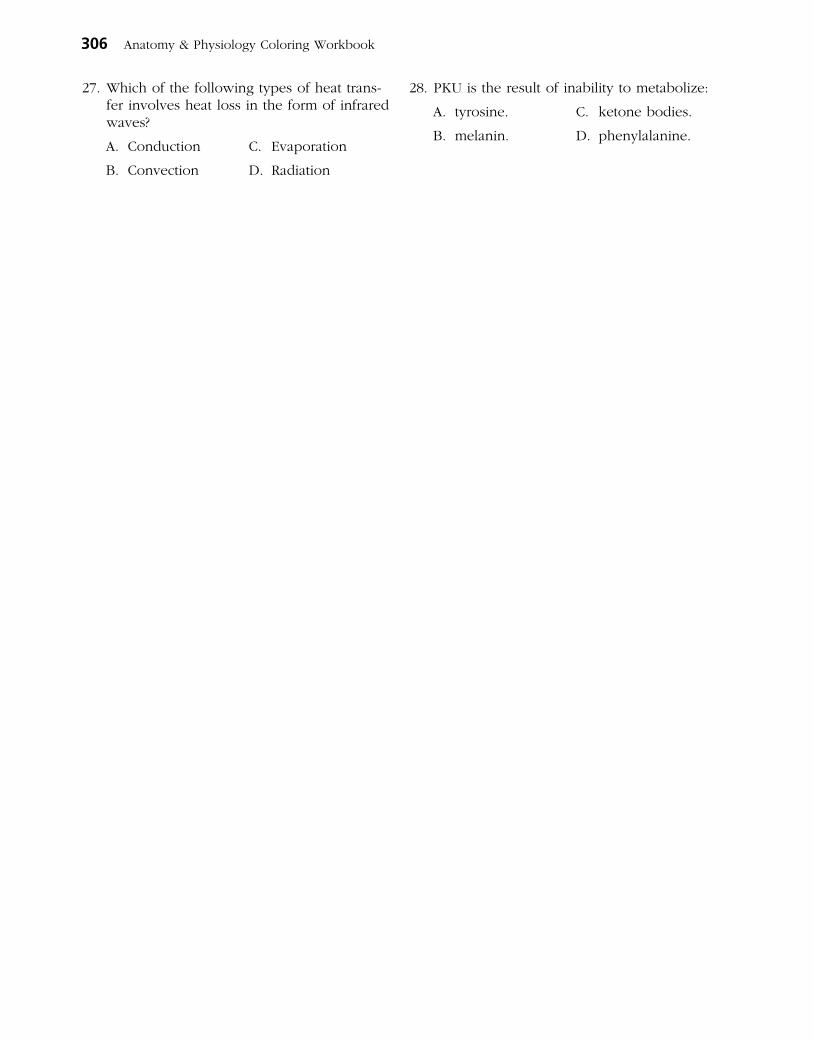
27. Which of the following types of heat transfer involves heat loss in the form of infrared waves?
A. Conduction C. Evaporation
B. Convection D. Radiation
28. PKU is the result of inability to metabolize:
A. tyrosine. C. ketone bodies.
B. melanin. D. phenylalanine.
306 Anatomy & Physiology Coloring Workbook

Metabolism of nutrients by body cells produces various wastes such as carbon dioxide and nitrogenous wastes (creatinine, urea, and ammonia), as well as imbalances of water and essential ions. The metabolic wastes and excesses must be eliminated from the body. Essential substances are retained to ensure proper body functioning.
Although several organ systems are involved in excretory processes, the urinary system bears the primary responsibility for removing nitrogenous wastes from the blood. In addition to this purely excretory function, the kidneys maintain the electrolyte, acid-base, and fluid balances of the blood. Thus, kidneys are major homeostatic organs of the body. Malfunction of the kidneys leads to a failure of homeostasis, resulting (unless corrected) in death.
Activities in this chapter are concerned with identification of urinary system structures, urine composition, and physiological processes involved in urine formation. It also focuses on the composition of the body’s fluid compartments and the water, electrolyte, and acid-base balance of these compartments.
1. Complete the following statements by inserting your answers in the answer blanks.
The kidney is referred to as an excretory organ because it excretes (1) wastes. It is also a major homeostatic organ because it maintains the electrolyte, (2) , and (3) balance of the blood. Urine is continuously formed by the (4) and is routed down the (5) by the mechanism of (6) to a storage organ called the (7) . Eventually the urine is con-ducted to the body exterior by the (8) . In males, this tube-like structure is about (9) inches long; in females, it is approximately (10) inches long.
THE URINARY SYSTEM15
_________________________ 1.
_________________________ 2.
_________________________ 3.
_________________________ 4.
_________________________ 5.
_________________________ 6.
_________________________ 7. _________________________ 9.
_________________________ 8. _________________________ 10.
307
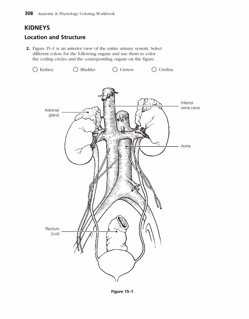
KIDNEYS
Location and Structure
2. Figure 15–1 is an anterior view of the entire urinary system. Select different colors for the following organs and use them to color the coding circles and the corresponding organs on the figure.
○ Kidney ○ Bladder ○ Ureters ○ Urethra
308 Anatomy & Physiology Coloring Workbook
Rectum(cut)
Adrenalgland
Inferiorvena cava
Aorta
Figure 15–1
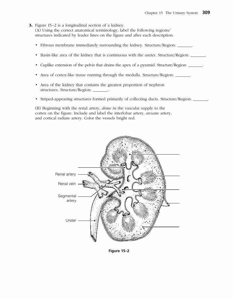
3. Figure 15–2 is a longitudinal section of a kidney. (A) Using the correct anatomical terminology, label the following regions/
structures indicated by leader lines on the figure and after each description.
• Fibrousmembraneimmediatelysurroundingthekidney.Structure/Region:_______.
• Basin-likeareaofthekidneythatiscontinuouswiththeureter.Structure/Region:_______.
• Cuplikeextensionofthepelvisthatdrainstheapexofapyramid.Structure/Region:_______.
• Areaofcortex-liketissuerunningthroughthemedulla.Structure/Region:_______.
• Areaofthekidneythatcontainsthegreatestproportionofnephron structures.Structure/Region:_______.
• Striped-appearingstructuresformedprimarilyofcollectingducts.Structure/Region:_______.
(B) Beginning with the renal artery, draw in the vascular supply to the cortex on the figure. Include and label the interlobar artery, arcuate artery, and cortical radiate artery. Color the vessels bright red.
Chapter 15 The Urinary System 309
Renal artery
Renal vein
Ureter
Segmentalartery
Figure 15–2
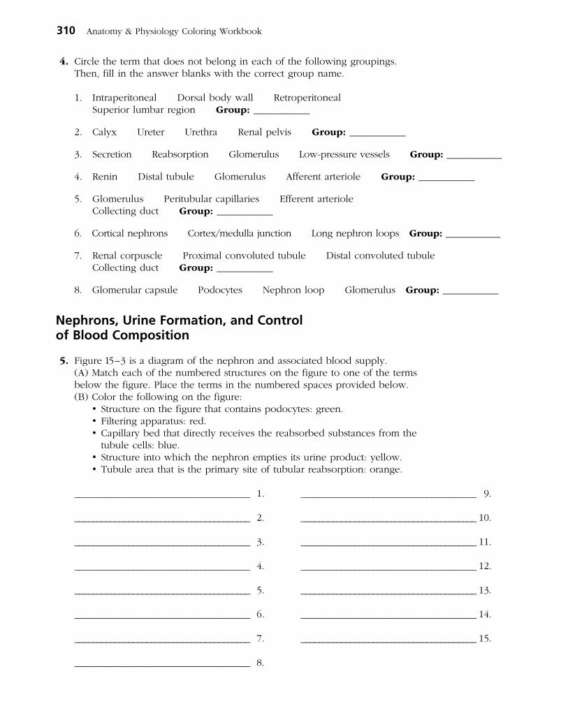
4. Circle the term that does not belong in each of the following groupings. Then, fill in the answer blanks with the correct group name.
1. Intraperitoneal Dorsalbodywall Retroperitoneal Superior lumbar region Group: ___________
2. Calyx Ureter Urethra Renalpelvis Group: ___________
3. Secretion Reabsorption Glomerulus Low-pressurevessels Group: ___________
4. Renin Distaltubule Glomerulus Afferentarteriole Group: ___________
5. Glomerulus Peritubularcapillaries Efferentarteriole Collecting duct Group: ___________
6. Corticalnephrons Cortex/medullajunction Longnephronloops Group: ___________
7. Renalcorpuscle Proximalconvolutedtubule Distalconvolutedtubule Collecting duct Group: ___________
8. Glomerularcapsule Podocytes Nephronloop Glomerulus Group: ___________
Nephrons, Urine Formation, and Control of Blood Composition
5. Figure 15–3 is a diagram of the nephron and associated blood supply. (A) Match each of the numbered structures on the figure to one of the terms
below the figure. Place the terms in the numbered spaces provided below. (B)Colorthefollowingonthefigure: •Structureonthefigurethatcontainspodocytes:green. •Filteringapparatus:red. •Capillarybedthatdirectlyreceivesthereabsorbedsubstancesfromthe
tubulecells:blue. •Structureintowhichthenephronemptiesitsurineproduct:yellow. •Tubuleareathatistheprimarysiteoftubularreabsorption:orange.
_______________________________________ 1. _______________________________________ 9.
_______________________________________ 2. _______________________________________ 10.
_______________________________________ 3. _______________________________________ 11.
_______________________________________ 4. _______________________________________ 12.
_______________________________________ 5. _______________________________________ 13.
_______________________________________ 6. _______________________________________ 14.
_______________________________________ 7. _______________________________________ 15.
_______________________________________ 8.
310 Anatomy & Physiology Coloring Workbook

Afferent arteriole Cortical radiate vein Interlobar artery
Arcuate artery Distal convoluted tubule Interlobar vein
Arcuatevein Efferentarteriole Nephronloop
Collectingduct Glomerularcapsule Peritubularcapillaries
Corticalradiateartery Glomerulus Proximalconvolutedtubule
Chapter 15 The Urinary System 311
1
2
4
5
8
9
3
10
11
12
1315 14
6
7
Figure 15–3

6. Figure 15–4 is a diagram of a nephron. (A) Add colored arrows on the figure as instructed to show the location and
direction of the following processes. Draw arrows showing the correct direc-tion to exemplify the processes described in each item.
1. Black arrows at the site of filtrate formation
2. Red arrows at the major site of amino acid and glucose reabsorption
3. Green arrows at the sites most responsive to action of ADH
4. Yellow arrows at the sites most responsive to the action of aldosterone
5. Blue arrows at the major site of tubular secretion
(B)Labeltheproximalconvolutedtubule(PCT),distalconvolutedtubule(DCT), nephron loop, glomerular capsule, and glomerulus on the figure. Also label the collecting duct (not part of the nephron).
312 Anatomy & Physiology Coloring Workbook
Figure 15–4
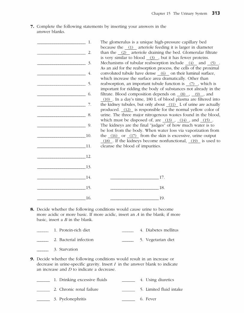
7. Complete the following statements by inserting your answers in the answer blanks.
The glomerulus is a unique high-pressure capillary bed because the (1) arteriole feeding it is larger in diameter than the (2) arterioledrainingthebed.Glomerularfiltrate is very similar to blood (3) , but it has fewer proteins. Mechanisms of tubular reabsorption include (4) and (5) . As an aid for the reabsorption process, the cells of the proximal convoluted tubule have dense (6) on their luminal surface, which increase the surface area dramatically. Other than reabsorp tion, an important tubule function is (7) , which is impor tant for ridding the body of substances not already in the filtrate. Blood composition depends on (8) , (9) , and
(10) .Inaday’stime,180Lofbloodplasmaarefilteredintothe kidney tubules, but only about (11) Lofurineareactuallyproduced. (12) is responsible for the normal yellow color of urine. The three major nitrogenous wastes found in the blood, which must be disposed of, are (13) , (14) , and (15) . The kidneys are the final “judges” of how much water is to be lost from the body. When water loss via vaporization from the (16) or (17) from the skin is excessive, urine output
(18) . If the kidneys become nonfunc tional, (19) is used to cleanse the blood of impurities.
8. Decide whether the following conditions would cause urine to become more acidic or more basic. If more acidic, insert an A in the blank; if more basic, insert a B in the blank.
______ 1. Protein-rich diet ______ 4. Diabetes mellitus
______ 2. Bacterial infection ______ 5. Vegetarian diet
______ 3. Starvation
9. Decide whether the following conditions would result in an increase or decrease in urine-specific gravity. Insert I in the answer blank to indicate an increase and D to indicate a decrease.
______ 1. Drinking excessive fluids ______ 4. Using diuretics
______ 2. Chronicrenalfailure ______ 5. Limitedfluidintake
______ 3. Pyelonephritis ______ 6. Fever
_________________________ 1.
_________________________ 2.
_________________________ 3.
_________________________ 4.
_________________________ 5.
_________________________ 6.
_________________________ 7.
_________________________ 8.
_________________________ 9.
_________________________ 10.
_________________________ 11.
_________________________ 12.
_________________________ 13.
_________________________ 14. _________________________ 17.
_________________________ 15. _________________________ 18.
_________________________ 16. _________________________ 19.
Chapter 15 The Urinary System 313

10. Assuming normal conditions, note whether each of the following substances would be (G ) in greater concentration in the urine than in the glomerular filtrate, (L ) in lesser concentration in the urine than in the glomerular filtrate, or (A) absent in both urine and glomerular filtrate. Place the correct letter in the answer blanks.
______ 1.Water ______ 5. Glucose ______ 9. Potassiumions
______ 2.Urea ______ 6. Albumin ______ 10. Redbloodcells
______ 3. Uric acid ______ 7. Creatinine ______ 11. Sodium ions
______ 4. Pus (white ______ 8. Hydrogen ions ______ 12. Amino acids blood cells)
11. Several specific terms are used to indicate the presence of abnormal urine constituents. Identify each of the following abnormalities by inserting the term that names the condition in the spaces provided. Then, for each condi-tion, provide one possible cause of the condition in the remaining spaces.
1. Presenceofredbloodcells:________________. Cause:___________________________________
2. Presenceofketones:_______________________. Cause:___________________________________
3. Presenceofalbumin:_______________________. Cause:___________________________________
4. Presenceofpus:____________________________. Cause:___________________________________
5. Presenceofbile:____________________________. Cause:___________________________________
6. Presenceof“sand:”__________________________. Cause:___________________________________
7. Presenceofglucose:_________________________. Cause:___________________________________
12.Glucoseandalbuminarebothnormallyabsentfromurine,butthe reasonfortheirexclusiondiffers.Respondtothefollowingquestions in the spaces provided.
1. Explain the reason for the absence of glucose in urine. ____________________________________
___________________________________________________________________________________________
2. Explain the reason for the absence of albumin in urine. ___________________________________
___________________________________________________________________________________________
314 Anatomy & Physiology Coloring Workbook

13. By which three methods is H+ concentration in body fluids regulated?
1. ___________________________________________________________________________________________
2. ___________________________________________________________________________________________
3. ___________________________________________________________________________________________
4. Which of these methods is the fastest? ______________________________________________________
5. Which acts slowly but is most important for acid-base balance? ____________________________
6. Which method removes CO2 from the body? ______________________________________________
14.Circlethecorrectterm(s)thatmatch(es)eachofthestatementsbelow:
1. Adultwithabout50%waterandlessmuscle:Maleorfemale
2. Adultwithlessbodywaterandmoreadiposetissue:Obeseorlean
3. Includesinterstitialfluidandplasma:Extracellularfluidorintracellularfluid
4. Hasanelectricchargeandconductsacurrent:Nonelectrolyteorion
5. Increaseswateroutputanddecreasesbloodvolume:IncreasedADHsecretion or decreased ADH secretion
6. Aldosterone:increasesNa+ reabsorption, increases K+ reabsorption and/or increases blood pressure
URETERS, URINARY BLADDER, AND URETHRA
15. Circle the term that does not belong in each of the following groupings. Then, fill in the answer blanks with the correct group name.
1. Urine storage Kidney Transitional epithelium Detrusor muscle Group: ___________
2. Ureter openings Urethral opening Bladder Forms urine Group: ___________
3. Surrounded by prostate gland Contains internal and external sphincters Continuous with renal pelvis Part of both urinary and reproductive systems Group: _____
4. Prostatic Female Membranous Spongy Group: ___________
Chapter 15 The Urinary System 315

16. Using the key choices, identify the structures that best fit the following descriptions. Insert the correct term(s) or corresponding letter(s) in the answer blanks. Items may have more than one answer.
Key Choices
A. Bladder B. Urethra C. Ureter
_________________________ 1. Drains the bladder
_________________________ 2. Storage area for urine
_________________________ 3. Contains the trigone
_________________________ 4. In males, has prostatic, membranous, and spongy parts
_________________________ 5. Conducts urine by peristalsis
_________________________ 6. Substantially longer in males than in females
_________________________ 7. A common site of “trapped” renal calculi
_________________________ 8. Contains transitional epithelium
_________________________ 9. Also transports sperm in males
17. Complete the following statements by inserting your answers in the answer blanks.
Another term that means voiding or emptying of the bladder is (1) . Voiding has both voluntary and involuntary aspects. As urine accumulates in the bladder, (2) receptors are activated. This results in a reflex that causes the muscular wall of the bladder to (3) , and urine is forced past the (4) urethral sphincter. The more distal (5) urethral sphincter is controlled (6) ; thus, an individual can temporarily postpone emptying the bladder until it has accumulated about (7) mLof urine. (8) is a condition in which voiding cannot be voluntarily controlled. It is normal in (9) because nervous control of the voluntary sphincter has not been achieved. Other conditions that might result in an inability to control the sphincter include (10) and (11) . (12) is essentially the opposite of incontinence and often is a problem in elderly men because of (13) enlargement.
_________________________ 13.
316 Anatomy & Physiology Coloring Workbook
_________________________ 1.
_________________________ 2.
_________________________ 3.
_________________________ 4.
_________________________ 5.
_________________________ 6.
_________________________ 7.
_________________________ 8.
_________________________ 9.
_________________________ 10.
_________________________ 11.
_________________________ 12.

18. Using the key choices, complete the crossword puzzle by answering each of the clues provided.
Key Choices
Anuria Hyperplasia Oliguria PyelonephritisCystitis Insipidus Polyuria PyuriaGlomerulonephritis Mellitus Ptosis UremiaHydronephrosis
Across 4. Inflammation of a kidney. 5. Dropping of the kidney to a more inferior position in the abdomen; may
result from a rapid weight loss that decreases the fatty cushion surrounding the kidney.
Down 1. Backup of urine into the kidney; often a result of a blockage in the urinary tract. 2. Diabetes _____ is a condition in which excessive amounts of urine are produced
because of a deficiency of antidiuretic hormone (ADH). 3. Toxic condition caused by renal failure.
1
2 3
4
5
FLUID, ELECTROLYTE, AND ACID-BASE BALANCE
19. Determine if the following descriptions refer to electrolytes (E) or to nonelectrolytes (N ).
______ 1. Lipids,monosaccharides,andneutralfats
______ 2. Have greater osmotic power at equal concentrations
______ 3. The most numerous solutes in the body’s fluid compartments
______ 4. Salts, acids, and bases
______ 5. Most of the mass of dissolved solutes in the body’s fluid compartments
______ 6. Dissociates into two or more ions
Chapter 15 The Urinary System 317

20. Circle the term that does not belong in each of the following groupings. Then, fill in the answer blanks with the correct group name.
1. Hypothalamus Aldosterone Osmoreceptors Group: ___________
2. Glomerulus Secretion Filtration ↑BP Group: ___________
3. ↑K+ secretion ↑Na+ reabsorption ↑K+ reabsorption ↑BP Group: ___________
4. ADH ↓BP ↑Blood volume ↑Water reabsorption Group: ___________
5. ↓Urine pH ↑H+ in urine ↑HCO3– in urine ↑Ketones Group: ___________
6. ↑ADH Dilute urine ↑Water absorption by collecting ducts Dehydration Group: ___________
7. Renin Angiotensin ↓BP Juxtaglomerular apparatus Group: ___________
21. Figure 15–5 illustrates the three major fluid compartments of the body. Arrows indicate direction of fluid flow.
(A) Select three different colors and color the coding circles and the fluid compartments on the figure.
(B)ReferringtoFigure15–5,respondtothestatementsthatfollow.Ifa statement is true, write T in the answer blank. If a statement is false, change the underlined word(s) and write the correct word(s) in the answer blank.
○ Interstitial fluid ○ Intracellular fluid ○ Plasma
318 Anatomy & Physiology Coloring Workbook
Tissue cellswith nuclei
Capillary
Blood
Proteinswithincapillary
Blood
A
BC
D
Figure 15–5
_________________________ 1. Exchanges between plasma and interstitial fluid compartments take place across the capillary membranes.
_________________________ 2. The fluid flow indicated by arrow A is driven by active transport.

_________________________ 3. If the osmolarity of the ECF is increased, the fluid flow indicated by arrow C will occur.
_________________________ 4. The excess of fluid flow at arrow A over that at arrow B normally enters the tissue cells.
_________________________ 5. Exchanges between the interstitial and intracellular fluid com- partments occur across capillary membranes.
_________________________ 6. Interstitial fluid serves as the link between the body’s external and internal environments.
22.Namethreesourcesofbodywaterandspecifywhichsourceaccountsforthebulk of body water.
_______________________________________________________________________________________________
_______________________________________________________________________________________________
_______________________________________________________________________________________________
23.Namefourroutesbywhichwaterislostfromthebodyandspecifywhichroute accounts for the greatest water loss.
________________________________________ ________________________________________
________________________________________ ________________________________________
24. Match the pH values in Column B with the conditions described in Column A.
Column A Column B
_________________________1.NormalpHofarterialblood A. pH6 7.00
_________________________ 2. Physiological alkalosis (arterial blood) B. pH = 7.00
_________________________ 3. Physiological acidosis (arterial blood) C. pH 6 7.35
_________________________ 4. Chemical neutrality; neither acidic nor basic D. pH = 7.35
_________________________ 5. Chemical acidity E. pH = 7.40
F. pH 7 7.45
25. Use the terms in Column B to complete the statements in Column A.
Column A Column B
_________________________ 1. Acids are proton ______. A. Acceptors
_________________________ 2. A strong acid dissociates ______. B. Donors
_________________________ 3. A weak acid dissociates ______. C. Completely
_________________________ 4. Strong bases bind ______ quickly. D. Hydrogen ions
E. Incompletely
26.Theactivityofthebicarbonatebuffersystemofthebloodisshownbytheequation:
CO2 + H2O ~ H2CO3 ~ H+ + HCO2–
1. Which chemical formulas refer to ions? ____________________________________________________
2. Which formula refers to a weak acid? _________________ Which is a weak base? ________________
3. If more CO2 enters the blood, the reaction shifts up to the (right/left) ______________________.
Chapter 15 The Urinary System 319

DEVELOPMENTAL ASPECTS OF THE URINARY SYSTEM
27. Complete the following statements by inserting your responses in the answer blanks.
Three separate sets of renal tubules develop in the embryo; however, embryonic nitrogenous wastes are actually disposed of by the (1) . A congenital condition typified by blister-like sacs in the kidneys is (2) disease. (3) is a congenital condition seen in (4) , when the urethral opening is located ventrally on the penis. A newborn baby voids frequently, which reflects its small (5) . Daytime control of the voluntary ure-thral sphincter is usually achieved by approximately (6) months. Urinary tract infections are fairly common and not usually severe with proper medical treatment. A particularly problematic condition, called (7) , may result later in life as a sequel to childhood streptococcal infection. In this disease, the renal filters become clogged with (8) complexes, urine output decreases, and (9) and (10) begin to appear in the urine. In old age, progressive (11) of the renal blood vessels results in the death of (12) cells. The loss of bladder tone leads to (13) and (14) and is particularly troublesome to elderly people.
_________________________ 13.
_________________________ 14.
A Visualization Exercise for the Urinary System
You see the kidney looming brownish red through the artery wall.
28. Where necessary, complete statements by inserting the missing word(s) in the answer blanks.
For your journey through the urinary system, you must be made small enough to filter through the filtration membrane from the bloodstream into a renal (1) . You will be injected into the subclavian vein and must pass through the heart before entering the arterial circulation. As you travel through
the systemic circulation, you have at least 2 minutes to relax before reaching the (2) artery, feed-ing a kidney. You see the kidney looming brownish red through the artery wall. Once you have entered the kidney, the blood vessel conduits become increasingly smaller until you finally reach
320 Anatomy & Physiology Coloring Workbook
_________________________ 1.
_________________________ 2.
_________________________ 1.
_________________________ 2.
_________________________ 3.
_________________________ 4.
_________________________ 5.
_________________________ 6.
_________________________ 7.
_________________________ 8.
_________________________ 9.
_________________________ 10.
_________________________ 11.
_________________________ 12.
INCREDIBLE JOURNEY

the (3) arteriole, feeding into the filtering device, or (4) . Once in the filter, you maneuver yourself so that you are directly in front of a pore. Within a fraction of a second, you are swept across the filtration membrane into the (5) part of the nephron. Drifting along, you lower the specimen cup to gather your first filtrate sample for testing. You study the readout from the sample and note that it is very similar in composition to (6) ,withoneexception:Thereareessenti-ally no (7) . Your next sample doesn’t have to be collected until you reach the “hairpin,” or, using the proper terminol-ogy, the (8) part of the tubule. As you continue your jour-ney, you notice that the tubule cells have dense finger-like projections extending from their surfaces into the lumen of the tubule. These are (9) , which increase the surface area of tubules because this portion of the tubule is very active in the process of (10) . Soon you collect your second sample, and then later, in the distal convoluted tubule, your third sample. When you read the computer’s summary of the third sample, you make the following notes in your register.
• Virtuallynonutrientssuchas (11) and (12) are left in the filtrate.
• ThepHisacidic,6.0.ThisisquiteachangefromthepHof (13) recorded for the newly formed filtrate.
• Thereisamuchhigherconcentrationof (14) wastes here.
• Therearemanyfewer (15) ions but more of the (16) ions noted.
• Thecolorofthefiltrateisyellow,indicatingahighrelativeconcentration of the pigment (17) .
Gradually,youbecomeawarethatyouaremovingalongmuch more quickly. You see that the water level has dropped dramatically and that the stream is turbulent and rushing. As you notice this, you realize that the hormone (18) must have been released recently to cause this water drop. You take an abrupt right turn and then drop straight downward. You realize that you must be in a (19) . Within a few seconds, you are in what appears to be a large tranquil sea with a tide flowing toward a darkened area at the far shore. You drift toward the darkened area, confident that you are in
the kidney (20) . As you reach and enter the dark tubelike structure seen from the opposite shore, your progress becomes rhythmic—something like being squeezed through a sausage skin. Then you realize that your progress is being regulated by the process of (21) . Suddenly, you free-fall and land in the previously stored (22) in the bladder, where the air is very close. Soon the walls of the bladder begin to gyrate, and you realize you are witnessing a (23) reflex. In a moment, you are propelled out of the bladder and through the (24) to exit from your host.
Chapter 15 The Urinary System 321
_________________________ 3.
_________________________ 4.
_________________________ 5.
_________________________ 6.
_________________________ 7.
_________________________ 8.
_________________________ 9.
_________________________ 10.
_________________________ 11.
_________________________ 12.
_________________________ 13.
_________________________ 14.
_________________________ 15.
_________________________ 16.
_________________________ 17.
_________________________ 18.
_________________________ 19.
_________________________ 20.
_________________________ 21.
_________________________ 22.
_________________________ 23.
_________________________ 24.

29. A man was admitted to the hospital after being trampled by his horse. He received crushing blows to his lower back on both sides. He is in consider-ablepain,andhischartshowsaurineoutputof70mLinthelast24hours.What is this specific symptom called? What will be required if the renal effects of his trauma persist?
30. Four-year-old Eddie is a chronic bed wetter. He wets the bed nearly every night. What might explain his problem?
31. If a tumor of the glucocorticoid-secreting cells of the adrenal cortex crowds out the cells that produce aldosterone, what is the likely effect on urine composition and volume?
32. Jimmy has been stressed out lately as he has been juggling two jobs while taking classes at a local college. He appears at the clinic complaining of a pounding headache. Tests show that he has high blood pressure, and his cortiscosteroid levels are elevated. What is the relationship between his stress and his signs and symptoms?
33. Mr. O’Toole is very drunk when he is brought to the emergency room after falling down City Hall steps during a political rally. He is complaining about his “cotton mouth.” Knowing that alcohol inhibits ADH’s action, you explain to him why his mouth is so dry. What do you tell him?
322 Anatomy & Physiology Coloring Workbook
AT THE CLINIC

34.Mrs.Rodriquesisbreathingrapidlyandisslurringherspeechwhenherhus-band calls the clinic in a panic. Shortly after, she becomes comatose. Tests show that her blood glucose and ketone levels are high, and her husband said that she was urinating every few minutes before she became lethargic. WhatisMrs.Rodriques’sproblem?WouldyouexpectherbloodpHtobeacidic or alkaline? What is the significance of her rapid breathing? Are her kidneys reabsorbing or secreting bicarbonate ions during this crisis?
35. Many employers now require that prospective employees’ urine be tested before they will consider hiring them. What aspect of kidney function is involved here and what is being investigated?
36. Conn’s syndrome results from adrenocortical tumors that secrete aldosterone in an unregulated way. What would you “guesstimate” would be the major symptom of this syndrome?
37. Select the best answer or answers from the choices given.
Chapter 15 The Urinary System 323
THE FINALE: MULTIPLE CHOICE
1. A radiologist is examining an X-ray of the lumbar region of a patient. Which of the following is (are) indicative of normal posi-tioning of the right kidney?
A. Slightly lower than the left kidney
B. More medial than the left kidney
C. Closer to the inferior vena cava than the left kidney
D. Anterior to the 12th rib
2. Which of the following encloses both kidney and adrenal gland?
A. Renalfascia
B. Perirenal fat capsule
C. Fibrous capsule
D. Visceral peritoneum

3. Microscopic examination of a section of the kidney shows a thick-walled vessel with renal corpuscles scattered in the tissue on one side of the vessel but not on the other side. Which vessel is this?
A. Interlobar artery
B. Cortical radiate artery
C. Cortical radiate vein
D. Arcuate artery
4. Structures that are at least partly composed ofsimplesquamousepitheliuminclude:
A. collecting ducts.
B. glomerulus.
C. glomerular capsule.
D. nephron loop.
5. Which structures are freely permeable to water?
A. Distal convoluted tubule
B. Thick segment of ascending limb of the nephron loop
C. Descending limb of the nephron loop
D. Proximal convoluted tubule
6.Amajorfunctionofthecollectingductsis:
A. secretion.
B. filtration.
C. concentrating urine.
D. lubrication with mucus.
7. What is the glomerulus?
A. The same as the renal corpuscle
B. The same as the renal tubule
C. The same as the nephron
D. Capillaries
8. Urine passes through the ureters by which mechanism?
A. Ciliary action
B. Peristalsis
C. Gravityalone
D. Suction
9.Sodiumdeficiencyhampersreabsorptionof:
A. glucose.
B. albumin.
C. creatinine.
D. water.
10. The main function of transitional epithelium intheureteris:
A. protection against kidney stones.
B. secretion of mucus.
C. reabsorption.
D. stretching.
11. Jim was standing at a urinal in a crowded public restroom and a long line was forming behind him. He became anxious (sym-pathetic response) and found he could not micturate no matter how hard he tried. Use logic to deduce Jim’s problem.
A. His internal urethral sphincter was con-stricted and would not relax.
B. His external urethral sphincter was con-stricted and would not relax.
C. His detrusor muscle was contracting too hard.
D. He almost certainly had a burst bladder.
12. Which of the following are normal values?
A. Urineoutputof1.5L/day
B. Specific gravity of 1.5
C. pH of 6
D. GFRof125mL/hour
13.Theureter:
A. is continuous with the renal pelvis.
B. is lined by the renal capsule.
C. exhibits peristalsis.
D. is much longer in the male than in the female.
14.Theurinarybladder:
A. is lined with transitional epithelium.
B. has a thick, muscular wall.
C. receives the ureteral orifices at its superior aspect.
D. is innervated by the renal plexus.
324 Anatomy & Physiology Coloring Workbook

15. Which of the following are controlled voluntarily?
A. Detrusor muscle
B. Internal urethral sphincter
C. External urethral sphincter
D. Levatoranimuscle
16.InmovementbetweenIFandICF:
A. water flow is bidirectional.
B. nutrient flow is unidirectional.
C. ion flow is selectively permitted.
D. ion fluxes are not permitted.
17. Which is a normal value for percentage of body weight that is water for a middle-aged man?
A. 73% C. 45%
B. 50% D. 60%
18.Thesmallestfluidcompartmentisthe:
A. ICF. C. plasma.
B. ECF. D. IF.
19. Which of the following are electrolytes?
A. Glucose
B. Lacticacid
C. Urea
D. Bicarbonate
20.Chlorideionreabsorption:
A. exactly parallels sodium ion reabsorption.
B. fluctuates according to blood pH.
C. increases during acidosis.
D. is controlled directly by aldosterone.
21.Respiratoryacidosisoccursin:
A. asthma.
B. emphysema.
C. barbiturate overdose.
D. cystic fibrosis.
22.Hyperkalemia:
A. triggers secretion of aldosterone.
B. may result from severe alcoholism.
C. disturbs acid-base balance.
D. results from widespread tissue injury.
23.Renaltubularsecretionofpotassiumis:
A. obligatory.
B. increased by aldosterone.
C. balanced by tubular reabsorption.
D. increased in alkalosis.
24. Which buffer system(s) is (are) not important urine buffers?
A. Phosphate C. Protein
B. Ammonium D. Bicarbonate
Chapter 15 The Urinary System 325

This page intentionally left blank

THE REPRODUCTIVE SYSTEM
16
The biological function of the reproductive system is to produce offspring. The essential organs are those producing the germ cells (testes in males and ovaries in females). The male manufactures sperm and delivers them to the female’s reproductive tract. The female, in turn, produces eggs. If the time is suitable, the egg and sperm fuse, producing a fertilized egg, which is the first cell of the new individual. Once fertilization has occurred, the female uterus protects and nurtures the developing embryo.
In this chapter, student activities concern the structures of the male and female reproductive systems, germ cell formation, the menstrual cycle, and embryonic development.
ANATOMY OF THE MALE REPRODUCTIVE SYSTEM
1. Using the following terms, trace the pathway of sperm from the testis to the urethra: rete testis, epididymis, seminiferous tubule, ductus deferens. List the terms in the proper order in the spaces provided.
.
2. How do the scrotal muscles help maintain temperature homeostasis of the testes?
_______________________________________________________________________________________________
_______________________________________________________________________________________________
_______________________________________________________________________________________________
327

3. Using the key choices, complete the crossword puzzle by answering each of the clues provided.
Key Choices
Bulbourethral glands Glans penis Scrotum UrethraDuctus deferens Penis Seminal vesicles Urinary bladderEjaculatory duct Prepuce Spermatic cordEpididymis Prostate Testes
Across 1. Tubular storage site for sperm; hugs the posterior aspect of the testes. 5. Cuff of skin encircling the glans penis. 7. Site of testosterone production. 9. Surrounds the urethra at the base of the bladder; produces a milky fluid. 10. Connective tissue sheath enclosing the ductus deferens, blood vessels,
and nerves.
Down 2. Passageway from the epididymis to the ejaculatory duct. 3. Produce more than half of the seminal fluid. 4. Organ that delivers semen to the female reproductive tract. 6. Conveys both sperm and urine down the length of the penis. 8. External skin sac that houses the testes.
1 2 3
4
5
6
7 8
9
10
4. Figure 16–1 is a sagittal view of the male reproductive structures. (A) Identify the following organs on the figure by placing each
term at the end of the appropriate leader line.
Bulbo-urethral gland Erectile tissue Scrotum
Ductus deferens Glans penis Seminal vesicle
Ejaculatory duct Prepuce Testis
Epididymis Prostate Urethra
328 Anatomy & Physiology Coloring Workbook
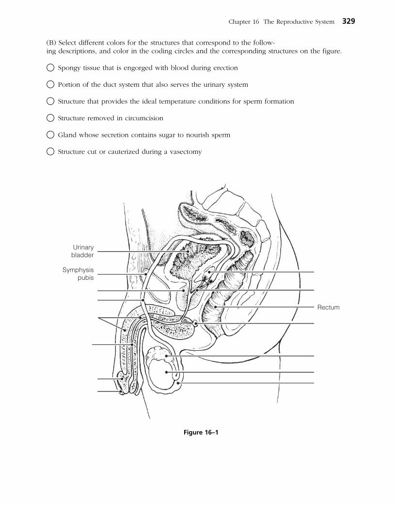
(B) Select different colors for the structures that correspond to the follow - ing descriptions, and color in the coding circles and the corre sponding structures on the figure.
○ Spongy tissue that is engorged with blood during erection
○ Portion of the duct system that also serves the urinary system
○ Structure that provides the ideal temperature conditions for sperm formation
○ Structure removed in circumcision
○ Gland whose secretion contains sugar to nourish sperm
○ Structure cut or cauterized during a vasectomy
Chapter 16 The Reproductive System 329
Figure 16–1

5. Figure 16–2 is a longitudinal section of a testis. (A) Select different colors for the structures that correspond to the following
descriptions. (B) Color the coding circles and color and label the corresponding structures
on the figure. Complete the labeling of the figure by adding the following terms: lobule, rete testis, and septum.
○ Site(s) of spermatogenesis
○ Tubular structure in which sperm mature and become motile
○ Fibrous coat protecting the testis
MALE REPRODUCTIVE FUNCTIONS
6. This section considers the process of sperm production in the testis. Figure 16–3 is a cross-sectional view of a seminiferous tubule in which spermatogenesis is occurring.
(A) Using the key choices, select the terms identified in the following descriptions.
330 Anatomy & Physiology Coloring Workbook
Ductusdeferens
Figure 16–2

Key Choices
Follicle-stimulating hormone (FSH) ○ Sperm
○ Primary spermatocyte ○ Spermatid
○ Secondary spermatocyte Testosterone
○ Spermatogonium
_________________________ 1. Primitive stem cell
_________________________ 2. Contain 23 chromosomes (3 answers)
_________________________ and _________________________
_________________________ 3. Product of meiosis I
_________________________ 4. Product of meiosis II
_________________________ 5. Functional motile gamete
_________________________ 6. Two hormones necessary for sperm production
_________________________
(B) Label the cells with leader lines. (C) Select different colors for the cell types with color-coding circles listed in the key choices and color in the coding circles and corresponding structures on the figure. (D) Label and color the cells that produce testosterone.
Chapter 16 The Reproductive System 331
Connective tissue areabetween adjacentseminiferous tubules
Portion of seminiferoustubule wall
Figure 16–3

7. Figure 16–4 illustrates a single sperm. (A) On the figure, bracket and label the head and the midpiece and circle
and label the tail. (B) Select different colors for the structures that correspond to the following
descriptions, and color the coding circles and corresponding structures on the figure.
(C) Label the structures, using correct terminology.
○ The DNA-containing area
○ The enzyme-containing sac that aids sperm penetration of the egg
○ Metabolically active organelles that provide ATP to energize sperm movement
8. The following statements refer to events that occur during cellular division. Using the key choices, indicate in which type of cellular division the described events occur. Place the correct term or letter response in the answer blanks.
Key Choices
A. Mitosis B. Meiosis C. Both mitosis and meiosis
_________________________ 1. The final product is two daughter cells, each with 46 chromosomes.
_________________________ 2. The final product is four daughter cells, each with 23 chromosomes.
_________________________ 3. This process involves the phases prophase, metaphase, anaphase, and telophase.
_________________________ 4. This process occurs in all body tissues.
_________________________ 5. This process occurs only in the gonads.
_________________________ 6. This process increases the cell number for growth and repair.
_________________________ 7. Daughter cells have the same number and types of chromosomes as the mother cell.
332 Anatomy & Physiology Coloring Workbook
Figure 16–4

_________________________ 8. Daughter cells are different from the mother cell in their chromosomal makeup.
_________________________ 9. Chromosomes are replicated before the division process begins.
_________________________ 10. This process provides cells for the reproduction of offspring.
_________________________ 11. This process consists of two consecutive divisions of the nucleus; chromo somes are not replicated before the second division.
9. Name four of the male secondary sex characteristics. Insert your answers on the lines provided.
______________________________________________________________________________________________
______________________________________________________________________________________________
______________________________________________________________________________________________
______________________________________________________________________________________________
ANATOMY OF THE FEMALE REPRODUCTIVE SYSTEM
10. Identify the female structures described by inserting your responses in the answer blanks.
_________________________ 1. Chamber that houses the developing fetus
_________________________ 2. Canal that receives the penis during sexual intercourse
_________________________ 3. Usual site of fertilization
_________________________ 4. Erects during sexual stimulation
_________________________ 5. Duct through which the ovum travels to reach the uterus
_________________________ 6. Membrane that partially closes the vaginal canal
_________________________ 7. Primary female reproductive organ
_________________________ 8. Move to create fluid currents to draw the ovulated egg into the uterine (fallopian) tube
Chapter 16 The Reproductive System 333
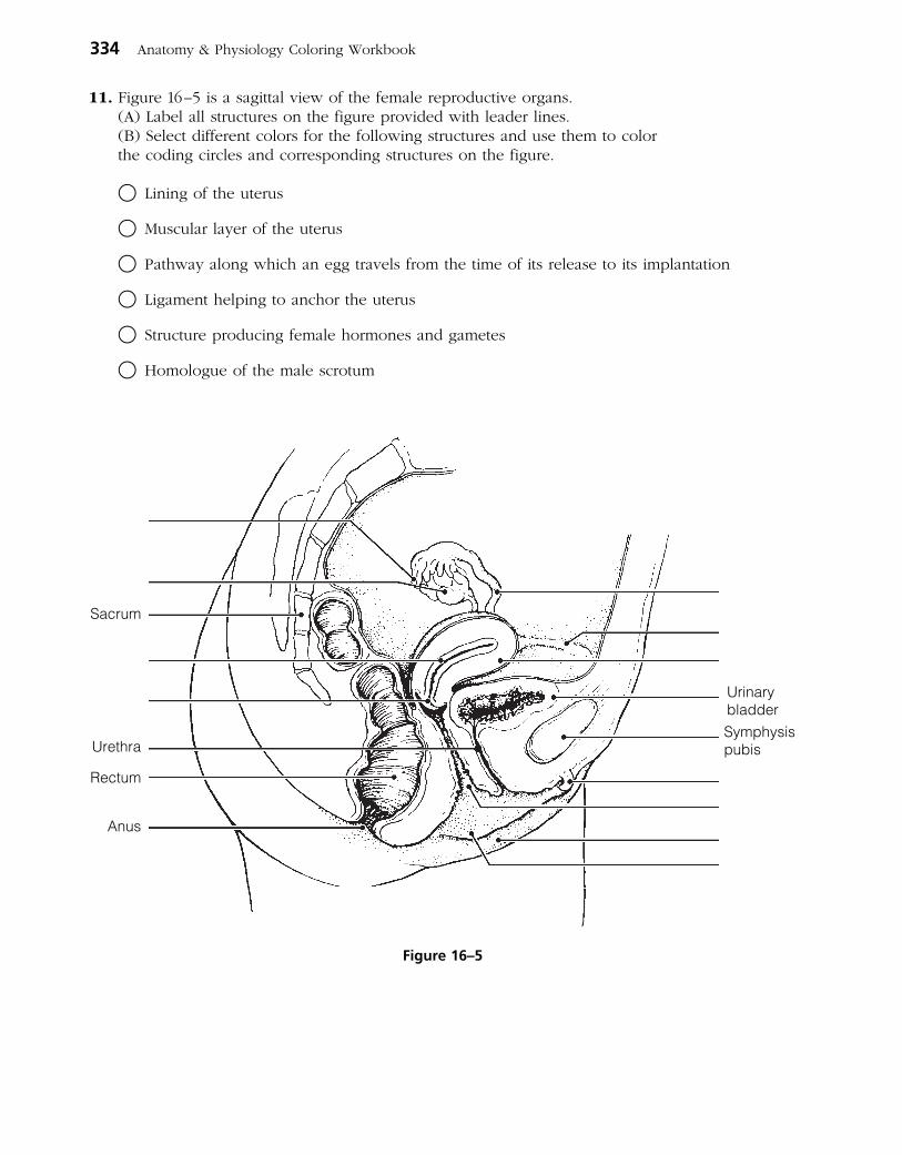
11. Figure 16–5 is a sagittal view of the female reproductive organs. (A) Label all structures on the figure provided with leader lines. (B) Select different colors for the following structures and use them to color
the coding circles and corresponding structures on the figure.
○ Lining of the uterus
○ Muscular layer of the uterus
○ Pathway along which an egg travels from the time of its release to its implantation
○ Ligament helping to anchor the uterus
○ Structure producing female hormones and gametes
○ Homologue of the male scrotum
334 Anatomy & Physiology Coloring Workbook
UrinarybladderSymphysispubis
Sacrum
Urethra
Rectum
Anus
Figure 16–5
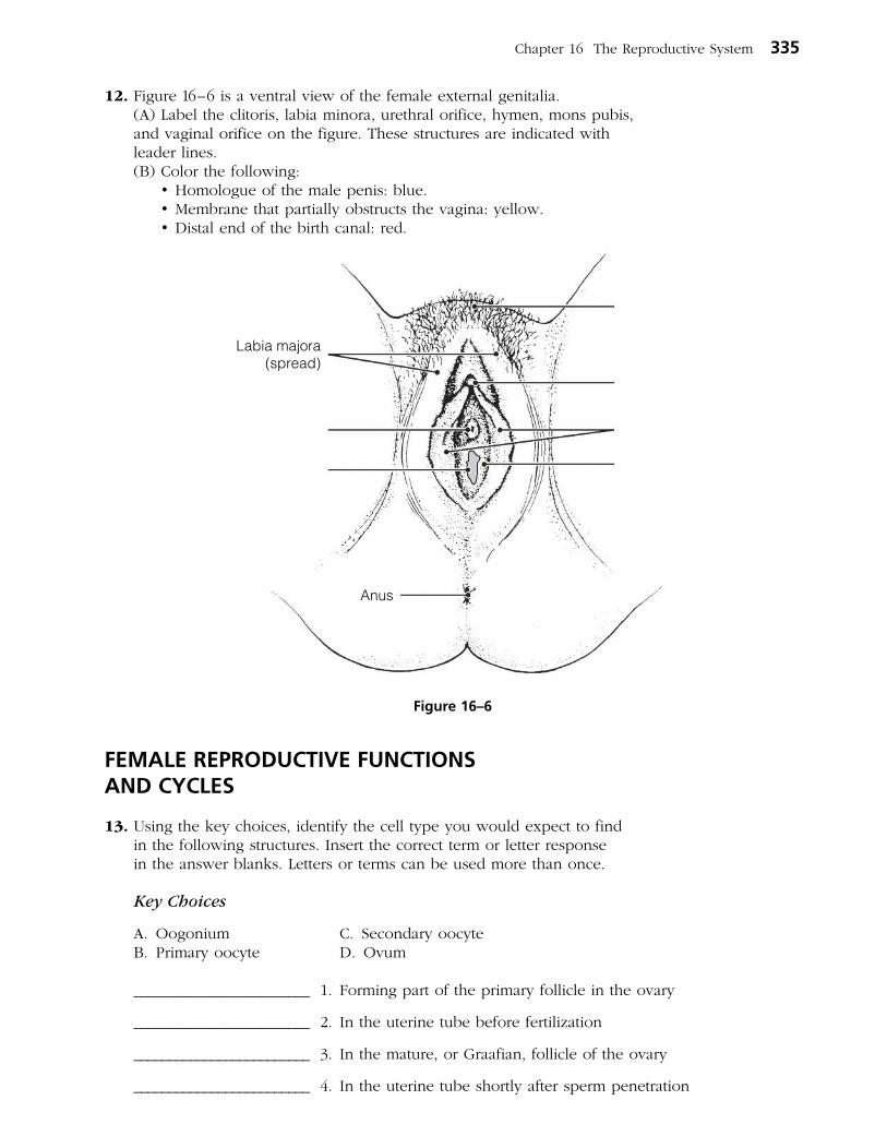
12. Figure 16–6 is a ventral view of the female external genitalia. (A) Label the clitoris, labia minora, urethral orifice, hymen, mons pubis,
and vaginal orifice on the figure. These structures are indicated with leader lines.
(B) Color the following: •Homologueofthemalepenis:blue. •Membranethatpartiallyobstructsthevagina:yellow. •Distalendofthebirthcanal:red.
FEMALE REPRODUCTIVE FUNCTIONS AND CYCLES
13. Using the key choices, identify the cell type you would expect to find in the following structures. Insert the correct term or letter response in the answer blanks. Letters or terms can be used more than once.
Key Choices
A. Oogonium C. Secondary oocyteB. Primary oocyte D. Ovum
_________________________ 1. Forming part of the primary follicle in the ovary
_________________________ 2. In the uterine tube before fertilization
_________________________ 3. In the mature, or Graafian, follicle of the ovary
_________________________ 4. In the uterine tube shortly after sperm penetration
Chapter 16 The Reproductive System 335
Labia majora(spread)
Anus
Figure 16–6

14. Figure 16–7 is a sectional view of the ovary. (A) Identify all structures indicated with leader lines on the figure. (B) Select different colors for the following structures and use them to color
the coding circles and corresponding structures on the figure.
○ Cells that produce estrogen
○ Glandular structure that produces progesterone
○ All oocytes
(C) In the space provided, name the event depicted as “Event A” on the figure. ___________
(D) Answer the following questions by inserting your answers in the spaces provided.
1. Are there any oogonia in a mature female’s ovary? ________________________________________
2. Into what area is the ovulated cell released? ______________________________________________
3. When is a mature ovum (egg) produced in humans? ______________________________________
4. What structure in the ovary becomes a corpus luteum? ____________________________________
5. What are the four final cell types produced by oogenesis in the female? (Name the cell
type and number of each.) _______________________________________________________________
336 Anatomy & Physiology Coloring Workbook
Cell type, specific
Event A
Figure 16–7

6. How does this compare with the final product of spermatogenesis in males? _______________
___________________________________________________________________________________________
7. What happens to the tiny cells nearly devoid of cytoplasm ultimately produced during
oogenesis? _______________________________________________________________________________
8. Why? ___________________________________________________________________________________
9. What name is given to the period of a woman’s life when her ovaries begin to become
nonfunctional? ___________________________________________________________________________
15. What is the significance of the fact that the uterine tubes are not structurally continuous with the ovaries? Address this question from both reproductive and health aspects.
_______________________________________________________________________________________________
_______________________________________________________________________________________________
_______________________________________________________________________________________________
16. The following statements deal with anterior pituitary and ovarian hormonal interrelationships. Name the hormone(s) described in each statement. Place your answers in the answer blanks.
_________________________ 1. Promotes growth of ovarian follicles and production of estrogen
_________________________ 2. Triggers ovulation
_________________________ 3. Inhibit follicle-stimulating hormone (FSH) release by the anterior pituitary
_________________________ 4. Stimulates luteinizing hormone (LH) release by the anterior pituitary
_________________________ 5. Converts the ruptured follicle into a corpus luteum and causes it to produce progesterone and estrogen
_________________________ 6. Maintains the hormonal production of the corpus luteum
17. Name four of the secondary sex characteristics of females. Place your answers in the spaces provided.
_______________________________________________________________________________________________
_______________________________________________________________________________________________
_______________________________________________________________________________________________
_______________________________________________________________________________________________
Chapter 16 The Reproductive System 337

18. Use the key choices to identify the ovarian hormone(s) responsible for the following events. Insert the correct term(s) or letter(s) in the answer blanks. Items may have more than one answer.
Key Choices
A. Estrogens B. Progesterone
_________________________ 1. Lack of this (these) causes the blood vessels to kink and the endometrium to slough off (menses).
_________________________ 2. This hormone causes the endometrial glands to begin the secretion of nutrients.
_________________________ 3. The endometrium is repaired and grows thick and velvety.
_________________________ 4. This hormone maintains the myometrium in an inactive state if implantation of an embryo has occurred.
_________________________ 5. Glands are formed in the endometrium.
_________________________ 6. This hormone is responsible for the secondary sex characteristics of females.
19. The following exercise refers to Figure 16–8 A–D.
(A) On Figure 16–8A: •Identifythebloodlevellinesfortheanteriorpituitarygonadotropic
hormones, FSH, and LH. •Selectdifferentcolorsforeachofthebloodlevellinesandcolorthem
in on the figure.
(B) On Figure 16–8B: •Identifythebloodlevellinesfortheovarianhormones,estrogensand
progesterone. •Selectdifferentcolorsforeachbloodlevelline,andcolortheminon
the figure.
(C) On Figure 16–8C: •Selectdifferentcolorsforthefollowingstructuresandusethemtocolor
in the coding circles and corresponding structures in the figure.
○ Primary follicle ○ Secondary (growing) follicle
○ Vesicular follicle ○ Corpus luteum
○ Ovulating follicle ○ Atretic (deteriorating) corpus luteum
(D) On Figure 16–8D: •Identifytheendometrialchangesoccurringduringthemenstrualcycle
by color-coding and coloring the areas depicting the three phases of that cycle.
○ Secretory phase ○ Menses ○ Proliferative phase
338 Anatomy & Physiology Coloring Workbook

Chapter 16 The Reproductive System 339
COva
rian
cycl
e
D
Menstrualflow
1 4 10 14Days 28
Ute
rine
(men
stru
al)
cycl
e
Blo
od le
vels
of a
nter
ior
pitu
itary
hor
mon
esB
lood
leve
ls o
fov
aria
n ho
rmon
es
Ovu
latio
n
B
A
282624222018161412108420 6
Figure 16–8

MAMMARY GLANDS
20. Figure 16–9 is a sagittal section of a breast. (A) Use the following terms to correctly label all structures provided with
leader lines on the figure.
Alveolar glands Areola Lactiferous duct Nipple
(B) Color the structures that produce milk blue and color the fatty tissue of the breast yellow.
SURVEY OF PREGNANCY AND EMBRYONIC DEVELOPMENT
21. Relative to events of sperm penetration:
1. What portion of the sperm actually enters the oocyte? _____________________________________
2. What is the functional importance of the acrosomal reaction? ______________________________
___________________________________________________________________________________________
___________________________________________________________________________________________
340 Anatomy & Physiology Coloring Workbook
Pectoralis majormuscle
Rib
Intercostalmuscles
Figure 16–9

22. Figure 16–10 depicts early embryonic events. (A) In questions #1–5, identify the events, cell types, or processes referring to
the figure. (B) Respond to question #6. Place your answers in the spaces provided.
1. Event A
2. Cell resulting from event A
3. Process B
4. Embryonic structure B1
5. Completed process C
6. Assume that a sperm has entered a polar body instead of a secondary oocyte and their nuclei fuse. Why would it be unlikely for that “fertilized cell” to develop into an embryo?
Chapter 16 The Reproductive System 341
Event Aresulting cell
Sperm
Secondaryoocyte
(enlarged)
Process B
Innercell
mass
B1
Ongoing Process C
Uterus
Ovary
Figure 16–10

23. Using the key choices, select the terms that are identified in the following descriptions. Insert the correct term(s) or letter(s) response in the answer blanks. Letters or terms may be used more than once and items may have more than one answer.
Key Choices
A. Amnion D. Fertilization G. Umbilical cordB. Chorionic villi E. Fetus H. ZygoteC. Endometrium F. Placenta
_________________________ 1. The fertilized egg
_________________________ 2. Secretes estrogen and progesterone to maintain the pregnancy
_________________________ 3. Cooperate to form the placenta
_________________________ 4. Fluid-filled sac surrounding the developing embryo/fetus
_________________________ 5. Attaches the embryo to the placenta
_________________________ 6. Finger-like projections of the blastocyst
_________________________ 7. The embryo after 8 weeks
_________________________ 8. The organ that delivers nutrients to and disposes of wastes for the fetus
_________________________ 9. Event leading to combination of ovum and sperm “genes”
24. Explain why the corpus luteum does not stop producing its hormones (estrogens and progesterone) when fertilization has occurred.
______________________________________________________________________________________________
______________________________________________________________________________________________
25. The first “tissues” of the embryo’s body are the primary germ layers:
A. Ectoderm B. Mesoderm C. Endoderm
Indicate which germ layer gives rise to each of the following structures by placing the corresponding letter in the answer blank.
______ 1. Heart and blood vessels ______ 5. Skin epidermis
______ 2. Digestive system mucosa ______ 6. Bones
______ 3. Brain and spinal cord ______ 7. Respiratory system mucosa
______ 4. Skeletal muscles ______ 8. Liver and pancreas
342 Anatomy & Physiology Coloring Workbook
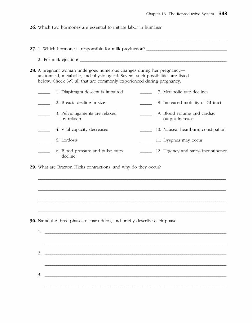
26. Which two hormones are essential to initiate labor in humans?
______________________________________________________________________________________________
27. 1. Which hormone is responsible for milk production? ________________________________________
2. For milk ejection? _________________________________________________________________________
28. A pregnant woman undergoes numerous changes during her pregnancy— anatomical, metabolic, and physiological. Several such possibilities are listed below. Check (✓) all that are commonly experienced during pregnancy.
______ 1. Diaphragm descent is impaired
______ 2. Breasts decline in size
______ 3. Pelvic ligaments are relaxed by relaxin
______ 4. Vital capacity decreases
______ 5. Lordosis
______ 6. Blood pressure and pulse rates decline
29. What are Braxton Hicks contractions, and why do they occur?
______________________________________________________________________________________________
______________________________________________________________________________________________
______________________________________________________________________________________________
______________________________________________________________________________________________
30. Name the three phases of parturition, and briefly describe each phase.
1. ___________________________________________________________________________________________
___________________________________________________________________________________________
2. ___________________________________________________________________________________________
___________________________________________________________________________________________
3. ___________________________________________________________________________________________
___________________________________________________________________________________________
Chapter 16 The Reproductive System 343
______ 7. Metabolic rate declines
______ 8. Increased mobility of GI tract
______ 9. Blood volume and cardiac output increase
______ 10. Nausea, heartburn, constipation
______ 11. Dyspnea may occur
______ 12. Urgency and stress incontinence
31. The very simple flowchart in Figure 16–11 illustrates the sequence of events that occur during labor. Complete the flowchart by filling in the missing terms in the boxes. Use color as desired.
32. How long will the cycle illustrated in Figure 16–11 continue to occur?
_______________________________________________________________________________________________
33. Labor is an example of a positive feedback mechanism. What does that mean?
_______________________________________________________________________________________________
_______________________________________________________________________________________________
_______________________________________________________________________________________________
344 Anatomy & Physiology Coloring Workbook
Infant descends in the birthcanal; head exerts pressure
on the cervix
Contracts morevigorously
Stimulates
Afferent impulses stimulate
Posteriorpituitary
Stimulates
Releases
Figure 16–11

DEVELOPMENTAL ASPECTS OF THE REPRODUCTIVE SYSTEM
34. Complete the following statements by inserting your responses in the answer blanks.
A male embryo has (1) sex chromosomes, whereas a female has (2) . During early development, the reproductive structures of both sexes are identical, but by the 8th week, male (3) and accessory structures begin to form if testosterone is present. In the absence of testosterone, female external genitalia and (4) form. The testes of a male fetus descend to the scrotum shortly before birth. If this does not occur, the resulting condition is called (5) .
The most common problem affecting the reproductive organs of women are infections, particularly (6) , (7) , and (8) . When the entire pelvis is inflamed, the condition is called
(9) . Most male problems involve inflammations resulting from (10) disease microorganisms. A leading cause of cancer death in adult women is cancer of the (11) ; the second most common female reproductive system cancer is cancer of the (12) . Thus, a yearly (13) is a very important preventive measure for early detection of this latter cancer type. The cessation of ovulation in an aging woman is called (14) . Intense vasodilation of blood vessels in the skin lead to uncomfortable (15) . Additionally, bone mass (16) and blood levels of cholesterol (17) when levels of the hormone
(18) wane. In contrast, healthy men are able to father children well into their 8th decade of life. Postmenopausal women are particularly susceptible to (19) inflammations. The single most common problem of elderly men involves the enlargement of the (20) , which interferes with the functioning of both the (21) and (22) systems.
Chapter 16 The Reproductive System 345
_________________________ 1.
_________________________ 2.
_________________________ 3.
_________________________ 4.
_________________________ 5.
_________________________ 6.
_________________________ 7.
_________________________ 8.
_________________________ 9.
_________________________ 10.
_________________________ 11.
_________________________ 12.
_________________________ 13.
_________________________ 14.
_________________________ 15.
_________________________ 16.
_________________________ 17.
_________________________ 18.
_________________________ 19.
_________________________ 20.
_________________________ 21.
_________________________ 22.
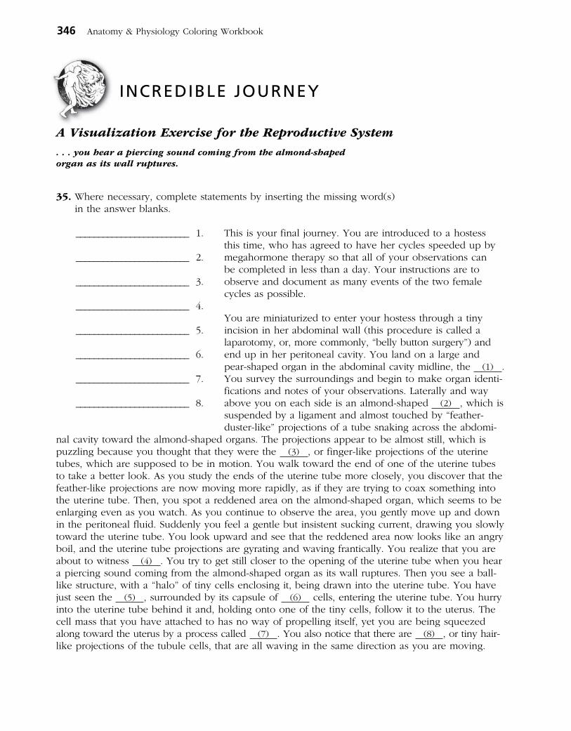
INCREDIBLE JOURNEY
A Visualization Exercise for the Reproductive System
. . . you hear a piercing sound coming from the almond-shaped organ as its wall ruptures.
35. Where necessary, complete statements by inserting the missing word(s) in the answer blanks.
This is your final journey. You are introduced to a hostess this time, who has agreed to have her cycles speeded up by mega hormone therapy so that all of your observations can be completed in less than a day. Your instructions are to observe and document as many events of the two female cycles as possible.
You are miniaturized to enter your hostess through a tiny incision in her abdominal wall (this procedure is called a laparotomy, or, more commonly, “belly button surgery”) and end up in her peritoneal cavity. You land on a large and pear-shaped organ in the abdominal cavity midline, the (1) . You survey the surroundings and begin to make organ identi-fications and notes of your observations. Laterally and way above you on each side is an almond-shaped (2) , which is suspended by a ligament and almost touched by “feather- duster-like” projections of a tube snaking across the abdomi-
nal cavity toward the almond-shaped organs. The projections appear to be almost still, which is puzzling because you thought that they were the (3) , or finger-like projections of the uterine tubes, which are supposed to be in motion. You walk toward the end of one of the uterine tubes to take a better look. As you study the ends of the uterine tube more closely, you discover that the feather-like projections are now moving more rapidly, as if they are trying to coax something into the uterine tube. Then, you spot a reddened area on the almond-shaped organ, which seems to be enlarging even as you watch. As you continue to observe the area, you gently move up and down in the peritoneal fluid. Suddenly you feel a gentle but insistent sucking current, drawing you slowly toward the uterine tube. You look upward and see that the reddened area now looks like an angry boil, and the uterine tube projections are gyrating and waving frantically. You realize that you are about to witness (4) . You try to get still closer to the opening of the uterine tube when you hear a piercing sound coming from the almond-shaped organ as its wall ruptures. Then you see a ball-like structure, with a “halo” of tiny cells enclosing it, being drawn into the uterine tube. You have just seen the (5) , surrounded by its capsule of (6) cells, entering the uterine tube. You hurry into the uterine tube behind it and, holding onto one of the tiny cells, follow it to the uterus. The cell mass that you have attached to has no way of propelling itself, yet you are being squeezed along toward the uterus by a process called (7) . You also notice that there are (8) , or tiny hair- like projections of the tubule cells, that are all waving in the same direction as you are moving.
346 Anatomy & Physiology Coloring Workbook
_________________________ 1.
_________________________ 2.
_________________________ 3.
_________________________ 4.
_________________________ 5.
_________________________ 6.
_________________________ 7.
_________________________ 8.

Nothing seems to change as you are carried along until finally you are startled by a deafening noise. Suddenly there are thousands of tadpole-like (9) swarming all around you and the sphere of cells. Their heads seem to explode as their
(10) break and liberate digestive enzymes. The cell mass now has hundreds of openings in it, and some of the small cells are beginning to fall away. As you peer through the rather transparent cell “halo,” you see that one of the tadpole- like structures has penetrated the large central cell. Chromosomes then appear, and that cell begins to divide. You have just witnessed the second (11) division. The prod- ucts of this division are one large cell, the (12) , and one very tiny cell, a (13) , which is now being ejected. This cell will soon be (14) because it has essentially no cytoplasm or food reserves. As you continue to watch, the sperm nucleus and that of the large central cell fuse, an event called (15) . You note that the new cell just formed by this fusion is called a (16) , the first cell of the embryonic body.
As you continue to move along the uterine tube, the central cell divides so fast that no cell growth occurs between the divisions. Thus, the number of cells forming the embryonic body increases, but the cells become smaller and smaller. This embryonic division process is called (17) .
Finally, the uterine chamber looms before you. As you drift into its cavity, you scrutinize its lining, the (18) . You notice that it is thick and velvety in appearance and that the fluids you are drifting in are slightly sweet. The embryo makes its first contact with the lining, detaches, and then makes a second contact at a slightly more inferior location. This time it sticks, and as you watch, the lining of the organ begins to erode away. The embryo is obviously beginning to burrow into the rich cushiony lining, and you realize that (19) is occurring.
You now leave the embryo and propel yourself well away from it. As you float in the cavity fluids, you watch the embryo disappear from sight beneath the lining. Then you continue to travel down-ward through your hostess’s reproductive tract, exiting her body at the external opening of the (20) .
Chapter 16 The Reproductive System 347
_________________________ 9.
_________________________ 10.
_________________________ 11.
_________________________ 12.
_________________________ 13.
_________________________ 14.
_________________________ 15.
_________________________ 16.
_________________________ 17.
_________________________ 18.
_________________________ 19.
_________________________ 20.
36. A 28-year-old primigravida (in first pregnancy) has been in the first stage of labor for several hours. Her uterine contractions are weak, and her labor is not progressing normally. Because the woman insists upon a vaginal delivery, the physician orders that Pitocin (a synthetic oxytocin) be infused. What will be the effect of Pitocin? What is the normal mechanism by which oxytocin acts to promote birth?
37. A 38-year-old male is upset about his low sperm count and visits a “practi-tioner” who commonly advertises his miracle cures for sterility. In fact, the practitioner is a quack who treats conditions of low sperm count with mega-doses of testosterone. Although his patients experience a huge surge in libido, their sperm count is even lower after hormone treatment. Explain why.
38. Mr. and Mrs. John Cary, a young couple who had been trying unsuccessfully to have a family for years, underwent a series of tests with a fertility clinic to try to determine the problem. Mr. Cary was found to have a normal sperm count, sperm morphology, and motility.
Mrs. Cary’s history sheet revealed that she had two episodes of pelvic inflammatory disease (PID) during her early 20s, and the time span between successive menses ranged from 21 to 30 days. She claimed that her family was “badgering” her about not giving them grandchildren and that she was frequently discouraged. A battery of hormonal tests was ordered, and Mrs. Cary was asked to perform cervical mucus testing and daily basal temperature recordings. Additionally, gas was blown through her uterine tubes to determine their patency (condition of being opened). Her tubes proved to be closed, and she was determined to be anovulatory. What do you sug-gest might have caused the closing of her tubes? Which of the tests done or ordered would have revealed her anovulatory condition?
348 Anatomy & Physiology Coloring Workbook
AT THE CLINIC

39. A man swam in a cold lake for an hour and then noticed that his scrotum was shrunken and wrinkled. His first thought was that he had lost his testicles. What had really happened?
40. Mary is a heavy smoker and has ignored a friend’s advice to stop smoking during her pregnancy. On the basis of what you know about the effect of smoking on physiology, describe how Mary’s smoking might affect her fetus.
41. Mrs. Ginko’s Pap smear shows some abnormal cells. What possibility should be investigated?
42. Mrs. Weibel has just given birth to an infant with a congenital deformity of the stomach. She is convinced that a viral infection she suffered during the third trimester of her pregnancy is responsible. Do you think she is right? Why or why not?
43. Julio is infected with gonorrhea and chlamydia. What clinical name is given to this general class of infections, and why is it crucial to inform his partners of his infection?
44. By what procedure was Julius Caesar supposedly born?
Chapter 16 The Reproductive System 349
350 Anatomy & Physiology Coloring Workbook
47. Select the best answer or answers from the choices given.
THE FINALE: MULTIPLE CHOICE
1. Which of the following structures have a region called the ampulla?
A. Ductus deferens
B. Uterine tube
C. Ejaculatory duct
D. Lactiferous duct
2. Seminal vesicle secretions have:
A. a low pH.
B. fructose.
C. a high pH.
D. sperm-activating enzymes.
3. If the uterine tube is a trumpet (“salpinx”), what part of it represents the wide, open end of the trumpet?
A. Isthmus C. Infundibulum
B. Ampulla D. Flagellum
4. The myometrium is the muscular layer of the uterus, and the endometrium is the _________________ layer.
A. serosa C. submucosa
B. adventitia D. mucosa
5. All of the following are true of the gonadotropins except that they are:
A. secreted by the pituitary gland.
B. LH and FSH.
C. hormones with important functions in both males and females.
D. the sex hormones secreted by the gonads.
6. The approximate area between the anus and clitoris in the female is the:
A. peritoneum. C. vulva.
B. perineum. D. labia.
7. A test to detect cancerous changes in cells of the uterus and cervix is:
A. pyelogram. C. D&C.
B. Pap smear. D. laparoscopy.
8. In humans, separation of the cells at the two-cell stage following fertilization may lead to the production of twins, which in this case, would be:
A. of different sexes. C. fraternal.
B. identical. D. dizygotic.
45. Jane started taking estradiol and progesterone immediately after the start of her menstrual period. What effect on ovulation should she expect?
46. Mary and Jim, fraternal twins, were enjoying a lunch together when she revealed that she had just had a tubal ligation procedure. “Oh, my gosh,” he said, “we do think alike. I just had a vasectomy.” How are these procedures alike structurally and functionally?

9. Human ova and sperm are similar in that:
A. about the same number of each is produced per month.
B. they have the same degree of motility.
C. they are about the same size.
D. they have the same number of chromosomes.
10. Which of the following attach to the ovary?
A. Fimbriae
B. Mesosalpinx
C. Suspensory ligaments
D. Broad ligament
11. As a result of crossover:
A. maternal genes can end up on a paternal chromosome.
B. synapsis occurs.
C. a tetrad is formed.
D. no two spermatids have exactly the same genetic makeup.
12. The first mitotic division in the zygote occurs as soon as:
A. male and female pronuclei fuse.
B. male and female chromosomes are replicated.
C. meiosis II in the oocyte nucleus is completed.
D. the second polar body is ejected.
13. The acrosomal reaction:
A. allows degradation of the corona radiata.
B. involves release of hyaluronidase.
C. occurs in the male urogenital tract.
D. involves only one sperm, which penetrates the oocyte membrane.
14. Which contain cells that ultimately become part of the embryo?
A. Blastocyst C. Cytotrophoblast
B. Trophoblast D. Inner cell mass
15. The blastocyst:
A. is the earliest stage at which differentia-tion is clearly evident.
B. is the stage at which implantation occurs.
C. has a three-layered inner cell mass.
D. can detect “readiness” of uterine endometrium.
16. Human chorionic gonadotropin is secreted by the:
A. trophoblast.
B. 5-month placenta.
C. chorion.
D. corpus luteum.
17. The first major event in organogenesis is:
A. gastrulation.
B. appearance of the notochord.
C. neurulation.
D. development of blood vessels in the umbilical cord.
18. Which of the following appears first in the development of the nervous system?
A. Neural crest cells
B. Neural folds
C. Neural plate
D. Neural tube
19. Which of these digestive structures develops from ectoderm?
A. Midgut
B. Liver
C. Lining of the mouth and anus
D. Lining of esophagus and pharynx
20. Mesodermal derivatives include:
A. somites.
B. mesenchyme.
C. most of the intestinal wall.
D. sweat glands.
Chapter 16 The Reproductive System 351

21. On day 17 of a woman’s monthly cycle:
A. FSH levels are rising.
B. progesterone is being secreted.
C. the ovary is in the ovulatory phase.
D. the uterus is in the proliferative phase.
22. A sudden decline in estrogen and proges-terone levels:
A. causes spasms of the spiral arteries.
B. triggers ovulation.
C. ends inhibition of FSH release.
D. causes fluid retention.
23. A sexually transmitted infection (STI) that is more easily detected in males than females, is treatable with penicillin, and can cause lesions in the nervous and cardiovascular systems is:
A. gonorrhea. C. syphilis.
B. chlamydia. D. herpes.
24. Which of the following are hormones asso-ciated with lactation?
A. Placental lactogen
B. Colostrum
C. Prolactin
D. Oxytocin
25. The outer layer of the blastocyst, which attaches to the uterine wall, is the:
A. yolk sac. C. amnion.
B. inner cell mass. D. trophoblast.
26. The notochord:
A. develops from the primitive streak.
B. develops from mesoderm beneath the primitive streak.
C. becomes the vertebral column.
D. persists as the nucleus pulposis in the intervertebral discs.
27. Amniotic fluid:
A. prevents fusion of embryonic parts.
B. contains cells and chemicals derived from the embryo.
C. is derived from embryonic endoderm.
D. helps maintain a constant temperature for the developing fetus.
28. Which of the following is a shunt to bypass the fetal liver?
A. Ductus arteriosus
B. Ductus venosus
C. Ligamentum teres
D. Umbilical vein
29. The usual and most desirable presentation for birth is:
A. vertex. C. nonvertex.
B. breech. D. head first.
352 Anatomy & Physiology Coloring Workbook

Chapter 1 The Human Body: An Orientation
An Overview of Anatomy and Physiology
1. 1. D or physiology. 2. A or anatomy. 3. B or homeostasis. 4. C or metabolism.
2. Physiological study: C, D, E, F, G, H, J, K. Anatomical study: A, B, I, K, L, M.
Levels of Structural Organization
3. Cells, tissues, organs, organ systems.
4. 1. Epithelium; Organs. 2. Heart; Basic tissue types. 3. Digestive system; Organisms.
5. Across 1. Lymphatic 4. Respiratory 6. Urinary 7. Nervous 8. Muscular 9. Skeletal 10. Endocrine
Down 2. Cardiovascular 3. Integumentary 4. Reproductive 5. Digestive
6.
Figure 1–1: Cardiovascular system Figure 1–2: Respiratory system Figure 1–3: Nervous system
Figure 1–4: Urinary system Figure 1–5: Digestive system Figure 1–6: Reproductive system
353
Answers
Nasal cavity
Trachea
LungsHeart
Bloodvessels
Spinalcord
Nerves
Brain
Kidney
Bladder
Ureter
Stomach
Esophagus
Intestines
Oral cavity
Ovary
Uterus
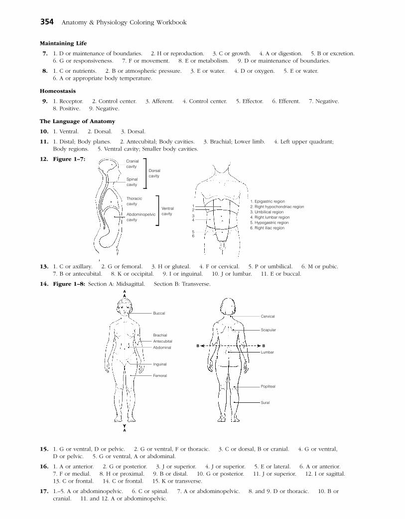
Maintaining Life
7. 1. D or maintenance of boundaries. 2. H or reproduction. 3. C or growth. 4. A or digestion. 5. B or excretion. 6. G or responsiveness. 7. F or movement. 8. E or metabolism. 9. D or maintenance of boundaries.
8. 1. C or nutrients. 2. B or atmospheric pressure. 3. E or water. 4. D or oxygen. 5. E or water. 6. A or appropriate body temperature.
Homeostasis
9. 1. Receptor. 2. Control center. 3. Afferent. 4. Control center. 5. Effector. 6. Efferent. 7. Negative. 8. Positive. 9. Negative.
The Language of Anatomy
10. 1. Ventral. 2. Dorsal. 3. Dorsal.
11. 1. Distal; Body planes. 2. Antecubital; Body cavities. 3. Brachial; Lower limb. 4. Left upper quadrant; Body regions. 5. Ventral cavity; Smaller body cavities.
12. Figure 1–7: Cranialcavity
Spinalcavity
Thoraciccavity
Abdominopelviccavity
Dorsalcavity
Ventralcavity
1234
56
1. Epigastric region2. Right hypochondriac region3. Umbilical region4. Right lumbar region5. Hypogastric region6. Right iliac region
13. 1. C or axillary. 2. G or femoral. 3. H or gluteal. 4. F or cervical. 5. P or umbilical. 6. M or pubic. 7. B or antecubital. 8. K or occipital. 9. I or inguinal. 10. J or lumbar. 11. E or buccal.
14. Figure 1–8: Section A: Midsagittal. Section B: Transverse.
Brachial
Antecubital
Inguinal
Femoral
Abdominal
A
A
Scapular
Cervical
Lumbar
Popliteal
Sural
B B
Buccal
15. 1. G or ventral, D or pelvic. 2. G or ventral, F or thoracic. 3. C or dorsal, B or cranial. 4. G or ventral, D or pelvic. 5. G or ventral, A or abdominal.
16. 1. A or anterior. 2. G or posterior. 3. J or superior. 4. J or superior. 5. E or lateral. 6. A or anterior. 7. F or medial. 8. H or proximal. 9. B or distal. 10. G or posterior. 11. J or superior. 12. I or sagittal. 13. C or frontal. 14. C or frontal. 15. K or transverse.
17. 1.–5. A or abdominopelvic. 6. C or spinal. 7. A or abdominopelvic. 8. and 9. D or thoracic. 10. B or cranial. 11. and 12. A or abdominopelvic.
354 Anatomy & Physiology Coloring Workbook
18. A. 2. B. 3. C. 1. D. 4.
At the Clinic
19. Skeletal, muscular, cardiovascular, integumentary, nervous.
20. The need for nutrients and water.
21. The anterior and lateral aspects of the abdomen have no bony (skeletal) protection.
22. John has a hernia in the area where his thigh and trunk meet, pain from his infected kidney radiating to his lower back, and bruises in his genital area.
23. Negative feedback causes the initial stimulus, TSH in this case, to decline.
24. The high blood pressure increases the workload on the heart. Circulation of blood decreases, and the heart itself begins to receive an inadequate blood supply. As the heart weakens further, the backup in the veins worsens, and the blood pressure rises even higher. Without intervention, circulation becomes so sluggish that organ failure sets in. A heart-strengthening medication will increase the force of the heartbeat so that more blood is pumped out with each beat. More blood can then flow into the heart, reducing backflow and blood pressure. The heart can then pump more blood, further reducing the backup and increasing circulation. The blood supply to the heart muscula-ture improves, and the heart becomes stronger.
25. CT and DSA utilize X-rays. MRI employs radio waves and magnetic fields. PET uses radioisotopes. CT, MRI, and PET scans can display body regions in sections.
26. Right side, below the rib cage.
27. He will apply the splint to his right wrist.
28. An MRI because it allows visualization of soft structures enclosed by bone (e.g., the skull).
The Finale: Multiple Choice
29. 1. B. 2. C. 3. A, B, C, D. 4. B. 5. B. 6. A, B, D. 7. A, B, D. 8. C. 9. C, E. 10. D. 11. A, C, E. 12. B, C, D, E. 13. A. 14. B. 15. B.
Chapter 2 Basic Chemistry
Concepts of Matter and Energy
1. 1. B, D. 2. A, B, C, D. 3. A, B.
2. 1. C or mechanical. 2. B or electrical, D or radiant. 3. C or mechanical. 4. A or chemical. 5. D or radiant.
Composition of Matter
3.
Particle Location Electrical charge Mass (amu)
Proton Nucleus +1 1
Neutron Nucleus 0 1
Electron Orbitals –1 0
4. 1. O. 2. C. 3. K. 4. I. 5. H. 6. N. 7. Ca. 8. Na. 9. P. 10. Mg. 11. Cl. 12. Fe.
5. Across 4. Atom 7. Molecule 8. Neutrons 9. Electrons
Down 1. Element 2. Valence 3. Protons 5. Matter 6. Ion 9. Energy
6. 1. T. 2. Protons. 3. More. 4. T. 5. Radioactive. 6. T. 7. Chlorine. 8. Iodine. 9. T.
Molecules, Chemical Bonds, and Chemical Reactions
7. 1. C or synthesis. 2. B or exchange. 3. A or decomposition.
8. Figure 2–1: The nucleus is the innermost circle containing 6P and 6N; the electrons are indicated by the small cir- cles in the orbits. 1. Atomic number is 6. 2. Atomic mass is 12 amu. 3. Carbon. 4. Isotope. 5. Chemi-cally active. 6. Four electrons. 7. Covalent because it would be very difficult to gain or lose four electrons.
9. H2O2 is one molecule of hydrogen peroxide (a compound). 2OH– represents two hydroxide ions.
Answers: Chapter 2 355
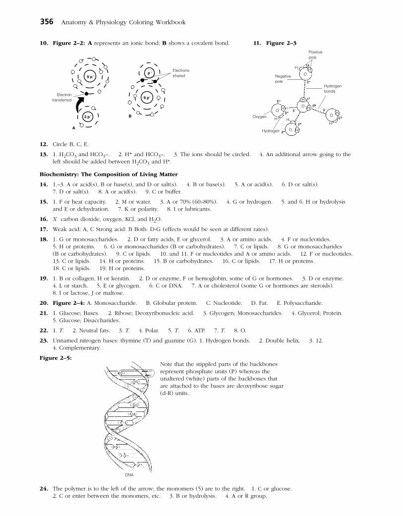
356 Anatomy & Physiology Coloring Workbook
10. Figure 2–2: A represents an ionic bond; B shows a covalent bond. 11. Figure 2–3
Positivepole
H HO
H
H
H
HH
H
H
H
OO
O
O
Hydrogenbonds
Oxygen
Hydrogen
Negativepole
p +
9 p+
9 p+
3 p+
Electrontransferred
Electronsshared
A
B
12. Circle B, C, E.
13. 1. H2CO3 and HCO3–. 2. H+ and HCO3–. 3. The ions should be circled. 4. An additional arrow going to the left should be added between H2CO3 and H+.
Biochemistry: The Composition of Living Matter
14. 1.–3. A or acid(s), B or base(s), and D or salt(s). 4. B or base(s). 5. A or acid(s). 6. D or salt(s). 7. D or salt(s). 8. A or acid(s). 9. C or buffer.
15. 1. F or heat capacity. 2. M or water. 3. A or 70% (60–80%). 4. G or hydrogen. 5. and 6. H or hydrolysis and E or dehydration. 7. K or polarity. 8. I or lubricants.
16. X carbon dioxide, oxygen, KCl, and H2O.
17. Weak acid: A, C Strong acid: B Both: D-G (effects would be seen at different rates).
18. 1. G or monosaccharides. 2. D or fatty acids, E or glycerol. 3. A or amino acids. 4. F or nucleotides. 5. H or proteins. 6. G or monosaccharides (B or carbohydrates). 7. C or lipids. 8. G or monosaccharides (B or carbohydrates). 9. C or lipids. 10. and 11. F or nucleotides and A or amino acids. 12. F or nucleotides. 13. C or lipids. 14. H or proteins. 15. B or carbohydrates. 16. C or lipids. 17. H or proteins. 18. C or lipids. 19. H or proteins.
19. 1. B or collagen, H or keratin. 2. D or enzyme, F or hemoglobin, some of G or hormones. 3. D or enzyme. 4. L or starch. 5. E or glycogen. 6. C or DNA. 7. A or cholesterol (some G or hormones are steroids). 8. I or lactose, J or maltose.
20. Figure 2–4: A. Monosaccharide. B. Globular protein. C. Nucleotide. D. Fat. E. Polysaccharide.
21. 1. Glucose; Bases. 2. Ribose; Deoxyribonucleic acid. 3. Glycogen; Monosaccharides. 4. Glycerol; Protein. 5. Glucose; Disaccharides.
22. 1. T. 2. Neutral fats. 3. T. 4. Polar. 5. T. 6. ATP. 7. T. 8. O.
23. Unnamed nitrogen bases: thymine (T) and guanine (G). 1. Hydrogen bonds. 2. Double helix. 3. 12. 4. Complementary.
Figure 2–5:
24. The polymer is to the left of the arrow; the monomers (5) are to the right. 1. C or glucose. 2. C or enter between the monomers, etc. 3. B or hydrolysis. 4. A or R group.
C G
G C
A
A
G C
G C
A T
T
A
T A
C G
T A
DNA
d-R
P
Note that the stippled parts of the backbones represent phosphate units (P) whereas the unaltered (white) parts of the backbones that are attached to the bases are deoxyribose sugar (d-R) units.

Answers: Chapter 3 357
Incredible Journey
25. 1. Negatively. 2. Positive. 3. Hydrogen bonds. 4. Red blood cells. 5. Protein. 6. Amino acids. 7. Peptide. 8. H+ and OH–. 9. Hydrolysis. 10. Enzyme. 11. Glucose. 12. Glycogen. 13. Dehydration synthesis. 14. H2O. 15. Increase.
At the Clinic
26. Acidosis means blood pH is below the normal range. The patient should be treated with something to raise the pH.
27. Each of the 20 amino acids has a different chemical group called the R group. The R group on each amino acid determines how it will fit in the folded, three-dimensional, tertiary structure of the protein and the bonds it may form. If the wrong amino acid is inserted, its R group might not fit into the tertiary structure properly, or required bonds might not be made; hence, the entire structure might be altered. Because function depends on structure, this means the protein will not function properly.
28. Heat increases the kinetic energy of molecules. Vital biological molecules, like proteins and nucleic acids, are dena-tured (rendered nonfunctional) by excessive heat because intramolecular bonds essential to their functional structure are broken. Because all enzymes are proteins, their destruction is lethal.
29. Stomach discomfort is frequently caused by excess stomach acidity (“acid indigestion”). An antacid contains a weak base that will neutralize the excess acid (H+).
30. Breaking ATP down to ADP and Pi releases the energy stored in the bonds. Only part of that potential energy is actually used by the cell. The rest is lost as heat. Nonetheless, the total amount of energy released (plus activation energy) must be absorbed to remake the bonds of ATP.
The Finale: Multiple Choice
31. 1. A, C, D. 2. E. 3. A, B, C, D, E. 4. D. 5. A. 6. C, E. 7. C, D. 8. C. 9. A. 10. A, D. 11. B, C. 12. B, C. 13. D. 14. B. 15. B, D. 16. A, B, D.
Chapter 3 Cells and Tissues
Cells
1. 1.–4. (in any order): Carbon, oxygen, nitrogen, hydrogen. 5. Water. 6. Calcium. 7. Iron. 8.–12. (five of the following, in any order): Metabolism, reproduction, irritability, mobility, ability to grow, ability to digest foods, ability to excrete waste. 13.–15. (three of the following, in any order): Cubelike, tilelike, disc-shaped, round spheres, branching, cylindrical. 16. Interstitial fluid (or ECF). 17. Squamous epithelial.
2. Figure 3–1:
CytosolVacuole
Smooth ER
Centriole(s)
Microvilli
Mitochondrion
Nuclear membrane
Golgi apparatusNucleolusChromatinthread
Rough ER
Plasma membrane
3. Figure 3–2: 1. Glycocalyx. 2. C. 3. Hydrophobic. 4. Enzymes, receptors, recognition sites, etc.
B
CCholesterol molecules
Cell interior
Phospholipidmolecules
Proteinmolecules
Carbohydratemolecules
ACell exterior
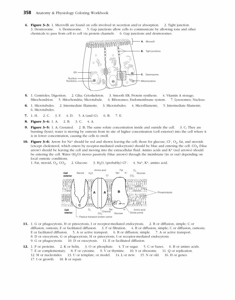
358 Anatomy & Physiology Coloring Workbook
4. Figure 3–3: 1. Microvilli are found on cells involved in secretion and/or absorption. 2. Tight junction. 3. Desmosome. 4. Desmosome. 5. Gap junctions allow cells to communicate by allowing ions and other chemicals to pass from cell to cell via protein channels. 6. Gap junctions and desmosomes.
A.
B.
C.
Microvilli
Tight junctions
Desmosome
MitochondrionNucleolusNucleus
ER
5. 1. Centrioles; Digestion. 2. Cilia; Cytoskeleton. 3. Smooth ER; Protein synthesis. 4. Vitamin A storage; Mitochondrion. 5. Mitochondria; Microtubule. 6. Ribosomes; Endomembrane system. 7. Lysosomes; Nucleus.
6. 1. Microtubules. 2. Intermediate filaments. 3. Microtubules. 4. Microfilaments. 5. Intermediate filaments. 6. Microtubules.
7. 1. H. 2. C. 3. F. 4. D. 5. A (and G). 6. B. 7. E.
8. Figure 3–4: 1. A. 2. B. 3. C. 4. A.
9. Figure 3–5: 1. A; Crenated. 2. B; The same solute concentration inside and outside the cell. 3. C; They are bursting (lysis); water is moving by osmosis from its site of higher concentration (cell exterior) into the cell where it is in lower concentration, causing the cells to swell.
10. Figure 3–6: Arrow for Na+ should be red and shown leaving the cell; those for glucose, Cl–, O2, fat, and steroids (except cholesterol, which enters by receptor-mediated endocytosis) should be blue and entering the cell. CO2 (blue arrow) should be leaving the cell and moving into the extracellular fluid. Amino acids and K+ (red arrows) should be entering the cell. Water (H2O) moves passively (blue arrows) through the membrane (in or out) depending on local osmotic conditions.
1. Fat, steroid, O2, CO2. 2. Glucose. 3. H2O, (probably) Cl–. 4. Na+, K+, amino acid.
Cellexterior
Cellinterior
Steroid H2OCO2
CO2
O2
O2
Amino acid
Amino acid
Glucose
Glucose
Cl–Na+
Na+
K+Fat
K+
Passive transport protein carrier
Channel Solute pump
Phospholipids
11. 1. G or phagocytosis, H or pinocytosis, I or receptor-mediated endocytosis. 2. B or diffusion, simple; C or diffusion, osmosis; E or facilitated diffusion. 3. F or filtration. 4. B or diffusion, simple; C or diffusion, osmosis; E or facilitated diffusion. 5. A or active transport. 6. B or diffusion, simple. 7. A or active transport. 8. D or exocytosis, G or phagocytosis, H or pinocytosis, I or receptor-mediated endocytosis. 9. G or phagocytosis. 10. D or exocytosis. 11. E or facilitated diffusion.
12. 1. P or proteins. 2. K or helix. 3. O or phosphate. 4. T or sugar. 5. C or bases. 6. B or amino acids. 7. E or complementary. 8. F or cytosine. 9. V or thymine. 10. S or ribosome. 11. Q or replication. 12. M or nucleotides. 13. U or template, or model. 14. L or new. 15. N or old. 16. H or genes. 17. I or growth. 18. R or repair.

Answers: Chapter 3 359
13. Figure 3–7: A. Prophase. B. Anaphase. C. Telophase. D. Metaphase.
Chromosomes
Spindle fibers
Nuclearmembrane
CentriolesNuclearmembrane
Nucleoli
Chromosomes
Centrioles
Spindle fibers
14. 1. C or prophase. 2. A or anaphase. 3. D or telophase. 4. D or telophase. 5. B or metaphase. 6. C or prophase. 7. C or prophase. 8. E or none of these. 9. C or prophase. 10. C or prophase. 11. D or telophase. 12. A or anaphase, B or metaphase. 13. E or none of these.
15. Across 3. Spindle 4. Binucleate 7. Centromeres
Down 1. Cytoplasm 2. Nucleus 5. Interphase 6. Coiled
16. Figure 3–8: 1. Transcription. 2. Translation. 3. Anticodon; triplet.
Nucleus Nuclearmembrane
Ribosome
12
AG C G
ACGAA
CUUAUA
TGC
TT
GA
ATAT
CGAACTTATA
G A AUAUUCAAGCC U U G A A
tRNAmolecules
Amino acidmolecules
Backbone of mRNA
Backbonesof DNA helix
Body Tissues
17. Figure 3–9:Nervous tissue: Internal communication• Brain
• Spinal cord
• Nerves
Muscle tissue: Contracts to cause movement• Muscles attached to bones (skeletal)
• Muscles of heart (cardiac)
• Muscles of walls of hollow organs (smooth)
Epithelial tissue: Forms boundaries between different environments, protects, secretes, absorbs, filters• Lining of GI tract organs and other hollow organs
• Skin surface (epidermis)
Connective tissue: Supports, protects, bindsother tissues together• Bones
• Tendons
• Fat and other soft padding tissue
18. Figure 3–10: A. Simple squamous epithelium. B. Simple cuboidal epithelium. C. Cardiac muscle. D. Dense fibrous connective tissue. E. Bone. F. Skeletal muscle. G. Nervous tissue. H. Hyaline cartilage. I. Smooth muscle tissue. J. Adipose (fat) tissue. K. Stratified squamous epithelium. L. Areolar connective tissue. The noncellular portions of D, E, H, J, and L are matrix.
19. The neuron has long cytoplasmic extensions that promote its ability to transmit impulses over long distances within the body.
20. 1. B or epithelium. 2. C or muscle. 3. D or nervous. 4. A or connective. 5. B or epithelium. 6. D or nervous. 7. C or muscle. 8. B or epithelium. 9. A or connective. 10. A or connective. 11. C or muscle. 12. A or connective. 13. D or nervous.

360 Anatomy & Physiology Coloring Workbook
21. 1. E or stratified squamous. 2. B or simple columnar. 3. E or stratified squamous. 4. A or pseudostratified columnar (ciliated). 5. A or pseudostratified columnar (ciliated). 6. F or transitional. 7. D or simple squamous.
22. 1. Skeletal. 2. Cardiac, smooth. 3. Skeletal, cardiac. 4. Smooth (most cardiac). 5.–7. Skeletal. 8. and 9. Smooth. 10. Cardiac. 11. Skeletal. 12. Cardiac. 13. Skeletal. 14. Smooth, cardiac. 15. Cardiac.
23. 1. Cell; Extracellular matrix (ECM). 2. Elastic fibers; Cellular extensions. 3. Bones; Epithelial tissue. 4. Nervous; Connective tissue. 5. Blood; Muscle tissue.
24. 1. C or dense fibrous. 2. A or adipose. 3. C or dense fibrous. 4. E or osseous tissue. 5. B or areolar. 6. D or hyaline cartilage. 7. A or adipose. 8. D or hyaline cartilage. 9. E or osseous tissue. 10. F or reticular.
25. 1. Inflammation. 2. Clotting proteins. 3. Granulation. 4. Regeneration. 5. T. 6. Collagen. 7. T.
Developmental Aspects of Cells and Tissues
26. 1. Tissues. 2. Growth. 3. Nervous. 4. Muscle. 5. Connective (scar). 6. Chemical. 7. Physical. 8. Genes (DNA). 9. Connective tissue changes. 10. Decreased endocrine system activity. 11. Dehydration of body tissues. 12. Division. 13. and 14. Benign, malignant. 15. Benign. 16. Malignant. 17. Biopsy. 18. Surgical removal. 19. Hyperplasia. 20. Atrophy.
Incredible Journey
27. 1. Cytoplasm (cytosol). 2. Nucleus. 3. Mitochondrion. 4. ATP. 5. Ribosomes. 6. Rough endoplasmic reticulum. 7. Pores. 8. Chromatin. 9. DNA. 10. Nucleoli. 11. Golgi apparatus. 12. Lysosome.
At the Clinic
28. The oxidases of ruptured peroxisomes were converting the hydrogen peroxide to water and (free) oxygen gas (which causes the bubbling).
29. Generally speaking, stratified epithelia consisting of several cell layers are more effective where abrasion is a problem than are simple epithelia (consisting of one cell layer).
30. Streptomycin inhibits bacterial protein synthesis. If the bacteria are unable to synthesize new proteins (many of which would be essential enzymes), they will die.
31. Considering connective tissue is the most widespread tissue in the body and is found either as part of or is associated with every body organ, the physician will most likely tell her that she can expect the effects of lupus to be very diffuse and widespread.
32. Granulation tissue secretes substances that kill bacteria.
33. Mitochondria are the site of most ATP synthesis, and muscle cells use tremendous amounts of ATP during contrac-tion. After ingesting bacteria or other debris, phagocytes must digest them, explaining the abundant lysosomes.
34. Recovery will be long and painful because tendons, like other dense connective tissue structures, are poorly vascularized.
35. Edema will occur because the filtration pressure exerted by the blood forces blood proteins into the interstitial space, and water follows down its concentration gradient.
36. Phagocytes engulf and remove debris from body tissues. A smoker’s lung would be expected to have carbon particles.
The Finale: Multiple Choice
37. 1. B. 2. A. 3. B. 4. C. 5. A, B, C, D. 6. C. 7. E. 8. A. 9. D. 10. C. 11. D. 12. C. 13. E. 14. C. 15. B. 16. C. 17. A, C, D. 18. A, B, C, D, E. 19. B.
Chapter 4 Skin and Body Membranes
Classification of Body Membranes
1. The mucous, serous, and cutaneous membranes are all composite membranes composed of an epithelial layer under laid by a connective tissue layer.
• Amucousmembraneisanepithelialsheetunderlaidbyaconnectivetissuelayercalledthelaminapropria. Mucosae line the respiratory, digestive, urinary, and reproductive tracts; functions include protection, lubrication, secretion, and absorption.
• Serousmembranesconsistofalayerofsimplesquamousepitheliumrestingonascantlayeroffineconnectivetissue. Serosae line internal ventral body cavities and cover their organs; their function is to produce a lubricating fluid that reduces friction.

Answers: Chapter 4 361
• Thecutaneousmembrane,orskin,iscomposedoftheepithelialepidermisandtheconnectivetissuedermis.Itcovers the body exterior and protects deeper body tissues from external insults.
The synovial membranes, which line joint cavities of synovial joints, are composed entirely of connective tissue. They function to produce lubrication to decrease friction within the joint cavity.
2. Figure 4–1: In each case, the visceral layer of the serosa covers the external surface of the organ, and the parietal layer lines the body cavity walls.
Integumentary System (Skin)
3. 1. B. 2. M. 3. C. 4. C, M. 5. C. 6. C.
4. Sunburn inhibits the immune response by depressing macrophage activity.
5. When body temperature begins to rise to undesirable levels, the sweat glands are activated by nerve fibers of the (sympathetic) nervous system. As sweat evaporates from the skin surface, it carries body heat with it.
6. 1. Nervous. 2–5. Temperature (heat and cold); Pain; Light pressure; Deep pressure. 6. Cholesterol. 7. UV light. 8. Calcium.
7. Figure 4–2:
Epidermis
Cutaneousmembrane
Dermis
Subcutaneous tissue(hypodermis)
Sweat gland
Hair shaft
Stratum corneumStratum basale
Sebaceous gland
Arrector pili muscle
Nerve fibersHair folliclePapilla of hair bulb
Fat or adipose tissue
8. 1. As the basal cells continue to divide, the more superficial cells are pushed farther and farther from the nutrient supply diffusing from the dermis. 2. Waterproofing substances (keratin and others) made by the keratinocytes effectively limit nutrient entry into the cells.
9. Across 4. Stratum corneum 5. Epidermis 6. Dermis
Down 1. Papillary layer 2. Stratum lucidum 3. Stratum basale
10. 1. Keratin; Dermis. 2. Wart; Pigment. 3. Stratum basale; Stratum spinosum. 4. Arrector pili; Sensory receptors.
11. 1. C or melanin. 2. A or carotene. 3. C or melanin. 4. B or hemoglobin. 5. C or melanin. 6. A or carotene. 7. B or hemoglobin.
12. 1. Heat. 2. Subcutaneous. 3. Vitamin D. 4. Elasticity. 5. Oxygen (blood flow). 6. Cyanosis.
13. 1. Sweat. 2. Keratin. 3. T. 4. Shaft. 5. Dermis.
Visceralpericardium(inner layer)
Parietalpericardium(outer layer)
Parietalpleura(outer layer)
Cutaneous membrane(skin)
Visceralpleura(inner layer)
Mucosae ofrespiratorytract
Mucosae ofdigestivetract
Synovial membranelining jointcavity

362 Anatomy & Physiology Coloring Workbook
14. Figure 4–3: 15. 1. Poor nutrition; Hair growth. 2. Keratin; Vitamin D production. 3. Dermis; Epidermal cells. 4. Eccrine glands; Apocrine glands. 5. Vellus hair; Terminal hair.
16. Alopecia.
17. 1. E or sebaceous glands. 2. A or arrector pili. 3. G or sweat gland (eccrine). 4. D or hair follicle(s). 5. F or sweat gland (apocrine). 6. C or hair. 7. B or cutaneous receptors. 8. E or sebaceous glands, F or sweat gland (apocrine). 9. G or sweat gland (eccrine). 10. E or sebaceous gland.
18. 1. Arrector pili; Epithelial tissue. 2. Absorption; Heat. 3. Epithelial sheath; Hair. 4. Hypodermis; Skin (cutaneous membrane). 5. Wrinkles; Skin color.
19. 1. The cuticle. 2. The stratum basale is thicker here, preventing the rosy cast of blood from flushing through.
20. Water/protein/electrolyte loss, circulatory collapse, renal shutdown.
21. 1. C or third-degree burn. 2. B or second-degree burn. 3. A or first-degree burn. 4. B or second-degree burn. 5. C or third-degree burn. 6. C or third-degree burn.
22. It allows estimation of the extent of burns so that fluid volume replacement can be correctly calculated.
23. 1. Squamous cell carcinoma. 2. Basal cell carcinoma. 3. Malignant melanoma.
24. Pigmented areas that are Asymmetrical, have irregular Borders, exhibit several Colors, and have a Diameter greater than 6 mm are likely to be cancerous.
Developmental Aspects of the Skin and Body Membranes
25. 1. C or dermatitis. 2. D or delayed-action gene. 3. F or milia. 4. B or cold intolerance. 5. A or acne. 6. G or vernix caseosa. 7. E or lanugo.
Incredible Journey
26. 1. Collagen. 2. Elastin (or elastic). 3. Dermis. 4. Phagocyte (macrophage). 5. Hair follicle connective tissue. 6. Epidermis. 7. Stratum basale. 8. Melanin. 9. Keratin. 10. Squamous (stratum corneum) cells.
At the Clinic
27. Chemotherapy drugs used to treat cancer kill the most rapidly dividing cells in the body, including many matrix cells in the hair follicles; thus, the hair falls out.
28. The baby has seborrhea, or cradle cap, a condition of overactive sebaceous glands. It is not serious; the oily deposit is easily removed with attentive washing and soon stops forming.
29. Bedridden patients are turned at regular intervals so that no region of their body is pressed against the bed long enough to deprive the blood supply to that skin; thus, bedsores are avoided.
30. The baby was cyanotic from lack of oxygen when born, a problem solved by breathing. Vernix caseosa, a cheesy substance made by the sebaceous glands covered her skin. This substance helps to protect the fetus’s skin in utero.
31. Norwegians in the United States. They are originally from a region of the world where the sun is always far away from them and have very fair skin; hence they have little protective melanin.
32. Besides storing fat as a source of nutrition, the hypodermis anchors the skin to underlying structures (such as muscles) and acts as an insulator against heat loss.
33. The body of a nail is its visible, attached part (not its white free edge). The root is the proximal part that is embed-ded in skin. The bed is the part of the epidermis upon which the nail lies. The matrix is the proximal part of the nail bed, and it is responsible for nail growth. The cuticle is the skin fold around the perimeter of the nail body. Because the matrix is gone, the nail will not grow back.
34. The peritoneum will be inflamed and infected. Because the peritoneum encloses so many richly vascularized organs, a spreading peritoneal infection can be life threatening.
35. He probably told her that regeneration would occur, and grafts would not be needed if infection was avoided.
36. Replacing lost fluid and electrolytes and prevention of infection.
37. Fat is a good insulator, so its lack or decrease results in a greater sensitivity to cold.
The Finale: Multiple Choice
38. 1. B, D. 2. D. 3. B. 4. B. 5. D. 6. C. 7. B. 8. C, D. 9. C. 10. C. 11. A, B, D. 12. D. 13. A, B. 14. B.
HairCuticle
MedullaCortex
Connectivetissue sheath
Folliclewall
Epithelialsheath

Answers: Chapter 5 363
Chapter 5 The Skeletal System
Bones—An Overview
1. 1. P. 2. P. 3. D. 4. D. 5. P. 6. D. 7. P. 8. P. 9. P.
2. 1. S. 2. F. 3. L. 4. L. 5. I. 6. L. 7. L. 8. F. 9. I.
3. 1. C or epiphysis. 2. A or diaphysis, C or epiphysis. 3. C or epiphysis, D or red marrow. 4. A or diaphysis. 5. E or yellow marrow cavity. 6. B or epiphyseal plate.
4. 1. G or parathyroid hormone. 2. F or osteocytes. 3. A or atrophy. 4. H or stress/tension. 5. D or osteoblasts. 6. B or calcitonin. 7. E or osteoclasts. 8. C or gravity.
5. Figure 5–1:
1. B or concentric lamellae. 2. C or lacunae. 3. A or central (Haversian) canal. 4. E or bone matrix. 5. D or canaliculi.
6. 1. Yellow marrow; RBC formation. 2. Osteoblasts; Blood supply. 3. Marrow cavity; Osteon. 4. Periosteum; Epiphysis.
7. Figure 5–2: The epiphyseal plate is the white band shown in the center region of the head; the articular cartilage is the white band on the external surface of the head. Red marrow is found within the spongy bone cavities; yellow marrow is found within the cavity of the diaphysis.
A B
Cavity
Compact bonePeriosteum
Endosteum
Spongy bone
Compact bone
Articular cartilageEpiphyseal plate
Diaphysis
8. 1. 4. 2. 3. 3. 2. 4. 1. 5. 5. 6. 6. NOTE: Events 2 and 3 may occur simultaneously.
Axial Skeleton
9. 1. B or frontal. 2. N or zygomatic. 3. E or mandible. 4. G or nasals. 5. I or palatines. 6. J or parietals. 7. H or occipital. 8. K or sphenoid. 9. D or lacrimals. 10. F or maxillae. 11. A or ethmoid. 12. L or temporals. 13. K or sphenoid. 14. A or ethmoid. 15. E or mandible. 16. L or temporals. 17.–20. A or ethmoid, B or frontal, F or maxillae, and K or sphenoid. 21. H or occipital. 22. H or occipital. 23. L or temporals. 24. M or vomer. 25. A or ethmoid. 26. L or temporals.
10. 1. Membranous. 2. T. 3. Osteoblasts. 4. Secondary. 5. Hyaline cartilage. 6. Endosteal. 7. T.
11. Figure 5–3:
A Squamous sutureFrontal
Coronalsuture
SphenoidZygomatic
Nasal
Zygomaticarch
Lacrimal
MandibularcondyleMaxilla
Mandible
Styloid process
Parietal
Temporal
Lambdoidsuture
Externalacousticmeatus
Mastoidprocess
Occipital
Palatine processof maxilla
OccipitalForamen magnum Occipital condyle
Parietal
Jugular foramenCarotid canalTemporalVomer
SphenoidPalatine
ZygomaticMaxilla Frontal
Ethmoid
Lacrimal
Zygomatic
Nasal
Alveolar margin
Mandible
Maxilla
Vomer
Sphenoid
Temporal
Parietal
Coronal suture
B C
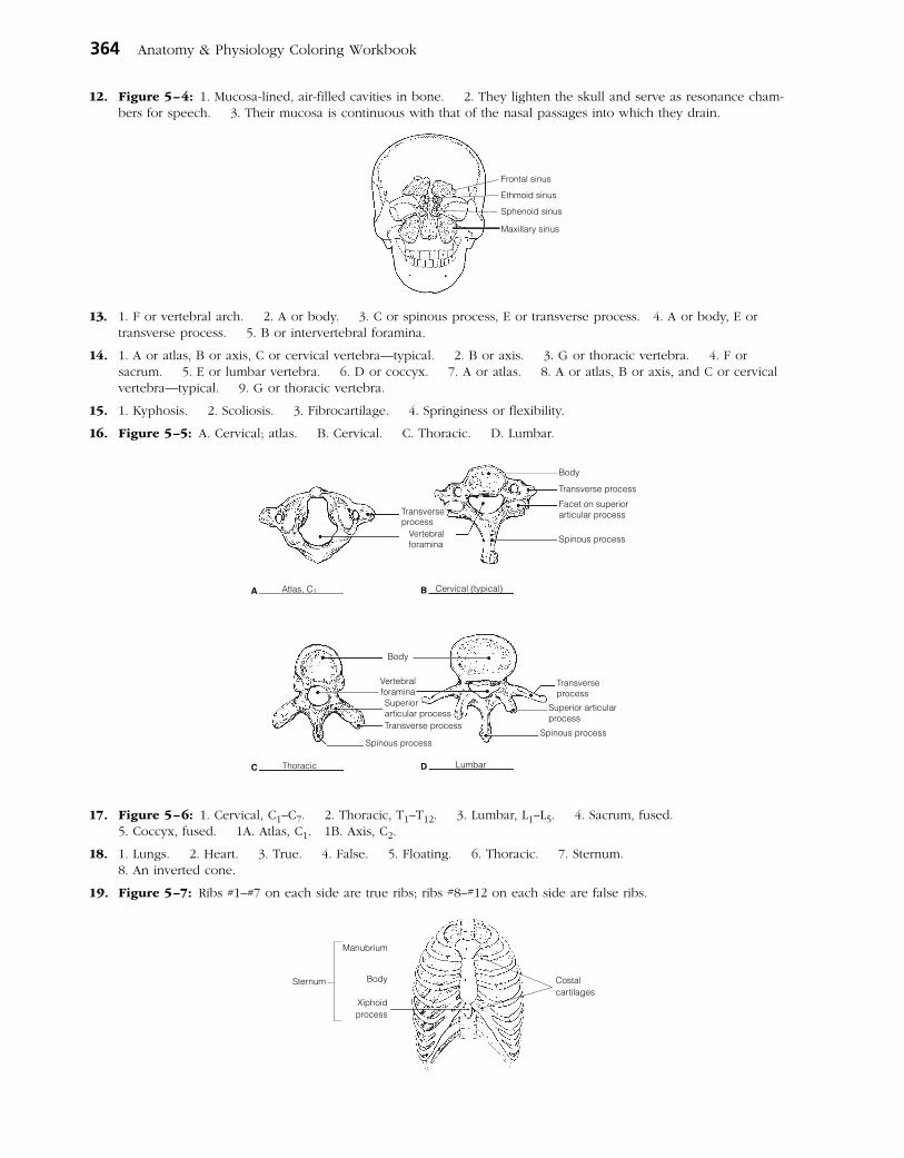
364 Anatomy & Physiology Coloring Workbook
12. Figure 5–4: 1. Mucosa-lined, air-filled cavities in bone. 2. They lighten the skull and serve as resonance cham-bers for speech. 3. Their mucosa is continuous with that of the nasal passages into which they drain.
Frontal sinus
Ethmoid sinus
Sphenoid sinus
Maxillary sinus
13. 1. F or vertebral arch. 2. A or body. 3. C or spinous process, E or transverse process. 4. A or body, E or transverse process. 5. B or intervertebral foramina.
14. 1. A or atlas, B or axis, C or cervical vertebra—typical. 2. B or axis. 3. G or thoracic vertebra. 4. F or sacrum. 5. E or lumbar vertebra. 6. D or coccyx. 7. A or atlas. 8. A or atlas, B or axis, and C or cervical vertebra—typical. 9. G or thoracic vertebra.
15. 1. Kyphosis. 2. Scoliosis. 3. Fibrocartilage. 4. Springiness or flexibility.
16. Figure 5–5: A. Cervical; atlas. B. Cervical. C. Thoracic. D. Lumbar.
A B
C D
Transverseprocess
Vertebralforamina
Body
Transverse process
Facet on superior articular process
Spinous process
Body
Superiorarticular processTransverse process
Vertebralforamina
Spinous process
Transverse process
Superior articularprocess
Spinous process
LumbarThoracic
Atlas, C1 Cervical (typical)
17. Figure 5–6: 1. Cervical, C1–C7. 2. Thoracic, T1–T12. 3. Lumbar, L1–L5. 4. Sacrum, fused. 5. Coccyx, fused. 1A. Atlas, C1. 1B. Axis, C2.
18. 1. Lungs. 2. Heart. 3. True. 4. False. 5. Floating. 6. Thoracic. 7. Sternum. 8. An inverted cone.
19. Figure 5–7: Ribs #1–#7 on each side are true ribs; ribs #8–#12 on each side are false ribs.
Manubrium
Body
Xiphoidprocess
Costalcartilages
Sternum

Answers: Chapter 5 365
Appendicular Skeleton
20. Figure 5–8: Scapula.Superior border
Lateralborder
AcromionGlenoid cavityLateral angle
Spine
Inferiorangle
Coracoid process
Superiorangle
Medialborder
21. Figure 5–9: A. Humerus. B. Ulna. C. Radius 22. Figure 5–10:
Lesser tubercleGreater tubercle
Deltoidtuberosity
Capitulum
Head
Trochlea
A Humerus
Olecranonprocess
Trochlear notchCoronoid process
Radial tuberosity
Head
Ulna Radius
Head
Styloid process
B CRadius
Ulna
Phalanges
Metacarpals
Carpals
23. Pectoral: A, C, D. Pelvic: B, E, F.
24. Across 2. Acromion 5. Deltoid tuberosity 7. Coracoid process 8. Phalanges 10. Clavicle
11. Metacarpals 14. Radius 15. Humerus
Down 1. Coronoid fossa 3. Scapula 4. Glenoid cavity 6. Trochlea 9. Ulna 12. Capitulum 13. Sternum
25. 1. Female inlet is larger and more circular. 2. Female sacrum is less curved; pubic arch is rounder. 3. Female ischial spines are shorter; pelvis is shallower/lighter.
Figure 5–11:
IliaccrestFalsepelvis
Anterior superioriliac spine
True pelvis
Acetabulum
Pubic symphysis Obturator foramen
Pubic ramisIschial spine
Pelvic brimSacrum
Coxal bone
26. 1. Ulna; Leg bones. 2. Pelvis; Axial skeleton. 3. Scapula; Coxal bone. 4. Mandible; Cranial bones. 5. Carpals; Ankle bones.
27. 1. I or ilium, K or ischium, S or pubis. 2. J or ischial tuberosity. 3. R or pubic symphysis. 4. H or iliac crest. 5. A or acetabulum. 6. T or sacroiliac joint. 7. C or femur. 8. D or fibula. 9. W or tibia. 10. C or femur, Q or patella, W or tibia. 11. X or tibial tuberosity. 12. Q or patella. 13. W or tibia. 14. N or medial malleolus. 15. L or lateral malleolus. 16. B or calcaneus. 17. U or talus, V or tarsals. 18. O or metatarsals. 19. P or obturator foramen. 20. G or greater and lesser trochanters, E or gluteal tuberosity. 21. U or talus.
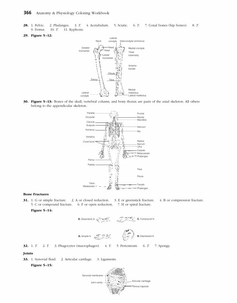
366 Anatomy & Physiology Coloring Workbook
28. 1. Pelvic. 2. Phalanges. 3. T. 4. Acetabulum. 5. Sciatic. 6. T. 7. Coxal bones (hip bones). 8. T. 9. Femur. 10. T. 11. Kyphosis.
29. Figure 5–12:
Lateralcondyle
Greatertrochanter
LateralcondyleNeck
HeadHead
Lessertrochanter
Medial condyle
Intercondylar eminence
Tibialtuberosity
Anteriorborder
MedialmalleolusLateral malleolus
Femur
Fibula
Tibia
30. Figure 5–13: Bones of the skull, vertebral column, and bony thorax are parts of the axial skeleton. All others belong to the appendicular skeleton.
MetatarsalsTalus
Patella
Femur
Coxal bone
Vertebra
Humerus
ScapulaClavicle
Occipital
Parietal Frontal
MaxillaMandible
Sternum
Rib
RadiusSacrumUlna
CarpalsMetacarpalsPhalanges
Tibia
Fibula
Tarsals
Phalanges
Bone Fractures
31. 1. G or simple fracture. 2. A or closed reduction. 3. E or greenstick fracture. 4. B or compression fracture. 5. C or compound fracture. 6. F or open reduction. 7. H or spiral fracture.
Figure 5–14:
C.�Compound fx
D.�Depressed fxG.�Simple fx
E.�Greenstick fx
32. 1. T. 2. T. 3. Phagocytes (macrophages). 4. T. 5. Periosteum. 6. T. 7. Spongy.
Joints
33. 1. Synovial fluid. 2. Articular cartilage. 3. Ligaments.
Figure 5–15:
Synovial membrane
Joint cavity Articular cartilage
Fibrous capsule

Answers: Chapter 6 367
34. 1. A or cartilaginous, B or fibrous. 2. C or synovial. 3. B or fibrous and 2 or suture. 4. B or fibrous and 2 or suture. 5. C or synovial. 6. C or synovial. 7. C or synovial. 8. A or cartilaginous and 3 or symphysis. 9. C or synovial. 10. B or fibrous and 2 or suture. 11. C or synovial. 12. A or cartilaginous and 1 or epiphyseal disc. 13. C or synovial. 14. C or synovial. 15. C or synovial.
35. Synovial joints, which are diarthroses or freely movable joints. The axial skeleton supports and protects internal organs; thus, strength is more important than mobility for joints of the axial skeleton.
36. 1. T. 2. Osteoarthritis. 3. Acute. 4. Vascularized. 5. T. 6. Gouty arthritis or gout. 7. Rickets. 8. T.
Developmental Aspects of the Skeleton
37. 1. D or nervous. 2. E or urinary. 3. A or endocrine. 4. C or muscular. 5. A or endocrine. 6. B or integumentary.
38. 1. Fontanels. 2. Compressed. 3. Growth. 4. Sutures. 5. Thoracic. 6. Sacral. 7. Primary. 8. Cervical. 9. Lumbar.
Incredible Journey
39. 1. Femur. 2. Spongy. 3. Stress (or tension). 4. Red blood cells (RBCs). 5. Red marrow. 6. Nerve. 7. Central or Haversian. 8. Compact. 9. Canaliculi. 10. Lacunae (osteocytes). 11. Matrix. 12. Osteoclast.
At the Clinic
40. Seven bones contribute to the orbit: frontal, sphenoid, zygomatic, maxilla, palatine, lacrimal, and ethmoid bones.
41. Mrs. Bruso has severe osteoporosis in which her bones have become increasingly fragile. The postmenopausal deficit of estrogen has placed her bones at risk. Weight-bearing exercise and supplemental calcium will probably be prescribed.
42. The cribriform plates of the ethmoid bone, which surround the olfactory nerves. These plates are quite fragile and are often crushed by a blow to the front of the skull. This severs the olfactory nerve fibers, which cannot grow back.
43. Rheumatoid arthritis, fairly common in middle-aged women, causes this type of deformity.
44. James has all the classic signs and symptoms of osteoarthritis.
45. Janet will be watched for signs of scoliosis because of injury to thoracic vertebrae (and probably associated muscles) on one side of the body.
46. The serving arm is subjected to much greater physical (mechanical) stress because of the additional requirement to serve the ball. Consequently, the bones grow thicker to respond to the greater stress.
47. The sternum is compressed during CPR.
48. Osteoporosis is the deterioration and breakdown of bone matrix. Osteoclasts are the cells that cause this breakdown.
The Finale: Multiple Choice
49. 1. A, B, C. 2. D. 3. D. 4. B. 5. A, C. 6. D. 7. A, C, D. 8. B. 9. B, D, E. 10. C. 11. B, D, E. 12. A, C, D. 13. B, D. 14. B, D. 15. A, B. 16. A, B, C, D. 17. D. 18. B. 19. B. 20. D. 21. C, D. 22. B, C.
Chapter 6 The Muscular System
Overview of Muscle Tissues
1. 1. A or cardiac, B or smooth. 2. A or cardiac, C or skeletal. 3. C or skeletal. 4. A or cardiac. 5. C or skeletal. 6. C or skeletal.
2.
A
B
Nucleus
Smooth muscle
Cardiac muscle
Nucleolus
Nucleolus
Nucleus
Striations
Intercalated discs
C
Multiplenuclei(nucleolinot visible)
Striations
Skeletal muscle
Figure 6–1:
3. 1. Bones; Movement. 2. Promotes labor during birth; Heart. 3. Contractility; Irritability (or responsiveness). 4. Stretchability; Muscle contraction. 5. Promotes growth; Skeletal muscle.

368 Anatomy & Physiology Coloring Workbook
Microscopic Anatomy of Skeletal Muscle
4. 1. G or perimysium. 2. B or epimysium. 3. I or sarcomere. 4. D or fiber. 5. A or endomysium. 6. H or sarcolemma. 7. F or myofibril. 8. E or myofilament. 9. J or tendon. 10. C or fascicle.
Figure 6–2: The endomysium is the connective tissue that surrounds each muscle cell (fiber).
Tendon
Endomysium
Myofibril
Fiber
PerimysiumEpimysium
5. Figure 6–3: In the student art of a contracted sarcomere, the myosin filaments should nearly touch the Z discs and the opposing actin filaments should nearly touch each other. The area of the myosin filaments should be labeled dark band, and the reduced area containing actin filaments labeled light band. Only the light band shortens during contraction.
Skeletal Muscle Activity
6. 1. Motor unit. 2. Axon terminals. 3. Synaptic cleft. 4. Acetylcholine. 5. Nerve impulse (or action potential). 6. Depolarization.
7. Figure 6–4:
8. 1. 1. 2. 4. 3. 7. 4. 2. 5. 5. 6. 3. 7. 6.
9. 1. F. 2. E. 3. C. 4. B. 5. H. 6. G. 7. I.
10. 1. G or fused tetanus. 2. B or isotonic contraction. 3. I or many motor units. 4. H or few motor units. 5. A or fatigue. 6. E or isometric contraction.
11. 1. B. 2. C. 3. A. 4. A, B. 5.–7. C. 8. B. 9. A.
12. Your rate of respiration (breathing) is much faster, and you breathe more deeply.
13. Check 1, 3, 4, and 7.
Muscle Movements, Types, and Names
14. Figure 6–5:Immovablebone
Origin
Restingmuscle
Tendon
Contractingmuscle
InsertionMovable bone

Answers: Chapter 6 369
15. 1. Plantar flexion. 2. Dorsiflexion. 3. Circumduction. 4. Adduct. 5. Flexion. 6. Extension. 7. Extension. 8. Flexed. 9. Flexion. 10. Rotation. 11. Circumduction. 12. Rotation. 13. Pronation. 14. Abduction.
16. 1. C or prime mover. 2. B or fixator. 3. D or synergist. 4. D or synergist. 5. A or antagonist. 6. B or fixator.
17. 1. E, G. 2. A, G. 3. D, E. 4. E, F. 5. A, E. 6. B. 7. E, F. 8. E, F.
Gross Anatomy of the Skeletal Muscles
18. Figure 6–6: 1. I. 2. A. 3. D. 4. B. 5. E. 6. C. 7. G. 8. F.
19. 1. D. 2. B. 3. G. 4. E. 5. F. 6. C.
20. Figure 6–7: 1. I. 2. H. 3. A. 4. D. 5. J. 6. F. 7. K. 8. C. 9. B.
21. Figure 6–8: 1. G. 2. E. 3. A. 4. B. 5. E. 6. F.
Zygomatic bone
FrontalisTemporalis
Orbicularisoculi
Zygomatic
BuccinatorOrbicularis
orisMasseter
Sternocleidomastoid
Aponeurosisnormallyoverlying
this musclehas beenremoved
Clavicle
Sternocleidomastoid
Sternum
Deltoid
Pectoralismajor
RectusabdominisExternaloblique
Trapezius
Latissimusdorsi
Deltoid
Quadratuslumborum

370 Anatomy & Physiology Coloring Workbook
22. Figure 6–9: 1. H. 2. E. 3. D. 4. O. 5. A. 6. I. 7. G. 8. F. 9. C. 10. K. 11. N.
23. The calf muscles must work against gravity whereas the ventral leg muscles do not.
24. Figure 6–10: 1. E. 2. D. 3. F. 4. A. 5. G. 6. B.
25. Across 4. Deltoid 6. Anterior 7. Forearm 8. Flex
Down 1. Calcaneal 2. Quadriceps 3. Posterior 5. Proximal
26. 1. Biceps femoris; Quadriceps. 2. Antagonists; Shoulder adduction. 3. Frontalis; Mastication. 4. Vastus medialis; Origin on coxal bone.
27. The iliopsoas and rectus femoris flex the hip. The quadriceps extends the knee. The tibialis anterior is the main dorsiflexor of the foot.
28. 1. 4. 2. 5. 3. 17. 4. 16. 5. 7. 6. 6. 7. 19. 8. 14. 9. 18. 10. 12. 11. 11. 12. 10. 13. 21. 14. 1. 15. 2. 16. 3. 17. 15. 18. 20. 19. 13. 20. 9. 21. 8.
29. 1. 2. 2. 1. 3. 5. 4. 9. 5. 7. 6. 4. 7. 12. 8. 3. 9. 8. 10. 10. 11. 11. 12. 6.
Developmental Aspects of the Muscular System
30. 1. Quickening. 2. Muscular dystrophy. 3.–4. Proximal-distal and cephalocaudal. 5. Gross. 6. Fine. 7. Exercised. 8. Atrophy. 9. Myasthenia gravis. 10. Weight. 11 and 12. Size and mass. 13. Connective (scar).
Incredible Journey
31. 1. Endomysium. 2. Motor unit. 3. Neuromuscular. 4. Acetylcholine. 5. Sodium. 6. Action potential. 7. Calcium. 8. Actin. 9. Myosin. 10. Calcium.
Semitendinosus
Gluteusmedius
Gluteusmaximus
Biceps femoris
Semimembranosus
Gastrocnemius
Hamstringgroup
Adductormuscles
Sartorius
Rectus femoris
Vastus medialis
Vastus lateralis
Fibularis muscles
Tibialisanterior
Quadricepsgroup
Soleus
Deltoid
Bicepsbrachii
Flexorcarpi ulnaris
Extensordigitorum
Triceps brachii

Answers: Chapter 7 371
At the Clinic
32. When we are in the fully bent-over position, the erector spinae are relaxed. When we reverse this hip flexion, they are totally inactive, leaving the gluteus maximus and hamstrings to initiate the action. Thus, sudden or improper lifting techniques are likely to injure both back ligaments and the erector spinae, causing them to go into painful spasms.
33. The hamstrings can be strained (pulled) when the hip is flexed and the knee is vigorously extended at the same time.
34. The rectus abdominis is a narrow, medially placed muscle that does not extend completely across the iliac regions. No, if the incision was made as described, the rectus abdominis was not cut.
35. The latissimus dorsi and the trapezius, which together cover most of the superficial surface of the back, are receiv-ing most of the massage therapist’s attention.
36. The chances are good that the boy has Duchenne’s muscular dystrophy. This condition is fatal when it impairs the respiratory muscles.
37. By reducing the size of the abdomen, the abdominal contents are forced into a smaller space, which would increase the intra-abdominal pressure. The rise in intra-abdominal pressure would, in turn, force the vertebrae to move farther apart, reducing vertebral compression and pressure on the nerve fibers that transmit pain.
38. The pesticide is a chemical that inhibits the enzyme that destroys acetylcholine. Acetylcholine remains in the synapse and stimulates muscle activity.
39. The pulled muscles are the adductor muscles.
40. Some muscles attach to fascia (connective tissue) or skin as well.
The Finale: Multiple Choice
41. 1. B. 2. B. 3. B, C. 4. C. 5. A, B, C, D. 6. C. 7. C. 8. C. 9. A, B, C, D. 10. A, C, D. 11. A, C, D. 12. C. 13. A, B, D. 14. C. 15. D. 16. D. 17. D. 18. A. 19. A, D.
Chapter 7 The Nervous System
1. 1. It monitors all information about changes occurring both inside and outside the body. 2. It processes and interprets the information received and integrates it to make decisions. 3. It commands responses by activating muscles, glands, and other parts of the nervous system.
Organization of the Nervous System
2. 1. B or central nervous system. 2. D or somatic nervous system. 3. C or peripheral nervous system. 4. A or autonomic nervous system. 5. B or central nervous system. 6. C or peripheral nervous system.
Nervous Tissue—Structure and Function
3. 1. B or neuroglia. 2.–4. A or neurons. 5. B or neuroglia.
4. 1. B or axon terminal. 2. C or dendrite. 3. D or myelin sheath. 4. E or cell body. 5. A or axon. 6. F or Nissl bodies.
5. 1. A or bare nerve endings, D or muscle spindle. 2. A or bare nerve endings, E or Pacinian corpuscle. 3. E or Pacinian corpuscle (perhaps also B and D). 4. B or Golgi tendon organ, D or muscle spindle. 5. C or Meissner’s (tactile) corpuscle.
6. 1. C or cutaneous sense organs. 2. L or Schwann cells. 3. M or synapse. 4. O or tract. 5. B or association neuron. 6. I or nodes of Ranvier. 7. E or ganglion. 8. D or efferent neuron. 9. K or proprioceptors. 10. N or stimuli. 11. A or afferent neuron. 12. G or neurotransmitters.
7. Figure 7–1:

372 Anatomy & Physiology Coloring Workbook
8. 1. Stimulus. 2. Receptor. 3. Afferent neuron. 4. Efferent neuron. 5. Effector organ.
9. Figure 7–2:
10. 1. E or refractory period. 2. B or depolarization. 3. C or polarized. 4. F or repolarization. 5. A or action potential. 6. D or potassium ions. 7. I or sodium-potassium pump. 8. G or resting period.
11. 1. A or somatic reflex(es). 2. B or autonomic reflex(es). 3. A or somatic reflex(es). 4. B or autonomic reflex(es). 5. A or somatic reflex(es). 6. B or autonomic reflex(es). 7. B or autonomic reflex(es).
12. 1. Pinprick pain. 2. Skeletal muscle. 3. Two (third with muscle).
Figure 7–3:
13. 1. Neurons; Neuroglia 2. K+ enters the cell; Repolarization 3. Unmyelinated; Myelinated axons 4. Voluntary act; Reflex 5. Microglia; Myelin 6. Stretch; Cutaneous receptors 7. High Na+; Intra cellular fluid (ICF)
Central Nervous System
14. 1. Cerebral hemispheres. 2. Brain stem. 3. Cerebellum. 4. Ventricles. 5. Cerebrospinal fluid.
15. Circle: Cerebral hemispheres, cerebellum, diencephalon.
16. 1. Gyrus. 2. Surface area. 3–4. Neuron cell bodies and unmyelinated fibers. 5. Myelinated fibers. 6. Basal nuclei.
17. Figure 7–4: 1. D. 2. L. 3. F. 4. C. 5. K. 6. B. 7. E. 8. A. 9. I. 10. H. 11. J. 12. G. Areas B and C should be striped.
18. Figure 7–5: 1. J. 2. L. 3. O. 4. M. 5. B. 6. D. 7. A. 8. K. 9. G. 10. I. 11. E. 12. N. 13. F. 14. H. 15. C. Structures #4, #6, #10, and #14 should be blue. Structure #2, the cavity enclosed by #15, #2, and #8, and the entire gray area around the brain should be colored yellow.
19. Across 3. Cerebellum 6. Mammillary body 7. Hypothalamus 8. Choroid plexus 9. Pons
Down 1. Corpus callosum 2. Cerebral aqueduct 4. Medulla oblongata 5. Cerebral peduncle
20. 1. G. 2. G. 3. W. 4. W. 5. W. 6. G. 7. W.
21. Figure 7–6:
Interneuron
Afferentneuron Receptor
region
Efferentneuron Effector
E or Inferior horn
A or Anterior horn
G or Lateral aperture
F or Interventricularforamen
Lateralventricle
H or Thirdventricle
B or Centralcanal
C or CerebralaqueductD or Fourthventricle
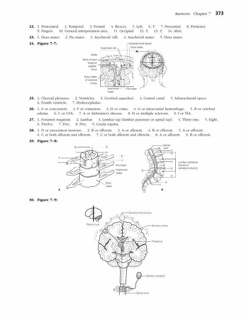
Answers: Chapter 7 373
22. 1. Postcentral. 2. Temporal. 3. Frontal. 4. Broca’s. 5. Left. 6. T. 7. Precentral. 8. Premotor. 9. Fingers. 10. General interpretation area. 11. Occipital. 12. T. 13. T. 14. Alert.
23. 1. Dura mater. 2. Pia mater. 3. Arachnoid villi. 4. Arachnoid mater. 5. Dura mater.
24. Figure 7–7:
25. 1. Choroid plexuses. 2. Ventricles. 3. Cerebral aqueduct. 4. Central canal. 5. Subarachnoid space. 6. Fourth ventricle. 7. Hydrocephalus.
26. 1. E or concussion. 2. F or contusion. 3. D or coma. 4. G or intracranial hemorrhage. 5. B or cerebral edema. 6. C or CVA. 7. A or Alzheimer’s disease. 8. H or multiple sclerosis. 9. I or TIA.
27. 1. Foramen magnum. 2. Lumbar. 3. Lumbar tap (lumbar puncture or spinal tap). 4. Thirty-one. 5. Eight. 6. Twelve. 7. Five. 8. Five. 9. Cauda equina.
28. 1. D or association neurons. 2. B or efferent. 3. A or afferent. 4. B or efferent. 5. A or afferent. 6. C or both afferent and efferent. 7. C or both afferent and efferent. 8. A or afferent. 9. B or efferent.
29. Figure 7–8:
30. Figure 7–9:
Superiorsagittal
sinus
Bone of skull
Scalp
Gray matterof cerebral
cortex
Arachnoid villi
Subarachnoid spaceDura mater
Arachnoidmater
Pia mater
H
F
I
B D
A
A
E
J
Duramater
Arachnoidmater
Pia mater
G
Spinalcord
CH
H
H
H
HH
Lumbar vertebrae(bones ofvertebral column)
L1
L2
L3
L4
L5
B
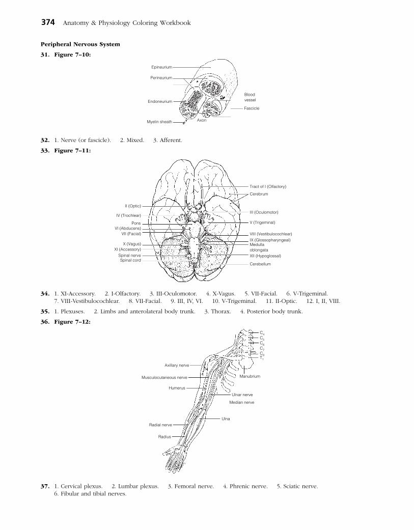
374 Anatomy & Physiology Coloring Workbook
Peripheral Nervous System
31. Figure 7–10:
32. 1. Nerve (or fascicle). 2. Mixed. 3. Afferent.
33. Figure 7–11:
34. 1. XI-Accessory. 2. I-Olfactory. 3. III-Oculomotor. 4. X-Vagus. 5. VII-Facial. 6. V-Trigeminal. 7. VIII-Vestibulocochlear. 8. VII-Facial. 9. III, IV, VI. 10. V-Trigeminal. 11. II-Optic. 12. I, II, VIII.
35. 1. Plexuses. 2. Limbs and anterolateral body trunk. 3. Thorax. 4. Posterior body trunk.
36. Figure 7–12:
37. 1. Cervical plexus. 2. Lumbar plexus. 3. Femoral nerve. 4. Phrenic nerve. 5. Sciatic nerve. 6. Fibular and tibial nerves.
Axon
Bloodvessel
Fascicle
Myelin sheath
Endoneurium
Epineurium
Perineurium
Cerebrum
Tract of I (Olfactory)
III (Oculomotor)
V (Trigeminal)
VIII (Vestibulocochlear)IX (Glossopharyngeal)Medullaoblongata
Cerebellum
XII (Hypoglossal)
Pons
Spinal nerveXI (Accessory)
X (Vagus)
VII (Facial)VI (Abducens)
Spinal cord
IV (Trochlear)
II (Optic)
Manubrium
Ulnar nerve
Median nerve
Ulna
Radius
Radial nerve
Humerus
Musculocutaneous nerve
Axillary nerve
C4C5C6C7C8T1

Answers: Chapter 7 375
38. Figure 7–13:
39. Check sympathetic for 1, 4, 6, 8, and 10. Check parasympathetic for 2, 3, 5, 7, 9, and 11.
1. Increased respiratory rate. 2. Increased heart rate and blood pressure. 3. Increased availability of blood glucose. 4. Pupils dilate; increased blood flow to heart, brain, and skeletal muscles.
Developmental Aspects of the Nervous System
40. 1. Hypothalamus. 2. Oxygen. 3. Cephalocaudal. 4. Gross. 5. Blood pressure. 6. Decreased oxygen (blood) to brain. 7. Senility. 8. Stroke (CVA).
Incredible Journey
41. 1. Cerebellum. 2. Medulla. 3. Hypothalamus. 4. Memories. 5. Temporal. 6. Broca’s area. 7. Reasoning. 8. Frontal. 9. Vagus (X). 10. Dura mater. 11. Subarachnoid space. 12. Fourth.
At the Clinic
42. Parasympathetic.
43. Considering the nerve cells are amitotic, the tumor is most likely a glioma, developing from one of the types of neuroglia.
44. During sympathetic activation, large amounts of epinephrine from the adrenal medulla pour into the blood. It will take time for the hormone to be broken down throughout the body.
45. The stroke has destroyed the trunk, hip, and lower limb region of the primary motor cortex that corresponds to those paralyzed areas on the left side of the body. (Remember, the motor pathways are crossed.)
46. 1. Cerebellum. 2. Basal nuclei. 3. Meningitis. 4. III (oculomotor). 5. Somatosensory cortex. 6. Broca’s area. 7. Electroencephalogram.
47. Marie has ataxia, indicating problems of the cerebellum.
48. Paresthesia, because the fiber tracts in the dorsal white matter are sensory tracts.
49. Sympathetic; the “fight or flight” response was activated.
50. The self-propagating change in membrane potential that travels along the membrane from the point of stimulation.
51. Peripheral nerves of the somatic nervous system carry both motor and sensory fibers, so you might expect the cut to affect both types of function.
52. Schwann cells, which myelinate the peripheral nerve fibers.
53. The somatic division is involved in stretching, sit-ups, walking, and brushing her teeth. The autonomic division causes stomach gurgling.
The Finale: Multiple Choice
54. 1. A, B. 2. C. 3. C. 4. A. 5. C. 6. B. 7. C. 8. A, C, D. 9. A, B, C. 10. C. 11. A, C. 12. A, C, D. 13. B, D. 14. B. 15. A. 16. D. 17. B. 18. A. 19. A. 20. C. 21. C. 22. B. 23. A. 24. B. 25. A. 26. A. 27. A. 28. B. 29. C.
ANSPreganglionic neuron(cell body)
Preganglionicaxon
Autonomicganglion
Somaticmotor neuron
White matterof spinal cord
ANSGanglionicneuron(cell body)
Gray matterof spinal cord
Postgang-lionicaxon
Smallintestine(or othervisceralorgan)
Effector of theautonomic motor neuron
Skeletalmuscle
CNS(spinal cord)
Effector of thesomatic motor neuron
Myelinsheath
Myelinsheath
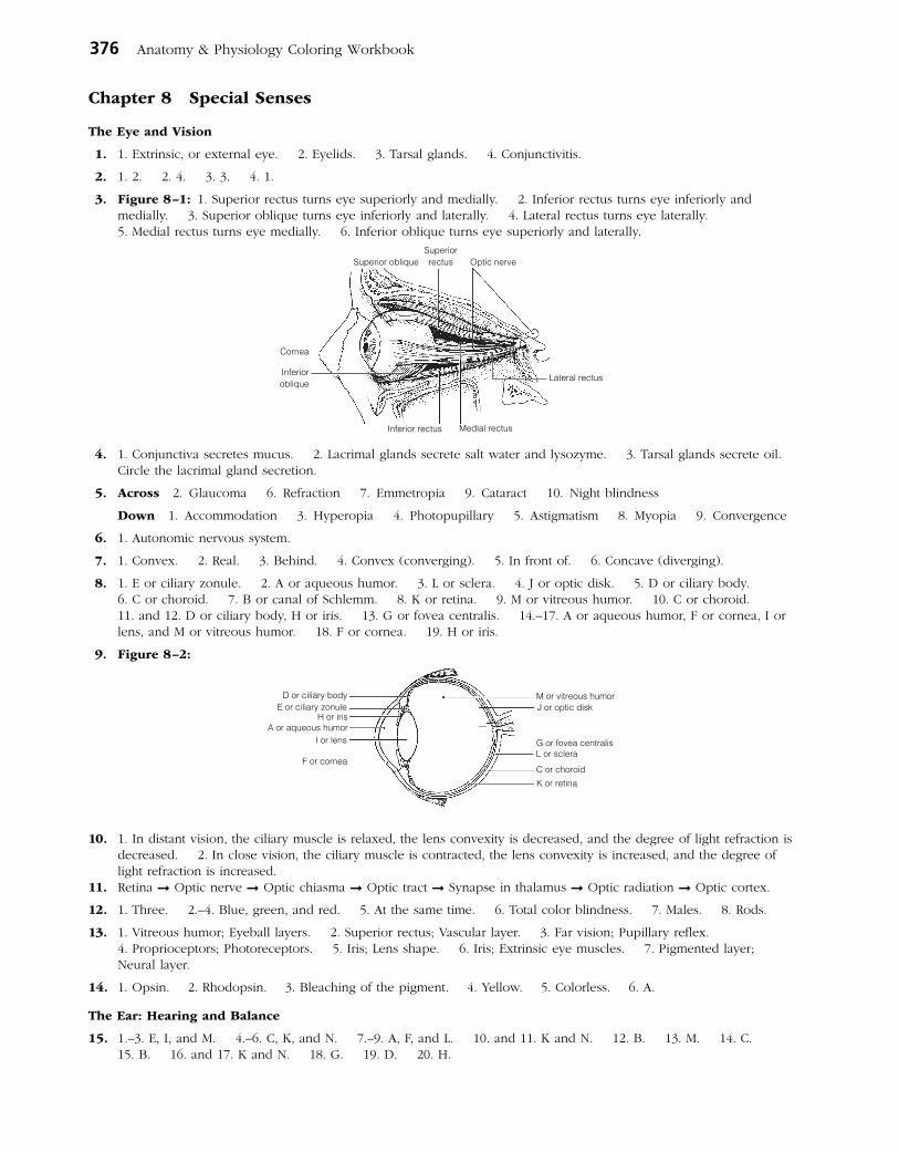
376 Anatomy & Physiology Coloring Workbook
Chapter 8 Special Senses
The Eye and Vision
1. 1. Extrinsic, or external eye. 2. Eyelids. 3. Tarsal glands. 4. Conjunctivitis.
2. 1. 2. 2. 4. 3. 3. 4. 1.
3. Figure 8–1: 1. Superior rectus turns eye superiorly and medially. 2. Inferior rectus turns eye inferiorly and medially. 3. Superior oblique turns eye inferiorly and laterally. 4. Lateral rectus turns eye laterally. 5. Medial rectus turns eye medially. 6. Inferior oblique turns eye superiorly and laterally.
4. 1. Conjunctiva secretes mucus. 2. Lacrimal glands secrete salt water and lysozyme. 3. Tarsal glands secrete oil. Circle the lacrimal gland secretion.
5. Across 2. Glaucoma 6. Refraction 7. Emmetropia 9. Cataract 10. Night blindness
Down 1. Accommodation 3. Hyperopia 4. Photopupillary 5. Astigmatism 8. Myopia 9. Convergence
6. 1. Autonomic nervous system.
7. 1. Convex. 2. Real. 3. Behind. 4. Convex (converging). 5. In front of. 6. Concave (diverging).
8. 1. E or ciliary zonule. 2. A or aqueous humor. 3. L or sclera. 4. J or optic disk. 5. D or ciliary body. 6. C or choroid. 7. B or canal of Schlemm. 8. K or retina. 9. M or vitreous humor. 10. C or choroid. 11. and 12. D or ciliary body, H or iris. 13. G or fovea centralis. 14.–17. A or aqueous humor, F or cornea, I or lens, and M or vitreous humor. 18. F or cornea. 19. H or iris.
9. Figure 8–2:
10. 1. In distant vision, the ciliary muscle is relaxed, the lens convexity is decreased, and the degree of light refraction is decreased. 2. In close vision, the ciliary muscle is contracted, the lens convexity is increased, and the degree of light refraction is increased.
11. Retina ➞ Optic nerve ➞ Optic chiasma ➞ Optic tract ➞ Synapse in thalamus ➞ Optic radiation ➞ Optic cortex.
12. 1. Three. 2.–4. Blue, green, and red. 5. At the same time. 6. Total color blindness. 7. Males. 8. Rods.
13. 1. Vitreous humor; Eyeball layers. 2. Superior rectus; Vascular layer. 3. Far vision; Pupillary reflex. 4. Proprioceptors; Photoreceptors. 5. Iris; Lens shape. 6. Iris; Extrinsic eye muscles. 7. Pigmented layer; Neural layer.
14. 1. Opsin. 2. Rhodopsin. 3. Bleaching of the pigment. 4. Yellow. 5. Colorless. 6. A.
The Ear: Hearing and Balance
15. 1.–3. E, I, and M. 4.–6. C, K, and N. 7.–9. A, F, and L. 10. and 11. K and N. 12. B. 13. M. 14. C. 15. B. 16. and 17. K and N. 18. G. 19. D. 20. H.
Cornea
Inferioroblique
Optic nerveSuperiorrectusSuperior oblique
Inferior rectus Medial rectus
Lateral rectus
D or ciliary bodyE or ciliary zonule
H or irisA or aqueous humor
I or lens
F or cornea
M or vitreous humorJ or optic disk
G or fovea centralisL or sclera
C or choroid
K or retina

Answers: Chapter 8 377
16. Figure 8–3: I, E, and M are yellow; A, F, and L are red; C is blue, and K (continuing to N) is green.
17. Eardrum ➞ Hammer ➞ Anvil ➞ Stirrup ➞ Oval window ➞ Perilymph ➞ Membrane ➞ Endolymph ➞ Hair cells.
18. Figure 8–4:
19. 1. C or dynamic. 2. I or semicircular canals. 3. A or angular/rotatory. 4. D or endolymph. 5. B or cupula. 6. J or static. 7. and 8. H or saccule, K or utricle. 9. E or gravity. 10. and 11. G or proprioception, L or vision.
20. 1. C. 2. S. 3. S. 4. C. 5. C, S. 6. C. 7. S.
21. Nausea, dizziness, and balance problems.
22. 1. Pinna; auditory ossicles. 2. Tectorial membrane; dynamic equilibrium. 3. Sound waves; Equilibrium. 4. Auditory tube; Vestibule. 5. Optic nerve; Cranial nerve VIII.
Chemical Senses: Smell and Taste
23. 1.–3. (in any order): VII-Facial, IX-Glossopharyngeal, X-Vagus. 4. I-Olfactory. 5. Mucosa of the “roof.” 6. Sniffing. 7. Taste buds. 8. or 9. Fungiform, circumvallate. 10.–14. (in any order): sweet, salty, bitter, sour, umami. 15. Bitter. 16. Smell. 17. Dry. 18. Memories.
Semicircular canals (green)
Hammer (red)Anvil (red)
Cochlea(blue)
Auditorytube
Vestibule(green)
Stirrup(red)
Tympanic membrane(eardrum)
(yellow)
External acousticmeatus (yellow)
Pinna (yellow)
Oval window
Round window
Cristaampullaris
Membranoussemicircularcanals
Macula
FD
E
SacculeCochlearduct
Ultricle
GEA
BH
ECOrgan
of Corti

378 Anatomy & Physiology Coloring Workbook
24. Figure 8–5:
25. Figure 8–6: 1. Mucus “captures” airborne odor molecules. 2. Olfactory neurons are bipolar neurons.
26. 1. Musky; Taste sensations. 2. Epithelial cell; Olfactory receptor. 3. Neuron; Gustatory cell. 4. Olfactory nerve; Cranial nerves that carry taste impulses. 5. Four receptor types; Olfactory receptor. 6. Metal ions; Sweet receptors response.
Developmental Aspects of the Special Senses
27. 1. Nervous system. 2. Measles (rubella). 3. Blindness. 4. Vision. 5. Hyperopic. 6. Elastic. 7. Presbyopia. 8. Cataract. 9. Presbycusis.
Incredible Journey
28. 1. Bony. 2. Perilymph. 3. and 4. Saccule, utricle. 5. Gel (otolithic membrane). 6. Otoliths. 7. Macula. 8. Static. 9. Cochlear. 10. Organ of Corti. 11. Hearing. 12. Cochlear division of cranial nerve VIII. 13. Semicircular canals. 14. Cupula. 15. Crista ampullaris. 16. Dynamic.
At the Clinic
29. Patching the strong eye to force the weaker eye muscles to become stronger.
30. Cataract; UV radiation, smoking.
31. Vision is poor because, without pigment within the eye (in the choroid), light scatters before it can be properly focused.
32. Night blindness; vitamin A; rods.
33. The proximal end close to the oval window; sensorineural.
34. Otitis externa, most likely because of his exposure to pool bacteria. This diagnosis would be confirmed by presence of an inflamed external ear canal. If it is otitis media, the middle ear would be inflamed. Bulging of the eardrum would suggest that inserting ear tubes might be recommended.
35. Cranial nerve I, the olfactory nerve.
36. Abducens nerve, cranial nerve VI.
37. Glaucoma, inadequate drainage of aqueous humor; blindness resulting from compression of retina and optic disc.
38. Taste bud cells are subjected to friction and heat and hence are rapidly dividing cells that will be targeted by chemotherapeutic drugs. A chef must have a fine sense of taste to be successful.
The Finale: Multiple Choice
39. 1. D. 2. B, C. 3. B, D. 4. A, B, D. 5. C. 6. C. 7. A, B, C. 8. D. 9. B. 10. A, B, D. 11. B, C, D. 12. B. 13. A, D. 14. C. 15. A, C, D. 16. A, B, D. 17. A, C. 18. B.

Answers: Chapter 9 379
Chapter 9 The Endocrine System
The Endocrine System and Hormone Function—An Overview
1. 1. F or slower and more prolonged. 2. E or nervous system. 3. B or hormones. 4. D or nerve impulses. 5. A or cardiovascular system.
2. 1. I or receptors. 2. N or target cell(s). 3. A or altering activity. 4. L or stimulating new or unusual activities. 5. K or steroid or amino acid–based. 6. G or neural. 7. C or hormonal. 8. D or humoral. 9. F or negative feedback. 10. B or anterior pituitary. 11. J or releasing hormones. 12. E or hypothalamus. 13. H or neuroendocrine.
3. Steroid hormones: B, C, D. Amino acid–based hormones: A, E.
The Major Endocrine Organs
4. Figure 9–1: Color: Testes, ovaries, thyroid and adrenal cortex.
5. Figure 9–2: A. Pineal. B. Posterior pituitary. C. Anterior pituitary. D. Thyroid. E. Thymus. F. Adrenal cortex. G. Adrenal medulla. H. Pancreas. I. Ovary. J. Testis. K. Parathyroids. L. Placenta.
6. 1. C. 2. B. 3.–4. F. 5. G. 6. I, L. 7. C. 8. H. 9. H. 10. C. 11. A. 12. B. 13. I, L. 14. C. 15. K. 16. C. 17. J. 18. E. 19. D. 20. C.
7. 1. Estrogen/testosterone. 2. PTH. 3. ADH. 4. Thyroxine. 5. Thyroxine. 6. Insulin. 7. Growth hormone. 8. Estrogen/progesterone. 9. Thyroxine.
8. 1. Growth hormone. 2. Thyroxine. 3. PTH. 4. Glucocorticoids. 5. Growth hormone. 6. Androgens (testosterone).
9. Across 6. Progesterone 7. TSH 8. ADH 10. Aldosterone 11. Thyroxine 12. Glucagon
Down 1. Epinephrine 2. Prolactin 3. Cortisol 4. Insulin 5. PTH 9. Thymosin 13. LH
10. 1. Polyuria—high sugar content in kidney filtrate causes large amount of water to be lost in the urine. 2. Polydipsia—thirst because of large volumes of urine excreted. 3. Polyphagia—hunger because blood sugar cannot be used as a body fuel even though blood levels are high.
Bones and muscles
Mammary glands
Testes or ovaries
andThyroid
Adrenal cortex
Releasing hormonesin portal circulation
Growthhormone(GH)
Prolactin(PRL) Follicle-
stimulatinghormone (FSH)
Thyroid-stimulatinghormone (TSH)
Luteinizinghormone (LH)
Adrenocorticotropichormone (ACTH)
Hypothalamus
Posterior pituitary
Turk’s saddleof sphenoid bone
Anterior pituitary

380 Anatomy & Physiology Coloring Workbook
11. Figure 9–3:
12. 1. Anterior lobe; Posterior lobe. 2. Steroid hormone; Nonsteroid hormones. 3. Cortisol; Adrenal medulla. 4. Increases blood Ca2+; Calcitonin. 5. Growth hormone; Adrenal cortex hormones. 6. Parafollicular cells; Follicular cells.
Other Hormone-Producing Tissues and Organs
13. Hormone Chemical makeup Source Effects
Gastrin Peptide Stomach Stimulates stomach glands to secrete HCl
Secretin Peptide Duodenum Stimulates the pancreas to secrete HCO3–-
rich juice and stimulates the liver to release more bile; inhibits stomach glands
Cholecystokinin Peptide Duodenum Stimulates the pancreas to secrete enzyme- rich juice and the gallbladder to contract; relaxes sphincter of Oddi
Erythropoietin Glycoprotein Kidney in response Stimulates production of red blood cells by to hypoxia bone marrow
Active vitamin D3 Steroid Skin; activated by Enhances intestinal absorption of calcium kidneys
Atrial natriuretic Peptide Heart Inhibits Na+ reabsorption by kidneys; peptide (ANP) inhibits aldosterone release by kidneys
Human chorionic Protein Placenta Stimulates corpus luteum to continue gonadotropin producing estrogens and progesterone, (hCG) preventing menses
Leptin Peptide Adipose tissue Targets the brain; reduces appetite; increases energy expenditure
Developmental Aspects of the Endocrine System
14. 1. Neoplasm. 2. Hypersecretion. 3. Iodine. 4. Estrogens. 5. Menopause. 6. Children. 7. Insulin.
Incredible Journey
15. 1. Insulin. 2. Pancreas. 3. Posterior pituitary, or hypothalamus. 4. ADH. 5. Parathyroid. 6. Calcium. 7. Adrenal medulla. 8. Epinephrine. 9. Thyroxine.
At the Clinic
16. Pituitary dwarfs who secrete inadequate amounts of GH have fairly normal proportions; cretins (hypothyroid individuals) retain childlike body proportions.
17. Hypothyroidism; iodine deficiency (treated by dietary iodine supplements) or thyroid cell burnout (treated by thyroid hormone supplements).

Answers: Chapter 10 381
18. Adrenal cortex.
19. For the giant, GH is being secreted in excess by the anterior pituitary, resulting in extraordinary height. For the dwarf, GH is deficient, resulting in very small stature but normal body proportions. For the fat man, T3 and T4 are not being produced adequately, resulting in depressed metabolism and obesity (myxedema). The bearded lady has a tumor of her adrenal cortex (androgen-secreting area), leading to excessive hairiness (hirsutism).
20. Stressor ➞ hypothalamus ➞ CRH (releasing hormone) released to the hypophysial portal system blood ➞ to ante-rior pituitary which releases ACTH ➞ acts on adrenal cortex to trigger release of glucocorticoids (cortisol, etc.).
21. Prolactin.
The Finale: Multiple Choice
22. 1. D. 2. A. 3. B. 4. A, B, C, D. 5. D. 6. B. 7. A, B, C. 8. C. 9. A, B, D. 10. A, B, C, D. 11. A, B, C, D. 12. A, C. 13. B. 14. A, C. 15. A. 16. C. 17. D. 18. C. 19. B.
Chapter 10 Blood
Composition and Functions of Blood
1. 1. Connective. 2. Formed. 3. Plasma. 4. Clotting. 5. Erythrocytes. 6. Hematocrit. 7. Plasma. 8. Leukocytes. 9. Platelets. 10. One. 11. Oxygen.
2. Across 6. Plasma 7. Monocyte 8. Eosinophil 9. Formed
Down 1. Neutrophil 2. Megakaryocyte 3. Erythrocyte 4. Basophil 5. Lymphocyte
3. Figure 10–1:
382 Anatomy & Physiology Coloring Workbook
4. Figure 10–2: A is a neutrophil, B is a monocyte, C is an eosinophil, D is a lymphocyte.
5. 1. Diapedesis. 2. T. 3. Kidneys. 4. 7.35. 5. 5.5. 6. T. 7. T. 8. 4.5–5.5. 9. Hematocrit. 10. Less. 11. Monocytes. 12. Lymphocytes.
6. 1. Erythrocytes; Leukocytes. 2. Monocytes; Granulocytes. 3. Lymphocyte; Oxygen transport. 4. Platelets; Phagocytosis. 5. Aneurysm; Clot. 6. Hemoglobin; Plasma. 7. Lymphocyte; Myeloid stem cell origin.
7. 1. 2. 2. 5. 3. 1. 4. 4. 5. 3.
8. Check 1, 2, 3.
Hemostasis
9. 1. A or break. 2. E or platelets. 3. I or serotonin. 4. K or tissue factor. 5. H or PF3. 6. G or prothrombin activator. 7. F or prothrombin. 8. J or thrombin. 9. D or fibrinogen. 10. C or fibrin. 11. B or erythrocytes.
10. 1. 3–6 min. 2. Heparin. 3. T.
Blood Groups and Transfusions
11. Agglutinogens or antigens Agglutinins or Can donate Can receive Blood type on RBC surface antibodies in plasma blood to type blood from type
1. Type A A Anti-B A, AB A, O
2. Type B B Anti-A B, AB B, O
3. Type AB A, B None AB A, B, AB, O
4. Type O None Anti-A, Anti-B A, B, AB, O O
12. Type O is the universal donor. AB is the universal recipient.
13. A reaction during which plasma antibodies attach to and lyse red blood cells different from your own.
Developmental Aspects of Blood
14. 1. F. 2. Jaundiced. 3. Sickle-cell. 4. Hemophilia. 5. Iron. 6. Pernicious. 7. B12. 8. Thrombi. 9. Leukemia.
Incredible Journey
15. 1. Hematopoiesis. 2. Hemostasis. 3. Hemocytoblasts. 4. Neutrophil. 5. Phagocyte. 6. Erythropoietin. 7. Red blood cells. 8. Hemoglobin. 9. Oxygen. 10. Lymphocytes. 11. Antibodies. 12.–15. (in any order): Basophils, eosinophils, monocytes, platelets. 16. Endothelium. 17. Platelets. 18. Serotonin. 19. Fibrin. 20. Clot. 21. Prothrombin activator. 22. Prothrombin. 23. Thrombin. 24. Fibrinogen. 25. Embolus.
At the Clinic
16. 1. Hemolytic disease of the newborn.
2. Its RBCs have been destroyed by the mother’s antibodies; therefore, the baby’s blood is carrying insufficient oxygen.
3. She must have received mismatched (Rh+) blood previously in a transfusion.
4. Give the mother RhoGAM to prevent her from becoming sensitized to the Rh+ antigen.
5. Fetal progress will be followed in expectation of hemolytic disease of the newborn; intrauterine transfusions will be given if necessary, as well as complete blood transfusion to the newborn.
17. No; A+ or AB+.
18. The stem cells for hematopoiesis in red bone marrow are a rapidly dividing cell population. Hence, they would be targeted (along with other rapidly dividing cells) by chemotherapeutic drugs.
19. Virtually all bones contain red marrow and functional hematopoietic tissue in young children, but in adults only the sternum, ilium, and a very few long bone epiphyses contain red marrow.
20. Erythrocytes, which account for nearly half of blood volume, will be produced in the largest numbers.
21. Stomach cells are the source of intrinsic factor needed to absorb vitamin B12. Apparently insufficient numbers of vitamin-producing cells remain after the stomach surgery. Vitamin B12 cannot be absorbed orally, so it must be injected. If he refuses the shots, pernicious anemia will ensue.

Answers: Chapter 11 383
The Finale: Multiple Choice
21. 1. A, B, D. 2. B. 3. A, B, C, D. 4. D. 5. A, B, D. 6. D. 7. C. 8. A. 9. C. 10. B, C, D. 11. C. 12. B, C, D. 13. A, D. 14. D. 15. B. 16. C.
Chapter 11 The Cardiovascular System
The Heart
1. 1. Thorax. 2. Diaphragm. 3. Second. 4. Aorta. 5. Right atrium. 6. Atria. 7. Ventricles. 8. Endocardium. 9. Epicardium. 10. Friction. 11. Cardiac.
2. 1. Right ventricle. 2. Pulmonary semilunar. 3. Pulmonary arteries. 4. Lungs. 5. Pulmonary veins. 6. Left atrium. 7. Mitral (bicuspid). 8. Left ventricle. 9. Aortic. 10. Aorta. 11. Capillary beds. 12. Superior vena cava. 13. Inferior vena cava.
In Figure 11–1, the white areas represent regions transporting O2-rich blood. The gray vessels transport O2-poor blood.
Figure 11–1:
3. Figure 11–2: 1. Right atrium. 2. Left atrium. 3. Right ventricle. 4. Left ventricle. 5. Superior vena cava. 6. Inferior vena cava. 7. Aorta. 8. Pulmonary trunk. 9. Left pulmonary artery. 10. Right pulmonary artery. 11. Right pulmonary veins. 12. Left pulmonary veins. 13. Coronary circulation. 14. Apex of heart. 15. Ligamentum arteriosum.
4. Figure 11–3:
5. 1. Systole. 2. Diastole. 3. Lub-dup. 4. Atrioventricular. 5. Semilunar. 6. Ventricles. 7. Atria. 8. Atria. 9. Ventricles. 10. Murmurs.
6. Figure 11–4: Red: left atrium, left ventricle, pulmonary veins, and aorta. Blue: superior and inferior venae cavae, right atrium, right ventricle, pulmonary trunk, and pulmonary arteries.
Superior and inferior venae cavae right atrium tricuspid valve right ventricle pulmonary semilunar valve pulmonary trunk right and left pulmonary arteries lungs right and left pulmonary veins left atrium bicuspid valve left ventricle aortic semilunar valve aorta.
A
F
D
C
E
B
Intercalated discs
Striations
Nucleiwithnucleoli
Muscle fiber
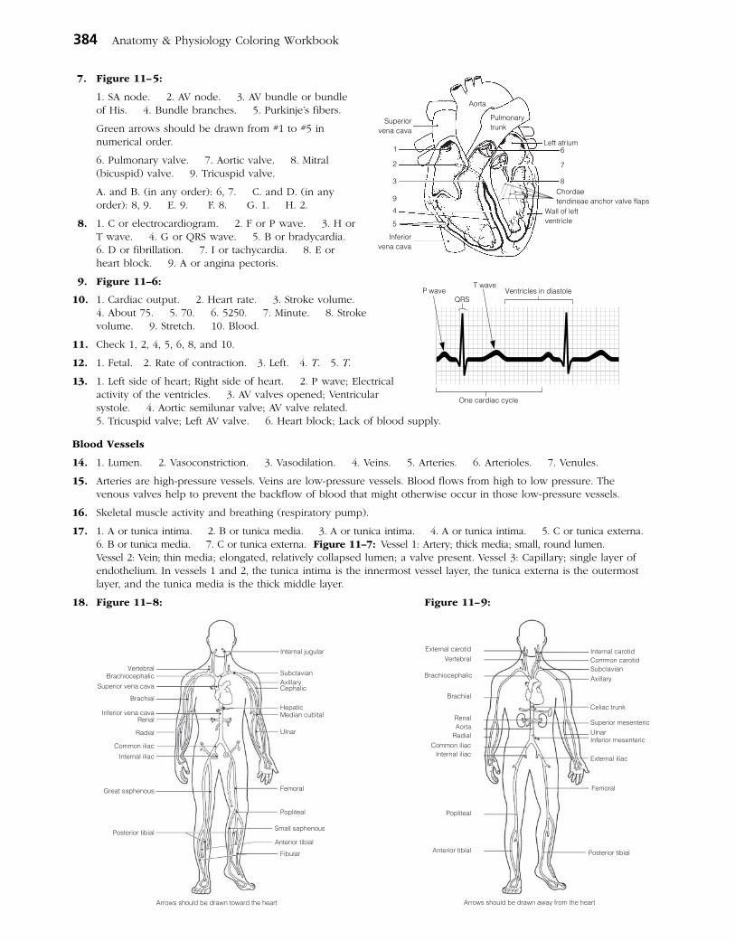
384 Anatomy & Physiology Coloring Workbook
7. Figure 11–5:
1. SA node. 2. AV node. 3. AV bundle or bundle of His. 4. Bundle branches. 5. Purkinje’s fibers.
Green arrows should be drawn from #1 to #5 in numerical order.
6. Pulmonary valve. 7. Aortic valve. 8. Mitral (bicuspid) valve. 9. Tricuspid valve.
A. and B. (in any order): 6, 7. C. and D. (in any order): 8, 9. E. 9. F. 8. G. 1. H. 2.
8. 1. C or electrocardiogram. 2. F or P wave. 3. H or T wave. 4. G or QRS wave. 5. B or bradycardia. 6. D or fibrillation. 7. I or tachycardia. 8. E or heart block. 9. A or angina pectoris.
9. Figure 11–6:
10. 1. Cardiac output. 2. Heart rate. 3. Stroke volume. 4. About 75. 5. 70. 6. 5250. 7. Minute. 8. Stroke volume. 9. Stretch. 10. Blood.
11. Check 1, 2, 4, 5, 6, 8, and 10.
12. 1. Fetal. 2. Rate of contraction. 3. Left. 4. T. 5. T.
13. 1. Left side of heart; Right side of heart. 2. P wave; Electrical activity of the ventricles. 3. AV valves opened; Ventricular systole. 4. Aortic semilunar valve; AV valve related. 5. Tricuspid valve; Left AV valve. 6. Heart block; Lack of blood supply.
Blood Vessels
14. 1. Lumen. 2. Vasoconstriction. 3. Vasodilation. 4. Veins. 5. Arteries. 6. Arterioles. 7. Venules.
15. Arteries are high-pressure vessels. Veins are low-pressure vessels. Blood flows from high to low pressure. The venous valves help to prevent the backflow of blood that might otherwise occur in those low-pressure vessels.
16. Skeletal muscle activity and breathing (respiratory pump).
17. 1. A or tunica intima. 2. B or tunica media. 3. A or tunica intima. 4. A or tunica intima. 5. C or tunica externa. 6. B or tunica media. 7. C or tunica externa. Figure 11–7: Vessel 1: Artery; thick media; small, round lumen. Vessel 2: Vein; thin media; elongated, relatively collapsed lumen; a valve present. Vessel 3: Capillary; single layer of endothelium. In vessels 1 and 2, the tunica intima is the innermost vessel layer, the tunica externa is the outermost layer, and the tunica media is the thick middle layer.
18. Figure 11–8: Figure 11–9:
Superiorvena cava
1
2
3
9
4
5
Inferiorvena cava
Wall of leftventricle
8
7
6Left atrium
Aorta
Pulmonarytrunk
Chordaetendineae anchor valve flaps
P waveQRS
T waveVentricles in diastole
One cardiac cycle
External carotidVertebral
Brachiocephalic
Brachial
RenalAorta
RadialCommon iliac
Internal iliac
Popliteal
Anterior tibial Posterior tibial
Arrows should be drawn away from the heart
Femoral
External iliac
Inferior mesentericUlnarSuperior mesenteric
Axillary
Celiac trunk
Subclavian
Internal carotidCommon carotid
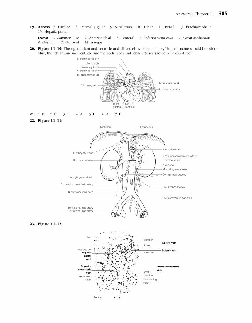
Answers: Chapter 11 385
19. Across 5. Cardiac 6. Internal jugular 9. Subclavian 10. Ulnar 11. Renal 13. Brachiocephalic 15. Hepatic portal
Down 1. Common iliac 2. Anterior tibial 3. Femoral 4. Inferior vena cava 7. Great saphenous 8. Gastric 12. Gonadal 14. Azygos
20. Figure 11–10: The right atrium and ventricle and all vessels with “pulmonary” in their name should be colored blue; the left atrium and ventricle and the aortic arch and lobar arteries should be colored red.
21. 1. F. 2. D. 3. B. 4. A. 5. D. 6. A. 7. E.
22. Figure 11–11:
23. Figure 11–12:
Liver
GallbladderHepatic
portalvein
Ascendingcolon
Superiormesenteric
vein
Descendingcolon
Small intestine
Spleen
Pancreas
Rectum
StomachGastric vein
Splenic vein
Inferior mesentericvein
386 Anatomy & Physiology Coloring Workbook
24. Across 1. Dorsalis pedis 3. Celiac trunk 5. Femoral 6. Subclavian 8. Phrenic 9. Aorta 10. Brachial 11. Peroneal 12. Radial 13. Coronary
Down 2. External carotid 3. Common iliac 4. Internal iliac 7. Vertebral
25. Figure 11–13:
26. Figure 11–14:
27. 1. C or circle of Willis. 2. J or umbilical vein. 3. E or ductus venosus. 4.–5. A or anterior cerebral artery, G or middle cerebral artery. 6. B or basilar artery. 7. D or ductus arteriosus. 8. F or foramen ovale.
28. The fetal lungs are not functioning in gas exchanges, and they are collapsed. The placenta makes the gas exchanges with the fetal blood.
29. 1. Vein; Artery. 2. Carotid artery; Coronary circulation. 3. Vasodilation; Venous return. 4. High blood pressure; Low blood volume. 5. Vasodilation; Peripheral resistance.
30. 1. H or pulse. 2. B or blood pressure. 3. and 4. C or cardiac output and F or peripheral resistance. 5. D or constriction of arterioles. 6. J or systolic blood pressure. 7. E or diastolic blood pressure. 8. A or over arter-ies. 9. G or pressure points. 10. I or sounds of Korotkoff.
31. 1. G or interstitial fluid. 2. C or diffusion. 3. E or fat soluble. 4.–6. (in any order): B or capillary clefts, D or fenestrations, I or vesicles. 7. D or fenestrations. 8.–9. B or capillary clefts; D or fenestrations.
32. 1. D. 2.–5. I. 6. D. 7. D. 8. I. 9.–11. D. 12. I. 13. I.
33. 1. Increase. 2. Orthostatic. 3. Brain. 4. Stethoscope. 5. Low. 6. T. 7. Vasoconstricting. 8. Hypertension.
Pituitary gland
Frontal lobeof cerebral
hemisphere
Internalcarotid artery
CerebellumVertebral artery
Pons Basilar artery
Posterior cerebral arteries
Communicating branches
Anteriorcerebral arteries Middle cerebral arteries
Communicating branches
Liver
Ductus venosus
Superior vena cava
Hepatic portal vein
Internal iliac artery
Common iliac artery
AortaInferior vena cava
Umbilical cord
Umbilical vein
Fetal bladder
Ductus arteriosus
Foramen ovale
Inferior vena cava
Enlarged view of heart
Ductus venosus
Umbilical arteries

Answers: Chapter 11 387
Figure 11–15:
34. 1. Through the vascular shunt. 2. A. 3. Capillary blood. 4. Capillary hydrostatic (blood) pressure (Hpc). 5. Blood pressure. 6. Capillary colloid osmotic pressure (Opc). 7. Albumin. 8. At the arteriole end. 9. It is picked up by lymphatic vessels for return to the bloodstream.
35. 1. Femoral artery. 2. Brachial artery. 3. Popliteal artery. 4. Facial artery. 5. Radial artery. 6. Temporal artery.
Developmental Aspects of the Cardiovascular System
36. 1. Fourth. 2. Lungs. 3. Ductus venosus. 4. Umbilical vein. 5. Placenta. 6. Fetal liver. 7. Umbilical arteries. 8. Occluded. 9. Deaths. 10. Atherosclerosis (and arteriosclerosis). 11. Menopause. 12. Aerobic exercise. 13. Atherosclerosis. 14. Varicose veins. 15. and 16. Feet and legs.
Incredible Journey
37. 1. Left atrium. 2. Left ventricle. 3. Mitral (bicuspid). 4. Chordae tendineae. 5. Diastole. 6. Systole/contraction. 7. Aortic semilunar. 8. Aorta. 9. Superior mesenteric. 10. Endothelial. 11. Superior mesenteric. 12. Splenic. 13. Nutrients. 14. Phagocytic (Kupffer). 15. Hepatic. 16. Inferior vena cava. 17. Right atrium. 18. Pulmonary trunk. 19. Pulmonary. 20. Lungs. 21. Subclavian.
At the Clinic
38. Zero; myocardial infarction. The posterior interventricular artery supplies much of the left ventricle, the systemic pump.
39. Bradycardia, which results from excessive vagal stimulation of the heart, can be determined by taking the pulse.
40. Peripheral congestion caused by right heart failure.
41. Thrombosis or atherosclerosis; an arterial anastomosis (circle of Willis), e.g., (1) Left internal carotid artery to left anterior cerebral artery. Then through anterior communicating branch to right anterior cerebral artery and (2) vertebral arteries to basilar artery to right posterior cerebral artery through the posterior communicating branch to right middle cerebral artery.
42. High; polycythemia increases blood viscosity (thus peripheral resistance), which increases blood pressure.
43. The stiffened valve flaps would not close properly and the valve would become incompetent. A heart murmur would be heard after the valve had (supposedly) closed and blood was flowing back through the valve.
44. Thrombophlebitis occurs when a thrombus (clot) forms in an inflamed blood vessel (a vein). The danger is that the clot may detach, leading to a pulmonary embolism.
45. An ECG only reveals electrical problems. It is not useful for revealing valvular problems.
46. If anything, exercise extends life by making the cardiovascular and respiratory systems more efficient. Heart rate drops and stroke volume increases.
47. When the environmental temperature is high, blood vessels serving the skin vasodilate and much of the blood supply will be found in dermal blood vessels. Then, when you stand suddenly, there will initially be inadequate blood volume in the larger, more central blood vessels to ensure that the brain receives a normal blood supply, thus the dizziness.
48. A drug that blocks calcium channels will decrease the force of heart contraction. Because contractile force is directly related to stroke volume, the SV will decrease.
388 Anatomy & Physiology Coloring Workbook
49. Acetylcholine slows heart rate (this is the neurotransmitter released by the vagus nerves). Thus, with a longer filling time the heart’s stroke volume will increase.
50. It reveals their elasticity. When the heart contracts and forces blood into the large arteries near the heart, they stretch to accommodate the greater blood volume (systolic pressure). Then, as the blood continues on in the circuit, their walls recoil, keeping pressure on the blood which keeps it moving (diastolic pressure).
The Finale: Multiple Choice
51. 1. A, D. 2. B. 3. A, D. 4. A, B, C, D. 5. D. 6. C. 7. A, C. 8. C. 9. A, B, C, D. 10. A, B, C. 11. A, B, C. 12. A, C. 13. B, D. 14. A, B, C. 15. C, D. 16. A. 17. A, B, C, D. 18. A, B, C. 19. B. 20. D. 21. A. 22. B. 23. D. 24. D.
Chapter 12 The Lymphatic System and Body Defenses
The Lymphatic System
1. 1. Pump. 2. Arteries. 3. Veins. 4. Valves. 5. Lymph. 6. 3.
2. Figure 12–1:
3. 1. Blood capillary; Lymphatic capillary. 2. Abundant supply of lymphatics; Excess fluid in interstitial fluid. 3. High-pressure gradient; Flow of lymph. 4. Impermeable; Lymphatic capillary.
4. 1. C or spleen. 2. A or lymph nodes. 3. D or thymus. 4. B or Peyer’s patches, E or tonsils. 5. B or Peyer’s patches.
5. Figure 12–2: Shade in the right upper limb and right side of the thorax and head.
6. 1. B lymphocytes. 2. They produce and release antibodies. 3. T lymphocytes. 4. Macrophages, phagocytes. 5. This slows the flow of lymph through the node, allowing time for immune cells and macro phages to respond to foreign substances present in the lymph. 6. Valves in the afferent and efferent lymphatics. 7. Cervical, axillary, inguinal.
Tissue fluid(becomes lymph)
A B
Loose connectivetissue aroundcapillaries
Lymph capillaries
Blood capillaries
Arteries
Heart
Veins
Lymphaticvessels
Lymphaticcollectingvessels
Valves
Lymph node
Lymphduct
Vein

Answers: Chapter 12 389
Figure 12–3:
7. 1. C. 2. A. 3. D. 4. B. 5. C.
Body Defenses
8. 1. Surface membrane barriers, mucosae. 2. Natural killer cells. 3. Chemicals (inflammatory and antimicrobial).
9. 1. Tears and saliva. 2. Stomach and female reproductive tract. 3. Sebaceous (oil) glands, skin. 4. Digestive.
10. Figure 12–4:
11. 1. Itching; Cardinal signs of inflammation. 2. Natural killer cells; Phagocytes. 3. Interferon; Inflammatory chemicals. 4. Inflammation; First line of defense. 5. Antibacterial; Interferons.
12. 1. B or lysozyme, F or sebum. 2. C or mucosae, G or skin. 3. A or acids, B or lysozyme, D or mucus, E or protein-digesting enzymes, F or sebum. 4. D or mucus. 5. A–G.
13. They propel mucus laden with trapped debris superiorly away from the lungs to the throat, where it can be swallowed or spat out.
14. Phagocytosis is ingestion and destruction of particulate material by certain cells. The rougher the particle, the more easily it is ingested.
15. Check 1, 3, 4.
16. Across 2. Edema 5. Neutrophils 6. Chemotaxis 7. Diapedesis
Down 1. Fibrin mesh 3. Macrophages 4. Histamine
17. 1. Proteins. 2. Activated. 3. Holes or lesions. 4. Water. 5. Lysis. 6. Opsonization.
18. Interferon is synthesized in response to viral infection of a cell. The cell produces and releases interferon proteins, which diffuse to nearby cells, where they prevent viruses from multiplying within those cells.
19. The adaptive immune system is antigen-specific, systemic, and has memory.
20. 1. Immune system. 2. Proteins. 3. Haptens. 4. Nonself.
21. 1. A or antigens. 2. E or humoral. 3. D or cellular. 4. and 5. B or B cells and I or T cells. 6. H or macrophages. 7. and 8. C or blood and F or lymph. 9. G or lymph nodes.
Fibrous capsule
Trabecula
Germinal centersof follicles
Medullary cord
Hilum
Cortex
Efferentlymphatics
Medullarysinus
Subscapularsinus
Afferentlymphatics
1
6
4
5
3
2
Epithelium
Endotheliumof capillary
Neutrophil
Fibrous repair tissue
Macrophage
Microorganisms
MonocyteErythrocytes
Endotheliumof capillary
Injuredtissue
Bloodvessel
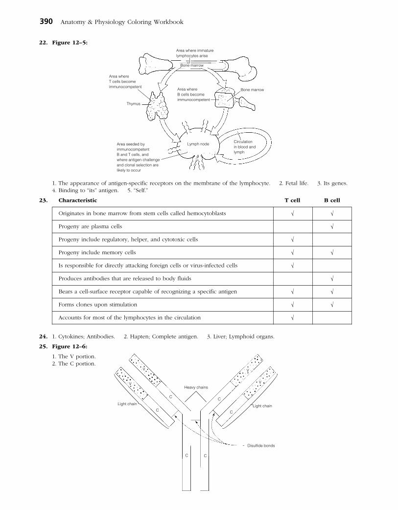
390 Anatomy & Physiology Coloring Workbook
22. Figure 12–5:
1. The appearance of antigen-specific receptors on the membrane of the lymphocyte. 2. Fetal life. 3. Its genes. 4. Binding to “its” antigen. 5. “Self.”
23. Characteristic T cell B cell
Originates in bone marrow from stem cells called hemocytoblasts √ √
Progeny are plasma cells √
Progeny include regulatory, helper, and cytotoxic cells √
Progeny include memory cells √ √
Is responsible for directly attacking foreign cells or virus-infected cells √
Produces antibodies that are released to body fluids √
Bears a cell-surface receptor capable of recognizing a specific antigen √ √
Forms clones upon stimulation √ √
Accounts for most of the lymphocytes in the circulation √
24. 1. Cytokines; Antibodies. 2. Hapten; Complete antigen. 3. Liver; Lymphoid organs.
25. Figure 12–6:
1. The V portion. 2. The C portion.
Thymus
Bone marrow
Circulationin blood andlymph
Lymph node
Bone marrow
Area where immaturelymphocytes arise
Area whereT cells becomeimmunocompetent
Area seeded byimmunocompetentB and T cells, andwhere antigen challengeand clonal selection arelikely to occur
Area whereB cells becomeimmunocompetent
V
V
C
C
C C
V
V
C
C
Disulfide bonds
Heavy chains
Light chain Light chain
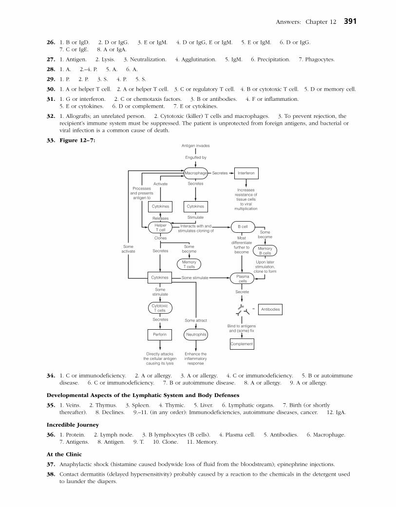
Answers: Chapter 12 391
26. 1. B or IgD. 2. D or IgG. 3. E or IgM. 4. D or IgG, E or IgM. 5. E or IgM. 6. D or IgG. 7. C or IgE. 8. A or IgA.
27. 1. Antigen. 2. Lysis. 3. Neutralization. 4. Agglutination. 5. IgM. 6. Precipitation. 7. Phagocytes.
28. 1. A. 2.–4. P. 5. A. 6. A.
29. 1. P. 2. P. 3. S. 4. P. 5. S.
30. 1. A or helper T cell. 2. A or helper T cell. 3. C or regulatory T cell. 4. B or cytotoxic T cell. 5. D or memory cell.
31. 1. G or interferon. 2. C or chemotaxis factors. 3. B or antibodies. 4. F or inflammation. 5. E or cytokines. 6. D or complement. 7. E or cytokines.
32. 1. Allografts; an unrelated person. 2. Cytotoxic (killer) T cells and macrophages. 3. To prevent rejection, the recipient’s immune system must be suppressed. The patient is unprotected from foreign antigens, and bacterial or viral infection is a common cause of death.
33. Figure 12–7:
34. 1. C or immunodeficiency. 2. A or allergy. 3. A or allergy. 4. C or immunodeficiency. 5. B or autoimmune disease. 6. C or immunodeficiency. 7. B or autoimmune disease. 8. A or allergy. 9. A or allergy.
Developmental Aspects of the Lymphatic System and Body Defenses
35. 1. Veins. 2. Thymus. 3. Spleen. 4. Thymic. 5. Liver. 6. Lymphatic organs. 7. Birth (or shortly thereafter). 8. Declines. 9.–11. (in any order): Immunodeficiencies, autoimmune diseases, cancer. 12. IgA.
Incredible Journey
36. 1. Protein. 2. Lymph node. 3. B lymphocytes (B cells). 4. Plasma cell. 5. Antibodies. 6. Macrophage. 7. Antigens. 8. Antigen. 9. T. 10. Clone. 11. Memory.
At the Clinic
37. Anaphylactic shock (histamine caused bodywide loss of fluid from the bloodstream); epinephrine injections.
38. Contact dermatitis (delayed hypersensitivity) probably caused by a reaction to the chemicals in the detergent used to launder the diapers.

392 Anatomy & Physiology Coloring Workbook
39. James is suffering from AIDS.
40. She has the classic signs of hypothyroidism (probably resulting from neck trauma) and she appears to be exhibiting an autoimmune reaction to formerly “hidden antigens” in the thyroid gland colloid.
41. Hemorrhage; the spleen is a blood reservoir. No; the liver, bone marrow, and other tissues can take over the spleen’s functions.
42. The acidity of the vaginal tract inhibits bacterial growth. Hence, anything that decreases vaginal acidity provides an opportunity for bacterial proliferation and vaginal inflammation.
43. Lymphedema or swelling caused by an accumulation of tissue fluid (lymph) in the area. No, the lymphatic vessels will eventually be replaced by budding from the veins in the area.
44. Most likely increased (or increasing) because it is the plasma cells that are the main source of antibodies.
45. Lipid-soluble because it enters the body through the skin cells.
The Finale: Multiple Choice
46. 1. C, D. 2. A, B, C, D. 3. A. 4. A, B, C. 5. A, B, D. 6. A, B, D. 7. B, D. 8. A, B, C. 9. A, C, D. 10. C. 11. B, C, D. 12. B, D. 13. B, C, D. 14. C. 15. C, D. 16. D. 17. A. 18. A.
Chapter 13 The Respiratory System
Functional Anatomy of the Respiratory System
1. 1. Nose, pharynx, larynx, trachea, bronchi and smaller branches. 2. To conduct air to the respiratory zone. 3. Alveoli.
2. 1. R. 2. L. 3. R.
3. 1. External nares or nostrils. 2. Septum. 3.–5. (in any order): Warm, moisten, cleanse. 6. Paranasal sinuses. 7. Speech. 8. Pharynx. 9. Larynx. 10. Tonsils. 11. Cartilage. 12. Pressure. 13. Anteriorly. 14. Thyroid. 15. Vocal folds or true vocal cords. 16. Speak.
4. 1. Mandibular; Paranasal sinuses location. 2. Alveolus; Conducting zone. 3. Larynx; Lungs. 4. Peritonitis; Inflammation in respiratory system structure. 5. Nasopharynx; Part of digestive and respiratory systems. 6. Main bronchus; Respiratory zone.
5. Figure 13–1: In color coding, the pharynx includes the nasopharynx, oropharynx, and laryngopharynx. The larynx runs from the laryngopharynx through the vocal folds to the trachea. The paranasal sinuses include the frontal and sphenoidal sinuses.
6. 1. B or bronchioles. 2. G or palate. 3. I or phrenic. 4. E or esophagus. 5. D or epiglottis. 6. K or trachea. 7. A or alveoli. 8. H or parietal pleura. 9. L or visceral pleura. 10. F or glottis. 11. C or conchae. 12. M or vocal cords.
7. 1. Elastic. 2. Gas. 3. Surfactant. 4. Reduce.
8. Figure 13–2: 1. Provides a patent airway; serves as a switching mechanism to route food into the posterior esophagus; acts in voice production (contains vocal folds). 2. Elastic. 3. Hyaline. 4. The epiglottis has to be flexible to be able to flap over the glottis during swallowing. The more rigid hyaline cartilages support the walls of the larynx. 5. Adam’s apple.
Lingual tonsil
Soft palateOral cavityHard palate
ConchaeNasal cavity
Frontal sinus
Trachea
Hyoid boneEpiglottis
Opening of auditory tube
Sphenoidalsinus
Pharyngeal tonsilNasopharynxOropharynxPalatine tonsil
Laryngopharynx
Vocal folds of larynx
EsophagusCricoid cartilage
Thyroid cartilage
Ligaments
Cricoid cartilage
Tracheal cartilages
Hyoid boneEpiglottis
Thyroidcartilage
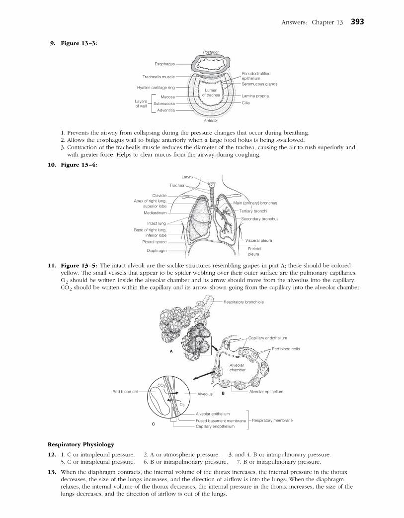
Answers: Chapter 13 393
9. Figure 13–3:
1. Prevents the airway from collapsing during the pressure changes that occur during breathing. 2. Allows the eosphagus wall to bulge anteriorly when a large food bolus is being swallowed. 3. Contraction of the trachealis muscle reduces the diameter of the trachea, causing the air to rush superiorly and
with greater force. Helps to clear mucus from the airway during coughing.
10. Figure 13–4:
11. Figure 13–5: The intact alveoli are the saclike structures resembling grapes in part A; these should be colored yellow. The small vessels that appear to be spider webbing over their outer surface are the pulmonary capillaries. O2 should be written inside the alveolar chamber and its arrow should move from the alveolus into the capillary. CO2 should be written within the capillary and its arrow shown going from the capillary into the alveolar chamber.
Respiratory Physiology
12. 1. C or intrapleural pressure. 2. A or atmospheric pressure. 3. and 4. B or intrapulmonary pressure. 5. C or intrapleural pressure. 6. B or intrapulmonary pressure. 7. B or intrapulmonary pressure.
13. When the diaphragm contracts, the internal volume of the thorax increases, the internal pressure in the thorax decreases, the size of the lungs increases, and the direction of airflow is into the lungs. When the diaphragm relaxes, the internal volume of the thorax decreases, the internal pressure in the thorax increases, the size of the lungs decreases, and the direction of airflow is out of the lungs.
Esophagus
Trachealis muscle
Hyaline cartilage ring
MucosaLayersof wall
Submucosa
Adventitia
PseudostratifiedepitheliumSeromucous glands
Lamina propria
Cilia
Posterior
Anterior
Lumenof trachea
Alveolus
CO2
O2
Respiratory bronchiole
Alveolar epithelium
Alveolar epithelium
Capillary endotheliumFused basement membrane Respiratory membrane
A
B
C
Capillary endothelium
Red blood cells
Red blood cell
Alveolarchamber
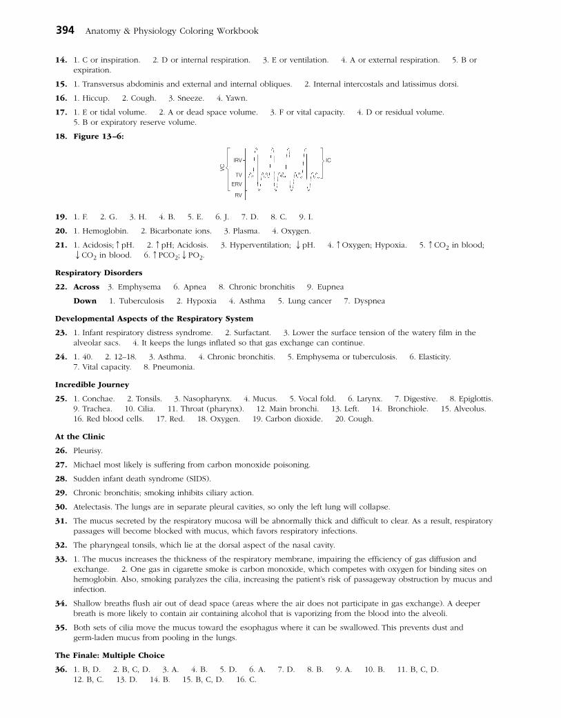
394 Anatomy & Physiology Coloring Workbook
14. 1. C or inspiration. 2. D or internal respiration. 3. E or ventilation. 4. A or external respiration. 5. B or expiration.
15. 1. Transversus abdominis and external and internal obliques. 2. Internal intercostals and latissimus dorsi.
16. 1. Hiccup. 2. Cough. 3. Sneeze. 4. Yawn.
17. 1. E or tidal volume. 2. A or dead space volume. 3. F or vital capacity. 4. D or residual volume. 5. B or expiratory reserve volume.
18. Figure 13–6:
19. 1. F. 2. G. 3. H. 4. B. 5. E. 6. J. 7. D. 8. C. 9. I.
20. 1. Hemoglobin. 2. Bicarbonate ions. 3. Plasma. 4. Oxygen.
21. 1. Acidosis;↑pH. 2. ↑pH; Acidosis. 3. Hyperventilation; ↓pH. 4. ↑Oxygen; Hypoxia. 5. ↑CO2 in blood; ↓CO2 in blood. 6. ↑PCO2;↓PO2.
Respiratory Disorders
22. Across 3. Emphysema 6. Apnea 8. Chronic bronchitis 9. Eupnea
Down 1. Tuberculosis 2. Hypoxia 4. Asthma 5. Lung cancer 7. Dyspnea
Developmental Aspects of the Respiratory System
23. 1. Infant respiratory distress syndrome. 2. Surfactant. 3. Lower the surface tension of the watery film in the alveolar sacs. 4. It keeps the lungs inflated so that gas exchange can continue.
24. 1. 40. 2. 12–18. 3. Asthma. 4. Chronic bronchitis. 5. Emphysema or tuberculosis. 6. Elasticity. 7. Vital capacity. 8. Pneumonia.
Incredible Journey
25. 1. Conchae. 2. Tonsils. 3. Nasopharynx. 4. Mucus. 5. Vocal fold. 6. Larynx. 7. Digestive. 8. Epiglottis. 9. Trachea. 10. Cilia. 11. Throat (pharynx). 12. Main bronchi. 13. Left. 14. Bronchiole. 15. Alveolus. 16. Red blood cells. 17. Red. 18. Oxygen. 19. Carbon dioxide. 20. Cough.
At the Clinic
26. Pleurisy.
27. Michael most likely is suffering from carbon monoxide poisoning.
28. Sudden infant death syndrome (SIDS).
29. Chronic bronchitis; smoking inhibits ciliary action.
30. Atelectasis. The lungs are in separate pleural cavities, so only the left lung will collapse.
31. The mucus secreted by the respiratory mucosa will be abnormally thick and difficult to clear. As a result, respiratory passages will become blocked with mucus, which favors respiratory infections.
32. The pharyngeal tonsils, which lie at the dorsal aspect of the nasal cavity.
33. 1. The mucus increases the thickness of the respiratory membrane, impairing the efficiency of gas diffusion and exchange. 2. One gas in cigarette smoke is carbon monoxide, which competes with oxygen for binding sites on hemoglobin. Also, smoking paralyzes the cilia, increasing the patient’s risk of passageway obstruction by mucus and infection.
34. Shallow breaths flush air out of dead space (areas where the air does not participate in gas exchange). A deeper breath is more likely to contain air containing alcohol that is vaporizing from the blood into the alveoli.
35. Both sets of cilia move the mucus toward the esophagus where it can be swallowed. This prevents dust and germ-laden mucus from pooling in the lungs.
The Finale: Multiple Choice
36. 1. B, D. 2. B, C, D. 3. A. 4. B. 5. D. 6. A. 7. D. 8. B. 9. A. 10. B. 11. B, C, D. 12. B, C. 13. D. 14. B. 15. B, C, D. 16. C.
IRV
TV
ERV
RV
VC
IC
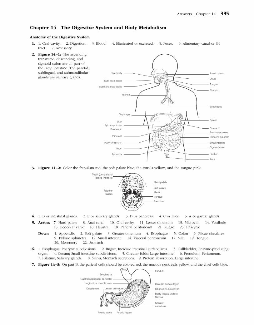
Answers: Chapter 14 395
Chapter 14 The Digestive System and Body Metabolism
Anatomy of the Digestive System
1. 1. Oral cavity. 2. Digestion. 3. Blood. 4. Eliminated or excreted. 5. Feces. 6. Alimentary canal or GI tract. 7. Accessory.
2. Figure 14–1: The ascending, transverse, descending, and sigmoid colon are all part of the large intestine. The parotid, sublingual, and sub mandibular glands are salivary glands.
3. Figure 14–2: Color the frenulum red; the soft palate blue; the tonsils yellow; and the tongue pink.
4. 1. B or intestinal glands. 2. E or salivary glands. 3. D or pancreas. 4. C or liver. 5. A or gastric glands.
5. Across 7. Hard palate 8. Anal canal 10. Oral cavity 11. Lesser omentum 13. Microvilli 14. Vestibule 15. Ileocecal valve 16. Haustra 18. Parietal peritoneum 21. Rugae 23. Pharynx
Down 1. Appendix 2. Soft palate 3. Greater omentum 4. Esophagus 5. Colon 6. Plicae circulares 9. Pyloric sphincter 12. Small intestine 14. Visceral peritoneum 17. Villi 19. Tongue 20. Mesentery 22. Stomach
6. 1. Esophagus; Pharynx subdivisions. 2. Rugae; Increase intestinal surface area. 3. Gallbladder; Enzyme-producing organ. 4. Cecum; Small intestine subdivisions. 5. Circular folds; Large intestine. 6. Frenulum; Peritoneum. 7. Palatine; Salivary glands. 8. Saliva; Stomach secretions. 9. Protein absorption; Large intestine.
7. Figure 14–3: On part B, the parietal cells should be colored red, the mucous neck cells yellow, and the chief cells blue.
Teeth (central andlateral incisors)
Palatinetonsils
Hard palate
Soft palate
Uvula
Frenulum
Tongue
Duodenum
Esophagus
Gastroesophageal sphincter
Lesser curvature
Longitudinal muscle layer
Pyloric valve Pyloric region
Greatercurvature
Serosa
Fundus
Circular muscle layer
Oblique muscle layer
Body (rugae visible)
Oral cavity
Sublingual gland
Submandibular gland
Trachea
Diaphragm
Liver
Duodenum
Pancreas
Ascending colon
Ileum
Appendix
Pyloric sphincter
Parotid gland
Uvula
Tongue
Pharynx
Esophagus
Spleen
Stomach
Descending colon
Transverse colon
Small intestine
Sigmoid colon
Rectum
Anus

396 Anatomy & Physiology Coloring Workbook
8. Figure 14–4:
9. Figure 14–5: 1. Mucosa. 2. Muscularis externa. 3. Submucosa. 4. Serosa.
10. Figure 14–6: 11. Figure 14–7:
12. 1. Deciduous. 2. 6 months. 3. 6 years. 4. Permanent. 5. 32. 6. 20. 7. Incisors. 8. Canine. 9. Premolars. 10. Molars. 11. Wisdom.
Submucosalgland
Submucosa
Gland outsidealimentary canal
Lumen
Mesentery
Lymph nodule
Villi
Serosa
Mucosa
Muscularis externa
Lumen
Tunics
Mucosa
Intestinal glands
ArterioleVenule
A
B
C
Mucosa Serosa
Sub-mucosa
Muscu-laris
Plicae circulares
Villi
Plicaecirculares
Villus
Lacteal
Capillary network
Surface epithelium
Duodenum
Gallbladder
Bile duct
Falciform ligamentLiver
Common hepatic ductCystic duct
PancreasPancreaticduct

Answers: Chapter 14 397
13. Figure 14–8: 1. A. 2. B. 3. E. 4. C.
Physiology of the Digestive System
14. 1. D or eating. 2. G or swallowing, H or segmentation and peristalsis. 3. E or chewing, F or churning. 4. B or enzymatic breakdown. 5. A or transport of nutrients from lumen to blood. 6. C or elimination of feces.
15. 1. G or peritonitis. 2. E or heartburn. 3. F or jaundice. 4. H or ulcer. 5. C or diarrhea. 6. D or gallstones. 7. B or constipation. 8. A or appendicitis.
16. 1. O or salivary amylase. 2. G or hormonal stimulus. 3. M or psychological stimulus. 4. I or mechanical stimulus. 5. L or pepsin. 6. F or HCl. 7. K or mucus. 8. N or rennin. 9. E or churning. 10. C or brush border enzymes. 11. A or bicarbonate-rich fluid. 12. H or lipases. 13. B or bile.
17. 1. A or cholecystokinin, C or secretin. 2. B or gastrin. 3. A or cholecystokinin. 4. C or secretin.
18. 1. C or fructose, D or galactose, E or glucose. 2. F or lactose, G or maltose, I or sucrose. 3. A or amino acids. 4. B or fatty acids. 5. E or glucose.
19. 1. P. 2. A. 3. A. 4. P. 5. A. Circle fatty acids.
20. 1. Deglutition. 2. Buccal. 3. Pharyngeal-esophageal. 4. Tongue. 5. Uvula. 6. Larynx. 7. Epiglottis. 8. Peristalsis. 9. Cardioesophageal. 10. and 11. Peristalsis, segmentation. 12. Segmentation. 13. Mass movement. 14. Rectum. 15. Defecation. 16. Emetic. 17. Vomiting.
Nutrition and Metabolism
21. 1. B or carbohydrates. 2. C or fats. 3. A or amino acids. 4. C or fats. 5. C or fats. 6. A or amino acids.
22. 1. A or bread/pasta, D or fruits, H or vegetables. 2. B or cheese/cream. 3. G or starch. 4. C or cellulose. 5. B or cheese/cream, E or meat/fish. 6. I or vitamins. 7. F or minerals.
23. Figure 14–9:
1. Glycolysis (#3) does not require oxygen. 2. Krebs cycle (#6) and the electron transport chains (#7) require oxy-gen. 3. In the form of hydrogen atoms bearing high-energy electrons. 4. and 5. The electron transport chain.
24. 1. K or glucose. 2. O or oxygen. 3. R or water. 4. H or carbon dioxide. 5. A or ATP. 6. N or mono-saccharides. 7. and 8. C or acetoacetic acid; D or acetone. 9. M or ketosis. 10. I or essential. 11. F or ammonia. 12. Q or urea.
25. 1. TMR; BMR. 2. ↓Metabolic rate; Thyroid hormone. 3. Child; Low metabolic rate. 4. Fats; Low kcal/gram. 5. Vasoconstriction; Heat loss.
26. 1. Albumin. 2. Clotting proteins. 3. Cholesterol. 4. Hyperglycemia. 5. Glycogen. 6. Hypoglycemia. 7. Glycogenolysis. 8. Gluconeogenesis. 9. Detoxification. 10. Phagocytic. 11. Lipoproteins. 12. Insoluble. 13. LDLs. 14. Membranes. 15. Steroid. 16. Liver. 17. Bile salts. 18. A. 19. Iron.
27. 1. D or heat. 2. B or constriction of skin blood vessels, K or shivering. 3. A or blood. 4. F or hypothalamus. 5. J or pyrogens. 6. C or frostbite. 7. H or perspiration, I or radiation. 8. G or hypothermia. 9. E or hyperthermia.
EnamelCrown
Gingiva
DentinBone tissue of jaw
Root canal
Periodontal membrane
Pulp
Cement
Root
1.
7.
9.
2.
5. 4.
8. 11.
10.
14. 13. 12.
3. (phase)
(phase)
(cellular site)
(product)
(product)
(product) (product)
(product)
(product)(cellular site)
6.
(phase)
Chemical energy(high-energy electron)
(high-energyelectron)
Chemical energy
Glycolysis
Glucose(substrate)
Pyruvic acidKrebscycle
Electrontransport chain
Cytosol MitochondrianWater
ATP ATP ATP
Carbon dioxide
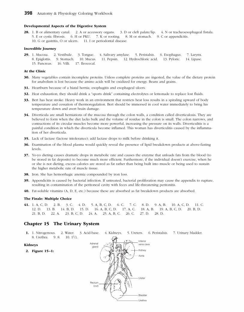
398 Anatomy & Physiology Coloring Workbook
Developmental Aspects of the Digestive System
28. 1. B or alimentary canal. 2. A or accessory organs. 3. D or cleft palate/lip. 4. N or tracheoesophageal fistula. 5. E or cystic fibrosis. 6. H or PKU. 7. K or rooting. 8. M or stomach. 9. C or appendicitis. 10. G or gastritis, O or ulcers. 11. I or periodontal disease.
Incredible Journey
29. 1. Mucosa. 2. Vestibule. 3. Tongue. 4. Salivary amylase. 5. Peristalsis. 6. Esophagus. 7. Larynx. 8. Epiglottis. 9. Stomach. 10. Mucus. 11. Pepsin. 12. Hydrochloric acid. 13. Pyloric. 14. Lipase. 15. Pancreas. 16. Villi. 17. Ileocecal.
At the Clinic
30. Many vegetables contain incomplete proteins. Unless complete proteins are ingested, the value of the dietary protein for anabolism is lost because the amino acids will be oxidized for energy. Beans and grains.
31. Heartburn because of a hiatal hernia; esophagitis and esophageal ulcers.
32. Heat exhaustion; they should drink a “sports drink” containing electrolytes or lemonade to replace lost fluids.
33. Bert has heat stroke. Heavy work in an environment that restricts heat loss results in a spiraling upward of body temperature and cessation of thermoregulation. Bert should be immersed in cool water immediately to bring his temperature down and avert brain damage.
34. Diverticula are small herniations of the mucosa through the colon walls, a condition called diverticulosis. They are believed to form when the diet lacks bulk and the volume of residue in the colon is small. The colon narrows, and contractions of its circular muscles become more powerful, increasing the pressure on its walls. Diverticulitis is a painful condition in which the diverticula become inflamed. This woman has diverticulitis caused by the inflamma-tion of her diverticula.
35. Lack of lactase (lactose intolerance); add lactase drops to milk before drinking it.
36. Examination of the blood plasma would quickly reveal the presence of lipid breakdown products at above-fasting levels.
37. Yo-yo dieting causes dramatic drops in metabolic rate and causes the enzyme that unloads fats from the blood (to be stored in fat deposits) to become much more efficient. Furthermore, if the individual doesn’t exercise, when he or she is not dieting, excess calories are stored as fat rather than being built into muscle or being used to sustain the higher metabolic rate of muscle tissue.
38. Iron. She has hemorrhagic anemia compounded by iron loss.
39. Appendicitis is caused by bacterial infection. If untreated, bacterial proliferation may cause the appendix to rupture, resulting in contamination of the peritoneal cavity with feces and life-threatening peritonitis.
40. Fat-soluble vitamins (A, D, E, etc.) because these are absorbed as fat breakdown products are absorbed.
The Finale: Multiple Choice
41. 1. A, C, D. 2. B. 3. C. 4. D. 5. A, B, C, D. 6. C. 7. C. 8. D. 9. A, B. 10. A, C, D. 11. C. 12. D. 13. B. 14. B, D. 15. D. 16. A, B, C, D. 17. A, C. 18. A, B. 19. A, B, C, D. 20. B, D. 21. B, D. 22. A. 23. B, C, D. 24. A. 25. A, B, C. 26. C. 27. D. 28. D.
Chapter 15 The Urinary System
1. 1. Nitrogenous. 2. Water. 3. Acid-base. 4. Kidneys. 5. Ureters. 6. Peristalsis. 7. Urinary bladder. 8. Urethra. 9. 8. 10. 11/2.
Kidneys
2. Figure 15–1:
Rectum(cut)
Adrenalgland
Inferiorvena cava
Aorta
Ureter
Bladder
Urethra
Kidney

Answers: Chapter 15 399
3. Figure 15–2: The fibrous membrane surrounding the kidney is the fibrous capsule; the basin-like pelvis is continu-ous with the ureter; a calyx is an extension of the pelvis; renal columns are extensions of cortical tissue into the medulla. The cortex contains the bulk of the nephron structures; the striped-appearing medullary pyramids are pri-marily formed by collecting ducts.
4. 1. Intraperitoneal; Kidney location. 2. Urethra; Drains kidney. 3. Glomerulus; Peritubular capillaries. 4. Glomerulus; Juxtaglomerular apparatus. 5. Collecting duct; Blood vessels. 6. Cortical nephrons; Juxtamedullary nephrons. 7. Collecting duct; Nephron. 8. Nephron loop; Renal corpuscle.
5. Figure 15–3: 1. Glomerular capsule. 2. Afferent arteriole. 3. Efferent arteriole. 4. Cortical radiate artery. 5. Cortical radiate vein. 6. Arcuate artery. 7. Arcuate vein. 8. Interlobar artery. 9. Interlobar vein. 10. Nephron loop. 11. Collecting duct. 12. Distal convoluted tubule. 13. Proximal convoluted tubule. 14. Peritubular capillaries. 15. Glomerulus. Relative to the coloring instructions, #1 is green, #15 is red, #14 is blue, #11 is yellow, and #13 is orange.
6. Figure 15–4: 1. Black arrows: Site of filtrate formation is the glomerulus. Arrows leave the glomerulus and enter glomerular (Bowman’s) capsule. 2. Red arrows: Major site of amino acid and glucose reabsorption. Shown going from the PCT interior and passing through the PCT walls to the capillary bed surrounding the PCT (the latter not shown). Nutrients leave the filtrate. 3. Green arrows: At site of ADH action. Arrows (indicating water movement) shown leaving the interior of the collecting duct and passing through the walls to enter the capillary bed surround-ing that duct. Water leaves the filtrate. 4. Yellow arrows: Site of aldosterone action. Arrows (indicating Na+ move-ment) leaving the collecting duct and the DCT and passing through their walls into the surrounding capillary bed. Na+ leaves the filtrate. 5. Blue arrows: Site of tubular secretion. Arrows shown entering the PCT to enter the filtrate.
7. 1. Afferent. 2. Efferent. 3. Plasma. 4. and 5. Diffusion; active transport. 6. Microvilli. 7. Secretion. 8.–10. Diet, cellular metabolism, urine output. 11. 1–1.8. 12. Urochrome. 13.–15. Urea, uric acid, creatinine. 16. Lungs. 17. Perspiration. 18. Decreases. 19. Dialysis.
8. 1. A. 2. B. 3. A. 4. A. 5. B.
9. 1. D. 2. D. 3. I. 4. D. 5. I. 6. I.
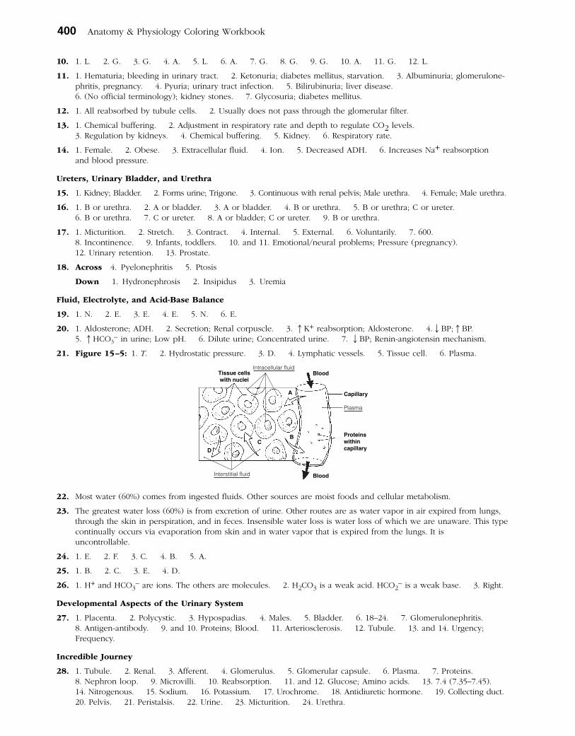
400 Anatomy & Physiology Coloring Workbook
10. 1. L. 2. G. 3. G. 4. A. 5. L. 6. A. 7. G. 8. G. 9. G. 10. A. 11. G. 12. L.
11. 1. Hematuria; bleeding in urinary tract. 2. Ketonuria; diabetes mellitus, starvation. 3. Albuminuria; glomerulone-phritis, pregnancy. 4. Pyuria; urinary tract infection. 5. Bilirubinuria; liver disease. 6. (No official terminology); kidney stones. 7. Glycosuria; diabetes mellitus.
12. 1. All reabsorbed by tubule cells. 2. Usually does not pass through the glomerular filter.
13. 1. Chemical buffering. 2. Adjustment in respiratory rate and depth to regulate CO2 levels. 3. Regulation by kidneys. 4. Chemical buffering. 5. Kidney. 6. Respiratory rate.
14. 1. Female. 2. Obese. 3. Extracellular fluid. 4. Ion. 5. Decreased ADH. 6. Increases Na+ reabsorption and blood pressure.
Ureters, Urinary Bladder, and Urethra
15. 1. Kidney; Bladder. 2. Forms urine; Trigone. 3. Continuous with renal pelvis; Male urethra. 4. Female; Male urethra.
16. 1. B or urethra. 2. A or bladder. 3. A or bladder. 4. B or urethra. 5. B or urethra; C or ureter. 6. B or urethra. 7. C or ureter. 8. A or bladder; C or ureter. 9. B or urethra.
17. 1. Micturition. 2. Stretch. 3. Contract. 4. Internal. 5. External. 6. Voluntarily. 7. 600. 8. Incontinence. 9. Infants, toddlers. 10. and 11. Emotional/neural problems; Pressure (pregnancy). 12. Urinary retention. 13. Prostate.
18. Across 4. Pyelonephritis 5. Ptosis
Down 1. Hydronephrosis 2. Insipidus 3. Uremia
Fluid, Electrolyte, and Acid-Base Balance
19. 1. N. 2. E. 3. E. 4. E. 5. N. 6. E.
20. 1. Aldosterone; ADH. 2. Secretion; Renal corpuscle. 3. ↑K+ reabsorption; Aldosterone. 4.↓BP;↑BP. 5. ↑HCO3
– in urine; Low pH. 6. Dilute urine; Concentrated urine. 7. ↓BP; Renin-angiotensin mechanism.
21. Figure 15–5: 1. T. 2. Hydrostatic pressure. 3. D. 4. Lymphatic vessels. 5. Tissue cell. 6. Plasma.
22. Most water (60%) comes from ingested fluids. Other sources are moist foods and cellular metabolism.
23. The greatest water loss (60%) is from excretion of urine. Other routes are as water vapor in air expired from lungs, through the skin in perspiration, and in feces. Insensible water loss is water loss of which we are unaware. This type continually occurs via evaporation from skin and in water vapor that is expired from the lungs. It is uncontrollable.
24. 1. E. 2. F. 3. C. 4. B. 5. A.
25. 1. B. 2. C. 3. E. 4. D.
26. 1. H+ and HCO3– are ions. The others are molecules. 2. H2CO3 is a weak acid. HCO2
– is a weak base. 3. Right.
Developmental Aspects of the Urinary System
27. 1. Placenta. 2. Polycystic. 3. Hypospadias. 4. Males. 5. Bladder. 6. 18–24. 7. Glomerulone phritis. 8. Antigen-antibody. 9. and 10. Proteins; Blood. 11. Arteriosclerosis. 12. Tubule. 13. and 14. Urgency; Frequency.
Incredible Journey
28. 1. Tubule. 2. Renal. 3. Afferent. 4. Glomerulus. 5. Glomerular capsule. 6. Plasma. 7. Proteins. 8. Nephron loop. 9. Microvilli. 10. Reabsorption. 11. and 12. Glucose; Amino acids. 13. 7.4 (7.35–7.45). 14. Nitrogenous. 15. Sodium. 16. Potassium. 17. Urochrome. 18. Antidiuretic hormone. 19. Collecting duct. 20. Pelvis. 21. Peristalsis. 22. Urine. 23. Micturition. 24. Urethra.
Tissue cellswith nuclei
Capillary
Proteinswithincapillary
Blood
Blood
A
BC
D
Plasma
Interstitial fluid
Intracellular fluid
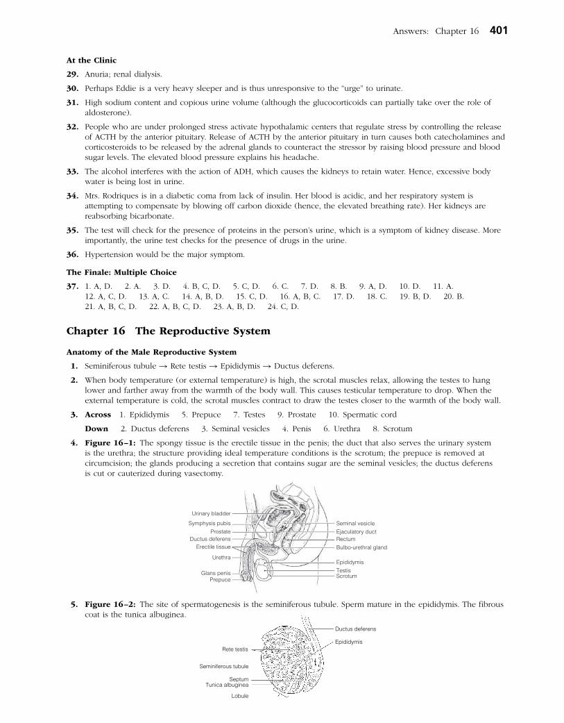
Answers: Chapter 16 401
At the Clinic
29. Anuria; renal dialysis.
30. Perhaps Eddie is a very heavy sleeper and is thus unresponsive to the “urge” to urinate.
31. High sodium content and copious urine volume (although the glucocorticoids can partially take over the role of aldosterone).
32. People who are under prolonged stress activate hypothalamic centers that regulate stress by controlling the release of ACTH by the anterior pituitary. Release of ACTH by the anterior pituitary in turn causes both catecholamines and corticosteroids to be released by the adrenal glands to counteract the stressor by raising blood pressure and blood sugar levels. The elevated blood pressure explains his headache.
33. The alcohol interferes with the action of ADH, which causes the kidneys to retain water. Hence, excessive body water is being lost in urine.
34. Mrs. Rodriques is in a diabetic coma from lack of insulin. Her blood is acidic, and her respiratory system is attempting to compensate by blowing off carbon dioxide (hence, the elevated breathing rate). Her kidneys are reabsorbing bicarbonate.
35. The test will check for the presence of proteins in the person’s urine, which is a symptom of kidney disease. More importantly, the urine test checks for the presence of drugs in the urine.
36. Hypertension would be the major symptom.
The Finale: Multiple Choice
37. 1. A, D. 2. A. 3. D. 4. B, C, D. 5. C, D. 6. C. 7. D. 8. B. 9. A, D. 10. D. 11. A. 12. A, C, D. 13. A, C. 14. A, B, D. 15. C, D. 16. A, B, C. 17. D. 18. C. 19. B, D. 20. B. 21. A, B, C, D. 22. A, B, C, D. 23. A, B, D. 24. C, D.
Chapter 16 The Reproductive System
Anatomy of the Male Reproductive System
1. Seminiferous tubule → Rete testis → Epididymis → Ductus deferens.
2. When body temperature (or external temperature) is high, the scrotal muscles relax, allowing the testes to hang lower and farther away from the warmth of the body wall. This causes testicular temperature to drop. When the external temperature is cold, the scrotal muscles contract to draw the testes closer to the warmth of the body wall.
3. Across 1. Epididymis 5. Prepuce 7. Testes 9. Prostate 10. Spermatic cord
Down 2. Ductus deferens 3. Seminal vesicles 4. Penis 6. Urethra 8. Scrotum
4. Figure 16–1: The spongy tissue is the erectile tissue in the penis; the duct that also serves the urinary system is the urethra; the structure providing ideal temperature conditions is the scrotum; the prepuce is removed at circumcision; the glands producing a secretion that contains sugar are the seminal vesicles; the ductus deferens is cut or cauterized during vasectomy.
5. Figure 16–2: The site of spermatogenesis is the seminiferous tubule. Sperm mature in the epididymis. The fibrous coat is the tunica albuginea.
Ductus deferens
EpididymisRete testis
Seminiferous tubule
SeptumTunica albuginea
Lobule

402 Anatomy & Physiology Coloring Workbook
Male Reproductive Functions
6. 1. Spermatogonium. 2. Secondary spermatocyte, sperm, spermatid. 3. Secondary spermatocyte. 4. Spermatid. 5. Sperm. 6. FSH, Testosterone.
Figure 16–3:
7. Figure 16–4:
8. 1. A or mitosis. 2. B or meiosis. 3. C or both mitosis and meiosis. 4. A or mitosis. 5. B or meiosis. 6. A or mitosis. 7. A or mitosis. 8. B or meiosis. 9. C or both mitosis and meiosis. 10. B or meiosis. 11. B or meiosis.
9. Deepening voice; formation of a beard and increased hair growth all over body, particularly in axillary/genital regions; enlargement of skeletal muscles; increased density of skeleton.
Anatomy of the Female Reproductive System
1 0. 1. Uterus. 2. Vagina. 3. Uterine, or fallopian, tube. 4. Clitoris. 5. Uterine tube. 6. Hymen. 7. Ovary. 8. Fimbriae.
11. Figure 16–5: The endometrium is the lining and the myometrium is the muscular layer of the uterus. The egg travels along the uterine tube after it is released from the ovary. The round ligament helps to anchor the uterus. The ovary produces hormones and gametes. The homologue of the male scrotum is the labium majus.
Mitochondria (metabolically active organelles) Acrosome (enzyme-containing sac)
TailNucleus (DNA containing area)
Midpiece Head
Uterus (myometrium)
Round ligament
Uterine tube
Urinary bladder
Symphysis pubis
ClitorisVagina
Labium majus
Sacrum
Endometrium
Cervix of uterus
Ovary
Fimbriae
Urethra
Rectum
Anus
Labium minus

Answers: Chapter 16 403
12. Figure 16–6: The clitoris should be colored blue; the hymen yellow; and the vaginal opening red.
Female Reproductive Functions and Cycles
13. 1. B or primary oocyte. 2. C or secondary oocyte. 3. C or secondary oocyte. 4. D or ovum.
14. The follicle (granulosa) cells produce estrogen, the corpus luteum produces progesterone, and oocytes are the cen-tral cells in all follicles. Event A = ovulation. 1. No. 2. Peritoneal cavity. 3. After sperm penetration occurs. 4. Ruptured (ovulated) follicle. 5. One ovum; three polar bodies. 6. Males produce four spermatids → four sperm. 7. They deteriorate. 8. They lack nutrient-containing cytoplasm. 9. Menopause.
Figure 16–7:
15. Because of this structural condition, many “eggs” (oocytes) are lost in the peritoneal cavity; therefore, they are unavailable for fertilization. The discontinuity also provides infectious microorganisms with access to the peritoneal cavity, possibly leading to pelvic inflammatory disease (PID).
16. 1. Follicle-stimulating hormone (FSH). 2. Luteinizing hormone (LH). 3. Estrogen and progesterone. 4. Estrogen. 5. LH. 6. LH.
17. Appearance of axillary/pubic hair, development of breasts, widening of pelvis, onset of menses.
18. 1. A or estrogens, B or progesterone. 2. B or progesterone. 3. A or estrogens. 4. B or progesterone. 5. and 6. A or estrogens.
19. Figure 16–8: From left to right on part C, the structures are the primary follicle, the secondary (growing) follicle, the vesicular follicle, the ovulating follicle, the corpus luteum, and an atretic (deteriorating) corpus luteum. In part D, menses is from day 0 to day 5, the proliferative phase is from day 6 to day 14, and the secretory phase is from day 15 to day 28.
Labia majora(spread)
Urethral orifice
Vaginal orifice
Anus
Mons pubis
Clitoris
Labia minora
Hymen
Blo
od le
vels
of a
nter
ior
pitu
itary
hor
mon
esB
lood
leve
ls o
fov
aria
n ho
rmon
es
B
A
282624222018161412108420 6
FSHLH
Ovu
latio
n
C
Ova
rian
cycl
e
D
Menstrualflow
1 4 10 14Days 28
Ute
rine
(men
stru
al)
cycl
e

404 Anatomy & Physiology Coloring Workbook
Mammary Glands
20. Figure 16–9: The alveolar glands should be colored blue, and the rest of the internal breast, excluding the duct system, should be colored yellow.
Survey of Pregnancy and Embryonic Development
21. 1. Just its head (the nucleus). 2. Digests away the cement holding the follicle cells together; allows sperm to reach the oocyte.
22. Figure 16–10: 1. Fertilization (sperm penetration). 2. Fertilized egg (zygote). 3. Cleavage. 4. Blastocyst (chorionic vesicle). 5. Implantation. 6. The polar body has virtually no cytoplasm. Without nutrients it would be unable to live until it reached the uterus.
23. 1. H or zygote. 2. F or placenta. 3. B or chorionic villi, C or endometrium. 4. A or amnion. 5. G or umbilical cord. 6. B or chorionic villi. 7. E or fetus. 8. F or placenta. 9. D or fertilization.
24. The blastocyst and then the placenta release hCG, which is like LH and sustains the function of the corpus luteum temporarily until the placenta can take over.
25. 1. B or mesoderm. 2. C or endoderm. 3. A or ectoderm. 4. B or mesoderm. 5. A or ectoderm. 6. B or mesoderm. 7. C or endoderm. 8. C or endoderm.
26. Oxytocin and prostaglandins.
27. 1. Prolactin. 2. Oxytocin.
28. Check 1, 3, 5, 9, 10, 11, 12.
29. False labor (irregular, ineffective uterine contractions). These occur because rising estrogen levels make the uterus more responsive to oxytocin and antagonize progesterone’s quieting influence on the myometrium.
30. 1. Dilation stage: The period from the beginning of labor until full dilation (approx. 10-cm diameter) of the cervix; the longest phase. 2. Expulsion stage: The period from full dilation to the birth (delivery). 3. Placental stage: Delivery of the placenta, which follows delivery of the infant.
31. Figure 16–11:
32. Each pass forces the baby farther into the birth passage. The cycle ends with the birth of the baby.
33. The response to the stimulus enhances the stimulus. For example, the more a baby descends into the pelvis and stretches the uterus, the more oxytocin is produced and the stronger the contractions become.
Pectoralis majormuscle
Rib
Intercostalmuscles
Alveolar glands
Areola
Nipple
Lactiferousducts
Infant descends in the birthcanal; head exerts pressure
on the cervix Contracts morevigorously
Stimulates
Afferent impulses stimulate
Posteriorpituitary
Stimulates
Releases
Uterine myometrium
Oxytocin
Hypothalamus

Answers: Chapter 16 405
Developmental Aspects of the Reproductive System
34. 1. Y and X. 2. 2 Xs. 3. External genitalia. 4. Duct system. 5. Cryptorchidism. 6.–8. (in any order) Esch-erichia coli, STDs or venereal disease, yeast infections. 9. PID (pelvic inflammatory disease). 10. Venereal. 11. Breast. 12. Cervix of the uterus. 13. Pap smear. 14. Menopause. 15. Hot flashes. 16. Declines. 17. Rise. 18. Estrogen. 19. Vaginal. 20. Prostate. 21. and 22. Urinary; reproductive.
Incredible Journey
35. 1. Uterus. 2. Ovary. 3. Fimbriae. 4. Ovulation. 5. Secondary oocyte. 6. Follicle. 7. Peristalsis. 8. Cilia. 9. Sperm. 10. Acrosomes. 11. Meiotic. 12. Ovum. 13. Polar body. 14. Dead. 15. Fertilization. 16. Zygote (fertilized egg). 17. Cleavage. 18. Endometrium. 19. Implantation. 20. Vagina.
At the Clinic
36. Pitocin will act on the placenta, stimulating production and release of prostaglandins. Pitocin and prostaglandins are powerful uterine muscle stimulants. Oxytocin normally causes frequent and vigorous contractions of the uterine wall.
37. Megadoses of testosterone would inhibit anterior pituitary gonadotropin (FSH) release. Spermatogenesis is inhibited in the absence of FSH stimulation.
38. Her tubes were probably scarred by PID. Hormonal testing and the daily basal temperature recordings would have indicated her anovulatory condition.
39. His scrotal muscles had contracted to draw the testes closer to the warmth of the abdominal cavity.
40. Mary’s fetus might have respiratory problems or even congenital defects caused by her smoking, because smoking causes vasoconstriction, which would hinder blood delivery to the placenta.
41. Cervical cancer.
42. There is little possibility that she is right. Body organs are laid down during the first trimester, and only growth and final differentiation occur after that.
43. Sexually transmitted diseases (STDs). It is important to inform his partner(s) that they might be infected also, particu-larly because some females do not exhibit any signs or symptoms of these particular infections but still need to be treated.
44. By the surgical procedure called a C-section (cesarean section).
45. These hormones exert negative feedback on the release of GnRH by the hypothalamus. This, in turn, would interfere with pituitary release of LH, thus interfering with ovulation.
46. Both procedures prevent the germ cells from reaching their normal destination during intercourse. Tubal ligation cuts through the fallopian tubes, whereas vasectomy interrupts the continuity of the vas deferens. Hence, these make the recipients sterile.
The Finale: Multiple Choice
47. 1. A, B. 2. B, C. 3. C. 4. D. 5. D. 6. B. 7. B. 8. B. 9. D. 10. C, D. 11. A, D. 12. B. 13. A, B. 14. A, D. 15. A, B, D. 16. A, C. 17. C. 18. C. 19. C. 20. A, B, C. 21. B. 22. A, C. 23. C. 24. A, C. 25. D. 26. B, D. 27. A, B, D. 28. B. 29. A, D.