Is Total Hip Arthroplasty after Hip Arthrodesis as Good as Primary Arthroplasty
Upload
khangminh22Category
view
1download
0
Cochrane Database of Systematic Reviews
Surgical interventions for treating extracapsular hip fractures
in adults: a network meta-analysis (Protocol)
Sreekanta A, Eardley WGP, Parker MJ, Felix LM, Wood H, Glanville JM, Cook J, Griffin XL
Sreekanta A, Eardley WGP, Parker MJ, Felix LM, Wood H, Glanville JM, Cook J, Griffin XL.
Surgical interventions for treating extracapsular hip fractures in adults: a networkmeta-analysis.
Cochrane Database of Systematic Reviews 2019, Issue 8. Art. No.: CD013405.
DOI: 10.1002/14651858.CD013405.
www.cochranelibrary.com
Surgical interventions for treating extracapsular hip fractures in adults: a networkmeta-analysis (Protocol)
Copyright © 2019 The Cochrane Collaboration. Published by John Wiley & Sons, Ltd.
T A B L E O F C O N T E N T S
1HEADER . . . . . . . . . . . . . . . . . . . . . . . . . . . . . . . . . . . . . . .
1ABSTRACT . . . . . . . . . . . . . . . . . . . . . . . . . . . . . . . . . . . . . .
1BACKGROUND . . . . . . . . . . . . . . . . . . . . . . . . . . . . . . . . . . . .
4OBJECTIVES . . . . . . . . . . . . . . . . . . . . . . . . . . . . . . . . . . . . .
4METHODS . . . . . . . . . . . . . . . . . . . . . . . . . . . . . . . . . . . . . .
12ACKNOWLEDGEMENTS . . . . . . . . . . . . . . . . . . . . . . . . . . . . . . . .
12REFERENCES . . . . . . . . . . . . . . . . . . . . . . . . . . . . . . . . . . . . .
16ADDITIONAL TABLES . . . . . . . . . . . . . . . . . . . . . . . . . . . . . . . . . .
32APPENDICES . . . . . . . . . . . . . . . . . . . . . . . . . . . . . . . . . . . . .
33CONTRIBUTIONS OF AUTHORS . . . . . . . . . . . . . . . . . . . . . . . . . . . . .
34DECLARATIONS OF INTEREST . . . . . . . . . . . . . . . . . . . . . . . . . . . . . .
34SOURCES OF SUPPORT . . . . . . . . . . . . . . . . . . . . . . . . . . . . . . . . .
iSurgical interventions for treating extracapsular hip fractures in adults: a network meta-analysis (Protocol)
Copyright © 2019 The Cochrane Collaboration. Published by John Wiley & Sons, Ltd.
[Intervention Protocol]
Surgical interventions for treating extracapsular hip fracturesin adults: a network meta-analysis
Ashwini Sreekanta1, Will GP Eardley2, Martyn J Parker3, Lambert M Felix1, Hannah Wood4 , Julie M Glanville4, Jonathan Cook1,
Xavier L Griffin1
1Nuffield Department of Orthopaedics, Rheumatology and Musculoskeletal Sciences (NDORMS), University of Oxford, Oxford, UK.2Department of Trauma and Orthopaedics, The James Cook University Hospital, Middlesbrough, UK. 3Department of Orthopaedics,
Peterborough and Stamford Hospitals NHS Foundation Trust, Peterborough, UK. 4York Health Economics Consortium, York, UK
Contact address: Ashwini Sreekanta, Nuffield Department of Orthopaedics, Rheumatology and Musculoskeletal Sciences
(NDORMS), University of Oxford, Kadoorie Centre, John Radcliffe Hospital, Headley Way, Oxford, OX3 9DU, UK.
[email protected], [email protected].
Editorial group: Cochrane Bone, Joint and Muscle Trauma Group.
Publication status and date: New, published in Issue 8, 2019.
Citation: Sreekanta A, Eardley WGP, Parker MJ, Felix LM, Wood H, Glanville JM, Cook J, Griffin XL. Surgical interventions for
treating extracapsular hip fractures in adults: a network meta-analysis. Cochrane Database of Systematic Reviews 2019, Issue 8. Art. No.:
CD013405. DOI: 10.1002/14651858.CD013405.
Copyright © 2019 The Cochrane Collaboration. Published by John Wiley & Sons, Ltd.
A B S T R A C T
This is a protocol for a Cochrane Review (Intervention). The objectives are as follows:
To assess the relative effects (benefits and harms) of all surgical treatments used in the management of extracapsular hip fractures in
adults, using a network meta-analysis of randomised trials, and to generate a hierarchy of interventions according to their outcomes.
B A C K G R O U N D
This protocol has been written in accordance with guidance for
authors on preparing a protocol for a systematic review with mul-
tiple interventions (Chaimani 2017; CMIMG 2014).
Description of the condition
Epidemiology
A hip fracture, or proximal femoral fracture, is a break in the upper
region of the femur (thigh bone) between the subcapital region
(the area just under the femoral head) and 5 cm below the lesser
trochanter (a bony projection of the upper femur). The incidence
of hip fractures rises with age; they are most common in the older
adult population (Court-Brown 2017; Kanis 2001). Those seen
in younger adults are usually associated with poor bone health
(Karantana 2011; Rogmark 2018). A very small proportion of
fractures in younger people are caused by high-energy trauma such
as road traffic collisions, industrial injuries and sports injuries. The
overwhelming majority of hip fractures are fragility fractures as-
sociated with osteoporosis; such fractures are caused by mechani-
cal forces that would not ordinarily result in fracture. The World
Health Organization has defined fragility fractures as those sus-
tained from injuries equivalent to a fall from a standing height or
less (Kanis 2001). In the UK, the mean age of a person with hip
fracture is 83 years, and approximately two-thirds occur in women
(NHFD 2017).
1Surgical interventions for treating extracapsular hip fractures in adults: a network meta-analysis (Protocol)
Copyright © 2019 The Cochrane Collaboration. Published by John Wiley & Sons, Ltd.

Hip fractures are a major healthcare problem at the individual
and population level; they present a huge challenge and burden
to patients, healthcare systems and society. The increased propor-
tion of older adults in the world population means that the ab-
solute number of hip fractures is rising rapidly across the globe.
For example, in 2016 there were 65,645 new presentations of hip
fracture to 177 trauma units in England, Wales and Northern Ire-
land (NHFD 2017). Based on population estimates for these re-
gions for mid-2016, this equates to an incidence rate of 109 cases
per 100,000 population (ONS 2016). By 2050, it is estimated
that the annual worldwide incidence of hip fracture will be 6 mil-
lion (Cooper 2011; Johnell 2004). Incident hip fracture rates are
higher in industrialised countries than in developing countries.
Northern Europe and the USA have the highest rates of hip frac-
ture, whereas Latin America and Africa have the lowest (Dhanwal
2011). European studies show that there are more hip fractures
in the north of the region than in the south, and there is also a
similar north-south gradient in the USA (Dhanwal 2011). Fac-
tors thought to be responsible for this variation are population
demographics (with older populations in countries with higher
incidence rates) and the influence of ethnicity, latitude and en-
vironmental factors such as socioeconomic deprivation (Bardsley
2013; Cooper 2011; Dhanwal 2011; Kanis 2012).
Burden of disease
Hip fractures are associated with a high risk of death. For example,
in England, Wales and Northern Ireland, the 30-day mortality
rate in 2016 remained high at 6.7%, despite a decline from 8.5%
in 2011 and 7.1% in 2015 (NHFD 2017). The mortality rate
one year after a hip fracture is approximately 30%; however, fewer
than half of deaths are attributable to the fracture itself, which
reflects the frailty of the patients and associated high prevalence
of comorbidities and complications (Parker 1991; SIGN 2009).
The impact of morbidity associated with hip fractures is similar
to that of stroke, and entails a substantial loss of healthy life-years
in older people (Griffin 2015). Hip fractures commonly result in
reduced mobility and greater dependency, with many people fail-
ing to return to their pre-injury residence. In addition, the pub-
lic health impact of hip fractures is significant: data from large
prospective cohorts show the burden of disease due to hip frac-
ture is 27 disability-adjusted life years (DALYs) per 1000 indi-
viduals, which equates to an average loss of 2.7% of the healthy
life expectancy in the population at risk of fragility hip fracture
(Papadimitriou 2017). The direct economic burden of hip frac-
tures is also substantial. Hip fractures are among the most expen-
sive conditions seen in hospitals; the aggregated cost for 316,000
inpatient episodes in the USA in 2011 was nearly USD 4.9 bil-
lion (Torio 2011). In England, Wales and Northern Ireland, hip
fracture patients occupy 1.5 million hospital bed days each year,
and cost the National Health Service and social care GBP 1 billion
(NHFD 2017). Combined health and social care costs incurred
during the first year following a hip fracture have been estimated
at USD 43,669, which is greater than the cost for non-commu-
nicable diseases such as acute coronary syndrome (USD 32,345)
and ischaemic stroke (USD 34,772) (Williamson 2017). In estab-
lished market economies, hip fractures represent 1.4% of the total
healthcare burden (Johnell 2004).
Extracapsular hip fracture
Hip fractures either involve the region of the bone which is en-
veloped by the ligamentous hip joint capsule (intracapsular), or
that outside the capsule (extracapsular). Extracapsular fractures
traverse the femur within the area of bone bounded by the in-
tertrochanteric line proximally, up to a distance of 5 cm from the
distal part of the lesser trochanter. Several classification methods
have been proposed to define different types of extracapsular frac-
tures (AO Foundation 2018; Evans 1949; Jensen 1980). They are
generally subdivided depending on their relationship to the greater
and lesser trochanters (the two bony projections present at the up-
per end of the femur) and the complexity of the fracture config-
uration. It is increasingly clear that each of these classifications is
limited in its generalisability since inter- and intra-observer agree-
ment is poor. Table 1 provides a description of the most recent
classification of trochanteric fractures (AO Foundation 2018). For
this review we plan to use a pragmatic simplification of these clas-
sifications, as follows.
• Trochanteric fractures: those which lie mostly between the
intertrochanteric line and a transverse line at the level of the
lesser trochanter. These can be further divided into simple two-
part stable fractures, and comminuted or reverse obliquity
unstable fractures.
• Subtrochanteric fractures: those which mostly lie in the
region bordered by the lesser trochanter and 5 cm distal to the
lesser trochanter.
Approximately 40% of hip fractures are extracapsular, of which
90% are trochanteric and 10% are subtrochanteric (NHFD 2017).
Description of the intervention
Internationally, many guidelines exist concerning the management
of hip fracture (e.g. AAOS 2014; Mak 2010; NICE 2011; SIGN
2009). Each recommend that early surgical management, gener-
ally within 24 to 48 hours, is the mainstay of care for the majority
of hip fractures. The overall goal of surgery in the older population
is to facilitate early rehabilitation, which enables early mobilisation
and the return to premorbid function while minimising the com-
plication risk. This approach has been associated with reductions
in mortality in many worldwide registries (Neufeld 2016; Sayers
2017).
2Surgical interventions for treating extracapsular hip fractures in adults: a network meta-analysis (Protocol)
Copyright © 2019 The Cochrane Collaboration. Published by John Wiley & Sons, Ltd.
Osteosynthesis
The most common surgical treatment for extracapsular fractures
is osteosynthesis. A variety of internal fixation implants exist, in-
cluding both extramedullary and intramedullary types. External
fixation, where external bars traverse the fracture and are attached
to the bones by threaded pins, has also been applied. A descrip-
tion and proposed grouping of interventions is given in Table 2.
Although less common, arthroplasty is an option in the manage-
ment of these fractures. A description and proposed grouping of
arthroplasty interventions is provided in Table 3.
In general, the majority of fractures must be reduced prior to
fixation. Typically, fragility fractures are reduced closed, under X-
ray control using an image intensifier. However, if a fracture is
irreducible using closed means, the fracture may be reduced open
(exposed surgically to aid reduction). The reduced fracture is held
by an implant passed across the fracture, or is bridged by an external
fixator.
Extramedullary implants are those where a side plate is screwed
to the lateral edge of the femur; they are grouped into static and
dynamic designs. In static designs, the part of the implant that
crosses the fracture is fixed in relation to the side plate; in dynamic
designs, this can slide within the side plate, allowing collapse of
the fracture along the axis of the femoral neck until the fracture
is stable. There are also variable angles between the plate and the
interfragmentary components of the system. In general there are
those which approximate a right angle, such as condylar screws
and blade plates, and those which approximate the native angle of
the femoral neck (approximately 130 degrees), such as the sliding
hip screw.
Intramedullary implants are those which run along the internal
course of the femur. They are grouped into cephalocondylic nails
which are advanced in an antegrade fashion into the femur, and
condylocephalic nails which are advanced retrograde. Retrograde
nails, such as Ender nails, are passed from distal to proximal and
a single implant traverses both the femoral canal and the fracture.
Cephalomedullary nails, such as the Gamma or Proximal Femoral
Nail (PFN), are passed from the tip of the greater trochanter or
piriformis fossa into the medullary canal and subsequently an
interfragmentary component is passed separately from the lat-
eral femur through the centre of the nail and across the fracture.
Cephalomedullary implants are grouped into short nails (where
the tip of the nail ends in the region of the mid femur) and long
nails (where the tip ends in the region of the distal metaphysis).
Arthroplasty
Arthroplasty entails replacing part or all of the hip joint with an
endoprosthesis, an implant constructed of non-biological materi-
als such as metal, ceramic, or polyethylene. Arthroplasties can be
grouped into two main categories: hemiarthroplasty (where only
the femoral head and neck are replaced) and total hip replace-
ment (THR) (where both the femoral head and the acetabulum
or socket are replaced).
Hemiarthroplasty
Hemiarthroplasty involves replacing the femoral head with a pros-
thesis whilst retaining the natural acetabulum and acetabular car-
tilage. The type of hemiarthroplasty can be broadly divided into
two groups: unipolar and bipolar. In unipolar hemiarthroplasties
the femoral head is a solid block of metal. Bipolar femoral heads
include a single articulation which allows movement to occur, not
only between the acetabulum and the prosthesis, but also at this
joint within the prosthesis itself.
The best-known of the early hemiarthroplasty designs are the
Moore prosthesis (1952) and the FR Thompson Hip Prosthesis
(1954). These are both monoblock implants and were designed
before the development of poly(methyl methacrylate) bone ce-
ment; they were therefore originally inserted as a ’press fit’. The
Moore prosthesis has a femoral stem, which is fenestrated and also
has a square stem with a shoulder to enable stabilisation within
the femur, which resists rotation within the femoral canal. It is
generally used without cement and, in the long term, bone in-
growth into the fenestrations can occur. The Thompson prosthe-
sis has a smaller stem without fenestrations and is now often used
in conjunction with cement. Numerous other designs of unipolar
hemiarthroplasties exist, based on stems that have been used for
total hip replacements.
In bipolar prostheses there is an articulation within the femoral
head component itself. In this type of prosthesis there is a spher-
ical inner metal head which measures between 22 and 36 mil-
limetres in diameter. This fits into a polyethylene shell, which in
turn is enclosed by a metal cap. The objective of the second joint
is to reduce acetabular wear by promoting movement at the in-
terprosthetic articulation rather than with the native acetabulum.
There are a number of different types of prostheses with different
stem designs. Examples of bipolar prostheses are the Charnley-
Hastings, Bateman, Giliberty and the Monk prostheses, but many
other types with different stem designs exist.
Total hip replacement
Total hip replacement involves the replacement of the acetabu-
lum in addition to the femoral head. The first successful total hip
replacement was developed by John Charnley, using metal alloy
femoral heads articulating with polyethylene acetabular compo-
nents. Subsequently, the articulating materials have diversified and
designs using metal alloys, ceramics and various polyethylenes in
various combinations have all been used.
Component fixation
3Surgical interventions for treating extracapsular hip fractures in adults: a network meta-analysis (Protocol)
Copyright © 2019 The Cochrane Collaboration. Published by John Wiley & Sons, Ltd.
Irrespective of the nature of the articulating surfaces, the compo-
nents must be fixed to the bone to ensure longevity of the arthro-
plasty. The two approaches used to achieve this fixation are ce-
mented and uncemented designs.
Cemented systems
In this approach, poly(methyl methacrylate) bone cement may
be inserted at the time of surgery. It sets hard and acts as grout
between the prosthesis and the implant at the time of surgery.
Potential advantages of cement are a reduced risk of intra-operative
fracture and later peri-prosthetic fracture, and that it does not rely
on integration of the prosthesis with osteoporotic bone. Major side
effects of cement are cardiac arrhythmias and cardio-respiratory
collapse, which occasionally occur following its insertion. These
complications may be fatal, and are caused by either embolism
from marrow contents forced into the circulation (Christie 1994),
or a direct toxic effect of the cement.
Uncemented systems
Uncemented systems rely on osseous integration forming a direct
mechanical linkage between the bone and the implant. A prosthe-
sis may be coated with a substance such as hydroxyapatite, which
promotes bone growth into the prosthesis. Alternatively, the sur-
face of the prosthesis may be macroscopically and microscopically
roughened so that bone grows onto the surface of the implant.
The complications of arthroplasty are those that are general to
surgical management of hip fracture, for example, pneumonia, ve-
nous thromboembolism, infection, acute coronary syndrome and
cerebrovascular accident; and those that are specific to arthroplasty,
including dislocation of the prosthesis, loosening of the compo-
nents, acetabular wear and periprosthetic fracture.
Non-operative management
Although the majority of extracapsular fractures are treated sur-
gically, some patients have non-operative or conservative treat-
ment. In 2016, 0.6% of patients with an extracapsular fracture in
England and Wales did not receive surgical management (NHFD
2017). Conservative or non-operative management can consist of
traction and may be of two types: skeletal traction (where traction
is applied to the injured limb either via a pin inserted into the
proximal tibia or distal femur) or skin traction (where adhesive
tape or bandages are applied to the injured leg). Traction is then
maintained, for a period of two to four months, by using 4 kg to
9 kg of weight. This ensures that the injured leg is immobilised
whilst the fracture heals. Non-operative treatment may be accept-
able where modern surgical facilities are unavailable, where low
income or different systems of care preclude the patient to surgery,
or in medically unfit patients with an unacceptably high risk of
perioperative death.
Why it is important to do this review
Currently, there are six independent Cochrane Reviews that have
focused on specific interventions for extracapsular fracture (Parker
2000; Parker 2006; Parker 2009; Parker 2010; Parker 2013;
Queally 2014). The findings of the reviews varied and although
the sliding hip screw (SHS) is widely used in practice, there is un-
certainty about the beneficial effects of intramedullary implants or
the most appropriate implant for the specific type of extracapsu-
lar fracture (Mak 2010). Moreover, the implant design of the in-
tramedullary nail is evolving substantially and a body of evidence
supporting their use in certain situations is building.
It is difficult to determine the most effective treatment option for
extracapsular fractures from the results of conventional pair-wise
meta-analyses of direct evidence for three reasons:
1. some pairs of treatments have not been directly compared
in a randomised controlled trial;
2. sometimes the direct evidence does not provide sufficient
data and we need to support it with indirect evidence;
3. there are frequently multiple overlapping comparisons that
potentially give inconsistent estimates of effect.
A network meta-analysis (NMA) overcomes these problems by si-
multaneously synthesising direct and indirect evidence (compar-
isons of treatments that have not been tested in a randomised con-
trolled trial). For each outcome, an NMA provides estimates of
effect for all possible pairwise comparisons. This allows the rank-
ing of the different interventions in order of effectiveness, and as-
sessment of their relative effectiveness.
A related Cochrane NMA, the protocol of which has been de-
veloped in parallel with this protocol, on surgical interventions
for treating intracapsular hip fractures in adults is also underway
(Sreekanta 2019).
O B J E C T I V E S
To assess the relative effects (benefits and harms) of all surgical
treatments used in the management of extracapsular hip fractures
in adults, using a network meta-analysis of randomised trials, and
to generate a hierarchy of interventions according to their out-
comes.
M E T H O D S
Criteria for considering studies for this review
Types of studies
4Surgical interventions for treating extracapsular hip fractures in adults: a network meta-analysis (Protocol)
Copyright © 2019 The Cochrane Collaboration. Published by John Wiley & Sons, Ltd.
We will include randomised controlled trials (RCTs) and quasi-
RCTs assessing surgical interventions for the management of pa-
tients with extracapsular hip fracture. Quasi-RCTs are defined as
trials in which the methods of allocating people to a trial are not
random, but are intended to produce similar groups when used to
allocate participants (Cochrane 2018). Studies published as con-
ference abstracts will be eligible for inclusion in the review, pro-
vided sufficient data relating to the methods and outcomes of in-
terest are reported. Unpublished data will also be considered for
inclusion.
Types of participants
Population
The fundamental assumption underpinning a network meta-anal-
ysis is that of transitivity (Caldwell 2005; Cipriani 2013). This im-
plies that the distribution of potential treatment effect modifiers
is balanced across the available direct comparisons. Therefore, we
assume that any patient who meets the inclusion criteria below is,
in principle, equally able to have been randomised to any of the
eligible interventions examined in this review; i.e. they are ’jointly
randomisable’ (Salanti 2012).
In order to be able to report the generality of evidence available
for these patients, we plan to take a wide and pragmatic approach
to defining the eligibility criteria. We will report details of the
population in the ’Characteristics of included studies’ table (see
Data extraction and management).
As a benchmark, representative of the general hip fracture popu-
lation, we would expect trial populations to have a mean age of
between 80 and 85 years and include 70% women, 30% with
chronic cognitive impairment, and 50% with an American Society
of Anesthesiologists (ASA) score greater than two (NHFD 2017;
NICE 2011).
To be included in this network meta-analysis, studies must report:
• all adults with a fragility (low-energy trauma) extracapsular
hip fracture (trochanteric or subtrochanteric) undergoing
surgery.
Studies will be excluded if they focus solely on the treatment of:
• patients younger than 16 years;
• patients with fractures caused by specific pathologies other
than osteoporosis;
• patients with high-energy fractures.
Mixed populations
Studies with mixed populations (fragility and other mechanisms,
ages or pathologies, or both) will also be eligible for inclusion.
Where data are reported separately we will extract those subgroup
data. Where a study has a mixed population, but subgroups are
not reported, the proportion of participants who have a standard
fragility fracture is likely to vastly outnumber the proportion of
those with high-energy or local pathological fractures; therefore
the results will be generalisable to the fragility-fracture population.
We will consider sensitivity analyses, where possible, to test this
assumption (see Sensitivity analysis).
Healthcare setting
The expected healthcare setting will be hospitals where operative
acute care is undertaken.
Types of interventions
Trials comparing at least two of the competing interventions in the
synthesis set will be eligible for inclusion. All the eligible interven-
tions are assumed to be legitimate treatment alternatives for pa-
tients with extracapsular fractures and therefore ’jointly randomis-
able’. Randomised groups are expected to be similar with respect
to cointerventions.
We plan to include the following interventions:
• any implant used for fixation of an extracapsular hip
fracture;
• all hip endoprostheses - unipolar hemiarthroplasty, bipolar
hemiarthroplasty, or total hip replacement (small and large head)
- applied with or without cement;
• non-operative treatment, including treatment with or
without traction.
Details of the interventions will be recorded in the ’Characteristics
of included studies’ table.
Grouping interventions
We plan to ask our clinical authors to group our interventions into
homogenous therapeutic classes by a consensus approach, and to
determine which are in worldwide use. We plan to then create a
more detailed table of the interventions displaying this informa-
tion. We will do this in collaboration with the clinical authors and
the International Fragility Fracture Network. A preliminary exer-
cise resulted in the proposed implant groupings given in Table 2
and Table 3. We will also specify the direction of the comparisons
by numbering the intervention groups and specifying that the in-
tervention will be designated as the group with the lower num-
ber. Subcategories, e.g. number of nails, will be similarly ordered
within the category. For example:
• intramedullary nail;
• extramedullary implant;
• external fixation;
• arthroplasty.
Once interventions have been grouped, these will form the main
nodes of the network. The nodes of the network may be split
to explore differences within intervention nodes in a secondary
analysis. The decision on grouping or splitting the nodes of the
5Surgical interventions for treating extracapsular hip fractures in adults: a network meta-analysis (Protocol)
Copyright © 2019 The Cochrane Collaboration. Published by John Wiley & Sons, Ltd.
network will be guided by the data as well as by considering the
underlying assumptions, such as whether merging insufficiently
similar interventions might violate transitivity.
In addition to the aforementioned interventions, there may be
unspecified interventions that may be considered for post hoc
inclusion in the network. The decision as to whether to include
these will be also be considered in the contexts of the transitivity
assumption and whether they provide information to the network
via a closed loop of treatment effects.
Interventions of direct and indirect interest
We will confirm with our clinical collaborators and the
International Fragility Fracture Network those interventions that
are currently in use anywhere in the world. We will include studies
that evaluate one or more of these interventions. If we identify
interventions that we are not aware of, we will consider them as
eligible and we will include them in the network after assessing
their comparability with the prespecified set of competing inter-
ventions. We will report the findings for these interventions in the
results and the conclusions of the review.
To supplement the analysis and increase the available indirect in-
formation in the network, we will also consider studies that evalu-
ate any other surgical interventions that are not currently in world-
wide use as eligible for inclusion.
Types of outcome measures
We have prioritised early outcomes over late recovery, in accor-
dance with the core outcome set for hip fracture (Haywood 2014).
We have selected four months as the definition of ’early’, since
the majority of early recovery has been achieved at this time point
(Griffin 2015).
We will extract the following outcomes.
• Mortality, defined as:
◦ early (up to and including four months’ follow-up); or
◦ late (greater than four months’ follow-up).
• Health-related quality of life (HRQoL), measured using
recognised scores such as the Short Form-36 (SF-36) (Ware
1992) or EuroQol-5D (EQ-5D) (Dolan 1997; EQ-5D), defined
as:
◦ early (up to and including four months’ follow-up); or
◦ late (greater than four months’ follow-up).
• Unplanned return to theatre: secondary procedure required
for a complication resulting directly or indirectly from the index
operation/primary procedure.
These outcomes were also chosen by considering all relevant out-
comes of benefit and harm and also taking into account input
from our stakeholder workshop (Sreekanta 2018). Depending on
the length of follow-up reported, we plan to categorise the end
points for each outcome as stipulated above.
Search methods for identification of studies
We will search for all published, unpublished and ongoing relevant
RCTs, without restrictions on language or date. Animal studies
will be removed where possible using the strategy.
We will develop general search strategies for the large bibliographic
databases to find records to feed into a number of Cochrane Re-
views on hip fracture surgery. We will use three approaches to iden-
tify eligible studies. The approaches are described conceptually as:
1. hip fractures AND RCT filter;
2. hip replacement AND fractures AND RCT filter;
3. internal fixtures AND hip fractures AND RCT filter;
4. 1 OR 2 OR 3.
In MEDLINE, we will use the sensitivity-maximising version
of the Cochrane Highly Sensitive Search Strategy for identify-
ing randomised trials (Lefebvre 2018). In Embase, we will use
the Cochrane Embase filter (https://www.cochranelibrary.com/
central/central-creation) to focus on RCTs.
Electronic searches
We will search the following electronic databases from their in-
ception.
• Cochrane Central Register of Controlled Trials
(CENTRAL)
• MEDLINE ALL (Ovid)
• Embase (Ovid)
• Science Citation Index (Web of Science)
• Cochrane Database of Systematic Reviews (CDSR) (the
Cochrane Library)
• Database of Abstracts of Reviews of Effects ( DARE) (
CRD website)
• Health Technology Assessment ( HTA) database ( CRD
website)
• Epistemonikos ( https://www.epistemonikos.org/)
• ClinicalTrials.gov ( https://clinicaltrials.gov/)
• WHO ICTRP ( www.who.int./ictrp/en/).
As CENTRAL is kept fully up-to-date with all records from the
BJMT Group’s Specialised Register we do not plan to search the
latter separately.
The search strategies for the above databases will be modelled on
the search strategy designed for MEDLINE (Appendix 1). Adap-
tation includes consideration of database interface differences as
well as adaptation to different indexing languages. The final search
strategies for each of the databases searched will be documented
in the review.
Searching other resources
Unpublished research, conference reports or research reported in
the grey literature will be sought by searching a range of resources,
including the following.
6Surgical interventions for treating extracapsular hip fractures in adults: a network meta-analysis (Protocol)
Copyright © 2019 The Cochrane Collaboration. Published by John Wiley & Sons, Ltd.
• Handsearching the following conference abstracts (2016 to
present)
◦ Fragility Fractures Network Congress
◦ British Orthopaedic Association Congress
◦ Orthopaedic World Congress (SICOT)
◦ Orthopaedic Trauma Association Annual Meeting
◦ Bone and Joint Journal Orthopaedic Proceedings
◦ American Academy of Orthopaedic Surgeons Annual
Meeting
• Proquest Dissertations and Theses
• National Technical Information Service (NTIS, for
technical reports)
To identify further studies, we will screen the reference lists of eli-
gible studies and systematic reviews published within the last five
years that have been retrieved by the searches. We will screen the
reference lists of the Cochrane Reviews that are relevant, irrespec-
tive of the date they were published.
Data collection and analysis
Selection of studies
Two review authors will screen titles and abstracts of all the re-
trieved bibliographic records in web-based systematic reviewing
platform, Rayyan (Ouzzani 2016). We will retrieve the full texts of
all potentially eligible records that pass the title and abstract screen-
ing, and two review authors will independently examine them for
eligibility (see Criteria for considering studies for this review). Full-
text screening will be carried out in another web-based platform,
Covidence. Disagreements will be resolved by discussion or adju-
dication by another author. Where necessary, we will correspond
with trial investigators where clarification is required to inform
study selection. Duplicates will be excluded and multiple reports
of the same study collated so that each study, rather than each re-
port, is the unit of interest in the review. A PRISMA flow diagram
(Moher 2009) will outline the study selection process, numbers
of records at each stage of selection, and reasons for exclusions for
full-text articles. We will also record details in a ’Characteristics of
excluded studies’ table.
Data extraction and management
Data will be extracted independently by two review authors using a
piloted, structured form to ensure consistency of information and
appraisal of each study. The form will be piloted independently
on at least one study before implementation. The data will be
extracted in agreement with recommendations in the DECiMAL
(Data Extraction for Complex Meta-Analysis) guide developed by
Pedder and colleagues, which optimises data extraction for NMAs
(Pedder 2016). The two review authors will ascertain that the data
are entered correctly into the final data set. We plan to extract
details on the following characteristics, where reported.
Study methodology
Sponsorship and funding for the trial and any notable conflicts
of interest of trial authors; study design; trial phase; number of
centres and location(s); size and type of setting (e.g. in-hospital,
out-of-hospital, mixed or community); study period and length
of follow-up; stated study objectives; study inclusion and exclu-
sion criteria; randomisation method; masking; study disposition
(number randomised, number by protocol, number available for
analysis).
Population
Baseline characteristics of the participants; these include age, gen-
der, comorbidities, functional status such as previous mobility,
fracture type and stability and cognitive status. See also ’Data on
potential effect modifiers’, below.
Interventions
We plan to extract data concerning the exact nature of the in-
terventions tested. These data may include detailed intervention
descriptions; for example, for internal fixation: plate treatment
(type and dynamic versus static); for arthroplasty: type (e.g. to-
tal or hemiarthroplasty; monoblock and modular; hydroxyapatite
coated or grit-blasted stem design); fixation strategy (cement or
not); articulation (e.g. hemi: monopolar, bipolar; total: single, dual
and triple; large and small head).
We will also extract, where available, any information on cointer-
ventions; for example, preoperative care (e.g. prophylaxis: antibi-
otics, venous thromboembolism, delirium); anaesthetic manage-
ment; and postoperative care (e.g. rehabilitation).
Outcome data
Where possible we will extract data by arm rather than the sum-
mary effect sizes. Outcome worksheets will be in ’one study per
row format’ and will specify the number of arms for each study
and number of participants in each arm; numbers randomised and
analysed for each outcome at each time point; number of events in
each arm (for rate, binary or categorical data); and means and stan-
dard deviations, effect measures, point estimates and confidence
limits (for continuous variables). Where available, outcomes will
be split into early and late, as previously described.
Data on potential effect modifiers
Where reported, we will extract data on the clinical and method-
ological variables that can act as effect modifiers across treatment
7Surgical interventions for treating extracapsular hip fractures in adults: a network meta-analysis (Protocol)
Copyright © 2019 The Cochrane Collaboration. Published by John Wiley & Sons, Ltd.
comparisons. For extracapsular hip fractures, these have been iden-
tified as age, gender, baseline comorbidity, cognitive status, frac-
ture type, and fracture stability.
Assessment of risk of bias in included studies
We will assess risk of bias in the included studies, using the tool
described for standard systematic reviews in the Cochrane Hand-
book for Systematic Reviews of Interventions (Higgins 2011a). The
assessment will be performed by two independent review authors
and any discrepancies will be resolved through discussion or by
consulting a third author. Where details of methods are unclear or
not reported, study authors will be contacted for more informa-
tion.
We will evaluate risk of bias for each study, in the following do-
mains: selection bias (random sequence generation and allocation
concealment), performance bias (blinding of participants and per-
sonnel), detection bias (blinding of outcome assessment), attrition
bias (incomplete outcome data), reporting bias (selective report-
ing) and other bias. Each potential source of bias will be graded as
high, low or unclear, and a quote will be provided from the study
report together with a justification for our judgement in the ’Risk
of bias’ table. We will summarise our ’Risk of bias’ judgements
across different studies for each of the domains listed.
Assessment of risk of bias is specific to a particular result for a
particular outcome (and time point) in the study. However, some
domains will apply generally to the whole study (such as random
sequence generation and allocation concealment); some will apply
mainly to the outcome being measured or measurement method
being used (such as blinding of participants and personnel, and
blinding of outcome assessment); and some will apply to the spe-
cific result (such as selective reporting bias).
In the domains specific to particular outcomes, considerations of
risk of bias for different types of outcomes will be given and assess-
ments made separately, for example for participant-reported out-
comes (e.g. HRQoL), observer-reported outcomes not involving
judgement (e.g. all-cause mortality), and outcomes that reflect de-
cisions made by the intervention provider (e.g. unplanned return
to theatre).
As trials frequently contribute multiple results, mainly through
contributing to multiple outcomes, several ’Risk of bias’ assess-
ments may be needed for each study. These assessments are likely
to align with the outcomes included in a ’Summary of findings’
table.
Measures of treatment effect
Summary measures
At each data point, we will extract either:
• number of observations, mean or mean change from the
baseline and standard deviations (SDs), or the information from
which SDs could be derived, such as standard error or
confidence interval (CI) for continuous outcomes per arm;
• number of observations and number of events per arm, or
odds ratio with a measure of uncertainty such as a standard error,
95% CI, or an exact P value for dichotomous data;
• number of observations, counts and total number of
participants per arm, or rate ratio with a measure of uncertainty
such as a standard error, 95% CI, or an exact P value for count
outcomes.
If a trial presents outcomes at more than one time point, we will
extract data for all relevant time points; however, we will analyse
early and late outcomes separately.
Relative treatment effects
We will report mean differences (MDs) with 95% CIs for con-
tinuous outcomes measured using the same scale. Where different
measures are used to assess the same outcome, data will be pooled
using standardised mean difference (SMD) (Hedges’s adjusted g).
We will enter data presented as a scale with a consistent direction
of effect across studies.
For dichotomous outcomes, we will report the risk ratio (RR) and
95% CI. Results from NMA will be presented as summary relative
effect sizes - MD, SMD or risk ratio (RR) - for each possible pair
of treatments.
Relative treatment ranking
We will obtain a treatment hierarchy using the surface under the
cumulative ranking curve (SUCRA), which is used to evaluate
superiority of different treatments (Konig 2013; Mavridis 2015;
Rucker 2015; Salanti 2008b; Salanti 2011; Salanti 2012). Gener-
ally, a larger SUCRA means a more effective intervention. SUCRA
can be expressed as a percentage, interpreted as the percentage
of efficacy/safety of a treatment that would be ranked first with-
out uncertainty. Computations for SUCRA values will be imple-
mented in STATA using the command ’sucra’ (Chaimani 2013;
Rucker 2015; Salanti 2011).
Unit of analysis issues
Cluster-randomised trials
We anticipate that the participant will be the unit of analysis. We
do not expect to encounter any within-person randomised trials
or cluster-randomised trials, but if we do identify any, we will treat
them in accordance with the advice given in Chapter 16 of the
Cochrane Handbook for Systematic Reviews of Interventions (Higgins
2011b).
8Surgical interventions for treating extracapsular hip fractures in adults: a network meta-analysis (Protocol)
Copyright © 2019 The Cochrane Collaboration. Published by John Wiley & Sons, Ltd.
Reports of outcomes at different time points
We anticipate outcomes to be reported at various time points.
We consider those reported up to and including four months to
be ’early’ and those around one year to be ’late’. Depending on
the availability of the data and geometry of the network we will
consider alternative methods of grouping these time points (see
Sensitivity analysis).
Studies with multiple treatment groups
We will include multi-armed trials and will account for the cor-
relation between the effect sizes in the network meta-analysis. We
will follow guidance provided in the Cochrane Handbook for Sys-
tematic Reviews of Interventions on dealing with multiple groups
from one study (Higgins 2011c) and NMA (Higgins 2011d).
We will assume that studies of different comparisons are similar
in all ways apart from the interventions being compared.
Dealing with missing data
We will contact corresponding authors of included studies to ob-
tain any unreported and missing data. Our primary interest is the
effect of assignment to intervention, so we will seek results for the
intention-to-treat (as randomised) population. If data are missing
due to participant dropout, we will use reported results for par-
ticipants that completed the study. A sensitivity analysis for unre-
ported and missing data will be performed, and any issues will be
recorded using the approaches adapted from the Cochrane Hand-
book for Systematic Reviews of Interventions (Higgins 2011e).
Assessment of clinical and methodological
heterogeneity within treatment comparisons
We will assess clinical and methodological diversity in terms of
participants, interventions, outcomes and study characteristics for
the included studies to determine whether a meta-analysis is ap-
propriate. We will conduct this assessment by generating the de-
scriptive statistics for trial and study population characteristics
across all eligible trials that compare each pair of interventions,
and observing these data from the data extraction tables.
Assessment of transitivity across treatment
comparisons
We will assess the plausibility of the assumption of transitivity by
comparing the distribution of the potential effect modifiers across
the different pairwise comparisons to ensure that they are, on
average, balanced. We will assess control groups for their similarity
across treatment comparisons.
Geometry of the network
Different eligibility criteria for interventions will result in different
collections of evidence in the synthesis, and because of the inter-
relationships across direct and indirect evidence, this can lead to
different effect estimates and relative rankings. A qualitative de-
scription of network geometry will be provided and accompanied
by a network diagram/plot of all competing interventions (using
the STATA command ’networkplot’ (StataCorp 2015). The di-
agram will give a comprehensive definition of the nodes in the
network and present any intended grouping or splitting of inter-
ventions as part of secondary analyses. The network diagram will
graphically depict the available evidence and give an indication
of the volume of evidence behind each comparison. The network
diagram also gives a visual representation of the possible compar-
isons where any two modalities can be compared, as long as both
are connected to the network. We will evaluate the quantitative
metrics by assessing features of network geometry: the size of the
nodes will reflect the amount of evidence accumulated for each
treatment (total number of participants), the breadth of each edge
will be proportional to the inverse of the variance of the summary
effect of each direct treatment comparison, and the colour of each
edge will represent risk of bias (low risk, unclear risk, or high risk;
see Assessment of risk of bias in included studies) (Salanti 2008a).
To understand which are the most influential comparisons in the
network, and how direct and indirect evidence influences the final
summary data, we will use a contribution matrix that describes the
percentage contribution of each direct meta-analysis to the entire
body of evidence (Chaimani 2015).
Presentation of results
We plan to present the following in our review, based on Salanti
2011:
• a network diagram as described in ‘Geometry of the
network’, above;
• direct pairwise results, i.e. the observed data, which we plan
to report in a triangle table as an appendix;
• relative effects and measure of between-study heterogeneity;
• relative effects for all pairwise comparisons, based on NMA;
• methods for ranking treatments, as described in Measures
of treatment effect.
Assessment of reporting biases
Standard systematic reviews consider the impact of possible re-
porting biases and small-study effects (e.g. funnel plots and Eg-
ger’s test). These approaches have been extended for NMAs and
will be explored when more than 10 relevant studies are available.
We will therefore consider the use of comparison-adjusted funnel
plots using the ’netfunnel’ command in Stata to investigate any
relationship between effect estimates and study size or precision
(Chaimani 2012; Chaimani 2013). For the comparison-adjusted
9Surgical interventions for treating extracapsular hip fractures in adults: a network meta-analysis (Protocol)
Copyright © 2019 The Cochrane Collaboration. Published by John Wiley & Sons, Ltd.
funnel plot, we will order interventions from the oldest to newest
treatments in the entire evidence base. As this ordering may be
difficult, we will use date of publication as a proxy for old to new.
We anticipate that published small trials may tend to be biased in
the direction of new treatments. We may also run network meta-
regression models to detect associations between study size and
effect size (Chaimani 2012).
Data synthesis
Methods for direct treatment comparisons
Initially, for every treatment comparison with at least two studies
we will perform standard pairwise meta-analyses using a random-
effects model in STATA (StataCorp 2015; White 2015). If there
are any problems evident with convergence we will re-analyse the
data using a fixed-effect model (White 2015). Please see ’Assess-
ment of statistical heterogeneity’, below.
Methods for indirect and mixed comparisons
For each pairwise comparison, we will synthesise data to obtain
summary SMDs for continuous outcomes or risk ratios for di-
chotomous outcomes. If the collected studies appear to be suffi-
ciently similar with respect to the distribution of effect modifiers,
we will conduct a random-effects NMA to synthesise all evidence
for each outcome and obtain a comprehensive ranking of all treat-
ments. We intend to perform our NMA with contrast-level data by
running the consistency and inconsistency (design by treatment
interaction) models, using multivariate meta-analysis approaches
within the frequentist framework (White 2015). We will use the
network suite of STATA commands (StataCorp 2015).
Assessment of statistical heterogeneity
Assumptions when estimating the heterogeneity
The network model will allow for heterogeneity between studies
within trial design by incorporating a study-specific random ef-
fect. In standard pairwise meta-analyses we will estimate different
heterogeneity variances for each pairwise comparison. In NMA
we will assume a common estimate for the heterogeneity variance
across the different comparisons.
Measures and tests for heterogeneity
Pairwise comparisons
We will assess statistical heterogeneity within each pairwise com-
parison by visual inspection of the forest plots to detect any large
differences of intervention effects across included studies. If the
studies are estimating the same intervention effect, there should
be overlap between the CIs for each effect estimate on the forest
plot; however if overlap is poor, or there are outliers, then statisti-
cal heterogeneity may be likely.
Review Manager 5 software automatically generates statistics that
test for heterogeneity when performing meta-analysis (Review
Manager 2014). These are: the Chi2 statistic, which is the test for
heterogeneity; and the I2 statistic, which is the test used to quantify
heterogeneity and which calculates the proportion of variation
due to heterogeneity rather than due to chance. Heterogeneity is
indicated by a Chi2 statistic greater than the degrees of freedom
(df ) and a small P value (e.g. P value less than 0.05). We will
interpret a Chi² test P value of 0.10 or less as indicative of statistical
heterogeneity.
The I2 value ranges from 0% to 100%, with higher values in-
dicating greater heterogeneity. As recommended in the Cochrane
Handbook for Systematic Reviews of Interventions, an I² value of 0%
to 40% may be interpreted as “might not be important”; 30% to
60% as “may represent moderate heterogeneity”; 50% to 90% as
“may represent substantial heterogeneity”; and 75% to 100% as
“considerable heterogeneity”(Deeks 2019)
Entire network
The assessment of statistical heterogeneity in the entire network
will be based on the magnitude of the heterogeneity variance pa-
rameter (τ2) estimated from the NMA models (Jackson 2014).
For dichotomous outcomes the magnitude of the heterogeneity
variance will be compared with the empirical distribution, as de-
rived by Turner (Turner 2012). For continuous outcomes where
an SMD is produced, the same approach will be carried out using
the empirical distribution produced by Rhodes (Rhodes 2015).
Assessment of statistical inconsistency
We will evaluate the statistical inconsistency - which is the sta-
tistical disagreement between direct estimates (from direct com-
parisons of treatment) and indirect estimates (derived from the
network comparisons) - by both local and global approaches, as
follows (Chaimani 2017; Donegan 2013).
Global approaches for evaluating inconsistency
To check the assumption of consistency in the entire network
we will use the ‘design-by-treatment interaction’ model (Higgins
2012; White 2012). This method accounts for different sources of
inconsistency that can occur when studies with different designs
(two-armed trials versus three-armed trials) give different results, as
well as disagreement between direct and indirect evidence. Using
this approach, we will infer about the presence of inconsistency
from any source in the entire network based on a Chi² test. The
design-by-treatment model will be performed in STATA, using the
10Surgical interventions for treating extracapsular hip fractures in adults: a network meta-analysis (Protocol)
Copyright © 2019 The Cochrane Collaboration. Published by John Wiley & Sons, Ltd.
’mvmeta’ command (StataCorp 2015). The results of this overall
approach will also be presented graphically in a forest plot using
the network forest command in STATA (StataCorp 2015).
Local approaches for evaluating inconsistency
To evaluate the presence of inconsistency locally, we will consider
using approaches such as the ’loop-specific’ approach. This method
evaluates the consistency assumption in each closed loop of the
network separately as the difference between direct and indirect es-
timates for a specific comparison in the loop (inconsistency factor).
Then, the magnitude of the inconsistency factors and their 95%
CIs can be used to infer about the presence of inconsistency in each
loop. We will assume a common heterogeneity estimate within
each loop. We will present the results of this approach graphically
in a forest plot using the ’ifplot’ command in STATA (StataCorp
2015). Moreover, the inconsistency between direct and indirect
comparisons will be evaluated using a statistical approach referred
to as ’node splitting’, conducted with the ’sidesplit’ command in
STATA, when a closed triangle or quadratic loop connecting no
less than three arms exists (Dias 2010).
Investigation of heterogeneity and inconsistency
If we find important heterogeneity or inconsistency (or both)
across treatment comparisons, we will explore the possible sources.
We will investigate the distribution of clinical and methodological
variables that can act as effect modifiers across treatment compar-
isons, and should sufficient studies be available, we will consider
performing network meta-regression. For extracapsular hip frac-
tures, the effect modifiers have been identified as:
• age;
• gender;
• baseline comorbidity index;
• baseline functional status;
• cognitive status;
• fracture type; and
• fracture stability.
Sensitivity analysis
If sufficient studies are available, we will assess the effect of exclud-
ing:
• studies with high risk of bias;
• studies with either substantial amounts of missing data, or
where study authors have imputed data from the analyses (to
ensure that imputations do not bias our NMA results);
• different approaches to grouping fracture subgroups;
• different approaches to pooling ’early’ and ’late’ outcome
data;
• studies reporting interventions which are no longer in
clinical use.
Subgroup analysis
Consistent with the grouping described in Description of the
condition, if sufficient data are available, we will consider sub-
grouping the data by fracture type (trochanteric versus sub-
trochanteric) in separate networks.
Credibility of the evidence and ’Summary of findings’
table
Credibility of the evidence
We will use the GRADE approach to assess the certainty of the
evidence for each outcome of interest in each paired comparison
for which there is direct evidence (i.e. where two interventions
have been compared in randomised trials). The GRADE system
classifies evidence as ’high’, ’moderate’, ’low’, or ’very low’ certainty.
The starting point for certainty in estimates for randomised trials
is high, but for direct comparisons may be rated down based on
limitations concerning risk of bias, imprecision, inconsistency, and
indirectness and publication bias (Guyatt 2008). We will present
our GRADE assessment in a ’Summary of findings’ table.
We will also use the GRADE approach to assess the certainty
in indirect and network (mixed) effect estimates (Brignardello-
Petersen 2018a; Puhan 2014). Using the ’node splitting’ method,
we will calculate indirect effect estimates from the available ’loops’
of evidence, including loops with a single common comparator
(first order) or more than one intervening treatment (higher order)
connecting the two interventions of the comparison of interest.
To assess the certainty in evidence for each indirect comparison
we will focus on the dominant first-order loop (i.e. the first-order
loop that contributes most to the indirect estimate). The certainty-
of-evidence rating for indirect comparisons will be the lower of
the ratings of certainty for the two direct estimates contributing
to the dominant first-order loop. For instance, if one of the direct
comparisons is rated as low-certainty and the other is rated as
moderate-certainty evidence, we will rate the certainty of indirect
evidence as low.
For ratings of certainty for indirect comparisons, we may addi-
tionally downgrade the certainty for intransitivity (Brignardello-
Petersen 2018a; Puhan 2014). The transitivity assumption im-
plies similarity of the bodies of evidence (for instance, the trials
assessing A versus C and B versus C informing a comparison of A
versus B) informing indirect comparisons in terms of population,
intervention, outcomes, settings and trial methodology (Salanti
2008b).
If both direct and indirect evidence are available and yield sim-
ilar results, the NMA mixed-estimate certainty rating will come
from the higher certainty of the two that contribute substantially
to the pooled estimate. If the direct and indirect estimates show
important differences (incoherence) - addressed by the difference
in point estimates, the extent of overlap of CIs, and a statistical
11Surgical interventions for treating extracapsular hip fractures in adults: a network meta-analysis (Protocol)
Copyright © 2019 The Cochrane Collaboration. Published by John Wiley & Sons, Ltd.
test of incoherence - we will consider further downgrading the cer-
tainty assessment of the mixed NMA effect (Brignardello-Petersen
2018b).
The full table presenting direct, indirect and network estimates,
and the associated GRADE judgements of certainty, will be pre-
sented in an appendix.
’Summary of findings’ tables
Typically, a ’Summary of findings’ table presents the GRADE rat-
ings, along with the intervention effects for the most important
outcomes of the systematic review. In NMA, the comparison of
multiple interventions is the main feature of the network and is
likely to drive the structure of the tables. We will follow the guid-
ance for producing ’Summary of findings’ tables for NMAs as
outlined in Chapter 11 of the Cochrane Handbook for Systematic
Reviews of Interventions (Chaimani 2018). If the network of in-
terventions is small (up to five competing interventions), we may
produce a separate table for each main outcome. In the presence
of many competing interventions (more than five), we may select
and report a reduced number of pairwise comparisons. Depend-
ing on our work to group interventions and create a decision set
(interventions of direct interest to our main conclusions, for ex-
ample those in worldwide use) we will provide a clear rationale for
the choice of the comparisons, which we will report in the ’Sum-
mary of findings’ tables (Chaimani 2018). We plan to present in
the tables: relative effect estimates for the highest certainty of the
evidence; baseline risk information for the population in included
studies; certainty of the evidence for the NMA; estimates with
judgements for downgrading the body of the evidence; ranking
treatment and its uncertainty; and text with definitions of NMA
aspects (e.g. ranking, absolute effects) (Yepes-Nuñez 2019).
A C K N O W L E D G E M E N T S
We thank Joanne Elliott, Helen Handoll and Maria Clarke for
editorial support. We thank Harman Chaudhry (external referee),
Gordon Guyatt and Maria Petropoulou (methodological referees),
and MingHui Yang (content expert, Trauma and Orthopaedics)
for their helpful comments.
This project was funded and supported by the National Institute
for Health Research (NIHR). The views and opinions expressed
therein are those of the authors and do not necessarily reflect those
of the Systematic Reviews Programme, NIHR, NHS or the De-
partment of Health.
R E F E R E N C E S
Additional references
AAOS 2014
American Academy of Orthopaedic Surgeons (AAOS).
Management of hip fractures in the elderly. Evidence-
based clinical practice guideline. www.aaos.org/research/
guidelines/HipFxGuideline.pdf (accessed 22 March 2019).
AO Foundation 2018
AO Foundation. Femur. Journal of Orthopaedic Trauma
2018;32 Suppl 1:S33–44. [PUBMED: 29256948]
Bardsley 2013
Smith P, Ariti C, Bardsley M. Nuffield Trust & Health
Foundation: Focus on hip fracture. Trends in emergency
admissions for fractured neck of femur, 2001 to 2011.
www.nuffieldtrust.org.uk/research/focus-on-hip-fracture
(accessed 9 January 2019).
Brignardello-Petersen 2018a
Brignardello-Petersen R, Bonner A, Alexander PE,
Siemieniuk RA, Furukawa TA, Rochwerg B, et al. GRADE
Working Group. Advances in the GRADE approach to rate
the certainty in estimates from a network meta-analysis.
Journal of Clinical Epidemiology 2018;93:36–44.
Brignardello-Petersen 2018b
Brignardello-Petersen R, Mustafa RA, Siemieniuk RAC,
Murad MH, Agoritsas T, et al. GRADE approach to rate
the certainty from a network meta-analysis: addressing
incoherence. Journal of Clinical Epidemiology 2018;108:
77–85.
Caldwell 2005
Caldwell DM, Ades AE, Higgins JPT. Simultaneous
comparison of multiple treatments: combining direct and
indirect evidence. BMJ 2005;331(7521):897–900.
Chaimani 2012
Chaimani A, Salanti G. Using network meta-analysis to
evaluate the existence of small-study effects in a network
of interventions. Research Synthesis Methods 2012;3(2):
161–76. [PUBMED: 26062088]
Chaimani 2013
Chaimani A, Higgins JPT, Mavridis D, Spyridonos P,
Salanti G. Graphical Tools for Network Meta-Analysis in
STATA. PLOS ONE 2013;8(10):e76654.
Chaimani 2015
Chaimani A, Salanti G. Visualizing assumptions and results
in network meta-analysis: the network graphs package.
Stata Journal 2015;15(4):905–50.
Chaimani 2017
Chaimani A, Caldwell DM, Li T, Higgins JPT, Salanti G.
Additional considerations are required when preparing a
protocol for a systematic review with multiple interventions.
Journal of Clinical Epidemiology 2017;83:65–74.
12Surgical interventions for treating extracapsular hip fractures in adults: a network meta-analysis (Protocol)
Copyright © 2019 The Cochrane Collaboration. Published by John Wiley & Sons, Ltd.

Chaimani 2018
Chaimani A, Caldwell DM, Li T, Higgins JPT, Salanti G.
Chapter 11: Undertaking network meta-analyses. Draft
version (16 September 2018) for inclusion in: Higgins
JPT, Thomas J, Chandler J, Cumpston MS, Li T, Page MJ,
Welch V (editors). Cochrane Handbook for Systematic
Reviews of Interventions. London: Cochrane.
Christie 1994
Christie J, Burnett R, Potts HR, Pell AC. Echocardiography
of transatrial embolism during cemented and uncemented
hemiarthroplasty of the hip. Journal of Bone and Joint
Surgery - British Volume 1994;76(3):409–12.
Cipriani 2013
Cipriani A, Higgins JP, Geddes JR, Salanti G. Conceptual
and technical challenges in network meta-analysis. Annals
of Internal Medicine 2013;159(2):130–7.
CMIMG 2014
Comparing Multiple Interventions Methods Group.
Protocol template for a Cochrane intervention review that
compares multiple interventions. methods.cochrane.org/
cmi/comparing-multiple-interventions-cochrane-reviews
(accessed 31 October 2018).
Cochrane 2018
Cochrane. Glossary. community.cochrane.org/glossary
(accessed 2 November 2018).
Cooper 2011
Cooper C, Cole ZA, Holroyd CR, Earl SC, Harvey NC,
Dennison EM, et al. Secular trends in the incidence of hip
and other osteoporotic fractures. Osteoporosis International
2011;22(5):1277–88.
Court-Brown 2017
Court-Brown CM, Clement ND, Duckworth AD, Biant
LC, McQueen MM. The changing epidemiology of fall-
related fractures in adults. Injury 2017;48(4):819–24.
Covidence [Computer program]
Veritas Health Innovation. Covidence systematic review
software. Melbourne, Australia: Veritas Health Innovation,
2013. Available at www.covidence.org.
Deeks 2019
Deeks JJ, Higgins JPT, Altman DG (editors). Chapter
10: Analysing data and undertaking meta-analyses. Draft
version (29 January 2019) for inclusion in: Higgins JPT,
Thomas J, Chandler J, Cumpston M, Li T, Page M, Welch
VA (editors). Cochrane Handbook for Systematic Reviews
of Interventions. London Cochrane.
Dhanwal 2011
Dhanwal DK, Dennison EM, Harvey NC, Cooper C.
Epidemiology of hip fracture: worldwide geographic
variation. Indian Journal of Orthopaedics 2011;45(1):15–22.
Dias 2010
Dias S, Welton N J, Caldwell D M, Ades A E. Checking
consistency in mixed treatment comparison meta-analysis.
Statistics in Medicine 2010;29(7-8):932–44.
Dolan 1997
Dolan P. Modeling valuations for EuroQol health states.
Medical Care 1997;35(11):1095–108.
Donegan 2013
Donegan S, Williamson P, D’Alessandro U, Tudur Smith
C. Assessing key assumptions of network meta-analysis: a
review of methods. Research Synthesis Methods 2013;4(4):
291–323. [PUBMED: 26053945]
EQ-5D
EQ-5D. https://euroqol.org/ (accessed 14 August 2019).
Evans 1949
Evans EM. The treatment of trochanteric fractures of the
femur. Journal of Bone and Joint Surgery - British Volume
1949;31(2):190–203. [PUBMED: 18150534]
Griffin 2015
Griffin XL, Parsons N, Achten J, Fernandez M, Costa
ML. Recovery of health-related quality of life in a United
Kingdom hip fracture population: The Warwick Hip
Trauma Evaluation - a prospective cohort study. Bone and
Joint Journal 2015;97(3):372–82.
Guyatt 2008
Guyatt GH, Oxman AD, Vist GE, Kunz R, Falck-Ytter Y,
Alonso-Coello P, et al. GRADE: an emerging consensus on
rating quality of evidence and strength of recommendations.
BMJ 2008;336(7650):924–6.
Haywood 2014
Haywood KL, Griffin XL, Achten J, Costa ML. Developing
a core outcome set for hip fracture trials. Bone and Joint
Journal 2014;96-B(8):1016–23.
Higgins 2011a
Higgins JPT, Altman DG. Chapter 8: Assessing risk
of bias in included studies. In: Higgins JPT, Green S
(editors). Cochrane Handbook for Systematic Reviews
of Interventions Version 5.1.0 (updated March 2011).
The Cochrane Collaboration, 2011. Available from
training.cochrane.org/handbook.
Higgins 2011b
Higgins JPT, Deeks JJ, Altman DG (editors). Chapter
16.3.4: Approximate analyses of cluster-randomized trials
for meta-analysis: effective sample sizes. In: Higgins JP,
Green S, editor(s). Cochrane Handbook for Systematic
Reviews of Interventions Version 5.1.0 (updated March
2011). The Cochrane Collaboration, 2011. Available
from training.cochrane.org/handbook. The Cochrane
Collaboration.
Higgins 2011c
Higgins JPT, Deeks JJ, Altman DG (editors). Chapter
16.5: Studies with more than two intervention groups.
In: Higgins JP, Green S, editor(s). Cochrane Handbook
for Systematic Reviews of Interventions Version 5.1.0
(updated March 2011). The Cochrane Collaboration,
2011. Available from training.cochrane.org/handbook.
Higgins 2011d
Higgins JPT, Deeks JJ, Altman DG (editors). Chapter
16.6: Indirect comparisons and multiple treatments meta-
analysis. In: Higgins JP, Green S, editor(s). Cochrane
Handbook for Systematic Reviews of Interventions Version
5.1.0 (updated March 2011). The Cochrane Collaboration,
13Surgical interventions for treating extracapsular hip fractures in adults: a network meta-analysis (Protocol)
Copyright © 2019 The Cochrane Collaboration. Published by John Wiley & Sons, Ltd.

2011. Available from training.cochrane.org/handbook.
The Cochrane Collaboration.
Higgins 2011e
Higgins JPT, Deeks JJ, Altman DG (editors). Chapter
16: Special topics in statistics. In: Higgins JP, Green S,
editor(s). Cochrane Handbook for Systematic Reviews
of Interventions Version 5.1.0 (updated March 2011).
The Cochrane Collaboration, 2011. Available from
training.cochrane.org/handbook.
Higgins 2012
Higgins JP, Jackson D, Barrett JK, Lu G, Ades AE, White
IR. Consistency and inconsistency in network meta-
analysis: concepts and models for multi-arm studies.
Research Synthesis Methods 2012;3(2):98–110. [PUBMED:
26062084]
Jackson 2014
Jackson D, Barrett JK, Rice S, White IR, Higgins JP. A
design-by-treatment interaction model for network meta-
analysis with random inconsistency effects. Statistics In
Medicine 2014;33(21):3639–54. [PUBMED: 24777711]
Jensen 1980
Jensen JS. Classification of trochanteric fractures.
Acta Orthopaedica Scandinavica 1980;51(5):803–10.
[PUBMED: 7468173]
Johnell 2004
Johnell O, Kanis JA. An estimate of the worldwide
prevalence, mortality and disability associated with hip
fracture. Osteoporosis International 2004;15(11):897–902.
Kanis 2001
Kanis JA, Oden A, Johnell O, Jonsson B, de Laet C, Dawson
A. The burden of osteoporotic fractures: a method for
setting intervention thresholds. Osteoporosis International
2001;12(5):417–27.
Kanis 2012
Kanis JA, Oden A, McCloskey EV, Johansson H, Wahl DA,
Cooper C. A systematic review of hip fracture incidence and
probability of fracture worldwide. Osteoporosis International
2012;23(9):2239–56.
Karantana 2011
Karantana A, Boulton C, Bouliotis G, Shu KS, Scammell
BE, Moran CG. Epidemiology and outcome of fracture of
the hip in women aged 65 years and under: a cohort study.
Journal of Bone Joint Surgery - British volume 2011;93(5):
658–64.
Konig 2013
Konig J, Krahn U, Binder H. Visualizing the flow of
evidence in network meta-analysis and characterizing mixed
treatment comparisons. Statistics in Medicine 2013;32(30):
5414–29.
Lefebvre 2018
Lefebvre C, Glanville J, Briscoe S, Littlewood A, Marshall
C, Metzendorf M-I, et al. Chapter 4: Searching for and
selecting studies. Draft version (13 September 2018)
for inclusion in Higgins JPT, Thomas J, Chandler J,
Cumpton MS, Li T, Page MJ, Welch V (editors). Cochrane
Handbook for Systematic Reviews of Interventions.
London: Cochrane.
Mak 2010
Mak JC, Cameron ID, March LM. Evidence-based
guidelines for the management of hip fractures in older
persons: an update. Medical Journal of Australia 2010;192
(1):37–41. [PUBMED: 20047547]
Mavridis 2015
Mavridis D, Giannatsi M, Cipriani A, Salanti G. A primer
on network meta-analysis with emphasis on mental health.
Evidence-Based Mental Health 2015;18(2):40–6.
Moher 2009
Moher D, Liberati A, Tetzlaff J, Altman DG. Preferred
reporting items for systematic reviews and meta-analyses:
the PRISMA statement. PLoS medicine 2009;6(7):
e1000097.
Neufeld 2016
Neufeld ME, O’Hara NN, Zhan M, Zhai Y, Broekhuyse
HM, Lefaivre KA, et al. Timing of hip fracture surgery and
30-day outcomes. Orthopedics 2016;39(6):361–8.
NHFD 2017
NHFD. The National Hip Fracture Database (NHFD)
annual report 2017. www.nhfd.co.uk (accessed 30 October
2018).
NICE 2011
National Clinical Guideline Centre (UK). The management
of hip fracture in adults. NICE clinical guidelines, no.
124. London: Royal College of Physicians (UK); 2011.
www.nice.org.uk/guidance/cg124/evidence/full-guideline-
pdf-183081997 (accessed 30 October 2018).
ONS 2016
Office for National Statistics. Mid-2016 population
estimates for the UK, England and Wales, Scotland and
Northern Ireland. https://bit.ly/28IITPF (accessed 30
October 2018).
Ouzzani 2016
Ouzzani M, Hammady H, Fedorowicz Z, Elmagarmid
A. Rayyan-a web and mobile app for systematic reviews.
Systematic Reviews 2016;5(1):210.
Papadimitriou 2017
Papadimitriou N, Tsilidis KK, Orfanos P, Benetou V,
Ntzani EE, Soerjomataram I, et al. Burden of hip fracture
using disability-adjusted life-years: a pooled analysis of
prospective cohorts in the CHANCES consortium. Lancet
Public Health 2017;2(5):e239–e46.
Parker 1991
Parker MJ, Anand JK. What is the true mortality of hip
fractures?. Public Health 1991;105(6):443–6.
Parker 2000
Parker MJ, Handoll HH, Bhonsle S, Gillespie WJ.
Condylocephalic nails versus extramedullary implants for
extracapsular hip fractures. Cochrane Database Systematic
Reviews 2000;2:CD000338. [PUBMED: 10796346]
14Surgical interventions for treating extracapsular hip fractures in adults: a network meta-analysis (Protocol)
Copyright © 2019 The Cochrane Collaboration. Published by John Wiley & Sons, Ltd.

Parker 2006
Parker MJ, Handoll HH. Replacement arthroplasty versus
internal fixation for extracapsular hip fractures in adults.
Cochrane Database Systematic Reviews 2006;2:CD000086.
[PUBMED: 16625528]
Parker 2009
Parker M J, Handoll HH. Osteotomy, compression and
other modifications of surgical techniques for internal
fixation of extracapsular hip fractures. Cochrane Database
of Systematic Reviews 2009, Issue 2. DOI: 10.1002/
14651858.CD000522.pub2
Parker 2010
Parker MJ, Handoll HH. Gamma and other cephalocondylic
intramedullary nails versus extramedullary implants for
extracapsular hip fractures in adults. Cochrane Database
of Systematic Reviews 2010, Issue 9. DOI: 10.1002/
14651858.CD000093.pub5
Parker 2013
Parker MJ, Das A. Extramedullary fixation implants and
external fixators for extracapsular hip fractures in adults.
Cochrane Database of Systematic Reviews 2013, Issue 2.
DOI: 10.1002/14651858.CD000339.pub3
Pedder 2016
Pedder Hugo, Sarri Grammati, Keeney Edna, Nunes
Vanessa, Dias Sofia. Data extraction for complex meta-
analysis (DECiMAL) guide. Systematic Reviews 2016;5:212.
Puhan 2014
Puhan MA, Schunemann HJ, Murad MH, Li T,
Brignardello-Petersen R, Singh JA, et al. A GRADE
Working Group approach for rating the quality of treatment
effect estimates from network meta-analysis. BMJ 2014;
349:g5630. [PUBMED: 25252733]
Queally 2014
Queally JM, Harris E, Handoll HH, Parker MJ.
Intramedullary nails for extracapsular hip fractures in
adults. Cochrane Database of Systematic Reviews 2014, Issue
9. DOI: 10.1002/14651858.CD004961.pub4
Review Manager 2014 [Computer program]
Nordic Cochrane Centre, The Cochrane Collaboration.
Review Manager 5 (RevMan 5). Version 5.3. Copenhagen:
Nordic Cochrane Centre, The Cochrane Collaboration,
2014.
Rhodes 2015
Rhodes KM, Turner RM, Higgins JP. Predictive
distributions were developed for the extent of heterogeneity
in meta-analyses of continuous outcome data. Journal of
Clinical Epidemiology 2015;68(1):52–60.
Rogmark 2018
Rogmark C, Kristensen MT, Viberg B, Ronnquist SS,
Overgaard S, Palm H. Hip fractures in the non-elderly-
Who, why and whither?. Injury 2018;49(8):1445–50.
Rucker 2015
Rucker G, Schwarzer G. Ranking treatments in frequentist
network meta-analysis works without resampling methods.
BMC Medical Research Methodology 2015;15:58.
Salanti 2008a
Salanti G, Kavvoura FK, Ioannidis JP. Exploring the
geometry of treatment networks. Annals of Internal Medicine
2008;148(7):544–53.
Salanti 2008b
Salanti G, Higgins JP, Ades AE, Ioannidis JP. Evaluation
of networks of randomized trials. Statistical Methods in
Medical Research 2008;17(3):279–301.
Salanti 2011
Salanti G, Ades AE, Ioannidis JPA. Graphical methods and
numerical summaries for presenting results from multiple-
treatment meta-analysis: an overview and tutorial. Journal
of Clinical Epidemiology 2011;64(2):163–71.
Salanti 2012
Salanti G. Indirect and mixed-treatment comparison,
network, or multiple-treatments meta-analysis: many
names, many benefits, many concerns for the next
generation evidence synthesis tool. Research Synthesis
Methods 2012;3(2):80–97.
Sayers 2017
Sayers A, Whitehouse MR, Berstock JR, Harding KA, Kelly
MB, Chesser TJ. The association between the day of the
week of milestones in the care pathway of patients with hip
fracture and 30-day mortality: findings from a prospective
national registry - The National Hip Fracture Database of
England and Wales. BMC Medicine 2017;15:62.
SIGN 2009
Scottish Intercollegiate Guidelines Network (SIGN).
Management of hip fracture in older people. A national
clinical guideline. https://www.sign.ac.uk/assets/
sign111.pdf (accessed 30 October 2018).
Sreekanta 2018
Sreekanta A, Felix L, Grant R, Handoll H, Elliott J, Cook
JA, Griffin XL. Optimising stakeholder engagement for
informing direction and scope of a programme of Cochrane
Reviews on hip fracture management. In: Abstracts of the
25th Cochrane Colloquium, Edinburgh, UK. Cochrane
Database of Systematic Reviews 2018;(9 Suppl 1):204
https://doi.org/10.1002/14651858.CD201801.
Sreekanta 2019
Sreekanta A, Eardley WGP, Wood H, Glanville JM, Yang
M, Cook J, et al. Surgical interventions for treating
intracapsular hip fractures in adults: a network meta-
analysis. Cochrane Database of Systematic Reviews 2019,
issue In Press.
StataCorp 2015
StataCorp, 2017. Stata Statistical Software: Release 2015.
College Station, TX: StataCorp LLC.
Torio 2011
Torio CM, Andrews RM. National inpatient hospital
costs: The most expensive conditions by Payer, 2011.
Healthcare Cost and Utilization Project Statistical Brief #
160. www.hcup-us.ahrq.gov/reports/statbriefs/sb160.pdf
(accessed 9 January 2019).
15Surgical interventions for treating extracapsular hip fractures in adults: a network meta-analysis (Protocol)
Copyright © 2019 The Cochrane Collaboration. Published by John Wiley & Sons, Ltd.
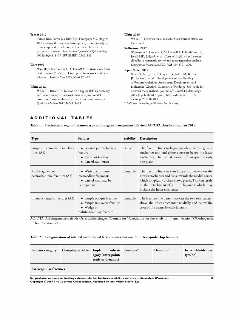
Turner 2012
Turner RM, Davey J, Clarke MJ, Thompson SG, Higgins
JP. Predicting the extent of heterogeneity in meta-analysis,
using empirical data from the Cochrane Database of
Systematic Reviews. International Journal of Epidemiology
2012;41(3):818–27. [PUBMED: 22461129]
Ware 1992
Ware JE Jr, Sherbourne CD. The MOS 36-item short-form
health survey (SF-36). I. Conceptual framework and item
selection. Medical Care 1992;30(6):473–83.
White 2012
White IR, Barrett JK, Jackson D, Higgins JPT. Consistency
and inconsistency in network meta-analysis: model
estimation using multivariate meta-regression. Research
Synthesis Methods 2012;3(2):111–25.
White 2015
White IR. Network meta-analysis. Stata Journal 2015; Vol.
15, issue 4.
Williamson 2017
Williamson S, Landeiro F, McConnell T, Fulford-Smith L,
Javaid MK, Judge A, et al. Costs of fragility hip fractures
globally: a systematic review and meta-regression analysis.
Osteoporosis International 2017;28(10):2791–800.
Yepes-Nuñez 2019
Yepes-Nuñez, JJ, Li, S, Guyatt, G, Jack, SM, Brozek,
JL, Beyene J, et al. Development of the Grading
of Recommendations Assessment, Development and
Evaluation (GRADE) Summary of Findings (SoF) table for
network meta-analysis. Journal of Clinical Epidemiology
2019 [Epub ahead of print]:https://doi.org/10.1016/
j.jclinepi.2019.04.018.∗ Indicates the major publication for the study
A D D I T I O N A L T A B L E S
Table 1. Trochanteric region fractures: type and surgical management (Revised AO/OTA classification, Jan 2018)
Type Features Stability Description
Simple, pertrochanteric frac-
tures (A1)
• Isolated pertrochanteric
fracture
• Two-part fracture
• Lateral wall intact
Stable The fracture line can begin anywhere on the greater
trochanter and end either above or below the lesser
trochanter. The medial cortex is interrupted in only
one place
Multifragmentary
pertrochanteric fractures (A2)
• With one or more
intermediate fragments
• Lateral wall may be
incompetent
Unstable The fracture line can start laterally anywhere on the
greater trochanter and runs towards the medial cortex
which is typically broken in two places. This can result
in the detachment of a third fragment which may
include the lesser trochanter
Intertrochanteric fractures (A3) • Simple oblique fracture
• Simple transverse fracture
• Wedge or
multifragmentary fracture
Unstable The fracture line passes between the two trochanters,
above the lesser trochanter medially and below the
crest of the vastus lateralis laterally
AO/OTA: Arbeitsgemeinschaft für Osteosynthesefragen (German for “Association for the Study of Internal Fixation”)’/Orthopaedic
Trauma Association
Table 2. Categorisation of internal and external fixation interventions for extracapsular hip fractures
Implant category Grouping variable Implant subcat-
egory (entry point/
static or dynamic)
Examplesa Description In worldwide use
(yes/no)
Extracapsular fractures
16Surgical interventions for treating extracapsular hip fractures in adults: a network meta-analysis (Protocol)
Copyright © 2019 The Cochrane Collaboration. Published by John Wiley & Sons, Ltd.
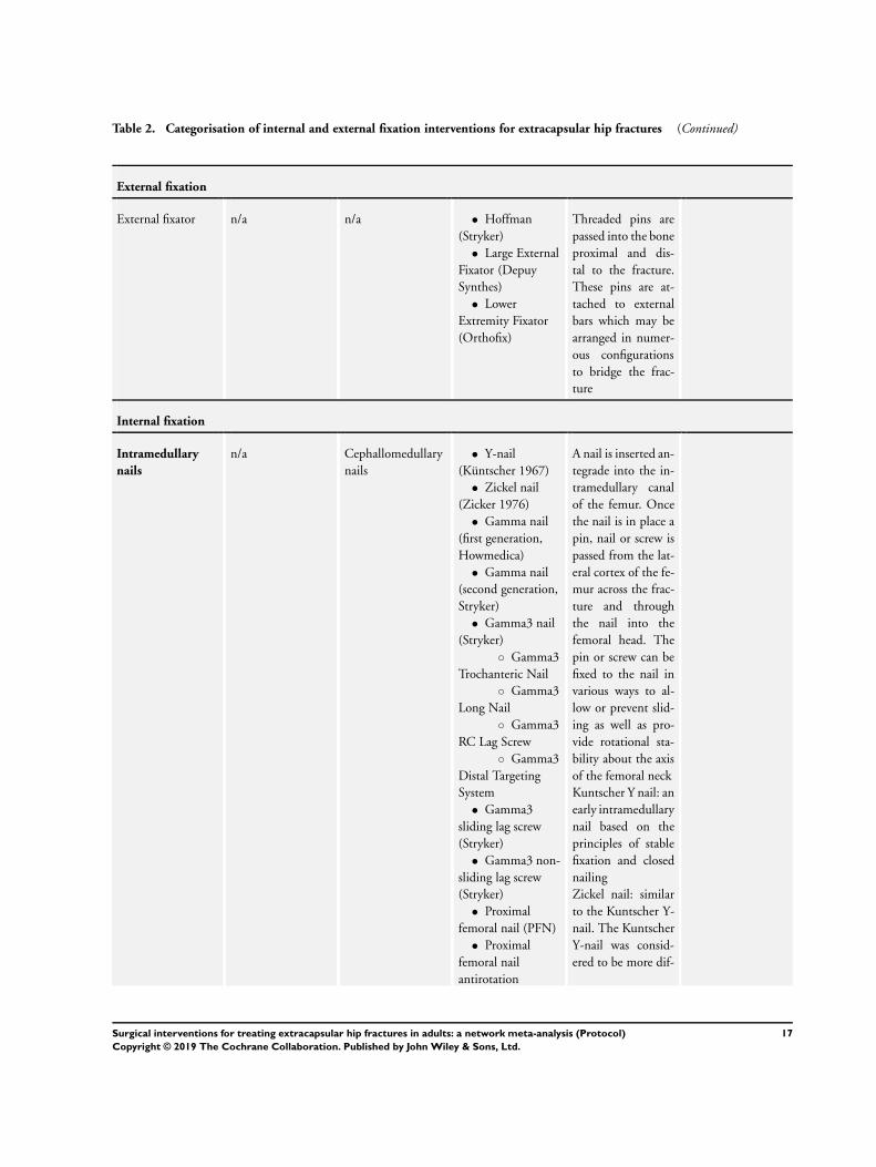
Table 2. Categorisation of internal and external fixation interventions for extracapsular hip fractures (Continued)
External fixation
External fixator n/a n/a • Hoffman
(Stryker)
• Large External
Fixator (Depuy
Synthes)
• Lower
Extremity Fixator
(Orthofix)
Threaded pins are
passed into the bone
proximal and dis-
tal to the fracture.
These pins are at-
tached to external
bars which may be
arranged in numer-
ous configurations
to bridge the frac-
ture
Internal fixation
Intramedullary
nails
n/a Cephallomedullary
nails
• Y-nail
(Küntscher 1967)
• Zickel nail
(Zicker 1976)
• Gamma nail
(first generation,
Howmedica)
• Gamma nail
(second generation,
Stryker)
• Gamma3 nail
(Stryker)
◦ Gamma3
Trochanteric Nail
◦ Gamma3
Long Nail
◦ Gamma3
RC Lag Screw
◦ Gamma3
Distal Targeting
System
• Gamma3
sliding lag screw
(Stryker)
• Gamma3 non-
sliding lag screw
(Stryker)
• Proximal
femoral nail (PFN)
• Proximal
femoral nail
antirotation
A nail is inserted an-
tegrade into the in-
tramedullary canal
of the femur. Once
the nail is in place a
pin, nail or screw is
passed from the lat-
eral cortex of the fe-
mur across the frac-
ture and through
the nail into the
femoral head. The
pin or screw can be
fixed to the nail in
various ways to al-
low or prevent slid-
ing as well as pro-
vide rotational sta-
bility about the axis
of the femoral neck
Kuntscher Y nail: an
early intramedullary
nail based on the
principles of stable
fixation and closed
nailing
Zickel nail: similar
to the Kuntscher Y-
nail. The Kuntscher
Y-nail was consid-
ered to be more dif-
17Surgical interventions for treating extracapsular hip fractures in adults: a network meta-analysis (Protocol)
Copyright © 2019 The Cochrane Collaboration. Published by John Wiley & Sons, Ltd.
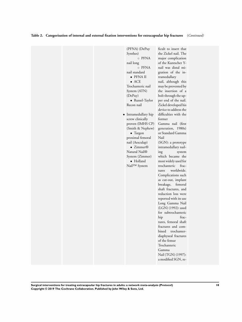
Table 2. Categorisation of internal and external fixation interventions for extracapsular hip fractures (Continued)
(PFNA) (DePuy
Synthes)
◦ PFNA
nail long
◦ PFNA
nail standard
• PFNA II
• ACE
Trochanteric nail
System (ATN)
(DePuy)
• Russel-Taylor
Recon nail
• Intramedullary hip
screw clinically
proven (IMHS CP)
(Smith & Nephew)
• Targon
proximal femoral
nail (Aesculap)
• Zimmer®
Natural Nail®
System (Zimmer)
• Holland
Nail™ System
ficult to insert that
the Zickel nail. The
major complication
of the Kuntscher Y-
nail was distal mi-
gration of the in-
tramedullary
nail, although this
may be prevented by
the insertion of a
bolt through the up-
per end of the nail.
Zickel developed his
device to address the
difficulties with the
former
Gamma nail (first
generation, 1980s)
or Standard Gamma
Nail
(SGN): a prototype
intramedullary nail-
ing system
which became the
most widely used for
trochanteric frac-
tures worldwide.
Complications such
as cut-out, implant
breakage, femoral
shaft fractures, and
reduction loss were
reported with its use
Long Gamma Nail
(LGN) (1992): used
for subtrochanteric
hip frac-
tures, femoral shaft
fractures and com-
bined trochanter-
diaphyseal fractures
of the femur
Trochanteric
Gamma
Nail (TGN) (1997):
a modified SGN, re-
18Surgical interventions for treating extracapsular hip fractures in adults: a network meta-analysis (Protocol)
Copyright © 2019 The Cochrane Collaboration. Published by John Wiley & Sons, Ltd.

Table 2. Categorisation of internal and external fixation interventions for extracapsular hip fractures (Continued)
placed the SGN
Gamma3 Nail-
ing System: third
generation of in-
tramedullary short
and long Gamma
fixation nails. Four
locking grooves al-
low for quarter-turn
advancement of 0.8
mm to allow for pre-
cise lap screw posi-
tioning
PFN (Synthes,
Solothurn, Switzer-
land) and ATN
(dePuy, Warsaw, IN,
USA): developed to
address the mechan-
ical complica-
tions of the standard
gamma nail. These
intramedullary im-
plants provide slid-
ing head-
neck screws. How-
ever complications
still occurred, such
as lateral migration
of head-neck screw,
cut-out from head-
neck fragment and
cut-through of the
anti-rotation screw
into the joint
PFNA (Synthes,
Solothurn, Switzer-
land) was designed
by the AO/ASIF
group for improving
the rotational stabil-
ity. It used a sin-
gle head-neck fixa-
tion device called a
’helical blade’
ACE Trochanteric
19Surgical interventions for treating extracapsular hip fractures in adults: a network meta-analysis (Protocol)
Copyright © 2019 The Cochrane Collaboration. Published by John Wiley & Sons, Ltd.
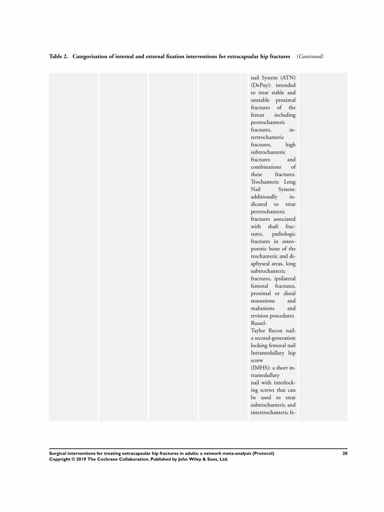
Table 2. Categorisation of internal and external fixation interventions for extracapsular hip fractures (Continued)
nail System (ATN)
(DePuy): intended
to treat stable and
unstable proximal
fractures of the
femur including
pertrochanteric
fractures, in-
tertrochanteric
fractures, high
subtrochanteric
fractures and
combinations of
these fractures.
Trochanteric Long
Nail System:
additionally in-
dicated to treat
pertrochanteric
fractures associated
with shaft frac-
tures, pathologic
fractures in osteo-
porotic bone of the
trochanteric and di-
aphyseal areas, long
subtrochanteric
fractures, ipsilateral
femoral fractures,
proximal or distal
nonunions and
malunions and
revision procedures
Russel-
Taylor Recon nail:
a second-generation
locking femoral nail
Intramedullary hip
screw
(IMHS): a short in-
tramedullary
nail with interlock-
ing screws that can
be used to treat
subtrochanteric and
intertrochanteric fe-
20Surgical interventions for treating extracapsular hip fractures in adults: a network meta-analysis (Protocol)
Copyright © 2019 The Cochrane Collaboration. Published by John Wiley & Sons, Ltd.

Table 2. Categorisation of internal and external fixation interventions for extracapsular hip fractures (Continued)
mur fractures. This
nail, which has the
biomechanical ad-
vantage of being an
intramedullary ap-
pliance but can be
placed per-
cutaneously, is in-
serted under fluoro-
scopic control with
the patient on a frac-
ture table. Reaming
is not usually neces-
sary
Intramedullary
hip screw clinically
proven (IMHS™
CP): intramedullary
hip screw device
that provides a bar-
rel through which a
lag screw can slide.
Introduced in 1991
with its design, the
IMHS system pro-
vided a more mini-
mally invasive tech-
nique than the tra-
ditional Compres-
sion Hip Screw. By
featuring a Center-
ing Sleeve to en-
hance Lag Screw
sliding and medi-
alizing the implant
to reduce the mo-
ment arm, this de-
sign improved im-
plant biomechanics
for the treatment of
hip fractures
Targon® PFT nail-
ing system: target-
ing device is suitable
for all CCD angles
and permits shorter
21Surgical interventions for treating extracapsular hip fractures in adults: a network meta-analysis (Protocol)
Copyright © 2019 The Cochrane Collaboration. Published by John Wiley & Sons, Ltd.
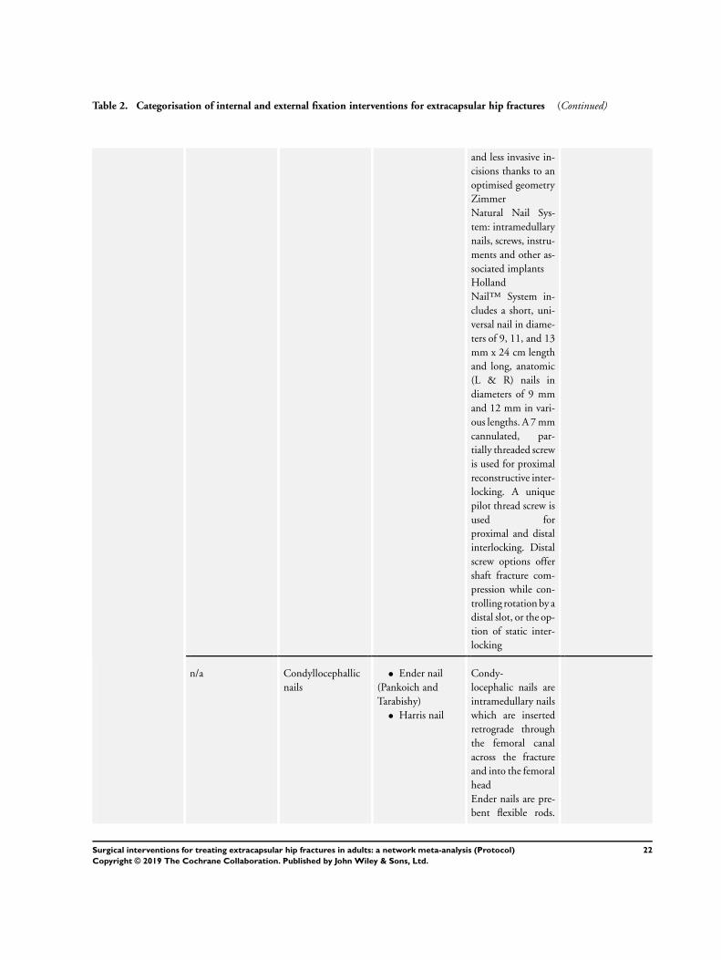
Table 2. Categorisation of internal and external fixation interventions for extracapsular hip fractures (Continued)
and less invasive in-
cisions thanks to an
optimised geometry
Zimmer
Natural Nail Sys-
tem: intramedullary
nails, screws, instru-
ments and other as-
sociated implants
Holland
Nail™ System in-
cludes a short, uni-
versal nail in diame-
ters of 9, 11, and 13
mm x 24 cm length
and long, anatomic
(L & R) nails in
diameters of 9 mm
and 12 mm in vari-
ous lengths. A 7 mm
cannulated, par-
tially threaded screw
is used for proximal
reconstructive inter-
locking. A unique
pilot thread screw is
used for
proximal and distal
interlocking. Distal
screw options offer
shaft fracture com-
pression while con-
trolling rotation by a
distal slot, or the op-
tion of static inter-
locking
n/a Condyllocephallic
nails
• Ender nail
(Pankoich and
Tarabishy)
• Harris nail
Condy-
locephalic nails are
intramedullary nails
which are inserted
retrograde through
the femoral canal
across the fracture
and into the femoral
head
Ender nails are pre-
bent flexible rods.
22Surgical interventions for treating extracapsular hip fractures in adults: a network meta-analysis (Protocol)
Copyright © 2019 The Cochrane Collaboration. Published by John Wiley & Sons, Ltd.
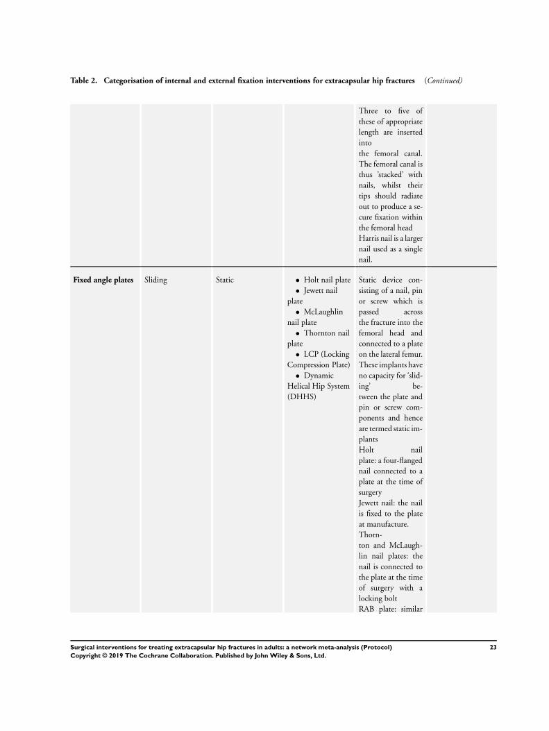
Table 2. Categorisation of internal and external fixation interventions for extracapsular hip fractures (Continued)
Three to five of
these of appropriate
length are inserted
into
the femoral canal.
The femoral canal is
thus ’stacked’ with
nails, whilst their
tips should radiate
out to produce a se-
cure fixation within
the femoral head
Harris nail is a larger
nail used as a single
nail.
Fixed angle plates Sliding Static • Holt nail plate
• Jewett nail
plate
• McLaughlin
nail plate
• Thornton nail
plate
• LCP (Locking
Compression Plate)
• Dynamic
Helical Hip System
(DHHS)
Static device con-
sisting of a nail, pin
or screw which is
passed across
the fracture into the
femoral head and
connected to a plate
on the lateral femur.
These implants have
no capacity for ‘slid-
ing’ be-
tween the plate and
pin or screw com-
ponents and hence
are termed static im-
plants
Holt nail
plate: a four-flanged
nail connected to a
plate at the time of
surgery
Jewett nail: the nail
is fixed to the plate
at manufacture.
Thorn-
ton and McLaugh-
lin nail plates: the
nail is connected to
the plate at the time
of surgery with a
locking bolt
RAB plate: similar
23Surgical interventions for treating extracapsular hip fractures in adults: a network meta-analysis (Protocol)
Copyright © 2019 The Cochrane Collaboration. Published by John Wiley & Sons, Ltd.
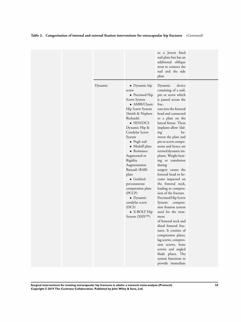
Table 2. Categorisation of internal and external fixation interventions for extracapsular hip fractures (Continued)
to a Jewett fixed
nail plate but has an
additional oblique
strut to connect the
nail and the side
plate
Dynamic • Dynamic hip
screw
• Precimed Hip
Screw System
• AMBI/Classic
Hip Screw System
(Smith & Nephew
Richards)
• HDS/DCS
Dynamic Hip &
Condylar Screw
System
• Pugh nail
• Medoff plate
• Resistance
Augmented or
Rigidity
Augmentation
Baixauli (RAB)
plate
• Gotfried
percutaneous
compression plate
(PCCP)
• Dynamic
condylar screw
(DCS)
• X-BOLT Hip
System (XHS™)
Dynamic device
consisting of a nail,
pin or screw which
is passed across the
frac-
ture into the femoral
head and connected
to a plate on the
lateral femur. These
implants allow ‘slid-
ing’ be-
tween the plate and
pin or screw compo-
nents and hence are
termed dynamic im-
plants. Weight bear-
ing or translation
during
surgery causes the
femoral head to be-
come impacted on
the femoral neck,
leading to compres-
sion of the fracture
Precimed Hip Screw
System: compres-
sion fixation system
used for the treat-
ment
of femoral neck and
distal femoral frac-
tures. It consists of
compression plates,
lag screws, compres-
sion screws, bone
screws and angled
blade plates. The
system functions to
provide immediate
24Surgical interventions for treating extracapsular hip fractures in adults: a network meta-analysis (Protocol)
Copyright © 2019 The Cochrane Collaboration. Published by John Wiley & Sons, Ltd.
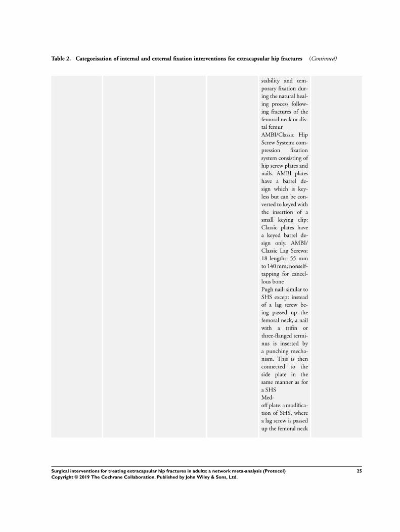
Table 2. Categorisation of internal and external fixation interventions for extracapsular hip fractures (Continued)
stability and tem-
porary fixation dur-
ing the natural heal-
ing process follow-
ing fractures of the
femoral neck or dis-
tal femur
AMBI/Classic Hip
Screw System: com-
pression fixation
system consisting of
hip screw plates and
nails. AMBI plates
have a barrel de-
sign which is key-
less but can be con-
verted to keyed with
the insertion of a
small keying clip;
Classic plates have
a keyed barrel de-
sign only. AMBI/
Classic Lag Screws:
18 lengths: 55 mm
to 140 mm; nonself-
tapping for cancel-
lous bone
Pugh nail: similar to
SHS except instead
of a lag screw be-
ing passed up the
femoral neck, a nail
with a trifin or
three-flanged termi-
nus is inserted by
a punching mecha-
nism. This is then
connected to the
side plate in the
same manner as for
a SHS
Med-
off plate: a modifica-
tion of SHS, where
a lag screw is passed
up the femoral neck
25Surgical interventions for treating extracapsular hip fractures in adults: a network meta-analysis (Protocol)
Copyright © 2019 The Cochrane Collaboration. Published by John Wiley & Sons, Ltd.
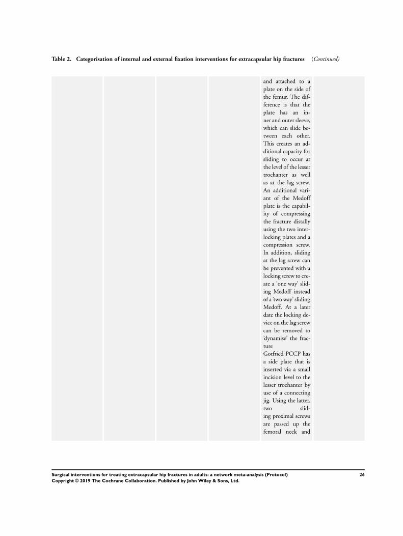
Table 2. Categorisation of internal and external fixation interventions for extracapsular hip fractures (Continued)
and attached to a
plate on the side of
the femur. The dif-
ference is that the
plate has an in-
ner and outer sleeve,
which can slide be-
tween each other.
This creates an ad-
ditional capacity for
sliding to occur at
the level of the lesser
trochanter as well
as at the lag screw.
An additional vari-
ant of the Medoff
plate is the capabil-
ity of compressing
the fracture distally
using the two inter-
locking plates and a
compression screw.
In addition, sliding
at the lag screw can
be prevented with a
locking screw to cre-
ate a ’one way’ slid-
ing Medoff instead
of a ’two way’ sliding
Medoff. At a later
date the locking de-
vice on the lag screw
can be removed to
’dynamise’ the frac-
ture
Gotfried PCCP has
a side plate that is
inserted via a small
incision level to the
lesser trochanter by
use of a connecting
jig. Using the latter,
two slid-
ing proximal screws
are passed up the
femoral neck and
26Surgical interventions for treating extracapsular hip fractures in adults: a network meta-analysis (Protocol)
Copyright © 2019 The Cochrane Collaboration. Published by John Wiley & Sons, Ltd.
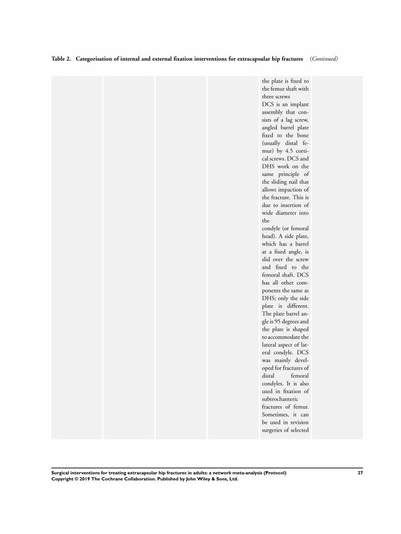
Table 2. Categorisation of internal and external fixation interventions for extracapsular hip fractures (Continued)
the plate is fixed to
the femur shaft with
three screws
DCS is an implant
assembly that con-
sists of a lag screw,
angled barrel plate
fixed to the bone
(usually distal fe-
mur) by 4.5 corti-
cal screws. DCS and
DHS work on the
same principle of
the sliding nail that
allows impaction of
the fracture. This is
due to insertion of
wide diameter into
the
condyle (or femoral
head). A side plate,
which has a barrel
at a fixed angle, is
slid over the screw
and fixed to the
femoral shaft. DCS
has all other com-
ponents the same as
DHS; only the side
plate is different.
The plate barrel an-
gle is 95 degrees and
the plate is shaped
to accommodate the
lateral aspect of lat-
eral condyle. DCS
was mainly devel-
oped for fractures of
distal femoral
condyles. It is also
used in fixation of
subtrochanteric
fractures of femur.
Sometimes, it can
be used in revision
surgeries of selected
27Surgical interventions for treating extracapsular hip fractures in adults: a network meta-analysis (Protocol)
Copyright © 2019 The Cochrane Collaboration. Published by John Wiley & Sons, Ltd.
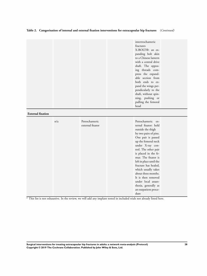
Table 2. Categorisation of internal and external fixation interventions for extracapsular hip fractures (Continued)
intertrochanteric
fractures
X-BOLT®: an ex-
panding bolt akin
to a Chinese lantern
with a central drive
shaft. The oppos-
ing threads com-
press the expand-
able section from
both ends to ex-
pand the wings per-
pendicularly to the
shaft, without spin-
ning, pushing or
pulling the femoral
head
External fixation
n/a Petrochanteric
external fixator
Petrochanteric ex-
ternal fixator: held
outside the thigh
by two pairs of pins.
One pair is passed
up the femoral neck
under X-ray con-
trol. The other pair
is placed in the fe-
mur. The fixator is
left in place until the
fracture has healed,
which usually takes
about three months.
It is then removed
under local anaes-
thesia, generally as
an outpatient proce-
durea This list is not exhaustive. In the review, we will add any implant tested in included trials not already listed here.
28Surgical interventions for treating extracapsular hip fractures in adults: a network meta-analysis (Protocol)
Copyright © 2019 The Cochrane Collaboration. Published by John Wiley & Sons, Ltd.
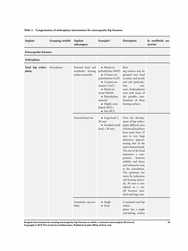
Table 3. Categorisation of arthroplasty interventions for extracapsular hip fractures
Implant Grouping variable Implant
subcategory
Examplesa Description In worldwide use
(yes/no)
Extracapsular fractures
Arthroplasty
Total hip arthro-
plasty
Articulation Femoral head and
acetabular bearing
surface materials
• Metal-on-
polyethylene (MoP)
• Ceramic-on-
polyethylene (CoP)
• Ceramic-on-
ceramic (CoC)
• Metal-on-
metal (MoM)
• Polyethylene
material
• Highly cross
linked (HCL)
• Not HCL
Bear-
ing surfaces may be
grouped into hard
(ceramic and metal)
and soft (polyethy-
lene vari-
ants). Arthroplasties
exist with many of
the possible com-
binations of these
bearing surfaces
Femoral head size • Large head ≥
36 mm
• Standard small
head < 36 mm
Over the develop-
ment of hip arthro-
plasty different sizes
of femoral head have
been used, from 22
mm to very large
diameters approxi-
mating that of the
native femoral head.
The size of the head
represents a com-
promise between
stability and linear
and volumetric wear
at the articulation.
The optimum size
varies by indication
and bearing materi-
als. 36 mm is con-
sidered as a cut-
off between stan-
dard and large sizes
Acetabular cup mo-
bility
• Single
• Dual
A standard total hip
arthro-
plasty has a single
articulating surface
29Surgical interventions for treating extracapsular hip fractures in adults: a network meta-analysis (Protocol)
Copyright © 2019 The Cochrane Collaboration. Published by John Wiley & Sons, Ltd.
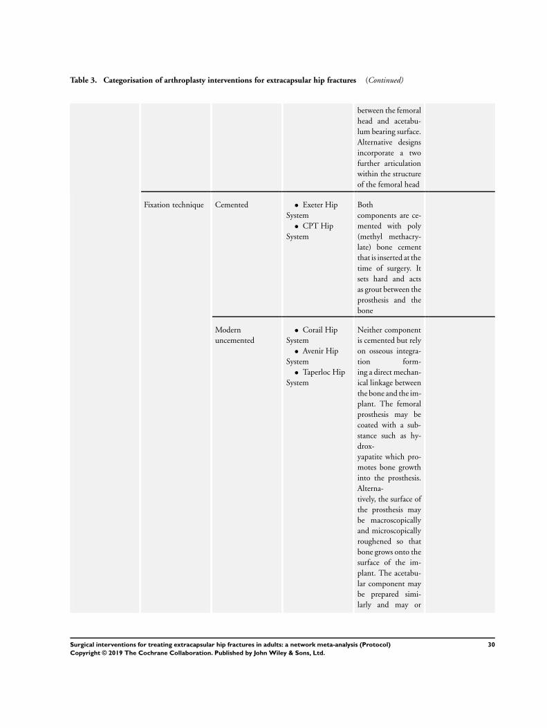
Table 3. Categorisation of arthroplasty interventions for extracapsular hip fractures (Continued)
between the femoral
head and acetabu-
lum bearing surface.
Alternative designs
incorporate a two
further articulation
within the structure
of the femoral head
Fixation technique Cemented • Exeter Hip
System
• CPT Hip
System
Both
components are ce-
mented with poly
(methyl methacry-
late) bone cement
that is inserted at the
time of surgery. It
sets hard and acts
as grout between the
prosthesis and the
bone
Modern
uncemented
• Corail Hip
System
• Avenir Hip
System
• Taperloc Hip
System
Neither component
is cemented but rely
on osseous integra-
tion form-
ing a direct mechan-
ical linkage between
the bone and the im-
plant. The femoral
prosthesis may be
coated with a sub-
stance such as hy-
drox-
yapatite which pro-
motes bone growth
into the prosthesis.
Alterna-
tively, the surface of
the prosthesis may
be macroscopically
and microscopically
roughened so that
bone grows onto the
surface of the im-
plant. The acetabu-
lar component may
be prepared simi-
larly and may or
30Surgical interventions for treating extracapsular hip fractures in adults: a network meta-analysis (Protocol)
Copyright © 2019 The Cochrane Collaboration. Published by John Wiley & Sons, Ltd.
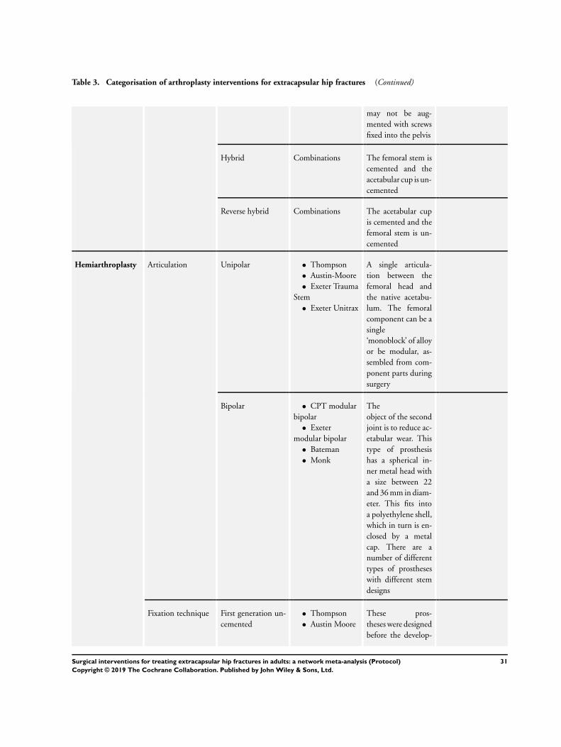
Table 3. Categorisation of arthroplasty interventions for extracapsular hip fractures (Continued)
may not be aug-
mented with screws
fixed into the pelvis
Hybrid Combinations The femoral stem is
cemented and the
acetabular cup is un-
cemented
Reverse hybrid Combinations The acetabular cup
is cemented and the
femoral stem is un-
cemented
Hemiarthroplasty Articulation Unipolar • Thompson
• Austin-Moore
• Exeter Trauma
Stem
• Exeter Unitrax
A single articula-
tion between the
femoral head and
the native acetabu-
lum. The femoral
component can be a
single
‘monoblock’ of alloy
or be modular, as-
sembled from com-
ponent parts during
surgery
Bipolar • CPT modular
bipolar
• Exeter
modular bipolar
• Bateman
• Monk
The
object of the second
joint is to reduce ac-
etabular wear. This
type of prosthesis
has a spherical in-
ner metal head with
a size between 22
and 36 mm in diam-
eter. This fits into
a polyethylene shell,
which in turn is en-
closed by a metal
cap. There are a
number of different
types of prostheses
with different stem
designs
Fixation technique First generation un-
cemented
• Thompson
• Austin Moore
These pros-
theses were designed
before the develop-
31Surgical interventions for treating extracapsular hip fractures in adults: a network meta-analysis (Protocol)
Copyright © 2019 The Cochrane Collaboration. Published by John Wiley & Sons, Ltd.
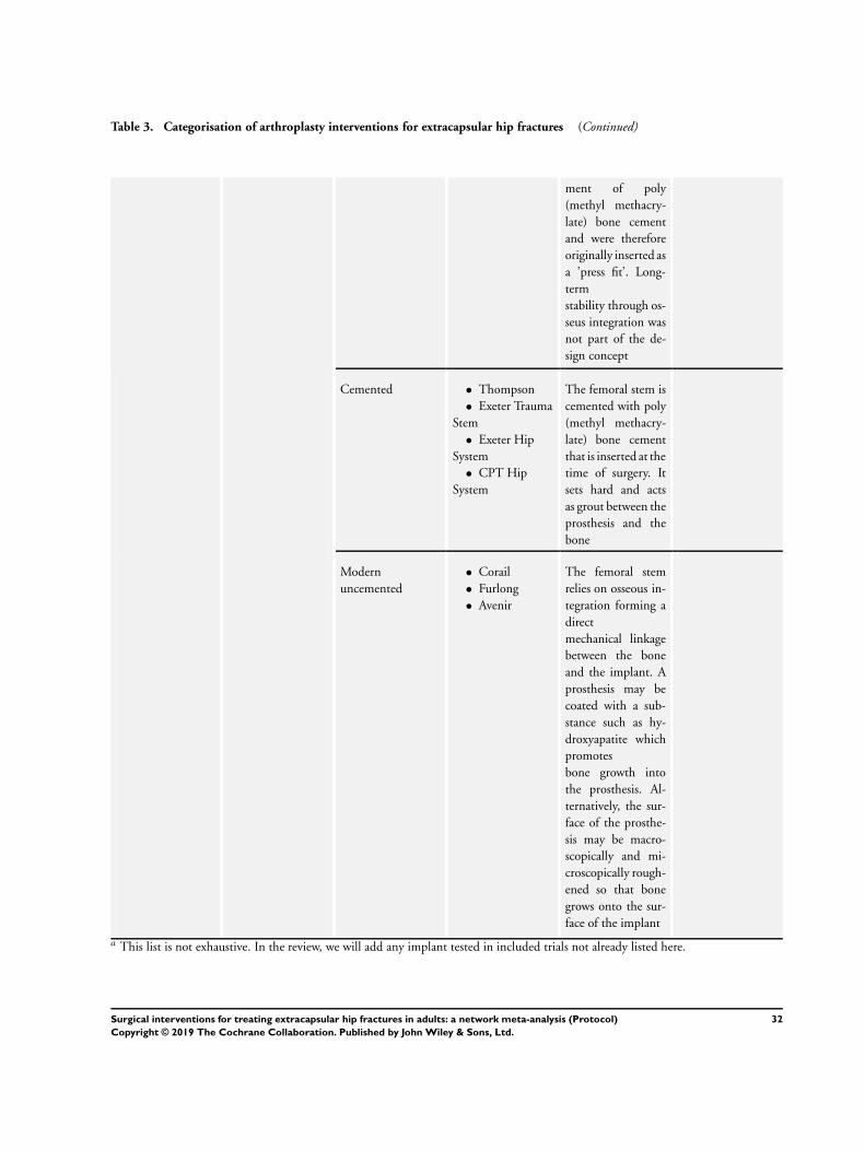
Table 3. Categorisation of arthroplasty interventions for extracapsular hip fractures (Continued)
ment of poly
(methyl methacry-
late) bone cement
and were therefore
originally inserted as
a ’press fit’. Long-
term
stability through os-
seus integration was
not part of the de-
sign concept
Cemented • Thompson
• Exeter Trauma
Stem
• Exeter Hip
System
• CPT Hip
System
The femoral stem is
cemented with poly
(methyl methacry-
late) bone cement
that is inserted at the
time of surgery. It
sets hard and acts
as grout between the
prosthesis and the
bone
Modern
uncemented
• Corail
• Furlong
• Avenir
The femoral stem
relies on osseous in-
tegration forming a
direct
mechanical linkage
between the bone
and the implant. A
prosthesis may be
coated with a sub-
stance such as hy-
droxyapatite which
promotes
bone growth into
the prosthesis. Al-
ternatively, the sur-
face of the prosthe-
sis may be macro-
scopically and mi-
croscopically rough-
ened so that bone
grows onto the sur-
face of the implant
a This list is not exhaustive. In the review, we will add any implant tested in included trials not already listed here.
32Surgical interventions for treating extracapsular hip fractures in adults: a network meta-analysis (Protocol)
Copyright © 2019 The Cochrane Collaboration. Published by John Wiley & Sons, Ltd.

A P P E N D I C E S
Appendix 1. MEDLINE search strategy
Ovid interface
1 exp Femoral Fractures/
2 ((hip or hips or cervical) adj5 (fracture$ or break$ or broke$)).ti,ab,kf.
3 ((femoral$ or femur$ or acetabul$) adj5 (fracture$ or break$ or broke$)).ti,ab,kf.
4 ((intracapsular or intra-capsular or subcapital or sub-capital or transcervical or trans-cervical or basicervical or basi-cervical) adj5
(fracture$ or break$ or broke$)).ti,ab,kf.
5 ((extracapsular or extra-capsular or trochant$ or subtrochant$ or pertrochant$ or intertrochant$) adj5 (fracture$ or break$ or
broke$)).ti,ab,kf.
6 (((head or neck or proximal) adj5 (fracture$ or break$ or broke$)) and (femoral$ or femur$)).ti,ab,kf.
7 or/1-6
8 randomized controlled trial.pt.
9 controlled clinical trial.pt.
10 randomized.ab.
11 placebo.ab.
12 clinical trials as topic.sh.
13 randomly.ab.
14 trial.ti.
15 8 or 9 or 10 or 11 or 12 or 13 or 14
16 7 and 15
17 Arthroplasty, Replacement, Hip/ or Hip Prosthesis/
18 Arthroplasty, Replacement/ or Hemiarthroplasty/ or Joint Prosthesis/
19 (arthroplast$ or hemiarthroplast$).ti,ab,kf.
20 ((hip or hips) adj5 (replac$ or prosthes$ or implant$)).ti,ab,kf.
21 ((joint$1 adj5 (replac$ or prosthes$ or implant$)) and (hip or hips or femur$ or femoral$ or acetabul$)).ti,ab,kf.
22 or/17-21
23 fractures, bone/ or exp fracture dislocation/ or fractures, closed/ or fractures, comminuted/ or fractures, compression/ or fractures,
malunited/ or fractures, multiple/ or fractures, open/ or fractures, spontaneous/ or exp fractures, stress/ or fractures, ununited/ or intra-
articular fractures/ or osteoporotic fractures/ or periprosthetic fractures/ (92406)
24 fracture$.ti,ab,kf.
25 23 or 24
26 22 and 25 and 15
27 (pin or pins or nail or nails or screw or screws or plate or plates).ti,ab,kf.
28 internal fixators/ or bone nails/ or bone plates/ or exp bone screws/
29 (static adj (device$1 or implant$1)).ti,ab,kf.
30 (dynamic adj (device$1 or implant$1)).ti,ab,kf.
31 or/27-30
32 ((hip or hips or femur$ or femoral$ or acetabul$) and (fracture$ or break$ or broke$)).ti,ab,kf.
33 (hip or hips or femur$ or femoral$ or acetabul$).ti,ab,kf. and (fractures, bone/ or exp fracture dislocation/ or fractures, closed/
or fractures, comminuted/ or fractures, compression/ or fractures, malunited/ or fractures, multiple/ or fractures, open/ or fractures,
spontaneous/ or exp fractures, stress/ or fractures, ununited/ or intra-articular fractures/ or osteoporotic fractures/ or periprosthetic
fractures/)
34 or/32-33
35 31 and 34 and 15
36 16 or 26 or 35
37 exp animals/ not humans/
38 36 not 37
33Surgical interventions for treating extracapsular hip fractures in adults: a network meta-analysis (Protocol)
Copyright © 2019 The Cochrane Collaboration. Published by John Wiley & Sons, Ltd.

C O N T R I B U T I O N S O F A U T H O R S
AS (systematic reviewer): drafted the protocol, reviewed and approved the final protocol.
WE (content expert, Trauma and Orthopaedics): reviewed and approved the final protocol.
MP (content expert, Orthopaedics): drafted the protocol, reviewed and approved the final protocol.
LF (systematic reviewer): drafted the protocol, reviewed and approved the final protocol.
HW (senior information specialist): designed the search strategies, reviewed and approved the final protocol.
JMG (senior information specialist): designed the search strategies, drafted the protocol, reviewed and approved the final protocol.
JC (statistician): reviewed and approved the final protocol.
XG (content expert, Trauma and Orthopaedics): drafted the protocol, reviewed and approved the final protocol, and is the guarantor
of the content.
Contributions of the editorial base
Helen Handoll (Co-ordinating Editor): edited the protocol; advised on methodology and protocol content; and approved the final
version for publication.
Joanne Elliott (Managing Editor): coordinated the editorial process; advised on content; and edited the protocol.
Maria Clarke (Information Specialist): checked the search methods section.
D E C L A R A T I O N S O F I N T E R E S T
AS: has no known conflicts of interest.
WE: has an advisory role on infection control with Orthofix and Bone Support, but this is unrelated to this review. He has no known
conflicts of interest
MP: has received expenses and honorarium from a number of commercial companies and organisations for giving lectures on different
aspects of hip fracture treatment. In addition he has received royalties from BBrawn Ltd. related to the design and development of an
implant used for the internal fixation of intracapsular hip fractures.
LF: has no known conflicts of interest.
HW: has no known conflicts of interest.
JMG: has no known conflicts of interest.
JC: has no known conflicts of interest.
XG is funded by a National Institute for Health Research Clinician Scientist Grant. Further funding from industry and charitable grants
are and have been made available to his institution. All decisions relating to the design, conduct, analysis, write-up and publication of
research are independent of these funders. He has ongoing expert consultancy with several companies; none involve the development
of any implant for use in hip fracture care. He will remain independent of study selection decisions, ’Risk of bias’ assessment and any
data extraction of any of the studies on which he is an author, co-applicant or has had an advisory role.
34Surgical interventions for treating extracapsular hip fractures in adults: a network meta-analysis (Protocol)
Copyright © 2019 The Cochrane Collaboration. Published by John Wiley & Sons, Ltd.
S O U R C E S O F S U P P O R T
Internal sources
• National Institute for Health Research Oxford Biomedical Research Centre, UK, UK.
External sources
• This project was supported by the National Institute for Health Research via Cochrane Infrastructure funding to the Cochrane
Bone, Joint and Muscle Trauma Group, UK.
The views and opinions expressed therein are those of the authors and do not necessarily reflect those of the Systematic Reviews
Programme, NIHR, NHS or the Department of Health.
35Surgical interventions for treating extracapsular hip fractures in adults: a network meta-analysis (Protocol)
Copyright © 2019 The Cochrane Collaboration. Published by John Wiley & Sons, Ltd.
Copyright © 2022 FDOKUMEN


![[Treating frostbite injuries]](https://static.fdokumen.com/doc/165x107/633ff39332b09e4bae09a1b5/treating-frostbite-injuries.jpg)