Interprosthetic femoral fractures - Elsevier
7
Rev Esp Cir Ortop Traumatol. 2017;61(1):1---7 www.elsevier.es/rot Revista Española de Cirugía Ortopédica y Traumatología ORIGINAL ARTICLE Interprosthetic femoral fractures: Treatment with a lateral angular-stable plate J. Albareda * , J. Gómez, L. Ezquerra, N. Blanco Servicio de Cirugía Ortopédica y Traumatología, Hospital Clínico Universitario «Lozano Blesa», Zaragoza, Spain Received 5 December 2015; accepted 20 September 2016 KEYWORDS Interprosthetic fracture; Peri-prosthetic fracture; Femoral fracture; Locking plate Abstract Objective: To study the causes and outcomes of patients with interprosthetic femoral fractures. Material and methods: A retrospective review conducted on 7 patients treated between 2010 and 2013. The knee arthroplasties had been implanted for at least 5 years, and those of the hip less than a year. They were bipolar cemented in 6 patients and totally non-cemented in one patient, all of them implanted due to a displaced femoral neck fracture. They were treated using osteosynthesis with angular stability plate covering the whole interprosthetic femoral segment. Except for one patient, all have been reviewed at least 12 months. Results: The patients included 6 women and one man, with a mean age of 84.7. The fracture, always by low energy, occurred between 2 and 8 months after that of the hip without the implants being mobilised. Four of them were located at diaphyseal level, and 3 at supracondylar level, and unrelated to the type of knee implant. There was consolidation in all patients at a mean of 4.5 months, without a re-operation in any of them, and with no mortality during the follow-up period. All patients walked independently at the time of the fracture, and all of them have managed to return to walking independently, having lost as average 20 ◦ of knee flexion in cases of supracondylar fracture. Conclusions: The most important factors in our patients regarding the producing of the fracture have been the changes in the ability to walk after knee replacement and bone fragility. Angular stability plates give good results in the treatment of interprosthetic femoral fractures. © 2016 SECOT. Published by Elsevier Espa˜ na, S.L.U. All rights reserved. Please cite this article as: Albareda J, Gómez J, Ezquerra L, Blanco N. Fracturas interprotésicas femorales. Tratamiento con placa lateral de estabilidad angular. Rev Esp Cir Ortop Traumatol. 2017;61:1---7. ∗ Corresponding author. E-mail address: [email protected] (J. Albareda). 1988-8856/© 2016 SECOT. Published by Elsevier Espa˜ na, S.L.U. All rights reserved.
-
Upload
khangminh22 -
Category
Documents
-
view
0 -
download
0
Transcript of Interprosthetic femoral fractures - Elsevier
Rev Esp Cir Ortop Traumatol. 2017;61(1):1---7
www.elsevier.es/rot
Revista Española de CirugíaOrtopédica y Traumatología
ORIGINAL ARTICLE
Interprosthetic femoral fractures: Treatment with a
lateral angular-stable plate�
J. Albareda ∗, J. Gómez, L. Ezquerra, N. Blanco
Servicio de Cirugía Ortopédica y Traumatología, Hospital Clínico Universitario «Lozano Blesa», Zaragoza, Spain
Received 5 December 2015; accepted 20 September 2016
KEYWORDSInterprostheticfracture;Peri-prostheticfracture;Femoral fracture;Locking plate
Abstract
Objective: To study the causes and outcomes of patients with interprosthetic femoral fractures.
Material and methods: A retrospective review conducted on 7 patients treated between 2010
and 2013. The knee arthroplasties had been implanted for at least 5 years, and those of the
hip less than a year. They were bipolar cemented in 6 patients and totally non-cemented in one
patient, all of them implanted due to a displaced femoral neck fracture. They were treated
using osteosynthesis with angular stability plate covering the whole interprosthetic femoral
segment. Except for one patient, all have been reviewed at least 12 months.
Results: The patients included 6 women and one man, with a mean age of 84.7. The fracture,
always by low energy, occurred between 2 and 8 months after that of the hip without the
implants being mobilised. Four of them were located at diaphyseal level, and 3 at supracondylar
level, and unrelated to the type of knee implant. There was consolidation in all patients at a
mean of 4.5 months, without a re-operation in any of them, and with no mortality during the
follow-up period. All patients walked independently at the time of the fracture, and all of them
have managed to return to walking independently, having lost as average 20◦ of knee flexion in
cases of supracondylar fracture.
Conclusions: The most important factors in our patients regarding the producing of the fracture
have been the changes in the ability to walk after knee replacement and bone fragility. Angular
stability plates give good results in the treatment of interprosthetic femoral fractures.
© 2016 SECOT. Published by Elsevier Espana, S.L.U. All rights reserved.
� Please cite this article as: Albareda J, Gómez J, Ezquerra L, Blanco N. Fracturas interprotésicas femorales. Tratamiento con placa lateral
de estabilidad angular. Rev Esp Cir Ortop Traumatol. 2017;61:1---7.∗ Corresponding author.
E-mail address: [email protected] (J. Albareda).
1988-8856/© 2016 SECOT. Published by Elsevier Espana, S.L.U. All rights reserved.
2 J. Albareda et al.
PALABRAS CLAVEFracturainterprotésica;Fracturaperiprotésica;Fractura femoral;Placa bloqueada
Fracturas interprotésicas femorales. Tratamiento con placa lateral de estabilidad
angular
Resumen
Objetivo: Estudiar las causas de producción y resultados en los pacientes tratados con fractura
interprotésica femoral.
Material y métodos: Revisión retrospectiva de 7 pacientes tratados entre 2010 y 2013. Las
artroplastias de rodilla tenían al menos 5 anos desde su implantación y las de cadera menos
de un ano siendo cementadas bipolares en 6 pacientes y total no cementada en un paciente,
implantadas todas por una fractura desplazada del cuello femoral. Han sido tratadas mediante
osteosíntesis con placa de estabilidad angular abarcando todo el segmento femoral interpro-
tésico. Excepto un paciente, los demás han sido revisados 12 meses como mínimo.
Resultados: Han sido 6 mujeres y un hombre con edad media de 84,7 anos. La fractura, siempre
por baja energía, se ha producido entre 2 y 8 meses tras la de cadera sin encontrarse movi-
lizados los implantes, estando localizadas 4 de ellas a nivel diafisario y 3 a nivel supracondíleo,
sin relación con el tipo de implante de rodilla. Se ha producido la consolidación en todos los
pacientes a los 4,5 meses de media sin reintervención en ninguno de ellos y sin mortalidad en
el tiempo que han sido controlados. Todos los pacientes deambulaban en carga en el momento
de producirse la fractura y todos ellos han conseguido volver a deambular en carga, habiendo
perdido como media 20◦ de flexión de rodilla en los casos de fractura supracondílea.
Conclusiones: Los factores más importantes en nuestros pacientes con respecto a la producción
de la fractura han sido la alteración de la capacidad de deambulación tras la artroplastia de
cadera y la fragilidad ósea. La placa de estabilidad angular concede buenos resultados en el
tratamiento de las fracturas interprotésicas femorales.
© 2016 SECOT. Publicado por Elsevier Espana, S.L.U. Todos los derechos reservados.
Interprosthetic femoral fractures occur in the femoralsegment between hip and knee arthroplasties.1 Thesearthroplasties affect how the fractures occur and howthey are treated.1,2 They only constitute 1.26% of femoralfractures3 but the increase in life expectancy and pros-thetic surgery procedures result in a larger at-risk populationand therefore their greater frequency is to be expected.These are not optimal patients since, as they presentlimited bone stock,3 it is difficult to stabilise fracturesdue to prosthetic implants4 and there is occasionally thepresence of medullary canal cement which creates prob-lems with biological union.1 In other words, treatment ofthese fractures poses mechanical and biological challenges.The fractures are always produced by low energy mecha-nisms, the effect of implants and their features, the effectof the presence and constriction of the knee implant5 andthe degree of osteoporosis6 are discussed in clinical andbiomechanical studies. Because these fractures are so infre-quent, the published series are short with a modest numberof patients1---5,7---9 and various materials and osteosynthesistechniques have been used for their treatment, sometimeswith a high complication, reoperation and failure rate.10
Lateral angular-stable plates have been recommended forthe treatment of these fractures, to cover the entire inter-prosthetic femoral segment in order to prevent the risk offracture due to increased stress in the segment of femurwithout implant,2---5,7,8,11 but there is no definitive algorithmfor the treatment of these fractures.7 In this short patientseries, our aim is to study the causes of these fractures,the features of the patients in whom they occurred and
the outcomes achieved using a single lateral angular-stableplate.
Material and method
We undertook a retrospective review of patients treatedbetween the years 2010 and 2013 with interprostheticfemoral fracture. There were 7 patients in the study, westudied their demographic characteristics, age and gender,causes of fracture, type of fracture according to Platzer’sradiological classification,2 degree of osteoporosis using theSingh index12 measured in the contralateral hip, anaestheticrisk (ASA), time since the arthroplasty implant, union time,ambulation, general and local complications, reoperations,relationship of the type of implant with the type of fractureand clinical deterioration of joint function.
All the patients have been treated surgically with spinalanaesthesia with reduction and internal fixation with anangular-stable plate. Scheduled surgery was performed onaverage 2.5 days from occurrence of the fracture, overlap-ping the femoral stem of the hip implant between 3 and10 cm, placing 3 percutaneously and 4 by open surgery. Theplate was stabilised with screws using accessory wires in theproximal segment depending on the fracture line. None ofthe hip or knee implants had been revised and no autologousor allografts had been used in any of the cases.
All the patients were kept non-weight bearing on thefractured limb until union of the fracture, physiotherapywas started immediately postoperatively. The fracture was
Interprosthetic femoral fractures 3
considered to have healed if clinically the patient couldwalk independently with no pain and two radiological pro-jections showed fracture callus. Delayed fracture union wasconsidered if at 6 months there had been no fracture unionbut there had been some progress towards it since the lastfollow-up X-ray, and pseudoarthrosis if no union had takenplace at 9 months with no progress towards it within thepast 3 months.7 Union with an angle greater than 5◦ wasconsidered non-union.
All the patients were monitored as outpatients until clin-ical and radiological union of the fracture, the minimumfollow-up period was 12 months, except one patient whowas lost to follow-up after 6 months.
Results
Six of the 7 patients treated were females and one male,with a mean age of 84.7 years (range 78---87). All the patientshad undergone knee arthroplasty due to gonarthrosis atleast 5 years prior to incurring the fracture. Ultracongruentpolyethylene was used in 4 patients and posterior stabilisedpolyethylene in 3. Furlong
®-type partially cemented bipolar
hip arthroplasty was performed on 6 patients (the females)and total non-cemented hip arthroplasty on one (the male)due to displaced, low-energy cervical fracture. All of thehip arthroplasties, except the total arthroplasty, and 4 kneearthroplasties were implanted in our department (Table 1).
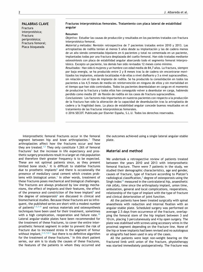
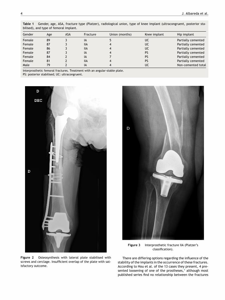
The interprosthetic fractures occurred between 2 and8 months after the hip surgery, all of them were low-energyproduced. In four cases they were located at diaphyseallevel and in three at supracondylar level, two with ultra-congruent and one with posterior stabilised polyethylene.In all cases both implants were stable and not affected bythe fracture line. In two cases the fracture line affected thedistal cement plug of the hip arthroplasty (Fig. 1). FollowingPlatzer’s classification, the fractures were 4 type IA, and 3type IIA adjacent to the knee arthroplasties (Figs. 3 and 4).Four patients were assessed with a Singh index of 3 and 3with an index of 4.
Four patients presented an ASA class II due to their back-ground and general condition, and 3 ASA III. None of thepatients died during the follow-up period.
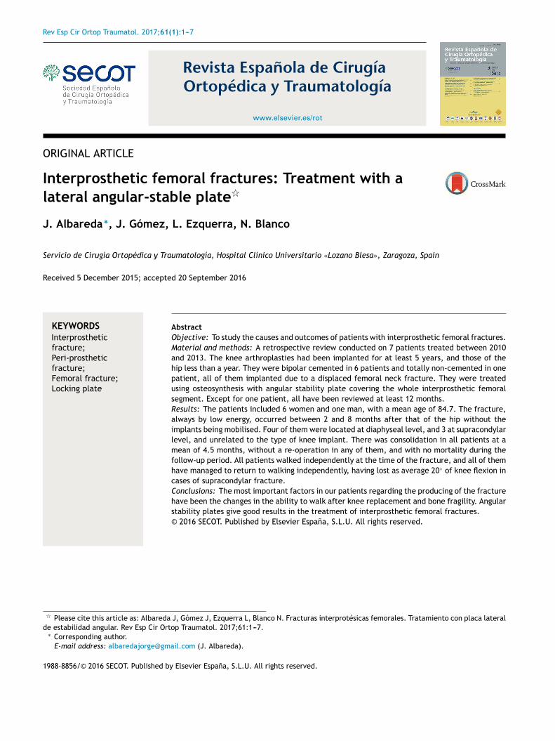
All of the patients walked with the aid of orthopaedicdevices prior to the fracture, 3 with a walking frame and 4with crutches. Union took place in all cases with a meanof 4.5 months,4---7 two of them had diaphyseal fractureswith delayed union. There were no angles in union greaterthan 5◦, no reoperations or complications or evolutionarycomplications presented, even in one patient with a shortplate which only overlapped 3 cm who made satisfactoryprogress (Fig. 2). All the patients regained their walkingability prior to the fracture except for one female patientwho was lost to follow-up at 6 months, the fracture havinghealed. Clinically, the three patients with a supracondy-lar fracture lost a mean of 20◦ knee flexion, previous hipmobility was not altered in any of the patients.
Discussion
Interprosthetic fractures are rare in orthopaedic surgerydepartments, therefore publications referring to them are
Figure 1 Interprosthetic diaphyseal fracture type IA.
also scarce, and there is little information available on howthey occur, the features of the patients who suffer them,the most appropriate treatment or the final outcome. Thetreatment of 141 patients has been published, the majorityin the past 4 years1---5,7---9,11 which is an indication both of therise in their frequency due an increase in life expectancyand arthroplasty surgery, and the growing interest in theirresolution due to the therapeutic difficulty they pose.
These are fractures that are typical in the elderly, muchmore common in women9 and almost always low-energyproduced,11 these features were also evident in our study.
The fractures occur in association with hip and kneeimplants which affect both their occurrence and theirlocation,11 in some series they are more common at supra-condylar level close to the knee arthroplasty8 although notin others,7 and not in our study. At this distal location,their occurrence has been associated with constrained kneeimplants,5 and although this is clinically logical to an extent,it has not been demonstrated in biomechanical studies.13
4 J. Albareda et al.
Table 1 Gender, age, ASA, fracture type (Platzer), radiological union, type of knee implant (ultracongruent, posterior sta-
bilised), and type of femoral implant.
Gender Age ASA Fracture Union (months) Knee implant Hip implant
Female 89 3 IA 5 UC Partially cemented
Female 87 3 IIA 4 UC Partially cemented
Female 86 3 IIA 4 UC Partially cemented
Female 87 3 IA 4 PS Partially cemented
Female 84 2 IA 7 PS Partially cemented
Female 81 2 IIA 4 PS Partially cemented
Male 79 2 IA 4 UC Non-cemented total
Interprosthetic femoral fractures. Treatment with an angular-stable plate.
PS: posterior stabilised; UC: ultracongruent.
Figure 2 Osteosynthesis with lateral plate stabilised with
screws and cerclage. Insufficient overlap of the plate with sat-
isfactory outcome.
Figure 3 Interprosthetic fracture IIA (Platzer’s
classification).
There are differing options regarding the influence of thestability of the implants in the occurrence of these fractures.According to Hou et al. of the 13 cases they present, 4 pre-sented loosening of one of the prostheses,4 although mostpublished series find no relationship between the fractures

Interprosthetic femoral fractures 5
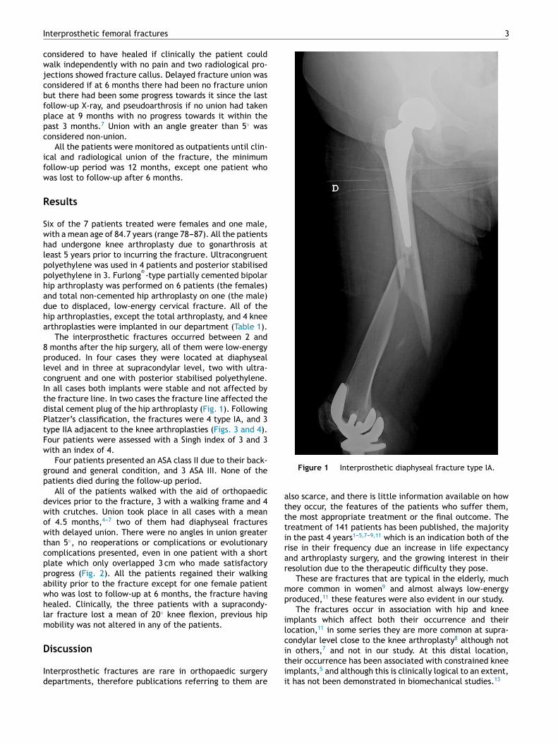
Figure 4 Interprosthetic fracture resolution IIA.
and implant loosening because all of them were stable.1,3,8
it is not clear whether the hip prosthesis being cementedor non-cemented affects occurrence of these fractures,1
or whether it is a total hip prosthesis or hemiarthroplasty.The fact that the arthroplasty has been revised or changed1
might have an influence, but this is not the case for kneearthroplasty because almost all cases published were pri-mary arthroplasties.
In an experimental study with cadaver femurs, Lehmannet al. found that the greatest risk of fracture was ifthe patient had a hip arthroplasty and a retrogradeintramedullary nail and the risk of fracture did notincrease if the patient had a knee arthroplasty, even ifthe knee arthroplasty was constrained.13 These outcomesmight change the concept of interprosthetic fracture, frac-tures occurring in patients bearing two femoral prostheses,whether or not they are arthroplasties, should be consid-ered, since distal femoral implants such as a retrograde nailor a short plate, affect the occurrence of fractures morethan the presence of a knee arthroplasty.13 If we take thisconcept into account, the number of fractures produced infemurs with two implants would increase considerably. Thisstudy clearly applies in terms of the treatment to be given,so that when a distal femoral fracture occurs in a bearer
of a hip arthroplasty, a retrograde implant should not beused in their treatment, but preferably an extramedullaryimplant.13 The femoral area between two intramedullaryfemoral implants, is an area that logically should be at highrisk of fracture due to an accumulation of stress, as hasbeen documented in a few cases,5 and the size of this areabetween implants would seem likely to affect the occur-rence of a fracture, so that the smaller the gap the greaterthe risk, because there is a greater concentration of forcein this area. Segal et al. in an experimental study usingartificial bones into which they implanted a cemented hiparthroplasty and a revision knee arthroplasty, found that ifthe gap was kept between 5 and 20 cm it did not affectfracture occurrence.14
In our series, the arthroplasties, both hip and knee, wereprimary, cemented except for one case and stable, 4 caseswere located in the diaphyseal segment without involve-ment of the implants and 3 cases were adjacent to the kneearthroplasty, ultracongruent polyethylene was used in twoof them: in other words, implant loosening, prosthetic revi-sion surgery, or constrained knee implant had no effect. Allour patients were bearers of hip arthroplasties implanteddue to a fracture; a fact that might have more influencethan the type of implant, their advanced age and the indi-cation for a cemented implant, in most cases, patients withpoorer bone stock who have already suffered at least oneosteoporotic fracture. All our patients had bone fragilitywith low Singh indices. The fact that the fracture occurreda few months after the hip fracture would indicate thefunctional limitations generated after the fracture whichled to a fall and the consequent fracture, which does notoccur after implantation of a knee or hip arthroplasty dueto degenerative disease. The influence of femoral hip pros-thetic implants in our series was much more fundamentalthan knee arthroplasty; all of them implanted several yearsbefore the fracture occurred. The influence of the degree ofosteoporosis has already been noted by Lesaca et al. who,in an experimental study, found the cortical thickness inthe interprosthetic femoral segment to be the most impor-tant and decisive factor in the occurrence of interprostheticfractures.
Variants of the Vancouver and de Lewis and Rorabeckclassifications have been applied in order to adapt themto interprosthetic fractures.1,4,5,7 Platzer et al. specificallyclassified these fractures as type I when the fracture isremote from both implants, type II adjacent to one implantand type III adjacent to both implants. These are furthersubdivided into type A, both components stable, type B, oneloose component, B1 hip and B2 knee, and type C, both com-ponents loose. The frequency of one or other type was 17%type I and III and 66% type II.2 Seventy-five percent had sta-ble components and one or both components were loose inonly 25%.2 In our series all of the patients presented stableimplants and none of the fractures affected the implants,although the supracondylar fractures were adjacent to theknee arthroplasties, and in two cases were affecting thecement distal to the femoral hip implant, 4 cases were clas-sified as type IA and 3 type IIA.
Treatments carried out a decade ago resulted in agreat many failures. Kenny et al. in 1998 published 4cases stabilised with the techniques available at the timeand the treatment failed in all 4 patients, some of them

6 J. Albareda et al.
eventually had an amputation above the fracture.10 Differ-ent types of treatment have been used, retrograde nails,nail plates, dynamic condylar screws, allografts, cerclagewiring, prosthesis revision surgery,2,4 tumour prostheses,15
etc., but there is no standard treatment since this is deter-mined by the characteristics of the fracture, the presenceof implants, the time since they were placed, the occasionalpresence of cement, bone quality, the patients’ age and fea-tures, etc. These factors can affect mechanical stability4
and the biology of fracture union.1 Given these constraints,treatment should be personalised.11
The treatment objectives are to restore femoral lengthand rotation, ensuring the functionality of the arthroplastyand to achieve sufficient stabilisation to enable early mobil-isation, providing the fracture the biomechanical featuresnecessary for its union. Currently the most used mate-rial, when the implants are not loose,4,9 are angular-stableplates, which has revolutionised the treatment in increasingstability of fixation, especially in osteoporotic bone, withminimum aggression to soft tissues.1,2,4,5,8 The plate shouldcover the entire interprosthetic femoral segment5,7,8,11 fromdistal to proximal3 and the screws should be fixed in atleast 8 femoral cortical bones in the proximal segment usinga cerclage or wire11 accessory to increase the stability ofthe screws. Cerclages, as they do not provide rotationalstability,1 should not be used for the main or primary fix-ation of the plate, but rather as a complementary system toscrews to provide greater stability and to be placed in themost proximal area of the plate.1 Hou et al. indicate thatthe plate used should overlap the intramedullary femoralimplant by at least two femoral diameters to prevent areasof stress,4 i.e., between 6 and 8 cm. In our series the overlapwas between 3 and 10 cm, no failures occurred as a resultof limited overlap in some. Some authors1,3,8 use lockedplates with the same concepts, but introduced by minimallyinvasive or percutaneous surgery, offering the advantages ofpreserving fracture haematoma and reducing injury to softtissues to a minimum. Union is achieved using these treat-ments in between 80 and 100%1,2,7,8 of the cases treated,very much depending on good technical surgery.1
Ochs et al. based on Platzer’s classification, created atherapeutic algorithm in which, depending on the type offracture and the stability of the implants, they indicatetreatment with angular-stable plates or prosthesis revi-sion surgery.16 If knee revision implants with intramedullarystem are used, a complementary osteosynthesis plate shouldbe added to prevent areas of stress between the twointramedullary implants.5
We have followed the abovementioned criteria, using asingle lateral angular-stable plate in all cases covering theentire interprosthetic femoral segment, achieving union inall cases with no reoperations or failures. The only problemthat we found was achieving an adequate overlap of theosteosynthesis plate to the femoral hip implant, since thereduced availability of plate lengths means that occasionallywe have to make a rather limited or excessive overlap; achoice has to be made between one or the other.
A wide approach and femoral bone removal are requiredin order to place a graft, and therefore their use is disputed.Spongy bone allografts have occasionally been used in thefracture site,1 but the use of allografts in palisade,9 seek-ing to increase fracture stability and long-term increase of
bone quality or stock, is exceptional due to the good stabilityprovided by angular-stable plates.
Another point of discussion is mortality. These are seri-ous, high-risk fractures in the elderly patient. To a greateror lesser degree, the majority of published series reportmortality,2---5,7 it is difficult to establish rates due to thelimited number of patients included in these series, as inours, with no mortality but with very limited review time,and therefore we have nothing to contribute on this point.
Our study, despite the homogeneity of the treatment,presents limitations due to the limited number of patientsand because it is retrospective, but we can conclude thatinterprosthetic fractures are very rare, although an increasein the short-term is predicted, and has already started. Themajority occur in females of advanced age, the presenceof implants, the functional deterioration which occasionallypresents after their placement for hip fracture and aboveall bone quality, are determining factors. Treatment shouldbe personalised according to the features of the patient andthe fracture but along with prosthetic implant stability, thebest mechanical and biological solutions are lateral angular-stable plates that should be placed percutaneously or in aminimally invasive manner without using allografts, cover-ing the entire interprosthetic femoral segment, overlappingthe femoral hip implant between 6 and 8 cm and using, ifnecessary, cerclages to enhance the fixation and stabilityof the implant in its proximal portion. Outcomes using thistechnique tend to be good with a high rate of union, low riskof fracture callus, and there has been a major decrease inthe number of reoperations and failures.
Level of evidence IV.
Ehtical disclosures
Protection of human and animal subjects. The authorsdeclare that neither human nor animal testing have beencarried out under this research.
Confidentiality of data. The authors declare that they havecomplied with their work centre protocols for the publica-tion of patient data.
Right to privacy and informed consent. The authorsdeclare that no patients’ data appear in this article.
Conflict of interests
The authors have no conflict of interests to declare.
References
1. Sah AP, Marshall A, Virkus WV, Estok DM 2nd, Della Valle CJ.
Interprosthetic fractures of the femur: treatment with a single-
locked plate. J Arthroplast. 2010;25:280---6.
2. Platzer P, Schuster R, Luxl M, Widhalm HK, Eipeldauer S,
Krusche-Mandl I, et al. Management and outcome of interpros-
thetic femoral fractures. Injury. 2011;42:1219---25.
3. Ehlinger M, Czekaj J, Adam P, Brinkert D, Ducrot G, Bonnomet
F. Minimally invasive fixation of type B and C interprosthetic
femoral fractures. Orthop Traumatol Surg Res. 2013;99:563---9.

Interprosthetic femoral fractures 7
4. Hou Z, Moore B, Bowen TR, Irgit K, Matzko ME, Strohecker KA,
et al. Treatment of interprosthetic fractures of the femur. J
Trauma. 2011;71:1715---9.
5. Soenen M, Migaud H, Bonnomet F, Girard J, Mathevon H, Ehlinger
M. Interprosthetic femoral fractures: analysis of 14 cases. Pro-
posal for an additional grade in the Vancouver and SOFCOT
classifications. Orthop Traumatol Surg Res. 2011;97:693---8.
6. Iesaka K, Kummer FJ, di Cesare PE. Stress risers between two
ipsilateral intramedullary stems: a finite-element and biome-
chanical analysis. J Arthroplast. 2005;20:386---91.
7. Ebraheim M, Carrol T, Moral MZ, Lea J, Hirschfeld A, Liu J.
Interprosthetic femoral fractures treated with locking plate. Int
Orthop. 2014;38:2183---9.
8. Mamczak CN, Gardner MJ, Bolhofner B, Borrelli J Jr, Streubel
PN, Ricci WM. Interprosthetic femoral fractures. J Orthop
Trauma. 2010;24:740---4.
9. Michla Y, Spalding L, Holland JP, Deehan D. The complex prob-
lem of the interprosthetic femoral fracture in the elderly
patient. Acta Orthop Belg. 2010;76:636---43.
10. Kenny P, Rice J, Quinlan W. Interprosthetic fracture of the
femoral shaft. J Arthroplast. 1998;13:361---4.
11. Solarino G, Vicenti G, Moretti L, Abate A, Spinarelli A, Moretti
B. Interprosthetic femoral fractures----a challenge of treatment.
A systematic review of the literature. Injury. 2014;45:362---8.
12. Singh M. Changes in trabecular pattern of the upper end
of the femuras an index osteoporosis. J Bone Jt Surg.
1970;52A:457---67.
13. Lehmann W, Rupprecht M, Nuechtern J, Melzner D, Sellenschloh
K, Kolb J, et al. What is the risk of stress risers for interpros-
thetic fractures of the femur? A biomechanical analysis. Int
Orthop. 2012;36:2441---6.
14. Segal O, Quirynen T, Bellemans J, Simon JP, Corten K. A biome-
chanical evaluation of the interprosthetic distance as a risk
factor for periprosthetic fractures of the femur: does the gap
distance matter? Poster no. 0425 ORS annual meeting. 2012.
15. Citak M, Klatte TO, Kendoff D, Haasper C, Gehrke T, Gebauer M.
Treatment of interprosthetic femoral fractures with an interpo-
sition prosthesis. A technical note. Acta Orthop. 2013;84:326---7.
16. Ochs B, Stöckle U, Gebhard F. Interprosthetic fractures----a chal-
lenge of treatment. Eur Orthop Traumatol. 2012;4:1---7.