Fractures of The Shoulder Girdle
134
Injuries of the Upper limb Dr. Isam Ali, PhD .
-
Upload
khangminh22 -
Category
Documents
-
view
1 -
download
0
Transcript of Fractures of The Shoulder Girdle

Injuries of the Upper limb
Dr. Isam Ali, PhD .

Fractures of The Shoulder Girdle
1 - Fr. of the Clavicle : junction of the middle and outer thirds
2 - Fr. of the lateral end of Clavicle
3 - Fr. of body of scapula
4 - Fr. of neck of scapula
5 - Fr. of acromion process
6 - Fr. of coracoid process

Fractures of the Clavicle
• Most fractures result from direct blow on the point of the shoulder; generally from a fall on the side .
• Less commonly ,force may be transmitted up the arm from a fall on the out stretched hand .
• Sport , road traffic .

Fractures of the Clavicle
• The commonest site : junction of middle and outer thirds.
• Also common site: throughout the middle third.
• Less common site : the outer end .

Fractures of the Clavicle
• Displacement :usually, the lateral fragment goes downwards and medially .

Treatment :
• Sling : for the relief of pain, for 2-3 weeks .
• Exercises should be practiced from an early stage especially in old persons.

Treatment :
Figure-of-eight Bandage
In cases of marked
displacement.
may Causes obstructions of the venous return or injuries of the nerve trunk .
Treatment :
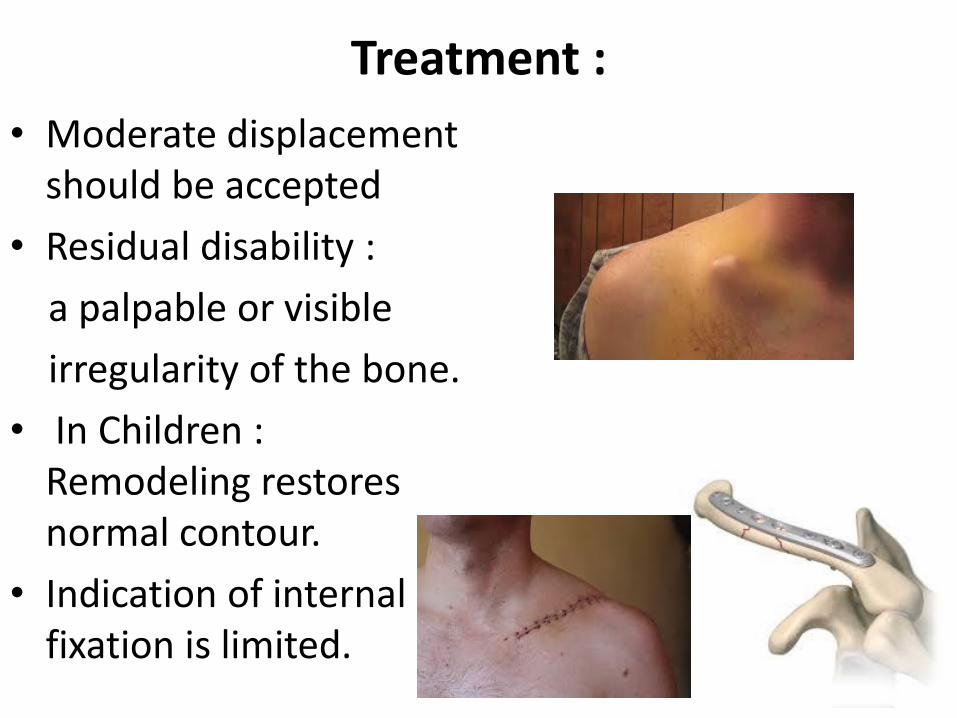
• Moderate displacement should be accepted
• Residual disability :
a palpable or visible
irregularity of the bone.
• In Children : Remodeling restores normal contour.
• Indication of internal fixation is limited.

Fracture of the Scapula
• By direct injury .
• Uncommon and unimportant.
• Might cause severe pain or extensive extravasation of blood into tissues .
• Sling might be used when necessary .
• Operation might be indicated according to the site and displacement.

Subluxation and Dislocation ofThe Sterno-Clavicular Joint
• Uncommon .
• the forward displacement of the medial end of clavicle (anterior displacement) is more common .
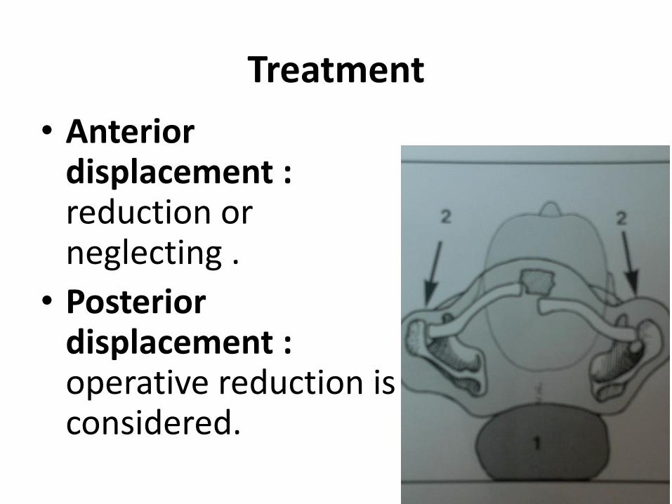
Treatment
• Anteriordisplacement : reduction or neglecting .
• Posterior displacement : operative reduction is considered.

Subluxation & Dislocation ofThe Acromio-Clavicular joint
• Inherently unstable joint (weak capsule) .
• Fall on the outer prominence of the shoulder.

pathology : in subluxation the capsule is torn and the conoid and trapezoid
ligaments are intact .

pathology : in dislocation the conoid and trapezoid ligaments are torn .

Treatment:
• Sprain : Sling if pain is severe.
• Subluxation: sling for 2-3 weeks .
• Dislocation: Operation :
1 - A screw through the clavicle into the coracoid
process.
2 - Or stiff wires from the acromion into the clavicle,
both should be removed after 8 -10 weeks.
3 – Plate and screws .
4 - In old persistent cases, end of the clavicle may be
excised.

Surgical treatment

Acute dislocation of the shoulder
• Common in adults , but seldom in Children .
• Pathology :
- Anterior dislocation :more common, Fall on the outstretched hand or on the region of the shoulder. humeral head is displaced through a rent in the capsule And lies in the infraclavicular fossa

Anterior dislocation of the shoulder
• Clinical features : severe pain , the patient carries his arm and refuses to attempt any movements of the shoulder.
• Radiographs show the humeral head out and down the glenoid fossa .
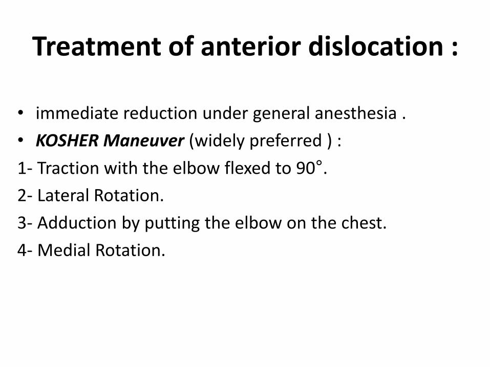
Treatment of anterior dislocation :
• immediate reduction under general anesthesia .
• KOSHER Maneuver (widely preferred ) :
1- Traction with the elbow flexed to 90°.
2- Lateral Rotation.
3- Adduction by putting the elbow on the chest.
4- Medial Rotation.

KOSHER Maneuver
1 2 3 4

Treatment of anterior dislocation :
• Other technique :
• Firm & steady pulling upon the semi-abducted arm against counter-traction in the axilla. With applying direct backward pressure over the humeral head.

Complications of anterior dislocation :
• Injury of axillary nerve : paralysis of the deltoid muscle, and assessing sensation in its distribution .
• Injury of the brachial plexus
• Vascular injury : circumflex artery .
• Associated Fracture:
- Fracture of the greater
tuberosity of the humerus .
- Fracture of the neck of the
humerus .
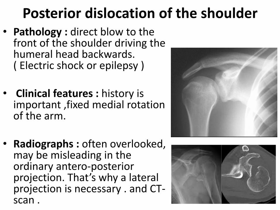
Posterior dislocation of the shoulder• Pathology : direct blow to the
front of the shoulder driving the humeral head backwards. ( Electric shock or epilepsy )
• Clinical features : history is important ,fixed medial rotation of the arm.
• Radiographs : often overlooked, may be misleading in the ordinary antero-posterior projection. That’s why a lateral projection is necessary . and CT-scan .

Treatment of posterior dislocation
1-applying traction to the arm in 90 degree abduction .
2-then externally rotating the limb .

Recurrent Anterior Dislocation Of the Shoulder
• Pathology :- Bankart lesion : detachment of the anterior capsule and the labrum from the anterior rim of the glenoid cavity . - Hill-sachs lesion : the humeral head is dented postero-laterally .

Recurrent Anterior Dislocation Of the Shoulder
• Diagnosis :
• History of recurrent dislocation ( when the arm is abducted, extended and rotated laterally ) , weakness in the muscles of the shoulder girdle .
• x-ray .
• CT scan with contrast .
• MRI .

Treatment :
• BANKART operation : reattach the capsule & the glenoid labrum to the front of glenoid rim.

Treatment :
• PUTTI-PLATT operation :
shortening the subscapularis muscle.

Rupture of rotator cuff of the shoulder(supraspinatus tear)

Rupture of rotator cuff of the shoulder
• Degeneration in the supraspinatus tendon .
• Fall on the shoulder or daily repetitive activities .
• In MAJOR TEAR , disability of initiation abduction.
• Treatment :
*Old patients :no treatment, only rest might help .
* Young patients : Operation .
• In MINOR TEAR, painful arch syndrome . Treated with rest , short waves , graduated exercises.

Fractures of the humerus
1- Fr. Of the neck of humerus
2- Fr. Of the greater tuberosity
3- Fr. Of the shaft of humerus
4- Supracondylar Fr.
5- Fr. Of the condyles
6- Fr. Of the epicondyles

Anatomy Of The proximal
Humerus
• Anatomical neck
• Surgical neck
• Greater tuberosity
• Lesser tuberosity

Fractures Of The Neck Of The Humerus
• Most common in elderly women .
• Caused by a fall on the side of the shoulder or on the outstretched hand .

Fractures Of The Neck Of The Humerus
• Displacement may be none, or moderate , or overlapped .
• Fragments may be firmly impacted
• or sever tilting (angulation) of the head fragment, The shaft either adducted or abducted in relation to the head .
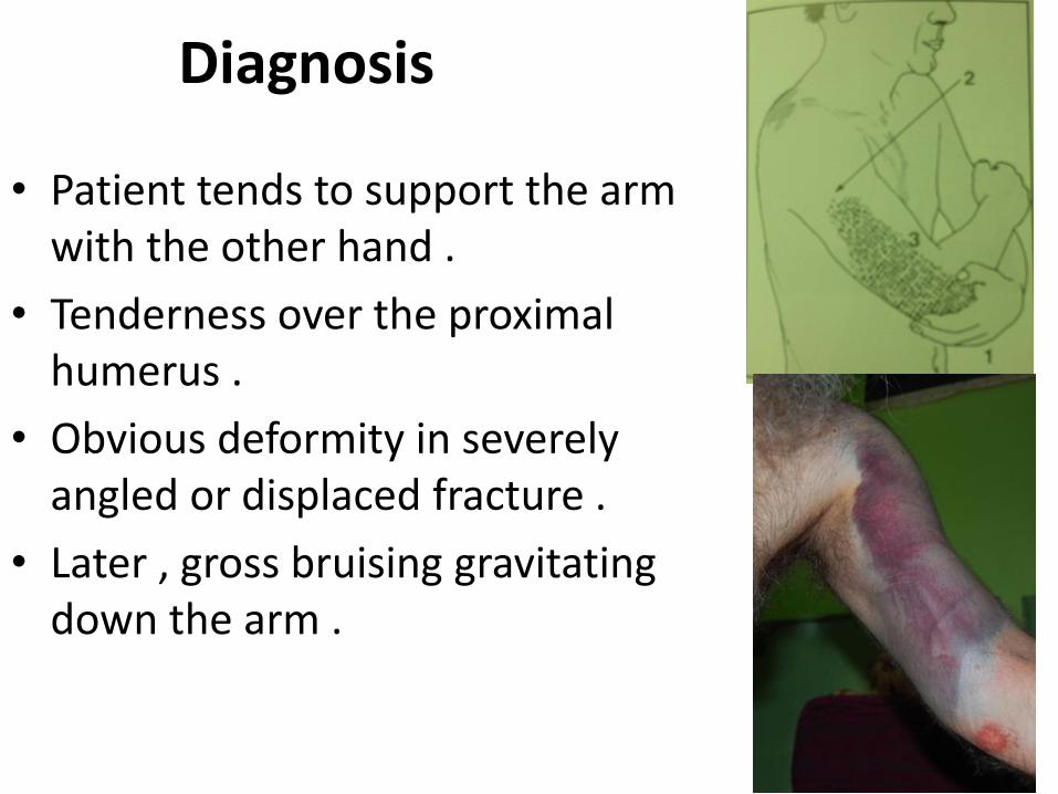
Diagnosis
• Patient tends to support the arm with the other hand .
• Tenderness over the proximal humerus .
• Obvious deformity in severely angled or displaced fracture .
• Later , gross bruising gravitating down the arm .

Radiographs
• Fracture may be impacted.
• Translateral projection will generally be able to clarify the relationships of the fragments.
• CT –scan is essential in some cases .

Treatment
• In elderly patients :
- Moderate displacement is accepted
- Sling with early movement .
- Arthroplasty : in comminuted displaced fractures .

Treatment
In young patients : closed reduction , or open reduction &internal fixation (k.wires , plating, nailing….)

Complications
• Joint Stiffness : in old patients
• Nerve Injury : Axillary nerve, impaired movement of the deltoid muscle, numbness or anaesthesia on the outer side of the upper arm
• Dislocation of the shoulder : rare

Fracture Of The Greater Tuberosity Of The Humerus
• Adults of any age .
• Fall on the shoulder .
• Usually there is no marked displacement .
• In some cases a small fragment might be lifted away .

Treatment
• Undisplaced : rest with early movement .
• Displaced : Fixing with abduction, or operation .
Complications
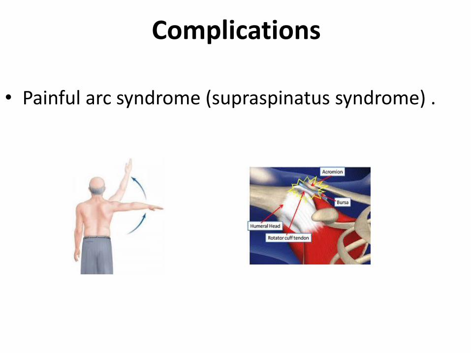
• Painful arc syndrome (supraspinatus syndrome) .

Fractures Of The Shaft Of The Humerus
• Usually in the middle third .
• - Indirect twisting force : Spiral fracture .
- Direct violence : transverse, short
oblique, or comminuted fracture
• Adults of any age, but seldom in children
• Displacement, angulation, or overlapping
• Proximal half of the humerus is a common place of pathological fracture.

Treatment
• Perfect end-to-end position is unnecessary .
• Contact over a quarter or a third is sufficient .
• Displaced : reduction under anesthesia with U-slap and sling for farther support,
or reduction by using hanging cast.
• Exercises of the shoulder and fingers are necessary.
• Unstable or Pathological : operation.

U – Slab application
Hanging cast

Surgical treatment
• Plating
• Interlocking nail

Complications
• Nerve Injury :
Radial nerve (drop-wrist ), usually contusion which recovers gradually, seldom a complete division which needs an operation with a bad prognosis .
• Non-union :
Bone-grafting operation with internal fixation.

Supracondylar Fractures
• Common in children .
• Important and dangerous .
• Fall on the outstretched hand
• The commonest displacement is posterior:
(The distal fragment is displaced posteriorly ) .

Diagnosis• Tenderness over the distal
humerus .
• There may be marked swelling and deformity
• Vascular & neurological evaluation is mandatory .
• The olecranon and medial and lateral epicondyles preserve their normal equilateral triangular relationship (unlike dislocation of the elbow).

Treatment
• Undisplaced in children : 3weeks protection in plaster .
• Displaced : Reduction under anesthesia , and plaster with elbow flexed little more than 90°. With precautions for circulation.
• If unstable: closed reduction or open reduction with fixation (K.wires in children , plates & screws in adults )

Complications• Brachial artery injury :
- simply or severely contused- spasm of the vessel wall
- thrombosis .(management : first of all; reduction and fixation of the
fracture , then reevaluation)
• Injury to median , ulnar or radial nerves .
• Deformity from mal-union : cubitus varus

Fractures of the condyles of the humerus
• Uncommon but troublesome .
• In Children by a fall .
• More common in the lateral condyle .
• Displacement is seldom sever, but even moderate displacement is important .

Treatment
• Undisplaced fracture: Plaster for 4-6 weeks, physiotherapy.
• Displaced fracture : reduction and internal fixation .

• Non-union,
• Osteoarthritis,
• Deformity: Cubitusvarus or valgus,
• Ulnar nerve injury .
Complications

Fractures of the epicondyles
• More common in the medial epicondyle.
• More common in
children than adults .
• By direct violence, but often by pulling the Epicondyle by the attached flexors during a fall.
• May be associated with dislocation or subluxation in the elbow.

Treatment
• Mild displaced fracture : symptomatic.
• Displaced fracture : Reduction with plaster with 90°flexing of the elbow and pronation (medial epicondyle ) or supination(lateral epicondyle ) , physiotherapy .
• Complicated fracture :
open reduction & internal fixation

Complications
• Inclusion of the epicondylar fragment in the joint .
• Injury of the ulnar nerve .

Injuries of the elbow
• Dislocations:
- Dislocation of the elbow
- Dislocation of the head of the radius
- Subluxation of the head of the radius

Dislocation of the elbow
• caused by a heavy full onto the outstretched hand
• common in children and adults
• Pathology: MOSTLY posterior or postero-lateral, the ulna and the radius are displaced backward ,
and dislocation may be associated with
fracture of the coronoid process of the
ulna, or the radial head.

Diagnosis• Distorted the
equilateral triangle formed by the olecranon & epicondyles
• Radiographs confirm the diagnosis

Treatment
• reduction is easy under anesthesia .
• pull steadily upon the forearm with the elbow semi-flexed while direct pressure is applied behind the olecranon .

Treatment
Reduction is confirmed by radioghraphic examination,
Plaster back slab in 90 degree of flexion for 2 weeks .
Exercises (VERY EARLY IF REDUCTION IS STABLE ) .

Complications
• vascular or nerve injury , occasionally the brachial artery or one of the major nerve trunks is damaged.
• Joint stiffness usually caused by intra-articular and peri-articular adhesions, manipulation and passive stretching should be avoided,
• a less common is post-traumatic ossification condition in which new bone forms in the hematoma beneath.

Dislocation of the head of the radius
Occasionally the head of the radius may be dislocated forwards without fracture caused by forced pronation.
Reduction by supinating the forearm while direct pressure is applied over the displaced radial head.

Dislocation of the head of the radius
• Caution: dislocation of the head of the radius as an isolated injury is rare, radiographic examination should always include the whole length of the ulna to rule out a fracture (Monteggia fracture -dislocation).

Subluxation of the head of the radius (pulled elbow)
Young child is lifted by the wrist, the head of the radius may be pulled partly out of the annular ligament.
Local pain, movements of
the elbow are restricted.
Reduction is easy by pushing the forearm upwards and rotating it into supination and pronation.


Fracture of the olecranon process
A fall onto the point of the elbow usually in an adult.
The fracture may take three forms :
- fracture without displacement,
- fracture with displacement
(two fragments) .
- comminuted fracture.
The fracture line nearly always enters the joint near the middle of the trochlear notch.

Treatment• Fracture without displacement:
protect the elbow by a plaster splint for 2 or 3 weeks , the right-angled position is more comfortable for the patient.
• Fracture with displacement : because the action of the triceps will angulate and distract the fragments, operation should be advised.

Treatment
• Comminuted fracture:
• open reduction and internal fixation if possible .
• or excision the
olecranon fragments
and securing the
triceps on the stump of
the ulna by strong
sutures(very limited
indication ).

Complications
non-union,
mal-union ,
osteoarthritis.

Fracture of the olecranon process with anterior dislocation of the elbow
• Uncommon injury
• Fall on the elbow, and forearm bones is pushed forwards .
• Very unstable injury .
• Surgical treatment .

Fracture of the coronoid process
Seldom except in association with posterior dislocation of the elbow.
Treatment depends on the size of the fragment and the stability of the elbow .

Fracture of head of radius One of the most common
fractures in young adults.
It is caused by a fall onto the outstretched hand with pronation.
In most cases the fracture is no more than vertical crack without displacement.
Sometimes the whole of the head is extensively comminuted.

Diagnosis
• Local tenderness, restriction of elbow movement (rotation).
• Painful supination .
• Radiology : may be easily over looked because it is sometimes not immediately obvious in the radiographs (impacted).

Treatment depends upon the severity of the damage :
- Slight damage: conservative treatment ,sling ,early active exercises .
- Severe damage: excision of the radial
head, after the operation the elbow is
rested in plaster for 2 weeks then active
exercises are begun.
Fracture in children: reduction by manipulation, excision of the radial head should never be advised in children.
Complications: joint stiffness- osteoarthritis.

Monteggia fracture - dislocation
Fracture of the ulna with dislocation of the head of the radius

Monteggia fracture - dislocation
Uncommon
caused by a fall with forced pronation of the forearm.
Or direct trauma

Monteggia fracture - dislocation
• The ulna is angled and the head of the radius is dislocated :
1-posteriorly
2-anteriorly
3-laterally

Monteggia fracture - dislocation
• Treatment: closed reduction is seldom possible.
• Operation is required to reduce and fix the ulna fracture, and reduction of radial head must be checked by palpation and x ray
• a plaster cast at a right angle for 4-6 weeks.
• Complications: elbow stiffness , mal-union, compartment syndrome

Fracture of the shaft of the forearm bones
Typical fracture of both bones of the forearm
with marked displacement

Fracture of the shaft of the forearm bones
Isolate fracture of ulna or radius is rare.

Fracture of the shaft of the forearm bones
• The cause is indirect force such as a fall onto the hand
• or a direct bow on the forearm

Treatment
• Accurate reduction is important because even slight residual displacement or angulation may disturb the relationship between radius and ulna.
• Axial rotation is unaccepted

Conservation treatment in children
manipulative reduction under anesthesia.
If this is successful a full-length arm plaster is applied with the elbow at a right angle and the forearm in a position midway between pronation and supination.
Radiographs' should be taken weekly for the first 3 weeks, the plaster is retained until union occurs

Full-length arm plaster with elbow flexed 90 degrees

Operative treatment
• especially in adults.
• it is very difficult to correct a severe displacement without open operation.

Complications
delayed and non-union, this fracture prone to delayed union or non-union.
Mal-union: if an imperfect position is accepted there is a risk of impaired function (in rotation), angulation of the fragments shortens one of the bones
cross union between bones.

Galeazzi fracture-dislocation
Fracture of the shaft of radius with dislocation of the distal radio-ulnar
joint

Galeazzi fracture-dislocation
• Often the fracture is near the junction of middle and lower thirds of the radius ,and the ligaments of the inferior radio-ulnar joint are ruptured.
The head of the ulna may be shifted medially, anteriorly or posteriorly.
The Galeazzi fracture - dislocation is more common than the Monteggia fracture - dislocation.

Treatment perfect reduction is essential for
restoration of full function.
In adults re-displacement is common after reduction by manual traction and supination, so operative reduction and internal fixation are indicated usually.
The position is held by a full-length arm plaster with the elbow flexed to the right angle and the forearm supinated.
the plaster is retained until union occurs.

Colles’s fracture
Fracture of the lower end of
the radius within 2.5 c m
of the wrist with dorsal and
radial displacement
(Typical Displacement)
Colles’s fracture
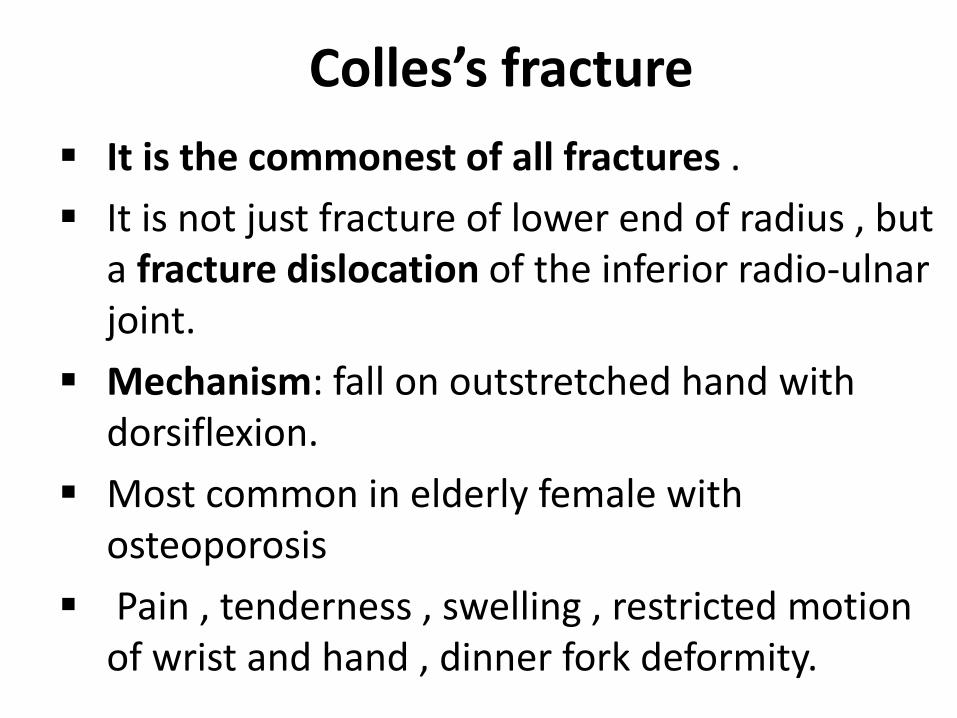
It is the commonest of all fractures .
It is not just fracture of lower end of radius , but a fracture dislocation of the inferior radio-ulnar joint.
Mechanism: fall on outstretched hand with dorsiflexion.
Most common in elderly female with osteoporosis
Pain , tenderness , swelling , restricted motion of wrist and hand , dinner fork deformity.

Colles’s fracture
Typical deformity (dinner fork ) from displaced fracture of the lower end
of the radius ( Colles’s fracture )

RadiologyAP and lateral views of wrist and lower end of
the radius.

Treatmentthe aim of the treatment is to restore fully functional hand :
1 - Conservative methods: reduction is carried out
by closed methods.
A below elbow or above elbow cast is put and the
plaster cast is removed after 6 weeks , and
then physiotherapy is begun.
2 - Closed reduction and percutaneous fixation.
3 - Operative methods (plate and screws , external
fixator in severely comminuted fractures ).

Reduction and cast

The fracture are seen through plaster after manipulative reduction

Closed reduction and percutaneous fixation

Operative methods (plate and screws , external fixator in severely
comminuted fractures ).

Complications1 – Mal-union: the most common.
no treatment is required if the patient has no functional abnormality.
2 - Rupture of extensor pollicislongus tendon :
usually occurs after 4 to 6 weeks. Treatment by transfer of extensor indicis to replace the ruptured extensor pollicislongus.

Complications
3 - Sudeck’s atrophy: due to abnormal sympathetic response which causes vasodilatation and osteoporosis at the fracture region.
The fingers are swollen and fingers flexion is restricted ,the hand and wrist are warm , tender and painful .

Complications4 – associated scaphoid fracture .
5 - Frozen hand shoulder syndrome: due to unnecessary voluntary shoulder immobilization.
6 - Carpal tunnel syndrome: compression on the median nerve due to mal-union.
7 - Nonunion: rare.

Fracture-separation of lower radial epiphysis in a child:
( epiphyseal injury )

Fracture of the lower end of radius with anterior displacement
Smith’s fracture volar Barton’s fracture

Smith’s fracture A fracture of distal one-third of
radius with palmar displacement.
Reverse colles’s fracture.
Less common than colles’s.
Mechanism: fall on the back of the dorsum.
Clinical features: pain , swelling , deformity.
Radiography: anterior displacement of the distal fragment with angulation .

Treatment closed reduction
and immobilization in a long arm cast with forearm in supination and wrist in extension.
Or internal fixation .
complications : same in colles’s fracture.

Barton’s fracture
Dorsal or volar rim could be involved and these fractures are invariably intra-articular.

Dorsal Barton Dorsal rim fracture of distal radius.
Mechanism : fall with dorsiflexion and pronation.
Clinical features: pain , swelling , tenderness , deformity and painful restrictions of the wrist.
Radiology: fracture is seen on the lateral view.
Treatment:
- Conservative : reduction , long arm cast is used in dorsal flexion (extension ) .
-Surgery: unstable fracture is fixed.

Volar Barton This is a form of Smith fracture
in which the anterior portion only of the radius is involved .
Clinical features: pain , swelling, tenderness , painful restrictions of the wrist.
Treatment:
-Conservative: reduction , long arm cast is used in volar flexion.
-Surgery: if reduction does not remain (plate & screws).

Carpal injuries

Carpal bones“she looks too pretty try to catch her”
• Scaphoid , lunate , triquetral , pisiform , trapezium, trapezoid , capitate and hamate.

Scaphoid fracture
It is common in young adults.
May be associated with other fracture or dislocation of carpus.
Mechanisms of injury :
1 - Results from (KICK –BACK)
2 - Fall on outstretched hand with severe hyperextension.
Scaphoid fracture
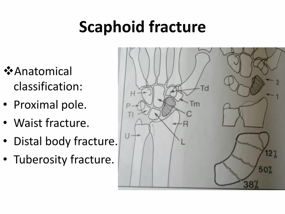
Anatomical classification:
• Proximal pole.
• Waist fracture.
• Distal body fracture.
• Tuberosity fracture.

Scaphoid fracture Clinical features:
pain , swelling , tenderness in the anatomical snuff box , The movements of the wrist is painful .
Radiology: AP , lateral ,scaphoid views .

Treatment: Undisplaced fracture
• conservative : below elbow cast applied to the level of metacarpal heads of fingers and interphalangeal joint of the thumb.
• Position assumes a glass-holding .
• Duration of cast : 10 – 12 weeks .
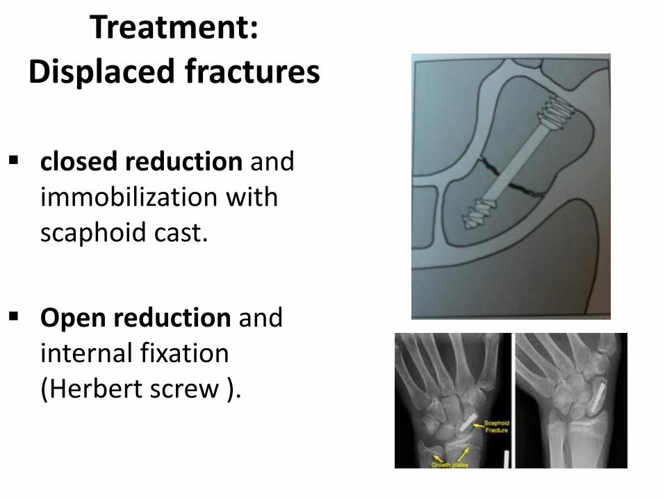
Treatment: Displaced fractures
closed reduction and immobilization with scaphoid cast.
Open reduction and internal fixation (Herbert screw ).

Complications• nonunion and delayed union :
nonunion requires open reduction , internal fixation and bone grafting.
• Incidence of avascular necrosis is as high as 40 percent , especially in proximal pole .
• Secondary Radio-carpal joint osteoarthritis .

Lunate fracture and dislocation Mechanism: this is
almost similar to scaphoid fractures.
Clinical features: pain , swelling , painful movements.
Radiology: easily diagnosed , the wrist normally forms a half moon shape , This is lost in dislocation.

Lunate dislocation Problems: may cause
compression of the median nerve if left untreated.
Treatment: if seen early, reduction is easy and immobilization for 3 weeks with wrist in slight flexion.
If seen after 3 weeks, open reduction.

Bennett’s fracture Fracture dislocation of the base of the first
metacarpal bone.
It is an intra-articular fracture.
Mechanism of injury: an axial blow directed against the partially flexed metacarpal.
Clinical features: pain , swelling , tenderness on the base of first metacarpal , movements of the thumb are painful and restricted.

Bennett’s fracture Peculiarity of this fracture: base of
the thumb metacarpal is pulled proximally and laterally by the abductor pollicis longus .
Radiology : shows the displacement

Treatment• Like all the other
intra-articular fractures, it needs accurate reduction and retention to prevent future posttraumatic osteoarthritis.
• Closed reduction and a percutaneous fixation .
• If it fails ORIF.

Rolando fracture This is also a
fracture of the base of the first metacarpal .
Closed reduction with a thumb spica.
If failed , ORIF using small special plate and screws .

Complications
• both bennett’s and rolando fractures could lead to secondary OA of the carpometacarpal and joint stiffness.
• Rarely injury of sensory branch of the radial nerve during surgical intervention.

Mallet finger It is a common finger injury and is due to avulsion
or avulsion fracture of the extensor tendon at the base of the distal phalanx.
Mechanism: is forcibly flexed.
Clinical features: pain , swelling , tenderness , flexion deformity of the tip of the finger.
Plain X-ray may show an avulsion fracture.

Treatment• immobilized in extension
by using a stack plastic mallet finger splint ,
• or surgical treatment if bony fragment is big (K . Wire or suturing ) .

Jersey finger
It is a due to avulsion of flexor digitorum profundus from its insertion on distal phalanx.
Patient is unable to flex the distal interphalangeal joint.
It is seen in football and rugby.

Metacarpal bones fracture
Common causes for these injuries are direct hit on the dorsum of the hand.
Plain X-ray.
These fractures should be accurately reduced with no rotational mal-alignment and immobilized with either plaster (common) or percutaneous k. wires or open (k. wires or plates ) .

Metacarpal neck fracture
The most common is Fracture of the fifth metacarpal neck is known as boxer’s fracture.
Treatment: closed reduction and may need percutaneous fixation.

Metacarpal head fracture
Frequently intra-articular and need open reduction and internal fixation.
Phalanges fracture Fracture of distal phalanx: caused by crushing
injuries, they are frequently comminuted and require only splinting.
Fracture of the middle or proximal phalanx: direct blow , rotational mal-alignment should be strictly avoided.
Undisplaced fractures are best managed by the unaffected finger is used as the supporting external splint.
Highly unstable oblique fracture require reduction and fixation.
Complications
The important complications are :
non-union
mal-union
tendon adhesions
joint stiffness .

The end