PLS3 Mutations in X-Linked Osteoporosis with Fractures
8
The new england journal of medicine n engl j med nejm.org 1 BRIEF REPORT PLS3 Mutations in X-Linked Osteoporosis with Fractures Fleur S. van Dijk, M.D., Ph.D., M. Carola Zillikens, M.D., Ph.D., Dimitra Micha, Ph.D., Markus Riessland, Ph.D., Carlo L.M. Marcelis, Ph.D., Christine E. de Die-Smulders, M.D., Ph.D., Janine Milbradt, B.S., Anton A. Franken, M.D., Ph.D., Arjan J. Harsevoort, M.S., Klaske D. Lichtenbelt, M.D., Hans E. Pruijs, M.D., Ph.D., M. Estela Rubio-Gozalbo, M.D., Ph.D., Rolf Zwertbroek, M.D., Youssef Moutaouakil, M.S., Jaqueline Egthuijsen, M.S., Matthias Hammerschmidt, Ph.D., Renate Bijman, B.S., Cor M. Semeins, M.S., Astrid D. Bakker, Ph.D., Vincent Everts, Ph.D., Jenneke Klein-Nulend, Ph.D., Natalia Campos-Obando, M.D., Albert Hofman M.D., Ph.D., Gerard J. te Meerman, Ph.D., Annemieke J.M.H. Verkerk, Ph.D., André G. Uitterlinden, Ph.D., Alessandra Maugeri, Ph.D., Erik A. Sistermans, Ph.D., Quinten Waisfisz, Ph.D., Hanne Meijers-Heijboer, M.D., Ph.D., Brunhilde Wirth, Ph.D., Marleen E.H. Simon, M.D., and Gerard Pals, Ph.D. The authors’ affiliations are listed in the Appendix. Address reprint requests to Dr. Pals at the Center for Connective Tissue Research, Department of Clinical Genetics, VU University Medical Center, P.O. Box 7057, 1007 MB Amsterdam, the Netherlands, or at [email protected]. Drs. van Dijk, Zillikens, Micha, and Riess- land contributed equally to this article. This article was published on October 2, 2013, at NEJM.org. N Engl J Med 2013. DOI: 10.1056/NEJMoa1308223 Copyright © 2013 Massachusetts Medical Society. SUMMARY Plastin 3 (PLS3), a protein involved in the formation of filamentous actin (F-actin) bundles, appears to be important in human bone health, on the basis of patho- genic variants in PLS3 in five families with X-linked osteoporosis and osteoporotic fractures that we report here. The bone-regulatory properties of PLS3 were sup- ported by in vivo analyses in zebrafish. Furthermore, in an additional five families (described in less detail) referred for diagnosis or ruling out of osteogenesis imper- fecta type I, a rare variant (rs140121121) in PLS3 was found. This variant was also associated with a risk of fracture among elderly heterozygous women that was two times as high as that among noncarriers, which indicates that genetic variation in PLS3 is a novel etiologic factor involved in common, multifactorial osteoporosis. O steoporosis is a prevalent disorder characterized by low bone mass and microarchitectural deterioration of bone tissue, which results in bone fragility and fractures. 1 It is diagnosed clinically and often confirmed by measuring bone mineral density (BMD). 1,2 An understanding of the causes of osteoporosis is important for its prevention, diagnosis, and treatment. The investi- gation of rare mendelian disorders with decreased BMD as a key diagnostic feature constitutes a strategy for identifying genetic determinants of osteoporosis. 3-7 We identified families with X-linked osteoporosis and fractures among patients with negative tests for the genes encoding collagen type Iα1 and type Iα2 ( COL1A1 and COL1A2, respectively) who had been referred to us for diagnosis or ruling out of osteogenesis imperfecta type I. Osteoporosis with fractures as an X-linked trait has been reported by Sillence. 8 We now report data from five families with X-linked osteoporosis and fractures related to pathogenic variants in the gene for plastin 3 (PLS3), provide functional evidence that PLS3 is a bone-regulatory protein, and describe a rare variant or single-nucleotide polymorphism (SNP) associated with
-
Upload
independent -
Category
Documents
-
view
1 -
download
0
Transcript of PLS3 Mutations in X-Linked Osteoporosis with Fractures
T h e n e w e ngl a nd j o u r na l o f m e dic i n e
n engl j med nejm.org 1
BRIEF REPORT
PLS3 Mutations in X-Linked Osteoporosis with Fractures
Fleur S. van Dijk, M.D., Ph.D., M. Carola Zillikens, M.D., Ph.D., Dimitra Micha, Ph.D., Markus Riessland, Ph.D., Carlo L.M. Marcelis, Ph.D.,
Christine E. de Die-Smulders, M.D., Ph.D., Janine Milbradt, B.S., Anton A. Franken, M.D., Ph.D., Arjan J. Harsevoort, M.S., Klaske D. Lichtenbelt, M.D.,
Hans E. Pruijs, M.D., Ph.D., M. Estela Rubio-Gozalbo, M.D., Ph.D., Rolf Zwertbroek, M.D., Youssef Moutaouakil, M.S., Jaqueline Egthuijsen, M.S., Matthias Hammerschmidt, Ph.D., Renate Bijman, B.S., Cor M. Semeins, M.S.,
Astrid D. Bakker, Ph.D., Vincent Everts, Ph.D., Jenneke Klein-Nulend, Ph.D., Natalia Campos-Obando, M.D., Albert Hofman M.D., Ph.D.,
Gerard J. te Meerman, Ph.D., Annemieke J.M.H. Verkerk, Ph.D., André G. Uitterlinden, Ph.D., Alessandra Maugeri, Ph.D., Erik A. Sistermans, Ph.D.,
Quinten Waisfisz, Ph.D., Hanne Meijers-Heijboer, M.D., Ph.D., Brunhilde Wirth, Ph.D., Marleen E.H. Simon, M.D., and Gerard Pals, Ph.D.
The authors’ affiliations are listed in the Appendix. Address reprint requests to Dr. Pals at the Center for Connective Tissue Research, Department of Clinical Genetics, VU University Medical Center, P.O. Box 7057, 1007 MB Amsterdam, the Netherlands, or at [email protected].
Drs. van Dijk, Zillikens, Micha, and Riess-land contributed equally to this article.
This article was published on October 2, 2013, at NEJM.org.
N Engl J Med 2013.DOI: 10.1056/NEJMoa1308223Copyright © 2013 Massachusetts Medical Society.
SUMM A R Y
Plastin 3 (PLS3), a protein involved in the formation of filamentous actin (F-actin) bundles, appears to be important in human bone health, on the basis of patho-genic variants in PLS3 in five families with X-linked osteoporosis and osteo porotic fractures that we report here. The bone-regulatory properties of PLS3 were sup-ported by in vivo analyses in zebrafish. Furthermore, in an additional five families (described in less detail) referred for diagnosis or ruling out of osteogenesis imper-fecta type I, a rare variant (rs140121121) in PLS3 was found. This variant was also associated with a risk of fracture among elderly hetero zygous women that was two times as high as that among noncarriers, which indicates that genetic variation in PLS3 is a novel etiologic factor involved in common, multi factorial osteoporosis.
Osteoporosis is a prevalent disorder characterized by low bone mass and micro architectural deterioration of bone tissue, which results in bone fragility and frac tures.1 It is diagnosed clinically and often confirmed
by measuring bone mineral density (BMD).1,2 An understanding of the causes of osteoporosis is important for its prevention, diagnosis, and treatment. The investi-gation of rare mendelian disorders with decreased BMD as a key diagnostic feature constitutes a strategy for identifying genetic determinants of osteoporosis.3-7
We identified families with X-linked osteoporosis and fractures among patients with negative tests for the genes encoding collagen type Iα1 and type Iα2 (COL1A1 and COL1A2, respectively) who had been referred to us for diagnosis or ruling out of osteogenesis imperfecta type I. Osteoporosis with fractures as an X-linked trait has been reported by Sillence.8 We now report data from five families with X-linked osteoporosis and fractures related to pathogenic variants in the gene for plastin 3 (PLS3), provide functional evidence that PLS3 is a bone-regulatory protein, and describe a rare variant or single-nucleotide polymorphism (SNP) associated with

T h e n e w e ngl a nd j o u r na l o f m e dic i n e
n engl j med nejm.org2
decreased BMD and an increased risk of fracture among heterozygous women in the general population.
ME THODS
FAMILIES
The pedigrees and clinical characteristics of Families 1 through 5 are provided in Figure 1 and Table 1, and Figure S1 and Table S1 in the Supplementary Appendix, available with the full text of this article at NEJM.org. Five additional families, designated Families 6 through 10, were also included in the study and are mentioned in less detail (Fig. S2 and Table S2 in the Supple-mentary Appendix).
GENETIC STUDIES
Three patients with osteoporosis and fractures from Family 1 (Patients 1.III-2, 1.IV-3, and 1.IV-7) underwent X-linked whole-exome sequencing.9,10 We then performed Sanger sequencing of all PLS3 exons in 95 affected male patients without COL1A1 or COL1A2 mutations who had been re-ferred for diagnosis or ruling out of osteogenesis imperfecta type I. Complementary DNA (cDNA) analysis was performed in Patients 1.III-2 and 3.II-1 and the index patient from Family 9. Linkage analy-sis was conducted in Families 1 and 2. Methodo-logic and other details of the studies performed are described in the Supplementary Appendix.
EPIDEMIOLOGIC STUDIES
The rs140121121 SNP was genotyped in three co-horts (RS-I, RS-II, and RS-III) of the prospective, population-based Rotterdam Study, which has analyzed, among other topics, the association of genetic factors with BMD and incident fractures in Dutch men and women 45 years of age or older.11 Details of these studies are provided in the Sup-plementary Appendix.
FUNCTIONAL STUDIES
Electrophoresis of type I collagen and Western blot analysis for PLS3 were performed in affected Pa-tients 1.III-2, 1.IV-2, 1.IV-7, 1.IV-8, 3.II-1, and 4.II-1 and the index patients from Families 7 and 9. PLS3, belonging to the family of plastins, is in-volved in the formation of F-actin bundles.12 The effect of PLS3 deficiency on F-actin cyto-skeleton was investigated in dermal fibroblasts with the use of immunofluorescence microsco-py. We hypothesized that PLS3 may be involved in
mechanosensing of osteocytes. Mechanical load-ing in the form of fluid shear stress increases the production of nitric oxide in bone cells,13 perio-dontal ligament, and gingival fibroblasts.14
In the absence of bone tissue from patients, we investigated the response to fluid shear stress of dermal fibroblasts from six patients with PLS3 mutations, as compared with three patients with molecularly confirmed osteogenesis imperfecta type I and eight controls. To characterize the effect of loss of PLS3 on bone morphology, we performed morpholino-mediated knockdown of the zebrafish homologue (National Center for Bio technology Information [NCBI] Reference Se-quence [RefSeq], NM_001002326.1). Since carti-laginous pharyngeal arches are the earliest formed craniofacial skeletal elements, we used a col1a1:eGFP (enhanced green fluorescent protein under the control of a col1α1-promoter) transgenic zebra fish line to monitor skeletal development.15 Details of these studies are provided in the Supplemen-tary Appendix.
R ESULT S
GENETIC STUDIESIdentification of Pathogenic Variants in PLS3We discovered a single deleterious hemizygous frameshift, c.235delT;p.(Tyr79Ilefs*6), in exon 3 of PLS3 (NCBI Reference Sequence, NM_005032.5; Mendelian Inheritance in Man number, 300131; chromosome-map location, Xq23) in Patients 1.III-2, 1.IV-3, and 1.IV-7 (Fig. S3A through S3F in the Supplementary Appendix). Sanger sequencing confirmed the presence of this variant in six af-fected male patients and its absence in one un-affected male patient (Fig. 1).
Sanger sequencing of all PLS3 exons in 95 af-fected male patients without COL1A1 or COL1A2 mutations yielded four pathogenic variants in Families 2 through 5 (Fig. 1). In Family 2, a non-sense mutation, c.1471C→T;p.(Gln491*), in exon 13 was identified in Patients 2.III-3 and 2.III-7. In Families 3, 4, and 5, three pathogenic var-iants were identified: a splice-site variant, c.748+1G→A, in exon 7 (in Patient 3.II-1); an inser-tion, c.759_760insAAT;p.(Ala253_Leu254insAsn), in exon 8 (in actin-binding domain 1, con-served from human down to tetraodon) (in Pa tient 4.II-1); and a frameshift variant, c.1647delC;p.(Ser550Alafs*9), in exon 15 (in Pa-tient 5.II-3). To our knowledge, none of these vari-ants are described in current databases of human
brief report
n engl j med nejm.org 3
sequence variants: data from the 1000 Genomes Project, the Single Nucleotide Polymorphism data-base (dbSNP, build 137), or data from the GO Exome Sequencing Project (ESP) of the National Heart, Lung, and Blood Institute (http://evs.gs .washington.edu/EVS).
In addition, a c.321T→A variant in exon 4b (Fig. S3F in the Supplementary Appendix), listed in dbSNP as rs140121121, was identified in 5 pa-tients (from Families 6 through 10) among the 95 male patients referred to us for possible osteo-genesis imperfecta type I (allele frequency, 0.05) (Table S2A in the Supplementary Appendix). For
this rare variant, the allele frequency was 0.01 among 1872 men in the ESP and 0.02 among the 5189 men in the Rotterdam Study, results that differ significantly from the frequency among our 95 male patients (P = 0.006 and P = 0.04 by two-tailed Fisher’s exact test for the two com-parisons, respectively).
cDNA AnalysisIn Family 3 (Patient 3.II-1), a partial skipping of exon 7 and use of a cryptic splice site, c.748+36, was detected (Fig. S4A and S4B in the Supple-mentary Appendix). Use of this cryptic splice site
I-1 I-2
Family 5
II-1 II-2 II-3
III-1 III-2 III-3
C*
IV-1
I-1 I-2
Family 4
II-1 II-2 II-3 II-4 II-5 II-6C*
III-1III-1
C*
I-1 I-2
Family 3
II-1
Osteopenia
Osteoporosis
Osteoporosis andosteoporotic fracturesat a young age
No PLS3 mutationidentified
N
Clinically evaluatedC
Identified PLS3 mutation
*
Family 1
I-1 I-2
II-1
C C
II-2
III-1 III-2 III-3
N
C
III-4
IV-2 IV-3 IV-4 IV-5 IV-6 IV-7 IV-8 IV-9C*
C*
C*
C*
C*
C*
C*
C*
C
Family 2
I-1 I-2
II-1 C II-4
IV-1 IV-2 IV-3 IV-4 IV-5 IV-6 IV-7
II-2 II-3
III-1 III-2
C *N N
C
III-10 III-11
N
C *
N
C*
C*
C*
C*
III-3 III-4 III-5 III-6 III-7 III-8 III-9
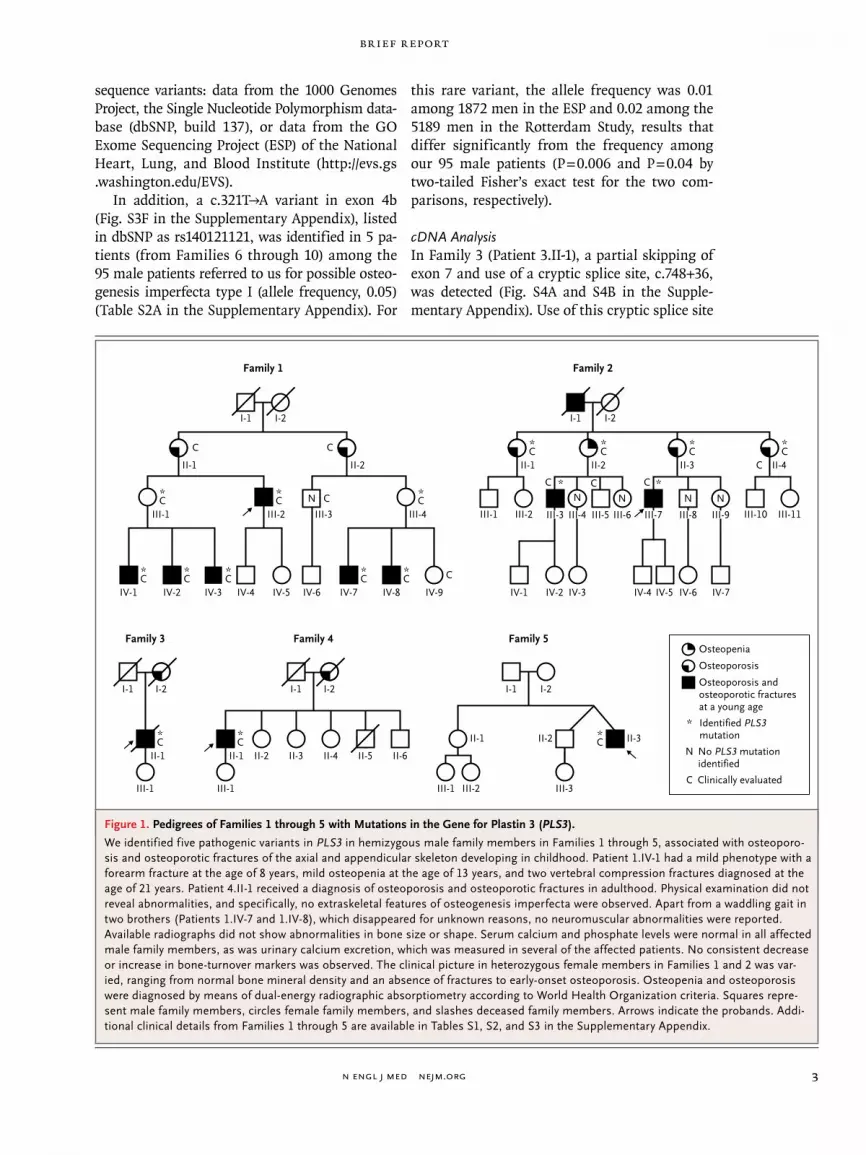
Figure 1. Pedigrees of Families 1 through 5 with Mutations in the Gene for Plastin 3 (PLS3).
We identified five pathogenic variants in PLS3 in hemizygous male family members in Families 1 through 5, associated with osteoporo-sis and osteoporotic fractures of the axial and appendicular skeleton developing in childhood. Patient 1.IV-1 had a mild phenotype with a forearm fracture at the age of 8 years, mild osteopenia at the age of 13 years, and two vertebral compression fractures diagnosed at the age of 21 years. Patient 4.II-1 received a diagnosis of osteoporosis and osteoporotic fractures in adulthood. Physical examination did not reveal abnormalities, and specifically, no extraskeletal features of osteogenesis imperfecta were observed. Apart from a waddling gait in two brothers (Patients 1.IV-7 and 1.IV-8), which disappeared for unknown reasons, no neuromuscular abnormalities were reported. Available radiographs did not show abnormalities in bone size or shape. Serum calcium and phosphate levels were normal in all affected male family members, as was urinary calcium excretion, which was measured in several of the affected patients. No consistent decrease or increase in bone-turnover markers was observed. The clinical picture in heterozygous female members in Families 1 and 2 was var-ied, ranging from normal bone mineral density and an absence of fractures to early-onset osteoporosis. Osteopenia and osteoporosis were diagnosed by means of dual-energy radiographic absorptiometry according to World Health Organization criteria. Squares repre-sent male family members, circles female family members, and slashes deceased family members. Arrows indicate the probands. Addi-tional clinical details from Families 1 through 5 are available in Tables S1, S2, and S3 in the Supplementary Appendix.

T h e n e w e ngl a nd j o u r na l o f m e dic i n e
n engl j med nejm.org4
Tabl
e 1.
Clin
ical
and
Bon
e-D
ensi
tom
etry
Fin
ding
s in
11
Mal
e Pa
tient
s fr
om F
ive
Fam
ilies
with
a P
atho
geni
c V
aria
nt in
the
Gen
e fo
r Pl
astin
3 (P
LS3)
.*
Patie
nt†
Bef
ore
Ther
apy
Aft
er T
hera
py‡
Low
-Im
pact
Pe
riph
eral
Fr
actu
res
Mul
tiple
V
erte
bral
Fr
actu
res
Oth
er C
linic
al F
indi
ngs§
Age
BM
D z
Sco
reA
geB
MD
z S
core
lum
bar s
pine
fem
oral
nec
kto
tal b
ody
lum
bar s
pine
fem
oral
nec
kto
tal b
ody
yryr
no.
1.II
I-2
32−5
.5−3
.4N
A40
−4.6
−3.1
NA
13Ye
sN
one
1.IV
-113
−1.2
NA
−1.5
NT
NT
NT
NT
1N
oN
one
1.IV
-121
−1.1
−0.8
−0.8
NT
NT
NT
NT
1Ye
sN
one
1.IV
-210
−2.1
NA
−3.0
170.
9N
A−0
.76
No
Acu
te ly
mph
atic
leuk
emia
1.IV
-34
−3.2
NA
−3.6
10−1
.2N
A−1
.41
No
Non
e
1.IV
-76
−3.7
NA
−4.6
140.
7N
A−1
.117
No
Pate
nt d
uctu
s ar
teri
osus
an
d, in
chi
ldho
od,
wad
dlin
g ga
it
1.IV
-810
−2.4
NA
−3.3
12−1
.1N
A−1
.9M
ultip
leN
oEp
ileps
y an
d, in
chi
ldho
od,
wad
dlin
g ga
it
2.II
I-3
36−2
.8−2
.3N
AN
AN
AN
AN
A5
No
Non
e
2.II
I-7
34−3
.4−3
.4N
AN
AN
AN
AN
A13
Yes
Non
e
3.II
-1N
AN
AN
AN
A47
−3.7
5−2
.5N
AM
ultip
leYe
sA
lcoh
ol a
buse
3.II
-1N
AN
AN
AN
A62
NA
−1.0
NA
Mul
tiple
Yes
Esop
hage
al c
arci
nom
a
4.II
-154
−2.5
−0.7
NA
61−1
.0−0
.6N
A1
Yes
Non
e
5.II
-341
−2.8
NA
NA
NA
NA
NA
NA
10Ye
sN
one
* H
emiz
ygou
s m
ale
fam
ily m
embe
rs w
ere
cons
ider
ed t
o be
affe
cted
if t
he b
one
min
eral
den
sity
(B
MD
) z
scor
e w
as b
elow
−2.
0 SD
or
the
T sc
ore
was
bel
ow −
2.5
SD. T
hey
wer
e al
so c
on-
side
red
to b
e af
fect
ed if
the
y ha
d m
ultip
le v
erte
bral
com
pres
sion
frac
ture
s an
d if
seco
ndar
y ca
uses
of o
steo
poro
sis
had
been
con
side
red
and
rule
d ou
t on
the
bas
is o
f the
med
ical
his
-to
ry, p
hysi
cal e
xam
inat
ion,
pro
tein
ele
ctro
phor
esis
, and
mea
sure
men
ts o
f ser
um le
vels
of c
alci
um, a
lbum
in, p
hosp
hate
, cre
atin
ine,
25-
hydr
oxyv
itam
in D
, thy
rotr
opin
, and
tes
tost
eron
e;
in s
ever
al p
atie
nts,
the
mea
sure
men
t of
uri
nary
cal
cium
exc
retio
n w
as a
lso
used
. NA
den
otes
not
ava
ilabl
e, a
nd N
T no
t tr
eate
d.†
Tw
o pa
tient
s (P
atie
nts
1.IV
-1 a
nd 3
.II-1
) un
derw
ent
mor
e th
an o
ne e
valu
atio
n.‡
The
rapy
ref
ers
to b
isph
osph
onat
e tr
eatm
ent (
pam
idro
nate
, ale
ndro
nate
, zol
edro
nate
, or
rised
rona
te),
whi
ch w
as in
itiat
ed in
alm
ost a
ll af
fect
ed p
atie
nts
and
was
ass
ocia
ted
with
a fa
vora
ble
outc
ome.
§ N
o sp
ecifi
c ex
tras
kele
tal f
eatu
res
of o
steo
gene
sis
impe
rfec
ta, s
uch
as b
lue
scle
rae,
hea
ring
loss
, or
dent
inog
enes
is im
perf
ecta
, wer
e no
ted.
Pat
ient
s 1.
IV-3
, 1.IV
-7, a
nd 1
.IV-8
had
join
t hy
perm
obili
ty.
brief report
n engl j med nejm.org 5
leads to an in-frame insertion of 36 nucleotides in the messenger RNA (mRNA) and an insertion of 12 amino acids in PLS3: p.(Glu249_Ala250ins12) (NCBI RefSeq, NP_001129497.1) in the highly conserved actin-binding domain 1. The in-frame insertion is consistent with the results of West-ern blot analysis, which showed a detectable but reduced PLS3 level (the difference in molecular weight of the proteins of approximately 1 kD is not detectable on Western blot testing) (Fig. S5 in the Supplementary Appendix). In fibroblasts from Family 9 with the c.321T→A exon 4 variant, cDNA with primers for exons 4 (forward) and 7 (reverse) was normal.
Linkage AnalysisThe combined LOD score in Families 1 and 2 was 3.40 (2.35 in Family 1 and 1.05 in Family 2). Thus, it is very likely that the identified variants in PLS3 were causative.
Epidemiologic Studies
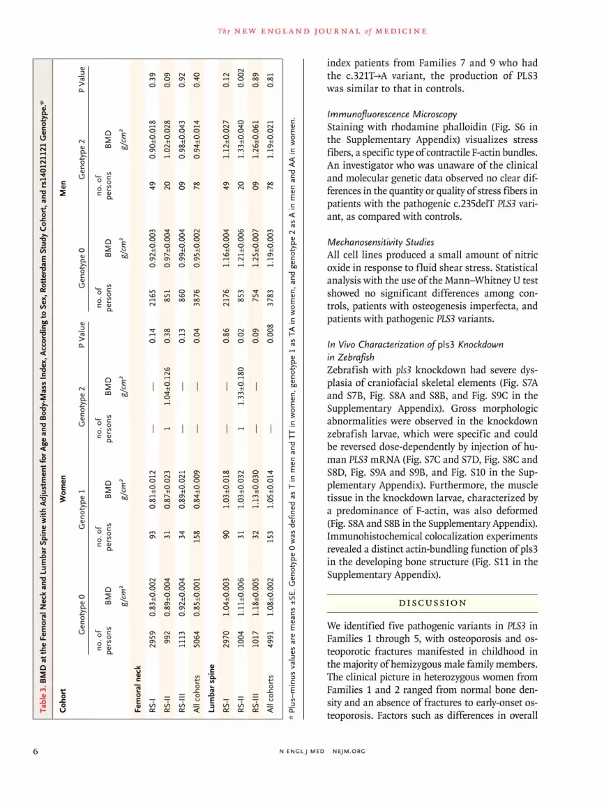
The minor allele frequencies of the rs140121121 SNP in men and women, respectively, in the RS-I, RS-II, and RS-III cohorts were 0.022 and 0.016, 0.024 and 0.017, and 0.012 and 0.016. To investigate the relationship of this variant with fracture risk, we performed sex-combined analyses for X-linked inheritance with adjustment for age and body-mass index but not sex, treating men as homo-zygous women.16
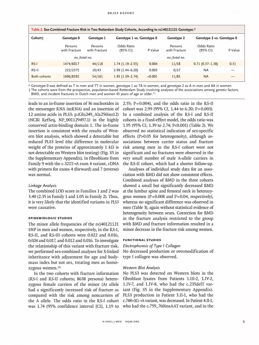
In the two cohorts with fracture information (RS-I and RS-II cohorts; 8638 persons) hetero-zygous female carriers of the minor (A) allele had a significantly increased risk of fracture as compared with the risk among noncarriers of the A allele. The odds ratio in the RS-I cohort was 1.74 (95% confidence interval [CI], 1.19 to
2.55; P = 0.004), and the odds ratio in the RS-II cohort was 2.99 (95% CI, 1.44 to 6.20; P = 0.003). In a combined analysis of the RS-I and RS-II cohorts in a fixed-effect model, the odds ratio was 1.95 (95% CI, 1.39 to 2.74; P<0.001) (Table 2). We observed no statistical indication of sex-specific effects (P>0.05 for heterogeneity), although as-sociations between carrier status and fracture risk among men in the RS-I cohort were not significant and no fractures were observed in the very small number of male A-allele carriers in the RS-II cohort, which had a shorter follow-up.
Analyses of individual study data for an asso-ciation with BMD did not show consistent effects. Combined analyses of BMD in the three cohorts showed a small but significantly decreased BMD at the lumbar spine and femoral neck in heterozy-gous women (P = 0.008 and P = 0.04, respectively), whereas no significant difference was observed in men (Table 3), again without statistical evidence of heterogeneity between sexes. Correction for BMD in the fracture analysis restricted to the group with BMD and fracture information resulted in a minor decrease in the fracture risk among women.
FUNCTIONAL STUDIES
Electrophoresis of Type I CollagenNo decreased production or overmodification of type I collagen was observed.
Western Blot AnalysisNo PLS3 was detected on Western blots in the fibroblast lysates from Patients 1.III-2, 1.IV-2, 1.IV-7, and 1.IV-8, who had the c.235delT var-iant (Fig. S5 in the Supplementary Appendix). PLS3 production in Patient 3.II-1, who had the c.748+1G→A variant, was decreased. In Patient 4.II-1, who had the c.759_760insAAT variant, and in the
Table 2. Sex-Combined Fracture Risk in Two Rotterdam Study Cohorts, According to rs140121121 Genotype.*
Cohort† Genotype 0 Genotype 1 Genotype 1 vs. Genotype 0 Genotype 2 Genotype 2 vs. Genotype 0
Persons with Fracture
Persons with Fracture
Odds Ratio (95% CI) P Value
Persons with Fracture
Odds Ratio (95% CI) P Value
no./total no. no./total no.
RS-I 1474/6017 44/118 1.74 (1.19–2.55) 0.004 11/58 0.71 (0.37–1.38) 0.31
RS-II 222/2375 10/43 2.99 (1.44–6.20) 0.003 0/27 NA —
Both cohorts 1696/8392 54/161 1.95 (1.39–2.74) <0.001 11/85 NA —
* Genotype 0 was defined as T in men and TT in women, genotype 1 as TA in women, and genotype 2 as A in men and AA in women.† The cohorts were from the prospective, population-based Rotterdam Study involving analyses of the associations among genetic factors,
BMD, and incident fractures in Dutch men and women 45 years of age or older.11
T h e n e w e ngl a nd j o u r na l o f m e dic i n e
n engl j med nejm.org6
index patients from Families 7 and 9 who had the c.321T→A variant, the production of PLS3 was similar to that in controls.
Immunofluorescence MicroscopyStaining with rhodamine phalloidin (Fig. S6 in the Supplementary Appendix) visualizes stress fibers, a specific type of contractile F-actin bundles. An investigator who was unaware of the clinical and molecular genetic data observed no clear dif-ferences in the quantity or quality of stress fibers in patients with the pathogenic c.235delT PLS3 vari-ant, as compared with controls.
Mechanosensitivity StudiesAll cell lines produced a small amount of nitric oxide in response to fluid shear stress. Statistical analysis with the use of the Mann–Whitney U test showed no significant differences among con-trols, patients with osteogenesis imperfecta, and patients with pathogenic PLS3 variants.
In Vivo Characterization of pls3 Knockdown in ZebrafishZebrafish with pls3 knockdown had severe dys-plasia of craniofacial skeletal elements (Fig. S7A and S7B, Fig. S8A and S8B, and Fig. S9C in the Supplementary Appendix). Gross morphologic abnormalities were observed in the knockdown zebrafish larvae, which were specific and could be reversed dose-dependently by injection of hu-man PLS3 mRNA (Fig. S7C and S7D, Fig. S8C and S8D, Fig. S9A and S9B, and Fig. S10 in the Sup-plementary Appendix). Furthermore, the muscle tissue in the knockdown larvae, characterized by a predominance of F-actin, was also deformed (Fig. S8A and S8B in the Supplementary Appendix). Immunohistochemical colocalization experiments revealed a distinct actin-bundling function of pls3 in the developing bone structure (Fig. S11 in the Supplementary Appendix).
DISCUSSION
We identified five pathogenic variants in PLS3 in Families 1 through 5, with osteoporosis and os-teoporotic fractures manifested in childhood in the majority of hemizygous male family members. The clinical picture in heterozygous women from Families 1 and 2 ranged from normal bone den-sity and an absence of fractures to early-onset os-teoporosis. Factors such as differences in overall Ta
ble
3. B
MD
at t
he F
emor
al N
eck
and
Lum
bar
Spin
e w
ith A
djus
tmen
t for
Age
and
Bod
y-M
ass
Inde
x, A
ccor
ding
to S
ex, R
otte
rdam
Stu
dy C
ohor
t, an
d rs
1401
2112
1 G
enot
ype.
*
Coh
ort
Wom
enM
en
Gen
otyp
e 0
Gen
otyp
e 1
Gen
otyp
e 2
P V
alue
Gen
otyp
e 0
Gen
otyp
e 2
P V
alue
no. o
f pe
rson
sB
MD
no. o
f pe
rson
sB
MD
no. o
f pe
rson
sB
MD
no. o
f pe
rson
sB
MD
no. o
f pe
rson
sB
MD
g/cm
2g/
cm2
g/cm
2g/
cm2
g/cm
2
Fem
oral
nec
k
RS-
I29
590.
83±0
.002
930.
81±0
.012
——
0.14
2165
0.92
±0.0
0349
0.90
±0.0
180.
39
RS-
II99
20.
89±0
.004
310.
87±0
.023
11.
04±0
.126
0.38
851
0.97
±0.0
0420
1.02
±0.0
280.
09
RS-
III
1113
0.92
±0.0
0434
0.89
±0.0
21—
—0.
1386
00.
99±0
.004
090.
98±0
.043
0.92
All
coho
rts
5064
0.85
±0.0
0115
80.
84±0
.009
——
0.04
3876
0.95
±0.0
0278
0.94
±0.0
140.
40
Lum
bar
spin
e
RS-
I29
701.
04±0
.003
901.
03±0
.018
——
0.86
2176
1.16
±0.0
0449
1.12
±0.0
270.
12
RS-
II10
041.
11±0
.006
311.
03±0
.032
11.
33±0
.180
0.02
853
1.21
±0.0
0620
1.33
±0.0
400.
002
RS-
III
1017
1.18
±0.0
0532
1.13
±0.0
30—
—0.
0975
41.
25±0
.007
091.
26±0
.061
0.89
All
coho
rts
4991
1.08
±0.0
0215
31.
05±0
.014
—0.
008
3783
1.19
±0.0
0378
1.19
±0.0
210.
81
* Pl
us–m
inus
val
ues
are
mea
ns ±
SE. G
enot
ype
0 w
as d
efin
ed a
s T
in m
en a
nd T
T in
wom
en, g
enot
ype
1 as
TA
in w
omen
, and
gen
otyp
e 2
as A
in m
en a
nd A
A in
wom
en.
brief report
n engl j med nejm.org 7
and local X-chromosome inactivation, postmeno-pausal status, and immobility could play a role.
In addition, we identified a rare variant in PLS3, c.321T→A in exon 4 (SNP rs140121121) in Fam-ilies 6 through 10. The prevalence of this vari-ant was significantly increased in our group of 95 male patients without COL1A1 or COL1A2 mutations who had been referred for diagnosis or ruling out of osteogenesis imperfecta type I. The clinical symptoms of patients in Families 6 through 10 were generally less severe and had a later onset (absent in one case) than those in Fam-ilies 1 through 5 with loss-of-function variants in PLS3. We hypothesized that the rs140121121 SNP may be associated with fractures, decreased BMD, or both in the general population.
A combined analysis of two cohorts (RS-I and RS-II) of 8638 elderly Dutch persons showed that heterozygous women had an increased odds of fracture of 1.95 (95% CI, 1.39 to 2.74) and that the SNP was significantly associated with decreased BMD. However, the association with fracture risk was not fully explained by BMD, which suggests that other factors leading to decreased bone strength may be involved. Associations in hemi-zygous men were not significant, a finding that may be due to the small size of this group or may indicate that additional (possibly genetic) factors play a role. The associations of the SNP with frac-tures and BMD in the general population need to be replicated in larger cohorts worldwide.
Our findings indicate that PLS3 has bone-regulatory properties. Overexpression of PLS3 has been reported to act as a protective modifier of spinal muscular atrophy, facilitating axonal growth and presynaptic F-actin–dependent processes at the neuromuscular junction.17,18 A knockdown of pls3 in zebrafish was used in an investigation of motor axon development.17 Since no other animal models were available, we used this model17 to analyze the role of PLS3 in skeletal development. Malformations of develop-ing craniofacial bone structure, body axis, and tail were present and could be reversed dose-dependently by the administration of human PLS3 mRNA. Muscles that contained F-actin ap-peared to be deformed as well, which is notable because the formation of pharyngeal cartilage and the formation of muscle occur simultane-ously.19 Immunohistochemical colocalization ex-periments confirmed a distinct actin-bundling function of pls3 in developing bone structure.
Taken together, the in vivo data suggest that PLS3 may be a regulator of bone development.
The exact mechanism by which PLS3 mutations cause osteoporosis and fractures is unknown. Fim-brin, the chicken homologue of PLS3,20 is abundant in osteocyte dendrites.21-23 These dendrites are im-portant for mechanosensing (converting mechan-ical signals into intracellular bio chemical signals to osteoblasts and osteoclasts).24 The loss of sensor-cell mechanosensitivity has been proposed as a cause of osteoporosis.25 We hypothesize that PLS3 mutations lead to decreased mechanosens-ing of osteocytes, with subsequent dysregulation of bone modeling or remodeling, which results in osteoporosis and fractures. Bone tissue from pa-tients with PLS3 mutations will be needed for in-vestigation of mechanosensing in osteocytes.
In conclusion, we identified loss-of-function variants in PLS3 as a monogenetic cause of X-linked osteoporosis and osteoporotic fractures. We pro-pose diagnostic analysis of PLS3 in boys and men who have clinical or radiologic signs of an inherited bone disorder with low BMD and frac-tures, early-onset osteoporosis, or a presumptive diagnosis of osteogenesis imperfecta type I with-out COL1A1 or COL1A2 mutations. Among elderly study participants, we identified a rare PLS3 vari-ant, which was associated with decreased BMD and a risk of fracture among heterozygous women that was two times as high as that among noncarriers, indicating genetic variation in PLS3 as a novel factor involved in common, multifactorial osteoporosis.
Supported by grants to the Rotterdam Study from Erasmus Medical Center and Erasmus University, Rotterdam; the Nether-lands Organization of Scientific Research (NWO) Investments (175.010.2005.011 and 911-03-012); the Research Institute for Dis-eases in the Elderly (014-93-015 and RIDE2); the Ministry of Educa-tion, Culture, and Science; the Ministry for Health, Welfare, and Sports; Directorate-General XII of the European Commission; the Netherlands Genomics Initiative and NWO Project (050-060-810); the Netherlands Consortium of Healthy Aging; and the Municipal-ity of Rotterdam. The zebrafish studies were supported by grants from the European Union Program FP7 Program (2012-305121), Deutsche Forschungsgemeinschaft (Wi945-14/1, to Dr. Wirth), and SMA-Europe (to Dr. Riessland). The col1a1:eGFP transgenic line was a gift from Dr. Shannon Fisher to Dr. Hammerschmidt’s laboratory.
Disclosure forms provided by the authors are available with the full text of this article at NEJM.org.
We thank the participants and the research center staff in the Rotterdam study; the contributors of the fracture data set, Mr. Pas-cal Arp for genotyping rs140121121, Dr. Fernando Rivadeneira for advice and help with the statistical analyses in the Rotterdam study, Mr. Seyyed Mohsen Hosseini-Barkooie for generating the PLS3-RNA expression vector, Dr. Birgit de Witte for help with the sta-tistical analyses of the mechanosensitivity experiments, Dr. Jan M. Cobben for assistance with the linkage analysis, and Dr. René J.P. Musters for advice on the immunofluorescence experiments.

n engl j med nejm.org8
brief report
1. Kanis JA, Melton LJ III, Christiansen C, Johnston CC, Khaltaev N. The diagno-sis of osteoporosis. J Bone Miner Res 1994; 9:1137-41.2. Laine CM, Koltin D, Susic M, et al. Primary osteoporosis without features of OI in children and adolescents: clinical and genetic characteristics. Am J Med Genet A 2012;158A:1252-61.3. Hartikka H, Mäkitie O, Männikkö M, et al. Heterozygous mutations in the LDL receptor-related protein 5 (LRP5) gene are associated with primary osteoporosis in children. J Bone Miner Res 2005;20:783-9.4. Estrada K, Styrkarsdottir U, Evange-lou E, et al. Genome-wide meta-analysis identifies 56 bone mineral density loci and reveals 14 loci associated with risk of fracture. Nat Genet 2012;44:491-501.5. Laine CM, Joeng KS, Campeau PM, et al. WNT1 mutations in early-onset osteoporo-sis and osteogenesis imperfecta. N Engl J Med 2013;368:1809-16.6. Keupp K, Beleggia F, Kayserili H, et al. Mutations in WNT1 cause different forms of bone fragility. Am J Hum Genet 2013; 92:565-74.7. van Dijk FS, Dalgleish R, Malfait F, et al. Clinical utility gene card for: osteogene-sis imperfecta. Eur J Hum Genet 2013;21: 698-9.8. Sillence DO. Bone dysplasia: genetic and ultrastructural aspects with special reference to osteogenesis imperfecta. Ann Arbor, MI: University Microfilms, 1980.9. Ameziane N, Sie D, Dentro S, et al. Diagnosis of Fanconi anemia: mutation analysis by next-generation sequencing. Anemia 2012;2012:132856.
10. Clarke L, Zheng-Bradley X, Smith R, et al. The 1000 Genomes Project: data management and community access. Nat Methods 2012;9:459-62.11. Hofman A, van Duijn CM, Franco OH, et al. The Rotterdam Study: 2012 objec-tives and design update. Eur J Epidemiol 2011;26:657-86.12. Delanote V, Vandekerckhove J, Gette-mans J. Plastins: versatile modulators of actin organization in (patho)physiologi-cal cellular processes. Acta Pharmacol Sin 2005;26:769-79.13. Bacabac RG, Smit TH, Van Loon JJWA, Doulabi BZ, Helder M, Klein-Nulend J. Bone cell responses to high-frequency vi-bration stress: does the nucleus oscillate within the cytoplasm? FASEB J 2006;20: 858-64.14. van der Pauw MTM, Klein-Nulend J, van den Bos T, Burger EH, Everts V, Beertsen W. Response of periodontal ligament fibroblasts and gingival fibro-blasts to pulsating f luid f low: nitric ox-ide and prostaglandin E2 release and expression of tissue non-specific alka-line phosphatase activity. J Periodontal Res 2000;35:335-43.15. Kague E, Gallagher M, Burke S, Par-sons M, Franz-Odendaal T, Fisher S. Skel-etogenic fate of zebrafish cranial and trunk neural crest. PLoS One 2012;7(11):e47394. 16. Clayton D. Testing for association on the X chromosome. Biostatistics 2008;9: 593-600.17. Oprea GE, Kröber S, McWhorter ML, et al. Plastin 3 is a protective modifier of autosomal recessive spinal muscular atro-phy. Science 2008;320:524-7.
18. Ackermann B, Kröber S, Torres-Benito L, et al. Plastin 3 ameliorates spinal mus-cular atrophy via delayed axon pruning and improves neuromuscular junction function-ality. Hum Mol Genet 2013;22:1328-47.19. Shwartz Y, Farkas Z, Stern T, Aszódi A, Zelzer E. Muscle contraction controls skeletal morphogenesis through regula-tion of chondrocyte convergent extension. Dev Biol 2012;370:154-63.20. de Arruda MV, Watson S, Lin CS, Leavitt J, Matsudaira P. Fimbrin is a homo-logue of the cytoplasmic phosphoprotein plastin and has domains homologous with calmodulin and actin gelation pro-teins. J Cell Biol 1990;111:1069-79.21. Bonewald LF. The amazing osteocyte. J Bone Miner Res 2011;26:229-38.22. Tanaka-Kamioka K, Kamioka H, Ris H, Lim SS. Osteocyte shape is dependent on actin filaments and osteocyte processes are unique actin-rich projections. J Bone Miner Res 1998;13:1555-68.23. Kamioka H, Sugawara Y, Honjo T, Yamashiro T, Takano-Yamamoto T. Termi-nal differentiation of osteoblasts to osteo-cytes is accompanied by dramatic changes in the distribution of actin-binding pro-teins. J Bone Miner Res 2004;19:471-8.24. Weinbaum S, Duan Y, Thi MM, You L. An integrative review of mechanotrans-duction in endothelial, epithelial (renal) and dendritic cells (osteocytes). Cell Mol Bioeng 2011;4:510-37.25. Mulvihill BM, Prendergast PJ. Mecha-nobiological regulation of the remodelling cycle in trabecular bone and possible bio-mechanical pathways for osteoporosis. Clin Biomech (Bristol, Avon) 2010;25:491-8.Copyright © 2013 Massachusetts Medical Society.
APPENDIXThe authors’ affiliations are as follows: the Center for Connective Tissue Disorders, Department of Clinical Genetics, VU University Medical Center, Amsterdam (F.S.D., D.M., Y.M., J.E., R.B., A.M., E.A.S., Q.W., H.M.-H., G.P.), the Departments of Internal Medicine (M.C.Z., N.C.-O., A.J.M.H.V., A.G.U.), Epidemiology (A.H., A.G.U.), and Clinical Genetics (M.E.H.S.), Erasmus Medical Center, Rot-terdam, the Department of Genetics, Radboud University Nijmegen Medical Center, Nijmegen (C.L.M.M.), the Departments of Clinical Genetics (C.E.D.-S.), Pediatrics (M.E.R.-G.), and Pediatrics and Laboratory Genetic Metabolic Diseases (M.E.R.-G.), Maastricht Univer-sity Medical Centre, Maastricht, the Departments of Internal Medicine (A.A.F.) and Orthopedics (A.J.H.), Isala Clinics Zwolle, Zwolle, the Department of Medical Genetics, University Medical Center Utrecht, Utrecht (K.D.L.), the Department of Pediatric Orthopedics University Medical Center Utrecht, Wilhelmina Children’s Hospital, Utrecht (H.E.P.), the Department of Internal Medicine, Westfries Gasthuis, Hoorn (R.Z.), the Department of Oral Cell Biology, Academic Center for Dentistry Amsterdam, University of Amsterdam and VU University Amsterdam, MOVE Research Institute Amsterdam, Amsterdam (C.M.S., A.D.B., V.E., J.K.-N.), and the Department of Genetics, University Medical Center Groningen, University of Groningen (G.J.M.) — all in the Netherlands; and the Institute of Human Genetics, Institute for Genetics and Center for Molecular Medicine Cologne (M.R., J.M., B.W.), and the Department of Developmental Biology (M.H.), University of Cologne — both in Cologne, Germany.
References