Proximal Humerus Fractures - Orthopaedic Trauma Association
51
Core Curriculum V5 Proximal Humerus Fractures Kevin J Perry, MD DPT Associate Professor Orthopaedic Trauma LSU Health Shreveport
-
Upload
khangminh22 -
Category
Documents
-
view
1 -
download
0
Transcript of Proximal Humerus Fractures - Orthopaedic Trauma Association

Core Curriculum V5
Proximal Humerus FracturesKevin J Perry, MD DPT
Associate ProfessorOrthopaedic Trauma
LSU Health Shreveport

Core Curriculum V5
Disclosures
• None

Core Curriculum V5
Objectives
• Review the principles of diagnosis and management of proximal humerus fractures
• Review fracture classification schemes• Review decision making and treatment options• Review outcomes and evidence• Review available resources for further education pertaining to
proximal humerus fractures
Core Curriculum V5
Proximal Humerus Fractures
• Defined as fractures occurring at or proximal to the surgical neck
Presenter
Presentation Notes
Image: Rockwood and Green Fx in Adults LWW Publications

Core Curriculum V5
Epidemiology
• Females > Males• Bimodal distribution – young males, older females• Incidence increases with age
• As population ages the incidence of proximal humerus fractures is expected to increase
• Highest risk in white females• Osteoporosis related fracture
• 3rd most common nonvertebral osteoporotic fracture

Core Curriculum V5
Risk Factors
• Other risk factors:• Poor vision, Hearing aids, Diabetes, Depression, Alcohol consumption,
Anticonvulsant medications, Maternal history of hip fracture, Personal history of spinal or extremity fracture
• Protective Factors:• Hormonal therapy, Calcium intake

Core Curriculum V5
Mechanisms of Injury
• Ground level fall• Vast majority
• High energy trauma in younger population• 3 Main Loading Modes:
• Compressive – Humeral head impacts at glenoid• Bending – Angular forces at surgical neck• Tension – Rotator cuff pulling on greater and less tuberosities
• Fall on outstretched hand• Valgus impacted proximal humerus fracture
• Fall directly onto lateral shoulder• Varus deformity with posterior rotational deformity

Core Curriculum V5
Associated Injuries
• Majority are isolated low energy injuries• Other MSK injuries:
• Ipsilateral distal radius• Hip fracture• Pelvic fracture• Head injury / Subdural hematoma• Nerve palsy – Suprascapular, Axillary, Musculocutaneous or Brachial plexus
palsy possible• Vascular injury – Fracture dislocations at risk for axillary artery/vein injury

Core Curriculum V5
Clinical Presentation
• Shoulder pain worse with motion• Immobility• Ecchymosis• Soft tissue swelling• Open fractures may occur in axilla but are rare
• Usually occur at lateral aspect of axilla as pec major displaces shaft medially

Core Curriculum V5
Anatomy/Deforming Forces/Parts
• Parts:• Head• Greater tuberosity• Lesser tuberosity• Shaft
Presenter
Presentation Notes
Image: Rockwood and Green Fractures in Adults LWW Publication

Core Curriculum V5
Anatomy/Deforming Forces/Parts
Presenter
Presentation Notes
Image: Rockwood and Green Fractures in Adults LWW Publication

Core Curriculum V5
Anatomy/Deforming Forces/Parts
• Deforming Forces• Supraspinatus/Infraspinatus
• Displaces greater tuberosity superiorly and posteriorly
• Subscapularis• Displaces lesser tuberosity medially
• Pectoralis major• Displaces humeral shaft medially and anteriorly
• Deltoid• Displaces humeral shaft proximally
Presenter
Presentation Notes
Image: Rockwood and Green Fractures in Adults LWW Publication

Core Curriculum V5
Imaging
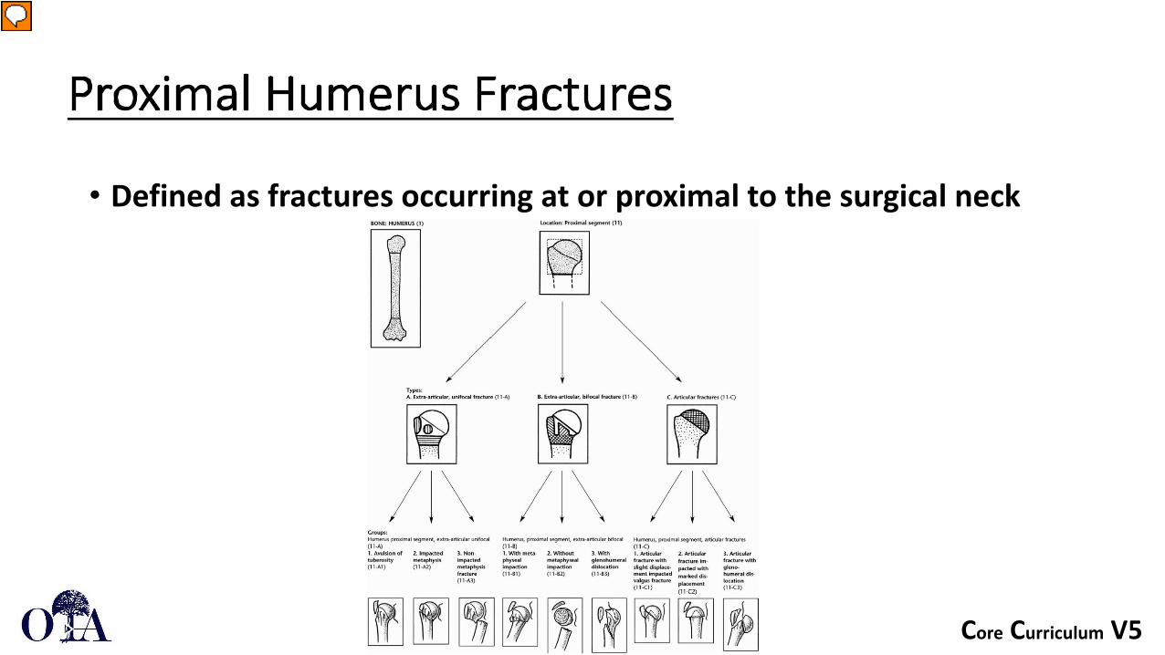
• Radiographs• Standard
• Grashey (True AP) view• Neer (Scapular Y) view• Axillary lateral view
• Additional• Velpeau view• Traction view
• Computed Tomography (CT) • Magnetic Resonance Imaging (MRI) • Ultrasound
Presenter
Presentation Notes
Image: Rockwood and Green Fractures in Adults LWW Publication

Core Curriculum V5
Imaging
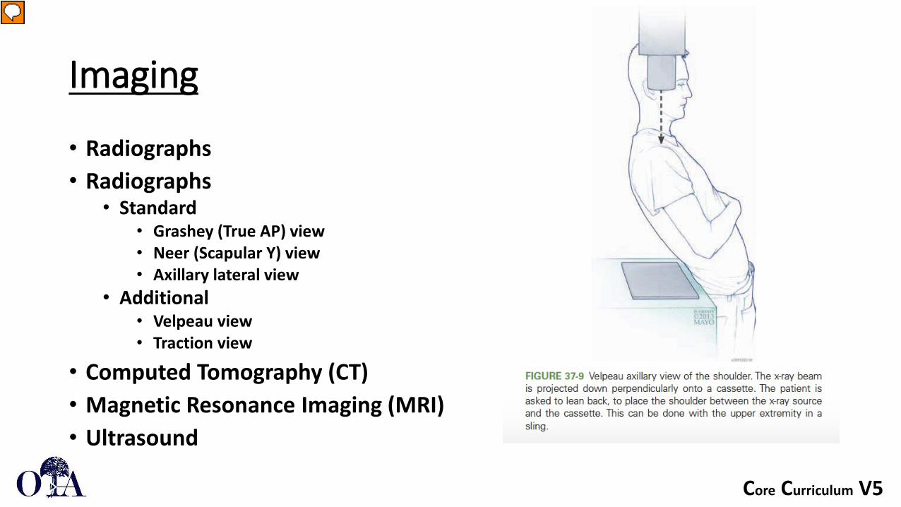
• Radiographs• Radiographs
• Standard• Grashey (True AP) view• Neer (Scapular Y) view• Axillary lateral view
• Additional• Velpeau view• Traction view
• Computed Tomography (CT) • Magnetic Resonance Imaging (MRI) • Ultrasound
Presenter
Presentation Notes
Image: Rockwood and Green Fractures in Adults LWW Publication
Core Curriculum V5
Imaging
• Radiographs• Radiographs
• Standard• Grashey (True AP) view• Neer (Scapular Y) view• Axillary lateral view
• Additional• Velpeau view• Traction view
• Computed Tomography (CT) • Magnetic Resonance Imaging (MRI) • Ultrasound
Presenter
Presentation Notes
Image: Rockwood and Green Fractures in Adults LWW Publication
Core Curriculum V5
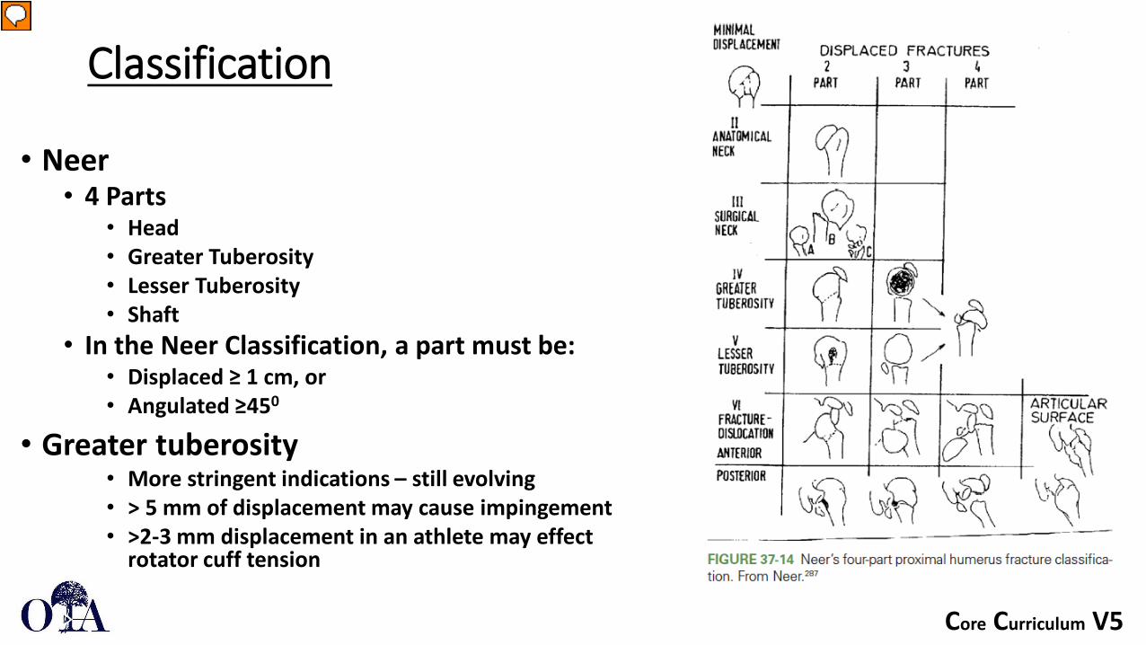
Classification
• Neer• 4 Parts
• Head• Greater Tuberosity• Lesser Tuberosity• Shaft
• In the Neer Classification, a part must be:• Displaced ≥ 1 cm, or • Angulated ≥450
• Greater tuberosity• More stringent indications – still evolving• > 5 mm of displacement may cause impingement• >2-3 mm displacement in an athlete may effect
rotator cuff tension
Presenter
Presentation Notes
Image: Rockwood and Green Fractures in Adults LWW Publication

Core Curriculum V5
Classification
• AO/OTA• Bone = 1 • Segment = 1• Pattern
• A = Extraarticular unifocal• B = Extraarticular bifocal• C = Intraarticular
Presenter
Presentation Notes
Image: Rockwood and Green Fractures in Adults LWW Publication

Core Curriculum V5
Classification• Greater tuberosity fragment surgical indications have evolved
• <3mm displacement in overhead athletes• <5mm displacement in healthy adults
Presenter
Presentation Notes
Images: Mutch JAJ, Rouleau DM, Laflamme, GY, Hagemeister N. Accurate Measurement of Greater Tuberosity Displacement Without Computed Tomorgraphy: Validation of a Method on Plain Radiography to Guide Surgical Treatment. Journal of Orthopaedic Trauma 2014;28:445-451.

Core Curriculum V5
Blood Supply to Humeral Head / AVN
• Vascular supply to humeral head• Arcuate artery is the terminal supply to the humeral head from the anterior
humeral circumflex artery• Disrupted with anatomic neck fractures
• Posterior humeral circumflex artery supplies posteromedial metaphysis of humerus
• Less likely to be injured at time of fracture displacement
• Predictors of Humeral Head AVN (Hertel’s Criteria)• Distal metaphyseal extension <8 mm • Disruption of medial hinge at level of calcar (Medial displacement of shaft)• Fracture through the anatomic neck

Core Curriculum V5
Blood Supply to Humeral Head
Presenter
Presentation Notes
Image: Rockwood and Green Fractures in Adults LWW Publication
Core Curriculum V5
Hertel’s Criteria
• Recently called into question• Original study used intraoperative doppler flowmetry as well as visual
bleeding from drill holes in the humeral head to determine vascular supply• A lack of return of bleeding from drill holes was associated with AVN
• Campochiaro et al 2015• Series of patients assessed for AVN after proximal humerus fx• Hertel’s criteria were less predictive of AVN, whereas poor reduction was
highly predictive.

Core Curriculum V5
Treatment Options
•Nonoperative – majority (80%)• Operative
• Suture fixation• Arthroscopic assisted repair• Closed reduction and percutaneous pinning• Open reduction internal fixation• Intramedullary nail • Arthroplasty
Core Curriculum V5
Nonoperative Treatment• Indications for nonoperative management:
• Older age• Lower demand• Unfit for surgery• Stable nondisplaced or minimally displaced patterns• Valgus impacted 2 or 3 part fractures
• Sling• Sling and swathe• Sling with abduction pillow• Shoulder immobilizer
• Early active-assisted motion including pendulums may prevent stiffness
Core Curriculum V5
The PROFHER Randomized Clinical Trial
• JAMA 2015• 1250 patients with proximal humerus fractures• 250 patients met surgical indications and were randomized to
operative vs nonoperative treatment• No difference in outcomes at 2 years follow up
• Controversy regarding groups and treatment conversion• 87 had “clear indication for surgery” and were not included in study• 16/125 were randomized to surgery and did not receive surgery• 66 surgeons involved
• Regardless, supports nonoperative management in select patients

Core Curriculum V5
Treatment Options
• Nonoperative
•Operative• Suture fixation• Arthroscopic assisted repair• Closed reduction and percutaneous pinning• Open reduction internal fixation• Intramedullary nail • Arthroplasty

Core Curriculum V5
Positioning
• Beach chair• Semi-supine
Presenter
Presentation Notes
Image: Rockwood and Green Fractures in Adults LWW Publishing
Core Curriculum V5
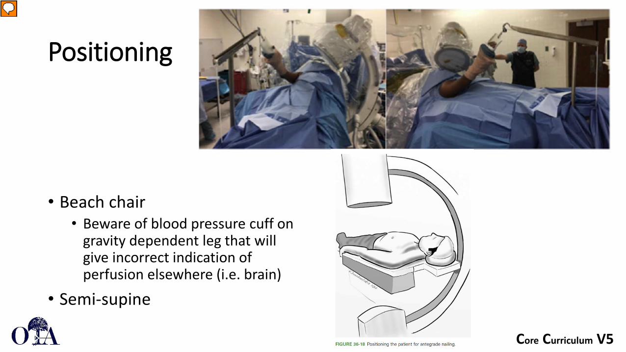
Positioning
• Beach chair• Beware of blood pressure cuff on
gravity dependent leg that will give incorrect indication of perfusion elsewhere (i.e. brain)
• Semi-supine
Presenter
Presentation Notes
Image: Top Image: Hightower C, Mobley K, Jones T. Semisupine Positioning for Proximal Humerus ORIF Using Arthroscopic shoulder distractor. JOT 2020;34:e148-e152. Figure 36 Rockwood and Green Fractures in Adults LWW Publishing
Core Curriculum V5
Approaches
• Deltopectoral• Can visualize the joint for head split fractures with lesser
tuberosity peel vs osteotomy• Extensile
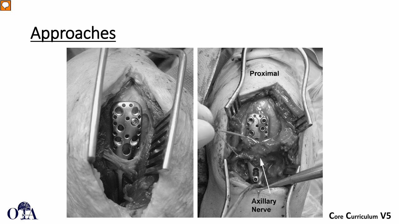
• Deltoid Splitting• Easier plate placement laterally• Axillary nerve protection (5-7 cm inferior to acromion)• Less retraction and positioning needed for lateral plate
placement• Can be extensile if you dissect and protect axillary nerve

Core Curriculum V5
Approaches
Presenter
Presentation Notes
Images: Rockwood and Green Fractures in Adults LWW Publishing

Core Curriculum V5
Approaches
Presenter
Presentation Notes
Image: Cherney SM, Murphy RA, Achor TS, Choo AM. Subscapularis Peel for Open Reduction and Internal Fixation of Proximal Humerus Fractures with a Head Split. JOT 2018; 32;12:E487-91.

Core Curriculum V5
Approaches
Presenter
Presentation Notes
Images: Gardner MJ, Boraiah S, Helfet DL, Lorich DG. The Anterolateral Acromial Approach for Fractures of the Proximal Humerus. JOT 2008;22:132-137.
Core Curriculum V5
Approaches
Presenter
Presentation Notes
Images: Gardner MJ, Boraiah S, Helfet DL, Lorich DG. The Anterolateral Acromial Approach for Fractures of the Proximal Humerus. JOT 2008;22:132-137.

Core Curriculum V5
Reduction Techniques
• Closed reduction usually accomplished with arm abducted and externally rotated
• Match the location of the proximal fragment with the distal segment
• If adequate reduction can be obtained by closed means, percutaneous fixation can follow with K wires, percutaneous screws, or retrograde intramedullary nails

Core Curriculum V5
Reduction Techniques
• Work through greater tuberosity fracture line to manipulate head fragment
• K-wire joysticks• Elevator as broad surface to manipulate head fragment• K-wire provisional fixation to shaft fragment
Presenter
Presentation Notes
Images: Rockwood and Green Fractures in Adults LWW Publishing

Core Curriculum V5
Reduction Techniques
• Reduce the medial hinge with the head fragment in valgus• Common deformity
• Apply lateral plate to translate greater tuberosity fragment and improve valgus reduction
Presenter
Presentation Notes
Intramedullary Cage Fixation for Proximal Humerus Fractures Has Low Reoperation Rates at 1 Year: Results of a Multicenter Study Journal of Orthopaedic Trauma34(4):193-198, April 2020.

Core Curriculum V5
Reduction Techniques
• Sutures in rotator cuff insertions can be helpful to manipulate greater and lesser tuberosity fracture fragments
• Sutures can be incorporated into plate and may supplement fixation
• Unstable head fragment can be pinned to glenoid with K wires to provide provisional stability
Presenter
Presentation Notes
Image: Park S, Ko, Y. Medial Buttress Plating for Humerus Fractures With Unstable Medial Column. Journal of Orthopaedic Trauma 2019;33:e352-359.

Core Curriculum V5
Presenter
Presentation Notes
Images: Left image: Wright JO, Ho A, Kalma J, Koueiter D, Esterle J, Marcantonio D, Wiater JM, Wiater B. Uncemented Reverse Total Shoulder Arthroplasty as Initial Treatment for Comminuted Proximal Humerus Fractures. JOT 2019;33;7:e263-9. Hertel Egg Shell - Rockwood and Green Fractures in Adults LWW Publication
Core Curriculum V5
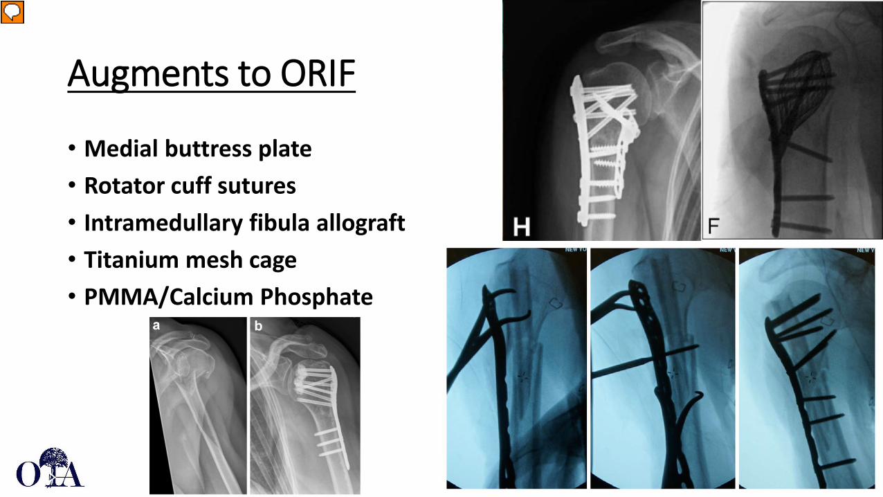
Augments to ORIF
• Medial buttress plate• Rotator cuff sutures• Intramedullary fibula allograft• Titanium mesh cage• PMMA/Calcium Phosphate
Presenter
Presentation Notes
Images: H: Park S, Ko, Y. Medial Buttress Plating for Humerus Fractures With Unstable Medial Column. Journal of Orthopaedic Trauma 2019;33:e352-359. F: Goodnough, LH et al. Intramedullary Cage Fixation for Proximal Humerus Fractures Has Low Reoperation Rates at 1 Year: Results of a Multicenter Study. Journal of Orthopaedic Trauma 2020;34:193-198. Berkes MB et al. Intramedullary Allograft Fibula as a Reduction and Fixation Tool for Treatment of Complex Proximal Humerus Fractures with Diaphyseal Extension. JOT 2014;28:e56-e64. AB: Knierzinger D, Crepaz-Eger U, Hengg C, Kralinger F. Does cement augmentation of the screws in angular stable plating for proximal humerus fractures influence the radiological outcome: a retrospective assessment. Archives of Orthopaedic and Trauma Surgery 2020; 140:1413-1421.
Core Curriculum V5
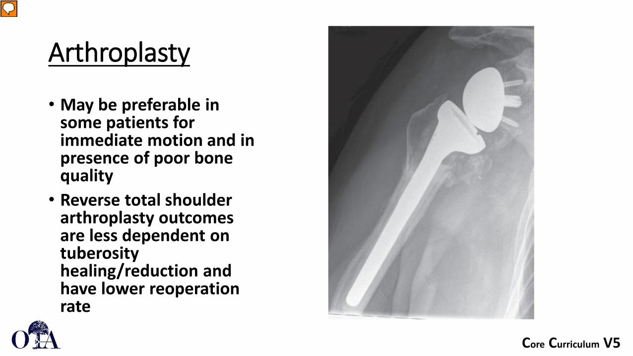
Arthroplasty
• May be preferable in some patients for immediate motion and in presence of poor bone quality
• Reverse total shoulder arthroplasty outcomes are less dependent on tuberosity healing/reduction and have lower reoperation rate
Presenter
Presentation Notes
Images: Rockwood and Green Fractures in Adults LWW Publication
Core Curriculum V5
Decision Making
• Age, functional status, bone quality, and fracture pattern can all be used to guide surgical decision making and implant selection
• Younger patients with good bone quality and a viable humeral head are ideally treated with ORIF
• Older frail patients with lower functional requirements are ideally treated with arthroplasty
Presenter
Presentation Notes

Core Curriculum V5
Presenter
Presentation Notes
Adeyamo A, Bertha N, Perry KJ, Updegrove G. Implant Selection for Proximal Humerus Fractures. Orthop Clinics 2021;52:167-175. Image produced by author and published elsewhere. Should have rights to reproduce as it is my own work.

Core Curriculum V5
Complications
• AVN• Impingement• Screw cut out• Loss of reduction
• Medial support, calcar screw placement• Combined cortical thickness• Deltoid tuberosity index
• Nonunion• Risk factors: Smoking, Alcohol abuse, Osteoporosis, Inflammatory arthropathy
• Malunion• Tuberosity malunion can cause symptoms of rotator cuff weakness or impingement
• Stiffness • Infection

Core Curriculum V5
Complications
• Impingement• Avoid with ideal plate placement just lateral to bicipital groove and 5-8 mm
distal to greater tuberosity• Tx: Hardware removal after union if symptomatic
• Screw cut out• Avoid screws in superior head• Advance screws to subchondral bone without penetrating articular surface
for ideal purchase• Pre-op CT thickness of central humeral head is predictive of screw cut out
• > 25 mm thickness is protective, <15 mm thickness predictive of screw cut out• Tx: May remove prominent screws without removal of all hardware if
fracture unites in acceptable position
Presenter
Presentation Notes
Stern L, Gorczyca MT, Gorczyca JT. Preoperative measurement of the thickness of the center of the humeral head predicts screw cutout after locked plating of proximal humerus fractures. JSES 2021;30:80-88.

Core Curriculum V5
Complications
• Risk factors for loss of reduction• Older age• Osteoporosis• Varus displacement• Medial comminution• Inadequate reduction• Insufficient medial support
Presenter
Presentation Notes
Images: Jung SW et al. Factors that Influence Reduction Loss in Proximal Humerus Fracture Surgery. JOT 2015;29:276-282.

Core Curriculum V5
Complications
• Malunion• Not all malunions are symptomatic• Tuberosity malunion may cause rotator cuff dysfunction• Symptomatic malunions are often salvaged with arthroplasty
• Nonunion• Tx: Repair of nonunion vs arthroplasty
• AVN• Minor humeral head collapse is often tolerated with minimal symptoms• Symptomatic AVN often salvaged with arthroplasty

Core Curriculum V5
More Resources:
• OTA Videos• OTA Techniques and Procedures: Proximal Humerus
• OTA Online Education Resources• OTA Online Education

Core Curriculum V5
References• Reuther F, Peterman M, Stangl R. Reverse Shoulder Arthroplasty in Acute Fractures of the Proximal Humerus: Does Tuberosity Healing Improve Clinical Outcomes? J
Orthop Trauma 2019;33:e46-e51.
• Mutch JAJ, Rouleau DM, Laflamme G, Hagemeister N. Accurate Measurement of Greater Tuberosity Displacement Without Computed Tomography: Validation of a Method on Plain Radiography to Guide Surgical Treatment. J Orthop Trauma 2014;28:445-451.
• Reid JS. Fractures of the proximal humerus. Current Opinion in Orthopaedics 2003;14:269-280.
• Gupta AK, Harris JD, Erickson BJ, Abrams GD, Bruce B, McCormick F, Nicholson GP, Romeo AA. Surgical Management of Complex Proximal Humerus Fractures – A Systematic Review of 92 Studies Including 4500 Patients. J Orthop Trauma 2015;29:54-59.
• Iyengar JJ, Devcic BS, Sproud RC, Feeley BT. Nonoperative Treatment of Proximal Humerus Fractures: A Systematic Review. J Orthop Trauma 2011;25:612-617.
• Merendez ME, Ring D. A Comparison of the Charlson and Elixhauser Comorbidity Measures to Predict Inpatient Mortality After Proximal Humerus Fracture. J Orthop Trauma 2015;29:488-493.
• Wu C, Ma C, Yeh J, Yen C, Yu S, Tu Y. Locked Plating for Proximal Humerus Fractures: Differences Between the Deltopectoral and Deltoid Splitting Approaches. J Trauma 2011;71:1364-1370.
• Court-Brown, CM, McQueen MM. Nonunions of the Proximal Humerus: Their Prevalence and Functional Outcome. J Trauma 2008;64:1517-1521.
• Lescheid J, Zdero R, Shah S, Kuzyk PR, Schemitsch EH. The Biomechanics of Locked Plating for Repairing Proximal Humerus Fractures With or Without Medical Cortical Support. J Trauma 2020;69:1235-1242.
• Goodnough LH, Campbell ST, Githens TC, DeBaun MR, Bishop JA, Gardner MJ. Intramedullary Cage Fixation for Proximal Humerus Fractures Has Low Reoperation Rates at 1 Year: Results of a Multicenter Study. J Orthop Trauma;2020:193-198.
• Shah KN, Sobel AD, Paxton S. Fixation of a Proximal Humerus Fracture Using a Polyaxial Locking Plate and Endosteal Fibular Strut. J Orthop Trauma 2018;32:S8-S9.
• Gardner MJ. Proximal Humerus Fracture Plating Through the Extended Anterolateral Approach. J Orthop Trauma 2016;30:S11-12.

Core Curriculum V5
References• Cherney SM, Murphy RA, Achor TS, Choo AM. Subscapularis Peel for Open Reduction and Internal Fixation of Proximal Humerus Fractures With a
Head Split. J Orthop Trauma 2018;2018;32:e487-e491.
• Park S, Ko Y. Medial Buttress Plating for Humerus Fractures With Unstable Medial Column. J Orthop Trauma 2019;33:e352-359.
• Walker R, Casteneda P, Putnam JG, Schemitsch EH, McKee MD. A Biomechanical Study of Tuberosity-Based Locked Plate Fixation Compared with Standard Proximal Humeral Locking Plate Fixation for 3-Part Proximal Humeral Fractures. J Orthop Trauma 2020;34:e233-e238.
• Belayneh R, Lott A, Haglin J, Konda S, Zuckerman JD, Egol KA. Osteonecrosis After Surgically Repaired Proximal Humerus Fractures Is a Predictor of Poor Outcomes. J Orthop Trauma 2018;32:e387-e393.
• McDonald E, Kwiat D, Kandemir U. Geometry of Proximal Humerus Locking Plates. J Orthop Trauma 2015;29:e425-e430.
• Donohue DM, Santoni BG, Stoops K, Tanner G, Diaz MA, Mighell M. Biomechanical Comparison of 3 Inferiorly Directed Versus 3 Superiorly Directed Locking Screws on Stability in a 3-Part Proximal Humerus Fracture Model. J Orthop Trauma 2018;32:306-312.
• Arvesen JE, Gill SW, Sinatra PM, Eng M, Bledsoe G, Kaar SG. Biomechanical Contribution of Tension-Reducing Rotator Cuff Sutures in 3-Part Proximal Humerus Fractures. J Orthop Trauma 2016;30:e262-266.
• Traver JL, Guzman MA, Cannada LK, Kaar SG. In the Axillary Nerve at Risk During a Deltoid-Splitting Approach for Proximal Humerus Fractures? J Orthop Trauma 2016;30:240-244.
• Nowak LL, Davis AM, Mamdani M, Beaton D, Kennedy C, Schemitsch EH. A Systematic Reivew and Standardized Comparison of Available Evidence for Outcome Measures Used to Evaluate Proximal Humerus Fracture Patients. J Orthop Trauma 2019;33:e256-e262.
• Gardner MJ, Weil Y, Barker JU, Kelly BT, Helfet DL, Lorich DG. The Importance of Medial Support in Locked Plating of Proximal Humerus Fractures. J Orthop Trauma 2007;21:185-191.
• Jung S, Shim S, Kim H, Lee J, Lim H. Factors that Influence Reduction Loss in Proximal Humerus Fracture Surgery. J Orthop Trauma 2015;29:276-282.
• Davids S, Allen D, Desarno M, Endres NK, Bartlett C, Shafritz A. Comparison of Locked Plating of Varus Displaced Proximal Humeral Fractures With and Without Fibula Allograft Augmentation. J Orthop Trauma 2020;34:186-192.

Core Curriculum V5
References• Carofino BC, Leopold SS. The Neer Classification for Proximal Humerus Fractures. Clin Orthop Relat Res 2013;471:39-43.• Wallace MJ, Bledsoe G, Moed BR, Israel HA, Kaar SG. Relationship of Cortical Thickness of the Proximal Humerus and Pullout
Strength of a Locked Plate and Screw Construct. J Orthop Trauma 2012;26:222-225.• Berkes MB, Little MTM, Lazaro LE, Cymerman RM, Pardee NC, Helfet DL, Dines JS, Lorich DG. Intramedullary Allograft Fibula as a
Reduction and Fixation Tool for Treatment of Complex Proximal Humerus Fractures with Diaphyseal Extension. J Orthop Trauma 2014;28:e56-e64.
• Mehta S, Chin M, Sanville J, Namdaria S, Hast MW. Calcar screw position in proximal humerus fracture fixation: Don’t miss high! Injury 2018:624-629.
• Campochiaro G, Rebuzzi M, Baudi P, Catani F. Complex proximal humerus fractures: Hertel’s criteria reliability to predict head necrosis. Musculoskelet Surg 2015;99:S9-S15.
• Dillon MT, Prentice HA, Burfeind WE, Chan PH, Navarro RA. The increasing role of reverse total shoulder arthroplasty in the treatment of proximal humerus fractures. Injury 2019:676-680.
• Rouleau DM, Mutch J, Laflamme G. Surgical Treatment of Displaced Greater Tuberosity Fractures of the Humerus. J Am AcadOrthop Surg 2016;24:46-56.
• Vallier HA. Treatment of Proximal Humerus Fractures. J Orthop Trauma 2007;21:469-476.• Sobel AD, Shah KN, Paxton ES. Fixation of a Proximal Humerus Fracture With an Intramedullary Nail. J Orthop Trauma 2017;31:S47-
S49. • Spross C, Meester J, Mazzucchelli RA, Puskas GJ, Zdravkovic V, Jost B. Evidence-based algorithm to treat patients with proximal
humerus fractures – a prospective study with early clinical and overall performance results. J Shoulder Elbow Surg 2019;28:1022-1032.
Core Curriculum V5
• Court-Brown CM, Heckman JD, McQueen MM, Ricci WM, Tornetta P. Rockwood and Green’s Fractures in Adults. 2015. Wolters Kluwer Health.
• Konrad G et al. Open Reduction and Internal Fixation of Proximal Humerus Fractures with Use of the Locking Proximal HumerusPlate: Surgical Technique. JBJS 2010;92:85-95.
• Gardner MJ, Henley MB. Harborview Illustrated Tips and Tricks in Fracture Surgery. Wolters Kluwer 2011. • Jost B, Spross C, Grehn H, Gerber C. Locking plate fixation of fractures of the proximal humerus: analysis of complications, revision
strategies and outcome. JSES 2013; 22:542-549.• Stern L, Gorczyca MT, Gorczyca JT. Preoperative measurement of the thickness of the center of the humeral head predicts screw
cutout after locked plating of proximal humerus fractures. JSES 2021;30:80-88.• Jost B, Spross C, Grehn H, Gerber C. Locking plate fixation of fractures of the proximal humerus: analysis of complications, revision
strategies and outcome. JSES 2013; 22:542-549.• Knierzinger D, Crepaz-Eger U, Hengg C, Kralinger F. Does cement augmentation of the screws in angular stable plating for proximal
humerus fractures influence the radiological outcome: a retrospective assessment. Archives of Orthopaedic and Trauma Surgery 2020; 140:1413-1421.
• Wright JO, Ho A, Kalma J, Koueiter D, Esterle J, Marcantonio D, Wiater JM, Wiater B. Uncemented Reverse Total Shoulder Arthroplasty as Initial Treatment for Comminuted Proximal Humerus Fractures. JOT 2019;33;7:e263-9.
• Adeyamo A, Bertha N, Perry KJ, Updegrove G. Implant Selection for Proximal Humerus Fractures. Orthop Clinics 2021;52:167-175.
References