Similar Outcomes for Nail versus Plate Fixation of Three-part Proximal Humeral Fractures
8
CLINICAL RESEARCH Similar Outcomes for Nail versus Plate Fixation of Three-part Proximal Humeral Fractures Gerhard Konrad MD, Laurent Audige ´ PhD, Simon Lambert MD, Ralph Hertel MD, Norbert P. Su ¨dkamp MD Received: 20 December 2010 / Accepted: 16 August 2011 / Published online: 31 August 2011 Ó The Association of Bone and Joint Surgeons1 2011 Abstract Background There is a lack of consensus regarding optimal surgical management of displaced and unstable three-part proximal humeral fractures. Questions/purposes The objective of this prospective observational study was to compare the clinical and radiologic outcomes of plate versus nail fixation of three- part proximal humeral fractures. Patients and Methods Two hundred eleven patients with unstable three-part proximal humeral fractures were treated with ORIF using plate (PHILOS [proximal humeral inter- locking system]/LPHP [locking proximal humerus plate]) or nail (PHN [proximal humeral nail]) osteosynthesis. Outcome measurements included pain, Constant and Murley and Neer scores, and the occurrence of complica- tions at 3, 6, and 12 months postsurgery. Regression analysis and the likelihood ratio test were used to evaluate differences between the cohorts. Results Throughout the 1-year followup period the Con- stant and Murley scores improved significantly for both cohorts; there was no significant difference between the nail group compared with the plate group. Also, 1-year Neer scores were similar between the two cohorts. Patients in the PHN group perceived significantly less pain compared with patients in the plate fixation group at 3, 6 and 12 months after surgery. We observed 79 local complications in 60 patients with no significant risk difference between the treatment groups; 35 intraoperative complications were directly related to the initial surgical procedure. Conclusions The similar 1-year outcomes for nail versus plate fixation of three-part proximal humeral fractures suggest that both techniques may be useful for internal fixation of these fractures. Many complications were rela- ted to incorrect surgical technique and therefore can be avoided. Advanced surgical skills and experience are considered to be more critical for successful operative Each author certifies that he or she has no commercial associations (eg, consultancies, stock ownership, equity interest, patent/licensing arrangements, etc) that might pose a conflict of interest in connection with the submitted article. All ICMJE Conflict of Interest Forms for authors and Clinical Orthopaedics and Related Research editors and board members are on file with the publication and can be viewed on request. Each author certifies that his or her institution has approved the human protocol for this investigation, that all investigations were conducted in accordance with the Declaration of Helsinki ethical standards, and that informed consent for participation in the study was obtained. This work was a multiinstitutional study, which included 25 participating clinics as listed in the Acknowledgments. G. Konrad (&) Orthopaedic and Trauma Surgery, KKH Erding, Bajuwarenstr. 5, 85435 Erding, Germany e-mail: [email protected] L. Audige ´ AO Clinical Investigation and Documentation, Du ¨bendorf, Switzerland S. Lambert Shoulder and Elbow Service, Royal National Orthopaedic Hospital, Stanmore, Great Britain, UK R. Hertel Clinic for Orthopaedic Surgery, Lindenhofspital Bern, Bern, Switzerland N. P. Su ¨dkamp Department of Orthopaedic and Trauma Surgery, Albert-Ludwigs-University Freiburg, Hugstetter Str. 55, 79106 Freiburg, Germany 123 Clin Orthop Relat Res (2012) 470:602–609 DOI 10.1007/s11999-011-2056-y Clinical Orthopaedics and Related Research ® A Publication of The Association of Bone and Joint Surgeons®
-
Upload
independent -
Category
Documents
-
view
0 -
download
0
Transcript of Similar Outcomes for Nail versus Plate Fixation of Three-part Proximal Humeral Fractures

CLINICAL RESEARCH
Similar Outcomes for Nail versus Plate Fixation of Three-partProximal Humeral Fractures
Gerhard Konrad MD, Laurent Audige PhD,Simon Lambert MD, Ralph Hertel MD,Norbert P. Sudkamp MD
Received: 20 December 2010 / Accepted: 16 August 2011 / Published online: 31 August 2011! The Association of Bone and Joint Surgeons1 2011
AbstractBackground There is a lack of consensus regarding
optimal surgical management of displaced and unstable
three-part proximal humeral fractures.Questions/purposes The objective of this prospective
observational study was to compare the clinical and
radiologic outcomes of plate versus nail fixation of three-part proximal humeral fractures.
Patients and Methods Two hundred eleven patients with
unstable three-part proximal humeral fractures were treatedwith ORIF using plate (PHILOS [proximal humeral inter-
locking system]/LPHP [locking proximal humerus plate])
or nail (PHN [proximal humeral nail]) osteosynthesis.Outcome measurements included pain, Constant and
Murley and Neer scores, and the occurrence of complica-
tions at 3, 6, and 12 months postsurgery. Regressionanalysis and the likelihood ratio test were used to evaluate
differences between the cohorts.
Results Throughout the 1-year followup period the Con-stant and Murley scores improved significantly for both
cohorts; there was no significant difference between the nail
group compared with the plate group. Also, 1-year Neerscores were similar between the two cohorts. Patients in the
PHN group perceived significantly less pain compared with
patients in the plate fixation group at 3, 6 and 12 monthsafter surgery. We observed 79 local complications in 60
patients with no significant risk difference between the
treatment groups; 35 intraoperative complications weredirectly related to the initial surgical procedure.
Conclusions The similar 1-year outcomes for nail versus
plate fixation of three-part proximal humeral fracturessuggest that both techniques may be useful for internal
fixation of these fractures. Many complications were rela-ted to incorrect surgical technique and therefore can be
avoided. Advanced surgical skills and experience are
considered to be more critical for successful operative
Each author certifies that he or she has no commercial associations(eg, consultancies, stock ownership, equity interest, patent/licensingarrangements, etc) that might pose a conflict of interest in connectionwith the submitted article.All ICMJE Conflict of Interest Forms for authors and ClinicalOrthopaedics and Related Research editors and board members areon file with the publication and can be viewed on request.Each author certifies that his or her institution has approved thehuman protocol for this investigation, that all investigations wereconducted in accordance with the Declaration of Helsinki ethicalstandards, and that informed consent for participation in the study wasobtained.This work was a multiinstitutional study, which included 25participating clinics as listed in the Acknowledgments.
G. Konrad (&)Orthopaedic and Trauma Surgery, KKH Erding,Bajuwarenstr. 5, 85435 Erding, Germanye-mail: [email protected]
L. AudigeAO Clinical Investigation and Documentation, Dubendorf,Switzerland
S. LambertShoulder and Elbow Service, Royal National OrthopaedicHospital, Stanmore, Great Britain, UK
R. HertelClinic for Orthopaedic Surgery, Lindenhofspital Bern,Bern, Switzerland
N. P. SudkampDepartment of Orthopaedic and Trauma Surgery,Albert-Ludwigs-University Freiburg,Hugstetter Str. 55, 79106 Freiburg, Germany
123
Clin Orthop Relat Res (2012) 470:602–609
DOI 10.1007/s11999-011-2056-y
Clinical Orthopaedicsand Related Research®A Publication of The Association of Bone and Joint Surgeons®

treatment of three-part proximal humeral fractures than the
selection of the implant.
Level of Evidence Level II, therapeutic study (prospectivecomparative study). See the Guidelines for Authors for a
complete description of levels of evidence.
Introduction
Complex three- and four-part fractures of the proximal
humerus occur in greater than 50% of patients older than
60 years [8]. The management of these fractures may bechallenging owing to displacement by the rotator cuff,
devascularization of the humeral head, and osteoporosis.
Treatment options of displaced and unstable proximalhumeral fractures vary widely, implying a lack of con-
sensus regarding optimal management of these injuries [14,
30, 31]. The goals of surgery are to obtain anatomic frac-ture reduction and stable primary fixation to ensure rapid
fracture healing and immediate postoperative functionaltherapy without prolonged immobilization [11, 12].
Although plate fixation offers high stability, an increased
risk of avascular necrosis might result from extensive sur-gical exposure. Alternately, rotator cuff tears occurring after
antegrade nailing might negatively influence a patient’s
functional outcome [12]. For both devices, implant-relatedcomplications such as nail or plate impingement and sec-
ondary screw perforation have been reported [9, 12, 25, 29].
Only a limited number of prospective clinical studies com-paring the results after operative stabilization of proximal
humeral fractures using different implants have been pub-
lished and most of these studies included a small number ofpatients [13, 22, 28]. Furthermore, most studies do not dis-
tinguish between different fracture types, eg, three- and
four-part fractures.The objective of this prospective multicenter observa-
tional study was to compare plate versus nail fixation of
three-part proximal humeral fractures regarding surgicalduration, time for intraoperative fluoroscopy, functional
assessment using the Constant Murley score and radiologic
assessment of quality of reduction, presence of hardware-related complications, and the rate of local complications.
Patients and Methods
The patients included in this investigation are a subset ofpatients from a large prospective cohort study who were
part of three case series with similar protocols [3, 4, 29],
whereby each participating center used only one of thethree investigated implants. Between 2002 and 2005,
384 consecutive patients from 25 European clinics treated
with the implant of interest and meeting the inclusion
criteria were enrolled; from this collective, all patients with
an operatively treated three-part fracture of the proximalhumerus (n = 211) were included in this analysis. The
study was approved by institutional review boards at each
of the 25 participating centers. Included patients wererequired to be at least 18 years of age, skeletally mature,
and provide written informed consent before enrollment.
All fractures either met the indications for operativetreatment as outlined by Neer [24], or were unstable when
tested with passive motion using an image intensifier. Theexclusion criteria included pseudarthrosis, pathologic
fractures and refractures, open fractures, or concomitant
fractures of the ipsilateral elbow or distal radius. In addi-tion, patients with existing disorders having an effect on
the healing process and function such as multiple sclerosis,
paraplegia or other relevant neurologic disorders, poly-trauma with an Injury Severity Score greater than 16, and
posttraumatic brachial plexus injury or peripheral nerve
palsy were excluded.The surgical approaches have been described for LPHP
[29], PHILOS [4], and PHN [3]. Fixation of the greater
tuberosity was achieved with the use of additional nonab-sorbable sutures which were tagged through the rotator cuff
or the tuberosity fragment and fixed at the blade’s base or
through holes in the plate. With these tagging sutures, thetuberosity fragments can be brought in continuity with the
lateral cortex of the shaft fragment, even if the nail was
inserted in the tuberosity fracture line. Surgeons involvedin the care of these patients had to have prior experience in
at least five osteosyntheses with the applied implant before
their patients were included in the study. All operating orsupervising surgeons also were required to be fellowship-
trained trauma surgeons. Postoperative treatment involved
immobilizing the arm in a sling and performing passiveROM exercises within 2 days postsurgery. Controlled
active mobilization with abduction and flexion beyond 90"was started 1 to 3 weeks postoperatively, depending on thestability of the osteosynthesis and bone quality.
The patient series treated with LPHP included 60 women
and 23 men with an average age of 64 years. This wassimilar to the PHILOS cohort including 53 women and 17
men with an average age of 67 years. The majority of LPHP-
(89%) and PHILOS- (82%) treated fractures resulted from alow-energy injury that occurred at home (LPHP = 36%
versus PHILOS = 41%). The dominant shoulder was
involved in 45 (54%) patients treated with the LPHP and in35 (50%) patients treated with the PHILOS. Most of the
fractures treated with the LPHP (81%) and PHILOS (69%)
were classified as AO Type B. All fractures from bothcohorts were closed with the majority (90%) localized to the
greater tuberosity. Since there was no significant difference
between the PHILOS and the LPHP groups, both wereanalyzed together and compared with the PHN group.
Volume 470, Number 2, February 2012 Treatment of Three-part Humeral Fractures 603
123
Among patients treated with PHN, there were 47 women
and 11 men with an average age of 65 years (Table 1).
Eighty-nine percent of the fractures were low-energyinjuries and 53% of the injuries occurred at home. The
dominant shoulder was involved in 53% of the patients.
Most of the fractures (93%) were classified as AO Type B.All fractures were closed and 57 (98%) were localized to
the greater tuberosity.
Among the 211 patients treated with a PHN or PHILOS/LPHP implant, 192 (51 versus 63/78) had a 3-month
evaluation, 182 (52 versus 60/70) had a 6-month evalua-
tion, and 178 (47 versus 59/72) had a 1-year evaluation.Thirty-three patients were lost by the 1-year followup: two
patients died of unrelated causes, and the remaining 31
could not be contacted or refused to come to the treatingclinic for further examination.
The duration of surgery and radiographic screening time
were documented. Each patient was examined and inter-viewed regarding pain at each followup. Mobility, strength,
and the Constant and Murley score for the injured and
contralateral shoulders were determined as previouslydescribed [7]. In addition, the Neer score was calculated at
the 1-year followup [24].
True AP and transscapular Y-view radiographs obtainedpostoperatively and at each followup were evaluated pri-
marily by each treating surgeon to assess fracture healing,
bone deviation (valgus/varus), and complication events.
The primary author (GK) and senior author (NS) made a
final review of all radiographic assessments.Treatment groups were compared for baseline patient
and injury. Factors considered to differ sufficiently between
the groups and potentially confound the outcome compar-ison were taken into account in the regression models by
making the appropriate statistical adjustments. For this
investigation, the following factors were identified aspotential confounders: age (continuous variable in years);
gender; fracture classification (AO Type C versus A/B); anddelay to surgery (continuous variable in days). The analysis
of the absolute Constant and Murley score also was adjusted
by the individual score documented from the contralateraluninjured side.
We made a post hoc power analysis using repeated-
measures ANOVA and the Constant and Murley score.Group sizes of 45 and 120 patients respectively, provided
greater than 99% power to detect a minimum difference of
10 points in the Constant and Murley score with a knownstandard deviation of 13; the correlations between contra-
lateral and injured sides, and between followups were set to
0.50 and 0.80, respectively. For achieving a power of 90%,16 and 44 patients in both groups would have been
required.
Although two continuous surgical parameters (operationduration and C-arm time) were analyzed by linear regres-
sion, one continuous outcome parameter (Constant and
Murley score) was investigated by repeated-measure linearregression, and four dichotomous outcome parameters
(pain level at each followup and the occurrence of local
complications within 1 year) were analyzed by logistic andbinomial regressions. We tested the null hypothesis that
there would be no difference when comparing the shoulder
function and health status between patients treated with thenail with those treated with the plate at the nominated
postoperative times.
In repeated-measure analyses, data were pooled andanalyzed together in one overall linear regression model,
while taking the repeated measurement of each patient into
account; we regressed on the two indicator variables,implant type and followup time and the interaction term
between implant type and time. The likelihood ratio test
was used to study the overall effect of the implants andtime on each outcome; a p value less than 0.05 was con-
sidered statistically significant. For each outcome showing
an overall significant implant effect, the full regressionmodel was used to estimate the effect size between the
implant groups at each followup using the Wald test [2].
Using binomial regression, the effect sizes were reportedas adjusted relative risks (RR), ie, the risk of experiencing
an outcome in a plate group relative to the risk of experi-
encing the same outcome in the nail group (taken as thestatistical reference).
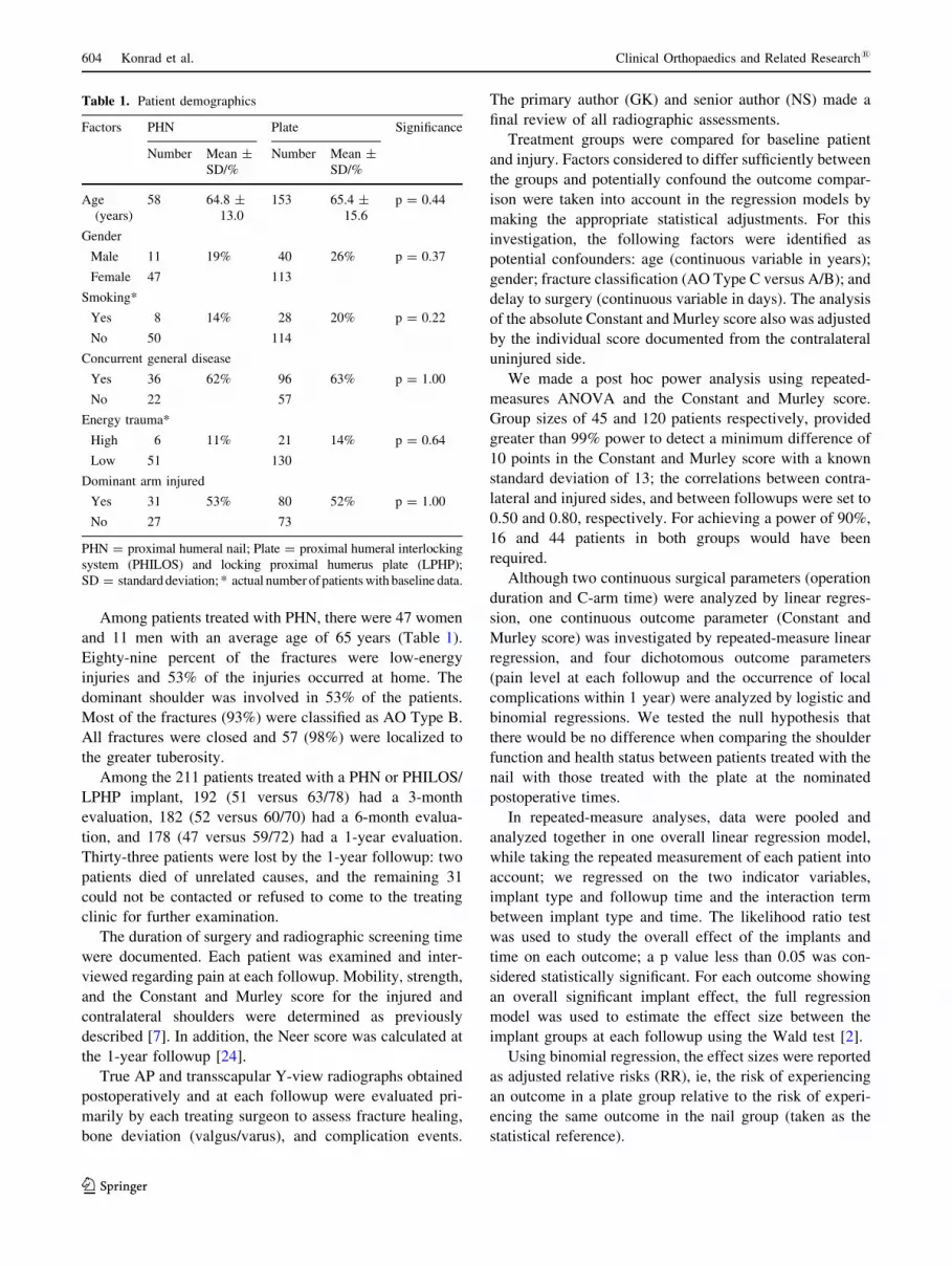
Table 1. Patient demographics
Factors PHN Plate Significance
Number Mean ±SD/%
Number Mean ±SD/%
Age(years)
58 64.8 ±13.0
153 65.4 ±15.6
p = 0.44
Gender
Male 11 19% 40 26% p = 0.37
Female 47 113
Smoking*
Yes 8 14% 28 20% p = 0.22
No 50 114
Concurrent general disease
Yes 36 62% 96 63% p = 1.00
No 22 57
Energy trauma*
High 6 11% 21 14% p = 0.64
Low 51 130
Dominant arm injured
Yes 31 53% 80 52% p = 1.00
No 27 73
PHN = proximal humeral nail; Plate = proximal humeral interlockingsystem (PHILOS) and locking proximal humerus plate (LPHP);SD = standard deviation; * actual number of patients with baseline data.
604 Konrad et al. Clinical Orthopaedics and Related Research1
123

Results
Throughout the 1-year followup period, the mean absolute
Constant and Murley scores for the injured shoulders
improved significantly for all cohorts (p \ 0.001) (Fig. 1).There was a trend toward a lower Constant and Murley
score for the plate group but the difference was not sta-
tistically significant (p = 0.05, 0.56, and 0.36 at the 3-, 6-,and 12-month examinations, respectively). A similar result
pattern was observed when considering the Constant and
Murley score relative to that recorded on the uninjuredcontralateral side. By the 1-year final evaluation, the mean
relative Constant and Murley scores were 89% (SD, 11) for
patients treated with the PHN and 87% (SD, 14) forpatients treated with a plate osteosynthesis.
Nailing of three-part proximal humeral fractures (mean ±
SD, 54 ± 21 minutes) was significantly faster than lockingplate fixation (87 ± 34 minutes) (p \ 0.001) (Fig. 2).
However, a significantly longer median fluoroscopy time of
15 seconds was required for PHN surgery (p \ 0.001).Pain at the fracture site as rated by the patients during
the Constant and Murley score assessment was signifi-
cantly different between treatment groups at the 3-, 6-, and12-month examinations (Table 2). Patients treated with a
plate were two to three times more likely to perceive pain
compared with those treated with the PHN.The 1-year Neer scores for the injured shoulders were
not significantly different between the patients treated withthe PHN and those treated with a locking plate (p = 0.39)
(Fig. 3).
The final radiologic outcomes at 1 year were classifiedas follows: anatomic ±15"; valgus [ 15"; varus [ 15"–30";
varus [ 30"–45"; varus [ 45" (Table 3). There was no
Fig. 1 The box plot shows the absolute Constant and Murley scoresfor the plate and nail osteosynthesis treatment groups at the 3-, 6-, and12-month followups.
Fig. 2 The box plot shows the surgical time taken for plate and nailosteosynthesis of three-part proximal humerus fractures. The ends ofeach rectangle correspond to the upper and lower quartiles of the datavalues. The line drawn through the rectangle corresponds to the medianvalue. The whiskers, starting at the ends of the rectangle (or pointsrepresenting extreme values), indicate minimum and maximum values.
Table 2. Pain at the fracture site
Followups Pain PHN Plate p Value§
Number* % Number* %
3 months Yes 21 41% 95 68% 0.003
No 30 45
6 months Yes 9 18% 69 53% 0.001
No 42 60
1 year Yes 9 19% 54 42% 0.015
No 38 75
PHN = proximal humeral nail; Plate = proximal humeral interlock-ing system (PHILOS) and locking proximal humerus plate (LPHP);* actual number of patients with followup data at the specific time;§Wald test of binomial regressions.
Fig. 3 The box plot shows the categorized Neer scores for the plateand nail osteosynthesis treatment groups at 1 year.
Volume 470, Number 2, February 2012 Treatment of Three-part Humeral Fractures 605
123

difference in varus deviations greater than 15" between the
cohorts (p = 0.54).
Thirty-five intraoperative and 44 postoperative localcomplications occurred in 60 of 211 patients (28%)
throughout the 1-year followup (Table 4). The risk of
experiencing at least one local complication did not sig-nificantly differ between patients in any of the two
treatment groups (p = 0.15). There were 26 primary screw
perforations of the humeral head that were not identifiedduring the surgical procedure, and in six patients, the nail
or plate was positioned too far cranially which led to
subacromial impingement. In three patients, a neurologiclesion was diagnosed. There were two cases each of deep
and superficial wound infections in the patients treated with
plate osteosynthesis. The most common local postoperativecomplications were secondary loss of reduction/impaction
(n = 25) and secondary screw perforation (n = 16). There
were no significant risk differences between the patientcohorts (p = 0.56) for local implant and bone/fracture
postoperative complications.
Thirty-four (16.1%) patients (9/58 in the nail group and25/153 in the plate group) had an unplanned secondary
operation within 12 months after the initial procedure. In
addition to the four infection-related revisions, there were21 early implant removals attributable to primary or sec-
ondary implant perforation or impingement. Implantation
of a shoulder prosthesis was performed in two patients as aresult of head necrosis, and seven patients with loss of
reduction, plate breakage, or plate pullout underwent a
reosteosynthesis.
Discussion
Proximal humeral fractures are the third most common
fractures in elderly patients after hip fractures and distalradial fractures [15]. For displaced and unstable proximal
humeral fractures, many surgical techniques have been
described [5, 11, 12, 18, 19, 26, 27, 32], but no singleapproach is considered the standard of care. Most studies
included a small number of patients and did not distinguish
between different fracture types, eg, three- and four-partfractures. The objective of this prospective multicenter
observational study was to compare plate versus nail
Table 3. Radiologic outcomes for the proximal humerus cohortstreated with plate or nail osteosynthesis at 1 year
Deviation PHN Plate
Number* % Number* %
Varus [ 45" 0 0% 9 6%
Varus [ 30"–45" 7 12% 19 12%
Varus [ 15"–30" 11 19% 34 22%
Anatomic ±15" 40 69% 85 56%
Valgus [ 15" 0 0% 6 4%
PHN = proximal humeral nail; Plate = proximal humeral interlock-ing system (PHILOS) and locking proximal humerus plate (LPHP);* actual number of patients with radiologically assessed deviationoutcomes.
Table 4. Complications
Complication type PHN Plate58* 153*Number Number
Intraoperative complications! 5 27
Primary screw perforation 3 23
Plate impingement 2 4
Nerve complication 1 2
Postoperative complications" 12 28
Implant complications 10 19
Secondary screw perforation 3 13
Implant loosening 1 0
Screw backing out 5 2
Plate and/or screw pull-out 0 2
Implant breakage 0 3
Other implant/surgery 1 0
Bone/fracture complications 9 19
Loss of reduction 4 10
Secondary dislocationfragment
1 3
Impaction 4 7
Delayed union 0 5
Nonunion 0 0
Head necrosis 1 2
Impingement 0 1
Other bone/fracture 0 0
Soft tissue/woundcomplications
0 4
Superficial infection 0 2
Deep infection 0 2
Hematoma 0 0
Other soft tissue 0 0
Any local complication§ 12 48
Complication risk (95% CI)|| 21% (11.2–33.4) 31% (24.1–39.4)
PHN = proximal humeral nail; Plate = proximal humeral interlock-ing system (PHILOS) and locking proximal humerus plate (LPHP);* total number of patients per cohort; !total number of patients withintraoperative complications takes into account patients with morethan one intraoperative event; "total number of patients with post-operative complications takes into account patients with more thanone postoperative event; §total number of patients experiencing atleast one local complication takes into account patients with morethan one intraoperative and/or postoperative event; ||estimated risk ofat least one local complication developing = the number of patientsexperiencing at least one local complication divided by the totalnumber of patients in each treatment group.
606 Konrad et al. Clinical Orthopaedics and Related Research1
123
fixation of three-part proximal humeral fractures regarding
surgical, radiologic, and functional outcomes and thecomplication rate.
A limitation of our study may be the fact that the quality
of the radiographs varied from hospital to hospital, as thefracture classification and the occurrence of complications
were dependent on the quality of radiographic analysis. All
radiographs were reviewed by each treating surgeon andthe primary author (GK) and senior author (NS) regarding
fracture classification, fracture healing, and the possibleoccurrence of complications to standardize the measure-
ments. The variability in surgical technique and post-
operative management between different surgeons ordifferent hospitals is another limitation. However, the
multicenter design might represent an average of the hos-
pitals treating proximal humeral fractures, and all surgeonsinvolved in the study are trained in trauma management
and had experience using either locking plates or nails.
Another limitation of the study is the lack of randomizationassociated with a higher level of evidence, as we cannot
exclude that compared groups differed according to factors
other than the investigated treatment. Restriction to three-part fractures and statistical adjustment for factors showing
differences among the groups were used to minimize
potential treatment allocation bias. Finally, the 1-yearresults might not be sufficient to draw final conclusions
regarding functional outcome after these challenging
fractures, especially when the occurrence of complicationsprolonged the functional recovery. Furthermore, the results
may deteriorate in the nail group over a longer followup
secondary to the higher incidence of rotator cuff tearing inthe nail group versus the plate group.
Nailing of three-part proximal humeral fractures was
significantly faster than locking plate fixation. This may beattributable to the less invasive approach used for intra-
medullary nailing. The average operation time in our study
is comparable to the values from other studies investigatingonly one treatment option. Lin [20] reported 65 minutes for
locked nailing of displaced three-part proximal humeral
fractures, whereas in another study, the mean operativetime was 55 minutes [16]. Stabilizing a proximal humeral
fracture using the PHN required longer radiographic
screening time than using an angular stable plate. We couldnot find any previously published data concerning xray
time for comparison with our results.
Three months, 6 months, and 12 months after openreduction and internal fixation, the PHN group showed
advantages regarding pain level compared with the angular
stable plate group. This may be explained by the minimalinvasive approach of the nail implant.
We observed no significant difference between the nail
and the plate groups concerning the Constant and Murleyscore at any followup, although there was a trend toward
lower scores for the locking plate cohort. However, the
mean Constant and Murley and Neer scores of the injuredside were not significantly different suggesting that all
three techniques may be useful for internal fixation of
three-part proximal humeral fractures.For our comparative series, the Constant and Murley
score was greater than 85% of the mean for the contralat-
eral side. Similar results were reported in a prospectivestudy with 28 patients [10]. One year postoperatively, the
mean Constant and Murley score of the injured side for ourpatients was 72 points corresponding to 87% of the score
on the contralateral side. In some other studies, the mean
Constant and Murley scores after angular stable plate orangular stable nail fixation of proximal humeral fractures
ranged between 72 and 89 points [12, 17, 21, 28, 30],
which also are comparable to our results. Nevertheless,most of these published studies do not distinguish between
different fracture types. Our intention of limiting this study
to a single fracture type allows the observed outcomes to betargeted and limits the possibility of generalizing the
results.
There are a limited number of studies investigatingantegrade angular stable nailing of proximal humeral
fractures [12, 20, 21]. In a multicenter study, Gradl et al.
[12] prospectively followed 152 patients with proximalhumeral fractures treated with either an antegrade angular
stable nail or an angular stable plate; relative 1-year post-
operative Constant and Murley scores were 81% and 77%,respectively. No differences were observed between the
two groups at 3, 6, and 12 months after surgery.
Sixty-four percent of all fractures showed an anatomicreduction at the 1-year followup. There was no difference
between the treatment groups. However, auxiliary explor-
atory analysis revealed a significant correlation betweendeviation and the functional result according to the Con-
stant and Murley score: patients with varus deviations
greater than 30" to 45" and greater than 45" had, onaverage, nine (p = 0.008) and 14 (p = 0.027) percentage
points less on the relative Constant and Murley score at
1 year, respectively. This finding is consistent with previ-ously reported results from other studies [1, 23, 28].
In previous studies focused on LPHP and PHILOS fix-
ation of complex fractures, reported complicationsincluded avascular necrosis, loss of reduction, plate
breakage, and pseudarthrosis/nonunion [6, 10, 17]. We
observed 79 local complications in 60 of 211 patientsduring the 1-year followup; 35 intraoperative complica-
tions were directly related to the initial surgical procedure.
Most common intraoperative complications were primaryscrew perforations of the humeral head which were
unrecognized during surgery and subacromial impingement
because the nail or the plate was positioned too far crani-ally. Adequate surgical skills and experiences with the
Volume 470, Number 2, February 2012 Treatment of Three-part Humeral Fractures 607
123
surgical technique are necessary to achieve correct implant
fixation and avoid these intraoperative errors.The similar outcomes for nail versus plate fixation of
three-part proximal humeral fractures after 1 year for our
patients suggest that all three techniques may be useful forinternal fixation of three-part proximal humeral fractures.
Many complications were related to incorrect surgical
technique and therefore can be avoided. Advanced surgicalskills and experience are considered to be more critical for
successful operative treatment of three-part proximalhumeral fractures than the selection of the implant.
Acknowledgments We thank the following clinics and investiga-tors for participation in this study:
PHN study arm: Medizinische Einrichtungen der Universitat,Unfallchirurgie, Bonn, Germany (C. Rangger); Centro Traumatolog-ico Ortopedico, Firenze, Italy (R. Angeloni); UZ Gasthuisberg,Leuven, Belgium (S. Nijs); Royal Liverpool University Hospital,Liverpool, United Kingdom (C. Sinopidis); Johannes GutenbergUniversitatsklinikum, Klinik fur Unfallchirurgie, Mainz, Germany(J. Blum); Universita di Padova, Ortopedia e Traumatologia, Padova,Italy (A. Olmeda); Vogtlandklinikum Plauen, Plauen, Germany(W. Merbold); Kardinal Schwarzenberg’schen Krankenhaus, Abtei-lung fur Unfallchirurgie, Schwarzach, Austria (F. Genelin);Unfallkrankenhaus Meidling, Wien, Austria (H. Matuschka); Klini-kum Worms gGmbH, Worms, Germany (J. Blum).
PHILOS study arm: Ratisches Kantons- und Regionalspital, Chur,Switzerland (C. Sommer); Spital und Pflegeheim Davos, Davos Platz,Switzerland (C. Ryf); Hopital Cantonal de Fribourg, Fribourg,Switzerland (G. Kohut); Westpfalz-Klinikum GmbH, UnfallchirurgieKlinik, Kaiserslautern, Germany (H. Winkler); Kantonsspital Luzern,Chirurgie/Traumatologie, Luzern, Switzerland (R. Babst); KlinikumRosenheim, Unfall- und Wiederherstellungschirurgie, Rosenheim,Germany (G. Regel); St. Goran’s Hospital, Stockholm, Sweden(A. Ekelund); BG Unfall- und Universitatsklinik, Tubingen, Germany(D. Hontzsch).
LPHP study arm: Charite Universitatsmedizin Berlin, Zentrum furMuskuloskeletale Chirurgie, Berlin, Germany (N. Haas); AllgemeinesKrankenhaus Celle, Celle, Germany (H-J. Oestern); Albert-Ludwigs-Universitat, Orthopadie und Traumatologie, Freiburg, Germany(N. Sudkamp); Universitatsklinik fur Unfallchirurgie Graz, Graz,Austria (M. Plecko); Academic Medical Center Groningen, Depart-ment of Traumatology, Groningen, Netherlands (H. Ten Duis);Evangelisches Diakoniewerk Friederikenstift, Unfallklinik, Hannover,Germany (H. Lill); Universitat Leipzig, Klinik fur Unfall- undWiederherstellungschirurgie, Leipzig, Germany (C. Josten).
References
1. Agudelo J, Schurmann M, Stahel P, Helwig P, Morgan SJ, ZechelW, Bahrs C, Parekh A, Ziran B, Williams A, Smith W. Analysisof efficacy and failure in proximal humerus fractures treated withlocking plates. J Orthop Trauma. 2007;21:676–681.
2. Armitage P, Berry G, Matthews JNS. Statistical Methods inMedical Research. Ed 4. Oxford, UK: Blackwell Publishing;2002.
3. Blum J, Hansen M, Muller M, Rommens PM, Matuschka H,Olmeda A, Radziejowski MJ, Merbold W, Nijs S, Angeloni R.Proximal humerus fractures and intramedullary nailing: experi-ence with a new nail system. Eur J Trauma Emerg Surg. 2009;5:489–498.
4. Brunner F, Sommer C, Bahrs C, Heuwinkel R, Hafner C, RillmanP, Kohut G, Ekelund A, Muller M, Audige L, Babst R. Openreduction and internal fixation of proximal humerus fracturesusing a proximal humeral locked plate: a prospective multicenteranalysis. J Orthop Trauma. 2009;23:163–172.
5. Cazeneuve JF, Cristofari DJ. The reverse shoulder prosthesis inthe treatment of fractures of the proximal humerus in the elderly.J Bone Joint Surg Br. 2010; 92:535–539.
6. Charalambous CP, Siddique I, Valluripalli K, Kovacevic M,Panose P, Srinivasan M, Marynissen H. Proximal humeralinternal locking system (PHILOS) for the treatment of proximalhumeral fractures. Arch Orthop Trauma Surg. 2007;127:205–210.
7. Constant CR, Murley AH. A clinical method of functionalassessment of the shoulder. Clin Orthop Relat Res. 1987;214:160–164.
8. Court-Brown CM, Garg A, McQueen MM. The epidemiology ofproximal humeral fractures. Acta Orthop Scand. 2001;72:365–371.
9. Egol KA, Ong CC, Walsh M, Jazrawi LM, Tejwani NC,Zuckerman JD. Early complications in proximal humerus frac-tures (OTA Types 11) treated with locked plates. J OrthopTrauma. 2008;22:159–164.
10. Fankhauser F, Boldin C, Schippinger G, Haunschmid C,Szyszkowitz R. A new locking plate for unstable fractures of theproximal humerus. Clin Orthop Relat Res. 2005;430:176–181.
11. Gerber C, Werner CM, Vienne P. Internal fixation of complexfractures of the proximal humerus. J Bone Joint Surg Br. 2004;86:848–855.
12. Gradl G, Dietze A, Arndt D, Beck M, Gierer P, Borsch T,Mittlmeier T. Angular and sliding stable antegrade nailing(Targon PH) for the treatment of proximal humeral fractures.Arch Orthop Trauma Surg. 2007;127:937–944.
13. Gradl G, Dietze A, Kaab M, Hopfenmuller W, Mittlmeier T. Islocking nailing of humeral head fractures superior to lockingplate fixation? Clin Orthop Relat Res. 2009;467:2986–2993.
14. Handoll HH, Ollivere BJ. Interventions for treating proximalhumeral fractures in adults. Cochrane Database Syst Rev. 2010;(12):CD000434.
15. Kannus P, Palvanen M, Niemi S, Parkkari J, Jarvinen M, Vuori I.Osteoporotic fractures of the proximal humerus in elderly Finnishpersons: sharp increase in 1970–1998 and alarming projectionsfor the new millennium. Acta Orthop Scand. 2000;71:465–470.
16. Kazakos K, Lyras DN, Galanis V, Verettas D, Psillakis I,Chatzipappas Ch, Xarchas K. Internal fixation of proximalhumerus fractures using the Polarus intramedullary nail. ArchOrthop Trauma Surg. 2007;127:503–508.
17. Kettler M, Biberthaler P, Braunstein V, Zeiler C, Kroetz M,Mutschler W. [Treatment of proximal humeral fractures with thePHILOS angular stable plate : presentation of 225 cases of dis-located fractures] [in German]. Unfallchirurg. 2006;109:1032–1040.
18. Koukakis A, Apostolou CD, Taneja T, Korres DS, Amini A.Fixation of proximal humerus fractures using the PHILOS plate:early experience. Clin Orthop Relat Res. 2006;442:115–120.
19. Kristiansen B, Christensen SW. Plate fixation of proximal hum-eral fractures. Acta Orthop Scand. 1986;57:320–323.
20. Lin J. Effectiveness of locked nailing for displaced three-partproximal humeral fractures. J Trauma. 2006;61:363–374.
21. Linhart W, Ueblacker P, Grossterlinden L, Kschowak P, BriemD, Janssen A, Hassunizadeh B, Schinke M, Windolf J, RuegerJM. Antegrade nailing of humeral head fractures with capturedinterlocking screws. J Orthop Trauma. 2007;21:285–294.
22. Matziolis D, Kaeaeb M, Zandi SS, Perka C, Greiner S. Surgicaltreatment of two-part fractures of the proximal humerus: com-parison of fixed-angle plate osteosynthesis and Zifko nails.Injury. 2010 May 18. [Epub ahead of print].
608 Konrad et al. Clinical Orthopaedics and Related Research1
123

23. Micic ID, Kim KC, Shin DJ, Shin SJ, Kim PT, Park IH, Jeon IH.Analysis of early failure of the locking compression plate inosteoporotic proximal humerus fractures. J Orthop Sci. 2009;14:596–601.
24. Neer CS. Displaced proximal humeral fractures: I. Classifica-tion and evaluation. J Bone Joint Surg Am. 1970;52:1077–1089.
25. Owsley KC, Gorczyca JT. Fracture displacement and screwcutout after open reduction and locked plate fixation of proximalhumeral fractures [corrected]. J Bone Joint Surg Am. 2008;90:233–240.
26. Resch H, Povacz P, Frohlich R, Wambacher M. Percutaneousfixation of three- and four-part fractures of the proximal humerus.J Bone Joint Surg Br. 1997;79:295–300.
27. Schmal H, Klemt C, Sudkamp NP. [Evaluation of shoulderarthroplasty in treatment of four-fragment fractures of the prox-imal humerus] [in German]. Unfallchirurg. 2004;107:575–582.
28. Solberg BD, Moon CN, Franco DP, Paiement GD. Surgicaltreatment of three and four-part proximal humeral fractures.J Bone Joint Surg Am. 2009;91:1689–1697.
29. Sudkamp N, Bayer J, Hepp P, Voigt C, Oestern H, Kaab M, LuoC, Plecko M, Wendt K, Kostler W, Konrad G. Open reductionand internal fixation of proximal humerus fractures with use ofthe locking proximal humeral plate: results of a prospective,multicenter, observational study. J Bone Joint Surg Am. 2009;91:1320–1328.
30. Thalhammer G, Platzer P, Oberleitner G, Fialka C, Greitbauer M,Vecsei V. Angular stable fixation of proximal humeral fractures.J Trauma. 2009;66:204–210.
31. Vallier HA. Treatment of proximal humerus fractures. J OrthopTrauma. 2007;21:469–476.
32. Wijgman AJ, Roolker W, Patt TW, Raaymakers EL, Marti RK.Open reduction and internal fixation of three and four-part frac-tures of the proximal part of the humerus. J Bone Joint Surg Am.2002;84:1919–1925.
Volume 470, Number 2, February 2012 Treatment of Three-part Humeral Fractures 609
123