Bennett Rathnayake v. The Sri Lanka Rupavahini Corporation ...
Upload
khangminh22Category
view
2download
0
Ttptjrept
IRdTiysvcy
CURRENTCONCEPTS
Thumb Trauma: Bennett Fractures, Rolando Fractures,
and Ulnar Collateral Ligament Injuries
Brian T. Carlsen, MD, Steven L. Moran, MD
Injuries to the thumb are predominated by fractures of the proximal phalanx, ligamentousinjuries about the metacarpophalangeal joint, and metacarpal base fractures. This article willattempt to summarize recent advancements within the realm of thumb trauma, withparticular attention to Bennett fractures, Rolando fractures, and ulnar collateral ligamentinjuries. (J Hand Surg 2009;34A:945–952. Copyright © 2009 by the American Societyfor Surgery of the Hand. All rights reserved.)
Key words Bennett fracture, Rolando fracture, thumb, trauma, ulnar collateral ligamentinjury.
titimi
RIattvathatobatatvm
PTf C
urrentConcepts
HE THUMB PROVIDES up to 40% of hand function.Total disability of the thumb is devastating andequilibrates to a loss of 22% of bodily func-
ion.1 The thumb’s uniqueness and versatility in man isrimarily due to the position of the thumb axis. Thehumb axis is based at the trapeziometacarpal (TM)oint and is pronated and flexed approximately 80° withespect to the other hand metacarpals. This positionnables circumduction, which permits opposition andrehension; however this position also exposes thehumb to unique injury.
NCIDENCEecently, Stanton and colleagues examined the inci-ence of fractures within the tubular bones of the hand.humb fractures were found to occur most commonly
n children and the elderly. In children (age 0–16ears), 22% of all tubular bone hand fractures occurredomewhere within the first ray. In retirement age indi-iduals (age �65 years), 20% of hand fractures oc-urred in the thumb, whereas only 12% of fractures inoung adults (age 17–40 years) were found to occur in
From the Mayo Clinic, Rochester, MN.
Received for publication February 27, 2009; accepted in revised form March 23, 2009.
No benefits in any form have been received or will be received related directly or indirectly to thesubject of this article.
Corresponding author: Steven L. Moran, MD, 200 First Street, SW, Rochester, MN 55905; e-mail:[email protected].
0363-5023/09/34A05-0026$36.00/0
idoi:10.1016/j.jhsa.2009.03.017
©
he thumb ray. First-ray fractures were half as commonn the 17- to 40-year age group when compared withhat for the elderly or the pediatric groups. In addition,n the elderly population, the thumb was the most com-on tubular bone fractured with fracture patterns tend-
ng to be oblique and intra-articular.2
ADIOGRAPHIC EVALUATIONn addition to careful physical exam, radiographic im-ging is an essential part of a complete evaluation afterhumb trauma. Because the thumb sits out of plane fromhe rest of the hand and fingers, special radiographiciews are necessary for appropriate evaluation. A truenteroposterior view of the thumb can be obtained withhe Robert’s view, which requires that the hand beyperpronated so that the dorsum of the thumb liesgainst the radiographic plate. To obtain a true lateral ofhe TM joint, the palm of the hand must be placed flatn the cassette with the hand pronated 15° to 35°; theeam is then directed 15° distal to proximal. This im-ging technique is referred to as the Bett’s view of thehumb. This image allows one to evaluate the TM jointnd the 3 additional articulations of the trapezium: therapezoid, the scaphoid, and index metacarpal. Bothiews are helpful when evaluating fracture displace-ent and joint congruency.
HALANGEAL FRACTUREShe management of extra-articular thumb phalangeal
ractures differs from that of finger phalangeal fractures
n that some angular displacement or malunion is ac-ASSH � Published by Elsevier, Inc. All rights reserved. � 945
946 THUMB TRAUMA
Curren
tConcep
ts
ceptable due to compensatory motion of the thumbmetacarpophalangeal (MCP) joint. In the proximal pha-lanx of the thumb, angular deformities up to 20° in thefrontal plane and 30° in the lateral plane may be func-tionally well tolerated; however, these can be a sourceof cosmetic complaint. Surgical fixation is recom-mended for deformities that exceed these guidelines, aswell as any open or unstable fractures. Fixation dependson the fracture type and surgeon preference; however,K-wires or interfragmentary screw fixations is oftenadequate. Stable fixation offers the advantages of earliermobilization.
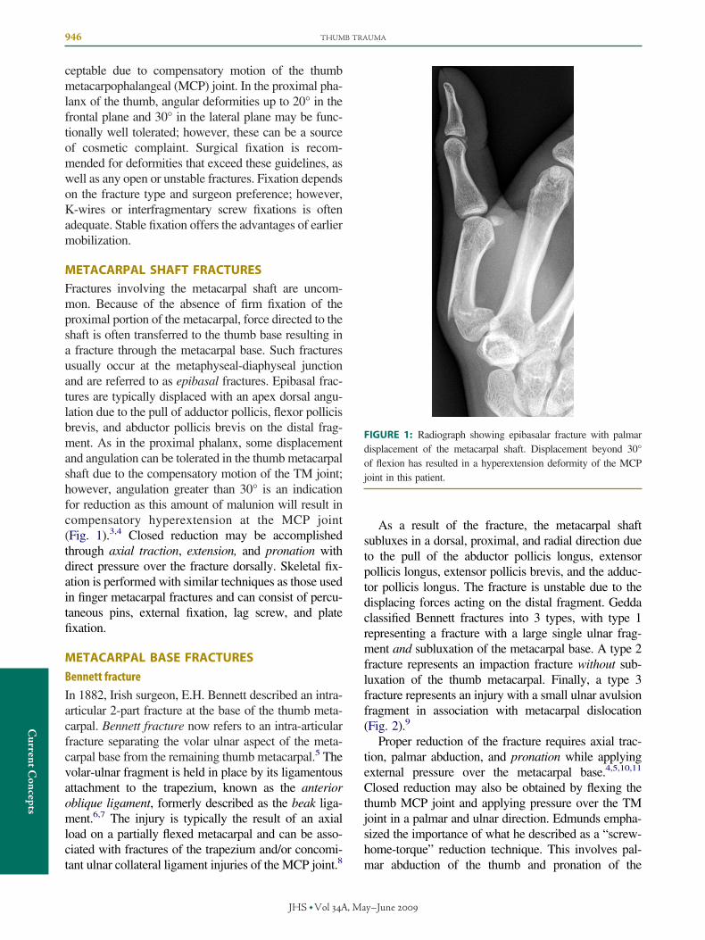
METACARPAL SHAFT FRACTURESFractures involving the metacarpal shaft are uncom-mon. Because of the absence of firm fixation of theproximal portion of the metacarpal, force directed to theshaft is often transferred to the thumb base resulting ina fracture through the metacarpal base. Such fracturesusually occur at the metaphyseal-diaphyseal junctionand are referred to as epibasal fractures. Epibasal frac-tures are typically displaced with an apex dorsal angu-lation due to the pull of adductor pollicis, flexor pollicisbrevis, and abductor pollicis brevis on the distal frag-ment. As in the proximal phalanx, some displacementand angulation can be tolerated in the thumb metacarpalshaft due to the compensatory motion of the TM joint;however, angulation greater than 30° is an indicationfor reduction as this amount of malunion will result incompensatory hyperextension at the MCP joint(Fig. 1).3,4 Closed reduction may be accomplishedthrough axial traction, extension, and pronation withdirect pressure over the fracture dorsally. Skeletal fix-ation is performed with similar techniques as those usedin finger metacarpal fractures and can consist of percu-taneous pins, external fixation, lag screw, and platefixation.
METACARPAL BASE FRACTURES
Bennett fracture
In 1882, Irish surgeon, E.H. Bennett described an intra-articular 2-part fracture at the base of the thumb meta-carpal. Bennett fracture now refers to an intra-articularfracture separating the volar ulnar aspect of the meta-carpal base from the remaining thumb metacarpal.5 Thevolar-ulnar fragment is held in place by its ligamentousattachment to the trapezium, known as the anterioroblique ligament, formerly described as the beak liga-ment.6,7 The injury is typically the result of an axialload on a partially flexed metacarpal and can be asso-ciated with fractures of the trapezium and/or concomi-
tant ulnar collateral ligament injuries of the MCP joint.8JHS �Vol A, Ma
As a result of the fracture, the metacarpal shaftsubluxes in a dorsal, proximal, and radial direction dueto the pull of the abductor pollicis longus, extensorpollicis longus, extensor pollicis brevis, and the adduc-tor pollicis longus. The fracture is unstable due to thedisplacing forces acting on the distal fragment. Geddaclassified Bennett fractures into 3 types, with type 1representing a fracture with a large single ulnar frag-ment and subluxation of the metacarpal base. A type 2fracture represents an impaction fracture without sub-luxation of the thumb metacarpal. Finally, a type 3fracture represents an injury with a small ulnar avulsionfragment in association with metacarpal dislocation(Fig. 2).9
Proper reduction of the fracture requires axial trac-tion, palmar abduction, and pronation while applyingexternal pressure over the metacarpal base.4,5,10,11
Closed reduction may also be obtained by flexing thethumb MCP joint and applying pressure over the TMjoint in a palmar and ulnar direction. Edmunds empha-sized the importance of what he described as a “screw-home-torque” reduction technique. This involves pal-
FIGURE 1: Radiograph showing epibasalar fracture with palmardisplacement of the metacarpal shaft. Displacement beyond 30°of flexion has resulted in a hyperextension deformity of the MCPjoint in this patient.
mar abduction of the thumb and pronation of the
y–June

vulsi
THUMB TRAUMA 947
CurrentConcepts
metacarpal base. This technique theoretically tensionsthe dorsal ligament complex allowing one to betterreduce the fracture fragment.5 Thumb extension (hitch-hiker position) has been shown to cause fracture dis-placement and should be avoided.5
In Bennett’s original article, he described treating 2patients with 4 weeks of cast immobilization. Closedreduction and casting remained the preferred method oftreatment until the 1970s. Although historical reportshave noted satisfactory outcomes with nonsurgicaltreatment, more recent studies have shown poor out-comes with casting alone for this injury.12–15
Surgical treatment is varied for the treatment ofBennett fractures but has typically included closed re-duction with percutaneous pinning or open reductionwith either pins or interfragmentary fixation. Obliquetraction pinning and external fixation have also beendescribed. All methods of fixation have been shown tobe effective in case reviews and series. Closed reduc-tion with intermetacarpal fixation to the second meta-carpal and or to the trapezium is typically effective atreducing the metacarpal shaft subluxation. An addi-tional K-wire to oppose the volar-ulnar fragment to themetacarpal may be added.
If open reduction of the metacarpal is performed, it ismost commonly performed through a Wagner incision.The decision to openly reduce the fracture, as opposedto closed reduction and pinning, is still a matter ofdebate. Recently, Lutz and colleagues reported on theresults of 32 patients with Bennett fractures with aver-age follow-up of 7 years. Patients were either treatedwith open reduction and internal fixation or closedreduction and percutaneous transarticular K-wires.Treatment type did not influence the clinical outcome orthe development of radiographic arthritis. However, a
FIGURE 2: Gedda classified the Bennett fractures into 3 typesand subluxation of the metacarpal base. B Type 2 represents anType 3 represents those injuries presenting with a small ulnar a
key point in this study was that for fractures deemed
JHS �Vol A, Ma
irreducible with an intra-articular step-off greater than 1mm, closed reduction should be abandoned in favor ofopen reduction. This suggests that anatomic congruityis more important than fracture fixation technique forlong-term success.16
The amount of anatomic incongruity that is accept-able has been examined by several authors. Earlierpublications suggest that there is no relationship be-tween fracture reduction and postoperative pain, mo-tion, or the development of arthritis.17 Demir and col-leagues retrospectively reviewed 24 Bennett fracturesand 6 comminuted intra-articular fractures and wereunable to correlate the quality of articular thumb baserestoration and the radiologic or subjective outcome.18
Biomechanical studies performed by Cullen and col-leagues noted that 2 mm of residual displacement at thearticular surface resulted in an overall increase in con-tact area at the TM joint, with a dorsal shift in contactpressures over the trapezial surface. In addition, noimportant increase in contact pressure was seen in thearea of the articular step-off. The authors concluded thata 2-mm articular step-off is acceptable and should bewell tolerated as long as the metacarpal was reduced.19
Such cadaveric studies are limited due to the constraintsinvolved with use of contact-pressure film but still sug-gest that reduction of the metacarpal subluxation andapposition of the bony fragments should be the primarygoal of surgical intervention, as opposed to obtainingcomplete articular congruency.
In contrast, various clinical studies have documentedan improvement in outcomes with anatomic reductionand minimal articular step-off.13–15 Kjaer-Petersen andcolleagues reported on 41 Bennett fractures treated withclosed reduction and casting, closed reduction and per-cutaneous pinning, or open reduction with internal fix-
ype 1 represents a fracture with a large single ulnar fragmentaction fracture without subluxation of the thumb metacarpal. Con fragment in association with metacarpal dislocation.
. A Timp
ation. Outcomes correlated notably with the quality of
y–June

erati
948 THUMB TRAUMA
Curren
tConcep
ts
the reduction: 86% of patients with an anatomic reduc-tion (�1 mm step-off) had no residual symptoms,whereas only 46% of patients with good or poor reduc-tion (�1 mm step-off) remained asymptomatic.12
Unfortunately, definitive treatment algorithms arelacking due to the small patient numbers in the afore-mentioned studies, lack of long-term radiographic fol-low-up, and absence of randomized prospective data.From the available literature, however, several treat-ment guidelines can be made: (1) Bennett fracturesshould be treated with some type of surgical fixation,whether using open or percutaneous techniques, tomaintain reduction of the metacarpal at the TM joint.(2) Fracture reduction techniques should include thumbpronation to aid in anatomic reduction of the metacarpalto the ulnar fragment. (3) Open reduction should beconsidered in those circumstances where greater than 2mm of articular step-off persists despite closed reduc-tion attempts.4
Rolando fracture
In 1910, Silvio Rolando described 3 cases of a Y-pattern fracture of the metacarpal base.20 The termRolando fracture is now applied to many comminutedfractures of the base of the first metacarpal but ideallyshould be reserved for Y- or T-pattern fractures thatinclude the volar-ulnar Bennett fragment in addition toa dorsal radial fragment (Fig. 3). This fracture pattern isconsiderably more difficult to treat and has a worseprognosis than that of the Bennett fracture. When there
FIGURE 3: A The term Rolando fracture is classically used tobase with a Y- or T-fracture pattern, as seen in this radiograpfixation. Such fractures often require transmetacarpal pinningprocess. C A Robert’s view of the same thumb 3 months postop
are 2 large fragments, without considerable comminu-
JHS �Vol A, Ma
tion, open reduction and internal fixation through aWagner approach can be successful. Various methodsof fixation have been described including K-wires, ten-sion banding, and plate and screw fixation.
For markedly comminuted fractures, distraction andreliance on ligamentous reduction of the fragments maybe necessary. Distraction can be achieved with obliquetraction pinning. Alternatively, the fracture can bespanned and distracted using external fixation.21 Radio-graphs obtained with the thumb in distraction can pro-vide an assessment of the reduction achievable withligamentotaxis alone.
There are few studies that report on the long-termoutcome of Rolando fractures. In 1991, Langhoff et al.reported their results on 17 Rolando fractures. Restora-tion of the articular surface was the goal of treatmentusing the open approach when closed reduction wasinadequate. Open techniques were required in the ma-jority (11 of 14) of patients. Long-term radiographicfollow-up (mean, 6 years) was available in only 11patients; however 6 of these had evidence of arthriticchanges. As with the Bennett fracture, the authorsfound that the quality of reduction did not directlycorrelate with symptoms at follow-up.22
Metacarpophalangeal ulnar collateral ligament injuries
The MCP joint of the thumb is a diarthrodial joint thatallows for flexion and extension as well as abductionand adduction. The thumb MCP joint remains stablethroughout the flexion-extension arc. In contrast, the
cribe a 3-part intra-articular fracture of the thumb metacarpalA radiograph of the same fracture after transosseous K-wire
tabilize and suspend the metacarpal shaft during the healingvely, showing adequate articular congruity.
desh. Bto s
finger MCP joints are stable in flexion and lax in ex-
y–June

d of
THUMB TRAUMA 949
CurrentConcepts
tension. Range of motion at the thumb (MCP) joint isthe most variable in the human body and may evendiffer between the right and left hands in the samepatient.
Lateral stability at the MCP joint is provided by astrong collateral ligament system consisting of a properand accessory collateral ligament. The proper collateralligaments are attached to the lateral condyle of themetacarpal and to the proximal phalanx. The accessorycollateral ligaments attach more volarly on the meta-carpal and insert distally on the sesamoid bones and thevolar plate. The proper collateral ligaments are taut inflexion, whereas the accessory collateral ligaments aretaut in extension. Additional dynamic stability is con-ferred to the joint through the tendinous attachments ofthe adductor pollicis, flexor pollicis brevis, and extensorpollicis brevis, with the adductor pollicis muscle insert-ing on the ulnar sesamoid and the flexor pollicis brevisinserting into the radial sesamoid.
Forceful radial deviation at the MCP joint can lead toulnar collateral ligament injury. Acute ulnar collateralligament injuries most commonly occur during fallswith the thumb outstretched and abducted. Such injuriesusually result in injury to both the proper and accessorycollateral ligaments. Forceful trauma can also result ininjury to the ulnar halves of the joint capsule, the volarplate, and the adductor aponeurosis.
Ulnar collateral ligament injuries occur 10 timesmore frequently than do radial collateral ligament inju-
FIGURE 4: A 64-year-old woman presents with marked pain aof the ulnar collateral ligament reveals marked laxity with gexploration, a Stener lesion is identified. The bony insertion oaponeurosis of the adductor pollicis muscle (fragment at the en
ries. The acute ulnar collateral ligament injury is espe-
JHS �Vol A, Ma
cially common in athletes and downhill skiers, hencethe eponym, skier’s thumb. In the case of the skier, thepole maintains the thumb in an abducted position, ex-posing the thumb to excessive radial abduction. Al-though the injury can occur either at the proximal ordistal attachment of the ligament, distal avulsion is themost common. Approximately 50% of these injurieswill have an associated fracture of the base of theproximal phalanx.
Complete ligament injuries are differentiated frommild sprains thru careful physical exam (Fig. 4). Hey-man and colleagues showed that if �35° of joint angu-lation was noted on valgus stress of the flexed MCPjoint, a complete tear of the proper collateral ligamentwas noted at the time of surgery. In each of these cases,instability of the injured thumb exceeded stability of theuninjured thumb by at least 15°. Tears of the accessorycollateral ligament were found consistently when val-gus stress testing of the extended MCP joint resulted inmore than 35° of joint angulation.23 Others have sug-gested evaluating the end point of MCP motion; if thereis a lack of a firm end point during valgus stress, oneshould suspect an ulnar collateral ligament injury.24
Unfortunately, in the acute setting, the ability to obtainan adequate exam is often hindered by the patient’s painand ongoing spasm of the adductor pollicis muscle. Amedian and radial nerve block at the wrist before stress-testing can facilitate evaluation of the joint.
In 1962, Stener described the displacement of the
stability at the ulnar aspect of the MCP joint. A Stress testingthan 35° of rotational deformity. B At the time of surgicalulnar collateral ligament can been seen sitting on top of the
dental probe).
nd inreaterf the
ulnar collateral ligament proximal and superficial to the
y–June

950 THUMB TRAUMA
Curren
tConcep
ts
leading edge of the adductor pollicis aponeurosis. Inthis Stener lesion, the aponeurosis of the adductor pol-licis is interposed between the ligament and its insertionon the proximal phalanx, prohibiting healing. The pres-ence of a Stener lesion is therefore an indication forsurgical ulnar collateral ligament repair; however, ac-curate diagnosis of a Stener lesion prior to surgery isoften a diagnostic dilemma. Such injury patterns oftenoccur in the absence of a substantial bony fragment.One occasionally may see a fracture fragment on plainx-ray proximal to the location of the adductor hood,suggesting a “bony Stener lesion.”25,26 A palpable massmay sometimes be identified proximal to the MCP jointin these cases. Arthrograms, ultrasound, and magneticresonance imaging (MRI) can provide further sensitiv-ity in the diagnosis of a Stener lesion.27 Although moreexpensive, MRI is considered the most accurate, withsensitivity and specificity reported as high as 96% and95%, respectively.28
Treatment of stable ulnar collateral ligament sprainsor partial tears entails 4 weeks of immobilization in athumb-spica splint or cast. When there is completerupture of the ulnar collateral ligament, surgical repairis the preferred treatment.24 The surgical approach isthrough a lazy-S or chevron incision with the apexlocated at the volar, ulnar aspect of the MCP joint.Mid-substance tears can be primarily repaired with 3-0or 4-0 braided, nonabsorbable suture. Distal ligamen-tous avulsions are treated with secure fixation of theligament to the bone. In acute injuries, the location ofinsertion on the proximal phalanx is apparent; howeverif there has been some delay in treatment, identifyingthe location of ulnar collateral ligament attachment maybe difficult. Bean and colleagues have shown that theisometric attachment point for the ulnar collateral liga-ment, to optimize MCP range of motion, is located 3mm distal to the articular surface and 3 mm dorsal to thevolar cortex of the proximal phalanx.29 The ligamentcan be secured to the proximal phalanx using pull-outsutures, bone anchors, or cerclage wire. A recent studyby Katolik et al. demonstrated shorter surgical times,decreased soft tissue complications, and lower cost us-ing suture anchors compared with use of pull-out suturetechniques.30
The treatment of thumb ulnar collateral ligamentavulsion fractures is controversial. Unlike ligamentousdisruptions of the ulnar collateral ligament without frac-ture, it has been presumed that the majority of ulnarcollateral ligament avulsion fractures do not have aStener lesion component and hence are capable of pri-
mary healing if minimally displaced. The literature hasJHS �Vol A, Ma
supported both surgical31,32 and nonsurgical manage-ment of these injury patterns.26,33,34
Kuz and colleagues reported on a series of 30 pa-tients treated nonsurgically with proximal phalanx avul-sion fractures.26 They excluded patients with �30%involvement of the articular surface and fractures dis-placed proximal to the adductor hood (bony Stenerlesion). Ulnar collateral ligament stability was not as-sessed due to the risk of causing further displacement ofthe fracture; however, the outcomes were generallygood with 19 patients pain-free, 10 with mild pain, and1 with moderate pain. Twenty patients were evaluatedclinically, and there was no marked difference in sta-bility or grip or pinch strength. Three patients on finalfollow-up were considered unstable (�35° angulationor �15° difference from contralateral thumb), but thesepatients had no subjective or objective limitations. Theauthors recommend nonsurgical treatment for mostacute ulnar collateral ligament avulsion fractures.26
This recommendation has also been made by otherauthors, provided there is no lateral instability of theMCP joint. A painless nonunion of the fragment willoccur in a considerable number of patients (25% to60%) treated without surgery.26,35
In contrast, Dinowitz and colleagues have reportedon the problems that can result from closed treatment ofsmall, minimally displaced avulsion fracture. They re-ported on 9 patients initially treated with cast immobi-lization for minimally displaced ulnar collateral liga-ment avulsion fractures.32 All 9 patients had persistentpain and diminished pinch and grip strength after aminimum of 6 weeks in the cast. Late ulnar collateralligament reconstruction was performed in all patientswith K-wire fixation and pull-through sutures at a meanof 6 months after the original injury. Subsequent out-comes were good with all patients obtaining radio-graphic union at 6 to 7 weeks, and grip and pinchstrength improved respectively with regard to grip andpinch strength.32 Hintermann and colleagues have alsoshown that avulsion fractures can occur with a concom-itant midsubstance tear.31 Other studies have also ver-ified the occurrence of a 2-level injury pattern.25,36
Hintermann stressed the importance of stress testing inall patients, even those with nondisplaced fractures.They recommend nonsurgical management only forpatients with nondisplaced fractures who are stable onstress-testing.31
Based on the available literature, open reduction andinternal fixation for avulsion fractures should be per-formed if (1) 20% or greater of the joint surface is
involved, (2) there is considerable fracture displace-y–June
THUMB TRAUMA 951
CurrentConcepts
ment, or (3) there is substantial instability with ulnarcollateral ligament testing.
Chronic ulnar collateral ligament injuries(gamekeeper’s thumb)
The “gamekeeper’s thumb” was originally described byCampell in 1955 in a report describing an injury patternseen in 24 Scottish gamekeepers who presented withchronic ulnar collateral ligament laxity. The injury wasa result of the method they used to break the necks ofrabbits between the thumb and index finger.37 Symp-tomatic chronic ulnar collateral ligament injuries in theabsence of MCP joint arthritis should have reconstruc-tion.
The techniques for reconstruction of chronic ulnarcollateral ligament injuries involve either dynamic orstatic procedures. Dynamic procedures use musculoten-dinous units to stabilize the MCP joint by pulling theproximal phalanx ulnarward. Neviaser et al. have de-scribed use of the adductor pollicis, which is attached tothe base of the proximal phalanx, and Sakellarides et al.have described use of the extensor pollicis brevis in-serted on the ulnar aspect of the proximal phalanx.38,39
Static procedures use free tendon grafts through bonetunnels or pull-out sutures to reconstruct the proper andaccessory collateral ligaments. Reports on static recon-structive techniques have noted that patients are able toreturn to work and sporting activities within 3 monthsof reconstruction.40,41 Static procedures have gained inpopularity as they allow for ligament reconstruction andpreservation of existing thumb function, whereas dy-namic procedures require the removal of existing mus-cle units and do not restore the anatomy of the ulnarcollateral ligament. Unfortunately, prospective long-term outcome studies comparing dynamic and staticreconstructions have not been performed.
Recent studies by Pai and others have shown thateven in the delayed setting, the ulnar collateral ligamentcan often be mobilized from surrounding scar tissue andadvanced distally back to its insertion on the proximalphalanx. These studies have shown that patients pre-senting as late as 2 years were still candidates forprimary repair with bone anchors. In such chroniccases, release of the contracted volar plate was found tobe helpful in correcting volar subluxation of the prox-imal phalanx prior to ligament reconstruction.42,43
Attempts at bone-ligament-bone reconstruction havealso been employed to reconstruct chronic ulnar collat-eral ligament injury cases.44 Though the case series issmall, Wong and colleagues have reported a minimumof 5-year follow-up in 7 patients having bone-ligament-
bone reconstruction of the ulnar collateral ligament.JHS �Vol A, Ma
Patients recovered normal pinch and had no evidence ofosteoarthritis.45 Further examination of this technique iswarranted.
Injuries to the thumb are relatively common andinclude fractures, dislocations, and soft tissue injurypatterns. Substantial disability mat result if thumbtrauma is inadequately diagnosed and/or treated. Frac-tures and soft tissue injuries are best managed withearly diagnosis and prompt treatment. Principles offracture management have been clearly defined with anemphasis on bony alignment and articular congruity.The optimum treatment of ulnar collateral ligamentinjuries depends upon the degree of ligament damageand remaining MCP joint stability.
REFERENCES1. Moran SL, Berger RA. The biomechanics of hand trauma: what you
need. Hand Clin 2003;19:17–31.2. Stanton JS, Dias JJ, Burke FD. Fractures of the tubular bones of the
hand. J Hand Surg 2007;32E:626–636.3. Chin SH, Vedder NB. MOC-PSSM CME article: metacarpal frac-
tures. Plast Reconstr Surg 2008;121(1 Suppl):1–13.4. Stern P. Fractures of the metacarpals and phalanges. In: Green DP,
Hotchkiss RN, Pederson WC, eds. Green’s operative hand surgery.Fifth edition. Philadelphia: Elsevier Churchill Livingstone, 2005:711–771.
5. Edmunds JO. Traumatic dislocations and instability of the trapezi-ometacarpal joint of the thumb. Hand Clin 2006;22:365–392.
6. Bettinger PC, Berger RA. Functional ligamentous anatomy of thetrapezium and trapeziometacarpal joint (gross and arthroscopic).Hand Clin 2001;17:151–168, vii.
7. Bettinger PC, Linscheid RL, Berger RA, Cooney WP III, An KN. Ananatomic study of the stabilizing ligaments of the trapezium andtrapeziometacarpal joint. J Hand Surg 1999;24A:786–798.
8. McGuigan FX, Culp RW. Surgical treatment of intra-articular frac-tures of the trapezium. J Hand Surg 2002;27A:697–703.
9. Gedda KO. Studies on Bennett’s fracture; anatomy, roentgenology,and therapy. Acta Chir Scand Suppl 1954;193:1–114.
10. Soyer AD. Fractures of the base of the first metacarpal: currenttreatment options. J Am Acad Orthop Surg 1999;7:403–412.
11. Kahler DM. Fractures and dislocations of the base of the thumb.J South Orthop Assoc 1995;4:69–76.
12. Kjaer-Petersen K, Langhoff O, Andersen K. Bennett’s fracture.J Hand Surg 1990;15B:58–61.
13. Oosterbos CJ, de Boer HH. Nonoperative treatment of Bennett’sfracture: a 13-year follow-up. J Orthop Trauma 1995;9:23–27.
14. Thurston AJ, Dempsey SM. Bennett’s fracture: a medium to long-term review. Aust N Z J Surg 1993;63:120–123.
15. Timmenga EJ, Blokhuis TJ, Maas M, Raaijmakers EL. Long-termevaluation of Bennett’s fracture. A comparison between open andclosed reduction. J Hand Surg 1994;19B:373–377.
16. Lutz M, Sailer R, Zimmermann M, Gabl M, Ulmer H, Pechlaner S.Closed reduction transarticular Kirschner wire fixation versus openreduction internal fixation in the treatment of Bennett’s fracturedislocation. J Hand Surg 2003;28B:142–147.
17. Cannon SR, Dowd GS, Williams DH, Scott JM. A long-term studyfollowing Bennett’s fracture. J Hand Surg 1986;11B:426–431.
18. Demir E, Unglaub F, Wittemann M, Germann G, Sauerbier M.Surgically treated intraarticular fractures of the trapeziometacarpaljoint-a clinical and radiological outcome study. [in German] Un-fallchirurg 2006;109:13–21.
19. Cullen JP, Parentis MA, Chinchilli VM, Pellegrini VD Jr. SimulatedBennett fracture treated with closed reduction and percutaneous
y–June

952 THUMB TRAUMA
Curren
tConcep
ts
pinning. A biomechanical analysis of residual incongruity of thejoint. J Bone Joint Surg 1997;79A:413–420.
20. Rolando S. Fracture of the base of the first metacarpal and a variationthat has not yet been described: 1910. [Trans. Roy A. Meals] ClinOrthop Relat Res 2006;445:15–18.
21. Bruske J, Bednarski M, Niedzwiedz Z, Zyluk A, Grzeszewski S. Theresults of operative treatment of fractures of the thumb metacarpalbase. Acta Orthop Belg 2001;67:368–373.
22. Langhoff O, Andersen K, Kjaer-Petersen K. Rolando’s fracture.J Hand Surg 1991;16B:454–459.
23. Heyman P, Gelberman RH, Duncan K, Hipp JA. Injuries of the ulnarcollateral ligament of the thumb metacarpophalangeal joint. Biome-chanical and prospective clinical studies on the usefulness of valgusstress testing. Clin Orthop Relat Res 1993;292:165–171.
24. Glickel S, Barron OA, Catalano LW. Dislocations and ligament injuriesin the digits. In: Green DP, Hotchkiss RN, Pederson WC, eds. Green’soperative hand surgery. Fifth edition. Philadelphia: Elsevier Churchill Liv-ingstone, 2005:366–388.
25. Thirkannad S, Wolff TW. The “two fleck sign” for an occult Stenerlesion. J Hand Surg 2008;33E:208–211.
26. Kuz JE, Husband JB, Tokar N, McPherson SA. Outcome of avulsionfractures of the ulnar base of the proximal phalanx of the thumbtreated nonsurgically. J Hand Surg 1999;24A:275–282.
27. Papandrea RF, Fowler T. Injury at the thumb UCL: is there a Stenerlesion? J Hand Surg 2008;33A:1882–1884.
28. Plancher KD, Ho CP, Cofield SS, Viola R, Hawkins RJ. Role of MRimaging in the management of ”skier’s thumb” injuries. Magn ResonImaging Clin N Am 1999;7:73–84, viii.
29. Bean CH, Tencer AF, Trumble TE. The effect of thumb metacarpo-phalangeal ulnar collateral ligament attachment site on joint range ofmotion: an in vitro study. J Hand Surg 1999;24A:283–287.
30. Katolik LI, Friedrich J, Trumble TE. Repair of acute ulnar collateralligament injuries of the thumb metacarpophalangeal joint: a retro-spective comparison of pull-out sutures and bone anchor techniques.Plast Reconstr Surg 2008;122:1451–1456.
31. Hintermann B, Holzach PJ, Schutz M, Matter P. Skier’s thumb—thesignificance of bony injuries. Am J Sports Med 1993;21:800–804.
32. Dinowitz M, Trumble T, Hanel D, Vedder NB, Gilbert M. Failure of
cast immobilization for thumb ulnar collateral ligament avulsionfractures. J Hand Surg 1997;22A:1057–1063.JHS �Vol A, Ma
33. Landsman JC, Seitz WH Jr, Froimson AI, Leb RB, Bachner EJ.Splint immobilization of gamekeeper’s thumb. Orthopedics 1995;18:1161–1165.
34. Husband JB, McPherson SA. Bony skier’s thumb injuries. ClinOrthop Relat Res 1996;327:79–84.
35. Sorene ED, Goodwin DR. Non-operative treatment of displacedavulsion fractures of the ulnar base of the proximal phalanx of thethumb. Scand J Plast Reconstr Surg Hand Surg 2003;37:225–227.
36. Giele H, Martin J. The two-level ulnar collateral ligament injury ofthe metacarpophalangeal joint of the thumb. J Hand Surg 2003;28B:92–93.
37. Campbell CS. Gamekeeper’s thumb. J Bone Joint Surg 1955;37B:148–149.
38. Neviaser RJ, Wilson JN, Lievano A. Rupture of the ulnar collateralligament of the thumb (gamekeeper’s thumb). Correction by dy-namic repair. J Bone Joint Surg 1971;53A:1357–1364.
39. Sakellarides HT, DeWeese JW. Instability of the metacarpophalan-geal joint of the thumb. Reconstruction of the collateral ligamentusing the extensor pollicis brevis tendon. J Bone Joint Surg 1976;58A:106–112.
40. Glickel SZ. Thumb metacarpophalangeal joint ulnar collateral liga-ment reconstruction using tendon graft. Tech Hand Up Extrem Surg2002;6:133–139.
41. Smith RJ. Post-traumatic instability of the metacarpophalangeal jointof the thumb. J Bone Joint Surg 1977;59A:14–21.
42. Arnold DM, Cooney WP, Wood MB. Surgical management ofchronic ligament insufficiency of the thumb metacarpophalangealjoint. Orthop Rev 1992;20:583–588.
43. Pai S, Smit A, Birch A, Hayton M. Delayed anatomical repair of theruptured ulnar collateral ligament injuries of the thumb using adissolvable polylatctic acid bone anchor. J Trauma 2008;65:1502–1506.
44. Guelmi K, Thebaud A, Werther JR, Candelier G, Barbato B, Dour-sounian L. Bone-retinaculum-bone reconstruction for chronic post-traumatic instability of the metacarpophalangeal joint of the thumb.J Hand Surg 2003;28A:685–695.
45. Wong TC, Ip FK, Wu WC. Bone-periosteum-bone graft reconstruc-tion for chronic ulnar instability of the metacarpophalangeal joint of
the thumb—minimum 5-year follow-up evaluation. J Hand Surg2009;34A:304–308.y–June
Copyright © 2022 FDOKUMEN