Offset femoral en los resultados clínicos y funcionales de los ...
Upload
khangminh22Category
view
6download
0
University of Southern Denmark
The floating knee
a review on ipsilateral femoral and tibial fracturesMuñoz Vives, Josep; Bel, Jean-Christophe; Capel Agundez, Arantxa; Chana Rodríguez,Francisco; Palomo Traver, José; Schultz-Larsen, Morten; Tosounidis, Theodoros
Published in:EFORT Open Reviews
DOI:10.1302/2058-5241.1.000042
Publication date:2016
Document version:Final published version
Citation for pulished version (APA):Muñoz Vives, J., Bel, J-C., Capel Agundez, A., Chana Rodríguez, F., Palomo Traver, J., Schultz-Larsen, M., &Tosounidis, T. (2016). The floating knee: a review on ipsilateral femoral and tibial fractures. EFORT OpenReviews, 1(11), 375-382. https://doi.org/10.1302/2058-5241.1.000042
Go to publication entry in University of Southern Denmark's Research Portal
Terms of useThis work is brought to you by the University of Southern Denmark.Unless otherwise specified it has been shared according to the terms for self-archiving.If no other license is stated, these terms apply:
• You may download this work for personal use only. • You may not further distribute the material or use it for any profit-making activity or commercial gain • You may freely distribute the URL identifying this open access versionIf you believe that this document breaches copyright please contact us providing details and we will investigate your claim.Please direct all enquiries to [email protected]
Download date: 20. Apr. 2022

EOR | volume 1 | November 2016DOI: 10.1302/2058-5241.1.000042
www.efort.org/openreviews
Josep Muñoz Vives1
Jean-Christophe Bel2
Arantxa Capel Agundez3
Francisco Chana Rodríguez4
José Palomo Traver5
Morten Schultz-Larsen6
Theodoros Tosounidis7
� In 1975, Blake and McBryde established the concept of ‘floating knee’ to describe ipsilateral fractures of the femur and tibia.1 This combination is much more than a bone lesion; the mechanism is usually a high-energy trauma in a patient with multiple injuries and a myriad of other lesions.
� After initial evaluation patients should be categorised, and only stable patients should undergo immediate reduction and internal fixation with the rest receiving external fixation.
� Definitive internal fixation of both bones yields the best results in almost all series.
� Nailing of both bones is the optimal fixation when both fractures (femoral and tibial) are extra-articular.
� Plates are the ‘standard of care’ in cases with articular fractures.
� A combination of implants are required by 40% of floating knees.
� Associated ligamentous and meniscal lesions are com-mon, but may be irrelevant in the case of an intra-articular fracture which gives the worst prognosis for this type of lesion.
Keywords: floating knee; femur fracture; tibia fracture; epidemiology; nailing; plating
Cite this article: Muñoz Vives K, Bel J-C, Capel Agundez A, Chana Rodríguez F, Palomo Traver J, Schultz-Larsen M, Tosounidis, T. The floating knee. EFORT Open Rev 2016;1:375-382. DOI: 10.1302/2058-5241.1.000042.
Definition, classification, initial managementDefinition
In 1975, Blake and McBryde1 established the concept of the ’floating knee’ to describe homolateral fractures of the femur and tibia, where the knee is disconnected from the rest of the limb. Type I (71%) constitutes the true ‘floating knee’ in which neither the femoral nor the tibia fracture extends to the knee, instep or hip. Type II (29%) is a vari-ant in which one or both fractures involve the knee.2
Classification
In 1978, Fraser3 classified type II according to knee injury type (Fig. 1). Type II a) (8%) is a tibia plateau fracture asso-ciated with a femoral shaft fracture, type II b) (12%) is an articular fracture of distal femur associated with a tibial shaft fracture and type II c) (9%) is a fracture of the tibia plateau and articular fracture of the distal femur.2
Initial management
The floating knee is much more than a bone lesion. The mechanism is usually a high-energy trauma in cyclists, collisions between cars and ‘knocked down’ pedestrians, often observed in young men.4 Severe associated injuries have a mean Injury Severity Score (ISS) of over 162,5,6 with severe head injury in 14%,7 and chest and abdominal lesions in addition to those of the affected limb, such as severe associated soft-tissues.8 Popliteal artery lesions affect 7% and at least the femur or the tibia fracture is open in 69% of the cases.2 Associated fractures can be pre-sent in 44% of patients.9 The death rate on admission can be up to 10%.10-12 Popliteal artery lesions and/or severe open fractures and mangled limbs can lead to amputation in 9% of the patients during the first 24 hours of admis-sion.13 Joint and knee ligament injuries are common, with a laxity up to 19%.8 Fat embolism and compartment syn-dromes are also common.1,4,14
It is mandatory to be aware of this complex paradigm in order to optimise the patient's surgical management (see Table 1), but surgeons should be aware that best lit-erature evidence is only level IV.2,3,5,10,15
The floating knee: a review on ipsilateral femoral and tibial fractures
1.0000EOR0010.1302/2058-5241.1.000042review-article2016
Trauma

376
In the past the concept of immediate definitive reduc-tion and fixation of femur fracture was thought to reduce complications and mortality by preventing fat embo-lism4,16-18 Today the condition of a patient who has sus-tained a major orthopaedic trauma must be ranked as ‘stable’, ‘borderline’, ‘unstable’ or ’in extremis’ and treat-ment should be guided according to the evolving concept of damage control orthopaedics.19 Chest and head injuries,
significant abdominal injuries, popliteal artery lesions and open fractures are to be treated first and femoral and tibial fractures should be temporary stabilised by external fixa-tion or traction. Immediate definitive reduction and fixa-tion is reserved for hemodynamically stable patients. Intramedullary nailing of both fractures is ideal - the femur fracture being fixed prior to the tibia fracture, except in the case of an open tibial fracture in which the tibia should be fixed first.
NailingSince the definition by Blake & McBride1 of floating knee as an ipsilateral fracture of femur and tibia, nailing has been a treatment option in the ‘true’ floating knee; that is to say when none of the fractures are intra-articular. Even before the term was coined, Ratliff20 already pointed out that this type of injury yielded better results when treated operatively. In his 1968 series of 45 patients, the group treated with nailing of both fractures had the better results. These results have been replicated by most subse-quent series, and even those in resource-constrained set-tings advocate surgical treatment of both bones as the results are better in the surgically-treated group.9
Antegrade nailing was advocated until 1996, when Gregory et al introduced retrograde nailing.12 Since then most authors have recommended this type of treatment for ‘true’ floating knees. Gregory et al performed retro-grade nailing of the femur either via a portal in the medial condyle or the intercondylar notch. The medial condyle portal had fallen into disuse and, in 2000, Ostrum21
Fig. 1 Fraser classification of the floating knee.
Table 1. Algorithm for management of floating knee injuries
- Characterisation of bone lesions- Associated injuries- Ischemia/open/compartment
- General assessment of trauma- Polytrauma?- Life threatening injuries ?
Popliteal ischemiacompartment syndrome;immediate emergency.
Ex-fix femur & tibia
Femoral fracture:open/closed
diaphyseal/distal
Tibial fracture:open/closed
diaphyseal/distal
Immediate complex fractures:reduction & fixation allowed.
Femur first, except if tibia is open
Life threatening injuries to be treated first
Damage controlorthopedics
Delayed definitive fixation
Ex-fix both Traction
EmergentTotal bone fixation
377
The floaTing knee: a review on ipsilaTeral femoral and Tibial fracTures
recommended the intercondylar notch portal for all type I fractures. Some proximal third femoral fractures cannot be fixed with a retrograde nail, so in these cases antegrade nailing should be chosen.
Nailing is not usually advocated for type II fractures, although in some type II b) fractures it is possible to fix first the articular surface of the femur and then nail the shaft. Retrograde nailing can be combined with screws or a slid-ing hip screw for segmental femoral fractures.
Most authors recommend nailing the femur first2,20 which allows for the removal of the patient from traction and mobi-lisation. Quick splinting of the tibia in situations where the patient becomes unstable permits positioning of the limb and provides sufficient knee flexion for tibial nailing.
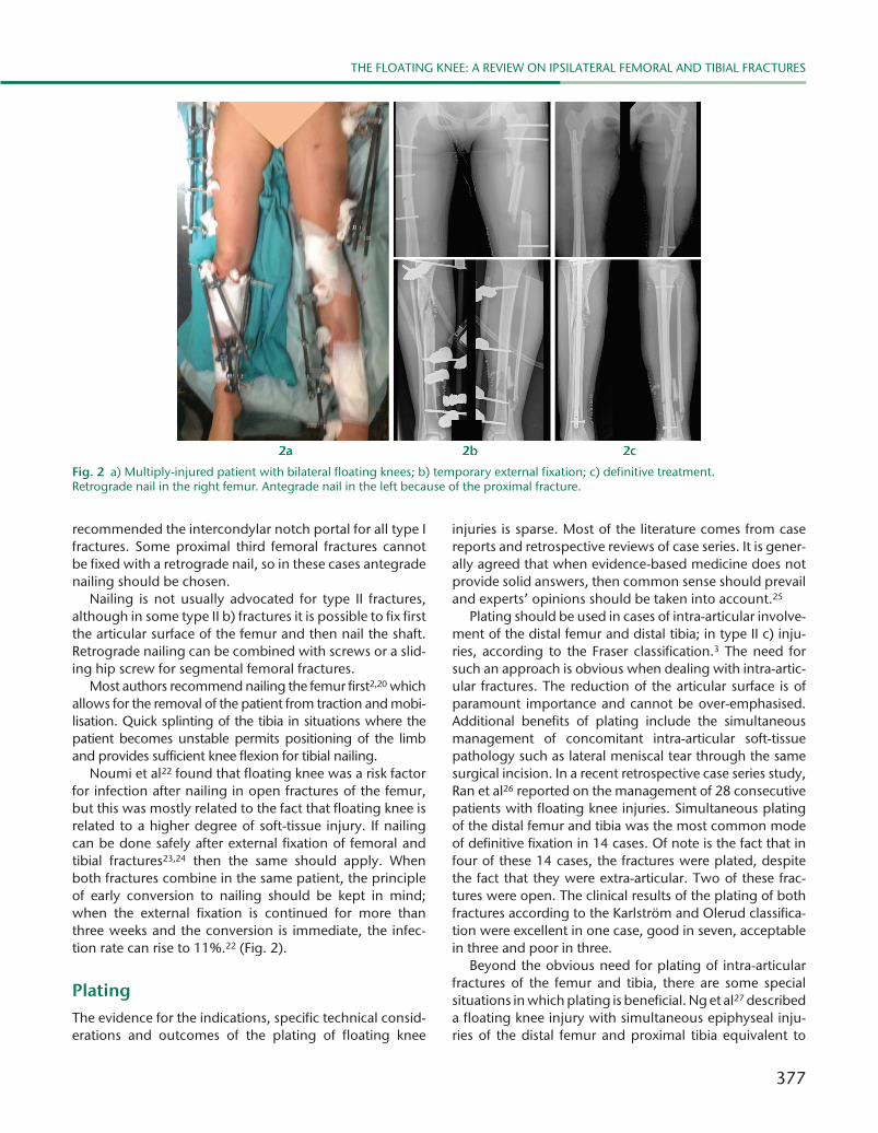
Noumi et al22 found that floating knee was a risk factor for infection after nailing in open fractures of the femur, but this was mostly related to the fact that floating knee is related to a higher degree of soft-tissue injury. If nailing can be done safely after external fixation of femoral and tibial fractures23,24 then the same should apply. When both fractures combine in the same patient, the principle of early conversion to nailing should be kept in mind; when the external fixation is continued for more than three weeks and the conversion is immediate, the infec-tion rate can rise to 11%.22 (Fig. 2).
PlatingThe evidence for the indications, specific technical consid-erations and outcomes of the plating of floating knee
injuries is sparse. Most of the literature comes from case reports and retrospective reviews of case series. It is gener-ally agreed that when evidence-based medicine does not provide solid answers, then common sense should prevail and experts’ opinions should be taken into account.25
Plating should be used in cases of intra-articular involve-ment of the distal femur and distal tibia; in type II c) inju-ries, according to the Fraser classification.3 The need for such an approach is obvious when dealing with intra-artic-ular fractures. The reduction of the articular surface is of paramount importance and cannot be over- emphasised. Additional benefits of plating include the simultaneous management of concomitant intra-articular soft-tissue pathology such as lateral meniscal tear through the same surgical incision. In a recent retrospective case series study, Ran et al26 reported on the management of 28 consecutive patients with floating knee injuries. Simultaneous plating of the distal femur and tibia was the most common mode of definitive fixation in 14 cases. Of note is the fact that in four of these 14 cases, the fractures were plated, despite the fact that they were extra-articular. Two of these frac-tures were open. The clinical results of the plating of both fractures according to the Karlström and Olerud classifica-tion were excellent in one case, good in seven, acceptable in three and poor in three.
Beyond the obvious need for plating of intra-articular fractures of the femur and tibia, there are some special situations in which plating is beneficial. Ng et al27 described a floating knee injury with simultaneous epiphyseal inju-ries of the distal femur and proximal tibia equivalent to
Fig. 2 a) Multiply-injured patient with bilateral floating knees; b) temporary external fixation; c) definitive treatment. Retrograde nail in the right femur. Antegrade nail in the left because of the proximal fracture.
378
Salter-Harris type II injuries, in a six-year-old patient who was managed by closed reduction and percutaneous fixa-tion with Kirschner (K-) wires. The authors pointed out the need for anatomical reduction of the physeal injury in these rare situations.
Peri-prosthetic fractures around the knee are on the rise. The distal femur is most commonly affected, followed by the proximal tibia and the patella. Peri-prosthetic frac-tures involving both the distal femur and proximal tibia have been documented in case reports.28,29 Plating should be considered as an option in fractures in close proximity to the components of the prosthesis when stability had previously been unsatisfactory. When the latter is the case, revision total knee arthroplasty (TKA) should be consid-ered. Nevertheless, the treating surgeon should keep in mind that not all fractures are amenable to revision and not every patient can tolerate a prolonged anaesthetic. Recent evidence30,31 suggests that double-plating (addi-tional medial plating) of the distal peri-prosthetic femoral fractures is a valuable option and offers sufficient stability even in the most distal periprosthetic fractures. In the same way it has been reported that double-plating of the proximal tibia should be considered in peri-prosthetic proximal tibial fractures.32 Other considerations that should be taken into account and subsequently divert management towards plating in peri-prosthetic fractures include a ‘closed box’ femoral implant and interprosthetic femoral fractures (fractures in a femur with TKA and total hip arthroplasty (THA)).).
Other clinical scenarios where plating of the distal femur and proximal tibia could be appropriate are frac-tures of the femur or tibia with pre-existing deformity (in which case a nail can cannot be used), when nail entry points of the nail (soft-tissue infection around the entry points) and in situations of damage control orthopaedics and fat embolism syndrome.33
Combination of implantsThe floating knee injury will always have two different fractures. These fractures range from simple diaphyseal to complex articular types. Although the precise incidence of floating knee injuries is not known, it is a relatively uncom-mon injury. The largest series reported in the literature was of 222 patients over an 11-year period.3 Accordingly, the treatment is more experience- than evidence-based.
As the fractures in the femur and tibia are often differ-ent it is not always possible to achieve optimal fixation with the same implant for both fractures. Furthermore soft-tissue injuries and prosthetic and other previous implants might influence the choice of implant for the individual fracture in the floating knee injury.
For the lower part of the femur, a retrograde nail and locking plates are the most common implants used and treatment choice should probably not differ from a similar isolated femur fracture, regardless of the tibial fracture. Retrograde nails and locking plates have shown similar outcomes and complication rates34 and it is therefore the surgeon’s personal experience that decides which implant is most suitable in each case.
For the tibia fracture in the upper half, antegrade nail and locking plates are used most widely. Nails with advanced locking options can manage some simple
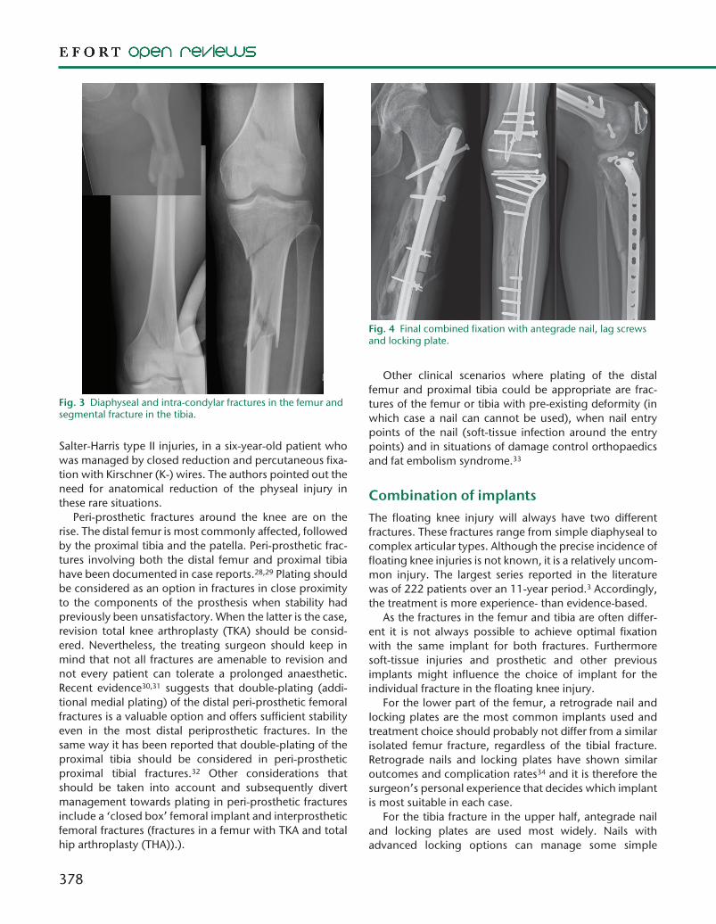
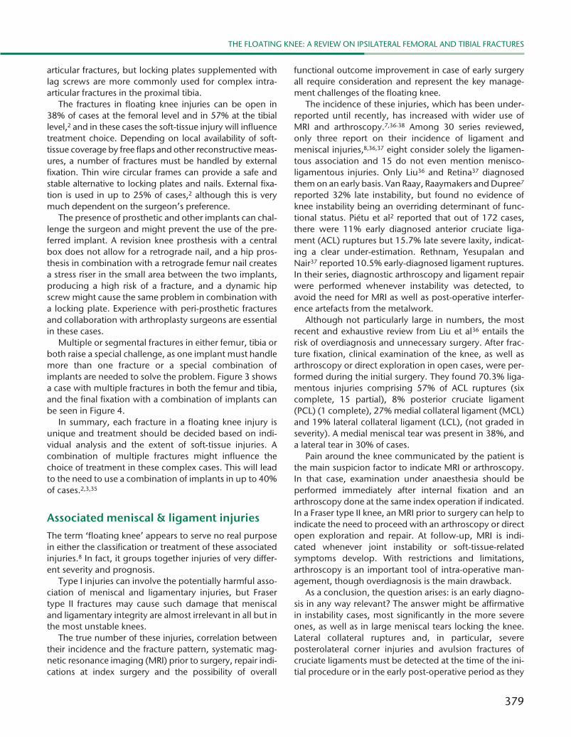
Fig. 3 Diaphyseal and intra-condylar fractures in the femur and segmental fracture in the tibia.
Fig. 4 Final combined fixation with antegrade nail, lag screws and locking plate.
379
The floaTing knee: a review on ipsilaTeral femoral and Tibial fracTures
articular fractures, but locking plates supplemented with lag screws are more commonly used for complex intra-articular fractures in the proximal tibia.
The fractures in floating knee injuries can be open in 38% of cases at the femoral level and in 57% at the tibial level,2 and in these cases the soft-tissue injury will influence treatment choice. Depending on local availability of soft-tissue coverage by free flaps and other reconstructive meas-ures, a number of fractures must be handled by external fixation. Thin wire circular frames can provide a safe and stable alternative to locking plates and nails. External fixa-tion is used in up to 25% of cases,2 although this is very much dependent on the surgeon’s preference.
The presence of prosthetic and other implants can chal-lenge the surgeon and might prevent the use of the pre-ferred implant. A revision knee prosthesis with a central box does not allow for a retrograde nail, and a hip pros-thesis in combination with a retrograde femur nail creates a stress riser in the small area between the two implants, producing a high risk of a fracture, and a dynamic hip screw might cause the same problem in combination with a locking plate. Experience with peri-prosthetic fractures and collaboration with arthroplasty surgeons are essential in these cases.
Multiple or segmental fractures in either femur, tibia or both raise a special challenge, as one implant must handle more than one fracture or a special combination of implants are needed to solve the problem. Figure 3 shows a case with multiple fractures in both the femur and tibia, and the final fixation with a combination of implants can be seen in Figure 4.
In summary, each fracture in a floating knee injury is unique and treatment should be decided based on indi-vidual analysis and the extent of soft-tissue injuries. A combination of multiple fractures might influence the choice of treatment in these complex cases. This will lead to the need to use a combination of implants in up to 40% of cases.2,3,35
Associated meniscal & ligament injuriesThe term ‘floating knee’ appears to serve no real purpose in either the classification or treatment of these associated injuries.8 In fact, it groups together injuries of very differ-ent severity and prognosis.
Type I injuries can involve the potentially harmful asso-ciation of meniscal and ligamentary injuries, but Fraser type II fractures may cause such damage that meniscal and ligamentary integrity are almost irrelevant in all but in the most unstable knees.
The true number of these injuries, correlation between their incidence and the fracture pattern, systematic mag-netic resonance imaging (MRI) prior to surgery, repair indi-cations at index surgery and the possibility of overall
functional outcome improvement in case of early surgery all require consideration and represent the key manage-ment challenges of the floating knee.
The incidence of these injuries, which has been under-reported until recently, has increased with wider use of MRI and arthroscopy.7,36-38 Among 30 series reviewed, only three report on their incidence of ligament and meniscal injuries,8,36,37 eight consider solely the ligamen-tous association and 15 do not even mention menisco-ligamentous injuries. Only Liu36 and Retina37 diagnosed them on an early basis. Van Raay, Raaymakers and Dupree7 reported 32% late instability, but found no evidence of knee instability being an overriding determinant of func-tional status. Piétu et al2 reported that out of 172 cases, there were 11% early diagnosed anterior cruciate liga-ment (ACL) ruptures but 15.7% late severe laxity, indicat-ing a clear under-estimation. Rethnam, Yesupalan and Nair37 reported 10.5% early-diagnosed ligament ruptures. In their series, diagnostic arthroscopy and ligament repair were performed whenever instability was detected, to avoid the need for MRI as well as post-operative interfer-ence artefacts from the metalwork.
Although not particularly large in numbers, the most recent and exhaustive review from Liu et al36 entails the risk of overdiagnosis and unnecessary surgery. After frac-ture fixation, clinical examination of the knee, as well as arthroscopy or direct exploration in open cases, were per-formed during the initial surgery. They found 70.3% liga-mentous injuries comprising 57% of ACL ruptures (six complete, 15 partial), 8% posterior cruciate ligament (PCL) (1 complete), 27% medial collateral ligament (MCL) and 19% lateral collateral ligament (LCL), (not graded in severity). A medial meniscal tear was present in 38%, and a lateral tear in 30% of cases.
Pain around the knee communicated by the patient is the main suspicion factor to indicate MRI or arthroscopy. In that case, examination under anaesthesia should be performed immediately after internal fixation and an arthroscopy done at the same index operation if indicated. In a Fraser type II knee, an MRI prior to surgery can help to indicate the need to proceed with an arthroscopy or direct open exploration and repair. At follow-up, MRI is indi-cated whenever joint instability or soft-tissue-related symptoms develop. With restrictions and limitations, arthroscopy is an important tool of intra-operative man-agement, though overdiagnosis is the main drawback.
As a conclusion, the question arises: is an early diagno-sis in any way relevant? The answer might be affirmative in instability cases, most significantly in the more severe ones, as well as in large meniscal tears locking the knee. Lateral collateral ruptures and, in particular, severe postero lateral corner injuries and avulsion fractures of cruciate ligaments must be detected at the time of the ini-tial procedure or in the early post-operative period as they
380
require early surgery. However, an asymptomatic or low-grade symptomatic ACL rupture or meniscal tear occur-ring with such a severe injury as a floating knee may be the last thing to consider.
OutcomesAlthough several papers have reported the outcome of floating knee injuries after operative or non-operative management, little interest has been paid to the factors that may influence the definitive outcome.
Ipsilateral fractures of the femur and tibia in the adult, or floating knee injury, is a complex lesion with more than just ipsilateral fractures of the femur and tibia.39 High rates of complications have been described.40 In such injuries it is difficult to achieve good functional results due to impor-tant soft-tissue injuries and associated injuries, often resulting in life-threatening trauma of the head, chest, and abdomen or amputation of the limb. Complications related to floating knee injuries include infection, exces-sive blood loss, fat embolism, malunion, delayed or non-union, knee stiffness, prolonged hospitalisation and inability to bear weight.39
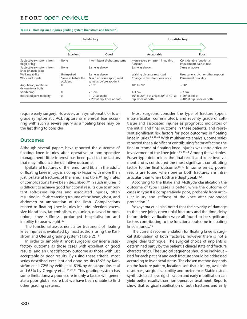
The functional assessment after treatment of floating knee injuries is evaluated by most authors using the Karl-ström and Olerud grading system (Table 2).18
In order to simplify it, most surgeons consider a satis-factory outcome as those cases with excellent or good results, and an unsatisfactory outcome as those with just acceptable or poor results. By using these criteria, most series described excellent and good results (86% by Karl-ström et al, 72% by Veith et al, 81% by Anastopoulos et al and 65% by Gregory et al.15,26,41 This grading system has some limitations; a poor score in only a factor will gener-ate a poor global score but we have been unable to find other grading systems.
Most surgeons consider the type of fracture (open, intra-articular, comminuted), and severity grade of soft-tissue and associated injuries as prognostic indicators of the initial and final outcome in these patients, and repre-sent significant risk factors for poor outcomes in floating knee injuries.15,39-41 With multivariate analysis, some series reported that a significant contributing factor affecting the final outcome of floating knee injuries was intra-articular involvement of the knee joint.15,39-41 Among the variables, Fraser type determines the final result and knee involve-ment and is considered the most significant contributing factor to the final outcome.15,40 In some series, poorer results are found when one or both fractures are intra-articular than when both are diaphyseal.15,41
According to the Blake and McBryde classification the outcome of type I cases is better, while the outcome of cases in type II is comparatively poor, probably from artic-ular injury and stiffness of the knee after prolonged protection.15
Yokoyama et al also noted that the severity of damage to the knee joint, open tibial fractures and the time delay before definitive fixation were all found to be significant factors contributing to the functional outcome in floating knee injuries.40
The current recommendation for floating knee is surgi-cal stabilisation of both fractures; however there is not a single ideal technique. The surgical choice of implants is determined partly by the patient’s clinical state and fracture characteristics. The surgical sequence should be individual-ised for each patient and each fracture should be addressed according to its general status. The chosen method depends on the fracture pattern, location, soft-tissue injury, available resources, surgical capability and preference. Stable osteo-synthesis to achieve rigid fixation and early mobilisation can yield better results than non-operative treatment. Reports show that surgical stabilisation of both fractures and early
Table 2. Floating knee injuries grading system (Karlström and Olerud18)
Satisfactory Unsatisfactory
Excellent Good Acceptable Poor
Subjective symptoms from thigh or leg
None Intermittent slight symptoms More severe symptom impairing function
Considerable functional impairment: pain at rest
Subjective symptoms from knee or ankle joint
None Same as above Same as above Same as above
Walking ability Unimpaired Same as above Walking distance restricted Uses cane, crutch or other supportWork and sports Same as before the
accidentGiven up some sport; work same as before accident
Change to less strenuous work Permanent disability
Angulation, rotational deformity or both
0 < 10° 10° to 20° > 20°
Shortening 0 < 1 cm 1-3 cm > 3 cmRestricted joint mobility 0 < 10° at ankle;
< 20° at hip, knee or both10° to 20° to at ankle; 20° to 40° at hip, knee or both
> 20° at ankle; > 40° at hip, knee or both
381
The floaTing knee: a review on ipsilaTeral femoral and Tibial fracTures
mobilisation can avoid most complications and achieve the best clinical results, but this statement is only true for type I injuries.15
It seems that the presence of open fractures is also a determinant in the final result, even affecting the con-tralateral side. Open fractures predict the likelihood of knee stiffness and delayed full weight-bearing ability. There is also a significant relation between age and out-come, which worsens with age.5
There is a high incidence of associated injuries with the floating knee. However most patients with associated injuries had an excellent or good outcome.37 The associ-ated injuries played a major role in the initial outcome with regards to delay in initial surgery, prolonged dura-tion of surgery and impediment in rehabilitation as higher injury severity scores are associated with delayed full weight-bearing ability. Despite this, associated injuries should be considered in planning of management.
Surprisingly, the incidence of vascular injury is report-edly low; however, if present, functional sequelae are common. Paul et al8 reported six (29%) vascular injuries in their series of 21 patients. However this high rate was not shared in larger studies. Kao et al in fact did not comment on vascular insult in their report on 419 patients.39 Fraser et al reported an incidence of 7% (16 of 222 patients).3
On the treatment of knee ligament injuries, opinions differ widely, but the less favourable results associated with late reconstruction of damaged knee ligaments are generally recognised.7
The vitally important question is, can we estimate the final result beforehand by analysing the patient’s initial condition on admission to the emergency department? Hee et al reported a multivariate analysis and suggested a pre-operative scoring system to determine poor predic-tors of outcome of these fractures which took into consid-eration the age, injury severity scores, smoking status at time of injury, open fractures, segmental fractures and extent of comminution.5 Nevertheless the reliability of this system continues to be debated, and is considered by some authors useless.15
Author informAtion1Hospital Nostra Senyora de Meritxell, Andorra, Spain2Hospices Civils de Lyon, Lyon, France3Hospital Universitario 12 de Octubre, Madrid, Spain4Hospital General Universitario Gregorio Marañón, Madrid, Spain5Hospital General de Castelló, Castelló de la Plana, Spain6Odense Universitetshospita, Odense, Denmark7Leeds General Infirmary, Leeds, UK
Correspondence should be sent to: Josep Muñoz Vives, Hospital Nostra Senyora de Meritxell, Trauma & Orthopaedic Surgery, Av. Fiter i Rossell 1-13, AD700, Escaldes-Engordany, Andorra, Spain.Email: [email protected]
iCmJE ConfliCt of intErEst stAtEmEntNone declared.
funding stAtEmEnt4. No benefits in any form have been received or will be received from a commercial party related directly or indirectly to the subject of this article.
liCEnCE©2016 The author(s)This article is distributed under the terms of the Creative Commons Attribution-NonCommercial 4.0 International (CC BY-NC 4.0) licence (https://creativecommons.org/licenses/by-nc/4.0/) which permits non-commercial use, reproduction and distribution of the work without further permission provided the original work is attributed.
rEfErEnCEs
1. Blake r, mcBryde A Jr. The floating knee: ipsilateral fractures of the tibia and femur. South Med J 1975;68:13-16.
2. Piétu g, Jacquot f, féron J-m, et les membres du gEtrAum. Le genou flottant: étude rétrospective de 172 cas. [The floating knee: a retrospective analysis of 172 cases]. Revue de Chirurgie Orthopédique 2007;93:627-634. (In French)
3. fraser rd, hunter gA, Waddell JP. Ipsilateral fracture of the femur and tibia. J Bone Joint Surg [Br] 1978;60-B:510-515.
4. Veith rg, Winquist rA, hansen st. Ipsilateral fractures of the femur and tibia. A report of fifty-seven consecutive cases. J Bone Joint Surg [Am] 1984;66-A:991-1002.
5. hee ht, Wong hP, low YP, myers l. Predictors of outcome of floating knee injuries in adults: 89 patients followed for 2-12 years. Acta Orthop Scand 2001;72:385-394.
6. Yokoyama K, tsukamoto t, Aoki s, et al. Evaluation of functional outcome of the floating knee injury using multivariate analysis. Arch Orthop Trauma Surg 2002;122:432-435.
7. van raay JJ, raaymakers El, dupree hW. Knee ligament injuries combined with ipsilateral tibial and femoral diaphyseal fractures: the “floating knee”. Arch Orthop Trauma Surg 1991;110:75-77.
8. Paul gr, sawka mW, Whitelaw gP. Fractures of the ipsilateral femur and tibia: emphasis on intra-articular and soft tissue injury. J Orthop Trauma 1990;4:309-314.
9. Akinyoola Al, Yusuf mB, orekha o. Challenges in the management of floating knee injuries in a resource constrained setting. Musculoskelet Surg 2013;97:45-49.
10. dwyer AJ, Paul r, mam mK, Kumar A, gosselin rA. Floating knee injuries: long-term results of four treatment methods. Int Orthop 2005;29:314-318.
11. Anastopoulos g, Assimakopoulos A, Exarchou E, Pantazopoulos t. Ipsilateral fractures of the femur and tibia. Injury 1992;23:439-441.
12. gregory P, diCicco J, Karpik K, et al. Ipsilateral fractures of the femur and tibia: treatment with retrograde femoral nailing and unreamed tibial nailing. J Orthop Trauma 1996;10:309-316.
13. hung sh, Chen tB, Cheng Ym, Cheng nJ, lin sY. Concomitant fractures of the ipsilateral femur and tibia with intra-articular extension into the knee joint. J Trauma 2000;48:547-551.
14. Adamson gJ, Wiss dA, lowery gl, Peters Cl. Type II floating knee: ipsilateral femoral and tibial fractures with intraarticular extension into the knee joint. J Orthop Trauma 1992;6:333-339.
382
15. rethnam u, Yesupalan rs, nair r. The floating knee: epidemiology, prognostic indicators & outcome following surgical management. J Trauma Manag Outcomes 2007;1:2.
16. Bone lB, Johnson Kd, Weigelt J, scheinberg r. Early versus delayed stabilization of femoral fractures. A prospective randomized study. J Bone Joint Surg [Am] 1989;71-A:336-340.
17. Johnson Kd, Cadambi A, seibert gB. Incidence of adult respiratory distress syndrome in patients with multiple musculoskeletal injuries: effect of early operative stabilization of fractures. J Trauma 1985;25:375-384.
18. Karlström g, olerud s. Ipsilateral fracture of the femur and tibia. J Bone Joint Surg [Am] 1977;59-A:240-243.
19. roberts Cs, Pape h-C, Jones Al, et al. Damage control orthopaedics: evolving concepts in the treatment of patients who have sustained orthopaedic trauma. Instr Course Lect 2005;54:447-462.
20. ratliff Ah. Fractures of the shaft of the femur and tibia in the same limb. Proc R Soc Med 1968;61:906-908.
21. ostrum rf. Treatment of floating knee injuries through a single percutaneous approach. Clin Orthop Relat Res 2000;375:43-50.
22. noumi t, Yokoyama K, ohtsuka h, nakamura K, itoman m. Intramedullary nailing for open fractures of the femoral shaft: evaluation of contributing factors on deep infection and nonunion using multivariate analysis. Injury 2005;36:1085-1093.
23. Bhandari m, Zlowodzki m, tornetta P iii, schmidt A, templeman dC. Intramedullary nailing following external fixation in femoral and tibial shaft fractures. J Orthop Trauma 2005;19:140-144.
24. Antich-Adrover P, martí-garin d, murias-Alvarez J, Puente-Alonso C. External fixation and secondary intramedullary nailing of open tibial fractures. A randomised, prospective trial. J Bone Joint Surg [Br] 1997;79-B:433-437.
25. Callaham ml. Expert opinion: supplementing the gaps in evidence-based medicine. Ann Emerg Med 2015;65:61-62.
26. ran t, hua X, Zhenyu Z, et al. Floating knee: a modified Fraser’s classification and the results of a series of 28 cases. Injury 2013;44:1033-1042.
27. ng A, morley Jr, Prasad rn, giannoudis PV, smith rm. The paediatric floating knee: a case report of ipsilateral epiphyseal injury to the distal femur and proximal tibia. J Pediatr Orthop B 2004;13:110-113.
28. Jeong gK, Pettrone sK, liporace fA, meere PA. “Floating total knee”: ipsilateral periprosthetic fractures of the distal femur and proximal tibia after total knee arthroplasty. J Arthroplasty 2006;21:138-140.
29. Jamali AA, lee mA, donthineni r, meehan JP. Minimally invasive management of a floating prosthesis injury with locking plates. J Arthroplasty 2007;22:928-933.
30. Kim JJ, oh hK, Bae J-Y, Kim JW. Radiological assessment of the safe zone for medial minimally invasive plate osteosynthesis in the distal femur with computed tomography angiography. Injury 2014;45:1964-1969.
31. Jiamton C, Apivatthakakul t. The safety and feasibility of minimally invasive plate osteosynthesis (MIPO) on the medial side of the femur: a cadaveric injection study. Injury 2015;46:2170-2176.
32. Kim J-W, oh J-K, oh C-W, Kyung h-s, Park K-h. Minimally invasive plate osteosynthesis for periprosthetic tibial fractures after total knee arthroplasty. 2015 Orthopaedic Trauma Association Annual Congress, San Diego.
33. Bertrand ml, Andrés-Cano P. Management of the floating knee in polytrauma patients. Open Orthop J 2015; 31; (Suppl 1: M10):347–55.
34. griffin Xl, Parsons n, Zbaeda mm, mcArthur J. Interventions for treating fractures of the distal femur in adults. Cochrane Database Syst Rev 2015;8:CD010606.
35. Elmrini A, Elibrahimi A, Agoumi o, et al. Ipsilateral fractures of tibia and femur or floating knee. Int Orthop 2006;30:325-328.
36. liu Y, Zhang J, Zhang s, li r, Yue X. Concomitant ligamentous and meniscal injuries in floating knee. Int J Clin Exp Med 2015;8:1168-1172.
37. rethnam u, Yesupalan rs, nair r. Impact of associated injuries in the floating knee: a retrospective study. BMC Musculoskelet Disord 2009;10:7.
38. szalay mJ, hosking or, Annear P. Injury of knee ligament associated with ipsilateral femoral shaft fractures and with ipsilateral femoral and tibial shaft fractures. Injury 1990;21:398-400.
39. Kao f-C, tu Y-K, hsu K-Y, et al. Floating knee injuries: a high complication rate. Orthopedics 2010;33:14.
40. Yokoyama K, nakamura t, shindo m, et al. Contributing factors influencing the functional outcome of floating knee injuries. Am J Orthop (Belle Mead NJ) 2000;29:721-729.
41. nouraei mh, hosseini A, Zarezadeh A, Zahiri m. Floating knee injuries: results of treatment and outcomes. J Res Med Sci 2013;18:1087-1091.
Copyright © 2022 FDOKUMEN