Ipsilateral directional encoding of joystick movements in human cortex
9
Ipsilateral Directional Encoding of Joystick Movements in Human Cortex Mohit Sharma, The Department of Biomedical Engineering, Washington University in St. Louis, St. Louis, MO Charles Gaona, The Department of Biomedical Engineering, Washington University in St. Louis, St. Louis, MO Jarod Roland, The Washington University School of Medicine, St. Louis, MO Nick Anderson, The Department of Biomedical Engineering, Washington University in St. Louis, St. Louis, MO Zachary Freudenberg, and The Department of Computer Sciences, Washington University in St. Louis, St. Louis, MO Eric C. Leuthardt The Department of Neurological Surgery, Washington University School of Medicine, St. Louis, MO and the Department of Biomedical Engineering, Washington University in St. Louis, St. Louis, MO Abstract The majority of Brain Computer Interfaces have relied on signals related to primary motor cortex and the operation of the contralateral limb. Recently, the physiology associated with same-sided (ipsilateral) motor movements has been found to have a unique cortical physiology. This study sets out to assess whether more complex motor movements can be discerned utilizing ipsilateral cortical signals. In this study, three invasively monitored human subjects were recorded while performing a center out joystick task with the hand ipsilateral to the hemispheric subdural grid array. It was found that directional tuning was present in ipsilateral cortex. This information was encoded in both distinct anatomic populations and spectral distributions. These findings support the notion that ipsilateral signals may provide added information for BCI operation in the future. I. Introduction To date, Brain Computer Interfaces (BCIs) have been put forth as novel engineering approaches to enhance communication and control for patients who have intact cortex but lack motor control due to brain stem stroke, spinal cord injury, or peripheral neuro-muscular dysfunction. With requisite improvements in performance and robustness, these systems have the potential to help tens of thousands of motor disabled subjects. The majority of BCI methodologies are based on a functioning motor cortex that is capable of controlling the contralateral limb. In recent years, there has been an evolving appreciation of how motor and motor related areas participate in same-sided, or ipsilateral, motor movements.[1–3] These findings have prompted further exploration into the underlying cortical physiology as a possible substrate for neuroprosthetic application. corresponding autho, phone: 314-362-8012; fax: 314-362-2107; [email protected]. NIH Public Access Author Manuscript Conf Proc IEEE Eng Med Biol Soc. Author manuscript; available in PMC 2010 August 30. Published in final edited form as: Conf Proc IEEE Eng Med Biol Soc. 2009 ; 2009: 5502–5505. doi:10.1109/IEMBS.2009.5334559. NIH-PA Author Manuscript NIH-PA Author Manuscript NIH-PA Author Manuscript
-
Upload
independent -
Category
Documents
-
view
5 -
download
0
Transcript of Ipsilateral directional encoding of joystick movements in human cortex
Ipsilateral Directional Encoding of Joystick Movements in HumanCortex
Mohit Sharma,The Department of Biomedical Engineering, Washington University in St. Louis, St. Louis, MO
Charles Gaona,The Department of Biomedical Engineering, Washington University in St. Louis, St. Louis, MO
Jarod Roland,The Washington University School of Medicine, St. Louis, MO
Nick Anderson,The Department of Biomedical Engineering, Washington University in St. Louis, St. Louis, MO
Zachary Freudenberg, andThe Department of Computer Sciences, Washington University in St. Louis, St. Louis, MO
Eric C. LeuthardtThe Department of Neurological Surgery, Washington University School of Medicine, St. Louis, MOand the Department of Biomedical Engineering, Washington University in St. Louis, St. Louis, MO
AbstractThe majority of Brain Computer Interfaces have relied on signals related to primary motor cortexand the operation of the contralateral limb. Recently, the physiology associated with same-sided(ipsilateral) motor movements has been found to have a unique cortical physiology. This study setsout to assess whether more complex motor movements can be discerned utilizing ipsilateral corticalsignals. In this study, three invasively monitored human subjects were recorded while performing acenter out joystick task with the hand ipsilateral to the hemispheric subdural grid array. It was foundthat directional tuning was present in ipsilateral cortex. This information was encoded in both distinctanatomic populations and spectral distributions. These findings support the notion that ipsilateralsignals may provide added information for BCI operation in the future.
I. IntroductionTo date, Brain Computer Interfaces (BCIs) have been put forth as novel engineering approachesto enhance communication and control for patients who have intact cortex but lack motorcontrol due to brain stem stroke, spinal cord injury, or peripheral neuro-muscular dysfunction.With requisite improvements in performance and robustness, these systems have the potentialto help tens of thousands of motor disabled subjects. The majority of BCI methodologies arebased on a functioning motor cortex that is capable of controlling the contralateral limb. Inrecent years, there has been an evolving appreciation of how motor and motor related areasparticipate in same-sided, or ipsilateral, motor movements.[1–3] These findings have promptedfurther exploration into the underlying cortical physiology as a possible substrate forneuroprosthetic application.
corresponding autho, phone: 314-362-8012; fax: 314-362-2107; [email protected].
NIH Public AccessAuthor ManuscriptConf Proc IEEE Eng Med Biol Soc. Author manuscript; available in PMC 2010 August 30.
Published in final edited form as:Conf Proc IEEE Eng Med Biol Soc. 2009 ; 2009: 5502–5505. doi:10.1109/IEMBS.2009.5334559.
NIH
-PA Author Manuscript
NIH
-PA Author Manuscript
NIH
-PA Author Manuscript

Wisneski et al. utilized electrocorticography (ECoG) to more definitively define thisphysiology in six motor intact patients undergoing invasive monitoring for seizure localization.Electrocorticographic signals were recorded while the subjects engaged in specific ipsilateralor contralateral hand motor tasks. Spectral changes were identified with regards to frequency,location, and timing. This study showed that ipsilateral hand movements were associated withelectrophysiological changes that occurred in lower frequency spectra (average 37.5Hz), atdistinct anatomic locations (most notably in premotor cortex), and earlier (by 160 ms) thanchanges associated with contralateral hand movements. Given that these cortical changesoccurred earlier and were localized preferentially in premotor cortex compared to thoseassociated with contralateral movements, the authors postulated that ipsilateral cortex is moreassociated with motor planning than its execution. Additionally, these changes were quitedistinct from those changes associated with contralateral motor movements which were moredominantly associated with higher gamma rhythms (average 106.9 HZ). The group furtherdemonstrated that brain derived control of a computer cursor could be achieved by using theanatomic sites or the lower frequency amplitude changes distinctive to ipsilateral movementswhich was comparable to contralateral derived control.[4]
In previous studies, ECoG signals have been found to carry very specific information aboutcontralateral joystick, hand, and finger movements.[5,6] Additionally, these distinct signalshave been able to provide for more complex multidimensional device control.[7] Though thecortical physiology associated with ipsilateral motor movements has been found to be differentthan that associated with contralateral movements, it has not yet been defined to what extentmore complex motor information is present. Using a classic center out task in three invasivelymonitored patients this study set out to examine whether similar levels of complex motormovements could be decoded from the ipsilateral hemisphere.
II. MethodsA. Subjects
The subjects in this study were three patients (ages 36, 48, Tand 58 years) with intractableepilepsy who underwent temporary placement of intracranial electrode arrays to localizeseizure foci prior to surgical resection. All had normal levels of cognitive function, were righthanded, and had left hemispheric 8×8 electrode grid arrays. All gave informed consent. Thestudy was approved by the Washington University Human Research Protection Office. Eachpatient studied was in a sitting position (semi-recumbent), approximately 75 cm from a videoscreen. In all experiments, we recorded ECoG from up to 64 electrodes using the general-purpose BCI system BCI2000 [8]. All electrodes were referenced to an inactive intracranialelectrode, amplified, bandpass filtered (0.5–500 Hz), digitized at 1200 Hz, and stored.
B. Behavioral ParadigmThe subjects performed a two dimensional center out reaching task using a MicrosoftSidewinder force feedback joystick operated by the left hand (ipsilateral to the intracranial gridarray). The subject would be cued to move a cursor from the center of the screen to one of eighttargets placed radially and equidistant around the center point on the periphery. The center wasapproximately 5 inches from any of the targets. Random delay periods ranging from 300–1200msec were added between cue and target presentation to dissociate attention and intentionaleffects from movement effects. The targets were presented in a pseudo randomized order. Allsubjects were presented each target a minimum of 8 times per run and the number of runs foreach subject varied from 4–10 depending on patient compatibility.
Sharma et al. Page 2
Conf Proc IEEE Eng Med Biol Soc. Author manuscript; available in PMC 2010 August 30.
NIH
-PA Author Manuscript
NIH
-PA Author Manuscript
NIH
-PA Author Manuscript

C. AnalysisThe analysis algorithm consisted of the following steps: converting the time domain signal intothe frequency domain, correlating ECoG spectra to joystick kinematics data for position andvelocity encoding, and statistical evaluation to quantify significant results. ECoG signal hasbeen previously shown to include sharp changes in the time domain and hence to fit spectrawith sharp features and increase the frequency resolution, the Maximum Entropy Method(MEM) was used.
The MEM is an Auto-Regressive (AR) filter model which does not reconstruct the powerspectra but estimates it based on the input parameters. It estimates the power spectrum of theinput signal using the following mathematical expression:
The parameters that determine the estimation are model order (M), sample data and thesampling frequency at which the data is acquired. The coefficients ak are computed from theautocorrelation function of the sample data. The parameter z of the power spectrum is definedas z = e2πifΔ (where f represents the frequency and Δ is the sampling interval) and can be usedto resolve the power spectrum to the required frequency resolution.
Movement period spectra generated using MEM was normalized by comparing to the baselinesignal spectra. Normalized spectra was regressed with the position coordinates of one of fourtargets (positioned at 0, 90, 180, and 270 degrees) on a trial by trial basis. Their correlation tothe ECoG spectra was computed using the MATLAB function regress which evaluates thelinear correlation coefficients as described by Georgopoulos et al. [9] The regression analysisis performed for individual electrodes over the complete frequency spectrum. We nextmeasured the strength of correlation using the Depth of Modulation (DOM) of the cosine tuningcurve, which is defined as the percent change in activity from baseline during movement inthe preferred direction. The following expression demonstrates the regression and cosinetuning analysis:
where Pj is the normalized activity spectra in the movement direction, xj and yj are positioncoordinates of final target positions, b’s are the regression coefficients, DOM is the depth ofmodulation and θPD-M is the angle between the preferred direction and movement direction.Features (frequency band at certain electrode location) were assessed to be statisticallysignificant if the DOM is greater than 0.05 and the p-value (strength of fit to the tuning curve)is less than 0.05.
D. Anatomic ReconstructionsRadiographs were used to identify the stereotactic coordinates of each grid electrode [10], andcortical areas were defined using Talairach’s Co-Planar Stereotaxic Atlas of the Human Brain[11] and a Talairach transformation database(http://ric.uthscsa.edu/projects/talairachdaemon.html). We obtained a template 3D corticalbrain model from the AFNI SUMA web site(http://afni.nimh.nih.gov/afni/suma).Stereotactically defined electrode locations were then mapped to this template brain model.We then created DOM activation plots for each patient using a customized MatLab program.
Sharma et al. Page 3
Conf Proc IEEE Eng Med Biol Soc. Author manuscript; available in PMC 2010 August 30.
NIH
-PA Author Manuscript
NIH
-PA Author Manuscript
NIH
-PA Author Manuscript
For each plot, only electrode sites with a p value of less than 0.05 were considered. The resultingmap showed the activation at each point on the brain model for the target direction considered.
III. ResultsIn this study, each of the three subjects tested showed significant cortical changes that werespecifically tuned to one of the four directions during joystick movement. Of the 176 totalelectrodes recorded over the three patients, 85 showed statistically significant tuning to one ormore directions (0, 90, 180, and 270 degrees). As shown in Figure 1, there was a distinctanatomic pattern of cortical activation for each target when examined across all frequenciesbetween 0.5–275 Hz. These populations were somewhat heterogeneous between subjects. Thislikely is accounted for by the variability in grid placement between individual subjects and the1 cm electrode spacing which is coarse from a functional standpoint. For each of the subjectsthere was directional tuning in all frequencies (See Figure 2)
Beyond cortical populations, there was also distinct directional tuning at individual sites.Individual electrode sites showed tuning for up to three of the four independent directions.Figure 3A shows the number of electrode sites that showed tuning for one, two, and threedirections. The majority (33.5%) of tuned electrodes showed tuning to a single direction. Asmaller subset of sites however showed tuning for more than one direction (14.8%). Figure 3Bshows an example of an electrode and its associated directional tuning to multiple directions.The separable directional tuning was based on distinct frequency bands showing independentdepth of modulation. Figure 3C shows the respective frequency bands that had significant DOMfor the associated directional tuning. When a single direction was encoded, it appears a broaddistribution of frequencies where represented. As a site showed a higher number of encodeddirections the frequency range between 65–115 Hz became much more prominent.
IV. DiscussionThis study demonstrates that two-dimensional information is present in electrocorticographicsignals taken from cortex ipsilateral to the hand operating a joystick. This information isencoded by distinct cortical populations demonstrating tuned depth of modulation in specificfrequency bands to specific directions. These cortical populations are represented in distinctanatomic locations (e.g 45Hz DOM tuned to the 90 degree target at several distinct corticallocations) and, for a given location, there is further directional encoding present in distinctfrequency spectra (e.g. for a single site, 70 Hz DOM encodes for a target at 180 degrees and a110Hz DOM encodes for a target at 270 degrees) For sites encoding a single direction thereappears to be a diverse range of frequency representations, as single sites encode morenumerous directions, the frequency bands that are tuned to multiple directions predominantlyrange between 65–115 Hz.
These findings build upon the current understanding of ipsilateral motor physiology and ECoGbased neuroprosthetics. There have been numerous studies demonstrating that ipsilateral limbmovements have been associated with a distinct and separable cortical physiology. [1,3,12,13] Initially this was felt to be the result of transcallosal inhibition.[14,15] More recent findingshave demonstrated this to be an active phenomenon thought to be more involved with motorplanning.[16,17] Additionally, this separable physiology as measured with ECoG has beenutilized for simple BCI device control. [4] Using ECoG, Schalk et al demonstrated that twodimensional joystick movements could be decoded from cortex contralateral to the operatinghand.[18] Later this same group demonstrated two-dimensional control was indeed possibleusing ECoG. [7] In this study, the results show for the first time that two dimensionalinformation is also present with ipsilateral joystick movements. This study supports the notion
Sharma et al. Page 4
Conf Proc IEEE Eng Med Biol Soc. Author manuscript; available in PMC 2010 August 30.
NIH
-PA Author Manuscript
NIH
-PA Author Manuscript
NIH
-PA Author Manuscript

that multidimensional control may also be possible using ipsilaterally derived corticalphysiology.
Further evaluation will be required to define the independence of these cortical signalsencoding ipsilateral directional information to that of contralateral derived directionalinformation. If separable, this ipsilateral kinematic information could potentially provide aparallel set of features for control that would be complementary to signals derived from classicmotor physiology associated with contralateral movement imagery. Beyond the potentialincrease in degrees of freedom of control, these findings provide important preliminaryevidence that a BCI could achieve “bisomatic” control—a unilateral neuroprosthetic that couldenable a single hemisphere to facilitate control of both sides of the body. This couldsubstantially improve the control capabilities of an ECoG based BCI for motor impaired patientpopulations.
AcknowledgmentsThis work was supported in part by the James S. McDonnell Foundation (Leuthardt), Department of Defense(W911NF-07-1-0415 & W911NF-08-1-0216), National Institute of Health (NINDS NIH R01-EB000856-06), and theChildren’s Discovery Institute.
V. References1. Cramer SC, Finklestein SP, Schaechter JD, Bush G, Rosen BR. Activation of distinct motor cortex
regions during ipsilateral and contralateral finger movements. Journal of neurophysiology 1999;81(1):383–387. [PubMed: 9914297]
2. Baraldi P, Porro CA, Serafini M, Pagnoni G, Murari C, Corazza R, Nichelli P. Bilateral representationof sequential finger movements in human cortical areas. Neuroscience letters 1999;269(2):95–98.[PubMed: 10430513]
3. Verstynen T, Diedrichsen J, Albert N, Aparicio P, Ivry RB. Ipsilateral motor cortex activity duringunimanual hand movements relates to task complexity. Journal of neurophysiology 2005;93(3):1209–1222. [PubMed: 15525809]
4. Wisneski KJ, Anderson N, Schalk G, Smyth M, Moran D, Leuthardt EC. Unique cortical physiologyassociated with ipsilateral hand movements and neuroprosthetic implications. Stroke; a journal ofcerebral circulation 2008;39(12):3351–3359.
5. Schalk G, Kubanek J, Miller KJ, Anderson NR, Leuthardt EC, Ojemann JG, Limbrick D, Moran D,Gerhardt LA, Wolpaw JR. Decoding two-dimensional movement trajectories usingelectrocorticographic signals in humans. J Neural Eng 2007;4(3):264–275. [PubMed: 17873429]
6. Miller KJ, Zanos S, Fetz EE, den Nijs M, Ojemann JG. Decoupling the cortical power spectrum revealsreal-time representation of individual finger movements in humans. J Neurosci 2009;29(10):3132–3137. [PubMed: 19279250]
7. Schalk G, Miller KJ, Anderson NR, Wilson JA, Smyth MD, Ojemann JG, Moran DW, Wolpaw JR,Leuthardt EC. Two-dimensional movement control using electrocorticographic signals in humans. JNeural Eng 2008;5(1):75–84. [PubMed: 18310813]
8. Schalk G, McFarland DJ, Hinterberger T, Birbaumer N, Wolpaw JR. BCI2000: a general-purposebrain-computer interface (BCI) system. IEEE Trans Biomed Eng 2004;51(6):1034–1043. [PubMed:15188875]
9. Georgopoulos AP, Kalaska JF, Caminiti R, Massey JT. On the relations between the direction of two-dimensional arm movements and cell discharge in primate motor cortex. J Neurosci 1982;2(11):1527–1537. [PubMed: 7143039]
10. Fox PT, Perlmutter JS, Raichle ME. A stereotactic method of anatomical localization for positronemission tomography. J Comput Assist Tomogr 1985;9(1):141–153. [PubMed: 3881487]
11. Talairach, J.; Tournoux, P. Co-Planar Sterotaxic Atlas of the Human Brain. Vol. 1988. ThiemeMedical Publishers, Inc; 1988.
Sharma et al. Page 5
Conf Proc IEEE Eng Med Biol Soc. Author manuscript; available in PMC 2010 August 30.
NIH
-PA Author Manuscript
NIH
-PA Author Manuscript
NIH
-PA Author Manuscript
12. Rao SM, Binder JR, Bandettini PA, Hammeke TA, Yetkin FZ, Jesmanowicz A, Lisk LM, Morris GL,Mueller WM, Estkowski LD, et al. Functional magnetic resonance imaging of complex humanmovements. Neurology 1993;43(11):2311–2318. [PubMed: 8232948]
13. Kim SG, Ashe J, Georgopoulos AP, Merkle H, Ellermann JM, Menon RS, Ogawa S, Ugurbil K.Functional imaging of human motor cortex at high magnetic field. Journal of neurophysiology1993;69(1):297–302. [PubMed: 8433133]
14. Ferbert A, Priori A, Rothwell JC, Day BL, Colebatch JG, Marsden CD. Interhemispheric inhibitionof the human motor cortex. J Physiol 1992;453:525–546. [PubMed: 1464843]
15. Newton JM, Sunderland A, Gowland PA. fMRI signal decreases in ipsilateral primary motor cortexduring unilateral hand movements are related to duration and side of movement. NeuroImage 2005;24(4):1080–1087. [PubMed: 15670685]
16. Johansen-Berg H, Rushworth MF, Bogdanovic MD, Kischka U, Wimalaratna S, Matthews PM. Therole of ipsilateral premotor cortex in hand movement after stroke. Proc Natl Acad Sci U S A 2002;99(22):14518–14523. [PubMed: 12376621]
17. Huang MX, Harrington DL, Paulson KM, Weisend MP, Lee RR. Temporal dynamics of ipsilateraland contralateral motor activity during voluntary finger movement. Hum Brain Mapp 2004;23(1):26–39. [PubMed: 15281139]
18. Schalk, G.; Kubánek, J.; Miller, KJ.; Anderson, NR.; Leuthardt, EC.; Ojemann, JG.; Limbrick, D.;Moran, D.; Gerhardt, LA.; Wolpaw, JR. Book Decoding two-dimensional movement trajectoriesusing electrocorticographic signals in humans. 2007. Decoding two-dimensional movementtrajectories using electrocorticographic signals in humans; p. 264-275.
Sharma et al. Page 6
Conf Proc IEEE Eng Med Biol Soc. Author manuscript; available in PMC 2010 August 30.
NIH
-PA Author Manuscript
NIH
-PA Author Manuscript
NIH
-PA Author Manuscript
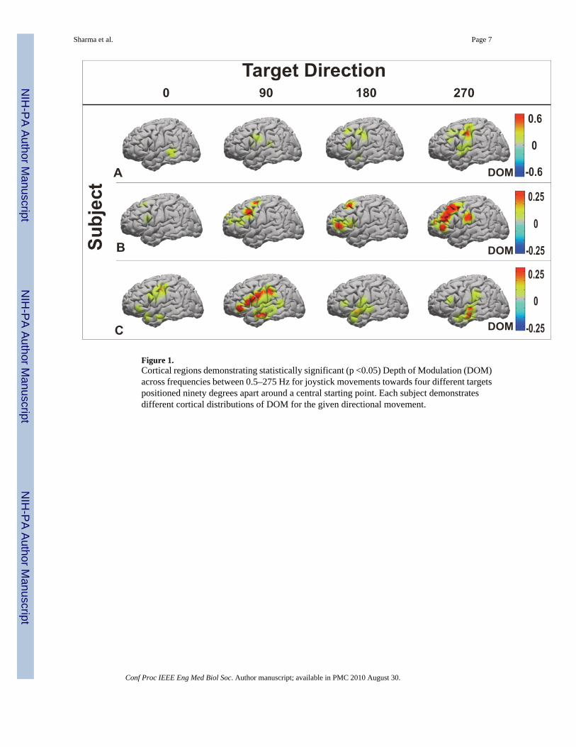
Figure 1.Cortical regions demonstrating statistically significant (p <0.05) Depth of Modulation (DOM)across frequencies between 0.5–275 Hz for joystick movements towards four different targetspositioned ninety degrees apart around a central starting point. Each subject demonstratesdifferent cortical distributions of DOM for the given directional movement.
Sharma et al. Page 7
Conf Proc IEEE Eng Med Biol Soc. Author manuscript; available in PMC 2010 August 30.
NIH
-PA Author Manuscript
NIH
-PA Author Manuscript
NIH
-PA Author Manuscript

Figure 2.The frequency spectra demonstrating significant Depth of Modulation (DOM) for any of thefour directions. The degree to which a frequency was represented was measured by the numberof electrode sites that showed significant DOM at a frequency within the respective ranges.The majority of spectra showing directionally tuned DOM were below 175 Hz.
Sharma et al. Page 8
Conf Proc IEEE Eng Med Biol Soc. Author manuscript; available in PMC 2010 August 30.
NIH
-PA Author Manuscript
NIH
-PA Author Manuscript
NIH
-PA Author Manuscript

Figure 3.A. Number of electrode sites showing significant Depth of Modulation (DOM) for a singledirection and multiple directions (across all patients). B. Demonstrates an example of how asingle site encodes for multiple directions. A significant DOM of one spectral band is tuned toone direction (blue), while another spectral band is tuned to another direction (red). C. Thefrequency distribution showing significant DOM for sites encoding one, two, and threedifferent directions. As a site encodes more directions the frequency range between 65–115Hz predominates in spectra showing tuned DOM.
Sharma et al. Page 9
Conf Proc IEEE Eng Med Biol Soc. Author manuscript; available in PMC 2010 August 30.
NIH
-PA Author Manuscript
NIH
-PA Author Manuscript
NIH
-PA Author Manuscript