Safety and feasibility of transulnar catheterization when ipsilateral radial access is not available
10
Safety and Feasibility of Transulnar Catheterization When Ipsilateral Radial Access Is Not Available Sasko Kedev, 1 * MD, PhD, Biljana Zafirovska, 1 MD, Surya Dharma, 2 MD, and Danica Petkoska, 1 MD Objectives: We evaluated the safety and feasibility of transulnar approach when ipsilat- eral radial access was not available. Methods and Results: From March 2011 until February 2013, 476 consecutive patients who underwent transulnar catheterization were included in a single center prospective registry of effectiveness and safety. Diag- nostic coronary angiography accounted for 42% of cases, percutaneous coronary intervention (PCI) for 38%, and 17% underwent carotid artery stenting. A subgroup analysis was done in 240 patients with documented ipsilateral radial artery occlusion (RAO). Procedural success was 97% with a crossover rate of 3% to transfemoral access. Hand ischemia was not observed in any patient on day 1 after procedure and on 1 month follow-up. None of the patients showed ulnar nerve injury. Two patients developed major forearm hematoma that resolved without clinical consequences. Minor access site hematoma occurred in 8%. Severe clinical spasm occurred in two patients. Asymptomatic ulnar artery occlusion at 1 month follow-up was detected in 3.1%. There was no difference between patients with or without RAO in terms of pro- cedural success and any vascular complication. Conclusion: Transulnar approach is safe and feasible alternative wrist access when performed by experienced radial oper- ators, providing high success rate and low incidence of vascular complications. V C 2013 Wiley Periodicals, Inc. Key words: transulnar approach; transradial approach; radial artery occlusion; percuta- neous coronary intervention; carotid artery stenting INTRODUCTION The first effort at retrograde arterial catheterization of the left ventricle involved the use of ulnar artery cutdown for arterial access. Zimmerman et al. [1] used this approach for the first successful retrograde cathe- terization of the left ventricle in 1949 for 11 patients with aortic insufficiency. Based on 2 decades of experience and data, a large European panel of experts has recommended radial access as the first choice for angiography and PCI when performed by operators experienced with the method [2]. Opposite to the transradial access (TRA), which is now rapidly becoming the preferred access site for cor- onary interventions worldwide, the transulnar artery approach (TUA) has received very little attention as a potential access for cardiac catheterization [3,4]. Reported technical failure for transradial procedures is between 1% and 7%, mainly related to the learning curve [4]. Furthermore, TRA has limitations in some condi- tions such as weak radial pulse after several previous TRA procedures, RAO [5,6], radial artery (RA) used for surgical grafts or fistulas, RA anomalies [7,8], RA spasm (RAS) [9,10], and procedures requiring large bore devices. TUA has been described as a viable alternative wrist access for coronary diagnostic and interventional 1 University Clinic of Cardiology, Medical Faculty, University of St.Cyril & Methodius, Skopje, Macedonia 2 Department of Cardiology and Vascular Medicine, Faculty of Medicine, University of Indonesia, National Cardiovascular Center Harapan Kita, Jakarta, Indonesia Conflict of interest: Nothing to report. *Correspondence to: Sasko Kedev, University Clinic of Cardiology, Medical Faculty, University of St. Cyril & Methodius, Vodnjanska 17, Skopje, Macedonia. E-mail: [email protected] Received 3 May 2013; Revision accepted 30 June 2013 DOI: 10.1002/ccd.25123 Published online 5 July 2013 in Wiley Online Library (wileyonlinelibrary.com). V C 2013 Wiley Periodicals, Inc. Catheterization and Cardiovascular Interventions 83:E51–E60 (2014)
-
Upload
independent -
Category
Documents
-
view
1 -
download
0
Transcript of Safety and feasibility of transulnar catheterization when ipsilateral radial access is not available

Safety and Feasibility of Transulnar CatheterizationWhen Ipsilateral Radial Access Is Not Available
Sasko Kedev,1* MD, PhD, Biljana Zafirovska,1 MD, Surya Dharma,2 MD, andDanica Petkoska,1 MD
Objectives: We evaluated the safety and feasibility of transulnar approach when ipsilat-eral radial access was not available. Methods and Results: From March 2011 untilFebruary 2013, 476 consecutive patients who underwent transulnar catheterizationwere included in a single center prospective registry of effectiveness and safety. Diag-nostic coronary angiography accounted for 42% of cases, percutaneous coronaryintervention (PCI) for 38%, and 17% underwent carotid artery stenting. A subgroupanalysis was done in 240 patients with documented ipsilateral radial artery occlusion(RAO). Procedural success was 97% with a crossover rate of 3% to transfemoralaccess. Hand ischemia was not observed in any patient on day 1 after procedure andon 1 month follow-up. None of the patients showed ulnar nerve injury. Two patientsdeveloped major forearm hematoma that resolved without clinical consequences.Minor access site hematoma occurred in 8%. Severe clinical spasm occurred in twopatients. Asymptomatic ulnar artery occlusion at 1 month follow-up was detected in3.1%. There was no difference between patients with or without RAO in terms of pro-cedural success and any vascular complication. Conclusion: Transulnar approach issafe and feasible alternative wrist access when performed by experienced radial oper-ators, providing high success rate and low incidence of vascular complications. VC 2013Wiley Periodicals, Inc.
Key words: transulnar approach; transradial approach; radial artery occlusion; percuta-neous coronary intervention; carotid artery stenting
INTRODUCTION
The first effort at retrograde arterial catheterizationof the left ventricle involved the use of ulnar arterycutdown for arterial access. Zimmerman et al. [1] usedthis approach for the first successful retrograde cathe-terization of the left ventricle in 1949 for 11 patientswith aortic insufficiency.
Based on 2 decades of experience and data, a largeEuropean panel of experts has recommended radialaccess as the first choice for angiography and PCIwhen performed by operators experienced with themethod [2].
Opposite to the transradial access (TRA), which isnow rapidly becoming the preferred access site for cor-onary interventions worldwide, the transulnar arteryapproach (TUA) has received very little attention as apotential access for cardiac catheterization [3,4].Reported technical failure for transradial procedures isbetween 1% and 7%, mainly related to the learningcurve [4].
Furthermore, TRA has limitations in some condi-tions such as weak radial pulse after several previous
TRA procedures, RAO [5,6], radial artery (RA) usedfor surgical grafts or fistulas, RA anomalies [7,8], RAspasm (RAS) [9,10], and procedures requiring largebore devices.
TUA has been described as a viable alternative wristaccess for coronary diagnostic and interventional
1University Clinic of Cardiology, Medical Faculty, University ofSt.Cyril & Methodius, Skopje, Macedonia2Department of Cardiology and Vascular Medicine, Faculty ofMedicine, University of Indonesia, National CardiovascularCenter Harapan Kita, Jakarta, Indonesia
Conflict of interest: Nothing to report.
*Correspondence to: Sasko Kedev, University Clinic of Cardiology,Medical Faculty, University of St. Cyril & Methodius, Vodnjanska17, Skopje, Macedonia. E-mail: [email protected]
Received 3 May 2013; Revision accepted 30 June 2013
DOI: 10.1002/ccd.25123Published online 5 July 2013 in Wiley Online Library(wileyonlinelibrary.com).
VC 2013 Wiley Periodicals, Inc.
Catheterization and Cardiovascular Interventions 83:E51–E60 (2014)

procedures, with favorable results reported in small ini-tial studies [11–16]. Larger studies has further con-firmed the safety and effectiveness of TUA as analternative approach to TRA for coronary procedures[17,18].
However, there are no large-scale studies on safetyand feasibility of TUA for interventions with largebore devices and in patients with coexisting ipsilateralRAO. The main objective of this study was to furtherevaluate the safety and feasibility of TUA for cardio-vascular procedures in a dedicated high volume TRAcenter, when ipsilateral radial access was not available.
METHODS
This study was designed as a single center prospectiveregistry of effectiveness and safety, and data were col-lected in a dedicated database. All procedures were doneby experienced high-volume operators performing morethan 95% of all procedures through the TRA, rangingfrom 300 to 600 procedures annually per operator.
Patient Population
From March 2011 to February 2013, a total of 9,139patients underwent TRA catheterization in our institu-tion. The right radial access was the most commonroute (97%).
TRA was not available in 504 (5.5%) patients due toinability to puncture the RA, weak or absence of radialpulse, RAS and dissection, hypoplastic or small RA,noncrossable radial loop, and high take off of smallcaliber RA.
In 476 consecutive patients (5.2%), the procedureswere performed through the ipsilateral TUA while only26 patients were switched to femoral approach (0.3%)and 2 patients underwent ipsilateral brachial catheteri-zation (0.02%). Crossover to femoral and brachialaccess was performed because ipsilateral ulnar arterywas not available (absence of ulnar pulse, extensiveUA calcifications). From 476 analyzed patients withTUA, diagnostic procedures accounted for 42%of cases, PCI for 38%, and 84 patients underwentCAS (17%).
Written, informed consent was obtained from eachsubject. This study has been approved by institutionalreview committee and the medical ethical committee.Study flow charts are shown in Figs. 1 and 2.
Ulnar Artery Cannulation and PreproceduralUlnar Artery Angiography
The wrist was hyperextended and the skin was pre-pared with local anesthesia (1 ml lidocaine 2%) around
Fig. 1. Flow chart of the study.
E52 Kedev et al.
Catheterization and Cardiovascular Interventions DOI 10.1002/ccd.Published on behalf of The Society for Cardiovascular Angiography and Interventions (SCAI).

0.5–3 cm proximal to the flexor crease skin fold alongthe axis with the most powerful pulsation of the UA.
The UA was accessed with a 20-G plastic cannulausing the counter-puncture technique. The cannula-over-needle was inserted at a 45–60
!angle along the
vessel axis and from lateral to medial, thus avoidingthe ulnar nerve. When a good arterial “back bleed”was obtained, the 0.02500 hydrophilic guidewire wasadvanced and the hydrophilic 5F or 6F sheath (Radifo-cus, Terumo, Tokyo, Japan) was introduced over theguidewire.
Intra-arterial vasodilator (5 mg verapamil) wasinjected to reduce ulnar artery spasm (UAS). Immedi-ately after sheath insertion, intravenous unfractionatedheparin (UH) was administered (50–70 l/kg, up to5,000 units).
UA angiography was performed through the cannulaor sheath, before catheter insertion (Fig. 3). The fore-arm angiography defined the radial and UA anatomyfrom mid forearm to brachial/axilar anastomosis. Adiluted solution of 3 ml of contrast mixed with 7 ml ofblood was injected briskly and recorded. In cases withUAS, tortuosity and/or ulnar loops, and high take-offUA, we used a 0.01400 soft PCI guidewire under fluo-roscopy guidance.
Coronary Procedure
When ulnar access was provided and sheath was inplace, the procedures were performed in the same fash-ion as from the TRA. Choice of diagnostic and guidingcatheters, wires, balloons, and stents were left to theoperator’s discretion. For larger bore devices (>6F)used in complex PCI of bifurcations, left main stenting,
chronic total occlusion, and when inserting a 7.5Fsheathless guiding catheter (ASAHI INTECC, Aichi,Japan), a more supportive stiff Amplatz type .03500
260-cm long guidewire was preferred (Fig. 4).All diagnostic coronary angiogram patients received
intravenous bolus of 5,000 IU UH. PCI patients already
Fig. 2. Flow chart of patients with or without RAO.
Fig. 3. Ulnar artery angiography through cannula with palmararches.
Safety and Feasibility of TUA E53
Catheterization and Cardiovascular Interventions DOI 10.1002/ccd.Published on behalf of The Society for Cardiovascular Angiography and Interventions (SCAI).
receiving 100 mg aspirin were preloaded with 600 mgclopidogrel and received 100 IU/kg body weight of in-travenous UH. Clinical and procedural variables wererecorded in all patients.
Carotid Artery Stenting Procedure
Initial carotid angiography was performed through a5F diagnostic catheter (predominantly Simmons 2). CASwas performed through a 5F or 6F, 90 cm guidingsheath: Shuttle sheath (Cook, Minneapolis, MN) or Desti-nation sheath (Terumo, Tokyo, Japan) (Fig. 5) or through7F or 8F guiding catheter. Compatibility with the guidingsheath diameter was determined by the UA size based onulnar angiography. All procedures were performed withdistal protection devices (Nav 6, Abbott Vascular, Teme-cula, CA). All patients were preloaded with 600 mg clo-pidogrel and maintained on 75 mg for at least 1 monthand on 100 mg aspirin daily, indefinitely.
Arterial Sheath Management
UA sheath was removed immediately after the pro-cedure, and hemostasis was achieved by TR band com-pression (Terumo, Tokyo, Japan). TR band was appliedby inflating 15–18 ml of air at the puncture site. AsUA is more deeply seated without nearby bone struc-ture, hemostasis with initially higher pressure thanTRA was necessary particularly after using larger boredevices. The pulse oximetry was used to confirm thathemoglobin oxygen saturation was more than 90% onthe involved hand after hemostasis was obtained (Fig.6). Compression was applied for approximately 2- to3-hour period (depending on the sheath size) withgradual relaxation of compression after the first hour.Patients were discharged within the same day or thefollowing days after a careful examination by theattending physician.
Study Endpoints
Primary endpoint of the study was procedural suc-cess. The occurrence of clinical UAS during the proce-dure, vascular access site and major vascularcomplications at 30 days were defined as the secondaryendpoints of the study. A subgroup analysis was donein 240 patients with documented ipsilateral RAO (diag-nosed from preprocedural ulnar artery angiography).Duplex ultrasonography of the upper extremity arterieswas performed at 1 month in all patients with ipsilat-eral RAO by a dedicated vascular ultrasonographistwho was blinded for the baseline characteristics andmethodology of the study.
Definitions
Procedural success was defined as completion of theplanned procedure through the initial access site [7]
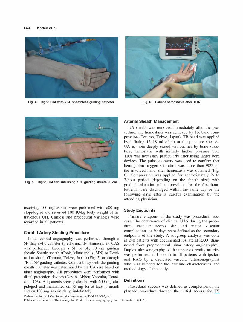
Fig. 4. Right TUA with 7.5F sheathless guiding catheter.
Fig. 5. Right TUA for CAS using a 6F guiding sheath 90 cm.
Fig. 6. Patient hemostasis after TUA.
E54 Kedev et al.
Catheterization and Cardiovascular Interventions DOI 10.1002/ccd.Published on behalf of The Society for Cardiovascular Angiography and Interventions (SCAI).

without associated in cath-lab major clinical complica-tions (e.g., stroke, death, or coronary artery perforation)[19].
Clinical UAS was classified as grade I: minimallocal pain and discomfort; grade II: significant localpain and discomfort, not precluding procedure comple-tion; grade III: severe local pain and discomfort neces-sitating crossover; and grade IV: catheter entrapmentwith severe local pain and discomfort. These criteriacorrelate with clinical signs of radial artery spasm[20].
Vascular access site complications were defined asthe occurrence of any of these conditions at the initialulnar access: formation of an aneurysm or fistula, he-matoma, loss of ulnar pulse, or ulnar nerve injury.Forearm hematoma was classified into five grades(grade I: local hematoma, superficial; grade II: hema-toma with moderate muscular infiltration; grade III:forearm hematoma and muscular infiltration, below theelbow; grade IV: hematoma and muscular infiltrationextending above the elbow; grade V: ischemic threat—compartment syndrome) [21].
Hand ischemia was defined as an inadequate bloodsupply to the hand resulting in pain, discoloration,frank ulcerations, tissue necrosis, and/or gangrene offingers. Ulnar artery occlusion (UAO) was demon-strated by the absence of spectral and color Dopplersignals in the interrogated vessel by color Dopplerultrasound imaging. Major vascular complications weredefined as hemoglobin drop >3 g/dL and requiringintervention or surgery and/or blood transfusion [22].
Statistical Analysis
For normally distributed numeric variables, datawere expressed as mean6 standard deviation and forcontinuous variables not fitting a normal distribution
were expressed as median (minimum-maximum). Per-centages were used to express categorical variables. v2
test was used to compare categorical variables, andStudent’s t-test or Mann-Whitney U-test was used tocompare differences between two groups. A P-value of<0.05 was considered statistically significant. Statisti-cal analysis were performed with SPSS 17.O for Win-dows (SPSS, Chicago, IL)
RESULTS
Baseline and procedural characteristics of patientsare shown in Tables I and II. The median age of thepatient was 60 years and 65% were male. Right UAwas used in 97%, and hypertension was the most com-mon risk factor. Most of the procedures were per-formed through a 6F catheter (63%). Majority of thepatients (82%) were discharged within 2 days and forsame day discharge was feasible in 47% of patients.The most frequently used technique for common
TABLE I. Baseline Characteristics of Transulnar Procedures
Clinical variables TUA (N¼ 476)
Age (years) 60 (32–82)
Male 310 (65%)BMI (kg/m2) 27 (21–47)CAD risk factor
Hypertension 336 (70.6%)Diabetes mellitus 95 (20%)Dyslipidemia 140 (29.4%)
Smoking 117 (24.6%)STEMI PCI 19 (4.0%)
Prior RA interventions 301(63.2%)Prior CABG 23 (4.8%)PAD 14 (2.9%)
Prior stroke 18 (3.8%)Length of stay < 2 days 380 (82%)
Same day discharged 219 (47.4%)
TABLE II. Procedural Characteristic of TransulnarProcedures
Procedural variables Procedures (N¼ 476)
ProcedureDiagnostic coronary angiography 198 (42%)PCI 180 (38%)
CAS 84 (17%)Right ulnar artery approach 462 (97%)Left ulnar artery approach 14 (3%)
Ulnar anatomySevere tortuosity 1 (0.2)
High take-off 4 (0.8)Heavily calcified 17 (3.6)Rudimented UA 3 (0.6%)
Sheath size5F 160 (33.6%)
6F 302 (63.4%)7F 4 (0.8%)7.5F Asahi Sheathless 9 (1.8%)
8F 1 (0.2%)Long 90 cm guiding sheath 5/6F 18 (3.7%)/50 (10%)
5F (Shuttle sheath/Destination sheath) 12 (66%)/(34%)6F (Shuttle sheath/Destination sheath) 31 (62%)/19 (38%)
Fluoroscopy time (minute)
Diagnostic 3.6 (1–20)PCI procedure 12 (2–46)CAS 10 (6–43)
Procedural time (minute)Diagnostic 22 (10–50)
PCI procedure 36 (20–75)CAS 25 (14–90)
BMI, body mass index; CAD, coronary artery disease; STEMI, ST seg-ment elevation myocardial infarction; CABG, coronary artery bypasssurgery; RA, radial artery; PAD, peripheral artery disease; PCI, percuta-
neous coronary intervention; CAS, carotid artery stenting; UA, ulnar ar-tery; F, French.
Safety and Feasibility of TUA E55
Catheterization and Cardiovascular Interventions DOI 10.1002/ccd.Published on behalf of The Society for Cardiovascular Angiography and Interventions (SCAI).

carotid artery cannulation during CAS was anchoringand simple looped method, predominantly with a 6Fguiding sheath—90 cm (64%) [23].
Study Endpoints
Procedural success was high (97%), with a crossoverrate of 3% (14 cases) to transfemoral approach (TFA).Crossovers were due to heavily calcified UA in ninepatients (1.9%), small UA size in three patients, andsevere clinical UAS (grade III) in two patients (0.4%).Minor UAS (grade I) was detected in 34 patients (7%),and moderate UAS (grade II) was present in fourpatients (0.8%).
None of the patients showed ulnar nerve injury. Twopatients (0.4%) developed major forearm hematoma(grade IV) that resolved without clinical consequences.Minor access site hematoma (grade I/II) occurred in5.9%.In 15 patients, and there was asymptomatic UAO at1 month follow-up (3.1%). No major vascular complica-tion was found at 30 days clinical follow-up (Table III).
Subgroup analysis of 240 patients with documentedipsilateral RAO showed that this group of patients hada significantly higher rate of previous radial procedures(P< 0.001) and previous use of 6F sheath (P¼ 0.001)compared to the non-RAO patients (Table IV). Therewas no difference in procedural outcome as well as inclinically relevant spasm, hematomas, and major vas-cular complications between patients with or withoutipsilateral RAO. There was no UAO at 30 days in agroup with ipsilateral RAO (Table V).
None of the patients had clinical signs of hand is-chemia at 30 day follow-up. Duplex ultrasonography
TABLE IV. Baseline Characteristics Between Ipsilateral RAO and non-RAO Group
Variables IRAO group (n¼ 240) Non-IRAO group (n¼ 236) P value
Age (years) 606 9.0 62.26 9.6 NSMale 162 (67%) 148 (62.7%) NSBMI (kg/m2) 26.66 3.3 26.56 3.2 NS
CAD risk factorHypertension 189 (78.7%) 147(62.3%) NS
Dyslipidemia 77(32%) 63 (26.7%) NSDiabetes meliitus 49 (20.4%) 46 (19.5%) NSSmoking 60(25%) 57(24%) NS
Previous transradial procedure 237(98.7%) 64 (27.1%) <0.001Previous sheath size used
5F 29 (12%) 16 (6.8%) NS6F 206 (85.8%) 41 (17.4%) <0.0017F 0 2 NS
Previous sheathless guiding catheter used7.5F 2 5 NS
Currently used sheath5F 68 (28.3%) 92 (39%) NS6F 158 (65.8%) 144 (61%) NS
7F 2 2 NS8F 0 1 /
Currently used sheathless guiding catheter 7.5F 6 3 NS
Current fluoroscopy time (minutes) 8.66 8.2 9.96 8.1 NSCurrent procedure time (minutes) 266 15.2 29.26 17.5 NS
IRAO, ipsilateral radial artery occlusion; BMI, body mass index; CAD, coronary artery disease; F, French; NS, nonsignificant.
TABLE III. Study Endpoints
Variables (n¼ 476)
Procedural success 462 (97%)
Crossover to TFA 14 (3%)Heavily calcified UA 9 (64.2%)Small UA size 3 (21.5%)
Severe clinical UAS 2 (14.3%)Clinical ulnar artery spasm 40 (8.4%)
Grade I 34 (7.1%)Grade II 4 (0.08%)Grade III 2 (0.04%)
Grade IV 0Access site bleeding complications 39 (8.2%)Hemathoma grade 1 18 (3.8%)
Hemathoma grade 2 10 (2.1%)Hemathoma grade 3 9 (1.8%)
Hemathoma grade 4 2 (0.04%)Hemathoma grade 5 0
Major vascular complication at 30 days 0
Sign of hand ischemia at 30 days follow-up 0Ulnar artery occlusion (UAO) at 30 days 15 (3.1%)
TFA, transfemoral approach; UAS, ulnar artery spasm; UAO, ulnarartery occlusion.
E56 Kedev et al.
Catheterization and Cardiovascular Interventions DOI 10.1002/ccd.Published on behalf of The Society for Cardiovascular Angiography and Interventions (SCAI).

examinations performed at 1 month in all patients withipsilateral RAO documented patent UA flow.
DISCUSSION
The most important finding in this study is thatTUA is a safe and useful alternative wrist approach forcardiovascular intervention when ipsilateral radialaccess is not available. The procedure was safely per-formed in our study population with a low complica-tion rate. However, TUA was not possible in 9% of allpatients with failed TRA due to absence of UA pulse,extensive UA calcifications, and small caliber UA. De-spite inclusion of complex procedures requiring largebore devices with high procedural success, our studyshowed a low incidence of clinically relevant UASgrade II/III (1.3%). This finding is lower compared tothe reported incidence of RAS (6–12%) [22]. Vaso-spasm has been related to vessel size and mediated bya-receptors response to epinephrine. The UA, generallyhas a larger diameter (1.3:1) and straighter course;loops and curvatures are very rare (Fig. 7). UA is lessprone to spasm with fewer a-receptors than the RA.That might result in a lower risk of vasospasm com-pared with TRA [24].
TUA was indicated for CAS in patients who had al-ready undergone TRA carotid stenting for bilateral ca-rotid stenosis or had several previous ipsilateral TRAprocedures. Five and six French guiding sheaths (90 cm,the hydrophilic Shuttle sheath and semihydrophilic Des-tination) were predominantly used in this study (Fig. 5).
TUA carotid stenting (when RA is not available)could be particularly beneficial for patients with exten-sive peripheral vascular disease and with anatomicalvariations that make cannulation of the common ca-
rotid more difficult from the femoral approach. Earlymobilization and reduction of access site bleeding andvascular complications are important additional advan-tages in women and the older population undergoingthese procedures.
Furthermore, there were 19 STEMI cases (4%) suc-cessfuly performed through the TUA after failed ipsi-lateral TRA. Adding the TUA expertise could furtherreduce the crossover rate to TFA and lower the intrin-sic risk of bleeding and vascular complications associ-ated with the femoral approach [16,25].
Vascular access site complications (hematoma gradeI/II) were found in 7.6%. Only two patients had hema-toma grade III (0.4%) that resolved without clinicalconsequences and none of the patients had major vas-cular complications.
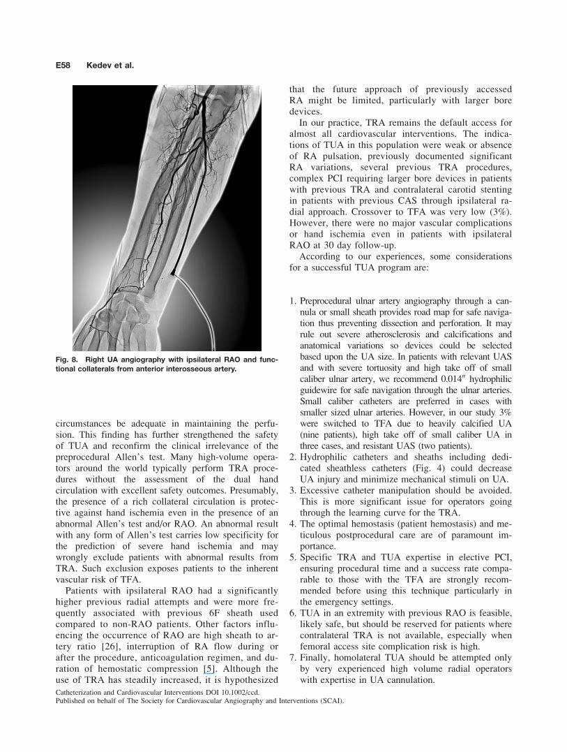
In all 240 patients with documented ipsilateral RAO,preprocedural ulnar angiography demonstrated func-tional collaterals predominantly from anterior interosse-ous artery (Fig. 8). In those patients, UA pulse wasconsiderably stronger than in patients with patent radialarteries. All patients with RAO underwent duplex ultra-sonography which showed patent ulnar artery flow andnone of them had signs and symptoms of hand ische-mia at 30 day follow-up. It appears that the anteriorinterosseal artery supply to the wrist may under certain
TABLE V. Outcomes of Patients Based on Ipsilateral RadialArtery Status
Variables
IRAO
group(n¼ 240)
Non-IRAOgroup (n¼ 236) P value
Procedural success 232 (97%) 230 (97%) NS
Clinical ulnar artery spasm 17 (7.0%) 17 (7.2%) NSVascular access site
complications
Hemathoma Grade I–V 19 (7.9%) 20 (8.4%) NSGrade 1 9 (3.7%) 9 (3.8%) NS
Grade 2 5 (2.0%) 5 (2.1%) NSGrade 3 4 (1.6%) 5 (2.1%) NSGrade 4 1 (0.4) 1 (0.4) NS
Grade 5 0 0 NSMajor vascular complication
at 30 days
0 0 NS
Ulnar artery occlusionat 30 days
0 15 (6.3%) <0.001
Fig. 7. Ulnar and radial artery angiography: clearly largersize UA.
Safety and Feasibility of TUA E57
Catheterization and Cardiovascular Interventions DOI 10.1002/ccd.Published on behalf of The Society for Cardiovascular Angiography and Interventions (SCAI).
circumstances be adequate in maintaining the perfu-sion. This finding has further strengthened the safetyof TUA and reconfirm the clinical irrelevance of thepreprocedural Allen’s test. Many high-volume opera-tors around the world typically perform TRA proce-dures without the assessment of the dual handcirculation with excellent safety outcomes. Presumably,the presence of a rich collateral circulation is protec-tive against hand ischemia even in the presence of anabnormal Allen’s test and/or RAO. An abnormal resultwith any form of Allen’s test carries low specificity forthe prediction of severe hand ischemia and maywrongly exclude patients with abnormal results fromTRA. Such exclusion exposes patients to the inherentvascular risk of TFA.
Patients with ipsilateral RAO had a significantlyhigher previous radial attempts and were more fre-quently associated with previous 6F sheath usedcompared to non-RAO patients. Other factors influ-encing the occurrence of RAO are high sheath to ar-tery ratio [26], interruption of RA flow during orafter the procedure, anticoagulation regimen, and du-ration of hemostatic compression [5]. Although theuse of TRA has steadily increased, it is hypothesized
that the future approach of previously accessedRA might be limited, particularly with larger boredevices.
In our practice, TRA remains the default access foralmost all cardiovascular interventions. The indica-tions of TUA in this population were weak or absenceof RA pulsation, previously documented significantRA variations, several previous TRA procedures,complex PCI requiring larger bore devices in patientswith previous TRA and contralateral carotid stentingin patients with previous CAS through ipsilateral ra-dial approach. Crossover to TFA was very low (3%).However, there were no major vascular complicationsor hand ischemia even in patients with ipsilateralRAO at 30 day follow-up.
According to our experiences, some considerationsfor a successful TUA program are:
1. Preprocedural ulnar artery angiography through a can-nula or small sheath provides road map for safe naviga-tion thus preventing dissection and perforation. It mayrule out severe atherosclerosis and calcifications andanatomical variations so devices could be selectedbased upon the UA size. In patients with relevant UASand with severe tortuosity and high take off of smallcaliber ulnar artery, we recommend 0.01400 hydrophilicguidewire for safe navigation through the ulnar arteries.Small caliber catheters are preferred in cases withsmaller sized ulnar arteries. However, in our study 3%were switched to TFA due to heavily calcified UA(nine patients), high take off of small caliber UA inthree cases, and resistant UAS (two patients).
2. Hydrophilic catheters and sheaths including dedi-cated sheathless catheters (Fig. 4) could decreaseUA injury and minimize mechanical stimuli on UA.
3. Excessive catheter manipulation should be avoided.This is more significant issue for operators goingthrough the learning curve for the TRA.
4. The optimal hemostasis (patient hemostasis) and me-ticulous postprocedural care are of paramount im-portance.
5. Specific TRA and TUA expertise in elective PCI,ensuring procedural time and a success rate compa-rable to those with the TFA are strongly recom-mended before using this technique particularly inthe emergency settings.
6. TUA in an extremity with previous RAO is feasible,likely safe, but should be reserved for patients wherecontralateral TRA is not available, especially whenfemoral access site complication risk is high.
7. Finally, homolateral TUA should be attempted onlyby very experienced high volume radial operatorswith expertise in UA cannulation.
Fig. 8. Right UA angiography with ipsilateral RAO and func-tional collaterals from anterior interosseous artery.
E58 Kedev et al.
Catheterization and Cardiovascular Interventions DOI 10.1002/ccd.Published on behalf of The Society for Cardiovascular Angiography and Interventions (SCAI).

STUDY LIMITATIONS
Several limitations of this study should be consid-ered. First of all, this is a nonrandomized study involv-ing consecutive patients. Second, diagnosis of UASwas subjective, based on clinical signs, as resistanceduring catheter maneuver, inability to freely manipulatethe catheter, and/or difficulty in moving the catheterwith concomitant forearm pain during the procedure.
Furthermore, all procedures were done by experiencedand high-volume transradial and transulnar operators.All CAS procedures were performed by a single opera-tor (SK) with extensive experience with both the TRAand CAS; the transradial learning curve may limit theimplementation of the described techniques.
CONCLUSION
Transulnar approach is a safe and feasible alternativewrist access for patients undergoing cardiovascularinterventions when ipsilateral radial access is not possi-ble. With careful technique and experienced operators,the procedure can be performed with a high successrate and low incidence of vascular complications.However, further large-scale randomized studies areneeded to reconfirm the role of TUA for cardiovascularprocedures when ipsilateral radial is not available.
REFERENCES
1. Zimmerman HA, Scott RW, Becker NO. Catheterization of theleft side of the heart in man. Circulation 1950;1:357–359.
2. Hamon M, Pristipino C, Di Mario C, Nolan J, Ludwig J, TubaroM, Sabate M, Mauri-Ferre J, Huber K, Niemela K, Haude M,Wijns W, Dudek D, Fajadet J, Kiemeneij F. Consensus docu-ment on the radial approach in percutaneous cardiovascularinterventions: Position paper by the EAPCI and WorkingGroups on Acute Cardiac Care and Thrombosis of the EuropeanSociety of Cardiology. EuroIntervention 2013;8:1242–1251.
3. Rao SV, Ou FS, Wang TY, Roe MT, Brindis R, Rumsfeld JS,Peterson ED. Trends in the prevalence and outcomes of radialand femoral approaches to percutaneous coronary intervention:A report from the National Cardiovascular Data Registry. JACCCardiovasc Interv 2008;1:379–386.
4. Rao SV, Cohen MG, Kandzari DE, Bertrand OF, Gilchrist IC.The transradial approach to percutaneous coronary intervention:Historical perspective, current concepts, and future directions. JAm Coll Cardiol 2010;55:2187–2195.
5. Pancholy SB, Patel TM. Effect of duration of hemostatic com-pression on radial artery occlusion after transradial access. Cath-eter Cardiovasc Interv 2012;79:78–81.
6. Stella PR, Kiemeneij F, Laarman GJ, Odekerken D, SlagboomT, van der Wieken R. Incidence and outcome of radial arteryocclusion following transradial artery coronary angioplasty.Catheter Cardiovasc Interv 1997;40:156–158.
7. Lo TS, Nolan J, Fountzopoulos E, Behan M, Butler R,Hetherington SL, Vijayalakshmi K, Rajagopal R, Fraser D, ZamanA, Hildick-Smith D. Radial artery anomaly and its influence ontransradial coronary procedural outcome. Heart 2009;95:410–415.
8. Valsecchi O, Vassileva A, Musumeci G, Rossini R, Tespili M,Guagliumi G, Mihalcsik L, Gavazzi A, Ferrazzi P. Failure oftransradial approach during coronary interventions: Anatomicconsiderations. Catheter Cardiovasc Interv 2006;67:870–878.
9. Hildick-Smith DJ, Lowe MD, Walsh JT, Ludman PF, StephensNG, Schofield PM, Stone DL, Shapiro LM, Petch MC. Coro-nary angiography from the radial artery—Experience, complica-tions and limitations. Int J Cardiol 1998;64:231–239.
10. Kiemeneij F. Prevention and management of radial arteryspasm. J Invasive Cardiol 2006;18:159–160.
11. Terashima M, Meguro T, Takeda H, Endoh N, Ito Y, MitsuokaM, Ohtomo T, Murai O, Fujiwara S, Honda H, Miyazaki Y,Kuhara R, Kawashima O, Isoyama S. Percutaneous ulnar arteryapproach for coronary angiography: A preliminary report innine patients. Catheter Cardiovasc Interv 2001;53:410–414.
12. Knebel AV, Cardoso CO, Rodrigues LHC, Sarmento-Leite REG,de Quadros AS, Gottschall CAM. Safety and feasibility of tran-sulnar cardiac catheterization. Tex Heart Inst J 2008;35:268–272.
13. Mangin L, Bertrand OF, De la Rochelliere R, Proulx G, LemayR, Barbeau G, Gleeton O, Rodes-Cabau J, Nguyen CM, Roy L.The trans-ulnar approach for coronary intervention: A safe alter-native to transradial approach in selected patients. J InvasiveCardiol 2005;17:77–79.
14. Dashkoff N, Dashkoff PB, Zizzi JA, Wadhwani J. Ulnar arterycannulation for coronary angiography and percutaneous coro-nary intervention: Case reports and anatomic considerations.Catheter Cardiovasc Interv 2002;55:93–96.
15. Aptecar E, Dupouy P, Chabane-Chaouch M, Bussy N, CatarinoG, Shahmir A, Elhajj Y, Pernes JM. Percutaneous transulnar ar-tery approach for diagnostic and therapeutic coronary interven-tion. J Invasive Cardiol 2005;17:312–317.
16. Andrade PB, Tebet MA, Andrade MV, Labrunie A, Mattos LA. Pri-mary percutaneous coronary intervention through transulnarapproach: Safety and effectiveness. Arq Bras Cardiol 2008;91:49–52.
17. Aptecar E, Pernes JM, Chaouch MC, Bussy N, Catarino, G,Shahmir A, Bougrini K, Dupouy P. Transulnar versus transradialartery approach for coronary angioplasty: The PCVI-CUBAstudy. Catheter Cardiovasc Interv 2006;67:711–720.
18. Andrade PB, Tebet MA, Nogueira EF, Esteves VC, AndradeMVA, Labrunie A, Mattos LAP. Transulnar approach as an al-ternative access site for coronary invasive procedures after trans-radial approach failure. Am Heart J 2012;164:462–467.
19. Smith SC Jr, Feldman TE, Hirshfeld JW Jr, Jacobs AK, KernMJ, King SB 3rd, Morrison DA, O’neill WW, Schaff HV,Whitlow PL, Williams DO, Antman EM, Smith SC Jr, AdamsCD, Anderson JL, Faxon DP, Fuster V, Halperin JL, HiratzkaLF, Hunt SA, Jacobs AK, Nishimura R, Ornato JP, Page RL,Riegel B; ACC/AHA/SCAI Writing Committee to Update the2001 Guidelines for PCI. ACC/AHA/SCAI 2005 guidelineupdate for PCI. J Am Coll Cardiol 2006;47:e1–e121.
20. Dharma S, Shah S, Radadiya R, Vyas C, Pancholy S, Patel T.Nitroglycerin plus diltiazem versus nitroglycerin alone forspasm prophylaxis with transradial approach. J Invasive Cardiol2012;24:122–125.
21. Bertrand OF, De Larochelliere R, Cabau JR, Proulx G, Gleeton O,Nguyen CM, Dery JP, Barbeau G, Noel B, Larose E, Poirier P,Roy L for Early Discharge After Transradial Stenting of CoronaryArteries (EASY) study investigators. A randomized study comparingsame-day home discharge and abciximab bolus only to overnighthospitalization and abciximab bolus and infusion after transradialcoronary stent implantation. Circulation 2006;114:2636–2643.
22. Burzotta F, Trani C, Mazzari MA, Tommasino A, Niccoli G,Porto I, Leone AM, Tinelli G, Coluccia V, De Vita M, BrancatiM, Mongiardo R, Schiavoni G, Crea F. Vascular complications
Safety and Feasibility of TUA E59
Catheterization and Cardiovascular Interventions DOI 10.1002/ccd.Published on behalf of The Society for Cardiovascular Angiography and Interventions (SCAI).

and access crossover in 10676 transradial percutaneous coronaryprocedures. Am Heart J 2012;163:230–238.
23. Etxegoien N, Rhyne D, Kedev S, Sachar R, Mann T. The trans-radial approach for carotid artery stenting. Catheter CardiovascInterv 2012;80:1081–1087.
24. Fukuda N, Iwahara S, Harada A, Yokoyama S, Akutsu K,Takano M, Kobayashi A, Kurokawa S, Izumi T. Vasospasms ofthe radial artery after the transradial approach for coronary angi-ography and angioplasty. Jpn Heart J 2004;45:723–731.
25. Agostoni P, Zuffi A, Biondi-Zoccai G. Pushing wrist access tothe limit: Homolateral right ulnar artery approach for primarypercutaneous coronary intervention after right radial failure dueto radial loop. Catheter Cardiovasc Interv 2011;78:894–897.
26. Saito S, Ikei H, Hosokawa G, Tanaka S. Influence of the ratiobetween radial artery inner diameter and sheath outer diameteron radial artery flow after transradial coronary intervention.Catheteter Cardiovasc Interv 1999;46:173–178.
E60 Kedev et al.
Catheterization and Cardiovascular Interventions DOI 10.1002/ccd.Published on behalf of The Society for Cardiovascular Angiography and Interventions (SCAI).