Does the Taylor Spatial Frame Accurately Correct Tibial ... - HSS
10
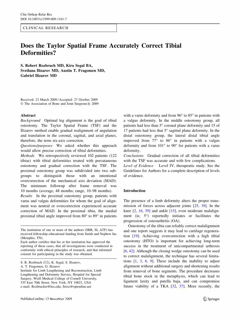
Clin Orthop Relat Res DOI 10.1007/s11999-009-1161-7 CLINICAL RESEARCH Does the Taylor Spatial Frame Accurately Correct Tibial Deformities? S. Robert Rozbruch MD, Kira Segal BA, Svetlana Ilizarov MD, Austin T. Fragomen MD, Gabriel Ilizarov MD Received: 23 March 2009 / Accepted: 27 October 2009 The Association of Bone and Joint Surgeons1 2009 Abstract Background Optimal leg alignment is the goal of tibial osteotomy. The Taylor Spatial Frame (TSF) and the Ilizarov method enable gradual realignment of angulation and translation in the coronal, sagittal, and axial planes, therefore, the term six-axis correction. Questions/purposes We asked whether this approach would allow precise correction of tibial deformities. Methods We retrospectively reviewed 102 patients (122 tibiae) with tibial deformities treated with percutaneous osteotomy and gradual correction with the TSF. The proximal osteotomy group was subdivided into two sub- groups to distinguish those with an intentional overcorrection of the mechanical axis deviation (MAD). The minimum followup after frame removal was 10 months (average, 48 months; range, 10–98 months). Results In the proximal osteotomy group, patients with varus and valgus deformities for whom the goal of align- ment was neutral or overcorrection experienced accurate correction of MAD. In the proximal tibia, the medial proximal tibial angle improved from 80 to 89 in patients The institution of one or more of the authors (SRR, SI, ATF) has received fellowship educational funding from Smith and Nephew Inc (Memphis, TN). Each author certifes that his or her institution has approved the reporting of these cases, that all investigations were conducted in conformity with ethical principles of research, and that informed consent for participating in the study was obtained. S. R. Rozbruch (&), K. Segal, S. Ilizarov, A. T. Fragomen, G. Ilizarov Institute for Limb Lengthening and Reconstruction, Limb Lengthening and Deformity Service, Hospital for Special Surgery, Weill Medical College of Cornell University, 535 East 70th Street, New York, NY 10021, USA e-mail: [email protected]; [email protected] with a varus deformity and from 96 to 85 in patients with a valgus deformity. In the middle osteotomy group, all patients had less than 5 coronal plane deformity and 15 of 17 patients had less that 5 sagittal plane deformity. In the distal osteotomy group, the lateral distal tibial angle improved from 77 to 86 in patients with a valgus deformity and from 101 to 90 for patients with a varus deformity. Conclusions Gradual correction of all tibial deformities with the TSF was accurate and with few complications. Level of Evidence Level IV, therapeutic study. See the Guidelines for Authors for a complete description of levels of evidence. Introduction The presence of a limb deformity alters the proper trans- mission of forces across adjacent joints [25, 39]. In the knee [2, 16, 39] and ankle [15], even moderate malalign- ment (ie, 5) reportedly initiates or facilitates the progression of osteoarthritis (OA). Osteotomy of the tibia can reliably correct malalignment and one report suggests it may lead to cartilage regenera- tion [19]. Achieving overcorrection with a high tibial osteotomy (HTO) is important for achieving long-term success in the treatment of unicompartmental arthrosis [6, 42]. Although the closing wedge osteotomy can be used to correct malalignment, the technique has several limita- tions [1, 3, 6, 9]. These include the inability to adjust alignment without additional surgery and shortening results from removal of bone segments. The procedure decreases tibial bone stock in the metaphysis, which can lead to ligament laxity and patella baja, and can compromise future viability of a TKA [32, 37]. More recently, the 123
-
Upload
khangminh22 -
Category
Documents
-
view
3 -
download
0
Transcript of Does the Taylor Spatial Frame Accurately Correct Tibial ... - HSS
Clin Orthop Relat Res
DOI 10.1007/s11999-009-1161-7
CLINICAL RESEARCH
Does the Taylor Spatial Frame Accurately Correct Tibial Deformities?
S. Robert Rozbruch MD, Kira Segal BA,
Svetlana Ilizarov MD, Austin T. Fragomen MD,
Gabriel Ilizarov MD
Received: 23 March 2009 / Accepted: 27 October 2009
The Association of Bone and Joint Surgeons1 2009
Abstract
Background Optimal leg alignment is the goal of tibial
osteotomy. The Taylor Spatial Frame (TSF) and the
Ilizarov method enable gradual realignment of angulation
and translation in the coronal, sagittal, and axial planes,
therefore, the term six-axis correction.
Questions/purposes We asked whether this approach
would allow precise correction of tibial deformities.
Methods We retrospectively reviewed 102 patients (122
tibiae) with tibial deformities treated with percutaneous
osteotomy and gradual correction with the TSF. The
proximal osteotomy group was subdivided into two sub-
groups to distinguish those with an intentional
overcorrection of the mechanical axis deviation (MAD).
The minimum followup after frame removal was
10 months (average, 48 months; range, 10–98 months).
Results In the proximal osteotomy group, patients with
varus and valgus deformities for whom the goal of align-
ment was neutral or overcorrection experienced accurate
correction of MAD. In the proximal tibia, the medial
proximal tibial angle improved from 80 to 89 in patients
The institution of one or more of the authors (SRR, SI, ATF) has
received fellowship educational funding from Smith and Nephew Inc
(Memphis, TN).
Each author certifes that his or her institution has approved the
reporting of these cases, that all investigations were conducted in
conformity with ethical principles of research, and that informed
consent for participating in the study was obtained.
S. R. Rozbruch (&), K. Segal, S. Ilizarov,
A. T. Fragomen, G. Ilizarov
Institute for Limb Lengthening and Reconstruction, Limb
Lengthening and Deformity Service, Hospital for Special
Surgery, Weill Medical College of Cornell University,
535 East 70th Street, New York, NY 10021, USA
e-mail: [email protected]; [email protected]
with a varus deformity and from 96 to 85 in patients with
a valgus deformity. In the middle osteotomy group, all
patients had less than 5 coronal plane deformity and 15 of
17 patients had less that 5 sagittal plane deformity. In the
distal osteotomy group, the lateral distal tibial angle
improved from 77 to 86 in patients with a valgus
deformity and from 101 to 90 for patients with a varus
deformity.
Conclusions Gradual correction of all tibial deformities
with the TSF was accurate and with few complications.
Level of Evidence Level IV, therapeutic study. See the
Guidelines for Authors for a complete description of levels
of evidence.
Introduction
The presence of a limb deformity alters the proper trans-
mission of forces across adjacent joints [25, 39]. In the
knee [2, 16, 39] and ankle [15], even moderate malalign-
ment (ie, 5) reportedly initiates or facilitates the
progression of osteoarthritis (OA).
Osteotomy of the tibia can reliably correct malalignment
and one report suggests it may lead to cartilage regenera-
tion [19]. Achieving overcorrection with a high tibial
osteotomy (HTO) is important for achieving long-term
success in the treatment of unicompartmental arthrosis
[6, 42]. Although the closing wedge osteotomy can be used
to correct malalignment, the technique has several limita-
tions [1, 3, 6, 9]. These include the inability to adjust
alignment without additional surgery and shortening results
from removal of bone segments. The procedure decreases
tibial bone stock in the metaphysis, which can lead to
ligament laxity and patella baja, and can compromise
future viability of a TKA [32, 37]. More recently, the
123
Rozbruch et al. Clinical Orthopaedics and Related Research1
medial opening wedge osteotomy has gained popularity as
another option to avoid the complications associated with
the closing wedge technique. This technique also requires
acute correction and no ability to correct any residual
deformity [7, 9].
A percutaneous osteotomy combined with gradual cor-
rection using the TSF provides a way to correct a tibial
deformity independent of magnitude, complexity, or loca-
tion. The procedure uses small incisions and minimal soft
tissue stripping, and can be used in all zones of the tibia.
Without the need for complex frame modifcations, the
TSF can be used to correct angulation and translation in the
coronal, sagittal, and axial planes around a virtual hinge,
therefore, the term six-axis correction. The associated web-
based software has simplifed planning and performance of
deformity correction for patients and physicians and has
been used to treat all aspects of deformities in the lower
extremities. Use of the TSF is associated with few com-
plications [4, 10, 14, 33, 34, 40, 41, 44, 47] and corrects
complex tibial deformities in adults and children.
However, published studies [4, 8, 10, 12, 13, 23, 24, 31,
38, 44] on the TSF have been in the form of case reports,
have included small numbers of patients, and have com-
bined various bones and etiologies. In addition, the
methods of reporting deformity correction and alignment
have been variable.
Using a larger series, we therefore asked the following
questions regarding the accuracy and outcome of tibial
deformity correction: (1) How accurate is the MAD cor-
rection at the proximal tibia? (2) How accurate is the
medial proximal tibial angle correction (MPTA) and the
lateral distal tibial angle (LDTA) correction at the proximal
and distal tibia, respectively? (3) How accurate is correc-
tion of a tibial diaphyseal deformity? (4) What are the
outcomes regarding SF-36 scores, American Academy of
Orthopaedic Surgeons (AAOS) lower limb module (LLM)
scores, the need for adjacent joint replacement surgery, and
complications?
Patients and Methods
We used our osteotomy registry to identify all 102 patients
(122 tibias) who underwent a tibial osteotomy surgery for
deformity correction using the TSF (Smith and Nephew,
Memphis, TN) between 2000 and 2007. Our indications for
use of the TSF were uniplanar coronal plane deformity of a
magnitude greater than 10, oblique plane deformity,
presence of rotational deformity, or compromised soft tis-
sue envelope. We excluded patients with nonunions,
patients who primarily underwent tibial lengthening, and
patients who underwent deformity correction with a dif-
ferent method than the TSF. These methods included a
monolateral frame for a coronal plane deformity in the
proximal tibia less than 10 and intramedullary nailing for
a diaphyseal deformity less than 10. There were 44
females and 58 males with an average age of 39 years
(range, 5–72 years). Twenty of the 102 patients had bilat-
eral surgeries. The causes of deformity included 30
posttraumatic malunions and 72 cases of nontraumatic
nature, including those of congenital, developmental, and
neurologic etiologies. We created three groups by the
location of the tibial osteotomy: proximal third (n = 84),
middle third (n = 17), and distal third (n = 21). The
proximal group was further divided into two subgroups: (1)
treatment goal was a MAD of 0 mm (center) or (2) treat-
ment goal was overcorrection of the MAD to 6 to 12 mm
medial or lateral depending on the presenting problem.
Twenty-three of the 84 limbs were intentionally over-
corrected [6, 7]. The patients who had intentional over-
correction had either unicompartmental arthritis or a
valgus deformity. The minimum followup after frame
removal was 10 months (average, 48 months; range, 10–
98 months). No patients were lost to followup. The study
refected a chart review and no patients were recalled
specifcally for this study. This was an Institutional Review
Board-approved retrospective study.
Clinical preoperative evaluation included history and
physical examination. Gait was observed. One of us
(SRR) measured frontal plane deformity on a 51-inch
erect leg bipedal radiograph. If there was a leg-length
discrepancy (LLD), blocks (to the nearest 5 mm) were
placed under the affected foot to level the pelvis and the
height of the blocks was recorded. Leveling the pelvis
improves reliability of the measurements of length and
alignment on the 51-inch radiograph [36]. LLD was
measured on the radiograph. MAD and joint orientation
angles, lateral distal femoral angle, MPTA, posterior
proximal tibial angle (PPTA), and LDTA were measured
using the methods described by Paley et al. [25, 27]. We
recorded the magnitude of the deformity by measuring
the angle formed by the intersection of a line drawn
from the center hip through the knee center with that of
the distal mechanical axis of the tibia. In cases in which
neutral alignment was the goal, the proximal mechanical
axis line was drawn through the center of the knee.
When overcorrection was the goal, the proximal
mechanical axis line was drawn to the desired location
on the knee. The mechanical axis of the opposite lower
extremity was not used as the goal. In addition, we
routinely obtained AP and lateral view radiographs of
the tibia. Ankle deformity was evaluated by radiographs
taken with the xray beam centered on the ankle. The
outcome includes MAD data points that are medial and
lateral to midline. To report the outcome most accu-
rately, we averaged the medial data points and the lateral
123
Tibial Deformity Correction with TSF
data points separately. This generates separate medial
and lateral values.
All surgeries were performed by the senior author
(SRR). Through a 1-cm incision, the tibial osteotomy was
performed using a multiple drill-hole technique and it was
completed with an osteotome. The location of the osteot-
omy was at or near the apex of the deformity. When the
osteotomy was away from the apex of the deformity,
intentional translation at the osteotomy site was needed to
correct the limb alignment (Fig. 1) [25, 27]. Osteotomies
were complete but left nondisplaced. Fibula osteotomies
were performed in all cases. The location of the fbula
osteotomy was the middle of the bone when accompanying
a proximal or middle tibial osteotomy and was in the distal
third when accompanying a distal tibia osteotomy. TSF
frames were fxated to the bone with tensioned wires and
hydroxyapatite-coated half pins. All corrections were made
gradually after a latency phase of 7 to 10 days.
We entered deformity parameters into the TSF web-
based software computer program [30, 43] and generated
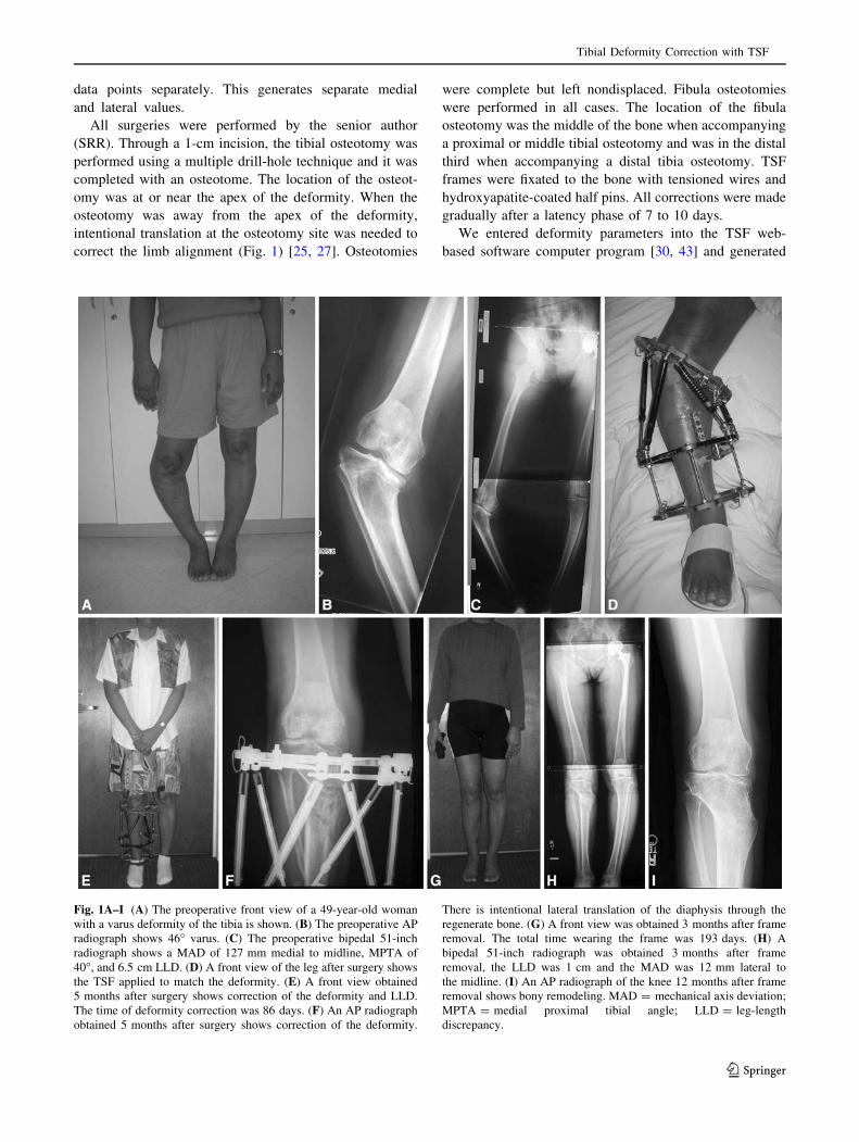
Fig. 1A–I (A) The preoperative front view of a 49-year-old woman
with a varus deformity of the tibia is shown. (B) The preoperative AP
radiograph shows 46 varus. (C) The preoperative bipedal 51-inch
radiograph shows a MAD of 127 mm medial to midline, MPTA of
40, and 6.5 cm LLD. (D) A front view of the leg after surgery shows
the TSF applied to match the deformity. (E) A front view obtained
5 months after surgery shows correction of the deformity and LLD.
The time of deformity correction was 86 days. (F) An AP radiograph
obtained 5 months after surgery shows correction of the deformity.
There is intentional lateral translation of the diaphysis through the
regenerate bone. (G) A front view was obtained 3 months after frame
removal. The total time wearing the frame was 193 days. (H) A
bipedal 51-inch radiograph was obtained 3 months after frame
removal, the LLD was 1 cm and the MAD was 12 mm lateral to
the midline. (I) An AP radiograph of the knee 12 months after frame
removal shows bony remodeling. MAD = mechanical axis deviation;
MPTA = medial proximal tibial angle; LLD = leg-length
discrepancy.
123

Rozbruch et al. Clinical Orthopaedics and Related Research1
an adjustment schedule. The program requires input of
deformity, frame, and mounting parameters, and a structure
at risk, which determines the rate of correction [30, 43].
The patient is instructed to perform gradual adjustments of
the six struts of the TSF three times per day. At the end of
the schedule, which typically lasts 2 to 6 weeks, one of us
(SRR) determined the limb alignment with physical
examination and radiographs. We inspected the patient
standing from the front, back, and side views and focused
on iliac crest symmetry and leg alignment. On the 51-inch
standing radiograph, we measured leg lengths, MAD, and
joint orientation angles using the same methods used
before surgery. When there was residual deformity, we
generated and implemented another correction schedule.
Our criteria for frame removal were time of at least
2.5 months for angular correction and a reasonable external
fxation index of 1.5 months per centimeter when length-
ening also was performed [13], ability to walk with
minimal assistance, and the presence of bridging callus on
three of four cortices using the AP and lateral radiographs.
The total time wearing the frame averaged 130 days
(range, 71–355 days), whereas the frame was used
dynamically to correct deformity for 34 days (range, 7–
99 days). Patients had an average of two schedules (range,
1–5 schedules), which they followed to turn struts on the
TSF. These included the initial schedule and additional
residual schedules. The total amount of simultaneous
lengthening was an average of 1 cm (range, 0–6.6 cm).
Twelve patients underwent simultaneous lengthening
greater than 2 cm.
After surgery, patients were allowed to bear weight as
tolerated and wean from the crutches as tolerated. For
unilateral cases, most patients were walking without the
need for two crutches at 6 to 8 weeks after surgery. Knee
and ankle ROM exercises were encouraged with supervi-
sion of a physical therapist three times per week for 1 hour.
Patients also were given a daily 1-hour home therapy
program. Patients with bilateral deformities had staged
correction with the second-side surgery typically at 6 to
8 weeks after the frst side. ROM exercises of the knee and
ankle were encouraged. A daily shower, including washing
the pin sites with antibacterial soap, was encouraged. This
was followed by daily pin care with half-strength hydrogen
peroxide and then coverage of pin sites with a dry sterile
gauze wrap. Patients were seen in the clinic every 10 to
14 days by the senior author (SRR) during the distraction
phase. Once the alignment was corrected and the adjust-
ments ended, patients were seen monthly until frame
removal.
Rotational deformity was measured clinically by
observing gait, foot progression angle, and thigh-foot axis
in the prone position [25]. The rotational deformity cor-
rections were as large as 40 (Table 1). We did not have a
cutoff level for inclusion.
Preoperatively and at the last visit, we obtained SF-36
Health Survey scores (physical function, role physical,
bodily pain, general health, vitality, social functioning, role
emotional, mental health) [35] and the AAOS LLM Patient
Health Outcome Score [34]. SF-36 health surveys and
LLM module scores were completed for 55 and 54 of 102
patients, respectively.
We recorded time wearing the frame, number of
schedules, complications, and knee and ankle ROM. For all
patients, deformity parameters, including degree of varus,
valgus, apex anterior and posterior deformity, and internal
and external rotation deformity, were extracted from the
frst schedule. This illustrated the magnitude and nature of
the preoperative deformity (Table 1). Alignment of the
proximal, middle, and distal tibial osteotomy groups were
evaluated separately using the best measurements for each
group. To assess alignment of the proximal tibia, MAD,
MPTA, and PPTA were measured preoperatively and
postoperatively by the senior author (SRR), and intraclass
correlation coeffcients were determined to test intraob-
server reliability. The mean intraobserver reliability for
these three measurements was 0.97 (range, 0.82–0.99). We
analyzed the outcomes of MAD according to the preoper-
ative treatment goal (normal versus overcorrection). To
assess alignment of the middle tibia, we measured an
absolute angular value obtained by the intersection of the
proximal and distal diaphyseal lines [25, 30]. Whereas the
MAD and MPTA are affected most by a proximal tibial
deformity and the LDTA is affected most by a distal tibial
deformity, these joint orientation angles are not a sensitive
measure of a middle tibial deformity. The middle tibial
deformity parameters instead were extracted from what
was input into the frst computer program (middle row of
Table 1) and compared with the amount of angular
Table 1. Taylor Spatial Frame deformity parameters for the entire cohort (degrees)
Osteotomy location Varus Valgus Apex anterior Apex posterior Internal rotation External rotation
Proximal tibia 13 (4–46) 13 (4–30) 11 (4–35) 10 (5–20) 15 (10–40) 14 (5–25)
Middle tibia 11 (7–30) 18 (10–37) 11 (4–35) 14 (7–23) 18 (10–35) 13 (5–20)
Distal tibia 12 (5–25) 17 (12–30) 9 (2–22) 10 (4–17) 5 (5) 18 (10–30)
Ranges shown in parentheses.
123
Tibial Deformity Correction with TSF
deformity present on the latest AP and lateral radiographs.
To assess alignment of the distal tibia, we measured pre-
operative and postoperative LDTA.
The aims of the analysis were to confrm a clinically
important improvement in certain measurements postop-
eratively at an average of 48 months as compared with the
preoperative measurements. The measurements of clinical
importance are MAD in the proximal group, MPTA in the
proximal group, LDTA in the distal group, SF-36 health
survey scores, and AAOS LLM scores.
The data set of cases was transferred to Systat v10.2
(Systat Software Inc, Richmond, CA). Descriptive statistics
were run on all the variables, means and standard devia-
tions, and percentiles. Because differences between two
continuous measurements on the same joints were the
results of interest, paired t-tests were used. We determined
the differences between the preoperative measurement and
the corresponding matched postoperative measurement (the
difference with time for each patient). No difference or
change would result in a mean of zero. The paired t-test
was used for each measurement: (1) MAD in the proximal
group; (2) MPTA in the proximal group; (3) LDTA in the
distal group; (4) SF-36 health survey scores; and (5) AAOS
LLM scores.
Results
In the proximal group, the MAD correction was accurate.
Patients with a varus deformity had a preoperative MAD of
38 mm medial to the midline. For patients with a goal of a
MAD of 0, this improved to an average of 5 mm medial
and 5 mm lateral to midline. In patients in whom the goal
was overcorrection, the MAD improved to 8 mm lateral to
the midline. In the proximal group, patients with a valgus
deformity had a preoperative MAD of 33 mm lateral to the
midline. For patients with a goal of a MAD of 0, this
improved to an average of 8 mm medial and 3 mm lateral
to midline. In patients in whom the goal was overcorrec-
tion, the MAD improved to 12 mm medial to the midline
(Table 2). Sagittal (apex anterior and apex posterior) and
axial planes (internal and external rotation) were corrected
to a satisfactory degree in all cases.
The corrections of MPTA and LDTA were accurate. In
the proximal group, the MPTA improved from 80 to 89 in patients with a varus deformity, and from 96 to 85 in
patients with a valgus deformity (Table 3). In the distal
group, the LDTA improved from 77 to 86 in patients
with a valgus deformity, and from 101 to 90 for patients
with a varus deformity (Table 4).
Table 2. Preoperative versus postoperative MAD for the proximal group (mm)
Preoperative deformity and p value Preoperative MAD Postoperative goal 0 Postoperative goal overcorrection
Medial Lateral Medial Lateral
MAD medial (varus) 38 (1–155) 5 (0–35) 5 (2–10) — 8 (0–17)
p Value \ 0.001 0.03 0.004
MAD lateral (valgus) 33 (4–83) 8 (4–14) 3 (0–9) 12 (4–29) —
p Value 0.01 0.05 0.1
Ranges shown in parentheses; MAD = mechanical axis deviation.
Table 3. Preoperative versus postoperative MPTA for the proximal group (degrees)
Preoperative deformity Preoperative MPTA
Preoperative MPTA less than 90 (varus) 80 (40–89)
Preoperative MPTA 90 or greater (valgus) 96 (90–123)
Postoperative MPTA
89 (80–97)
85 (74–101)
p Value
\ 0.001
0.001
Ranges shown in parentheses; MPTA = medial proximal tibial angle.
Table 4. Preoperative versus postoperative LDTA for the distal group (degrees)
Preoperative deformity Preoperative LDTA Postoperative LDTA p Value
Preoperative LDTA less than 90 (valgus) 77 (75–79) 86 (82–88) 0.4
Preoperative LDTA 90 or greater (varus) 101 (90–111) 90 (90–92) 0.09
Ranges shown in parentheses; LDTA = lateral distal tibial angle.
123
Rozbruch et al. Clinical Orthopaedics and Related Research1
Table 5. Preoperative versus postoperative SF-36 Health Survey scores
Time and
p value
Physical
functioning
Role
physical
Bodily pain General
health
Vitality Social
functioning
Role
emotional
Mental
health
Preoperative
Postoperative
p Value
47 (0–100)
66 (10–100)
\ 0.001
39 (0–100)
65 (0–100)
0.002
47 (10–100)
66 (0–100)
\ 0.001
74 (20–100)
75 (22–100)
0.6
52 (10–90)
62 (5–100)
0.06
62 (0–100)
78 (0–100)
0.005
67 (0–100)
79 (0–100)
0.8
68 (16–100)
79 (40–100)
0.007
Ranges shown in parentheses.
Table 6. Preoperative versus postoperative knee and ankle ROM (degrees)
Time/p value Knee ROM Ankle ROM
Extension Flexion Dorsifexion Plantar fexion
Preoperative
Postoperative
p Value
0 (30 to 20)
0 (10 to 10)
0.54
126 (60–140)
125 (70–145)
1.00
10 (30 to 30)
11 (0–30)
0.70
40 (20–70)
38 (0–70)
0.10
ROM = range of motion; ranges in parentheses.
The correction of tibial diaphyseal deformity was
accurate. The preoperative middle tibial deformity was
multiplanar (Table 1, row 2). Postoperatively, all patients
had less than 5 of coronal plane deformity and 15 of 17
patients had less that 5 of sagittal plane deformity. All
rotational deformities were corrected.
The SF-36 Health Survey scores improved in all cate-
gories (Table 5). LLM scores improved from 76 (range, 5–
100) to 86 (range, 51–100) (p \ 0.001). There were no
differences between preoperative and postoperative ankle
and knee ROM (Table 6). There were six complications.
According to the complication classifcation described by
Paley [26], there were three complications that resolved
without surgery, two complications that required operative
intervention, and one major complication. Two patients
had cellulitis develop that required a 10-day course of
intravenous antibiotics. One patient (a man who had breast
cancer and was being treated with tamoxifen) had delayed
union and lost some of the correction after frame removal.
His preoperative MAD was 68 mm medial and he under-
went correction to neutral, but after frame removal, he had
partial recurrence of the deformity and his fnal MAD was
35 mm medial (Table 2, row 1). He elected not to have
additional surgery. Two patients (three legs) had peroneal
nerve neurapraxia that resolved by slowing the correction
in one patient and with bilateral nerve release in one
patient. These patients had scar tissue from previous sur-
gery. Most patients had superfcial pin infections at some
point during the treatment that successfully responded to
oral antibiotics. There were no deep infections. The goals
of surgery were achieved in 121 of 122 limbs (99%). The
TSF was used to correct all aspects of a tibial deformity. At
the time of review, none of the patients had undergone
conversion surgery to TKA, unicompartmental knee
arthroplasty, total ankle arthroplasty, or had ankle fusion.
Discussion
Although deformity correction of the tibia often can be
accomplished with an acute correction and the use of
internal fxation, this method has limitations [1, 28, 42].
The presence of poor skin, multiplanar deformity, history
of infection and shortening, and lack of postoperative
adjustability shows the limitations of this method. The
Ilizarov method using the TSF offers a versatile approach
to correct all aspects of a tibial deformity. We therefore
asked: (1) How accurate is the MAD correction at the
proximal tibia? (2) How accurate are the MPTA and LDTA
corrections at the proximal and distal tibia, respectively?
(3) How accurate is the correction of tibial diaphyseal
deformity? (4) What are the outcomes regarding SF-36
scores, AAOS LLM scores, need for adjacent joint
replacement surgery, and complications?
Our study has several limitations. First, the patients were
reviewed retrospectively and all data were retrieved from
charts. Most of the radiographic measurements were
recorded in the chart, but there were times that we needed
to measure radiographs retrospectively. However, the
measurements were made by one author using a uniform
method and our intraobserver reliability was high (0.97).
Second, complete sets of the SF-36 and AAOS LLM scores
were available for approximately 50% of patients. This
may refect bias and must be taken into consideration.
Third, we combined all regions of the tibia in this study,
although each region of the tibia has its own
123
Ta
ble
7.
Lit
erat
ure
su
mm
ary
on
Tay
lor
Sp
atia
l F
ram
e (T
SF
) fo
r d
efo
rmit
y c
orr
ecti
on
Stu
dy
P
op
ula
tio
n
Des
ign
A
nat
om
ic r
egio
n
Def
orm
ity
co
rrec
tio
n
Co
ncl
usi
on
s re
gar
din
g T
SF
Eid
elm
an e
t al
. [8
] 30 c
hil
dre
n a
nd a
dole
scen
ts
Ret
rosp
ecti
ve
Mix
ed b
ones
30/3
1 a
nat
om
ical
ly c
orr
ecte
d
Acc
ura
tef
xat
or
for
def
orm
ity c
orr
ecti
on
Fel
dm
an e
t al
. [1
2]
18 a
dult
s R
etro
spec
tive
Tib
ia
17/1
8 a
chie
ved
sig
nif
cant
def
orm
ity
Eff
ecti
ve
tech
niq
ue
for
def
orm
ity
corr
ecti
on a
nd u
nio
n
corr
ecti
on
11 m
alunio
ns
7 n
onunio
ns
Fel
dm
an e
t al
. [1
0]
19 c
hil
dre
n a
nd a
dole
scen
ts
Ret
rosp
ecti
ve
Tib
ia v
ara
21/2
2 c
orr
ecte
d t
o w
ithin
3
Acc
ura
te a
nd s
afe
corr
ecti
on
(22 t
ibia
s)
(pro
xim
al)
Fel
dm
an e
t al
. [1
1]
18 c
hil
dre
n a
nd a
dole
scen
ts
Ret
rosp
ecti
ve
com
par
ison o
f T
ibia
var
a M
AD
was
3.1
mm
in g
radual
corr
ecti
on
Gra
dual
corr
ecti
on w
ith T
SF
is
more
gra
dual
corr
ecti
on u
sing T
SF
(p
roxim
al)
gro
up c
om
par
ed w
ith 1
7.1
mm
in a
cute
ac
cura
te t
han
acu
te c
orr
ecti
on w
ith E
BI
wit
h(1
8)
acute
corr
ecti
on u
sing
corr
ecti
on g
roup.
MP
TA
wit
hin
38
of
fram
e
EB
I fr
ame
(14)
norm
al i
n 1
7/1
8
Fra
gom
en e
t al
. [1
4]
Tec
hniq
ue
arti
cle
Pro
xim
al t
ibia
Naq
ui
et a
l. [
23
] 53 c
hil
dre
n a
nd
Ret
rosp
ecti
ve
Tib
ia (
44)
52/5
5 l
imbs
ended
wit
h \
15 m
m L
LD
E
ffec
tive
and e
ffci
ent
way
to c
orr
ect
a
adole
scen
ts (
55 l
imbs)
an
d 58
angula
r def
orm
ity.
wid
e var
iety
of
sim
ple
and c
om
ple
xF
emur
(11)
def
orm
itie
s
Nho e
t al
. [2
4]
Cas
e re
port
T
ibia
In
tenti
onal
def
orm
ity w
as t
empora
rily
T
SF
can
be
use
d i
n a
ver
sati
le f
ashio
n t
o
imple
men
ted t
o f
acil
itat
e w
ound h
eali
ng.
tem
pora
rily
cre
ate
and t
hen
corr
ect
Def
orm
ity c
orr
ecti
on,
length
enin
g,
and
tibia
l def
orm
ity
unio
n s
ubse
quen
tly w
ere
achie
ved
Rozb
ruch
et
al.
[30]
Tec
hniq
ue
arti
cle
Tib
ia
Sia
pkar
a et
al.
[40
] 3 a
dole
scen
ts w
ith a
nte
rior
gro
wth
C
ase
seri
es
Pro
xim
al t
ibia
P
PT
A a
nd c
oro
nal
pla
ne
def
orm
ity w
ere
TS
F w
as u
sed s
ucc
essf
ull
y t
o c
orr
ect
arre
st a
nd r
ecurv
atum
co
rrec
ted t
o n
orm
al.
def
orm
ity a
nd L
LD
.
def
orm
ity
LL
D w
as c
orr
ecte
d
Tel
lisi
et
al.
[44]
2 a
dult
s w
ith c
ongen
ital
lim
b
Cas
e se
ries
P
roxim
al t
ibia
O
ne
pat
ient
wit
h v
arus
and s
hort
enin
g h
ad
TS
F c
an b
e use
d t
o c
orr
ect
def
orm
ity a
nd
def
cien
cies
co
rrec
tion t
o n
eutr
al a
nd l
ength
enin
g;
the
length
en a
res
idual
lim
b t
o i
mpro
ve
seco
nd p
atie
nt
wit
h v
algus
had
corr
ecti
on
pro
sthes
isf
t an
d f
unct
ion
to n
eutr
al
Tsa
ridis
et
al.
[45]
One
pat
ient
wit
h P
aget
’s d
isea
se
Cas
e re
port
P
roxim
al t
ibia
S
ever
e ti
bia
l def
orm
ity w
as c
orr
ecte
d b
efore
a
TS
F u
sed t
o c
orr
ect
def
orm
ity i
n P
aget
’s
stag
ed T
KA
dis
ease
.
Wan
tanab
e et
al.
[47]
One
pat
ient
wit
h f
aile
d o
pen
ing
Cas
e re
port
P
roxim
al t
ibia
D
eform
ity w
as c
orr
ecte
d
TS
F s
ucc
essf
ull
y u
sed f
or
revis
ion H
TO
wed
ge
HT
O
afte
r fa
iled
open
ing w
edge
corr
ecti
on
Curr
ent
study
102 a
dult
s an
d c
hil
dre
n (
122 t
ibia
) R
etro
spec
tive
Tib
ia (
all
zones
) M
AD
was
3 m
m l
ater
al t
o 8
mm
med
ial
afte
r G
radual
corr
ecti
on w
ith T
SF
of
all
tibia
l
wit
h c
om
ple
x d
eform
itie
s neu
tral
corr
ecti
on;
afte
r in
tenti
onal
def
orm
itie
s is
saf
e an
d p
reci
se
over
corr
ecti
on,
MA
D w
as 8
mm
lat
eral
to
12 m
m m
edia
l. M
PT
A i
mpro
ved
to 8
58–
898.
LD
TA
im
pro
ved
to 8
68–
898;
15/1
7
had
les
s th
an 58
dia
physe
al d
eform
ity
Tibial Deformity Correction with TSF
123
MA
D =
mec
han
ical
ax
is d
evia
tio
n;
MP
TA
= m
edia
l p
rox
imal
tib
ial
ang
le;
PP
TA
= p
ost
erio
r p
rox
imal
tib
ial
ang
le;
HT
O =
hig
h t
ibia
l o
steo
tom
y;
LD
TA
= l
ater
al d
ista
l ti
bia
l an
gle
;
LL
D =
leg
-len
gth
dis
crep
ancy
.
Rozbruch et al. Clinical Orthopaedics and Related Research1
measurements. However we thought it was important to
consolidate these into one group of tibial deformity to
illustrate the comprehensive nature of this approach. We
did use different and the most relevant radiographic mea-
surements [25, 27, 36] for proximal, middle, and distal
groups, and we evaluated the groups separately for align-
ment (Tables 2–4) [12, 17, 31, 34, 41, 43].
The TSF has been used for fracture treatment [4] and
reconstruction of the tibia [8, 10, 11, 14, 23, 24, 28, 40, 44,
45, 47], ankle [22], femur [8, 21, 29], and upper extremity
[38] in children and adults. Precise deformity correction
and ease of use have been cited as advantages of the TSF
[20, 23, 49].
Gradual correction [11, 44, 45] was done [17, 18].
Patients had an average of two schedules (range, 1–5),
which they followed to turn struts on the TSF. The total
time wearing the frame averaged 130 days. Although this
patient group included only those with deformities, there
was associated LLD in some patients (1 cm average; range,
0–6.6 cm). The average fnal LLD was 0.3 cm (range, 0–
5 cm). This explains the long distraction time and time
wearing the frame for some patients. Patients who under-
went deformity correction without lengthening typically
wore the frame for 3 months.
Our outcomes were similar to those in other studies of
the TSF [8, 10–12, 14, 23, 24, 30, 40, 44, 45, 47], but our
analysis of deformity correction was more detailed
(Table 7). In the proximal tibia, the goal of correction is
often variable and for this reason, we divided the groups
into a neutral goal and a goal of overcorrection. MAD
outcome data points are either medial or lateral to the
midline, and we reported MAD lateral and medial to the
midline separately. All medial MAD data points were
averaged and the range was recorded. All lateral MAD data
points were averaged and the range was recorded. This
results in two separate averages. These were not combined
(with a positive and negative value) because in doing so,
the mean would be erroneously close to zero despite even a
large range. The other option of using an absolute value for
MAD and ignoring whether it was medial or lateral was not
used in this study. The absolute value method gives less
precise outcome information because medial and lateral
data points are combined and only a generic distance from
the midline is generated. Similarly, MPTA and LDTA were
analyzed separately in two groups, less than 90 and 90or
greater [1, 5, 37, 46, 48]. We wanted to determine the
accuracy of MAD correction of the proximal tibia. Only
one study [11] examined MAD correction by comparing
acute and gradual corrections at a proximal osteotomy. Our
results were comparable to their outcome of 3.1 mm.
However we looked at goals of neutral and overcorrection.
Another goal of our study was to examine the accuracy
of joint orientation angle (MPTA and LDTA) correction.
The study by Feldman et al. [11] is the only one that
examined MPTA correction. They reported correction to
within 3 of normal in 17 of 18 patients. Our outcomes
were comparable. We observed correction of MPTA in varus
and valgus deformities of the proximal tibia in 84 cases.
Table 8. Literature on tibial deformity correction with external fxation (not Taylor Spatial Frame)
Study Population Design Anatomic Outcome Conclusion
region
Adili 30 adults undergoing HTO Comparison between Proximal Ilizarov group had better Both procedures produce
et al. closing wedge and tibia decrease in pain, comparable outcomes [1] using Ilizarov satisfaction, and
function.
Catagni HTO for medial compartment Technique article Proximal HTO with Ilizarov is quick,
et al. OA tibia simple, safe, and effective
[5]
Sen et al. 53 adults undergoing HTO for Retrospective Proximal Ilizarov group had better Ilizarov frame is good for
[37] medial compartment OA comparison tibia HSS knee scores, obtaining precise alignment
between IF and alignment, and and has advantages over IF Ilizarov frame prevention of OA method
progression
Tsumaki 21 patients undergoing bilateral Comparison of bone Proximal Bone mineral density was Ultrasound accelerates callus
et al. HTO for medial compartment healing between 2 tibia greater in ultrasound maturation after open wedge
[46] OA with monolateral frame sides (one treated group in 18/21 patients HTO by hemicallotasis and hemicallotasis with ultrasound)
Weale 65 patients (76 tibia) undergoing Retrospective review Proximal Survivorship was 89% and Ilizarov outcomes are
et al. HTO for medial compartment tibia 63% at 5 and 10 years. comparable or better than
[48] OA with Ilizarov other techniques; subsequent
TKA was straightforward
HTO = high tibial osteotomy; OA = osteoarthritis; HSS = Hospital for Special Surgery; IF = internal fxation.
123
Tibial Deformity Correction with TSF
We did not fnd a study that examined correction of the
LDTA in the distal tibia.
The accuracy of diaphyseal tibial deformity correction
was examined. Less than 5 of deformity was achieved in
almost all cases. This was comparable to the accuracy of
deformity correction reported in other studies [4, 12, 34],
but details specifcally regarding a middle diaphyseal
deformity of the tibia were scant.
Finally, reporting outcomes of our group regarding SF-
36 scores, LLM scores, need for joint replacement surgery,
and complications was a goal. We did not fnd other studies
that examined SF-36 or LLM scores. Our complications
were comparable to those experienced by others [4, 10–12,
21, 37, 48]. None of the TSF studies reported on patients
undergoing subsequent joint replacement. Tibial deformity
correction with the Ilizarov method not using the TSF also
has been used with success (Table 8) [1, 5, 37, 46, 48].
Survivorship rates after a HTO for a medial compartment
OA using an Ilizarov frame reportedly are 89% and 63% at
5 and 10 years, respectively [48]. Although we did not
have any patients who went on to have a joint replacement,
our followup was relatively short (48 months).
Our experience suggests one can comprehensively
approach the spectrum of tibial deformities with the TSF.
This is particularly useful when there is a history of
infection, LLD, and a poor soft tissue envelope.
Acknowledgments We thank Margaret G. E. Peterson, PhD, for
assistance with statistical analysis.
References
1. Adili A, Bhandari M, Giffn R, Whately C, Kwok DC. Valgus
high tibial osteotomy: comparison between an Ilizarov and a
Coventry wedge technique for the treatment of medial compart-
ment osteoarthritis of the knee. Knee Surg Sports Traumatol Arthrosc. 2002;10:169–176.
2. Ahlback S. Osteoarthrosis of the knee: a radiographic investiga-
tion. Acta Radiol Diagn (Stockh). 1968;Suppl 277:7–72.
3. Akizuki S, Shibakawa A, Takizawa T, Yamazaki I, Horiuchi H.
The long-term outcome of high tibial osteotomy: a ten- to 20-year
follow-up. J Bone Joint Surg Br. 2008;90:592–596.
4. Al-Sayyad MJ. Taylor Spatial Frame in the treatment of pediatric
and adolescent tibial shaft fractures. J Pediatr Orthop. 2006;26:
164–170.
5. Catagni MA, Guerreschi F, Ahmad TS, Cattaneo R. Treatment of
genu varum in medial compartment osteoarthritis of the knee using
the Ilizarov method. Orthop Clin North Am. 1994;25:509–514.
6. Coventry MB, Ilstrup DM, Wallrichs SL. Proximal tibial oste-
otomy: a critical long-term study of eighty-seven cases. J Bone Joint Surg Am. 1993;75:196–201.
7. Deirmengian CA, Lonner JH. What’s new in adult reconstructive
knee surgery. J Bone Joint Surg Am. 2008;90:2556–2565.
8. Eidelman M, Bialik V, Katzman A. Correction of deformities in
children using the Taylor spatial frame. J Pediatr Orthop B. 2006;15:387–395.
9. El-Azab H, Halawa A, Anetzberger H, Imhoff AB, Hinterwimmer
S. The effect of closed- and open-wedge high tibial osteotomy on
tibial slope: a retrospective radiological review of 120 cases.
J Bone Joint Surg Br. 2008;90:1193–1197.
10. Feldman DS, Madan SS, Koval KJ, van Bosse HJ, Bazzi J,
Lehman WB. Correction of tibia vara with six-axis deformity
analysis and the Taylor Spatial Frame. J Pediatr Orthop. 2003;23:387–391.
11. Feldman DS, Madan SS, Ruchelsman DE, Sala DA, Lehman WB.
Accuracy of correction of tibia vara: acute versus gradual cor-
rection. J Pediatr Orthop. 2006;26:794–798.
12. Feldman DS, Shin SS, Madan S, Koval KJ. Correction of tibial
malunion and nonunion with six-axis analysis deformity correc-
tion using the Taylor Spatial Frame. J Orthop Trauma. 2003;17:
549–554.
13. Fischgrund J, Paley D, Suter C. Variables affecting time to bone
healing during limb lengthening. Clin Orthop Relat Res. 1994;
301:31–37.
14. Fragomen A, Ilizarov S, Blyakher A, Rozbruch SR. Proximal
tibial osteotomy for medial compartment osteoarthritis of the
knee using the Taylor Spatial Frame. Techn Knee Surg. 2005;4:
175–183.
15. Graehl PM, Hersh MR, Heckman JD. Supramalleolar osteotomy
for the treatment of symptomatic tibial malunion. J Orthop Trauma. 1987;1:281–292.
16. Hochberg MC, Altman RD, Brandt KD, Clark BM, Dieppe PA,
Griffn MR, Moskowitz RW, Schnitzer TJ. Guidelines for the
medical management of osteoarthritis: Part II. Osteoarthritis of
the knee. American College of Rheumatology. Arthritis Rheum. 1995;38:1541–1546.
17. Ilizarov GA. Clinical application of the tension-stress effect for
limb lengthening. Clin Orthop Relat Res. 1990;250:8–26.
18. Ilizarov GA. Transosseous Osteosynthesis. Ed 1. Berlin,
Germany: Springer-Verlag; 1992.
19. Koshino T, Wada S, Ara Y, Saito T. Regeneration of degenerated
articular cartilage after high tibial valgus osteotomy for medial
compartmental osteoarthritis of the knee. Knee. 2003;10:229–
236.
20. Kristiansen LP, Steen H, Reikeras O. No difference in tibial
lengthening index by use of Taylor spatial frame or Ilizarov
external fxator. Acta Orthop. 2006;77:772–777.
21. Marangoz S, Feldman DS, Sala DA, Hyman JE, Vitale MG.
Femoral deformity correction in children and young adults using
Taylor Spatial Frame. Clin Orthop Relat Res. 2008;466:3018–3024. 22. Matsubara H, Tsuchiya H, Takato K, Tomita K. Correction of
ankle ankylosis with deformity using the Taylor Spatial Frame: a
report of three cases. Foot Ankle Int. 2007;28:1290–1294.
23. Naqui SZ, Thiryayi W, Foster A, Tselentakis G, Evans M,
Day JB. Correction of simple and complex pediatric deformities
using the Taylor-Spatial Frame. J Pediatr Orthop. 2008;28:640–
647.
24. Nho SJ, Helfet DL, Rozbruch SR. Temporary intentional leg
shortening and deformation to facilitate wound closure using the
Ilizarov/Taylor spatial frame. J Orthop Trauma. 2006;20:419–424.
25. Paley D. Problems, obstacles, and complications of limb
lengthening by the Ilizarov technique. Clin Orthop Relat Res. 1990;250:81–104.
26. Paley D. Principles of Deformity Correction. Ed 1. Berlin,
Germany: Springer-Verlag; 2005.
27. Paley D, Herzenberg JE, Tetsworth K, McKie J, Bhave A.
Deformity planning for frontal and sagittal plane corrective
osteotomies. Orthop Clin North Am. 1994;25:425–465.
28. Pugh K, Rozbruch SR. Nonunions and malunions. In:
Baumgaertner MR, Tornetta P, eds. Orthopaedic Knowledge Update Trauma 3. Rosemont, IL: American Academy of Ortho-
paedic Surgeons; 2005:115–130.
29. Rogers MJ, McFadyen I, Livingstone JA, Monsell F, Jackson M,
Atkins RM. Computer hexapod assisted orthopaedic surgery
123
Rozbruch et al. Clinical Orthopaedics and Related Research1
(CHAOS) in the correction of long bone fracture and deformity.
J Orthop Trauma. 2007;21:337–342.
30. Rozbruch SR, Fragomen AT, Ilizarov S. Correction of tibial
deformity with use of the Ilizarov-Taylor spatial frame. J Bone Joint Surg Am. 2006;88(suppl 4):156–174.
31. Rozbruch SR, Helfet DL, Blyakher A. Distraction of hypertrophic
nonunion of tibia with deformity using Ilizarov/Taylor Spatial
Frame: report of two cases. Arch Orthop Trauma Surg. 2002;
122:295–298.
32. Rozbruch SR, Herzenberg JE, Tetsworth K, Tuten HR, Paley D.
Distraction osteogenesis for nonunion after high tibial osteotomy.
Clin Orthop Relat Res. 2002;394:227–235.
33. Rozbruch SR, Kleinman D, Fragomen AT, Ilizarov S. Limb
lengthening and then insertion of an intramedullary nail: a case-
matched comparison. Clin Orthop Relat Res. 2008;466:2923–
2932.
34. Rozbruch SR, Pugsley JS, Fragomen AT, Ilizarov S. Repair of
tibial nonunions and bone defects with the Taylor Spatial Frame.
J Orthop Trauma. 2008;22:88–95.
35. Ruta D, Garratt A, Abdalla M, Buckingham K, Russell I. The SF
36 health survey questionnaire: a valid measure of health status.
BMJ. 1993;307:448–449.
36. Sabharwal S, Kumar A. Methods for assessing leg length dis-
crepancy. Clin Orthop Relat Res. 2008;466:2910–2922.
37. Sen C, Kocaoglu M, Eralp L. The advantages of circular external
fxation used in high tibial osteotomy (average 6 years follow-
up). Knee Surg Sports Traumatol Arthrosc. 2003;11:139–144.
38. Seybold D, Gessmann J, Muhr G, Graf M. Deformity correction
with the Taylor spatial frame after growth arrest of the distal radius:
a technical note on 2 cases. Acta Orthop. 2008;79:571–575.
39. Sharma L, Song J, Felson DT, Cahue S, Shamiyeh E, Dunlop DD.
The role of knee alignment in disease progression and functional
decline in knee osteoarthritis. JAMA. 2001;286:188–195.
40. Siapkara A, Nordin L, Hill RA. Spatial frame correction of
anterior growth arrest of the proximal tibia: report of three cases.
J Pediatr Orthop B. 2008;17:61–64.
41. Sluga M, Pfeiffer M, Kotz R, Nehrer S. Lower limb deformities
in children: two-stage correction using the Taylor spatial frame.
J Pediatr Orthop B. 2003;12:123–128.
42. Sprenger TR, Doerzbacher JF. Tibial osteotomy for the treatment
of varus gonarthrosis: survival and failure analysis to twenty-two
years. J Bone Joint Surg Am. 2003;85:469–474.
43. Taylor JC. Taylor Spatial Frame. In: Rozbruch SR, Ilizarov S,
eds. Limb Lengthening and Reconstruction Surgery. New York,
NY: Informa; 2007:613–637.
44. Tellisi N, Fragomen AT, Ilizarov S, Rozbruch SR. Lengthening
and reconstruction of congenital leg defciencies for enhanced
prosthetic wear. Clin Orthop Relat Res. 2008;466:495–499.
45. Tsaridis E, Sarikloglou S, Papasoulis E, Lykoudis S,
Koutroumpas I, Avtzakis V. Correction of tibial deformity in
Paget’s disease using the Taylor spatial frame. J Bone Joint Surg Br. 2008;90:243–244.
46. Tsumaki N, Kakiuchi M, Sasaki J, Ochi T, Yoshikawa H.
Low-intensity pulsed ultrasound accelerates maturation of callus
in patients treated with opening-wedge high tibial osteotomy by
hemicallotasis. J Bone Joint Surg Am. 2004;86:2399–2405.
47. Watanabe K, Tsuchiya H, Matsubara H, Kitano S, Tomita K.
Revision high tibial osteotomy with the Taylor spatial frame for
failed opening-wedge high tibial osteotomy. J Orthop Sci. 2008;13:145–149.
48. Weale AE, Lee AS, MacEachern AG. High tibial osteotomy
using a dynamic axial external fxator. Clin Orthop Relat Res. 2001;382:154–167.
49. Whitehouse MR, Livingstone JA. Taylor Spatial Frame applica-
tion with the aid of a fne wire half frame. J Orthop Trauma.
2008;22:276–281.
123