Ilizarov Method for Acute Paediatric Tibial Fractures
7
CLINICAL TECHNIQUE Ilizarov Method for Acute Paediatric Tibial Fractures Juergen Messner 1 , Christopher P Prior 2 , Bethan Pincher 3 , Simon Brien 4 , Paul J Harwood 5 , Patrick AL Foster 6 A BSTRACT Aim and background: A systemic method for the application of Ilizarov fixators and on-table fracture reduction is described in this instructional article. This technique has been developed from the unit’s practice in adult patients. The indications, underlying principles and rationale for the method are also discussed. Technique: The basic concept involves the construction of a series of concentric, colinear rings aligned with the mechanical axis of the limb. An orthogonal ring block is initially placed on the proximal segment and extended distally. Wire to ring reduction techniques are used resulting in the contact, alignment and stability required for early full weight-bearing, free movement of knee and ankle, and subsequent healing. Conclusion and clinical significance: Our step-by-step guide takes the reader through a systematic approach to surgery along with tips and tricks on how to achieve reduction and avoid the common pitfalls. With this method, it is possible to achieve an on-table reduction and correction of a multiplanar deformity without the use of expensive hexapod technology. This may allow less experienced users reproduce the technique with a shorter learning curve. Keywords: Ilizarov technique, paediatric tibial fracture, wire to ring reduction technique, external fixation. Strategies in Trauma and Limb Reconstruction (2021): 10.5005/jp-journals-10080-1517 B ACKGROUND Fractures of the tibia are the third most common injury to long bones in the paediatric population. 1 We describe an adaptation of our systematic approach to Ilizarov surgery for adult tibial fractures for the treatment of children. 2-4 The basic concept involves constructing a four-ring construct, orthogonally aligned with the proximal segment and using wire to ring techniques to reduce the distal segment. The intended result is to generate contact, axial alignment and stability at the fracture, to allow early full weight-bearing, free movement of the knee and ankle, and subsequent union. This acute on-table reduction technique does not require expensive hexapod technology. In this article, we present the method with a step-by-step guide for achieving a stable ring construct and on-table reduction. In our practice, we consider this technique for the treatment of acute tibial fractures in the following situations: • Unstable fracture pattern • Segmental fractures, particularly when associated with physeal injuries • Open fractures • Failure of conservative or other operative treatment method • Polytrauma including bilateral tibia fractures to facilitate early weight-bearing • Children above a weight where other relevant treatment methods would be contraindicated T ECHNIQUE Preoperative management is according to basic principles with analgesia, splinting and elevation, initiated in the emergency department. Open fractures are managed according to the Trauma BOAST (British Orthopaedic Association Standards: Open fractures and Diagnosis and management of compartment syndrome of the lower limb). 5,6 Surgical management is undertaken as soon as possible, unless delay is required for other clinical reasons, on planned trauma lists. © Jaypee Brothers Medical Publishers. 2021 Open Access This article is distributed under the terms of the Creative Commons Attribution 4.0 International License (https://creativecommons.org/licenses/by-nc/4.0/), which permits unrestricted use, distribution, and non-commercial reproduction in any medium, provided you give appropriate credit to the original author(s) and the source, provide a link to the Creative Commons license, and indicate if changes were made. The Creative Commons Public Domain Dedication waiver (http://creativecommons.org/publicdomain/zero/1.0/) applies to the data made available in this article, unless otherwise stated. 1 Paediatric Orthopaedics, Royal Hospital for Children and Young People, Edinburgh, United Kingdom 2 Paediatric Orthopaedics, Alder Hey Children’s Hospital, Liverpool, United Kingdom 3-6 Limb Reconstruction Unit, Leeds General Infirmary, Leeds, United Kingdom Corresponding Author: Juergen Messner, Paediatric Orthopaedics, Royal Hospital for Children and Young People, Edinburgh, United Kingdom, Phone: +0752 5139544, e-mail: [email protected] How to cite this article: Messner J, Prior CP, Pincher B et al. Ilizarov Method for Acute Paediatric Tibial Fractures. Strategies Trauma Limb Reconstr 2021;16(1):46–52. Source of support: Nil Conflict of interest: None To achieve mechanical stability, a construct with two rings per fracture segment is planned wherever possible. A minimum of four fixation elements, ideally two per ring, are used per segment. Where segments are too short to allow two rings to be placed, one ring with three fixation elements is used, with these dropped from the ring as appropriate to afford near-far fixation. We aim to reduce all acute fractures less than 14 days from injury. Where management has been delayed, and fracture union has commenced enough to block acute reduction, gradual correction is undertaken using either a hexapod or Ilizarov deformity correction construct. It is important to note that the tibia is not a straight bone but has a natural S-shaped curve, variable between patients. 7 Full- length anteroposterior (AP) and lateral radiographs of the tibia are essential to assess the degree of curvature preoperatively. The images obtained intraoperatively, using an image intensifier, do not allow for these views to be directly reproduced on-table. Therefore, during the procedure, we ascertain the alignment with the placement of long ‘alignment’ rods placed through the frame construct.
-
Upload
khangminh22 -
Category
Documents
-
view
0 -
download
0
Transcript of Ilizarov Method for Acute Paediatric Tibial Fractures

CLINICAL TECHNIQUE
Ilizarov Method for Acute Paediatric Tibial FracturesJuergen Messner1, Christopher P Prior2, Bethan Pincher3, Simon Britten4, Paul J Harwood5, Patrick AL Foster6
Ab s t r Ac tAim and background: A systemic method for the application of Ilizarov fixators and on-table fracture reduction is described in this instructional article. This technique has been developed from the unit’s practice in adult patients. The indications, underlying principles and rationale for the method are also discussed. Technique: The basic concept involves the construction of a series of concentric, colinear rings aligned with the mechanical axis of the limb. An orthogonal ring block is initially placed on the proximal segment and extended distally. Wire to ring reduction techniques are used resulting in the contact, alignment and stability required for early full weight-bearing, free movement of knee and ankle, and subsequent healing.Conclusion and clinical significance: Our step-by-step guide takes the reader through a systematic approach to surgery along with tips and tricks on how to achieve reduction and avoid the common pitfalls. With this method, it is possible to achieve an on-table reduction and correction of a multiplanar deformity without the use of expensive hexapod technology. This may allow less experienced users reproduce the technique with a shorter learning curve.Keywords: Ilizarov technique, paediatric tibial fracture, wire to ring reduction technique, external fixation.Strategies in Trauma and Limb Reconstruction (2021): 10.5005/jp-journals-10080-1517
bAc kg r o u n dFractures of the tibia are the third most common injury to long bones in the paediatric population.1 We describe an adaptation of our systematic approach to Ilizarov surgery for adult tibial fractures for the treatment of children.2-4 The basic concept involves constructing a four-ring construct, orthogonally aligned with the proximal segment and using wire to ring techniques to reduce the distal segment. The intended result is to generate contact, axial alignment and stability at the fracture, to allow early full weight-bearing, free movement of the knee and ankle, and subsequent union. This acute on-table reduction technique does not require expensive hexapod technology. In this article, we present the method with a step-by-step guide for achieving a stable ring construct and on-table reduction.
In our practice, we consider this technique for the treatment of acute tibial fractures in the following situations:
• Unstable fracture pattern• Segmental fractures, particularly when associated with physeal
injuries• Open fractures • Failure of conservative or other operative treatment method • Polytrauma including bilateral tibia fractures to facilitate early
weight-bearing• Children above a weight where other relevant treatment
methods would be contraindicated
te c h n i q u ePreoperative management is according to basic principles with analgesia, splinting and elevation, initiated in the emergency department. Open fractures are managed according to the Trauma BOAST (British Orthopaedic Association Standards: Open fractures and Diagnosis and management of compartment syndrome of the lower limb).5,6 Surgical management is undertaken as soon as possible, unless delay is required for other clinical reasons, on planned trauma lists.
© Jaypee Brothers Medical Publishers. 2021 Open Access This article is distributed under the terms of the Creative Commons Attribution 4.0 International License (https://creativecommons.org/licenses/by-nc/4.0/), which permits unrestricted use, distribution, and non-commercial reproduction in any medium, provided you give appropriate credit to the original author(s) and the source, provide a link to the Creative Commons license, and indicate if changes were made. The Creative Commons Public Domain Dedication waiver (http://creativecommons.org/publicdomain/zero/1.0/) applies to the data made available in this article, unless otherwise stated.
1Paediatric Orthopaedics, Royal Hospital for Children and Young People, Edinburgh, United Kingdom2Paediatric Orthopaedics, Alder Hey Children’s Hospital, Liverpool, United Kingdom3-6Limb Reconstruction Unit, Leeds General Infirmary, Leeds, United KingdomCorresponding Author: Juergen Messner, Paediatric Orthopaedics, Royal Hospital for Children and Young People, Edinburgh, United Kingdom, Phone: +0752 5139544, e-mail: [email protected] to cite this article: Messner J, Prior CP, Pincher B et al. Ilizarov Method for Acute Paediatric Tibial Fractures. Strategies Trauma Limb Reconstr 2021;16(1):46–52.Source of support: NilConflict of interest: None
To achieve mechanical stability, a construct with two rings per fracture segment is planned wherever possible. A minimum of four fixation elements, ideally two per ring, are used per segment. Where segments are too short to allow two rings to be placed, one ring with three fixation elements is used, with these dropped from the ring as appropriate to afford near-far fixation. We aim to reduce all acute fractures less than 14 days from injury. Where management has been delayed, and fracture union has commenced enough to block acute reduction, gradual correction is undertaken using either a hexapod or Ilizarov deformity correction construct.
It is important to note that the tibia is not a straight bone but has a natural S-shaped curve, variable between patients.7 Full-length anteroposterior (AP) and lateral radiographs of the tibia are essential to assess the degree of curvature preoperatively. The images obtained intraoperatively, using an image intensifier, do not allow for these views to be directly reproduced on-table. Therefore, during the procedure, we ascertain the alignment with the placement of long ‘alignment’ rods placed through the frame construct.
Ilizarov Method for Acute Paediatric Tibial Fractures
Strategies in Trauma and Limb Reconstruction, Volume 16 Issue 1 (January–April 2021) 47
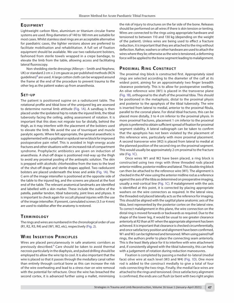
the risk of injury to structures on the far side of the bone. Releases should be performed on all wires if there is skin tension or tenting. Wires are connected to the rings using appropriate hardware and tensioned to between 110 and 130 kg (depending on the weight of the patient). Unless wires are being used to effect a fracture reduction, it is important that they are attached to the ring without deflection. Rather, washers or other hardware are used to attach the wires where they lie; otherwise as the wire is tensioned, a deforming force will be applied to the bone segment leading to malalignment.
Pr ox i m A l ri n g co n s t r u c tThe proximal ring block is constructed first. Appropriately sized rings are selected according to the diameter of the calf at its widest point, aiming for an approximately two finger breadth clearance posteriorly. This is to allow for postoperative swelling. An olive reference wire (W1) is placed in the transverse plane (Fig. 1B), orthogonal to the shaft of the proximal tibia. This should be positioned in the metaphysis, distal to the proximal physis and posterior to the apophysis of the tibial tuberosity. The wire is inserted from lateral to medial, anterior to the proximal fibula, parallel to the coronal plane. For distal tibial fractures, this can be placed more distally, 3 to 4 cm inferior to the proximal physis. In more proximal fractures, placement 1 cm inferior to the proximal physis is preferred to obtain sufficient spread of fixation for proximal segment stability. A lateral radiograph can be taken to confirm that the apophysis has not been violated by the placement of this reference wire, particularly with more proximal placement. A second transverse wire (W2) is placed from lateral to medial at the planned position of the second ring on the proximal segment. This would usually be approximately 2 cm proximal to the fracture site (Fig. 1C).
Once wires W1 and W2 have been placed, a ring block is constructed using two rings with three threaded rods placed anterior midline, posterior midline and lateral. This double ring block can then be attached to the reference wire (W1). The alignment is checked in the AP view using the anterior midline rod as a reference against the axis of the tibia as determined on preoperative imaging, usually a mid-cortical line (Fig. 1C). If malalignment with the axis is identified at this point, it is corrected by placing appropriate washers on the wire connectors as required. In the lateral view, the threaded rod placed laterally acts as the reference for imaging. This should be aligned with the sagittal plane anatomic axis of the tibia, best represented by the posterior cortex on the lateral view. To correct malalignment in this plane, the wire connection on the distal ring is moved forwards or backwards as required. Due to the shape of the lower leg, it would be usual to see greater clearance anteriorly at R2 than at R1 when the appropriate alignment has been achieved. It is important that clearance is checked circumferentially, and once satisfactory position and alignment have been confirmed, W1 and W2 can be tightened and tensioned. When using paired half rings, the authors prefer to place the connecting seam anteriorly. This is the least likely place for it to interfere with wire attachment and, if consistently aligned with the tibial tuberosity, this can help with a judgement of rotation during reduction manoeuvres.
Fixation is completed by passing a medial-to-lateral (medial face) olive wire at each level (W3 and W4) (Fig. 1D). One more rod is added to the construct medially to give a total of four rods connecting the two rings. Finally, the medial face wires are attached to the rings and tensioned. Once satisfactory alignment is confirmed, the ends are cut flush (or bent with two right angles
eq u i Pm e n tLightweight carbon fibre, aluminium or titanium circular frame systems are used. Ring diameters of 140 to 180 mm are suitable for most cases. Whilst stainless steel rings are an acceptable alternative for paediatric cases, the lighter versions above are preferred to facilitate mobilisation and rehabilitation. A full set of fixation equipment should be available. We use two radiolucent bolsters, fashioned from sterile towels wrapped in a crepe bandage, to elevate the limb from the table, allowing access and facilitating lateral fluoroscopy.
Non-shedding sterile dressings (Allevyn – Smith and Nephew, UK) or standard 2 cm × 2 cm gauze as per published methods (RCN guidelines)8 are used. A large cotton cloth can be wrapped around the frame at the end of the procedure to prevent trauma to the other leg as the patient wakes up from anaesthesia.
se t-u PThe patient is positioned supine on a radiolucent table. The rotational profile and tibial bow of the uninjured leg are assessed to determine normal limb attitude (Fig. 1A). A sandbag is then placed under the ipsilateral hip to rotate the injured limb, the tibial tuberosity facing the ceiling, aiding assessment of rotation. It is important that this does not migrate too far distally, behind the thigh, as it may interfere with the placement of the bolsters used to elevate the limb. We avoid the use of tourniquet and muscle paralytic agents. Where felt appropriate, the general anaesthetic is supplemented with a popliteal or epidural nerve block for improved postoperative pain relief. This is avoided in high-energy acute fractures and other situations with an increased risk of compartment syndrome. Prophylactic antibiotics are given on induction of anaesthesia. A shut-off drape is positioned mid-way up the thigh to avoid any proximal pooling of the antiseptic solution. The skin is prepped with alcoholic chlorhexidine from the toes to the level of the shut-off drape and sterile drapes applied. Two radiolucent bolsters are placed underneath the knee and ankle (Fig. 1A). The C-arm of the image intensifier is positioned at the opposite side of the table to the injured leg, and the screens are placed at the foot end of the table. The relevant anatomical landmarks are identified and labelled with a skin marker. These include the outline of the patella, patellar tendon, tibial tuberosity, joint lines and physes. It is important to check again for occult physeal injuries with the use of the image intensifier. If present, cannulated screws (3.5 or 4 mm) are used to stabilise after the anatomy is restored.
te r m i n o lo g yThe rings and wires are numbered in the chronological order of use (R1, R2, R3, R4) and (W1, W2, etc), respectively (Fig. 2).
Wi r e in s e r t i o n Pr i n c i P l e sWires are placed percutaneously in safe anatomic corridors as previously described.9 Care should be taken to avoid thermal necrosis particularly in the diaphysis, and pulsed drilling should be employed to allow the wire tip to cool. It is also important that the wire is placed so that it passes through the medullary canal rather than entirely through cortical bone as this can increase the risk of the wire overheating and lead to a stress riser on wire removal with the potential for refracture. Once the wire has breached the second cortex, it is advanced further using a mallet, minimising

Ilizarov Method for Acute Paediatric Tibial Fractures
Strategies in Trauma and Limb Reconstruction, Volume 16 Issue 1 (January–April 2021)48

Ilizarov Method for Acute Paediatric Tibial Fractures
Strategies in Trauma and Limb Reconstruction, Volume 16 Issue 1 (January–April 2021) 49
Mechanically, it may be preferable to place a pin on each ring, approximately in the same direction, oriented as close as possible to the sagittal plane.11
di s tA l ri n g co n s t r u c tFirst, a reference wire (W5) is inserted in the distal tibia (Fig. 1E). The assistant holds the foot, applying gentle in-line traction and rotation
around the ring) and the proximal ring construct is complete (Figs 1A to D and 2A and B).
Hydroxyapatite (HA)-coated half-pins can be used in larger patients (weighing over 80 kg) to provide extra support to the frame construct. These are placed at R1 and R2 within the antero-medial arc as per Nayagam et al.10 Half-pins can also be substituted for wires as necessary to avoid compromised soft tissue in, for example, open fractures or patients who have had compartment syndrome.
Figs 1A and B: Step-by-step guidance (A to K) from preoperative set-up to reference wire (W 5)

Ilizarov Method for Acute Paediatric Tibial Fractures
Strategies in Trauma and Limb Reconstruction, Volume 16 Issue 1 (January–April 2021)50
at R2 to R3 as interlocking of the main fracture fragments might hinder subsequent reduction manoeuvres. If that does not succeed, un-tension and re-attach W5 until satisfactory rotational and sagittal reduction is achieved (Figs 1G and H). Do not proceed until this stage of reduction appears satisfactory.
3. Translation in coronal plane – ‘Sliding manoeuvre’Some residual translation and angulation may be present due to soft tissue tension or impingement of fragments. This is corrected by sliding the distal fragment along the smooth wire (W5) (Fig. 1H), thereby shifting the ankle centre underneath the proximal segment. This is best achieved by tapping the medial or lateral side of frame with a mallet whilst holding the foot firmly.
There is often a natural varus bow to the distal tibia in children, and the majority of patients displace into valgus and translate laterally. In this situation, the foot is held on the lateral border and the most distal ring is tapped from the medial side. This brings the distal fragment from lateral to medial and will usually correct any residual valgus deformity. If the translation is medial, then the manoeuvre is reversed.
4. The ‘near fracture’ wire for further adjustmentsIf minor malalignment in the coronal plane remains, this can be corrected using a ‘near fracture’ pulling olive wire (W6) on ring R3 (Fig. 1I). Here the wire is passed from the direction that the bone needs correcting from. For example, when correcting residual lateral translation, a transverse olive wire is inserted from the lateral side from the direction of maximal displacement. Before securing this wire to the medial side of the ring, the tensioner is applied and carefully used to draw the wire to it, pulling the bone with the olive. X-ray control is used, and once the desired position is achieved, the wire is secured as usual and the tensioning completed. As this applies further fixation and begins to lock the distal fragment in position, it is prudent to check for pro- or recurvatum in the sagittal plane before finalising this step.
Minor residual angulation in the sagittal plane (pro- or recurvatum) can be due to impaction of the fracture fragments, in which case more distraction (between R2 and R3) and gentle manual manipulation can lead to improved reduction. If sufficient distraction has been applied, then applying a rotational force by plantar or dorsiflexing the foot can correct angulation of this nature by pivoting the fragment around the reference wire (W5). If successful, then this force will need to be maintained whilst further fixation is applied. Finally, if these manoeuvres do not result in correction of sagittal plane angulation, then an arched reduction wire (W6) can be placed on ring R3. Following insertion, the wire is deformed either anteriorly or posteriorly depending on the desired correction. The arched wire is straightened once tensioned, moving the bone at that level anteriorly or posteriorly, correcting angulation.
Two adjustment techniques are described here – one for residual angulation or translation or both in the coronal plane and another for residual sagittal plane pro- or recurvatum. Both techniques may have to be combined into one step (achieving fracture reduction with the same wire) if both types of residual deformity exist concurrently.
5. CompletionOnce the reduction is acceptable after tightening and tensioning W6, the position of the bone is essentially set, fixation is then completed for stability. A trans-fibular wire on ring 4 (W7) and
until a mortise view of the ankle is achieved. Under fluoroscopic guidance, a transverse plain reference wire (W5) is passed from lateral to medial, 5 to 10 mm proximal to the distal physis. This wire should be placed perpendicular to the axis of the distal tibial segment, which is usually parallel to the projection of the talar dome. The wire entry point should be just anterior to the fibula, exiting centrally above the medial malleolus. It is important that this reference wire is placed in the coronal plane of the tibia (20 degrees offset to the transmalleolar axis); otherwise, any correction along this wire later may lead to a sagittal plane deformity.
The distal ring block, consisting of two rings (R3 and R4), is then built with two connecting rods (or posts for very distal fractures), placed anteriorly and posteriorly (Fig. 1F). The distal ring block is now connected to the proximal ring block with four threaded rods, placed anteromedial, anterolateral, posteromedial, and posterolateral. These rods should be attached flush and fully tightened at R3 but with enough spare length at their only finger-tight attachment to the proximal block at R2. This allows swift adjustment for distraction/compression manoeuvres as outlined below. These four rods should all be the same length resulting in the proximal and distal ring blocks being parallel, concentric, and colinear.
re co m m e n d e d 5-s t e P se q u e n c e f o r Ac u t e re d u c t i o nThe frame (R1–R4) has now been constructed, aligned with the proximal segment as reference in the sagittal and coronal planes (Figs 1F and 2C). If the reduction manoeuvres described below do not result in accurate reduction, it is usually the case that a previous step is incorrect. If problems are encountered, it is usually necessary to move back and repeat.
1. RotationIn the AP view, the distal segment is manually positioned in within the frame construct by applying in line traction at the foot. Rotation is considered first. This is best assessed clinically, by looking from the foot of the patient in plantigrade position from the bottom of the operating table towards their patella and tibial tuberosity. This essentially sets the equivalent of the thigh-foot angle. If the proximal ring block seam has been aligned with the tibial tuberosity, then it can aid with judgement of rotation. This usually falls at approximately the second ray of the foot. Clues can also be gained from lateral radiographs. If the fibula fracture re-aligns, then tibial rotation is likely to be approximately correct. Similarly, the proximal and distal spikes of torsional tibial fractures should align once rotation is corrected.
2. Translation in sagittal planeIn the lateral view, the centre of the talar dome should align with the proximal tibial shaft (midline). If this is incorrect, translation is adjusted by moving the connection of the wire (W5), and therefore the distal segment, anteriorly or posteriorly on the ring (R4), without changing rotation. It is important to note that residual sagittal plane angulation (pro- or recurvatum) may persist at this point and can be corrected later. We recommend adjusting the rod length through the frame construct (at R2–R3), so that there is a slight downward bend (apex of bend proximal) on W5, particularly if the fracture appears shortened. This introduces distraction at the fracture site, once W5 tensioned. If the resulting position is unsatisfactory, attempt further distraction by rod lengthening

Ilizarov Method for Acute Paediatric Tibial Fractures
Strategies in Trauma and Limb Reconstruction, Volume 16 Issue 1 (January–April 2021) 51
and patients then allowed to weight-bear with crutches immediately after. Occasionally, an older child would tolerate removal under nitrous oxide inhalation in the outpatient clinic.
If reduction fails, or there is a delayed presentation or delay in treatment:This may arise after a period of attempted cast treatment, for example. Treatment follows the principles of the Ilizarov tension-stress effect where the callus is pulled apart and realigned gradually at a rate of 1 mm or less per day in order to gain the contact and alignment for a healed straight bone. The key is to recognise this eventuality before entering the operating theatre and have the appropriate equipment available. Most hexapod struts and their associated software cannot function on standard Ilizarov rings.
The bailout option for failing to achieve reduction is to either use hexapod technology (between R2 and R3) or traditional Ilizarov axial hinges and motors to remove the residual deformity. In advanced callus, the senior author has used hexapod struts in chronic mode and dialled in the correction on table to achieve reduction by gradually deforming the callus until resistance felt. This can be further corrected over the following week(s). Only in hardened callus would we recommend a corrective osteotomy, in which case the presented technique above for fracture fixation and reduction can be used.
Alongside these techniques, it is important to consider what constitutes an acceptable level of ‘malreduction’, as the aim is not always to achieve an absolute perfect reduction at the expense of excessive theatre time and multiple revised wires – a few millimetres of translation is likely to give an acceptable mechanical and functional result as will a few degrees of apex posterior angulation distally for example. Varus and valgus angulation and malrotation should not be tolerated.
co n c lu s i o nThis systematic approach, adapted from our technique used in adult patients, is effective for the treatment of a wide variety of tibial fractures in children of all ages. It is reproducible and is helpful to frame surgeons working in units with lower caseload volumes and limited availability of expensive hexapod technology.
medial face wires on ring 3 and ring 4 (W8 and W9) are added for this purpose. The reference wire on ring 4 (W5) is the most distal wire – all other wires on ring 4 should be proximal to the distal ring. This way the distal tibial physis is protected. If, at the end of the procedure, W5 has been significantly deformed by the reduction manoeuvres, this can be replaced with a new wire and tensioned.
It may be necessary to compress between R2 and R3 if over-distraction was required to achieve reduction. If minor angular deformity in the coronal or sagittal planes remains, this can be fine-tuned by adjusting the length of the connecting rods between R2 and R3 individually. This results in differential tension between the rods across the fracture and produces a degree of ring-to-ring reduction through a slight manipulation of the relative position of the ring blocks. Corrections of this nature should be limited to approximately 3 to 5 degrees; otherwise, too much bend in the rods occurs (Figs 1H to J and 2C and D). If greater amounts of ring-to-ring correction is required, then either the previous steps need to be repeated for a better result, or (a) replace the rods between R2 and R3 with rods matched to conical washers; (b) assemble hinges to accommodate the non-parallel arrangement of the proximal and distal ring blocks.
The approach described here is directly relevant to the management of simple, essentially, 2-part extra-articular tibial fractures. Once confidence in the technique is achieved, it can be adapted for more complex fractures, including segmental or multi-fragmentary ones.
Af t e r c A r eRoutine follow-up is provided to our patients at two weekly intervals including pin site care, psychological support (if necessary) and monitoring of fracture alignment and healing progress. In the paediatric population, delayed or non-union is rare, and therefore, routine use of dynamisation methods to accelerate bone healing is not carried out. In rare occasions of delayed healing, a short period of supportive compression across the fracture site (1/4 mm per day on each rod for 2 weeks) can enhance the contact and mechanical environment for healing.
The frame is removed after X-ray evidence of three-cortical healing is confirmed. Removal is done under a general anaesthetic
Figs 2A to D: Wire to ring reduction technique: After building the anatomically aligned proximal ring construct (R1-2 in A), the frame is extended distally with further two rings (R3-4 in B). The ankle reference wire (W5) is then attached to R4 after positioning of the limb (C). Note the intentional distraction between R2-3 to disimpact fracture fragments (C). Reduction techniques as described in text complete reduction and contact at fracture site (D).

Ilizarov Method for Acute Paediatric Tibial Fractures
Strategies in Trauma and Limb Reconstruction, Volume 16 Issue 1 (January–April 2021)52
5. BOA. Trauma BOASTs [Internet]. Available from: https://www.boa.ac.uk/standards-guidance/boasts/trauma-boasts.html.
6. BOA/BAPRAS. The management of severe open lower limb fractures; 2009. Available from: https://www.boa.ac.uk/wp-content/uploads/2014/05/BOAST-4-The-Management-of-Sever-Open-Lower-Limb-Fractures.pdf.
7. Brzobohatá H, Krajíček V, Velemínský P, et al. Three-dimensional geometry of human tibial anterior curvature in chronologically distinct population samples of Central Europeans (2900 BC – 21st century AD). Sci Rep 2019;9(1):4234. DOI: 10.1038/s41598-019-40625-3.
8. Timms A, Vincent M, Santy-Tomlinson J, et al. Guidance on pin site care. Royal College of Nursing; 2011. Available from: https://www2.rcn.org.uk/__data/assets/pdf_file/0009/413982/004137.pdf.
9. Catagni MA. Atlas for the insertion of transosseus wires and half-pins. North. 2003.
10. Nayagam S. Safe corridors in external fixation: the lower leg (tibia, fibula, hindfoot and forefoot). Strateg Trauma Limb Reconstr 2007;2(2–3):105–110. DOI: 10.1007/s11751-007-0023-7.
11. Henderson DJ, Rushbrook JL, Stewart TD, et al. What are the biomechanical effects of half-pin and fine-wire configurations on fracture site movement in circular frames? Clin Orthop Relat Res 2016;474(4):1041–1049. DOI: 10.1007/s11999-015-4652-8.
12. Messner J, Johnson L, Taylor DM, et al. Treatment and functional outcomes of complex tibial fractures in children and adolescents using the Ilizarov method. Bone Joint J 2018;100-B(3):396–403. DOI: 10.1302/0301-620X.100B3.BJJ-2017-0863.R1.
13. Johnson L, Messner J, Igoe EJ, et al. Quality of life and post-traumatic stress symptoms in paediatric patients with tibial fractures during treatment with cast or Ilizarov frame. Injury 2020;51(2):199–206. DOI: 10.1016/j.injury.2019.10.077.
With experience, the frame construction can be completed rapidly. By following this method, on-table reduction is possible for the majority of these fractures avoiding the need of postoperative correction. Achieving immediate reduction with contact, alignment and stability of the fracture helps facilitate early weight-bearing and rehabilitation. Available evidence would suggest that Ilizarov management is safe, effective and well tolerated in children with tibial fractures.12,13
Ac k n oW l e d g m e n tsThe authors would like to thank Manuel Messner for support with graphics.
re f e r e n c e s 1. Mooney J, Hennrikus W, Flynn JM, et al. Rockwood and Wilkins’
fractures in children. 8th ed. vol. 1. Wolters Kluwer; 2015. 2. Foster PA, Barton SB, Jones SC, et al. The treatment of complex
tibial shaft fractures by the Ilizarov method. J Bone Jt Surg Br 2012;94(12):1678–1683. DOI: 10.1302/0301-620X.94B12.29266.
3. Harwood P, Taylor DM. Segmental tibia shaft fractures – extra articular ilizarov reduction techniques. In: Giannoudis P, editor. Fracture reduction and fixation techniques. Cham: Springer International Publishing; 2020. p. 351–371.
4. Giannoudis V, Ewins E, Foster P, et al. Clinical and functional outcome following distal tibia fracture treated by circular external fixation. Orthop Proc 2017;99-B(SUPP_13):16. doi/abs/10.1302/1358-992x.99BSUPP_13.BLRS2017-016