Medial Opening Wedge High Tibial Osteotomy in Knee ... - MDPI
Upload
khangminh22Category
view
0download
0
Case ReportTotal Knee Arthroplasty after Correction of Tibial DiaphysealNonunion with Clamshell Osteotomy
Pingal Desai ,1,2,3 Vivek Sharma,2 and Karanvir Prakash2
1Honorary Clinical Associate Professor of Orthopedics, University of Kansas, Lawrence, USA2Colonial Orthopedics, Colonial Heights, USA3Orthopedic Surgeon-Southwest Medical Center, Liberal, Kansas, USA
Correspondence should be addressed to Pingal Desai; [email protected]
Received 11 February 2018; Revised 30 July 2018; Accepted 16 August 2018; Published 25 September 2018
Academic Editor: Dimitrios S. Karataglis
Copyright © 2018 Pingal Desai et al. This is an open access article distributed under the Creative Commons Attribution License,which permits unrestricted use, distribution, and reproduction in any medium, provided the original work is properly cited.
Total knee arthroplasty is mostly done to relieve pain and disability from a severe and degenerated knee. Deformities in thecoronal and sagittal plane could be corrected with the help of cuts made in tibia and femur during total knee replacement aswell as with ligament release. However, large deformities in the lower extremity particularly in the diaphysis region needcorrection prior to the total knee replacement. It helps to limit the amount of bone that will be cut and helps theligament release. Several extra articular and intra-articular methods for the correction of diaphyseal deformity have beendescribed. We present the case of clamshell osteotomy for the correction of diaphyseal deformity in the tibia and a totalknee replacement after the osteotomy site healed.
1. Introduction
Clamshell osteotomy was first described by Russell et al. in2009 to correct deformities in the long bone [1]. Itinvolves two transverse osteotomies: one on the proximaland one on the distal aspect of malunion site of the longbone. The third longitudinal osteotomy is carried out inthe fragment in between two transverse osteotomies. Theosteotomized fragment would open like a clamshell andit would allow realignment of the mechanical axis of theextremity. This is followed by fixing proximal with distalfragment and stabilizing the osteotomy. We report the firstcase of total knee arthroplasty (TKA) after extra-articularcorrection of mechanical axis of lower extremity in tibiawith clamshell osteotomy.
2. Case Report
A 65-year-old male with a BMI of 42 and uncontrolledtype II diabetes mellitus came to our clinic with a fractureon the left tibial shaft. He was treated conservatively for20 years. Radiographs showed a malunited fracture onthe middle third left tibia in 20-degrees varus, 15-degrees
apex anterior angulation with a 1 cm anterior translationof distal segment, 20-degrees internal rotation and 1.5 cmshortening. He also had severe tricompartmental osteoar-thritis on the left knee (Figures 1(a) and 1(b)). A CT scanconfirmed the presence of malunion (Figure 2). Bloodsugar was controlled by an endocrine physician and aclamshell osteotomy of the tibia was planned.
3. Surgical Procedure for Clamshell Osteotomy
Clamshell osteotomy was performed as described by Rus-sell et al. [1]. Patient was placed on radiolucent table,and radiolucent triangular bump was used to keep kneein 90 degree flexion position. Skin incision was mademedial to patellar tendon of 3 cm extending proximal totibial tuberosity. Plane was developed superficial to thefat layer taking care that joint is not exposed. Entry ofguide wire was made just medial to lateral intercondylareminence tibia proximal to tibial tuberosity and in lineof the medullary canal under fluoroscopy. At this point,tip of guide wire was left just proximal to the fracture site.The proximal and distal site of the fracture was markedunder the fluoroscopy and anterolateral skin incision was
HindawiCase Reports in OrthopedicsVolume 2018, Article ID 2632963, 6 pageshttps://doi.org/10.1155/2018/2632963

made just lateral to the shin of tibia. Skin and subcutane-ous tissue were cut, muscles of anterior compartment wereretracted laterally, and fracture site on tibia was exposed.The proximal and distal aspect of the extent of long obli-que fracture was marked on tibia under fluoroscopy. Theclamshell fragment of the bone was 8.5 cm long and sev-eral vertical drill holes were made on the anterior borderof the tibia over clamshell fragment with a 2.5mm drillbit. Drill holes extended through anterior and posteriorcortex to create a path for the vertical osteotomy. Thiswas followed by transverse osteotomies performed throughthe proximal and distal parts of bone under fluoroscopycontrol with oscillating saw followed by vertical osteotomythrough the drill holes in the middle fragment with oscil-lating saw. The middle fragment opened up like a clam-shell. Then, 1 cm of fibula was excised through aseparate lateral incision over fracture site to allow goodcollapse of the osteotomy and was used as bone graft.Tibia was properly aligned and “blocking screw” wasplaced laterally on the distal fragment to maintain cor-rected varus position of tibia (Figures 3(a) and 3(b)).Guide wire was passed from proximal to the distal frag-ment and fascia and subcutaneous tissue were closed overthe osteotomy site to preserve the reamings. Tibial canalwas reamed with 9–13 reamers and no 12 tibial nail (Bio-met Versa) was inserted. Nail was locked with two proxi-mal and two distal locking screws (Figures 3(a) and 3(b)).
(a)
LT
(b)
Figure 1: (a) Leg length view showing 30 degrees varus of tibia and several osteoarthritis left knee. (b) Lateral view showing 20 degrees apexanterior angulation at malunited fracture site.
Figure 2: CT scan—malunited fracture of the middle third left tibia.
2 Case Reports in Orthopedics

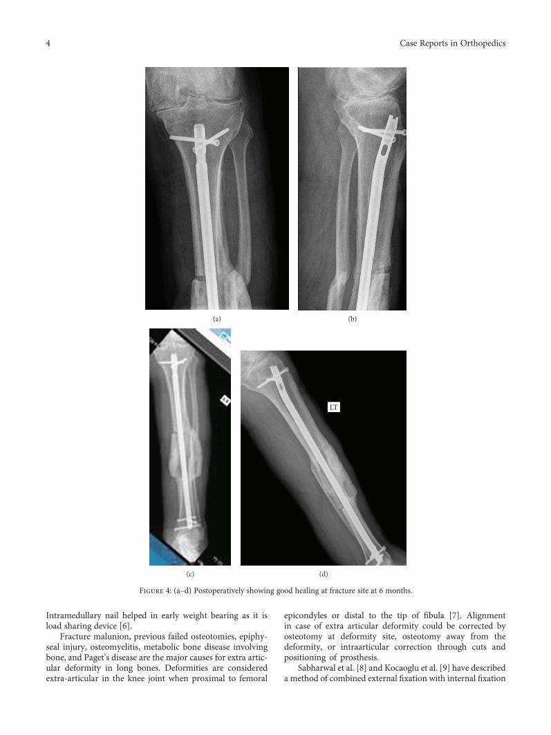
There are several studies which have concluded thatcalcium and vitamin D levels affect healing of the fracture[2, 3]. So calcium and vitamin D levels were monitoredand corrected as needed after surgery. Blood sugar was con-trolled postoperatively. Radiologic healing of fracture wasconfirmed by three cortices healing on X-ray at 6 months(Figures 4(a)–4(d)). CT scan of the left knee and tibia wasdone (Figure 5) to confirm union and preoperative planningfor custom-made cutting blocks to avoid intramedullary tem-plating or instrumentation during total knee replacement.
3.1. Planning and Surgical Procedure for Total KneeReplacement. Plan was made for extramedullary referencingfor both tibia and femur. Custom-made cutting blocks wereprepared prior to the surgery (Zimmer Biomet, Signaturesystem; Warsaw, IN, USA) based on CT scan. The femoralvalgus angle was set to 4.5 degrees and tibial varus-valgusangle was set to 0 degrees. The posterior slope on tibia wasset to 3 degrees and tibial resection below lateral low pointwas 8mm while below medial low point was 2mm.
During surgery, two proximal screws from tibia nail weretaken out using a fluoroscope. Plan was made for removal ofthe tibial nail if needed. A medial knee parapatellar approachfor total knee replacement was made and distal femur andtibia cuts were made using custom made jig (Zimmer BiometSignature). Distal femur, patella, and proximal tibial prepara-tion was done. There was no interference of nail during tibialcanal reaming and impaction of wings of tibial component.Remaining surgery was performed in routine manner.
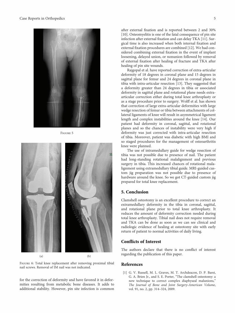
Postoperative X-rays showed good alignment (Figure 6).Mechanical axis of lower extremity was restored to 3 degrees
with vertical axis of the body. Patient was started physicaltherapy and weight bearing operated extremity on sameday. Patient regained 0–95 degrees of knee movement at 4weeks and had 110 degrees movement at 6 months.
4. Discussion
Clamshell osteotomy reported by Russell et al. [1] was pre-liminary experience at three institutions in USA. Two morecase report of clamshell osteotomy have been reported inthe literature [4, 5]. Wilson et al. reported a case of total kneearthroplasty after correction of extra-articular femoral defor-mity caused by malunited femoral shaft fracture. To ourknowledge, this is the first case of total knee arthroplasty aftercorrection of extra-articular deformity in tibia using clam-shell osteotomy.
Deformities in tibia which included varus, anterior angu-lation, translation, rotation, and shortening were correctedusing this technique. The placement of blocking screw lateralto the nail helped in preventing distal fragment going intovarus. Russell et al. [1] have suggested avoiding correctionof shortening more than 5 cm in femur and 3 cm in tibia.Shortening in our patient was 1.5 cm and we did not haveany difficulty in lengthening 1.5 cm. There were no clinicalfindings of excessive neurovascular stretching postopera-tively. The fibular segment excised was used as bone graftand we did not use any allograft or synthetic osteoconductivematerials. We also considered regarding adequate soft tissuecoverage and early weight bearing as described by Russellet al. [1]. Our patient had malunited tibial fracture for 20years and skin coverage over deformity site was adequate.
(a) (b)
Figure 3: (a, b) Internal fixation after clamshell osteotomy. (b) Blocking screw was used in distal tibia for proper positioning of rod whichhelped correction of deformity.
3Case Reports in Orthopedics
Intramedullary nail helped in early weight bearing as it isload sharing device [6].
Fracture malunion, previous failed osteotomies, epiphy-seal injury, osteomyelitis, metabolic bone disease involvingbone, and Paget’s disease are the major causes for extra artic-ular deformity in long bones. Deformities are consideredextra-articular in the knee joint when proximal to femoral
epicondyles or distal to the tip of fibula [7]. Alignmentin case of extra articular deformity could be corrected byosteotomy at deformity site, osteotomy away from thedeformity, or intraarticular correction through cuts andpositioning of prosthesis.
Sabharwal et al. [8] and Kocaoglu et al. [9] have describeda method of combined external fixation with internal fixation
(a) (b)
(c)
LT
(d)
Figure 4: (a–d) Postoperatively showing good healing at fracture site at 6 months.
4 Case Reports in Orthopedics
for the correction of deformity and have favored it in defor-mities resulting from metabolic bone diseases. It adds toadditional stability. However, pin site infection is common
after external fixation and is reported between 2 and 30%[10]. Osteomyelitis is one of the fatal consequence of pin siteinfection after external fixation and can delay TKA [11]. Sur-gical time is also increased when both internal fixation andexternal fixation procedures are combined [12]. We had con-sidered combining external fixation in the event of implantloosening, delayed union, or nonunion followed by removalof external fixation after healing of fracture and TKA afterhealing of pin site wounds.
Rajgopal et al. have reported correction of extra-articulardeformity of 18 degrees in coronal plane and 15 degrees insagittal plane for femur and 24 degrees in coronal plane intibia with intra-articular resection [13]. They suggested thata deformity greater than 24 degrees in tibia or associateddeformity in sagittal plane and rotational plane needs extra-articular correction either during total knee arthroplasty oras a stage procedure prior to surgery. Wolff et al. has shownthat correction of large extra-articular deformities with largewedge resection of femur or tibia between attachments of col-lateral ligaments of knee will result in asymmetrical ligamentlength and complex instabilities around the knee [14]. Ourpatient had deformity in coronal, sagittal, and rotationalplanes and so the chances of instability were very high ifdeformity was just corrected with intra-articular resectionof tibia. Moreover, patient was diabetic with high BMI andso staged procedures for the management of osteoarthritisknee were planned.
The use of intramedullary guide for wedge resection oftibia was not possible due to presence of nail. The patienthad long-standing rotational malalignment and previoussurgery in tibia. This increased chances of rotational mala-lignment using extramedullary tibial guide. MRI-guided cus-tom jig preparation was not possible due to presence ofhardware around the knee. So we got CT-guided custom jigprepared for total knee replacement.
5. Conclusion
Clamshell osteotomy is an excellent procedure to correct anextramedullary deformity in the tibia in coronal, sagittal,and rotational plane prior to total knee arthroplasty. Itreduces the amount of deformity correction needed duringtotal knee arthroplasty. Tibial nail does not require removaland TKA can be done as soon as we can see clinical andradiologic evidence of healing at osteotomy site with earlyreturn of patient to normal activities of daily living.
Conflicts of Interest
The authors declare that there is no conflict of interestregarding the publication of this paper.
References
[1] G. V. Russell, M. L. Graves, M. T. Archdeacon, D. P. Barei,G. A. Brien Jr., and S. E. Porter, “The clamshell osteotomy: anew technique to correct complex diaphyseal malunions,”The Journal of Bone and Joint Surgery-American Volume,vol. 91, no. 2, pp. 314–324, 2009.
(a) (b)
Figure 6: Total knee replacement after removing proximal tibialnail screws. Removal of IM nail was not indicated.
Figure 5
5Case Reports in Orthopedics

[2] A. M. Doetsch, J. Faber, N. Lynnerup, I. Wätjen, H. Bliddal,and B. Danneskiold–Samsøe, “The effect of calcium and vita-min D3 supplementation on the healing of the proximalhumerus fracture: a randomized placebo-controlled study,”Calcified Tissue International, vol. 75, no. 3, pp. 183–188, 2004.
[3] V. Heidler, M. Haffner-Luntzer, K. Prystaz et al., “Calcium andvitamin-D supplementation post-trauma improves bonehealing and decreases posttraumatic bone resorption in anosteoporotic mouse model,” Bone Abstracts, vol. 5, p. 55, 2016.
[4] A. J. Wilson, S. Nandi, C. E. Robbins, and J. V. Bono, “TKAafter clamshell osteotomy for femoral diaphyseal malunion,”Orthopedics, vol. 35, no. 6, pp. e969–e972, 2012.
[5] D. Santoro, S. Tantavisut, D. Aloj, and M. D. Karam, “Diaph-yseal osteotomy after post-traumatic malalignment,” CurrentReviews in Musculoskeletal Medicine, vol. 7, no. 4, pp. 312–322, 2014.
[6] S. Kuhn, J. Greenfield, C. Arand et al., “Treatment of distalintraarticular tibial fractures: a biomechanical evaluation ofintramedullary nailing vs. angle-stable plate osteosynthesis,”Injury, vol. 46, pp. S99–S103, 2015.
[7] K. G. Vince and V. Bozic, “Management of extra-articulardeformities in total knee arthroplasty,” in Total Knee Arthro-plastypp. 205–211, Springer.
[8] S. Sabharwal, S. Green, J. McCarthy, and R. C. Hamdy,“What's new in limb lengthening and deformity correction,”The Journal of Bone and Joint Surgery-American Volume,vol. 93, no. 2, pp. 213–221, 2011.
[9] M. Kocaoglu, F. E. Bilen, C. Sen, L. Eralp, and H. I. Balci,“Combined technique for the correction of lower-limb defor-mities resulting from metabolic bone disease,” The Journal ofBone and Joint Surgery, vol. 93-B, no. 1, pp. 52–56, 2011.
[10] A. Massè, A. Bruno, M. Bosetti, A. Biasibetti, M. Cannas, andP. Gallinaro, “Prevention of pin track infection in externalfixation with silver coated pins: clinical and microbiologicalresults,” Journal of Biomedical Materials Research, vol. 53,no. 5, pp. 600–604, 2000.
[11] D. J. Wickens, G. West, P. J. Kelly, J. Verran, S. Lynch, andK. A. Whitehead, “Antimicrobial activity of nanocompositezirconium nitride/silver coatings to combat external bonefixation pin infections,” The International Journal of ArtificialOrgans, vol. 35, no. 10, pp. 817–825, 2012.
[12] K. T. M. Seah, R. Shafi, A. T. Fragomen, and S. R. Rozbruch,“Distal femoral osteotomy: is internal fixation better thanexternal?,” Clinical Orthopaedics and Related Research,vol. 469, no. 7, pp. 2003–2011, 2011.
[13] A. Rajgopal, V. Dahiya, A. Vasdev, H. Kochhar, and V. Tyagi,“Long-term results of total knee arthroplasty for valgus knees:soft-tissue release technique and implant selection,” Journal ofOrthopaedic Surgery, vol. 19, no. 1, pp. 60–63, 2011.
[14] M. Wolff, D. S. Hungerford, and C. L. Pepe, “The effect ofextraarticular varus and valgus deformity on total knee arthro-plasty,” Clinical Orthopaedics and Related Research, vol. 271,pp. 35–51, 1991.
6 Case Reports in Orthopedics

Stem Cells International
Hindawiwww.hindawi.com Volume 2018
Hindawiwww.hindawi.com Volume 2018
MEDIATORSINFLAMMATION
of
EndocrinologyInternational Journal of
Hindawiwww.hindawi.com Volume 2018
Hindawiwww.hindawi.com Volume 2018
Disease Markers
Hindawiwww.hindawi.com Volume 2018
BioMed Research International
OncologyJournal of
Hindawiwww.hindawi.com Volume 2013
Hindawiwww.hindawi.com Volume 2018
Oxidative Medicine and Cellular Longevity
Hindawiwww.hindawi.com Volume 2018
PPAR Research
Hindawi Publishing Corporation http://www.hindawi.com Volume 2013Hindawiwww.hindawi.com
The Scientific World Journal
Volume 2018
Immunology ResearchHindawiwww.hindawi.com Volume 2018
Journal of
ObesityJournal of
Hindawiwww.hindawi.com Volume 2018
Hindawiwww.hindawi.com Volume 2018
Computational and Mathematical Methods in Medicine
Hindawiwww.hindawi.com Volume 2018
Behavioural Neurology
OphthalmologyJournal of
Hindawiwww.hindawi.com Volume 2018
Diabetes ResearchJournal of
Hindawiwww.hindawi.com Volume 2018
Hindawiwww.hindawi.com Volume 2018
Research and TreatmentAIDS
Hindawiwww.hindawi.com Volume 2018
Gastroenterology Research and Practice
Hindawiwww.hindawi.com Volume 2018
Parkinson’s Disease
Evidence-Based Complementary andAlternative Medicine
Volume 2018Hindawiwww.hindawi.com
Submit your manuscripts atwww.hindawi.com
Copyright © 2022 FDOKUMEN