Therapeutic Exercises for the Shoulder Region
14
PART 4 • The Shoulder Region in Upper Extremity Pain Syndromes Therapeutic Exercises for the Shoulder Region Johnson McEvoy, Kieran O’Sullivan, Carel Bron Chapter 33 selection of muscles, without focusing on one specific clinical population. Clinical Background Essential to an understanding of therapeutic exercise is an in-depth knowledge of anatomy, physiology and function, specifically related to the neuromuscular and musculoskeletal systems (Kendall 2002). The shoulder is a complex functional system producing movement of the arm on the trunk and allowing the upper limb and hand to be dynamically moved and positioned for function. The shoulder consists of the scapula, clavicle and humerus, giving rise to the sternocla- vicular, clavicular, humeral and scapulothoracic joints, and has a close relationship to the neck, thorax and ribs. The shoulder is supported by capsular, ligamentous and muscular systems with complex neuromuscular processing that offer a wide range of motion, but with a subsequent compromise in joint stability. This trade-off in stability makes the shoulder potentially vulnerable to dysfunction and injury, and stability is often the main focus of therapeutic exercise for the shoulder complex. Readers should refer to the appropriate chapters of this book and other texts for a comprehensive review of shoulder anatomy, biomechanics, kinesiology and pathome- chanics (Donatelli 2004a; Oatis 2004). Further, knowledge of connective tissue properties, force applications, tissue injury (bone, ligament, tendon, muscle, fascia, nerve, etc.) and tissue- healing concepts and timelines (inflammation, proliferation, maturation) is an important precursor to the development of a suitable and safe therapeutic exercise programme (Tippet & Voight 1995; Paris & Loubert 1999; Houglum 2005). Prior to the development of a rehabilitation programme for the shoulder complex a comprehensive assessment and physi- cal examination should be performed with reference to the principles of physical therapy practice so as to ascertain per- tinent information and physical characteristics of the indi- vidual patient. Indications for therapeutic exercise of the shoulder are listed in Box 33.1 and are diverse; they include specific and non-specific musculoskeletal, orthopaedic, surgi- cal and neurological conditions and dysfunctions, and also postural and performance enhancement and injury preven- tion strategy. CHAPTER CONTENTS Introduction 373 Clinical background 373 Shoulder exercise: evidence 374 Principles of exercise 374 Posture 376 Stretching 376 Isometric exercise of the shoulder 378 Isotonic exercises of the shoulder 378 Supraspinatus muscle 379 Infraspinatus and teres minor muscles 379 Subscapularis muscle 380 Trapezius muscle 380 Serratus anterior muscle 381 Functional exercises 381 Conclusion 383 Introduction Therapeutic exercise is a cornerstone of physiotherapy prac- tice and was initially referred to as medical gymnastics. The development of medical gymnastics in physical therapy has had many diverse influences including Dr Francis Fuller, author of Medicina gymnasticia (1740), Swedish gymnast Per Henrik Ling (1776–1839) and the Dutch physical education teacher and physician Dr Johann Georg Mezger (1838–1909) (Barclay 1994; Terlouw 2007). More recently Kendall (2002) summed up the role of therapeutic exercise in physical therapy: ‘Central to the practice of physical therapy is the prevention of movement dysfunction and the rehabilitation through restoration and maintenance of active movement – in other words, therapeutic exercise in its broadest sense’. The focus of this chapter is to introduce general principles of therapeutic exercise for the shoulder, and to stimulate clinical reasoning and rational rehabilitation. The chapter will briefly discuss posture, stretching and strengthening of a
-
Upload
khangminh22 -
Category
Documents
-
view
0 -
download
0
Transcript of Therapeutic Exercises for the Shoulder Region

PART 4 • The Shoulder Region in Upper Extremity Pain Syndromes
Therapeutic Exercises for the Shoulder RegionJohnson McEvoy, Kieran O’Sullivan, Carel Bron
Chapter 33
selection of muscles, without focusing on one specific clinical population.
Clinical BackgroundEssential to an understanding of therapeutic exercise is an in-depth knowledge of anatomy, physiology and function, specifically related to the neuromuscular and musculoskeletal systems (Kendall 2002). The shoulder is a complex functional system producing movement of the arm on the trunk and allowing the upper limb and hand to be dynamically moved and positioned for function. The shoulder consists of the scapula, clavicle and humerus, giving rise to the sternocla-vicular, clavicular, humeral and scapulothoracic joints, and has a close relationship to the neck, thorax and ribs. The shoulder is supported by capsular, ligamentous and muscular systems with complex neuromuscular processing that offer a wide range of motion, but with a subsequent compromise in joint stability. This trade-off in stability makes the shoulder potentially vulnerable to dysfunction and injury, and stability is often the main focus of therapeutic exercise for the shoulder complex. Readers should refer to the appropriate chapters of this book and other texts for a comprehensive review of shoulder anatomy, biomechanics, kinesiology and pathome-chanics (Donatelli 2004a; Oatis 2004). Further, knowledge of connective tissue properties, force applications, tissue injury (bone, ligament, tendon, muscle, fascia, nerve, etc.) and tissue-healing concepts and timelines (inflammation, proliferation, maturation) is an important precursor to the development of a suitable and safe therapeutic exercise programme (Tippet & Voight 1995; Paris & Loubert 1999; Houglum 2005).
Prior to the development of a rehabilitation programme for the shoulder complex a comprehensive assessment and physi-cal examination should be performed with reference to the principles of physical therapy practice so as to ascertain per-tinent information and physical characteristics of the indi-vidual patient. Indications for therapeutic exercise of the shoulder are listed in Box 33.1 and are diverse; they include specific and non-specific musculoskeletal, orthopaedic, surgi-cal and neurological conditions and dysfunctions, and also postural and performance enhancement and injury preven-tion strategy.
C H A P T E R C O N T E N T S
Introduction 373Clinical background 373Shoulder exercise: evidence 374Principles of exercise 374Posture 376Stretching 376Isometric exercise of the shoulder 378Isotonic exercises of the shoulder 378
Supraspinatus muscle 379Infraspinatus and teres minor muscles 379Subscapularis muscle 380Trapezius muscle 380Serratus anterior muscle 381
Functional exercises 381Conclusion 383
IntroductionTherapeutic exercise is a cornerstone of physiotherapy prac-tice and was initially referred to as medical gymnastics. The development of medical gymnastics in physical therapy has had many diverse influences including Dr Francis Fuller, author of Medicina gymnasticia (1740), Swedish gymnast Per Henrik Ling (1776–1839) and the Dutch physical education teacher and physician Dr Johann Georg Mezger (1838–1909) (Barclay 1994; Terlouw 2007). More recently Kendall (2002) summed up the role of therapeutic exercise in physical therapy: ‘Central to the practice of physical therapy is the prevention of movement dysfunction and the rehabilitation through restoration and maintenance of active movement – in other words, therapeutic exercise in its broadest sense’. The focus of this chapter is to introduce general principles of therapeutic exercise for the shoulder, and to stimulate clinical reasoning and rational rehabilitation. The chapter will briefly discuss posture, stretching and strengthening of a

374 33 • Therapeutic exercises for the shoulder regionPART 4 •
Shoulder Exercise: EvidenceA wide variety of shoulder disorders have demonstrated alterations in shoulder range of motion (Hall & Elvey 1999; Vermeulen et al 2002; McClure et al 2006), scapular kinemat-ics (Lukasiewicz et al 1999; Ludewig & Cook 2000; McClure et al 2006; Roy et al 2009; Tate et al 2009), scapular and rotator cuff muscle activation (Ludewig & Cook 2000; Cools et al 2007; Moraes et al 2008; Myers et al 2009), humeral translation (Chen et al 1999; Ludewig & Cook 2002), repositioning sense (Naughton et al 2005) and shoulder strength (McClure et al 2006; Lombardi et al 2008; Baydar et al 2009; Bigoni et al 2009). Therefore, therapeutic exercises are commonly advo-cated to address these dysfunctions in mobility, posture, muscle activation, proprioception and strength.
Overall, the evidence that therapeutic exercise is effective for non-specific shoulder pain is mixed (Smidt et al 2005), similar to other approaches including manual therapy (Ho et al 2009) and acupuncture (Green et al 2005). However, exercise appears to be as effective for non-specific shoulder pain as more expensive treatments such as multidisciplinary bio-psychosocial rehabilitation (Karjalainen et al 2001). Fur-thermore, when specific shoulder disorders are considered there is little evidence that alternative approaches are supe-rior to therapeutic exercise. For example medium- and long-term outcomes after therapeutic exercise in adhesive capsulitis are similar to those after other treatments including arthro-graphic distension (Buchbinder et al 2008) and corticosteroid
injection (Winters et al 1997, 1999; Buchbinder et al 2003). There is also evidence that combining corticosteroid injection with physiotherapy including therapeutic exercise results in greater improvement than either treatment in isolation (Carette et al 2003).
The use of therapeutic exercise in the management of specific disorders including subacromial impingement syn-drome (SAIS) and rotator cuff lesions is supported by much research (Bang & Deyle 2000; Desmeules et al 2003; Green et al 2003; Michener et al 2004; Dickens et al 2005; Jonsson et al 2006; Trampas & Kitsios 2006; Senbursa et al 2007; Lombardi et al 2008; Baydar et al 2009; Chen et al 2009; Kuhn 2009; Roy et al 2009). Furthermore, outcomes following con-servative treatment (incorporating therapeutic exercise) appear to be similar to those after surgical intervention in SAIS and rotator cuff lesions (Haahr & Andersen 2006; Dorrestijn et al 2009). This key role of therapeutic exercise in shoulder rehabilitation is emphasized by the fact that good clinical outcomes have been associated with normalization of scapular kinematics (Roy et al 2009) and recovery of strength (Nho et al 2009).
Principles of ExerciseA clinical assessment should be completed prior to exercise prescription and clinicians should remain cognisant of the various facets of an exercise programme and suit the needs to the individual patient: posture, flexibility and stretching, stability, strengthening, proprioception and functional pro-gression (Tippet & Voight 1995; Lephart & Fu 2000; Alter 2004; Donatelli 2004b, 2006; Kraemer & Ratamess 2004; Weerapong et al 2004; Houglum 2005; Kendall et al 2005; MacIntosh et al 2006). It is important for the clinician to gather information including the subjective history, objective exami-nation, special tests, functional ability, impairment, dysfunc-tions, diagnosis and any other pertinent information. Two-way communication with other team members (e.g. medical, sur-gical, psychological, coach, strength and conditioning, etc.) is essential in order to enhance the overall physical therapy plan of care, and set appropriate and safe goals. Clinicians should employ evidence-based practice and clinical reasoning with respect to current research, and patient-orientated goals as the basis for rational rehabilitation (Cicerone 2005). Safety is of paramount importance and clinicians should ensure that exercises are suitable and safe for individual patients. Furthermore, since painful sensory input may alter motor output during exercise, reduction of the pain where possible with appropriate physical, pharmacological and / or psychological strategies is an important part of the rehabilita-tion process.
There are three phases of a therapeutic exercise pro-gramme, which are worked through progressively based on the requirements of the individual patient; these include: (1) posture, joint range of motion and flexibility, (2) muscle strength and endurance, and (3) functional aspects including proprioception, coordination and agility (Houglum 2005). For example, the exercise prescription and goals of a patient with adhesive capsulitis will differ significantly from those of a patient with humeral instability. Principles for guiding rehabilitation include avoidance of aggravation, timing of
Box 33.1 Indications for therapeutic shoulder exercises
• Glenohumeraljointlesions,dysfunctionsandinstability• Rotatorcufflesionsanddysfunctions• Subacromialimpingementsyndrome• Acromioclavicularjointlesionsanddysfunctions• Sternoclavicularjointlesionsanddysfunctions• Superiorlabrumanterior-to-posterior(SLAP)lesions• Adhesivecapsulitis(frozenshoulder)• Arthropathies:arthrosis,arthritis,rheumatoidarthritis• Postfractureandtrauma• Softtissueinjuriesandsyndromes• Sportsinjuries• Myofascialpainanddysfunctionfromtriggerpoints• Hypermobilitysyndromes• Posturaldysfunction• Movementdisorders• Performanceenhancementandperformanceoptimization• Injuryprevention• Postshouldersurgeryandarthroscopy• Shoulderreplacement• Thoracicsurgerywithshoulderinvolvement(e.g.
mastectomy)• Spinalcordinjuriesandnerverootsyndromes• Peripheralnerveinjuries• Centralnervoussystemdisorders(e.g.hemiplegia)
375Principles of exercise
with non-athletes (Wang et al 2005). On the other hand, over-loading of bone and soft tissue can result in injury such as bone stress fracture or tendon failure.
The principle of specific adaptations to imposed demands (SAID) refers to the body’s ability to change according to specific demands placed upon it and therefore has implica-tions for rehabilitation design in that exercises should mimic the expected functional stressors of the individual patient as much as possible (Houglum 2005). Implementing variance of activities and rest phases is important so as to allow adapta-tion. An example of the relevance of these principles is when considering the introduction of eccentric strength training into the rehabilitation programme. Eccentric strength training programmes appear to be effective in the management of knee and ankle tendon pathology (Alfredson et al 1998; Young et al 2005). There has been less research on eccentric pro-grammes for rotator cuff tendon pathology; however, initial results are encouraging (Jonsson et al 2006). Eccentric pro-grammes are, however, associated with muscle damage (Clarkson & Hubal 2002). Before placing such high stresses on previously injured tissues, basic isometric and isotonic strength programmes should be already in place. Further, the introduction of such eccentric training programmes should be progressed.
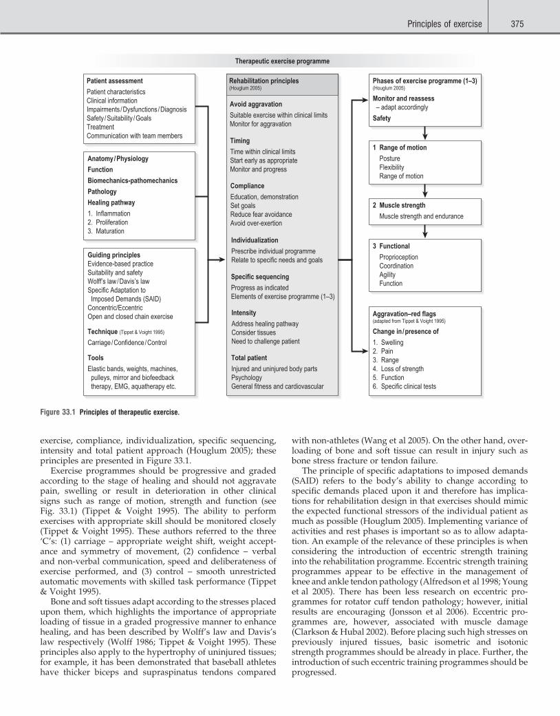
exercise, compliance, individualization, specific sequencing, intensity and total patient approach (Houglum 2005); these principles are presented in Figure 33.1.
Exercise programmes should be progressive and graded according to the stage of healing and should not aggravate pain, swelling or result in deterioration in other clinical signs such as range of motion, strength and function (see Fig. 33.1) (Tippet & Voight 1995). The ability to perform exercises with appropriate skill should be monitored closely (Tippet & Voight 1995). These authors referred to the three ‘C’s: (1) carriage – appropriate weight shift, weight accept-ance and symmetry of movement, (2) confidence – verbal and non-verbal communication, speed and deliberateness of exercise performed, and (3) control – smooth unrestricted automatic movements with skilled task performance (Tippet & Voight 1995).
Bone and soft tissues adapt according to the stresses placed upon them, which highlights the importance of appropriate loading of tissue in a graded progressive manner to enhance healing, and has been described by Wolff’s law and Davis’s law respectively (Wolff 1986; Tippet & Voight 1995). These principles also apply to the hypertrophy of uninjured tissues; for example, it has been demonstrated that baseball athletes have thicker biceps and supraspinatus tendons compared
Figure 33.1 Principles of therapeutic exercise.
Patient assessment
Patient characteristicsClinical informationImpairments /Dysfunctions /DiagnosisSafety /Suitability /GoalsTreatmentCommunication with team members
Anatomy/Physiology
Function
Biomechanics-pathomechanics
Pathology
Healing pathway
1. Inflammation2. Proliferation3. Maturation
Guiding principlesEvidence-based practiceSuitability and safetyWolff’s law/Davis’s lawSpecific Adaptation to Imposed Demands (SAID)Concentric/EccentricOpen and closed chain exercise
Technique (Tippet & Voight 1995)
Carriage /Confidence /Control
Tools
Elastic bands, weights, machines, pulleys, mirror and biofeedback therapy, EMG, aquatherapy etc.
Rehabilitation principles(Houglum 2005)
Avoid aggravation
Suitable exercise within clinical limitsMonitor for aggravation
Timing
Time within clinical limitsStart early as appropriateMonitor and progress
Compliance
Education, demonstrationSet goalsReduce fear avoidanceAvoid over-exertion
Individualization
Prescribe individual programmeRelate to specific needs and goals
Specific sequencing
Progress as indicatedElements of exercise programme (1–3)
Intensity
Address healing pathwayConsider tissuesNeed to challenge patient
Total patient
Injured and uninjured body partsPsychologyGeneral fitness and cardiovascular
Phases of exercise programme (1–3)(Houglum 2005)
Monitor and reassess – adapt accordingly
Safety
1 Range of motion
Posture Flexibility Range of motion
2 Muscle strength
Muscle strength and endurance
3 Functional
Proprioception Coordination Agility Function
Aggravation–red flags(adapted from Tippet & Voight 1995)
Change in /presence of
1. Swelling2. Pain3. Range4. Loss of strength5. Function6. Specific clinical tests
Therapeutic exercise programme

376 33 • Therapeutic exercises for the shoulder regionPART 4 •
pectorals may be felt in the front of the shoulder and arm (Simons et al 1999) and sometimes even in the upper back region (Dejung et al 2003). (See Ch 59 for a review of these mechanisms and muscle referral patterns.)
Sustained contractions impair normal blood flow in skel-etal muscles. Optimal posture allows muscles the opportunity to relax in between contractions, which permits and facilitates recovery of circulation (Otten 1988; Sjogaard & Sogaard 1998; Palmerud et al 2000). Combining postural exercises with myofeedback / EMG is helpful when teaching patients how to use their muscles in an economic and healthy manner (Peper et al 2003; Voerman et al 2006). Though there is a wide range of postures, clinicians should consider the optimal posture for each patient and individualize exercise programmes, rather than focusing on an idealized posture suitable for all. Assump-tion of an appropriate upright trunk posture can change muscle activation and modify range of motion and symptoms (Bullock et al 2005). Scapular taping can be used as a tempo-rary means of altering scapular muscle activation (Selkowitz et al 2007). Furthermore, Lucas et al (2004) demonstrated that latent trigger points can alter muscle activation patterns of the shoulder as assessed by EMG and subsequently reported that dry needling and stretch, when compared with placebo ultra-sound, was found to improve the muscle activation patterns significantly and similar to controls.
Treatment for postural dysfunctions may include manual therapies, including: joint mobilization and manipulation, massage and myofascial trigger point release, myofascial release techniques, trigger point dry needling, biofeedback and EMG, stretching, stability and strengthening and cogni-tive and behavioural strategies.
StretchingFlexibility and stretching is a broad topic with conflicting opinions in the literature, and a full discussion of this topic is beyond the scope of this chapter. Readers are referred else-where for a comprehensive review of stretching (Alter 1996; Weerapong et al 2004). A rehabilitation programme of the shoulder may incorporate a muscle-stretching programme, which is usually employed for muscle lengthening and associ-ated clinical implications, pain inhibition and potential injury prevention.
It has been reported that alterations in scapular movement are related to changes in myofascial length (Borstad & Ludewig 2005; Borstad 2006). The addition of appropriate manual therapy techniques may increase the effectiveness of therapeutic exercise (Winters et al 1997; Conroy & Hayes 1998; Bang & Deyle 2000; Desmeules et al 2003; Bergman et al 2004; Michener et al 2004; Senbursa et al 2007; Boyles et al 2009). These techniques may include soft tissue techniques, passive stretching and joint mobilization, and may increase range of motion in subjects with shoulder pain (Vermeulen et al 2006; Johnson et al 2007). Therapeutic exercise alone, however, may be as effective as adding passive joint mobiliza-tions to therapeutic exercise (Trampas & Kitsios 2006; Chen et al 2009). (Different joint mobilization techniques are described in detail in Ch 31.)
A muscle-stretching programme should be based on assess-ment of muscle length and end feel. Muscles and fascia may
Shoulder muscle balance ratios have been reported, includ-ing ratios between the external and internal rotators of 1.5 : 1 (66%) for both fast and slow isokinetic torque arm speed in normal subjects (Ivey et al 1985). Ratios have also been pre-sented for professional baseball pitchers (Ellenbecker & Mattalino 1997). Clinicians should consider these ratios in exercise programme design. A discussion of isokinetics is beyond the scope of this chapter, but has been reviewed by Ellenbecker and Davies (2000).
The following sections will discuss, posture, stretching and strengthening (isometric and isotonic) and briefly mention functional exercise. Specific parameters for timing and repeti-tions of stretching and strengthening will be covered under each appropriate section.
PosturePostural assessment is an important part of the objective eval-uation and ideal static postural alignments have been sug-gested (Kendall et al 2005). However, it is important to assess both static and dynamic postures to ascertain the patient’s functional movement and ability to self-correct a static habitus. An example of this is a boxer, who enhances a hyperkyphotic and rounded shoulder posture to reduce his target size for strategic advantage, but when dynamically tested may be able to self-correct the seemingly poor posture.
It is important to assess for muscle length, joint mobility and muscle control. Altered posture may be related to muscle imbalances and altered joint position, which ultimately could result in movement dysfunction and pain. Deviations in normal upright positions may include a forward head posi-tion, an exaggerated curve in the thoracic kyphosis, and rounded shoulders. Deviations in scapular kinematics may present in multiple planes, including changes in scapular elevation, protraction, tilt and rotation, affecting the size of the subacromial space (Solem-Bertoft et al 1993), as well as both activation (Roy et al 2009) and mechanical advantage (Kibler et al 2006) of muscular structures. It has been demon-strated that the size of the subacromial space is reduced in the presence of thoracic hyperkyphosis (Raine & Twomey 1997; Gumina et al 2008) and shoulder protraction (Solem-Bertoft et al 1993). It is, however, uncertain whether a strong correla-tion exists between narrowing of the subacromial space and shoulder symptoms (Graichen et al 2001; Roberts et al 2002; Hinterwimmer et al 2003; Lewis et al 2005; Mayerhoefer et al 2009). In fact, although it has been assumed that there is a definitive association between these postural deviations, a study of 160 asymptomatic subjects found no such correlation (Raine & Twomey 1997). Therefore, although there may be a relationship between posture and subacromial space, this is not yet fully understood.
Thoracic kyphosis and forward shoulder position influence the length of the upper back and scapular muscles and place the intervertebral joints in an end-range position (Griegel-Morris et al 1992). The sustained strain on these soft tissues may lead to upper back pain or shoulder pain. In the front of the body the pectoral muscles may shorten (Borstad & Ludewig 2006; Muraki et al 2009). Sustained muscle shorten-ing may lead to the development or activation of myofascial trigger points (Simons et al 1999). Referred pain from the

377Stretching
present with neuromuscular, viscoelastic or connective tissue alterations (Chaitow & Liebenson 2001). It is important to evaluate muscle length, and its influence on the length–tension relationship should not be overlooked (Janda 1993; Sahrmann 2002; Ekstrom & Osborn 2004; Kendall et al 2005). Though individual patients will present with varying degrees of muscle length, the following patterns, as outlined by Janda and others, are often seen in clinic practice (Chaitow & Liebenson 2001):• short muscles and often facilitated: pectoralis major and
minor, latissimus dorsi, levator scapula, upper trapezius (at times)
• long muscles and often inhibited: serratus anterior, lower and middle trapezius.
A stretch for the levator scapula and a clinician-assisted stretch for the pectorals and latissimus dorsi muscles are pre-sented in Figures 33.2 and 33.3 respectively. Other self-stretch exercises for the pectorals and latissimus dorsi may include the doorway stretch and one-sided unilateral self-stretch of the pectoralis minor, which has been shown to be superior to a supine manual stretch and a sitting manual stretch (Borstad & Ludewig 2006).
Muscle-stretching techniques include static, ballistic, dynamic and proprioceptive neuromuscular facilitation (Weerapong et al 2004; Houglum 2005). Other techniques have been described including post-isometric relaxation (Lewit & Simons 1984; Lewit 1986, 1999), muscle energy tech-nique (Greenman 1989; Chaitow & Crenshaw 2006), activated isolated stretching (Mattes 1995) and spray and stretch (Travell & Simons 1983; Simons et al 1999; Kostopoulos & Rizopoulos 2008). Stretching has been employed for the treatment of pain, especially in relation to the treatment of myofascial trigger points (Simons et al 1999).
Figure 33.2 Levator scapula stretch. Ipsilateral arm-elevated position is proposed to assist in isolating the levator scapula from the upper trapezius.
Figure 33.3 Pectoral and latissimus dorsi, clinician-assisted stretch. The patient maintains a neutral lumbar spine and a towel can be used to reduce thoracic kyphosis. The clinician applies a low-grade smooth stretch against soft tissue barrier. For appropriate modesty, the patient’s opposite hand can be placed across the chest and the clinician’s hand can be placed on top. Contract relax application can also be added to augment stretch.
The recommended duration of static stretching varies, but it is reasonable to recommend a 15–30-second hold with 3–5 repetitions (Taylor et al 1990; Houglum 2005) repeated daily or several times / day. Good form should be maintained during stretching technique, which should be smooth and within the clinical limits of the presenting problem. Longer hold times up to and beyond 5 minutes have been recom-mended for fascial tissue release (Barnes 1999).
Patients with a history of subluxation, dislocation, hyper-mobility of the shoulder or general hypermobility syndrome need to be identified, as a stretching programme may be inap-propriate and potentially detrimental in these individuals. The patient history, muscle length tests, joint end feel, passive joint tests and the Beigthon score (Alter 1996) may assist the clinician in identifying hypermobility and instability. Up to 11.7% of people have some form of joint hypermobility, and this has been reported to be up to three times more prevalent in females than in males (Hakim & Grahame 2003; Seckin et al 2005).
The majority of current research does not support the hypothesis that stretching prevents injury (Shrier 1999; Weerapong et al 2004). However, there is some evidence to suggest that lower limb stretching can reduce the risk of injury (Hartig & Henderson 1999; Amako et al 2003; Jamtvedt et al 2009), or the rate of return from injury (Malliaropoulos et al 2004). Interestingly, though, reviewed research on stretching has demonstrated a negative effect on muscle strength and functional performance (Weerapong et al 2004). Further, the fact that most research has focused on the lower extremities raises validity issues about the validity of extrapo-lating the findings to the upper extremity. However, clinicians should consider these issues when prescribing flexibility pro-grammes, especially in relation to performance athletes and players. More research is required to assist in a better under-standing of the role of stretching in injury management and prevention.
External rotation is fundamental for elevation and shoul-der function and it is important to restore passive and active external rotation (Donatelli 2004a). External rotation is

378 33 • Therapeutic exercises for the shoulder regionPART 4 •
slow-twitch fibres respectively (MacDougall et al 1980). During immobilization of the upper limb, strength training with maximal isometric exercise 5 days / week of the free limb may prevent atrophy of the immobilized limb (Farthing et al 2009). Further research has suggested that adding a 0.5 kg weight to the ipsilateral hand during isometric and dynamic shoulder exertions increases shoulder muscle activity by 4% maximum voluntary excitation (Antony & Keir 2010). Static exercises for the shoulder are presented in Figure 33.6. A belt is employed to allow multidirectional static exercises; however, other options include resistance against a wall. A hand-held weight of 0.5 kg is used to assist in increasing shoulder muscle activity (Antony & Keir 2010). Suggested parameters for isometric exercises include pain-free 5- to 10-second holds with 10 repetitions, graded to maximal con-traction and repeated several times per day with progression as indicated (Houglum 2005).
Isotonic Exercises of the ShoulderThere is a plethora of exercises for the shoulder girdle, and research employing EMG has aimed at identifying exercises that target specific shoulder muscles; here we briefly review a selection of exercises that target the rotator cuff, trapezius and serratus anterior muscles. For a further expansion of this, readers are recommended to review other publications (Ekstrom & Osborn 2004; Houglum 2005; Reinold et al 2009). When designing a strengthening programme, the clinician should target muscles identified as weak during the evalua-tion and, on the basis of this, prescribe suitable exercises. The clinician should prescribe the specific exercise, resist-ance (or none), repetitions, sets and frequency of the pro-gramme. This programme should be monitored, adjusted and advanced progressively. A programme can be initiated with or without weight, as appropriate. Recommendations have been made in relation to exercise repetitions (reps) and include 1–6 reps for strength, 6–12 for hypertrophy and 12–15 for endurance (Kraemer & Ratamess 2004). The weight used is appropriate to cause fatigue towards the end of the stated number of repetitions. It has been found that two
primarily limited at 0° by the subscapularis, at 45° of abduc-tion by the subscapularis, middle and inferior humeral liga-ment and at 90° of abduction by the inferior humeral ligament (Turkel et al 1981). Muscle length testing of the subscapularis is carried out with the arm in neutral and testing into external rotation (Donatelli 2004b). An auto-assisted stretch for the subscapularis, using a cane, is presented in Figure 33.4. The stretching position, for example at 0°, 45°, 90° abduction, etc., should be based on any restrictions of the subscapularis and humeral capsule and ligaments identified from the physical assessment. The contribution of the humeral joint capsule (and other posterior soft tissue structures including the infra-spinatus, teres minor and deltoid) to shoulder movement should not be overlooked and has been proposed to be par-ticularly important in certain shoulder disorders, including SAIS (Donatelli 2004a). Reduced cross-body adduction has been linked to tightness of the posterior capsule, and associ-ated with abnormal humeral translation (Ludewig & Cook 2002). Cross-body adduction and the ‘sleeper stretch’ (internal rotation of the shoulder in 90° of shoulder flexion) have been recommended as stretches for posterior shoulder capsular tightness (Cooper et al 2004; McClure et al 2007; Laudner et al 2008). The ‘sleeper stretch’ is presented in Figure 33.5. However, modification into less shoulder flexion may be nec-essary if symptoms are aggravated in this position.
Isometric Exercise of the ShoulderIsometric exercise is usually utilized in the early phase of rehabilitation to minimize muscle atrophy when movement of the shoulder is limited. Studies have demonstrated up to a 41% decrease in isometric strength after immobilization of the upper extremity for 5–6 weeks, with significant decreases in muscle fibre area by 33% and 25% for fast- and
Figure 33.4 Subscapularis stretch, self-assisted stretch with cane. The supine position offers stability of the scapula while external rotation of the glenohumeral joint is assisted with self-control using the cane. A towel is placed under the elbow to maintain alignment of the humerus.
Figure 33.5 Sleeper stretch. The 90° position stabilizes the scapula and downward pressure is applied with a self-stretch to the opposite hand into internal rotation.

379Isotonic exercises of the shoulder
loading during abduction and scaption movements, peaking at 30–60° of elevation (Reinold et al 2009). Reinold et al (2007) demonstrated that EMG activity was similar across three exer-cises: full can, empty can and prone full can. The full-can exercise results in significantly less activity of the middle and posterior deltoid, which may reduce harmful shear force on the humeral joint from deltoid activity (Reinold et al 2007, 2009). In addition, it reduces the potential for subacromial impingement because of the external rotation component (Ekstrom & Osborn 2004). Moreover, this exercise has been recommended by previous research (Kelly et al 1996). The full-can exercise in the plane of the scapula with external rota-tion of the shoulder is presented in Figure 33.7.
Infraspinatus and teres minor musclesThe actions of the infraspinatus and teres minor are primarily external rotation and functionally assisting the stability of the humeral joint during elevation movements (Reinold et al 2009). Stabilization of the shoulder by these muscles is also achieved by opposing superior and anterior humeral head translation (Reinold et al 2009). The infraspinatus has poten-tially a role in abduction and horizontal abduction, with the teres minor involved in adduction, the difference apparently being due to different moment arms (Oatis 2004). EMG analy-sis demonstrated the best isolation of the infraspinatus in 0° abduction with 45° of medial rotation from neutral (Kelly et al 1996), and Reinold et al (2009) suggested incorporating this position as an exercise to any rehabilitation programme when focusing on increasing external rotation strength. Addition of a roll support between the arm and the trunk (Fig. 33.8) has been shown to increase EMG activity in the infraspinatus and teres minor muscles by up to 25% (Reinold et al 2004, 2009).
to six sets per exercise produced significant increases in muscular strength in both trained and untrained individuals (Kraemer & Ratamess 2004).
Other recommendations include 6–15 repetitions of two sets where the patient can control the weight, progressing to 20–25 repetitions of three sets (Houglum 2005). When this is reached, the weight then is progressed accordingly and the process started again with 6–15 repetitions of two sets, etc. (Houglum 2005). There are various exercise progressions that can be considered including that of Delorme and Watkins (1948), Oxford technique (Zinovieff 1951) and daily adjusted progressive resistive exercise (Knight 1985; Houglum 2005). The clinician should consider the principles of exercise as outlined in Figure 33.1 when prescribing strength pro-grammes. The rotator cuff muscles are important stabilizers of the humeral joint and assist in stabilizing the humerus in the glenoid by compression and preventing shear and upward movement of the humeral head during arm movements (Oatis 2004). Other muscles assist in stabilizing the scapulothoracic complex and in dynamic stability (Oatis 2004) and these muscle-specific exercises are covered below. For muscles such as the deltoid, levator scapulae and rhomboids, and indica-tions for strengthening, readers are recommended to review the article by Reinold et al (2009).
Supraspinatus muscleThe supraspinatus is the most superior of the rotator cuff muscles and lies deep to the subacromial bursa and the cora-coacromial ligament within the subacromial space (Oatis 2004). The reported actions of this muscle include abduction, external rotation and stabilization of the shoulder (Oatis 2004). Activity of the supraspinatus increases with increased
Figure 33.6 Isometric exercises for the shoulder. The use of a belt allows the patient to perform isometric exercise in multiple directions. This can be also done against a wall. Internal and external rotation is performed with self-assisted resistance. The use of a hand-held weight of 0.5 kg has been shown to assist in increasing shoulder muscle EMG by 4%. The arrows indicate direction of force, but as an isometric exercise there is no movement.

380 33 • Therapeutic exercises for the shoulder regionPART 4 •
Subscapularis muscleThe subscapularis is the largest of the rotator cuff muscles and acts to rotate internally, flex, extend, abduct, adduct, adduct horizontally and stabilize the shoulder, with broad agreement that internal rotation and stabilization are the primary roles (Oatis 2004). Subscapularis weakness leads to significant decrease in internal rotation strength and may contribute to anterior instability of the shoulder (Oatis 2004). The lift-off test as described by Gerber and Krushell (1991) has been demonstrated to isolate the subscapularis (Greis et al 1996; Kelly et al 1996). The lift-off exercise for the subscapularis muscle is presented in Figure 33.10.
Trapezius muscleThe trapezius is an expansive muscle that has three distinct muscle sections: upper, middle and lower, with each having
A second exercise worth considering is standing external rota-tion in the scapular plane (45° of abduction) (Fig. 33.9) as this has demonstrated good EMG activation of the infraspinatus and teres minor (Reinold et al 2004), and isokinetic external rotational strength values in the plane of the scapula have been reported to be significantly higher than in the frontal plane (Greenfield et al 1990).
Other exercises for external rotation have been recom-mended that place the shoulder in a more compromised posi-tion (e.g. external rotation in 90° abduction) and clinicians should carefully consider the appropriateness of these exer-cises in the presence of capsulolabral dysfunction and pathol-ogy (Reinold et al 2009).
Figure 33.7 Supraspinatus full-can strengthening. This is carried out in the plane of the scapula, slowly and controlled with the thumb up to ensure a degree of external rotation.
Figure 33.8 Infraspinatus and teres minor muscles strengthening. In side-lying, the arm is brought from internal into external rotation. A towel positioned between the arm and trunk has been shown to increase EMG of the muscles by 25%.
Figure 33.9 External rotation in the plane of the scapula. The shoulder is rotated from internal to external rotation.
Figure 33.10 Subscapularis strengthening (Gerber’s lift-off test). The hand is raised upwards from the trunk.

381Functional exercises
Serratus anterior muscleThe serratus anterior muscle action has been reported as pro-traction, abduction, upward rotation and elevation of the scapula; the muscle functions in actions such as pushing a revolving door and weakness may lead to winging of the scapula and difficulty with overhead activities (Oatis 2004). Shoulder abduction in the plane of the scapula above 120° (to avoid painful arc) in the standing position has demonstrated more EMG activity in the serratus anterior than does straight scapular protraction (Fig. 33.13). The increased serratus ante-rior activation should, however, be balanced against the increased risk of impingement when exercises are performed in elevation (Roberts et al 2002). Other recommended exer-cises for the serratus anterior include the dynamic hug, push-up with a plus and punch exercises (Decker et al 1999; Reinold et al 2009).
Functional ExercisesThe daily tasks and movements performed by the individual should be considered when prescribing therapeutic exercise, so that the exercises take into account the specific functional demands of that person. Functional progression may include movement from isolated plane to multiplane strengthening (Fig. 33.14) and eventually plyometric exercise (Houglum 2005). Upper limb tasks are commonly open kinetic chain movements. In athlete subjects with recurrent anterior shoul-der dislocation, rehabilitation near / in the zone of instability is indicated in late-stage rehabilitation and therefore tasks that load the rotator cuff in semi-compromised positions may help replicate the stability action required on return to sport. Closed chain and stability exercises (Figs 33.15 and 33.16) for the shoulder girdle are important to assist in motor control and re-education (Houglum 2005) and are discussed in Chapter 32. The shoulder girdle relationship to the kinetic
a distinct function and combining to assist in the overall func-tion of the trapezius (Oatis 2004). The actions of the three sections have been reported as follows (Oatis 2004): upper trapezius – elevation of the scapula, adduction and upward rotation of the scapula; middle trapezius – adduction of the scapula; and lower trapezius – depression, adduction and upward rotation of the scapula. In particular, the upper and lower trapezius form an anatomical force couple that assists in stabilizing the scapula, and maintaining a balance between these muscle segments is important for optimal function (Oatis 2004). Furthermore, the lower fibres have been reported to play an important role in posterior tilt and upward rotation of the scapula during shoulder elevation (Ludewig et al 1996). Therefore, the lower trapezius and serratus anterior comprise an important target for rehabilitation and prevention of shoul-der dysfunction and impingement syndromes (Ludewig & Cook 2000). With regard to exercises for the upper trapezius, the shoulder shrug has been reported to produce the greatest EMG activity (Ekstrom et al 2003). However, it has been further reported that the shrug exercise also highly activates the levator scapula and so if this needs to be avoided, owing to the levator scapulae action of scapular downward rotation, the military press may be more appropriate (Ekstrom & Osborn 2004).
For the middle trapezius, abduction and external rotation of the shoulder at 90° in prone (Fig. 33.11) has been shown to induce good EMG activity and is considered a suitable exer-cise (Moseley et al 1992; Ekstrom & Osborn 2004; Reinold et al 2009). This exercise has also been recommended for strength-ening the trapezius as a whole, owing to high EMG activity in the upper, middle and lower muscle segments (Ekstrom et al 2003; Ekstrom & Osborn 2004).
The lower trapezius has been shown to be best activated with the arm raise overhead exercise in the prone position, performed at approximately 120° (Reinold et al 2009) to 135° of abduction or with the arm positioned in line with the lower fibres of trapezius (Fig. 33.12) (Ekstrom et al 2003; Ekstrom & Osborn 2004).
Figure 33.11 Trapezius strengthening. This targets the upper, middle and lower sections of the trapezius muscle. The thumb is maintained in an upright position.
Figure 33.12 Trapezius strengthening. This targets mainly the lower fibres of trapezius performed at approximately 120–135° of abduction or with the arm positioned in line with the lower fibres of the trapezius.

382 33 • Therapeutic exercises for the shoulder regionPART 4 •
Figure 33.13 Serratus anterior strengthening. Shoulder abduction is performed in the plane of the scapula above 120° (to avoid painful arc) in the standing position.
Figure 33.14 Proprioceptive neuromuscular facilitation with elastic band. Elastic bands can assist with creating open chain coordinated movements that mimic functional patterns.
Figure 33.15 Shoulder dip. This is a closed chain kinetic loading and proprioceptive exercise.

383Conclusion
Figure 33.16 Prone knee, single-arm raise. The abdominals are primed prior to the lift. This offers a closed chain exercise for weight-bearing shoulder and a dynamic open chain exercise for the opposite arm. This exercise can be advanced to alternative arm and leg lift for stability.
chain should be considered in the overall management of the patient and exercise programmes may incorporate standing balance and eye–hand coordination tasks, etc. (Donatelli 2006). A common barrier to shoulder rehabilitation is diffi-culty in replicating the therapeutic exercise correctly at home, possibly owing to reduced positional sense (Naughton et al 2005). Tasks that challenge the proprioceptive acuity and load-bearing ability of the shoulder region may help to restore positional sense awareness (see Figs 33.14–33.16). Clinicians can also incorporate the use of exercise equipment to assist in functional progression, including elastic bands, pulleys, Theraballs, wobble boards, proprioceptive exercise devices and feedback devices such as mirrors, etc.
ConclusionTherapeutic exercises can play an important role in the man-agement of shoulder pain. This chapter has outlined some basic principles on the type of exercise that may be indicated in the clinical setting, especially in relation to strength of the rotator cuff and stabilizers of the scapula. Clinicians should remain cognisant of the need for a comprehensive assessment and ensure safe and suitable exercise prescription and pro-gression. The nature, intensity and volume of exercise pre-scribed must be matched to the clinical presentation. Individual therapists may use the principles outlined in this chapter as a guide when prescribing a therapeutic exercise programme that is suitable for the needs of each individual patient or athlete. Further research is required and should focus on identifying exercises to suit the specific patient presentation.
AcknowledgementsThank you to the photographic volunteers: Derek Malone, Irish Paralympic athlete and S.M.
ReferencesAlfredson H, Pietilä T, Jonshon P, et al. 1998. Heavy-load eccentric calf muscle
training for the treatment of chronic Achilles tendinosis. Am J Sports Med 26: 360–366.
Alter MJ. 1996. Science of flexibility, 2nd edn. Champaign, IL: Human Kinetics.
Alter MJ. 2004. Science of flexibility, 3rd edn. Champaign, IL: Human Kinetics.
Amako M, Oda T, Masuoka K, et al. 2003. Effect of static stretching on preven-tion of injuries for military recruits. Mil Med 168: 442–446.
Antony NT, Keir PJ. 2010. Effects of posture, movement and hand load on shoulder muscle activity. J Electromyogr Kinesiol 20: 191–198.
Bang M, Deyle G. 2000. Comparison of supervised exercise with and without manual physical therapy for patients with shoulder impingement syn-drome. J Orthop Sports Phys Ther 30: 126–137.
Barclay J. 1994. In good hands: the history of the Chartered Society of Physi-otherapy 1894–1994. Oxford: Butterworth Heinemann.
Barnes J. 1999. Myofascial release. In: Hammer WI (ed) Functional soft tissue examination and treatment by manual methods: new perspectives, 2nd edn. Gaithersburg, MD: Aspen Publishers, x, p 625.
Baydar M, Akalin E, El O, et al. 2009. The efficacy of conservative treatment in patients with full-thickness rotator cuff tears. Rheumatol Int 29: 623–628.
Bergman G, Winters J, Groenier K, et al. 2004. Manipulative therapy in addition to usual medical care for patients with shoulder dysfunc-tion and pain: a randomized, controlled trial. Ann Intern Med 141: 432–439.
Bigoni M, Gorla M, Guerraio S, et al. 2009. Shoulder evaluation with isokinetic strength testing after arthroscopic rotator cuff repairs. J Shoulder Elbow Surg 18: 178–183.
Borstad J. 2006. Resting posture variables at the shoulder: evidence to support a posture-impairment association. Phys Ther 86: 549–557.
Borstad J, Ludewig P. 2005. The effect of long versus short pectoralis minor resting length on scapular kinematics in healthy individuals. J Orthop Sports Phys Ther 35: 227–238.
Borstad JD, Ludewig PM. 2006. Comparison of three stretches for the pecto-ralis minor muscle. J Shoulder Elbow Surg 15: 324–330.
Boyles RE, Ritland BM, Miracle BM, et al. 2009. The short-term effects of tho-racic spine thrust manipulation on patients with shoulder impingement syndrome. Man Ther 14: 375–380.
Buchbinder R, Green S, Youd JM, et al. 2003. Corticosteroid injections for shoulder pain. Cochrane Database Syst Rev 1: CD004016.
Buchbinder R, Green S, Youd JM, et al. 2008. Arthrographic distension for adhesive capsulitis. Cochrane Database Syst Rev 23(1): CD007005. doi: 10.1002/14651858.CD007005.
Bullock M, Foster N, Wright C. 2005. Shoulder impingement: the effect of sitting posture on shoulder pain and range of motion. Man Ther 10: 28–37.
Carette S, Moffet H, Tardif J, et al. 2003. Intra-articular corticosteroids, super-vised physiotherapy, or a combination of the two in the treatment of adhesive capsulitis of the shoulder: a placebo-controlled trial. Arthritis Rheum 48: 829–838.
Chaitow L, Crenshaw K. 2006. Muscle energy techniques: with accompanying DVD. Edinburgh: Churchill Livingstone Elsevier.
Chaitow L, Liebenson C. 2001. Muscle energy techniques. Edinburgh: Church-ill Livingstone Elsevier.
Chen S, Simonian P, Wickiewicz T, et al. 1999. Radiographic evaluation of gleno-humeral kinematics: a muscle fatigue model. J Shoulder Elbow Surg 8: 49–52.
Chen J, Ginn KA, Herbert R, et al. 2009. Passive mobilisation of shoulder region joints plus advice and exercise does not reduce pain and disability more than advice and exercise alone: a randomised trial. Aust J Physiother 55: 17–23.
Cicerone KD. 2005. Evidence-based practice and the limits of rational rehabili-tation. Arch Phys Med Rehabil 86: 1073–1074.
Clarkson PM, Hubal MJ. 2002. Exercise-induced muscle damage in humans. Am J Phys Med Rehabil 81: S52S69.
Conroy D, Hayes K. 1998. The effect of joint mobilisation as a component of comprehensive treatment for primary shoulder impingement syndrome. J Orthop Sports Phys Ther 28: 3–14.
Cools A, Witvrouw E, Declercq G, et al. 2007. Scapular muscle recruitment patterns: trapezius muscle latency with and without impingement symp-toms. Am J Sports Med 31: 542–549.

384 33 • Therapeutic exercises for the shoulder regionPART 4 •
Hartig DE, Henderson JM. 1999. Increasing hamstring flexibility decreases lower extremity overuse injuries in military basic trainees. Am J Sports Med 27: 173–176.
Hinterwimmer S, Von Eisenhart-Rothe R, Siebert M, et al. 2003. Influence of adducting and abducting muscle forces on the sub-acromial space width. Med Sci Sports Exerc 35: 2055–2059.
Ho C, Sole G, Munn J. 2009. The effectiveness of manual therapy in the man-agement of musculoskeletal disorders of the shoulder: a systematic review. Man Ther 14: 463–474.
Houglum PA. 2005. Therapeutic exercise for musculoskeletal injuries. Champaign, IL: Human Kinetics.
Ivey FM, Calhoun JH, Rusche K, et al. 1985. Isokinetic testing of shoulder strength: normal values. Arch Phys Med Rehabil 66: 384–386.
Jamtvedt G, Herbert RD, Flottorp S, et al. 2009. A pragmatic randomised trial of stretching before and after physical activity to prevent injury and sore-ness. Br J Sports Med 44(14): 1002–1009.
Janda V. 1993. Muscle strength in relation to muscle length, pain, and muscle imbalance. In: Harms-Ringdahl K (ed) Muscle strength. Edinburgh: Churchill Livingstone, pp 83–91.
Johnson A, Godges J, Zimmerman G, et al. 2007. The effect of anterior versus posterior glide joint mobilization on external rotation range of motion in patients with shoulder adhesive capsulitis. J Orthop Sports Phys Ther 37: 88–99.
Jonsson P, Wahlstrom P, Ohberg L, et al. 2006. Eccentric training in chronic painful impingement syndrome of the shoulder: results of a pilot study. Knee Surg Sports Traumatol Arthrosc 14: 76–81.
Karjalainen K, Malmivaara A, Van Tulder M, et al. 2001. Multidisciplinary biopsychosocial rehabilitation for neck and shoulder pain among working age adults: a systematic review within the framework of the Cochrane Collaboration Back Review Group. Spine 26: 174–181.
Kelly BT, Kadrmas WR, Speer KP. 1996. The manual muscle examination for rotator cuff strength. An electromyographic investigation. Am J Sports Med 24: 581–588.
Kendall FP. 2002. Kendall urges a return to basics. PT Bulletin Online 3.Kendall F, Kendall McCreary E, Provance P, et al. 2005. Muscles: testing and
function with posture and pain. Baltimore, MD: Lippincott Williams & Wilkins.
Kibler W, Sciascia A, Dome D. 2006. Evaluation of apparent and absolute supraspinatus strength in patients with shoulder injury using the scapular retraction test. Am J Sports Med 34 1643–1647.
Knight KL. 1985. Guidelines for rehabilitation of sports injuries. Clin Sports Med 4: 405–416.
Kostopoulos D, Rizopoulos K. 2008. Effect of topical aerosol skin refrigerant (spray and stretch technique) on passive and active stretching. J Bodyw Mov Ther 12: 96–104.
Kraemer WJ, Ratamess NA. 2004. Fundamentals of resistance training: pro-gression and exercise prescription. Med Sci Sports Exerc 36: 674–688.
Kuhn JE. 2009. Exercise in the treatment of rotator cuff impingement: a sys-tematic review and a synthesized evidence-based rehabilitation protocol. J Shoulder Elbow Surg 18: 138–160.
Laudner KG, Sipes RC, Wilson JT. 2008. The acute effects of sleeper stretches on shoulder range of motion. J Athl Train 43: 359–363.
Lephart SM, Fu FH. 2000. Proprioception and neuromuscular control in joint stability. Champaign, IL: Human Kinetics.
Lewis JS, Green A, Wright C. 2005. Subacromial impingement syndrome: the role of posture and muscle imbalance. J Shoulder Elbow Surg 14: 385–392.
Lewit K. 1986. Postisometric relaxation in combination with other methods of muscular facilitation and inhibition. Manual Med 2: 101–104.
Lewit K. 1999. Manipulative therapy in rehabilitation of the locomotor system. Oxford: Butterworth-Heinemann.
Lewit K, Simons DG. 1984. Myofascial pain: relief by post-isometric relaxation. Arch Phys Med Rehabil 65: 452–456.
Lombardi IJ, Magri A, Fleury A, et al. 2008. Progressive resistance training in patients with shoulder impingement syndrome: a randomized controlled trial. Arthritis Rheum 59: 615–622.
Lucas KR, Polus BI, Rich PS. 2004. Latent myofascial trigger points: their effect on muscle activation and movement efficiency. J Bodyw Mov Ther 8: 160–166.
Ludewig PM, Cook T. 2000. Alterations in shoulder kinematics and associated muscle activity in people with symptoms of shoulder impingement. Phys Ther 80: 276–291.
Ludewig PM, Cook T. 2002. Translations of the humerus in persons with shoulder impingement symptoms. J Orthop Sports Phys Ther 32: 248–259.
Cooper J, Donley P, Morgan C, et al. 2004. Throwing injuries. In: Donatelli R (ed) Physical therapy of the shoulder. St Louis, MO: Churchill Livingstone, pp 29–78.
Decker MJ, Hintermeister RA, Faber KJ, et al. 1999. Serratus anterior muscle activity during selected rehabilitation exercises. Am J Sports Med 27: 784–791.
Dejung B, Gröbli C, Colla F, et al. 2003. Triggerpunkttherapie. Bern: Hans Huber.
Delorme TL, Watkins AL. 1948. Technics of progressive resistance exercise. Arch Phys Med Rehabil l(29): 263–273.
Desmeules F, Côté CH, Fremont P, et al. 2003. Therapeutic exercise and ortho-pedic manual therapy for impingement syndrome: a systematic review. Clin J Sport Med 13: 176–182.
Dickens V, Willimas J, Bhamra M. 2005. Role of physiotherapy in the treatment of sub-acromial impingement syndrome: a prospective study. Physiother-apy 91: 159–164.
Donatelli R. 2004a. Functional anatomy and mechanics. In: Donatelli R (ed) Physical therapy of the shoulder. St Louis, MO: Churchill Livingstone, pp 11–28.
Donatelli R. 2004b. Physical therapy of the shoulder. St Louis, MO: Churchill Livingstone.
Donatelli R. 2006. Sports-specific rehabilitation. St Louis, MO: Churchill Livingstone Elsevier.
Dorrestijn O, Stevens M, Winers J, et al. 2009. Conservative or surgical treat-ment for sub-acromial impingement syndrome? A systematic review. J Shoulder Elbow Surg 18: 652–660.
Ekstrom R, Osborn R. 2004. Muscle length testing and electromyographic data for manual strength testing and exercises for the shoulder. In: Donatelli R (ed) Physical therapy of the thoulder. St Louis, MO: Churchill Livingstone, pp 435–463.
Ekstrom RA, Donatelli RA, Soderberg GL. 2003. Surface electromyographic analysis of exercises for the trapezius and serratus anterior muscles. J Orthop Sports Phys Ther 33: 247–258.
Ellenbecker TS, Davies GJ. 2000. The application of isokinetics in testing and rehabilitation of the shoulder complex. J Athl Train 35: 338–350.
Ellenbecker TS, Mattalino AJ. 1997. Concentric isokinetic shoulder internal and external rotation strength in professional baseball pitchers. J Orthop Sports Phys Ther 25: 323–328.
Farthing JP, Krentz JR, Magnus CR. 2009. Strength training the free limb attenuates strength loss during unilateral immobilization. J Appl Physiol 106: 830–836.
Gerber C, Krushell RJ. 1991. Isolated rupture of the tendon of the sub- scapularis muscle. Clinical features in 16 cases. J Bone Joint Surg Br 73: 389–394.
Graichen H, Bonel H, Stammberger T, et al. 2001. Sex-specific differences of subacromial space width during abduction, with and without muscular activity, and correlation with anthropometric variables. J Bone Joint Surg 10: 129–135.
Green S, Buchbinder R, Hetrick S. 2003. Physiotherapy interventions for shoul-der pain. Cochrane Database Syst Rev 2: CD004258.
Green S, Buchbinder R, Hetrick S. 2005. Acupuncture for shoulder pain. Cochrane Database Syst Rev 2: CD005319.
Greenfield BH, Donatelli R, Wooden MJ, et al. 1990. Isokinetic evaluation of shoulder rotational strength between the plane of scapula and the frontal plane. Am J Sports Med 18: 124–128.
Greenman PE. 1989. Principles of manual medicine. Baltimore, MD: Williams & Wilkins.
Greis PE, Kuhn JE, Schultheis J, et al. 1996. Validation of the lift-off test and analysis of sub-scapularis activity during maximal internal rotation. Am J Sports Med 24: 589–593.
Griegel-Morris P, Larson K, Mueller-Klaus K, et al. 1992. Incidence of common postural abnormalities in the cervical, shoulder, and thoracic regions and their association with pain in two age groups of healthy subjects. Phys Ther 72: 425–431.
Gumina S, Di Giorgio G, Postacchini F, et al. 2008. Sub-acromial space in adult patients with thoracic hyperkyphosis and in healthy volunteers. Chir Organi Mov 91: 93–96.
Haahr JP, Andersen JH. 2006. Exercises may be as efficient as subacromial decompression in patients with subacromial stage II impingement: 4–8-years’ follow-up in a prospective, randomized study. Scand J Rheumatol 35: 224–228.
Hakim A, Grahame R. 2003. Joint hypermobility. Best Pract Res Clin Rheuma-tol 17: 989–1004.
Hall T, Elvey R. 1999. Nerve trunk pain: physical diagnosis and treatment. Man Ther 4: 63–73.

385Conclusion
Sahrmann S. 2002. Diagnosis and treatment of movement impairment syn-dromes. St Louis, MO: Mosby.
Seckin U, Tur BS, Yilmaz O, et al. 2005. The prevalence of joint hypermobility among high school students. Rheumatol Int 25: 260–263.
Selkowitz D, Chaney C, Stuckey S, et al. 2007. The effects of scapular taping on the surface electromyographic signal amplitude of shoulder girdle muscles during upper extremity elevation in individuals with suspected shoulder impingement syndrome. J Orthop Sports Phys Ther 37: 694–702.
Senbursa G, Baltacı G, Atay A. 2007. Comparison of conservative treatment with and without manual physical therapy for patients with shoulder impingement syndrome: a prospective, randomized clinical trial. Knee Surg Sports Traumatol Arthrosc 15: 915–921.
Shrier I. 1999. Stretching before exercise does not reduce the risk of local muscle injury: a critical review of the clinical and basic science literature. Clin J Sport Med 9: 221–227.
Simons DG, Travell JG, Simons L. 1999. Travell and Simons’ myofascial pain and dysfunction: the trigger point manual. Baltimore, MD: Williams & Wilkins.
Sjogaard G, Sogaard K. 1998. Muscle injury in repetitive motion disorders. Clin Orthop Relat Res 351: 21–31.
Smidt N, De Vet H, Bouter L, et al. 2005. Effectiveness of exercise therapy: a best-evidence summary of systematic reviews. Aust J Physiother 51: 71–85.
Solem-Bertoft E, Thuomas KA, Westerberg CE. 1993. The influence of scapular retraction and protraction on the width of the subacromial space. An MRI study. Clin Orthop Relat Res 296: 99–103.
Tate A, McClure P, Kareha S, et al. 2009. A clinical method for identifying scapular dyskinesis. Part 2: Validity. J Athl Train 44: 165–173.
Taylor DC, Dalton JD, Seaber AV, et al. 1990. Viscoelastic properties of muscle-tendon units. The biomechanical effects of stretching. Am J Sports Med 18: 300–309.
Terlouw TJ. 2007. Roots of physical medicine, physical therapy, and mechano-therapy in the Netherlands in the 19th century: a disputed area within the healthcare domain. J Man Manip Ther 15: E23–E41.
Tippet SR, Voight ML. 1995. Functional progression for sports rehabilitation. Champaign, IL: Human Kinetics.
Trampas A, Kitsios A. 2006. Exercise and manual therapy for the treatment of impingement syndrome of the shoulder: a systematic review. Phys Ther Rev 11: 125–142.
Travell JG, Simons DG. 1983. Myofascial pain and dysfunction: the trigger point manual. Baltimore, MD: Williams & Wilkins.
Turkel SJ, Panio MW, Marshall JL, et al. 1981. Stabilizing mechanisms prevent-ing anterior dislocation of the glenohumeral joint. J Bone Joint Surg Am 63: 1208–1217.
Vermeulen HM, Stokdijk M, Eilers PH, et al. 2002. Measurement of three dimensional shoulder movement patterns with an electromagnetic track-ing device in patients with a frozen shoulder. Ann Rheum Dis 61: 115–120.
Vermeulen HM, Rozing PM, Obermann WR, et al. 2006. Comparison of high-grade and low-grade mobilization techniques in the management of adhe-sive capsulitis of the shoulder: randomized controlled trial. Phys Ther 86: 355–368.
Voerman GE, Vollenbroek-Hutten MM, Hermens HJ. 2006. Changes in pain, disability, and muscle activation patterns in chronic whiplash patients after ambulant myofeedback training. Clin J Pain 22: 656–663.
Wang HK, Lin JJ, Pan SL, et al. 2005. Sonographic evaluations in elite college baseball athletes. Scand J Med Sci Sports 15: 29–35.
Weerapong P, Hume PA, Kolt GS. 2004. Stretching: mechanisms and benefits for sport performance and injury prevention. Phys Ther Rev 9: 189–206.
Winters J, Sobel J, Groenier K, et al. 1997. Comparison of physiotherapy, manipulation and corticosteroid injection for treating shoulder complaints in general practice: randomised, single blind study. BMJ 314: 1320–1325.
Winters J, Jorritsma W, Groenier K, et al. 1999. Treatment of shoulder com-plaints in general practice: long-term results of a randomised, single blind study comparing physiotherapy, manipulation, and corticosteroid injec-tion. BMJ 318: 1395–1396.
Wolff J. 1986. The law of bone remodelling. Berlin, New York: Springer– Verlag.
Young M, Cook J, Purdam C, et al. 2005. Eccentric decline squat protocol offers superior results at 12 months compared with traditional eccentric protocol for patellar tendinopathy in volleyball players. Br J Sports Med 39: 102–105.
Zinovieff AN. 1951. Heavy-resistance exercises: the ‘Oxford technique’. Br J Phys Med 14: 129–132.
Ludewig PM, Cook TM, Nawoczenski DA. 1996. Three-dimensional scapular orientation and muscle activity at selected positions of humeral elevation. J Orthop Sports Phys Ther 24: 57–65.
Lukasiewicz A, McClure P, Michener L, et al. 1999. Comparison of 3-dimensional scapular position and orientation between subjects with and without shoulder impingement. J Orthop Sports Phys Ther 29: 574–583.
MacDougall JD, Elder GC, Sal DG, et al. 1980. Effects of strength training and immobilization on human muscle fibres. Eur J Appl Physiol Occup Physiol 43: 25–34.
MacIntosh BR, Gardiner PF, McComas AJ. 2006. Skeletal muscle: form and function. Leeds, Champaign, IL: Human Kinetics.
Malliaropoulos N, Papalexandris S, Papalada A, et al. 2004. The role of stretch-ing in rehabilitation of hamstring injuries: 80 athletes follow-up. Med Sci Sports Exerc 36: 756–759.
Mattes AL (ed). 1995. Active isolated stretching. Sarasota, FL: Aaron Mattes Therapy.
Mayerhoefer ME, Breitenseher MJ, Wurnig C, et al. 2009. Shoulder impinge-ment: relationship of clinical symptoms and imaging criteria. Clin J Sport Med 19: 83–89.
McClure PW, Michener LA, Karduna AR. 2006. Shoulder function and 3-dimensional scapular kinematics in people with and without shoulder impingement syndrome. Phys Ther 86: 1075–1090.
McClure P, Balaicuis J, Heiland D, et al. 2007. A randomized controlled com-parison of stretching procedures for posterior shoulder tightness. J Orthop Sports Phys Ther 37: 108–114.
Michener L, Walsworth M, Burnet E. 2004. Effectiveness of rehabilitation for patients with sub-acromial impingement syndrome: a systematic review. J Hand Ther 17: 152–164.
Moraes G, Faria C, Teixeira-Salmela L. 2008. Scapular muscle recruitment patterns and isokinetic strength ratios of the shoulder rotator muscles in individuals with and without impingement syndrome. J Shoulder Elbow Surg 17: 48S–53S.
Moseley JB, Jobe FW, Pink M, et al. 1992. EMG analysis of the scapular muscles during a shoulder rehabilitation program. Am J Sports Med 20: 128–134.
Muraki T, Aoki M, Izumi T, et al. 2009. Lengthening of the pectoralis minor muscle during passive shoulder motions and stretching techniques: a cadaveric biomechanical study. Phys Ther 89: 333–341.
Myers JB, Hwang JH, Pasquale MR, et al. 2009. Rotator cuff coactivation ratios in participants with subacromial impingement syndrome. J Sci Med Sport 12: 603–608.
Naughton J, Adams R, Maher C. 2005. Upper-body wobbleboard training effects on the post-dislocation shoulder. Phys Ther Sport 6: 31–37.
Nho SJ, Brown BS, Lyman S et al. 2009. Prospective analysis of arthroscopic rotator cuff repair: prognostic factors affecting clinical and ultrasound outcome. J Shoulder Elbow Surg 18: 13–20.
Oatis CA. 2004. Kinesiology: the mechanics and pathomechanics of human movement. Baltimore, MD: Lippincott Williams & Wilkins.
Otten E. 1988. Concepts and models of functional architecture in skeletal muscle. Exerc Sport Sci Rev 16: 89–137.
Palmerud G, Forsman M, Sporrong H, et al. 2000. Intramuscular pressure of the infra- and supraspinatus muscles in relation to hand load and arm posture. Eur J Appl Physiol 83: 223–230.
Paris S, Loubert P. 1999. Foundations of clinical orthopaedics. St Augustine, FL: Institute of Physical Therapy, University of St Augustine.
Peper E, Wilson VS, Gibney KH, et al. 2003. The integration of electromyog-raphy (SEMG) at the workstation: assessment, treatment, and prevention of repetitive strain injury (RSI). Appl Psychophysiol Biofeedback 28: 167–182.
Raine S, Twomey LT. 1997. Head and shoulder posture variations in 160 asymptomatic women and men. Arch Phys Med Rehabil 78: 1215–1223.
Reinold MM, Wilk KE, Fleisig GS, et al. 2004. Electromyographic analysis of the rotator cuff and deltoid musculature during common shoulder external rotation exercises. J Orthop Sports Phys Ther 34: 385–394.
Reinold MM, Macrina LC, Wilk KE, et al. 2007. Electromyographic analysis of the supraspinatus and deltoid muscles during 3 common rehabilitation exercises. J Athl Train 42: 464–469.
Reinold MM, Escamilla RF, Wilk KE. 2009. Current concepts in the scientific and clinical rationale behind exercises for glenohumeral and scapulotho-racic musculature. J Orthop Sports Phys Ther 39: 105–117.
Roberts C, Davila J, Hushek S, et al. 2002. Magnetic resonance imaging analysis of the subacromial space in the impingement sign positions. J Shoulder Elbow Surg 11: 595–599.
Roy JS, Moffet H, Hébert LJ, et al. 2009. Effect of motor control and strengthen-ing exercises on shoulder function in persons with impingement syndrome: a single-subject study design. Man Ther 14: 180–188.