Radiological Assessment of the Shoulder Region - Turkish ...
10
Review / Derleme DOI: 10.5152/tftrd.2014.36744 Radiological Assessment of the Shoulder Region Omuz Bölgesinin Radyolojik Değerlendirmesi Üstün AYDINGÖZ 1 , Nazan CANBULAT 2 , Mehmet DEMİRHAN 3 1 Department of Radiology, Hacettepe University Faculty of Medicine, Ankara, Turkey 2 Department of Physical Medicine and Rehabilitation, Koç University Faculty of Medicine, İstanbul, Turkey 3 Department of Orthopedics and Traumatology, Koç University Faculty of Medicine, İstanbul, Turkey Özet Görüntüleme yöntemleri omuz problemlerinin değerlendirilmesinde anahtar rol oynar. Klinisyenlerin kullanımına açık pek çok farklı radyo- lojik görüntüleme seçeneğinin olması omuz bölgesinin farklı seviyelerde ve tiplerdeki anormalliklerinde uygun görüntüleme yönteminin seçimini zorlaştırabilmektedir. Bu yazıda amacımız omuz problemlerini değerlen- dirmede kullanılan farklı görüntüleme yöntemlerini gözden geçirmek ve uygun görüntüleme yöntemini seçerken dikkat edilmesi gereken nok- taları belirtmektir. Ayrıca, omuz problemi olan kişilerde karşılaşılan bazı radyolojik bulgulardan da söz edilmektedir. Bazen omuz problemleri bi- çiminde ortaya çıkan servikal omurga anormallikleri ve bunların görün- tülemesinin değerlendirilmesi bu yazının kapsamında değildir. Omuz so- runlarında kullanılacak görüntüleme yöntemlerine karar verirken uzman radyologlarla yakın işbirliği içerisinde çalışmak zaman ve kaynak tasarrufu sağlar. Bununla birlikte, omuz bölgesinin sık görülen sorunlarına yönelik bazı görüntüleme algoritmaları da vardır. Anahtar Kelimeler: Omuz, manyetik rezonans görüntüleme, bilgisayarlı tomografi, ultrasonografi, düz radyografi Abstract Imaging plays a key role in the assessment of shoulder problems. Given the wide array of radiological options at the clinician’s disposal, selec- tion of proper imaging modalities at different levels or types of shoulder abnormalities may be challenging. We aim in this article to review the various imaging techniques that are available for the evaluation of shoul- der problems and to highlight the key points in choosing the relevant imaging examinations. We also mention some of the radiological find- ings encountered in patients with shoulder problems. Cervical spinal ab- normalities, which may sometimes present with shoulder problems, and their imaging assessments are beyond the scope of this review. In many cases, close collaboration with imaging experts is essential in deciding on the imaging approach to shoulder problems in a timely and cost-effective manner. Nevertheless, some imaging algorithms are available for com- mon problems related to the shoulder region. Key Words: Shoulder, magnetic resonance imaging, computed tomog- raphy, ultrasonography, plain films Address for Correspondence / Yazışma Adresi: Üstün Aydıngöz, MD, Department of Radiology, Hacettepe University Faculty of Medicine, Ankara, Turkey Phone: +90 312 305 11 88 E-mail: [email protected] Received/Geliş Tarihi: February/Şubat 2014 Accepted/Kabul Tarihi: March/Mart 2014 ©Telif Hakkı 2014 Türkiye Fiziksel Tıp ve Rehabilitasyon Derneği - Makale metnine www.ftrdergisi.com web sayfasından ulaşılabilir. ©Copyright 2014 by Turkish Society of Physical Medicine and Rehabilitation - Available online at www.ftrdergisi.com The shoulder is the region in the musculoskeletal system with the greatest range of motion and the least stability. Imaging meth- ods that are employed in individuals with shoulder complaints play a significant role in both the diagnosis and treatment. Proper imaging methods should be used in various clinical presentations due to the considerably complex anatomy and biomechanics of the joints and soft tissues that form the shoulder. In this article, we will first present an outline of imaging methods that are employed in shoulder problems. We will then highlight the key points in choosing relevant imaging methods in the clinical approach to common problems encountered in the shoulder region and will also mention some radiological findings. An important issue in shoulder problems is the necessity of evaluating disorders of the cervical spine that may cause reflect- ed pain. Therefore, spinal imaging should be performed when- ever needed. However, spinal imaging methods are beyond the scope of this review. S68 Turk J Phys Med Rehab 2014;60 (Supp. 1):S68-S77 Türk Fiz T›p Rehab Derg 2014;60 (Özel Sayı 1):S68-S77
-
Upload
khangminh22 -
Category
Documents
-
view
0 -
download
0
Transcript of Radiological Assessment of the Shoulder Region - Turkish ...

Review / DerlemeDOI: 10.5152/tftrd.2014.36744
Radiological Assessment of the Shoulder RegionOmuz Bölgesinin Radyolojik Değerlendirmesi
Üstün AYDINGÖZ1, Nazan CANBULAT2, Mehmet DEMİRHAN3
1Department of Radiology, Hacettepe University Faculty of Medicine, Ankara, Turkey2Department of Physical Medicine and Rehabilitation, Koç University Faculty of Medicine, İstanbul, Turkey3Department of Orthopedics and Traumatology, Koç University Faculty of Medicine, İstanbul, Turkey
Özet
Görüntüleme yöntemleri omuz problemlerinin değerlendirilmesinde anahtar rol oynar. Klinisyenlerin kullanımına açık pek çok farklı radyo-lojik görüntüleme seçeneğinin olması omuz bölgesinin farklı seviyelerde ve tiplerdeki anormalliklerinde uygun görüntüleme yönteminin seçimini zorlaştırabilmektedir. Bu yazıda amacımız omuz problemlerini değerlen-dirmede kullanılan farklı görüntüleme yöntemlerini gözden geçirmek ve uygun görüntüleme yöntemini seçerken dikkat edilmesi gereken nok-taları belirtmektir. Ayrıca, omuz problemi olan kişilerde karşılaşılan bazı radyolojik bulgulardan da söz edilmektedir. Bazen omuz problemleri bi-çiminde ortaya çıkan servikal omurga anormallikleri ve bunların görün-tülemesinin değerlendirilmesi bu yazının kapsamında değildir. Omuz so-runlarında kullanılacak görüntüleme yöntemlerine karar verirken uzman radyologlarla yakın işbirliği içerisinde çalışmak zaman ve kaynak tasarrufu sağlar. Bununla birlikte, omuz bölgesinin sık görülen sorunlarına yönelik bazı görüntüleme algoritmaları da vardır.Anahtar Kelimeler: Omuz, manyetik rezonans görüntüleme, bilgisayarlı tomografi, ultrasonografi, düz radyografi
Abstract
Imaging plays a key role in the assessment of shoulder problems. Given the wide array of radiological options at the clinician’s disposal, selec-tion of proper imaging modalities at different levels or types of shoulder abnormalities may be challenging. We aim in this article to review the various imaging techniques that are available for the evaluation of shoul-der problems and to highlight the key points in choosing the relevant imaging examinations. We also mention some of the radiological find-ings encountered in patients with shoulder problems. Cervical spinal ab-normalities, which may sometimes present with shoulder problems, and their imaging assessments are beyond the scope of this review. In many cases, close collaboration with imaging experts is essential in deciding on the imaging approach to shoulder problems in a timely and cost-effective manner. Nevertheless, some imaging algorithms are available for com-mon problems related to the shoulder region.Key Words: Shoulder, magnetic resonance imaging, computed tomog-raphy, ultrasonography, plain films
Address for Correspondence / Yazışma Adresi: Üstün Aydıngöz, MD, Department of Radiology, Hacettepe University Faculty of Medicine, Ankara, Turkey Phone: +90 312 305 11 88 E-mail: [email protected]
Received/Geliş Tarihi: February/Şubat 2014 Accepted/Kabul Tarihi: March/Mart 2014©Telif Hakkı 2014 Türkiye Fiziksel Tıp ve Rehabilitasyon Derneği - Makale metnine www.ftrdergisi.com web sayfasından ulaşılabilir.
©Copyright 2014 by Turkish Society of Physical Medicine and Rehabilitation - Available online at www.ftrdergisi.com
The shoulder is the region in the musculoskeletal system with the greatest range of motion and the least stability. Imaging meth-ods that are employed in individuals with shoulder complaints play a significant role in both the diagnosis and treatment. Proper imaging methods should be used in various clinical presentations due to the considerably complex anatomy and biomechanics of the joints and soft tissues that form the shoulder.
In this article, we will first present an outline of imaging methods that are employed in shoulder problems. We will then
highlight the key points in choosing relevant imaging methods in the clinical approach to common problems encountered in the shoulder region and will also mention some radiological findings.
An important issue in shoulder problems is the necessity of evaluating disorders of the cervical spine that may cause reflect-ed pain. Therefore, spinal imaging should be performed when-ever needed. However, spinal imaging methods are beyond the scope of this review.
S68
Turk J Phys Med Rehab 2014;60 (Supp. 1):S68-S77Türk Fiz T›p Rehab Derg 2014;60 (Özel Sayı 1):S68-S77

Cooperation between the clinicians and the radiologists who are involved in problems of the musculoskeletal system is crucial in order to increase the effectiveness and efficacy of imaging methods. Sharing basic characteristics of a case with the radi-ologist before imaging studies not only provides a time-sparing, cost-effective approach but also increases the likelihood of a cor-rect diagnosis as well.
Deciding on a proper imaging method should take place af-ter obtaining a detailed history and performing a careful physi-cal examination. Every single imaging method has some advan-tages and disadvantages in various clinical conditions. Acuteness of the injury, injured tissues in question, and age and expecta-tions of the patient should be considered in choosing the proper imaging method. In general, while the purpose of imaging is to exclude the presence of a fracture or a dislocation in acute cases, determining soft tissue injuries that might not be seen with plain films (X-rays) is more important in chronic processes. When nec-essary, the clinician should seek the help of a radiologist in order to decide about the most proper imaging method.
Imaging methods for the shoulder regionPlain films (X-rays, radiographs)Plain films are the first-line imaging method to be used for
shoulder conditions (1). They are cheap and non-invasive when compared with other imaging methods. Frequently, plain films are all that is needed for the imaging of bone and joint prob-lems. Proper assessment of routine plain films constitutes a guide for choosing further imaging methods. In order to visualize the anatomy of bones and joints, plain films that are obtained in more than one projection are needed (1).
It is a general principle that at least two perpendicular planes that include the affected region should be obtained in order to assess the pathology of the musculoskeletal system. This well-established principle is frequently forgotten at the shoulder re-gion. However, the shoulder is a complex anatomic region. The scapula, which is located at posterolateral portion of the chest, forms a 45° angle with respect to the thorax on the frontal plane. In other words, the longitudinal planes of the glenohu-meral joint and the thorax are not parallel. The shoulder joint is in an oblique position on routine anteroposterior (AP) shoulder plain films. AP shoulder plain films that are obtained at the tra-ditional internal and external rotation positions are inadequate in almost all shoulder problems other than calcifications of the rotator cuff. As internal and external rotations of the humerus do not change the position of the scapula against X-rays, plain films of the shoulder joint should be obtained at least in two projections (e.g., AP and lateral). Standard series of shoulder plain films in conditions, like arthritis, trauma, instability, and impingement, should include the AP and actual AP (posterior oblique at 40°, “actual” being with respect to the glenohumeral joint space) projections, axillary view, and the modified scapu-lar Y view, which is also known as the “outlet” view. Additional views may be obtained in specific pathologic conditions of the shoulder region (2,3).
Nowadays, digital plain films have vastly replaced analog X-rays, in which assessment is only possible on the surface of
a “hard” film. On the other hand, in digital plain films, where X-rays are converted to digital signals via specific detectors, im-ages may be magnified, their screen contrast can be adjusted, they may be rotated or flipped, and they can be transferred to more than one clinician. These digital data may be easily ex-amined in computer monitors that are connected to a picture archiving and communication system (PACS).
Although plain films provide valuable information about trauma, calcified tendonitis, and arthritis, they are inadequate in diagnoses of bone marrow edema, occult fractures, and particu-larly pathologic conditions of rotator cuff muscles and tendons and the glenoid labrum.
Anteroposterior (AP) projectionStandard AP radiographs are the main component of shoul-
der X-ray series. Common use of this projection entails obtain-ing two AP plain films: the arm is rotated internally on one and externally on the other. As the glenohumeral joint is aligned an-terolaterally on a 35°-40° angle, the glenoid and the humeral head overlap on these images (Figure 1A) (4). With such an overlap, only the humeral head is displayed from two different directions on these images. As the other three projections de-scribed below will readily show the humeral head adequately from various directions, we suggest that a single AP radiograph in a neutral position of the arm is better than two AP radio-graphs in the internal and external rotations. A standard AP view of the shoulder shows the glenohumeral and acromioclavicular (AC) joints and distal part of the clavicle.
The following points are assessed on a standard AP shoulder radiograph (5):• Relationbetweenthehumerusandtheglenoidcavityiseval-
uated. The “empty glenoid” sign suggests posterior disloca-tions. There is an overlap of the glenoid and the humerus in a normal radiograph. In posterior dislocations, this overlap disappears or decreases.
• Relationbetweenthedistalclavicleandtheacromionisex-amined.
• Thephyseallineoftheproximalhumerusissearched.Ifthisline is visible, it is determined whether there is an abnormality.
• Thetendontracks(particularlythesupraspinatusandinfra-spinatus) are inspected for the presence of calcifications.
• Theconfigurationofthelowersurfaceoftheacromionisinspect-ed for the presence of a bony protuberance or osteophyte (6).
• Internalrotationofthehumerusmayexposeadefectattheposterolateral aspect of the humeral head. This condition is known as a Hill-Sachs lesion and represents a compression fracture due to repetitive anterior shoulder dislocations.
• The space between the acromion and the humerus is in-spected. The width of the space between these two struc-tures is normally 7-14 mm. A decrease in this distance may be an indicator of rotator cuff tears (7). AP views without and with stress may be used to display
injury at the AC joint. The patient holds a load of 5 kilograms with each hand in order to provide traction to the arms. If there is a third degree of AC injury, the so-called “step sign” is seen at the AC space.
Aydıngöz et al.Radiological Assessment of the Shoulder Region
S69

Aydıngöz et al.Radiological Assessment of the Shoulder Region
Grashey projection: the actual AP radiograph of the shoulder in neutral positionAs the scapula is located at the superior posterolateral part
of the chest, the actual AP radiograph of the glenohumeral joint is obtained by administering X-rays with an angle of 45° from the medial aspect towards the lateral direction. These plain films may be obtained in the supine position or while the patient is standing. The arm may rest along the patient’s side or be placed in a hanging position. Alternatively, the patient is turned laterally in order make the scapula lie on the film or X-ray detector cas-sette. Then, the beam is directed perpendicularly on the scapu-la. In another method, the skin over the scapular spine is defined by a marker, and the cassette is placed on the posterior part of the scapula and glenohumeral joint in a parallel fashion with respect to that line. The beam is directed perpendicularly to the line on the skin. Although the scapular spine is not completely parallel to the scapular plane, this method is shown to be effec-tive in practice (2). The main advantages of the actual AP image of the Grashey projection over conventional AP radiographs are profiling the glenohumeral joint and distinguishing the glenoid clearly from the humeral head (Figure 1B). If the humeral head overlaps the scapular glenoid on an actual AP radiograph (i.e., Grashey view), it means that the glenohumeral joint is dislo-cated either anteriorly or posteriorly.
Grashey projection is particularly valuable for the diagnosis of glenohumeral arthritis; fractures of the coracoid, glenoid, and proximal humerus; and posterior glenohumeral instability. It is also used to evaluate the position of the humeral head against
the glenoid, the AC joint and its arthritis, calcifications at the rotator cuff, and acromial bone spurs.
Axillary projectionThe axillary view shows the relation between the humeral
head and glenoid in detail (Figure 1C). It is used for the diagnosis of glenohumeral joint dislocation. Abduction of the arm to a level of 70°-90° is a must. The radiograph may be obtained in supine position or while the patient is standing. The X-ray beam is di-rected to the axilla from the bottom to the top, and the cassette is placed over the shoulder of the patient. The reduction in or the disappearance of the space between the glenoid and the humeral head indicates cartilage loss at the glenohumeral joint. The axil-lary projection shows narrowing of the glenohumeral joint space well. It also displays dislocations, subluxations, or compression fractures (including Hill-Sachs lesions) of the humeral head and anterior or posterior glenoid rim fractures. Os acromiale, glenoid erosion, AC joint, and some fractures of the coracoid and the ac-romion may be seen at the axillary projection in detail.
In cases of acute trauma, abduction of the arm may be so difficult for patients that obtaining axillary radiographs in this valuable projection may not be possible. In these cases, modi-fied axillary radiographs (Velpeau axillary lateral projection, Stripp axial lateral projection, trauma axillary lateral projection) or scapular Y radiographs should be obtained (2). The patient bends slightly forward in one of these “modified” radiographs, and the X-ray beam is sent to the cassette on the wall with a craniocaudal angle of 30°-45° (8).
“Outlet” (modified scapular Y) projection“Outlet” radiographs display a cross-section of the exit (out-
let) of supraspinatus towards the arm and highlight the relation between the subacromial space and the acromion well by show-ing the undersurface of the acromion (Figure 1D). Morphologi-cal structure of the undersurface of the acromion is important for orthopedic surgeons in planning the operation. The infor-mation that is obtained from this projection or sagittal oblique cross-sections of magnetic resonance imaging (MRI) studies guides the acromioplasty that is performed as a part of rotator cuff surgery.
The patient stands up in front of the wall in order to form an anterior oblique angle of 60° so that the shoulder of interest is in touch with the cassette, and the X-ray beam is sent with a craniocaudal angle of 15°-30°. That radiograph is a modified version of the scapular Y projection in which the angle of the X-ray beam is adjusted to a craniocaudal angle of 0°-10°. The rationale of the “outlet” radiograph is to overlap the coracoacro-mial curve and the curve formed by the scapular body and spine on the sagittal plane (Figure 2) and to profile the undersurface of the acromion so as to present optimum information for surgi-cal intervention. Exact positioning is so important that the guid-ance of fluoroscopy may be needed.
The scapular Y radiograph that forms the basis of the “out-let” projection is also known as a scapulolateral radiograph, trans-scapular radiograph, tangential lateral radiograph, or Y lat-eral radiograph (9). The abridged version of the “Y view” is used in general. In case of painful shoulder, meticulous positioning is
Figure 1. Four standard radiographs that we recommend for shoulder. (A) Anteroposterior (AP) projection with the arm in neutral position. (B) Actual AP (Grashey) projection. (C) Axillary projection. (D) “Outlet” projection
A
C
B
D
S70

not needed. It may be obtained while the arm is in medial rota-tion in an arm support. Thus, obtaining that radiograph in acute trauma is easier than a lateral axillary radiograph. The cassette is placed perpendicular to the anterolateral line of the shoulder, and the X-ray beam is sent to the cassette with a craniocaudal angle of 0°-10° with respect to the transverse axis of the body. The appearance is a real lateral view of the scapula. Moreover, the lateral aspect of the glenohumeral joint is visualized.
Lateral projection of the scapula forms the letter “Y.” Up-per arms of the Y are formed by the coracoid process anteriorly and the scapular spine posteriorly. The vertical arm of the let-ter is the body of the scapula. The glenoid fossa is located at the intersection of the three arms. The head of the humerus overlaps the glenoid fossa in a normal shoulder. Medial and lat-eral borders of the scapula overlap each other at a successfully positioned Y radiograph. Y radiographs particularly exhibit the relation between the humeral head and the glenoid fossa at a sagittal oblique plane. While the humeral head is displaced to the anterior (or, more precisely, commonly anteroinferior) as-
pect of the glenoid fossa during anterior shoulder dislocations, it is displaced posteriorly with respect to the glenoid fossa during posterior dislocations.
Scapulolateral radiographs do not show the fractures of an-terior or posterior glenoid rim, but determination of displaced fractures of the greater tubercle of the humerus is possible. It provides information about dislocation of the shoulder and frac-tures of the proximal humerus or the scapula. Hill-Sachs lesions are seen more clearly on the Y view than on lateral axillary ra-diographs.
When “outlet” radiographs are combined with actual AP and lateral axillary radiographs, three different views that are perpen-dicular to each other are obtained, and the level of information that may be achieved with plain films about all clinical condi-tions related to the shoulder reaches the maximum (Table 1, Figure 1).
Computed tomographyComputed tomography (CT) is a method that provides de-
tailed information about bone structures of the shoulder in a considerably short time (the whole exam is generally completed within a few minutes). As very thin axial sections can be ob-tained, novel views at any desired plane may be reconstituted. CT is used in the diagnosis of occult or complex fractures of the glenohumeral joint or the scapula and in the evaluation of frac-ture dislocations and shoulder prostheses. CT shows complex fractures, the extent of displacement and angulation, whether the fracture line reaches the joint surface and causes step-off, and whether there is a fracture fragment within the joint space. It is beneficial in determining and monitoring some bone tu-mors and dysplasias. The possibility of obtaining reformatted images at any desired plane, like axial oblique, sagittal oblique, and coronal oblique, and three-dimensional reconstructions fa-cilitates both interpretation and pre-operative planning. When it is not possible to perform magnetic resonance (MR) arthrogra-phy, CT arthrography is being increasingly performed in order to evaluate the rotator cuff and labrum (10). If any suspicion of a Bankart lesion arises in an MR imaging (MRI) study, performance of CT arthrography instead of or in addition to MR arthrogra-phy is particularly beneficial in ascertaining the presence of the Bankart lesion and, if present, the dimensions and location of a bone fragment and in the evaluation of glenoid bone stock. When there is a bony component of the Bankart lesion, the ex-clusion of the humeral head from three-dimensional CT images provides the possibility of viewing the glenoid cavity en face and gives the opportunity to evaluate the expected benefits of surgi-cal intervention (Figure 3) (11,12).
Magnetic resonance imagingMagnetic resonance imaging (MRI) is the best method to
show the entire anatomy of shoulder soft tissues. MRI is the mainstay for imaging assessment of the joint cartilage, labrum, muscles, tendons, ligaments, and bursae. The rotator cuff, the biceps tendon, and the subacromial-subdeltoid bursa are visual-ized clearly. The best imaging method to show bone marrow is also MRI (it is definitely superior to CT in this regard). MRI is a sensitive modality in order to evaluate occult fractures of
Aydıngöz et al.Radiological Assessment of the Shoulder Region
Figure 2. Acceptable (A) and suboptimal (B) examples for shoulder outlet projection. (A) Coracoacromial arch (dashed line) does not overlap with the arch formed by the scapular body and spine (dotted line). (B) These two arches overlap with each other
A B
Table 1. Recommended series of standard radiographs for the shoulder
AP projection in neutral shoulder positionAP shoulder view with the arm on that side not in internal or external rotation
Actual AP projectionGrashey view, where the imaged side is closer to the cassette and is placed 45° posterior obliquely with respect to the cassette
Axillary projectionRadiograph obtained with the patient supine or standing, the arm in 70°-90° abduction, and the cassette placed over the shoulder. X-ray beam is sent towards the axilla caudocranially
“Outlet” projectionModified scapular Y projection, where the imaged side is closer to the cassette and is placed 60° anterior obliquely with respect to the cassette. X-ray beam is angled 15°–30° craniocaudally
S71

Aydıngöz et al.Radiological Assessment of the Shoulder Region
the bone, stress reactions, bone contusion and edema, occult fractures and erosive changes at the distal clavicle, degenerative changes in the AC joint, the acromion morphology, adhesive capsulitis, muscular atrophy, and other denervation changes of the muscles.
However, MRI has some limitations. The presence of cardiac pacemakers or defibrillators, some aneurysm clips, metallic for-eign bodies, cochlear implants, and various electronic materials that are implanted in the body are contraindications for MRI. If there is a suspicion of a metallic foreign body, a plain film should be first obtained for screening. Many orthopedic materi-als that are implanted are MRI-compatible by the sixth postop-erative week. It is suggested that adequate fibrous tissue and bone healing are present in order to protect the material from displacement after 6 weeks (1). If there is a safety concern, the condition should be discussed with the radiologist, and related guidelines should be followed. All models are not tested, and the title of the manufacturer and item number of the implanted material should be known (13). Another limitation of MRI study is claustrophobia. Oral sedation may be administered to adults with claustrophobia. Intravenous conscious sedation should be administered to patients with excessive symptoms and children
younger than 6 years of age; vital signs should be monitored during this type of sedation. Although open MRI systems are developed, the resolution capability of these devices is behind conventional MRI systems.
During shoulder MRI examination, the patient should lie supine, and the shoulder should be in a neutral position or in slight external rotation. While internal rotation causes relaxation of the anterior capsular structures-hence, an irregular appear-ance at this location-excessive external rotation causes difficulty in maintaining the position and can create motion artifacts (1).
The shoulder is imaged in three planes during the MRI study (Figure 4). These are sagittal oblique (perpendicular to the su-praspinatus tendon), coronal oblique (along the supraspinatus tendon or, if it is not visible, longitudinal axis of the supraspina-tus muscle), and axial (i.e., transverse) planes.
Various MRI sequences may be used according to the char-acteristics of the MRI device. In general, fat-suppressed T2-weighted MRI sequences obtained at the planes defined above are important in the diagnosis of tears. The presence of a fluid-filled gap within or along the tendon is the characteristic MRI finding of a rotator cuff tear (Figure 5). Additionally, sagittal oblique T1-weighted MR images contribute to deciding about
Figure 3. Transverse CT sections of a patient with anterior shoulder dislocation (A-C) show compression fracture at posterolateral portion of the humerus head (Hill-Sachs lesion; A, arrow), bone defect at the anterior inferior glenoid (B, arrow), and a fracture fragment adjacent to that defect (C, arrow). Three-dimensional image that is reconstructed from transverse CT sections and where humeral head is “extracted” provides direct visualization of the glenoid en face and optimal evaluation of the fracture fragment (D, large arrow) adjacent to the anterior inferior glenoid bone loss (D, smaller arrows)
A
C
B
D
Figure 4. The shoulder is imaged in three planes in MRI examination. Coronal oblique images are obtained parallel to the supraspinatus tendon seen on transverse sections (A, arrow) or perpendicular to the glenoid cavity (B, arrow) if that tendon can not be visualized clearly, secondary to retraction due to a full thickness tear. Sagittal images should include medial aspect adequately (B, dashed arrow), so that the rotator cuff muscle bellies can be evaluated. Transverse sections (C, D) should be set up to include the entire acromioclavicular joint (C, arrow)
A
C
B
D
S72

the atrophy at rotator cuff muscle bellies, evaluating fat tissue obliteration within the rotator interval (an important finding for the diagnosis of adhesive capsulitis; Figure 6), and assessing the bone marrow.
The definitive imaging method to assess glenoid labral tears and shoulder instability is MR arthrography (Figure 7). When MR arthrography is not available, the imaging method of choice would be MRI (14). MR arthrography is also the best method to exhibit “superior labrum anterior-to-posterior” (SLAP) lesions, which have various subtypes and form an important subgroup of labral tears (15).
Fat-suppressed T1-weighted MRI sequences following in-travenous contrast material administration are important in the evaluation of tumors and inflammatory arthropathies. “Short tau inversion recovery” (STIR) sequence, which provides fat suppression, is particularly important in cases with suspicion of stress reactions or occult fractures (i.e., fractures that are not readily visible on radiographs or even on CT).
Coronal oblique and sagittal oblique planes are ideal to eval-uate the rotator cuff (Figure 5). The axial (transverse) plane is ideal to inspect the glenoid labrum, the biceps tendon within its proximal humeral groove, and the subscapularis tendon. While
T1-weighted MRI sequences best display the structural anatomy of osseous and fatty tissues, T2-weighted and proton-density MR images are especially important in depicting pathologic conditions of the rotator cuff and joint cartilage and in display-ing bone marrow edema (1).
Determining the degree of atrophy in rotator cuff muscle bellies is particularly important in the presence of rotator cuff tendinopathy. The expected benefit from surgery is directly re-lated to the degree of muscle atrophy. Severe atrophy that is usually seen in cases of longstanding tendinopathy limits the benefits of surgery. Goutallier and Fuchs (modified Goutallier) classifications are used in order to evaluate the atrophy degree of rotator cuff muscles on CT or MR images (Table 2, Figure 8)
Aydıngöz et al.Radiological Assessment of the Shoulder Region
Table 2. Grading of fatty degeneration of rotator cuff muscles (18)
Goutallier et al. Proportion of Fuchs et al. grade (16) the muscle stage (17)
0 No fatty deposits Normal muscle
1 Some fatty streaks
2 Muscle >fat Moderately pathologic muscle
3 Muscle=fat Advanced degeneration
4 Muscle <fat
Figure 5. A, B. Full-thickness tear (A, arrow) at supraspinatus tendon insertion to the humerus on a coronal oblique MR image has developed in the background of tendonosis (A, dashed arrow); the tear ends at the joint capsule; and there are some motion artifacts. The anteroposterior dimensions of such a tear (B, arrows) on sagittal oblique images are very important for the orthopedic surgeon
A B
Figure 7. Transverse MR arthrography image shows a Perthes lesion (arrow), which is a type of tear at the anterior-inferior glenoid labrum. The anterior-inferior labrum (or labroligamentous complex) is torn, and the overlying periosteum has stripped away from the glenoid; the difference from a Bankart lesion is that the periosteum is stripped but not torn thoroughly “along its thickness.”
Figure 6. Fibroinflammatory changes causing obliteration of rotator interval fat (arrows) on T2-weighted MR images with (A) and without (B) fat suppression are consistent with adhesive capsulitis. Pericapsular inflammatory tissue (C, arrow) at the lateral aspect of axillary recess on a coronal oblique MR image in another patient is also suggestive of adhesive capsulitis
A B C
S73

Aydıngöz et al.Radiological Assessment of the Shoulder Region
(16,17). The problem with these classifications, however, is the interobserver and intraobserver variabilities (18,19), decreasing their reliability.
The injuries of the AC joint may be easily assessed with shoul-der MRI. AC joint sprain is usually manifested as fluid or edema within the joint space and edema at the periarticular bone mar-row and soft tissues. As coracoclavicular, coracoacromial, and coracohumeral ligaments are readily visible on MRI, it is possible to determine whether injuries of these ligaments accompany the AC joint injury (20).
Post-traumatic distal clavicular osteolysis is another condi-tion that is easily determined with MRI. This condition is seen on MRI as a stress reaction in the form of bone marrow edema/con-tusion or stress fracture, before characteristic osteolysis of distal clavicle is visible on conventional radiographs (Figure 9) (21).
The complexity of the human body and anatomic variants sometimes make exact diagnosis impossible, even with the most advanced imaging techniques. A few clinical guidelines are de-veloped for this purpose; however, they should not be consid-
ered unchangeable rules. A clinical guideline for MRI evaluation of the shoulder, jointly developed by the American College of Radiology and the Society of Skeletal Radiology, is a useful re-source (22).
Magnetic resonance imaging interpretations may exhibit variances between observers. The cooperation between the clinician and the radiologist is particularly important in the as-sessment of images. The clinician should provide necessary, adequate, and meaningful information to the radiologist. The abnormalities that are determined with MRI studies should be interpreted along with clinical findings. Abnormal MRI findings may not always explain the clinical condition of the patient.
UltrasonographyUltrasonography (US) is not widely used in North America
as a shoulder imaging method. As US examinations are gen-erally performed by technologists and are then interpreted by radiologists and evaluating musculoskeletal system by US neces-sitates special expertise, MRI is the preferred exam for shoulder imaging in the USA. In Europe, however, musculoskeletal system US is the method of choice for the diagnosis of many soft tis-sue injuries. US exquisitely shows superficial muscle and tendon anatomy. It is a non-invasive and relatively cheap method. An important advantage of US examination is its dynamic nature. Rotator cuff and biceps tendon may be evaluated during the motions of the shoulder. US is particularly beneficial in rotator cuff impingement syndrome and subluxations of the biceps ten-don (23-25). The presence of calcific deposits or muscle atrophy can be easily ascertained with US, and the subacromial space may be measured as well. In the presence of shoulder prosthe-ses or other metallic implants, US is invaluable in the diagnosis of soft tissue pathologic conditions.
Ultrasonography may also be employed in order to guide some therapeutic interventions for shoulder problems. In calci-fied tendinitis of the rotator cuff (Figure 10), which is a common shoulder problem causing severe pain and/or tenderness, lavage and aspiration with one or two needles under US guidance is one of the treatment options (26,27). Although calcified ten-dinopathy of the rotator cuff can be a self-limiting condition, some cases do not respond to conservative treatment. Lavage under US guidance is recommended as an effective and mini-mally invasive treatment option over surgery (26,27).
Figure 8. T1-weighted MR image (A) shows marked atrophy of the supraspinatus and infraspinatus muscles in a patient who had tendon tears of these two muscles with medial retraction; there is also atrophy of the teres minor to a lesser degree (anterior is to the left). A more lateral fat-saturated proton-density MR image (B) shows that the acromiohumeral space is markedly narrowed secondary to complete tears of the supraspinatus and infraspinatus; as these tendons have retracted medially, they are not at their normal locations (i.e., between the acromion and the humerus) on that section
A B
Figure 9. Transverse (A), coronal oblique (B), and sagittal oblique (C) MR images of a 15-year-old boy who injured his acromioclavicular (AC) joint during forced exercise show a post-traumatic distal clavicular stress fracture (A, arrows), bone marrow edema, and mild intracapsular and pericapsular edema at the AC joint
A B C
Figure 10. Coronal oblique (A), transverse (B), and sagittal oblique (C) MR images show a focus of calcified tendonitis/bursitis (arrows) that migrated into the subdeltoid bursa from the anterior-most insertion point of the supraspinatus tendon to the humerus. Inflammation at the subacromial-subdeltoid bursa involves the deltoid muscle as well
A B C
S74
Bone scintigraphyBone scintigraphy is not employed frequently as an im-
aging method for the shoulder. It is particularly sensitive in the determination of stress fractures, areas of infarction, avascular necrosis, and infections; however, it is not superior to MRI.
As the artifacts created by prostheses degrade MR images, bone scintigraphy may be used following arthroplasty in order to confirm the presence of infection and to investigate for me-tastases.
ArthrographyIntra-articular injections of contrast materials increase the
diagnostic accuracy of conventional radiographs, CT exami-nations, and MRI studies. In order to access joint space, fluo-roscopy or US guidance may be used (expert hands may even prefer blind insertion of the needle into the joint space). The contrast difference and joint space distention that are caused by contrast material help to distinguish normal and injured tissues within the joint. Contrast extravasation into the subacromial-subdeltoid space is seen with full-thickness tears of the rotator cuff tendons.
Conventional arthrographyIn this method, plain films are taken following the injec-
tion of a radiopaque contrast material into the joint. This method may be used in the diagnosis of frozen shoulder. Shoulder capsule distention during arthrography may help in the treatment as well. Nevertheless, diagnostic arthrography consisting only of fluoroscopic images and plain films (with-out a follow-on MRI and/or CT examination) is almost com-pletely obsolete now.
Magnetic resonance arthrographyMagnetic resonance arthrography is performed following
the injection of a gadolinium-based contrast material into the joint space. It is considered the gold standard imaging method
in the evaluation of shoulder instability and labral tears (Fig-ure 7) (28,29). In addition, if there is suspicion of a rotator cuff tear and conventional MRI examination is inconclusive, MR arthrography should be performed. Gadolinium-based contrast drugs may increase the risk for the development of nephrogenic systemic fibrosis in individuals who have signifi-cant renal impairment (i.e., glomerular filtration rate <30 mL/min). If contrast administration (intravenous or intra-articular) is needed in individuals with renal impairment, hemodialysis may be performed following the MRI study.
Computed tomography arthrographyComputed tomography arthrography is performed when
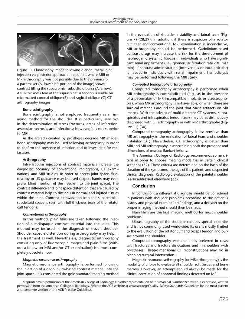
MR arthrography is contraindicated (e.g., as in the presence of a pacemaker or MR-incompatible implants or claustropho-bia), when MR arthrography is not available, or when there are surgical materials around the joint that cause artifacts on MR images. With the advent of multi-detector CT systems, supra-spinatus and infraspinatus tendon tears may be as distinctively diagnosed with CT arthrography as with MR arthrography (Fig-ure 11) (30).
Computed tomography arthrography is less sensitive than MR arthrography in the evaluation of labral tears and shoulder instability (31). Nevertheless, CT arthrography is better than MRI and MR arthrography in ascertaining both the presence and dimensions of osseous Bankart lesions.
The American College of Radiology recommends some cri-teria in order to choose imaging modalities in certain clinical scenarios (32). These criteria are determined on the basis of the duration of the symptoms, the age of the patient, and suspected clinical diagnosis. Radiologic evaluation of the painful shoulder is also addressed elsewhere (33).
Conclusion
In conclusion, a differential diagnosis should be considered in patients with shoulder problems according to the patient’s history and physical examination findings, and a decision on the proper imaging method should then be made.
Plain films are the first imaging method for most shoulder problems.
Ultrasonography of the shoulder requires special expertise and is not commonly used worldwide. Its use is mostly limited to the evaluation of the rotator cuff and biceps tendon and bur-sae around the shoulder.
Computed tomography examination is preferred in cases with fractures and fracture dislocations and in shoulders with prostheses. Three-dimensional CT reconstructions may aid in planning surgical intervention.
Magnetic resonance arthrography (or MR arthrography) is the modality of choice to evaluate all shoulder soft tissues and bone marrow. However, an attempt should always be made for the clinical correlation of abnormal findings detected on MRI.
aReprinted with permission of the American College of Radiology. No other representation of this material is authorized without expressed, written permission from the American College of Radiology. Refer to the ACR website at www.acr.org/Quality-Safety/Standards-Guidelines for the most current and complete version of the ACR Practice Guidelines.
Aydıngöz et al.Radiological Assessment of the Shoulder Region
Figure 11. Fluoroscopy image following glenohumeral joint injection via posterior approach in a patient where MRI or MR arthrography was not possible due to the presence of a pacemaker (A, lower left portion of the image) shows contrast filling the subacromial-subdeltoid bursa (A, arrow). A full-thickness tear at the supraspinatus tendon is visible on reformatted coronal oblique (B) and sagittal oblique (C) CT arthrography images
A B C
S75

Aydıngöz et al.Radiological Assessment of the Shoulder Region
Appendix
Excerpt from the American College of Radiology-Society of Skeletal Radiology (ACR-SSR) Practice Guideline for MRI of the Shoulder (reprinted with permission from reference 22a).
Magnetic resonance imaging (MRI) is an established and prov-en imaging modality for the detection, evaluation, assessment, staging, and follow-up of disorders of the shoulder. Properly per-formed and interpreted, MRI contributes not only to diagnosis but also to treatment planning and prognostication. However, it should be performed only for a valid medical reason and after careful consideration of alternative diagnostic modalities.
Magnetic resonance imaging of the shoulder may be per-formed without contrast, following intra-articular contrast injec-tion (“direct” MR arthrography) to increase conspicuity of intra-articular abnormalities, or with intravenous contrast to identify hyperemic lesions or to create “indirect” arthrographic images by enhancing synovial-lined structures and their contents.
An analysis of the strengths and potential risks of MRI and other diagnostic modalities should be weighed against their suitability for specific patients and particular clinical conditions. Radiographs usually are the first imaging test performed for most suspected abnormalities in the shoulder and will often suf-fice to diagnose or exclude an abnormality or will direct further imaging evaluation. Radionuclide bone scanning can screen the entire skeleton in addition to the shoulder for radiographically occult bone disease, such as metastases. Other nuclear medi-cine examinations have a role for specific clinical scenarios (e.g., a labeled white blood cell study for suspected osteomyelitis). Conventional single-contrast or double-contrast arthrography can accurately depict most articular surface and full-thickness tears of the rotator cuff. Sonography can be used to evaluate the rotator cuff and biceps tendon and has the advantage of imag-ing during physiologic motion. Computed tomography (CT) is used to evaluate the bone integrity of the glenoid fossa and hu-merus and the alignment and congruence of the glenohumeral joint. When combined with arthrography, CT can also be used for evaluating the labrum, articular cartilage, and loose bodies. Lastly, arthroscopy provides a detailed examination of the inter-nal structures of the shoulder, allowing the surgeon to treat as well as diagnose many internal derangements.
While MRI is one of the most sensitive diagnostic tests for de-tecting anatomic abnormalities of the extremities, findings may be misleading if not closely correlated with other imaging stud-ies, clinical history, clinical examination, and physiologic tests. Adherence to the following guideline will enhance the probabil-ity of accurately diagnosing such abnormalities.
IndicationsA. Primary indications for MRI of the shoulder include, but are
not limited to, diagnosis, exclusion, and grading of suspected:1. Rotator cuff tendon abnormalities: full-thickness, partial-
thickness, and recurrent (postoperative) tears; tendinopathy; tendinitis; and cuff tear arthropathyb.
2. Disorders of the long head of the biceps brachii: full-thick-ness, partial-thickness, and recurrent (postoperative) tears; tendinopathy, tendinitis; subluxation; and dislocationb.
3. Conditions affecting the supraspinatus outlet: acromial shape, os acromiale, subacromial spurs, acromioclavicular joint disor-ders, coracoacromial ligament integrity, subacromial bursitisb.
4. Labral abnormalities: cysts, degeneration, and tears, including superior labrum anterior posterior (SLAP) lesions, Bankart lesions and their variants, and recurrent (postoperative) labral tearsb.
5. Abnormalities of the rotator interval and biceps pulleyb.6. Muscle disorders affecting the shoulder girdle: atrophy, hy-
pertrophy, denervation, masses, injuries.7. Glenohumeral chondral and osteochondral abnormalities:
osteochondral fractures and osteochondritis dissecans, ar-ticular cartilage degeneration, fissures, fractures, flaps, and separationsb.
8. Intra-articular bodiesb.9. Synovial-based disorders: synovitis, bursitis, metaplasia, and
neoplasiac.10. Marrow abnormalities: osteonecrosis, marrow edema syn-
dromes, and stress fracturesc.11. Neoplasms, masses, and cysts of bone, joint, or soft tissuec.12. Infections of bone, joint, or soft tissuec.13. Congenital and developmental conditions, including dyspla-
sia and normal variantsc.14. Vascular conditions: entrapment, aneurysm, stenosis, and
occlusionc.15. Neurologic conditions: entrapment, compression, masses,
and peripheral neuritisc.
B. Magnetic resonance imaging of the shoulder may be indi-cated to further clarify and stage conditions diagnosed clini-cally and/or suggested by other imaging modalities, includ-ing, but not limited to:
1. Arthritides: inflammatory, infectious, neuropathic, degenera-tive, crystal-induced, post-traumaticc.
2. Frozen shoulder and adhesive capsulitisb.3. Primary and secondary bone and soft tissue tumorsc.4. Fractures and dislocations.
C. Magnetic resonance imaging of the shoulder may be useful to evaluate specific clinical scenarios, including, but not limited to:
1. Prolonged, refractory, or unexplained shoulder painb,c.2. Acute shoulder trauma.3. Impingement syndromes: subacromial, subcoracoid, internalb.4. Glenohumeral instability: chronic, recurrent, subacute, and
acute dislocation and subluxationb.5. Shoulder symptoms in the overhead or throwing athleteb.6. Mechanical shoulder symptoms: catching, locking, snap-
ping, crepitusb.7. Limited or painful range of motion.8. Swelling, enlargement, mass, or atrophyc.9. Patients for whom diagnostic or therapeutic arthroscopy is
plannedb.10. Patients with recurrent, residual, or new symptoms follow-
ing shoulder surgeryb.
bConditions in which intra-articular contrast (performed by direct intra-articular injection or indirect joint opacification following intravenous administration) may be useful.
cConditions in which intravenous contrast may be useful.
S76

Peer-review: This manuscript was prepared by the invitation of the Editorial Board and its scientific evaluation was carried out by the Editorial Board.
Author Contributions: Concept - N.C.; Design - N.C., Ü.A.; Supervision - Ü.A., M.D.; Materials - Ü.A., N.C.; Analysis and/or Interpretation - Ü.A., N.C., M.D.; Literature Review - N.C., Ü.A.; Writer - N.C., Ü.A.; Critical Review - Ü.A., M.D.
Conflict of Interest: No conflict of interest was declared by the authors.
Financial Disclosure: The authors declared that this study has received no financial support.
Hakem değerlendirmesi: Bu makale Editörler Kurulu’nun davetiyle hazırlandığından bilimsel değerlendirmesi Editörler Kurulu tarafından yapılmıştır.
Yazar Katkıları: Fikir - N.C.; Tasarım - N.C., Ü.A.; Denetleme - Ü.A., M.D.; Malzemeler - Ü.A., N.C.; Analiz ve/veya yorum - Ü.A., N.C., M.D.; Literatür taraması - N.C., Ü.A.; Yazıyı yazan - N.C., Ü.A.; Eleştirel İnceleme - Ü.A., M.D.
Çıkar Çatışması: Yazarlar çıkar çatışması bildirmemişlerdir.
Finansal Destek: Yazarlar bu çalışma için finansal destek almadıklarını beyan etmişlerdir.
References1. Willick SE, Sanders RK. Radiologic evaluation of the shoulder girdle.
Phys Med Rehabil Clin N Am 2004;15:373-406. [CrossRef]2. Rockwood Jr CA, Jensen KL. X-ray evaluation of shoulder problems.
In: Rockwood Jr CA, Matsen III FA, Wirth MA, Harryman DT, eds. The Shoulder. 2nd ed. Philadelphia: WB Saunders; 1998:199-231.
3. Goud A, Segal D, Hedayati P, Pan JJ, Weissman BN. Radiographic evaluation of the shoulder. Eur J Radiol 2008;68:2-15. [CrossRef]
4. Blaine TA, Bigliani LU, Levine WN. Fractures of the proximal humer-us. In: Rockwood Jr CA, Matsen III FA, Wirth MA, Harryman DT, eds. The Shoulder. 3rd ed. Philadelphia: WB Saunders; 2004.p.355-412.
5. Magee DJ. Ortopedic physical assessment. 3rd edition. Philadel-phia: WB Saunders; 1997.pp.175-246.
6. Bigliani LU, Ticker JB, Flatow EL, Soslowsky LJ, Mow VC. The rela-tionship of acromial architecture to rotator cuff disease. Clin Sports Med 1991;10:823-38.
7. Weiner DS, Macnab I. Superior migration of the humeral head. A radiological aid in the diagnosis of tears of the rotator cuff. J Bone Joint Surg Br 1970;52:524-7.
8. Geusens E, Pans S, Verhulst D, Brys P. The modified axillary view of the shoulder, a painless alternative. Emerg Radiol 2006;12:227-30. [CrossRef]
9. Rubin SA, Gray RL, Green WR. The scapular “Y”: a diagnostic aid in shoulder trauma. Radiology 1974;110:725-6.
10. Coumas J, Waite R, Goss T, Ferrari DA, Kanzaria PK, Pappas AM. CT and MR evaluation of the labral capsular ligamentous complex of the shoulder. AJR Am J Roentgenol 1992;158:591-7. [CrossRef]
11. Baudi P, Righi P, Bolognesi D, Rivetta S, Rossi Urtoler E, Guicciardi N, et al. How to identify and calculate glenoid bone deficit. Chir Organi Mov 2005;90:145-52.
12. Magarelli N, Milano G, Sergio P, Santagada DA, Fabbriciani C, Bonomo L. Intra-observer and interobserver reliability of the ‘Pico’ computed to-mography method for quantification of glenoid bone defect in anterior shoulder instability. Skeletal Radiol 2009;38:1071-5. [CrossRef]
13. Shellock FG. Reference manual for magnetic resonance safety. Salt Lake City: Amirsys, Inc ; 2003.
14. Modarresi S, Jude CM. Radiologic evaluation of the painful shoulder 2012 UpTo Date. Available from: URL: www.uptodate.com.
15. De Maeseneer M, Van Roy F, Lenchik L, Shahabpour M, Jacobson J, Ryu KN, et al. CT and MR arthrography of the normal and patho-logic anterosuperior labrum and labral-bicipital complex. Radio-graphics 2000;20:67-81. [CrossRef]
16. Goutallier D, Postel JM, Bernageau J, Lavau L, Voisin MC. Fatty mus-cle degeneration in cuff ruptures. Pre- and postoperative evaluation by CT scan. Clin Orthop Relat Res 1994;304:78-83.
17. Fuchs B, Weishaupt D, Zanetti M, Hodler J, Gerber C. Fatty degen-eration of the muscles of the rotator cuff: assessment by computed tomography versus magnetic resonance imaging. J Shoulder Elbow Surg 1999;8:599-605. [CrossRef]
18. Oh JH, Kim SH, Choi JA, Kim Y, Oh CH. Reliability of the grading system for fatty degeneration of rotator cuff muscles. Clin Orthop Relat Res 2010;468:1558-64. [CrossRef]
19. Slabaugh MA, Friel NA, Karas V, Romeo AA, Verma NN, Cole BJ. Interob-server and intraobserver reliability of the Goutallier classification using magnetic resonance imaging: proposal of a simplified classification sys-tem to increase reliability. Am J Sports Med 2012;40:1728-34. [CrossRef]
20. Alyas F, Curtis M, Speed C, Saifuddin A, Connell D. MR imaging appearances of acromioclavicular joint dislocation. Radiographics 2008;28:463-79. [CrossRef]
21. de la Puente R, Boutin RD, Theodorou DJ, Hooper A, Schweitzer M, Resnick D. Post-traumatic and stress-induced osteolysis of the distal clavicle: MR imaging findings in 17 patients. Skeletal Radiol 1999;28:202-8. [CrossRef]
22. ACR-SSR Practice Guideline for the Performance and Interpretation of Magnetic Resonance Imaging (MRI) of the Shoulder. American Col-lege of Radiology (ACR). Reston, Virginia,USA 2010. (http://www.acr.org/~/media/ACR/Documents/PGTS/guidelines/MRI_Shoulder.pdf).
23. Demirhan M, Akman Ş, Akalın Y. Rotator manşet patolojilerinde ul-trasonografik tanı. Acta Orthop Traumatol Turc 1994;28:177-80.
24. Backhaus M, Burmester GR, Gerber T, Grassi W, Machold KP, Swen WA, et al. Guidelines for musculoskeletal ultrasound in rheumatol-ogy. Ann Rheum Dis 2001;60:641-9. [CrossRef]
25. Read JW, Perko M. Shoulder ultrasound: diagnostic accuracy for im-pingement syndrome, rotator cuff tear, and biceps tendon pathol-ogy. J Shoulder Elbow Surg 1998;7:264-71. [CrossRef]
26. Lee KS, Rosas HG. Musculoskeletal ultrasound: how to treat calcific tendinitis of the rotator cuff by ultrasound-guided single-needle la-vage technique. AJR Am J Roentgenol 2010;195:638. [CrossRef]
27. Serafini G, Sconfienza LM, Lacelli F, Silvestri E, Aliprandi A, Sardanelli F. Rotator cuff calcific tendonitis: short-term and 10-year outcomes after two-needle US-guided percutaneous treatment-nonrandom-ized controlled trial. Radiology 2009;252:157-64. [CrossRef]
28. Schulte-Altedorneburg G, Gebhard M, Wohlgemuth W, Fischer W, Zentner J, Wegener R, et al. MR arthrography: pharmacology, efficacy and safety in clinical trials. Skeletal Radiol 2003;32:1-12. [CrossRef]
29. Steinbach LS, Palmer WE, Schweitzer MH. Special focus session. MR arthrography. Radiographics 2002;22:1223-46. [CrossRef]
30. Charousset C, Bellaïche L, Duranthon LD, Grimberg J. Accuracy of CT arthrography in the assessment of tears of the rotator cuff. J Bone Joint Surg Br 2005;87:824-8. [CrossRef]
31. Bitzer M, Nasko M, Krackhardt T, Schick F, Schöber W, Wiskirchen J, et al. Direct CT-arthrography versus direct MR-arthrography in chronic shoulder instability: comparison of modalities after the introduction of multidetector-CT technology. Rofo 2004;176:1770-5. [CrossRef]
32. Expert Panel on Musculoskeletal Imaging. Acute shoulder pain. American College of Radiology (ACR) Appropriateness Criteria. Res-ton, Virginia, USA. 2010.
33. Radiological evaluation of painful shoulder. UpToDate® website (http://www.uptodate.com/contents/radiologic-evaluation-of-the-painful-shoulder). Wolters Kluwer Health Clinical Solutions. Phila-delphia, Pennsylvania, USA. Accessed 18 February 2014.
Aydıngöz et al.Radiological Assessment of the Shoulder Region
S77