JOURNAL - Elsevier
94
Pulmonology Journal volume 26 / number 6 / November/December 2020 JOURNAL ISSN 2531-0437 www.journalpulmonology.org volume 26 / number 6 / November/December 2020 Previously Revista Portuguesa de Pneumologia Editorials The lung microbiome and pneumonia: Where precision medicine meets pulmonology Pulmonary telerehabilitation: An international call for action Will the COVID tsunami be able to impose tele-rehabilitation as a system opportunity? Original articles Tuberculosis Disseminated Bacillus Calmette-Guérin (BCG) infection with pulmonary and renal involvement: A rare complication of BCG immunotherapy. A case report and narrative review Host factors associated to false negative and indeterminate results in an interferon-γ release assay in patients with active tuberculosis Home care The addition of a humidifier device to a circuit and its impact on home ventilator performance: a bench study
-
Upload
khangminh22 -
Category
Documents
-
view
0 -
download
0
Transcript of JOURNAL - Elsevier

Pulm
ono
log
y J
ourn
al
vo
lum
e 2
6 / n
um
ber 6
/ No
vem
ber/D
ecem
ber 2
020
JOURNAL
ISSN 2531-0437 www.journalpulmonology.org
volume 26 / number 6 / November/December 2020
Previously Revista Portuguesa de Pneumologia
EditorialsThe lung microbiome and pneumonia: Where precision medicine meets pulmonology
Pulmonary telerehabilitation: An international call for action
Will the COVID tsunami be able to impose tele-rehabilitation as a system opportunity?
Original articles
Tuberculosis
Disseminated Bacillus Calmette-Guérin (BCG) infection with pulmonary and renal involvement: A rare complication of BCG immunotherapy. A case report and
narrative review
Host factors associated to false negative and indeterminate results in an interferon-γ release assay in patients with active tuberculosis
Home care
The addition of a humidifi er device to a circuit and its impact on home ventilator performance: a bench study


EDITOR IN CHIEFNicolino Ambrosino
ASSOCIATE EDITORSTiago Alfaro (Portugal)
Katerina Antoniou (Greece)
Luis Azevedo (Portugal)
Teresa Bandeira (Portugal)
Konrad Bloch (Switzerland)
Demosthenes Bouros (Greece)
Antonio Bugalho (Portugal)
António Bugalho de Almeida (Portugal)
Claudia Chaves Loureiro (Portugal)
Enrico Clini (Italy)
Marta Drummond (Portugal)
Raquel Duarte (Portugal)
Frits Franssen (The Netherlands)
Venceslau Hespanhol (Portugal)
Ildiko Horvath (Hungary)
Jessica Jones (Portugal)
Manuela Latorre (Italy)
Pierantonio Laveneziana (France)
Sebastian Ley (Germany)
José Melo Cristino (Portugal)
Giovanni Migliori (Italy)
Stefano Nava (Italy)
Hilario Nunes (France)
Paula Pinto (Portugal)
Venerino Poletti (Italy)
Luis Puente-Maestu (Spain)
Fátima Rodrigues (Portugal)
Nikos Siafakas (Greece)
Giovanni Sotgiu (Italy)
Richard Staats (Portugal)
Paul van Schil (Belgium)
Michele Vitacca (Italy)
Joao Winck (Portugal)
Richard Zu Wallack (USA)
INTERNATIONAL EDITORIAL BOARDSemra Bilaceroglu (Turkey), Jean Bousquet (France), Mina Gaga (Greece)
Geraldo Lorenzi-Filho (Brazil), Florin Mihaltan (Romania), Branislava Milenkovic
(Serbia), Marc Miravitlles (Spain), Pier Luigi Paggiaro (Italy) Fabio Pitta (Brazil)
Menaldi Rasmin (Indonesia)
NATIONAL ADVISORY BOARDJosé Alves, Fernando Barata, Cristina Bárbara, António Bensabat Rendas,
Paula Campos, João Cardoso, Aurora Carvalho, Jorge Ferreira, Filipe Froes,
Miguel Goncalves, Agostinho Marques, Maria João Marques Gomes,
Fernando Martel, António Morais, Henrique Queiroga, Carlos Robalo Cordeiro,
Renato Sotto-Mayor, Conceição Souto Moura, Lina Vaz
Av. Josep Tarradellas, 20-30, 1º08029 Barcelona (Spain)Phone: +34 932 000 711Paseo de la Castellana, 16328046 Madrid (Spain)Phone: +34 914 021 212
Data protection: Elsevier España, S.L.U.. declares that it complies with that established by Organic Law 3/2018, of December 5, Protection of Personal Data and Guarantee of Digital Rights (LOPDGDD).
Indexed in: Science Citation IndexExpanded (SCIE), Journal of Citation Reports(JCR), Index Medicus/MEDLINE,Scopus, EMBASE/Excerpt MedicaISSN 2531-0437 (online)
Register 122.190 of Gabinete paraos Meios de Comunicação Social
Printed in SpainPrinted in acid free paper
Legal deposit: B-20.760-2019
© SOCIEDADE PORTUGUESADE PNEUMOLOGIA (2020)
www.sppneumologia.pt
This Journal and the individual contributions contained in it are protected by the copyright laws, and the following terms and conditions are applied to its use, as well as the terms of any Creative Commons licence that the Editor may have applied to each specifi c article:
Photocopying. Individual articles can be photocopied for personal use according to that permitted by the copyright laws. Permission is not required to photocopy articles published under the CC BY licence or to photocopy for non-commercial purposes in accordance with any other user licence applied by the Editor. For all other photocopies, permission and the payment of a fee is required is required from the publishing company (in this case, should be directed to CEDRO [www.cedro.org]).
Derived products. The users can reproduce tables of contents or prepare lists of articles, including the internal circulation of abstracts within their institutions or companies. Apart from the articles published under the CC BY licence, authorisation is required from the publisher for its re-sale or distribution outside the institution or company that subscribes. For any other or other articles subscribed under a CC BY-NC-ND licence, authorisation is required from the publisher for all other derived works, including compilations and translations.
Storage or use. Except for that indicated previously, or according to that established in the corresponding licence of use, no part of this publication may be reproduced, stored in recovery systems or transmitted in any form or by any medium, whether electronic, mechanical, photocopy, recorded or any other means, without the prior permission in writing by the Editor.
Author rights. The author or authors may have additional rights over their articles depending on that agreed with the Editor (more information at: http://www.elsevier.com/authorsrights).No responsibility is assumed by the Publisher or the SOCIEDADE PORTUGUESA DE PNEUMOLOGIA for any injury and/or damage to persons or property as a matter of products liability, negligence or otherwise, or from any use or operation of any methods, products, instructions or ideas contained in the material herein. Although all advertising material is expected to conform to ethical standards, inclusion in this publication does not constitute a guarantee or endorsement of the quality or value of such product or of the claims made of it by its manufacturer.
Published every 2 months (6 issues per year).www.journalpulmonology.org
Reprints information:Clarissa Felix: [email protected]
Subscription of printed version available One issue 30.00 € (VAT not included) Anual 120.00 € (VAT not included)
(prices valid only for Portugal)
Subscriptions orders: [email protected]

PEACE, LOVE AND HYPOTHESISThe world needs your research. You need Scopus.
With up to 230% more coverage of published research worldwide, 16 million author profiles, daily content updates and more – your next big discovery starts with Scopus.
For more information visit elsevier.com/Scopus
Scopus_Ad-A4-Researcher-v2.pdf 1 5/11/18 10:06 AM

www.revportpneumol.orgwww.journalpulmonology.org
Volume 26 . Number 6 . November - December 2020
CONTENTS
EditorialsThe lung microbiome and pneumonia: Where precision medicine meets pulmonology
A. Araghi . . . . . . . . . . . . . . . . . . . . . . . . . . . . . . . . . . . . . . . . . . . . . . . . . . . . . . . . . . . . 333Pulmonary telerehabilitation: An international call for action
C. Jácome, A. Marques, A. Oliveira, L.V. Rodrigues and I. Sanches . . . . . . . . . . . . . . . . . . . . . 335Will the COVID tsunami be able to impose tele-rehabilitation as a system opportunity?
M. Vitacca . . . . . . . . . . . . . . . . . . . . . . . . . . . . . . . . . . . . . . . . . . . . . . . . . . . . . . . . . . . 338
CommentaryThe scientifi c production during 2009 swine fl u pandemic and 2019/2020 COVID-19 pandemic
T.A. Carvalho, T.M. Lima, V.F. Melani, M.F. Mendes, L.R. Pereira and F.A.L. Marson . . . . . . . . . . 340
Original articles
Tuberculosis
Disseminated Bacillus Calmette-Guérin (BCG) infection with pulmonary and renal involvement: A rare complication of BCG immunotherapy. A case report and narrative reviewM. Marques, D. Vazquez, S. Sousa, G. Mesquita, M. Duarte and R. Ferreira . . . . . . . . . . . . . . . . 346
Host factors associated to false negative and indeterminate results in an interferon-γ release assay in patients with active tuberculosisJ.A. Santos, R. Duarte and C. Nunes . . . . . . . . . . . . . . . . . . . . . . . . . . . . . . . . . . . . . . . . . 353
Home care
The addition of a humidifi er device to a circuit and its impact on home ventilator performance: a bench studyJ. Collada-Carrasco, C. Lamolda-Puyol, M. Luján, A. Castaño-Menéndez, M. Jiménez-Gómez, A. Hernández-Voth and J. Sayas-Catalán . . . . . . . . . . . . . . . . . . . . . . . . . . . . . . . . . . . . . . . 363
ReviewsEffectiveness of different treatments in obesity hypoventilation syndrome
V.R. Ramírez Molina, J.F. Masa Jiménez, F.J. Gómez de Terreros Caro and J. Corral Peñafi el . . . . 370The validity of surface EMG of extra-diaphragmatic muscles in assessing respiratory responses during
mechanical ventilation: A systematic reviewH.Y. AbuNurah, D.W. Russell and J.D. Lowman . . . . . . . . . . . . . . . . . . . . . . . . . . . . . . . . . . . 378
Special ArticleRecommendations for interventional pulmonology during COVID-19 outbreak: a consensus statement
from the Portuguese Pulmonology SocietyF. Guedes, J.P. Boléo-Tomé, L.V. Rodrigues, H.N. Bastos, S. Campainha, M. de Santis, L. Mota and A. Bugalho . . . . . . . . . . . . . . . . . . . . . . . . . . . . . . . . . . . . . . . . . . . . . . . . . . . . . . . . 386
Letters to EditorEndobronchial ultrasound-transvascular needle aspiration (EBUS-TVNA) in the diagnosis of a hilar
metastasis of an extrapulmonary neoplasmF. Guedes and T. Oliveira . . . . . . . . . . . . . . . . . . . . . . . . . . . . . . . . . . . . . . . . . . . . . . . . . 398
Cystic tuberculosis: a very unusual aspect of a common diseaseJ. Perim, E. Scarlatelli Pimenta and E. Marchiori . . . . . . . . . . . . . . . . . . . . . . . . . . . . . . . . . 400
Diffuse cystic lung disease as the primary tomographic manifestation of bronchiolitis: A case seriesM.R. de Oliveira, O.M. Dias, A.F. Amaral, E.C.T. do Nascimento, M. Wanderley, C.R.R. Carvalho and B.G. Baldi . . . . . . . . . . . . . . . . . . . . . . . . . . . . . . . . . . . . . . . . . . . . . . . . . . . . . . . . 403
A rare case of pulmonary disease combining alpha-1-antitrypsin defi ciency and common variable immunodefi ciencyC.S. Sousa, V. Teixeira, V. Pereira, R.B. Pinheiro, S. Seixas and N. Martins . . . . . . . . . . . . . . . . 406
Pulmonary intravascular lymphoma mimicking hypersensitivity pneumonitisR. Kikuchi, M. Ishiwari, H. Takoi, Y. Kono, A. Yoshimura and S. Abe . . . . . . . . . . . . . . . . . . . . . 409
Poly-resistant tuberculosis outbreak in Northern Portugal: a nine year taleB. Gomes, G. Molina-Correa, L. Neves-Reina, A.C. Oliveira, R. Macedo, C. Carvalho and A.M. Correia . . . . . . . . . . . . . . . . . . . . . . . . . . . . . . . . . . . . . . . . . . . . . . . . . . . . . . 412
Unusual effectiveness of systemic steroids in Whipple diseaseM. Fontana, S. Cerri, G. Bernardelli, L. Brugioni, E. Clini and R. Tonelli . . . . . . . . . . . . . . . . . 415
CorrespondencePneumocystosis pneumonia in immunocompromised patients
B.Joob and V. Wiwanitkit . . . . . . . . . . . . . . . . . . . . . . . . . . . . . . . . . . . . . . . . . . . . . . . . . 418
Book Commentary“Book Commentary: Donner CF, Ambrosino N, Goldstein RS. Pulmonary Rehabilitation, 2nd Edition. CRC
Press Pub. Pp 518.”A. Ries . . . . . . . . . . . . . . . . . . . . . . . . . . . . . . . . . . . . . . . . . . . . . . . . . . . . . . . . . . . . . 419

Pulmonol. 2020;26(6):333---334
www.journalpulmonology.org
EDITORIAL
The lung microbiome and pneumonia: Where precisionmedicine meets pulmonology
Human wellbeing is the result of dynamic networkingbetween five nodes: mind---brain, genes, epigenetics, envi-ronment, and microbiome.1 These nodes communicate witheach other through messengers, including neuroendocrinepeptides and microbial metabolites. One can imagine thisgrand system as a symphony orchestra except without apermanent conductor. To hear a well-tuned and performedsymphony, a healthy state, the function of each node is syn-chronized with the other four. The disorder is the phenotypeof any disturbance in the network.
So far, mostly our attention spectrum to study, prevent,and treat each disorder(s), has been narrowed to a lim-ited cause and effect or association between an agent andthe affected organ. When it comes to multisystem disorderslike multiple system organ failures in a septic patient withpneumonia, our capability to prevent, predict and treat thepatient is as good as the best survival rate for septic shock.
In this narrative review article, using the above-proposedmodel to study a disease, our new understanding of thepathogenesis of pneumonia and its potential implications forprevention, treatment, and policy-making are discussed.
The classic theory of the pathogenesis of pneumonia2
assumes that the lung is a sterile organ, and the etiologicmicroorganism enters, colonizes, and invades pure airwaysand pulmonary parenchyma either through aspiration fromthe digestive tract or circulation. Advances in genetics tech-niques to detect microbes, like 16S rRNA gene sequencing3
and metagenomics, have shown that the lung is not a sterileorgan as we previously assumed. Indeed, a constellation ofdifferent microorganisms lives in small airways and alveoli.Compared with the gut microbiome, however, the densityand diversity of the lung microbiome are limited. The lungwas considered a sterile organ because the routine sputumcultures fail to detect anaerobic microorganisms.4
In healthy lungs, Proteobacteria, Firmicutes, and Bac-teroidetes are the most commonly identified bacteria,while, Streptococcus, Prevotella, Fusobacteria, and Veil-lonella predominate, with potential pathogens, such asHaemophilus and Neisseria, are a smaller fraction of ahealthy lung microbiome. Interestingly, the same bacterial
population is the normal flora of the mouth in healthyindividuals.5 The formation and diversity of the pulmonarymicrobiome (PM) start with the exposure of newborn oralmucosa to maternal vaginal flora with subsequent microaspi-ration of the newly formed oral flora into the airwaysand alveolar epithelial cells. Microaspiration is the primarysource of populating PM, inhalation and intestinal micro-biome are other sources. Intestinal microbiome influencesPM directly by the migration of microorganisms to PM via thelymphatic system, and indirectly, by immune modulation.Submucosal lymphocytes, dendritic cells, and macrophagesexchange information with the intraluminal microbiome.These interactions set the immune tone and affect thefunction of immune cells in response to newly invadingpathogens. Migrating immune cells from the intestine tobronchial submucosa and lung interstitial space by lymphaticflow, modulate the pulmonary defenses and susceptibilityto virulent pathogens. Altered intestinal microbiome afterbroad-spectrum antibiotic therapy or intestinal ischemia inacute illness can increase the risk of developing pneumoniaor ARDS.6
The content and diversity of the pulmonary micro-biome at any given time depends on dynamic interactionsbetween immigration, colonization, and elimination pro-cesses. Through aging, the diversity of the PM evolvesand adapts to the living environment, diet, and submu-cosal immune tone, among other factors. For example, ahigh fiber diet by producing more short-chain fatty acids(SCFA) enhances Bacteroides dominance both in gut andlung. It has been shown that the high fiber diet can decreasethe incidence of asthma.6 Most studies of the PM focusedon the bacterial population. However, it is expected thata variety of viruses and fungi are part of PM. All thesemicroorganisms continuously communicate with each otherand their host through their metabolites. Symbiosis is adynamic, healthy state when PM has a ‘‘normal’’ diver-sity, and the host response/immune tone is well-tuned andadjusted. As a result of immigration and poor elimination ofa hostile pathogen, altered immune tone, i.e., acute stresssuppresses immunity, the PM diversity is reduced, and a
https://doi.org/10.1016/j.pulmoe.2020.04.0052531-0437/© 2020 Sociedade Portuguesa de Pneumologia. Published by Elsevier Espana, S.L.U. This is an open access article under the CCBY-NC-ND license (http://creativecommons.org/licenses/by-nc-nd/4.0/).
334 EDITORIAL
dominant microorganism can act as a virulent pathogen, thatprovokes an inflammatory host response which is called theclinical phenotype of pneumonia. Bos et al. summarized thisprocess as ‘‘pneumonia could be defined in ecological termsas ‘‘the acute loss of biodiversity due to the overgrowth ofa single or several pathogenic microorganisms causing lunginflammation and damage.’’4
It seems that PM diversity in a healthy state correlateswith mouth microbiome diversity. It is well documented thatthe change in the oral microbiome is related to the sever-ity of acute illness rather than the admission unit of thepatient. This finding emphasizes the detrimental role of thepatient’s altered immune response to severe stress and itsrole in transforming symbiosis to dysbiosis.7
The practical question is how this new concept of devel-oping pneumonia will change our diagnostic, therapeutic,and health administration policies of pneumonia. First,one cannot draw a sharp line between rigid definitionsof ‘‘community-acquired pneumonia’’ vs. ‘‘healthcare-associated pneumonia’’ or ‘‘ventilator-associated pneumo-nia.’’
Increasingly, we encounter patients without a history ofexposure to a health care environment that presents withPseudomonas or Acinetobacter pneumonia. Based on thenew concept, one can imagine that even a healthy personwould have a few pathogenic gram-negative rods or gram-positive cocci in lower airways or alveolar epithelium. IfPM diversity and immune tone maintain symbiosis, thosepathogens will be contained and do not evoke a patho-logic inflammatory response. In this view, even acute mentalstress by changing the submucosal immune tone can promotedysbiosis and finally manifest as gram-negative rod (GRD)pneumonia.
This would account for the fact that, if a patient is beingtreated as ‘‘community-acquired pneumonia’’ with Ceftri-axone and Azithromycin, while the pathogen is an entericGRD or methicillin-resistant staphylococcus aureus (MRSA),there would be a higher risk of treatment failure, morbid-ity, and mortality. To apply the personalized or precisionmedicine principles to patients with pneumonia, there areno other options than to optimize respiratory sampling andemploy non-culture based microbiologic harvesting meth-ods to detect the pathogenic microorganism(s) and tailorantibiotics against them. There is no doubt that shortly ourguidelines will be changed to embrace the new concept
and methodologies. Still, until then, for practitioners, thetake-home message is by considering the new definitionof pneumonia based on PM dynamics, we should be ableto detect treatment failures faster and tailor antibioticsaccordingly.
Applying the new concept to reimbursement policies bythird-party payers as well as epidemiological studies, willuproot the current practices. For example, in the US, Medi-care needs to revise its penalties based on narrow definitionsof ventilator-associated pneumonia.
In conclusion: advancements in non-culture-basedmicroorganisms detecting methods show that the lunghas its dynamic microbiome that interacts with the hostthrough symbiosis and causes pneumonia through dysbiosis.This concept will open the door for personalized/precisionmedicine in the diagnosis and treatment of pneumonia.
References
1. Thomas S, Izard J, Walsh E, Batich K, Chongsathidkiet P, ClarkeG, et al. The host microbiome regulates and maintains humanhealth: a primer and perspective for non-microbiologists. CancerRes. 2017;77:1783---812.
2. Musher DM, Thorner AR. Community-acquired pneumonia. N EnglJ Med. 2014;371:1619---28.
3. Watts G, Youens-Clark K, Slepian M, Wolk D, Oshiro M, MetzgerG, et al. 16S rRNA gene sequencing on a benchtop sequencer:accuracy for identification of clinically important bacteria. J ApplMicrobiol. 2017;123:1584---96.
4. Bos LDJ, Rylance J, Gordon SB. The lung bacterial microbiome incommunity-acquired and nosocomial pneumonia. In: Cox MJ, EgeMJ, von Mutius E, editors. The lung microbiome; ERS monograph.2019. p. 188---94.
5. Dickson RP, Erb-Downward JR, Martinez FJ, Huffnagle GB.The microbiome and the respiratory tract. Annu Rev Physiol.2016;78:481---504.
6. Marsland BJ, Trompette A, Gollwitzer ES. The Gut-Lung axisin respiratory disease. Ann Am Thorac Soc. 2015;12 Suppl.2:S150---6.
7. Dickson RP. The microbiome and critical illness. Lancet RespirMed. 2016;4:59---72.
A. AraghiGeorgia College of Medicine, Augusta University, Augusta,
GA, USAAvailable online 29 May 2020

Pulmonol. 2020;26(6):335---337
www.journalpulmonology.org
LETTER TO THE EDITOR
Pulmonary telerehabilitation:An international call for action
The COVID-19 pandemic has impacted dramatically on peo-ple’s lives and health care systems worldwide. Resourcesof the national health services have been focused on themonitoring and management of patients with COVID-19 andso chronic respiratory disease management, namely throughpulmonary rehabilitation (PR), has become even more chal-lenging than it used to be before COVID-19.
Following national and international recommendations,PR programmes were advised to suspend their activitiesand to provide care remotely using telehealth solutions(e.g., phone, video-calls, telerehabilitation).1,2 Major con-cerns already existed about the lack of access to PRprogrammes (either hospital-, primary care- or community-based) worldwide.3 In Portugal, the reported percentage ofpatients having access to PR programmes is between 0.5and 2%.4,5 This situation has most certainly been aggra-vated by the COVID-19 outbreak due to the interruptionof PR, but also due to the increased number of patientswith acquired respiratory diseases that is expected inthe coming months. Given the high scientific evidence ofPR in improving symptoms, physiological and psychosocialdomains in patients with chronic respiratory disease, it isurgent to explore innovative avenues to overcome the pastand present difficulties.3
During (and most certainly after) this outbreak, theimplementation of different technological solutions allowedus to overcome many of the hindrances imposed by forcedsocial distancing. This was the case of telerehabilitationstrategies that in the short term have been largely focusedon patients who had previous on-site access to PR and onpatients with COVID-19.6---10 In the long run, these may befeasible strategies to increase access to PR for those inneed since patients eligible for PR have access to and feelconfident using digital technologies.11 But how prepared arehealth services to implement telerehabilitation?
According to the World Health Organization, telehealthis the ‘‘delivery of health care services, where patients andproviders are separated by distance through the use of infor-mation and communications technology (ICT).12 Telehealthcan be employed in several clinical areas, such as teleradiol-ogy, teledermatology, telepsychiatry and telerehabilitation.Telerehabilitation has been previously defined as the deliv-ery of rehabilitation through a variety of ICT. Similarly to
telehealth, this definition still encompasses a large diver-sity of procedures within the realm of rehabilitation, wherePR can be included.13 However, pulmonary telerehabilitationis far from being a reality yet. For example in Portugal, inthe most recent characterisation of PR, from the 24 centresdelivering PR programmes, none was telehealth supported.4
Although efforts to increase access to PR have been maderecently (primary care centres were advised to implementprogrammes in well-selected patients),14 telerehabilitationguidance was never provided.
The use of telehealth is increasingly included in nationalhealth services.12 In Portugal there has been a TeleHealthNational Centre dedicated to the development and imple-mentation of telehealth solutions since 2016.15 This centrehas produced guidance for teleconsultation, teleradiol-ogy, teledermatology and remote patient monitoring. Yet,guidelines for telerehabilitation are still missing. A similarscenario is present in several countries worldwide and there-fore guidelines for telerehabilitation are urgently neededbut should be broad enough to adapt to all types of rehabil-itation.
A fundamental pillar of PR programmes is its mul-tidisciplinary nature to address the needs of patientswith chronic respiratory diseases and therefore, to stan-dardise pulmonary telerehabilitation, a joint effort bynational organisations, scientific and professional societiesis required. This effort should also be developed in articu-lation with the most relevant international societies in thearea of respiratory medicine, such as the European Respira-tory Society (ERS) and the American Thoracic Society (ATS).Despite the recognised difficulties, e.g., ATS has publiclyacknowledged not being able to endorse a specific approachto PR during the current challenges,16 it is urgent to findalternatives to conventional PR, whilst seeking to increaseaccess to a higher number of patients who can benefit.
In the process of developing guidelines for telereha-bilitation, it could be of interest to start by analysingthe available examples of PR programmes already beingdelivered remotely to patients with chronic respiratorydiseases.17---19 The available literature reports telerehabil-itation to be as effective as onsite-PR programmes andwith potential for successful implementation even with fewresources in patients’ homes.20 Combining this previousknowledge with the experience gathered from the imple-mentation of telerehabilitation in patients with COVID-19 isnow required.9,21,22
https://doi.org/10.1016/j.pulmoe.2020.05.0182531-0437/© 2020 Sociedade Portuguesa de Pneumologia. Published by Elsevier Espana, S.L.U. This is an open access article under the CCBY-NC-ND license (http://creativecommons.org/licenses/by-nc-nd/4.0/).
336 LETTER TO THE EDITOR
Pulmonary rehabilitation, even if delivered remotelymust preserve its cornerstone components, i.e., exer-cise training, education, and behaviour change but, aserious debate about the selection criteria, outcomemeasures, emergency plans, intervention design and equip-ment/technology is needed. The discussion should alsoinvolve technological specialists to aid healthcare providersin selecting and combining cost-benefit and friendly-usertelemonitoring technology such as respiratory monitors,pulse oximeters, activity trackers, environmental sensors,monitors of physiological variables (e.g., heart rate, bloodpressure, temperature) and communication systems.23 Con-cerns about sharing data and meeting General DataProtection Regulation (GDPR) requirements when using thedifferent telemonitoring systems also need to be addressed.Additionally, a significant effort may be needed to try topreserve the social component of PR the role of which isindusputable during on-site programmes but may be lostduring telerehabilitation.
Different discussions involving all relevant stakeholdersin PR, from patients and families to healthcare providers,policy makers and scientists are urgently needed to shift PRfrom conventional to telerehabilitation and increase accessto this fundamental intervention. Telerehabilitation can bea sustainable solution to the increasing burden of chronicrespiratory diseases worldwide.
Conflicts of interest
The authors have no conflicts of interest to declare.
References
1. Direcão-Geral da Saúde. Orientacão n◦ 020/2020 de03/04/2020: COVID-19: FASE DE MITIGACÃO Cuidados deReabilitacão e Respiratórios Domiciliários. URL: https://www.dgs.pt/directrizes-da-dgs/orientacoes-e-circulares-informativas/orientacao-n-0202020-de-03042020-pdf.aspx[accessed 21.05.20].
2. National Health Service. Clinical guide for the managementof respiratory patients during the coronavirus pandemic.URL: https://www.england.nhs.uk/coronavirus/wp-content/uploads/sites/52/2020/03/C0063-Specialty-guide- Respiratory-and-Coronavirus- v1 26-March.pdf [accessed 25.05.20].
3. Rochester CL, Vogiatzis I, Holland AE, Lareau SC, MarciniukDD, Puhan MA, et al. An Official American Thoracic Soci-ety/European Respiratory Society Policy Statement: enhancingimplementation, use, and delivery of pulmonary rehabilitation.Am J Respir Crit Care Med. 2015;192:1373---86.
4. Programa Nacional para as Doencas Respiratórias 2012-2016.Relatório de análise da capacidade instalada de reabilitacão res-piratória nos hospitais do servico nacional de saúde; 2015. URL:https://www.dgs.pt/portal-da-estatistica-da-saude/diretorio-de-informacao/diretorio-de-informacao/por-serie-506885-pdf.aspx?v=%3d%3dDwAAAB%2bLCAAAAAAABAArySzItzVUy81MsTU1MDAFAHzFEfkPAAAA [accessed 21.05.20].
5. Observatório Nacional das Doencas Respiratórias. 13◦ Relatóriodo Observatório Nacional das Doencas Respiratórias; 2018. URL:https://www.ondr.pt/files/Relatorio ONDR 2018.pdf [accessed21.05.20].
6. Pan American Health Organization. Rehabilitation considera-tions during the COVID-19 outbreak. URL: https://iris.paho.org/bitstream/handle/10665.2/52035/NMHMHCOVID19200010eng.pdf?sequence=6&isAllowed=y [accessed 25.05.20].
7. Balbi B, Berney S, Brooks D, Burtin C, Clini E, Franssen FME,et al. Report of an ad-Hoc International task force to developan expert-based opinion on early and short-term rehabilitativeinterventions (after the acute hospital setting) in COVID-19 sur-vivors (version April 3, 2020). URL: https://ers.app.box.com/s/npzkvigtl4w3pb0vbsth4y0fxe7ae9z9 [accessed 25.05.20].
8. Zhao H-M, Xie Y-X, Wang C. Recommendations for respiratoryrehabilitation in adults with COVID-19. Chin Med J. 2020. PMID:32251002.
9. Yang L-L, Yang T. Pulmonary rehabilitation for patients withcoronavirus disease 2019 (COVID-19). Chron Dis Transl Med.2020. PMID: 32411496.
10. Vitacca M, Carone M, Clini EM, Paneroni M, Lazzeri M, Lanza A,et al. Joint statement on the role of respiratory rehabilitationin the COVID-19 crisis: the Italian position paper. Respiration.2020.
11. Jácome C, Marques F, Paixao C, Rebelo P, Oliveira A, CruzJ, et al. Embracing digital technology in chronic respiratorycare: surveying patients access and confidence. Pulmonology.2020;26:56---9.
12. World Health Organization. Telehealth: analysis of third globalsurvey on eHealth based on the reported data by countries;2016. URL: https://www.who.int/gho/goe/telehealth/en/[accessed 21.05.20].
13. Hoaas H, Andreassen HK, Lien LA, Hjalmarsen A, Zanaboni P.Adherence and factors affecting satisfaction in long-term tel-erehabilitation for patients with chronic obstructive pulmonarydisease: a mixed methods study. BMC Med Inform Decis Mak.2016;16:26.
14. Direcão-Geral da Saúde. Orientacão Clínica 014/2019 Pro-gramas de Reabilitacão Respiratória nos Cuidados de SaúdePrimários. URL: https://www.dgs.pt/directrizes-da-dgs/orientacoes-e-circulares-informativas/orientacao-n-0142019-de-070820191.aspx [accessed 21.05.20].
15. Centro Nacional de TeleSaúde. Plano estratégico nacional paraa telessaúde 2019-2022. URL: http://www.cnts.min-saude.pt/wp-content/uploads/2019/12/PENTS PT.pdf [accessed21.05.20].
16. Garvey C, Holland A, Corn J. Pulmonary rehabilitationresources in a complex and rapidly changing world. URL:https://www.thoracic.org/members/assemblies/assemblies/pr/resources/pr-resources-in-a-complex-and-rapidly-changing-world-3-27-2020.pdf [accessed 21.05.20].
17. Santos CD, Neves RCd, Rodrigues F, Bárbara C. PC 190.First steps on home-based pulmonary telerehabilitation byCentro Hospitalar Universitário Lisboa Norte. Pulmonology.2019;25(SC3):120---1.
18. Hansen H, Bieler T, Beyer N, Kallemose T, Wilcke JT, Øster-gaard LM, et al. Supervised pulmonary tele-rehabilitation versuspulmonary rehabilitation in severe COPD: a randomised multi-centre trial. Thorax. 2020;75:413---21.
19. Selzler AM, Wald J, Sedeno M, Jourdain T, Janaudis-FerreiraT, Goldstein R, et al. Telehealth pulmonary rehabilitation: areview of the literature and an example of a nationwide initia-tive to improve the accessibility of pulmonary rehabilitation.Chron Respir Dis. 2018;15:41---7.
20. Vasilopoulou M, Papaioannou AI, Kaltsakas G, Louvaris Z,Chynkiamis N, Spetsioti S, et al. Home-based maintenance tele-rehabilitation reduces the risk for acute exacerbations of COPD,hospitalisations and emergency department visits. Eur Respir J.2017;49, 1602129.
21. Mukaino M, Tatemoto T, Kumazawa N, Tanabe S, Katoh M, SaitohE, et al. Staying active in isolation: telerehabilitation for indi-viduals with the severe acute respiratory syndrome coronavirus2 infection. Am J Phys Med Rehabil. 2020;99:478---9.
22. Exum E, Hull BL, Lee ACW, Gumieny A, Villarreal C, Long-necker D. Applying telehealth technologies and strategies toprovide acute care consultation and treatment of patients

LETTER TO THE EDITOR 337
with confirmed or possible COVID-19. J Acute Care Phys Ther-apy. 2020, http://dx.doi.org/10.1097/JAT.00000000000000143,publish ahead of print.
23. Angelucci A, Aliverti A. Telemonitoring systems for respi-ratory patients: technological aspects. Pulmonology. 2020,http://dx.doi.org/10.1016/j.pulmoe.2019.11.006, publishahead of print.
C. Jácomea,b,∗, A. Marquesc,d, A. Oliveirac,e,f,L.V. Rodriguesg,h, I. Sanches i
a Center for Health Technology and Services Research(CINTESIS), Faculty of Medicine, University of Porto(FMUP), Porto, Portugalb Department of Community Medicine, Information andHealth Decision Sciences (MEDCIDS), University of PortoFaculty of Medicine, Porto, Portugal
c Lab3R --- Respiratory Research and RehabilitationLaboratory, School of Health Sciences, University of Aveiro(ESSUA), Aveiro, Portugald iBiMED --- Institute of Biomedicine, Department ofMedical Sciences, University of Aveiro, Aveiro, Portugale School of Rehabilitation Science, McMaster University,Hamilton, ON, Canadaf West Park Healthcare Centre, Toronto, ON, Canadag Pulmonology Department, Unidade Local de Saúde daGuarda E.P.E. --- Hospital Sousa Martins, Portugalh Faculty of Health Sciences, University of Beira Interior,Covilhã, Portugali Pulmonology Department, Centro Hospitalar Vila Nova deGaia/Espinho, Vila Nova de Gaia, Portugal∗ Corresponding author.E-mail address: [email protected] (C. Jácome).Available online 24 June 2020

Pulmonol. 2020;26(6):338---339
www.journalpulmonology.org
EDITORIAL
Will the COVID tsunami be able to imposetele-rehabilitation as a system opportunity?
We accept with enthusiasm the call by Jacome et al.published in this issue of Pulmonology.1 Pulmonary reha-bilitation may be used for a wide range of purposes andmay include decreasing hospital care services, reducing thecost of care, improving adherence to physical activities,training and correcting life styles, improving accessibility,extending services to remote locations, improving self-monitoring, better understanding of prescribed treatments,improving adherence and better communication with healthprofessionals.2
Tele-health has been defined as the use of informa-tion and communication technologies to deliver healthcare services and transmit medical data over long andshort distances.3 It encompasses a wide variety of tech-nologies such as videoconferencing, internet platforms,store-and-forward devices, streaming media, and groundand wireless communication. Tele-rehabilitation works toaddress a basic question: how to improve access to rehabili-tation services for patients, in an efficacious, cost-effective,and safe manner? It may provide an ideal opportunityto either improve access to pulmonary rehabilitation (PR)and/or help maintain positive results following a tradi-tional program. Tele-rehabilitation reduces barriers such asinsufficient programs and inadequate numbers of qualifiedhealth professionals, particularly in rural and regional areas,reduces problems of transportation, accessible parking, aswell as walking distance from parking to the hospital. Anemerging area of application of technology refers to theuse of wearable sensors to facilitate the implementationof home-based rehabilitation interventions. Systems thataim to facilitate the implementation of rehabilitation exer-cise programs often leverage the combination of sensingtechnology and interactive gaming or virtual reality (VR)environments.4
Previous studies illustrated the potential of tele-health tofacilitate the delivery of PR to patients with chronic obstruc-tive pulmonary disease in their home, as well as to remotesettings without the benefits of an established program.5,6
The Coronavirus (COVID-19) pandemic ‘‘day after’’ is com-ing and people, who suffered from mild to severe pneumonia
up to hypoxemic respiratory failure, might be at risk of long-term impairment and disability.7
Like all patients who have undergone critical illnesses,COVID-19 patients can present dyspnoea and fatigue atrest and during activities of daily living, disability, exerciseintolerance, reduction in peripheral muscle function and innutritional status with significant weight loss. In particular,they may be at risk of residual or worsening parenchymaldamage with respiratory muscle function impairment. Fur-thermore, the infection can negatively affect also otherorgans like heart, kidneys, muscles and brain, with signif-icant health impacts that may persist. Additionally, peoplerequiring intensive care are at increased risk of post-traumatic stress disorder, anxiety, and depression.8,9
The newly discovered Coronavirus (COVID-19) and therigorous request for social distancing has put tele-health(tele-coaching/tele-monitoring/telerehabilitation) in thefront line. Tele-rehabilitation may represent the mostappropriate response in the post-acute COVID phase bycombining need for rehabilitation with need for socialdistancing.10 It should be adopted in post COVID patientswith mild to moderate disabilities, who need frequent mon-itoring, reside in isolated areas or are not available toparticipate in standard programs. Our recent experiencein this field in a subgroup of post COVID patients (unpub-lished data) with reduced exercise tolerance, exerciseinduced desaturation, mild restrictive ventilatory patternand persistent pathological lung imaging, has given promis-ing results: average adherence to a 30-day program was88% with improvement in exercise tolerance, dyspnoea andmuscle fatigue. Strong monitoring should be maintainedthrough wireless devices and when available wearable tech-nology. Contacts by video-call or phone in order to verifypatient adherence to rehabilitation sessions and quality ofsignals are needed. Despite this preliminary observation,the ideal post COVID candidate, duration of intervention,demonstration of efficacy equivalent to a traditional reha-bilitation program to be applied and cost effectivenessare still unknown. Many patients who attend rehabilita-tion programs are older and may not be using, or have
https://doi.org/10.1016/j.pulmoe.2020.08.0052531-0437/© 2020 Sociedade Portuguesa de Pneumologia. Published by Elsevier Espana, S.L.U. This is an open access article under the CCBY-NC-ND license (http://creativecommons.org/licenses/by-nc-nd/4.0/).

EDITORIAL 339
the capacity to use the technology required to deliverytele-rehabilitation. These factors may influence the tele-rehabilitation care environment, and as a consequence, thehealth outcomes. Patient empowerment and digital healthliteracy are essential for successful e-Health deployment.Another uncertainty in post COVID patients is the aim that isexpected: a substitute for standard programs? purely rein-forcement? maintenance program? a modality to improveaccess? Lack of different modalities of supervision is a cru-cial point: how to evaluate frequency, intensity, types andtiming and how to monitor patients’ adherence remain anunsolved question. Also the time required from staff aswell as the amount of data to be interpreted in real timeneed to be elucidated. Proper training of health profession-als and checking the technological requirements, especiallyin the patient’s home, are also required. Adequate care-giver support may be necessary in cases of residual disabilityor for technological setting up. Legal problems associatedwith tele-rehabilitation are still controversial. The patientmust be fully aware of the characteristics of the ser-vice, the potential risks, the precautions to reduce themand to ensure the confidentiality of the information.11 Theassociated safety issues are complex and include not onlyapprehension about malfunctioning equipment, but alsoconcerns regarding potential adverse effects on patientmanagement decisions through delayed or missing informa-tion, misunderstood advice, or inaccurate findings.12 Lastbut not least, the type of equipment used could representa different per-patient cost, while currently there is insuffi-cient evidence to properly advise about cost-effectiveness.How to perform quality control and modality of reimburse-ment remains a challenge.
The use of tele-health technology promises to addresssome major barriers for pulmonary rehabilitation deliveryin that it allows for distribution of healthcare services andexchange of information between a healthcare provider anda patient in different geographical locations and there-fore can provide an important resource to reach peoplewho live in remote communities or have difficulty accessingtraditional centres. National governments should promotecommon, ethical, legal, regulatory, technical, administra-tive standards for remote rehabilitation providing safe andeffective services. The potential of Tele-rehabilitation hasthe enticing potential of reducing barriers and improv-ing care. However, much of the research to date has notexplored the impact of its introduction at a systems level,incorporating data beyond efficacy in the planning andimplementation.
In conclusion we join the international call,1 lookingtowards wider participation and operative actions.
Funding
This work was supported by the ‘‘Ricerca Corrente’’ fundingscheme of the Italian Ministry of Health.
Conflict of interest
The author does not report any conflict of interest.
References
1. Jácome C, Marques A, Oliveira A, Rodrigues LV, SanchesI. Pulmonary telerehabilitation: an international callfor action. Pulmonology. 2020;26, http://dx.doi.org/10.1016/j.pulmoe.2020.05.018. S2531-0437(20)30135-5.
2. Spruit MA, Singh SJ, Garvey C, ZuWallack R, Nici L, RochesterC, et al. An official American Thoracic Society/EuropeanRespiratory Society statement: key concepts and advancesin pulmonary rehabilitation. Am J Respir Crit Care Med.2013;188:e13---64.
3. Standardization IOf. ISO Strategy for Services - Case study 1:International SOS (ISO/TS 13131, Telehealth services); 2016.
4. Angelucci A, Aliverti A. Telemonitoring systems for respiratorypatients: technological aspects. Pulmonology. 2020;26:221---32,http://dx.doi.org/10.1016/j.pulmoe.2019.11.006.
5. Vasilopoulou M, Papaioannou AI, Kaltsakas G, Louvaris Z,Chynkiamis N, Spetsioti S, et al. Home-based maintenancetelerehabilitation reduces the risk for acute exacerbations ofCOPD, hospitalisations and emergency department visits. EurRespir J. 2017;49:1602129.
6. Holland AE, Mahal A, Hill CJ, Lee AI, Burge AT, Cox NS,et al. Home-based rehabilitation for COPD using min-imal resources: a randomised, controlled equivalencetrial. Thorax. 2017;72:57---65, http://dx.doi.org/10.1136/thoraxjnl-2016-208514.
7. Vitacca M, Carone M, Clini EM, Paneroni M, Lazzeri M, Lanza A,et al. Joint statement on the role of respiratory rehabilitationin the COVID-19 crisis: the Italian position paper. Respiration.2020;99(6):493---9, http://dx.doi.org/10.1159/000508399.
8. Marchioni A, Tonelli R, Sdanganelli A, Gozzi F, Musarò L,Fantini R, et al. Prevalence and development of chroniccritical illness in acute patients admitted to a respira-tory intensive care setting. Pulmonology. 2020;26:151---8,http://dx.doi.org/10.1016/j.pulmoe.2019.09.006.
9. Spagnolo P, Balestro E, Aliberti S, Cocconcelli E, Biondini D, CasaGD, et al. Pulmonary fibrosis secondary to COVID-19: a call toarms? Lancet Respir Med. 2020;8(8):750---2.
10. Vitacca M, Lazzeri M, Guffanti E, Frigerio P, D’AbroscaF, Gianola S, et al. An Italian consensus on pulmonaryrehabilitation in COVID-19 patients recovering fromacute respiratory failure: results of a Delphi process.Monaldi Arch Chest Dis. 2020;(June):90, http://dx.doi.org/10.4081/monaldi.2020.1444.
11. Vitacca M, Paneroni M, Ambrosino N. Pulmonary rehabilitationin post acute patients with Covid-19. In: Donner CF, AmbrosinoN, Goldstein RS, editors. Pulmonary Rehabilitation. 2nd editionCRC Press Pub.; 2020. p. 503---10.
12. Bauer KA. The ethical and social dimensions of home-basedtelemedicine. Crit Rev Biomed Eng. 2000;28:541---4.
Michele Vitacca ∗
Istituti Clinici Scientifici Maugeri IRCCS, RespiratoryRehabilitation of the Institute of Lumezzane, Brescia, Italy
∗ Correspondence to: Istituti Clinici Scientifici MaugeriIRCCS, Via S. Maugeri 4, 27100 Pavia, Italy.
E-mail address: [email protected]
19 August 2020Available online 6 September 2020

Pulmonol. 2020;26(6):340---345
www.journalpulmonology.org
COMMENT
The scientific production during 2009 swine flupandemic and 2019/2020 COVID-19 pandemic
T.A. Carvalhoa,b,1, T.M. Limaa,b,1, V.F. Melania,b,1, M.F. Mendesa,b,1, L.R. Pereiraa,b,1,F.A.L. Marsona,b,∗,1
a Laboratory of Cell and Molecular Tumor Biology and Bioactive Compounds, São Francisco University, Braganca Paulista, SãoPaulo, Brazilb Laboratory of Human and Medical Genetics, São Francisco University, Braganca Paulista, São Paulo, Brazil
Received 29 May 2020; accepted 12 July 2020
KEYWORDSCOVID-19;Lung disease;H1N1;Pandemic;SARS-CoV-2;Virus
The 2009 swine flu pandemic caused by Influenza A virus sub-type H1N1 (H1N1) virus affected more than 214 countriesand overseas territories or communities and over 18,449
∗ Corresponding author at: University of São Francisco, Post grad-uate Program in Health Science, Laboratory of Cell and MolecularTumor Biology and Bioactive Compounds and Laboratory of Humanand Medical Genetics, Avenida São Francisco de Assis, 218, JardimSão José, Braganca Paulista, São Paulo 12916-900, Brazil.
E-mail addresses: [email protected] (T.A. Carvalho),[email protected] (T.M. Lima),[email protected] (V.F. Melani),[email protected] (M.F. Mendes),[email protected] (L.R. Pereira),[email protected], [email protected](F.A.L. Marson).
1 The authors contributed equally to this study.
deaths caused by the H1N1 infection1 were confirmed. Afterten years, a new pandemic named Coronavirus Disease 2019(COVID-19) caused by severe acute respiratory syndromecoronavirus 2 (SARS-CoV-2) virus emerged in 2019 in thecity of Wuhan, China. Both diseases were declared a pan-demic by the World Health Organization (WHO), swine fluon 24th April 2009 and COVID-19 on 11th March 2020. Todate, 7th July 2020, the COVID-19 disease affected nearly12 million inhabitants reaching 213 countries and territo-ries around the world and two international conveyances. Inaddition, ∼550,000 deaths were associated with the diseasein 185 locations worldwide. The COVID-19 started in Chinaand spread worldwide changing its epicenter first to Europe,followed by the United State of America and, now, fromMay to July 2020 South America, mainly affecting patientsin Brazil causing the health system to collapse in severalstates such as Amazonas and Rio de Janeiro. On 7th July
https://doi.org/10.1016/j.pulmoe.2020.07.0092531-0437/© 2020 Sociedade Portuguesa de Pneumologia. Published by Elsevier Espana, S.L.U. This is an open access article under the CCBY-NC-ND license (http://creativecommons.org/licenses/by-nc-nd/4.0/).

Science and pandemics 341
2020, Brazil presented a total number of 1,674,655 patientswith COVID-19; 535,558 active cases and 1,072,229 clinicallyrecovered cases, 66,868 deaths related to the disease, anda case fatality rate of 3.99. Brazil occupies the 105 positionworldwide for the number of real-time polymerase chainreaction by one million of inhabitants to screen SARS-CoV-2virus.2,3
Both the 2009 swine flu pandemic and 2019/2020 COVID-19 pandemic resulted in a high number of published articlesin a short period of time. The number of publicationscan be associated with the great impact on science inseveral areas including medicine, sociology, environment,physics, mathematics, biology and many other knowledgeareas that act individually or are multidisciplinary in scope.Another striking aspect is that the collection of articles andother resources on the COVID-19 outbreak, including clinicalreports, management guidelines, and comments are freelyavailable to be used by researchers, health professionalsand the community. The free availability of data resultedfrom the commitment of top scientific (e.g. Science andNature) and Medical (e.g. New England Journal of Medicineand Lancet) journals to supply online articles fast and free ofcharge. To date, the Public Health Emergency COVID-19 Ini-tiative has over fifty authors publishing (collaborators) thathave volunteered to make their coronavirus-related articlesaccessible in PubMed Central® in formats and under licenseterms that facilitate text mining and secondary analysis.
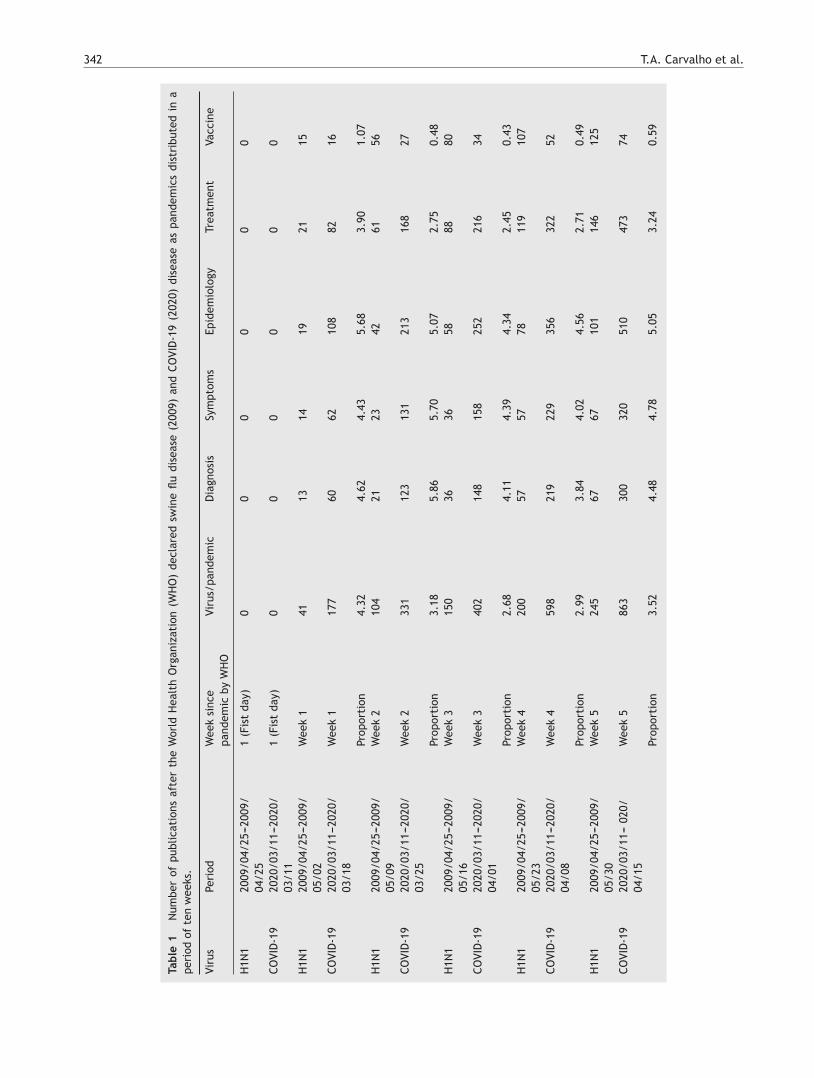
In our study, a comparison was made between both pan-demics regarding the number of publications divided into sixmain themes as follows: (i) the number of articles relatedwith each disease caused by H1N1 virus and SARS-CoV-2virus; (ii) the number of articles related with each diseaseconsidering the theme diagnosis; (iii) the number of articlesrelated with each disease considering the theme symptoms;(iv) the number of articles related with each disease con-sidering the theme epidemiology; (v) the number of articlesrelated with each disease considering the theme treatment;and (vi) the number of articles related with each diseaseconsidering the theme vaccine. Each theme was individuallyanalyzed and no exclusion was carried out using a specifictheme as dominant because each theme cooperated equallyto improve the Scientific knowledge during both pandemics.Moreover, one article can contain information about two (ormore) themes with equal contextualization.
The data search was done using the PubMed (Pub-lic/Publisher MEDLINE) (https://pubmed.ncbi.nlm.nih.gov)using of the following descriptors: (i) (swine flu OR H1N1 ORH1N1 influenza virus OR influenza virus OR H1N1 influenzaOR H1N1/09 OR H1N1 virus OR A(H1N1)pdm09 OR H1N1 fluOR Mexican flu OR influenza A); (ii) (coronavirus disease-19 OR coronavirus disease OR corona virus OR COVID-19 ORCOVID19 OR SARS-CoV-2); (iii) the descriptors of (i) and (ii)were used along with the following descriptors: (iiia) AND(diagnosis); (iiib) AND (symptoms); (iiic) AND (epidemiol-ogy); (iiid) AND (treatment); (iiie) AND (vaccine). The dataextraction was carried out weekly for ten weeks after theWHO declared the swine flu (25th April 2009) and COVID-19(11th March 2020) as pandemic (Table 1 shows the periodsof analysis).
The proportion between the number of published articlesfor COVID-19 and H1N1 pandemics was also set in our data.Only published studies written in English (filter 1) and about
Human species (filter 2) were evaluated. In addition, in thePubMed, the advanced search tool was used to limit the timeto collect the number of studies as previously described andthe terms ‘‘Date - completion’’ were considered to give onlythe information about the studies published as their finalversion. In brief, the number of articles published for bothpandemics and the proportion between them are shown inTable 1.
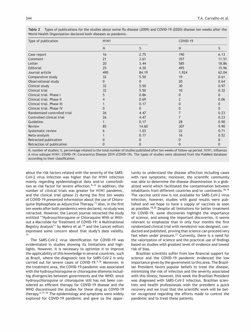
To improve the information about the studies publishedduring both pandemic periods, the number of articles col-lected throughout the ten weeks after the WHO declared theswine flu and COVID-19 diseases as pandemic were catego-rized using the description for article type from PubMed asfollows: case report, comment, letter to the editor, edito-rial, journal article, comparative study, observational study,clinical study, clinical trial (phase I, phase II, phase III orphase IV), randomized controlled trial, controlled clinicaltrial, guidelines, review, systematic review, meta-analysis,retracted publication or retraction of publication (Table 2).The percentage of each type of study was calculated basedon the total number of articles published during each follow-up period.
The number of published articles about each virus or pan-demic showed different numbers of publications during thefirst weeks after the WHO declared the two diseases a pan-demic. The difference for the number of articles betweenpandemics improved because the number of studies aboutCOVID-19 presented a faster increase, achieving four-timesthe number of publications about the swine flu pandemicfrom week 7 to week 10 after the WHO declared the swine fluand COVID-19 as pandemic. The same result was found whenthe studies were grouped for diagnosis, symptoms, epidemi-ology and treatment. However, the ‘‘vaccine’’ term wasassociated with a lower number of publications for COVID-19 pandemic when compared with the swine flu pandemic,showing only ∼0.48 of the number of studies during thefirst two weeks after the WHO declared the diseases as pan-demics. However, the number of publications for COVID-19pandemic achieved the mark of ∼0.77 when compared toswine flu pandemic during the week 10.
Science is crucial to promoting knowledge based onevidence and appears as a central pillar during criticalevents such as the two pandemics described in our data.The time lapse between the two events (pandemics) wasonly ten years and differences occurred in the number ofpublications considering the topics concerned. The highernumber of studies published during the second evaluatedperiod, namely COVID-19 pandemic, is evident. However,also noticeable is the higher number of case reports(N = 190), comments (N = 357), editorials (N = 495) and let-ters to the editors (N = 585) during the COVID-19 pandemicwhich increased the number of studies during this pan-demic. These types of papers represent the lowest levelsof the evidence pyramid showing a higher risk of bias,5 also,these types of studies promote the possibility of fast discus-sion and dialogue among specialists, favoring better insightsfor future investigations. Moreover, the number of studiesincluding the need for individual protection equipment andsocial isolation to control the dissemination of the SARS-CoV-2 virus is evident in the literature, highlighting the needof psychological care.5---8 Likewise, the number of reviews,systematic reviews and meta-analysis including information
342 T.A. Carvalho et al.
Tabl
e1
Num
ber
ofpu
blic
atio
nsaf
ter
the
Wor
ldH
ealt
hO
rgan
izat
ion
(WH
O)
decl
ared
swin
efl
udi
seas
e(2
009)
and
COVI
D-1
9(2
020)
dise
ase
aspa
ndem
ics
dist
ribu
ted
ina
peri
odof
ten
wee
ks.
Viru
sPe
riod
Wee
ksi
nce
pand
emic
byW
HO
Viru
s/pa
ndem
icD
iagn
osis
Sym
ptom
sEp
idem
iolo
gyTr
eatm
ent
Vacc
ine
H1N
120
09/0
4/25
---20
09/
04/2
51
(Fis
tda
y)0
00
00
0
COVI
D-1
920
20/0
3/11
---20
20/
03/1
11
(Fis
tda
y)0
00
00
0
H1N
120
09/0
4/25
---20
09/
05/0
2W
eek
141
1314
1921
15
COVI
D-1
920
20/0
3/11
---20
20/
03/1
8W
eek
117
760
6210
882
16
Prop
orti
on4.
324.
624.
435.
683.
901.
07H
1N1
2009
/04/
25---
2009
/05
/09
Wee
k2
104
2123
4261
56
COVI
D-1
920
20/0
3/11
---20
20/
03/2
5W
eek
233
112
313
121
316
827
Prop
orti
on3.
185.
865.
705.
072.
750.
48H
1N1
2009
/04/
25---
2009
/05
/16
Wee
k3
150
3636
5888
80
COVI
D-1
920
20/0
3/11
---20
20/
04/0
1W
eek
340
214
815
825
221
634
Prop
orti
on2.
684.
114.
394.
342.
450.
43H
1N1
2009
/04/
25---
2009
/05
/23
Wee
k4
200
5757
7811
910
7
COVI
D-1
920
20/0
3/11
---20
20/
04/0
8W
eek
459
821
922
935
632
252
Prop
orti
on2.
993.
844.
024.
562.
710.
49H
1N1
2009
/04/
25---
2009
/05
/30
Wee
k5
245
6767
101
146
125
COVI
D-1
920
20/0
3/11
---02
0/04
/15
Wee
k5
863
300
320
510
473
74
Prop
orti
on3.
524.
484.
785.
053.
240.
59

Science and pandemics 343
Tabl
e1
(Con
tinu
ed)
Viru
sPe
riod
Wee
ksi
nce
pand
emic
byW
HO
Viru
s/pa
ndem
icD
iagn
osis
Sym
ptom
sEp
idem
iolo
gyTr
eatm
ent
Vacc
ine
H1N
120
09/0
4/25
---20
09/
06/0
6W
eek
629
380
8212
217
013
9
COVI
D-1
920
20/0
3/11
---20
20/
04/2
2W
eek
61,
099
382
406
633
596
89
Prop
orti
on3.
754.
784.
955.
193.
510.
64H
1N1
2009
/04/
25---
2009
/06
/13
Wee
k7
404
113
117
165
228
193
COVI
D-1
920
20/0
3/11
---20
20/
04/2
9W
eek
71,
512
494
527
827
829
109
Prop
orti
on3.
744.
374.
505.
013.
640.
56H
1N1
2009
/04/
25---
2009
/06
/20
Wee
k8
463
122
127
189
262
218
COVI
D-1
920
20/0
3/11
---20
20/
05/0
6W
eek
81,
989
656
697
1,09
01,
102
138
Prop
orti
on4.
305.
385.
495.
774.
210.
63H
1N1
2009
/04/
25---
2009
/06
/27
Wee
k9
534
141
147
216
307
245
COVI
D-1
920
20/0
3/11
---20
20/
05/1
3W
eek
92,
502
830
879
1,35
51,
388
161
Prop
orti
on4.
695.
895.
986.
274.
520.
66H
1N1
2009
/04/
25---
2009
/07
/04
Wee
k10
582
158
165
240
330
262
COVI
D-1
920
20/0
3/11
---20
20/
05/2
0W
eek
103,
101
1,02
41,
093
1,67
01,
731
203
Prop
orti
on5.
336.
486.
626.
965.
250.
77
Each
them
ew
asin
divi
dual
lyan
alyz
edan
dno
excl
usio
nw
asdo
neus
ing
asp
ecifi
cth
eme
asdo
min
ant.
The
data
sear
chw
asca
rrie
dou
tus
ing
the
PubM
ed(P
ublic
/Pub
lishe
rM
EDLI
NE)
(htt
ps:/
/pub
med
.ncb
i.nl
m.n
ih.g
ov-D
ate
-com
plet
ion)
for:
(i)(
swin
efl
uO
RH
1N1
OR
H1N
1in
flue
nza
viru
sO
Rin
flue
nza
viru
sO
RH
1N1
infl
uenz
aO
RH
1N1/
09O
RH
1N1
viru
sO
RA(
H1N
1)pd
m09
OR
H1N
1fl
uO
RM
exic
anfl
uO
Rin
flue
nza
A);
(ii)
(cor
onav
irus
dise
ase-
19O
Rco
rona
viru
sdi
seas
eO
Rco
rona
viru
sO
RCO
VID
-19
OR
COVI
D19
OR
SARS
-CoV
-2);
(iii)
the
desc
ript
ors
of(i
)an
d(i
i)w
ere
used
alon
gw
ith
the
follo
win
gde
scri
ptor
s:(i
iia)
AND
(dia
gnos
is);
(iiib
)AN
D(s
ympt
oms)
;(i
iic)
AND
(epi
dem
iolo
gy);
(iiid
)AN
D(t
reat
men
t);
(iiie
)AN
D(v
acci
ne).
Dat
aex
trac
tion
was
carr
ied
out
wee
kly
for
ten
wee
ksaf
ter
the
WH
Ode
clar
edth
esw
ine
flu
(25t
hAp
ril
2009
)an
dCO
VID
-19
(11t
hM
arch
2020
)di
seas
esas
pand
emic
.O
nly
publ
ishe
dst
udie
sw
ritt
enin
Engl
ish
(filt
er1)
and
abou
tH
uman
spec
ies
(filt
er2)
wer
eev
alua
ted.
344 T.A. Carvalho et al.
Table 2 Types of publications for the studies about swine flu disease (2009) and COVID-19 (2020) disease ten weeks after theWorld Health Organization declared both diseases as pandemic.
Type of publication H1N1 COVID-19
N % N %
Case report 16 2.75 190 6.13Comment 21 3.61 357 11.51Letter 20 3.44 585 18.86Editorial 25 4.30 495 15.96Journal article 490 84.19 1,924 62.04Comparative study 32 5.50 19 0.61Observational study 0 0 20 0.64Clinical study 32 5.50 30 0.97Clinical trial 32 5.50 10 0.32Clinical trial. Phase I 5 0.86 0 0Clinical trial. Phase II 4 0.69 2 0.06Clinical trial. Phase III 1 0.17 0 0Clinical trial. Phase IV 0 0 0 0Randomized controlled trial 26 4.47 7 0.23Controlled clinical trial 26 4.47 7 0.23Guideline 1 0.17 28 0.90Review 85 14.60 307 9.90Systematic review 6 1.03 22 0.71Meta-analysis 1 0.17 16 0.52Retracted publication 0 0 0 0Retraction of publication 0 0 0 0
N, number of studies; %, percentage related to the total number of studies published after ten weeks of follow-up period. H1N1, InfluenzaA virus subtype H1N1; COVID-19; Coronavirus Disease 2019 (COVID-19). The types of studies were obtained from the PubMed databaseaccording to their classification.
about the risk factors related with the severity of the SARS-CoV-2 virus infection was higher than for H1N1 infectionmainly regarding epidemiological data and/or comorbidi-ties as risk factor for severe affection.9---12 In addition, thenumber of clinical trials was greater for H1N1 pandemic,and the clinical trial (phase 2) during the first ten weeksof COVID-19 presented information about the use of Chloro-quine Diphosphate as Adjunctive Therapy.13 Also, in the firstten weeks after both pandemics were declared, no study wasretracted. However, the Lancet journal retracted the studyentitled ‘‘Hydroxychloroquine or Chloroquine With or With-out a Macrolide for Treatment of COVID-19: A MultinationalRegistry Analysis’’ by Mehra et al.14 and the Lancet editorsexpressed some concern about that study’s data validity.15
The SARS-CoV-2 virus identification for COVID-19 wasevidentiated in studies showing its limitations and high-lights. However, it is necessary to optimize it to improvethe applicability of this knowledge in several countries, suchas Brazil, where the diagnostic test for SARS-CoV-2 is onlycarried out for severe cases of COVID-19.4,16 Moreover, inthe treatment area, the COVID-19 pandemic was associatedwith the hydroxychloroquine or chloroquine dilemma includ-ing divergencies between governments and the WHO; sincehydroxychloroquine or chloroquine still has not been con-sidered an efficient therapy for COVID-19 disease and theWHO discontinued the studies for these drug as COVID-19therapy.4,17---19 The epidemiology and symptoms were widelyexplored for COVID-19 pandemic and gave us the oppor-
tunity to understand the disease affection including caseswith rare symptoms; moreover, the scientific communitywas able to determine the disease dissemination in a glob-alized world which facilitated the contamination betweeninhabitants from different countries and/or continents.18,19
The vaccine until now is not available for SARS-CoV-2 virusinfection, however, studies with good results were pub-lished and we hope to have a supply of vaccines as soonas possible.18,20 Despite all limitations for better treatmentfor COVID-19, some discoveries highlight the importanceof science, and among the important discoveries, it seemsrelevant to emphasize that in only four months the firstrandomized clinical trial with remdezivir was designed, con-ducted and published, proving that science can proceed veryfast when under pressure.21 Currently, there is a need forthe valorization of science and the practical use of findingsbased on studies with greatest level of evidence and lowestrisk of bias.
Brazilian scientists suffer from low financial support forscience and the COVID-19 pandemic evidenced the lowimportance given by the government to this area. The Brazil-ian President favors popular beliefs to treat the disease,minimizing the risk of infection and the severity associatedwith this illness; however, this week the Brazilian Presidentwas diagnosed with SARS-CoV-2 infection. Brazilian scien-tists and health professionals wish the president a quickrecovery and we trust that the scientific work will be bet-ter recognized regarding the efforts made to control thepandemic and to treat these patients.

Science and pandemics 345
In conclusion, science give us the knowledge to dealwith situations such as the pandemics. However, in somecountries, including Brazil, science should be better valuedby the governments and the community. Special attentionshould be paid to the number of published studies hurriedlywith low level of evidence and high risk of bias.22 Readersshould always evaluate the quality of each study and havea critical point of view to put into practice the knowledgeacquired from the publications.
Conflicts of interest
The authors have no conflicts of interest to declare.
References
1. https://www.who.int/emergencies/diseases/novel-coronavirus-2019.[Accessed on 07th July 2020].
2. Secretarias de Saúde do Brasil. https://covid.saude.gov.br.[Accessed on 07th July 2020].
3. https://www.worldometers.info/coronavirus. [Accessed on07th July 2020].
4. Pascarella G, Strumia A, Piliego C, Bruno F, Del BuonoR, Costa F, et al. COVID-19 diagnosis and manage-ment: a comprehensive review. J Intern Med. 2020,http://dx.doi.org/10.1111/joim.13091. Online ahead ofprint.
5. Murad MH, Asi N, Alsawas M, Alahdab F. New evi-dence pyramid. Evid Based Med. 2016;21:125---7,http://dx.doi.org/10.1136/ebmed-2016-110401.
6. Bergman D, Bethell C, Gombojav N, Hassink S, Stange KC.Physical distancing with social connectedness. Ann Fam Med.2020;18:272---7, http://dx.doi.org/10.1370/afm.2538.
7. Malay DS. COVID-19, pandemic, and social dis-tancing. J Foot Ankle Surg. 2020;59:447---8,http://dx.doi.org/10.1053/j.jfas.2020.03.010.
8. Machida M, Nakamura I, Saito R, Nakaya T, HanibuchiT, Takamiya T, et al. Adoption of personal protec-tive measures by ordinary citizens during the COVID-19outbreak in Japan. Int J Infect Dis. 2020;94:139---44,http://dx.doi.org/10.1016/j.ijid.2020.04.014.
9. Alqahtani JS, Oyelade T, Aldhahir AM, Alghamdi SM,Almehmadi M, Alqahtani AS, et al. Prevalence, sever-ity and mortality associated with COPD and smokingin patients with COVID-19: a rapid systematic reviewand meta-analysis. PLoS One. 2020;15:e0233147,http://dx.doi.org/10.1371/journal.pone.0233147.
10. Fu L, Wang B, Yuan T, Chen X, Ao Y, FitzpatrickT, et al. Clinical characteristics of coronavirus dis-ease 2019 (COVID-19) in China: a systematic reviewand meta-analysis. J Infect. 2020;80(6):656---65,http://dx.doi.org/10.1016/j.jinf.2020.03.041.
11. Wang B, Li R, Lu Z, Huang Y. Does comorbidity increasethe risk of patients with COVID-19: evidence from
meta-analysis. Aging (Albany NY). 2020;12:6049---57,http://dx.doi.org/10.18632/aging.103000.
12. Rodriguez-Morales AJ, Cardona-Ospina JA, Gutiérrez-Ocampo E, Villamizar-Pena R, Holguin-Rivera Y,Escalera-Antezana JP, et al. Clinical, laboratory andimaging features of COVID-19: a systematic review andmeta-analysis. Travel Med Infect Dis. 2020;34:101623,http://dx.doi.org/10.1016/j.tmaid.2020.101623.
13. Borba MGS, Val FFA, Sampaio VS, Alexandre MAA,Melo GC, Brito M, et al. Effect of high vs low dosesof chloroquine diphosphate as adjunctive therapy forpatients hospitalized with severe acute respiratorysyndrome coronavirus 2 (SARS-CoV-2) infection: a ran-domized clinical Trial. JAMA Netw Open. 2020;3:e208857,http://dx.doi.org/10.1001/jamanetworkopen.2020.8857.
14. Mehra MR, Desai SS, Ruschitzka F, Patel AN.RETRACTED: hydroxychloroquine or chloroquine withor without a macrolide for treatment of COVID-19: a multinational registry analysis. Lancet. 2020,http://dx.doi.org/10.1016/S0140-6736(20)31180-6. S0140-6736(20)31180-31186.
15. The Lancet Editors. Expression of concern: hydrox-ychloroquine or chloroquine with or without amacrolide for treatment of COVID-19: a multi-national registry analysis. Lancet. 2020;395:e102,http://dx.doi.org/10.1016/S0140-6736(20)31290-3.
16. Marson FAL. One million cases of COVID-19: what havewe learned? Rev Med (São Paulo). 2020;99(2):209---12,http://dx.doi.org/10.11606/issn.1679-9836.v99i2p209-212.
17. Mehra MR, Desai SS, Ruschitzka F, Patel AN. Hydroxychloroquineor chloroquine with or without a macrolide for treatment ofCOVID-19: a multinational registry analysis. Lancet. 2020 [EpubAhead of Print].
18. Ahn DG, Shin HJ, Kim MH, Lee S, Kim HS, Myoung J,et al. Current status of epidemiology, diagnosis, ther-apeutics, and vaccines for novel coronavirus disease2019 (COVID-19). J Microbiol Biotechnol. 2020;30:313---24,http://dx.doi.org/10.4014/jmb.2003.03011.
19. Ge H, Wang X, Yuan X, Xiao G, Wang C, Deng T,et al. The epidemiology and clinical information aboutCOVID-19. Eur J Clin Microbiol Infect Dis. 2020;39:1011---9,http://dx.doi.org/10.1007/s10096-020-03874-z.
20. Chen WH, Strych U, Hotez PJ, Bottazzi ME. The SARS-CoV-2vaccine pipeline: an overview. Curr Trop Med Rep. 2020:1---4,http://dx.doi.org/10.1007/s40475-020-00201-6. Online aheadof print.
21. Wang Y, Zhang D, Du G, Du R, Zhao J, Jin Y,et al. Remdesivir in adults with severe COVID-19: arandomised, double-blind, placebo-controlled, mul-ticentre trial. Lancet. 2020;395(10236):1569---78,http://dx.doi.org/10.1016/S0140-6736(20)31022-9.
22. Brainard J. Scientists are drowning in COVID-19 papers. Cannew tools keep them afloat? Posted in: Scientific Community.doi:https://doi.org/10.1126/science.abc7839.

Pulmonol. 2020;26(6):346---352
www.journalpulmonology.org
REVIEW
Disseminated Bacillus Calmette-Guérin (BCG) infectionwith pulmonary and renal involvement: A rarecomplication of BCG immunotherapy. A case report andnarrative review
Marta Marques ∗, Dolores Vazquez, Susana Sousa, Goncalo Mesquita,Maria Duarte, Rosa Ferreira
Servico de Medicina Interna, Centro Hospitalar Póvoa de Varzim/Vila do Conde, Portugal
Received 12 September 2019; accepted 4 October 2019
KEYWORDSBacillusCalmette-Guérin(BCG)immunotherapy;Disseminated BCGinfection;BCGitis;Nephritis;Pneumonitis;Miliary tuberculosis
Abstract Intravesical Bacillus Calmette-Guérin (BCG) instillation is a mainstay of adjunctivetherapy for superficial bladder cancer that increases length of disease progression-free survival.Although usually well tolerated, moderate to severe local and systemic infectious complicationscan occur with this immunotherapy. Diagnosis is difficult and often based on high clinical suspi-cion since in many cases Mycobacterium bovis is not isolated. Treatment is not fully standardizedbut the combination of anti-tuberculosis drugs and corticosteroids is advocated in severe cases.
The authors present an unusual case of a severe infectious complication following intravesicalBCG instillation with pulmonary and kidney involvement. Prompt anti-tuberculosis treatmentassociated to corticosteroid resulted in a marked clinical and radiological improvement, sup-porting the diagnosis of disseminated BCG infection. Based on this, the authors aimed to reviewthe literature on this exceptional complication of this immunotherapy.© 2019 Sociedade Portuguesa de Pneumologia. Published by Elsevier Espana, S.L.U. This is anopen access article under the CC BY-NC-ND license (http://creativecommons.org/licenses/by-nc-nd/4.0/).
Introduction
Intravesical instillation of bacillus Calmette-Guérin (BCG),a live attenuated strain of Mycobacterium bovis, is an effi-
∗ Corresponding author.E-mail address: [email protected] (M. Marques).
cient immunotherapy for superficial bladder cancer, which isassociated with increased length of disease progression-freesurvival.1 The BCG therapy is usually well tolerated, withsome minor side effects reported, generally self-limitingwithout specific treatment.2
Serious adverse events are uncommon (less than 5%) andare usually related to disseminated BCG infection (<1%).3 Itsdiagnosis is challenging since at least half of the cases do not
https://doi.org/10.1016/j.pulmoe.2019.10.0012531-0437/© 2019 Sociedade Portuguesa de Pneumologia. Published by Elsevier Espana, S.L.U. This is an open access article under the CCBY-NC-ND license (http://creativecommons.org/licenses/by-nc-nd/4.0/).

Disseminated Bacillus Calmette-Guerin infection with pulmonary and renal involvement 347
Figure 1 Chest radiograph on admission showing a diffuseopacities and nodules.
yield positive microbiological results.1,4 Sepsis is its most ful-minant manifestation but infection of several organs, suchas the liver or the bone, can arise. Pulmonary involvementis very rare (0.3---0.7% of the cases), usually presenting asinterstitial pneumonitis or miliary dissemination.5
In this manuscript, we report a rare case of a patient witha disseminated infection with pulmonary involvement afterBCG intravesical instillation, and conduct a narrative reviewof the literature on this topic.
Case report
A 69-year-old man, with history of hypertension and dys-lipidemia, was diagnosed with high-grade bladder urothelialcarcinoma (pT1). A transurethral resection of the bladderwas performed, followed by mensal BCG intravesical instilla-tion in the last 18 months, without major adverse reactionsexcept flu-like symptoms lasting less than 48 h after eachdose. Two days after the last instillation, he presentedmacroscopic hematuria and severe fatigue, sweating andpersistent non-productive cough. Eight days later, due tothe persistence of symptoms and the appearance of dysp-nea and fever, he went to the emergency department. Hehad no recent sick or tuberculosis contacts. On admission, hewas febrile (38.3 ◦C), hypotensive (89/40 mmHg), in respira-tory distress with inspiratory crackles in both lungs. Arterialblood gas analysis showed, in ambient air, hypoxemia[partial pressure of oxygen 63 mmHg) and respiratory alka-losis. Laboratory tests revealed mild anemia (hemoglobin11.9 g/dL) with normal white blood cell and platelet count,increased levels of C-reactive protein (10.9 mg/dL) and ery-throcyte sedimentation rate (57 mm/h), acute kidney injury[serum creatinine (sCr) 2.35 mg/dl], hematuria, leukocy-turia and proteinuria. Serological tests were negative forhepatitis B and C and human immunodeficiency virus.Markers of immune-mediated disease (antinuclear, antineu-trophil cytoplasmic, antiglomerular basement membraneand anti-streptolysin O antibodies) were all negative. Serumimmunoglobulins and complement levels were normal. Thechest radiography showed a bilateral reticulonodular infil-trate (Fig. 1) and the thoracic computed tomography (CT)
Figure 2 Thoracic CT scan showing pulmonary nodules in amiliary pattern.
demonstrated a diffuse pulmonary micronodulation, in amiliary pattern (Fig. 2). Renal ultrasonography revealednormal-sized kidneys with a reduced corticomedullary dif-ferentiation. Specimens for microbiological exam werecollected and empirical broad-spectrum antibiotic treat-ment was started. On the fifth day of treatment, both feverand acute respiratory failure persisted, lung opacities wereunmodified and renal function worsened (sCr 3.52 mg/dL).Multiple cultures of sputum, urine and blood were negativefor both bacteria and mycobacteria, including polymerasechain reaction (PCR). A bronchoscopy with bronchoalveolarlavage (BAL) was performed which revealed a prevalenceof lymphocytes (48.2%) and an elevated CD4/CD8 ratio(11), absence of malignant cells and bacteriological andmycobacteriological exams were negative. Due to the lackof clinical and imagiological improvement, the diagnosis ofdisseminated BCG infection was suggested and empiric anti-tuberculosis treatment (ATT) [isoniazid 300 mg, rifampin600 mg and ethambutol 1200 mg; once daily] associated witha corticosteroid (prednisolone 60 mg once daily) was startedand, few days later, patient’s clinical condition improved,with sustained apyrexia, resolution of acute respiratory fail-ure and slow but steady recovery of renal function. He wasdischarged in good general condition and continued ATT for6 months. Steroids were discontinued tapering down thedosage for 5 months. A high-resolution CT scan of thorax,performed 6 months after discharge, showed almost com-plete resolution of the lung opacities with a reduction inthe number and size of bilateral pulmonary nodules (Fig. 3)and kidney function was fully recovered (sCr 0.86 mg/dL).BCG immunotherapy was discontinued. No sign of relapsewas observed after one year of follow-up.
Discussion
BCG immunotherapy provides the best risk-benefit ther-apy in patients with high-risk superficial bladder cancerand isthe most commonly used and effective adjunctivetherapy for this malignancy.2 Several studies have shownthat intravesical BCG is more effective than intravesicalchemotherapy.6 BCG infusions promote an immunomodu-

348 M. Marques et al.
Figure 3 The high-resolution CT scan of thorax performed6 months after discharge showed a marked reduction in thenumber and size of bilateral pulmonary nodules.
latory process characterized by a local immune activationinvolving a multitude of cytokines and local migration ofpolymorphonuclear cells, ultimately leading to the deathof tumor cells.7 Due to this inflammatory challenge, minorside effects are commonly reported including genitourinarysymptoms, such as cystitis (91%), macroscopic hematuria(1%), bladder contracture (0,2%), granulomatous prostatitis(0,9%), epididymo-orchitis (0,4%) and urethral obstruction(0,3%). Low-grade fever, malaise and chills following instil-lation are also common and usually self-limiting within a fewhours or days.3,7 Serious adverse events were reported in lessthan 5% of over 2600 patients treated with BCG instillations,which include pneumonitis or hepatitis (0,7%), cytopenia(0,1%) and sepsis (0,4%).3 Similar results were described ina pooled analysis of case reports8 and in a recent retrospec-tive review9 that revealed an incidence of BCG infection of4.3% and 1.3%, respectively.
Disseminated BCG infection, also referred as “BCGitis”,should be suspected in any patient who develops moderate-to-severe genitourinary or systemic symptoms, following ≥1instillation of intravesical BCG, responding to ATT and withno alternative diagnosis. Microbiologic or histopathologicevidence of mycobacterial infection is not necessary fordiagnosis.8
In our patient, dissemination of M. bovis devel-oped following an intravesical BCG instillation. Althoughmycobacterial identification was not achieved, a presump-tive diagnosis of disseminated BCG was justified by theclinical presentation consistent with active tuberculosis,associated with radiological findings consistent with pul-monary spread of M. bovis. Additionally, the evidence ofmacroscopic hematuria and acute kidney injury suggestedan initial regional effect with generalized “BCGitis” coursedwith pneumonitis.
The lack of mycobacterial identification is not unusualsince, as pointed out by a recent review, serological testsand cultures are negative in approximately 60% of cases,being useful especially for differential diagnosis.8 The exclu-sion of an alternative diagnosis and the prompt response totreatment should be considered the cornerstones of BCGitisdiagnosis, as we observed in our patient.
Risk factors for disseminated infection include host char-acteristics, such as the extent of bladder mucosal damageand immunodeficiency, which are considered more impor-tant than therapeutic regimen features.8,10 Our patientreceived 18 BCG instillations without major complicationsbut this repeated exposure may have predisposed him tosubsequent dissemination, although dissemination can occurat any time in the course of treatment.8 Interestingly, allreported cases of disseminated BCG infection treated withBCG immunotherapy occurred in males, suggesting a poten-tial influence of sex hormones in this susceptibility.11
Pulmonary involvement is very rare and can presentas hypersensitivity or mycobacterial pneumonia. Hypersen-sitivity is usually characterized by an interstitial patternin chest x-ray and lymphocytosis on BAL.12 Miliary pat-tern nodules in lung parenchyma is rarely reported in theliterature. An extensive and systematic review of the lit-erature, retrieved 26 published cases11---36 (Table 1) whichprovide a comprehensive clinical, radiological and micro-biological information of miliary tuberculosis caused bydisseminated BCG infection in adults treated with intrav-esical BCG immunotherapy.
The diagnosis can be confirmed by the presenceof epithelio-giganto-cellular granulomas with or withoutcaseous necrosis in bronchial or pulmonary biopsy. How-ever, since they are present in only 40% of biopsies, inmost of the cases they do not contribute to the diagnosis.37
As previously reported, caseous necrosis is usually absent(Table 1). Pulmonary parenchymal changes also includefibrosis of the lung and lymphocytic alveolar infiltrates con-sistent with hypersensitivity alveolitis. BAL cellular profileusually shows lymphocytosis with an elevated CD4/CD8 ratioconsistent with a T helper alveolitis,38 as we documented inour patient.
Although its pathogenesis is not fully understood, someauthors believe that BCGitis is caused by hypersensitivityreactions and the granulomas are due to type 4 hypersensi-tivity reaction and not infection, supported by the negativeresults of cultures and of serological and molecular tests,along with successful response to steroids.12,22---24,35,36 On theother hand, some case reports support the theory of an ongo-ing active BCG infection demonstrated by the presence ofviable bacilli inside the lesions16 or detection of mycobacte-rial genome by Polymerase Chain Reaction assays,11,26,27,30,34
that require specific treatment. This supports the absenceof a standardized treatment for BCG complications.
In our case, it can be argued that patient’s symptomswere due to hypersensitivity reaction, because of negativeculture results from BAL as well as absence of acid-fastbacilli on direct microscopy, and also the BAL cellularprofiles revealed lymphocytosis with an elevated CD4/CD8ratio. However, due to the persistence and severity of clini-cal symptoms with respiratory failure, antituberculous wereadded to steroids with clinical improvement within 3 days.The speed of improvement suggests the more important roleof hypersensitivity rather than infection.
Until now, trials done have not been good enough todetermine the most effective treatment. In addition, insome case reports, simple discontinuation of BCG alone wassufficient. Venn et al. illustrated a case who had febrile ill-ness for more than one month with persisting pulmonarynodules, that improved without any treatment. The authors

Disseminated Bacillus Calmette-Guerin infection with pulmonary and renal involvement 349
Tabl
e1
Publ
ishe
dca
ses
ofm
iliar
ytu
berc
ulos
isca
used
bydi
ssem
inat
edBC
Gin
fect
ion
inad
ults
trea
ted
wit
hin
trav
esic
alBC
Gim
mun
othe
rapy
.
Pati
ent,
refe
renc
eJo
urna
l,st
udy
desi
gnAg
e,ge
nder
Sym
ptom
sN
o.of
BCG
inst
illat
ions
Path
olog
ical
findi
ngs
Mic
robi
olog
ical
diag
nosi
sTr
eatm
ent;
dura
tion
(mon
ths)
Out
com
e
1,G
upta
etal
.,19
8813
Ches
t;CR
78,
MFe
ver,
coug
h10
NCG
Alln
egat
ive
INH
,RI
F,EM
B(9
)Re
solu
tion
2,Ke
sten
etal
.,19
9014
Thor
ax;
CR67
,M
Feve
r,w
eigh
tlo
ss4
NCG
Alln
egat
ive
INH
,RI
FN
SRe
solu
tion
3,Ba
loir
aVi
llar
etal
.,19
9215
Med
Clin
(Bar
c);
CR78
,M
Feve
r,w
eigh
tlo
ss,h
aem
opty
sis
5CG
Acid
fast
stai
nIN
H,
RIF,
PZA
(6)
Reso
luti
on
4,M
cPar
land
etal
.,19
9216
Am
Rev
Resp
irD
is;
CR69
,M
Feve
r,ch
ills,
coug
h6
NCG
Cult
ure
M.b
ovis
(lun
gbi
opsi
es)
INH
,RI
F,PZ
A,ST
M,
ster
oids
;(1
2)Re
solu
tion
5,Sm
ith
etal
.,19
9317
Canc
er;
CR69
,M
Asym
ptom
atic
6CG
Alln
egat
ive
INH
,RI
F,PZ
AN
SD
eath
6,Pa
laye
wet
al.,
1993
18Ch
est;
CR57
,M
Wei
ght
loss
,co
ugh,
dysp
nea
10N
CGAc
idfa
stst
ain
INH
,RI
F,EM
Bst
eroi
ds;
(9)
Reso
luti
on
7,Ja
smer
etal
.,19
9619
Radi
olog
y;CR
73,
MFe
ver,
wei
ght
loss
9CG
Alln
egat
ive
INH
,RI
F,EM
B(9
)Re
solu
tion
8,Pa
rede
set
al.,
1996
20A
rch
Bron
cone
umol
;CR
67,
MFe
ver,
chill
s14
CGAl
lneg
ativ
eIN
H,
RIF,
EMB
NS
Dea
th
9,Ia
ntor
noet
al.,
1998
21J
Uro
;CR
79,
MFe
ver,
chill
s,co
ugh,
dysp
nea
8N
CGAl
lneg
ativ
eIN
H,
RIF,
EMB
PZA;
NS
Reso
luti
on
10,
Rabe
etal
.,19
9922
AJR
Am
JRo
entg
enol
;CR
60,
MFe
ver,
mal
aise
,ar
thra
lgia
s16
NCG
Alln
egat
ive
INH
,RI
F,EM
Bst
eroi
ds;
(3)
Reso
luti
on
11,
Elka
bani
etal
.,20
0023
Canc
erCo
ntro
l;CR
67,
MFe
ver,
wea
knes
s,dy
spne
a16
ND
Alln
egat
ive
INH
,RI
F,LE
Vst
eroi
ds;
(12)
Reso
luti
on
12,
Audi
gier
etal
.,20
0024
Rev
Mal
Resp
ir;
CR69
,M
Feve
r,dy
spne
a5
NCG
Alln
egat
ive
INH
,RI
Fst
eroi
ds;
(9)
Reso
luti
on
13,
Mig
non
etal
.,20
0225
JRa
diol
;CR
76,
MFe
ver
10N
DAl
lneg
ativ
eIN
H,
RIF,
EMB
(6)
Reso
luti
on
14,
Tosc
ano
etal
.,20
0326
Eur
JCl
inM
icro
biol
Infe
ctD
is;
CR
72,
MFe
ver,
wei
ght
loss
,co
ugh
6N
SPC
RM
.tu
berc
ulos
isco
mpl
ex
INH
,RI
F,EM
Bst
eroi
ds;
(12)
Reso
luti
on
15,
Cast
illo
etal
.,20
0627
Med
Inte
nsiv
a;CR
75,
MFe
ver,
dysp
nea
NS
Dif
fuse
infl
amm
ator
sign
s
PCR
M.
tube
rcul
osis
com
plex
(BAL
)
INH
,RI
F,EM
Bst
eroi
ds;
NS
Dea
th
16,
Nad
asy
etal
.,20
0828
Sout
hM
edJ;
CR62
,M
Feve
r,m
alai
se,
coug
h3
CGAc
idfa
stst
ain
INH
,RI
F,LE
Vst
eroi
ds;
(6)
Reso
luti
on
17,
Coba
sPa
zet
al.,
2010
29A
rch
Bron
cone
umol
;CR
62,
MAs
ympt
omat
ic6
NCG
Alln
egat
ive
INH
,RI
F,EM
B(6
)Re
solu
tion

350 M. Marques et al.
Tabl
e1
(Con
tinu
ed)
Pati
ent,
refe
renc
eJo
urna
l,st
udy
desi
gnAg
e,ge
nder
Sym
ptom
sN
o.of
BCG
inst
illat
ions
Path
olog
ical
findi
ngs
Mic
robi
olog
ical
diag
nosi
sTr
eatm
ent;
dura
tion
(mon
ths)
Out
com
e
18,
Colm
ener
oet
al.,
2012
30D
iagn
Mic
robi
olIn
fect
Dis
;CR
and
Revi
ew
56,
MFe
ver,
chill
s,m
alai
se,
coug
h9
ND
PCR
M.
tube
rcul
osis
com
plex
(BAL
)
INH
,RI
Fst
eroi
ds;
(9)
Reso
luti
on
19,
Cara
mor
iet
al.,
2013
11M
onal
diA
rch
Ches
tD
is;
CRan
dRe
view
66,
MFe
ver,
dysp
nea
18N
CGPC
RM
.tu
berc
ulos
isco
mpl
ex(B
AL)
INH
,RI
F,EM
BPZ
A,st
eroi
ds;
(9)
Reso
luti
on
20,
Choi
CRet
al.,
2014
31BM
JCa
seRe
p;CR
51,
MW
eakn
ess,
wei
ght
loss
5N
CGAl
lneg
ativ
eIN
H,
RIF,
EMB
NS
Reso
luti
on
21,
Venn
etal
.,20
1432
BMJ
Case
Rep;
CR74
,M
Feve
r,ni
ght
swea
ts,
anor
exia
7N
DAl
lneg
ativ
eN
one
Reso
luti
on
22,
Rosa
tiet
al.,
2016
12U
rolo
gia;
CR63
,M
Feve
r,co
ugh
6N
CGAl
lneg
ativ
eIN
H,
RIF,
EMB
ster
oids
;(9
)Re
solu
tion
23,
Smit
het
al.,
2016
33BM
JCa
seRe
p;CR
69,
MCo
ugh,
nigh
tsw
eats
,w
eigh
tlo
ss
NS
ND
Alln
egat
ive
INH
,RI
F,EM
B(9
)Re
solu
tion
24,
Calle
ris
etal
.,20
1734
LeIn
fezi
oni
inM
edic
ina;
CR73
,M
Feve
r,w
eakn
ess,
nigh
tsw
eats
NS
ND
PCR
M.
tube
rcul
osis
(spu
tum
)
INH
,RI
F,EM
BN
SRe
solu
tion
25,
Kabu
raki
etal
.,20
1735
Inte
rnM
ed;
CR62
,M
Feve
r,fa
tigu
e8
NCG
Alln
egat
ive
INH
,RI
F,EM
Bst
eroi
ds;
(9)
Reso
luti
on
26,
Clér
igo
etal
.,20
1936
Act
aM
edPo
rt;
CR67
,M
Feve
r6
NCG
Alln
egat
ive
INH
,RI
F,EM
Bst
eroi
ds;
(12)
Reso
luti
on
Abbr
evia
tion
s:BA
LBro
ncho
alve
olar
cult
ures
;CG
Case
atin
ggr
anul
oma;
CRca
sere
port
;EM
BEth
ambu
tol;
INH
Ison
iazi
d;LE
VLev
oflox
acin
;M
mal
e;N
CGN
onca
seat
ing
gran
ulom
a;N
SNot
spec
ified
;N
DN
otdo
ne;
PCRp
olym
eras
ech
ain
reac
tion
;PZ
APyr
azin
amid
e;RI
FRif
ampi
n;ST
MSt
rept
omyc
in.
[Obt
aine
dw
ith
the
data
from
Refs
.10---
33

Disseminated Bacillus Calmette-Guerin infection with pulmonary and renal involvement 351
suggested that some pulmonary reactions, with mild initialpresentation, may resolve spontaneously, whether due tohypersensitivity or natural clearance of BCG.32
Nevertheless, in disseminated BCG infection, the combi-nation of antimycobacterial therapy and corticosteroids issuggested.3 M. bovis is inherently resistant to pyrazinamideand cycloserine, so a regimen that includes isoniazid andrifampin for 6 months with ethambutol for 2 months can beused. When liver toxicity is a concern, regimens includingethambutol, fluoroquinolones and amikacin have been pro-posed. The corticosteroid adjuvant therapy is particularlyimportant where there is extensive miliary involvement andrespiratory failure, but until now, no standardized regimenhas been recommended.8 A retrospective review of eightcases revealed improvement with just steroid treatmentin patients with clinical signs of hypersensitivity, includingpneumonitis and hepatitis.39
Since no routine prophylaxis protocol has demonstratedlong-term efficacy for systemic BCG infection prevention,32
most authors consider a previous systemic BCG infection acontraindication to restarting immunotherapy.9
The prognosis seems to be favourable in most patientswhen an appropriate therapy is started. Pérez-Jacoiste Asínet al.8 observed a 5,4% attributable mortality, generally dueto respiratory, liver and multiple organ failure.
This case highlights the importance of recognizing dis-seminated BCG infection as a potential complication inpatients with previous BCG exposure, but more studies arenecessary not only to fully understand its pathogenesis butalso to standardize the diagnosis and treatment.
References
1. Lamm DL. Efficacy and safety of Bacille Calmette-Guérinimmunotherapy in superficial bladder cancer. Clin Infect Dis.2000;31(3):S86---90.
2. Rischmann P, Desgrandchamps F, Malavaud B, Chopin DK.BCG intravesical instillations: recommendations for side-effectsmanagement. Eur Urol. 2000;37(1):33---6.
3. Lamm DL, van der Meijden PM, Morales A, Brosman SA, CatalonaWJ, Herr HW, et al. Incidence and treatment of complicationsof bacillus Calmette-Guerin intravesical therapy in superficialbladder cancer. J Urol. 1992;147:596---600.
4. Dammert P, Boujaoude Z, Rafferty W, Kass J. Fever of unknownorigin and pancytopenia caused by culture-proven delayedonset disseminated bacillus Calmette-Guérin (BCG) infectionafter intravesical instillation. BMJ Case Rep. 2013:10---2.
5. Calleris G, Marra G, Corcione S, Oderda M, Cardellino C, Audag-notto S, et al. Miliary pulmonary infection after BCG intravesicalinstillation: a rare, misdiagnosed and mistreated complication.Infez Med. 2017:366---70.
6. Shelley MD, Wilt TJ, Court J, Coles B, Kynaston H, MasonMD. Intravesical bacillus Calmette---Guerin is superior to mit-omycin C in reducing tumour recurrence in high-risk superficialbladder cancer: a meta-analysis of randomized trials. BJU Int.2004;93:485---90.
7. Thompson DB, Siref LE, Feloney MP, Hauke RJ, Agrawal DK.Immunological basis in the pathogenesis and treatment of blad-der cancer. Expert Rev Clin Immunol. 2015;11(2):265---79.
8. Pérez-Jacoiste Asín MA, Fernández-Ruiz M, López-MedranoF, Lumbreras C, Tejido A, San Juan R, et al. BacillusCalmette-Gérrin (BCG) infection following intravesical BCGadministration as adjunctive therapy for bladder cancer.Medicine. 2014;93:236---54.
9. González-Del Vecchio M, Ruíz-Serrano MJ, Gijón P, Sánchez-Somolinos M, Egea V, García de Viedma D, et al. Differencesbetween a probable and proven BCG infection following intrav-esical instillations: 16 years experience in a tertiary carehospital. Diagn Microbiol Infect Dis. 2016;85(3):338---43.
10. Brausi M, Oddens J, Sylvester R, Bono A, van de Beek C, vanAndelr G, et al. Side effects of bacillus Calmette-Guérin (BCG)in the treatment of intermediate- and high-risk Ta, T1 papillarycarcinoma of the bladder: results of the EORTC genito-urinarycancers group randomized phase 3 study comparing one-thirddose with full dose and 1 year with 3 years of maintenanceBCG. Eur Urol. 2014;65(1):69---76.
11. Caramori G, Artioli D, Ferrara G, Cazzuffi R, Pasquini C, LibanoreM, et al. Severe pneumonia after intravesical BCG instillation ina patient with invasive bladder cancer: case report and litera-ture review. Monaldi Arch Chest Dis. 2013;79(1):44---8.
12. Rosati Y, Fabiani A, Taccari T, Ranaldi R, Mammana G, Tubaldi A.Intravesical BCG therapy as cause of miliary pulmonary tuber-culosis. Urologia. 2016;83(1):49---53.
13. Gupta RC, Lavengood R Jr, Smith JP. Miliary tuberculosisdue to intravesical bacillus Calmette---Guérin therapy. Chest.1988;94:1296---8.
14. Kesten S, Title L, Mullen B, Grossman R. Pulmonary diseasefollowing intravesical BCG treatment. Thorax. 1990;45:709---10.
15. Baloira Villar A, Agüero Balbín R, Bustamante Ruiz A, JiménezGómez A. Miliary tuberculosis secondary to intravesical instilla-tion of Calmette---Guerin bacilli. Med Clin (Barc). 1992;99:158.
16. McParland C, Cotton DJ, Gowda KS, Hoeppner VH, Martin WT,Weckworth PF. Miliary Mycobacterium bovis induced by intrav-esical bacille Calmette---Guérin immunotherapy. Am Rev RespirDis. 1992;146:1330---3.
17. Smith RL, Alexander RF, Aranda CP. Pulmonary granulo-mata: a complication of intravesical administration of bacillusCalmette---Guérin for superficial bladder carcinoma. Cancer.1993;71:1846---7.
18. Palayew M, Briedis D, Libman M, Michel RP, Levy RD.Disseminated infection after intravesical BCG immunother-apy: detection of organisms in pulmonary tissue. Chest.1993;104:307---9.
19. Jasmer RM, McCowin MJ, Webb WR. Miliary lung disease afterintravesical bacillus Calmette---Guérin immunotherapy. Radiol-ogy. 1996;201:43---4.
20. Paredes Arranz C, Frutos Arribas JF, Calleja Escudero J, BlancoCabero M, Campo Matías F, Velasco MC. Miliary dissemina-tion with pulmonary involvement secondary to intravesicalimmunotherapy with Calmette---Guérin bacillus. Arch Bron-coneumol. 1996;32:100---2.
21. Iantorno R, Nicolai M, Storto ML, Ciccotosto C, Cipollone G, Mas-troprimiano G, et al. Miliary tuberculosis of the lung in a patienttreated with bacillus Calmette---Guerin for superficial bladdercancer. J Urol. 1998;159:1639---40.
22. Rabe J, Neff KW, Lehmann KJ, Mechtersheimer U, Georgi M.Miliary tuberculosis after intravesical bacille Calmette---Guérinimmunotherapy for carcinoma of the bladder. AJR Am JRoentgenol. 1999;172:748---50.
23. Elkabani M, Greene J, Vincent A, Vanhook S, Sandim R. Dis-seminated mycobacterium bovis after intravesicular bacilluscalmette-Guérin treatments for bladder cancer. Cancer Con-trol. 2000;7(5):476---81.
24. Audigier C, Nesme P, Perol M, Guerin JC. Rare pulmonarycomplication in intravesical BCG treatment. Rev Mal Respir.2000;17:679---81.
25. Mignon F, Chevrière A, Mesurolle B, El Hajam M, Morel H,Lacombe P. Miliary induced by intravesical BCG immunother-apy for carcinoma of the bladder: CT findings. J Radiol.2002;83:368---71.
26. Toscano Rico M, Machado J, Cardoso O, Salvado C, RodriguesP, Proenca R. Severe disseminated tuberculosis after intravesi-

352 M. Marques et al.
cal instillation of Bacillus Calmette-Guérin. Eur J Clin MicrobiolInfect Dis. 2003;22(7):447---8.
27. Del Castillo Duran Y, Santos Bodí F, Castander Serentill D,Jubert Montaperto P, Espinosa Valencia P, Rabassó Sole C.Tuberculosis miliar in a patient treated with intravesical instil-lations of bacillus Calmette-Guérin. Med Intensiva. 2006;30(3):116---9.
28. Nadasy KA, Patel RS, Emmett M, Murillo RA, Tribble MA, BlackRD, et al. Four cases of disseminated Mycobacterium bovisinfection following intravesical BCG instillation for treatmentof bladder carcinoma. South Med J. 2008;101(1):91---5.
29. Cobas Paz A, García Tejedor JL, González Pineiro A, Fernández-Villar A. Miliary tuberculosis due to BCG in an asymptomaticpatient: initial onset or a condition not yet described? ArchBronconeumol. 2010;46:394---5.
30. Colmenero JD, Sanjuan-Jimenez R, Ramos B, Morata P. Mil-iary pulmonary tuberculosis following intravesical BCG therapy:case report and literature review. Diagn Microbiol Infect Dis.2012;74:70---2.
31. Choi CHR, Lee SO, Smith G. Subclinical miliary Mycobac-terium bovis following BCG immunotherapy for transitional cellcarcinoma of the bladder. BMJ Case Rep. 2014;2014, bcr-3013-201202.
32. Venn RM, Sharma N. Resolution without treatment of granulo-matous pneumonitis due to intravesical BCG for bladder cancer.BMJ Case Rep. 2014:1---3.
33. Smith DM. BCG-osis following intravesical BCG treatment lead-ing to miliary pulmonary nodules, penile granulomas anda mycotic aortic aneurysm. BMJ Case Rep. 2016, bcr-2016-215635.
34. Calleris G, Marra G, Corcione S, Oderda M, Cardellino C, Audag-notto S, et al. Miliary pulmonary infection after BCG intravesicalinstillation: a rare, misdiagnosed and mistreated complication.Le Infezioni in Medicina. 2017;(4):366---70.
35. Kaburaki K, Sugino K, Sekiya M, Takai Y, Shibuya K, Homma S.Milliary tuberculosis that developed after intravesical BacillusCalmette-Guerin therapy. Intern Med. 2017;(56):1563---7.
36. Clérigo V, Castro A, Mourato T, Gomes C. A rare case of granulo-matous Pneumonitis due to intravesical BCG for bladder cancer.Acta Med Port. 2019;32(4):316---20.
37. Attou R, Albrich T, Kadou J, Redant S, Honoré PM, De BelsD. Favorable outcome in a patient with systemic BCGitis afterintra-bladder instillation of Calmette-Guerin bacillus highlight-ing the importance of making the correct diagnosis in this rareform of sepsis. J Transl Int Med. 2019;7(1):34---8.
38. Namen AM, Grosvenor AR, Chin R, Daybell D, Adair N, WoodruffRD, et al. Pulmonary infiltrates after intravesical BacilleCalmette-Guerin: two cases and review of the literature. ClinPulmon Med. 2001;8(3):177---9.
39. Gonzalez OY, Musher DM, Brar I, et al. Spectrum ofBacille Calmette-Guérin (BCG) infection after intravesical BCGimmunotherapy. Clin Infect Dis. 2003;36(2):140---8.

Pulmonol. 2020;26(6):353---362
www.journalpulmonology.org
ORIGINAL ARTICLE
Host factors associated to false negative andindeterminate results in an interferon-� release assayin patients with active tuberculosis
J.A. Santosa,b,∗, R. Duartec,d, C. Nunesa
a NOVA National School of Public Health, Public Health Research Centre, Universidade NOVA de Lisboa, Portugal Avenida PadreCruz, 1600-560 Lisboa, Portugalb National Health Institute Dr. Ricardo Jorge, Portugal Avenida Padre Cruz, 1600-560 Lisboa, Portugalc Chest Disease Center, Vila Nova de Gaia, Portugal Rua Conselheiro Veloso da Cruz, 4400-092 Vila Nova de Gaia, Portugald Faculdade de Medicina da Universidade do Porto, Portugal Alameda Prof. Hernâni Monteiro, 4200-319 Porto, Portugal
Received 19 September 2019; accepted 11 November 2019Available online 13 December 2019
KEYWORDSTuberculosis;Interferon-gammarelease tests;Tuberculin test;Public health;Latent tuberculosis;Preventive medicine
AbstractIntroduction: Information on host factors that contribute to false negative and indeterminateresults in interferon-� release assays (IGRA) are critical to improve the usefulness of these testsin the fight against tuberculosis (TB) epidemics.
The aim of this study was to estimate and compare the sensitivity of an IGRA and the tuber-culin skin test (TST), independently and as a combined approach, in patients with TB and toidentify risk factors associated with false negative and indeterminate IGRA results.Methods: Retrospective cohort study of all active TB notifications with an IGRA result (n = 1230),from 2008 to 2015. 68.0 % (n = 727) of these patients had a TST result interpreted using a 5 mm(TST-5 mm) and 10 mm (TST-10 mm) cutoff. Sensitivity was determined for both tests.
Logistic regression analysis was used to evaluate the association of sociodemographic andclinical factors to the risk of false negative or indeterminate IGRA results.Results: IGRA, TST-5 mm and TST-10 mm were positive in 82.4 %, 84.5 % and 78.4 % of thepatients that performed both tests. When used combined, IGRA/TST-5 mm sensitivity was 91.7% and IGRA/TST-10 mm sensitivity was 90.6 %. Age≥65 years, alcohol abuse and pulmonary TBwere predictive factors for indeterminate results. Inflammatory diseases and pulmonary TBwere statistically associated with false negative IGRA results.
∗ Corresponding author at: Epidemiology and Statistics Department, Escola Nacional de Saúde Pública, Universidade NOVA de Lisboa,Avenida Padre Cruz, 1600-560 Lisboa, Portugal.
E-mail addresses: [email protected] (J.A. Santos), [email protected] (R. Duarte), [email protected] (C. Nunes).
https://doi.org/10.1016/j.pulmoe.2019.11.0012531-0437/© 2019 Sociedade Portuguesa de Pneumologia. Published by Elsevier Espana, S.L.U. This is an open access article under the CCBY-NC-ND license (http://creativecommons.org/licenses/by-nc-nd/4.0/).
354 J.A. Santos et al.
Conclusion: Inflammatory diseases and pulmonary TB were identified as factors for false neg-ative IGRA results. Our results indicate that the use of both tests in a combined approach,especially in specific risk groups of the population, could increase the sensitivity of the screeningprocess and accelerate the achievement of the WHO End TB Strategy goals.© 2019 Sociedade Portuguesa de Pneumologia. Published by Elsevier Espana, S.L.U. This is anopen access article under the CC BY-NC-ND license (http://creativecommons.org/licenses/by-nc-nd/4.0/).
Introduction
The timely identification of latent tuberculosis infection(LTBI) cases and implementation of preventive treatmentcan substantially decrease the risk of people developingtuberculosis (TB) disease from a latent infection and, conse-quently, reduce TB-related morbidity and mortality, reducetransmission, and the burden of the disease.1
The existing screening tests are indirect methods thatonly provide immunological evidence of host sensitization toMycobacterium tuberculosis antigens, do not differentiateaccurately between LTBI and active TB or provide evidenceregarding the stage and potential progression from infectionto disease.2
WHO guidelines recommend the use of the tuberculinskin test (TST) and the interferon-� release assays (IGRA)as screening tests.
IGRAs measure T-cell release of interferon-� in responseto stimulation by two strongly immunogenic but highlyspecific M. tuberculosis complex antigens (ESAT-6 and CFP-10). Two IGRAs are commercially available, QuantiFERON-TBGold assay (Qiagen) and the T-SPOT.TB assay (OxfordImmunotec), both detecting the release of interferon-� inresponse to the specific M. tuberculosis antigens throughdifferent methods.
IGRAs have higher specificity for M. tuberculosis than theTST, as the antigens used are not encoded in the genomesof any of the BCG vaccine strains or most species of non-tuberculous mycobacteria.3 Furthermore, these tests havelogistic advantages when compared to TST (e.g. require onlyone visit), have positive and negative controls (e.g. identi-fication of potential cases of anergy), and no boost effectwhen repeated.2
However, these assays are more expensive and requirelaboratory infrastructure. Also, T-cell assays such as IGRAare susceptible to variability by different factors at mul-tiple levels, including assay manufacturing, preanalyticalprocessing (e.g. blood volume, sample transport tempera-ture, incubation time) or analytical testing.3
IGRAs have shown reduced sensitivity in immunocompro-mised patients, especially in those with a severe immunedepression, such as patients with diabetes4 or HIV infection.5
IGRAs have also presented a lower sensitivity in young(<5years of age) or immunocompromised children.6,7
Another drawback of IGRA testing is the occurrence ofindeterminate results, which may result from an insufficientimmune response to the positive control or a high level ofresponse in the negative control,8 with previous studies pre-senting rates of indeterminate IGRA results ranging between2 % and 11 %.8
In order to improve the usefulness of the IGRAs as a diag-nostic aid for detection of LTBI, it is essential to betterunderstand which factors are associated with false negativeand indeterminate IGRA results.
Since there is no diagnostic gold standard for LTBI to helpevaluate the IGRAs, sensitivity can be estimated using surro-gate reference standards such as IGRA results from patientswith a definitive TB diagnosis and, through collected epi-demiological data, determine factors that may be associatedwith false negative results.
The objective of this study was to analyse the perfor-mance of an IGRA test in patients with active TB. The specificobjectives were to: (i) determine and compare the sensitiv-ity of IGRA and TST tests; (ii) search for risk factors thatcould be linked with indeterminate IGRA results; and (iii)ascertain risk factors that could be associated with falsenegative IGRA results.
Material and methods
Study design and data source
Retrospective cohort study centered on data from activeTB cases notified in the Portuguese National TuberculosisSurveillance System (SVIG-TB), from 2008 to 2015. This offi-cial database is generated by direct compulsory reporting byhealth care providers and the data from this official systemof notification is complemented and updated during TB casesfollow-up. Patients notified in the database are diagnosedbased on laboratory confirmed TB (through identification ofM. tuberculosis by microscopy, cultural and/or molecularmethods) and/or based on clinical and radiological findingsand a favorable TB treatment response.
For this study ethics committee approval or informedconsent was not required, as all patient’s informationretrieved from the SVIG-TB database was fully anonymized.
Study data
The study included all patients notified in the SVIG-TBdatabase with an IGRA test result. The IGRA test used inthe patients enrolled in the study was the QuantiFERON-TBGold In-Tube (Qiagen).
The clinical and sociodemographic variables analyzedwere sex, age group, comorbidities (chronic renal failurein dialysis, oncologic diseases, inflammatory diseases, HIVinfection, chronic obstructive pulmonary disease [COPD] anddiabetes), substance abuse (alcohol and drug abuse) andsite of disease (pulmonary or extrapulmonary TB). IGRA and

Host factors associated to false negative and indeterminate results in interferon-gamma release assays 355
TST results were the dependent variable (event: negativeresults).
Indeterminate IGRA results were assessed by dividing theresults into determinate (positive and negative results) andindeterminate (event).
Statistical analysis
TST induration measurements were interpreted using twocutoff points, ≥5 mm (TST-5 mm) and ≥10 mm (TST-10 mm),9
and converted to positive or negative results.Results from patients that underwent both diagnostic
tests were used to calculate the sensitivity with 95 % confi-dence interval for each test separately and in combination(IGRA/TST-5 mm or IGRA/TST-10 mm). Sensitivity outcomeswere compared using McNemar’s test.10
Bivariate and multivariate logistic regression analysis10
was used to determine the variables that were significan-tly associated with the occurrence of indeterminate andfalse negative IGRA results. Multivariate logistic regressionanalysis with forward stepwise (Likelihood Ratio) method,logit function (entry-0.05; removal-0.10), included variablesthat were significant in bivariate analyses. Odds ratio werereported with 95 % confidence intervals (95 %CI). Statisti-cal analyses were performed using IBM® SPSS® Statistics forWindows, version 23 (IBM Corp., N.Y., USA).
Results
There were 1230 patients notified with active TB in the SVIG-TB database (2008---2015) with an IGRA test, 857 patients(69.7 %) had a positive test result, 212 (17.2 %) had a neg-ative result and 161 (13.1 %) had an indeterminate result.Among the 1069 patients with a determinate IGRA result,727 (68.0 %) also had a TST result.
The mean (±standard deviation) age of the patientswith a determinate IGRA result (n = 1069, 86.9 %) was 47.9(±20.0), ranging from less than 1 to 91 years. Portugal wasthe main country of origin (n = 931; 87.1 %), followed byAngola (n = 31, 2.9 %), Cabo Verde (n = 23, 2.2 %), Guinea-Bissau (n = 19, 1.8 %) and Mozambique (n = 12, 1.1 %).
Diabetes mellitus was the most common comorbidity andchronic renal failure was the least frequent.
IGRA and TST sensitivity
Considering the patients who performed IGRA and TST assays(n = 727), overall sensitivity was 82.4 %, 84.6 % and 78.4 %for IGRA, TST-5 mm and TST-10 mm, correspondingly. Theseresults indicate that 128 (17.6 %), 112 (15.4 %) and 157 (21.6%) patients with a diagnosis of active TB had a false negativeresult with IGRA, TST-5 mm and TST-10 mm tests, respec-tively. Table 1 presents the sensitivity results for IGRA andTST for total of patients and stratified by sociodemographicand clinical variables.
Comparing the sensitivity of the tests separately withthe combined sensitivity of IGRA/TST-5 mm and IGRA/TST-10 mm (91.7 % and 90.6 %, respectively), the latter wasconsistently higher and this difference was statistically sig-nificant (p < 0.001) --- Fig. 1.
Risk factors for IGRA indeterminate results
Of the 161 patients with an indeterminate result, the major-ity were male (67.7 %) with a mean age of 56.0 (±19.4) thatranged from 5 to 92 years, and more than half were ≥50years old (57.1 %).
The proportion of indeterminate results increases as theage increases, with patients over 80 years old presentingthe highest proportion of indeterminate results (34.5 %) andpatients under 20 years old presenting the lowest proportionof indeterminate results (2.9 %).
Being older than 70 years, alcohol abuse and pulmonaryTB were independent predictive factors with a statisticallysignificant association to higher probability of indeterminateIGRA results in TB patients (Table 2).
Risk factors for IGRA false negative results
Among the risk factors analyzed through logistic regression(Table 3), two presented a statistically significant associa-tion with an increased risk of obtaining false negative IGRAresults - inflammatory diseases and pulmonary TB.
Discussion
The objective of this study was to analyse the performanceof an IGRA test in patients with active TB which involvedestablishing and comparing the sensitivity of IGRA and TSTtests.
IGRA sensitivity results are heterogeneous across the lit-erature, ranging from 60 %11 to nearly 100 %12 with moststudies presenting an sensitivity above 70 %, as demon-strated by three different meta-analyzes that obtained apooled sensitivity of 81.0 %,13 80.0 %14 and 84.2 %.15 Inour study, IGRA overall sensitivity (82.4 %) was close to thepooled sensitivity of these meta-analyzes. Nonetheless, 17.6% of the results were false negative, demonstrating thatalthough these tests can facilitate diagnostic decisions, neg-ative results should not be used alone to exclude an M.tuberculosis infection but interpreted in conjunction withother clinical findings and diagnoses.
Our TST sensitivity was higher than the pooled sensitiv-ity obtained in three different meta-analysis - 66 %,15 70 %13
and 77 %16 - but comparable to a US TB surveillance studythat included almost 65 000 culture-confirmed TB patients,in which 84.2 % of the patients had a TST≥5 mm and 81.6% a TST ≥ 10 mm.17 Despite the differences in sensitivityobtained between IGRA and TST-5 mm or TST-10 mm, onlythe difference between IGRA and TST-10 mm was statisti-cally significant (p = 0.021), which indicates that IGRA andTST-5 mm had an similar performance in the identificationof cases of infection and presented a better performancethan TST-10 mm.
By combining the results of the two tests, the sensitivityincreased to more than 90.0 %, and the difference betweenthese results and those obtained by the tests separately wasstatistically significant (p < 0.001). Choi et al. presented sim-ilar sensitivity results, obtaining 85.3 % (TST), 70.3 % (IGRA)and 93.3 % (IGRA/TST).4 This increase in sensitivity suggeststhat the combined use of the two tests could promote theidentification of more cases of infection than if used sepa-

356 J.A. Santos et al.
Tabl
e1
IGRA
and
TST
sens
itiv
ity
(5m
man
d10
mm
cut-
offs
)ac
cord
ing
tose
x,ag
egr
oup,
com
orbi
diti
es(c
hron
icre
nal
failu
re,
onco
logi
cdi
seas
e,in
flam
mat
ory
dise
ase,
HIV
,di
abet
esan
dCO
PD),
subs
tanc
eab
use
(alc
ohol
ordr
ugab
use)
and
site
ofdi
seas
e(p
ulm
onar
yor
extr
apul
mon
ary)
inpa
tien
tssu
bmit
ted
tobo
thas
says
(n=
727)
.
Vari
able
sPa
tien
tsIG
RATS
T-
5m
mTS
T-
10m
m
n/N
(%)
Posi
tive
(n)
Sens
itiv
ity
%(I
C95
%)Po
siti
ve(n
)Se
nsit
ivit
y%
(IC
95%)
Posi
tive
(n)
Sens
itiv
ity
%(I
C95
%)
Tota
l72
759
982
.4(7
9.5-
--84.
9)61
584
.6(8
2.0-
--87.
2)57
078
.4(7
5.4-
--81.
4)Se
xM
ale
362/
727
(49.
8)30
484
.0(7
9.9-
--87.
4)31
185
.2(8
1.6-
--88.
8)29
291
.2(8
8.3-
--94.
1)Fe
mal
e36
5/72
7(5
0.2)
295
80.8
(76.
5---8
4.5)
304
84.0
(80.
2---8
7.8)
278
92.3
(89.
5---9
5.0)
Age
grou
p≤1
548
/727
(6.6
)37
77.1
(63.
5---8
6.7)
3572
.9(6
0.3-
--85.
5)31
85.4
(75.
4---9
5.4)
16---
6452
3/72
7(7
1.9)
431
82.4
(78.
9---8
5.4)
458
87.6
(84.
7---9
0.4)
423
92.5
(90.
3---9
5.0)
≥65
156/
727
(21.
5)13
184
.0(7
7.4-
--88.
9)12
278
.2(7
1.7-
--84.
7)11
691
.0(8
6.5-
--95.
5)Co
mor
bidi
ties
Chro
nic
rena
lfai
lure
5/72
7(0
.7)
480
.0(3
7.6-
--96.
4)3
60.0
(14.
7---9
4.7)
380
.0(2
8.4-
--99.
5)O
ncol
ogic
dise
ases
36/7
27(5
.0)
2363
.9(4
7.6-
--77.
5)23
63.9
(48.
2---7
9.6)
2072
.2(5
7.6-
--86.
9)In
flam
mat
ory
dise
ases
32/7
27(4
.4)
2062
.5(4
5.3-
--77.
1)24
75.0
(60.
0---9
0.0)
2175
.0(6
0.0-
--90.
0)H
IVin
fect
ion
25/6
32(3
.4)
1872
.0(5
2.4-
--85.
7)16
64.0
(44.
5---7
9.8)
1576
.0(5
6.6-
--88.
5)D
iabe
tes
41/7
27(5
.6)
3278
.0(6
3.3-
--88.
0)31
75.6
(62.
5---8
8.8)
2780
.5(6
8.4-
--92.
6)CO
PD16
/727
(2.2
)15
93.8
(71.
7---9
8.9)
1610
0(7
9.4-
--100
)15
100
(79.
4---1
00)
Subs
tanc
eab
use
Alco
hola
buse
31/7
06(4
.3)
2890
.3(7
5.1-
--96.
7)27
87.1
(75.
3---9
8.9)
2593
.5(7
8.6-
--99.
2)D
rug
abus
e32
/709
(4.4
)27
84.4
(68.
3---9
3.1)
3196
.9(8
3.8-
--99.
9)31
100
(89.
1---1
00)
Site
ofdi
seas
ePT
B34
3/72
7(4
7.2)
275
80.2
(75.
6---8
4.1)
292
85.1
(81.
4---8
8.9)
270
91.5
(88.
6---9
4.5)
ETB
384/
727
(52.
8)32
484
.4(8
0.4-
--87.
7)32
384
.1(8
0.5-
--87.
8)30
091
.9(8
9.2-
--94.
7)
PTB
=Pu
lmon
ary
tube
rcul
osis
;ET
B=
Extr
apul
mon
ary
tube
rcul
osis
.

Host factors associated to false negative and indeterminate results in interferon-gamma release assays 357
0.0
20.0
40.0
60.0
80.0
100.0
Total Male Female ≤15 16 – 64 ≥ 65 CRFOncol.
diseasesInflam.
diseasesHIV Diabetes COPD
Alcoholabuse
Drugabuse
PTB ETB
IGRA 82.4 84.0 80.8 77.1 82.4 84.0 80.0 63.9 62.5 72.0 78.0 93.8 90.3 84.4 80.2 84.4
TST-5mm 84.6 85.2 84.0 72.9 87.6 78.2 60.0 63.9 75.0 64.0 75.6 100.0 87.1 96.9 85.1 84.1
TST-10mm 78.4 80.0 76.8 64.6 80.9 74.4 60.0 55.6 65.6 60.0 65.9 93.8 80.6 96.9 78.7 78.1
IGRA + TST-5mm 91.7 91.2 92.3 85.4 92.5 91.0 80.0 72.2 75.0 76.0 80.5 100.0 93.5 100.0 91.5 91.9
IGRA + TST-10mm 90.6 91.0 90.3 85.4 91.2 90.4 80.0 69.4 71.9 76.0 80.5 100.0 93.5 100.0 90.7 90.6
Sen
sitiv
y (%
)
n=727
IGRA TST-5mm TST-10mm IGRA + TST-5mm IGRA + TST-10mm
Figure 1 Sensitivity of IGRA and TST when used separately and when used together (IGRA/TST-5 mm and IGRA/TST-10 mm) inpatients with both test results (n = 727). Sensitivity was assessed according to sex, age group (≤15, 16-64, ≥65 years), comorbidities(chronic renal failure [CRF], oncologic disease, inflammatory disease, HIV infection, diabetes and COPD), substance abuse (alcoholor drug abuse) and site of disease (pulmonary TB [PTB] or extrapulmonary TB [ETB]).
rately and in substitution of one another. The combined usecould enhance the detection of LTBI cases among vulnera-ble populations,18 such as homeless people, drug or alcoholabusers, prisoners and people living with HIV. This approachcould be especially helpful in low-incidence countries, as themajority of TB cases occur due to the progression from LTBIto TB disease.19 Furthermore, these vulnerable populationsare frequently associated with individual and contextual fac-tors, as presented by Zão et al,20 that contribute to patientand healthcare system delay in the diagnosis and treatmentof TB. A false negative result could contribute to longerdelays, thus perpetuating the spread of the disease.
Indeterminate results represent a considerable problemfor clinical management, since they imply the lack of clearinformation about the patient’s TB infection status.
Unlike other studies,21,22 our results do not show old ageas a risk factor for false negative IGRA results. However, weobserved that older age was a predictive factor for inde-terminate results, which was consistent with the findings ofother studies.21,23 Several studies analyzed the associationbetween age and indeterminate IGRA results and concludedthat indeterminate results are significantly more commonin children23---25 and adolescents23 and in the elderly.23,26 Ourresults confirm part of their conclusions, with patients aged>80 years being seven times more likely to have an indeter-minate IGRA result. In addition, when we stratified patientsin ten-year age groups, we observed that the proportion ofindeterminate results increased as the age increases, withpatients over 80 years old presenting the highest proportionof indeterminate results, results shared with Kobashi et al.26
In our study younger patients did not present any associationwith false negative or indeterminate IGRA results.
Our study also found an association between alcoholabuse and indeterminate IGRA results in which patients withan alcohol problem were four times more likely to have anindeterminate outcome. Both acute and chronic alcohol usehave profound regulatory effects on the immune system,27
which possibly impairs the patient’s ability to respond cor-rectly to the IGRA, resulting in an indeterminate result.Because alcohol dependence was based on self-report anddaily alcohol intake was not determined, further studieswith a more detailed information on alcohol consumptionare needed to better understand this potential association.
From the studies available involving IGRA, PTB patientsand ETB patients, the test sensitivity remains similar in bothforms of TB, as shown by Di et al.28 (89.7 % vs 79.7 %), Jiet al.29 (89.7 % vs 87.6 %), Azghay et al.30 (78.9 % vs 87.8 %)and in our study (80.2 % vs 84.4 %). However, pulmonary TBwas presented as a predictive factor for indeterminate andfalse negative results, in which these patients were almostthree times more likely to have an indeterminate outcomeand nearly twice as likely to have a false negative resultcompared to extrapulmonary TB (ETB) patients.
Clinical manifestations of ETB may vary from an acute ordisseminated disease, such as meningitis or miliary TB, to achronic localized infection with an insidious onset and slowprogression, such as lymph node or osteoarticular TB.31,32 Intwo analyses of risk factors of false-negative IGRA results,only meningitis TB from the eight forms of ETB in one studyand 6 forms in the other was significantly associated withfalse negative results, an acute clinical manifestation.28,33
Thus, the fact that most ETB manifestations have a slowerprogression to severe disease, possibly allows patients tomaintain a functional immune system and consequently

358 J.A. Santos et al.
Tabl
e2
Soci
odem
ogra
phic
and
clin
ical
char
acte
rist
ics
of16
1pa
tien
tsw
ith
inde
term
inat
eIG
RAre
sult
san
dth
eir
asso
ciat
ion
wit
hth
eoc
curr
ence
ofth
ein
d ete
rmin
ate
test
resu
lts
inpa
tien
tsw
ith
acti
veTB
.
Vari
able
s(r
efer
ence
clas
s)In
dete
rmin
ate
IGRA
Crud
eO
dds
Rati
oAd
just
edO
dds
rati
o(b
yse
xan
dag
e)Fi
nalM
odel
(FR)
n(%
)O
R(9
5%C
I)p
OR
(95
%CI)
pO
R(9
5%C
I)P
Sex
(Fem
ale)
Mal
e10
9(6
7.7)
1.94
(1.3
7---2
.76)
<0.0
011.
95(1
.37-
--2.8
)<0
.001
Age
grou
p(4
1---5
0)
<10
1(0
.6)
0.23
(0.0
3---1
.75)
0.15
50.
23(0
.03-
--1.7
9)0.
161
11-2
02
(1.2
)0.
24(0
.06-
--1.0
5)0.
059
0.23
(0.0
5---1
.01)
0.06
121
-30
12(7
.5)
0.83
(0.4
0---1
.71)
0.61
60.
88(0
.43-
--1.8
2)0.
735
31-4
028
(17.
4)1.
36(0
.77-
--2.4
0)0.
296
1.41
(0.7
9---2
.49)
0.24
651
-60
26(1
6.1)
1.10
(0.5
9---2
.03)
0.75
31.
11(0
.60-
--2.0
5)0.
741
61-7
021
(13.
0)1.
26(0
.63-
--2.1
2)0.
639
1.23
(0.6
7---2
.26)
0.50
871
-80
30(1
8.6)
1.79
(1.0
1---3
.15)
0.04
51.
83(1
.03-
--3.2
4)0.
039
2.52
(1.1
6---5
.48)
0.02
0>
8019
(11.
8)4.
24(2
.13-
--8.4
5)<0
.001
4.21
(2.1
---8.
44)
<0.0
017.
22(2
.83-
18.4
)<0
.001
Com
orbi
diti
es(N
o)Ch
roni
cre
nalf
ailu
re1
(0.6
)0.
51(0
.07-
--3.9
1)0.
515
0.34
(0.0
4---2
.76)
0.31
3O
ncol
ogic
dise
ases
13(8
.1)
1.45
(0.7
8---2
.71)
0.24
11.
06(0
.55-
--2.0
3)0.
873
Infl
amm
ator
ydi
seas
es5
(3.1
)0.
67(0
.26-
--1.7
0)0.
397
0.63
(0.2
4---1
.64)
0.34
0H
IVin
fect
ion
14(8
.7)
1.79
(0.9
7---3
.33)
0.06
11.
76(0
.91-
--3.4
)0.
092
Dia
bete
s16
(9.9
)1.
79(1
.01-
--3.1
9)0.
047
1.59
(0.8
7---2
.95)
0.13
4CO
PD9
(5.6
)2.
69(1
.22-
--5.9
3)0.
014
1.81
(0.7
9---4
.15)
0.15
9Su
bsta
nce
abus
e(N
o)Al
coho
labu
se27
(16.
8)4.
15(2
.52-
--6.8
3)<0
.001
4.07
(2.3
6---7
.01)
<0.0
014.
17(2
.23-
--7.8
1)<0
.001
Dru
gab
use
15(9
.3)
2.52
(1.3
7---4
.64)
0.00
32.
75(1
.41-
--5.3
5)0.
003
Site
ofdi
seas
e(E
TB)
PTB
116
(72.
0)/4
5(2
8.0)
3.15
(2.1
9---4
.54)
<0.0
013.
544
(2.4
3---5
.18)
<0.0
012.
85(1
.79-
--4.5
3)<0
.001
OR
=O
dds
rati
o;CI
=co
nfide
nce
inte
rval
;PT
B=
Pulm
onar
ytu
berc
ulos
is;
ETB
=Ex
trap
ulm
onar
ytu
berc
ulos
is.
FR-
Forw
ard
step
wis
e(L
R).

Host factors associated to false negative and indeterminate results in interferon-gamma release assays 359
Tabl
e3
Asso
ciat
ion
betw
een
fals
ene
gati
veIG
RAre
sult
san
dso
ciod
emog
raph
ican
dcl
inic
alva
riab
les
amon
gpa
tien
tsw
ith
noti
fied
TB(n
=10
69).
Vari
able
s(r
efer
ence
clas
s)Pa
tien
tsCr
ude
Odd
sRa
tio
Adju
sted
Odd
sra
tio
(by
sex
and
age)
Fina
lMod
el(F
R)
n/N
(%)
OR
(95
%CI)
pO
R(9
5%C
I)p
OR
(95
%CI)
p
Sex
(Fem
ale)
Mal
e55
5/10
69(5
1.9)
0.87
(0.6
4---1
.17)
0.35
20.
86(0
.63-
--1.1
6)0.
325
Age
grou
p(4
1---5
0)
<10
35/1
069
(3.3
)1.
34(0
.60-
--2.9
9)0.
472
1.34
(0.6
0---2
.98)
0.47
611
-20
66/1
069
(6.2
)0.
75(0
.37-
--1.5
1)0.
413
0.75
(0.3
7---1
.52)
0.42
821
-30
116/
1069
(10.
9)0.
97(0
.56-
--1.6
7)0.
909
0.96
(0.5
5---1
.65)
0.87
231
-40
166/
1069
(15.
5)0.
83(0
.51-
--1.3
7)0.
471
0.82
(0.4
9---1
.36)
0.44
551
-60
209/
1069
(19.
6)0.
62(0
.36-
--1.0
7)0.
088
0.62
(0.3
6---1
.07)
0.08
461
-70
153/
1069
(14.
3)0.
66(0
.38-
--1.1
2)0.
122
0.65
(0.3
8---1
.11)
0.11
171
-80
153/
1069
(14.
3)0.
84(0
.49-
--1.4
3)0.
516
0.83
(0.4
9---1
.41)
0.49
3>
8013
5/10
69(1
2.6)
0.81
(0.3
3---1
.96)
0.64
0.82
(0.3
4---1
.99)
0.66
Com
orbi
diti
es(N
o)Ch
roni
cre
nalf
ailu
re13
/106
9(1
.2)
0.73
(0.1
6-3.
33)
0.68
70.
85(0
.18-
3.92
)0.
835
Onc
olog
icdi
seas
es61
/106
9(5
.7)
1.61
1(0
.90-
--2.8
8)0.
108
1.64
(0.9
0---2
.97)
0.10
5In
flam
mat
ory
dise
ases
49/1
069
(4.6
)2.
25(1
.22-
--4.1
3)0.
009
2.40
(1.2
9---4
.46)
0.00
62.
04(1
.04-
--4.0
1)0.
039
HIV
infe
ctio
n59
/931
(5.5
)1.
22(0
.66-
--2.2
8)0.
528
1.18
(0.6
2---2
.24)
0.62
Dia
bete
s62
/106
9(5
.8)
0.77
(0.3
8---1
.53)
0.45
20.
89(0
.44-
--1.8
4)0.
767
COPD
23/1
069
(2.2
)0.
85(0
.29-
--2.5
2)0.
767
0.93
(0.3
1---2
.79)
0.89
2Su
bsta
nce
abus
e(N
o)Al
coho
labu
se57
/102
9(5
.3)
0.65
(0.3
0-1.
39)
0.26
30.
69(0
.32-
1.53
)0.
371
Dru
gab
use
47/1
042
(4.4
)0.
96(0
.46-
--2.0
1)0.
904
0.93
(0.4
3---2
.0)
0.85
Site
ofdi
seas
e(E
TB)
PTB
481/
1069
(45.
0)1.
71(1
.26-
--2.3
1)0.
001
1.67
(1.2
2---2
.29)
0.00
11.
69(1
.22-
--2.3
5)0.
002
OR
=O
dds
rati
o;CI
=co
nfide
nce
inte
rval
;PT
B=
Pulm
onar
ytu
berc
ulos
is;
ETB
=Ex
trap
ulm
onar
ytu
berc
ulos
is.
360 J.A. Santos et al.
functional T-cells which, when presented with TB antigens,will respond with the production of interferon-�. Anotherreason for these results may lie in the severity of pulmonaryTB cases. Different studies have shown that severe pul-monary disease can be associated with immune suppressionwhich may lead to reduced interferon-� production.34,35 Dur-ing TB progression, the natural cytokine balance is alteredwhile the bacterial load increases, potentially influencingthe performance of IGRA tests resulting in a reductionin M. tuberculosis specific immune responses, especiallyinterferon-� production.36,37
Considering the existence of highly heterogeneous clin-ical manifestations associated to the fact that most formsof ETB do not contribute to the transmission of TB, and alsoconsidering that research in ETB and their influence in theimmune response is scarce, it is imperative to develop fur-ther studies in order to better understand the performanceof the test in the different clinical manifestations of TB.
We observed that inflammatory diseases were an inde-pendent predictive factor for negative IGRA results inpatients with active TB. With the growing use of bio-logic therapies for inflammatory rheumatic diseases and theincreased risk of TB associated38 with their use, our resultsshow that the use of IGRA alone may not be sufficient toeliminate the possibility of an M. tuberculosis infection. Inview of the high risk of TB in these patients and the pos-sibility of false negative results, a dual testing strategy ofIGRA and TST could be more effective for LTBI diagnosis, asother studies involving patients with inflammatory diseasesconcluded.39---41
In our study, five of the 49 patients with inflammatorydiseases presented an indeterminate IGRA result but no sig-nificant association was found. Some studies have identifiedan association,39,42,43 with the corticosteroids used in thetreatment of these diseases being the common predictivefactor for indeterminate results. No treatment data wasavailable in our study.
Our study has limitations. T-cell assays like IGRA are sus-ceptible to variability by different factors associated withassay manufacturing, pre-analytical processing or analyticaltesting.3 However, despite their importance we were unableto determine the impact of these factors on the IGRA resultsused in the study. Due to the absence of healthy individu-als in our study population, it was not possible to estimatethe specificity for both immunologic tests, TST and IGRA.The presence of variables based on patient self-report (e.g.alcohol abuse, drug abuse) may have led to a less accu-rate estimate of the impact of these variables. The use ofactive TB patients as a proxy to assess the sensitivity of IGRAand TST has the disadvantage that patients with active TBmay present immunosuppression due to the disease itself,thus obtaining a different response than with patients witha latent infection or recent infection.
On the other hand, our analysis of the performance ofthe IGRA test was comprehensive. It involved a consider-able number of patients with different risk factors, whichallowed observation of the performance of IGRA in differentcontexts, contributing to a better understanding of how touse this test in populations with certain risk factors. In addi-tion, the comparison with the other screening method forLTBI available (TST) provided additional information on how
these tests can be used in order to enhance the detectionof cases of infection.
Conclusion
In our study we identified three factors associated with theoccurrence of indetermined IGRA results - age over 70 years,alcohol abuse and pulmonary TB.
Inflammatory diseases and pulmonary TB were indepen-dent risk factors for false negative IGRA results in patientswith active TB.
Given the impact that treatments of inflammatory dis-eases have on the development of active TB, our resultssuggest a careful interpretation of a negative result, sincethere is a significant risk of false negative results.
The fact that the IGRA appears to perform well in patientswith ETB (less indeterminate and false negative results thanin patients with pulmonary TB), makes it a valuable diag-nostic tool in these cases.
Funding
Fundacão para a Ciência e Tecnologia provided financialsupport to conduct this research [Grant: PTDC/SAU-PUB/31346/2017] but had no involvement in the researchitself.
Declarations of interest
None.
Appendix A. Supplementary data
Supplementary material related to this article can be found,in the online version, at doi:https://doi.org/10.1016/j.pulmoe.2019.11.001.
References
1. Getahun H, Matteelli A, Abubakar I, Abdel Aziz M, Baddeley A,Barreira D, et al. Management of latent Mycobacterium tuber-culosis infection: WHO guidelines for low tuberculosis burdencountries. Eur Respir J. 2015;46(6):1563---76.
2. Matteelli A, Sulis G, Capone S, D’Ambrosio L, Migliori GB,Getahun H. Tuberculosis elimination and the challenge of latenttuberculosis. Press Medicale. 2017;46(2):e13---21.
3. Pai M, Denkinger C, Kik SV, Rangaka M, Zwerling A, OxladeO, et al. Gamma interferon release assays for detectionof Mycobacterium tuberculosis infection. Clin Microbiol Rev.2014;27(1):3---20.
4. Choi JC, Jarlsberg LG, Grinsdale JA, Osmond DH, Higashi J,Hopewell PC, et al. Reduced sensitivity of the QuantiFERON®
test in diabetic patients with smear-negative tuberculosis. IntJ Tuberc Lung Dis. 2015;19(5):582---8.
5. Metcalfe JZ, Everett CK, Steingart KR, Cattamanchi A, HuangL, Hopewell PC, et al. Interferon-� release assays for activepulmonary tuberculosis diagnosis in adults in low-and middle-income countries: Systematic review and meta-analysis. J InfectDis. 2011;204 SUPPL. 4.
6. Sollai S, Galli L, de Martino M, Chiappini E. Systematic reviewand meta-analysis on the utility of Interferon-gamma release
Host factors associated to false negative and indeterminate results in interferon-gamma release assays 361
assays for the diagnosis of Mycobacterium tuberculosis infec-tion in children: a 2013 update. BMC Infect Dis. 2014;14 Suppl1(Suppl 1):S6.
7. Mandalakas AM, Detjen AK, Hesseling AC, Benedetti A, MenziesD. Interferon-gamma release assays and childhood tuberculo-sis: Systematic review and meta-analysis. Int J Tuberc Lung Dis.2011;15(8):1018---32.
8. Sharninghausen JC, Shapiro AE, Koelle DM, Kim HN. RiskFactors for Indeterminate Outcome on Interferon GammaRelease Assay in Non-US-Born Persons Screened for LatentTuberculosis Infection. Open Forum Infect Dis. 2018;5(8):1---8.
9. American Thoracic Society, CDC. Targeted tuberculin testingand treatment of latent tuberculosis infection. Am J Respir CritCare Med. 2000;161:221---47.
10. Altman D. Practical statistics for medical research. first editLondon: CRC Press; 1991.
11. Dewan PK, Grinsdale J, Kawamura LM. Low Sensitivity of aWhole-Blood Interferon-� Release Assay for Detection of ActiveTuberculosis. Clin Infect Dis. 2007;44(1):69---73.
12. Takasaki J, Manabe T, Morino E, Muto Y, Hashimoto M, IikuraM, et al. Sensitivity and specificity of QuantiFERON-TB GoldPlus compared with QuantiFERON-TB Gold In-Tube and T-SPOT.TB on active tuberculosis in Japan. J Infect Chemother.2018;24(3):188---92.
13. Diel R, Loddenkemper R, Nienhaus A. Evidence-Based Compar-ison of Commercial Interferon-� Release Assays for DetectingActive TB. Chest. 2010;137(April (4)):952---68.
14. Sester M, Sotgiu G, Lange C, Giehl C, Girardi E, Migliori GB,et al. Interferon-� release assays for the diagnosis of activetuberculosis: A systematic review and meta-analysis. Eur RespirJ. 2011;37(1):100---11.
15. Lu P, Chen X, Zhu L, Yang H. Interferon-gamma release assaysfor the diagnosis of tuberculosis: a systematic review and meta-analysis. Lung. 2016;194(June 2 (3)):447---58.
16. Pai M, Zwerling A, Menzies D. Systematic Review : T-Cell --- basedAssays for the Diagnosis of Latent Tuberculosis Infection : AnUpdate. Ann Intern Med. 2008;149:177---84.
17. Auld SC, Click ES, Heilig CM, Miramontes R, Cain KP, BissonGP, et al. Association Between Tuberculin Skin Test Result andClinical Presentation of Tuberculosis Disease. BMC Infect Dis.2013;13:460.
18. Heuvelings CC, Vries SG, Grobusch MP. Tackling TB in low-incidence countries: improving diagnosis and management invulnerable populations. Int J Infect Dis. 2017;56(March):77---80.
19. Blumberg HM, Ernst JD. The Challenge of Latent TB Infection.JAMA. 2016;316(September 6 (9)):931.
20. Zão I, Ribeiro AI, Apolinário D, Duarte R. Why does it take solong? The reasons behind tuberculosis treatment delay in Por-tugal. Pulmonology. 2019;25(4):215---22.
21. Cho K, Cho E, Kwon S, Im S, Sohn I, Song S, et al. Fac-tors Associated with Indeterminate and False Negative Resultsof QuantiFERON-TB Gold In-Tube Test in Active Tuberculosis.Tuberc Respir Dis (Seoul). 2012;72(5):416.
22. Kwon Y-S, Kim YH, Jeon K, Jeong B-H, Ryu YJ, Choi JC,et al. Factors that Predict Negative Results of QuantiFERON-TBGold In-Tube Test in Patients with Culture-Confirmed Tuber-culosis: A Multicenter Retrospective Cohort Study. PLoS One.2015;10(6):e0129792.
23. Tebruegge M, De Graaf H, Sukhtankar P, Elkington P, MarshallB, Schuster H, et al. Extremes of age are associated with inde-terminate QuantiFERON-TB gold assay results. J Clin Microbiol.2014;52(7):2694---7.
24. Bergamini BM, Losi M, Vaienti F, D’Amico R, Meccugni B,Meacci M, et al. Performance of Commercial Blood Testsfor the Diagnosis of Latent Tuberculosis Infection in Chil-dren and Adolescents. Pediatrics. 2009;123(March 1 (3)):e419---24.
25. Rose MV, Kimaro G, Nissen TN, Kroidl I, Hoelscher M, BygbjergIC, et al. Quantiferon®-TB gold in-tube performance for diag-nosing active tuberculosis in children and adults in a high burdensetting. PLoS One. 2012;7(7):1---10.
26. Kobashi Y, Sugiu T, Mouri K, Obase Y, Miyashita N, Oka M. Indeter-minate results of QuantiFERON TB-2G test performed in routineclinical practice. Eur Respir J. 2009;33(4):812---5.
27. Friedman H, Pross S, Klein TW. Addictive drugs and their rela-tionship with infectious diseases. FEMS Immunol Med Microbiol.2006;47(3):330---42.
28. Di L, Li Y. The risk factor of false-negative and false-positivefor T-SPOT.TB in active tuberculosis. J Clin Lab Anal. 2018;32(2).
29. Ji L, Lou YL, Wu ZX, Jiang JQ, Fan XL, Wang LF, et al.Usefulness of interferon-� release assay for the diagnosis ofsputum smear-negative pulmonary and extra-pulmonary TBin Zhejiang Province. China. Infect Dis Poverty. 2017;6(1):1---5.
30. Azghay M, Bouchaud O, Mechai F, Nicaise P, Fain O, StirnemannJ. Utility of QuantiFERON-TB Gold In-Tube assay in adult, pul-monary and extrapulmonary, active tuberculosis diagnosis. IntJ Infect Dis. 2016;44:25---30.
31. Houston A, Macallan DC. Extrapulmonary tuberculosis. Medicine(Baltimore). 2014;42(January (1)):18---22.
32. Sanches I, Carvalho A, Duarte R. Who are the patientswith extrapulmonary tuberculosis? Rev Port Pneumol.2015;21(2):90---3.
33. Kim YJ, Kang JY, Kim S Il, Chang MS, Kim YR, Park YJ. Pre-dictors for false-negative QuantiFERON-TB Gold assay resultsin patients with extrapulmonary tuberculosis. BMC Infect Dis.2018;18(1):457.
34. Shams H, Wizel B, Weis SE, Samten B, Barnes PF. Contri-bution of CD8(+) T cells to gamma interferon productionin human tuberculosis. Infect Immun. 2001;69(May (5)):3497---501.
35. Sodhi A, Gong J, Silva C, Qian D, Barnes PF. Clinical Correlates ofInterferon Production in Patients with Tuberculosis. Clin InfectDis. 1997;25(3):617---20.
36. Vanham G, Toossi Z, Hirsch CS, Wallis RS, SchwanderSK, Rich EA, et al. Examining a paradox in the patho-genesis of human pulmonary tuberculosis: Immune activa-tion and suppression/anergy. Tuber Lung Dis. 1997;78(3---4):145---58.
37. de Visser V, Sotgiu G, Lange C, Aabye MG, Bakker M, BartalesiF, et al. False-negative interferon-� release assay results inactive tuberculosis: a TBNET study. Eur Respir J. 2015;45(Jan-uary (1)):279---83.
38. Dixon WG, Hyrich KL, Watson KD, Lunt M, Galloway J,Ustianowski A, et al. Drug-specific risk of tuberculosis inpatients with rheumatoid arthritis treated with anti-TNFtherapy: results from the British Society for Rheumatol-ogy Biologics Register. Ann Rheum Dis. 2010;69(March (3)):522---8.
39. Vassilopoulos D, Tsikrika S, Hatzara C, Podia V, Kandili A, Sta-moulis N, et al. Comparison of two gamma interferon releaseassays and tuberculin skin testing for tuberculosis screen-ing in a cohort of patients with rheumatic diseases startinganti-tumor necrosis factor therapy. Clin Vaccine Immunol.2011;18(12):2102---8.
40. So H, Yuen CS, Yip RM. Comparison of a commercial interferon-gamma release assay and tuberculin skin test for the detectionof latent tuberculosis infection in Hong Kong arthritis patientswho are candidates for biologic agents. Hong Kong Med J.2017:1---5.
41. Song G, Bae S, Lee Y. Interferon-gamma release assaysversus tuberculin skin testing in patients with rheumatoidarthritis (Provisional abstract). Int J Rheum Dis. 2013;16:279---83.

362 J.A. Santos et al.
42. Cho H, Kim YW, Suh C-H, Jung J-Y, Um Y-J, Jung J-H, et al.Concordance between the tuberculin skin test and interferongamma release assay (IGRA) for diagnosing latent tuberculo-sis infection in patients with systemic lupus erythematosus andpatient characteristics associated with an indeterminate IGRA.Lupus. 2016;25(Oct 11 (12)):1341---8.
43. Kaur M, Singapura P, Kalakota N, Cruz G, Shukla R, Ahsan S,et al. Factors That Contribute to Indeterminate Results Fromthe QuantiFERON-TB Gold In-Tube Test in Patients With Inflam-matory Bowel Disease. Clin Gastroenterol Hepatol. 2018;16(10),1616-1621.e1.

Pulmonol. 2020;26(6):363---369
www.journalpulmonology.org
ORIGINAL ARTICLE
The addition of a humidifier device to a circuit and itsimpact on home ventilator performance: a bench study
Javier Collada-Carrascoa,∗, Cristina Lamolda-Puyolb, Manel Lujánb,Alba Castano-Menéndeza, Miguel Jiménez-Gómeza, Ana Hernández-Votha,Javier Sayas-Catalána
a Hospital Universitario Doce De Octubre, Madrid, Spainb Hospital Parc Taulí De Sabadell, Barcelona, Spain
Received 26 July 2019; accepted 1 November 2019Available online 26 December 2019
KEYWORDSNon-invasiveventilation;Humidifiers;Proof of conceptstudy;Airflow obstruction
AbstractIntroduction and objectives: Humidification and non-invasive ventilation are frequently usedtogether, despite the lack of precise recommendations regarding this practice. We aimed toanalyse the impact of active external and built-in humidifiers on the performance of home ven-tilators, focusing on their pressurization efficacy and their behaviour under different inspiratoryefforts.Methods: We designed a bench study of a lung simulator programmed to emulate mechanicalconditions similar to those experienced by real respiratory patients and to simulate three dif-ferent levels of inspiratory effort: five different commonly used home NIV devices and activehumidifiers attached to the latter (internal or ‘‘built-in’’) or to the circuit (external). To testventilator pressurization under different humidification and effort settings, pressure-time prod-ucts in the first 300 ms and 500 ms of the respiratory cycle were calculated in the 45 situationssimulated. Inferential statistical analysis was performed.Results: A significant reduction of PTP 300 and PTP 500 was observed with the external humid-ifier in three of the devices. The same pattern was noted for another device with an internalhumidifier, and only one device showed no significant changes. This impact on pressurizationwas commonly higher under high inspiratory effort.
Abbreviations: NIV, non-invasive ventilation; PTP, pressure-time product; HI, internal humidifier; HE, external humidifier; IE, inspira-tion:expiration; TV, tidal volume; P/T, pressure/time; PPlat, pressure-plateau; NS, non-significant; ADC, analogue to digital converter.
∗ Corresponding author at: Hospital Universitario Doce de Octubre, Avda. de Córdoba S/N. 28041, Madrid, Spain.E-mail addresses: [email protected], [email protected] (J. Collada-Carrasco), [email protected] (C.
Lamolda-Puyol), [email protected] (M. Luján), [email protected] (A. Castano-Menéndez), [email protected] (M.Jiménez-Gómez), [email protected] (A. Hernández-Voth), [email protected] (J. Sayas-Catalán).
https://doi.org/10.1016/j.pulmoe.2019.11.0042531-0437/© 2019 Sociedade Portuguesa de Pneumologia. Published by Elsevier Espana, S.L.U. This is an open access article under the CCBY-NC-ND license (http://creativecommons.org/licenses/by-nc-nd/4.0/).

364 J. Collada-Carrasco et al.
Conclusions: These results indicate the need to monitor pressure changes in the use of externalhumidification devices in some home NIV ventilators.© 2019 Sociedade Portuguesa de Pneumologia. Published by Elsevier Espana, S.L.U. This is anopen access article under the CC BY-NC-ND license (http://creativecommons.org/licenses/by-nc-nd/4.0/).
Introduction
Data on the efficacy of non-invasive mechanical ventilation(NIV) in improving breathlessness, arterial blood gases andrespiratory drive and its usefulness in preventing the needfor intubation in many cases is currently undeniable.1 A largenumber of patients benefit from this therapy during our dailyclinical practice. Despite the absence of high-quality evi-dence on its use and efficacy, humidification during therapywith NIV is widespread, both in the acute care setting andin home care.
The need for air humidification is widely accepted ininvasive mechanical ventilation to prevent harmful effectson respiratory airways and to preserve normal respiratoryfunction.2 However, the importance of this measure hasbeen traditionally overlooked in NIV, which has been con-sidered to respect the physiological humidification of air inthe upper airways. However, physiological conditioning ofair could become insufficient with NIV, as the air deliveredby the ventilator is colder and dryer than ambient air, as aresult of the common presence of leaks in the NIV circuit andthe high unidirectional flow by which this air is delivered.3
Although upper airways are not bypassed during NIV, highflow from turbine-driven ventilators can make physiologicalhumidification less effective.
Active humidification devices are preferred in NIV.7 Thereare two types available for home care ventilators: inter-nal humidifiers (HI; built into the ventilator) and externalhumidifiers (HE; often connected to the ventilator by pas-sive, unheated tubing). Humidifiers could be beneficialin reducing airflow resistance and patient discomfort andintolerance,4,5 usually linked to NIV failure. Even when NIVfails, humidification turns out to be useful in some studiesin facilitating patient intubation when indicated.6 However,only recent studies have examined the effect of using dif-ferent types of hospital care humidifiers, heaters and tubingon the performance of ventilators regarding pressurization.8
To date, no studies have examined this effect on home careventilators.
The aim of the present study was to assess the effect onventilator pressurization performance of adding an exter-nal or internal (built-in) humidifier to the patient-ventilatorcircuit.
Material and methods
We conducted an experimental bench study connecting5 NIV devices commonly employed in home non-invasiveventilation to a lung simulator. The effect on pressuriza-tion performance was calculated using the pressure-time
product in the first 300 and 500 ms under three differ-ent clinical conditions with regard to inspiratory effort (noeffort-controlled breaths, medium effort and high effort).We evaluated the performance of the ventilator regardinghumidification in three different situations: no humidifica-tion (ambient air, no humidifier device attached), externalhumidifier (HE) and internal (built-in) humidifier (HI).
Simulation model
A lung simulator device (QuickLung® + Breather®, IngmarMedical, Pittsburgh, PA United States) was used for all tests.This device is based on a pneumatic balloon, and it canemulate a wide range of different lung compliance and air-way resistance values, also allowing us to set the requiredrespiratory rate, inspiration:expiration (I:E) ratio and spon-taneous tidal volume (TV).
Previous studies9 have established the relationshipbetween the level of patient inspiratory effort and the vari-able P 0.1, defined as the airway pressure generated over100 ms by the inspiratory effort. Thus, a P 0.1 value of−4 cmH2O is correlated with medium inspiratory effort, anda P 0.1 value of −8 cmH2O is correlated with high inspiratoryeffort. Taking this into account, we set the simulator deviceto the required tidal volume to achieve these P 0.1 values,emulating the aforementioned three different scenarios:no effort (controlled ventilation), medium effort (assistedventilation, simulator tidal volume 350 mL) and high effort(assisted ventilation, simulator tidal volume 610 mL).
A pre-defined compliance value of 50 mL/cmH2O andan airway resistance value of 20 cmH2O/L/s was set inthe model to simulate a patient with a severe obstructiveventilator disease, as these obstructive patients may bemore prone to flow demand and under-assistance when lowpressurization rates are employed. We set a spontaneousrespiratory rate of 15 bpm, with an I:E ratio of 0.45.
Standard 22-mm diameter and 160-cm length (Model5805000, Intersurgical®, Berkshire, United Kingdom) tubingwas employed to connect the simulator device with eachevaluated ventilator. We attached a standard, calibratedpassive leak port to the circuit, proximal to the simula-tor (Whisper Swivel®, Philips Respironics, Murrysville, PAUnited States), similar to others commonly used in hospitalNIV (Fig. 1).
Measurement of variables
The flow delivered by each ventilator was measured by acalibrated pneumotachograph (MLT1000L®, Ad Instruments,New South Wales, Australia) located close to the simulator

The addition of a humidifier and its impact on home ventilator performance 365
Pressuresensor
ADCLeakport
Software
External humidifier
Ventilator
Ventilator
Pneumotachograph
Lung simulator
Ventilator+
internalhumidifier
Figure 1 Simplified scheme of model composition. Every ventilator was connected by means of standard tubing to the lungsimulator. Data measured by interposed pneumotachograph and pressure sensor were converted and analysed by specific software.ADC: Analogue to digital converter.
inlet distal to the intentional leak. The signal was acquired,filtered and processed through a differential pressure sen-sor (SpirometryPod®, Ad Instruments, New South Wales,Australia). The pressure was measured by a high-precisionsensor, and the signal was conditioned and filtered with apressure transducer (MLT380® and BridgePod®, Ad Instru-ments, New South Wales, Australia).
Both flow and pressure signals were digitalized at anacquisition rate of 200 Hz with an analogue to digitalconverter (PowerLab® 26 T, Ad Instruments, New SouthWales, Australia), and data were subsequently analysedwith dedicated software (LabChart® 8 and Peak Analysis,Ad Instruments, New South Wales, Australia) (Fig. 1).
Study protocol
We tested five different home NIV devices commonlyemployed in our NIV unit and easily available in the EU. Allof them were suitable for the use of an internal (built-in)humidifier and an external humidifier:
Ventilator 1: Vivo®40, Breas Medical, Mölnlycke, Sweden.Ventilator 2: Stellar®150, ResMed, San Diego, CA United
States.Ventilator 3: VPAP S9®, ResMed, San Diego, CA United
States.Ventilator 4: DreamStation®, Philips Respironics, Mur-
rysville, PA United States.Ventilator 5: Lumis®150, ResMed, San Diego, CA United
States.Each ventilator was programmed with common pre-set
parameters: 1. IPAP 15 cmH2O, EPAP 4 cmH2O; 2. Backuprespiratory rate 5 bpm; 3. Rise time value as low as allowedby each device; 4. Inspiratory trigger as sensitive as possi-ble without auto-triggering; and 5. Expiratory trigger 50 %of peak inspiratory flow (in this case, ventilator 4 was pro-grammed Autotrak mode). The maximum inspiratory time,when adjustable, was set as high as possible to avoid time-cycling. In devices 1 and 4, a pre-set inspiratory time of1.2 s was set for controlled breaths. Whenever a circuit
recognition option was present (ventilators 2 and 4), circuitrecognition was performed before each test.
Each ventilator was tested in three different scenar-ios according to humidifiers: no humidification, externalhumidifier (HE) and internal ‘‘built-in’’ humidifier (HI)(Fig. 1). The external humidifier employed was anHC500® (Fisher&Paykel®, East Tamaki, New Zealand),commonly recommended by manufacturers. We usedroom-temperature water (approximately 25 ◦C) to avoid dif-ferences in air temperature and density, which could addextra variability to our measurements. All tests were per-formed the same day within a time frame of 4 h and in roomair with no added oxygen. A small 22-mm tube connected thehumidifier chamber to the ventilator when the HE was used(24-inch gray 1006833 REMstar® humidifier tubing, PhilipsRespironics, Murrysville, PA, United States)
In this model, the total circuit air volume (so-calledcompliant volume) resulting after the addition of the humid-ifier was considerably higher with HE (350 mL) than withHI (200 mL); both were always filled to the maximum rec-ommended level to reduce compliant volume as much aspossible.
With respect to the ventilators, three different humid-ifier situations and three different efforts were tested, soeach ventilator was tested in 9 different model configura-tions.
Two minutes of digital flow/time and pressure/time (P/T)curves were recorded in each situation, and data from fiveconsecutive representative respiratory cycles (specifically,data concerning five consecutive P/T curves) were selectedfor analysis in each case. The analysis was carried out usingthe Peak Analysis tool (LabChart® 8), obtaining the medianarea under the P/T curve (AUC) value, both in the first300 ms and in the first 500 ms of each cycle. These valuesof PTP300 (cmH2O × ms) and PTP500 (cmH2O × ms) reflectthe pressurization rate of the ventilator during the initialphase (PTP300) and during the pressure maintenance phase(PTP500).10

366 J. Collada-Carrasco et al.
Figure 2 Comparative representation of median PTP300 and PTP500 according to NIV device and type of humidification.HE: external humidifier; HI: internal humidifier; V1: Vivo®40; V2: Stellar®; V3: VPAP S9®; V4: DreamST®; V5: Lumis® 150; *: p < 0.05;**p < 0.01. NS: non-significant.
Statistical analysis
We employed SPSS® Statistics v.22 (IBM®, Armonk, NYUnited States) software for data analysis. Outcome varia-bles (PTP300 and PTP500) were tested for normality withthe Kolmogorov-Smirnov and Shapiro-Wilk normality tests.We used the Mann-Whitney U test to contrast the value ofPTP300 and PTP500 when each ventilator was connected toHE and HI with its value without a humidification device. Theresults were considered significant with a p value <0.05.
We performed linear univariate analysis describing theinfluence of each factor (NIV device, humidification, inspi-ratory effort) or the distinct combination of factors over thevalue of PTP300 and PTP500. We considered the F value tobe significant when the p value was < 0.05.
Results
A significant reduction of PTP300 and PTP500 was found withthe use of HE compared with no humidifier in ventilators 1and 4, and ventilator 3 exhibited a reduction only in PTP500.
In addition, a significant reduction of PTP300 and PTP500was observed in ventilator 2 with the use of an HI and wasnot found with an external humidifier.
Ventilator 5 was the only one that could to maintainPTP300 and PTP500, despite the use of different humidifiersand within diverse effort scenarios (Fig. 2).
Both the ventilator model and the type of humidifi-cation were shown to significantly influence the valuesof PTP300 and PTP500, up to 46 % and 41 %, respec-tively (PTP300: F = 17.7, p < 0.01, R2 = 0.46; PTP500: F = 36.3,p < 0.01, R2 = 0.41). When the degree of inspiratory effortwas included, the results were also significant, to the pointof increasing the influence over PTP300 and PTP500 to 98% when the three factors were interacting (F = 2.597 andF = 2.218, respectively; p < 0.01 and R2 = 0.98 in both cases)(Fig. 3).
A further analysis of the influence of inspiratory effort oneach ventilator and humidification scenario was performed.Both PTP300 and PTP500 increased their values with mediumand high degrees of effort. Whenever a PTP300 and PTP500drop occurred with the addition of a humidifier, higher effortled to a greater drop in both variables (Fig. 3).
The negative influence of HE on the pressurization rate ofventilators 1 and 4 was significantly greater with high effort(Mann-Whitney U test, p < 0.01) (Fig. 4). Similarly, ventilator3 showed a drop in PTP500 with HE through different levelsof effort. In contrast, the addition of HI did not influencePTP300 in these devices regardless of the varying levels ofeffort (Fig. 4). Ventilator 5 maintained pressurization withany humidifier and effort setting. In contrast to others, ven-tilator 2 displayed a pressurization drop with its specificHI through all inspiratory effort scenarios (Mann-Whitney Utest, p < 0.01).

The addition of a humidifier and its impact on home ventilator performance 367
Figure 3 Diagram showing the whole PTP300 and PTP500 range in our study based on ventilator, humidification and inspiratoryeffort.HE: external humidifier; HI: internal humidifier; Med: medium effort; Hi: high effort.V1: Vivo®40; V2: Stellar®; V3: VPAP S9®; V4: DreamST®; V5: Lumis® 150.
Figure 4 Detailed representation of the value of PTP300 and PTP500 when different humidification and inspiratory effort situationsare combined, in the particular case of ventilator 4.BASAL: No humidifier; HE: external humidifier; HI: internal humidifier; NS: non-significant.
Discussion
In the present study, three out of five NIV ventilators dis-played a negative impact on pressurization when an external
humidifier was employed. This effect was attenuated withthe use of a built-in humidifier. This can be explained by theincrease in circuit compliant volume due to the addition ofhumidifiers.

368 J. Collada-Carrasco et al.
The results were remarkably variable on different ven-tilators. Presumably, ventilators with the best performanceon HI were the ones designed by the manufacturer to imple-ment changes in turbine behaviour when their humidifierwas detected (i.e., ventilator 5).
A unique case of worse performance with HI was shownon ventilator 2. According to the information provided bythe manufacturer, no changes in turbine behaviour are madein this case when the humidifier is plugged. This humidifierchamber is derived from an older model (VPAP IV®, ResMed,San Diego, CA United States) and is not advisable for usein acute care settings or for invasive ventilation with thebuilt-in humidifier attached.
Similar results were observed in the recent publicationby Alonso-Inigo and co-workers,8 which demonstrate thatresults on ventilatory measurements (i.e., peak inspiratorypressure and flow, Pplat or tidal volume) show significantvariation with distinct single-limb heated wire circuits avail-able for clinical use, in spite of ventilator settings and leakcompensation algorithms. The explanation they provide fortheir results relies on the distinct volume, resistance andturbulent flow generated by the circuits.
The reduction of pressurization efficacy observed in sev-eral cases with humidifiers seems to be, according to ourresults, of larger magnitude with high inspiratory effort.This situation may be more relevant to acute or acute-on-chronic respiratory failure when patient respiratory drive isespecially high and vigorous inspiratory effort is exerted.Lellouche et al.11 demonstrated how adding a heat andmoisture exchange (HME) filter can increase the work ofbreathing over a heated humidifier chamber, but no com-parison was made regarding their impact over NIV alone ina dedicated turbine ventilator.
In contrast, but equally relevant, chronic NIV setting fre-quently requires the use of humidifiers, as they are meant toimprove day-to-day tolerance. Their utilization is wide, andchanges in the ventilator model, humidifier chamber andcircuit elements are frequent, with greater or lesser knowl-edge by the attending physician. This type of modificationcan distort patient-ventilator synchrony in patients alreadyadapted to the ventilator, potentially modifying the toler-ance and efficacy of this therapy in the long-term setting.
Non-invasive ventilation success or failure is determinedby many elements, such as the cause and severity of res-piratory failure, the neurological or haemodynamic stateor, relating to NIV, patient tolerance and patient-ventilatorsynchrony.8,12,13 The latter is in turn influenced by, amongother factors, ventilator pressurization efficacy.
There are some limitations concerning our study. First,the NIV devices analysed were limited to our more fre-quently used models. Newer devices have improved airwaycircuit resistance/compliance detection, thus minimizingthese effects.
Second, as we previously explained and to avoidadditional confounding factors, we performed our measure-ments using room-temperature water, although the heatingfunction of humidifiers is frequently used in clinical practice.
Another limitation was the absence of a test on restric-tive pulmonary defects. With a restrictive disease (i.e.,neuromuscular disease or kyphoscoliosis), the patient mightnot have as high of a drive as those suffering an obstruc-tive disease, and the influence of the humidifier might beless significant in this setting. Previous studies have sug-gested that patients with obstructive defects could be moreprone to asynchrony than patients with restrictive defects.14
Moreover, high resistance in the obstructive airways entailsa greater challenge for turbine pressurization, and it wasour intention to examine the worst-case scenario to proveour hypothesis.
Finally, even though the simulated mechanical conditionscould be similar to those of real patients with obstructiveventilatory defects, our study was performed in a controlledand artificial environment. Thus, the results are restricted tothe field of simulation and must be interpreted cautiously.Live studies on patients in clinical practice are needed toprovide a contrast to these results.
In conclusion, when a humidifier is used with home NIVdevices, careful monitoring of possible pressure drops lead-ing to under-assistance should be performed, especially inthe acute setting or for end-stage respiratory patients.15
It is advisable for manufacturers to improve pressurizationadjustments when built-in humidifiers are attached to ven-tilators.
Ethical disclosures
The authors claim that this publication did not involve theuse of human subjects and did not imply animal experimentseither. The assessment and recommendations made by localethical committee were taken into account. Since our studywas based on a simulation model, use of informed consentwas not necessary for its realization.
Conflicts of interest
Manel Luján declares speaking fees from ResMed® and PhilipsRespironics®, being an active member of the Breas® ClinicalAdvisory Board.
Javier Sayas-Catalán declares fees from lectures andteaching activities from ResMed®, Philips Respironics® andChiesi®.
Ana Hernández-Voth declares fees from Chiesi®.
Acknowledgements
This research did not receive any specific grant from fundingagencies in the public, commercial, or not-for-profit sectors.
References
1. Simonds AK. Home mechanical ventilation: an overview. AnnAm Thorac Soc. 2016;13:2035---44 http://10.0.5.233/AnnalsATS.201606-454FR

The addition of a humidifier and its impact on home ventilator performance 369
2. Tucci MR, Costa EL. Humidification during invasive mechanicalventilation: less lung inflammation with optimal gas con-ditioning. Resp Care. 2015;60:1854---5, http://dx.doi.org/10.4187/respcare.04512.
3. Tuggey JM, DelMastro M, Elliott MW. The effect of mouthleak and humidification during nasal non-invasive ventila-tion. Respir Med. 2007;101(9):1874---9, http://dx.doi.org/10.1016/j.rmed.2007.05.005.
4. Branson RD, Gentile MA. Is humidification always necessaryduring non-invasive ventilation in the hospital? Resp Care.2010;55:209---16.
5. Lellouche F, Maggiore SM, Lyazidi A, Deye N, Taillé S, Brochard L.Water content of delivered gases during noninvasive ventilationin healthy subjects. Intensive Care Med. 2009;35(6):987---95,http://dx.doi.org/10.1007/s00134-009-1455-y.
6. Esquinas A, Nava S, Scala R, Carrillo A, González G. Humid-ification and difficult endotracheal intubation in failure ofnoninvasive mechanical ventilation (NIMV). Am J Respir CritCare Med. 2008;177:A644.
7. Nava S, Cirio S, Fanfulla F, Carlucci A, Navarra A, Negri A, et al.Comparison of two humidification systems for long-term non-invasive mechanical ventilation. Eur Respir J. 2008;32:460---4,http://dx.doi.org/10.1183/09031936.00000208.
8. Alonso-Inigo JM, Fas MJ, Abert A, Dolz A, CarrataláJM, Lobato SD. Effects on humidification and ventilatoryparameters of three single-limb heated-wired circuits for non-invasive ventilation: a bench study. Arch Bronconeumol. 2019,http://dx.doi.org/10.1016/j.arbres.2019.04.011.
9. Koyama Y, Uchiyama A, Yoshida J, Yoshida T, Yamashita T,Fujino Y. A comparison of the adjustable ranges of inspiratorypressurization during pressure controlled continuous mandatoryventilation of 5 ICU ventilators. Resp Care. 2018;63:936---40,http://dx.doi.org/10.4187/respcare.05286.
10. Jaber S, Tassaux D, Sebbane M, Pouzeratte Y, BattistiA, Capdevila X, et al. Performance characteristics of fivenew anesthesia ventilators and four intensive care ventila-tors in pressure-support mode: a comparative bench study.Anesthesiology. 2006;105:944---52, http://dx.doi.org/10.1097/00000542-200611000-00015.
11. Lellouche F, Maggiore SM, Deye N, Taillé S, Pigeot J,Harf A, et al. Effect of the humidification device on thework of breathing during noninvasive ventilation. IntensiveCare Med. 2002;28(11):1582---9, http://dx.doi.org/10.1007/s00134-002-1518-9.
12. Garofalo E, Bruni A, Pelaia C, Liparota L, Lombardo N, LonghiniF, et al. Recognizing, quantifying and managing patient-ventilator asynchrony in invasive and noninvasive ventilation.Expert Rev. Respir. Med. 2018;12:557---67, http://dx.doi.org/10.1080/17476348.2018.1480941.
13. Lellouche F, Pignataro C, Maggiore SM, Girou E, DeyeN, Taillé S, et al. Short-term effects of humidificationdevices on respiratory pattern and arterial blood gases dur-ing noninvasive ventilation. Respir Care. 2012;57:1879---86,http://dx.doi.org/10.4187/respcare.01278.
14. Oto J, Chenelle CT, Marchese AD, Kacmarek RM. A comparisonof leak compensation during pediatric noninvasive ventila-tion: a lung model study. Respir Care. 2014;59(2):241---51,http://dx.doi.org/10.4187/respcare.02616.
15. González-Bermejo J, Janssens P, Rabec C, Perrin C, LofasoF, Langevin B, et al. SOMNONIV Group. Framework forpatient-ventilator asynchrony during long-term non-invasiveventilation. Thorax. 2019;0:1---3, http://dx.doi.org/10.1136/thoraxjnl-2018-213022.

Pulmonol. 2020;26(6):370---377
www.journalpulmonology.org
REVIEW
Effectiveness of different treatments in obesityhypoventilation syndrome
V.R. Ramírez Molinaa, J.F. Masa Jiménezb,c,∗, F.J. Gómez de Terreros Carob,J. Corral Penafielb
a Pulmonary and Sleep Medicine, Regional General Hospital N.2 of the Mexican Social Security Institute (IMSS), Querétaro, Mexicob Division of Pulmonary Medicine, San Pedro de Alcántara Hospital, Cáceres, Spainc CIBER of Respiratory Diseases (CIBERES), Madrid, Spain
Received 14 May 2020; accepted 15 May 2020
KEYWORDSObesity;Hypoventilation;Sleep apnea;CPAP;Positive airwaytherapy
Abstract Obesity hypoventilation syndrome (OHS) is an undesirable consequence of obesity,defined as daytime hypoventilation, sleep disorder breathing and obesity; during the pastfew years the prevalence of extreme obesity has markedly increased worldwide consequentlyincreasing the prevalence of OHS. Patients with OHS have a lower quality of life and a higherrisk of unfavourable cardiometabolic consequences. Early diagnosis and effective treatmentcan lead to significant improvement in patient outcomes; therefore, such data has noticeablyraised interest in the management and treatment of this sleep disorder. This paper will discussthe findings on the main current treatment modalities OHS will be discussed.© 2020 Sociedade Portuguesa de Pneumologia. Published by Elsevier Espana, S.L.U. This is anopen access article under the CC BY-NC-ND license (http://creativecommons.org/licenses/by-nc-nd/4.0/).
Introduction
Obesity hypoventilation syndrome (OHS) is a disordercharacterised by obesity (BMI (body mass index) > 30), day-time hypercapnia and sleep-disordered breathing includingsevere or moderate obstructive sleep apnea (OSA), com-
Abbreviations: AHI, apnea---hypopnea index; BMI, body massindex; CO2, carbon dioxide; CPAP, continuous positive airway pres-sure; OHS, obesity hypoventilation syndrome; OSA, obstructivesleep apnea; PAP, positive airway pressure; PaO2, partial pressure ofoxygen; PCO2, partial pressure of carbon dioxide; NIV, noninvasiveventilation.
∗ Corresponding author.E-mail address: [email protected] (J.F. Masa Jiménez).
bined OSA and hypoventilation, or isolated OHS; afterexcluding other etiologies of hypoventilation.1 See Table 1.Around 90% of patients with OHS have related obstruc-tive sleep apnea (OSA)2 with 73% having severe OSA.3
Only 10% of patients with OHS do not have OSA butrather have non-obstructive sleep hypoventilation.4---6 OHSis prevalent, and if untreated, can lead to significantadverse outcomes, increasing risk of hospitalisation anddeath,7---12 likely because of respiratory and cardiovascu-lar complications.12---15 The prevalence of OHS is likely toincrease globally with the increasing prevalence of severeobesity.16---18
Over the past decade, increasing attention has been paidto the evaluation and management of OHS; rising rates ofglobal obesity along with greater awareness of the signif-icant health and social costs of this disorder have been
https://doi.org/10.1016/j.pulmoe.2020.05.0122531-0437/© 2020 Sociedade Portuguesa de Pneumologia. Published by Elsevier Espana, S.L.U. This is an open access article under the CCBY-NC-ND license (http://creativecommons.org/licenses/by-nc-nd/4.0/).

Pickwick’s treatment 371
Table 1 Diagnostic criteria of OHS. The OHS diagnosis is clinical; the complementary findings could be present or not in allOHS patients, so are not necessary to establish the diagnosis of OHS.
Diagnostic criteria of OHS
Clinical diagnostic (A) BMI > 30 kg/m2
(B) PaCO2 > 45 mmHg measured by arterial blood gasometry(C) Exclude other causes of hypoventilation
Complementary (A) Serum bicarbonate level > 27 mEq/L(B) Hypoxaemia during wakefulness measured by pulse oximetry
Sleep study test (A) Polysomnography or respiratory polygraphy to identify phenotype of OHS based on severity ofOSA and the degree of hypoventilation during sleep with transcutaneous CO2 or end tidal CO2
BMI: body mass index; CO2: carbon dioxide; OSA: obstructive sleep apnea; OHS: obesity hypoventilation syndrome; PaCO2: partial pressureof carbon dioxide. Authors: Victor R. Ramírez Molina; Juan F. Masa Jiménez.
driving factors fuelling interest in how best to manage thosewith OHS.19
The evaluation, management and treatment of OHS mustbe multidisciplinary and as such should include exper-tise from chest physicians, sleep specialists, cardiologists,nutritionists and/or bariatricians; most treatment strategiesfocus on treating sleep-disordered breathing (SDB) with pos-itive airway pressure (PAP) therapy during sleep, as opposedto aiming to reduce cardiovascular risk profile, also includelifestyle changes, weight loss, bariatric surgery and rehabil-itation programmes.20 See Fig. 1.
In this review we will explore treatment of OHS in the dif-ferent modalities, with special attention on PAP treatment.
Weight loss and bariatric surgery
Despite appropriate adherence to PAP therapy, multiplestudies have shown that cardio-metabolic risk factors ofsevere obesity are persistent,3,21,22 with also occurs withcardiovascular morbidity and mortality, remaining high inpatients with OHS.23---25
Weight loss continues to be the ideal treatment, asit has been proven to improve diurnal respiratory fail-ure, pulmonary hypertension, sleep-disordered breathingas well as improvements in cardiovascular and metabolicoutcomes.4,26 It has been suggested that loss of 25---30% ofactual body weight can lead to the resolution of the OHS.20,26
However, it is difficult to achieve and maintain this degreeof weight loss without bariatric surgery.26 When comparedto non-surgical methods of weight loss, bariatric surgery hasproven to be much more effective for sustained weight lossin patients who suffer from severe obesity (BMI > 40 kg/m2).
Bariatric interventions are effective in achieving signif-icant, sustainable, weight loss that can improve cardio-vascular and metabolic outcomes. The safety of bariatricprocedures has improved over time; most recent clini-cal trials have reported improvements in metabolic andcardiovascular morbidities and reductions in all-cause andcardiovascular mortality in patients undergoing laparoscopicsleeve gastrectomy or gastric bypass surgery.26 Laparoscopicsleeve gastrectomy, Roux-en-Y gastric bypass or biliopancre-atic diversion with duodenal switch are more likely to leadto the magnitude of weight loss necessary to lead to theresolution of OHS than laparoscopic gastric banding.26
A systematic review and meta-analysis in 201427 exam-ined effectiveness and risks of bariatric surgery; gastric
bypass was shown to be the most effective method to dateregarding weight loss but has proved to be associated withmore complications as well. Data has shown adjustable gas-tric banding to have a lower mortality and complicationrates, yet the reoperation rate presented by the patientswas higher and weight loss was less substantial than gastricbypass; sleeve gastrectomy appeared to be more effective inweight loss than adjustable gastric banding and comparableto gastric bypass.
Another systematic review20 examined whether a weightloss intervention should be performed in patients with OHS;the studies found that a comprehensive weight loss pro-gramme (including motivational counselling, dieting, andexercise) can reduce weight by 6---7%, but confers no clini-cally significant effects compared to standard care. Bariatricsurgery, on the other hand, is associated with more robustweight loss (15---64.6% depending on the type of inter-vention), reduction of obstructive sleep apnea severity(18---44% reduction of the AHI (apnea---hypopnea index)), andimprovement in gas exchange (17---20% reduction in PaCO2),ultimately leading to the resolution of OHS. Moreover, day-time sleepiness and pulmonary artery pressure also improvewith significant weight loss. Bariatric surgery is associatedwith adverse effects in roughly one-fifth of patients, butserious adverse effects are very rare. The level of certaintyin the estimated effects has shown to be very low for mostadverse outcomes.
The impact of bariatric surgery on improving OSA usingthe metric of AHI has presented a high rate in variations.In a meta-analysis of 12 studies, which included a total of342 patients, Greenburg et al. showed a decrease in the AHIof 55 episodes/h to 16 episodes/h in patients with OSA andsevere obesity; however, many of these patients remainedwith moderate or worse OSA (AHI > 15 episodes/h) and there-fore continue to require treatment for OSA.28 In patientswith OHS, 14% continue to require positive airway pressuretreatment after surgical weight loss.29 Despite numerousclaims in the lay press that bariatric surgery can cure OSA,several studies have shown that OSA may persist followingweight loss. Recurrence or worsening of sleep apnea hasbeen observed following an initial weight reduction evenwithout a concomitant weight increase. The most importantpredictor of OSA severity following weight loss is the preop-erative severity of disease, as measured by the AHI.30,31
There are limited long-term data on the efficacy ofbariatric surgery in OHS29 and it is not a safe option for
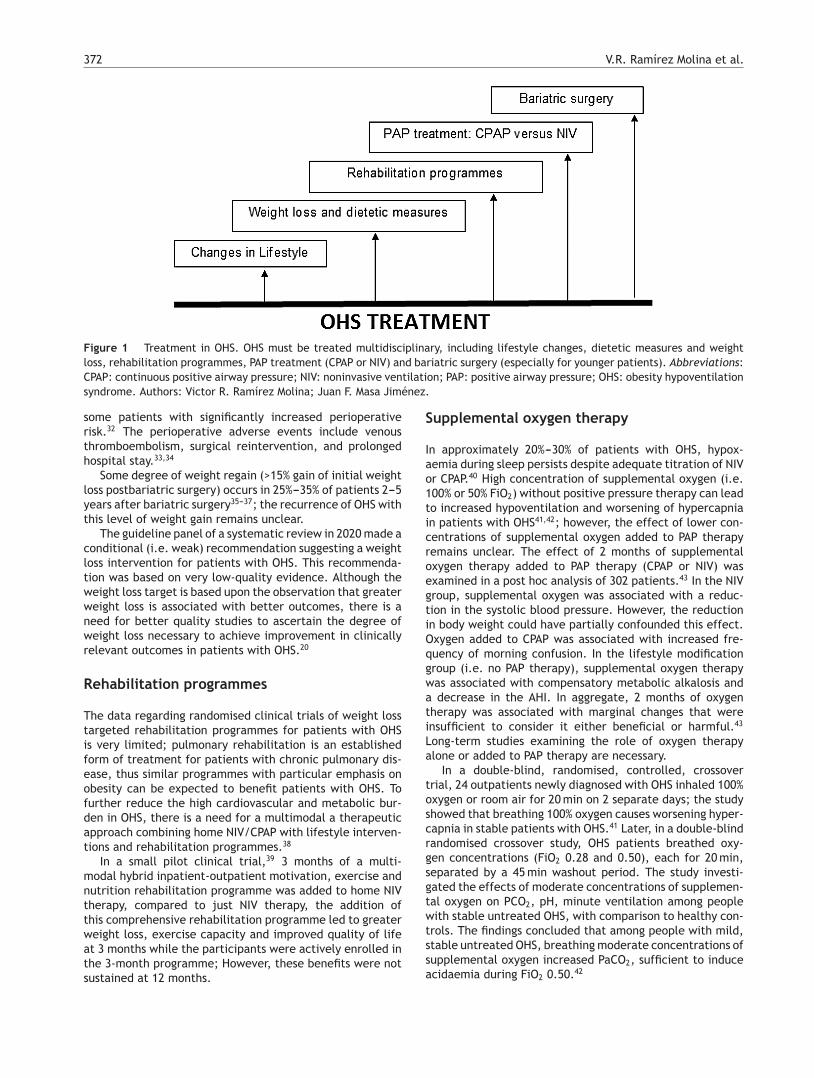
372 V.R. Ramírez Molina et al.
Figure 1 Treatment in OHS. OHS must be treated multidisciplinary, including lifestyle changes, dietetic measures and weightloss, rehabilitation programmes, PAP treatment (CPAP or NIV) and bariatric surgery (especially for younger patients). Abbreviations:CPAP: continuous positive airway pressure; NIV: noninvasive ventilation; PAP: positive airway pressure; OHS: obesity hypoventilationsyndrome. Authors: Victor R. Ramírez Molina; Juan F. Masa Jiménez.
some patients with significantly increased perioperativerisk.32 The perioperative adverse events include venousthromboembolism, surgical reintervention, and prolongedhospital stay.33,34
Some degree of weight regain (>15% gain of initial weightloss postbariatric surgery) occurs in 25%---35% of patients 2---5years after bariatric surgery35---37; the recurrence of OHS withthis level of weight gain remains unclear.
The guideline panel of a systematic review in 2020 made aconditional (i.e. weak) recommendation suggesting a weightloss intervention for patients with OHS. This recommenda-tion was based on very low-quality evidence. Although theweight loss target is based upon the observation that greaterweight loss is associated with better outcomes, there is aneed for better quality studies to ascertain the degree ofweight loss necessary to achieve improvement in clinicallyrelevant outcomes in patients with OHS.20
Rehabilitation programmes
The data regarding randomised clinical trials of weight losstargeted rehabilitation programmes for patients with OHSis very limited; pulmonary rehabilitation is an establishedform of treatment for patients with chronic pulmonary dis-ease, thus similar programmes with particular emphasis onobesity can be expected to benefit patients with OHS. Tofurther reduce the high cardiovascular and metabolic bur-den in OHS, there is a need for a multimodal a therapeuticapproach combining home NIV/CPAP with lifestyle interven-tions and rehabilitation programmes.38
In a small pilot clinical trial,39 3 months of a multi-modal hybrid inpatient-outpatient motivation, exercise andnutrition rehabilitation programme was added to home NIVtherapy, compared to just NIV therapy, the addition ofthis comprehensive rehabilitation programme led to greaterweight loss, exercise capacity and improved quality of lifeat 3 months while the participants were actively enrolled inthe 3-month programme; However, these benefits were notsustained at 12 months.
Supplemental oxygen therapy
In approximately 20%---30% of patients with OHS, hypox-aemia during sleep persists despite adequate titration of NIVor CPAP.40 High concentration of supplemental oxygen (i.e.100% or 50% FiO2) without positive pressure therapy can leadto increased hypoventilation and worsening of hypercapniain patients with OHS41,42; however, the effect of lower con-centrations of supplemental oxygen added to PAP therapyremains unclear. The effect of 2 months of supplementaloxygen therapy added to PAP therapy (CPAP or NIV) wasexamined in a post hoc analysis of 302 patients.43 In the NIVgroup, supplemental oxygen was associated with a reduc-tion in the systolic blood pressure. However, the reductionin body weight could have partially confounded this effect.Oxygen added to CPAP was associated with increased fre-quency of morning confusion. In the lifestyle modificationgroup (i.e. no PAP therapy), supplemental oxygen therapywas associated with compensatory metabolic alkalosis anda decrease in the AHI. In aggregate, 2 months of oxygentherapy was associated with marginal changes that wereinsufficient to consider it either beneficial or harmful.43
Long-term studies examining the role of oxygen therapyalone or added to PAP therapy are necessary.
In a double-blind, randomised, controlled, crossovertrial, 24 outpatients newly diagnosed with OHS inhaled 100%oxygen or room air for 20 min on 2 separate days; the studyshowed that breathing 100% oxygen causes worsening hyper-capnia in stable patients with OHS.41 Later, in a double-blindrandomised crossover study, OHS patients breathed oxy-gen concentrations (FiO2 0.28 and 0.50), each for 20 min,separated by a 45 min washout period. The study investi-gated the effects of moderate concentrations of supplemen-tal oxygen on PCO2, pH, minute ventilation among peoplewith stable untreated OHS, with comparison to healthy con-trols. The findings concluded that among people with mild,stable untreated OHS, breathing moderate concentrations ofsupplemental oxygen increased PaCO2, sufficient to induceacidaemia during FiO2 0.50.42

Pickwick’s treatment 373
Positive airway pressure therapy
OHS is treated with positive airway pressure (PAP) therapyduring sleep. The two most commonly used PAP modalitiesare continuous positive airway pressure (CPAP) and nonin-vasive ventilation (NIV). Although CPAP can splint the upperairway open and effectively treat OSA, it does not increaseventilation as effectively as NIV does.44
The short-term benefits of CPAP include improvementin gas exchange and sleep-disordered breathing45 with anobserved response between 50% and 80% of cases although itmay vary in the sleep apnea severity and the time of follow-up. This improvement is directly proportional to the hours ofCPAP use, as each hour of use of PAP therapy decreased thePaCO2 by 1.8 mmHg and the PaO2 increased by 3 mmHg.40
PAP therapy improves gas exchange, respiratory sleep dis-orders and probably lung function and central respiratoryimpulse to carbon dioxide (CO2). Night-time hypoventila-tion can be effectively improved, but not in all cases,46---49
and daytime PaCO2 reduced or restored to normal values.48
The effectiveness of NIV has been assessed in several long-term, observational studies1,7,23,44,50---55 and medium-termrandomised trials.3,21,56
In patients with OHS and severe OSA, medium-term ran-domised controlled trials7,38,50,57---59 and a long-term clinicaltrial24,26,60 have shown CPAP and NIV to be equally effec-tive in improving symptoms, quality of life and sleep, gasexchange during waking and sleep, as well as spirometricand polysomnographic parameters.
Mechanisms of improvement in hypercapnia withPAP use
The mechanisms by which diurnal hypercapnia improveswith PAP are complex and not fully understood. PAP therapycan influence the following mechanisms: abnormal respira-tory mechanics, central responses to hypercapnia and/orneurohormonal dysfunction (leptin resistance) and sleep-disordered breathing.61 See Table 2.
NIV can reduce inspiratory muscular activity,62 so thatit can efficiently decrease the mechanical load favouringmuscular rest and greater muscular efficacy during the dayafter nocturnal NIV treatment. Continuous positive airwaypressure and NIV may decrease the mechanical load avoidingupper airway repetitive obstructions during sleep.
As for leptin resistance, the levels of serum leptindecrease to normal limits in patients with OSA treatedwith CPAP,63,64 but it is assumed that apneas and hypop-neas are the cause of the elevated leptin levels rather thanbeing the result of them.65,66 Leptinaemia also decreaseswith NIV treatment67,68 as does daytime hypercapnia, andsome studies have shown a correlation between leptinaemiaand a reduction in the hypercapnic ventilatory response,69
while another study68 reported contradictory results, i.e.,an increase of leptin with NIV. Therefore, the role of leptinin how NIV treatment achieves improvement, is still unclear.
Finally, in regards to sleep-disordered breathing, repeti-tive obstructive events produce increasing hypercapnia, notresolved with the hyperventilation that occurs at the endof obstructive events. Despite correction of these noctur-nal obstructive events with CPAP, daytime PaCO2 does not
return to normal in all cases. Several studies have high-lighted that the CPAP response may vary depending on thepredominance of nocturnal obstructive events10,44 and thetime of follow-up because CPAP may have a delay in its effi-cacy related to NIV.22 Non-invasive ventilation has shown toprevent obstructive events and reduce hypoventilation dur-ing sleep (including rapid eye movement [REM] sleep). BothNIV and CPAP should decrease nocturnal hypercapnia, lead-ing to lower daytime serum bicarbonate and consequentlyless blunting of the central carbon dioxide response.70
Scientific research base
There are several randomised controlled studies that com-pare different treatments in OHS.3,22,24,56,71 One of thesestudies compared the short-term efficacy of NIV and CPAPtreatments in 36 patients with OHS selected in consideringtheir favourable response to a night of CPAP treatment.71
After 3 months, the improvements in daytime sleepinessand in clinical and gas exchange parameters were similarbetween CPAP and NIV groups.
In another trial including 38 patients with mild hyper-capnia with NIV compared to a control group treated withconservative measures, the NIV group had a significantreduction in daytime PaCO2, bicarbonate and an increasein pH. Therapy with NIV, as expected, was associated witha great improvement in all sleep variables analysed, sleeparchitecture, average oxygen saturation, oxygen saturationtime less than 90%, apnea and hypopnea index, with a pos-itive and significant correlation between average oxygensaturation during sleep and diurnal arterial blood gases. Incontrast, no change was observed in any of the metabolicand inflammatory parameters studied, but the follow-up wasonly one month, so no other conclusions could be drawn.21
In this study, the patients had a lower BMI and were lesshypercapnic than the subjects included in other trials.3,71
The Pickwick project is the largest multicenter ran-domised controlled trial designed to assess medium-term(two months) effectiveness of NIV, CPAP, and lifestyle modifi-cation (control group) and long-term (3 years) effectivenessbetween NIV and CPAP in OHS65,72; there are two clinicaltrials in parallel depending on the existence or absence ofsevere OSA (AHI ≥ 30). The trial which includes severe OSAhas three arms: NIV, CPAP and change in lifestyle for twomonths (first phase). After this period of time, the lifestylechange group was re-randomised to NIV or CPAP to continueat least 36 months (second phase). Patients with an AHI < 30were randomised to NIV or change in lifestyle for at least36 months (second phase), although an evaluation of resultswas performed at two months (first phase). The results ofthe first phase were entirely published.3 The first publica-tion included 221 patients with severe OSA randomised toNIV, CPAP and change in lifestyle; PaCO2 who improved witheach of the three treatments, but the improvement wasgreater with the use of NIV, with a significant differencein relation to the group of conservative measures. In theCPAP group, the reduction of PaCO2 depended on compli-ance with the treatment. Thus, NIV and CPAP decreasedblood bicarbonate levels but after adjusting baseline dataonly NIV achieved statistical significance compared to thecontrol group. Sleep variables improved notably with the

374 V.R. Ramírez Molina et al.
Table 2 Potential mechanism of improvement with PAP therapy in OHS. PAP therapy can influence in the following mechanismsto improve daytime hypercapnia: mechanical load, leptine resistance and breathing sleep disorder; including reduce inspiratorymuscular activity favouring muscular rest, decrease in central resistance leptine and decrease in nocturnal obstructive eventsand sleep hypercapnia.
Mechanism of diurnal hypercapnia Influence and effect of PAP treatment
1. Abnormal respiratory mechanics NIV can reduce inspiratory muscular activity. Decrease the mechanical loadfavouring muscular rest and greater muscular efficacy.NIV and CPAP may decrease the mechanical load avoiding upper airway repetitiveobstructions during sleep.
1. Central responses to hypercapniaand/or neurohormonal dysfunction(leptin resistance)
NIV and CPAP decrease to normal limits the levels of serum leptin; reduction in thehypercapnic ventilatory response.
2. Sleep-disordered breathing NIV and CPAP correct the obstructive events (apneas and hypopneas).NIV can prevent obstructive events and reduce hypoventilation during sleep(including REM sleep).
CPAP: continuous positive airway pressure; NIV: noninvasive ventilation; OHS: obesity hypoventilation syndrome; PAP: positive airwaypressure; REM: rapid eye movement. Authors: Victor R. Ramírez Molina; Juan F. Masa Jiménez.
use of NIV and CPAP, both proving to be equally efficientand with little or no difference between the two; only theNIV group presented an increase in the FVC, FEV1 values andthe 6-minute walk test. In another publication of this Pick-wick study, 86 patients were randomised and treated for twomonths with NIV or lifestyle modifications.56 The NIV groupsignificantly improved PaCO2 and serum bicarbonate levelscompared to the control group.
In another randomised controlled trial, NIV or CPAP wasused for 3 months with 60 participants. The primary objec-tive was to identify the frequency of treatment failuredefined as, hospital admission, persistent ventilatory insuf-ficiency or lack of adherence, while secondary objectivesincluded life quality related to health and drowsiness. Atotal of 57 patients completed the follow-up without dif-ferences in treatment failure between the groups (NIV14.8% versus CPAP 13.3%; p = 0.87). It is worth noting thatadherence to treatment and PaCO2 in wakefulness at threemonths were similar (NIV 5.3 h/night; CPAP 5.0 h/night;p = 0.62, and PaCO2 in wakefulness at three months of 44,2 and 45.9 mmHg, respectively; p = 0.60). The differencesbetween the groups in the improvement of sleepiness andthe life quality were not significant. Baseline severity ofventilatory failure (based on PaCO2 levels) was the only sig-nificant predictor for such insufficiency after a three monthsperiods of the study (OR, 2.3; p = 0.03).22
The multicentre, open-label, randomised controlledPickwick trial published its long-term results on 97 OHSpatients with severe OSA treated with NIV and 107treated with CPAP.24 The median follow-up was 5.44 years(interquartile range [IQR] 4.45---6.37) for all patients, 5.37years (4.36---6.32) in the CPAP group, and 5.55 years(4.53---6.50) in the NIV group. The hospitalisation days perpatient-year were 1.63 (standard deviation [SD] 3.74) inthe CPAP group and 1.44 (3.07) in the NIV group (adjustedrate ratio 0.78, 95% CI 0.34---1.77; p = 0.561). Changes inother hospital resource utilisation, blood pressure, arterialblood gases, spirometry, quality of life, clinical symptomsand supplemental oxygen therapy remained similar betweenPAP modalities. Both NIV and CPAP also similarly improvedthe pulmonary artery pressure and diastolic left ventricu-lar dysfunction.72 Given that CPAP has lower complexity and
cost, CPAP might be the preferred first-line PAP treatmentmodality.24
Cost-effectiveness of PAP therapy modalities
CPAP is simpler to implement and is less costly than NIV.24
To investigate which of the two treatments is more cost-effective, the only study reported is from Masa and cols,whom carried out a post hoc, within-trial, cost-effectivenessanalysis using the large multicentre, open-labelled, ran-domised controlled study (Pickwick study)3,24,43,56,65,73,74; theaim was to determine the comparative cost-effectivenessrelationship between NIV and CPAP based on 3 years offollow-up, using hospitalisation days as the primary outcomemeasure in a cost-effectiveness analysis, or considering thehospitalisation days in monetary terms where the valueof a hospitalisation day is approximately its cost, in acost---benefit analysis.
In total, 363 patients were selected, 215 were ran-domised and 202 were available for the analysis. The median(IQR) follow-up was 3.01 (2.91---3.14) years for NIV group and3.00 (2.92---3.17) years for CPAP. The mean (SD) Bayesianestimated hospital days was 2.13 (0.73) for CPAP and 1.89(0.78) for NIV. The mean (SD) Bayesian estimated cost perpatient/year in the NIV arm, excluding hospitalisation costs,was D 2075.98 (91.6), which was higher than the cost inthe CPAP arm of D 1219.06 (52.3); mean difference D 857.6(105.5). CPAP was more cost-effective than NIV (99.5% prob-ability) because longer hospital stay in the CPAP arm wascompensated for by its lower costs. Similar findings wereobserved in the high and low adherence subgroups; Thus, theconclusion of this study is that CPAP is more cost-effectivethan NIV; therefore, CPAP should be the preferred treatmentfor patients with OHS with severe OSA.75
Initial PAP treatment suggestion
CPAP should be the initial treatment modality in patientswith OHS if severe OSA is present, due to its relative simplic-ity, low cost and efficacy.4,46,75 In patients with OHS withoutsevere OSA, NIV is the preferred PAP modality, because inthese patients without a significant number of obstructive

Pickwick’s treatment 375
Figure 2 Initial PAP treatment election in OHS. CPAP should be the initial modality treatment if the main cause of diurnalhypoventilation is the predominance of obstructive events (severe---moderate OSA) and if in subsequent evaluations at 1---3 monthsthe patient responds with adequate oxygenation (PaO2) and ventilation (PaCO2) the CPAP should be continued; if the patient doesnot responds or if the OHS patient has hypoventilation predominance (mild OSA), NIV should be the initial modality treatmentpreferred. Abbreviations: CPAP: continuous positive airway pressure; NIV: noninvasive ventilation; OHS: obesity hypoventilationsyndrome; OSA: obstructive sleep apnea; PaO2: partial pressure of oxygen; PaCO2: partial pressure of carbon dioxide; PAP: positiveairway pressure. Authors: Victor R. Ramírez Molina; Juan F. Masa Jiménez.
apneas and hypopneas, their nocturnal hypoventilation maydepend on other mechanisms (e.g. obesity). See Fig. 2. How-ever, a case by case evaluation is necessary to determine theinitial treatment.26
In acute hypercapnic respiratory failure or hospitalisedpatients, NIV should be the first option, due to its potentiallygreater efficacy against hypoventilation and the underlyingseverity of respiratory failure.61
Conclusions
Obesity is a worldwide, increasingly ubiquitous health issuethat has triggered the wide spread of several related medicalconditions; OHS is one of the respiratory sleep disorders thathas the greatest impact on increasing cardiovascular risk.Therefore, OHS must be recognised promptly, and optimallytreated; a multidisciplinary approach is likely to be moreeffective for improving long-term outcomes, including lossweight, rehabilitation programmes and PAP therapy; CPAPshould be the initial treatment when indicated.
Authors’ contributions
All authors equally contributed to this paper with conceptionand design of the study, literature review, critical revision,editing and final approval of the final version.
Conflicts of interest
The authors declare that they have no conflicts of interest.
Acknowledgements
None declared.
References
1. Borel JC, Burel B, Tamisier R, Dias-Domingos S, Baguet JP, LevyP, et al. Comorbidities and mortality in hypercapnic obese underdomiciliary noninvasive ventilation. PLoS One. 2013;8:e52006.
2. Kessler R, Chaouat A, Schinkewitch P, Faller M, Casel S,Krieger J, et al. The obesity-hypoventilation syndrome revis-ited: a prospective study of 34 consecutive cases. Chest.2001;120:369---76.
3. Masa JF, Corral J, Alonso ML, Ordax E, Troncoso MF, GonzalezM, et al. Efficacy of different treatment alternatives for obesityhypoventilation syndrome. Pickwick study. Am J Respir Crit CareMed. 2015;192:86---95.
4. Olson AL, Zwillich C. The obesity hypoventilation syndrome. AmJ Med. 2005;118:948---56.
5. Mokhlesi B, Tulaimat A, Faibussowitsch I, Wang Y, Evans AT.Obesity hypoventilation syndrome: prevalence and predic-tors in patients with obstructive sleep apnea. Sleep Breath.2007;11:117---24.

376 V.R. Ramírez Molina et al.
6. Piper AJ, Grunstein RR. Obesity hypoventilation syndrome:mechanisms and management. Am J Respir Crit Care Med.2011;183:292---8.
7. Priou P, Hamel J-F, Person C, Meslier N, Racineux JL, UrbanT, et al. Long-term outcome of noninvasive positive pres-sure ventilation for obesity hypoventilation syndrome. Chest.2010;138:84---90.
8. Berg G, Delaive K, Manfreda J, Walld R, Kryger MH. The useof health-care resources in obesity hypoventilation syndrome.Chest. 2001;120:377---83.
9. Jennum P, Kjellberg J. Health, social and economical conse-quences of sleep-disordered breathing: a controlled nationalstudy. Thorax. 2011;66:560---6.
10. Pérez de Llano LA, Golpe R, Ortiz Piquer M, Veres A, VázquezM, Caballero O, et al. Short-term and long-term effects of nasalintermittent positive pressure ventilation in patients with obe-sity hypoventilation syndrome. Chest. 2005;128:587---94.
11. Ojeda Castillejo E, de Lucas Ramos P, López Martin S, ResanoBarrios P, Rodríguez Rodríguez P, Morán Caicedo L. Noninvasivemechanical ventilation in patients with obesity hypoventila-tion syndrome: long-term outcome and prognostic factors. ArchBronconeumol. 2015.
12. Nowbar S, Burkart KM, Gonzales R, Fedorowicsz A, Gonzan-sky WS, Gaudio JC, et al. Obesity-associated hypoventilationin hospitalized patients: prevalence, effects, and outcome. AmJ Med. 2004;116:1---7.
13. Zwillich CW, Sutton FD, Pierson DJ, Greagh EM, WeilJV. Decreased hypoxic ventilatory drive in the obesity-hypoventilation syndrome. Am J Med. 1975;59:343---8.
14. MacGregor M, Block AJ, Ball WC. Topics in clinical medicine:serious complications and sudden death in the Pickwickian syn-drome. Hopkins Med J. 1970;126:279---95.
15. Miller A, Granada M. In-hospital mortality in the Pickwickiansyndrome. Am J Med. 1974;56:144---50.
16. Hales CM, Fryar CD, Carroll MD, Freedman DS, Ogden CL.Trends in obesity and severe obesity prevalence in US youthand adults by sex and age, 2007---2008 to 2015---2016. JAMA.2018;19:1723---5.
17. Flegal KM, Kruszon-Moran D, Carroll MD, Fryar CD, Ogden CL.Trends in obesity among adults in the United States, 2005 to2014. JAMA. 2016;315:2284---91.
18. Ng M, Fleming T, Robinson M, Thomson B, Graetz N, MargonoC, et al. Global, regional, and national prevalence of over-weight and obesity in children and adults during 1980---2013:a systematic analysis for the Global Burden of Disease Study2013. Lancet. 2014;384:766---8.
19. Piper A, Pepin JL, Hart N. Positive airway pressure inobesity hypoventilation syndrome: is it worth it? Thorax.2020, http://dx.doi.org/10.1136/thoraxjnl-2019-214403, pii:thoraxjnl-2019-214403.
20. Kakazu MT, Soghier I, Afshar M, Brozek JL, Wil-son KC, Masa JF, et al. Weight loss interventions astreatment of obesity hypoventilation syndrome. A sys-tematic review. Ann Am Thorac Soc. 2020;17:492---502,http://dx.doi.org/10.1513/AnnalsATS.201907-554OC.
21. Borel JC, Tamisier R, Gonzalez-Bermejo J, Baguet JP, Mon-neret D, Arnol N, et al. Noninvasive ventilation in mildobesity hypoventilation syndrome: a randomized controlledtrial. Chest. 2012;141:692---702.
22. Howard ME, Piper AJ, Stevens B, Holland AE, Yee BJ, Dab-scheck E, et al. A randomised controlled trial of CPAPversus non-invasive ventilation for initial treatment of obesityhypoventilation syndrome. Thorax. 2017;72:437---44.
23. Castro-Anón O, Pérez de Llano LA, De la Fuente Sánchez S,Golpe R, Méndez Marote L, Castro-Castro J, et al. Obesity-hypoventilation syndrome: increased risk of death over sleepapnea syndrome. PLoS One. 2015;10:e0117808.
24. Masa JF, Mokhlesi B, Benítez I, Gomez de Terreros FJ, Sánchez-Quiroga MA, Romero a, et al. Long-term clinical effectiveness ofcontinuous positive airway pressure therapy versus non-invasiveventilation therapy in patients with obesity hypoventilation syn-drome: a multicentre, open-label, randomised controlled trial.Lancet. 2019;27:1721---32.
25. Bouloukaki I, Mermigkis C, Michelakis S, Moniaki V, MauroudiE, Tzanakis N, et al. The association between adherence topositive airway pressure therapy and long-term outcomes inpatients with obesity hypoventilation syndrome: a prospectiveobservational study. J Clin Sleep Med. 2018;14:1539---50.
26. Mokhlesi B, Masa JF, Brozek JL, Gurubhagavatula I, MurphyPB, Piper AJ, et al. Evaluation and management of obesityhypoventilation syndrome. An Official American Thoracic Soci-ety Clinical Practice Guideline. Am J Respir Crit Care Med.2019;200:e6---24.
27. Chang SH, Stoll CR, Song J, Varela JE, Eagon CJ, Colditz GA.The effectiveness and risks of bariatric surgery: an updatedsystematic review and meta-analysis, 2003---2012. JAMA Surg.2014;149:275---87.
28. Greenburg D, Lettieri C, Eliasson A. Effects of surgical weightloss on measures of obstructive sleep apnea: a meta-analysis.Am J Med. 2009;122:535---42.
29. Martí-Valeri C, Sabaté A, Masdevall C, Dalmau A. Improve-ment of associated respiratory problems in morbidly obesepatients after open roux-en-y gastric bypass. Obes Surg.2007;17:1102---10.
30. Pillarm G, Peled R, Lavie P. Recurrence of sleep apnea withoutconcomitant weight reduction increases 75 years after weightreduction surgery. Chest. 1994;106:1702---4.
31. Lettieri CJ, Eliasson AH, Greenburg DL. Persistence of obstruc-tive sleep apnea after surgical weight loss. J Clin Sleep Med.2008;4:333---8.
32. Sugerman HJ, Fairman RP, Sood RK, Engle K, Wolfe L, KellumJM. Long-term effects of gastric surgery for treating respiratoryinsufficiency of obesity. Am J Clin Nutr. 1992;55:597S---601S.
33. Fernández AJ, Demaria EJ, Tichansky DS, Kellum JM, Wolfe LG,Meador J, et al. Multivariate analysis of risk factors for deathfollowing gastric bypass for treatment of morbid obesity. AnnSurg. 2004;239:698---702.
34. Flum D, Belle SH, King WC, Wahed AS, Berk P, Chapman W, et al.Perioperative safety in the longitudinal assessment of bariatricsurgery. N Engl J Med. 2009;361:445---54.
35. Bastos EC, Barbosa EM, Soriano GM, dos Santos EA, Lima SM.Determinants of weight regain after bariatric surgery. Arq BrasCir Dig. 2013;26 Suppl.:26---32.
36. Marchesini SD, Bareta GA, Cambi MP, Marchesini JB. Endoscopicplasma argon coagulation in treatment of weight regain afterbariatric surgery: what does the patient think about this? ArqBras Cir Dig. 2015;27 Suppl.:47---50.
37. Cooper TC, Simmons EB, Webb K, Burns JL, Kushner RF. Trendsin weight regain following Roux-en-Y gastric bypass (RYGB)bariatric surgery. Obes Surg. 2015;25:1474---81.
38. Masa F, Pépin J, Borel J, Mokhlesi B, Murphy PB, Sánchez-Quiroga MA. Obesity hypoventilation syndrome. Eur Repir Rev.2019;28:180097.
39. Mandal S, Suh ES, Harding R, Vaughan-France A, Ram-say M, Connolly B, et al. Nutrition and exercise reha-bilitation in obesity hypoventilation syndrome (NERO): apilot randomised controlled trial. Thorax. 2018;73:62---9,http://dx.doi.org/10.1136/thoraxjnl-2016-209826.
40. Mokhlesi B, Tulaimat A, Evans AT, Wang Y, Itani AA, HassaballaHA, et al. Impact of adherence with positive airway pressuretherapy on hypercapnia in obstructive sleep apnea. J Clin SleepMed. 2006;2:57---62.
41. Wijesinghe M, Williams M, Perrin K, Weatherall M, Beasley R.The effect of supplemental oxygen on hypercapnia in subjects
Pickwick’s treatment 377
with obesity-associated hypoventilation: a randomized cross-over clinical study. Chest. 2011;139:1018---24.
42. Hollier CA, Harmer AR, Maxwell LJ, Menadue C, Willson GN,Unger G, et al. Moderate concentrations of supplementaloxygen worsen hypercapnia in obesity hypoventilation syn-drome: a randomised crossover study. Thorax. 2014;69:346---53,http://dx.doi.org/10.1136/thoraxjnl-2013-204389.
43. Masa JF, Corral J, Romero A, Caballero C, Terán-Santos J,Alonso-Álvarez ML, et al. The effect of supplemental oxy-gen in obesity hypoventilation syndrome. J Clin Sleep Med.2016;12:1379---88.
44. Berger KI, Ayappa I, Chatr-Amontri B, Marfatia A, Sorkin IB,Rapoport DM, et al. Obesity hypoventilation syndrome asa spectrum of respiratory disturbances during sleep. Chest.2001;120:1231---8.
45. Mokhlesi B. Obesity hypoventilation syndrome: a state-of-the-art review. Respir Care. 2010;55:1347---62.
46. Chau EH, Mokhlesi B, Chung F. Obesity hypoventilation syndromeand anesthesia. Sleep Med Clin. 2013;8:135---47.
47. Lee WY, Mokhlesi B. Diagnosis and management of obe-sity hypoventilation syndrome in the ICU. Crit Care Clin.2008;24:533---49.
48. Masa JF, Celli BR, Riesco JA, Sánchez de Cos J, Disdier C, Sojo A.Noninvasive positive pressure ventilation and not oxygen mayprevent overt ventilatory failure in patients with chest walldiseases. Chest. 1997;112:207---13.
49. Hida W, Okabe S, Tatsumi K, Kimura H, Akasiba T, Chin K,et al. Nasal continuous positive airway pressure improves qual-ity of life in obesity hypoventilation syndrome. Sleep Breath.2003;7:3---12.
50. Basoglu OK, Tasbakan MS. Comparison of clinical characteristicsin patients with obesity hypoventilation syndrome and obeseobstructive sleep apnea syndrome: a case---control study. ClinRespir J. 2014;8:167---74.
51. Budweiser S, Riedl SG, Jörres RA, Heinemann F, Pfeifer M.Mortality and prognostic factors in patients with obesity-hypoventilation syndrome undergoing noninvasive ventilation.J Intern Med. 2007;261:375---83.
52. de Lucas-Ramos P, de Miguel-Díez J, Santacruz-Siminiani A,González-Moro JMR, Buendía-García MJ, Izquierdo-Alonso JL.Benefits at 1 year of nocturnal intermittent positive pressureventilation in patients with obesity hypoventilation syndrome.Respir Med. 2004;98:961---7.
53. Janssens JP, Derivaz S, Breitenstein E, De Mural B, Fitting JW,Chevrolet JC, et al. Changing patterns in long-term noninvasiveventilation: a 7-year prospective study in the Geneva Lake area.Chest. 2003;123:67---79.
54. Heinemann F, Budweiser S, Dobroschke J, Pfeifer M. Non-invasive positive pressure ventilation improves lung vol-umes in the obesity hypoventilation syndrome. Respir Med.2007;101:1229---35.
55. Windisch W, Quality of Life in Home Mechanical VentilationStudy Group. Impact of home mechanical ventilation on health-related quality of life. Eur Respir J. 2008;32:1328---36.
56. Masa JF, Corral J, Caballero C, Barrot E, Terán-Santos J,Alonso-Álvarez ML, et al. Noninvasive ventilation in obesityhypoventilation syndrome without severe sleep apnea. Thorax.2016;71:899---906.
57. Castro-Anón O, Golpe R, Pérez-de-Llano LA, López MJ, EscalonaEJ, Pérez R, et al. Haemodynamic effects of non-invasiveventilation in patients with obesity-hypoventilation syndrome.Respirology. 2012;17:1269---74.
58. Sugerman HJ, Baron PL, Fairman RP, Evans CR, Vetrovec GW.Hemodynamic dysfunction in obesity hypoventilation syndromeand the effects of treatment with surgically induced weight loss.Ann Surg. 1988;207:604---13.
59. Kauppert CA, Dvorak I, Kollert F, Heinemann F, Jörres RA, PfeiferM, et al. Pulmonary hypertension in obesity-hypoventilationsyndrome. Respir Med. 2013;107:2061---70.
60. Soghier I, Brozek JL, Afshar M, Tamae M, Wilson KC,Masa JF, et al. Noninvasive ventilation versus CPAPas initial treatment of obesity hypoventilation syn-drome: a systematic review. Ann Am Thorac Soc. 2019,http://dx.doi.org/10.1513/AnnalsATS.201905-380OC [Epubahead of print].
61. Ramírez-Molina VR, Gómez-de-Terreros FJ, Barca-Durán J, MasaJF. Non-invasive positive airway pressure in obesity hypoven-tilation syndrome and chronic obstructive pulmonary disease:present and future perspectives. COPD. 2017;14:418---28.
62. Pankow W, Hijjeh N, Schüttler F, Penzel T, Becker HF, PeterJH. Influence of noninvasive positive pressure ventilation oninspiratory muscle activity in obese subjects. Eur Respir J.1997;10:2847---52.
63. Ip MS, Lam KS, Ho C, Tsang KW, Lam W. Serum leptin and vascularrisk factors in obstructive sleep apnea. Chest. 2000;118:580---6.
64. Phipps PR, Starrit E, Caterson I, Grunstein RR. Association ofserum leptin with hypoventilation in human obesity. Thorax.2002;57:75---6.
65. López-Jiménez MJ, Masa JF, Corral J, Terán J, Ordaz E, TroncosoMF, et al. Mid- and long-term efficacy of non-invasive ventilationin obesity hypoventilation syndrome: the Pickwick’s Study. ArchBronconeumol. 2016;52:158---65.
66. Fitzpatrick M. Leptin and the obesity hypoventilation syndrome:a leap of faith? Thorax. 2002;57:1---2.
67. Yee BJ, Cheung J, Phipps P, Banerjee D, Piper AJ, GrunsteinRR. Treatment of obesity hypoventilation syndrome and serumleptin. Respiration. 2006;73:209---12.
68. Redolfi S, Corda L, La Piana G, Spandrio S, Prometti P, TantucciC. Long-term noninvasive ventilation increases chemosensitiv-ity and leptin in obesity-hypoventilation syndrome. Respir Med.2007;101:1191---5.
69. Campo A, Freihbeck G, Zulueta JJ, Iriarte J, Seijo LM, AlcaideAB, et al. Hypercapnic response in obese patients. Eur Respir J.2007;30:223---31.
70. Mokhlesi B, Tulaimat A, Faibursowitsch I, Wang Y, Evans AT.Obesity hypoventilation syndrome: prevalence and predic-tors in patients with obstructive sleep apnea. Sleep Breath.2007;11:117---24.
71. Piper AJ, Wang D, Yee BJ, Barnes DJ, Grunstein RR. Randomisedtrial of CPAP vs bilevel support in the treatment of obesityhypoventilation syndrome without severe nocturnal desatura-tion. Thorax. 2008;63:395---401.
72. Masa JF, Mokhlesi B, Benitez I, Mogollon MV, Gomezde Terreros FJ, Sánchez-Quiroga MA, et al. Ecocardio-graphic changes with positive airway pressure therapy inobesity hypoventilation syndrome: long-term Pickwick ran-domized controlled trial. AM J Respir Crit Care Med. 2019,http://dx.doi.org/10.1164/rccm.20199061122OC.
73. Masa JF, Corral J, Romero A, Caballero C, Terán-Santos J,Alonso-Álvarez ML, et al. Protective cardiovascular effect ofsleep apnea severity in obesity hypoventilation syndrome.Chest. 2016;150:68---79.
74. Corral J, Mogollon MV, Sánchez-Quiroga M-Á, Gómez de TerrerosJ, Romero A, Caballero C, et al. Echocardiographic changes withnon-invasive ventilation and CPAP in obesity hypoventilationsyndrome. Thorax. 2018;73:361---8.
75. Masa JF, Mokhlesi B, Benítez I, Gómez de Terreros FJ, Sánchez-Quiroga M-Ángeles, Romero A, et al. Cost-effectiveness ofpositive airway pressure modalities in obesity hypoventila-tion syndrome with severe obstructive sleep apnoea. Thorax.2020:1---9, http://dx.doi.org/10.1136/thoraxjnl-2019-213622.

Pulmonol. 2020;26(6):378---385
www.journalpulmonology.org
ORIGINAL ARTICLE
The validity of surface EMG of extra-diaphragmaticmuscles in assessing respiratory responses duringmechanical ventilation: A systematic review
H.Y. AbuNuraha,∗, D.W. Russell b, J.D. Lowmanc
a Department of Respiratory Therapy, King Saud bin Abdulaziz University for Health Sciences, KSA & the PhD in RehabilitationScience Program, University of Alabama at Birmingham, Birmingham, AL, USAb Division of Pulmonary, Allergy and Critical Care Medicine, Department of Medicine, University of Alabama at Birmingham, USAc Department of Physical Therapy, University of Alabama at Birmingham, Birmingham, AL, USA
Received 1 August 2019; accepted 11 February 2020Available online 2 April 2020
KEYWORDSsurfaceelectromyography;mechanicalventilation;diaphragm;respiratory muscles;extra-diaphragmaticmuscles;respiratorymechanics
AbstractPurpose: Evidence supporting the utilization of surface EMG (sEMG) of extra-diaphragmaticmuscles for monitoring of mechanical ventilation (MV) assistance is unclear. The purpose of thisreview was to assess the quality of literature available on using extra-diaphragmatic sEMG asan assessment technique of respiratory responses during MV.Methods: Studies using sEMG of extra-diaphragmatic respiratory muscles during MV wereselected by two independent researchers after performing a database search of PubMed,CINAHL, GOOGLE SCHOLAR. Exclusion criteria were studies of patients with neuromuscular dis-orders, receiving neuromuscular blocking agents, receiving non-invasive MV, using needle EMG,and studies written in languages other than English. Quality of identified studies was assessedwith the Quality Assessment of Diagnostic Accuracy Studies (QUADAS-2). This study is registeredwith PROSPERO, number (CRD42018081341).
Abbreviations: ASV, Adaptive Support Ventilation; Edi, Electrical activity of the diaphragm; EMG, Electromyography; EMGMAX, Maximumelectrical activity; EMGMEAN, Mean electrical activity; EMGMIN, EMG activity in one minute; EMGTi, Duration of electrical activity; sEMG,Surface electromyography; ICU, Intensive Care Unit; MV, Mechanical ventilation; NAVA, Neural Assisted Ventilation; NRD, Neuro-respiratorydrive; P0.1, Occlusion pressure at 1 milliseconds; Pdi, Trans-diaphragmatic pressure; Pes, Esophageal pressure; PETCO2 , End tidal CO2; PRISMA-DTA, Preferred Reporting Items for Systematic Reviews and Meta-Analyses of Diagnostic Test Accuracy; PSV, Pressure Support Ventilation;PTP, Pressure-time product; QUADAS-2, Quality Assessment of Diagnostic Accuracy Studies; SCM, Sternocleidomastoid; SIMV, SynchronizedIntermittent Mandatory Ventilation; TIe, Mechanical duration of inspiratory effort; VAS, Visual Analog Scale; VE, Minute ventilation; WOB,Work of Breathing.
∗ Corresponding author at: Department of Physical Therapy, School of Health Professions, University of Alabama at Birmingham, SHPB 344,1720 2ND AVE S, Birmingham, AL, USA.
E-mail address: [email protected] (H.Y. AbuNurah).
https://doi.org/10.1016/j.pulmoe.2020.02.0082531-0437/© 2020 Published by Elsevier Espana, S.L.U. on behalf of Sociedade Portuguesa de Pneumologia. This is an open access articleunder the CC BY-NC-ND license (http://creativecommons.org/licenses/by-nc-nd/4.0/).

Surface EMG assessment of extra-diaphragmatic muscles during mechanical ventilation 379
Results: 596 references were identified. Of the identified studies, 7 studies were included in thereview. Findings demonstrate that sEMG of extra-diaphragmatic muscle activity is a valid andapplicable tool to evaluate mechanical loading/unloading of respiratory muscles and respiratorydrive or sensation. However, the quality of literature supporting sEMG as monitoring tool ofrespiratory responses were characterized by a high and unclear risk of bias.Conclusions: Although it appears to be a valid and applicable tool, there is a scarcity of litera-ture that directly demonstrates the diagnostic accuracy of sEMG of extra-diaphragmatic musclesin monitoring respiratory mechanics and respiratory drive or sensation during MV assistanceacross wide populations and conditions.© 2020 Published by Elsevier Espana, S.L.U. on behalf of Sociedade Portuguesa de Pneumologia.This is an open access article under the CC BY-NC-ND license (http://creativecommons.org/licenses/by-nc-nd/4.0/).
Introduction
Although mechanical ventilation (MV) is an essential life-saving therapy, it may result in a rapid diaphragmaticweakness due to its reduced activity.1 Diaphragmatic dis-use atrophy and protein proteolysis can occur rapidly duringMV, resulting in a progressive diaphragmatic dysfunction.1,2
Hence, tailoring MV assistance to the patients’ needs isan important objective to maintain adequate diaphrag-matic function and, thus, accelerating the MV liberationprocess.3,4 This objective increases the need for rigorousassessment of patient-ventilator interaction.5
Increased work of breathing (WOB) is manifested bythe use of respiratory extra-diaphragmatic muscles (e.g.,external intercostal, parasternal intercostal, sternocleido-mastoid, scalene) to compensate for the overload imposedon diaphragmatic capacity.6---10 This sign becomes prominentas neuro-respiratory drive (NRD) increases in patients dur-ing MV liberation failure.10---12 In other words, the increasedactivation of respiratory extra-diaphragmatic muscles isan attempt to achieve balance between mechanical load(demand) and respiratory muscle capacity (supply).13
NRD can be evaluated invasively via an esophageal EMGcatheter placed at the level of the diaphragm14; this tech-nique is associated with technical complexity and potentialrisks.15,16 In contrast, the use of surface EMG (sEMG) of extra-diaphragmatic muscles is another method for assessing NRDwhich has been introduced as a promising assessment toolto evaluate respiratory loading/unloading and respiratorysensation during MV.17---19
The rationale behind using sEMG stems from its non-invasiveness and easy practical use.20,21 These advantagesare highlighted, as patients in the intensive care unit(ICU) are vulnerable to infections and at an increased riskof complications.16,22 In addition, the activation of extra-diaphragmatic muscles when ventilatory demand outweighsventilatory capacity makes these muscles a practical choicefor the detection of increased loading and neural drive.17
Also, the diaphragm moves significantly during inspiration,making it difficult to obtain accurate EMG signal fromsurface electrodes, whereas this is not an issue for extra-diaphragmatic muscles.23
Although sEMG has been used to detect clinical deteri-oration, inspiratory muscle fatigue, and respiratory muscle
endurance,24,25 the quality of evidence supporting its utiliza-tion with extra-diaphragmatic muscles during MV assistanceis unclear. Therefore, the aim of this non-comparative sys-tematic review is to assess the evidence supporting theuse of extra-diaphragmatic sEMG to evaluate respiratoryresponses during MV assistance.
Methods
Given the novelty of using sEMG to assess respiratorymechanics with patients receiving MV, the database searchwas performed with no date restrictions (last search wascompleted October 2019). This review was prospectivelyregistered with PROSPERO (CRD42018081341). A medicallibrarian was consulted and helped select appropriatedatabases and search terms. A comprehensive search wasperformed using the following databases: PubMed, CINAHL,and GOOGLE SCHOLAR. For example, in PubMed, the searchstrategy resulted in the following string: ‘‘(respiratory mus-cles OR inspiratory muscles OR accessory muscles OR scaleneOR sternocleidomastoid OR parasternal OR intercostal) AND(electromyography OR EMG) AND (mechanical ventilation ORartificial respiration) AND (muscle activity OR respiratorymechanics OR evaluation OR assessment).’’ Medical SubjectHeadings (MeSH) were used to facilitate records identifica-tion. The same search terms were used in all databases,respecting the differences in search strategy for each one.
The inclusion criteria were (1) studies using sEMG (2)of extra-diaphragmatic muscles (3) during invasive MV, (4)in adolescent and adult patients (≥13 years old). Studieswere excluded based on the following exclusion criteria: (1)patients with neuromuscular disorders, (2) patients receiv-ing neuromuscular blocking agents, (3) use of needle EMG,and (4) studies written in languages other than English.
Following the comprehensive search of databases, twoindependent reviewers screened titles and abstracts accord-ing to the inclusion criteria. Then, the selected studies wentthrough full text screening to determine their eligibility forthe review and therefore, data extraction. The studies wereselected based on the consensus of the two reviewers (HYAand JDL); there were no disagreements necessitating a thirdreviewer. The main results of each of the selected articleswere summarized and tabulated. Methodological quality ofselected studies was assessed with the Quality Assessment

380 H.Y. AbuNurah et al.
Records identified(n = 596)
Records screenedby title and abstract
(n = 499)
97 duplicates removed
484 irrelevant studies excluded
Full-text articlesassessed for
eligibility(n= 15)
Total recordsanalyzed(n = 7)In
clud
edE
ligib
ility
Scr
eeni
ngId
entif
icat
ion
8 studies excluded:• Not sEMG of accessory
muscles (n = 5)• Not on invasive mechanical
ventilation (n = 2)• Population under 13 years old
(n = 1)
Figure 1 PRISMA Flow Diagram of the Selection Process and The Study Search Results.
of Diagnostic Accuracy Studies (QUADAS-2) tool, which isrecommended for use in systematic reviews by CochraneCollaboration.22
Results
The literature search yielded a total of 596 studies whichwere identified based on their titles and abstracts. Thefinal included studies were defined by consensus of thetwo reviewers, and 7 studies were included in the presentreview (Fig. 1). The 7 studies were written by 6 differentauthor groups. A single group of 3 researchers collaboratedon 2 different studies that met the inclusion criteria ofthis review.17,26 Three of the studies were conducted inFrance, 2 studies in Switzerland, 1 study in Germany, and1 in Italy.
Heterogeneity of the methods used in the includedstudies and results presented precluded our planned meta-analysis. Hence, we described the studies qualitativelyaccording to the first author and publication year, samplesize, study design and population, type of intervention or MVsettings, index tests, reference tests, and target conditionsor responses. A summary of the results is described in theTable 1 provided following the guidelines of the PreferredReporting Items for Systematic Reviews and Meta-Analysesof Diagnostic Test Accuracy (PRISMA-DTA).27
Maximum EMG activity (EMGMAX or EMGMAX%) was a com-monly used parameter to assess muscle activity (n = 5); thisvalue represents EMG activity relative to the peak EMG sig-nal obtained during any inspiratory effort.17,28---31 EMG areaunder the curve (EMGAUC) was used in 3 studies17,26,32; thisis the mathematical integral of the absolute value of raw
EMG signals, expressed as a proportion of the maximumvalue. Duration of EMG activation (EMGTi) was reported in1 study for SCM.30 EMG activity per minute (EMG/min), wascalculated as EMGAUC x (total respiratory rate).17 Mean EMGactivity (EMGMEAN; average EMG activity during 40 respiratorycycles), and EMGMAX-EMGMIN (EMGMAX-MIN; during 40 respira-tory cycles) were reported in 1 study.28
Extra-diaphragmatic muscles tested were: stern-ocleidomastoid (SCM) (n = 4 studies),28,30---32 parasternal(n = 3),17,28,29 scalene (n = 2),17,26 and ala-nasi (n = 2).17,26 Ofthe included studies, diaphragmatic activity was assessedvia invsavie espohageal catheter (Edi) in 4 studies26,28,30,31
and via sEMG in 2 studies.28,29
All of the included studies were prospective and usedpressure support ventilation (PSV) as a spontaneous mode ofMV for weaning, or incorporated PSV with the use of synchro-nized intermittent mandatory ventilation (SIMV; n = 2),30,32
adaptive support ventilation (ASV; n = 1),32 neurally adjustedventilatory assist (NAVA; n = 1).26 Study populations mainlyincluded subjects with respiratory failure due to variouscardiopulmonary conditions and/or prolonged MV.
sEMG of extra-diaphragmatic muscles andrespiratory conditions
All studies used sEMG to detect respiratory muscle respon-siveness to varying levels of MV support, modes, or bodypositions. We found that sEMG of extra-diaphragmaticmuscles was used as a surrogate tool to monitor twomain respiratory responses or target conditions: mechani-

Surface EMG assessment of extra-diaphragmatic muscles during mechanical ventilation 381
Table 1 Overview of sEMG Index Tests and Its Assessment Ability of Respiratory Mechanics and Respiratory Sensation.
Study (Sample Size) &Population
Intervention &Design
Muscle: EMGIndex Test
ReferenceStandard
Target Response
RespiratoryMechanics(Load-ing/Unloading)
RespiratorySensation(Dyspnea)
Brochard et al.(1989)
(N = 8) subjectswith RF & PMV
4 levels of MV support(PSV 0 - 15 cmH2O),prospective cohort
SCM:EMGMAX
RR, VT, VE, Pdi,Edi, �Pes - �V,VO2 , &diaphragm H/Lfrequency ratio
+ N/A
Imsand et al.(1994)
(N = 5) subjectswith RF
3 levels of MV support(During SIMV-PSVmode: Low, medium& high), prospectivecohort
SCM:EMGMAX &EMGTi
RR, VT, VE, PTP,Edi, �Pes - �V,diaphragmaticEMGTi and TIe,& P0.1
+ N/A
Tassaux et al.(2002)
(N = 10)subjects withRF
Equal levels of MVsupport (duringSIMV-PS & ASVmodes), prospectivecohort
SCM:EMGAUC
RR, VT, VE, &P0.1
+ N/A
Schmidt et al.(2013)
(N = 12)subjects withRF
2 low & 2 high levels0f MV support (PSV6-12 ml/kg),prospective cohort
Para, scalene &ala-nasi:EMGMAX ,EMG/min &EMGAUC
RR, Vt, PVA &VAS-dyspnea
+ +
Cecchini et al.(2014)
(N = 12)subjects withRF & PMV
7 levels of MV support(during PSV & NAVA7-20 cmH2O),prospective cohort
Scalene &ala-nasi:EMGAUC
RR, VT, Edi &PETCO2
+ N/A
Walterspacheret al. (2016)
(N = 9) subjectswith RF & PMV
PCV & PSV atdifferent bodyposition, prospectivecohort
Para:EMGMAX
RR, Borg scale&diaphragmaticsEMG
N/A +
Bellani et al.(2018)
(N = 14)subjects withRF
3 levels MV support(during PSV changingby 4 cmH2O),prospective cohort
Para & SCM:EMGMAX,EMG/min &EMGMEAN
Pmus, Edi &diaphragmaticsEMG
N/A N/A
N/A, data not available, + responsive; PSV, pressure support ventilation; SIMV, synchronized intermittent mandatory ventilation; ASV,adaptive synchronized ventilation; NAVA, neurally adjusted ventilatory assist; PCV, pressure control ventilation; COPD, chronic obstructivepulmonary disease; RF, respiratory failure; PMV, prolonged mechanical ventilation; SCM, sternocleidomastoid; para, parasternal; EMGMAX,maximum EMG activity; EMGAUC, EMG area under the curve; EMG/min, EMG activity per minute; EMGMIN, minimum EMG activity; EMGMEAN,mean EMG activity; EMGMAX-MIN, maximum-minimum EMG activity; Pdi, trans-diaphragmatic pressure; Edi, electrical activity of thediaphragm; RR, respiratory rate; VT, tidal volume; VE,minute ventilation; VO2 , oxygen uptake; P0.1, occlusion pressure at 1 milliseconds;PTP, pressure time product; �Pes ---�V, esophageal pressure --- volume curve; EMGTi, duration of electrical activity; TIe mechanicalduration of inspiratory effort; H/L, high/low; VAS, visual analogue scale; PVA, patient-ventilator asynchrony; Pmus, inspiratory musclepressure; PETCO2 , end tidal carbon dioxide.
cal loading/unloading of respiratory muscles and respiratorysensation or dyspnea.
Data showed that extra-diaphragmatic muscle activitywas responsive to mechanical loading/unloading and/orrespiratory sensation during MV assistance (Table 1). Physi-ological measurements referenced to the response of sEMGactivity that may reflect mechanical loading/unloading ofrespiratory muscles include: respiratory rate (RR), tidalvolume (VT), minute ventilation (VE), oxygen consumption(VO2 ), WOB (quantified by esophageal pressure (Pes) plot-ted against volume (V) during active and passive breathing),
end-tidal CO2 (PETCO2 ), diaphragmatic activity measured viaEdi, diaphragmatic activity measured with sEMG, high/lowdiaphragmatic EMG frequency ratio (to detect diaphrag-matic fatigue), pressure-time product (PTP) (calculated by(Pes) plotted against Time (T)), and trans-diaphragmaticpressure (Pdi) (calculated by gastric pressure --- Pes),mechanical duration of inspiratory effort (TIe) and EMG dura-tion (EMGTi) of the diaphragm. Physiological measurementsreferenced to sEMG in monitoring respiratory sensationinclude: visual analog scale (VAS) and Borg scale.
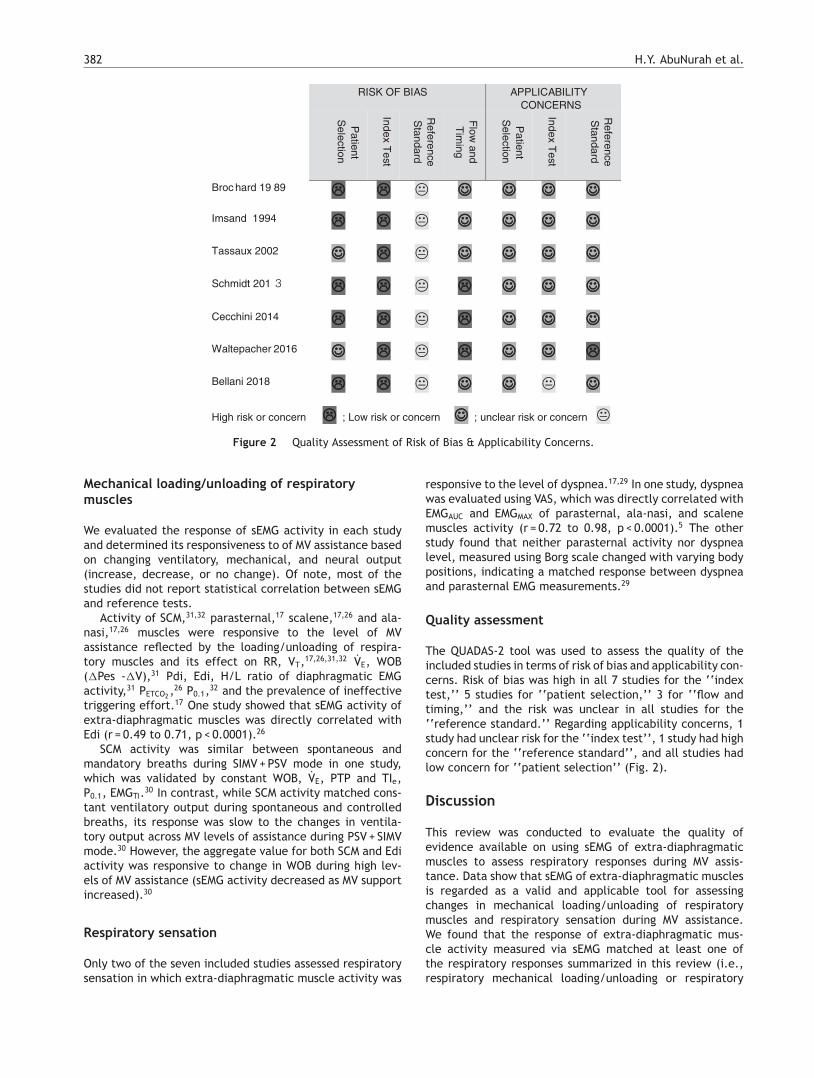
382 H.Y. AbuNurah et al.
RISK OF BIAS APPLICABILITY CONCERNS
Patient
Selection
Index Test
Reference
Standard
Flow
and T
iming
Patient
Sel ection
Index Test
Reference
Sta ndard
Broc hard 19 89 �� �� � ☺☺ ☺☺ ☺☺ ☺☺
Imsand 1994 �� �� � ☺☺ ☺☺ ☺☺ ☺☺
Tassaux 2002 ☺☺ �� � ☺☺ ☺☺ ☺☺ ☺☺
Schmidt 201 3 �� �� � �� ☺☺ ☺☺ ☺☺
Cecchini 2014 �� �� � �� ☺☺ ☺☺ ☺☺
Waltepacher 2016 ☺☺ �� � �� ☺☺ ☺☺ ��
Bellani 2018 �� �� � ☺☺ ☺☺ � ☺☺
High risk or concern �� ; Low risk or concern ☺☺ ; unclear risk or concern �
Figure 2 Quality Assessment of Risk of Bias & Applicability Concerns.
Mechanical loading/unloading of respiratorymuscles
We evaluated the response of sEMG activity in each studyand determined its responsiveness to of MV assistance basedon changing ventilatory, mechanical, and neural output(increase, decrease, or no change). Of note, most of thestudies did not report statistical correlation between sEMGand reference tests.
Activity of SCM,31,32 parasternal,17 scalene,17,26 and ala-nasi,17,26 muscles were responsive to the level of MVassistance reflected by the loading/unloading of respira-tory muscles and its effect on RR, VT,17,26,31,32 VE, WOB(�Pes -�V),31 Pdi, Edi, H/L ratio of diaphragmatic EMGactivity,31 PETCO2 ,26 P0.1,32 and the prevalence of ineffectivetriggering effort.17 One study showed that sEMG activity ofextra-diaphragmatic muscles was directly correlated withEdi (r = 0.49 to 0.71, p < 0.0001).26
SCM activity was similar between spontaneous andmandatory breaths during SIMV + PSV mode in one study,which was validated by constant WOB, VE, PTP and TIe,P0.1, EMGTi.30 In contrast, while SCM activity matched cons-tant ventilatory output during spontaneous and controlledbreaths, its response was slow to the changes in ventila-tory output across MV levels of assistance during PSV + SIMVmode.30 However, the aggregate value for both SCM and Ediactivity was responsive to change in WOB during high lev-els of MV assistance (sEMG activity decreased as MV supportincreased).30
Respiratory sensation
Only two of the seven included studies assessed respiratorysensation in which extra-diaphragmatic muscle activity was
responsive to the level of dyspnea.17,29 In one study, dyspneawas evaluated using VAS, which was directly correlated withEMGAUC and EMGMAX of parasternal, ala-nasi, and scalenemuscles activity (r = 0.72 to 0.98, p < 0.0001).5 The otherstudy found that neither parasternal activity nor dyspnealevel, measured using Borg scale changed with varying bodypositions, indicating a matched response between dyspneaand parasternal EMG measurements.29
Quality assessment
The QUADAS-2 tool was used to assess the quality of theincluded studies in terms of risk of bias and applicability con-cerns. Risk of bias was high in all 7 studies for the ‘‘indextest,’’ 5 studies for ‘‘patient selection,’’ 3 for ‘‘flow andtiming,’’ and the risk was unclear in all studies for the‘‘reference standard.’’ Regarding applicability concerns, 1study had unclear risk for the ‘‘index test’’, 1 study had highconcern for the ‘‘reference standard’’, and all studies hadlow concern for ‘‘patient selection’’ (Fig. 2).
Discussion
This review was conducted to evaluate the quality ofevidence available on using sEMG of extra-diaphragmaticmuscles to assess respiratory responses during MV assis-tance. Data show that sEMG of extra-diaphragmatic musclesis regarded as a valid and applicable tool for assessingchanges in mechanical loading/unloading of respiratorymuscles and respiratory sensation during MV assistance.We found that the response of extra-diaphragmatic mus-cle activity measured via sEMG matched at least one ofthe respiratory responses summarized in this review (i.e.,respiratory mechanical loading/unloading or respiratory

Surface EMG assessment of extra-diaphragmatic muscles during mechanical ventilation 383
sensation) in all studies except for one study, which didnot report sufficient data of extra-diaphragmatic musclesresponses in their results.28 However, the included studieslack the evidence of sEMG accuracy in assessing MV clinicaloutcomes (i.e., respiratory failure, MV liberation readiness,or success/failure). This is partly because these studies didnot primarily use sEMG to determine its diagnostic accuracyand only used it as a research tool to show the effectivenessof various MV interventions.
The matching response of extra-diaphragmatic sEMGactivity with the ventilatory output is due to the activationof these muscles as a compensatory response for increasedventilatory load and NRD, which are associated with weakdiaphragm or under-assistance in patients with MV.11,17,31
During the early phase of a failed MV liberation trial,patients display an increased mechanical load comparedwith those who had successful trial.10 Extra-diaphragmaticmuscle activity is increased to offset the declining ventila-tory function,11 which can explain the strong relationshipbetween dyspnea and EMG activity of extra-diaphragmaticmuscles in one of the included studies in this review.17
In light of this review’s findings, the response of sEMGof extra-diaphragmatic muscle activation to MV assistanceshould be interpreted with caution. The mode of MV canbe a confounding factor that interferes with the mechani-cal or neural output. For example, in one study the use ofVE was a weak parameter as a reference standard duringASV mode because it is pre-set to deliver a constant levelof MV assistance.32 Similarly, inspiratory neuromuscular out-put is pre-programmed for a given level of assistance and notbased on a breath-by-breath basis during SIMV mode.30 Thisexplains the slow response of ventilatory parameters andSCM re-programming after a sustained change in ventilatoryload in one study.30 In addition, ventilatory compensationcan be achieved by both RR and VT to reach a constant VE
with the changing level of MV assistance.17,30,32 One studyreported an increase in RR but did not report VT,29 giv-ing an incomplete picture of ventilatory output, since itcould be interpreted as shallow breathing and not increasedventilatory efficiency as the study implies. Hence, extra-diaphragmatic sEMG activity should be considered with thereporting of both RR and VT collectively as reference stan-dards. Finally, posture should be taken into account as it caninfluence respiratory muscle activation.33 Sitting positioncompared to supine and semi-recumbent positions reducedNRD to the diaphragm and not the parasternal muscle ordyspnea level during MV liberation trial.29 Similarly, sittingposition was found to require less activation of diaphragmand intercostal muscles compared to supine position with noeffect on ventilatory output.34
The high risk of bias reported for patient selection ismainly related to the design of the studies in which randomsampling was not performed or not reported (n = 4)17,26,30,31
and for the exclusion of patients with COPD (n = 1).28 Thehigh risk of bias found in all of the studies for the ‘‘indextest’’ is due to the fact that these studies did not use blind-ing of the researcher to the reference standards. This meansthat sEMG was interpreted with the prior knowledge of thereference standard results, which mainly represent instanta-neous changes in ventilatory output during MV. Likewise, notblinding the researcher to the index test affected the qual-ity of evidence for the ‘‘reference standards.’’ However,
sEMG results often take time for processing which makesthem less likely to generate bias in the instant results of thereference standards (ventilatory output). Hence, in additionto the lack of reporting of blinding, we evaluated the riskof bias for the reference standards to be unclear in all thestudies.
High risk of bias for flow and timing in three studieswas due to the exclusion of a total of 4 patients fromthe analysis in two studies due to the inability to recordscalene sEMG17,26 and one patient for the inability of mea-suring maximum inspiratory pressure (MIP).29 Generally,there were few applicability concerns as the included stud-ies matched most of the quality questions. The only highapplicability concern was regarding the reference standarddue to the incomplete information reported on respira-tory mechanics in one study.29 Also, unclear applicabilityconcern of the index test was found in one study for thelack of sufficient reporting of sEMG of extra-diaphragmaticmuscles.28
This review is limited by couple of factors that affect thequality of evidence of using sEMG of extra-diaphragmaticmuscles as an assessment tool of respiratory responses dur-ing MV. The included studies had small sample sizes and,thus, the evidence of usefulness of this tool across a broadpopulation of patients on MV is limited. Additionally, there isa lack of a systematic and well-designed approach for assess-ing sEMG diagnostic performance, which mainly includes:random sampling of patients, blinding to index test and refe-rence standards, and the use of gold standard referencetests for assessing MV outcomes (i.e., rapid shallow breath-ing index (RSBI) and MIP).
Future directions: The findings of this review may stim-ulate further research to test the accuracy of sEMG as aclinical diagnostic technique, which might help in the deci-sion making of MV liberation. Further studies addressingits diagnostic accuracy should aim to examine its perfor-mance in predicting MV outcomes such as MV liberationsuccess/failure. Additionally, studies should investigate itscost and complexity in comparison with other standardmethods of MV monitoring used in the critical care set-tings.
Conclusion
The use of sEMG of extra-diaphragmatic muscles appears tobe a valid monitoring tool with low applicability concernsfor assessing respiratory mechanical loading/unloading andrespiratory sensation during MV. However, high risk of biaswas associated with the identified studies in introducingthis technique as an assessment tool. This quality flaw wasmainly attributed to the fact that these studies were notprimarily designed to evaluate sEMG diagnostic accuracy ofMV monitoring.
Funding
This research did not receive any specific grant from fundingagencies in the public, commercial, or not-for-profit sectors.

384 H.Y. AbuNurah et al.
Conflicts of interest
The authors have no conflicts of interest to declare.
Authors contribution
- Both Mr. AbuNurah & Dr. Lowman contributed to thereview search, data collection, study design, analysis ofdata, and manuscript preparation. Dr. Russell contributedto the manuscript preparation, review and editing.
- This study was performed at the Department of Physi-cal Therapy, School of Health Professions, University ofAlabama at Birmingham, Birmingham, Alabama.
- No potential conflict of interest relevant to this articlewas reported.
- King Saud bin Abdulaziz University for Health Sciences,KSA, supported Mr. AbuNurah’s work on the present study.
References
1. Levine S, Nguyen T, Taylor N, Friscia ME, Budak MT, Rothenberg P,et al. Rapid disuse atrophy of diaphragm fibers in mechanicallyventilated humans. New Engl J Med. 2008;358(13):1327---35.
2. Jaber S, Jung B, Matecki S, Petrof BJ. Clinical review:Ventilator-induced diaphragmatic dysfunction-human studiesconfirm animal model findings! Crit Care. 2011;15(1):206.
3. Goligher EC, Dres M, Fan E, Rubenfeld GD, Scales DC, HerridgeMS, et al. Mechanical ventilation-induced diaphragm atrophystrongly impacts clinical outcomes. Am J Respir Crit Care Med.2018;197(2):204---13.
4. Goligher EC, Ferguson ND, Brochard LJ. Clinical chal-lenges in mechanical ventilation. Lancet (London, England).2016;387(10030):1856---66.
5. MacIntyre NR. Respiratory mechanics in the patient who isweaning from the ventilator. Respir Care. 2005;50(2):275---86,discussion 284-276.
6. Chiti L, Biondi G, Morelot-Panzini C, Raux M, Similowski T, HugF. Scalene muscle activity during progressive inspiratory loadingunder pressure support ventilation in normal humans. RespirPhysiol Neurobiol. 2008;164(3):441---8.
7. Jung JH, Kim NS. Relative activity of respiratory muscles duringprescribed inspiratory muscle training in healthy people. J PhysTher Sci. 2016;28(3):1046---9.
8. Perlovitch R, Gefen A, Elad D, Ratnovsky A, Kramer MR, HalpernP. Inspiratory muscles experience fatigue faster than the calfmuscles during treadmill marching. Respir Physiol Neurobiol.2007;156(1):61---8.
9. Ratnovsky A, Elad D, Halpern P. Mechanics of respiratory mus-cles. Respir Physiol Neurobiol. 2008;163(1-3):82---9.
10. Laghi F, Cattapan SE, Jubran A, Parthasarathy S, WarshawskyP, Choi Y-SA, et al. Is weaning failure caused by low-frequency fatigue of the diaphragm? Am J Respir Crit Care Med.2003;167(2):120---7.
11. Parthasarathy S, Jubran A, Laghi F, Tobin MJ. Sternomastoid, ribcage, and expiratory muscle activity during weaning failure. JAppl Physiol. 2007;103(1):140---7.
12. Dres M, Schmidt M, Ferre A, Mayaux J, Similowski T, Demoule A.Diaphragm electromyographic activity as a predictor of weaningfailure. Intens Care Med. 2012;38(12):2017---25.
13. Yan S, Kaminski D, Sliwinski P. Inspiratory muscle mechanicsof patients with chronic obstructive pulmonary disease duringincremental exercise. Am J Respir Crit Care Med. 1997;156 3 Pt1:807---13.
14. Sinderby C, Navalesi P, Beck J, Skrobik Y, Comtois N, FribergS, et al. Neural control of mechanical ventilation in respiratoryfailure. Nat Med. 1999;5(12):1433---6.
15. Beck J, Sinderby C, Lindstrom L, Grassino A. Influence of bipolaresophageal electrode positioning on measurements of humancrural diaphragm electromyogram. J Appl Physiol (Bethesda, Md: 1985). 1996;81(3):1434---49.
16. Pillai JB, Vegas A, Brister S. Thoracic complications of nasogas-tric tube: review of safe practice. Interact Cardiovasc ThoracSurg. 2005;4(5):429---33.
17. Schmidt M, Kindler F, Gottfried SB, et al. Dyspnea and sur-face inspiratory electromyograms in mechanically ventilatedpatients. Intensive Care Med. 2013;39(8):1368---76.
18. Dos Reis IMM, Ohara DG, Januario LB, Basso-Vanelli RP, OliveiraAB, Jamami M. Surface electromyography in inspiratory mus-cles in adults and elderly individuals: A systematic review. JElectromyogr Kinesiol. 2019;44:139---55.
19. Kacmarek RM, Berra L, Villar J. On the road to surface mon-itoring of diaphragmatic activity in mechanically ventilatedpatients. Respir Care. 2018;63(11):1457---8.
20. Maarsingh EJ, van Eykern LA, Sprikkelman AB, Hoekstra MO, vanAalderen WM. Respiratory muscle activity measured with a non-invasive EMG technique: technical aspects and reproducibility.J Appl Physiol (Bethesda, Md : 1985). 2000;88(6):1955---61.
21. Duiverman ML, Van Eykern LA, Vennik PW, Koëter GH, Maars-ingh EJW, Wijkstra PJ. Reproducibility and responsivenessof a noninvasive EMG technique of the respiratory musclesin COPD patients and in healthy subjects. J Appl Physiol.2004;96(5):1723---9.
22. Vincent JL, Rello J, Marshall J, Silva E, Anzueto A, Martin CD,et al. International study of the prevalence and outcomes ofinfection in intensive care units. Jama. 2009;302(21):2323---9.
23. Gandevia SC, McKenzie DK. Human diaphragmatic EMG:changes with lung volume and posture during supramaximalphrenic stimulation. J Appl Physiol (Bethesda, Md : 1985).1986;60(4):1420---8.
24. Suh ES, Mandal S, Harding R, Ramsay M, Kamalanathan M,Henderson K, et al. Neural respiratory drive predicts clinicaldeterioration and safe discharge in exacerbations of COPD. Tho-rax. 2015;70(12):1123---30.
25. Zocchi L, Fitting J, Majani U, Fracchia C, Rampulla C, GrassinoA. Effect of pressure and timing of contraction on human ribcage muscle fatigue. Am Rev Respir Dis. 1993;147(4):857---64.
26. Cecchini J, Schmidt M, Demoule A, Similowski T. Increaseddiaphragmatic contribution to inspiratory effort duringneurally adjusted ventilatory assistance versus pressuresupport: an electromyographic study. Anesthesiology.2014;121(5):1028---36.
27. Whiting PF, Rutjes AW, Westwood ME, Mallett S, Deeks JJ,Reitsma JB, et al. QUADAS-2: a revised tool for the qualityassessment of diagnostic accuracy studies. Ann Intern Med.2011;155(8):529---36.
28. Bellani G, Bronco A, Arrigoni Marocco S, Pozzi M, Sala V, EroniaN, et al. Measurement of diaphragmatic electrical activity bysurface electromyography in intubated subjects and its relation-ship with inspiratory effort. Respir Care. 2018;63(11):1341---9.
29. Walterspacher S, Guckler J, Pietsch F, Walker DJ, Kabitz HJ,Dreher M. Activation of respiratory muscles during weaning frommechanical ventilation. J Crit Care. 2017;38:202---8.
30. Imsand C, Feihl F, Perret C, Fitting JW. Regulation of inspi-ratory neuromuscular output during synchronized intermittentmechanical ventilation. Anesthesiology. 1994;80(1):13---22.
31. Brochard L, Harf A, Lorino H, Lemaire F. Inspiratory pressuresupport prevents diaphragmatic fatigue during weaning frommechanical ventilation. Am Rev Respir Dis. 1989;139(2):513---21.

Surface EMG assessment of extra-diaphragmatic muscles during mechanical ventilation 385
32. Tassaux D, Dalmas E, Gratadour P, Jolliet P. Patient-ventilatorinteractions during partial ventilatory support: a preliminarystudy comparing the effects of adaptive support ventila-tion with synchronized intermittent mandatory ventilationplus inspiratory pressure support. Crit Care Med. 2002;30(4):801---7.
33. Segizbaeva MO, Pogodin MA, Aleksandrova NP. Effects of bodypositions on respiratory muscle activation during maximal inspi-ratory maneuvers. Adv Exp Med Biol. 2013;756:355---63.
34. Wanke T, Lahrmann H, Formanek D, Zwick H. Effect of postureon inspiratory muscle electromyogram response to hypercapnia.Eur J Appl Physiol Occup Physiol. 1992;64(3):266---71.

Pulmonol. 2020;26(6):386---397
www.journalpulmonology.org
SPECIAL ARTICLE
Recommendations for interventional pulmonologyduring COVID-19 outbreak: a consensus statementfrom the Portuguese Pulmonology Society
F. Guedesa,b,c,∗, J.P. Boléo-Toméd, L.V. Rodriguese,f, H.N. Bastosg,h,i, S. Campainhaj,M. de Santisk, L. Mota l, A. Bugalhom,n
a Centro Hospitalar do Porto (CHP), Hospital Geral de Santo António (HGSA), Unidade de Broncologia, Servico de Pneumologia,Porto, Portugalb Departamento de Clínicas Veterinárias, Instituto de Ciências Biomédicas de Abel Salazar (ICBAS), Universidade do Porto (UP),Porto, Portugalc Centro de Estudos de Ciência Animal (CECA), Instituto de Ciências, Tecnologias e Agroambiente (ICETA) da Universidade doPorto, Praca Gomes Teixeira, Apartado 55142, 4051-401, Porto, Portugald Pulmonology Department, Hospital Prof. Doutor Fernando Fonseca, Amadora, Portugale Pulmonology Department, Hospital Sousa Martins, Unidade Local de Saúde da Guarda, Guarda, Portugalf Faculty of Health Sciences, University of Beira Interior, Covilhã, Portugalg Department of Pneumology, Centro Hospitalar São João, Porto, Portugalh Faculty of Medicine, University of Porto, Porto, Portugali IBMC/i3S - Instituto de Biologia Molecular e Celular / Instituto de Investigacão e Inovacão em Saúde, University of Porto,Portugalj Pulmonology Department, Vila Nova de Gaia-Espinho Hospital Center, Vila Nova de Gaia, Portugalk Pulmonology Department, Instituto Português de Oncologia (IPO), Coimbra, Portugall Pulmonology Department, Hospital Pulido Valente, Centro Hospitalar Lisboa Norte, Lisboa, Portugalm Pulmonology Department, CUF Infante Santo Hospital and CUF Descobertas Hospital, Lisbon, Portugaln Comprehensive Health Research Centre, Chronic Diseases Research Center (CEDOC), NOVA Medical School, Lisbon, Portugal
Received 24 June 2020; accepted 16 July 2020Available online 5 August 2020
KEYWORDSInterventionalpulmonology;Bronchoscopy;Thoracocentesis;COVID-19;Consensus statement
Abstract Coronavirus disease 2019 (COVID-19) is an emerging infectious disease caused by anovel SARS-CoV-2 pathogen. Its capacity for human-to-human transmission through respiratorydroplets, coupled with a high-level of population mobility, has resulted in a rapid disseminationworldwide. Healthcare workers have been particularly exposed to the risk of infection andrepresent a significant proportion of COVID-19 cases in the worst affected regions of Europe.
Like other open airway procedures or aerosol-generating procedures, bronchoscopy posesa significant risk of spreading contaminated droplets, and medical workers must adapt the
∗ Corresponding author.E-mail address: [email protected] (F. Guedes).
https://doi.org/10.1016/j.pulmoe.2020.07.0072531-0437/© 2020 Sociedade Portuguesa de Pneumologia. Published by Elsevier Espana, S.L.U. This is an open access article under the CCBY-NC-ND license (http://creativecommons.org/licenses/by-nc-nd/4.0/).

Recommendations for interventional pulmonology during COVID-19 outbreak 387
procedures to ensure safety of both patients and staff. Several recommendation documentswere published at the beginning of the pandemic, but as the situation evolves, our thoughtsshould not only focus on the present, but should also reflect on how we are going to deal with thepresence of the virus in the community until there is a vaccine or specific treatment available.It is in this sense that this document aims to guide interventional pulmonology throughoutthis period, providing a set of recommendations on how to perform bronchoscopy or pleuralprocedures safely and efficiently.© 2020 Sociedade Portuguesa de Pneumologia. Published by Elsevier Espana, S.L.U. This is anopen access article under the CC BY-NC-ND license (http://creativecommons.org/licenses/by-nc-nd/4.0/).
Introduction
Coronavirus Disease 2019 (COVID-19), a new infectiousdisease that emerged in early December 2019 in Wuhan(China),1 is triggered by a novel pathogen with phylogeneticsimilarity to what caused the severe acute respiratory syn-drome (SARS) outbreak in 2003, and was called SARS-CoV-2.2
Its capacity for human-to-human transmission and interna-tional air travel facilitated the rapid dissemination on anunprecedented scale to the rest of the world.3,4
In Italy, the latest figures reported that 9% of COVID-19cases were health care workers (HCW), while in Spain therate of medical staff infected reached 26%, the highest inEurope.5 In Portugal, by 12th May 2020, 11.3% of infectionsoccurred in HCW.6 There are at least two explanations forsuch a high number of infected personnel. First, the lack ofproper personal protective equipment (PPE) at the begin-ning of the epidemic, when assisting both confirmed andsuspected patients with COVID-19. Second, the duration ofexposure to infected patients undergoing aerosol-generatingprocedures, such as non-invasive ventilation (NIV) and bron-choscopy, directly resulting in a significant increase in therisk of transmission to HCW.
The Portuguese Society of Pulmonology recently issueda set of recommendations for bronchoscopic procedures,7
shortly after the diagnosis of the first cases in Portugal.The document aimed to guarantee the protection of bothpatients and medical practicioners, and to ensure that thehealthcare workforce would be conserved to fullfill theirmission throughout the period. Since then, a significantamount of scientific evidence has been accumulated; so,the present document gives an update of the available lit-erature, providing practical suggestions for pulmonologistsundergoing bronchoscopy or pleural procedures in the set-ting of the current and post-pandemic phases.
Risk of transmission
Respiratory droplets comprise the main route of SARS-CoV-2 transmission, although airborne transmission is alsopossible through aerosol-generating procedures, such asbronchoscopy.8 One study during the H1N1 pandemic pro-vided experimental evidence that bronchoscopic proceduresincreases more than 4 times the viral copy number per litrein positive air samples.9 While the heavy droplets rapidlysettle, aerosol particles are much smaller (<5−10 �m) and
are dispersed in the air over extensive distances, posing aconsiderable risk of infection in enclosed spaces, speciallyif poorly ventilated.10
The contribution of asymptomatic carriers has also beensubject of debate.11,12 A significant proportion of themhave lung abnormalities on chest CT scans13,14 and a highlevel of viral shedding may be detected in presymptomaticpatients,14 so it is likely that transmission occurs in the earlystages of infection when patients are either minimally symp-tomatic or asymptomatic. Unrecognized patients pose a realchallenge to infection control and, when not promptly han-dled with appropriate airborne precautions, are one of themost critical factors for SARS-CoV-2 infection spread in thehealthcare setting.
Methods
The Portuguese Pulmonology Society appointed FG to chairthis consensus group. Seven national IP specialists wereselected based on their clinical expertise and differentsettings (university vs. non-university hospitals; state vs.private hospitals; pulmonologists vs. critical care special-ists; ...). At the first online consensus meeting, attendedby all members, a primary draft with several sections wascreated. This was shared online and further improved bywritten comments and suggestions. Then, each IP specialistwas assigned a specific section presented in this documentand was responsible for reviewing and evaluating the rel-evant available literature related to the topic. Electronicdatabases (Pubmed, OVID Medline and Embase, Web of Sci-ence, Cochrane Central Register of Controlled Trials) wereused to search for the terms ‘‘COVID-19’’ OR ‘‘SARS-CoV-2’’AND (‘‘bronchoscopy’’ OR ‘‘interventional pulmology’’ OR‘‘thoracentesis’’ OR ‘‘thoracocentesis’’ OR ‘‘pleural effu-sion’’ OR ‘‘pneumothorax’’ OR ‘‘rigid bronchoscopy’’ OR‘‘thoracoscopy’’ OR ‘‘chest drain’’). Position papers frommajor health organizations (US Centers for Disease Con-trol and Prevention, European Centre for Disease Preventionand Control and World Health Organization) and importantscientific societies (European Respiratory Society, EuropeanAssociation for Bronchology and Interventional Pulmonology,American Association for Bronchology and InterventionalPulmonology, World Association for Bronchology and Inter-ventional Pulmonology and British Thoracic Society) werealso reviewed.

388 F. Guedes et al.
In a second online conference the complete draft wasevaluted by all team members and two working groups werecreated. They were responsible for discussing and revis-ing different sections, and editing the text for consistency.Afterwards, the final manuscript was distributed to theconsensus group members and assessed for final approval.
Adaptations of the interventional pulmonology(IP) department
Although there is still some heterogeneity in the definitionand scope of of ‘‘interventional pulmonology’’ (IP), it hasbecome the most widely accepted term to describe the useof techniques for the diagnosis and treatment of a growingnumber of thoracic disorders.15
In the context of this document, the term IP is usedto encompass the concepts of bronchoscopy (diagnosticor therapeutic), advanced bronchoscopy (flexible or rigidbronchoscopy and all its associated techniques), pleu-roscopy (rigid or semi-flexible) and other simpler pleuraltechniques (such as thoracentesis, placement of thoracicdrainage systems and indwelling pleural catheters). Thoughwe acknowledge this wider definition of IP may be contro-versial, it covers all technical domains that most Portuguesepulmonologists need to address, and for the purpose of thisdocument, it positions us to issue general recommendations.In the following subsections, specific scenarios of differ-ent technical specializations will be addressed in order toovercome this broader definition and to apply it better toindividual settings.
The IP department is a high-risk area, given the typeof procedures that are performed with airway manipulationand with multiple staff involved. Although this setting is gen-erally designed to deal with occasional airborne infectiousdiseases, such as tuberculosis, it is not prepared to systemat-ically assess high-risk cases that need additional resources,diminish productivity and effectiveness and generate a hugeworkload.
Thus, each IP unit must rethink their administrativeand logistic circuits in different areas, as well as the typeand timing of performed procedures, to protect both HCWand patients. Moreover, as international health associa-tions advocate, an infection-control program in healthcaresettings should be implemented, consisting of a three-level hierarchy, including administrative, environmental andengineering controls, and personal protection equipment(PPE).16 In the following subsections, each of the abovelisted hierachic levels are briefly presented.
Administrative and organizational issues
Administrative and logistic measures are crucial to ensur-ing safety while still maintaining IP activity.17 Some generalprecautions include:
- All referrals and requests to the IP unit must preferablybe made by telephone or digital means.
- Upon schedule and 24−48 h prior to arrival at the IP Unit,patients should be contacted by telephone and submittedto a pre-screening checklist that includes questions about1) recent symptoms suggestive of COVID-19 (e.g. fever,
cough, chills, muscle pain, shortness of breath/difficultybreathing, headache, sore throat, loss of taste or smell);2) contact with suspicious/confirmed SARS-CoV-2 cases;and 3) occupational exposure.
- Patients who have recent respiratory and infectious symp-toms and/or chest imaging suggestive of COVID-19, shouldhave their elective procedures postponed and rescheduledafter all symptoms are resolved.
- On arrival at the IP Unit, all patients must be askedagain for respiratory symptoms and have their temper-ature checked.
- If possible, all patients should have at least one negativeRT-PCR for SARS-CoV-2 in the 24−48 h preceding the exam.In patients with a positive RT-PCR SARS-CoV-2, the deci-sion to procede with the intervention will be based on theurgency of the procedure (Chart 1 and Table 1).
- The IP unit should keep a record of deferred patients toreschedule their procedures according to the COVID-19outbreak situation, as proposed in Table 2.
Environmental and engineering control
Physical space preparationThe design of strategies to minimize risks and a protocolfitting the characteristics of each specific Unit are crucial.18
- Reception, administrative, clinical and waiting areasshould separate confirmed/high-risk patients fromnegative/low-risk ones. In addition, inpatients should besegregated from outpatients, either by time or physicallocation, to prevent cross infection.
- Specific circuits and written workflow plans must beprepared, covering the pre-procedural area, proceduralroom, post-procedural area, decontamination and repro-cessing. The implementation of a flowchart with differentareas and walking paths using a visual colour zone sys-tem can be useful: 1) red zone for contaminated areas;2) yellow zone for transition areas, and 3) green zone fornon-COVID-19 safe areas19 (Fig. 1A). These need to be for-mulated by internal elements from the IP unit with thecooperation of a multidisciplinary team of hospital mem-bers, including administration, engineers, and infectioncontrol board.
- A specific place to store and retrieve all items requiredfor PPE should be defined inside the Unit.
- A designated area in the Unit should be selected, close tothe procedural suite, for gowning and removal of all PPE,according to hospital protocol and standards, in order toreduce exposure to contaminated particles and droplets.When an anteroom is available, it may be used as an areafor donning and doffing of PPE (Fig. 1B).
- Stations should be created to facilitate frequent handhygiene and to distribute waste containers according tolocal infectious control recommendations. Posters andother visual aids should be placed at strategic locationsaround the intervention suite to act as reminders.
- Emergency procedures in COVID-19 positive patientsshould preferably be performed within the ICU enviro-ment, with controlled airway through cuffed endotrachealtube and assisted ventilation.

Recommendations for interventional pulmonology during COVID-19 outbreak 389
Chart 1 Proposed triage of IP procedures during the COVID-19 outbreak.
Table 1 Priorization of IP exams according to SARS-CoV-2 status and procedure urgency.

390 F. Guedes et al.
Table 2 Schedule of IP procedures according to the stageof COVID-19 pandemic.
COVID-19 in the community IP Unit
Exponential increase of newcases
Urgent cases --- only
Rapid increase of new cases Urgent cases --- onlyElective but not delayable ---evaluate case-by-case
Decrease in new cases Urgent cases --- full capacityElective, but not delayable ---full capacityElective and delayable ---resume partial capacity
Absence of new cases in thelast 2 weeks
Resume all cases with fullcapacity
- Elective procedures should be reserved for COVID-19 neg-ative patients (Chart 1 and Table 1). Nevertheless, theseprocedures should still be performed in a dedicated neg-ative pressure room (see below, ventilation requirement)with strict isolation precautions and sufficient ventilationto avoid aerosol contamination.20 If these requirementsare not met in the bronchoscopy suite, then in a differentvenue, such as an operating theatre, isolation room or theICU with negative pressure, if available.
- If negative pressure rooms are unavailable througout theinstituition, a specific and dedicated room with adequatenatural ventilation (see requirement below) may be analternative, provided that appropriate intervals betweenprocedures are reserved and that the suspected COVID-19cases be programmed after all planned non-COVID dailyactivity, so that the unit can be carefully cleaned (follow-ing the disinfection policy) and ventilated.
- Keep the endoscopy room for procedures only (all otheractivities, such as planning, reporting and laboratory req-uisition should take place elsewhere).
- Suspected and confirmed cases of COVID-19 must beplaced in an airborne infection isolation room with neg-ative pressure before and after the procedure. Low-riskand negative patients can remain in the pre-procedural
and recovery area, if there is adequate room ventilation,protective equipment (e.g. surgical mask) and physicaldistance (>2 m) from other negative patients.
Ventilation
- Patient source control strategies, such as wearing a maskshould be encouraged.
- Whenever feasible, it is recommended procedures arepereformed in a room that meets the ventilation require-ments for Airborne Infection Isolation (AII), ensuring thedilution and removal of contaminated air. The preferredsystem is a negative pressure room with at least 12 airchanges per hour (ACH) with airflow direction control(single-pass or recirculation systems with HEPA filtration).Alternatively, natural ventilation with an airflow of atleast 160 L/s is an option.19,21
- Enough time should be allowed to ensure that contam-inated air is removed from the room before performinganother procedure in the same room (depending on ACHand disinfection methods, but at least 30 min). Localadaptations must be considered according to the charac-teristics of the IP unit.
Cleaning and disinfecting patient care equipment androomsEndoscopes are considered semi-critical medical instru-ments according to the Spaulding classification.22 Rec-ommendations from the Centers for Disease Controland Prevention (CDC) on reprocessing procedures shouldbe followed. These include pre-cleaning, leak-testing,manual cleaning and visual inspection followed by disinfec-tion/sterilization.
- A high-level manual disinfection or using an automatedendoscope reprocessor is recommended.
- Proper storage and documentation are also an integralpart of the reprocessing workflow.
- A pathway of contaminated equipment must be defined,as well as adequate packaging to minimize exposure (forexample, a hermetic box).
- If available, disposable bronchoscopes are recommendedfor confirmed COVID-19 patients with clear advantages in
Figure 1 A. Implementation of specific circuits with colour visual zone system to distinguish contaminated (1, red zone), transition(2, yellow zone) and safe cleaned areas (3, green zone). B. Designated area for donning and doffing of PPE, where posters and othervisual aids were placed strategically to act as reminders.
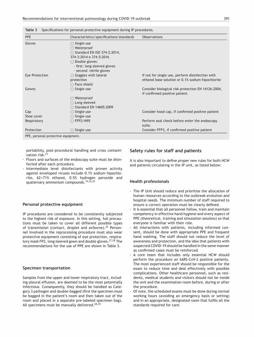
Recommendations for interventional pulmonology during COVID-19 outbreak 391
Table 3 Specifications for personal protective equipment during IP procedures.
PPE Characteristics/specifications/standards Observations
Gloves © Single-use© Waterproof© Standard EN ISO 374-2:2014,374-3:2014 e 374.5:2016© Double gloves:
- first: long sleeved gloves- second: nitrile gloves
Eye Protection © Goggles with lateralprotection
If not for single use, perform disinfection withethanol base solution or 0.1% sodium hipochlorite
© Face shieldGowns © Single-use Consider biological risk protection EN 14126:2004,
if confirmed positive patient© Waterproof© Long sleeved© Standard EN 14605:2009
Cap © Single-use Consider hood cap, if confirmed positive patientShoe cover © Single-useRespiratory © FFP2/N95 Perform seal check before enter the endoscopy
suite.Protection © Single-use Consider FFP3, if confirmed positive patient
PPE, personal protective equipment.
portability, post-procedural handling and cross contami-nation risk.23
- Floors and surfaces of the endoscopy suite must be disin-fected after each procedure.
- Intermediate level disinfectants with proven activityagainst enveloped viruses include 0.1% sodium hipochlo-rite, 62---71% ethanol, 0.5% hydrogen peroxide andquaternary ammonium compounds.19,24,25
Personal protective equipment
IP procedures are considered to be consistently subjectedto the highest risk of exposure. In this setting, full precau-tions must be taken to cover all different possible typesof transmission (contact, droplet and airborne).26 Person-nel involved in the reprocessing procedure must also wearprotective equipment consisting of eye protection, respira-tory mask FP2, long sleeved gown and double gloves.27,28 Therecommendations for the use of PPE are shown in Table 3.
Specimen transportation
Samples from the upper and lower respiratory tract, includ-ing pleural effusion, are deemed to be the most potentiallyinfectious. Consequently, they should be handled as Cate-gory 3 pathogen and double-bagged (first the specimen mustbe bagged in the patient’s room and then taken out of theroom and placed in a separate pre-labeled specimen bag).All specimens must be manually delivered.28,29
Safety rules for staff and patients
It is also important to define proper new rules for both HCWand patients circulating in the IP unit, as listed below:
Health professionals
- The IP Unit should reduce and prioritise the allocation ofhuman resources according to the outbreak evolution andhospital needs. The minimum number of staff required toensure a correct operation must be clearly defined.
- It is essential that all personnel follow, train and maintaincompetency in effective hand hygiene and every aspect ofPPE (theoretical, training and simulation sessions) so thateveryone is familiar with their role.
- All interactions with patients, including informed con-sent, should be done with appropriate PPE and frequenthand washing. The staff should not reduce the level ofawareness and protection, and the idea that patients withsuspected COVID-19 should be handled in the same manneras confirmed cases must be reinforced.
- A core team that includes only essential HCW shouldperform the procedure on SARS-CoV-2 positive patients.The most experienced staff should be responsible for theexam to reduce time and deal effectively with possiblecomplications. Other healthcare personnel, such as resi-dents, medical students and visitors should not be insidethe unit and the examination room before, during or afterthe procedure.
- Of note, the scheduled exams must be done during normalworking hours (avoiding an emergency basis or setting)and in an appropriate, designated room that fulfils all thestandards required for care.

392 F. Guedes et al.
Patients and other personel
- Respiratory and contact isolation should be standard andmandatory for all patients. Outpatients and inpatientsshould always enter the IP Unit with a suitable face maskand keep it on at all times (until the beginning and afterthe end of the procedure) to minimize the risk of trans-mission. No unnecessary personal items should be broughtinto the IP unit.
- Family members and caregivers should not stay in the IPwaiting rooms. In case of children or patients in need ofsupport, the Unit can allow a single relative to enter thepreparation area to provide aid.
- The entry into the Unit of suppliers and medical devicessales representatives must be restricted.
Prioritization of procedures
Scheduled elective procedures should be reviewed and can-celled if potentially delayable, until local control of theoutbreak is achieved. After flattening the infectious curve,many elective IP procedures will have to be performed, asthey are essential to provide a definitive diagnosis and effec-tive treatment. At this time, it is advisable to evaluate thedelayed requests and to optimize the procedure planningbased on clinical needs and operational capability.
A suggested rational approach for stratification of pro-cedures is provided in Table 1, but we recognize that, incertain cases, the indication may not be straightforward,and the risk-benefit must be weighted on an individual basisby the IP team.31 Although rescheduling certain proceduresis obvious in other cases it may not be desirable or eth-ical. It is important to note that these indications maychange according to local epidemiological conditions andthe response capabilities of the healthcare system. Severalsocieties have recommended different levels of procedurestratification.26,30,31 Briefly, what is recommended is a step-wise reopening of elective IP procedures according to thenational and local COVID-19 outbreak situation, dependingon the number of new confirmed cases, hospital admittedcases (ward and ICU), availability of equipment and health-care staff, time elapse and number of postponed IP cases.Some authors34 have proposed a summary of the electiveendoscopic procedure by phases, as shown in Table 2. Any-way, it should be noted that the evolving procedural criteriashould always be communicated to other physicians whorefer patients for invasive respiratory procedures and to thehospital administration.
Recommendations for bronchoscopy
Bronchoscopy under spontaneous ventilation
The following reccomendations are expert opinion-basedand should be adapted to local regulations and guidelines.In an optimal scenario, it is safer to perform electivebronchoscopy under general anesthesia and orotrachealintubation, clinical conditions permitting. If this is not pos-sible, bronchoscopy can be performed under spontaneousventilation. Some recommendations are listed below:
- Operator should be standing behind the patient’s head toreduce direct exposure. Oxygen supplementation shouldbe done without the use of humidification, either througha nasal cannula or preferably with an oxygen mask withan entrance to the bronchoscope (Fig. 2A).
- For flexible bronchoscopy, a transnasal approach should bepreferred, and a surgical mask should be placed over thepatient’s mouth to minimize droplet emission (Fig. 2B).
- In hypoxemic patients, bronchoscopy can be performedunder NIV, using a closed circuit ventilation (double cir-cuit with viral filters in both arms) and non-ventilatedmasks with a dedicated bronchoscope entrance (Fig. 2C).High performance NIV ventilators with FiO2 regulation arepreferable. From the end of the procedure, NIV should becontinued for 1−2 h, titrating the FiO2 to obtain an SpO2
of around 94---95%.- Bronchoscopy under nasal high-flow oxygen therapy is not
reccomended and thus should be avoided.- Nebulized medications should be avoided before or after
the procedure.- Proper sedation should be used to minimize cough reflex
and to increase patient cooperation.- An oral aspiration cannula should be available during the
procedure (Fig. 2B).- A transparent protective box may enhance safety by con-
taining dispersal of droplet particles (Fig. 2D). The box isplaced over the patient’s head prior to bronchoscopy, withthe anesthesia equipment already in place. The broncho-scope is inserted through the covered opening behind thepatient (Fig. 2E).
Bronchoscopy in the intubated patient
The following recommendations are directed for patientsunder mechanical ventilation in an ICU setting due torespiratory failure. As reported, 5% of COVID-19 patientscan develop respiratory failure and will need ventilatorysupport32; moreover, associated bacterial, viral and fun-gal co-infection should not be negleted.33 In critically illpatients under invasive ventilation, ventilator-associatedpneumonia occurs in up to 30% and lobar collapse is fre-quent and multifactorial.34 The same adaptations apply toelective procedures under general anesthesia, performed inthe Bronchoscopy Unit or Operating Theatre.
- A cuffed endotracheal tube is preferred over supragloticdevices, such as a laryngeal mask; cuff pressure should bemaintained between 25---30 cmH2O.35
- General anesthesia with muscle relaxant is recommendedto reduce the aerosol production.
- FiO2 should be adjusted to 100%.- Volume control, pressure-limited mode is preferable and
PEEP should be kept at the same level during the proce-dure. Adjustments can be made dynamically, with a priorassessment of the anticipated risks (e.g., lung derecruit-ment and desaturation, arrhythmias, pneumothorax).
- To avoid aerosol dispersion, a simple and appropriatemaneuver consists of clamping the ventilation circuit justbefore introduction of bronchoscope, repeating the samestep just before withdrawal.
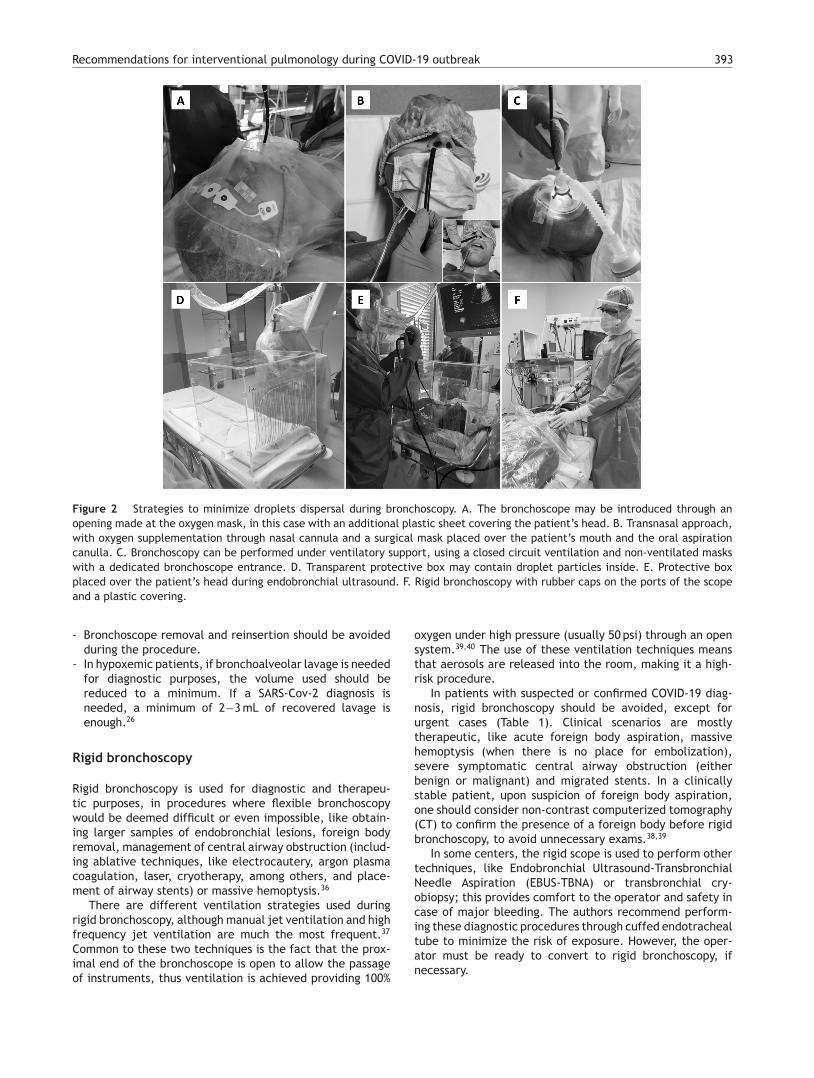
Recommendations for interventional pulmonology during COVID-19 outbreak 393
Figure 2 Strategies to minimize droplets dispersal during bronchoscopy. A. The bronchoscope may be introduced through anopening made at the oxygen mask, in this case with an additional plastic sheet covering the patient’s head. B. Transnasal approach,with oxygen supplementation through nasal cannula and a surgical mask placed over the patient’s mouth and the oral aspirationcanulla. C. Bronchoscopy can be performed under ventilatory support, using a closed circuit ventilation and non-ventilated maskswith a dedicated bronchoscope entrance. D. Transparent protective box may contain droplet particles inside. E. Protective boxplaced over the patient’s head during endobronchial ultrasound. F. Rigid bronchoscopy with rubber caps on the ports of the scopeand a plastic covering.
- Bronchoscope removal and reinsertion should be avoidedduring the procedure.
- In hypoxemic patients, if bronchoalveolar lavage is neededfor diagnostic purposes, the volume used should bereduced to a minimum. If a SARS-Cov-2 diagnosis isneeded, a minimum of 2−3 mL of recovered lavage isenough.26
Rigid bronchoscopy
Rigid bronchoscopy is used for diagnostic and therapeu-tic purposes, in procedures where flexible bronchoscopywould be deemed difficult or even impossible, like obtain-ing larger samples of endobronchial lesions, foreign bodyremoval, management of central airway obstruction (includ-ing ablative techniques, like electrocautery, argon plasmacoagulation, laser, cryotherapy, among others, and place-ment of airway stents) or massive hemoptysis.36
There are different ventilation strategies used duringrigid bronchoscopy, although manual jet ventilation and highfrequency jet ventilation are much the most frequent.37
Common to these two techniques is the fact that the prox-imal end of the bronchoscope is open to allow the passageof instruments, thus ventilation is achieved providing 100%
oxygen under high pressure (usually 50 psi) through an opensystem.39,40 The use of these ventilation techniques meansthat aerosols are released into the room, making it a high-risk procedure.
In patients with suspected or confirmed COVID-19 diag-nosis, rigid bronchoscopy should be avoided, except forurgent cases (Table 1). Clinical scenarios are mostlytherapeutic, like acute foreign body aspiration, massivehemoptysis (when there is no place for embolization),severe symptomatic central airway obstruction (eitherbenign or malignant) and migrated stents. In a clinicallystable patient, upon suspicion of foreign body aspiration,one should consider non-contrast computerized tomography(CT) to confirm the presence of a foreign body before rigidbronchoscopy, to avoid unnecessary exams.38,39
In some centers, the rigid scope is used to perform othertechniques, like Endobronchial Ultrasound-TransbronchialNeedle Aspiration (EBUS-TBNA) or transbronchial cry-obiopsy; this provides comfort to the operator and safety incase of major bleeding. The authors recommend perform-ing these diagnostic procedures through cuffed endotrachealtube to minimize the risk of exposure. However, the oper-ator must be ready to convert to rigid bronchoscopy, ifnecessary.

394 F. Guedes et al.
In a patient undergoing rigid bronchoscopy, it is recom-mended that:
- Rigid bronchoscopy should always be performed in a neg-ative pressure room.
- Controlled ventilation is preferred, with the rigid bron-choscope used like an endotracheal tube.
- Air leaks should be reduced using rubber caps on theports of the rigid scope, as well as using a plastic cover-ing (Fig. 2F) or filling the mouth with gauze.40 While thisstrategy is more appealing to minimize aerosol spread,the operator may find it challenging to handle instrumentsthrough the working channel.
Recommendations for pleural techniques
Pleural effusion does not appear to be a prominent featureof COVID-19. It occurs in 5.3---5.8% of patients, according totwo recent meta-analyses.41,42 There have been occasionalreports of bilateral effusion that resolved spontaneously.43
As bacterial superinfection is common in severe patients,they can also develop complicated effusions or empyema,requiring targeted treatment. There have been a fewanecdotal reports of spontaneous pneumothorax and pneu-momediastinum in severe COVID-19 pneumonia, requiringdrainage.44,45 This may be more frequent in critical patientson invasive ventilation, which can lead to bronchopleuralfistulae.46 It is, therefore, plausible that pleural drainagemay be necessary in some COVID-19 patients, either in theICU or in the ward, and indications for drainage do notdiffer from the standard clinical guidelines. However, aswith any invasive procedure in confirmed COVID-19 patients,all precautions regarding the full use of protective equip-ment should be taken. The procedure must be performedby trained and dedicated staff to reduce its duration andto minimize the risk of complications. In other situations,the use of ultrasound may be very helpful with COVID-19 patients.47 Besides its wide availability, safety and lowcost, it is easy to use at the bedside and allows medicalstaff to detect small pleural effusions and to guide pleuralfluid collection and drainage, if needed. On the other hand,even patients without suspected COVID-19 can have assymp-tomatic infection; so, any procedure should be considered asa possible COVID-19 case and precautions should be taken.Indeed, although some procedures may be postponed, inmany situations they should not be deferred, especially insuspected or confirmed cancer patients. It is crucial thatcancer patients do not experience delays in diagnostic ortherapeutic procedures due to the present contingency.48
Few societies have published guidelines addressing pleu-ral procedures during COVID-19 pandemic. The BritishThoracic Society has issued guidance on pleural ser-vices provision,49 mainly to minimize hospital visits andadmissions and to ensure both patient and staff safety.Nonetheless, although we recognize lack of published evi-dence supporting these reccomendations, this documentwill adopt some of them.
First of all, despite pleural procedures not being listedas Aerosol Generating Procedure (AGP) in the CDC updatedreccomendations,50 they should be considered so and Level2 PPE should be worn, as described above. Other societies
have considered potential AGP as any procedure ‘‘likelyto induce coughing, that should be performed cautiouslyand avoided if possible’’.51 This is particularly relevant toopen procedures, such as thoracoscopy and indwelling pleu-ral catheter insertion, and in case of pneumothorax withsuspected bronchial-pleural fistula. Thus, we recommendtaking the following precautions:
Pleural effusion
- On suspicion of malignant effusion, diagnostic pleural fluidaspiration should be performed, especially if the patientis a candidate for systemic therapy or if the effusion issymptomatic (Table 1).
- Consider placement of indwelling pleural catheters forrecurrent malignant pleural effusions, avoiding repeatedhospital drainage or admission.
- On suspicion of pleural infection, pleural fluid aspirationand analysis should be performed.
- In case of benign pleural effusions, the risk-benefit shouldbe discussed with the attending physician; procedures canbe postponed if the patient is not symptomatic.
- Thoracoscopy is not recommended as a principle; how-ever, in the absence of alternative options, its risk-benefitmust be assessed.
Pneumothorax
- Spontaneous primary pneumothorax can be managed inoutpatient care if the risk assessment allows and if thereis local team experience; needle aspiration and dischargeshould be considered if the patient is minimally symp-tomatic.
- When pleural drainage placement is necessary, if there islocal expertise and the patient is at low risk, the use ofpleural vent systems should be considered, thus allowingthe patient to be managed at home.
Chest drainage placement and care
- Extra care must be taken when placing the chest tubes, inorder to avoid open communication with the pleural spaceand the potentialemission of droplets and aerossols.
- When chest tubes are placed in ventilated patients, con-sideration should be given to clamping the ventilatorcircuit before assessing the pleural cavity, so that positivepressure spreading of pleural air or fluid can be prevented.
- Whenever possible, the use of non-wired pleural drainageshould be considered; it can be connected to the drainagesystem before insertion into the pleural cavity (closed cir-cuit).
- When pleuroscopy is required, the use of one way valvetrocars should be preferred to assess the pleural cavityand properly seal the entrance port of the pleuroscope.
- In case of prolonged air-leaks, the use of wall suctionshould be weighted to create a closed system.

Recommendations for interventional pulmonology during COVID-19 outbreak 395
Table 4 IP Unit checklist during COVID-19 outbreak.
Concluding remarks
As with other societal consensus papers, this documentwas developed by a restricted panel of experts fromthe Portuguese Society of Pulmonology; individual clini-cal judgment and local resources may lead to alternativeperspectives. This guidance was based on the current knowl-edge of COVID-19, but, as new data appears, this statementshould be revised in the future to accommodate updatedrecommendations. At present, one of the controversialassumptions is that, every patient, even if asymptomatic,should be assumed as potentially infected with SARS-CoV-2. Therefore, it is mandatory that contact precautions andproper training on donning and doffing of PPE be providedto all HCWs involved in IP. Another key element is to planin advance and keep each IP Unit well-organized (Table 4).Although the reduction in the number of elective proceduresrepresents one of the central strategies to improve safety,it is crucial that patients do not suffer unnecessary delays indiagnostic or therapeutic procedures due to the current con-tingency. Taken together, our ultimate intention is to bringfull attention to this and future outbreaks or other emergingmedical situations.
Conflicts of interest
The authors have no conflicts of interest to declare.
References
1. Huang C, Wang Y, Li X, Ren L, Zhao J, Hu Y, et al.Clinical features of patients infected with 2019 novel coro-navirus in Wuhan, China. Lancet. 2020;395(10223):497---506,http://dx.doi.org/10.1016/S0140-6736(20)30183-5.
2. Zhu N, Zhang D, Wang W, et al. A novel coronavirus from patientswith pneumonia in China, 2019. N Engl J Med. 2020;382:727---33.
3. Lu R, Zhao X, Li J, Niu P, Yang B, Wu H, et al. Genomiccharacterisation and epidemiology of 2019 novel coronavirus:implications for virus origins and receptor binding. Lancet.2020;395(10224):565---74.
4. World Health Organization (WHO). Coronavirus disease 2019(COVID-19) situation report 99; 2020.
5. European Center for Disease Prevention and Control. Coron-avirus disease 2019 (COVID-19) in the EU/EEA and the UK ---eighth update, 8 April 2020. https://www.ecdc.europa.eu/sites/default/files/documents/covid-19-rapid-risk-assessment-coronavirus-disease-2019-eighth-update-8-april-2020.pdf(accessed on 15th June 2020).
6. https://covid19.min-saude.pt/ (accessed 15th June 2020).7. https://www.sppneumologia.pt/uploads/subcanais2 conteudos
ficheiros/posicao-da-spp-para-a-realizacao-de-broncoscopias-durante-a-pandemia-do-coronavirus-(2).pdf (accessed on 15th
June 2020).8. Wilson NM, Norton A, Young FP, Collins DW. Airborne trans-
mission of severe acute respiratory syndrome coronavirus-2to healthcare workers: a narrative review. Anaesthesia.2020;(April), http://dx.doi.org/10.1111/anae.15093.
9. Thompson KA, Pappachan JV, Bennett AM, et al. Influenzaaerosols in UK hospitals during the H1N1 (2009) pandemic----therisk of aerosol generation during medical procedures. PLoS One.2013. PMID: 23418548.
396 F. Guedes et al.
10. Tellier R, Li Y, Cowling BJ, Tang JW. Recognition of aerosol trans-mission of infectious agents: a commentary. BMC Infect Dis.2019;19, 101-101.
11. Mizumoto K, Kagaya K, Zarebski A, et al. Estimating the asymp-tomatic proportion of coronavirus disease 2019 (COVID-19)cases on board the Diamond Princess cruise ship, Yokohama,Japan, 2020. Euro Surveill. 2020. PMID: 32183930.
12. Nishiura H, Kobayashi T, Miyamaet T, et al. Estimation of theasymptomatic ratio of novel coronavirus infections (COVID-19).Int J Infect Dis. 2020. PMID: 32179137 f.
13. Inui S, Fujikawa A, Jitsu M, Kunishima N, et al. Chest CT findingsin cases from the cruise ship ‘‘Diamond Princess’’ with Coron-avirus Disease 2019 (COVID-19). Radiol: Cardiothorac Imaging.2020;2(2):e200110.
14. Zou L, Ruan F, Huanget M, et al. SARS-CoV-2 Viral Load in UpperRespiratory Specimens of Infected Patients. N Engl J Med. 2020.PMID: 32074444.
15. Seijo LM, Sterman DH. Interventional pulmonology. NEJM.2001;344(10):740---9.
16. WHO Technical guidance. https://www.who.int/emergencies/diseases/novel-coronavirus-2019/technical-guidance/infection-prevention-and-control, 2020 (accessed on 15th June 2020).
17. Centers for Disease Control and Prevention. Interim Infec-tion Prevention and Control Recommendations for Patientswith Suspected or Confirmed Coronavirus Disease 2019(COVID-19) in Healthcare Settings. CDC. 13 April 2020.https://www.cdc.gov/coronavirus/2019-ncov/hcp/infection-control-recommendations.html (accessed on 15th June 2020).
18. Baldwin D.R., Lim W.S., Rintoul R., Navani N., Fuller L., Wool-house I., et al. Recommendations for day case bronchoscopyservices during the COVID-19 pandemic. British Thoracic Soci-ety. Version 1.2. March 2020.
19. World Health Organization. Health Systems Respond to COVID-19 Technical Guidance #2 Creating surge capacity for acute andintensive care Recommendations for the WHO European Region6 April 2020.
20. Adhikari U, Chabrelie A, Weir M, Boehnke K, McKenzie E,Ikner L, et al. A case study evaluating the risk of infec-tion from middle eastern respiratory syndrome coronavirus(MERS-CoV) in a hospital setting through Bioaerosols. Risk Anal.2019;39(12):2608---24.
21. American Institute of Architects. Guidelines for design and cons-truction of hospital and health care facilities, 2001. Washington,DC: American Institute of Architects Press; 2001.
22. Rutala WA, Weber DJ. Disinfection of endoscopes: review ofnew chemical sterilants used for high-level disinfection. InfectControl Hosp Epidemiol. 1999;20:69---76.
23. Wahidi MM, Lamb C, Murgu S, et al. American Association forBronchology and Interventional Pulmonology (AABIP) statementon the use of bronchoscopy and respiratory specimen collectionin patients with suspected or confirmed COVID-19 infection.March 9, 2020 and expert consensus of the AABIP COVID-19Task Force. J Bronchology Interv Pulmonol. 2020;(March),http://dx.doi.org/10.1097/LBR.0000000000000681https://aabronchology.org/wp-content/uploads/2020/03/2020-AABIP-Statement-on-Bronchoscopy-COVID.GAE-updated-Version.pdf (accessed on 15th june 2020.
24. Centers for Disease Control and Prevention. Guidelinefor Disinfection and Sterilization in Healthcare Facil-ities, 2008. May 2019 update. https://www.cdc.gov/infectioncontrol/pdf/guidelines/disinfection-guidelines-H.pdf(accessed on 15th june 2020).
25. Chen CC, Chi CY. Biosafety in the preparation and processingof citology specimens with potential coronavirus (COVID-19) infection: perspective from Taiwan. Cancer Cytopathol.2020;128(May (5)):309---16.
26. Tran K, Cimon K, Severn M, et al. Aerosol generatingprocedures and risk of transmission of acute respiratory infec-
tions to healthcare workers: a systematic review. PLoS One.2012;7:e35797.
27. Healthcare Infection Control Practices Advisory Committee.Essential Elements of a Reprocessing Program for FlexibleEndoscopes --- The Recommendations of the Healthcare Infec-tion Control Practices Advisory Committee (HICPAC). 2016.https://www.cdc.gov/hicpac/pdf/flexible-endoscoperepro-cessing.pdf (accessed on 15th June 2020).
28. Pritchett MA, Oberg CL, Belanger A. Society for advanced bron-choscopy consensus statement and guidelines for bronchoscopyand airway management amid the COVID-19 pandemic. J ThoracDis. 2020;12(5):1781---98.
29. World Health Organization (WHO). Laboratory testingfor Coronavirus Disease 2019 (COVID-19) in sus-pected human cases: interim Guidance, 19March 2020.https://www.who.int/publications/i/item/10665-331501(accessed on 15th June 2020).
30. Mazzone PJ, Gould MK, Arenberg DA, et al. Managementof lung nodules and lung cancer screening during theCOVID-19 pandemic: CHEST Expert Panel Report. Chest.2020;(April), http://dx.doi.org/10.1016/j.chest.2020.04.020,pii: S0012-3692(20)30758-3.
31. Wahidi M.M., Lamb C., Murgu S., et al. March 19th, 2020Update to the American Association for Bronchology andInterventional Pulmonology (AABIP) Statement on the Useof Bronchoscopy and Respiratory Specimen Collection inPatients with Suspected or Confirmed COVID-19 Infection.https://aabronchology.org/wp-content/uploads/2020/03/AABIP-Statement-on-Bronchoscopy-COVID 3-12-2020-Statement-plus-3-19-2020-updates-V3.pdf (accessed on 15th June 2020).
32. Wu Z, McGoogan JM. Characteristics of and important lessonsfrom the coronavirus disease 2019 (COVID-19) outbreak inChina: summary of a report of 72 314 cases from theChinese Center for Disease Control and Prevention. Jama.2020;323(13):1239---42.
33. Ding Q, Lu P, Fan Y, et al. The clinical characteristics of pneu-monia patients co-infected with 2019 novel coronavirus andinfluenza virus in Wuhan, China. J Med Virol. 2020;(March),http://dx.doi.org/10.1002/jmv.25781.
34. Hellyer TP, McAuley DF, Walsh TS, et al. Biomarker-guidedantibiotic stewardship in suspected ventilator-associated pneu-monia (VAPrapid2): a randomised controlled trial and processevaluation. Lancet Respir Med. 2020;8(2):182---91.
35. Biselli P, Fricke K, Grote L, et al. Reductions in dead spaceventilation with nasal high flow depend on physiological deadspace volume: metabolic hood measurements during sleep inpatients with COPD and controls. Eur Respir J. 2018;51(5):1702251.
36. Batra H, Yannus L. Indications and complications of rigid bron-choscopy. Expert Rev Respir Med. 2018;12(June (6)):509---20.
37. Pathak V, Welsby V, Mahmood K, et al. Ventilation and anes-thetic approaches for rigid bronchoscopy. Ann Am Thorac Soc.2014;11(May (4)):628---34.
38. Pitiot V, Grall M, Ploin D, et al. The use of CT-scan in foreign bodyaspiration in children: a 6 years’ experience. Int. J. Pediatr.Otorhinolaryngol. 2017;102:169---73.
39. Ahmed OG, Guillerman RP, Giannoni CM. Protocol incorporatingairway CT decreases negative bronchoscopy rates for suspectedforeign bodies in pediatric patients. Int J Pediatr Otorhinolaryn-gol. 2018;109:133---7.
40. Yarmus L, Feller-Kopman D. New bronchoscopic instrumenta-tion: a review and update in rigid bronchoscopy. In: BeamisJF, Mathur PN, Mehta AC, editors. Interventional pulmonarymedicine. Lung biology in health and disease 230. New York:Informa Healthcare; 2010. p. 1---9.
41. Zhu J, Zhong Z, Li H, et al. CT imaging features of 4,121patients with COVID-19: a meta-analysis. J Med Virol. 2020,http://dx.doi.org/10.1002/jmv.25910 [Epub ahead of print].
Recommendations for interventional pulmonology during COVID-19 outbreak 397
42. Salehi S, Abedi A, Balakrishnan S, et al. Coronavirus disease2019 (covid-19): a systematic review of imaging findings in 919patients. AJR Am J Roentgenol. 2020;(March):1---7.
43. Lin C, Ding Y, Xie B, Sun Z, Li X, Chen Z, et al. Asymp-tomatic novel coronavirus pneumonia patient outside Wuhan:the value of CT images in the course of the disease. Clin Imaging.2020;63:7---9.
44. Chen N, Zhou M, Dong X, et al. Epidemiological and clinicalcharacteristics of 99 cases of 2019 novel coronavirus pneumoniain Wuhan, China: a descriptive study. Lancet. 2020;(January).S0140-6736(20)30211-30217.
45. Sun R, Liu H, Wang X. Mediastinal emphysema, giantbulla, and pneumothorax developed during the courseof covid-19 pneumonia. Korean J Radiol. 2020;(March),http://dx.doi.org/10.3348/kjr.2020.0180 [Epub ahead ofprint].
46. Aiolfi A, Biraghi T, Montisci A, et al. Management of per-sistent pneumothorax with thoracoscopy and blebs resectionin covid-19 patients. Ann Thorac Surg. 2020;(April). S0003-4975(20)30604-4.
47. Peng Q, Wang X, Zhang L. Findings of lung ultrasonography ofnovel corona virus pneumonia during the 2019---2020 epidemic.Intensive Care Med. 2020;46(May (5)):849---50.
48. Recomendacões da Sociedade Portuguesa de Pneumologia edo Grupo de Estudos de Cancro do Pulmão na abordagemdos doentes com Cancro do Pulmão durante a Pandemia deCOVID-19. https://www.sppneumologia.pt/uploads/subcanaisconteudos ficheiros/orientacoes-da-ct-de-pneumologia-oncologica-da-spp-no-cancro-do-pulmao-durante-a-pandemia-de-covid-19.pdf (accessed on 15th June 2020).
49. Hallifax R., Wrightson J.M., Bibby A., et al. Pleural servicesduring the COVID-19 Pandemic. British Thoracic Society.https://www.brit-thoracic.org.uk/about-us/covid-19-informa-tion-for-the-respiratory-community (accessed on 15th June2020).
50. Centers for Disease Control and Prevention. HealthcareInfection Prevention and Control FAQs for COVID-19.https://www.cdc.gov/coronavirus/2019-ncov/hcp/infection-control-faq.html (accessed on 15th June 2020).
51. Society of Interventional Radiology. Aerosol GeneratingProcedures Performed by Interventional Radiology ClinicalNotification from the Society of Interventional Radiol-ogy. https://www.sirweb.org/practice-resources/covid-19-resources/covid-19-clinical-notification-3-26-20 (accessed on15th June 2020).

Pulmonol. 2020;26(6):398---417
www.journalpulmonology.org
LETTER TO THE EDITOR
Endobronchialultrasound-transvascular needleaspiration (EBUS-TVNA) in thediagnosis of a hilar metastasis of anextrapulmonary neoplasm
To the Editor,
Convex-probe endobronchial ultrasound-transbronchial nee-dle aspiration (EBUS-TBNA) and esophageal ultrasound withultrasound bronchoscope (EUS-B) are nowadays the inter-ventional pulmonology cornerstone of non-small-cell lungcancer mediastinal and hilar staging; these techniques alsoallow the diagnosis of mediastinal and hilar involvementrelated to extrapulmonary primary neoplasms, infectionsand inflammatory diseases.
Mediastinal and hilar lymph node stations beyond majormediastinal blood vessels, without direct contact with thetracheobronchial tree, are not routinely accessed throughEBUS-TBNA or EUS-B, especially due to procedural safetyconcerns.1 However, since 2007, there have been severalcase reports and series presenting endoscopic transvas-cular needle aspiration (TVNA) as a feasible and safetechnique.2---4
To the best of our knowledge, we are reporting the firstPortuguese case of a technically successful hilar lymph nodesampling through EBUS-TVNA, resulting in a definitive cyto-logical diagnosis.
A 76-year-old man, former smoker (45 pack-years),retired from metallurgical industry, with a previous medi-cal history of arterial hypertension, atrial fibrillation underanticoagulant therapy and dyslipidemia, experienced onsetof neck pain, hoarseness and progressive exertional dysp-nea. The patient presented an Eastern Cooperative OncologyGroup Performance Status (ECOG PS) grade equal to 1. Con-stitutional symptoms, anorexia or dysphagia were absent.Neck and chest CT scan revealed a 2 cm-sized nodular lesionin the right lateral wall of the hypopharynx and unsus-picious mediastinal and hilar lymph nodes. A pharyngealbiopsy was diagnostic for invasive squamous cell carcinomaof the hypopharynx (T2N2aM0, stage IV-A). Chemotherapyand radiation therapy led to favorable clinical and imagingresponses.
Fourteen months after the initial diagnosis, a follow-up chest CT scan identified ‘de novo’ small left pleural
effusion and a 2 cm-sized left hilar lymph node suspi-cious for metastasis (Fig. 1). There was a multidisciplinaryconsensus decision to perform diagnostic bronchoscopy andEBUS-TBNA.
In our center, we perform EBUS under general anesthesia.Anticoagulant therapy has been properly preemptively dis-continued. Fiberoptic bronchoscopy performed before EBUSwas normal. A systematic inspection of mediastinal and hilarlymph node stations was performed through EBUS accord-ing to the European Society of Gastrointestinal Endoscopy(ESGE) Guideline, in cooperation with the European Respi-ratory Society (ERS) and the European Society of ThoracicSurgeons (ESTS).5 The left interlobar (11 L) lymph nodestation was the only one warranting sampling due to sus-picious imaging criteria; however, color Doppler ultrasoundshowed the need for TVNA to reach the lymph node sta-tion (Fig. 2). We used a 22-gauge EBUS needle to performa transvascular puncture of the 11 L lymph node station.A good quality sample was obtained after one pass; thus,no further needle passes were done. Rapid on-site cyto-logical evaluation (ROSE) was not performed due to theunavailability of this technique in our institution. No post-procedural complications occurred, allowing the patient tobe discharged home after a short period of clinical surveil-lance.
Cytological analysis of the sampled lymph node sta-tion was diagnostic for mediastinal metastasis of squamouscell carcinoma. No immunohistochemical or mutationalanalysis was performed because at the time, in our institu-tion, these techniques were available only for histologicalspecimens. Second-line therapy with immunotherapy wasstarted and the patient remains under treatment andfollow-up.
Currently, EBUS-TVNA is not included in mediastinal orhilar assessment as it should not be performed routinelyuntil prospective data becomes available. Additionally,it should be only executed by experienced broncho-scopists in carefully selected patients and proceduralsettings.
Declarations of interest
NoneThis research did not receive any specific grant from
funding agencies in the public, commercial, or not-for-profitsectors.

LETTER TO THE EDITOR 399
Fig. 1 Contrast-enhanced chest CT scan images (A: axial image; B:sagittal image) showing the anatomical relationships betweenthe interlobar branch of the left pulmonary artery (outlined by blue dots), the 11 L lymph node station (outlined by red dashedlines) and the left lower lobe bronchus. Note that, in this patient, the 11 L lymph node station does not have direct contact withthe bronchial wall.
Fig. 2 EBUS-TBNA color Doppler ultrasound images of the transvascular puncture of the 11 L lymph node station: the left pulmonaryartery branch is outlined by blue dots and the 11 L lymph node station is roughly outlined by red dashed lines. A: the lymph nodedoes not have any direct contact with the bronchial wall due to the interposition of the left pulmonary artery branch. B: the EBUSneedle traversed the vessel and successfully reached the lymph node.
References
1. Kazakov J, et al. Endobronchial and endoscopic ultrasound-guided transvascular biopsy of mediastinal, hilar, and lunglesions. Ann Thorac Surg. 2017;103(3):951---5.
2. Wallace MB, et al. Transaortic fine-needle aspiration of centrallylocated lung cancer under endoscopic ultrasound guidance: thefinal frontier. Ann Thorac Surg. 2007;84(3):1019---21.
3. Mehta RM, et al. Defining expanded areas in EBUS sampling: EBUSguided trans- and intra-pulmonary artery needle aspiration, withreview of transvascular EBUS. Clin Respir J. 2018;12(5):1958---63.
4. Panchabhai TS, et al. Endobronchial Ultrasound-guided Transvas-cular Needle Aspiration: A Single-Center Experience. J Bronchol-ogy Interv Pulmonol. 2015;22(4):306---11.
5. Vilmann P, et al. Combined endobronchial and esophagealendosonography for the diagnosis and staging of lung cancer:European Society of Gastrointestinal Endoscopy (ESGE) Guide-line, in cooperation with the European Respiratory Society(ERS) and the European Society of Thoracic Surgeons (ESTS).Endoscopy. 2015;47(06):545---59.
Fernando Guedesa,b,c,∗, Tiago Oliveiraa,b
a Pulmonology Department, Centro HospitalarUniversitário do Porto --- Hospital de Santo António, Largodo Prof. Abel Salazar, 4099-001 Porto, Portugalb Abel Salazar Institute of Biomedical Sciences, Universityof Porto, Rua de Jorge Viterbo Ferreira, 228, 4050-313Porto, Portugalc Centro de Estudos de Ciência Animal (CECA), Instituto deCiências, Tecnologias e Agroambiente (ICETA) daUniversidade do Porto, Praca Gomes Teixeira, Apartado55142, 4051-401, Porto, Portugal∗ Corresponding author.E-mail address: [email protected] (F. Guedes).
1 November 2019Available online 24 December 2019
https://doi.org/10.1016/j.pulmoe.2019.11.0052531-0437/ © 2019 Sociedade Portuguesa de Pneumologia.Published by Elsevier Espana, S.L.U. This is an open access articleunder the CC BY-NC-ND license (http://creativecommons.org/licenses/by-nc-nd/4.0/).

400 LETTER TO THE EDITOR
Cystic tuberculosis: a very unusualaspect of a common disease
Introduction
Increase in cases of tuberculosis have been reported inrecent years in several countries, particularly in urban cen-ters and regions with high prevalence of HIV infection.1 Withthis worldwide resurgence of M. tuberculosis infection, therecognition of complications and sequelae is very important.Patients with pulmonary tuberculosis present a wide rangeof CT findings, including airspace or interstitial nodules,the tree-in-bud pattern, consolidations, cavitation, fibro-sis, bronchial wall thickening, lymph node enlargement, andpleural effusion.2 However, cystic changes associated withpulmonary tuberculosis have rarely been reported, and areamong the rarest presentations of this common disease.3---8
The aim of this report was to describe a case of cys-tic tuberculosis and to perform a review of the previousreports of this uncommon finding. The MEDLINE databasewas searched for articles that were published between Jan-uary 1994 and October 2019. All searches were performedusing medical subject heading (MeSH) or free text word. Wecombined search terms for the primary disease (tuberculo-sis), the diagnostic test (computed tomography) and pattern(cystic disease). We also manually searched the referencelists of the eligible studies.
Case report
A 28-year-old homeless woman, a drug-user was admittedto the Emergency Department in an inebriated state underthe influence of alcohol. On physical examination, she wasfound to be disoriented and to have a persistent cough. Herlaboratory test results were unremarkable. Chest computedtomography (CT) showed bizarre, irregular cystic formationsin the upper regions of the lungs associated with ill-definedcentrilobular nodules (Fig. 1). Based on this tomographicpattern, the initial diagnostic suspicion was Langerhans cellhistiocytosis.
After clinical improvement, when the patient was lucidand oriented, she reported that she had been admitted toa hospital in a nearby city and diagnosed with pulmonarytuberculosis 9 months previously. We contacted the hospitaland obtained the following information: the patient pre-sented with complaints of fever for 2 months, productivecough, shortness of breath, asthenia, and 12 kg weight loss.The patient’s sputum was positive for acid-fast bacilli, anda culture was positive for Mycobacterium tuberculosis. Ahuman immunodeficiency virus (HIV) test also yielded posi-tive findings. The patient was treated with antituberculousdrugs for 6 months. A CT examination performed at that time(9 months before the current examination) showed diffusemultiple small nodules, some of which were cavitated, in theupper lobes (Fig. 2). Thus, the final diagnosis was pulmonarytuberculosis evolving into cystic formations.
Discussion
Our patient initially presented with disseminated tuber-culosis, which evolved to cystic lesions, predominantly inthe upper lung zones. Cystic lesions may develop beforeor during antituberculous treatment in patients with pul-monary tuberculosis.5 Tuberculosis cysts may evolve withvaried outcomes and severity during the course of thedisease.4 In some cases, the cysts are reversible and dis-appear almost completely after antituberculous therapy;in others, the cysts persist after treatment.3,4,6 The rup-ture of these cystic lesions may cause pneumothorax7 orpneumomediastinum.6
Several mechanisms have been suggested for the patho-genesis of cystic lung lesions due to tuberculosis: a) acheck-valve mechanism due to the granulomatous involve-ment of bronchioles and the excavation of caseous necroticmaterial by bronchial drainage;3,4---7 b) the communicationof tuberculous lesions containing caseous necrosis with thebronchi, resulting in the excavation of necrotic materialand cystic changes;7 and c) the cystic lesions representationof areas of dilated bronchioles. Immunohistochemical stud-ies and electron microscopic examinations have revealedthat the proteinases secreted from the inflammatory cellsof peribronchiolar granulomas are partly responsible for thedegradation of elastic fibers along the bronchioles, alveolarducts, and alveolar walls.5,7
On CT, these cysts appear predominantly in the upperand middle lung zones. The cystic lesions are associated withcentrilobular nodules and branching opacities in surroundingareas.2,7 The lesions are irregular in shape, increase in size,and have a tendency to coalesce. The upper zone predom-inance may be related to the fact that tuberculous lesionsare more common in this region.6
Pulmonary tuberculosis with multiple cysts should be dif-ferentiated from various cystic lung diseases, particularlyLangerhans cell histiocytosis, lymphangioleiomyomatosis,lymphocytic interstitial pneumonia, and Pneumocystisjiroveci pneumonia,2,6 and from diseases that mimic cysts,such as bullous emphysema and pneumatoceles.4 In theproper clinical context, cystic changes in pulmonary tuber-culosis can be easily diagnosed.3 In our patient, the maindifferential diagnosis was Langerhans cell histiocytosis, par-ticularly because of the upper lobe predominance of thecysts. In summary, pulmonary tuberculosis may on rare occa-sions present as cystic lung disease and should be recognizedas a possible cause of acquired cystic lung disease in appro-priate clinical settings (Table 1).
Authors’ contributions
JP, ESP, EM were responsible for the conception anddesign of the study, and wrote and edited the manuscript.EM contributed to the drafting and revision of themanuscript. All authors read and approved the finalmanuscript.

LETTER TO THE EDITOR 401
Table 1 Cases of cystic tuberculosis previously described in the literature.
Author and year of publication Study design Main findings
Ko et al.6 Case ReportsCase 1. Female, 26 y.o, HIV (-)Fever, cough, dyspneaSputum, BAL and culture (+)CT: micronodules; consolidations; ground-glass;cavitationControl CT (8 months): nearly complete disappearanceof cystic lesions; areas of irregular lines.Case 2. Female, 27 y.o, HIV (-)Fever, cough, dyspnea, loss of weightSputum and biopsy (+)CT: cysts in upper lobes; consolidationsControl CT (8 months): nearly complete disappearanceof cystic lesions; irregular lines and centrilobular lesionsin the right lung.Case 3. Female, 52 y.o, HIV (-)Fever, cough, dyspneaSputum, BAL and culture (+)CT: micronodules; diffuse ground glassControl CT (6 months): nearly complete disappearanceof cysts.remaining micronodules and irregular lines.
Takemura et al.7 Case Report Female, 30 y.o, immunocompromised - HIV (-)Fever, cough, loss of weight - PneumothoraxBAL and culture (+)CT: small cysts in upper lobesPatient died after 5 months
Lee et al.5 Case Report Male, 24 y.o, HIV (-)Fever, cough, dyspnea - PneumothoraxSputum and culture (+)CT: centrilobular nodules; tree-in-bud pattern;consolidations,Cavitation on the left lungCT control (4 months): cysts larger than previous TC.
Cai et al.2 Case Report Male, 64 y.o, HIV (-)Fever and coughSputum (-) - BAL and biopsy (+)CT: cysts and bilateral ground glass
Ko et al.3 Case Report Male, 48 y.o, HIV (-)Fever, cough, dyspneaSputum, BAL and culture (+)CT: micronodules; diffuse ground glassCT control (6 months): reduction of the cystic lesions;fibrosis in the upper lobes
Ray et al.4 Case Report Female, 13 y.o, HIV (-)Fever, dyspnea, loss of weightBAL and culture (+)CT: centrilobular nodules; multiple cysts and diffuseground glass.CT control (6 months): reduction of the cysts, nodulesand opacities.
Periwal et al.8 Case Report Female, 14 y.o, HIV (-)Fever, loss of weight - pneumothoraxBAL and culture (+)CT: cysts and bilateral ground-glass.

402 LETTER TO THE EDITOR
a
d e f
b c
Fig. 1 Axial (A), coronal (B), and sagittal (C) reformatted CT images showing multiple thin-walled cysts and ill-defined centrilobularnodules, mainly in the upper lung zones. The cysts have bizarre shapes and a branching appearance. (D---F) Minimum intensityprojection reformatted images in the same planes as A---C better demonstrate the thin-walled bizarre cysts.
a b
c d
Fig. 2 Axial CT images obtained 9 months before those presented in Fig.1 show diffuse multiple small nodules, some of whichare cavitated, in the upper lobes.
Conflicts of interest
The author has no conflicts of interest to declare.On behalf of all authors, the corresponding author states
that there is no conflict of interest.
Funding
This study was undertaken without funding.
References
[1].Belknap RW. Current medical management of pulmonary tuber-culosis. Thorac Surg Clin. 2019;29:27---35.
[2].Cai HR, Cao M, Meng FQ, Li WC. Cystic changes associatedwith pulmonary tuberculosis: a case report. Chin Med J (Engl).2006;119:1125---8.
[3].Ko SM, Seo SJ, Choi WI, Jeon YJ. Cystic change in pulmonarytuberculosis in an immunocompetent adult: a case report. JKorean Radiol Soc. 2008;58:261---4.
[4].Ray A, Suri JC, Sen MK, Khanna A. Cystic lung disease in tuber-culosis: an unusual presentation. Lung India. 2013;30:351---3.
[5].Lee HJ, Goo JM, Im JG. Rapid and irreversible cystic change ofpulmonary tuberculosis in an immunocompetent adult. J ThoracImaging. 2003;18:254---6.
[6].Ko KS, Lee KS, Kim Y, Kim SJ, Kwon OJ, Kim JS. Reversible cys-tic disease associated with pulmonary tuberculosis: radiologicfindings. Radiology. 1997;204:165---9.
[7].Takemura T, Akiyama O, Yanagawa T, Ikushima S, Ando T, Oritsu M.Pulmonary tuberculosis with unusual cystic change in an immuno-compromised host. Pathol Int. 2000;50:672---7.

LETTER TO THE EDITOR 403
[8].Periwal P, Khanna A, Gothi R, Talwar D. Bronchocentric granu-lomatosis with extensive cystic lung disease in tuberculosis: Anunusual presentation. Lung India. 2016;33:320---2.
Joane Perim, Eduardo Scarlatelli Pimenta, Edson Marchiori ∗
Department of Radiology --- Federal, University of Rio deJaneiro, Rua Thomaz Cameron, 438. Valparaiso. CEP25685.120, Petrópolis. Rio de Janeiro, Brazil
∗ Corresponding author.E-mail address: [email protected] (E. Marchiori).
1 December 2019
https://doi.org/10.1016/j.pulmoe.2019.12.0012531-0437/ © 2019 Sociedade Portuguesa de Pneumologia.Published by Elsevier Espana, S.L.U. This is an open access articleunder the CC BY-NC-ND license (http://creativecommons.org/licenses/by-nc-nd/4.0/).
Diffuse cystic lung disease as theprimary tomographic manifestationof bronchiolitis: A case series
To the Editor,
The differential diagnosis of diffuse cystic lung diseases(DCLDs) includes a wide range of etiologies with differ-ent underlying pathophysiologic mechanisms.1,2 Althoughmorphological features, such as shape, distribution withinthe lung parenchyma and adjacent structures, and thepresence of other pulmonary manifestations may suggesta specific underlying disease, a significant overlap existsbetween tomographic findings from different etiologies. Inthese cases, a lung biopsy may be required to establish adiagnosis.1,2
Lymphangioleiomyomatosis (LAM) is a rare slowly pro-gressive neoplastic lung disease, which has a characteristicradiological appearance and affects mainly women ofchildbearing age. However, in women with regular and thin-walled cysts without extrapulmonary features compatiblewith LAM and with low levels of serum vascular endothelialgrowth factor D (VEGF-D), other potential rare etiologiesmay be included in the differential diagnosis, such as bron-chiolitis, and smoking-related DCLDs.3---5 We present thecases of eight women that were initially suspected withLAM whose histopathological analysis was compatible withbronchiolitis.
Among 347 patients with DCLDs followed at our centersince 2006, eight (2.3%) had diffuse pulmonary cysts on HRCTand a histological diagnosis of cellular and constrictive bron-chiolitis and were assessed in this study. Clinical, functional,tomographic, and histological features were analyzed. Writ-ten informed consent was obtained from all patients.
Pulmonary function tests adhered to recommendedguidelines.6---8 Computed tomography was performed in asupine position. Quantification of the volume of the cys-tic lesions was obtained automatically by densitovolumetryusing a computer program (Advantage Workstation Tho-racic VCAR software; GE Medical Systems, Milwaukee, WS,USA) and by selecting pixels between −1000 and −950 HUon soft tissue filter images. Paraffin blocks of lung tissuewere retrieved for histological analysis (hematoxylin andeosin stain). Immunohistochemical staining for smooth mus-cle actin (SMA) and human melanoma black-45 (HMB-45)antibodies were examined.
Clinical and functional features at the time of lung biopsyare summarized in Table 1. All patients were non-smoking
Table 1 Demographic, clinical and functional characteris-tics (n = 8).
Female 8 (100%)Age at diagnosis (years) 43 ± 14Time between onset of symptoms and
diagnosis (years)2 ± 2
Current or former smokers 0Environmental exposure 4 (50%)
Previous (mold and birds) 2 (25%)Current (only birds) 2 (25%)
Clinical manifestations at diagnosisDyspnea 6 (75%)
mMRC 1 (0---1)Cough 4 (50%)Wheezing 1 (12.5%)Pneumothorax 1 (12.5%)Pleuritic chest pain 2 (25%)Xerostomy 2 (25%)Xerophtalmia 1 (12.5%)Skin lesions 0
SpO2 on room air (%) 97 ± 2Oxygen use 0Pulmonary function tests
FVC (L) 3.01 ± 0.73FVC (%predicted) 88 ± 12FEV1 (L) 2.32 ± 0.85FEV1 (%predicted) 79 ± 23FEV1/FVC 0.73 ± 0.14DLCO (mL/min/mmHg) 18.2 ± 4.6DLCO (%predicted) 76 ± 17
Functional patternsNormal spirometry 5 (63%)Obstructive 2 (25%)Restrictive 1 (12%)Air trappinga 2 (29%)Reduced DLCO 3 (38%)Positive response to BDb 1 (17%)
Values are expressed as mean ± SD, median (25th---75th per-centiles) or n (%).Definition of abbreviations: BD: bronchodilator; DLCO: carbonmonoxide diffusing capacity; FEV1: forced expiratory volume inthe first second; FVC: forced vital capacity; mMRC: modifiedmedical research council dyspnea scale; SpO2: oxyhaemoglobinsaturation by pulse oximetry.
a Information available for 7 patients.b Information available for 6 patients.
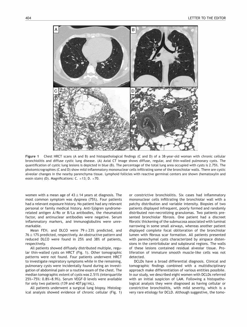
404 LETTER TO THE EDITOR
Figure 1 Chest HRCT scans (A and B) and histopathological findings (C and D) of a 38-year-old woman with chronic cellularbronchiolitis and diffuse cystic lung disease. (A) Axial CT image shows diffuse, regular, and thin-walled pulmonary cysts. Thequantification of cystic lung lesions is depicted in blue (B). The percentage of the total lung area occupied with cysts is 2.75%. Thephotomicrographies (C and D) show mild inflammatory mononuclear cells infiltrating some of the bronchiolar walls. There are cysticalveolar changes in the nearby parenchyma tissue. Lymphoid follicles with reactive germinal centers are shown (hematoxylin andeosin stain) (D). Magnifications: C. ×13; D. ×70.
women with a mean age of 43 ± 14 years at diagnosis. Themost common symptom was dyspnea (75%). Four patientshad a relevant exposure history. No patient had any relevantpersonal or family medical history. Anti-Sjögren syndrome-related antigen A/Ro or B/La antibodies, the rheumatoidfactor, and antinuclear antibodies were negative. Seruminflammatory markers, and immunoglobulins were unre-markable.
Mean FEV1 and DLCO were 79 ± 23% predicted, and76 ± 17% predicted, respectively. An obstructive pattern andreduced DLCO were found in 25% and 38% of patients,respectively.
All patients showed diffusely distributed multiple, regu-lar thin-walled cysts on HRCT (Fig. 1). Other tomographicpatterns were not found. Four patients underwent HRCTto investigate respiratory symptoms while in the remaining,pulmonary cysts were incidentally found during an investi-gation of abdominal pain or a routine exam of the chest. Themedian tomographic extent of cysts was 2.51% (interquartile25%---75%: 0.8%---8.9%). Serum VEGF-D levels were availablefor only two patients (139 and 407 pg/mL).
All patients underwent a surgical lung biopsy. Histolog-ical analysis showed evidence of chronic cellular (Fig. 1)
or constrictive bronchiolitis. Six cases had inflammatorymononuclear cells infiltrating the bronchiolar wall with apatchy distribution and variable intensity. Biopsies of twopatients displayed infrequent, poorly formed and randomlydistributed non-necrotizing granulomas. Two patients pre-sented bronchiolar fibrosis. One patient had a discreetfibrotic thickening of the submucosa associated with luminalnarrowing in some small airways, whereas another patientdisplayed complete focal obliteration of the bronchiolarlumen with fibrous scar formation. All patients presentedwith parenchymal cysts characterized by airspace disten-sions in the centrilobular and subpleural regions. The wallsof these lesions contained residual alveolar tissue. Pro-liferation of immature smooth muscle-like cells was notdetected.
DCLDs have a broad differential diagnosis. Clinical andtomographic findings combined with a multidisciplinaryapproach make differentiation of various entities possibile.In our study, we described eight women with DCLDs referredwith an initial suspicion of LAM. Following a histopatho-logical analysis they were diagnosed as having cellular orconstrictive bronchiolitis, with mild severity, which is avery rare etiology for DCLD. Although suggestive, the tomo-

LETTER TO THE EDITOR 405
graphic patterns of the diffuse, regular and thin-walled cystsin women are not specific to LAM.2,4,5 In the absence of def-inite findings, a pulmonary biopsy is mandatory to diagnoseLAM.3 Low availability of serum VEGF-D dosage is a limitationof our study.
Some findings on HRCT contribute to establishing the eti-ology of DCLDs. In pulmonary Langerhans histiocytosis, cystsare usually irregular, predominate in the upper and middlelung zones and may be associated with nodules.1,2 In Birt-Hogg-Dubé (BHD) syndrome, cysts are multiple, thin-walledand predominantly basilar and paramediastinal.1,2 Cystsin lymphocytic interstitial pneumonia occur in the lowerlobes along the peribronchovascular bundle, frequently withground-glass opacities.2
The typical pathological and immunohistochemical fea-tures of LAM were not identified in our study. Histologically,LAM nodules consist of two cellular subpopulations: the spin-dle cells express SMA and forms the core of the nodulessurrounded by epithelioid cells that exhibit immunoreactiv-ity for HMB-45 antibody.9 On histopathology, the morphologyof BHD cysts may resemble what was found in our cases.However, we excluded BHD based on clinical and tomo-graphic features.10 Cysts, emphysema, and respiratorybronchiolitis are identified in smoking-related DCLDs, butwere not found in our study.5 A combination of the DCLDsand small airway disease is mainly identified in specific con-ditions, such as Sjögren’s syndrome, and it is thought thatchronic small airway damage might lead to cyst formationin these patients. A possible explanation for the lung cystformation in bronchiolitis is a check-valve obstruction withdistal airspace overinflation related to air trapping, distal tothe abnormal airways.1,2
In two cases, histological analysis showed evidence ofchronic cellular bronchiolitis associated with infrequent,poorly formed, non-necrotizing granulomas, frequently seenin hypersensitivity pneumonitis (HP). Pulmonary cysts havebeen described in HP possibly due to partial bronchiolarobstruction.2
Potential etiologies for our patients with bronchiolitisinclude autoimmune disorders, such as Sjögren syndrome,post viral infections and HP. However, none of the etiologiesraised were confirmed.
In summary, LAM is a prototypical DCLD with a character-istic appearance on HRCT. However, cystic features aloneare inadequate to establish a diagnosis of LAM. In such con-text, bronchiolitis should be considered in the differentialdiagnosis of DCLDs.
Informed consent
Patients gave informed consent for this study.
Funding
This research did not receive any specific grant from fundingagencies in the public, commercial, or not-for-profit sectors.
Conflicts of interest
The authors have no conflicts of interest to declare.
References
1. Gupta N, Vassallo R, Wikenheiser-Brokamp KA,McCormack FX. Diffuse cystic lung disease. PartII. Am J Respir Crit Care Med. 2015;192(1):17---29,http://dx.doi.org/10.1164/rccm.201411-2096CI.
2. Baldi BG, Carvalho CRR, Dias OM, Marchiori E,Hochhegger B. Diffuse cystic lung diseases: differ-ential diagnosis. J Bras Pneumol. 2017;43(2):140---9,http://dx.doi.org/10.1590/S1806-37562016000000341.
3. Gupta N, Finlay GA, Kotloff RM, Strange C, Wilson KC,Young LR, et al. Lymphangioleiomyomatosis diagno-sis and management: high-resolution chest computedtomography, transbronchial lung biopsy, and pleuraldisease management. An Official American ThoracicSociety/Japanese Respiratory Society Clinical PracticeGuideline. Am J Respir Crit Care Med. 2017;196(10):1337---48,http://dx.doi.org/10.1164/rccm.201709-1965ST.
4. Rowan C, Hansell DM, Renzoni E, Maher TM, Wells AU,Polkey MI, et al. Diffuse cystic lung disease of unex-plained cause with coexistent small airway disease: a possiblecausal relationship? Am J Surg Pathol. 2012;36(2):228---34,http://dx.doi.org/10.1097/PAS.0b013e318237c599.
5. Gupta N, Colby TV, Meyer CA, McCormack FX,Wikenheiser-Brokamp KA. Smoking-related dif-fuse cystic lung disease. Chest. 2018;154(2):e31---5,http://dx.doi.org/10.1016/j.chest.2018.02.027.
6. Macintyre N, Crapo RO, Viegi G, Johnson DC, vander Grinten CP, Brusasco V, et al. Standardisation ofthe single-breath determination of carbon monoxideuptake in the lung. Eur Respir J. 2005;26(4):720---35,http://dx.doi.org/10.1183/09031936.05.00034905.
7. Miller MR, Hankinson J, Brusasco V, Burgos F,Casaburi R, Coates A, et al. Standardisationof spirometry. Eur Respir J. 2005;26(2):319---38,http://dx.doi.org/10.1183/09031936.05.00034805.
8. Wanger J, Clausen JL, Coates A, Pedersen OF, BrusascoV, Burgos F, et al. Standardisation of the measure-ment of lung volumes. Eur Respir J. 2005;26(3):511---22,http://dx.doi.org/10.1183/09031936.05.00035005.
9. Zhang X, Travis WD. Pulmonary lymphangioleiomy-omatosis. Arch Pathol Lab Med. 2010;134(12):1823---8,http://dx.doi.org/10.1043/2009-0576-RS.1.
10. Koga S, Furuya M, Takahashi Y, Tanaka R, YamaguchiA, Yasukufu K, et al. Lung cysts in Birt-Hogg-Dubésyndrome: histopathological characteristics and aber-rance sequence repeats. Pathol Int. 2009;59(10):720---8,http://dx.doi.org/10.1111/j.1440-1827.2009.02434.x.
M.R. de Oliveira, O.M. Dias, A.F. Amaral
Divisao de Pneumologia, Instituto do Coracao (InCor),Hospital das Clinicas HCFMUSP, Faculdade de Medicina,Universidade de Sao Paulo, Sao Paulo, SP, Brazil
E.C.T. do Nascimento
Departamento de Patologia, Hospital das ClinicasHCFMUSP, Faculdade de Medicina, Universidade de SaoPaulo, Sao Paulo, SP, Brazil
M. Wanderley
Departamento de Radiologia, Hospital das ClinicasHCFMUSP, Faculdade de Medicina, Universidade de SaoPaulo, Sao Paulo, SP, Brazil
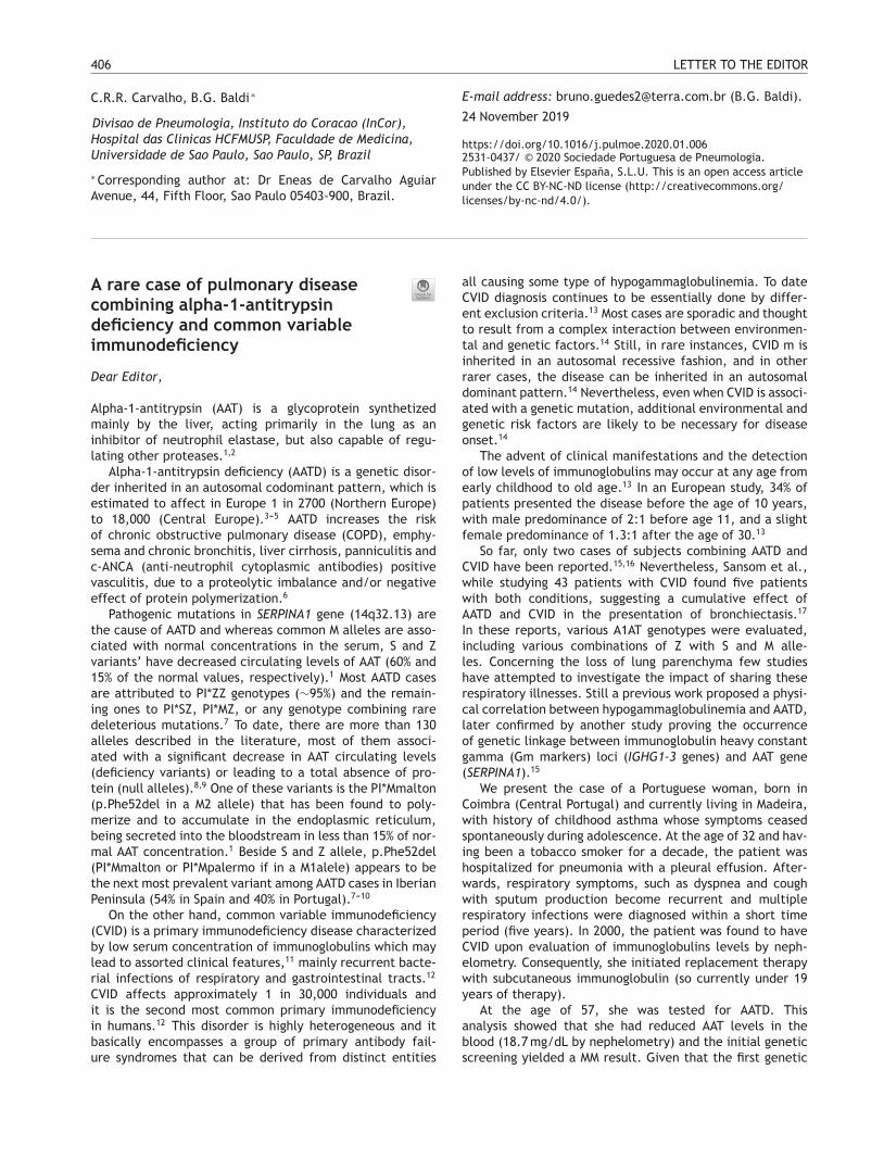
406 LETTER TO THE EDITOR
C.R.R. Carvalho, B.G. Baldi ∗
Divisao de Pneumologia, Instituto do Coracao (InCor),Hospital das Clinicas HCFMUSP, Faculdade de Medicina,Universidade de Sao Paulo, Sao Paulo, SP, Brazil
∗ Corresponding author at: Dr Eneas de Carvalho AguiarAvenue, 44, Fifth Floor, Sao Paulo 05403-900, Brazil.
E-mail address: [email protected] (B.G. Baldi).
24 November 2019
https://doi.org/10.1016/j.pulmoe.2020.01.0062531-0437/ © 2020 Sociedade Portuguesa de Pneumologia.Published by Elsevier Espana, S.L.U. This is an open access articleunder the CC BY-NC-ND license (http://creativecommons.org/licenses/by-nc-nd/4.0/).
A rare case of pulmonary diseasecombining alpha-1-antitrypsindeficiency and common variableimmunodeficiency
Dear Editor,
Alpha-1-antitrypsin (AAT) is a glycoprotein synthetizedmainly by the liver, acting primarily in the lung as aninhibitor of neutrophil elastase, but also capable of regu-lating other proteases.1,2
Alpha-1-antitrypsin deficiency (AATD) is a genetic disor-der inherited in an autosomal codominant pattern, which isestimated to affect in Europe 1 in 2700 (Northern Europe)to 18,000 (Central Europe).3---5 AATD increases the riskof chronic obstructive pulmonary disease (COPD), emphy-sema and chronic bronchitis, liver cirrhosis, panniculitis andc-ANCA (anti-neutrophil cytoplasmic antibodies) positivevasculitis, due to a proteolytic imbalance and/or negativeeffect of protein polymerization.6
Pathogenic mutations in SERPINA1 gene (14q32.13) arethe cause of AATD and whereas common M alleles are asso-ciated with normal concentrations in the serum, S and Zvariants’ have decreased circulating levels of AAT (60% and15% of the normal values, respectively).1 Most AATD casesare attributed to PI*ZZ genotypes (∼95%) and the remain-ing ones to PI*SZ, PI*MZ, or any genotype combining raredeleterious mutations.7 To date, there are more than 130alleles described in the literature, most of them associ-ated with a significant decrease in AAT circulating levels(deficiency variants) or leading to a total absence of pro-tein (null alleles).8,9 One of these variants is the PI*Mmalton(p.Phe52del in a M2 allele) that has been found to poly-merize and to accumulate in the endoplasmic reticulum,being secreted into the bloodstream in less than 15% of nor-mal AAT concentration.1 Beside S and Z allele, p.Phe52del(PI*Mmalton or PI*Mpalermo if in a M1alele) appears to bethe next most prevalent variant among AATD cases in IberianPeninsula (54% in Spain and 40% in Portugal).7---10
On the other hand, common variable immunodeficiency(CVID) is a primary immunodeficiency disease characterizedby low serum concentration of immunoglobulins which maylead to assorted clinical features,11 mainly recurrent bacte-rial infections of respiratory and gastrointestinal tracts.12
CVID affects approximately 1 in 30,000 individuals andit is the second most common primary immunodeficiencyin humans.12 This disorder is highly heterogeneous and itbasically encompasses a group of primary antibody fail-ure syndromes that can be derived from distinct entities
all causing some type of hypogammaglobulinemia. To dateCVID diagnosis continues to be essentially done by differ-ent exclusion criteria.13 Most cases are sporadic and thoughtto result from a complex interaction between environmen-tal and genetic factors.14 Still, in rare instances, CVID m isinherited in an autosomal recessive fashion, and in otherrarer cases, the disease can be inherited in an autosomaldominant pattern.14 Nevertheless, even when CVID is associ-ated with a genetic mutation, additional environmental andgenetic risk factors are likely to be necessary for diseaseonset.14
The advent of clinical manifestations and the detectionof low levels of immunoglobulins may occur at any age fromearly childhood to old age.13 In an European study, 34% ofpatients presented the disease before the age of 10 years,with male predominance of 2:1 before age 11, and a slightfemale predominance of 1.3:1 after the age of 30.13
So far, only two cases of subjects combining AATD andCVID have been reported.15,16 Nevertheless, Sansom et al.,while studying 43 patients with CVID found five patientswith both conditions, suggesting a cumulative effect ofAATD and CVID in the presentation of bronchiectasis.17
In these reports, various A1AT genotypes were evaluated,including various combinations of Z with S and M alle-les. Concerning the loss of lung parenchyma few studieshave attempted to investigate the impact of sharing theserespiratory illnesses. Still a previous work proposed a physi-cal correlation between hypogammaglobulinemia and AATD,later confirmed by another study proving the occurrenceof genetic linkage between immunoglobulin heavy constantgamma (Gm markers) loci (IGHG1-3 genes) and AAT gene(SERPINA1).15
We present the case of a Portuguese woman, born inCoimbra (Central Portugal) and currently living in Madeira,with history of childhood asthma whose symptoms ceasedspontaneously during adolescence. At the age of 32 and hav-ing been a tobacco smoker for a decade, the patient washospitalized for pneumonia with a pleural effusion. After-wards, respiratory symptoms, such as dyspnea and coughwith sputum production become recurrent and multiplerespiratory infections were diagnosed within a short timeperiod (five years). In 2000, the patient was found to haveCVID upon evaluation of immunoglobulins levels by neph-elometry. Consequently, she initiated replacement therapywith subcutaneous immunoglobulin (so currently under 19years of therapy).
At the age of 57, she was tested for AATD. Thisanalysis showed that she had reduced AAT levels in theblood (18.7 mg/dL by nephelometry) and the initial geneticscreening yielded a MM result. Given that the first genetic
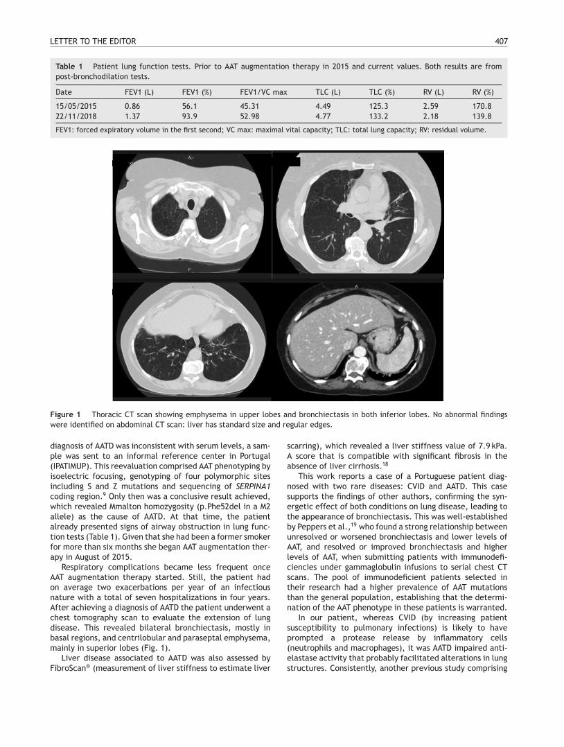
LETTER TO THE EDITOR 407
Table 1 Patient lung function tests. Prior to AAT augmentation therapy in 2015 and current values. Both results are frompost-bronchodilation tests.
Date FEV1 (L) FEV1 (%) FEV1/VC max TLC (L) TLC (%) RV (L) RV (%)
15/05/2015 0.86 56.1 45.31 4.49 125.3 2.59 170.822/11/2018 1.37 93.9 52.98 4.77 133.2 2.18 139.8
FEV1: forced expiratory volume in the first second; VC max: maximal vital capacity; TLC: total lung capacity; RV: residual volume.
Figure 1 Thoracic CT scan showing emphysema in upper lobes and bronchiectasis in both inferior lobes. No abnormal findingswere identified on abdominal CT scan: liver has standard size and regular edges.
diagnosis of AATD was inconsistent with serum levels, a sam-ple was sent to an informal reference center in Portugal(IPATIMUP). This reevaluation comprised AAT phenotyping byisoelectric focusing, genotyping of four polymorphic sitesincluding S and Z mutations and sequencing of SERPINA1coding region.9 Only then was a conclusive result achieved,which revealed Mmalton homozygosity (p.Phe52del in a M2allele) as the cause of AATD. At that time, the patientalready presented signs of airway obstruction in lung func-tion tests (Table 1). Given that she had been a former smokerfor more than six months she began AAT augmentation ther-apy in August of 2015.
Respiratory complications became less frequent onceAAT augmentation therapy started. Still, the patient hadon average two exacerbations per year of an infectiousnature with a total of seven hospitalizations in four years.After achieving a diagnosis of AATD the patient underwent achest tomography scan to evaluate the extension of lungdisease. This revealed bilateral bronchiectasis, mostly inbasal regions, and centrilobular and paraseptal emphysema,mainly in superior lobes (Fig. 1).
Liver disease associated to AATD was also assessed byFibroScan® (measurement of liver stiffness to estimate liver
scarring), which revealed a liver stiffness value of 7.9 kPa.A score that is compatible with significant fibrosis in theabsence of liver cirrhosis.18
This work reports a case of a Portuguese patient diag-nosed with two rare diseases: CVID and AATD. This casesupports the findings of other authors, confirming the syn-ergetic effect of both conditions on lung disease, leading tothe appearance of bronchiectasis. This was well-establishedby Peppers et al.,19 who found a strong relationship betweenunresolved or worsened bronchiectasis and lower levels ofAAT, and resolved or improved bronchiectasis and higherlevels of AAT, when submitting patients with immunodefi-ciencies under gammaglobulin infusions to serial chest CTscans. The pool of immunodeficient patients selected intheir research had a higher prevalence of AAT mutationsthan the general population, establishing that the determi-nation of the AAT phenotype in these patients is warranted.
In our patient, whereas CVID (by increasing patientsusceptibility to pulmonary infections) is likely to haveprompted a protease release by inflammatory cells(neutrophils and macrophages), it was AATD impaired anti-elastase activity that probably facilitated alterations in lungstructures. Consistently, another previous study comprising

408 LETTER TO THE EDITOR
a total of 43 patients with CVID in which five were foundto have AATD, also suggested an additive effect of thesedisorders into pulmonary illness.17
Despite so few reports of patients combining CVID andAATD, some studies have attempted to physically correlatethese disorders: one of them, performed in 1983, failed todetect any evidence of linkage between these conditionsin a family where hypogammaglobulinemia and AATD werefound to segregate.16 Interestingly, SERPINA1 encoding AATis located in chromosome 14 approximately 11 Mb apart froma cluster of immunoglobulin genes (IGHG1-3 among others),which could explain in some rare cases a co-segregation ofthese disorders. However, a genetic screening of CVID wasnot performed in this case due to its low impact on patientfollow up as well as it potential high costs to healthcaresystem.13
In result of the CVID and AATD joint diagnosis, this patientis currently undergoing two replacement therapies withsubcutaneous immunoglobulin and endovenous human AAT.Here, it is important to stress that AAT infusion containssmall amounts of IgA. This obligates a careful administra-tion of AATD therapy in patients with IgA deficiency sincethey present a greater risk of adverse anaphylactic reactionsdue to the pre-existence of IgA antibodies. The Portugueseconsensus document for AATD management actually discour-ages augmentation therapy in patients with IgA deficiency,20
but so far this patient has been successfully treated withno record of side effects. Furthermore, lung functions testsof this patient have shown a remarkable increase in forcedexpiratory volume in one second (FEV1) after three yearsof AAT therapy. Although this phenomenon has not beendescribed before, we believe it might be explained by acombination of optimized inhaled bronchodilators and AATtherapies together with cigarette smoking cessation prior totreatment in 2015 and inter-operator dependent differencesin test execution and by the variability in the capacity of per-formance of the tests by the patient. On the other hand, thefinding of liver fibrosis as confirmed by FibroScan® assess-ment were in accordance with previous studies of Mmaltonpatients (homozygous and/or heterozygous) showing thatthis allele is associated with hepatic accumulation of AATand subsequently with liver disease, being to some degreea functional equivalent to a Z allele.21
Even though we could not rule out a genetic linkagebetween CVID and AATD in this case, we provide evidencethat these might be somehow correlated with observed clin-ical features of lung disease. This report also emphasizesthe singularity of combining two uncommon replacementtherapies in controlling the worse outcomes of CVID (recur-rent exacerbations) and AATD (severe airflow obstruction).
Conflict of interests
The authors declare no conflict of interests.
Acknowledgements
N. Martins would like to thank the Portuguese Foun-dation for Science and Technology (FCT-Portugal) forthe Strategic project ref. UID/BIM/04293/2013 and‘‘NORTE2020 --- Northern Regional Operational Program’’
(NORTE-01-0145-FEDER-000012). IPATIMUP integrates thei3S Research Unit, which is partially supported by thePortuguese Foundation for Science and Technology (FCT;projects PEstC/SAU/LA0003/2013 and POCI-01-0145-FEDER-007274) and by Programa Operacional Regional doNorte (ON.2 --- O Novo Norte and Norte 2020), throughFEDER funds under the Quadro de Referência EstratégicoNacional (QREN; projects NORTE-07-0162-FEDER-00018 andNORTE-070162-FEDER-000067, and NORTE-01-0145-FEDER-000029).
References
1. Seixas SA [PhD thesis] Analysis of genetic diversity patternsin three functionally relevant regions of the human genome:apolipoprotein E (APOE), alpha 1-antitrypsin (PI), Duffy BloodGroup (FY). University of Porto; 2002.
2. Gupta R, Sridhara S, Wood J. A rare case of alpha 1-antitrypsindeficiency associated with hypogammaglobulinemia and recur-rent pulmonary thrombosis. Ann Thorac Med. 2014;9:39.
3. Blanco I, de Serres FJ, Fernandez-Bustillo E, Lara B, MiravitllesM. Estimated numbers and prevalence of PI*S and PI*Z alleles ofalpha1-antitrypsin deficiency in European countries. Eur RespirJ. 2006;27:77---84.
4. de Serres FJ, Blanco I. Prevalence of �1-antitrypsin defi-ciency alleles PI*S and PI*Z worldwide and effective screeningfor each of the five phenotypic classes PI*MS, PI*MZ, PI*SS,PI*SZ, and PI*ZZ: a comprehensive review. Ther Adv Respir Dis.2012;6:277---95.
5. Blanco I, Bueno P, Diego I, Pérez-Holanda S, Lara B, Casas-Maldonado F, et al. Alpha-1 antitrypsin Pi*SZ genotype:estimated prevalence and number of SZ subjects worldwide.Int J Chron Obstruct Pulmon Dis. 2017;8:1683---94.
6. Stoller JK, Lacbawan FL, Aboussouan LS. In: University ofWashington, editor. Alpha-1 antitrypsin deficiency. Summary.Diagnosis. Suggestive findings. Seattle: U.S. National Library ofMedicine; 2019. p. 1---27.
7. Lara B, Martínez-Delgado B, Torres ML, Marín-Arguedas S,Bustamantee A, Miravitlles M. Alpha-1-antitrypsin deficiencyassociated with the mattawa variant. Arch Bronconeumol.2016;52:617---8.
8. Rodriguez-Frias F, Miravitlles M, Vidal R, Camos S, Jardi R. Rarealpha-1-antitrypsin variants: are they really so rare? Ther AdvRespir Dis. 2012;6:79---85.
9. Silva D, Oliveira MJ, Guimarães M, Lima R, Gomes S, Seixas S.Alpha-1-antitrypsin (SERPINA1) mutation spectrum: three novelvariants and haplotype characterization of rare deficiency alle-les identified in Portugal. Respir Med. 2016;116:8---18.
10. Meira L, Boaventura R, Seixas S, Sucena M. Alpha-1 antitrypsindeficiency detection in a portuguese population. COPD J ChronicObstr Pulm Dis. 2018;15:4---9.
11. Cunningham-Rundles C, Bodian C. Common variable immunod-eficiency: clinical and immunological features of 248 patients.Clin Immunol. 1999;92:34---48.
12. Grimbacher B, Hutloff A, Schlesier M, Glocker E, Warnatz K,Dräger R, et al. Homozygous loss of ICOS is associated withadult-onset common variable immunodeficiency. Nat Immunol.2003;4:261---8.
13. Bonilla FA, Barlan I, Chapel H, Costa-Carvalho BT, Cunningham-Rundles C, de la Morena MT, et al. International ConsensusDocument (ICON): common variable immunodeficiency disor-ders. J Allergy Clin Immunol Pract [Internet]. 2016;4:38---59.Available from: http://www.elsevier.com/journals/the-journal-of-allergy-and-clinical-immunology-in-practice/2213-2198%0Ahttp://ovidsp.ovid.com/ovidweb.cgi?T=JS&PAGE=reference&D=emed17&NEWS=N&AN=607686990
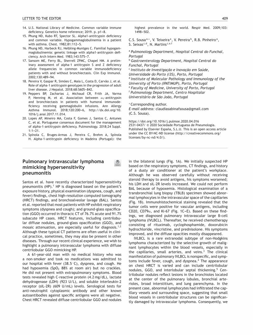
LETTER TO THE EDITOR 409
14. U.S. National Library of Medicine. Common variable immunedeficiency. Genetics home reference; 2019. p. p1---8.
15. Phung ND, Kubo RT, Spector SL. Alpha1-antitrypsin deficiencyand common variable. Hypogammaglobulinemia in a patientwith asthma. Chest. 1982;81:112---5.
16. Phung ND, Harbeck RJ, Helbling-Muntges C. Familial hypogam-maglobulinemia: genetic linkage with alpha1-antitrypsin defi-ciency. Arch Intern Med. 1983;143:575---7.
17. Sansom ME, Ferry BL, Sherrell ZPMC, Chapel HM. A prelim-inary assessment of alpha-1 antitrypsin S and Z deficiencyallele frequencies in common variable immunodeficiencypatients with and without bronchiectasis. Clin Exp Immunol.2002;130:489---94.
18. Pereira V, Gaspar R, Simões C, Maia L, Costa D, Carvão J, et al.Role of alpha-1 antitrypsin genotypes in the progression of adultliver disease. J Hepatol. 2018;68:S605---842.
19. Peppers BP, Zacharias J, Michaud CR, Frith JA, VarmaP, Henning M, et al. Association between �1-antitrypsinand bronchiectasis in patients with humoral immunode-ficiency receiving gammaglobulin infusions. Ann AllergyAsthma Immunol. 2018;120:200---6, http://dx.doi.org/10.1016/j.anai.2017.11.014.
20. Lopes AP, Mineiro MA, Costa F, Gomes J, Santos C, AntunesC, et al. Portuguese consensus document for the managementof alpha-1-antitrypsin deficiency. Pulmonology. 2018;24 Suppl.1:1---21.
21. Spínola C, Bruges-Armas J, Pereira C, Brehm A, SpínolaH. Alpha-1-antitrypsin deficiency in Madeira (Portugal): the
highest prevalence in the world. Respir Med. 2009;103:1498---502.
C.S. Sousaa,∗, V. Teixeiraa, V. Pereirab, R.B. Pinheiroa,S. Seixasc,d, N. Martinsc,e,f
a Pulmonology Department, Hospital Central do Funchal,Portugalb Gastroenterology Department, Hospital Central doFunchal, Portugalc Instituto de Investigacão e Inovacão em Saúde,Universidade do Porto (i3S), Porto, Portugald Institute of Molecular Pathology and Immunology of theUniversity of Porto (IPATIMUP), Porto, Portugale Faculty of Medicine, University of Porto, Portugalf Pulmonology Department, Centro HospitalarUniversitário de São João, Portugal
∗ Corresponding author.E-mail address: [email protected](C.S. Sousa).
https://doi.org/10.1016/j.pulmoe.2020.04.0162531-0437/ © 2020 Sociedade Portuguesa de Pneumologia.Published by Elsevier Espana, S.L.U. This is an open access articleunder the CC BY-NC-ND license (http://creativecommons.org/licenses/by-nc-nd/4.0/).
Pulmonary intravascular lymphomamimicking hypersensitivitypneumonitis
Santos et al. have recently characterized hypersensitivitypneumonitis (HP).1 HP is diagnosed based on the patient’sexposure history, physical examination (dyspnea, cough, andfever) findings, chest high-resolution computed tomography(HRCT) findings, and bronchoalveolar lavage (BAL). Santoset al. reported that most patients with HP exhibit respiratorysymptoms (dyspnea and cough); and ground-glass opacifica-tion (GGO) occurred in thoracic CT of 76.7% acute and 91.7%subacute HP cases. HRCT features, including centrilobu-lar diffuse nodules, ground-glass opacification (GGO), andmosaic attenuation, are especially useful for diagnosis.1,2
Although these typical CT patterns are often useful in clini-cal practice, sometimes, they may also be present in otherdiseases. Through our recent clinical experience, we wish tohighlight a pulmonary intravascular lymphoma with diffusecentrilobular GGO mimicking HP.
A 61-year-old man with no medical history who wasa non-smoker and took no medications was admitted toour hospital with fever (38.5 ◦C), dyspnea, and cough. Hehad hypoxemia (SpO2 88% at room air) but no crackles.He did not present with extrapulmonary symptoms. Bloodtests revealed high C-reactive protein (4.2 mg/dL), lactatedehydrogenase (LDH) (923 U/L), and soluble interleukin-2receptor (sIL-2R) (609 U/mL) levels. Serological tests foranti-neutrophil cytoplasmic antibody and other knownautoantibodies against specific antigens were all negative.Chest HRCT revealed diffuse centrilobular GGO and nodules
in the bilateral lungs (Fig. 1A). We initially suspected HPbased on the respiratory symptoms, CT findings, and historyof a dusty air conditioner at the patient’s workplace.Although he was observed carefully without receivingsteroid therapy to avoid antigens, his symptoms worsened;his LDH and sIL-2R levels increased. We could not performBAL because of hypoxemia. Histological examination of atransbronchial lung biopsy (TBLB) specimen showed abnor-mal lymphocytes in the intravascular space of the capillaries(Fig. 1B). Immunohistochemical staining revealed that thetumor cells were positive for vascular antigens, includingCD20, CD79�, and Ki-67 (Fig. 1C---E). Based on these find-ings, we diagnosed pulmonary intravascular large B-celllymphoma (IVLBCL). Thereafter, he received chemotherapyconsisting of rituximab, cyclophosphamide, doxorubicinhydrochloride, vincristine, and prednisolone. His symptomsimproved, and the diffuse opacities mostly disappeared.
IVLBCL is a rare extranodal subtype of non-Hodgkinslymphoma characterized by the selective growth of malig-nant lymphocytes within the blood vessels, especially inthe capillaries, small arteries, and veins.3 The clinicalmanifestation of pulmonary IVLBCL is nonspecific, and symp-toms include fever, cough, and dyspnea.4 The appearanceon chest HRCT is varied and can include centrilobularnodules, GGO, and interlobular septal thickening.5 Cen-trilobular nodules reflect lesions in the bronchioles locatedat the center of the pulmonary lobules, bronchial arte-rioles, broad interstitium, and lung parenchyma. In thepresent case, abnormal lymphocytes had infiltrated the cap-illary vessels and surrounding space, suggesting that smallblood vessels in centrilobular structures can be significan-tly damaged by intravascular lymphoma. Consequently, we

410 LETTER TO THE EDITOR
Tabl
e1
Prev
ious
case
repo
rts
ofpu
lmon
ary
intr
avas
cula
rla
rge
Bce
llly
mph
oma
diag
nose
dby
tran
sbro
nchi
allu
ngbi
opsy
.
No.
Age
Sex
Sym
ptom
sLD
H(I
U/L
)sI
L2R
(U/L
)CT
imag
ing
Trea
tmen
tO
utco
me
182
Mal
eFo
rget
fuln
ess
3040
1263
GG
OR-
CHO
PIm
prov
emen
t2
58Fe
mal
eFe
ver,
wei
ght
loss
Unk
now
n67
74N
orm
alfin
ding
sU
nkno
wn
Unk
now
n3
60Fe
mal
eD
yspn
ea,
feve
r14
34U
nkno
wn
GG
O,
nodu
les
Unk
now
nD
eath
469
Fem
ale
Dys
pnea
,fe
ver,
coug
h10
95U
nkno
wn
GG
O,
inte
rlob
ular
sept
alth
icke
ning
CHO
PU
nkno
wn
569
Fem
ale
Fati
gue,
feve
r,dy
spne
a14
7527
71N
orm
alfin
ding
sU
nkno
wn
Unk
now
n6
62M
ale
Dys
pnea
1482
1570
Nod
ules
R-CH
OP
Com
plet
ere
mis
sion
757
Mal
eFe
ver,
dysp
nea
Unk
now
nU
nkno
wn
Haz
ines
sU
nkno
wn
Unk
now
n8
85M
ale
Dys
pnea
829
2000
GG
OR-
CHO
PIm
prov
emen
t9
59M
ale
Feve
r,dy
spne
a60
523
20G
GO
,in
terl
obul
arse
ptal
thic
keni
ngR-
CHO
PIm
prov
emen
t
1035
Fem
ale
Coug
h,dy
spne
a,fe
ver
1554
Unk
now
nCo
nsol
idat
ion,
GG
OR-
CHO
PD
eath
1146
Mal
eCo
ugh,
feve
r76
519
100
Nod
ules
,gr
anul
arsh
adow
sR-
CHO
PCo
mpl
ete
rem
issi
on
1271
Fem
ale
Feve
rU
nkno
wn
Unk
now
nN
orm
alfin
ding
sR-
CHO
PPa
rtia
lres
pons
e13
84M
ale
Dys
pnea
1120
2238
Nor
mal
findi
ngs
R-CH
OP
Com
plet
ere
mis
sion
1461
Mal
eN
ight
swea
ts,
dysp
nea
698
4130
Nor
mal
findi
ngs
R-CH
OP
Com
plet
ere
mis
sion
1558
Fem
ale
Coug
h,fe
ver
570
3699
Nor
mal
findi
ngs
R-CH
OP
Impr
ovem
ent
1639
Mal
eD
yspn
ea,
feve
r22
1419
50N
orm
alfin
ding
sR-
CHO
PIm
prov
emen
t17
50Fe
mal
eFe
ver,
anor
exia
,oe
dem
a,m
uscu
lar
pain
3386
6499
Nor
mal
findi
ngs
R-CH
OP
Com
plet
ere
mis
sion
1865
Mal
eFe
ver,
dysp
nea
1625
2105
Inte
rsti
tial
shad
ows
CHO
PCo
mpl
ete
rem
issi
on19
73M
ale
Feve
r,dy
spne
a,sk
inle
sion
s12
4852
90In
ters
titi
alsh
adow
sCH
OP
Com
plet
ere
mis
sion
2049
Fem
ale
Feve
r,co
ugh
5938
Unk
now
nIn
ters
titi
alsh
adow
sCi
spla
tin,
cyta
rabi
ne,
dexa
met
haso
ne
Impr
ovem
ent
2165
Mal
eD
yspn
ea,
dem
enti
a23
2718
20Co
nsol
idat
ion,
bron
chov
ascu
lar
bund
les
thic
keni
ng
CHO
PIm
prov
emen
t
2263
Mal
eD
yspn
ea,
coug
h,fe
ver,
wei
ght
loss
1825
Unk
now
nM
osai
cat
tenu
atio
nsCH
OP
Com
plet
ere
mis
sion
Refe
renc
es:
1.Ka
ito
Set
al.
AmJ
Case
Rep.
2019
,2.
Yam
asak
iMet
al.
Inte
rnM
ed.
2018
,3.
Zhan
gY
etal
.O
ncol
Lett
.20
18,
4.Zh
ang
Yet
al.
Onc
olLe
tt.
2018
,5.
Nad
aT
etal
.J
Gen
Fam
Med
.20
17,
6.Ta
kesh
ige
Tet
al.
Inte
rnM
ed.
2017
,7.
Wu
Fet
al.
Clin
Nuc
lM
ed.
2016
,8.
Yam
akaw
aH
etal
.In
tern
Med
.20
15,
9.N
ishi
zaw
aT
etal
.BM
JCa
seRe
p.20
14,
10.
C hen
Yet
al.
JTh
orac
Dis
.20
14,
11.
Seki
guch
iYet
al.
Int
JCl
inEx
pPa
thol
.20
14,
12.
Wag
ner
Tet
al.
Canc
erIm
agin
g.20
12,
13.
Nak
azat
oT
etal
.An
nH
emat
ol.
2012
,14
.N
iida
Tet
al.
JM
edCa
seRe
p.20
11,
15.
Kaku
Net
al.
Inte
rnM
ed.
2010
,16
.Ko
take
Tet
al.
Inte
rnM
ed.
2010
,17
.Ki
tana
kaA
etal
.H
emat
olO
ncol
.20
09,
18.
Asad
aN
etal
.M
ayo
Clin
Proc
.20
07,
19.
Asad
aN
etal
.M
ayo
Clin
Proc
.20
07,
20.
Goh
SGet
al.
Path
olog
y.20
02,
21.
Kaw
amur
aT
etal
.Jp
nJ
Clin
Onc
ol.
2001
,22
.W
alls
JGet
al.
Ches
t.19
99.

LETTER TO THE EDITOR 411
Figure 1 (A) Chest high-resolution computed tomography revealing diffuse ground glass opacities and nodules with centrilobularaugmentation in the bilateral lungs. (B) Microphotographs of the transbronchial lung biopsy specimen. Atypical lymphocytes areseen in the intravascular space of capillary blood vessels. Immunochemical staining revealing that these cells were positive for CD20(C), CD79� (D), and Ki-67 (E).

412 LETTER TO THE EDITOR
considered that HRCT revealed HP-like pale GGO and nod-ules with a centrilobular distribution pattern. It is probablethat the resulting occlusive proliferation of tumor cells inthe vascular lumens led to multiple minor infarctions, whichmanifested as coughing, dyspnea, and fever.
IVLBCL is often not identified during the lifetime, is onlydiagnosed at autopsy. To the best of our knowledge, only 22cases, diagnosed by TBLB, have been reported during thepast 20 years (Table 1). Respiratory symptoms including dys-pnea and cough were present in 18 of those 22 cases, andboth LDH and sIL2-R were elevated. Thoracic CT revealedGGO and nodules in some cases but was normal in others,and the response to treatment was mostly good. Based onthese findings, TBLB may assist with diagnosis in patientscomplaining chiefly of respiratory symptoms, and early diag-nosis may contribute to improved prognosis. TBLB may alsobe valuable in the differential diagnosis of other diseases,including HP (particularly acute/subacute HP).
Although the diagnosis of pulmonary IVLBCL is difficultdue to its nonspecific clinical manifestation, in cases of dif-fuse centrilobular GGO and nodules on chest HRCT, it isnecessary to consider not only HP but also pulmonary IVL-BCL. Moreover, increased serum LDH and sIL-2R levels shouldsuggest a diagnosis of IVLBCL, and TBLB be performed tomake a definitive diagnosis.
References
1. Santos V, Martins N, Sousa C, Jacob M, Padrão E, Melo N,et al. Hypersensitivity pneumonitis: main features character-ization in a Portuguese cohort. Pulmonology. 2020;26:130---7,http://dx.doi.org/10.1016/j.pulmoe.2019.09.004. Epub aheadof print.
2. Vasakova M, Morell F, Walsh S, Leslie K, Raghu G. Hypersensitivitypneumonitis: perspectives in diagnosis and management. Am JRespir Crit Care Med. 2017;196:680---9.
3. Ponzoni M, Ferreri AJ, Campo E, Facchetti F, Mazzucchelli L,Yoshino T, et al. Definition, diagnosis, and management ofintravascular large B-cell lymphoma: proposals and perspec-tives from an international consensus meeting. J Clin Oncol.2007;25:3168---73.
4. Ferreri AJ, Campo E, Seymour JF, Willemze R, Ilariucci F,Ambrosetti A, et al. Intravascular lymphoma: clinical presen-tation, natural history, management and prognostic factors ina series of 38 cases, with special emphasis on the ‘cutaneousvariant’. Br J Haematol. 2004;127:173---83.
5. Cha MJ, Lee KS, Hwang HS, Kim TJ, Kim TS, Kim BT, et al.Pulmonary intravascular lymphomatosis: clinical, CT, and PETfindings, correlation of CT and pathologic results, and survivaloutcome. Radiology. 2016;280:602---10.
R. Kikuchia,∗, M. Ishiwari a, H. Takoia, Y. Konoa,A. Yoshimurab, S. Abea
a Department of Respiratory Medicine, Tokyo MedicalUniversity Hospital, 6-7-1 Nishishinjuku, Shinjuku-ku,Tokyo, Japanb Department of Clinical Oncology, Tokyo MedicalUniversity Hospital, Tokyo, Japan
∗ Corresponding author.E-mail address: [email protected] (R. Kikuchi).
https://doi.org/10.1016/j.pulmoe.2020.03.0082531-0437/ © 2020 Sociedade Portuguesa de Pneumologia.Published by Elsevier Espana, S.L.U. This is an open access articleunder the CC BY-NC-ND license (http://creativecommons.org/licenses/by-nc-nd/4.0/).
Poly-resistant tuberculosis outbreakin Northern Portugal: a nine year tale
In 2017, the notification rate of tuberculosis in the NorthernRegion of Portugal was 21.1 cases per 100 000 inhabitants.1
According to the National Tuberculosis Programme Surveil-lance System (SVIG-TB), poly-resistance to isoniazid andstreptomycin (PR-HS) represented 2.6---5% of the cases noti-fied in the Northern Region between 2009 and 2017 Althoughthey represent a relatively low proportion of cases in Por-tugal, poly-resistances such as PR-HS are precursors tomultidrug resistance and, consequently, should be closelymonitored.2
Seventeen cases of pulmonary tuberculosis (TB) withPR-HS were recorded during 2009---2015 in Santa Mariada Feira (SMF), a municipality with 139 312 inhabitants(National Statistics Institute 2011) that belongs to Entre-Douro-e-Vouga-I community health centre cluster (EDVI).This represented a total of 9.3% of the TB notificationsin EDVI in this period and was consistently higher thanexpected in the region (4.1%).
The existence of a cluster of 17 cases with an identicalprofile of antibacterial resistance led to the generation ofthe hypothesis of an ongoing TB outbreak. One epidemiolog-
ical link had been established between two cases, but theremaining cases were apparently not associated. Researchwas carried out in order to confirm the existence of theoutbreak.
Epidemiological surveys performed at the time of noti-fication of PR-HS diagnosed cases from 2009 to 2015 werereviewed by the local Public Health Unit. Cases (or theirclosest relative alive) were re-interviewed, including clar-ification of social and occupational history in the twoyears prior to diagnosis. Specimens of culture-positive caseswere sent to the National Mycobacteria Reference Lab-oratory (INSA) for strain genotyping, using MycobacterialInterspersed Repetitive Units-Variable Number of TandemRepeats method (MIRU-VNTR).
All 17 cases included were new cases. Eleven were male(64.7%), with a median age of 50 years (IQR: 22) at thetime of diagnosis. All cases had pulmonary involvement,82.4% were cavitated and 47.1% had positive sputum smearmicroscopy.
At the time of diagnosis, all cases were resident in SMF.Considering residence and occupation, the review of epi-demiological information and the collection of new datamade it possible to identify three contiguous parishes (Fig. 1)corresponding to place of residence or work in nine cases(53%).

LETTER TO THE EDITOR 413
Figure 1 Entre Douro e Vouga I municipalities ---- Arouca and Santa Maria da Feira: parishes according to occupation and residenceof cases of poly-resistance to isoniazid and streptomycin in 2009 and 2018. Arrifana, Escapães and Milheirós de Poiares had aconnection with 60% of the total cases.
A median of six people per case were identified forcontact tracing (IQR: 3). No cases of active disease weredetected and, out of the 146 contacts screened, 12 indi-viduals were diagnosed with latent tuberculosis infection(8.2%). In 15 of the cases only cohabitants were screened,whereas in the other two cases screening involved 22 and 24contacts at the workplace. No common frequency of socialestablishments was established.
Two additional epidemiological links were established ----two neighbours and two social acquaintances (Fig. 2). Basedon the three identified epidemiological links, five specimenswere sent to INSA, for analysis through MIRU-VNTR. The fivestrains had an identical MIRU result, belonging to the Haar-lem family.
Three additional HS poly-resistant cases were detectedin 2016---2017. All cases belonged to the same previouslyidentified risk area and had an identical MIRU profile. Noepidemiological links between these cases were detectednor between these cases and the previous ones. In 2018 and2019 no new HS poly-resistant cases were reported.
The initial contact investigation failed to identify twoepidemiological links that were subsequently confirmed.However, 20---40% of the cases paired by genotyping werenot identified during epidemiological investigations,3 eitherdue to insufficient epidemiological investigation or dueto the inability/reluctance of patients to identify closecontacts.
Lack of routine genotyping hinders the detection oflinks between cases that have no apparent epidemiologi-cal association and consequently the control of communitytransmission.4 In this instance, the high prevalence of
PR-HS should have triggered further investigation at an ear-lier stage. The proportion of latent tuberculosis infectionswas also below the threshold expected (up to 15% in thePortuguese population), supporting the hypothesis of unsuc-cessful contact identification.5
The strain identified belongs to the Haarlem familywhich is more prevalent in Europe, Central America andthe Caribbean, with a known association to resistance andoutbreaks.6 So far, there is no knowledge of identical strainsin nearby areas, although similar resistance phenotypesin cases not epidemiologically linked were detected in2011---2013 in bordering parishes ---- unavailable strains ordifferent genotypic profiles.
Although only eight out of 20 cases reported from 2009 to2017 in EDVI were genotyped, one has a reasonable degreeof suspicion that all cases could belong to the same trans-mission chain. At this point, two years have elapsed sincethe last notified case, the most likely period of progres-sion to active disease since a contact with an infectiouscase.7
Retrospective analysis strongly supports the existenceof a long transmission chain of poly-resistant TB in EDVI,partially confirmed by genotyping. The added value of geno-typing of specific strains allows the follow-up of diseasecontrol and the pinpointing of shortcomings of epidemio-logical investigations.
Conflicts of interest
The authors have no conflicts of interest to declare.

414 LETTER TO THE EDITOR
Figure 2 Temporal distribution of poly-resistant cases of tuberculosis in Entre Douro e Vouga I. Only cases underlined weregenotyped ---- all had the same genotype (according to MIRU-VNTR method). Epidemiological links (C2---C10, C5---C11, C7---C15) arematched by color.
CRediT authorship contribution statement
B. Gomes: Conceptualization, Investigation, Writing - origi-nal draft. G. Molina-Correa: Investigation. L. Neves-Reina:Investigation. A.C. Oliveira: Investigation, Supervision. R.Macedo Methodology. C. Carvalho: Validation, Writingreview & editing. A.M. Correia: Validation, Writing review& editing.
References
1. C. Carvalho, A.L. Rodrigues, B. Gomes, M. Gomes. Tuberculosena Região de Saúde do Norte (2017). Departamento de SaúdePública, Administracão de Saúde da Região de Saúde do Norte,I.P, 2020 [To be published oficial report].
2. Manson AL, Cohen KA, Abeel T, Desjardins CA, Armstrong DT,Barry CE III, et al. Genomic analysis of globally diverse Mycobac-terium tuberculosis strains provides insights into the emergenceand spread of multidrug resistance. Nat Genet. 2017;49:395---402,http://dx.doi.org/10.1038/ng.3767.
3. National Tuberculosis Controllers Association. Guidelines for theinvestigation of contacts of persons with infectious tuberculo-sis. Recommendations from the National Tuberculosis ControllersAssociation and CDC. MMWR: Recommend Rep. 2005;54(RR-15):1.
4. David S. Utilizacão estratégica da genotipagem do Mycobac-terium tuberculosis no controlo da tuberculose. Rev Port Pneumol(English Ed). 2008;14(4):509---16, http://dx.doi.org/10.1016/S2173-5115(08)70278-1.
5. Lopes JS, Rodrigues P, Pinho ST, Andrade RF, Duarte R, GomesMGM. Interpreting measures of tuberculosis transmission: acase study on the Portuguese population. BMC Infect Dis.2014;14(1):340, http://dx.doi.org/10.1186/1471-2334-14-340.
6. Cubillos-Ruiz A, Sandoval A, Ritacco V, López B, RobledoJ, Correa N, et al. Genomic signatures of the haar-
lem lineage of Mycobacterium tuberculosis: implications ofstrain genetic variation in drug and vaccine development.J Clin Microbiol. 2010;48(10):3614---23, http://dx.doi.org/10.1128/JCM.00157-10.
7. Behr MA, Edelstein PH, Ramakrishnan L. Revisiting thetimetable of tuberculosis. Bmj. 2018;362, http://dx.doi.org/10.1136/bmj.k2738.
B. Gomesa,b,c,∗, G. Molina-Correaa, L. Neves-Reinaa,A.C. Oliveiraa, R. Macedod, C. Carvalhob,e, A.M. Correiab
a Entre Douro e Vouga I Public Health Unit, NorthernRegional Health Administration, Santa Maria da Feira,Portugalb Department of Public Health, Northern Regional HealthAdministration, Porto, Portugalc Instituto de Saúde Pública da Universidade do Porto,EPIUnit, Porto, Portugald Head of the National Reference Laboratory forMycobacteria, National Institute of Health Dr. RicardoJorge, Lisbon, Portugale Multidisciplinary Unit for Biomedical Research (UMIB),Institute of Biomedical Sciences Abel Salazar, University ofPorto, Porto, Portugal
∗ Corresponding author.E-mail address: [email protected] (B. Gomes).
29 January 2020
https://doi.org/10.1016/j.pulmoe.2020.02.0062531-0437/ © 2020 Sociedade Portuguesa de Pneumologia.Published by Elsevier Espana, S.L.U. This is an open access articleunder the CC BY-NC-ND license (http://creativecommons.org/licenses/by-nc-nd/4.0/).

LETTER TO THE EDITOR 415
Unusual effectiveness of systemicsteroids in Whipple disease
To the Editor,
Whipple’s disease is a rare disorder caused by the Gram-positive bacterium Tropheryma whipplei (TW), formerlyknown as Tropheryma whippelii, that localizes initially in thelamina propria of small bowel with prominent gastrointesti-nal symptoms and weight loss.1 However, it can also causea wide range of clinical manifestations, due to its spread-ing to other sites (i.e. endocardium, lymph nodes, centralneurologic system, serum membranes)2 including pulmonarymanifestations.3,4
We draw here to attention a quite unique case of Whippledisease complicated by lung involvement probably due to animmune reconstitution syndrome.
This refers to a caucasian 77-year-old man diagnosed withWhipple’s disease in 2015 (relapsed in 2018) treated withdoxycycline and hydroxychloroquine then withheld becauseof gastrointestinal intolerance. In the past medical history,he was diagnosed with a type 1 myelomonocytic chronicleukemia, peripheral axonal neuropathy, dermatitis due toa possible immune reconstitution syndrome, osteoporosisdue to steroid therapy treated with vertebroplasty, hyper-tension, aortic insufficiency, bilateral glaucoma, benignprostatic hypertrophy and intestinal-type gastric adenocar-cinoma treated with endoscopic resection in 2019.
He was admitted on September 2019 to the emer-gency room due to high fever the night before, thoracalgiaand arterial blood gases abnormalities (pO2 58 mmHg,pCO2 29 mmHg, pH 7.45); chest X-ray documented lunginvolvement with pleural effusion (Fig. 1). Vital parame-ters indicated mild hypotension (90/50 mmHg), tachycardia(115 bpm), tachypnoea (30/min), no alteration in bodytemperature. Blood tests showed anemia (6.8 g/dl) mild ele-vation of C-Reactive Protein (2.8 mg/dl), increased whitecells (40.130 mmc), Pro-calcitonin (34 pg/ml), and creati-nine (1.6 mg/dl), while d-dimer was inconsistent and cardiacenzymes were negative. The patient was transferred toour ward on oxygen (2 L/min flow by nasal prongs), drug
Figure 1 Chest-X-Ray at admission in emergency room show-ing left lung parenchymal consolidation and ipsilateral pleuraleffusion.
regimen with piperacillin/tazobactam and prophylactic lowmolecular weight heparin was started, blood transfusion wasalso performed. Due to clinical signs of malabsorption, par-enteral nutrition was instituted.
Chest ultrasound confirmed a bilateral pleural effusion,larger on the left, with extended B-lines. Bilateral apicalopacities appeared at the chest-CT (Fig. 2 upper panels).The whole body exams performed to search for the mostcommon pathogens (bacteria and respiratory viruses) werenegative, and the bronchiolo-alveolar lavage also showed nosignificant isolates.
Following an initial clinical improvement, the patientcondition deteriorated with fever, tachypnea and wors-ening of arterial hypoxemia (pO2 40 mmHg with 2 L/minoxygen) and impending fatigue of the respiratory system.Ceftazidime was added, taking the history of TW diseaseinto account. A second chest-CT documented the worsen-ing of the lung involvement (Fig. 2 lower panels) so patientwas transferred to our respiratory intensive care unit, oxy-gen supplementation was set up to FiO2 50% and antibioticregimen with cephalosporin was started.
The decision to perform trans-bronchial biopsies of thelung parenchymal consolidations with rigid bronchoscopewas thus taken with patient under sedation after intubation;histology is reported in Fig. 3. After the procedure, a steroidtherapy with 1.5 mg/kg of methylprednisolone was startedand a rapid clinical improvement with a parallel resolutionof the respiratory failure was observed over the followingdays. In agreement with the infectious diseases consultant,the therapy with doxycycline and hydroxychloroquine wasstarted. Finally, the patient was discharged without any oxy-gen supplementation and steroid therapy was prolonged andgradually tapered during follow-up.
The clinical manifestation of Whipple’s disease involv-ing lung parenchyma in our patient follows other very rarecases in the past literature.4 As for the other manifesta-tions involving different sites,2 diagnosis is confirmed at thebiopsy stain both by finding the pathogen with PCR methodand with the PAS positivity.1
However, the case here reported was atypical: first, wedid not find PCR positivity for TW in the biopsy (despitethe PAS positivity); second, patient clinically improved oncesystemic steroid therapy was instituted, although steroidsare not a cornerstone of disease treatment.5 Indeed, sincethen, patient continued to deteriorate and lung infiltratescontinued to worsen despite antibiotic treatment withcephalosporins, an effective first-line therapy in severeWhipple’s disease.1
In the clinical history of this patient we have foundthat he had a dermatological manifestation probably causedby an immune reconstitution syndrome after starting thetherapy for Whipple’s disease. The immune reconstitutionsyndrome in Whipple disease is extremely rare but reportedin the literature6 with a positive PAS staining, but nega-tive PCR for TW, probably due to the previous clearanceof the pathogen with antibiotic therapy.7 That was exactlywhat happened in our patient with lung involvement. Prob-ably, steroid therapy might have played a role in limitingimmune reconstitution. In addition, due to his hematolog-ical pathology, this patient was exposed to another risk ofa immune-mediated complication i.e. the alteration of themonocytes function connected to an abnormal phagocytosis

416 LETTER TO THE EDITOR
Figure 2 The first chest-CT performed in the ward demonstrated bilateral lung opacities with the appearance of ground-glass inthe upper lobes (see upper panel 1 in different slices a to c).The second chest-CT performed after patient’s worsening demonstratedprogression of the lung parenchimal opacities, with consolidations and reticolo-nodular interstitial thickening (see lower panel 2 indifferent slices a to c).
Figure 3 Histologic specimen shows a non-caseating granulomatous inflammation (left panel); the PAS-stain was positive (rightpanel) but the PCR detection for Tropheryma whipplei was negative.
and proliferation of TW.8 The fluctuation in the number ofwhite blood cells that we observed during admission couldhave led to the development of an aberrant response toantibiotics and the elimination of the pathogen.6 At dis-charge, specific therapy for Whipple’s disease was continuedbecause of the risk of relapse, of worsening when systemicsteroids or other immunosuppressants are administered, andof manifestations due to the immune reconstitution whensteroid therapy is suspended rapidly.1
To the best of our knowledge, this specific manifestationinvolving the lung has not been previously described and itsheds light on whether steroid therapy could be added insome cases of Whipple’s disease to avoid the risk of furthercomplications, such as immune-reconstitution syndrome.
Funding
The authors declare that no funding was received for thispaper.
Consent to publish data
Informed consent to publish data was obtained by thepatient.
Conflicts of interest
The authors have no conflicts of interest to declare.

LETTER TO THE EDITOR 417
Acknowledgments
We want to thank Professional Editor Colin Woodham forlanguage editing.
References
1. Fenollar F, Lagier JC, Raoult D. Tropheryma whipplei and Whip-ple’s disease. J Infect. 2014;69:103---12.
2. Lagier JC, Lepidi H, Raoult D, Fenollar F. Systemic tropherymawhipplei: clinical presentation of 142 patients with infectionsdiagnosed or confirmed in a reference center. Medicine (Balti-more). 2010;89:337---45.
3. Kelly CA, Egan M, Rawlinson J. Whipple’s disease presenting withlung involvement. Thorax. 1996;51:343---4.
4. Urbanski G, et al. Whipple disease revealed by lung involvement:a case report and literature review. Chest. 2012;141:1595---8.
5. Mahnel R, et al. Immunosuppressive therapy in Whipple’s diseasepatients is associated with the appearance of gastrointestinalmanifestations. Am J Gastroenterol. 2005;100:1167---73.
6. Moos V, et al. Immunopathology of immune reconstitutioninflammatory syndrome in Whipple’s Disease. J Immunol.2013;190:2354---61.
7. Feurle GE, et al. The immune reconstitution inflammatory syn-drome in Whipple disease: a cohort study. Ann. Intern. Med.2010;153:710---7.
8. Marth T, Neurath M, Cuccherini BA, Strober W. Defects ofmonocyte interleukin 12 production and humoral immunity inWhipple’s disease. Gastroenterology. 1997;113:442---8.
M. Fontanaa, S. Cerri a, G. Bernardelli b, L. Brugioni c,E. Clini a,∗, R. Tonelli a,d
a University Hospital of Modena, Respiratory Diseases Unit,Department of Medical and Surgical Sciences, University ofModena Reggio Emilia, Italyb University Hospital of Modena, Pathologic Anatomy Unit,Modena, Italyc University Hospital of Modena, Internal and EmergencyMedicine Unit, Modena, Italyd Clinical and Experimental Medicine PhD Program,University of Modena Reggio Emilia, Italy
∗ Corresponding author at: University of Modena and ReggioEmilia, Chair of Respiratory Medicine, Director Post-doctoralSchool in Respiratory Medicine, Azienda Ospedaliera-Universitaria di Modena, Policlinico, Director, PneumologyUnit, Largo del Pozzo 71, 41125 Modena, Italy.E-mail address: [email protected] (E. Clini).
8 February 2020
https://doi.org/10.1016/j.pulmoe.2020.02.0072531-0437/ © 2020 Sociedade Portuguesa de Pneumologia.Published by Elsevier Espana, S.L.U. This is an open access articleunder the CC BY-NC-ND license (http://creativecommons.org/licenses/by-nc-nd/4.0/).

Pulmonol. 2020;26(6):418
www.journalpulmonology.org
LETTER TO THE EDITOR
Pneumocystosis pneumonia inimmunocompromised patients
Dear Editor,
We read the publication on ‘‘Pneumocystosis pneumonia:A comparison study between HIV and non-HIV immuno-compromised patients’’ with a great interest.1 Rego deFigueiredo et al. reported that ‘‘non-HIV patients had worseoutcomes with higher incidence of invasive mechanicalventilation.’’1 Pneumocystosis pneumonia is an importantinfection that can be seen in an immunocompromised host,regardless of HIV serological status. General practitionersusually recognize pneumocystosis pneumonia as an impor-tant complication in HIV - infected patients. The worseclinical outcome in non-HIV patientsmight be due to delayedor under diagnosis. According to a recent report by Liet al.,2 a delay between admission and starting antimicro-bial therapy was common and a longer delay time period wasobserved in non-HIV infected cases.3 In addition, specificguidelines for management and prophylaxis of pneumocys-tosis pneumonia in non-HIV immunocompromised patientare seldom mentioned.3,4 Practitioners should recognize andinclude of pneumocystosis pneumonia in the differentialdiagnosis of pneumonia in any immunocompromised hostregardless of HIV serostatus.
Ethical disclosure
No conflict of interest.No funding.The work is a letter to editor and requires no ethical
committee approval or written informed consent.Both authors equally contribute and approve for the final
manuscript.
Conflict of interest
The authors have no conflicts of interest to declare.
References
1. Rego de Figueiredo I, Vieira Alves R, Drummond Borges D, TorresM, Lourenco F, Antunes AM, et al. Pneumocystosis pneumonia: acomparison study between HIV and non-HIV immunocompromisedpatients. Pulmonology. 2019;25(5):271---4.
2. Li MC, Lee NY, Lee CC, Lee HC, Chang CM, Ko WC. Pneumocys-tis jiroveci pneumonia in immunocompromised patients: delayeddiagnosis and poor outcomes in non-HIV-infected individuals. JMicrobiol Immunol Infect. 2014;47(1):42---7.
3. Salzer HJF, Schäfer G, Hoenigl M, Günther G, HoffmannC, Kalsdorf B, et al. Clinical, diagnostic, and treatmentdisparities between HIV-infected and non-HIV-infected immuno-compromised patients with pneumocystis jirovecii pneumonia.Respiration. 2018;96(1):52---65.
4. Avino LJ, Naylor SM, Roecker AM. Pneumocystis jirovecii Pneu-monia in the Non-HIV-Infected Population. Ann Pharmacother.2016;50(8):673---9.
Beuy Jooba,∗
a Sanitation 1 Medical Academic Center, Bangkok, Thailand
Viroj Wiwanitkitb
b Honorary professor, dr DY Patil University, Pune, India
∗ Corresponding author.E-mail address: [email protected] (B. Joob).
21 October 2019Available online 17 November 2019
https://doi.org/10.1016/j.pulmoe.2019.10.0052531-0437/© 2019 Sociedade Portuguesa de Pneumologia. Published by Elsevier Espana, S.L.U. This is an open access article under the CCBY-NC-ND license (http://creativecommons.org/licenses/by-nc-nd/4.0/).

Pulmonol. 2020;26(6):419
www.journalpulmonology.org
COMMENT
"Book Commentary: Donner CF, Ambrosino N,Goldstein RS. Pulmonary Rehabilitation, 2nd Edition.CRC Press Pub. Pp 518."
Andrew Ries
Departments of Medicine and Family Medicine and Public Health, University of California, San Diego, CA, USA
The second edition of Pulmonary Rehabilitation edited byDonner, Ambrosino, and Goldstein is a welcome, timely, andcomprehensive reference that draws upon an internationalroster of experts and rising stars in pulmonary rehabilita-tion. The book will prove to be an excellence source ofinformation for both practitioners and investigators withextensive documentation and timely references focused onnew reports since publication of the first edition in 2005.Topics span the past, present, and future horizons of thisrapidly emerging field, and are particularly relevant to thetransformational changes in the world and in healthcareresulting from the COVID-19 pandemic. Chapters docu-ment not only the practice and benefits of well-establishedprograms for patients with chronic obstructive pulmonarydisease (COPD) but also cover the growing usefulness of pul-monary rehabilitation principles in conditions and diseasesbeyond COPD and to settings beyond traditional hospital andcenter-based programs. These include: 1) conditions suchas neuromuscular disorders, interstitial lung diseases, cystic
E-mail address: [email protected]
fibrosis, and bronchiectasis; 2) the usefulness of pulmonaryrehabilitation as adjunctive therapy to surgical proceduressuch as lung transplantation and lung volume reductionsurgery; and 3) the use of pulmonary rehabilitation in theintensive care unit and inpatients hospitalized with exacer-bations of underlying chronic lung diseases. Of particularinterest in the COVID-19 era that seriously compromisedtraditional outpatient programs is the discussion of newapproaches to expand the reach of pulmonary rehabilitationto the home and via the potential use of telerehabilitation,and even a timely chapter on pulmonary rehabilitation inpatients recovering from COVID-19. Finally, there are chap-ters reviewing several new and innovative rehabilitationtools such as noninvasive ventilation, whole-body vibrationtraining, and neuromuscular electrical stimulation. Overall,this book is well worthwhile as a reference for current pul-monary rehabilitation practices as well as for new ideas andhorizons in this rapidly emerging field.
https://doi.org/10.1016/j.pulmoe.2020.08.0042531-0437/© 2020 Sociedade Portuguesa de Pneumologia. Published by Elsevier Espana, S.L.U. This is an open access article under the CCBY-NC-ND license (http://creativecommons.org/licenses/by-nc-nd/4.0/).

facebook.com/Elsevier.ScienceDirect @sciencedirect www.sciencedirect.com
Improve the way you searchDiscover ScienceDirect and experience fast and reliable access
Start today at ScienceDirect.com and:
Register Set up alerts