Infection after primary hip arthroplasty - Bergen Open Research ...

T(FthH
fuN
OR
Hip Arthroscopy: Prevalence of Intra-articular PathologicFindings After Traumatic Injury of the Hip
Vickas Khanna, M.D., F.R.C.S.C., Adam Harris, M.D., Forough Farrokhyar, B.Sc., M.Phil.,Hema N. Choudur, M.D., F.R.C.S.C., and Ivan H. Wong, M.D., F.R.C.S.C.
Purpose: The purpose of this study was to document and compare the incidence of intra-articular hip pathologic findingsidentified using arthroscopy versus conventional imaging in patients with acute trauma to the hip. Methods: This was ablinded prospective case series study designed to review the incidence of intra-articular pathologic disorders in patients withpost-traumatic injury between the ages of 18 and 65 years who were referred to a single surgeon. Injuries included hipdislocation, proximal femur fracture, pelvic ring fracture, acetabular fracture, penetrating injury (gunshot wound), and softtissue injury. Preoperative radiographs, computed tomographic (CT) scans, or magnetic resonance imaging/magneticresonance angiography (MRI/MRA) scans (or a combination of these) were obtained. Findings were documented andcompared with intraoperative findings. Results: A total of 29 post-traumatic hips were enrolled in this study. Hiparthroscopy identified 17 of 29 hips (59%) as having loose bodies, 11 of 29 (38%) hips as having an intra-articular stepdeformity, 14 of 29 (49%) hips as having an osteochondral lesion, and 27 of 29 (93%) hips as having a labral tear. Plainradiographs and CT scans yielded low sensitivity when compared with arthroscopy for the identification of loose bodies andstep deformities. MRI/MRA comparison with arthroscopic findings suggest that MRI/MRA is an accurate tool for identifi-cation of labral tears, because 91% of tears seen on arthroscopy were also identified byMRI/MRA. In 4 hips, however, MRI/MRA failed to identify osteochondral lesions that were subsequently identified by arthroscopy. Conclusions: Traumaticinjuries of the hip result in substantial intra-articular pathologic findings, including loose bodies, labral tears, step de-formities, and osteochondral lesions. The arthroscope is a powerful tool in identifying these injuries. Plain radiographs andCT scans appear to underestimate the true incidence of loose bodies and step deformities within the joint when comparedwith hip arthroscopy after a traumatic injury of the hip. Level of Evidence: Level IV, diagnostic case series.
ip arthroscopy is an increasingly popularminimally
Hinvasive procedure for intra-articular conditions.The application of hip arthroscopy for the diagnosis andtreatment of hip trauma has been studied previously andreported in the literature.1-6 However, we believe thatthis procedure requires further study to clarify its in-dications, focusing on all forms of hip trauma and notonly dislocations. In particular, the indication for the useFrom the Department of Orthopaedics, University of Toronto (V.K.),oronto; Department of Surgery (F.F.), Clinical Epidemiology & Biostatistics.F.), and Radiology (H.N.C.), McMaster University, Hamilton, Ontario; ande Division of Orthopedic Surgery (A.H., I.H.W.), Dalhousie University,alifax, Nova Scotia, Canada.The authors report the following potential conflict of interest or source ofnding in relation to this article: I.H.W. receives support from Smith &ephew.Received April 3, 2013; accepted November 26, 2013.Address correspondence to Vickas Khanna, M.D., F.R.C.S.C., Department ofrthopedics, University of Toronto, Mount Sinai Hospital, 600 Universityoad, Toronto ON M5G 1X5, Canada. E-mail: [email protected]� 2014 by the Arthroscopy Association of North America0749-8063/13220/$36.00http://dx.doi.org/10.1016/j.arthro.2013.11.027
Arthroscopy: The Journal of Arthroscopic and Related S
of an arthroscopic intervention after a traumatic hipinjury has not been clearly defined.Traumatic injuries to the hip range from simple
dislocation without a fracture to complex injuriesinvolving gross displacement and fracture of the ace-tabulum, pelvic ring, or femoral head/neck. Often, thelatter injuries with increased severity require openreduction and internal fixation (ORIF). However,despite fractures or dislocations, or both, being treatedwith anatomical reduction, many patients still fail toreturn to their preinjury functional level.7 Loss offunction is particularly significant with traumatic hipinjuries because these patients tend to be young and arepart of the working class.7-9 Our study aims to show thata potential cause for failure in these patients consists ofunderappreciated intra-articular pathologic disorders.Recent literature suggests that even simple traumatic
injuries of the hip are associated with intra-articularconditions that may not be evident on radiographs,computed tomographic (CT) scans, or magneticresonance imaging/arthrography (MRI/MRA), yet areidentifiable with hip arthroscopy.1-6 These injuries
urgery, Vol 30, No 3 (March), 2014: pp 299-304 299
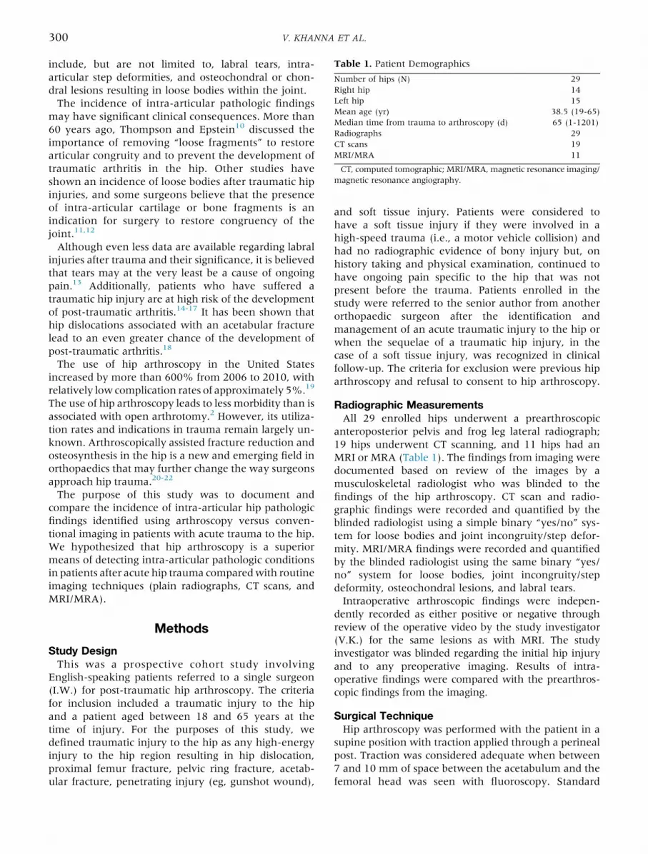
Table 1. Patient Demographics
Number of hips (N) 29Right hip 14Left hip 15Mean age (yr) 38.5 (19-65)Median time from trauma to arthroscopy (d) 65 (1-1201)Radiographs 29CT scans 19MRI/MRA 11
CT, computed tomographic; MRI/MRA, magnetic resonance imaging/magnetic resonance angiography.
300 V. KHANNA ET AL.
include, but are not limited to, labral tears, intra-articular step deformities, and osteochondral or chon-dral lesions resulting in loose bodies within the joint.The incidence of intra-articular pathologic findings
may have significant clinical consequences. More than60 years ago, Thompson and Epstein10 discussed theimportance of removing “loose fragments” to restorearticular congruity and to prevent the development oftraumatic arthritis in the hip. Other studies haveshown an incidence of loose bodies after traumatic hipinjuries, and some surgeons believe that the presenceof intra-articular cartilage or bone fragments is anindication for surgery to restore congruency of thejoint.11,12
Although even less data are available regarding labralinjuries after trauma and their significance, it is believedthat tears may at the very least be a cause of ongoingpain.13 Additionally, patients who have suffered atraumatic hip injury are at high risk of the developmentof post-traumatic arthritis.14-17 It has been shown thathip dislocations associated with an acetabular fracturelead to an even greater chance of the development ofpost-traumatic arthritis.18
The use of hip arthroscopy in the United Statesincreased by more than 600% from 2006 to 2010, withrelatively low complication rates of approximately 5%.19
The use of hip arthroscopy leads to less morbidity than isassociated with open arthrotomy.2 However, its utiliza-tion rates and indications in trauma remain largely un-known. Arthroscopically assisted fracture reduction andosteosynthesis in the hip is a new and emerging field inorthopaedics that may further change the way surgeonsapproach hip trauma.20-22
The purpose of this study was to document andcompare the incidence of intra-articular hip pathologicfindings identified using arthroscopy versus conven-tional imaging in patients with acute trauma to the hip.We hypothesized that hip arthroscopy is a superiormeans of detecting intra-articular pathologic conditionsin patients after acute hip trauma comparedwith routineimaging techniques (plain radiographs, CT scans, andMRI/MRA).
Methods
Study DesignThis was a prospective cohort study involving
English-speaking patients referred to a single surgeon(I.W.) for post-traumatic hip arthroscopy. The criteriafor inclusion included a traumatic injury to the hipand a patient aged between 18 and 65 years at thetime of injury. For the purposes of this study, wedefined traumatic injury to the hip as any high-energyinjury to the hip region resulting in hip dislocation,proximal femur fracture, pelvic ring fracture, acetab-ular fracture, penetrating injury (eg, gunshot wound),
and soft tissue injury. Patients were considered tohave a soft tissue injury if they were involved in ahigh-speed trauma (i.e., a motor vehicle collision) andhad no radiographic evidence of bony injury but, onhistory taking and physical examination, continued tohave ongoing pain specific to the hip that was notpresent before the trauma. Patients enrolled in thestudy were referred to the senior author from anotherorthopaedic surgeon after the identification andmanagement of an acute traumatic injury to the hip orwhen the sequelae of a traumatic hip injury, in thecase of a soft tissue injury, was recognized in clinicalfollow-up. The criteria for exclusion were previous hiparthroscopy and refusal to consent to hip arthroscopy.
Radiographic MeasurementsAll 29 enrolled hips underwent a prearthroscopic
anteroposterior pelvis and frog leg lateral radiograph;19 hips underwent CT scanning, and 11 hips had anMRI or MRA (Table 1). The findings from imaging weredocumented based on review of the images by amusculoskeletal radiologist who was blinded to thefindings of the hip arthroscopy. CT scan and radio-graphic findings were recorded and quantified by theblinded radiologist using a simple binary “yes/no” sys-tem for loose bodies and joint incongruity/step defor-mity. MRI/MRA findings were recorded and quantifiedby the blinded radiologist using the same binary “yes/no” system for loose bodies, joint incongruity/stepdeformity, osteochondral lesions, and labral tears.Intraoperative arthroscopic findings were indepen-
dently recorded as either positive or negative throughreview of the operative video by the study investigator(V.K.) for the same lesions as with MRI. The studyinvestigator was blinded regarding the initial hip injuryand to any preoperative imaging. Results of intra-operative findings were compared with the prearthros-copic findings from the imaging.
Surgical TechniqueHip arthroscopy was performed with the patient in a
supine position with traction applied through a perinealpost. Traction was considered adequate when between7 and 10 mm of space between the acetabulum and thefemoral head was seen with fluoroscopy. Standard

Table 2. Intra-articular Pathologic Findings Identified by Arthroscopy in Each Category of Traumatic Hip Injury (Total N ¼ 29)
Pelvic RingFracturen ¼ 5
AcetabularFracturen ¼ 6
Proximal FemurFracturen ¼ 7
Posterior HipDislocation
n ¼ 2
SoftTissue Injury
n ¼ 8
GunshotWoundn ¼ 1
Loose body 3 of 5 6 of 6 4 of 7 0 of 2 3 of 8 1 of 1Step deformity 2 of 5 5 of 6 2 of 7 0 of 2 1 of 8 1 of 1Osteochondral lesion 2 of 5 5 of 6 4 of 7 1 of 2 1 of 8 1 of 1Labral tear 5 of 5 5 of 6 7 of 7 2 of 2 8 of 8 0 of 1
HIP ARTHROSCOPY IN TRAUMA 301
anterolateral and lateral portals were used. Additionalportals were added under direct vision if necessary. Acomplete diagnostic arthroscopy was performed foreach hip to identify intra-articular pathologic disorders.If a pathologic condition amenable to treatment wasidentified, patients underwent the appropriate inter-vention at the time of arthroscopy. For example, pa-tients with labral tears underwent labral repair withsuture anchors, patients with loose bodies amenable tocapture by a grasper had them removed or suctionedwith the shaver, and patients with osteochondral le-sions or step deformities had the region debrided. Theentire procedure was video recorded for analysis.
StatisticsDescriptive statistics were used to summarize patients’
demographic and baseline characteristics. Categoricalvariables were reported as counts and percentages, andcontinuous variables as mean and standard deviation ormedian and range if not normally distributed.The primary objective of the study was to estimate the
sensitivity and specificity of arthroscopic surgery versusconventional imaging in identifying hip pathologicconditions. Sensitivity, specificity, positive predictivevalue (PPV) and negative predictive value (NPV), andtheir corresponding 95% confidence intervals (CIs)were calculated for all patients.
Table 3. Radiographic and Computed CT Loose BodyIdentification (95% CI)
Radiograph CT Scan
Sensitivity 6% (1%-27%) 14% (4%-40%)Specificity 100% (75%-100%) 100% (56%-100%)PPV 100% (21%-100%) 100% (34%-100%)NPV 43% (26%-65%) 29% (13%-53%)
CI, confidence interval; CT, computed tomographic; NPV, negativepredictive value; PPV, positive predictive value.
ResultsA total of 29 hips were enrolled from 28 patients (one
patient had bilateral hips enrolled). All patients whowere approached for enrollment in the study ultimatelyunderwent hip arthroscopy. Sex distribution consistedof 10 women and 18 men. Fourteen of the cases con-sisted of right hips, and 15 of the cases consisted of lefthips The average age of the patient was 38.5 years, andthe median age was 37 years (range, 19 to 65 years).The median time from trauma to hip arthroscopy was65 days (range, 1 to 1,201 days). The main traumaticinjury of the hip was recorded. Pelvic ring fractureswere the main injury in 5 hips, 6 hips had acetabularfractures, 7 hips had fractures of the proximal femur(head/neck), and 2 hips were simple dislocations withno associated fracture. Eight hips were involved inhigh-energy motor vehicle collisions with no obvious
fracture, but there was continued ongoing hip pain.One enrolled patient had a gunshot wound to the hip.Of the 29 post-traumatic hips that underwent hip
arthroscopy, 17 hips (59%) were identified byarthroscopy as having intra-articular loose bodies, 11hips (38%) were identified as having intra-articularstep deformities, 14 hips (48%) were identified ashaving osteochondral lesions that included cartilagedelamination, and 27 of the 29 hips (93%) had labraltears seen through the arthroscope. The intra-articularhip pathologic findings identified using arthroscopyare organized by category of traumatic hip injury inTable 2.Of the 17 hips identified as having loose bodies by
arthroscopy, one was identified preoperatively byradiography and 2 by computed tomography. Thisyielded a sensitivity of 6% (95% CI, 1% to 27%) forradiography and a sensitivity of 14% (95% CI, 4% to40%) for computed tomography. Specificity was 100%for both (95% CI, 75% to 100% for radiography and56% to 100% for computed tomography). The NPV ofradiography and computed tomography for the pres-ence of loose bodies was 43% (95% CI, 26.5% to 61%)and 29% (95% CI, 13% to 53%), respectively. PPV was100% for both (Table 3).Of the 11 hips identified as having intra-articular step
deformities by arthroscopy, 5 were identified by pre-operative radiography and 6 were identified bycomputed tomography, yielding a sensitivity of 45%(95% CI, 21% to 72%) for radiographic identificationof step deformity and a sensitivity of 67% (95% CI,35% to 88%) for computed tomography. The specificitywas 100% (95% CI, 82% to 100%) for radiographyand 90% (95% CI, 60 to 98%) for computed tomog-raphy. The NPV of radiography and computed to-mography for the presence of an intra-articular stepdeformity was 75% for both (95% CI, 55% to 88% for

Table 4. Radiograph and CT Joint Incongruity/StepDeformity Identification (95% CI)
Radiograph CT Scan
Sensitivity 45% (21%-72%) 67% (35%-88%)Specificity 100% (82%-100%) 90% (60%-98%)PPV 100% (56%-100%) 86% (49%-97%)NPV 75% (55%-88%) 75% (47%-91%)
CI, confidence interval; CT, computed tomographic; NPV, negativepredictive value; PPV, positive predictive value.
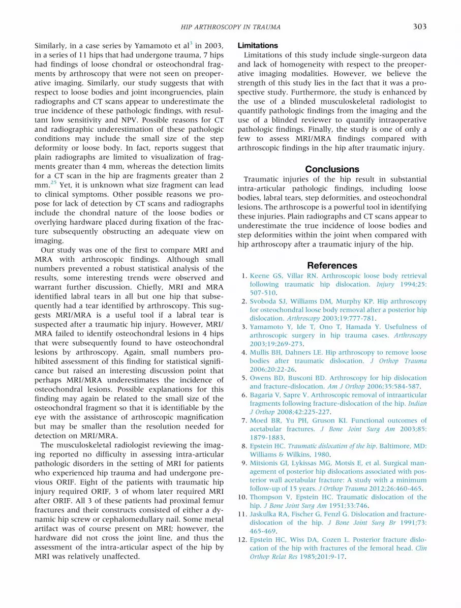
Fig 1. Images from a 35-year-old man after a simple posteriorleft hip dislocation. Treated with closed reduction, his groinpain persisted. Magnetic resonance angiogram suggestedpresence of labral tear. (A) Intraoperative finding of a labraltear as well as a large cartilaginous defect of the acetabulum.(B) Intraoperative photograph of the osteochondral lesionafter debridement.
302 V. KHANNA ET AL.
radiography and 47% to 91% for computed tomogra-phy). The PPV was 100% for radiography and 85.7%for computed tomography (Table 4). Radiographs andCT scans were not used to assess for soft tissue injuriessuch as labral tears, nor were they used to assess forchondral injuries.Because only 11 patients had MRI/MRA followed by
hip arthroscopy, the incidence of imaging-identifiedversus arthroscopically identified intra-articular patho-logic conditions was limited. As a result, the numberswere too low to permit appropriate 2 � 2 table statis-tical analysis, and thus only observations are reportedhere.In the assessment of the presence of loose bodies, 1 of
11 (9%) hips was identified as positive through MRI/MRA and 2 of 11 (18%) hips were identified byarthroscopy as having loose bodies. When assessingintra-articular step deformities, 0 of 11 hips (0%) werefound to have pathologic conditions by MRI/MRA, andone of 11 hips (9%) was found to have a step deformityby arthroscopy (Table 5).The incidence of osteochondral lesions identified by
MRI/MRA was 0 of 11 hips (0%), but 4 of these 11 hips(36%) were found to have osteochondral lesionsintraoperatively during arthroscopy (Fig 1). MRI/MRAidentified 10 of 11 hips (91%) as having labral tearsversus 11 of 11 (100%) hips identified as having labraltears by arthroscopy (Table 5).
DiscussionCurrently, the role for hip arthroscopy in the post-
traumatic patient remains undefined. Although severalstudies have retrospectively reviewed the findings of hiparthroscopy in the traumatic hip, few to no prospectivedata exist. In this prospective study, we set out toinvestigate the utility of hip arthroscopy as a diagnostic
Table 5. Intra-articular Pathologic Findings Identified byMRI/MRA Versus Arthroscopy
MRI/MRA Arthroscopy
Loose bodies 1 of 11 (9%) 2 of 11 (18%)Step deformity 0 of 11 (0%) 1 of 11 (9%)Osteochondral lesion 0 of 11 (0%) 4 of 11 (36%)Labral tears 10 of 11 (91%) 11 of 11 (100%)
MRI/MRA, magnetic resonance imaging/magnetic resonanceangiography.
tool compared with traditional imaging modalities inpatients who have undergone trauma.A recent study of hip arthroscopy after hip disloca-
tions by Philippon et al.23 in 2009 observed chondralinjuries and labral tears in all 14 of their enrolled pa-tients, and 11 of 14 patients had loose bodies within thejoint. Similarly, in a study by Ilizaliturri et al.24 in 2011,of 17 patients with posterior hip dislocations who sub-sequently underwent arthroscopy, 14 were found tohave intra-articular loose bodies, and all were found tohave chondral injuries and labral tears. These findingsare in keeping with our results identifying considerableintra-articular pathologic conditions. In particular, lab-ral tears and loose bodies were common findings whenthe arthroscope was introduced into a post-traumatichip. Although most other studies focused on intra-articular pathologic findings solely after hip disloca-tions, our data suggest that intra-articular injuries arenot limited to hip dislocations only but can be associ-ated with proximal femur fractures, pelvic ring frac-tures, acetabular fractures, and even soft tissue injuriesnot associated with a fracture. Early identificationand treatment of these pathologic disorders mayprevent subsequent development of early post-traumatic osteoarthritis. In one study, a traumaticarthritis prevalence rate of 24% was reported in pa-tients 5 years after injury.17 This rate was seen in pa-tients who experienced a simple hip dislocation withoutan associated fracture. The prevalence rate rose as highas 54% when more complex injuries, such as fracture-dislocations, were included.14-16
A recent retrospective study by Mullis et al.4 reportedthat loose bodies were found by arthroscopy in 7 of 9cases in which standard radiographs and CT scansfound no evidence of loose bodies preoperatively.
HIP ARTHROSCOPY IN TRAUMA 303
Similarly, in a case series by Yamamoto et al3 in 2003,in a series of 11 hips that had undergone trauma, 7 hipshad findings of loose chondral or osteochondral frag-ments by arthroscopy that were not seen on preoper-ative imaging. Similarly, our study suggests that withrespect to loose bodies and joint incongruencies, plainradiographs and CT scans appear to underestimate thetrue incidence of these pathologic findings, with resul-tant low sensitivity and NPV. Possible reasons for CTand radiographic underestimation of these pathologicconditions may include the small size of the stepdeformity or loose body. In fact, reports suggest thatplain radiographs are limited to visualization of frag-ments greater than 4 mm, whereas the detection limitsfor a CT scan in the hip are fragments greater than 2mm.25 Yet, it is unknown what size fragment can leadto clinical symptoms. Other possible reasons we pro-pose for lack of detection by CT scans and radiographsinclude the chondral nature of the loose bodies oroverlying hardware placed during fixation of the frac-ture subsequently obstructing an adequate view onimaging.Our study was one of the first to compare MRI and
MRA with arthroscopic findings. Although smallnumbers prevented a robust statistical analysis of theresults, some interesting trends were observed andwarrant further discussion. Chiefly, MRI and MRAidentified labral tears in all but one hip that subse-quently had a tear identified by arthroscopy. This sug-gests MRI/MRA is a useful tool if a labral tear issuspected after a traumatic hip injury. However, MRI/MRA failed to identify osteochondral lesions in 4 hipsthat were subsequently found to have osteochondrallesions by arthroscopy. Again, small numbers pro-hibited assessment of this finding for statistical signifi-cance but raised an interesting discussion point thatperhaps MRI/MRA underestimates the incidence ofosteochondral lesions. Possible explanations for thisfinding may again be related to the small size of theosteochondral fragment so that it is identifiable by theeye with the assistance of arthroscopic magnificationbut may be smaller than the resolution needed fordetection on MRI/MRA.The musculoskeletal radiologist reviewing the imag-
ing reported no difficulty in assessing intra-articularpathologic disorders in the setting of MRI for patientswho experienced hip trauma and had undergone pre-vious ORIF. Eight of the patients with traumatic hipinjury required ORIF, 3 of whom later required MRIafter ORIF. All 3 of these patients had proximal femurfractures and their constructs consisted of either a dy-namic hip screw or cephalomedullary nail. Some metalartifact was of course present on MRI; however, thehardware did not cross the joint line, and thus theassessment of the intra-articular aspect of the hip byMRI was relatively unaffected.
LimitationsLimitations of this study include single-surgeon data
and lack of homogeneity with respect to the preoper-ative imaging modalities. However, we believe thestrength of this study lies in the fact that it was a pro-spective study. Furthermore, the study is enhanced bythe use of a blinded musculoskeletal radiologist toquantify pathologic findings from the imaging and theuse of a blinded reviewer to quantify intraoperativepathologic findings. Finally, the study is one of only afew to assess MRI/MRA findings compared witharthroscopic findings in the hip after traumatic injury.
ConclusionsTraumatic injuries of the hip result in substantial
intra-articular pathologic findings, including loosebodies, labral tears, step deformities, and osteochondrallesions. The arthroscope is a powerful tool in identifyingthese injuries. Plain radiographs and CT scans appear tounderestimate the true incidence of loose bodies andstep deformities within the joint when compared withhip arthroscopy after a traumatic injury of the hip.
References1. Keene GS, Villar RN. Arthroscopic loose body retrieval
following traumatic hip dislocation. Injury 1994;25:507-510.
2. Svoboda SJ, Williams DM, Murphy KP. Hip arthroscopyfor osteochondral loose body removal after a posterior hipdislocation. Arthroscopy 2003;19:777-781.
3. Yamamoto Y, Ide T, Ono T, Hamada Y. Usefulness ofarthroscopic surgery in hip trauma cases. Arthroscopy2003;19:269-273.
4. Mullis BH, Dahners LE. Hip arthroscopy to remove loosebodies after traumatic dislocation. J Orthop Trauma2006;20:22-26.
5. Owens BD, Busconi BD. Arthroscopy for hip dislocationand fracture-dislocation. Am J Orthop 2006;35:584-587.
6. Bagaria V, Sapre V. Arthroscopic removal of intraarticularfragments following fracture-dislocation of the hip. IndianJ Orthop 2008;42:225-227.
7. Moed BR, Yu PH, Gruson KI. Functional outcomes ofacetabular fractures. J Bone Joint Surg Am 2003;85:1879-1883.
8. Epstein HC. Traumatic dislocation of the hip. Baltimore, MD:Williams & Wilkins, 1980.
9. Mitsionis GI, Lykissas MG, Motsis E, et al. Surgical man-agement of posterior hip dislocations associated with pos-terior wall acetabular fracture: A study with a minimumfollow-up of 15 years. J Orthop Trauma 2012;26:460-465.
10. Thompson V, Epstein HC. Traumatic dislocation of thehip. J Bone Joint Surg Am 1951;33:746.
11. Jaskulka RA, Fischer G, Fenzl G. Dislocation and fracture-dislocation of the hip. J Bone Joint Surg Br 1991;73:465-469.
12. Epstein HC, Wiss DA, Cozen L. Posterior fracture dislo-cation of the hip with fractures of the femoral head. ClinOrthop Relat Res 1985;201:9-17.

304 V. KHANNA ET AL.
13. Byrd JW. Labral lesions: An elusive source of hip pain:Case reports and literature review. Arthroscopy 1996;12:603-612.
14. Armstrong JR. Traumatic dislocation of the hip joint:Review of 101 dislocations. J Bone Joint Surg Br 1948;30:430-435.
15. Stewart MJ, McCarroll HR Jr, Mulhollan JS. Fracture-dislocation of the hip. Acta Orthop Scand 1975;46:507-525.
16. Brav CE. Traumatic dislocation of the hip. J Bone Joint SurgAm 1962;44:1115-1134.
17. Upadhyay SS, Moulton A, Srikrishnamurthy K. An anal-ysis of the late effects of traumatic posterior dislocation ofthe hip without fracture. J Bone Joint Surg Br 1983;65:150-152.
18. Upadhyay SS, Moulton A. The long-term results of trau-matic posterior dislocation of the hip. J Bone Joint Surg Br1981;63:548-551.
19. Bozic KJ, Chan V, Valone FH III, Feeley BT, Vail TP.Trends in hip arthroscopy utilization in the United States.J Arthroplasty 2013;28(8 suppl):140-143.
ARTHROSCOPY
Have you submitted your new Tech Nback? Share your interesting new
Submit at http://ees.
20. Lansford T, Munns SW. Arthroscopic treatment of Pipkintype I femoral head fractures: A report of 2 cases. J OrthopTrauma 2012;26:e94-e96.
21. Matsuda DK. The clamshell fracture and adjunctive ace-tabuloplasty in the arthroscopic osteosynthesis of femoralhead fractures with femoroacetabular impingement.Arthrosc Tech 2012;1:e5-e10.
22. Yang JH, Chouhan DK, Oh KJ. Percutaneous screw fixa-tion of acetabular fractures: Applicability of hip arthros-copy. Arthroscopy 2010;26:1556-1561.
23. Philippon MJ, Kuppersmith DA, Wolff AB, Briggs KK.Arthroscopic findings following traumatic hip disloca-tion in 14 professional athletes. Arthroscopy 2009;25:169-174.
24. Ilizaliturri VM, Gonzalez-Gutierrez B, Gonzalez-Ugalde H,Camacho-Galindo J. Hip arthroscopy after traumatic hipdislocation. Am J Sports Med 2011;39:50S-57S.
25. Baird RA, Schobert WE, Pais MJ, et al. Radiographicidentification of loose bodies in the traumatized hip joint.Radiology 1982;145:661-665.
TECHNIQUES
ote and Video? What’s holding youdiscovery with your colleagues!
elsevier.com/arth/
Copyright © 2022 FDOKUMEN