Selecting the surgical approach for revision total hip arthroplasty
Upload
khangminh22Category
view
0download
0
Dissertation for the degree of philosophiae doctor (PhD)
at the University of Bergen
2013
Dissertation date: 08.02.2013
The present PhD project was initiated in 2007 while I was working as a consultant at
the Department of Orthopaedic Surgery, Haukeland University Hospital, Bergen.
Since August 2007 my primary workplaces have been the Department of
Orthopaedic Rehabilitation and the Norwegian Arthroplasty Register, Haukeland
University Hospital.
Supervision has been given by the staff at the Norwegian Arthroplasty Register and
in particular by supervisor Professor Lars Birger Engesæter and co-supervisors
Professor Leif I Havelin and Professor Birgitte Espehaug.
Parts of the project have been conducted in cooperation with the Norwegian
Institute of Public Health and the Nordic Arthroplasty Register Association.
The project was financed by the Department of Orthopaedic Surgery, Haukeland
University Hospital.
The thesis is a part of the PhD programme at the Department of Surgical Sciences,
University of Bergen.
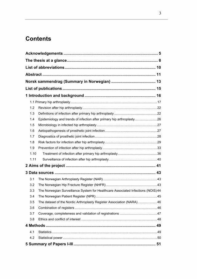
1.1 Primary hip arthroplasty...............................................................................................17 1.2 Revision after hip arthroplasty..................................................................................22 1.3 Definitions of infection after primary hip arthroplasty ...............................................22 1.4 Epidemiology and trends of infection after primary hip arthroplasty ........................26 1.5 Microbiology in infected hip arthroplasty..................................................................27 1.6 Aetiopathogenesis of prosthetic joint infection.........................................................27 1.7 Diagnostics of prosthetic joint infection....................................................................28 1.8 Risk factors for infection after hip arthroplasty.........................................................29 1.9 Prevention of infection after hip arthroplasty............................................................33 1.10 Treatment of infection after primary hip arthroplasty...........................................36 1.11 Surveillance of infection after hip arthroplasty.....................................................40
3.1 The Norwegian Arthroplasty Register (NAR) ...........................................................43 3.2 The Norwegian Hip Fracture Register (NHFR)........................................................43 3.3 The Norwegian Surveillance System for Healthcare Associated Infections (NOIS)44 3.4 The Norwegian Patient Register (NPR) ...................................................................45 3.5 The dataset of the Nordic Arthroplasty Register Association (NARA).....................46 3.6 Combination of registers ..........................................................................................46 3.7 Coverage, completeness and validation of registrations .........................................47 3.8 Ethics and conflict of interest ...................................................................................48
4.1 Statistics ...................................................................................................................49 4.2 Statistical power .......................................................................................................50
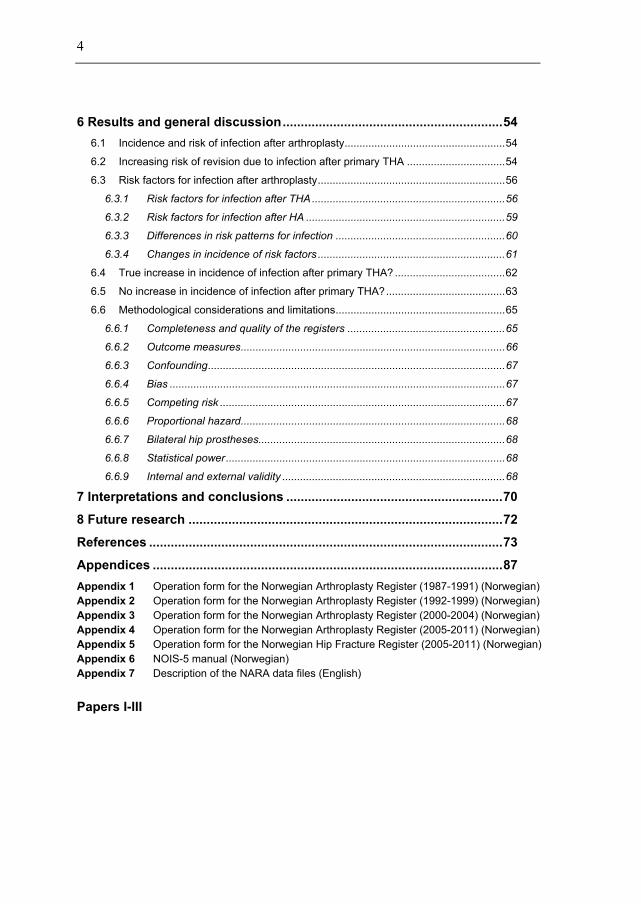
6.1 Incidence and risk of infection after arthroplasty......................................................54 6.2 Increasing risk of revision due to infection after primary THA .................................54 6.3 Risk factors for infection after arthroplasty...............................................................56
6.4 True increase in incidence of infection after primary THA? .....................................62 6.5 No increase in incidence of infection after primary THA?........................................63 6.6 Methodological considerations and limitations.........................................................65
Operation form for the Norwegian Arthroplasty Register (1987-1991) (Norwegian) Operation form for the Norwegian Arthroplasty Register (1992-1999) (Norwegian)
Operation form for the Norwegian Arthroplasty Register (2000-2004) (Norwegian) Operation form for the Norwegian Arthroplasty Register (2005-2011) (Norwegian) Operation form for the Norwegian Hip Fracture Register (2005-2011) (Norwegian) NOIS-5 manual (Norwegian) Description of the NARA data files (English)

This PhD project was carried out at the Department of Orthopaedic Surgery,
Haukeland University Hospital in Bergen, Norway during the years 2007-2012.
First of all I would like to thank the people who aroused my interest in and taught me
about orthopaedic surgery: (my father)
and many others, who have all served as great examples and
inspirations, and who have let me learn from their vast surgical skills and
experience, always in an inclusive atmosphere of dedication, earnestness and
humour.
Since my start at Haukeland in 2002 I have realized the significant amount of high
level scientific work produced at the Department of Orthopaedic Surgery and at the
Norwegian Arthroplasty Register (NAR). I have felt privileged to have the ever
enthusiastic and knowledgeable co-founder and present leader of the NAR,
Professor , as my supervisor throughout this PhD project. Your
enthusiasm has been a great inspiration, as has your scientific and human
commitment. I am also grateful that you let me take part in the inclusive atmosphere
at the NAR, and for being allowed to use the high quality data of the NAR, collected
conscientiously since 1987.
My co-supervisor, the former head of the Department of Orthopaedic Surgery and
former leader of the NAR, and the person who engaged me at Haukeland in the first
place, Professor , deserves special thanks for his conscientious and
caring guidance into the world of science. For every draft, discussion and question,
you have given your full attention and a thorough evaluation or answer. Combining
the highest level of knowledge with a sense of humour and integrity, you have
enriched every discussion and sharpened every text.
I would also like to express my gratitude to the statistician in the supervision team,
my co-supervisor Professor . patient guidance and help in
understanding and performing statistical analyses has been invaluable.
I am in great debt to Professor , present Head of the Orthopaedic
Department and former leader of the NAR, for having faith in my academic abilities
despite my own doubts. Your enthusiasm for science seems to know no bounds.
You have also arranged time for me to work on this study, and for that both my
family and I myself are truly grateful.
All these professors were vital in helping me through this PhD project. For guiding
me in this way and educating me in research, I extend my thanks to the Department
of Surgical Science at the University of Bergen.
I am also grateful to the Norwegian Institute of Public Health’s Surveillance System
for Healthcare-Acquired Infections (NOIS) and the Nordic Arthroplasty Register
Association (NARA) for letting me assess their registries and for co-authoring the
resulting manuscripts. Collaboration between large national health registries
facilitates unique transparency, knowledge and improved healthcare quality in the
Nordic countries, for the benefit of our patients. I feel proud and privileged to be
allowed to take part in such important work.
A special thank you goes to the director of the Orthopaedic Department
, the head of the Department of Orthopaedic Rehabilitation
, and the head nurse of the Department of Orthopaedic Rehabilitation
for allowing me to work at the Department of Orthopaedic
Rehabilitation during my PhD project. Thank you all for your patience and flexibility,
and for supporting me financially.
My gratitude also goes to the secretaries and statisticians of
the NAR for their indefatigable and accurate plotting, quality assurance and
evaluation of the reported data. Your stamina and meticulous work is invaluable to
the register and the research it facilitates. Thanks also to the surgeons and staff of
the Norwegian and other Nordic hospitals for conscientious reporting to the health
registries.

I also truly appreciate the relentless love and support throughout my life from my
parents, Eldbjørg and Gudleik Dale. I am also grateful to my sister Kristin Selvig and
brother Eirik Dale for going easy on me, their annoying younger brother.
Finally, I acknowledge the never ending love and support from my beautiful beloved
wife, Elisabeth, and our children Ingeborg, Sunniva and Magnus. You give me a
strong sense of belonging and put all my striving into perspective.
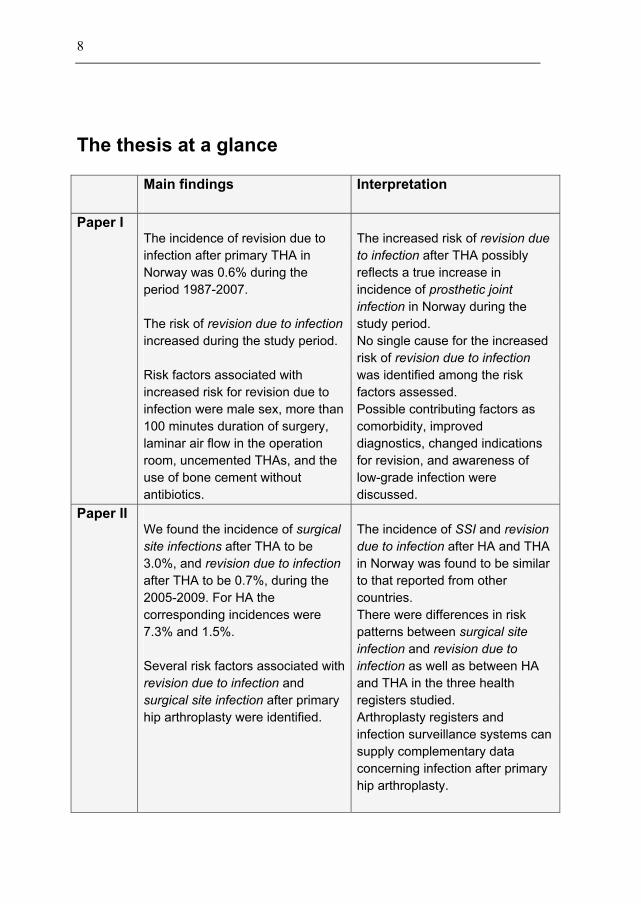
The incidence of revision due to infection after primary THA in Norway was 0.6% during the period 1987-2007. The risk of increased during the study period. Risk factors associated with increased risk for revision due to infection were male sex, more than 100 minutes duration of surgery, laminar air flow in the operation room, uncemented THAs, and the use of bone cement without antibiotics.
The increased risk of
after THA possibly reflects a true increase in incidence of
in Norway during the study period. No single cause for the increased risk of was identified among the risk factors assessed. Possible contributing factors as comorbidity, improved diagnostics, changed indications for revision, and awareness of low-grade infection were discussed.
We found the incidence of
after THA to be 3.0%, and after THA to be 0.7%, during the 2005-2009. For HA the corresponding incidences were 7.3% and 1.5%. Several risk factors associated with
and after primary
hip arthroplasty were identified.
The incidence of and
after HA and THA in Norway was found to be similar to that reported from other countries. There were differences in risk patterns between
and as well as between HA
and THA in the three health registers studied. Arthroplasty registers and infection surveillance systems can supply complementary data concerning infection after primary hip arthroplasty.
The incidence of
after primary THA in the dataset of the Nordic Arthroplasty Register Association (NARA) was 0.6% during the period 1995-2009. The risk of increased in Denmark, Finland, Norway, and Sweden during the study period. The increase in risk was most prominent the first three postoperative months. Risk factors for
were male sex, hybrid fixation, cement without antibiotics and THA performed due to inflammatory disease, hip fracture or femoral head necrosis.
The increased risk of after THA possibly
reflects a true increase in incidence of
in Denmark, Finland, Norway and Sweden during 1995-2009. The study confirmed that increasing risk of
is a common feature in the Nordic countries. No single cause for the increased risk of was identified among the risk factors studied. Possible causes and contributing factors were discussed.
ASA American Society of Anaesthesiologists
BMI Body Mass Index
CDC Centres for Disease Control and Prevention (USA)
CoNS Coagulase-Negative Staphylococci
CRP C-Reactive Protein
ESR Erythrocyte Sedimentation Rate
FDA Food and Drug Administration (USA)
HA Hemiarthroplasty of the hip
HELICS Hospitals in Europe Link for Infection Control through Surveillance
ICD International Classification of Diseases
LAF Laminar air flow
MRSA Methicillin-Resistant Staphylococcus Aureus
MRSE Methicillin-Resistant Staphylococcus Epidermidis
NAR Norwegian Arthroplasty Register
NARA Nordic Arthroplasty Register Association
NHFR Norwegian Hip Fracture Register
NNIS National Nosocomial Infections Surveillance (UK)
NOIS Norwegian acronym for:
Norwegian Surveillance System for Healthcare-Associated Infections
NOMESCO Nordic Medico-Statistical Committee
NPR Norwegian Patient Register
OA Osteoarthritis
PCR Polymerase Chain Reaction
PJI Prosthetic Joint Infection
SSI Surgical Site Infection
THA Total Hip Arthroplasty
WBC White Blood Cells
Every year, more than 10,000 Norwegians undergo hip replacement (7,360 THAs
and 3,214 HAs in 2011). This may be due to osteoarthritis (OA), inflammatory joint
disease, fractures, fracture sequelae, aseptic femoral head necrosis or sequelae
after childhood hip disease. The native hip joint is replaced by a total hip
arthroplasty (THA) or a hemiarthroplasty (HA). The implants constitute large foreign
bodies that could be predilection spots for adherence of microorganisms, and
postoperative infections are a feared complication. Such infections are difficult to
treat and impose increased morbidity and mortality on the patients.
To meet the challenge of everal risk factors have been
identified and prophylactic measures have been introduced. The Norwegian
Arthroplasty Register (NAR) has had several publications on antibiotic prophylaxis,
systemically and in bone cement, for THA, and probably contributed to that
Norwegian orthopaedic surgeons changed their routines. The starting point of the
present PhD project was to assess whether these changes in antibiotic prophylaxis
had changed the risk of .
We found that, in spite of the anticipated improved antibiotic prophylaxis, the risk of
after primary THA had increased threefold from 1987-1992
to 2003-2007 (Paper I). In the Nordic Arthroplasty Register Association’s (NARA)
dataset from Denmark, Finland, Norway and Sweden, a similar increase in risk of
after primary THA was found between 1995-1999 and
2005-2009 (Paper III). The reason for this increase could not be explained by any
known changes in the risk factors assessed in the two studies (Papers I and III). The
possibility of a true increase in and other possible
explanations were discussed.
In Norway there are no systematic registrations of true .
should be reported to the NAR and the Norwegian Hip
Fracture Register (NHFR), and should be reported to the
Norwegian Surveillance System for Healthcare-Associated Infections (NOIS). In
Paper II we assessed risk factors and risk patterns for these two endpoints for both
THA and HA. The first-year incidence of after primary
arthroplasty was found to be nearly five times higher than the first-year incidence of
There also seems to be differences in the risk patterns
between and and between HA and
THA.
The risk factors associated with increased risk of after
primary THA were male sex, advanced age (70-90 years when adjusted for
comorbidity), comorbidity (ASA class > 1), long duration of surgery (> 100 minutes),
uncemented or hybrid fixation, bone cement without antibiotics, laminar air flow in
the operation room, NNIS risk index higher than one, and THA performed due to
inflammatory disease, hip fracture or femoral head necrosis.
Risk factors of after THA was advanced age (> 80 years),
comorbidity (ASA class > 2), and short duration of surgery (< 60 minutes).
For primary HAs the only risk factor associated with increased risk of
was young age (< 60 years), whereas no statistically significant risk factors
of were identified.
The overall conclusion of this thesis is that the risk of after
primary THA has been increasing. Definite causes of this increased risk could not
be established in the three papers. Considering risk factors and possible
confounders we still believe that there might have been a true increase in the
incidence of
Hvert år får mere enn 10 000 nordmenn erstattet sitt hofteledd med en hofteprotese
(7 360 totalproteser og 3 214 hemiproteser i 2011). Dette kan skyldes «slitasjegikt»
(artrose), inflammatorisk leddsykdom, lårhalsbrudd, komplikasjoner etter brudd,
aseptisk nekrose av lårbeinshodet eller ettervirkninger etter barnehoftelidelser.
Hofteleddet kan erstattes av en total hofteprotese eller en hemiprotese.
Hofteproteser utgjør store fremmedlegemer som kan være utsatt for
mikroorganismer, og postoperative infeksjoner er en fryktet komplikasjon. Slike
infeksjoner er vanskelig å behandle og fører til økt sykelighet og dødelighet for de
pasientene som rammes.
For å møte utfordringen med proteseinfeksjoner, har flere risikofaktorer blitt
identifisert og forebyggende tiltak er innført. Nasjonalt register for leddproteser
(NRL) har hatt flere publikasjoner om antibiotikaprofylakse, systemisk og i
beinsementen, ved innsetting av totalprotese i hoften, og har sannsynligvis bidratt til
at kirurgene har endret sine rutiner. Utgangspunktet for dette doktorgradsarbeidet
var å vurdere om disse endringene i antibiotikaprofylakse hadde endret risiko for
.
Vi fant at til tross for at antibiotikaprofylaksen ved hofteproteseoperasjoner var
endret i tråd med funnene, var risikoen for tredoblet
fra 1987-1992 til 2003-2007 (Artikkel I). Vi fant også økning i risikoen for
etter primær total hofteprotese mellom 1995-1999 og 2005-2009
i Nordic Arthroplasty Register Association (NARA) sitt datasett fra Danmark,
Finland, Norge og Sverige (Artikkel III). Grunnen til denne økningen kan ikke
forklares med registrerte endringer i risikofaktorer vurdert i de to studiene (Artikkel I
og III). Muligheten for en sann økning av og andre mulige
forklaringer ble diskutert.
I Norge har vi ikke systematisk registrering av sanne .
skal rapporteres NRL og Nasjonalt hoftebruddregister
(NHBR), og rapporteres til Norsk overvåkingssystem
for antibiotikabruk og helsetjenesteassosierte infeksjoner (NOIS). I Artikkel II
vurderte vi risikofaktorer og risikomønstre for og
for både primær hemi- og totalprotese i hofte. Den
samlede forekomst av det første året etter primær
protesekirurgi ble funnet å være nesten fem ganger så høy som forekomsten av
det første året postoperativt. Det så også ut til å være
forskjeller i risikomønstre mellom og
og mellom hemi- og totalprotese.
Risikofaktorer som var forbundet med økt risiko for
etter primær totalprotese i hofte var menn, høy alder (70-90 år når det justeres for
andre sykdommer), andre sykdommer (ASA-klasse> 1), lang varighet av kirurgi (>
130 minutter), usementert eller hybrid fiksering, beinsement uten antibiotika,
laminær luftstrøm på operasjonsstuen, NNIS risiko indeks høyere enn én, og
totalprotese på grunn av inflammatorisk leddsykdom, hoftebrudd eller aseptisk
nekrose av lårbeinshodet.
Risikofaktorer for etter totalprotese i hoften var høy alder
(> 80 år), andre sykdommer (ASA-klasse> 2), og kort varighet av kirurgi (<60
minutter).
Ved primær hemiprotese i hoften var bare ung alder (<60 år) forbundet med økt
risiko for , mens vi ikke fant noen statistisk signifikante
risikofaktorer for .
Konklusjonen av denne avhandlingen er at risikoen for
har vært økende hos pasienter som har fått innsatt primær totalprotese i
hofte. Årsakene til denne økningen ble ikke funnet blant de risikofaktorene som ble
studert i denne doktoravhandlingen. Vurdert utfra mulige risikofaktorer og andre
faktorer (effektforvekslere) som kan ha påvirket resultatene, tror vi at det har vært
en sann økning i forekomsten av infeksjoner etter innsetting av totalprotese i hofte.
The thesis is based on the following papers, referred to in the text by their Roman
numerals:
Dale H, Hallan G, Espehaug B, Havelin L I, Engesæter L B.
. Acta Orthop 2009; 80 (6): 639-45.
Dale H, Skråmm I, Løwer H L, Eriksen H M, Espehaug B, Furnes O,
Skjeldestad F E, Havelin L I, Engesæter L B.
. Acta Orthop 2011; 82 (6): 646-54.
Dale H, Fenstad A M, Hallan G, Havelin L I, Furnes O, Overgaard S,
Pedersen A B, Kärrholm J, Garellick G, Pulkkinen P, Eskelinen A,
Mäkelä K, and Engesæter L B.
. Acta Orthop 2012; 83 (5): 449-
58.
Every year, more than 10,000 Norwegians undergo surgery to replace their native
hip joint with a hip prosthesis, a primary hip arthroplasty or hip replacement (7,360
THAs and 3,214 HAs in 2011) (The Norwegian Arthroplasty Register 2012, The
Norwegian Hip Fracture Register 2012). The implanted prostheses constitute large
foreign bodies that are predilection areas for adherence of microorganisms, and
postoperative infection is a feared complication. Sir John Charnley stated that
“postoperative infection is the saddest of all complications” (Waugh and Charnley
1990). Symptoms can vary from pain, sometimes due to loosening of the prosthesis,
without other accompanying signs of infection, to fulminant prosthetic joint infections
with life-threatening septicaemia. The treatment is multidisciplinary and involves
surgery, often repetitive, and prolonged antibiotic treatment.
For the individual patient a prosthetic joint infection imposes extra suffering with
extensive surgery and medical treatment often associated with complications,
adverse effects and functional loss (Westberg et al. 2012, Aslam and Darouiche
2012). For the healthcare services THA infections imply great medical challenges,
long hospital stays and 3-4 times increased costs compared to uncomplicated
primary THA (Whitehouse et al. 2002, Kurtz et al. 2007, Aslam and Darouiche
2012).
The Norwegian Arthroplasty Register (NAR) has published studies on antibiotic
prophylaxis against infection after THA, and the Norwegian orthopaedic surgeons
have complied with the findings and changed their routines accordingly (Espehaug
et al. 1997, Engesæter et al. 2003, Engesæter et al. 2006).
The starting point of the present PhD project was to study whether these changes in
antibiotic prophylaxis had had an impact on the risk of (Dale
et al. 2008). We wanted to assess these time trends and possible contributing risk
factors.
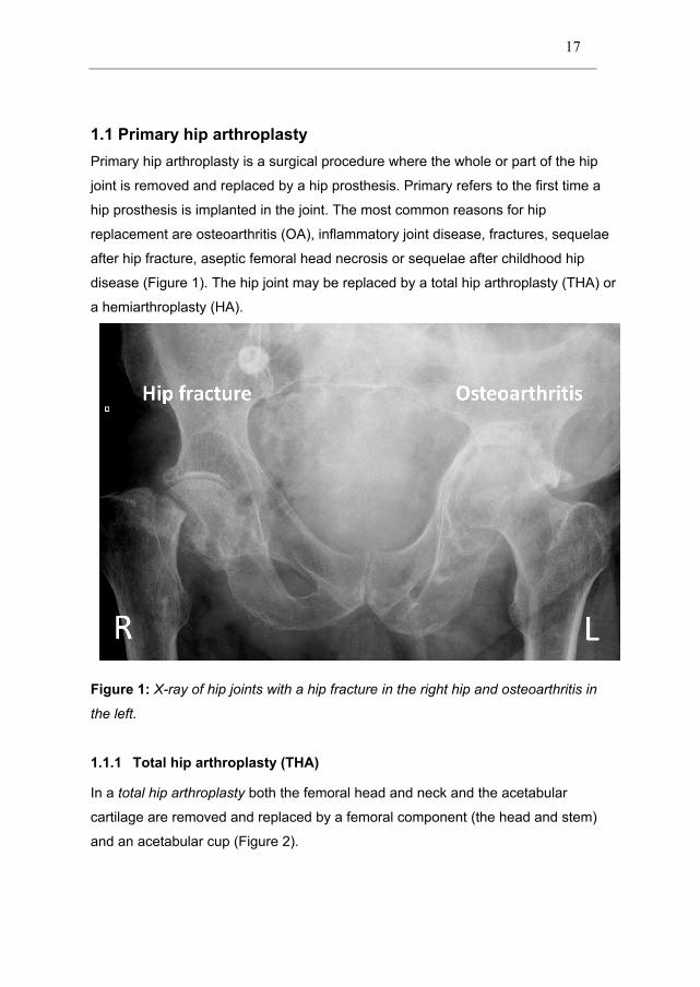
Primary hip arthroplasty is a surgical procedure where the whole or part of the hip
joint is removed and replaced by a hip prosthesis. Primary refers to the first time a
hip prosthesis is implanted in the joint. The most common reasons for hip
replacement are osteoarthritis (OA), inflammatory joint disease, fractures, sequelae
after hip fracture, aseptic femoral head necrosis or sequelae after childhood hip
disease (Figure 1). The hip joint may be replaced by a total hip arthroplasty (THA) or
a hemiarthroplasty (HA).
In a both the femoral head and neck and the acetabular
cartilage are removed and replaced by a femoral component (the head and stem)
and an acetabular cup (Figure 2).
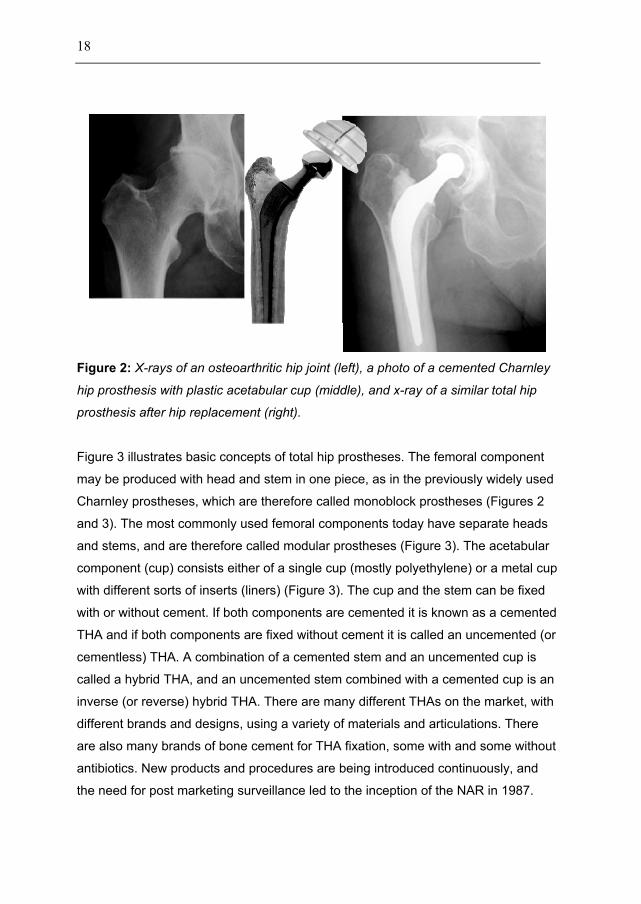
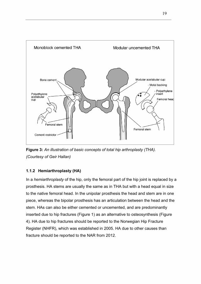
Figure 3 illustrates basic concepts of total hip prostheses. The femoral component
may be produced with head and stem in one piece, as in the previously widely used
Charnley prostheses, which are therefore called monoblock prostheses (Figures 2
and 3). The most commonly used femoral components today have separate heads
and stems, and are therefore called modular prostheses (Figure 3). The acetabular
component (cup) consists either of a single cup (mostly polyethylene) or a metal cup
with different sorts of inserts (liners) (Figure 3). The cup and the stem can be fixed
with or without cement. If both components are cemented it is known as a cemented
THA and if both components are fixed without cement it is called an uncemented (or
cementless) THA. A combination of a cemented stem and an uncemented cup is
called a hybrid THA, and an uncemented stem combined with a cemented cup is an
inverse (or reverse) hybrid THA. There are many different THAs on the market, with
different brands and designs, using a variety of materials and articulations. There
are also many brands of bone cement for THA fixation, some with and some without
antibiotics. New products and procedures are being introduced continuously, and
the need for post marketing surveillance led to the inception of the NAR in 1987.
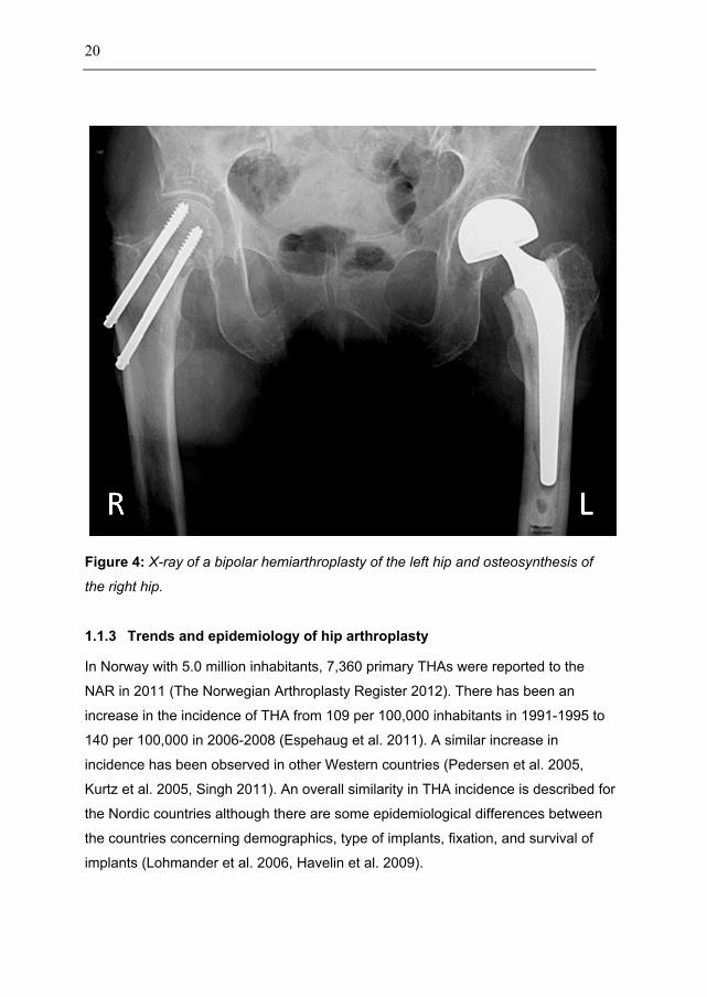
In a of the hip, only the femoral part of the hip joint is replaced by a
prosthesis. HA stems are usually the same as in THA but with a head equal in size
to the native femoral head. In the unipolar prosthesis the head and stem are in one
piece, whereas the bipolar prosthesis has an articulation between the head and the
stem. HAs can also be either cemented or uncemented, and are predominantly
inserted due to hip fractures (Figure 1) as an alternative to osteosynthesis (Figure
4). HA due to hip fractures should be reported to the Norwegian Hip Fracture
Register (NHFR), which was established in 2005. HA due to other causes than
fracture should be reported to the NAR from 2012.
In Norway with 5.0 million inhabitants, 7,360 primary THAs were reported to the
NAR in 2011 (The Norwegian Arthroplasty Register 2012). There has been an
increase in the incidence of THA from 109 per 100,000 inhabitants in 1991-1995 to
140 per 100,000 in 2006-2008 (Espehaug et al. 2011). A similar increase in
incidence has been observed in other Western countries (Pedersen et al. 2005,
Kurtz et al. 2005, Singh 2011). An overall similarity in THA incidence is described for
the Nordic countries although there are some epidemiological differences between
the countries concerning demographics, type of implants, fixation, and survival of
implants (Lohmander et al. 2006, Havelin et al. 2009).
In 2011 the number of primary HAs in Norway was 3,214 (The Norwegian Hip
Fracture Register 2012). The fraction of patients treated with HA instead of
osteosynthesis for their hip fracture is increasing (Jain et al. 2008, The Norwegian
Hip Fracture Register 2012).
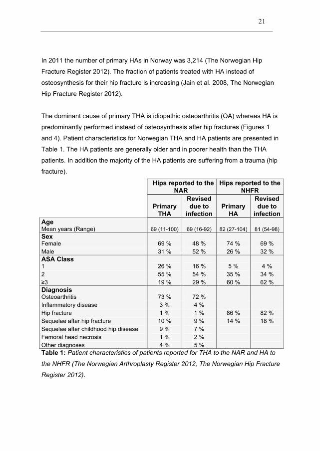
The dominant cause of primary THA is idiopathic osteoarthritis (OA) whereas HA is
predominantly performed instead of osteosynthesis after hip fractures (Figures 1
and 4). Patient characteristics for Norwegian THA and HA patients are presented in
Table 1. The HA patients are generally older and in poorer health than the THA
patients. In addition the majority of the HA patients are suffering from a trauma (hip
fracture).
Mean years (Range) 69 (11-100) 69 (16-92) 82 (27-104) 81 (54-98)
Female 69 % 48 % 74 % 69 % Male 31 % 52 % 26 % 32 %
1 26 % 16 % 5 % 4 % 2 55 % 54 % 35 % 34 % 3 19 % 29 % 60 % 62 %
Osteoarthritis 73 % 72 % Inflammatory disease 3 % 4 % Hip fracture 1 % 1 % 86 % 82 % Sequelae after hip fracture 10 % 9 % 14 % 18 % Sequelae after childhood hip disease 9 % 7 % Femoral head necrosis 1 % 2 % Other diagnoses 4 % 5 %
.
22
Revision after arthroplasty is defined as surgical removal or exchange of the
prosthesis or prosthesis parts. Such operations are reported to the NAR and the
NHFR. The most common causes of revision are a loose component, luxation, deep
infection, fracture, osteolysis, or wear of liner. The annual revision rate reported to
the NAR and the NHFR is approximately 0.5% after THA and 0.3% after HA (The
Norwegian Arthroplasty Register 2012, The Norwegian Hip Fracture Register 2012).
Infection after primary
arthroplasty is not unambiguous as a notion, and different publications use different
definitions of infection. Some publications may use diagnostic codes as a measure of
“infection”, without clarifying the diagnostic criteria or extent of the infection
(Kurtz et al. 2008, Wolf et al. 2012). These “infections” may include both superficial
and true , and may or may not be
reoperated or revised. Time trends and risk patterns may vary for different definitions
of infection after arthroplasty. The most commonly used definitions of infection after
arthroplasty are the Centres of Disease Control and Prevention’s (CDC) criteria for
postoperative , The Mayo Clinic’s criteria for
and the arthroplasty registries’ definition
(Horan et al. 1992, Espehaug et al. 1997, Berbari et al. 1998, Mangram et al. 1999).
In the three publications included in the present thesis we used the definitions of
(Paper II) and in the NAR and
N (Papers I-III) (Horan et al. 1992, Espehaug et al. 1997).
There is at present no international consensus about the criteria for a true
. A commonly used definition is from the Mayo Clinic (Berbari et al.
1998, Del Pozo and Patel 2009):
Presence of at least 1 of the following:
1) Acute periprosthetic inflammation on histopathological examination
2) Sinus tract communicating with the prosthesis
3) Gross purulence in the joint space
4) Isolation of significant amounts of the same microorganism from 2
cultures of joint aspirates
In the USA the Workgroup of the Musculoskeletal Infection Society have proposed
the following criteria for a definite prosthetic joint infection (Parvizi et al. 2011):
1) There is a sinus tract communicating with the prosthesis; or
2) A pathogen is isolated by culture from at least two separate tissue or fluid
samples obtained from the affected prosthetic joint; or
3) Four of the following six criteria exist:
a) Elevated serum erythrocyte sedimentation rate (ESR) and serum
C-reactive protein (CRP) concentration,
b) Elevated synovial leukocyte count,
c) Elevated synovial neutrophil percentage (PMN %),
d) Presence of purulence in the affected joint,
e) Isolation of a microorganism in one culture of periprosthetic
tissue or fluid, or
f) More than five neutrophils per high-power field in five high-power
fields observed from histologic analysis of periprosthetic tissue at
9400 times magnification
Prosthetic joint infection may be present if fewer than four of these criteria are met.
Postoperative is the outcome measure used by postoperative
infection surveillance systems like the Norwegian NOIS and the European HELICS.
The aim is to monitor incidence and outbursts of postoperative infection after some
common surgical procedures. One of these procedures is primary hip arthroplasty.
The Norwegian NOIS surveys both primary HA and THA. is
defined in three categories as follows (Horan et al. 1992, Mangram et al. 1999,
HELICS 2004) (Appendix 6):
Infection occurs within 30 days (365 for arthroplasty) of primary surgery and
involves only skin and subcutaneous tissue of the incision and at least one of the
following:
1) Purulent drainage from the superficial incision
2) Organisms isolated from aseptically obtained samples
3) At least one sign and symptom of infection and the superficial incision
is deliberately opened by the surgeon unless incision is culture-
negative
Infection occurs within 365 days of primary arthroplasty and appears to be related to
the operation and infection involves deep soft tissue of the incision and at least one
of the following:
1) Purulent drainage from the deep incision
2) Spontaneous dehiscence or deliberate surgical opening of the deep
incision on a patient with at least one sign or symptom of local
infection.
3) Clinical, surgical, radiological or histopathological finding of an abscess
on direct examination in the deep incision
Infection occurs within 365 days of primary arthroplasty and appears to be related to
the operation and infection involves any part of the anatomy other than the incision
(bone, implant and joint in THA) and at least one of the following:
1) Purulent drainage from a stab drain into the periprosthetic space
2) Organisms isolated from aseptically obtained samples from fluid or
tissue in the periprosthetic space
3) Clinical, surgical, radiological or histopathological finding of an abscess
or other evidence of infection involving the periprosthetic space found
on direct examination in the deep incision
All diagnoses have to be made by a surgeon or attending physician.
The definition of is wider than for true
and by including also superficial wound infections, but
follow-up is limited by only including infections during the first postoperative year.
is any kind of surgical procedure performed to treat a
postoperative infection after e.g. hip arthroplasty. Such procedures might include a
debridement of a superficial wound, drainage of an abscess or a full debridement
and “wash-out” procedure on a monoblock THA. is also a
reoperation. Reoperations without a are to be reported to
the NOIS and the NHFR. These reoperations were not to be reported to the NAR
until 2011 but since then should also be reported to the NAR (Appendices 4-6). The
NARA dataset does not contain information on reoperations without revision.
is defined as surgical removal or exchange of the whole
prosthesis or parts of the prosthesis due to infection. In the NHFR, NAR and NARA
the infection as cause of the revision is determined by the operating surgeon
immediately after surgery, based on the pre- and peroperative evaluation
(Appendices 1-5). Unexpected isolation of organisms in peroperative samples found
at a later stage will not be reported to the registries. In Norway, there has been an
increase in the use of minor revisions for infected prostheses in recent years without
a concordant decrease in major revisions (Engesæter et al. 2011).
When modern primary hip arthroplasty was introduced on a large scale in the 1960s
periprosthetic infection rates were high at 7-9% (Charnley 1972). Through
systematic improvements of ventilation and aseptic procedures in the operating
room and stricter pre- and peroperative routines during the 1970s this was reduced
to 3-5% (Charnley 1972, Lidgren et al. 2003). Introduction of prophylactic antibiotics
systemically and in the cement reduced the revision rate due to infection in Norway
to 0.5% in the 1990s (Engesæter et al. 2003).
Pedersen reported an incidence of revision due to infection of 0.7% in Denmark for
the period 1995-2008, and an increased risk of for the
period 2005-2008 compared to 1995-1997 (Pedersen et al. 2010b).
Kurtz reported a trend of increase in “total infection burden” in the USA from 0.7% to
1.3% between 1990 and 2004 based on the United States Nationwide Inpatient
Sample (Kurtz et al. 2008). In contrast Wolf reported a decrease in incidence of the
diagnosis of infection during the first 90 days postoperatively from 0.8 to 0.6%
during 1991-2008, based on the United States Medicare Database (Wolf et al.
2012). Both Kurtz and Wolf defined infection by ICD-9 diagnostic codes.
rates after THA are reported to be 0.9-4.6% (Ridgeway et al.
2005, HELICS 2006, Wilson et al. 2007, Manniën et al. 2008). Manniën reported a
60% decrease in incidence of in the Netherlands between
1996 and 2006 using the Dutch surveillance system for healthcare-acquired
infections (PREZIES) and the CDC definitions of (Manniën et
al. 2008). In other words, there is controversy regarding the time trend of infection
after THA.
The rate of revision due to infection after HA in Sweden is reported to be 1.1% (The
Swedish Hip Arthroplasty Register 2010). Incidence of after
HA is reported to be 2.4-5.0% (Ridgeway et al. 2005, Wilson et al. 2008, Health
Protection Agency 2011). There are to my knowledge no publications on time trends
of infections after HA.
The most common bacteria causing are
(CoNS) and (Moran et al. 2007,
Sharma et al. 2008, Stefánsdóttir et al. 2009a, Langvatn et al. 2010). In
Scandinavia, in contrast to most of the world, the problem with methicillin-resistant
(MRSA) infections after arthroplasty has so far been
negligible (Stefánsdóttir et al. 2009a, Lutro et al. 2010). There is however an
increasing resistance against methicillin and gentamicin among CoNS
(Stefánsdóttir et al. 2009a, Lutro et al. 2010). One example is methicillin-resistant
(MRSE). Also CoNS have emerged as an important
agent of low grade implant infection, whereas they previously often were considered
as contaminants (Raad et al. 1998, Costerton et al. 1999, von Eiff et al. 2006).
Bacterial biofilm formation is a common feature of implant infections (Zimmerli et al.
2004, Neut et al. 2007). This biofilm consists of a glycocalyx protecting aggregated
bacteria, making microorganisms difficult to identify and protected against
antimicrobial agents. Biofilm-forming bacteria may cause low grade chronic
infections without planktonic bacteria, and thereby mimic aseptic loosening
(Zimmerli et al. 2004, Neut et al. 2007, Moojen et al. 2010). Antibiotic agents may
have poor penetration in such biofilm (Costerton et al. 1999, Fux et al. 2005).
Staphylococci form biofilm in the interphase between tissue and the prosthesis. This
makes them difficult to treat with antibiotics alone. Other difficult-to-treat
microorganisms causing are streptococci and enterococci,
and fungi
are assumed to be caused by peroperative bacterial
contamination, direct bacterial spread from a local infection (e.g. superficial
) or haematogenous spread from an infection in other parts of the body
(e.g. respiratory, urinary, gastrointestinal, dental or skin infections) (Zimmerli et al.
2004).
Within minutes of implantation “the race for the surface” is on (Gristina 1987). This is
a contest between tissue repair and bacterial adhesion in the tissue-implant
interface (Neut et al. 2007). Plasma proteins and platelets cover the implant and
facilitate adhesion of contaminant bacteria that may multiply and encase themselves
in the slimy matrix called biofilm (Costerton et al. 1999). This biofilm formation may
start within hours and protect the bacteria against host defence mechanisms and
make bacterial adhesion irreversible. The colonization of the implant and
periprosthetic tissue will, if uninterrupted by antibiotics and host defence
mechanisms, lead to . Virulent bacteria may cause acute
symptoms of inflammation or even sepsis, whereas less virulent bacteria embedded
in a biofilm may be asymptomatic for years before returning to the planktonic phase
to cause a low-grade late infection resembling aseptic loosening (Zimmerli et al.
2004).
The clinical presentation of may vary from an acute
fulminant septic condition to a low-grade infection with pain and loosening of the
prosthesis as the only signs. The infections may be classified as (debut of
symptoms < 3 months after surgery and mainly due to peroperative contamination),
(3-24 months after surgery), or (>24 months after surgery and probably
due to haematological bacterial spread) (Garvin and Hanssen 1995, Zimmerli et al.
2004). The diagnosis is made by a combination of clinical symptoms, radiological
findings, bacterial samples and histopathological examination of periprosthetic
tissue and fluid. Preferably the microbial agent with its susceptibility pattern should
be identified before the start of antibiotic treatment and revision surgery (Zimmerli et
al. 2004, Moran et al. 2010). Laboratory markers include white blood cell count
(WBC), neutrophil count, erythrocyte sedimentation rate (ESR) and C-reactive
protein (CRP). Tissue samples should include at least three tissue biopsies for
bacteriological and histopathological examination. Synovial fluid aspirate may be
analysed for leukocyte and granulocyte count, in addition to bacterial culturing.
Polymerase chain reaction (PCR) and Gram staining may be used for bacterial
identification (Zimmerli et al. 2004, Moojen et al. 2007, Ghanem et al. 2008, Moran
et al. 2010, Bjerkan et al. 2012). Plain serial radiographs can be of some use in the
case of low-grade infections (Tigges et al. 1994). Postoperative sonication of the
removed implant and culturing and PCR testing of the sonicate fluid may be of help
in identifying the bacterial agent (Dempsey et al. 2007, Bjerkan et al. 2009). The
individual diagnostic tests may have insufficient specificity and sensitivity which
must be taken into consideration when interpreting the results, and culture negative
are still frequent. The diagnostics therefore should include
a combination anamnestic information, clinical evaluation, tissue and fluid samples,
radiological evaluation, laboratorial tests, and bacterial sampling.
Risk factors for infection after hip arthroplasty have been presented in numerous
publications, with a variety of definitions of infection, methodology and quality.
Because infection after arthroplasty is a relatively rare event, a large number of
THAs or considerable differences in risk estimates are needed to achieve sufficient
power of conclusions. Thus, most studies on risk factors are based on data from
surveillance systems, health registries and arthroplasty registries. The Cochrane
Collaboration has no conclusive systematic reviews on infection after arthroplasty.
There is one systematic review on risk factors of after THA
(Urquhart et al. 2010). In the following chapters some risk factors of infection will be
briefly presented. Different publications may conclude differently about some of the
risk factors, and risk patterns may vary for different definitions of infection, and
between HA and THA.
In the following, risk factors of infection after primary THA will be sorted according to
the definition of arthroplasty infection, and into patient and surgery related risk
factors in addition to postoperative risk factors of infection.
Risk factors of after THA
- Systemic malignancy
- Rheumatologic disease
- Obesity (body mass index > 40)
- Coagulopathy
- Preoperative anaemia
- Comorbidity (ASA score > 2)
- Immunosuppression
- Cardiovascular disease
- Excessive anticoagulation (INR > 1.5)
- Diabetes
- Prior surgery on the joint
- Allogeneic blood transfusion
- Duration of surgery
- NNIS risk index score > 0
- Prolonged wound drainage
- Prolonged hospital stay
- Postoperative superficial
(Berbari et al. 1998, Parvizi et al. 2007, Lai et al. 2007, Pulido et al. 2008, Bozic et
al. 2012, Berbari et al. 2012)
Risk factors of after THA
- Advanced age (> 75 years)
- Comorbidity (ASA score, Charlson index)
- Low income
- Arthroplasty performed after trauma
- Smoking
- Diabetes/Hyperglycaemia
- Obesity
- NNIS risk index score >0
- Prolonged wound drainage and haematoma
(Saleh et al. 2002, Ridgeway et al. 2005, Mraovic et al. 2011, Singh et al. 2011)
Risk factors of after THA
- Male sex
- Comorbidity (Charlson comorbidity index > 1)
- THA due to avascular necrosis of the femoral head
- THA due to proximal femoral fracture
- Diabetes
- Cemented implants
- Cement without antibiotics
- Hybrid fixation
- Prolonged duration of surgery (> 120 minutes)
(Småbrekke et al. 2004, Engesæter et al. 2006, Pedersen et al. 2010a, Pedersen et
al. 2010b)
There are few publications on time trends of risk factors for infection after THA.
Wolf, who found reduced incidence of 90 days postoperative infection in the elderly
in the USA, also found increased incidence of the risk factors diabetes (7.3% to
15.2%), obesity (2.3% to 7.2%), congestive heart failure (3.0% to 4.4%), renal
failure (0.5% to 3.7%), and also the number of comorbid conditions for each patient
increased during the period 1991-2008, whereas the median length of stay
decreased (8 days to 3 days) (Wolf et al. 2012).
In Norway the comorbidity of patients receiving THA, according to reported ASA
class, increased during 2005-2010 (The Norwegian Arthroplasty Register 2012).
The general incidence of specific comorbidities associated with increased risk of
infection after THA, such as obesity and diabetes, is increasing in several countries
(Pedersen et al. 2010a, Danaei et al. 2011, Haverkamp et al. 2011, Mraovic et al.
2011, Doak et al. 2012, Iorio et al. 2012, Jämsen et al. 2012, Wolf et al. 2012, Witsø
2012). Also the fraction of THA patients on prophylactic antithrombotic treatment
due to cardiovascular disease may have increased (Wolf et al. 2012). There has
also been an increase in the duration of thrombotic prophylaxis after THA in the last
decade (The Norwegian Arthroplasty Register 2012).
In general it looks as if THA is performed on more patients with risk factors for
infection in recent years than previously.
There are to our knowledge only a few studies on risk factors of infection after
hemiarthroplasty of the hip and time trends of such risk factors. Despite
methodological limitations, findings from two studies will be presented below.
Risk factors of after HA
- Female gender
- Previous surgery
- Obesity (body mass index > 30)
- Immunosuppressive medication
- Prolonged duration of surgery
- Prolonged wound drainage and haematoma
- Dislocation
- Skin, urinary and/or abdominal infection
(Cordero-Ampuero and de Dios 2010)
Risk factors of after HA
- Advanced age (> 80 years)
- Comorbidity (ASA class 3)
(Ridgeway et al. 2005)
Risk factors of after HA
There are to our knowledge no studies of risk factors of
after HA except Paper II in the present thesis.
Prevention of infection after arthroplasty is most important, and has been in focus
since Sir John Charnley started his studies to reduce the risk of peroperative air
contamination (Charnley 1972). Such prevention of postoperative infection consists
of a wide range of pre-, per- and postoperative preventive measures in combination
with antibiotic prophylaxis.
Studies on prophylactic measures to counteract infection after arthroplasty are
abundant. Systematic reviews have resulted in guidelines that involve risk
assessment of the patient, preparation of the patient before surgery, antibiotic
prophylaxis before and during surgery, reduction of peroperative contamination,
surgical technique, postoperative routines, and organization of the ward, staff and
hospital stay (National institute of health and clinical excellence (NICE) 2008,
Swedish Council on Health Technology Assessment (SBU) 2010, WHO Patient
Safety 2011, Merollini et al. 2012). Some recommendations apply to surgery in
general rather than specifically to orthopaedic surgery or hip arthroplasty, and the
level of evidence varies for the different prophylactic measures.
It is beyond the scope of this thesis to thoroughly discuss all prophylactic measures
against postoperative infection concerning primary arthroplasty. The only measure
that is considered sufficiently evidence-based is systemic pre- and peroperative
antibiotic prophylaxis (Chapter 1.9.2) (Merollini et al. 2012).
Reduction in the incidence of prosthetic joint infection through air cleanliness has
been in focus and has resulted in extensive use of ultraclean air ventilation systems
in operating rooms, first by the use of a ultraclean “greenhouse” system with “tents”
and surgical “spacesuits”, and later in the form of laminar air flow ventilation trough
HEPA filters (Charnley 1972, Lidwell et al. 1982, Stocks et al. 2011). However, the
positive effects of laminar air flow have recently been questioned (Gastmeier et al.
2012, Merollini et al. 2012).
At the time of inception of the NAR in 1987 total hip arthroplasty was highly
specialised surgery performed in most hospitals with strict antiseptic and aseptic
routines by few and dedicated surgeons. There are indications that the standards of
prophylactic measures may have fallen. The demand for an economically effective
health care system may have resulted in overcrowded, under-staffed, mixed patient
wards with less compliance to prophylactic routines (Borg et al. 2008, Griffiths et al.
2009, Schwab et al. 2012).
Over the years THA has become a part of the basic training for all orthopaedic
surgeons and is performed in almost all hospitals. Through THA becoming routine
surgery, often performed on low-volume hospitals or by inexperienced surgeons and
staff, the quality of prophylactic measures and surgery may have been reduced
(Geubbels et al. 2005, Kurtz et al. 2008, Stefánsdóttir et al. 2009b, Ames et al.
2010, Harrison et al. 2012). As Stefánsdóttir stated: “This “industrialization” has
probably made it increasingly difficult to constantly maintain important prophylactic
measures” (Stefánsdóttir et al. 2009b).
The efficacy of antibiotic prophylaxis given systemically for THA is well documented,
and it is used routinely by most surgeons (Engesæter et al. 2003, Albuhairan et al.
2008, Gillespie and Walenkamp 2010, Jämsen et al. 2010a). The discussion is
about type, timing and duration of the intravenous antibiotic prophylaxis (Espehaug
et al. 1997, Engesæter et al. 2003, van Kasteren et al. 2007, Albuhairan et al. 2008,
Stefánsdóttir et al. 2009b).
The most commonly used antibiotic prophylaxis in arthroplasty in the Nordic
countries today is first or second generation cephalosporins or -lactam resistant
penicillin, which targets the most common microorganisms of implant infections (The
Danish Arthroplasty Register 2011, The Swedish Arthroplasty Register 2011, The
Norwegian Arthroplasty Register 2012). Optimally the prophylaxis should be
administered between 30-60 minutes before surgery and repeated p operatively
according to the half-life of the drug in a total of 1-4 doses (Engesæter et al. 2003,
van Kasteren et al. 2007, Stefánsdóttir et al. 2009b, The Swedish Arthroplasty
Register 2011).
If a cemented primary hip arthroplasty is performed, systemic antibiotic prophylaxis
in combination with cement containing antibiotics seems to provide better survival
(Engesæter et al. 2006, Parvizi et al. 2008, Pedersen et al. 2010b). The efficacy of
bone cement containing antibiotics is documented (Engesæter et al. 2003, Parvizi et
al. 2008, Gillespie and Walenkamp 2010). But the use of cement containing
antibiotics in primary arthroplasty remains controversial and is not approved by FDA
in the USA (van de Belt et al. 2001, Block and Stubbs 2005, Jiranek et al. 2006,
Parvizi et al. 2008, Campoccia et al. 2010).
Cements containing antibiotics have unfavourable aspects with regard to release
dynamics of antibiotics, biofilm formation and effects on microorganisms (van de
Belt et al. 2001, Neut et al. 2007, Campoccia et al. 2010). For instance, after high
initial release of antibiotics from the cement, concentrations below the levels
required to inhibit susceptible pathogens are present in the interface and the
surroundings of the prosthesis (Fletcher et al. 2004). This may lead to false negative
cultures in some patients with failing implants, and will provide a selective pressure
for the emergence of resistance where infection is present in other patients (Hope et
al. 1989, Fletcher et al. 2004, Campoccia et al. 2010). Both plain and antibiotic-
loaded bone cement have shown increased colonization of
compared to metal and polyethylene, due to surface
properties (Oga et al. 1988, van de Belt H. et al. 2000, Campoccia et al. 2010).
However, the better survival provided by cement containing antibiotics in primary
THA has resulted in use of antibiotic-laded cement in almost all cemented
arthroplasties in the Nordic countries in the last decade (The Danish Arthroplasty
Register 2011, The Swedish Arthroplasty Register 2011, The Norwegian
Arthroplasty Register 2012).
Treatment of infections after arthroplasty may span from a small superficial
debridement of the wound to extensive multidisciplinary, multistage, long-lasting
treatment for an infected prosthetic hip joint. To choose the right management of a
prosthetic joint infection may be challenging. The primary goal of the treatment is an
infection-free, painless and functional hip. This is dependent on a stable prosthesis.
Other important factors for the choice of management are the time since operation
or the duration of symptoms of infection, patient risk factors (physical state, mobility,
comorbidity, etc.), identification, virulence and antibiotic susceptibility of the
microorganism, and the state of periprosthetic bone and soft tissue. The
combination of these factors will decide what treatment options may be considered
(Zimmerli et al. 2004, Aslam and Darouiche 2012). Below the most common
strategies are listed.
Superficial s after arthroplasty are isolated soft tissue or wound
infections superficial to the fascia lata. The treatment may be small reoperations like
drainage of an abscess, removal of sutures with irrigation of the wound or a
superficial debridement. These reoperations may be combined with short-term
targeted antibiotic treatment.
If the duration of clinical symptoms is less than three weeks or it is less than three
months since primary surgery, thorough debridement and irrigation, exchange of
modular prosthesis parts but retention of the prosthesis, and postoperative targeted
antibiotic treatment is one possible treatment (Zimmerli et al. 2004, Del Pozo and
Patel 2009). Preconditions for this treatment are also a stable implant, mainly intact
bone and soft tissue, and growth of microorganisms susceptible to antibiotics
against surface-adhering microbes (Zimmerli et al. 2004). The success rate is
reported to vary between 20 and 100% according to indication and inclusion criteria
(Zimmerli et al. 2004, Azzam et al. 2010, Aslam and Darouiche 2012, Choi et al.
2012).This treatment has been increasingly used in recent years in Norway and
survival of implants after this minor revision is reported to be 71-76% (Engesæter et
al. 2011, Westberg et al. 2012).
If any of the conditions for retention of the prosthesis are not fulfilled, all foreign
material (prosthesis and cement), unviable tissue and biofilm have to be removed in
order to cure the infection. If the damage of periprosthetic soft tissue is minor and
the infection is not caused by a difficult-to-treat microorganism, a one-stage revision
is an option (Zimmerli et al. 2004, Aslam and Darouiche 2012). This procedure
involves extraction of all components of the prosthesis together with cement and
thorough debridement of the periprosthetic tissue, before implanting a new
prosthesis during the same session. The operation is then followed by targeted
antibiotic treatment. The success rate of one-stage exchange revisions due to
infection is reported to be 82-100% (Zimmerli et al. 2004, Lange et al. 2012,
Klouche et al. 2012). In Norway the two-year survival rate of this one-stage revision
is 88% (Engesæter et al. 2011).
In cases with longer duration of symptoms, damaged periprosthetic tissue and
difficult-to-treat microorganisms identified, a two-stage revision is the recommended
option (Zimmerli et al. 2004, Aslam and Darouiche 2012). This procedure involves
extraction of all components of the prosthesis together with cement and thorough
debridement of the periprosthetic tissue in a first stage of the revision. A spacer
eluting antibiotics or antibiotic beads may or may not be implanted during the first
stage of the revision. After an interval of 2-12 weeks with targeted antibiotic
treatment and remission a new prosthesis is implanted in a second operation. This
treatment strategy has the best success rate with regard to eradication of the
infection, but also imposes two major surgeries and a substantial burden on the
patient (Zimmerli et al. 2004, Klouche et al. 2012, Lange et al. 2012). The success
rate of two-stage exchange revisions due to infection is reported to be 82-96%
(Zimmerli et al. 2004, Aslam and Darouiche 2012, Choi et al. 2012). In Norway the
two-year survival of this two-stage revision is 92% (Engesæter et al. 2011).
If there is moderate to severe damage to periprosthetic bone and soft tissue, several
eradication attempts have failed, or there are underlying problems like severe
immunosuppression, intravenous drug abuse, short life expectancy or no expected
functional improvement from an exchange arthroplasty, extraction of all components
of the prosthesis together with cement and thorough debridement of the
periprosthetic tissue without later implantation of a new implant may be considered
(Girdlestone procedure) (Zimmerli et al. 2004, Aslam and Darouiche 2012). This
leaves the patient with severe disability but a fair chance of eradicating the infection.
In the NAR such resection revisions constitute 13% of the
(Engesæter et al. 2006).
If the general health status of a patient is poor, life expectancy is short and the
general surgical risk is high, one may opt for long-term antibiotic suppression
without revision surgery for low-grade prosthetic joint infections. This may also be
an alternative if the patient refuses further surgery. Such suppression treatment may
only be given if the infecting microorganisms are susceptible to the antibiotic given
and the adverse effects are tolerable. This is a palliative strategy where the goal is
control of the clinical manifestations of infection rather than eradication. The result is
normally poor, mainly due to sustained symptoms and adverse effects of antibiotics
(Goulet et al. 1988, Garvin and Hanssen 1995). Such infections are not to be
reported to the NAR and the NHFR, so we do not know to what extent long-term
suppression is used in Norway.
Surgical treatment of should always be combined with
antibiotic treatment. The antibiotic treatment should be instituted and coordinated by
a specialist in infectious diseases on the basis of thorough identification of the
microbe by a microbiologist. Antibiotics can be delivered locally in the joint by
impregnated spacers or beads and systemically by oral or intravenous
administration. If possible, the treatment should be targeted, based on good and
representative pre- or peroperative samples with identification of microbes and
susceptibility pattern. Some infections are difficult to treat due to biofilm, resistance
and growth pattern (Zimmerli et al. 2004, Neut et al. 2007). Preferably, the antibiotic
agents should have good bioavailability and bactericidal activity against surface-
adhering, slow-growing and biofilm-producing microorganisms (Zimmerli et al.
2004). Mostly the need is for long-term treatment (weeks or months) with a
combination of antibiotics (Trampuz and Zimmerli 2006). Empiric treatment should
only be used after thorough sampling and for life-threatening septicaemias, clinically
defined culture-negative infections, or if there are concerns about awaiting results of
bacterial samples.
Surveillance of infection after hip arthroplasty is facilitated by two prospective
systems, the infection surveillance systems and the arthroplasty registers (Mangram
et al. 1999, Havelin et al. 2000, HELICS 2004, Havelin et al. 2009).
The purpose of the arthroplasty registers is to identify inferior implants and surgical
techniques and supply hospitals with information on their long-term results
compared to other hospitals, concerning patients, surgery, implants and outcome
(Havelin et al. 2000). Revision due to infection is one outcome that is to be reported
to the arthroplasty registers. The NAR and NHFR are examples of such registers,
whereas the Nordic Arthroplasty Register Association (NARA) is an example of
collaboration between national registers.
The aim of infection surveillance systems is to survey, describe and evaluate the
incidence of after certain procedures (HELICS 2004).
Furthermore, the intention is to assess effects of prophylactic interventions and
discover cases of . The Norwegian Surveillance System for
Healthcare Associated Infections (NOIS) is the Norwegian infection surveillance
organization, whereas the Hospitals in Europe Link for Infection Control through
Surveillance (HELICS) is a collaboration between the European national infection
surveillance systems.
The overall objective of this thesis was to utilize comprehensive health registers to
identify risk factors, determine incidences, and assess changes in risk of infection
after hip arthroplasty.
The specific aims of the three papers included in the thesis were:
- To estimate the incidence of in Norway for
the period 1987-2007
- To investigate time trends of after primary
THAs reported to the Norwegian Arthroplasty Register.
- To assess risk factors associated with .
- To estimate the incidence of and
after primary HA and THA in Norway during the period
2005-2009.
To compare the registrations on infection after HA and THA in data
from the Norwegian Arthroplasty Register, the Norwegian Hip Fracture
Register and the Norwegian Surveillance System for Healthcare-
Associated Infections.
- To assess risk factors for revision due to infection and surgical site
infection after primary HA and THA.
To investigate differences in risk patterns between of infection for HA
and THA.
To investigate differences in risk patterns between
and .
- To estimate the incidence of in four Nordic
countries for the period 1995-2010

- To investigate if increased risk of was a
common feature in the Nordic countries of Denmark, Finland, Norway,
and Sweden by utilizing the dataset of the Nordic Arthroplasty Register
Association.
- To assess risk factors associated with .
Since its inception in 1987 the NAR has registered data on primary THAs and THA
revisions. These data include the patients’ identity and characteristics, the indication
for primary THA and revision, the surgical procedure, and prostheses inserted or
removed. The unique identification number of each Norwegian citizen is used to link
the primary THA to a later revision (Havelin et al. 2000)(Appendices 1- ).
was the primary infection event in the NAR in the
present thesis. Isolated soft tissue revisions were not reported to the NAR before
2012 and are therefore not assessed.
The case report form is filled in by the surgeon immediately after surgery
(Appendices 1- ). In Paper II, detailed information on the arthroplasty was sorted
into the NOMESCO groups, cemented (NFB 40), uncemented (NFB 20) and hybrid
THAs (NFB 30), to enable comparison with registrations in the NOIS and the NPR.
The NAR did not register HAs until 2012.
All THAs were followed until their first revision due to deep infection or revision due
to other causes, until the date of death or emigration of the patient, or until the end
of follow-up. Paper I included 97,344 THAs from the period 1997-2007. Paper II
included 31,086 primary THAs from the period 2005-2009.
The NHFR has a similar administrative basis and purpose as the NAR. Since
January 1, 2005 all hip fractures treated surgically with HA or osteosynthesis and
later revisions have been reported using a similar case report form as for
registration in the NAR (Gjertsen et al. 2008) (Appendi 5). THAs due to hip
fractures were reported directly to the NAR.
Procedures included in Paper II of the present thesis were HAs performed as a
primary operation for a femoral neck fracture, and HAs inserted secondary to failure
of the primary osteosynthesis of a femoral neck fracture.
The primary endpoint in the present thesis was, as for the NAR,
.
In Paper II, for comparison of registrations in the NHFR with the NOIS and the NPR,
the groups cemented (NFB 12) and uncemented HA (NFB 02) were defined based
on detailed information on implant type and fixation reported. HA inserted due to
other causes than hip fractures or complications after hip fractures (e.g.
osteoarthritis or malignancies) are not registered in the NHFR. All HA were followed
until their first revision due to deep infection or revision due to other causes, until the
date of death or emigration of the patient, or until the end of follow-up.
Paper II included 10,972 primary HAs from the period 2005-2009.
The NOIS is based on a modified version of the HELICS infection surveillance
system manual, which is again based on the Centres for Disease Control and
Prevention (CDC) infection surveillance system (Mangram et al. 1999, HELICS
2004 ).
From 2005 it has been mandatory for all Norwegian hospitals to report arthroplasty
or 4 other procedures (Caesarean section, coronary by-pass, appendectomy, and
cholecystectomy) over a three-month period (September-November) each year.
Data are collected either electronically from the patients’ medical records or entered
manually by infection control nurses into a standardized case report form. Among
the information collected is hospital affiliation, patient characteristics, date of
admission, surgery, discharge, first infection and last follow-up, type of arthroplasty,
type of infection, source of diagnosis (patient or physician), and reoperations.
The verification of is by a general physician, or from the
hospital’s medical records if the patient had the diagnosed at a
hospital.
The endpoint in the NOIS was and was defined according to
the CDC guidelines (Horan et al. 1992, Mangram et al. 1999). Reoperations
reported to the NOIS comprise all types of surgery due to infection including
debridement and revision due to infection. If no infection was recorded, the patient
was censored at death or last date of surveillance.
Registration of is done at discharge and by questionnaires to
the patients and evaluation of the medical records at 30 and 365 days
postoperatively. If patients reported a postoperative infection they were urged to
attend a general physician or hospital for verification.
The procedures included in Paper II were primary THAs and HAs with the
NOMESCO codes NFB 02, 12, 20, 30 and 40.
Contrary to the NHFR, the NOIS also included HA due to other causes than femoral
neck fractures. With this exception, the THAs in the NOIS should also have been
reported to the NAR and the HAs should have been reported to the NHFR.
In Paper II 5,540 primary THAs and 1,416 primary HAs from the period 2005-2009
met the inclusion criteria.
The NPR is a national administrative health register. It is compulsory by law to
report medical treatment to the NPR, which is the basis for funding of Norwegian
hospitals. Primary THAs and HAs with the NOMESCO codes NFB 02, 12, 20, 30
and 40, regardless of diagnosis, were included for the assessment of case reporting
in Paper II. 12,115 primary HAs and 33,865 primary THAs were reported to the NPR
during the period 2005-2009.
The NARA dataset contains merged individual-based data from the Danish, Finnish.
Norwegian and Swedish arthroplasty registers (Herberts et al. 1989, Havelin et al.
2000, Herberts and Malchau 2000, Lucht 2000, Puolakka et al. 2001, Malchau et al.
2005, Havelin et al. 2009). Within each register the selected data are categorized
according to a common set of definitions, and revisions are linked to the primary
procedures . The data are then de-identified nationally before the
anonymous data are merged into the NARA dataset. The data are treated in full
confidentiality and in compliance with the rules of each country (Havelin et al. 2009).
The NARA dataset contains information on primary THAs and first revisions from
1995-2009, and information on year of primary surgery and first revision, age, sex,
diagnosis (OA, inflammatory hip disease, hip fracture, childhood hip disease,
femoral head necrosis or other diagnoses), prosthesis (monoblock or modular) and
type of fixation (uncemented, cemented, hybrid or inverse hybrid, with plain or
antibiotic-loaded cement). The national datasets were harmonized according to
definitions before being merged into the NARA dataset. 432,168 primary THAs met
the inclusion criteria in Paper III, of which Denmark contributed 83,853, Finland
78,106, Norway 88,455 and Sweden 181,754.
There was no true combination of the different registers in the three papers in this
thesis. The NOIS and the NPR contain both THA and HA. These implants can
therefore be compared within the registers. The NHFR and the NAR were
harmonized and merged as a dataset in Paper II to enable comparison of HA and
THA. However, the NPR just recently became person-identifiable, and a
retrospective coupling was impossible. We therefore compared the registers by
assessing similar primary arthroplasties from the same period of time in Paper II.
The NARA dataset is a merged, anonymized dataset that is combined yearly from
limited datasets from the four Nordic arthroplasty registers; hence the NARA
produces yearly datasets and is not in itself a merged register.
The data in the present thesis is only partly checked for coverage and completeness
of reporting of arthroplasty. There is also limited validation of the infection endpoints
( and
The completeness of reporting to the NAR was 98% for primary THAs during the
period 1999-2002, while the reporting of revisions was even higher (Espehaug et al.
2006). According to the annual report the coverage has been nearly 100% (The
Norwegian Arthroplasty Register 2012). Completeness studies on the NAR have
demonstrated 10-20% underreporting of Girdlestone procedures, which is a
common procedure of revision surgery in cases of deep infection (Arthursson et al.
2005, Espehaug et al. 2006).
The Danish and Swedish arthroplasty registers (and thereby partly the NARA) had
95-99% coverage and completeness of primary THAs in 2010 (The Swedish
Arthroplasty Register 2011, The Danish Arthroplasty Register 2011). An individual-
based completeness study of the Danish Arthroplasty Register found 94%
completeness for primary THAs and 81% for revisions during the years 1995-2000
(Pedersen et al. 2004).
There is limited data on coverage and completeness for the NHFR and the NOIS.
The coverage presented in Paper II for primary THA was 94% in the NOIS and
100% in the NAR, whereas the coverage of primary HAs was 93% for the NHFR
and 90% in the NOIS. But the NOIS only contains registrations from three months of
every year. The accumulated completeness in the NHFR was 99% compared to the
NPR for primary HAs (Paper II). The completeness of reported
and has not been assessed and validations of these
specific events have not been performed.
None of the studies in the present thesis needed approval from the regional ethical
committee since they had already been approved by the permissions and
regulations of the individual registers. All co-authors declared no conflict of interest.
All registers involved had governmental funding, and the data were treated in full
confidentiality and within laws and regulations.
The present thesis includes primary arthroplasty (HA and THA). The primary
endpoints were first (Papers I-III) and
(Paper II). Secondary endpoints were other causes of revision (Papers I and III).
The cases were observed until the first revision, death, emigration or end of follow-
up. Descriptive statistics were used for presentation of the characteristics of patient
and procedure. Unadjusted cumulative risks were estimated by the Kaplan-Meier
(KM) method (Kaplan and Meier 1958). Adjusted Cox regression analyses were
performed to assess relative risk estimates and to estimate adjusted cumulative
probabilities (risks) of the different endpoints (Cox 1972). The risk estimates were
given with 95% confidence intervals (CI). The Cox analyses were performed with as
long follow-up as available in addition to sub-analyses with homogenous follow-up
for groups and time periods.
In Papers I and III changes in the revision rates due to deep infection as a function
of the year of operation were assessed, in order to give a graphical display of the
relationship based on a generalized additive model for survival data (Hastie and
Tibshirani 1990).
The analyses were performed in concordance with the guidelines for statistical
analysis of arthroplasty register data (Ranstam et al. 2011). The proportionality of
the main risk factors was checked and verified by the log minus log test in Papers I
and II, and assessed by smoothed Schoenfeld residuals in Paper III (Mantel and
Haenzel 1959, Schoenfeld D. 1982, Therneau and Grambsch 2000, Ranstam et al.
2011). Potential overestimation of the incidence of revision due to infection through
the effect of competing risks (death and revision due to other causes than infection)
was assessed by the cumulative incidence function (Fine and Gray 1999, Gillam et
al. 2010). The extent of bilateral THA was estimated and considered to have
negligible influence on the results (Lie et al. 2004, Ranstam and Robertsson 2010,
Ranstam et al. 2011).
The level of significance was set at 0.05. The SPSS, S-Plus and R statistical
software packages were used for analysis.
Statistical power may be explained as the probability of a detected difference
between two groups being statistically significant, given that there is a difference.
For a reasonable assessment the risk of a false positive conclusion should be less
than 20%, hence the statistical power should be over 80%. In our context, the power
of statistical test results will depend on the number of hip arthroplasties and
or , the sizes of the groups
compared, the chosen level of significance (e.g. 0.05), the anticipated size of the
difference in relative risks between the groups (effect size), and the loss to follow-
up. In the case of gender and hip arthroplasty, to be able to conclude with a power
of 80% that there is a 50% increased risk of revision due to infection after hip
arthroplasty (approx. 1% incidence), with a level of significance of 0.05 between the
two groups of patients, with 1/3 men and 2/3 women, and with 95% completeness of
registration of the endpoint, one would need approximately 18,000 arthroplasties
included in the analysis. At least twice that number is needed for a risk factor
stratified into four groups.
Paper I
Dale H, Hallan G, Espehaug B, Havelin L I, Engesæter L B. . Acta Orthop 2009; 80 (6):
639-45.
Over the decades, improvements in surgery and
perioperative routines have reduced the incidence of deep infections after total hip
arthroplasty (THA). There is, however, some evidence to suggest that the incidence
of infection is increasing again. We assessed the risk of
for primary THAs reported to the Norwegian Arthroplasty Register (NAR)
over the period 1987-2007.
We included all primary cemented and uncemented THAs reported to the
NAR from September 15, 1987 to January 1, 2008, and performed adjusted Cox
regression analyses with the first as the endpoint.
Changes in revision rate as a function of year of operation were investigated.
Of the 97,344 primary THAs that met the inclusion criteria, 614 THAs had
been (5-year survival rate 99.46%). Risk of revision
due to deep infection increased throughout the period studied. Compared to the
THAs implanted in 1987-1992, the risk of was 1.3 times
higher (95% CI 1.0-1.7) for those implanted in 1993-1997, 1.5 times (95% CI 1.2-
2.0) for 1998-2002, and 3.0 times (95% CI 2.2-4.0) for 2003-2007. The most
pronounced increase in risk of being revised due to deep infection was for the
subgroup of uncemented THAs from 2003-2007, which was 5 times greater (95% CI
2.6-11) than for uncemented THAs from 1987-1992.
The incidence of deep infection after THA increased during the
period 1987-2007. Concomitant changes in confounding factors, however,
complicate the interpretation of the results.
Paper II
Dale H, Skråmm I, Løwer H L, Eriksen H M, Espehaug B, Furnes O, Skjeldestad F E, Havelin L I, Engesæter L B. . Acta Orthop 2011; 82 (6): 646-54.
The aim of this study was to assess incidence and risk
factors for infection after hip arthroplasty in data from 3 national health registries.
We investigated differences in risk pattern between (SSI) and
after primary total hip arthroplasty (THA) and
hemiarthroplasty (HA).
This observational study was based on prospective data
from 2005-2009 on primary THAs and HAs from the Norwegian Arthroplasty
Register (NAR), the Norwegian Hip Fracture Register (NHFR), and the Norwegian
Surveillance System for Healthcare Associated Infections (NOIS). The Norwegian
Patient Register (NPR) was used for evaluation of case reporting. Cox regression
analyses were performed on the data from the NAR and the NHFR on
, and on the data from the NOIS on .
The one-year incidence of in the NOIS was 3.0% after THAs
(167/5,540) and 7.3% after HAs (103/1,416), while the one-year incidence of
was 0.7% for THAs in the NAR (128/24,512) and 1.5% for
HAs in the NHFR (128/8,262). Risk factors for after THA were advanced age,
ASA class higher than 2, and short duration of surgery. For THAs, the risk factors
for were male sex, advanced age, ASA class higher than 1,
emergency surgery, uncemented fixation, and a National Nosocomial Infection
Surveillance (NNIS) risk index of 2 or more. For HA inserted after fracture, age less
than 60 risk factor of .
The incidences of and after primary hip
replacements in Norway are similar to those in other countries. There may be
differences in risk pattern between and after
arthroplasty. The risk patterns for appear to differ between
HA and THA.
Paper IIIDale H, Fenstad A M, Hallan G, Havelin L I, Furnes O, Overgaard S, Pedersen A B, Kärrholm J, Garellick G, Pulkkinen P, Eskelinen A, Mäkelä K, and Engesæter L B.
. Acta Orthop 2012; 83 (5): 449-58 (In press)
The risk of after primary total
hip arthroplasty (THA) has been reported to be increasing in Norway. We
investigated whether this increase was a common feature in the Nordic countries
(Denmark, Finland, Norway and Sweden).
The study was based on the dataset of the Nordic
Arthroplasty Register Association (NARA). 432,168 primary THAs from 1995 to
2009 were included (Denmark 83,853, Finland 78,106, Norway 88,455 and Sweden
181,754). Adjusted survival analyses were performed using Cox regression models
with as the endpoint. The effect of risk factors such as the
year of surgery, age, sex, diagnosis, type of prosthesis and fixation were assessed.
2,778 (0.6%) of the primary THAs were revised due to infection.
Compared to the period 1995-1999, the relative risk (with 95% CI) of
was 1.1 (1.0-1.2) in 2000-2004 and 1.6 (1.4-1.7) in 2005-2009. Adjusted
cumulative 5-year revision rates due to infection were 0.46% (0.42-0.50) in 1995-
1999, 0.54% (0.50-0.58) in 2000-2004, and 0.71% (0.66-0.76) in 2005-2009. The
entire increase in risk of was within 1 year of primary
surgery, and most notably in the first 3 months. The risk of
increased in all 4 countries. Risk factors for were male sex,
hybrid fixation, cement without antibiotics and THA performed due to inflammatory
disease, hip fracture or femoral head necrosis. None of these risk factors increased
in incidence during the study period.
We found increased relative risk of revision and cumulative 5-year
revision rates due to infection after primary THA during the period 1995-2009. No
change in risk factors in the NARA dataset could explain this increase. We believe
that there has been an actual increase in the incidence of prosthetic joint infections
after THA.
The total incidence of was 0.6% after primary THAs
registered in the NAR during 1987-2007 (Paper I), and 0.8% during the period 2005-
2009 (Paper II). The adjusted cumulative five-year rate of
was 0.54% (Paper I). In Paper III the incidence of after
primary THA in the four Nordic countries during the period 1995-2010 was 0.6%
whereas the overall cumulative five-year rate of was 0.62%.
The one-year incidence of after primary THA during 2005-
2009 was 3.0%, and nearly five times more frequent than one-year incidence of
(0.7%) (Paper II).
For primary HAs during 2005-2009 the one-year incidence rates for
and were 7.3% and 1.5% respectively (Paper II).
Although there are difficulties in comparing incidences across countries due to
methodology issues, we found the incidences of and
after primary THA and HA in Norway to be similar to the other
Nordic and Western countries (Ridgeway et al. 2005, HELICS 2006, Wilson et al.
2007, The Health Protection Agency 2007, Manniën et al. 2008, Havelin et al. 2009,
Urquhart et al. 2010, The National Joint Replacement Registry 2011).
Our main finding in Papers I and III was an increased risk of
after primary THA in Denmark, Finland, Norway and Sweden in recent years. Also
the cumulative rate of after THA increased and we
concluded that there seems to be a true increase in incidence of
. Such an increase has also been suggested by two other publications
(Kurtz et al. 2008, Pedersen et al. 2010b). Pedersen’s study from Denmark was
based on the Danish Hip Arthroplasty Register from 1995-2008 and these data were
also included in our Paper III. Kurtz reported a two-fold increase in overall incidence
of deep infection after THA from 0.66% in 1990 to 1.23% in 2004 (Kurtz et al. 2008).
This study on “total infection burden” was based on aggregated data from the
American Inpatient Sample, without linkage between primary THA and revision after
discharge and with both primary and revision arthroplasty included in the analyses
(Agency for Healthcare Research and Quality 2010). For primary THAs only, they
found a lower incidence of infection, probably due to shorter length of hospital stay.
Another study based on adverse outcomes of total hip arthroplasty based on the US
Medicare database found a reduction in the 90-day incidence of the diagnosis of
infection after primary THA between 1991 and 2008 (Wolf et al. 2012). Both these
studies from the USA have limitations which make it difficult to conclude about time
trend of infection after primary THA.
The Dutch National Nosocomial Surveillance Network (PREZIES) reported a
decrease in surgical site infections after primary THA between 1996 and 2006, as
did the British mandatory surveillance of between 2004 and
2010 (Manniën et al. 2008, Health Protection Agency 2011). Capturing of
is highly dependent on length of stay after primary THA or type and
length of post-discharge surveillance (Huotari and Lyytikäinen 2006). For instance
low-grade , presented by pain and loosening of the implant
at a later stage, will generally be missed in surveillance
programmes. The reported decrease in the incidence of may
therefore be due to shorter length of stay and lack of or incomplete post-discharge
surveillance, and not to a reduction in the incidence of in
need of revision (Manniën et al. 2008, Health Protection Agency 2011).
If the increase in risk of reflects a true increase in
, what may be the changes that have caused the increase? These
may include changes in patient-related factors (e.g. more comorbidity), changes in
microbiology (e.g. increased bacterial virulence or more resistant strains) or
changes in surgery-related factors (e.g. duration of surgery or changed surgical
technique). These possible causes will be discussed below (Chapter 6.5). The
changes in risk of may also be influenced by confounding
factors causing increased reporting (e.g. changes in diagnostics, new surgical
methods for treating infections after arthroplasty, altered awareness of
). Such possible confounders will be discussed in Chapter 6.6.
Old was not found to be a risk factor for after THA
(Papers I and III). However, when adjusting for comorbidity (ASA class), old age is a
risk factor for both and (Paper II). This
may indicate that young patients are in poorer health at THA compared to the
average population of the same age, whereas old patients (> 80 years) are healthier
than average when selected for THA (Lie et al. 2000). A recent large Danish study,
adjusted for co-morbidity, did not find age as a risk factor (Pedersen et al. 2010b).
On the other hand, revision surgery on hip arthroplasty is extensive, and surgeons
may perhaps sometimes choose a non-operative approach in old and frail patients.
These cases of infections may therefore not be registered in our data.
was a risk factor of after THAs (Papers I, II and
III). Some other studies have also shown this (Ong et al. 2009, Pedersen et al.
2010b), whereas other studies have not (Mahomed et al. 2003, Ridgeway et al.
2005). It appears from our study that males had a higher risk than females of
(Paper II). For the risk of there was a
smaller difference between the sexes (Paper II). The gender difference in revision
due to infection may be due to different thresholds for referral or revision surgery, or
to the fact that surgery on males represents a greater degree of surgical trauma and
tissue damage (Franks and Clancy 1997, Ridgeway et al. 2005, Borkhoff et al.
2008, Pedersen et al. 2010b). There may also be differences in bacterial carriage
between men and women (Skråmm et al. 2007, Skråmm et al. 2011). Why the
gender difference in relative risk is not so pronounced for
remains unclear, but may either indicate that surgeons are more prone to revise
these infections in males than in females, or that there is a true difference in risk
pattern between these two definitions of infection.
The cause of the primary THA, the , was found to be a risk factor for
in Paper III. THA performed due to inflammatory disease,
hip fracture or femoral head necrosis had increased risk of .
The reason for this may be the association of these conditions with comorbidity and
trauma. This was also found in other publications (Pedersen et al. 2010b, Bozic et
al. 2012, Berbari et al. 2012). This trend was also seen for inflammatory disease in
Paper I, but was not statistically significant. In Paper II THA due to hip fracture was
associated with increased risk of , with a similar trend for
, as also found by Ridgeway (Ridgeway et al. 2005).
(ASA class) has only been reported to the NAR since 2005. In Paper II
elevated ASA class was associated with increased risk of
(ASA > 1) and (ASA 3). Comorbidity is a well-documented
risk factor of revision due to infection, and
(Mahomed et al. 2003, Ridgeway et al. 2005, Pulido et al. 2008, Pedersen
et al. 2010b, Berbari et al. 2012).
was found to be associated with infection after THA. In Paper I,
duration of surgery longer than 100 minutes had increased risk for
. A similar trend was found for and
in Paper II. However THA of less than 60 minutes more than doubled the
risk of (Paper II). Ridgeway also found this for revision THAs,
and it may indicate that too rapid surgery may result in inferior soft tissue treatment
and haemostasis and thereby increased risk of (Ridgeway et
al. 2005). Long duration of surgery as a risk factor for infection is also found in other
publications (Småbrekke et al. 2004, Ridgeway et al. 2005, Ong et al. 2009,
Pedersen et al. 2010b, Berbari et al. 2012).
Uncemented compared to fixation with cement containing antibiotics was
associated with increased risk of after THA in Papers I and
II, but not in Paper III. The risk of was not influenced by type of
fixation (Paper II). In Papers I and III we also found that plain bone cement was
inferior to cement containing antibiotics. Previous studies from the NAR have shown
similar risk for uncemented fixation and fixation with cement containing antibiotics
(Engesæter et al. 2006). Sub-analyses in the work on the present thesis indicate
that the protection by cement containing antibiotics is more pronounced in old and
comorbid patients.
In Paper III the overall risk of was similar for cemented and
uncemented THAs, in contrast to Papers I and II that showed higher risk for
uncemented THAs in Norway. A publication from the Danish Arthroplasty Register
found cemented THA to have a higher risk of than
uncemented THA (Pedersen et al. 2010b). As in most other Western countries,
Danish orthopaedic surgeons have a tradition of choosing uncemented implants for
primary THA. Reasons for choosing THAs inserted with cement containing
antibiotics within such a tradition may be comorbidity or higher surgical risk.
Therefore there may be a selection bias towards sicker patients for cemented
implants that could explain the finding of higher risk of revision due to infection, as
found in Denmark.
The efficacy of is well documented and accepted
(Engesæter et al. 2003, Albuhairan et al. 2008). In Paper I we assessed systemic
antibiotic prophylaxis and found a trend of lower risk of .
However, nearly all patients received antibiotic prophylaxis systemically, making
further investigations difficult (Paper I). We found that improvements in antibiotic
prophylaxis in primary THAs in Norway during 1987-2007 reduced the risk of
(Dale et al. 2008).

in the operating room has had a strong focus in preventing infections in
the history of arthroplasty (Charnley 1972, Evans 2011). Although Evans’ review
recommends vertical laminar air flow (LAF), some recent studies indicate no
improvement regarding incidence of infection after THA for vertical LAF compared to
conventional turbulent ventilation (Pedersen et al. 2010b, Hooper et al. 2011,
Gastmeier et al. 2012). In Paper I we found higher risk for revision due to infection
when the primary THA was performed in a LAF operation room compared to
conventional ventilation. Danish health authorities no longer recommend LAF in
operation theatres (The Danish National Board of Health 2011). Large scale
randomized studies and further investigation of register data seem to be needed on
this topic.
The is a combined surgery-related assessment tool developed to
identify high risk patients and to evaluate risk of (Mangram et
al. 1999). The NNIS index comprises ASA class of more than 2, duration of surgery
longer than the 75th percentile for the procedure, and contamination of the wound.
A high NNIS risk index has been found to be associated with increased risk of
and (Berbari et al. 1998, Ridgeway et
al. 2005, Muilwijk et al. 2006). For after HA and THA this could
not be reproduced, but the risk for after THA increased for
an NNIS risk index of two or more in Paper II. Considering our findings on ASA
class, duration of surgery and the fact that arthroplasty is a clean procedure, the
NNIS does not appear to be optimal for identifying patients at risk of infection after
arthroplasty (Paper II).
In Paper II, patients with femoral neck fractures, younger than 60 years had increased
risk of . No risk factors were identified for
after HA. In Norway the common policy is to use HA in young patients only if they have
a high risk of complications or a short life expectancy.
There was also a trend of increased risk of and
at advanced age (>90 years) in Paper II, as has been found in another
study (Ridgeway et al. 2005).
As mentioned for THAs, rapid surgery may result in inferior
surgery and haemostasis and thereby increased risk of infection. This may lead to
prolonged wound drainage and haematoma, which are found to be risk factors for
after HA (Berbari et al. 1998, Cordero-Ampuero and de Dios
2010). A surprising finding in Paper II was that comorbidity (> ASA class) did not
result in increased risk of infection after HA. This may be due to the small number of
primary HAs in the study. However, it may also be that ASA class is too crude a
measurement for the state of health of the old HA patient who may be frail, obese or
malnourished, may have diabetes with hyperglycaemia and may be suffering from a
major trauma.
A Spanish case-control study on late infection (more than three months) after
arthroplasty found differences in risk patterns between HA and THA with regard to
postoperative infection (Cordero-Ampuero and de Dios 2010). In HA the more
frequent risk factors associated with late , compared to THA,
were female sex, previous surgery, obesity (BMI > 30), immunosuppression,
inadequate antibiotic prophylaxis, and haematoma (Cordero-Ampuero and de Dios
2010). Risk factors more frequently associated with associated late infection after
THA were THA due to posttraumatic osteoarthritis, previous or subsequent surgery,
chronic liver disease, and alcohol or intravenous drug abuse (Cordero-Ampuero and
de Dios 2010).
In Paper II we also found indications of differences in risk patterns between HA and
THA. Age below 60 was a risk factor for after HA but not
THA. Risk factors associated with after THA but not HA
were comorbidity (ASA class > 1), THA performed as emergency surgery, and NNIS
risk index of two or more.
For comorbidity (ASA class 3) was a risk factor for THA but
not HA. ASA class estimations may not capture the effect of the physiological

inflammatory responses to an acute trauma, tissue damage or haematoma, or
medical frailty of the old which would be more frequent among the HA patients who
have suffered a femoral neck fracture. This may be one reason why ASA class was
not found to be a risk factor for
. Surgeons may also be reluctant to revise an infected HA if the
patient is in poor health, has a short life expectancy, and has acquired an infection
susceptible to long-term antibiotic suppression.
appeared to be nearly five times more frequent than
after arthroplasty (Paper II). We also found indications of
differences in risk patterns between surgical and
in Paper II. After THA, age over 90 was associated with increased risk of
but not for . After HA, age below 60
had a higher risk of but no increased risk of
. Also less than 60 minutes duration of THA surgery was associated with
increased risk of , but not These
findings indicate that different definitions of infection and different arthroplasty
procedures may have distinct risk patterns.
None of the risk factors we assessed in Papers I and III could fully explain the
increase in the risk of during the period studied. The
incidence of unfavourable risk factors did not increase through the study period
except for the use of laminar air flow in Norway (Paper I). The increase in risk with
laminar air flow and the increase in operating rooms having such ventilation
seem too small to explain our finding of increased . In
addition these confounders were adjusted for in the analyses.
Since none of the risk factors we assessed in Papers I and III could fully explain the
finding of increased risk of , an increased incidence of
would have to be caused by factors not registered in the
NAR or NARA datasets. These may include changes in patient-related factors (e.g.
more comorbidity), changes in microbiology (e.g. increased bacterial virulence or
more resistant strains) or changes in surgery-related factors (e.g. duration of
surgery or altered surgical technique).
The datasets in Papers I and III contain limited information on comorbidity, which is
a well-documented risk factor for infection after THA (Mahomed et al. 2003,
Ridgeway et al. 2005, Huotari et al. 2007, Pulido et al. 2008, Pedersen et al. 2010b,
Berbari et al. 2012). If THA was performed on more patients with poor health in the
later part of our study period, an increased incidence of
could result. In Norway the comorbidity of patients receiving THA, according to
reported ASA class, increased during 2005-2010 (The Norwegian Arthroplasty
Register 2012).
The general incidence of specific comorbidities associated with increased risk of
infection after THA, such as obesity and diabetes, is increasing in several countries
(Pedersen et al. 2010a, Danaei et al. 2011, Haverkamp et al. 2011, Mraovic et al.
2011, Doak et al. 2012, Iorio et al. 2012, Jämsen et al. 2012, Wolf et al. 2012, Witsø
2012). Also the fraction of THA patients on prophylactic antithrombotic treatment
due to cardiovascular disease may have increased. Increased or “excessive”
antithrombotic treatment in relation to hip surgery may lead to prolonged wound
drainage and subsequently and
(Parvizi et al. 2007, Cordero-Ampuero and de Dios 2010, Berbari et al. 2012). Given
that the THA patients reported to the NAR and NARA are representative of the
general population, an increased incidence of requiring
revision could result.
It may also be that more peroperative anticoagulation and extended thrombotic
prophylaxis after hip arthroplasty in recent years has led to more haematomas and
prolonged wound drainage prone to postoperative infection (Saleh et al. 2002, Patel
et al. 2007, Cordero-Ampuero and de Dios 2010, Berbari et al. 2012).
Surgery-related risk factors such as duration of surgery and timing and type of
systemic antibiotic prophylaxis are not included in the NARA dataset. However,
there are indications that both short and long duration of surgery are risk factors of
infection in Papers I and II and in other publications (Ridgeway et al. 2005, Pulido et
al. 2008, Pedersen et al. 2010b, Berbari et al. 2012). In Paper I there is a trend
toward shorter duration of surgery. If the proportion of very rapid THAs has
increased, this could lead to a greater risk of infection.
Less compliance to guidelines for optimal systemic antibiotic prophylaxis could also
have contributed to increased incidence of , as could
increase in the bacterial virulence or resistance to the antibiotic prophylaxis (Kerttula
et al. 2007, Stefánsdóttir et al. 2009a, Stefánsdóttir et al. 2009b, Lutro et al. 2010).
Finally, also changes in operation room ventilation, as found in Paper I, or changed
adherence to guidelines of prophylactic routines may have influenced the trend of
increased risk of , as could reduction in the volume of THA
and the experience of the individual surgeons performing THA (Geubbels et al.
2005, National institute of health and clinical excellence (NICE) 2008, Kurtz et al.
2008, Merollini et al. 2012).
Other factors not recorded in the arthroplasty registers, such as improved
awareness and reporting, may have contributed to an increase in
without reflecting a corresponding increase in the true incidence of
. Other unknown confounders could be changes in revision
policy and in the threshold of revision (e.g. new surgical methods) or changes in
diagnostics (e.g. improved microbiological detection methods and changed
definitions).
Since 2000, in Norway, there has been an increase in the reporting of minor revision
procedures, such as soft tissue debridement with exchange of removable parts
(head and liner) of modular implants and retention of the femoral stem and the
acetabular cup (Engesæter et al. 2011). These minor revisions may have a lower
threshold for performance. However, since the incidence of major
in Norway also increased during 1995-2009, we do not believe that
increased use of modular implants and the changes in revision policy alone can
explain the increased risk of revision due to infection (Engesæter et al. 2011).
There have been improvements in the diagnostics of .
Some bacteria like coagulase-negative staphylococci have been increasingly
acknowledged for their pathogenicity (von Eiff et al. 2006). In addition,
improvements in preoperative bacterial sampling and identification may also have
increased the number of infections being identified preoperatively, leading to more
correct reporting of low-grade infections (Tunney et al. 1998, Ince et al. 2004,
Trampuz and Widmer 2006, Moojen et al. 2007, Moojen et al. 2010). .
If knowledge and awareness have changed throughout the study period, there may
have been a corresponding change in reporting of infection as the cause of the
revision. Unexpectedly positive peroperative bacterial samples will be identified
postoperatively and will not be reported to the registers. Some
may therefore erroneously have been registered as aseptic loosening, but
possibly to a lesser extent in the later parts of the study period due to improvements
in diagnostics.
The definition of is wider than for prosthetic joint infection and
revision due to infection, the risk pattern is different and the follow-up is limited
compared to arthroplasty registers (HELICS 2004). It may be that the treatment
strategy of early postoperative soft tissue infections, or even prolonged wound
drainage, has become more aggressive in recent years, resulting in an increased
revision rate of superficial (Berbari et al. 1998, Cordero-
Ampuero and de Dios 2010, Witsø 2012, Berbari et al. 2012).
In Paper II it appeared that only one fifth of the reported to
the Norwegian Surveillance System for Healthcare-Associated Infections after
primary THAs were also reported to the Norwegian Arthroplasty Register for
in the period 2005-2009. In light of the above, both
and will be surrogate endpoints of true
and the time trends may be different.
Large populations and long follow-up are needed to study a rare complication like
revision due to infection. It would hardly be possible to study time trends of such an
event in other ways than through large observational studies on data from extensive
health registers. Even risk factors of infection after arthroplasty would need a
substantial number of cases and sufficient length of follow-up to be adequately
addressed. However register studies have limitations on data and methodology. In
all Papers (I-III) methodological limitations were addressed. The analyses in Paper
III were performed in concordance with the recently presented guidelines for
statistical analyses of arthroplasty register data (Ranstam et al. 2011).
As mentioned in Chapter 3.6, the data in the four Nordic arthroplasty registers are
prospective and show high completeness (Soderman et al. 2000, Pedersen et al.
2004, Espehaug et al. 2006). However the NHFR and NOIS have only partly been
subject to studies on completeness and coverage. Reporting to the Swedish and
Danish Arthroplasty Registers is mandatory, and these registers also have the
advantage of yearly evaluation of coverage and completeness of both primary
arthroplasty and revisions. The Finnish Arthroplasty Register, the NAR, the NHFR
and the NOIS would benefit from updated completeness data by individual-based
coupling to the respective national patient registers. Such individual-based coupling
of registers has not been possible until recently in Norway. However all registers in
Papers I, II and III are national and have high aggregated completeness (>90%).
There are few studies on the validity of the reports and registrations of primary
arthroplasty (Soderman et al. 2000, Arthursson et al. 2005). Some registers (the
NOIS and the Swedish Arthroplasty Register) have electronic reporting, whereas

others use reports in paper form that are subsequently loaded into the database (the
NHFR and the NAR). The reports to the NHFR and the NAR are filled in by the
operating surgeons and checked and loaded into the register by secretaries. In the
case of other registers the report is filled in by nurses or secretaries with variable
involvement in the treatment of the patient, which may affect the quality of the
reporting. However, considering all the data available on coverage and
completeness, we considered the data of the registers to be of good quality.
The reported outcome measures in Papers I, II and III, and
are also only partly validated. may have been
underreported to the NOIS just as revision due to infection has been to the NAR and
other registers (Arthursson et al. 2005, Espehaug et al. 2006, Huotari et al. 2007,
Jämsen et al. 2009, Jämsen et al. 2010b). There is also a possibility of
overestimation of in surveillance systems as superficial
infections may be difficult to distinguish from aseptic wound complications, and it
has been found to be great inter-observer variability in the evaluation of superficial
(Allami et al. 2005, Walenkamp 2009). There is thus a need
for systematic validation of outcome measures and reporting. This can be achieved
through individual coupling with administrative databases, as in Sweden and
Denmark, or by random sampling of patients’ medical records.
Differences in follow-up between THAs from different time periods will result in an
underestimation of cumulative risk of in periods with short
follow-up. is an early event (<30 days) (Paper II). The majority
of are reported in the first two years postoperatively but
can occur throughout the lifespan of a patient or implant (Papers I, II and III).
are mostly later events (Papers I and III). The 5-
year cumulative risks in the last periods of time trend studies, as in Papers I and III,
are therefore probably underestimated. This potential underestimation may be more
pronounced for outcome measures occurring late postoperatively. We checked for
underestimation due to inequities in follow-up in all three papers, by performing sub-
analyses with similar follow-up for all time periods and risk factors.

The number of variables in health registers is limited. Therefore, register studies,
even if adjusted for several well-known confounders, will be subject to unmeasured
confounding (e.g. cementing technique, awareness, diagnostics etc.). For our
studies possible confounders are discussed in Chapter 6.6. Confounders may also
change over time, making the confounding time-dependent, which may have an
influence in time trend studies. In Papers I and III we have addressed this by
assessing the risk factors for each time period separately or adjusting the analyses
for year of primary surgery. When similar results were found for each time period we
concluded that time-dependent confounding was minor. The causes of increasing
risk as found in Papers I and III can only be discussed
and causality cannot be concluded by the methods used in the present thesis.
The high completeness in our studies indicates minor . However, as
may be the case for low-grade THA infections resembling aseptic loosening,
erroneous reporting may occur. This would represent a , and the
extent of such bias in our data is not known. The selection of primary THAs in the
NOIS is based on calendar month of primary arthroplasty only. The selected cases
are therefore thought to be representative.
In Papers II and III potential overestimation of incidence of
through the effect of competing risks (death and revision due to other causes than
infection) was assessed by the cumulative incidence function (Fine and Gray 1999,
Gillam et al. 2010). The THAs revised due to other causes than infection and the
THA patients that died during the follow-up imposed a negligible effect on the Cox
analyses.
In Papers I and II the proportional hazards assumptions of the Cox analyses were
inspected and verified by the log minus log plot (Mantel and Haenzel 1959). In
Paper III we assessed the proportionality of the main risk factors by smoothed
Schoenfeld residuals (Schoenfeld D. 1982, Therneau and Grambsch 2000). This
resulted in assessment of the risk factors before and after 1 year, since the adjusted
revision rates of the 3 time periods were not fully proportional.
Bilateral arthroplasties are not independent observations but were included in the
analyses of all three papers. For example, in Paper III, the extent of bilateral THA
was estimated at 18% and the incidence of revision due to infection was 0.6% both
in the first and second hip. Only 0.05% of the bilateral THAs were identified with
in both hips. We therefore considered bilateral THA to
have negligible influence on the results, which is in accordance with findings by
other authors (Lie et al. 2004, Ranstam and Robertsson 2010, Ranstam et al. 2011)
As mentioned in Chapter 3.6, a rare event like infection after arthroplasty needs
large numbers to enable a detailed study of risk factors with sufficient certainty of
the conclusions. In Papers I and III we considered the number of primary THAs and
events to be large enough to achieve sufficient power, whereas Paper II was
considered to have insufficient power for some of the analyses due to low numbers
and loss to follow-up. In the case of the NOIS it is important to set up the register to
accumulate sufficient data to fulfil the aims of the register. It may therefore have
been insufficient to have registrations on arthroplasties from only three months a
year; however, the NOIS is at present about to start all-year registrations.
Because of the documented good quality of the NAR data, we consider the findings
in Paper I to have high internal validity (Arthursson et al. 2005, Espehaug et al.

2006). The fact that the main finding of increased risk of revision due to infection is
reproduced in Paper III also indicates external validity. Due to low numbers we
consider some of the risk assessments in Paper II to have limited internal and
external validity.
- The incidence of after primary THA in Norway was 0.6%
during 1987-2007.
- The relative risk of increased throughout the study period
for cemented and uncemented THAs.
- Risk factors associated with increased risk for revision due to infection were male
sex, more than 100 minutes duration of surgery, laminar air flow in the operation
room, uncemented THAs, and the use of plain bone cement.
- Only laminar air flow in the operation room increased in incidence throughout the
study period.
We believe that the incidence of after primary THA has
increased in Norway during the study period, but concomitant changes in
confounding factors complicate the interpretation of the results.
- The incidence of in Norway was 0.8% after primary THA
and 1.5% after primary HA during 2005-2009.
- The incidence of in Norway was 3.0% after primary THA and
7.3% after primary HA during 2005-2009.
- Risk factors for after THA were age over 80, ASA class
higher than two, and duration of surgery of less than 60 minutes.
-Risk factors for after THA were male sex, age between 70
and 90, ASA class higher than 1, emergency surgery, uncemented fixation, and
NNIS risk index of two or more.
- Risk factor for after HA after hip fracture was age below
60 years.
- We found differences in risk patterns between HA and THA and between
and .
The incidences of and after primary
hip arthroplasty are similar to those in other countries. Probably only a fraction of the
reported superficial are revised due to infection.
- The incidence of revision due to infection was 0.6% in the Nordic countries during
1996-2009.
- The relative risk of revision due to infection increased throughout the study period.
- Risk factors for were male sex, hybrid fixation, cement
without antibiotics and THA performed due to inflammatory disease, hip fracture or
femoral head necrosis.
- None of these risk factors increased in incidence during the study period.
The increase in is a common feature for the Nordic
countries, and we believe it reflects a true increase in incidence of
. This study could not provide an answer as to why there may be such an
increased risk for infection.
Important topics for future research will be:
- Validation of data on exposures and outcomes in the health registries through true
individual-based combinations.
- Studies on risk factors, course, treatment and outcome of
in such combinations of registries.
- To identify modifiable risk factors and effective prophylactic measures against
.
- To identify the best treatment of through a combination of
studies of course, treatment and outcome of infection in merged health registries,
including patient-reported outcomes.
Because of the NPR, NAR, NHFR and NOIS, Norway has a unique potential for
such research, to the benefit of patients, if regulatory authorities support utilization.
Agency for Healthcare Research and Quality. Nationwide Inpatient Sample. 2010. Available from http://www.hcup-us.ahrq.gov/db/nation/nis/NISIntroduction2010.pdf
Albuhairan B, Hind D, Hutchinson A. Antibiotic prophylaxis for wound infections in total joint arthroplasty: A systematic review. J Bone Joint Surg Br 2008; 90 (7): 915-9.
Allami M K, Jamil W, Fourie B, Ashton V, Gregg P J. Superficial incisional infection in arthroplasty of the lower limb. Interobserver reliability of the current diagnostic criteria. J Bone Joint Surg Br 2005; 87 (9): 1267-71.
Ames J B, Lurie J D, Tomek I M, Zhou W, Koval K J. Does surgeon volume for total hip arthroplasty affect outcomes after hemiarthroplasty for femoral neck fracture? Am J Orthop (Belle Mead NJ) 2010; 39 (8): E84-E89.
Arthursson A J, Furnes O, Espehaug B, Havelin L I, Søreide J A. Validation of data in the Norwegian Arthroplasty Register and the Norwegian Patient Register: 5,134 primary total hip arthroplasties and revisions operated at a single hospital between 1987 and 2003. Acta Orthop 2005; 76 (6): 823-8.
Aslam S, Darouiche R O. Prosthetic Joint Infections. Curr Infect Dis Rep 2012.
Azzam K A, Seeley M, Ghanem E, Austin M S, Purtill J J, Parvizi J. Irrigation and debridement in the management of prosthetic joint infection: traditional indications revisited. J Arthroplasty 2010; 25 (7): 1022-7.
Berbari E F, Hanssen A D, Duffy M C, Steckelberg J M, Ilstrup D M, Harmsen W S, Osmon D R. Risk factors for prosthetic joint infection: case-control study. Clin Infect Dis 1998; 27 (5): 1247-54.
Berbari E F, Osmon D R, Lahr B, Eckel-Passow J E, Tsaras G, Hanssen A D, Mabry T, Steckelberg J, Thompson R. The Mayo prosthetic joint infection risk score: implication for surgical site infection reporting and risk stratification. Infect Control Hosp Epidemiol 2012; 33 (8): 774-81.
Bjerkan G, Witsø E, Bergh K. Sonication is superior to scraping for retrieval of bacteria in biofilm on titanium and steel surfaces in vitro. Acta Orthop 2009; 80 (2): 245-50.
Bjerkan G, Witsø E, Nor A, Viset T, Løseth K, Lydersen S, Persen L, Bergh K. A comprehensive microbiological evaluation of fifty-four patients undergoing
revision surgery due to prosthetic joint loosening. J Med Microbiol 2012; 61 (Pt 4): 572-81.
Block J E, Stubbs H A. Reducing the risk of deep wound infection in primary joint arthroplasty with antibiotic bone cement. Orthopedics 2005; 28 (11): 1334-45.
Borg M A, Suda D, Scicluna E. Time-series analysis of the impact of bed occupancy rates on the incidence of methicillin-resistant Staphylococcus aureus infection in overcrowded general wards. Infect Control Hosp Epidemiol 2008; 29 (6): 496-502.
Borkhoff C M, Hawker G A, Kreder H J, Glazier R H, Mahomed N N, Wright J G. The effect of patients' sex on physicians' recommendations for total knee arthroplasty. CMAJ 2008; 178 (6): 681-7.
Bozic K J, Lau E, Kurtz S, Ong K, Rubash H, Vail T P, Berry D J. Patient-related risk factors for periprosthetic joint infection and postoperative mortality following total hip arthroplasty in medicare patients. J Bone Joint Surg Am 2012; 94 (9): 794-800.
Campoccia D, Montanaro L, Speziale P, Arciola C R. Antibiotic-loaded biomaterials and the risks for the spread of antibiotic resistance following their prophylactic and therapeutic clinical use. Biomaterials 2010; 31 (25): 6363-77.
Charnley J. Postoperative infection after total hip replacement with special reference to air contamination in the operating room. Clin Orthop Relat Res 1972; 87167-87.
Choi H R, von K F, Kandil A O, Zurakowski D, Moore S, Malchau H. Retention treatment after periprosthetic total hip arthroplasty infection. Int Orthop 2012; 36 (4): 723-9.
Cordero-Ampuero J, de Dios M. What are the risk factors for infection in hemiarthroplasties and total hip arthroplasties? Clin Orthop Relat Res 2010; 468 (12): 3268-77.
Costerton J W, Stewart P S, Greenberg E P. Bacterial biofilms: a common cause of persistent infections. Science 1999; 284 (5418): 1318-22.
Cox D R. Regression models and life-tables. J Roy Stat Soc 1972; Series B (34): 187-202.
Dale H, Espehaug B, Havelin LI, Engesæter LB. The impact of changed antibiotic prophylaxis on revision risk of THAs. NOF 2008 Congress . 2008. Available from https://www.eventsinteractive.com/amc/viewpdf.esp?id=319&file=%5C%5Cserenity%5CEP10%24%5CEventwin%5CPool%5COffice1%5Cdocs%5Cpdf%5C08801NOF2008Abstract00367%2Epdf
Dale H, Fenstad A M, Hallan G, Havelin L I, Furnes O, Overgaard S, Pedersen A B, Karrholm J, Garellick G, Pulkkinen P, Eskelinen A, Makela K, Engesaeter L B. Increasing risk of prosthetic joint infection after total hip arthroplasty. Acta Orthop 2012; 83 (5): 449-58.
Dale H, Hallan G, Espehaug B, Havelin L I, Engesæter L B. Increasing risk of revision due to deep infection after hip arthroplasty. Acta Orthop 2009; 80 (6): 639-45.
Dale H, Skråmm I, Løwer H L, Eriksen H M, Espehaug B, Furnes O, Skjeldestad F E, Havelin L I, Engesæter L B. Infection after primary hip arthroplasty. Acta Orthop 2011; 82 (6): 646-54.
Danaei G, Finucane M M, Lu Y, Singh G M, Cowan M J, Paciorek C J, Lin J K, Farzadfar F, Khang Y H, Stevens G A, Rao M, Ali M K, Riley L M, Robinson C A, Ezzati M. National, regional, and global trends in fasting plasma glucose and diabetes prevalence since 1980: systematic analysis of health examination surveys and epidemiological studies with 370 country-years and 2.7 million participants. Lancet 2011; 378 (9785): 31-40.
Del Pozo J L, Patel R. Clinical practice. Infection associated with prosthetic joints. N Engl J Med 2009; 361 (8): 787-94.
Dempsey K E, Riggio M P, Lennon A, Hannah V E, Ramage G, Allan D, Bagg J. Identification of bacteria on the surface of clinically infected and non-infected prosthetic hip joints removed during revision arthroplasties by 16S rRNA gene sequencing and by microbiological culture. Arthritis Res Ther 2007; 9 (3): R46.
Doak C M, Wijnhoven T M, Schokker D F, Visscher T L, Seidell J C. Age standardization in mapping adult overweight and obesity trends in the WHO European Region. Obes Rev 2012; 13 (2): 174-91.
Engesæter L B, Dale H, Schrama J C, Hallan G, Lie S A. Surgical procedures in the treatment of 784 infected THAs reported to the Norwegian Arthroplasty Register. Acta Orthop 2011; 82 (5): 530-7.
Engesæter L B, Espehaug B, Lie S A, Furnes O, Havelin L I. Does cement increase the risk of infection in primary total hip arthroplasty? Revision rates in 56,275 cemented and uncemented primary THAs followed for 0-16 years in the Norwegian Arthroplasty Register. Acta Orthop 2006; 77 (3): 351-8.
Engesæter L B, Lie S A, Espehaug B, Furnes O, Vollset S E, Havelin L I. Antibiotic prophylaxis in total hip arthroplasty: effects of antibiotic prophylaxis systemically and in bone cement on the revision rate of 22,170 primary hip replacements followed 0-14 years in the Norwegian Arthroplasty Register. Acta Orthop Scand 2003; 74 (6): 644-51.
Espehaug B, Engesæter L B, Vollset S E, Havelin L I, Langeland N. Antibiotic prophylaxis in total hip arthroplasty. Review of 10,905 primary cemented total hip
replacements reported to the Norwegian arthroplasty register, 1987 to 1995. J Bone Joint Surg Br 1997; 79 (4): 590-5.
Espehaug B, Furnes O, Engesæter L B, Havelin L I. Hip arthroplasty in Norway 1989-2008. Tidsskr Nor Laegeforen 2011; 131 (16): 1543-8.
Espehaug B, Furnes O, Havelin L I, Engesæter L B, Vollset S E, Kindseth O. Registration completeness in the Norwegian Arthroplasty Register. Acta Orthop 2006; 77 (1): 49-56.
Evans R P. Current concepts for clean air and total joint arthroplasty: laminar airflow and ultraviolet radiation: a systematic review. Clin Orthop Relat Res 2011; 469 (4): 945-53.
Fine J, Gray R. A proportional hazards model for the subdistribution of a competing risk. J Am Stat Assoc 1999; 94 (446): 496-509.
Fletcher M D, Spencer R F, Langkamer V G, Lovering A M. Gentamicin concentrations in diagnostic aspirates from 25 patients with hip and knee arthroplasties. Acta Orthop Scand 2004; 75 (2): 173-6.
Franks P, Clancy C M. Referrals of adult patients from primary care: demographic disparities and their relationship to HMO insurance. J Fam Pract 1997; 45 (1): 47-53.
Fux C A, Costerton J W, Stewart P S, Stoodley P. Survival strategies of infectious biofilms. Trends Microbiol 2005; 13 (1): 34-40.
Garvin K L, Hanssen A D. Infection after total hip arthroplasty. Past, present, and future. J Bone Joint Surg Am 1995; 77 (10): 1576-88.
Gastmeier P, Breier A C, Brandt C. Influence of laminar airflow on prosthetic joint infections: a systematic review. J Hosp Infect 2012; 81 (2): 73-8.
Geubbels E L, Wille J C, Nagelkerke N J, Vandenbroucke-Grauls C M, Grobbee D E, de Boer A S. Hospital-related determinants for surgical-site infection following hip arthroplasty. Infect Control Hosp Epidemiol 2005; 26 (5): 435-41.
Ghanem E, Parvizi J, Burnett R S, Sharkey P F, Keshavarzi N, Aggarwal A, Barrack R L. Cell count and differential of aspirated fluid in the diagnosis of infection at the site of total knee arthroplasty. J Bone Joint Surg Am 2008; 90 (8): 1637-43.
Gillam M H, Ryan P, Graves S E, Miller L N, de Steiger R N, Salter A. Competing risks survival analysis applied to data from the Australian Orthopaedic Association National Joint Replacement Registry. Acta Orthop 2010; 81 (5): 548-55.
Gillespie W J, Walenkamp G H. Antibiotic prophylaxis for surgery for proximal femoral and other closed long bone fractures. Cochrane Database Syst Rev 2010; (3): CD000244.
Gjertsen J E, Engesæter L B, Furnes O, Havelin L I, Steindal K, Vinje T, Fevang J M. The Norwegian Hip Fracture Register: experiences after the first 2 years and 15,576 reported operations. Acta Orthop 2008; 79 (5): 583-93.
Goulet J A, Pellicci P M, Brause B D, Salvati E M. Prolonged suppression of infection in total hip arthroplasty. J Arthroplasty 1988; 3 (2): 109-16.
Griffiths P, Renz A, Hughes J, Rafferty A M. Impact of organisation and management factors on infection control in hospitals: a scoping review. J Hosp Infect 2009; 73 (1): 1-14.
Gristina A G. Biomaterial-centered infection: microbial adhesion versus tissue integration. Science 1987; 237 (4822): 1588-95.
Harrison T, Robinson P, Cook A, Parker M J. Factors affecting the incidence of deep wound infection after hip fracture surgery. J Bone Joint Surg Br 2012; 94 (2): 237-40.
Hastie T J, Tibshirani R J. Generalized additive models. Chapman & Hall, London 1990.
Havelin L I, Engesæter L B, Espehaug B, Furnes O, Lie S A, Vollset S E. The Norwegian Arthroplasty Register: 11 years and 73,000 arthroplasties. Acta Orthop Scand 2000; 71 (4): 337-53.
Havelin L I, Fenstad A M, Salomonsson R, Mehnert F, Furnes O, Overgaard S, Pedersen A B, Herberts P, Kärrholm J, Garellick G. The Nordic Arthroplasty Register Association: a unique collaboration between 3 national hip arthroplasty registries with 280,201 THRs. Acta Orthop 2009; 80 (4): 393-401.
Haverkamp D, Klinkenbijl M N, Somford M P, Albers G H, van der Vis H M. Obesity in total hip arthroplasty--does it really matter? A meta-analysis. Acta Orthop 2011; 82 (4): 417-22.
Health Protection Agency. Sixth report of the mandatory surveillance of surgical site infection in orthopaedic surgery: April 2004 to March 2010. 2011. Available from http://www.hpa.org.uk/webc/HPAwebFile/HPAweb_C/1287147699571
HELICS. Hospital in Europe Link for Infection Control through Surveillance Surgical Site Infection, Surveillance of Surgical Site Infections, Protocol. European Centre for Disease Prevention and Control, 2004. Available from
HELICS. Hospital in Europe Link for Infection Control through Surveillance Surgical Site Infection Statistical Report 2004. European Centre for Disease Prevention
and Control, 2006. Available from http://helics.univ-lyon1.fr/documents/HELICS-SSI%20Stat%20Report%202004%20Final%20Version%20180406.pdf
Herberts P, Ahnfelt L, Malchau H, Strömberg C, Andersson G B. Multicenter clinical trials and their value in assessing total joint arthroplasty. Clin Orthop Relat Res 1989; (249): 48-55.
Herberts P, Malchau H. Long-term registration has improved the quality of hip replacement: a review of the Swedish THR Register comparing 160,000 cases. Acta Orthop Scand 2000; 71 (2): 111-21.
Hooper G J, Rothwell A G, Frampton C, Wyatt M C. Does the use of laminar flow and space suits reduce early deep infection after total hip and knee replacement?: the ten-year results of the New Zealand Joint Registry. J Bone Joint Surg Br 2011; 93 (1): 85-90.
Hope P G, Kristinsson K G, Norman P, Elson R A. Deep infection of cemented total hip arthroplasties caused by coagulase-negative staphylococci. J Bone Joint Surg Br 1989; 71 (5): 851-5.
Horan T C, Gaynes R P, Martone W J, Jarvis W R, Emori T G. CDC definitions of nosocomial surgical site infections, 1992: a modification of CDC definitions of surgical wound infections. Infect Control Hosp Epidemiol 1992; 13 (10): 606-8.
Huotari K, Agthe N, Lyytikäinen O. Validation of surgical site infection surveillance in orthopedic procedures. Am J Infect Control 2007; 35 (4): 216-21.
Huotari K, Lyytikäinen O. Impact of postdischarge surveillance on the rate of surgical site infection after orthopedic surgery. Infect Control Hosp Epidemiol 2006; 27 (12): 1324-9.
Ince A, Rupp J, Frommelt L, Katzer A, Gille J, Lohr J F. Is "aseptic" loosening of the prosthetic cup after total hip replacement due to nonculturable bacterial pathogens in patients with low-grade infection? Clin Infect Dis 2004; 39 (11): 1599-603.
Iorio R, Williams K M, Marcantonio A J, Specht L M, Tilzey J F, Healy W L. Diabetes mellitus, hemoglobin A1C, and the incidence of total joint arthroplasty infection. J Arthroplasty 2012; 27 (5): 726-9.
Jain N B, Losina E, Ward D M, Harris M B, Katz J N. Trends in surgical management of femoral neck fractures in the United States. Clin Orthop Relat Res 2008; 466 (12): 3116-22.
Jämsen E, Furnes O, Engesæter L B, Konttinen Y T, Odgaard A, Stefánsdóttir A, Lidgren L. Prevention of deep infection in joint replacement surgery. Acta Orthop 2010a; 81 (6): 660-6.
Jämsen E, Huotari K, Huhtala H, Nevalainen J, Konttinen Y T. Low rate of infected knee replacements in a nationwide series--is it an underestimate? Acta Orthop 2009; 80 (2): 205-12.
Jämsen E, Nevalainen P, Eskelinen A, Huotari K, Kalliovalkama J, Moilanen T. Obesity, diabetes, and preoperative hyperglycemia as predictors of periprosthetic joint infection: a single-center analysis of 7181 primary hip and knee replacements for osteoarthritis. J Bone Joint Surg Am 2012; 94 (14): e1011-e1019.
Jämsen E, Varonen M, Huhtala H, Lehto M U, Lumio J, Konttinen Y T, Moilanen T. Incidence of prosthetic joint infections after primary knee arthroplasty. J Arthroplasty 2010b; 25 (1): 87-92.
Jiranek W A, Hanssen A D, Greenwald A S. Antibiotic-loaded bone cement for infection prophylaxis in total joint replacement. J Bone Joint Surg Am 2006; 88 (11): 2487-500.
Kaplan E L, Meier P. Nonparametric estimation from incomplete observations. J Am Stat Assoc 1958; 53457-81.
Kerttula A M, Lyytikäinen O, Karden-Lilja M, Ibrahem S, Salmenlinna S, Virolainen A, Vuopio-Varkila J. Nationwide trends in molecular epidemiology of methicillin-resistant Staphylococcus aureus, Finland, 1997-2004. BMC Infect Dis 2007; 7:94.
Klouche S, Leonard P, Zeller V, Lhotellier L, Graff W, Leclerc P, Mamoudy P, Sariali E. Infected total hip arthroplasty revision: one- or two-stage procedure? Orthop Traumatol Surg Res 2012; 98 (2): 144-50.
Kurtz S, Mowat F, Ong K, Chan N, Lau E, Halpern M. Prevalence of primary and revision total hip and knee arthroplasty in the United States from 1990 through 2002. J Bone Joint Surg Am 2005; 87 (7): 1487-97.
Kurtz S M, Lau E, Schmier J, Ong K L, Zhao K, Parvizi J. Infection Burden for Hip and Knee Arthroplasty in the United States. J Arthroplasty 2008; 23 (7): 984-91.
Kurtz S M, Ong K L, Schmier J, Mowat F, Saleh K, Dybvik E, Kärrholm J, Garellick G, Havelin L I, Furnes O, Malchau H, Lau E. Future clinical and economic impact of revision total hip and knee arthroplasty. J Bone Joint Surg Am 2007; 89 Suppl 3144-51.
Lai K, Bohm E R, Burnell C, Hedden D R. Presence of medical comorbidities in patients with infected primary hip or knee arthroplasties. J Arthroplasty 2007; 22 (5): 651-6.
Lange J, Troelsen A, Thomsen R W, Söballe K. Chronic infections in hip arthroplasties: comparing risk of reinfection following one-stage and two-stage revision: a systematic review and meta-analysis. Clin Epidemiol 2012; 457-73.
Langvatn H, Lutro O, Schrama JC, Hallan G, Dale H, Espehaug B, Sjursen H, Engesaeter LB. Bacteriology of infected arthroplasties. Nordic Orthopaedic Federation 55.Congress Abstract Book 2010 . 2010. Available from http://www.centraloffice-europe.com/nof2010/detail.asp?id=107
Lidgren L, Knutson K, Stefánsdóttir A. Infection and arthritis. Infection of prosthetic joints. Best Pract Res Clin Rheumatol 2003; 17 (2): 209-18.
Lidwell O M, Lowbury E J, Whyte W, Blowers R, Stanley S J, Lowe D. Effect of ultraclean air in operating rooms on deep sepsis in the joint after total hip or knee replacement: a randomised study. Br Med J (Clin Res Ed) 1982; 285 (6334): 10-4.
Lie S A, Engesæter L B, Havelin L I, Gjessing H K, Vollset S E. Mortality after total hip replacement: 0-10-year follow-up of 39,543 patients in the Norwegian Arthroplasty Register. Acta Orthop Scand 2000; 71 (1): 19-27.
Lie S A, Engesæter L B, Havelin L I, Gjessing H K, Vollset S E. Dependency issues in survival analyses of 55,782 primary hip replacements from 47,355 patients. Stat Med 2004; 23 (20): 3227-40.
Lohmander L S, Engesæter L B, Herberts P, Ingvarsson T, Lucht U, Puolakka T J. Standardized incidence rates of total hip replacement for primary hip osteoarthritis in the 5 Nordic countries: similarities and differences. Acta Orthop 2006; 77 (5): 733-40.
Lucht U. The Danish Hip Arthroplasty Register. Acta Orthop Scand 2000; 71 (5): 433-9.
Lutro O, Langvatn H, Schrama J, Hallan G, Dale H, Espehaug B, Sjursen H, Engesæter L. Resistance of staphylococci isolated from infected hip arthroplasties in Norway. NOF Congress 2010 . 2010. Available from http://www.centraloffice-europe.com/nof2010/detail.asp?id=109
Mahomed N N, Barrett J A, Katz J N, Phillips C B, Losina E, Lew R A, Guadagnoli E, Harris W H, Poss R, Baron J A. Rates and outcomes of primary and revision total hip replacement in the United States medicare population. J Bone Joint Surg Am 2003; 85-A (1): 27-32.
Makary M A, Segev D L, Pronovost P J, Syin D, Bandeen-Roche K, Patel P, Takenaga R, Devgan L, Holzmueller C G, Tian J, Fried L P. Frailty as a predictor of surgical outcomes in older patients. J Am Coll Surg 2010; 210 (6): 901-8.
Malchau H, Garellick G, Eisler T, Kärrholm J, Herberts P. Presidential guest address: the Swedish Hip Registry: increasing the sensitivity by patient outcome data. Clin Orthop Relat Res 2005; 441:19-29.
Mangram A J, Horan T C, Pearson M L, Silver L C, Jarvis W R. Guideline for prevention of surgical site infection, 1999. Hospital Infection Control Practices Advisory Committee. Infect Control Hosp Epidemiol 1999; 20 (4): 250-78.
Manniën J, van den H S, Muilwijk J, van den Broek P J, van Benthem B, Wille J C. Trends in the incidence of surgical site infection in the Netherlands. Infect Control Hosp Epidemiol 2008; 29 (12): 1132-8.
Mantel N, Haenzel W. Statistical aspects of the analysis of data from retrospective studies of disease. J Natl Cancer Inst 1959; 22 (4): 719-48.
Merollini K M, Zheng H, Graves N. Most relevant strategies for preventing surgical site infection after total hip arthroplasty: Guideline recommendations and expert opinion. Am J Infect Control 2012.
Moojen D J, Spijkers S N, Schot C S, Nijhof M W, Vogely H C, Fleer A, Verbout A J, Castelein R M, Dhert W J, Schouls L M. Identification of orthopaedic infections using broad-range polymerase chain reaction and reverse line blot hybridization. J Bone Joint Surg Am 2007; 89 (6): 1298-305.
Moojen D J, van Hellemondt G, Vogely H C, Burger B J, Walenkamp G H, Tulp N J, Schreurs B W, de Meulemeester F R, Schot C S, van de Pol I, Fujishiro T, Schouls L M, Bauer T W, Dhert W J. Incidence of low-grade infection in aseptic loosening of total hip arthroplasty. Acta Orthop 2010; 81 (6): 667-73.
Moran E, Byren I, Atkins B L. The diagnosis and management of prosthetic joint infections. J Antimicrob Chemother 2010; 65 Suppl 3iii45-iii54.
Moran E, Masters S, Berendt A R, McLardy-Smith P, Byren I, Atkins B L. Guiding empirical antibiotic therapy in orthopaedics: The microbiology of prosthetic joint infection managed by debridement, irrigation and prosthesis retention. J Infect 2007; 55 (1): 1-7.
Mraovic B, Suh D, Jacovides C, Parvizi J. Perioperative hyperglycemia and postoperative infection after lower limb arthroplasty. J Diabetes Sci Technol 2011; 5 (2): 412-8.
Muilwijk J, Walenkamp G H, Voss A, Wille J C, van den Hof S. Random effect modelling of patient-related risk factors in orthopaedic procedures: results from the Dutch nosocomial infection surveillance network 'PREZIES'. J Hosp Infect 2006; 62 (3): 319-26.
National institute of health and clinical excellence (NICE). Surgical site infection, prevention and treatment of surgical site infection, clinical guideline. CG74. 2008. Available from www.nice.org.uk/nicemedia/pdf/CG74FullGuideline.pdf
Neut D, van der Mei H C, Bulstra S K, Busscher H J. The role of small-colony variants in failure to diagnose and treat biofilm infections in orthopedics. Acta Orthop 2007; 78 (3): 299-308.
Oga M, Sugioka Y, Hobgood C D, Gristina A G, Myrvik Q N. Surgical biomaterials and differential colonization by Staphylococcus epidermidis. Biomaterials 1988; 9 (3): 285-9.
Ong K L, Kurtz S M, Lau E, Bozic K J, Berry D J, Parvizi J. Prosthetic joint infection risk after total hip arthroplasty in the Medicare population. J Arthroplasty 2009; 24 (6 Suppl): 105-9.
Parvizi J, Ghanem E, Joshi A, Sharkey P F, Hozack W J, Rothman R H. Does "excessive" anticoagulation predispose to periprosthetic infection? J Arthroplasty 2007; 22 (6 Suppl 2): 24-8.
Parvizi J, Saleh K J, Ragland P S, Pour A E, Mont M A. Efficacy of antibiotic-impregnated cement in total hip replacement. Acta Orthop 2008; 79 (3): 335-41.
Parvizi J, Zmistowski B, Berbari E F, Bauer T W, Springer B D, Della Valle C J, Garvin K L, Mont M A, Wongworawat M D, Zalavras C G. New definition for periprosthetic joint infection: from the Workgroup of the Musculoskeletal Infection Society. Clin Orthop Relat Res 2011; 469 (11): 2992-4.
Patel V P, Walsh M, Sehgal B, Preston C, DeWal H, Di Cesare P E. Factors associated with prolonged wound drainage after primary total hip and knee arthroplasty. J Bone Joint Surg Am 2007; 89 (1): 33-8.
Pedersen A, Johnsen S, Overgaard S, Søballe K, Sørensen H T, Lucht U. Registration in the danish hip arthroplasty registry: completeness of total hip arthroplasties and positive predictive value of registered diagnosis and postoperative complications. Acta Orthop Scand 2004; 75 (4): 434-41.
Pedersen A B, Johnsen S P, Overgaard S, Søballe K, Sørensen H T, Lucht U. Total hip arthroplasty in Denmark: incidence of primary operations and revisions during 1996-2002 and estimated future demands. Acta Orthop 2005; 76 (2): 182-9.
Pedersen A B, Mehnert F, Johnsen S P, Sørensen H T. Risk of revision of a total hip replacement in patients with diabetes mellitus: a population-based follow up study. J Bone Joint Surg Br 2010a; 92 (7): 929-34.
Pedersen A B, Svendsson J E, Johnsen S P, Riis A, Overgaard S. Risk factors for revision due to infection after primary total hip arthroplasty. A population-based study of 80,756 primary procedures in the Danish Hip Arthroplasty Registry. Acta Orthop 2010b; 81 (5): 542-7.
Pulido L, Ghanem E, Joshi A, Purtill J J, Parvizi J. Periprosthetic joint infection: the incidence, timing, and predisposing factors. Clin Orthop Relat Res 2008; 466 (7): 1710-5.
Puolakka T J, Pajamäki K J, Halonen P J, Pulkkinen P O, Paavolainen P, Nevalainen J K. The Finnish Arthroplasty Register: report of the hip register. Acta Orthop Scand 2001; 72 (5): 433-41.
Raad I, Alrahwan A, Rolston K. Staphylococcus epidermidis: emerging resistance and need for alternative agents. Clin Infect Dis 1998; 26 (5): 1182-7.
Ranstam J, Kärrholm J, Pulkkinen P, Makela K, Espehaug B, Pedersen A B, Mehnert F, Furnes O. Statistical analysis of arthroplasty data. II. Guidelines. Acta Orthop 2011; 82 (3): 258-67.
Ranstam J, Robertsson O. Statistical analysis of arthroplasty register data. Acta Orthop 2010; 81 (1): 10-4.
Ridgeway S, Wilson J, Charlet A, Kafatos G, Pearson A, Coello R. Infection of the surgical site after arthroplasty of the hip. J Bone Joint Surg Br 2005; 87 (6): 844-50.
Saleh K, Olson M, Resig S, Bershadsky B, Kuskowski M, Gioe T, Robinson H, Schmidt R, McElfresh E. Predictors of wound infection in hip and knee joint replacement: results from a 20 year surveillance program. J Orthop Res 2002; 20 (3): 506-15.
Schoenfeld D. Partial residuals for the proportional hazards regression model. Biometrika 1982; 69239-41.
Schwab F, Meyer E, Geffers C, Gastmeier P. Understaffing, overcrowding, inappropriate nurse:ventilated patient ratio and nosocomial infections: which parameter is the best reflection of deficits? J Hosp Infect 2012; 80 (2): 133-9.
Sharma D, Douglas J, Coulter C, Weinrauch P, Crawford R. Microbiology of infected arthroplasty: implications for empiric peri-operative antibiotics. J Orthop Surg (Hong Kong ) 2008; 16 (3): 339-42.
Singh J A. Epidemiology of knee and hip arthroplasty: a systematic review. Open Orthop J 2011; 580-5.
Singh J A, Houston T K, Ponce B A, Maddox G, Bishop M J, Richman J, Campagna E J, Henderson W G, Hawn M T. Smoking as a risk factor for short-term outcomes following primary total hip and total knee replacement in veterans. Arthritis Care Res (Hoboken ) 2011; 63 (10): 1365-74.
Skråmm I, Moen A E, Alm-Kristiansen K, Bukholm G. Nasal carriage of Staphylococcus aureus: which sequence types do orthopedic surgical healthcare workers carry? Infect Control Hosp Epidemiol 2007; 28 (6): 737-9.
Skråmm I, Moen A E, Bukholm G. Nasal carriage of Staphylococcus aureus: frequency and molecular diversity in a randomly sampled Norwegian community population. APMIS 2011; 119 (8): 522-8.
Småbrekke A, Espehaug B, Havelin L I, Furnes O. Operating time and survival of primary total hip replacements: an analysis of 31,745 primary cemented and uncemented total hip replacements from local hospitals reported to the
Norwegian Arthroplasty Register 1987-2001. Acta Orthop Scand 2004; 75 (5): 524-32.
Soderman P, Malchau H, Herberts P, Johnell O. Are the findings in the Swedish National Total Hip Arthroplasty Register valid? A comparison between the Swedish National Total Hip Arthroplasty Register, the National Discharge Register, and the National Death Register. J Arthroplasty 2000; 15 (7): 884-9.
Stefánsdóttir A, Johansson D, Knutson K, Lidgren L, Robertsson O. Microbiology of the infected knee arthroplasty: report from the Swedish Knee Arthroplasty Register on 426 surgically revised cases. Scand J Infect Dis 2009a; 41 (11-12): 831-40.
Stefánsdóttir A, Robertsson O, Dahl A, Kiernan S, Gustafson P, Lidgren L. Inadequate timing of prophylactic antibiotics in orthopedic surgery. We can do better. Acta Orthop 2009b; 80 (6): 633-8.
Stocks G W, O'Connor D P, Self S D, Marcek G A, Thompson B L. Directed air flow to reduce airborne particulate and bacterial contamination in the surgical field during total hip arthroplasty. J Arthroplasty 2011; 26 (5): 771-6.
Swedish Council on Health Technology Assessment (SBU). Antibiotikaprofylax vid kirurgiska ingrepp. Swedish Council on Health Technology Assessment (SBU), 2010. Available from http://sbu.se/upload/Publikationer/Content0/1/Antibiotikaprofylax_fulltext.pdf
The Danish Arthroplasty Register. The Danish Arthroplasty Register's Annual Report 2011. 2011. Available from http://www.dhr.dk/Ny%20mappe/rapporter/DHR%20Aarsrapport_2011%20t_web.pdf
The Danish National Board of Health. Ventilation systems in operating rooms a health technology assessment. 2011. Available from http://www.sst.dk/publ/Publ2011/DAKS/MTV/VentilationSystemsOperationsRoomsSummary.pdf
The Health Protection Agency. Third Report of the Mandatory Surveillance of Surgical Site Infection in Orthopaedic Surgery. April 2004 to March 2007. Health Protection Agency 2007, 2007. Available from http://www.hpa.org.uk/web/HPAwebFile/HPAweb_C/1197382185219
The National Joint Replacement Registry A. Annual Report 2011. 2011. Available from http://www.dmac.adelaide.edu.au/aoanjrr/publications.jsp?section=reports2011
The Norwegian Arthroplasty Register. Annual Report 2012. The Norwegian Arthroplasty Register, 2012. Available from http://nrlweb.ihelse.net/eng/Report_2012.pdf
The Norwegian Hip Fracture Register. Annual Report 2012. The Norwegian Arthroplasty Register, 2012. Available from http://nrlweb.ihelse.net/eng/Report_2012.pdf
The Swedish Arthroplasty Register. The Swedish Arthroplasty Register's Annual Report 2010. 2011. Available from http://www.shpr.se/Libraries/Documents/AnnualReport-2010-3.sflb.ashx
The Swedish Hip Arthroplasty Register. Annual Report 2010. 2010. Available from http://www.shpr.se/Libraries/Documents/AnnualReport-2010-3.sflb.ashx
Therneau T M, Grambsch P M. Modeling survival data: extending the Cox model. Springer, New York 2000.
Tigges S, Stiles R G, Roberson J R. Appearance of septic hip prostheses on plain radiographs. AJR Am J Roentgenol 1994; 163 (2): 377-80.
Trampuz A, Widmer A F. Infections associated with orthopedic implants. Curr Opin Infect Dis 2006; 19 (4): 349-56.
Trampuz A, Zimmerli W. Antimicrobial agents in orthopaedic surgery: Prophylaxis and treatment. Drugs 2006; 66 (8): 1089-105.
Tunney M M, Patrick S, Gorman S P, Nixon J R, Anderson N, Davis R I, Hanna D, Ramage G. Improved detection of infection in hip replacements. A currently underestimated problem. J Bone Joint Surg Br 1998; 80 (4): 568-72.
Urquhart D M, Hanna F S, Brennan S L, Wluka A E, Leder K, Cameron P A, Graves S E, Cicuttini F M. Incidence and risk factors for deep surgical site infection after primary total hip arthroplasty: a systematic review. J Arthroplasty 2010; 25 (8): 1216-22.
van de Belt H., Neut D, Schenk W, van Horn J R, van der Mei H C, Busscher H J. Gentamicin release from polymethylmethacrylate bone cements and Staphylococcus aureus biofilm formation. Acta Orthop Scand 2000; 71 (6): 625-9.
van de Belt H, Neut D, Schenk W, van Horn J R, van der Mei H C, Busscher H J. Infection of orthopedic implants and the use of antibiotic-loaded bone cements. A review. Acta Orthop Scand 2001; 72 (6): 557-71.
van Kasteren M E, Männien J, Ott A, Kullberg B J, de Boer A S, Gyssens I C. Antibiotic prophylaxis and the risk of surgical site infections following total hip arthroplasty: timely administration is the most important factor. Clin Infect Dis 2007; 44 (7): 921-7.
von Eiff C, Arciola C R, Montanaro L, Becker K, Campoccia D. Emerging Staphylococcus species as new pathogens in implant infections. Int J Artif Organs 2006; 29 (4): 360-7.
Walenkamp G H. Joint prosthetic infections: a success story or a continuous concern? Acta Orthop 2009; 80 (6): 629-32.
Waugh H, Charnley. The Man and The Hip. In: Springer Verlag, 1990; 153-169.
Westberg M, Grøgaard B, Snorrason F. Early prosthetic joint infections treated with debridement and implant retention: 38 primary hip arthroplasties prospectively recorded and followed for median 4 years. Acta Orthop 2012; 83 (3): 227-32.
Whitehouse J D, Friedman N D, Kirkland K B, Richardson W J, Sexton D J. The impact of surgical-site infections following orthopedic surgery at a community hospital and a university hospital: adverse quality of life, excess length of stay, and extra cost. Infect Control Hosp Epidemiol 2002; 23 (4): 183-9.
WHO Patient Safety. WHO Guidelines for Safe Surgery. 2011. Available from http://whqlibdoc.who.int/publications/2009/9789241598552_eng.pdf
Wilson J, Charlett A, Leong G, McDougall C, Duckworth G. Rates of surgical site infection after hip replacement as a hospital performance indicator: analysis of data from the english mandatory surveillance system *. Infect Control Hosp Epidemiol 2008; 29 (3): 219-26.
Wilson J, Ramboer I, Suetens C. Hospitals in Europe Link for Infection Control through Surveillance (HELICS). Inter-country comparison of rates of surgical site infection--opportunities and limitations. J Hosp Infect 2007; 65 Suppl 2165-70.
Witsø E. The role of infection-associated risk factors in prosthetic surgery. Hip Int 2012; 22 Suppl 85-8.
Wolf B R, Lu X, Li Y, Callaghan J J, Cram P. Adverse outcomes in hip arthroplasty: long-term trends. J Bone Joint Surg Am 2012; 94 (14): e1038.
Zimmerli W, Trampuz A, Ochsner P E. Prosthetic-joint infections. N Engl J Med 2004; 351 (16): 1645-54.
Operation form for the Norwegian Arthroplasty Register (1987-1991) (Norwegian) Operation form for the Norwegian Arthroplasty Register (1992-1999) (Norwegian)
Operation form for the Norwegian Arthroplasty Register (2000-2004) (Norwegian) Operation form for the Norwegian Arthroplasty Register (2005-2011) (Norwegian) Operation form for the Norwegian Hip Fracture Register (2005-2011) (Norwegian) NOIS-5 manual (Norwegian) Description of the NARA data files (English)


App
. I


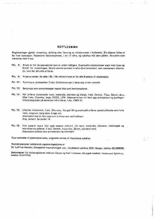

App
. II

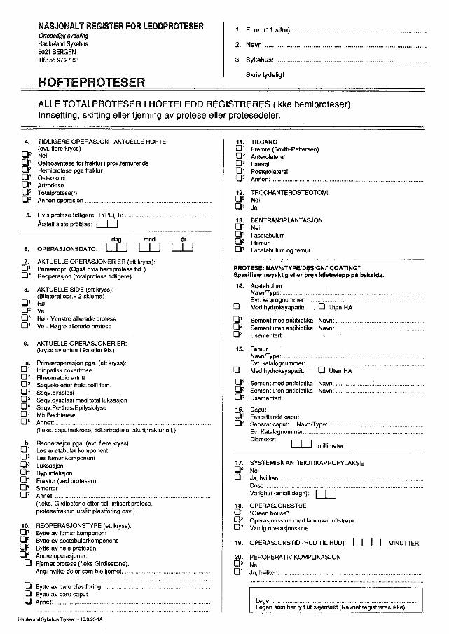
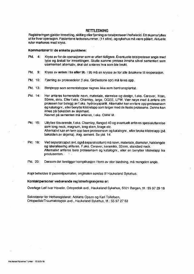

App
. III

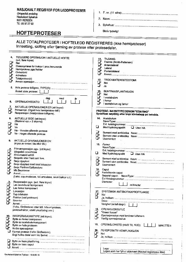
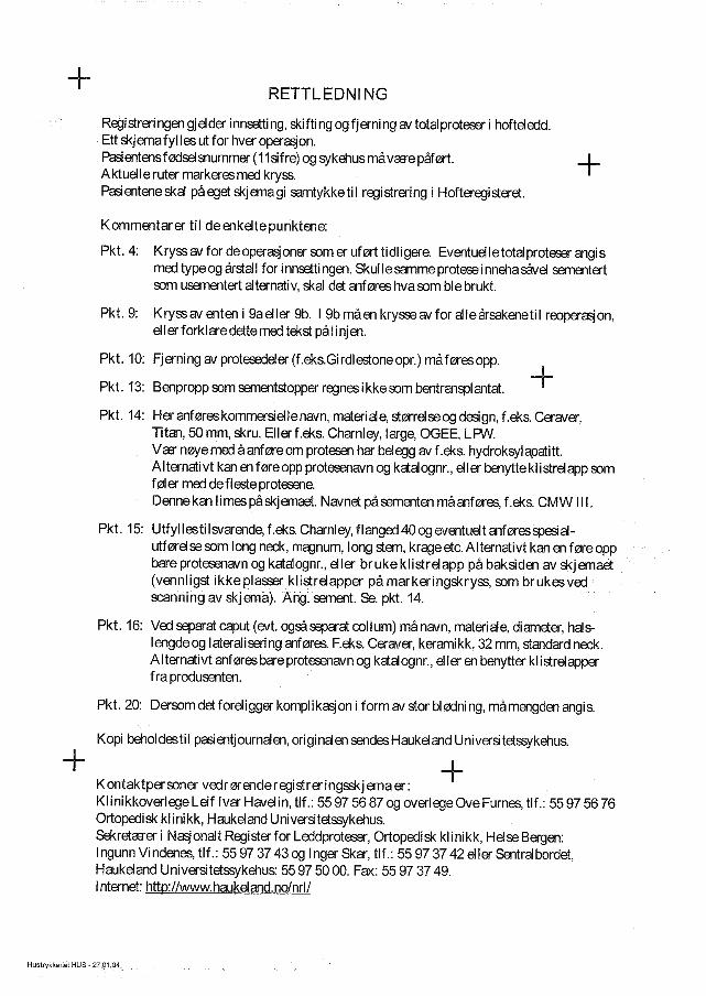

App
. IV

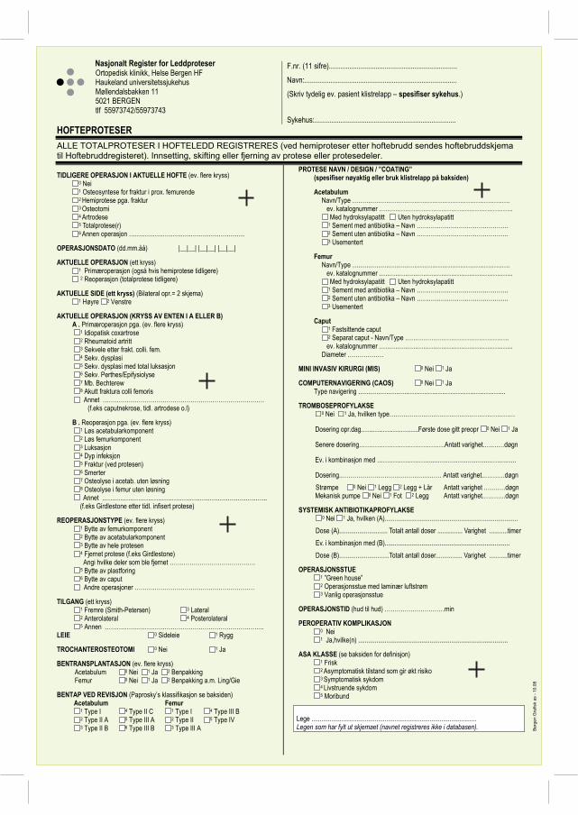
Nasjonalt Register for Leddproteser Ortopedisk klinikk, Helse Bergen HF Haukeland universitetssjukehus Møllendalsbakken 11 5021 BERGEN tlf 55973742/55973743
HOFTEPROTESER ALLE TOTALPROTESER I HOFTELEDD REGISTRERES (ved hemiproteser etter hoftebrudd sendes hoftebruddskjema til Hoftebruddregisteret). Innsetting, skifting eller fjerning av protese eller protesedeler.
F.nr. (11 sifre)..................................................................... Navn:.................................................................................. (Skriv tydelig ev. pasient klistrelapp – spesifiser sykehus.)
Sykehus:............................................................................
TIDLIGERE OPERASJON I AKTUELLE HOFTE (ev. flere kryss)0 Nei 1 Osteosyntese for fraktur i prox. femurende 2 Hemiprotese pga. fraktur 3 Osteotomi 4 Artrodese 5 Totalprotese(r) 6 Annen operasjon ………………………………………………….
OPERASJONSDATO (dd.mm.åå) |__|__| |__|__| |__|__|
AKTUELLE OPERASJON (ett kryss)1 Primæroperasjon (også hvis hemiprotese tidligere) 2 Reoperasjon (totalprotese tidligere)
AKTUELLE SIDE (ett kryss) (Bilateral opr.= 2 skjema) 1 Høyre 2 Venstre
AKTUELLE OPERASJON (KRYSS AV ENTEN I A ELLER B) A . Primæroperasjon pga. (ev. flere kryss)
1 Idiopatisk coxartrose 2 Rheumatoid artritt 3 Sekvele etter frakt. colli. fem. 4 Sekv. dysplasi 5 Sekv. dysplasi med total luksasjon 6 Sekv. Perthes/Epifysiolyse 7 Mb. Bechterew 8 Akutt fraktura colli femoris Annet ………………………………………………………………………
(f.eks caputnekrose, tidl. artrodese o.l)
B . Reoperasjon pga. (ev. flere kryss) 1 Løs acetabularkomponent 2 Løs femurkomponent 3 Luksasjon 4 Dyp infeksjon 5 Fraktur (ved protesen) 6 Smerter 7 Osteolyse i acetab. uten løsning 8 Osteolyse i femur uten løsning Annet ………………………………………………………………………..
(f.eks Girdlestone etter tidl. infisert protese)
REOPERASJONSTYPE (ev. flere kryss)1 Bytte av femurkomponent 2 Bytte av acetabularkomponent 3 Bytte av hele protesen 4 Fjernet protese (f.eks Girdlestone)
Angi hvilke deler som ble fjernet ……………………………………. 5 Bytte av plastforing 6 Bytte av caput Andre operasjoner ……………………………………………………
TILGANG (ett kryss)1 Fremre (Smith-Petersen) 3 Lateral 2 Anterolateral 4 Posterolateral 5 Annen ……………………………………………………………………..
LEIE 0 Sideleie 1 Rygg
TROCHANTEROSTEOTOMI 0 Nei 1 Ja
BENTRANSPLANTASJON (ev. flere kryss) Acetabulum 0 Nei 1 Ja 2 Benpakking Femur 0 Nei 1 Ja 2 Benpakking a.m. Ling/Gie
BENTAP VED REVISJON (Paprosky’s klassifikasjon se baksiden) Acetabulum Femur
1 Type I 4 Type II C 1 Type I 4 Type III B 2 Type II A 5 Type III A 2 Type II 5 Type IV 3 Type II B 6 Type III B 3 Type III A
PROTESE NAVN / DESIGN / ”COATING” (spesifiser nøyaktig eller bruk klistrelapp på baksiden)
AcetabulumNavn/Type …………………………………………………………………….
ev. katalognummer …………………………………………………………. Med hydroksylapatitt Uten hydroksylapatitt 1 Sement med antibiotika – Navn ………………………………………. 2 Sement uten antibiotika – Navn ………………………………………. 3 Usementert
Femur Navn/Type …………………………………………………………………….
ev. katalognummer …………………………………………………………. Med hydroksylapatitt Uten hydroksylapatitt 1 Sement med antibiotika – Navn ………………………………………. 2 Sement uten antibiotika – Navn ………………………………………. 3 Usementert
Caput 1 Fastsittende caput 2 Separat caput - Navn/Type ……………………………………………. ev. katalognummer …………………………………………………………. Diameter ………………
MINI INVASIV KIRURGI (MIS) 0 Nei 1 Ja
COMPUTERNAVIGERING (CAOS) 0 Nei 1 Ja Type navigering ………………………………………………………………..
TROMBOSEPROFYLAKSE 0 Nei 1 Ja, hvilken type………………………………………………………
Dosering opr.dag………………………..Første dose gitt preopr 0 Nei 1 Ja Senere dosering…………………………………….Antatt varighet.….……døgn Ev. i kombinasjon med ………………………...……………………..……….….. Dosering..……………………………………..…… Antatt varighet..…….…døgn
Strømpe 0 Nei 1 Legg 2 Legg + Lår Antatt varighet .….……døgn Mekanisk pumpe 0 Nei 1 Fot 2 Legg Antatt varighet.………..døgn
SYSTEMISK ANTIBIOTIKAPROFYLAKSE 0 Nei 1 Ja, hvilken (A)................................................................................
Dose (A)............................. Totalt antall doser ............... Varighet ...........timer Ev. i kombinasjon med (B)........................................................................... Dose (B)..............................Totalt antall doser................ Varighet ...........timer
OPERASJONSSTUE 1 ”Green house” 2 Operasjonsstue med laminær luftstrøm 3 Vanlig operasjonsstue
OPERASJONSTID (hud til hud) …………………………min
PEROPERATIV KOMPLIKASJON 0 Nei1 Ja,hvilke(n) ..........................................................................................
ASA KLASSE (se baksiden for definisjon)1 Frisk2 Asymptomatisk tilstand som gir økt risiko 3 Symptomatisk sykdom 4 Livstruende sykdom 5 Moribund
Lege ................................................................................................... Legen som har fylt ut skjemaet (navnet registreres ikke i databasen). B
erge
n G
rafis
k as
- 1
0.08
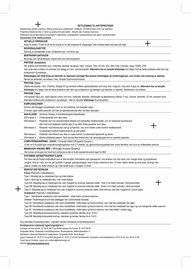
RETTLEDNING TIL HOFTEPROTESER Registreringen gjelder innsetting, skifting og fjerning av totalproteser i hofteledd. Ett skjema fylles ut for hver operasjon.Pasientens fødselsnummer (11sifre) og sykehus må være påført. Aktuelle ruter markeres med kryss. Pasientene skal på eget skjema gi samtykke til registrering i Leddregisteret, samtykkeskjema skal lagres i pasientjournalen. Kommentarer til de enkelte punktene AKTUELLE OPERASJON Kryss av enten i A eller B.I B må en krysse av for alle årsakene til reoperasjon, eller forklare dette med tekst på linjen. REOPERASJONSTYPE Fjerning av protesedeler (f.eks. Girdlestone opr.) må føres opp. BENTRANSPLANTASJON Benpropp som sementstopper regnes ikke som bentransplantat.
PROTESE. Acetabulum. Her anføres kommersielle navn, materiale, størrelse og design, f.eks. Ceraver, Titan, 50 mm, skru. Eller f.eks. Charnley, large, OGEE, LPW. Vær nøye med å anføre om protesen har belegg av f.eks. hydroksylapatitt. Alternativt kan en benytte klistrelapp som følger med de fleste protesene eller føre opp
protesenavn og katalognr., . Klistrelappen bør helst limes på baksiden av skjemaet (vennligst ikke plasser klistrelapper på markeringskryss, som brukes ved scanning av skjema).Navnet på sementen må anføres, f.eks. Simplex Erythromycin/colistin.
PROTESE. Femur Utfylles tilsvarende, f.eks. Charnley, flanged 40 og eventuelt anføres spesialutførelse som long neck, magnum, long stem, krage etc. Alternativt kan en benytte
klistrelapp som følger med de fleste protesene eller føre opp protesenavn og katalognr (på baksiden av skjema). Sementnavn må anføres. PROTESE. Caput Ved separat caput (evt. også separat collum) må navn, materiale, diameter, halslengde og lateralisering anføres. F.eks. Ceraver, keramikk, 32 mm, standard neck. Alternativt anføres bare protesenavn og katalognr., eller en benytter klistrelapp fra produsenten. KOMPLIKASJONER Dersom det foreligger komplikasjon i form av stor blødning, må mengden angis. Vi ønsker også meldt pasienter som dør på operasjonsbordet eller rett etter operasjon.
ASA-KLASSE American Society of Anesthesiologists klassifikasjon ASA-klasse 1: Friske pasienter som ikke røker
ASA-klasse 2: Pasienter som har asymptomatisk tilstand som behandles medikamentelt, som for eksempel hypertensjon eller med kost (diabetes mellitus type 2) og ellers friske pasienter som røker ASA-klasse 3: Pasienter med tilstand som kan gi symptomer, men som holdes under kontroll medikamentelt for eksempel moderat angina pectoris og mild astma ASA-klasse 4: Pasienter med tilstand som ikke er under kontroll, for eksempel hjertesvikt og astma ASA-klasse 5: Moribund/døende pasient. Skal normalt ikke forekomme i vår pasientgruppe som er opererte pasienter
COMPUTERNAVIGERING (CAOS = Computer Aided Orthopaedic Surgery.) Vi ber om å få angitt type computernavigeringsutstyr som CT-veiledet, rtg. gjennomlysningveiledet eller andre teknikker som bruk av hofteleddets sentrum.
MINIINVASIV KIRURGI (MIS = Minimally Invasive Surgery) Her menes at kirurgen har brukt kort snitt pluss at det er brukt spesialinstrument laget for MIS SYSTEMISK ANTIBIOTIKAPROFYLAKSE Her føres det på hvilket antibiotikum som er blitt benyttet i forbindelse med operasjonen. Det anføres hvor stor dose, hvor mange doser og profylaksens varighet. Hvis en f.eks. kun har gitt 2g Keflin 4 ganger operasjonsdagen med 4 timers mellomrom dvs. 12 timer mellom første og siste dose, så angis det i skjema: Hvilken (A) Keflin Dose(A) 2g Totalt antall doser 4 Varighet 12 timer. BEINTAP VED REVISJON
Femur (Paprosky`s klassifikasjon) Type I: Minimalt tap av metafysært ben og intakt diafyse. Type II: Stort tap av metafysært ben, men intakt diafyse. Type IIIA: Betydelig tap av metafysært ben uten mulighet for proximal mekanisk støtte. Over 4 cm intakt corticalis i isthmusområdet. Type IIIB: Betydelig tap av metafysært ben uten mulighet for proximal mekanisk støtte. Under 4 cm intakt corticalis i isthmusområdet. Type IV: Betydelig tap av metafysært ben uten mulighet for proximal mekanisk støtte. Bred isthmus med liten mulighet for cortical støtte.
Acetabulum (Paprosky`s klassifikasjon) Type I: Hemisfærisk acetabulum uten kantdefekter. Intakt bakre og fremre kolonne. Defekter i forankringshull som ikke ødelegger den subchondrale benplate. Type IIA: Hemisfærisk acetabulum uten store kantdefekter, intakt bakre og fremre kolonne, men med lite metafysært ben igjen. Type IIB: Hemisfærisk acetabulum uten store kantdefekter, intakt bakre og fremre kolonne, men med lite metafysært ben igjen og noe manglende støtte superiort. Type IIC: Hemisfærisk acetabulum uten store kantdefekter, intakt bakre og fremre kollonne, men med defekt i medial vegg. Type IIIA: Betydelig komponentvandring, osteolyse og bentap. Bentap fra kl. 10 til 2. Type IIIB: Betydelig komponentvandring, osteolyse og bentap. Bentap fra kl. 9 til 5.
Kopi beholdes til pasientjournalen, originalen sendes Haukeland universitetssjukehus.Kontaktpersoner vedrørende registreringsskjema er Overlege Leif Ivar Havelin, tlf.: 55 97 56 87 og klinikkoverlege Ove Furnes, tlf.: 55 97 56 80 Ortopedisk klinikk, Haukeland universitetssjukehus. Besøksadresse: Møllendalsbakken 11. Sekretærer i Nasjonalt Register for Leddproteser, Ortopedisk klinikk, Helse Bergen: Ingunn Vindenes, tlf.: 55 97 37 43 og Ruth Wasmuth, tlf.: 55 97 37 42 eller Sentralbordet, Haukeland : 55 97 50 00. Fax: 55 97 37 49. Epost Ingunn Vindenes: [email protected] Internet: http://www.haukeland.no/nrl/
universitetssjukehus

App
. V

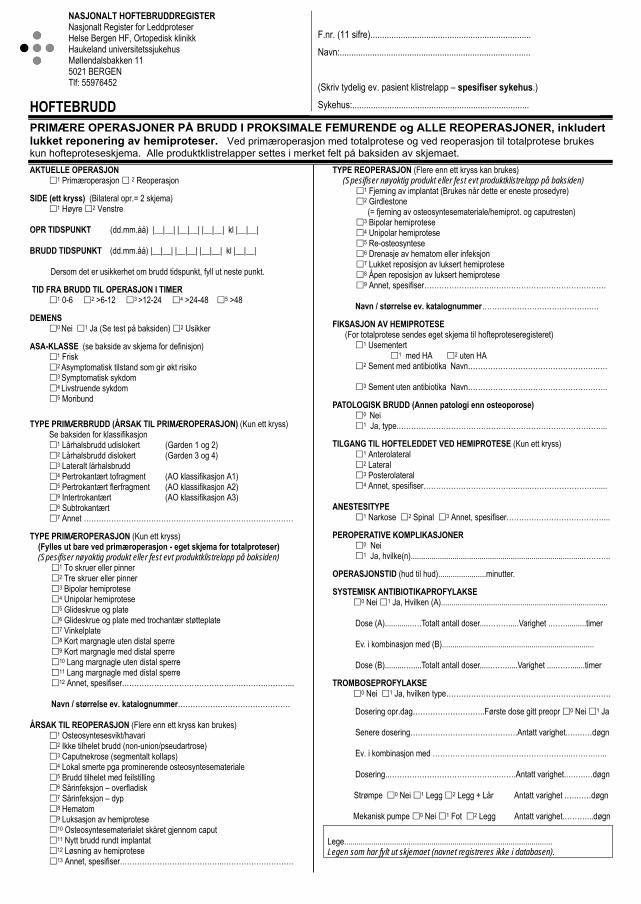
NASJONALT HOFTEBRUDDREGISTER Nasjonalt Register for Leddproteser Helse Bergen HF, Ortopedisk klinikk Haukeland universitetssjukehus Møllendalsbakken 11 5021 BERGEN Tlf: 55976452
HOFTEBRUDD
F.nr. (11 sifre)..................................................................... Navn:..................................................................................
(Skriv tydelig ev. pasient klistrelapp – spesifiser sykehus.) Sykehus:............................................................................
AKTUELLE OPERASJON 1 Primæroperasjon 2 Reoperasjon
SIDE (ett kryss) (Bilateral opr.= 2 skjema) 1 Høyre 2 Venstre
OPR TIDSPUNKT (dd.mm.åå) |__|__| |__|__| |__|__| kl |__|__|
BRUDD TIDSPUNKT (dd.mm.åå) |__|__| |__|__| |__|__| kl |__|__| Dersom det er usikkerhet om brudd tidspunkt, fyll ut neste punkt.
TID FRA BRUDD TIL OPERASJON I TIMER 1 0-6 2 >6-12 3 >12-24 4 >24-48 5 >48
DEMENS0 Nei 1 Ja (Se test på baksiden) 2 Usikker
ASA-KLASSE (se bakside av skjema for definisjon)1 Frisk2 Asymptomatisk tilstand som gir økt risiko 3 Symptomatisk sykdom 4 Livstruende sykdom 5 Moribund
TYPE PRIMÆRBRUDD (ÅRSAK TIL PRIMÆROPERASJON) (Kun ett kryss)Se baksiden for klassifikasjon
1 Lårhalsbrudd udislokert (Garden 1 og 2) 2 Lårhalsbrudd dislokert (Garden 3 og 4) 3 Lateralt lårhalsbrudd 4 Pertrokantært tofragment (AO klassifikasjon A1) 5 Pertrokantært flerfragment (AO klassifikasjon A2) 9 Intertrokantært (AO klassifikasjon A3) 6 Subtrokantært 7 Annet …………………………………………………………………………
TYPE PRIMÆROPERASJON (Kun ett kryss) (Fylles ut bare ved primæroperasjon - eget skjema for totalproteser) (Spesifiser nøyaktig produkt eller fest evt produktklistrelapp på baksiden)
1 To skruer eller pinner 2 Tre skruer eller pinner 3 Bipolar hemiprotese 4 Unipolar hemiprotese 5 Glideskrue og plate 6 Glideskrue og plate med trochantær støtteplate 7 Vinkelplate 8 Kort margnagle uten distal sperre 9 Kort margnagle med distal sperre 10 Lang margnagle uten distal sperre 11 Lang margnagle med distal sperre 12 Annet, spesifiser.…………………………………….….……….………...
Navn / størrelse ev. katalognummer………………………………………
ÅRSAK TIL REOPERASJON (Flere enn ett kryss kan brukes)1 Osteosyntesesvikt/havari 2 Ikke tilhelet brudd (non-union/pseudartrose) 3 Caputnekrose (segmentalt kollaps) 4 Lokal smerte pga prominerende osteosyntesemateriale 5 Brudd tilhelet med feilstilling 6 Sårinfeksjon – overfladisk 7 Sårinfeksjon – dyp 8 Hematom 9 Luksasjon av hemiprotese 10 Osteosyntesematerialet skåret gjennom caput 11 Nytt brudd rundt implantat 12 Løsning av hemiprotese 13 Annet, spesifiser.…………………………………..……………………….
TYPE REOPERASJON (Flere enn ett kryss kan brukes) (Spesifiser nøyaktig produkt eller fest evt produktklistrelapp på baksiden) 1 Fjerning av implantat (Brukes når dette er eneste prosedyre) 2 Girdlestone (= fjerning av osteosyntesemateriale/hemiprot. og caputresten) 3 Bipolar hemiprotese 4 Unipolar hemiprotese 5 Re-osteosyntese 6 Drenasje av hematom eller infeksjon 7 Lukket reposisjon av luksert hemiprotese 8 Åpen reposisjon av luksert hemiprotese 9 Annet, spesifiser……………………….………………………………………
Navn / størrelse ev. katalognummer…………………………………….….
FIKSASJON AV HEMIPROTESE (For totalprotese sendes eget skjema til hofteproteseregisteret) 1 Usementert
1 med HA 2 uten HA 2 Sement med antibiotika Navn…………………………………………….….
3 Sement uten antibiotika Navn………………………………………………..
PATOLOGISK BRUDD (Annen patologi enn osteoporose) 0 Nei 1 Ja, type.………………………………………………………………………...
TILGANG TIL HOFTELEDDET VED HEMIPROTESE (Kun ett kryss) 1 Anterolateral 2 Lateral 3 Posterolateral 4 Annet, spesifiser………………………………..…..……………………….....
ANESTESITYPE 1 Narkose 2 Spinal 3 Annet, spesifiser…………………………………...
PEROPERATIVE KOMPLIKASJONER 0 Nei 1 Ja, hvilke(n)...................................................................................………..
OPERASJONSTID (hud til hud).......................minutter.
SYSTEMISK ANTIBIOTIKAPROFYLAKSE 0 Nei 1 Ja, Hvilken (A)................................................................................ Dose (A).............….Totalt antall doser...……….....Varighet .……..........timer Ev. i kombinasjon med (B).........................................................................
Dose (B).........….....Totalt antall doser.....……......Varighet ....…….......timer
TROMBOSEPROFYLAKSE 0 Nei 1 Ja, hvilken type…………………………………………………………
Dosering opr.dag………………………..Første dose gitt preopr 0 Nei 1 Ja
Senere dosering…………………………………….Antatt varighet.….……døgn
Ev. i kombinasjon med ………………………...……………………..……….….. Dosering..……………………………………..…….Antatt varighet..…….…døgn
Strømpe 0 Nei 1 Legg 2 Legg + Lår Antatt varighet .….……døgn
Mekanisk pumpe 0 Nei 1 Fot 2 Legg Antatt varighet.………...døgn
Lege.................................................................................................... Legen som har fylt ut skjemaet (navnet registreres ikke i databasen).
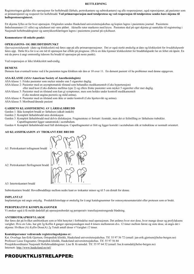
RETTLEDNING
Ved primæroperasjon med totalprotese og ved reoperasjon til totalprotese sendes bare skjema til hofteproteseregisteret.
Kommentarer til enkelte punkt:
OPERASJONS- OG BRUDDTIDSPUNKTOperasjonstidspunkt bruddtidspunkt
DEMENS
ASA-KLASSE (ASA=American Society of Anesthesiologists)
GARDENS KLASSIFISERING AV LÅRHALSBRUDD
AO KLASSIFIKASJON AV TROKANTÆRE BRUDD
IMPLANTAT
PEROPERATIVE KOMPLIKASJONER
ANTIBIOTIKAPROFYLAKSE
Keflin 2g 4 12
Kontaktpersoner vedrørende registreringsskjema er:
PRODUKTKLISTRELAPPER:

App
. VI


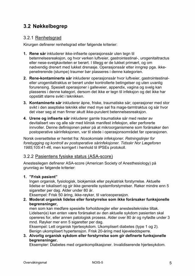


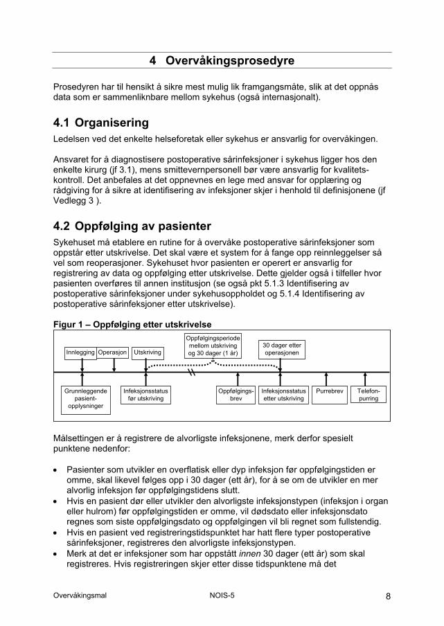
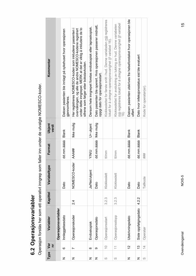
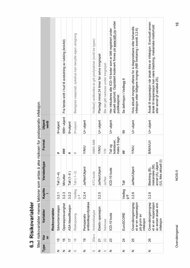
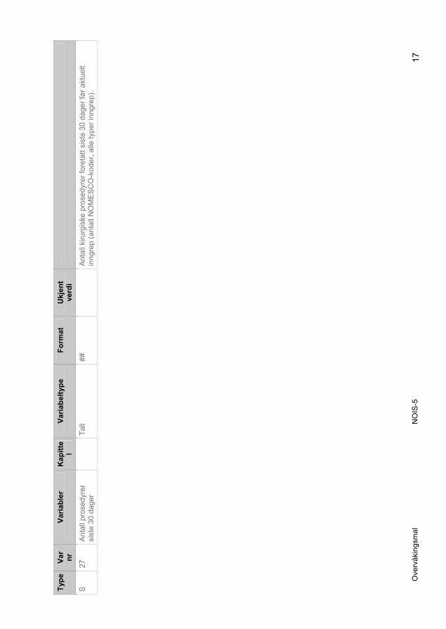
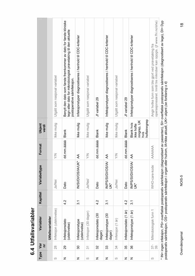
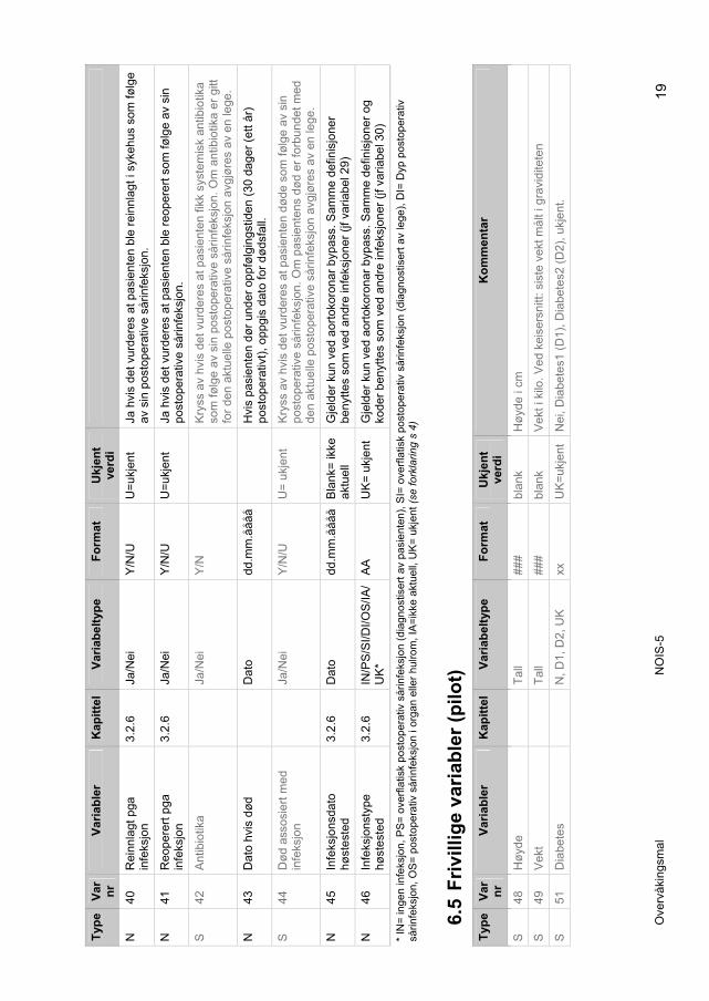
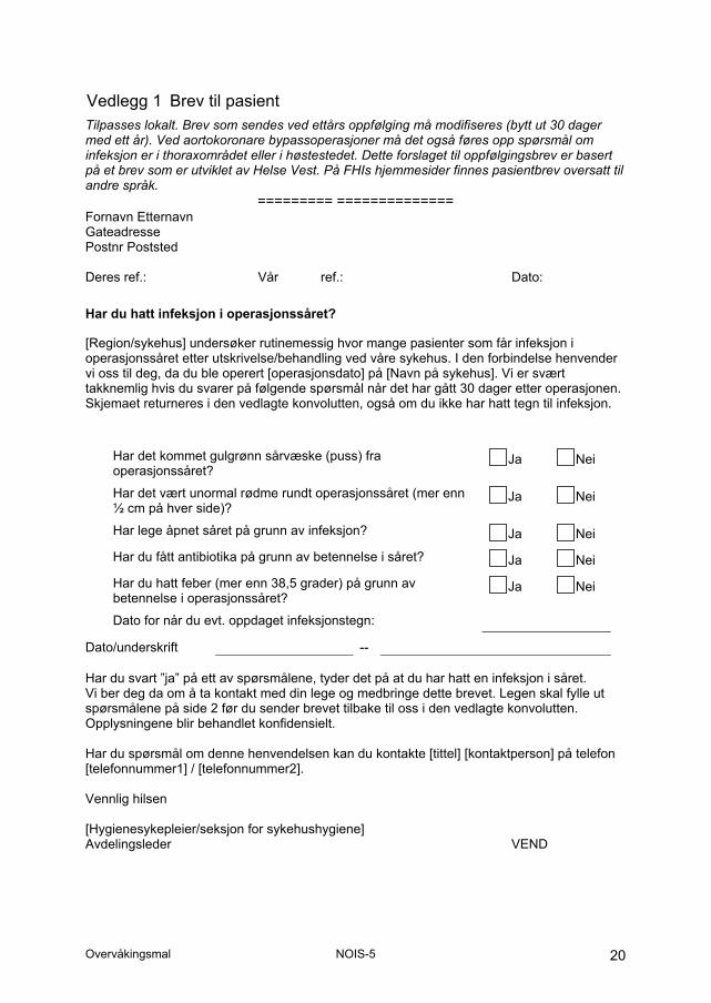
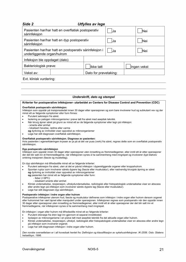
Side 2 Utfylles av lege
Underskrift, dato og stempel


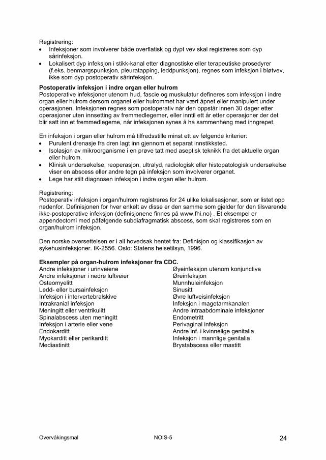
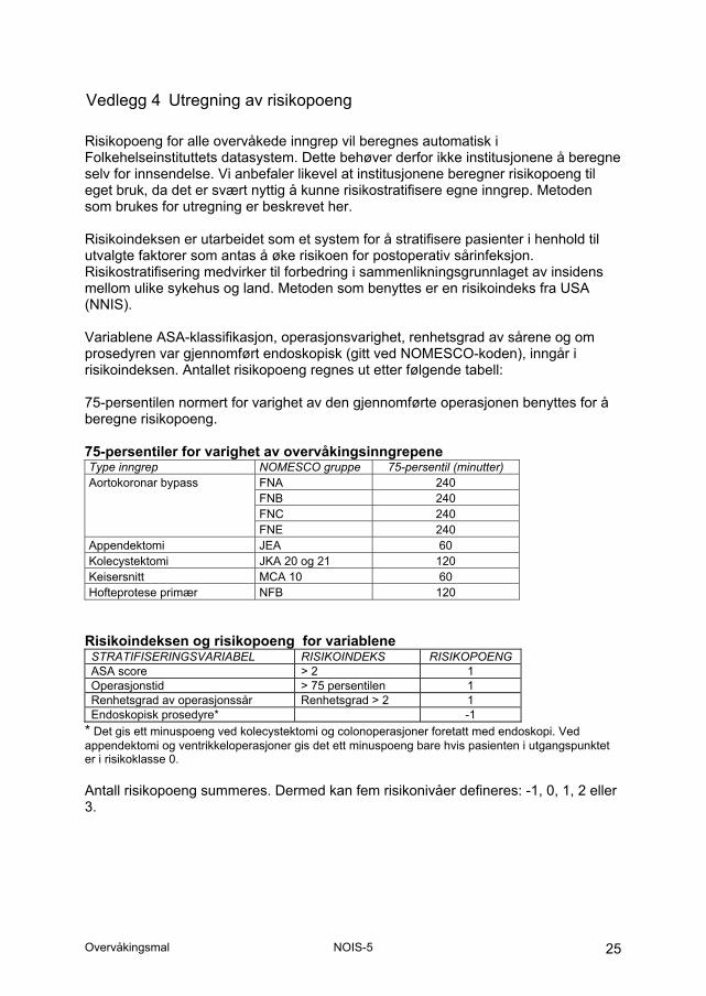
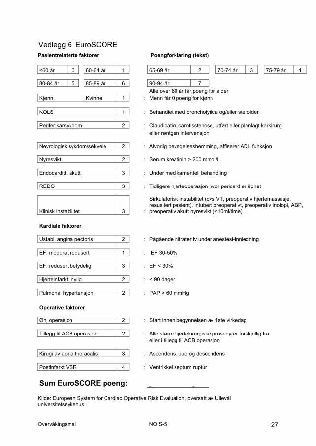

App
. VII

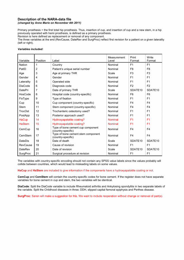
Description of the NARA-data file (changed by Anne Marie on November 4th 2011)
Primary prosthesis = the first total hip prosthesis. Thus, insertion of cup, and insertion of cup and a new stem, in a hip previously operated with hemi prosthesis, is defined as a primary prosthesis. Revision is here defined as replacement or removal of any component. The three variables at the end (RevCause, DateRev and SurgProc) reflect first revision for a patient on a given laterality (left or right).
Variables included:
Variable Position Label Measurement Level
Print Format
Write Format
Nation 1 Country Nominal F1 F1 PatID 2 Patient’s unique serial number Nominal F8 F8 Age 3 Age at primary THR Scale F3 F3 Gender 4 Gender Nominal F1 F1 Laterality 5 Operated side Nominal F1 F1 DiaCode 6 Diagnosis code Nominal F2 F2 DatePri 7 Date of primary THR Scale SDATE10 SDATE10 HosCode 8 Hospital code (country-specific) Nominal F6 F6 FixType 9 Type of fixation Nominal F1 F1 Cup 10 Cup component (country-specific) Nominal F4 F4 Stem 11 Stem component (country-specific) Nominal F4 F4 TrocOst 12 Trochanteric osteotomy used? Nominal F1 F1 PostApp 13 Posterior approach used? Nominal F1 F1 HaCup 14 Hydroxyapatatite coating? Nominal F1 F1 HaStem 15 Hydroxyapatatite coating? Nominal F1 F1
CemCup 16 Type of bone cement cup component (country-specific) Nominal F4 F4
CemStem 17 Type of bone cement stem component (country-specific) Nominal F4 F4
DateDis 18 Date of death Scale SDATE10 SDATE10 RevCause 19 Cause of revision Nominal F1 F1 DateRev 20 Date of revision Scale SDATE10 SDATE10 SurgProc 21 Surgical procedure at revision Nominal F1 F1
The variables with country-specific encoding should not contain any SPSS value labels since the values probably will collide between countries, which would lead to misleading labels on some values.
HaCup and HaStem are included to give information if the components have a hydroxyapatatite coating or not.
CemCup and CemStem will contain the country-specific codes for bone cement. If the register does not have separate variables for bone cement in cup and stem, the two variables will be identical.
DiaCode: Split the DiaCode variable to include Rheumatoid arthritis and Ankylosing spondylitis in two separate labels of the variable. Split the Childhood diseases in three; DDH, slipped capital femoral epiphysis and Perthes disease.
SurgProc: Søren will make a suggestion for this. We want to include reoperation without change or removal of part(s).
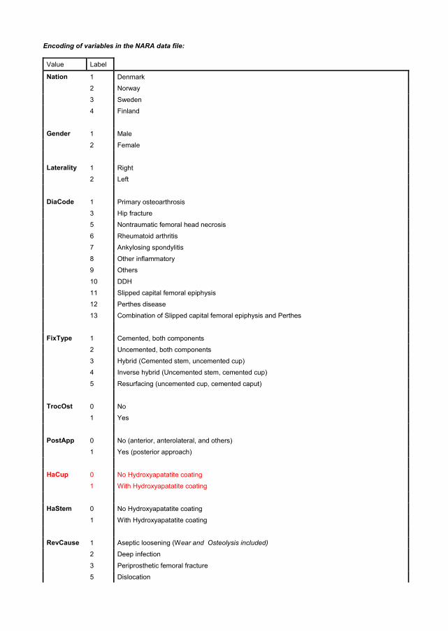
Encoding of variables in the NARA data file:
Value Label
Nation 1 Denmark
2 Norway
3 Sweden
4 Finland
Gender 1 Male
2 Female
Laterality 1 Right
2 Left
DiaCode 1 Primary osteoarthrosis
3 Hip fracture
5 Nontraumatic femoral head necrosis
6 Rheumatoid arthritis
7 Ankylosing spondylitis
8 Other inflammatory
9 Others
10 DDH
11 Slipped capital femoral epiphysis
12 Perthes disease
13 Combination of Slipped capital femoral epiphysis and Perthes
FixType 1 Cemented, both components
2 Uncemented, both components
3 Hybrid (Cemented stem, uncemented cup)
4 Inverse hybrid (Uncemented stem, cemented cup)
5 Resurfacing (uncemented cup, cemented caput)
TrocOst 0 No
1 Yes
PostApp 0 No (anterior, anterolateral, and others)
1 Yes (posterior approach)
HaCup 0 No Hydroxyapatatite coating
1 With Hydroxyapatatite coating
HaStem 0 No Hydroxyapatatite coating
1 With Hydroxyapatatite coating
RevCause 1 Aseptic loosening (Wear and Osteolysis included)
2 Deep infection
3 Periprosthetic femoral fracture
5 Dislocation

7 Pain only
9 Others
SurgProc 1 Both cup and stem replaced
2 Only stem replaced
3 Only cup or liner replaced
4
9
Extraction of the total prosthesis (Girdlestone), permanent or temporary
Others (such as exchange of caput)


Pape
r I

640 Acta Orthopaedica 2009; 80 (6): 639–645
different brands of cement for different components, and were also excluded. 1,689 additional THAs were excluded because of missing values for other adjustment variables. There were 97,344 THAs with complete information where both compo-nents were either cemented or uncemented, and these were eligible for analysis.
All THAs were followed until their first revision due to deep infection or revision for other causes, until date of death or emigration of the patient, or until January 1, 2008. Thus, follow-up was 0–20 years. 4 time periods were compared: 1987–1992, 1993–1997, 1998–2002, and 2003–2007, with subanalyses on cemented and uncemented THAs.
As a control, we performed a subanalysis on Charnley pros-theses fixed with antibiotic-loaded bone cement and given antibiotic prophylaxis systemically. This prosthesis was the most used in Norway from 1987 to 2008, and it was used extensively throughout the whole period of observation.
StatisticsSurvival analyses were performed with a Cox regression model, with time period as main risk factor and revision due to deep infection as the endpoint. Revision rate ratios (RRs) for the time periods are presented with 95% confidence interval (CI) and p-values relative to the first time period. We adjusted for differences over time concerning sex, age (< 40, 40–59, 60–69, 70–79, 80 years), diagnosis (osteoarthritis, inflam-matory disease, other), monoblock or modular prosthesis, type of fixation (uncemented, cemented with cement containing or not containing antibiotics), antibiotic prophylaxis systemi-cally (yes, no), type of operation room ventilation (ordinary, laminar flow, greenhouse), and duration of surgery (< 70, 70–99, 100–129, or 130 min). Cox regression analyses with time period as stratification factor were used to construct cumulative revision curves (1 minus cumulative survival) at mean values of the covariates, and to assess 5-year survival percentages. We also performed a separate Cox analysis with revision due to aseptic loosening as endpoint for all THAs, in order to be able to compare these findings with our find-ings for revision due to deep infection. Furthermore, to ensure similar potential follow-up for operations in all time periods, additional analyses were performed with follow-up restricted to 0–5 years.
We also investigated changes in the revision rate due to deep infection as a function of year of operation. These analyses gave a graphical display of the relationship based on a gener-alized additive model for survival data (Hastie and Tibshirani 1990). The curves are presented with 95% CI.
Risk ratio analyses were performed for the different risk factors and prophylactic measures for each time period sepa-rately, and for the whole 20-year period adjusted for year of primary surgery.
Values of p less than 0.05 were considered statistically sig-nificant. We used SPSS software version 15.0.
Results
97,344 primary THAs in 79,820 patients met the inclusion criteria for this study. 614 first revisions due to deep infection were reported in 610 patients. The 5-year survival was 99.46% with revision due to deep infection as endpoint.
The distribution of patient characteristics such as sex, age, and diagnosis of patients undergoing primary THA was stable throughout the period studied (Table 1), except for the group of primary uncemented THAs, where mean age increased from 52 (SD 12) in 1987–1992 to 61 (SD 13) in 2003–2007. There was a shift from monoblock towards modular THAs (Table 1). Duration of surgery decreased slightly, whereas the use of an operating room with laminar air flow increased through the 4 time periods (Table 1). Antibiotic-loaded bone cement was used more extensively, and cement containing antibiotics was used in most cemented THAs towards the end of the study period (Table 1). Except during the first time period, prophy-lactic antibiotics were administered systemically in almost all operations (Table 1).
Table 1. Primary THAs included over the four 5-year time periods
Variable 1987–1992 1993–1997 1998–2002 2003–2007
No. of THAs 20,913 22,519 26,230 27,682Sex (%) Male 30 30 29 31 Female 70 70 71 69Age (%) < 40 2 2 2 1 40–59 15 14 15 14 60–69 31 27 26 26 70–79 41 43 41 39
80 12 14 17 19Diagnosis (%) Osteoarthritis 67 70 73 77 Inflammatory 4 4 4 3 Other 29 26 23 20Prosthesis (%) Monoblock 58 53 38 22 Modular 42 47 62 78Duration (min) of surgery (%) < 70 11 10 11 15 70–99 41 45 45 45 100–129 31 31 31 29 130 18 15 13 12Operation room ventilation (%) “Greenhouse” 12 2 1 1 Laminar flow 29 45 53 56 Ordinary 59 53 46 44Antibiotic prophylaxis systemically (%) No 8 0 0 0 Yes 92 100 100 100Method of fixation (%) Uncemented 15 15 14 16 Cement with antibiotics 38 56 82 83 without antibiotics 48 29 4 2
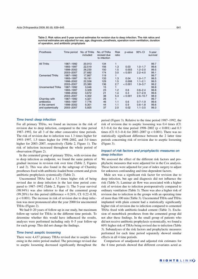
Acta Orthopaedica 2009; 80 (6): 639–645 641
Time trend: deep infectionFor all primary THAs, we found an increase in the risk of revision due to deep infection, compared to the time period 1987–1992, for all 3 of the other consecutive time periods. The risk of revision due to infection was 1.3 times higher for 1993–1997, 1.5 times higher for 1998–2002, and 3.0 times higher for 2003–2007, respectively (Table 2, Figure 1). The risk of infection increased throughout the whole period of observation (Figure 2).
In the cemented group of primary THAs, with revision due to deep infection as endpoint, we found the same pattern of gradual increase in revision risk over time (Table 2, Figures 1 and 2). This was also found in the subgroup of Charnley prostheses fixed with antibiotic-loaded bone cement and given antibiotic prophylaxis systemically (Table 2).
Uncemented THAs had a 5.3 times higher risk of being revised due to deep infection in the last time period com-pared to 1987–1992 (Table 2, Figure 1). The 5-year survival (98.94%) was also inferior to that of the cemented group (99.20%) for this period (difference = 0.26%, CI: 0.22–0.30, p < 0.001). The increase in risk of revision due to deep infec-tion was most pronounced after the year 2000 for uncemented THAs (Figure 2).
We had 0–20 years of follow-up in our study, but maximum follow-up varied for THAs in the different time periods. To determine whether this would have influenced the results, analyses were performed including only 0–5 year follow-up for each group. This did not change the findings.
Time trend: aseptic loosening There were 4,437 primary THAs revised due to aseptic loos-ening in the entire period studied. The percentage revised due to aseptic loosening decreased significantly throughout the
period (Figure 3). Relative to the time period 1987–1992, the risk of revision due to aseptic loosening was 0.4 times (CI: 0.3–0.4) for the time period 1998–2002 (p < 0.001) and 0.3 times (CI: 0.3–0.4) for 2003–2007 (p < 0.001). There was no statistically significant difference between the 2 latter time periods concerning risk of revision due to aseptic loosening (Figure 3).
Impact of risk factors and prophylactic measures on deep infection We assessed the effect of the different risk factors and pro-phylactic measures that were adjusted for in the Cox analysis. These factors were adjusted for year of index surgery to adjust for unknown confounding and time-dependent factors.
Male sex was a significant risk factor for revision due to deep infection, but age and diagnosis did not influence the risk (Table 3). Laminar air flow was associated with a higher risk of revision due to infection postoperatively compared to ordinary ventilation (Table 3). There was also a higher risk of revision due to infection in the groups with an operating time of more than 100 min (Table 3). Uncemented THAs and THAs implanted with plain cement had a statistically significantly higher risk of revision due to infection compared to cemented THAs fixed with antibiotic-loaded cement (Table 3). Exclu-sion of monoblock prostheses from the cemented group did not alter these findings. In the small group of patients who did not receive antibiotic prophylaxis systemically, we found a 60% higher risk of THAs being revised due to infection (Table 3). Subanalyses of the risk factors and prophylactic measures performed for each time period separately showed similar effects in all 4 time periods.
Comparison of unadjusted and adjusted risk estimates for the 4 time periods showed that different covariates acted as
Table 2. Risk ratios and 5-year survival estimates for revision due to deep infection. The risk ratios and survival estimates are adjusted for sex, age, diagnosis, prosthesis, operation room ventilation, duration of operation, and antibiotic prophylaxis
Prosthesis Time period No. of THAs No. of THAs Risk p-value 95% CI 5-year included revised due ratio survival to infection
All THAs 1987–1992 20,913 134 1 99.7 1993–1997 22,519 156 1.3 0.03 1.0–1.7 99.6 1998–2002 26,230 150 1.5 0.003 1.2–2.0 99.5 2003–2007 27,682 174 3.0 < 0.001 2.2–4.0 99.1Cemented THAs 1987–1992 17,867 119 1 99.7 1993–1997 19,191 133 1.3 0.04 1.0–1.7 99.5 1998–2002 22,558 129 1.5 0.008 1.1–2.1 99.5 2003–2007 23,380 136 2.7 < 0.001 1.9–3.7 99.2Uncemented THAs 1987–1992 3,046 15 1 99.8 1993–1997 3,328 23 1.2 0.6 0.6–2.4 99.8 1998–2002 3,672 21 1.4 0.3 0.7–2.9 99.6 2003–2007 4,302 38 5.3 < 0.001 2.6–10.7 98.9Charnley with 1987–1992 4,321 26 1 99.7antibiotics 1993–1997 7,776 46 1.1 0.6 0.7–1.9 99.6in the cement 1998–2002 9,301 44 1.1 0.9 0.6–1.8 99.6and systemically 2003–2007 5,925 37 2.0 0.02 1.1–3.5 99.3
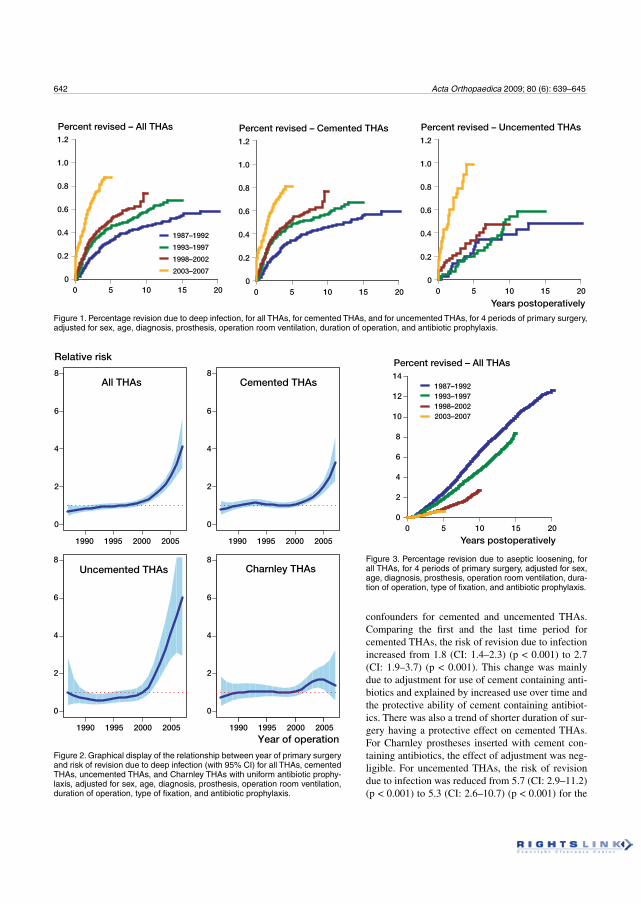
642 Acta Orthopaedica 2009; 80 (6): 639–645
Figure 1. Percentage revision due to deep infection, for all THAs, for cemented THAs, and for uncemented THAs, for 4 periods of primary surgery, adjusted for sex, age, diagnosis, prosthesis, operation room ventilation, duration of operation, and antibiotic prophylaxis.
1.2
1.0
0.8
0.6
0.4
0.2
00 5 10 15 20
1987–1992
1993–1997
1998–2002
2003–2007
Percent revised – All THAs
14
10
12
8
6
4
2
00 5 10 15 20
1987–19921993–19971998–20022003–2007
Percent revised – All THAs
Years postoperatively
0
2
4
6
8All THAs
Relative risk
Cemented THAs
1990 1995 2000 2005
Uncemented THAs
1990 1995 2000 2005
Charnley THAs
Year of operation
1990 1995 2000 2005 1990 1995 2000 2005
0
2
4
6
8
0
2
4
6
8
0
2
4
6
8
Percent revised – Cemented THAs1.2
1.0
0.8
0.6
0.4
0.2
00 5 10 15 20
Percent revised – Uncemented THAs1.2
1.0
0.8
0.6
0.4
0.2
00 5 10 15 20
Years postoperatively
Figure 3. Percentage revision due to aseptic loosening, for all THAs, for 4 periods of primary surgery, adjusted for sex, age, diagnosis, prosthesis, operation room ventilation, dura-tion of operation, type of fixation, and antibiotic prophylaxis.
Figure 2. Graphical display of the relationship between year of primary surgery and risk of revision due to deep infection (with 95% CI) for all THAs, cemented THAs, uncemented THAs, and Charnley THAs with uniform antibiotic prophy-laxis, adjusted for sex, age, diagnosis, prosthesis, operation room ventilation, duration of operation, type of fixation, and antibiotic prophylaxis.
confounders for cemented and uncemented THAs. Comparing the first and the last time period for cemented THAs, the risk of revision due to infection increased from 1.8 (CI: 1.4–2.3) (p < 0.001) to 2.7 (CI: 1.9–3.7) (p < 0.001). This change was mainly due to adjustment for use of cement containing anti-biotics and explained by increased use over time and the protective ability of cement containing antibiot-ics. There was also a trend of shorter duration of sur-gery having a protective effect on cemented THAs. For Charnley prostheses inserted with cement con-taining antibiotics, the effect of adjustment was neg-ligible. For uncemented THAs, the risk of revision due to infection was reduced from 5.7 (CI: 2.9–11.2) (p < 0.001) to 5.3 (CI: 2.6–10.7) (p < 0.001) for the
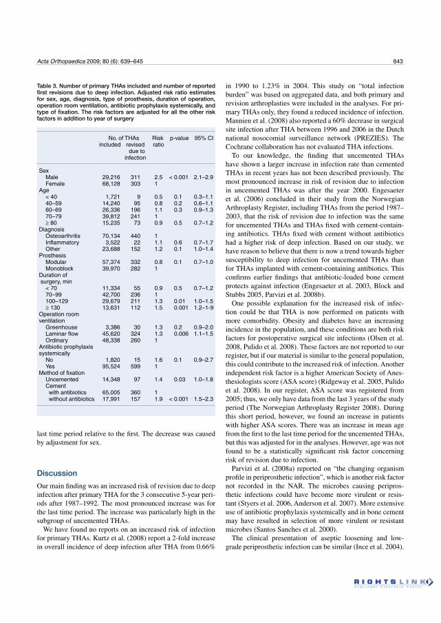
Acta Orthopaedica 2009; 80 (6): 639–645 643
last time period relative to the first. The decrease was caused by adjustment for sex.
Discussion
Our main finding was an increased risk of revision due to deep infection after primary THA for the 3 consecutive 5-year peri-ods after 1987–1992. The most pronounced increase was for the last time period. The increase was particularly high in the subgroup of uncemented THAs.
We have found no reports on an increased risk of infection for primary THAs. Kurtz et al. (2008) report a 2-fold increase in overall incidence of deep infection after THA from 0.66%
in 1990 to 1.23% in 2004. This study on “total infection burden” was based on aggregated data, and both primary and revision arthroplasties were included in the analyses. For pri-mary THAs only, they found a reduced incidence of infection. Mannien et al. (2008) also reported a 60% decrease in surgical site infection after THA between 1996 and 2006 in the Dutch national nosocomial surveillance network (PREZIES). The Cochrane collaboration has not evaluated THA infections.
To our knowledge, the finding that uncemented THAs have shown a larger increase in infection rate than cemented THAs in recent years has not been described previously. The most pronounced increase in risk of revision due to infection in uncemented THAs was after the year 2000. Engesaeter et al. (2006) concluded in their study from the Norwegian Arthroplasty Register, including THAs from the period 1987–2003, that the risk of revision due to infection was the same for uncemented THAs and THAs fixed with cement-contain-ing antibiotics. THAs fixed with cement without antibiotics had a higher risk of deep infection. Based on our study, we have reason to believe that there is now a trend towards higher susceptibility to deep infection for uncemented THAs than for THAs implanted with cement-containing antibiotics. This confirms earlier findings that antibiotic-loaded bone cement protects against infection (Engesaeter et al. 2003, Block and Stubbs 2005, Parvizi et al. 2008b).
One possible explanation for the increased risk of infec-tion could be that THA is now performed on patients with more comorbidity. Obesity and diabetes have an increasing incidence in the population, and these conditions are both risk factors for postoperative surgical site infections (Olsen et al. 2008, Pulido et al. 2008). These factors are not reported to our register, but if our material is similar to the general population, this could contribute to the increased risk of infection. Another independent risk factor is a higher American Society of Anes-thesiologists score (ASA score) (Ridgeway et al. 2005, Pulido et al. 2008). In our register, ASA score was registered from 2005; thus, we only have data from the last 3 years of the study period (The Norwegian Arthroplasty Register 2008). During this short period, however, we found an increase in patients with higher ASA scores. There was an increase in mean age from the first to the last time period for the uncemented THAs, but this was adjusted for in the analyses. However, age was not found to be a statistically significant risk factor concerning risk of revision due to infection.
Parvizi et al. (2008a) reported on “the changing organism profile in periprosthetic infection”, which is another risk factor not recorded in the NAR. The microbes causing peripros-thetic infections could have become more virulent or resis-tant (Styers et al. 2006, Anderson et al. 2007). More extensive use of antibiotic prophylaxis systemically and in bone cement may have resulted in selection of more virulent or resistant microbes (Santos Sanches et al. 2000).
The clinical presentation of aseptic loosening and low-grade periprosthetic infection can be similar (Ince et al. 2004).
Table 3. Number of primary THAs included and number of reported first revisions due to deep infection. Adjusted risk ratio estimates for sex, age, diagnosis, type of prosthesis, duration of operation, operation room ventilation, antibiotic prophylaxis systemically, and type of fixation. The risk factors are adjusted for all the other risk factors in addition to year of surgery
No. of THAs Risk p-value 95% CI included revised ratio due to infection
Sex Male 29,216 311 2.5 < 0.001 2.1–2.9 Female 68,128 303 1 Age < 40 1,721 9 0.5 0.1 0.3–1.1 40–59 14,240 95 0.8 0.2 0.6–1.1 60–69 26,336 196 1.1 0.3 0.9–1.3 70–79 39,812 241 1 80 15,235 73 0.9 0.5 0.7–1.2Diagnosis Osteoarthritis 70,134 440 1 Inflammatory 3,522 22 1.1 0.6 0.7–1.7 Other 23,688 152 1.2 0.1 1.0–1.4Prosthesis Modular 57,374 332 0.8 0.1 0.7–1.0 Monoblock 39,970 282 1 Duration of surgery, min < 70 11,334 55 0.9 0.5 0.7–1.2 70–99 42,700 236 1 100–129 29,679 211 1.3 0.01 1.0–1.5 130 13,631 112 1.5 0.001 1.2–1-9Operation room ventilation Greenhouse 3,386 30 1.3 0.2 0.9–2.0 Laminar flow 45,620 324 1.3 0.006 1.1–1.5 Ordinary 48,338 260 1 Antibiotic prophylaxis systemically No 1,820 15 1.6 0.1 0.9–2.7 Yes 95,524 599 1Method of fixation Uncemented 14,348 97 1.4 0.03 1.0–1.8 Cement with antibiotics 65,005 360 1 without antibiotics 17,991 157 1.9 < 0.001 1.5–2.3
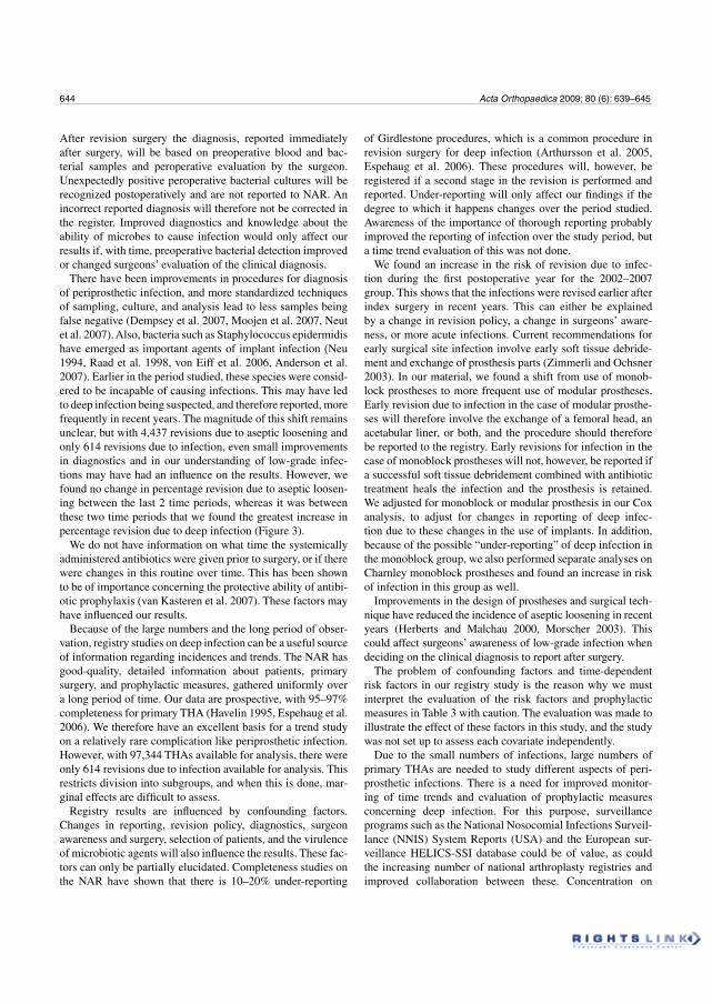
644 Acta Orthopaedica 2009; 80 (6): 639–645
After revision surgery the diagnosis, reported immediately after surgery, will be based on preoperative blood and bac-terial samples and peroperative evaluation by the surgeon. Unexpectedly positive peroperative bacterial cultures will be recognized postoperatively and are not reported to NAR. An incorrect reported diagnosis will therefore not be corrected in the register. Improved diagnostics and knowledge about the ability of microbes to cause infection would only affect our results if, with time, preoperative bacterial detection improved or changed surgeons’ evaluation of the clinical diagnosis.
There have been improvements in procedures for diagnosis of periprosthetic infection, and more standardized techniques of sampling, culture, and analysis lead to less samples being false negative (Dempsey et al. 2007, Moojen et al. 2007, Neut et al. 2007). Also, bacteria such as Staphylococcus epidermidis have emerged as important agents of implant infection (Neu 1994, Raad et al. 1998, von Eiff et al. 2006, Anderson et al. 2007). Earlier in the period studied, these species were consid-ered to be incapable of causing infections. This may have led to deep infection being suspected, and therefore reported, more frequently in recent years. The magnitude of this shift remains unclear, but with 4,437 revisions due to aseptic loosening and only 614 revisions due to infection, even small improvements in diagnostics and in our understanding of low-grade infec-tions may have had an influence on the results. However, we found no change in percentage revision due to aseptic loosen-ing between the last 2 time periods, whereas it was between these two time periods that we found the greatest increase in percentage revision due to deep infection (Figure 3).
We do not have information on what time the systemically administered antibiotics were given prior to surgery, or if there were changes in this routine over time. This has been shown to be of importance concerning the protective ability of antibi-otic prophylaxis (van Kasteren et al. 2007). These factors may have influenced our results.
Because of the large numbers and the long period of obser-vation, registry studies on deep infection can be a useful source of information regarding incidences and trends. The NAR has good-quality, detailed information about patients, primary surgery, and prophylactic measures, gathered uniformly over a long period of time. Our data are prospective, with 95–97% completeness for primary THA (Havelin 1995, Espehaug et al. 2006). We therefore have an excellent basis for a trend study on a relatively rare complication like periprosthetic infection. However, with 97,344 THAs available for analysis, there were only 614 revisions due to infection available for analysis. This restricts division into subgroups, and when this is done, mar-ginal effects are difficult to assess.
Registry results are influenced by confounding factors. Changes in reporting, revision policy, diagnostics, surgeon awareness and surgery, selection of patients, and the virulence of microbiotic agents will also influence the results. These fac-tors can only be partially elucidated. Completeness studies on the NAR have shown that there is 10–20% under-reporting
of Girdlestone procedures, which is a common procedure in revision surgery for deep infection (Arthursson et al. 2005, Espehaug et al. 2006). These procedures will, however, be registered if a second stage in the revision is performed and reported. Under-reporting will only affect our findings if the degree to which it happens changes over the period studied. Awareness of the importance of thorough reporting probably improved the reporting of infection over the study period, but a time trend evaluation of this was not done.
We found an increase in the risk of revision due to infec-tion during the first postoperative year for the 2002–2007 group. This shows that the infections were revised earlier after index surgery in recent years. This can either be explained by a change in revision policy, a change in surgeons’ aware-ness, or more acute infections. Current recommendations for early surgical site infection involve early soft tissue debride-ment and exchange of prosthesis parts (Zimmerli and Ochsner 2003). In our material, we found a shift from use of monob-lock prostheses to more frequent use of modular prostheses. Early revision due to infection in the case of modular prosthe-ses will therefore involve the exchange of a femoral head, an acetabular liner, or both, and the procedure should therefore be reported to the registry. Early revisions for infection in the case of monoblock prostheses will not, however, be reported if a successful soft tissue debridement combined with antibiotic treatment heals the infection and the prosthesis is retained. We adjusted for monoblock or modular prosthesis in our Cox analysis, to adjust for changes in reporting of deep infec-tion due to these changes in the use of implants. In addition, because of the possible “under-reporting” of deep infection in the monoblock group, we also performed separate analyses on Charnley monoblock prostheses and found an increase in risk of infection in this group as well.
Improvements in the design of prostheses and surgical tech-nique have reduced the incidence of aseptic loosening in recent years (Herberts and Malchau 2000, Morscher 2003). This could affect surgeons’ awareness of low-grade infection when deciding on the clinical diagnosis to report after surgery.
The problem of confounding factors and time-dependent risk factors in our registry study is the reason why we must interpret the evaluation of the risk factors and prophylactic measures in Table 3 with caution. The evaluation was made to illustrate the effect of these factors in this study, and the study was not set up to assess each covariate independently.
Due to the small numbers of infections, large numbers of primary THAs are needed to study different aspects of peri-prosthetic infections. There is a need for improved monitor-ing of time trends and evaluation of prophylactic measures concerning deep infection. For this purpose, surveillance programs such as the National Nosocomial Infections Surveil-lance (NNIS) System Reports (USA) and the European sur-veillance HELICS-SSI database could be of value, as could the increasing number of national arthroplasty registries and improved collaboration between these. Concentration on
Acta Orthopaedica 2009; 80 (6): 639–645 645
and improvement of prophylaxis, diagnostics, and treatment of these infections will be of great importance to limit any increase in this serious complication.
The first author performed data analysis and wrote the manuscript. All authors contributed to the conception and design of the study, critical analysis of the data, interpretation of the findings, and critical revision of the manuscript.
The authors thank the Norwegian surgeons for reporting to the NAR, and the secretaries of the NAR for their accurate registration of data. We also extend our thanks to the statisticians of the NAR for their advice.
No competing interests declared.
Anderson D J, Sexton D J, Kanafani Z A, Auten G, Kaye K S. Severe surgical site infection in community hospitals: epidemiology, key procedures, and the changing prevalence of methicillin-resistant Staphylococcus aureus. Infect Control Hosp Epidemiol 2007; 28: 1047-53.
Arthursson A J, Furnes O, Espehaug B, Havelin L I, Soreide J A. Validation of data in the Norwegian Arthroplasty Register and the Norwegian Patient Register: 5,134 primary total hip arthroplasties and revisions operated at a single hospital between 1987 and 2003. Acta Orthop 2005; 76: 823-8.
Block J E, Stubbs H A. Reducing the risk of deep wound infection in pri-mary joint arthroplasty with antibiotic bone cement. Orthopedics 2005; 28: 1334-45.
Charnley J. Postoperative infection after total hip replacement with special reference to air contamination in the operating room. Clin Orthop 1972; (87): 167-87.
Dempsey K E, Riggio M P, Lennon A, Hannah V E, Ramage G, Allan D, Bagg J. Identification of bacteria on the surface of clinically infected and non-infected prosthetic hip joints removed during revision arthroplasties by 16S rRNA gene sequencing and by microbiological culture. Arthritis Res Ther 2007; 9: R46.
Engesaeter L B, Lie S A, Espehaug B, Furnes O, Vollset S E, Havelin L I. Anti-biotic prophylaxis in total hip arthroplasty: effects of antibiotic prophylaxis systemically and in bone cement on the revision rate of 22,170 primary hip replacements followed 0-14 years in the Norwegian Arthroplasty Register. Acta Orthop Scand 2003; 74: 644-51.
Engesaeter L B, Espehaug B, Lie S A, Furnes O, Havelin L I. Does cement increase the risk of infection in primary total hip arthroplasty? Revision rates in 56,275 cemented and uncemented primary THAs followed for 0-16 years in the Norwegian Arthroplasty Register. Acta Orthop 2006; 77: 351-8.
Espehaug B, Furnes O, Havelin L I, Engesaeter L B, Vollset S E, Kindseth O. Registration completeness in the Norwegian Arthroplasty Register. Acta Orthop 2006; 77: 49-56.
Gaine W J, Ramamohan N A, Hussein N A, Hullin M G, McCreath S W. Wound infection in hip and knee arthroplasty. J Bone Joint Surg (Br) 2000; 82: 561-5.
Hastie T J, Tibshirani R J. Generalized additive models. Chapman & Hall, London 1990.
Havelin I. The Norwegian Arthroplasty Register Thesis. 1995. University of Bergen, Norway.
Havelin L I, Engesaeter L B, Espehaug B, Furnes O, Lie S A, Vollset S E. The Norwegian Arthroplasty Register: 11 years and 73,000 arthroplasties. Acta Orthop Scand 2000; 71: 337-53.
Herberts P, Malchau H. Long-term registration has improved the quality of hip replacement: a review of the Swedish THR Register comparing 160,000 cases. Acta Orthop Scand 2000; 71: 111-21.
Ince A, Rupp J, Frommelt L, Katzer A, Gille J, Lohr J F. Is “aseptic” loosen-ing of the prosthetic cup after total hip replacement due to nonculturable bacterial pathogens in patients with low-grade infection? Clin Infect Dis 2004; 39: 1599-603.
Kurtz S M, Lau E, Schmier J, Ong K L, Zhao K, Parvizi J. Infection burden for hip and knee arthroplasty in the United States. J Arthroplasty 2008; 23(7): 984-91.
Mannien J, van den H S, Muilwijk J, van den Broek P J, van Benthem B, Wille J C. Trends in the incidence of surgical site infection in the Netherlands. Infect Control Hosp Epidemiol 2008; 29: 1132-8.
Moojen D J, Spijkers S N, Schot C S, Nijhof M W, Vogely H C, Fleer A, Ver-bout A J, Castelein R M, Dhert W J, Schouls L M. Identification of ortho-paedic infections using broad-range polymerase chain reaction and reverse line blot hybridization. J Bone Joint Surg (Am) 2007; 89: 1298-305.
Morscher E W. Failures and successes in total hip replacement--why good ideas may not work. Scand J Surg 2003; 92: 113-20.
Neu H C. Emerging trends in antimicrobial resistance in surgical infections. A review. Eur J Surg Suppl 1994: 7-18.
Neut D, van der Mei H C, Bulstra S K, Busscher H J. The role of small-colony variants in failure to diagnose and treat biofilm infections in orthopedics. Acta Orthop 2007; 78: 299-308.
Olsen M A, Nepple J J, Riew D, Lenke L G, Bridwell K H, Mayfield J, Fraser V J. Risk factors for surgical site infection following orthopaedic spinal operations. J Bone Joint Surg (Am) 2008; 90: 62-9.
Parvizi J, Ghanem E, Steinbrecher J, Hickok N, Barrack R. The Changing Organism Profile in Periprosthetic Infection. AAOS 75th Annual Meeting Podium Presentations. 2008a. Ref Type: Abstract
Parvizi J, Saleh K J, Ragland P S, Pour A E, Mont M A. Efficacy of antibi-otic-impregnated cement in total hip replacement. Acta Orthop 2008b; 79: 335-41.
Phillips J E, Crane T P, Noy M, Elliott T S, Grimer R J. The incidence of deep prosthetic infections in a specialist orthopaedic hospital: a 15-year prospec-tive survey. J Bone Joint Surg (Br) 2006; 88: 943-8.
Pulido L, Ghanem E, Joshi A, Purtill J J, Parvizi J. Periprosthetic joint infec-tion: the incidence, timing, and predisposing factors. Clin Orthop 2008; (466): 1710-5.
Raad I, Alrahwan A, Rolston K. Staphylococcus epidermidis: emerging resis-tance and need for alternative agents. Clin Infect Dis 1998; 26: 1182-7.
Ridgeway S, Wilson J, Charlet A, Kafatos G, Pearson A, Coello R. Infection of the surgical site after arthroplasty of the hip. J Bone Joint Surg (Br) 2005; 87: 844-50.
Santos Sanches I, Mato R, de Lencastre H, Tomasz A. Patterns of multidrug resistance among methicillin-resistant hospital isolates of coagulase-posi-tive and coagulase-negative staphylococci collected in the international multicenter study RESIST in 1997 and 1998. Microb Drug Resist 2000; 6: 199-211.
Styers D, Sheehan D J, Hogan P, Sahm D F. Laboratory-based surveillance of current antimicrobial resistance patterns and trends among Staphylococcus aureus: 2005 status in the United States. Ann Clin Microbiol Antimicrob 2006; 5: 2.
The Norwegian Arthroplasty Register. Annual Report 2008. 2008. Ref Type: Report
van Kasteren M E, Mannien J, Ott A, Kullberg B J, de Boer A S, Gyssens I C. Antibiotic prophylaxis and the risk of surgical site infections following total hip arthroplasty: Timely administration is the most important factor. Clin Infect Dis 2007; 7: 921-7.
von Eiff C, Arciola C R, Montanaro L, Becker K, Campoccia D. Emerging Staphylococcus species as new pathogens in implant infections. Int J Artif Organs 2006; 29: 360-7.
Zimmerli W, Ochsner P E. Management of infection associated with pros-thetic joints. Infection 2003; 31: 99-108.


Pape
r II

646 Acta Orthopaedica 2011; 82 (6): 646–654
Infection after primary hip arthroplastyA comparison of 3 Norwegian health registers
Håvard Dale1, Inge Skråmm2, Hege L Løwer3, Hanne M Eriksen3, Birgitte Espehaug1, Ove Furnes1,4, Finn Egil Skjeldestad3, Leif I Havelin1,4, and Lars B Engesæter1,4
1The Norwegian Arthroplasty Register, Department of Orthopaedic Surgery, Haukeland University Hospital, Bergen; 2Department of Orthopaedic Surgery, Akershus University Hospital, Lørenskog; 3Norwegian Institute of Public Health, Oslo; 4Department of Surgical Sciences, Faculty of Medicine and Dentistry, University of Bergen, Bergen, NorwayCorrespondence: haavard.dale@helse–bergen.no Submitted 10–09–15. Accepted 11–07–12
Open Access – This article is distributed under the terms of the Creative Commons Attribution Noncommercial License which permits any noncommercial use, distribution, and reproduction in any medium, provided the source is credited.DOI 10.3109/17453674.2011.636671
Background and purpose The aim of the present study was to assess incidence of and risk factors for infection after hip arthroplasty in data from 3 national health registries. We inves-tigated differences in risk patterns between surgical site infec-tion (SSI) and revision due to infection after primary total hip arthroplasty (THA) and hemiarthroplasty (HA).
Materials and methods This observational study was based on prospective data from 2005–2009 on primary THAs and HAs from the Norwegian Arthroplasty Register (NAR), the Norwegian Hip Fracture Register (NHFR), and the Norwegian Surveillance System for Healthcare–Associated Infections (NOIS). The Nor-wegian Patient Register (NPR) was used for evaluation of case reporting. Cox regression analyses were performed with revi-sion due to infection as endpoint for data from the NAR and the NHFR, and with SSI as the endpoint for data from the NOIS.
Results The 1–year incidence of SSI in the NOIS was 3.0% after THA (167/5,540) and 7.3% after HA (103/1,416). The 1–year incidence of revision due to infection was 0.7% for THAs in the NAR (182/24,512) and 1.5% for HAs in the NHFR (128/8,262). Risk factors for SSI after THA were advanced age, ASA class higher than 2, and short duration of surgery. For THA, the risk factors for revision due to infection were male sex, advanced age, ASA class higher than 1, emergency surgery, uncemented fixa-tion, and a National Nosocomial Infection Surveillance (NNIS) risk index of 2 or more. For HAs inserted after fracture, age less than 60 and short duration of surgery were risk factors of revision due to infection.
Interpretation The incidences of SSI and revision due to infec-tion after primary hip replacements in Norway are similar to those in other countries. There may be differences in risk pattern between SSI and revision due to infection after arthroplasty. The risk patterns for revision due to infection appear to be different for HA and THA.
�
Increasing incidence of revision due to infection after primary total hip arthroplasty (THA) has been observed in different countries during the last decade (Kurtz et al. 2008, Dale et al. 2009, Pedersen et al. 2010). There have been several studies on incidence of and risk factors for infection based on data from surveillance systems (Ridgeway et al. 2005, Mannien et al. 2008), arthroplasty (quality) registers (Berbari et al. 1998, Dale et al. 2009, Pedersen et al. 2010), and administrative databases (Mahomed et al. 2003, Kurtz et al. 2008, Ong et al. 2009). There have been reviews on incidence of and risk fac-tors for infection after hip arthroplasty, based on publications from databases with different definitions of infection (Urqu-hart et al. 2009, Jämsen et al. 2010a). Superficial surgical site infections (SSIs) may have risk factors that are different from those of full surgical revisions due to infection. Furthermore, THA and hip hemiarthroplasty (HA) may have different pat-terns of risk of infection (Ridgeway et al. 2005, Cordero–Ampuero and de Dios 2010).
In the present study, we used data from 3 national health registries in Norway to assess incidence and some risk factors for infection after primary hip arthroplasty. Differences in risk patterns between SSI and revision due to infection were inves-tigated for HA and THA.
Material and methods
In Norway, 3 national health registries representing 2 differ-ent surveillance systems record information on primary hip replacement surgery and postoperative infections: the Nor-wegian Arthroplasty Register (NAR) and the Norwegian Hip Fracture Register (NHFR). These are quality registers, while the Norwegian Surveillance System for Healthcare–Asso-ciated Infections (NOIS (Norwegian acronym)) is an infec-tion surveillance system. We compared these registries for
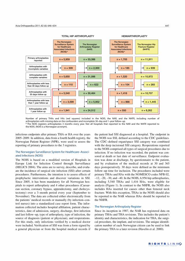
Acta Orthopaedica 2011; 82 (6): 646–654 647
infectious endpoints after primary THA or HA over the years 2005–2009. In addition, data from a fourth health registry, the Norwegian Patient Register (NPR), were used to assess the reporting of primary procedures to the 3 registries.
The Norwegian Surveillance System for Healthcare–Associ-ated Infections (NOIS)The NOIS is based on a modified version of Hospitals in Europe Link for Infection Control through Surveillance (HELICS 2004). The aims are to survey, describe, and evalu-ate the incidence of surgical site infection (SSI) after certain procedures. Furthermore, the intention is to assess effects of prophylactic interventions and discover variations in SSI. Since 2005, it has been mandatory for all Norwegian hos-pitals to report arthroplasty and 4 other procedures (Caesar-ean section, coronary bypass, appendectomy, and cholecys-tectomy) over a 3–month period every year (September to November). The data are collected either electronically from the patients’ medical records or manually (by infection–con-trol nurses) into a standardized case report form. The infor-mation collected includes hospital affiliation, patient charac-teristics, date of admission, surgery, discharge, first infection and last follow–up, type of arthroplasty, type of infection, the source of diagnosis (patient or physician), and reoperations. For this study, only infections verified by a medical doctor were included. Verification of SSI was from a form signed by a general physician or from the hospital medical records if
the patient had SSI diagnosed at a hospital. The endpoint in the NOIS was SSI, defined according to the CDC guidelines. The CDC–defined organ/space SSI category was combined with the deep incisional SSI category. Reoperations reported to the NOIS comprised all types of surgical procedures due to infection. If no infection was recorded, the patient was cen-sored at death or last date of surveillance. Endpoint evalua-tion was done at discharge, by questionnaire to the patient, and by evaluation of the medical records at 30 and 365 days postoperatively. 30 days were defined as the minimum follow–up time for inclusion. The procedures included were primary THAs and HAs with the NOMESCO codes NFB 02, –12, –20, –30, and –40. In the NOIS, 6,956 hip arthroplasties, including 5,540 THAs and 1,416 HAs, were eligible for analysis (Figure 1). In contrast to the NHFR, the NOIS also includes HAs inserted for causes other than femoral neck fracture. With this exception, THAs in the NOIS should also be reported to the NAR whereas HAs should be reported to the NHFR.
The Norwegian Arthroplasty RegisterSince its inception in 1987, the NAR has registered data on primary THAs and THA revisions. This includes the patient’s identity and characteristics, the indication for THA, the surgi-cal procedure, the implant, and revisions. The unique identifi-cation number of each Norwegian citizen can be used to link the primary THA to a later revision (Havelin et al. 2000).
Number of primary THAs and HAs (red squares) included in the NOIS, the NAR, and the NHFR, including number of arthroplasties with missing data on the confounders and incomplete 30–day and 1–year follow–up. a The NOIS registers arthroplasties 3 months every year. Not all hospitals that reported to the NAR and the NHFR reported to the NOIS. (NOIS is a Norwegian acronym).
TOTAL HIP ARTHROPLASTY HEMIARTHROPLASTY
Primary arthroplastiesreported
Arthroplasties withincomplete variables
Arthroplasties withcomplete variables
Arthroplasties with lessthan 30 days follow up
Arthroplasties with 30 days follow up
Arthroplasties with lessthan 1 year follow up
Arthroplasties with 1 year follow up
The NorwegianSurveillance System
for HealthcareAssociated Infections
(NOIS) a
The NorwegianArthroplasty Register
(NAR)
The Norwegian HipFracture Register
(NHFR)
The NorwegianSurveillance System
for HealthcareAssociated Infections
(NOIS) a
n = 5,959 n = 33,386 n = 1,705 n = 11,911
n = 5,650
n = 309 n = 2,300 n = 185 n = 939
n = 110 n = 622 n = 104 n = 265
n = 3,599 n = 5,952 n = 866 n = 1,445
n = 31,086 n = 1,520 n = 10,972
n = 5,540 n = 30,464 n = 1,416 n = 10,707
n = 1,941 n = 24,512 n = 550 n = 8,262
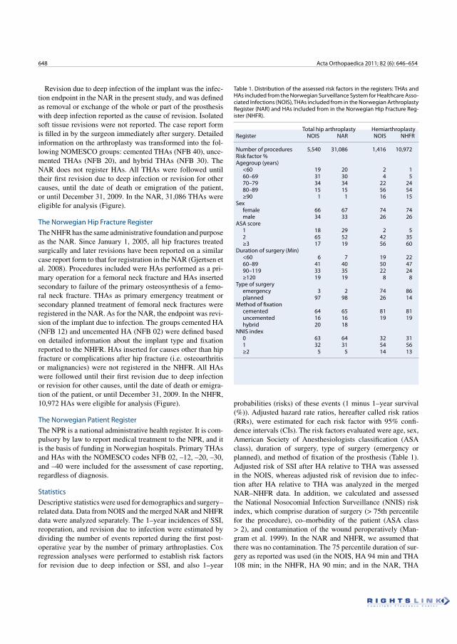
648 Acta Orthopaedica 2011; 82 (6): 646–654
Revision due to deep infection of the implant was the infec-tion endpoint in the NAR in the present study, and was defined as removal or exchange of the whole or part of the prosthesis with deep infection reported as the cause of revision. Isolated soft tissue revisions were not reported. The case report form is filled in by the surgeon immediately after surgery. Detailed information on the arthroplasty was transformed into the fol-lowing NOMESCO groups: cemented THAs (NFB 40), unce-mented THAs (NFB 20), and hybrid THAs (NFB 30). The NAR does not register HAs. All THAs were followed until their first revision due to deep infection or revision for other causes, until the date of death or emigration of the patient, or until December 31, 2009. In the NAR, 31,086 THAs were eligible for analysis (Figure).
The Norwegian Hip Fracture RegisterThe NHFR has the same administrative foundation and purpose as the NAR. Since January 1, 2005, all hip fractures treated surgically and later revisions have been reported on a similar case report form to that for registration in the NAR (Gjertsen et al. 2008). Procedures included were HAs performed as a pri-mary operation for a femoral neck fracture and HAs inserted secondary to failure of the primary osteosynthesis of a femo-ral neck fracture. THAs as primary emergency treatment or secondary planned treatment of femoral neck fractures were registered in the NAR. As for the NAR, the endpoint was revi-sion of the implant due to infection. The groups cemented HA (NFB 12) and uncemented HA (NFB 02) were defined based on detailed information about the implant type and fixation reported to the NHFR. HAs inserted for causes other than hip fracture or complications after hip fracture (i.e. osteoarthritis or malignancies) were not registered in the NHFR. All HAs were followed until their first revision due to deep infection or revision for other causes, until the date of death or emigra-tion of the patient, or until December 31, 2009. In the NHFR, 10,972 HAs were eligible for analysis (Figure).
The Norwegian Patient RegisterThe NPR is a national administrative health register. It is com-pulsory by law to report medical treatment to the NPR, and it is the basis of funding in Norwegian hospitals. Primary THAs and HAs with the NOMESCO codes NFB 02, –12, –20, –30, and –40 were included for the assessment of case reporting, regardless of diagnosis.
StatisticsDescriptive statistics were used for demographics and surgery–related data. Data from NOIS and the merged NAR and NHFR data were analyzed separately. The 1–year incidences of SSI, reoperation, and revision due to infection were estimated by dividing the number of events reported during the first post-operative year by the number of primary arthroplasties. Cox regression analyses were performed to establish risk factors for revision due to deep infection or SSI, and also 1–year
probabilities (risks) of these events (1 minus 1–year survival (%)). Adjusted hazard rate ratios, hereafter called risk ratios (RRs), were estimated for each risk factor with 95% confi-dence intervals (CIs). The risk factors evaluated were age, sex, American Society of Anesthesiologists classification (ASA class), duration of surgery, type of surgery (emergency or planned), and method of fixation of the prosthesis (Table 1). Adjusted risk of SSI after HA relative to THA was assessed in the NOIS, whereas adjusted risk of revision due to infec-tion after HA relative to THA was analyzed in the merged NAR–NHFR data. In addition, we calculated and assessed the National Nosocomial Infection Surveillance (NNIS) risk index, which comprise duration of surgery (> 75th percentile for the procedure), co–morbidity of the patient (ASA class > 2), and contamination of the wound peroperatively (Man-gram et al. 1999). In the NAR and NHFR, we assumed that there was no contamination. The 75 percentile duration of sur-gery as reported was used (in the NOIS, HA 94 min and THA 108 min; in the NHFR, HA 90 min; and in the NAR, THA
Table 1. Distribution of the assessed risk factors in the registers: THAs and HAs included from the Norwegian Surveillance System for Healthcare Asso-ciated Infections (NOIS), THAs included from in the Norwegian Arthroplasty Register (NAR) and HAs included from in the Norwegian Hip Fracture Reg-ister (NHFR).
Total hip arthroplasty HemiarthroplastyRegister NOIS NAR NOIS NHFR
Number of procedures 5,540 31,086 1,416 10,972Risk factor % Agegroup (years) <60 19 20 2 1 60–69 31 30 4 5 70–79 34 34 22 24 80–89 15 15 56 54 ≥90 1 1 16 15Sex female 66 67 74 74 male 34 33 26 26ASA score 1 18 29 2 5 2 65 52 42 35 ≥3 17 19 56 60Duration of surgery (Min) <60 6 7 19 22 60–89 41 40 50 47 90–119 33 35 22 24 ≥120 19 19 8 8Type of surgery emergency 3 2 74 86 planned 97 98 26 14Method of fixation cemented 64 65 81 81 uncemented 16 16 19 19 hybrid 20 18NNIS index 0 63 64 32 31 1 32 31 54 56 ≥2 5 5 14 13
Acta Orthopaedica 2011; 82 (6): 646–654 649
110 min), and not the 120 min estimated for HA and THA in the HELICS guidelines. Follow–up for the NAR and the NHFR analyses was 0–5 years and for the NOIS it was 0–1 year. However, to ensure similar follow–up for all 3 registries, additional analyses were performed with follow–up restricted to 1 year for all available cases. Stratified analyses were per-formed on the NOIS data with deep SSI as separate endpoint. Any p–values of less than 0.05 were considered statistically significant. SPSS software version 18.0 and PASS 2008 soft-ware were used for statistical analysis.
ResultsCase reporting and distribution of risk factors32 hospitals reported THAs to the NPR and the NAR during the study period. 30 hospitals reported THAs to the NOIS; there was an increase in hospitals reporting THA, from 8 in the first year.
29 hospitals reported HAs to the NPR, whereas 27 reported HAs to the NHFR and 26 reported HAs to the NOIS over the study period. The number of hospitals reporting HAs increased from 5 to 26 in the NOIS and from 26 to 27 in the NHFR. 33,466 primary THAs and 12,069 primary HAs were reported to the NPR during the study period. The comparable number of procedures reported to each of the other registries is presented in Figure 1. The distribution of risk factors was similar for THAs in the NOIS and the NAR, and for HAs in the NOIS and the NHFR (Table 1). The exception was ASA classification (Table 1).
Incidence and risk of infectionThe 1–year incidence of SSI was 3.0% after primary THA (Table 2). 6/94 of the superficial SSIs and 52/73 of the deep SSIs after THA in the NOIS were reported to have been reop-erated due to the infection, whereas in the NAR the 1–year incidence of revision due to infection was 0.7% (Table 2).
In primary HAs, the 1–year incidence of SSI was 7.3% (Table 2). 50/51 of the reoperations due to infection after HA in the NOIS were due to deep SSIs, and 50/72 of the deep SSIs were reported to have been reoperated. 1.5% of the HAs were reported to the NHFR to have been revised due to infection (Table 2).
In the NOIS, the adjusted risk of SSI after HA compared to THA was 1.2 (CI: 0.8–1.9). The adjusted risk of revision due to infection was 1.8 times higher for HAs (CI: 1.2–2.7) than for THA in the merged NAR/NHFR data.
Time to SSI and revision due to infectionFor THAs, the median postoperative time to diagnosis of SSI was 16 (2–214) days. Median time to revision due to infection was 29 (4–343) days, when restricting follow–up to 1 year, and 47 (4–1,782) days with 0–5 years of follow–up. For HAs, the median postoperative time to SSI was 15 (2–79) days. Median time to revision due to infection was 20 (4–304) days with 1–year follow up and 20 (4–701) days with 0–5 years of follow–up. 72% of the SSIs in the NOIS were identified in the post discharge surveillance, but only 9 cases of SSI were identified between 30 and 365 days postoperatively.
Risk factors for infection after THAThe following factors were associated with increased risk of revision due to infection: 70–89 years of age, male sex, and ASA class higher than 1 (Table 3). Emergency surgery as opposed to planned surgery and a National Nosocomial Infec-tion Surveillance Systems (NNIS) risk index of > 1 were also associated with a higher risk of revision due to infection after THA. Uncemented fixation of the prosthesis had a 50% higher risk of revision due to infection compared to cemented THAs.
Risk factors for SSI after THA were short duration of sur-gery (< 60 min), which was also the finding when cemented, uncemented, and hybrid fixations were analyzed separately. Patients older than 80 years of age also had higher risk of SSI than those who were less than 60 years of age. The risk patterns for SSI and revision due to infection were different regarding effects of gender, duration of surgery < 60 min, and method of fixation. Separate analyses of all cases with 1–year follow–up in the NOIS and restricted follow–up of 1 year for the NAR as in the NOIS did not change the findings concern-ing risk factors for SSI or revision due to infection. Restriction to deep incisional SSI in the NOIS had only minor effects on the risk estimates.
Risk factors for infection after HAIn the NHFR, age less than 60 years and duration of sur-gery of less than 60 min were associated with increased risk of revision due to infection (Table 4). No risk factors were identified for SSI after HA. HA had a different risk profile from that of THA, for both SSI and revision due to infection (Tables 3 and 4).
Table 2. 1-year incidence of SSI and reoperation after primary arthroplasties as reported to the Norwegian Surveillance System for Healthcare Associ-ated Infections (NOIS) and 1-year incidence of revisions due to infection after THAs as reported to the Norwegian Arthroplasty Register (NAR) and after HAs as reported to the Norwegian Hip Fracture Register (NHFR)
Total hip arthroplasty HemiarthroplastyRegistera Endpoint 1–year incidence 1–year incidence
NOIS Sugical Site Infection 3.0% (167/5,540) 7.3% (103/1,416) Superficial SSI 1.7% (94/5,540) 2.2% (31/1,416) Deep SSI 1.3% (73/5,540) 5.1% (72/1,415) SSI reoperated 1.0% (58/5,540) 3.6% (51/1.416)NAR / NHFR Revision due to infection 0.7% (182/24,512) 1.5% (128/8,262)
a The NOIS and the NAR/NHFR represents different selections of cases
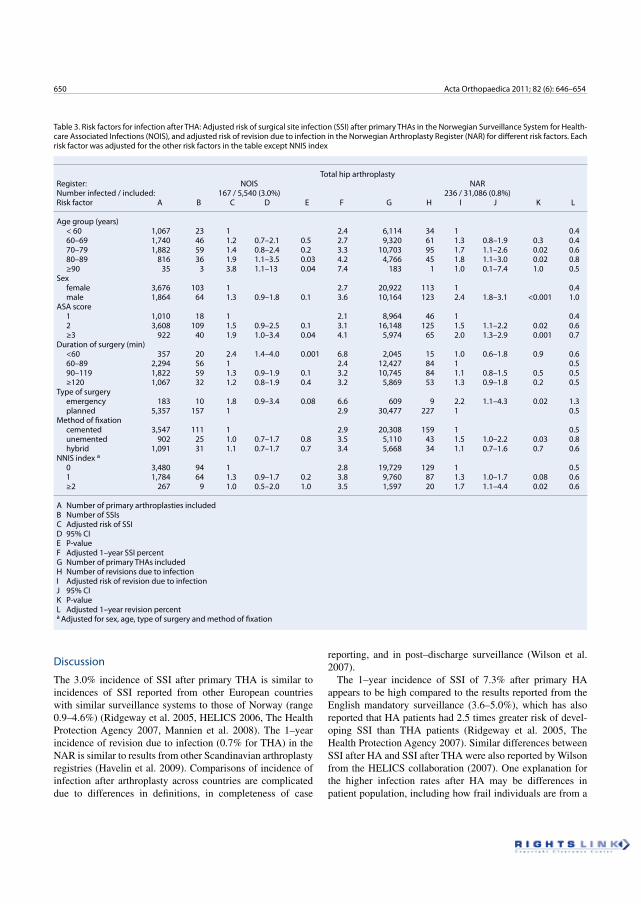
650 Acta Orthopaedica 2011; 82 (6): 646–654
Discussion
The 3.0% incidence of SSI after primary THA is similar to incidences of SSI reported from other European countries with similar surveillance systems to those of Norway (range 0.9–4.6%) (Ridgeway et al. 2005, HELICS 2006, The Health Protection Agency 2007, Mannien et al. 2008). The 1–year incidence of revision due to infection (0.7% for THA) in the NAR is similar to results from other Scandinavian arthroplasty registries (Havelin et al. 2009). Comparisons of incidence of infection after arthroplasty across countries are complicated due to differences in definitions, in completeness of case
reporting, and in post–discharge surveillance (Wilson et al. 2007).
The 1–year incidence of SSI of 7.3% after primary HA appears to be high compared to the results reported from the English mandatory surveillance (3.6–5.0%), which has also reported that HA patients had 2.5 times greater risk of devel-oping SSI than THA patients (Ridgeway et al. 2005, The Health Protection Agency 2007). Similar differences between SSI after HA and SSI after THA were also reported by Wilson from the HELICS collaboration (2007). One explanation for the higher infection rates after HA may be differences in patient population, including how frail individuals are from a
Table 3. Risk factors for infection after THA: Adjusted risk of surgical site infection (SSI) after primary THAs in the Norwegian Surveillance System for Health-care Associated Infections (NOIS), and adjusted risk of revision due to infection in the Norwegian Arthroplasty Register (NAR) for different risk factors. Each risk factor was adjusted for the other risk factors in the table except NNIS index
Total hip arthroplastyRegister: NOIS NARNumber infected / included: 167 / 5,540 (3.0%) 236 / 31,086 (0.8%)Risk factor A B C D E F G H I J K L
Age group (years) < 60 1,067 23 1 2.4 6,114 34 1 0.4 60–69 1,740 46 1.2 0.7–2.1 0.5 2.7 9,320 61 1.3 0.8–1.9 0.3 0.4 70–79 1,882 59 1.4 0.8–2.4 0.2 3.3 10,703 95 1.7 1.1–2.6 0.02 0.6 80–89 816 36 1.9 1.1–3.5 0.03 4.2 4,766 45 1.8 1.1–3.0 0.02 0.8 ≥90 35 3 3.8 1.1–13 0.04 7.4 183 1 1.0 0.1–7.4 1.0 0.5Sex female 3,676 103 1 2.7 20,922 113 1 0.4 male 1,864 64 1.3 0.9–1.8 0.1 3.6 10,164 123 2.4 1.8–3.1 <0.001 1.0ASA score 1 1,010 18 1 2.1 8,964 46 1 0.4 2 3,608 109 1.5 0.9–2.5 0.1 3.1 16,148 125 1.5 1.1–2.2 0.02 0.6 ≥3 922 40 1.9 1.0–3.4 0.04 4.1 5,974 65 2.0 1.3–2.9 0.001 0.7Duration of surgery (min) <60 357 20 2.4 1.4–4.0 0.001 6.8 2,045 15 1.0 0.6–1.8 0.9 0.6 60–89 2,294 56 1 2.4 12,427 84 1 0.5 90–119 1,822 59 1.3 0.9–1.9 0.1 3.2 10,745 84 1.1 0.8–1.5 0.5 0.5 ≥120 1,067 32 1.2 0.8–1.9 0.4 3.2 5,869 53 1.3 0.9–1.8 0.2 0.5Type of surgery emergency 183 10 1.8 0.9–3.4 0.08 6.6 609 9 2.2 1.1–4.3 0.02 1.3 planned 5,357 157 1 2.9 30,477 227 1 0.5Method of fixation cemented 3,547 111 1 2.9 20,308 159 1 0.5 unemented 902 25 1.0 0.7–1.7 0.8 3.5 5,110 43 1.5 1.0–2.2 0.03 0.8 hybrid 1,091 31 1.1 0.7–1.7 0.7 3.4 5,668 34 1.1 0.7–1.6 0.7 0.6NNIS index a 0 3,480 94 1 2.8 19,729 129 1 0.5 1 1,784 64 1.3 0.9–1.7 0.2 3.8 9,760 87 1.3 1.0–1.7 0.08 0.6 ≥2 267 9 1.0 0.5–2.0 1.0 3.5 1,597 20 1.7 1.1–4.4 0.02 0.6
A Number of primary arthroplasties included B Number of SSIs C Adjusted risk of SSI D 95% CIE P-valueF Adjusted 1–year SSI percent G Number of primary THAs included H Number of revisions due to infection I Adjusted risk of revision due to infection J 95% CI K P-value L Adjusted 1–year revision percent a Adjusted for sex, age, type of surgery and method of fixation
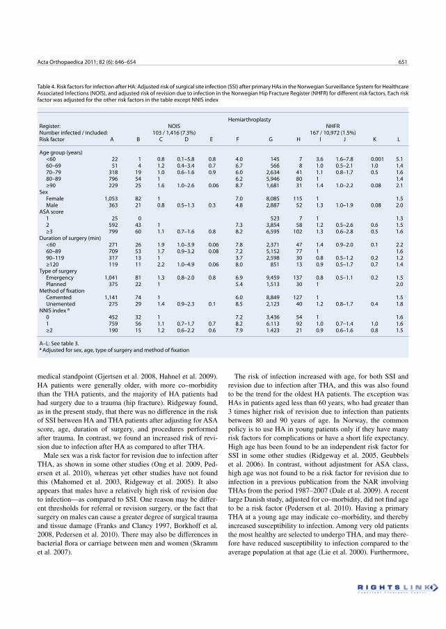
Acta Orthopaedica 2011; 82 (6): 646–654 651
medical standpoint (Gjertsen et al. 2008, Hahnel et al. 2009). HA patients were generally older, with more co–morbidity than the THA patients, and the majority of HA patients had had surgery due to a trauma (hip fracture). Ridgeway found, as in the present study, that there was no difference in the risk of SSI between HA and THA patients after adjusting for ASA score, age, duration of surgery, and procedures performed after trauma. In contrast, we found an increased risk of revi-sion due to infection after HA as compared to after THA.
Male sex was a risk factor for revision due to infection after THA, as shown in some other studies (Ong et al. 2009, Ped-ersen et al. 2010), whereas yet other studies have not found this (Mahomed et al. 2003, Ridgeway et al. 2005). It also appears that males have a relatively high risk of revision due to infection—as compared to SSI. One reason may be differ-ent thresholds for referral or revision surgery, or the fact that surgery on males can cause a greater degree of surgical trauma and tissue damage (Franks and Clancy 1997, Borkhoff et al. 2008, Pedersen et al. 2010). There may also be differences in bacterial flora or carriage between men and women (Skramm et al. 2007).
The risk of infection increased with age, for both SSI and revision due to infection after THA, and this was also found to be the trend for the oldest HA patients. The exception was HAs in patients aged less than 60 years, who had greater than 3 times higher risk of revision due to infection than patients between 80 and 90 years of age. In Norway, the common policy is to use HA in young patients only if they have many risk factors for complications or have a short life expectancy. High age has been found to be an independent risk factor for SSI in some other studies (Ridgeway et al. 2005, Geubbels et al. 2006). In contrast, without adjustment for ASA class, high age was not found to be a risk factor for revision due to infection in a previous publication from the NAR involving THAs from the period 1987–2007 (Dale et al. 2009). A recent large Danish study, adjusted for co–morbidity, did not find age to be a risk factor (Pedersen et al. 2010). Having a primary THA at a young age may indicate co–morbidity, and thereby increased susceptibility to infection. Among very old patients the most healthy are selected to undergo THA, and may there-fore have reduced susceptibility to infection compared to the average population at that age (Lie et al. 2000). Furthermore,
Table 4. Risk factors for infection after HA: Adjusted risk of surgical site infection (SSI) after primary HAs in the Norwegian Surveillance System for Healthcare Associated Infections (NOIS), and adjusted risk of revision due to infection in the Norwegian Hip Fracture Register (NHFR) for different risk factors. Each risk factor was adjusted for the other risk factors in the table except NNIS index
HemiarthroplastyRegister: NOIS NHFRNumber infected / included: 103 / 1,416 (7.3%) 167 / 10,972 (1.5%)Risk factor A B C D E F G H I J K L
Age group (years) <60 22 1 0.8 0.1–5.8 0.8 4.0 145 7 3.6 1.6–7.8 0.001 5.1 60–69 51 4 1.2 0.4–3.4 0.7 6.7 566 8 1.0 0.5–2.1 1.0 1.4 70–79 318 19 1.0 0.6–1.6 0.9 6.0 2,634 41 1.1 0.8–1.7 0.5 1.6 80–89 796 54 1 6.2 5,946 80 1 1.4 ≥90 229 25 1.6 1.0–2.6 0.06 8.7 1,681 31 1.4 1.0–2.2 0.08 2.1Sex Female 1,053 82 1 7.0 8,085 115 1 1.5 Male 363 21 0.8 0.5–1.3 0.3 4.8 2,887 52 1.3 1.0–1.9 0.08 2.0ASA score 1 25 0 523 7 1 1.3 2 592 43 1 7.3 3,854 58 1.2 0.5–2.6 0.6 1.5 ≥3 799 60 1.1 0.7–1.6 0.8 8.2 6,595 102 1.3 0.6–2.8 0.5 1.6Duration of surgery (min) <60 271 26 1.9 1.0–3.9 0.06 7.8 2,371 47 1.4 0.9–2.0 0.1 2.2 60–89 709 53 1.7 0.9–3.2 0.08 7.2 5,152 77 1 1.6 90–119 317 13 1 3.7 2,598 30 0.8 0.5–1.2 0.2 1.2 ≥120 119 11 2.2 1.0–4.9 0.06 8.0 851 13 0.9 0.5–1.7 0.7 1.4Type of surgery Emergency 1,041 81 1.3 0.8–2.0 0.8 6.9 9,459 137 0.8 0.5–1.1 0.2 1.5 Planned 375 22 1 5.4 1,513 30 1 2.0Method of fixation Cemented 1,141 74 1 6.0 8,849 127 1 1.5 Unemented 275 29 1.4 0.9–2.3 0.1 8.5 2,123 40 1.2 0.8–1.7 0.4 1.8NNIS index a 0 452 32 1 7.2 3,436 54 1 1.6 1 759 56 1.1 0.7–1.7 0.7 8.2 6.113 92 1.0 0.7–1.4 1.0 1.6 ≥2 190 15 1.2 0.6–2.2 0.6 7.9 1.423 21 0.9 0.6–1.6 0.8 1.5
A–L: See table 3. a Adjusted for sex, age, type of surgery and method of fixation
652 Acta Orthopaedica 2011; 82 (6): 646–654
a revision operation on hip arthroplasty is extensive surgery, and surgeons may sometimes choose a nonoperative approach in old and frail patients—an option that is not reported to the NAR and NHFR.
ASA class is a crude approximation of physical status, and works poorly at the individual level where there will be large inter–observer variability. In addition, different co–morbidi-ties may have different effects on infection rates. However, ASA class has predictive value for complications in epidemio-logical studies like the present one, where the number of cases is large (Ridgeway et al. 2005, Bjørgul et al. 2010). Thus, all 3 registries have chosen the ASA classification as their measure of physical state. An ASA score higher than 1 had an increased risk of revision due to infection after THAs. This indicates that even minor co–morbidities may increase the risk of post-operative infection. For patients with an infected prosthesis, the treatment strategy may be nonoperative for higher ASA classes. In the latter case, some surgeons may choose lifelong antibiotic suppression rather than reoperation for a low–grade implant infection. This may be one explanation for why higher ASA scores had no increased risk of revision due to infection after HA in our study. It may also be that ASA class does not capture frailty in the elderly in a sufficient way in our study population (Makary et al. 2010).
We could not confirm findings from previous studies that longer duration of surgery is associated with higher risk of SSI and higher risk of revision due to infection after THA (Små-brekke et al. 2004, Ridgeway et al. 2005, Dale et al. 2009, Ong et al. 2009, Pedersen et al. 2010). However, duration of surgery less than 60 min was associated with higher risk of SSI after arthroplasty and risk of revision due to infection after HA. Similar findings were reported for SSI after revi-sion arthroplasties, but not primary HA or THA, by Ridge-way (2005). Rapid surgery may result in inferior soft tissue treatment and hemostasis, thereby leading to increased risk of infection.
Primary arthroplasty performed as an emergency procedure after a femoral neck fracture increased the risk of both SSI and revision due to infection after THA. Ridgeway (2005) also found trauma to be a risk factor for SSI after THA. This may be due to local or systemic reactions to the trauma itself, to frailty of the patients, or to other confounders not reported to the registers. For HAs, there was no difference in the risk of revision due to infection between arthroplasty performed in the acute phase and planned surgery. A primary arthroplasty per-formed as planned surgery caused by a failed osteosynthesis is a reoperation, and may therefore resemble a revision arthroplasty more than a genuine primary arthroplasty. Revi-sion arthroplasty and arthroplasty secondary to fractures are found to have higher susceptibility to infection (Berbari et al. 1998, Ridgeway et al. 2005, Jämsen et al. 2009a).
Cementless fixation had a higher risk of revision due to deep infection after THA, but not a higher risk of SSI. In Norway, nearly all cemented THAs are inserted with cement
containing antibiotics (The Norwegian Arthroplasty Register 2010). Uncemented THAs can only be protected by antibi-otic prophylaxis given systemically, and this was adminis-tered in nearly all hip arthroplasties in Norway over the study period (The Norwegian Arthroplasty Register 2010). Anti-biotic eluted from cement is delivered locally, and protects the implant and periprosthetic tissue (Espehaug et al. 1997, Engesæter et al. 2003, Hendriks et al. 2005, Dale et al. 2009). This local antibiotic treatment appears to be less effective for protection against SSI.
The NNIS risk index is a combined surgery–related assess-ment tool developed to identify high–risk patients, and to eval-uate the risk of SSI (Mangram et al. 1999). The NNIS index combines ASA class of greater than 2, duration of surgery longer than the 75th percentile for the procedure, and contami-nation of the wound. Considering our findings on ASA class and duration of surgery, and that arthroplasty is a clean proce-dure, the NNIS does not appear to be optimal for identification of patients who are at risk of infection after arthroplasty.
All data on completeness of case reporting to the NAR, the NHFR, and the NOIS, indicate that there would be minor selection bias in our study. The arthroplasties reported to the NOIS were similar, regarding the characteristics of patients and procedures, to the all–year–round registrations in the NAR and the NHFR. SSIs may have been underreported to the NOIS, just as revision due to infection has been to the NAR and other registers (Arthursson et al. 2005, Espehaug et al. 2006, Huotari et al. 2007, Jämsen et al. 2009b, Jämsen et al. 2010b). There is also a possibility of overestimation of SSI in surveillance systems, as superficial infections may be difficult to distinguish from aseptic wound complications (Walenkamp 2009). The lack of validation of endpoints is therefore a weak-ness in our study, even though we performed separate analyses on overall and deep infections without any major changes in risk assessment. This should be addressed in future studies. Surgical policy was also a possible confounder in the present study on the NHFR and the NAR, since different subgroups—such as patients with higher ASA classes and advanced age—may have been subject to different treatment strategies. For the NOIS and the NHFR, the number of cases included makes statistical power an issue when differences between subgroups are small or the numbers are low.
The 2 endpoints of infection in our study may reflect differ-ent types of infections, or at least different stages of infection. The NOIS is more likely to capture the acute virulent post-operative infections whereas the NAR/NHFR is more likely to capture either a more advanced stage of infection or more low–grade, late infections. This fact will affect the findings of incidence and risk patterns of infection, and it is important for the interpretation of results of studies with different defini-tions of infection and different follow–up.
The majority of SSIs were identified after discharge, which confirms earlier findings that post–discharge surveillance is important to capture the true incidence of SSI after hip
Acta Orthopaedica 2011; 82 (6): 646–654 653
replacement (Huenger et al. 2005, Huotari and Lyytikainen 2006, Mannien et al. 2008). Infection surveillance appears to reduce the incidence of SSI (Brandt et al. 2006), which is also the aim for the NOIS. The NAR has improved THA sur-gery in Norway over the last 25 years (Fevang et al. 2010). In 2005, the NHFR was established on the same basis with the same methodology. This has led to changes in the treat-ment of femoral neck fractures towards more use of HA (The Norwegian Hip Fracture Register 2010, Gjertsen et al. 2010). Adverse effects of such changes, such as infection, should be evaluated, which requires good–quality surveillance through registers like the NOIS, the NAR, and the NHFR.
HD performed the data analysis and wrote the manuscript. BE, HLL, HME, FES, LIH, LBE, and OF contributed to the conception and design of the study, critical analysis of the data, interpretation of the findings, and critical revision of the manuscript through all stages of the study. IS contributed to critical evaluation of the analyses, and to revision of the manuscript.
We thank the Norwegian surgeons for their contribution with thorough report-ing to the NAR and the NHFR. We also thank all the infection control staff at the Norwegian hospitals involved in data collection for the NOIS, for their invaluable contribution, and the staff at the NPR, NAR, NHFR, and NOIS for their conscientious registration of data.
No competing interests declared.
Arthursson A J, Furnes O, Espehaug B, Havelin L I, Soreide J A. Validation of data in the Norwegian Arthroplasty Register and the Norwegian Patient Register: 5,134 primary total hip arthroplasties and revisions operated at a single hospital between 1987 and 2003. Acta Orthop 2005; 76 (6): 823–8.
Berbari E F, Hanssen A D, Duffy M C, Steckelberg J M, Ilstrup D M, Harm-sen W S, Osmon D R. Risk factors for prosthetic joint infection: case–con-trol study. Clin Infect Dis 1998; 27 (5): 1247–54.
Bjørgul K, Novicoff W M, Saleh K J. Evaluating comorbidities in total hip and knee arthroplasty: available instruments. J Orthop Traumatol 2010; 11 (4): 203–9.
Borkhoff C M, Hawker G A, Kreder H J, Glazier R H, Mahomed N N, Wright J G. The effect of patients’ sex on physicians’ recommendations for total knee arthroplasty. CMAJ 2008; 178 (6): 681–7.
Brandt C, Sohr D, Behnke M, Daschner F, Ruden H, Gastmeier P. Reduction of surgical site infection rates associated with active surveillance. Infect Control Hosp Epidemiol 2006; 27 (12): 1347–51.
Cordero–Ampuero J, de Dios M . What are the risk factors for infection in hemiarthroplasties and total hip arthroplasties? Clin Orthop 2010; (468) (12): 3268–77.
Dale H, Hallan G, Espehaug B, Havelin L I, Engeæter L B. Increasing risk of revision due to deep infection after hip arthroplasty. Acta Orthop 2009; 80 (6): 639–45.
Engesæter L B, Lie S A, Espehaug B, Furnes O, Vollset S E, Havelin L I. Anti-biotic prophylaxis in total hip arthroplasty: effects of antibiotic prophylaxis systemically and in bone cement on the revision rate of 22,170 primary hip replacements followed 0–14 years in the Norwegian Arthroplasty Register. Acta Orthop Scand 2003; 74 (6): 644–51.
Espehaug B, Engesæter L B, Vollset S E, Havelin L I, Langeland N. Antibiotic prophylaxis in total hip arthroplasty. Review of 10,905 primary cemented total hip replacements reported to the Norwegian arthroplasty register, 1987 to 1995. J Bone Joint Surg (Br) 1997; 79 (4): 590–5.
Espehaug B, Furnes O, Havelin L I, Engesæter L B, Vollset S E, Kindseth O. Registration completeness in the Norwegian Arthroplasty Register. Acta Orthop 2006; 77 (1): 49–56.
Fevang B T, Lie S A, Havelin L I, Engesaeter L B, Furnes O. Improved results of primary total hip replacement. Acta Orthop 2010; 81 (6): 649–59.
Franks P, Clancy C M. Referrals of adult patients from primary care: demo-graphic disparities and their relationship to HMO insurance. J Fam Pract 1997; 45 (1): 47–53.
Geubbels E L, Grobbee D E, Vandenbroucke–Grauls C M, Wille J C, de Boer A S. Improved risk adjustment for comparison of surgical site infection rates. Infect Control Hosp Epidemiol 2006; 27 (12): 1330–9.
Gjertsen J E, Engesæter L B, Furnes O, Havelin L I, Steindal K, Vinje T, Fevang J M. The Norwegian Hip Fracture Register: experiences after the first 2 years and 15,576 reported operations. Acta Orthop 2008; 79 (5): 583–93.
Gjertsen J E, Vinje T, Engesaeter L B, Lie S A, Havelin L I, Furnes O, Fevang J M. Internal screw fixation compared with bipolar hemiarthroplasty for treatment of displaced femoral neck fractures in elderly patients. J Bone Joint Surg (Am) 2010; 92 (3): 619–28.
Hahnel J, Burdekin H, Anand S. Re–admissions following hip fracture sur-gery. Ann R Coll Surg Engl 2009; 91 (7): 591–5.
Havelin L I, Engesæter L B, Espehaug B, Furnes O, Lie S A, Vollset S E. The Norwegian Arthroplasty Register: 11 years and 73,000 arthroplasties. Acta Orthop Scand 2000; 71 (4): 337–53.
Havelin L I, Fenstad A M, Salomonsson R, Mehnert F, Furnes O, Over-gaard S, Pedersen A B, Herberts P, Karrholm J, Garellick G. The Nordic Arthroplasty Register Association: a unique collaboration between 3 national hip arthroplasty registries with 280,201 THRs. Acta Orthop 2009; 80 (4): 393–401.
HELICS. Hospital in Europe Link for Infection Control through Surveillance Surgical Site Infection, Surveillance of Surgical Site Infections, Protocol. European Centre for Disease Prevention and Control, 2004. Available from http://helics.univ–lyon1.fr/
HELICS. Hospital in Europe Link for Infection Control through Surveil-lance Surgical Site Infection Statistical Report 2004. European Centre for Disease Prevention and Control, 2006. Available from http://helics.univ–lyon1.fr/documents/HELICS–SSI%20Stat%20Report%202004%20Final%20Version%20180406.pdf
Hendriks J G, Neut D, van Horn J R, van der Mei H C, Busscher H J. Bacterial survival in the interfacial gap in gentamicin–loaded acrylic bone cements. J Bone Joint Surg (Br) 2005; 87 (2): 272–6.
Huenger F, Schmachtenberg A, Haefner H, Zolldann D, Nowicki K, Wirtz D C, Blasius K, Lutticken R, Lemmen S W. Evaluation of postdischarge surveillance of surgical site infections after total hip and knee arthroplasty. Am J Infect Control 2005; 33 (8): 455–62.
Huotari K, Lyytikainen O. Impact of postdischarge surveillance on the rate of surgical site infection after orthopedic surgery. Infect Control Hosp Epide-miol 2006; 27 (12): 1324–9.
Huotari K, Agthe N, Lyytikainen O. Validation of surgical site infection surveillance in orthopedic procedures. Am J Infect Control 2007; 35 (4): 216–21.
Jämsen E, Huhtala H, Puolakka T, Moilanen T. Risk factors for infection after knee arthroplasty. A register–based analysis of 43,149 cases. J Bone Joint Surg (Am) 2009a; 91 (1): 38–47.
Jämsen E, Huotari K, Huhtala H, Nevalainen J, Konttinen Y T. Low rate of infected knee replacements in a nationwide series–is it an underestimate? Acta Orthop 2009b; 80 (2): 205–12.
Jämsen E, Furnes O, Engesæter L B, Konttinen Y T, Odgaard A, Stefansdottir A, Lidgren L. Prevention of deep infection in joint replacement surgery. Acta Orthop 2010a; 81 (6): 660–6.
Jämsen E, Varonen M, Huhtala H, Lehto M U, Lumio J, Konttinen Y T, Moilanen T. Incidence of prosthetic joint infections after primary knee arthroplasty. J Arthroplasty 2010b; 25 (1): 87–92.
Kurtz S M, Lau E, Schmier J, Ong K L, Zhao K, Parvizi J. Infection Burden for Hip and Knee Arthroplasty in the United States. J Arthroplasty 2008; 23 (7): 984-91.
654 Acta Orthopaedica 2011; 82 (6): 646–654
Lie S A, Engesæter L B, Havelin L I, Gjessing H K, Vollset S E. Mortality after total hip replacement: 0–10–year follow–up of 39,543 patients in the Norwegian Arthroplasty Register. Acta Orthop Scand 2000; 71 (1): 19–27.
Mahomed N N, Barrett J A, Katz J N, Phillips C B, Losina E, Lew R A, Gua-dagnoli E, Harris W H, Poss R, Baron J A. Rates and outcomes of primary and revision total hip replacement in the United States medicare popula-tion. J Bone Joint Surg (Am) 2003; 85 (1): 27–32.
Makary M A, Segev D L, Pronovost P J, Syin D, Bandeen–Roche K, Patel P, Takenaga R, Devgan L, Holzmueller C G, Tian J, Fried L P. Frailty as a predictor of surgical outcomes in older patients. J Am Coll Surg 2010; 210 (6): 901–8.
Mangram A J, Horan T C, Pearson M L, Silver L C, Jarvis W R. Guideline for prevention of surgical site infection, 1999. Hospital Infection Control Practices Advisory Committee. Infect Control Hosp Epidemiol 1999; 20 (4): 250–78.
Mannien J, van den H S, Muilwijk J, van den Broek P J, van Benthem B, Wille J C. Trends in the incidence of surgical site infection in the Netherlands. Infect Control Hosp Epidemiol 2008; 29 (12): 1132–8.
Ong K L, Kurtz S M, Lau E, Bozic K J, Berry D J, Parvizi J. Prosthetic joint infection risk after total hip arthroplasty in the Medicare population. J Arthroplasty (6 Suppl) 2009; 24: 105–9.
Pedersen A B, Svendsson J E, Johnsen S P, Riis A, Overgaard S. Risk fac-tors for revision due to infection after primary total hip arthroplasty. A population–based study of 80,756 primary procedures in the Danish Hip Arthroplasty Registry. Acta Orthop 2010; 81 (5): 542–7.
Ridgeway S, Wilson J, Charlet A, Kafatos G, Pearson A, Coello R. Infection of the surgical site after arthroplasty of the hip. J Bone Joint Surg (Br) 2005; 87 (6): 844–50.
Skramm I, Moen A E, Alm–Kristiansen K, Bukholm G. Nasal carriage of Staphylococcus aureus: which sequence types do orthopedic surgical healthcare workers carry? Infect Control Hosp Epidemiol 2007; 28 (6): 737–9.
Småbrekke A, Espehaug B, Havelin L I, Furnes O. Operating time and sur-vival of primary total hip replacements: an analysis of 31,745 primary cemented and uncemented total hip replacements from local hospitals reported to the Norwegian Arthroplasty Register 1987–2001. Acta Orthop Scand 2004; 75 (5): 524–32.
The Health Protection Agency. Third Report of the Mandatory Surveillance of Surgical Site Infection in Orthopaedic Surgery. April 2004 to March 2007. Health Protection Agency 2007, 2007. Available from http://www.hpa.org.uk/web/HPAwebFile/HPAweb_C/1197382185219
The Norwegian Arthroplasty Register. Annual Report 2010. The Norwegian Arthroplasty Register, 2010. Available from http://nrlweb.ihelse.net/eng/Report_2010.pdf
The Norwegian Hip Fracture Register. Annual Report 2010. The Norwegian Arthroplasty Register, 2010. Available from http://nrlweb.ihelse.net/eng/Report_2010.pdf
Urquhart D M, Hanna F S, Brennan S L, Wluka A E, Leder K, Cameron P A, Graves S E, Cicuttini F M. Incidence and Risk Factors for Deep Surgical Site Infection After Primary Total Hip Arthroplasty: A Systematic Review. J Arthroplasty 2009; 25 (8): 1216-22.
Walenkamp G H. Joint prosthetic infections: a success story or a continuous concern? Acta Orthop 2009; 80 (6): 629–32.
Wilson J, Ramboer I, Suetens C. Hospitals in Europe Link for Infection Con-trol through Surveillance (HELICS). Inter–country comparison of rates of surgical site infection––opportunities and limitations. J Hosp Infect (Suppl) 2007; 65 2165–70.


Pape
r III


Acta Orthopaedica 2012; 83 (5): 449–458 449
Increasing risk of prosthetic joint infection after total hip arthroplasty2,778 revisions due to infection after 432,168 primary THAs in the Nordic Arthroplasty Register Association (NARA)
Håvard Dale1, Anne M Fenstad1, Geir Hallan1, Leif I Havelin1,2, Ove Furnes1,2, Søren Overgaard3,4, Alma B Pedersen5, Johan Kärrholm6, Göran Garellick6, Pekka Pulkkinen7, Antti Eskelinen8, Keijo Mäkelä9, and Lars B Engesæter1,2
1The Norwegian Arthroplasty Register, Department of Orthopaedic Surgery, Haukeland University Hospital; 2Institute of Surgical Sciences, University of Bergen, Bergen, Norway; 3Department of Orthopaedic Surgery and Traumatology, Odense University Hospital; 4Institute of Clinical Research, University of Southern Denmark, Odense; 5Department of Clinical Epidemiology, Aarhus University Hospital, Aarhus, Denmark; 6Department of Orthopaedics, Institute of Surgical Science, Sahlgrenska University Hospital, Gothenburg University, Mölndal, Sweden; 7Department of Public Health, University of Helsinki, Helsinki; 8The Coxa Hospital for Joint Replacement, Tampere; 9Department of Orthopaedics and Traumatology, Turku University Hospital, Turku, Finland.Correspondence: haavard.dale@helse–bergen.no Submitted 12–04–20. Accepted 12–07–03
Open Access – This article is distributed under the terms of the Creative Commons Attribution Noncommercial License which permits any noncommercial use, distribution, and reproduction in any medium, provided the source is credited.DOI 10.3109/17453674.2012.733918
Background and purpose The risk of revision due to infection after primary total hip arthroplasty (THA) has been reported to be increasing in Norway. We investigated whether this increase is a common feature in the Nordic countries (Denmark, Finland, Norway, and Sweden).
Materials and methods The study was based on the Nordic Arthroplasty Register Association (NARA) dataset. 432,168 pri-mary THAs from 1995 to 2009 were included (Denmark: 83,853, Finland 78,106, Norway 88,455, and Sweden 181,754). Adjusted survival analyses were performed using Cox regression models with revision due to infection as the endpoint. The effect of risk factors such as the year of surgery, age, sex, diagnosis, type of prosthesis, and fixation were assessed.
Results 2,778 (0.6%) of the primary THAs were revised due to infection. Compared to the period 1995–1999, the relative risk (with 95% CI) of revision due to infection was 1.1 (1.0–1.2) in 2000–2004 and 1.6 (1.4–1.7) in 2005–2009. Adjusted cumula-tive 5–year revision rates due to infection were 0.46% (0.42–0.50) in 1995–1999, 0.54% (0.50–0.58) in 2000–2004, and 0.71% (0.66–0.76) in 2005–2009. The entire increase in risk of revision due to infection was within 1 year of primary surgery, and most notably in the first 3 months. The risk of revision due to infection increased in all 4 countries. Risk factors for revision due to infec-tion were male sex, hybrid fixation, cement without antibiotics, and THA performed due to inflammatory disease, hip fracture, or femoral head necrosis. None of these risk factors increased in incidence during the study period.
Interpretation We found increased relative risk of revision and increased cumulative 5–year revision rates due to infection
after primary THA during the period 1995–2009. No change in risk factors in the NARA dataset could explain this increase. We believe that there has been an actual increase in the incidence of prosthetic joint infections after THA.
■
The outcome of hip replacement surgery and the survival of implants have improved during the last decades (Herberts and Malchau 2000, Liu et al. 2009, Fevang et al. 2010). How-ever, an increase in the risk of revision due to infection after THA has also been reported in recent years (Kurtz et al. 2008, Dale et al. 2009, Pedersen et al. 2010b). We wanted to assess whether the increase in risk of revision due to infection is a common feature in the Nordic countries, and we therefore assessed time trends and risk factors for revision due to infec-tion after primary total hip arthroplasty (THA) in the Nordic countries (Denmark, Finland, Norway, and Sweden). The aim was to compare revision rates due to infection in different time periods and different patient and implant groups, and to investigate factors that influence the risk of revision due to infection.
Materials and methodsThe Nordic Arthroplasty Register Association datasetThe NARA dataset contains merged individual-based data from the Danish, Finnish, Norwegian, and Swedish arthroplasty reg-
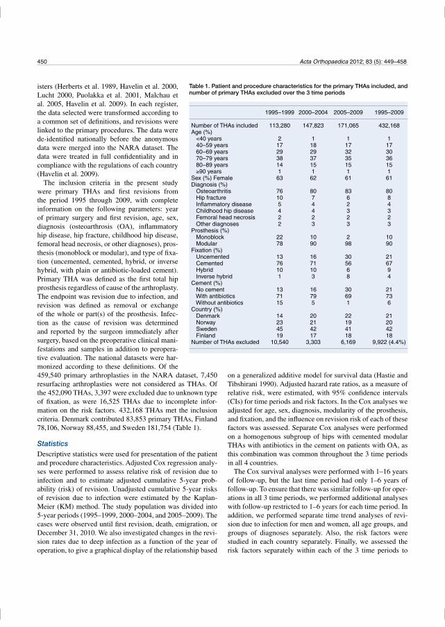
450 Acta Orthopaedica 2012; 83 (5): 449–458
459,540 primary arthroplasties in the NARA dataset, 7,450 resurfacing arthroplasties were not considered as THAs. Of the 452,090 THAs, 3,397 were excluded due to unknown type of fixation, as were 16,525 THAs due to incomplete infor-mation on the risk factors. 432,168 THAs met the inclusion criteria. Denmark contributed 83,853 primary THAs, Finland 78,106, Norway 88,455, and Sweden 181,754 (Table 1).
StatisticsDescriptive statistics were used for presentation of the patient and procedure characteristics. Adjusted Cox regression analy-ses were performed to assess relative risk of revision due to infection and to estimate adjusted cumulative 5-year prob-ability (risk) of revision. Unadjusted cumulative 5-year risks of revision due to infection were estimated by the Kaplan-Meier (KM) method. The study population was divided into 5-year periods (1995–1999, 2000–2004, and 2005–2009). The cases were observed until first revision, death, emigration, or December 31, 2010. We also investigated changes in the revi-sion rates due to deep infection as a function of the year of operation, to give a graphical display of the relationship based
on a generalized additive model for survival data (Hastie and Tibshirani 1990). Adjusted hazard rate ratios, as a measure of relative risk, were estimated, with 95% confidence intervals (CIs) for time periods and risk factors. In the Cox analyses we adjusted for age, sex, diagnosis, modularity of the prosthesis, and fixation, and the influence on revision risk of each of these factors was assessed. Separate Cox analyses were performed on a homogenous subgroup of hips with cemented modular THAs with antibiotics in the cement on patients with OA, as this combination was common throughout the 3 time periods in all 4 countries.
The Cox survival analyses were performed with 1–16 years of follow-up, but the last time period had only 1–6 years of follow-up. To ensure that there was similar follow-up for oper-ations in all 3 time periods, we performed additional analyses with follow-up restricted to 1–6 years for each time period. In addition, we performed separate time trend analyses of revi-sion due to infection for men and women, all age groups, and groups of diagnoses separately. Also, the risk factors were studied in each country separately. Finally, we assessed the risk factors separately within each of the 3 time periods to
Table 1. Patient and procedure characteristics for the primary THAs included, and number of primary THAs excluded over the 3 time periods
1995–1999 2000–2004 2005–2009 1995–2009
Number of THAs included 113,280 147,823 171,065 432,168Age (%) <40 years 2 1 1 1 40–59 years 17 18 17 17 60–69 years 29 29 32 30 70–79 years 38 37 35 36 80–89 years 14 15 15 15 90 years 1 1 1 1Sex (%) Female 63 62 61 61Diagnosis (%) Osteoarthritis 76 80 83 80 Hip fracture 10 7 6 8 Inflammatory disease 5 4 2 4 Childhood hip disease 4 4 3 3 Femoral head necrosis 2 2 2 2 Other diagnoses 2 3 3 3Prosthesis (%) Monoblock 22 10 2 10 Modular 78 90 98 90Fixation (%) Uncemented 13 16 30 21 Cemented 76 71 56 67 Hybrid 10 10 6 9 Inverse hybrid 1 3 8 4Cement (%) No cement 13 16 30 21 With antibiotics 71 79 69 73 Without antibiotics 15 5 1 6Country (%) Denmark 14 20 22 21 Norway 23 21 19 20 Sweden 45 42 41 42 Finland 19 17 18 18Number of THAs excluded 10,540 3,303 6,169 9,922 (4.4%)
isters (Herberts et al. 1989, Havelin et al. 2000, Lucht 2000, Puolakka et al. 2001, Malchau et al. 2005, Havelin et al. 2009). In each register, the data selected were transformed according to a common set of definitions, and revisions were linked to the primary procedures. The data were de-identified nationally before the anonymous data were merged into the NARA dataset. The data were treated in full confidentiality and in compliance with the regulations of each country (Havelin et al. 2009).
The inclusion criteria in the present study were primary THAs and first revisions from the period 1995 through 2009, with complete information on the following parameters: year of primary surgery and first revision, age, sex, diagnosis (osteoarthrosis (OA), inflammatory hip disease, hip fracture, childhood hip disease, femoral head necrosis, or other diagnoses), pros-thesis (monoblock or modular), and type of fixa-tion (uncemented, cemented, hybrid, or inverse hybrid, with plain or antibiotic-loaded cement). Primary THA was defined as the first total hip prosthesis regardless of cause of the arthroplasty. The endpoint was revision due to infection, and revision was defined as removal or exchange of the whole or part(s) of the prosthesis. Infec-tion as the cause of revision was determined and reported by the surgeon immediately after surgery, based on the preoperative clinical mani-festations and samples in addition to peropera-tive evaluation. The national datasets were har-monized according to these definitions. Of the
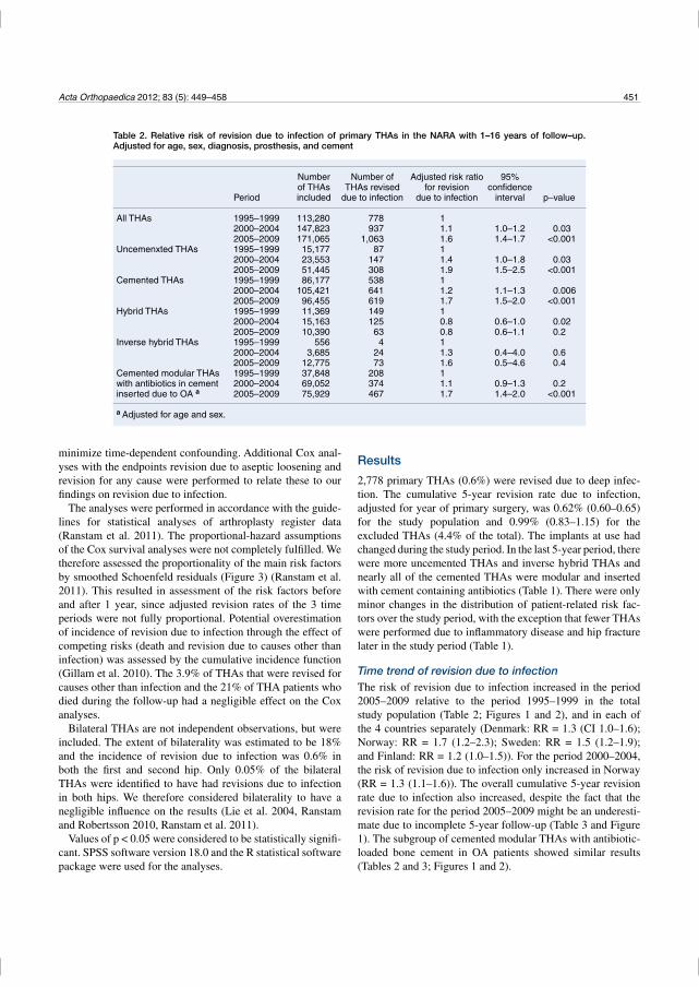
Acta Orthopaedica 2012; 83 (5): 449–458 451
minimize time-dependent confounding. Additional Cox anal-yses with the endpoints revision due to aseptic loosening and revision for any cause were performed to relate these to our findings on revision due to infection.
The analyses were performed in accordance with the guide-lines for statistical analyses of arthroplasty register data (Ranstam et al. 2011). The proportional-hazard assumptions of the Cox survival analyses were not completely fulfilled. We therefore assessed the proportionality of the main risk factors by smoothed Schoenfeld residuals (Figure 3) (Ranstam et al. 2011). This resulted in assessment of the risk factors before and after 1 year, since adjusted revision rates of the 3 time periods were not fully proportional. Potential overestimation of incidence of revision due to infection through the effect of competing risks (death and revision due to causes other than infection) was assessed by the cumulative incidence function (Gillam et al. 2010). The 3.9% of THAs that were revised for causes other than infection and the 21% of THA patients who died during the follow-up had a negligible effect on the Cox analyses.
Bilateral THAs are not independent observations, but were included. The extent of bilaterality was estimated to be 18% and the incidence of revision due to infection was 0.6% in both the first and second hip. Only 0.05% of the bilateral THAs were identified to have had revisions due to infection in both hips. We therefore considered bilaterality to have a negligible influence on the results (Lie et al. 2004, Ranstam and Robertsson 2010, Ranstam et al. 2011).
Values of p < 0.05 were considered to be statistically signifi-cant. SPSS software version 18.0 and the R statistical software package were used for the analyses.
Results
2,778 primary THAs (0.6%) were revised due to deep infec-tion. The cumulative 5-year revision rate due to infection, adjusted for year of primary surgery, was 0.62% (0.60–0.65) for the study population and 0.99% (0.83–1.15) for the excluded THAs (4.4% of the total). The implants at use had changed during the study period. In the last 5-year period, there were more uncemented THAs and inverse hybrid THAs and nearly all of the cemented THAs were modular and inserted with cement containing antibiotics (Table 1). There were only minor changes in the distribution of patient-related risk fac-tors over the study period, with the exception that fewer THAs were performed due to inflammatory disease and hip fracture later in the study period (Table 1).
Time trend of revision due to infectionThe risk of revision due to infection increased in the period 2005–2009 relative to the period 1995–1999 in the total study population (Table 2; Figures 1 and 2), and in each of the 4 countries separately (Denmark: RR = 1.3 (CI 1.0–1.6); Norway: RR = 1.7 (1.2–2.3); Sweden: RR = 1.5 (1.2–1.9); and Finland: RR = 1.2 (1.0–1.5)). For the period 2000–2004, the risk of revision due to infection only increased in Norway (RR = 1.3 (1.1–1.6)). The overall cumulative 5-year revision rate due to infection also increased, despite the fact that the revision rate for the period 2005–2009 might be an underesti-mate due to incomplete 5-year follow-up (Table 3 and Figure 1). The subgroup of cemented modular THAs with antibiotic-loaded bone cement in OA patients showed similar results (Tables 2 and 3; Figures 1 and 2).
Table 2. Relative risk of revision due to infection of primary THAs in the NARA with 1–16 years of follow–up. Adjusted for age, sex, diagnosis, prosthesis, and cement
Number Number of Adjusted risk ratio 95% of THAs THAs revised for revision confidence Period included due to infection due to infection interval p–value
All THAs 1995–1999 113,280 778 1 2000–2004 147,823 937 1.1 1.0–1.2 0.03 2005–2009 171,065 1,063 1.6 1.4–1.7 <0.001Uncemenxted THAs 1995–1999 15,177 87 1 2000–2004 23,553 147 1.4 1.0–1.8 0.03 2005–2009 51,445 308 1.9 1.5–2.5 <0.001Cemented THAs 1995–1999 86,177 538 1 2000–2004 105,421 641 1.2 1.1–1.3 0.006 2005–2009 96,455 619 1.7 1.5–2.0 <0.001Hybrid THAs 1995–1999 11,369 149 1 2000–2004 15,163 125 0.8 0.6–1.0 0.02 2005–2009 10,390 63 0.8 0.6–1.1 0.2Inverse hybrid THAs 1995–1999 556 4 1 2000–2004 3,685 24 1.3 0.4–4.0 0.6 2005–2009 12,775 73 1.6 0.5–4.6 0.4Cemented modular THAs 1995–1999 37,848 208 1with antibiotics in cement 2000–2004 69,052 374 1.1 0.9–1.3 0.2inserted due to OA a 2005–2009 75,929 467 1.7 1.4–2.0 <0.001
a Adjusted for age and sex.
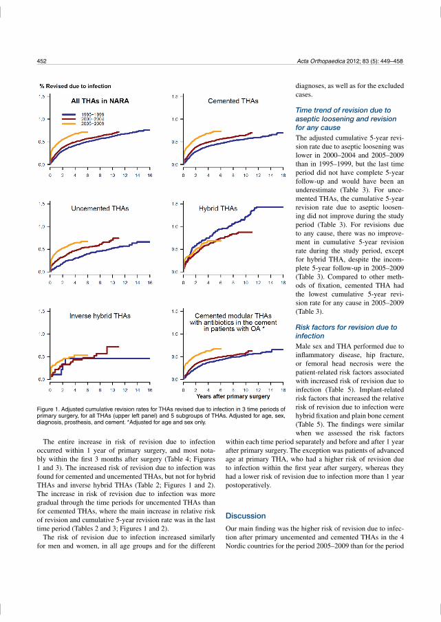
452 Acta Orthopaedica 2012; 83 (5): 449–458
The entire increase in risk of revision due to infection occurred within 1 year of primary surgery, and most nota-bly within the first 3 months after surgery (Table 4; Figures 1 and 3). The increased risk of revision due to infection was found for cemented and uncemented THAs, but not for hybrid THAs and inverse hybrid THAs (Table 2; Figures 1 and 2). The increase in risk of revision due to infection was more gradual through the time periods for uncemented THAs than for cemented THAs, where the main increase in relative risk of revision and cumulative 5-year revision rate was in the last time period (Tables 2 and 3; Figures 1 and 2).
The risk of revision due to infection increased similarly for men and women, in all age groups and for the different
within each time period separately and before and after 1 year after primary surgery. The exception was patients of advanced age at primary THA, who had a higher risk of revision due to infection within the first year after surgery, whereas they had a lower risk of revision due to infection more than 1 year postoperatively.
Discussion
Our main finding was the higher risk of revision due to infec-tion after primary uncemented and cemented THAs in the 4 Nordic countries for the period 2005–2009 than for the period
Figure 1. Adjusted cumulative revision rates for THAs revised due to infection in 3 time periods of primary surgery, for all THAs (upper left panel) and 5 subgroups of THAs. Adjusted for age, sex, diagnosis, prosthesis, and cement. *Adjusted for age and sex only.
diagnoses, as well as for the excluded cases.
Time trend of revision due to aseptic loosening and revision for any causeThe adjusted cumulative 5-year revi-sion rate due to aseptic loosening was lower in 2000–2004 and 2005–2009 than in 1995–1999, but the last time period did not have complete 5-year follow-up and would have been an underestimate (Table 3). For unce-mented THAs, the cumulative 5-year revision rate due to aseptic loosen-ing did not improve during the study period (Table 3). For revisions due to any cause, there was no improve-ment in cumulative 5-year revision rate during the study period, except for hybrid THA, despite the incom-plete 5-year follow-up in 2005–2009 (Table 3). Compared to other meth-ods of fixation, cemented THA had the lowest cumulative 5-year revi-sion rate for any cause in 2005–2009 (Table 3).
Risk factors for revision due to infectionMale sex and THA performed due to inflammatory disease, hip fracture, or femoral head necrosis were the patient-related risk factors associated with increased risk of revision due to infection (Table 5). Implant-related risk factors that increased the relative risk of revision due to infection were hybrid fixation and plain bone cement (Table 5). The findings were similar when we assessed the risk factors
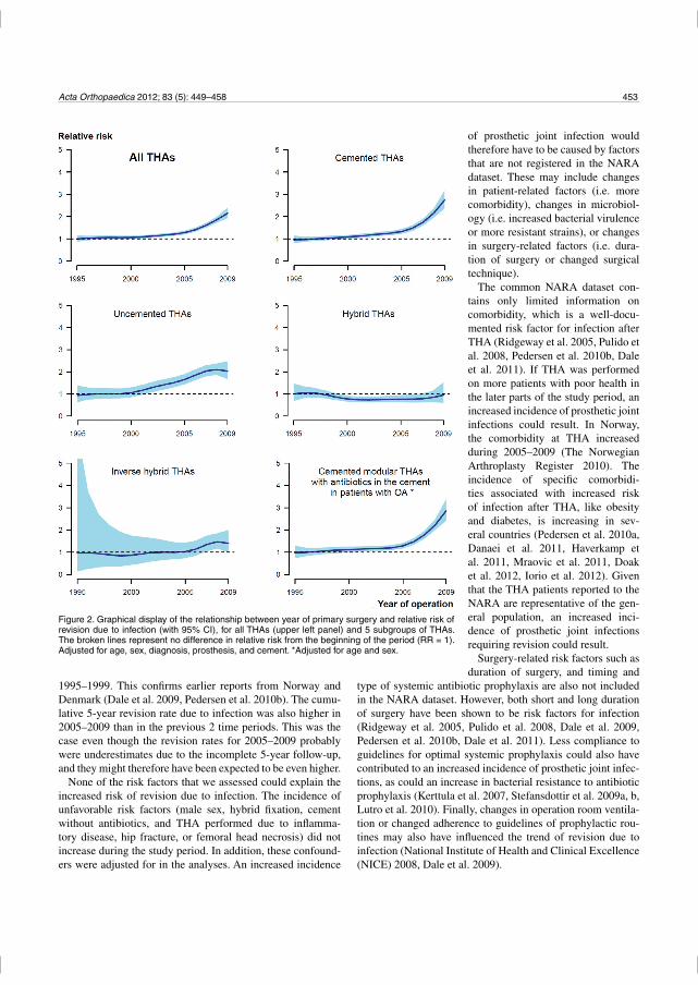
Acta Orthopaedica 2012; 83 (5): 449–458 453
1995–1999. This confirms earlier reports from Norway and Denmark (Dale et al. 2009, Pedersen et al. 2010b). The cumu-lative 5-year revision rate due to infection was also higher in 2005–2009 than in the previous 2 time periods. This was the case even though the revision rates for 2005–2009 probably were underestimates due to the incomplete 5-year follow-up, and they might therefore have been expected to be even higher.
None of the risk factors that we assessed could explain the increased risk of revision due to infection. The incidence of unfavorable risk factors (male sex, hybrid fixation, cement without antibiotics, and THA performed due to inflamma-tory disease, hip fracture, or femoral head necrosis) did not increase during the study period. In addition, these confound-ers were adjusted for in the analyses. An increased incidence
type of systemic antibiotic prophylaxis are also not included in the NARA dataset. However, both short and long duration of surgery have been shown to be risk factors for infection (Ridgeway et al. 2005, Pulido et al. 2008, Dale et al. 2009, Pedersen et al. 2010b, Dale et al. 2011). Less compliance to guidelines for optimal systemic prophylaxis could also have contributed to an increased incidence of prosthetic joint infec-tions, as could an increase in bacterial resistance to antibiotic prophylaxis (Kerttula et al. 2007, Stefansdottir et al. 2009a, b, Lutro et al. 2010). Finally, changes in operation room ventila-tion or changed adherence to guidelines of prophylactic rou-tines may also have influenced the trend of revision due to infection (National Institute of Health and Clinical Excellence (NICE) 2008, Dale et al. 2009).
Figure 2. Graphical display of the relationship between year of primary surgery and relative risk of revision due to infection (with 95% CI), for all THAs (upper left panel) and 5 subgroups of THAs. The broken lines represent no difference in relative risk from the beginning of the period (RR = 1). Adjusted for age, sex, diagnosis, prosthesis, and cement. *Adjusted for age and sex.
of prosthetic joint infection would therefore have to be caused by factors that are not registered in the NARA dataset. These may include changes in patient-related factors (i.e. more comorbidity), changes in microbiol-ogy (i.e. increased bacterial virulence or more resistant strains), or changes in surgery-related factors (i.e. dura-tion of surgery or changed surgical technique).
The common NARA dataset con-tains only limited information on comorbidity, which is a well-docu-mented risk factor for infection after THA (Ridgeway et al. 2005, Pulido et al. 2008, Pedersen et al. 2010b, Dale et al. 2011). If THA was performed on more patients with poor health in the later parts of the study period, an increased incidence of prosthetic joint infections could result. In Norway, the comorbidity at THA increased during 2005–2009 (The Norwegian Arthroplasty Register 2010). The incidence of specific comorbidi-ties associated with increased risk of infection after THA, like obesity and diabetes, is increasing in sev-eral countries (Pedersen et al. 2010a, Danaei et al. 2011, Haverkamp et al. 2011, Mraovic et al. 2011, Doak et al. 2012, Iorio et al. 2012). Given that the THA patients reported to the NARA are representative of the gen-eral population, an increased inci-dence of prosthetic joint infections requiring revision could result.
Surgery-related risk factors such as duration of surgery, and timing and
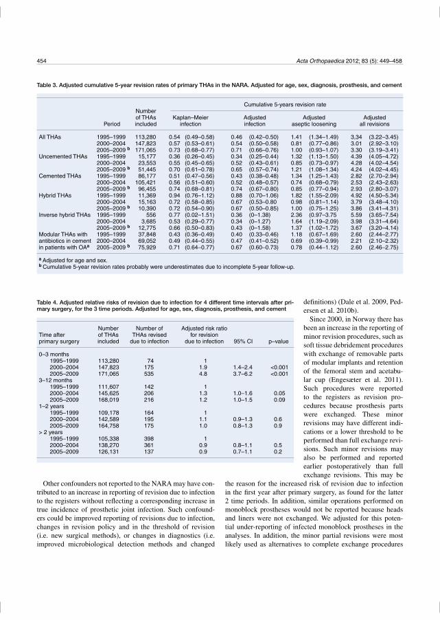
454 Acta Orthopaedica 2012; 83 (5): 449–458
Other confounders not reported to the NARA may have con-tributed to an increase in reporting of revision due to infection to the registers without reflecting a corresponding increase in true incidence of prosthetic joint infection. Such confound-ers could be improved reporting of revisions due to infection, changes in revision policy and in the threshold of revision (i.e. new surgical methods), or changes in diagnostics (i.e. improved microbiological detection methods and changed
the reason for the increased risk of revision due to infection in the first year after primary surgery, as found for the latter 2 time periods. In addition, similar operations performed on monoblock prostheses would not be reported because heads and liners were not exchanged. We adjusted for this poten-tial under-reporting of infected monoblock prostheses in the analyses. In addition, the minor partial revisions were most likely used as alternatives to complete exchange procedures
Table 3. Adjusted cumulative 5-year revision rates of primary THAs in the NARA. Adjusted for age, sex, diagnosis, prosthesis, and cement
Cumulative 5-years revision rate Number of THAs Kaplan–Meier Adjusted Adjusted Adjusted Period included infection infection aseptic loosening all revisions
All THAs 1995–1999 113,280 0.54 (0.49–0.58) 0.46 (0.42–0.50) 1.41 (1.34–1.49) 3,34 (3.22–3.45) 2000–2004 147,823 0.57 (0.53–0.61) 0.54 (0.50–0.58) 0.81 (0.77–0.86) 3.01 (2.92–3.10) 2005–2009 b 171,065 0.73 (0.68–0.77) 0.71 (0.66–0.76) 1.00 (0.93–1.07) 3.30 (3.19–3.41)Uncemented THAs 1995–1999 15,177 0.36 (0.26–0.45) 0.34 (0.25–0.44) 1.32 (1.13–1.50) 4.39 (4.05–4.72) 2000–2004 23,553 0.55 (0.45–0.65) 0.52 (0.43–0.61) 0.85 (0.73–0.97) 4.28 (4.02–4.54) 2005–2009 b 51,445 0.70 (0.61–0.78) 0.65 (0.57–0.74) 1.21 (1.08–1.34) 4.24 (4.02–4.45)Cemented THAs 1995–1999 86,177 0.51 (0.47–0.56) 0.43 (0.38–0.48) 1.34 (1.25–1.43) 2.82 (2.70–2.94) 2000–2004 105,421 0.56 (0.51–0.60) 0.52 (0.48–0.57) 0.74 (0.68–0.79) 2.53 (2.43–2.63) 2005–2009 b 96,455 0.74 (0.68–0.81) 0.74 (0.67–0.80) 0.85 (0.77–0.94) 2.93 (2.80–3.07)Hybrid THAs 1995–1999 11,369 0.94 (0.76–1.12) 0.88 (0.70–1.06) 1.82 (1.55–2.09) 4.92 (4.50–5.34) 2000–2004 15,163 0.72 (0.58–0.85) 0.67 (0.53–0.80 0.98 (0.81–1.14) 3.79 (3.48–4.10) 2005–2009 b 10,390 0.72 (0.54–0.90) 0.67 (0.50–0.85) 1.00 (0.75–1.25) 3.86 (3.41–4.31)Inverse hybrid THAs 1995–1999 556 0.77 (0.02–1.51) 0.36 (0–1.38) 2.36 (0.97–3.75 5.59 (3.65–7.54) 2000–2004 3,685 0.53 (0.29–0.77) 0.34 (0–1.27) 1.64 (1.19–2.09) 3.98 (3.31–4.64) 2005–2009 b 12,775 0.66 (0.50–0.83) 0.43 (0–1.58) 1.37 (1.02–1.72) 3.67 (3.20–4.14)Modular THAs with 1995–1999 37,848 0.43 (0.36–0.49) 0.40 (0.33–0.46) 1.18 (0.67–1.69) 2.60 (2.44–2.77)antibiotics in cement 2000–2004 69,052 0.49 (0.44–0.55) 0.47 (0.41–0.52) 0.69 (0.39–0.99) 2.21 (2.10–2.32)in patients with OAa 2005–2009 b 75,929 0.71 (0.64–0.77) 0.67 (0.60–0.73) 0.78 (0.44–1.12) 2.60 (2.46–2.75)
a Adjusted for age and sex.b Cumulative 5-year revision rates probably were underestimates due to incomplete 5-year follow-up.
Table 4. Adjusted relative risks of revision due to infection for 4 different time intervals after pri-mary surgery, for the 3 time periods. Adjusted for age, sex, diagnosis, prosthesis, and cement
Number Number of Adjusted risk ratioTime after of THAs THAs revised for revision primary surgery included due to infection due to infection 95% CI p–value
0–3 months 1995–1999 113,280 74 1 2000–2004 147,823 175 1.9 1.4–2.4 <0.001 2005–2009 171,065 535 4.8 3.7–6.2 <0.0013–12 months 1995–1999 111,607 142 1 2000–2004 145,625 206 1.3 1.0–1.6 0.05 2005–2009 168,019 216 1.2 1.0–1.5 0.091–2 years 1995–1999 109,178 164 1 2000–2004 142,589 195 1.1 0.9–1.3 0.6 2005–2009 164,758 175 1.0 0.8–1.3 0.9> 2 years 1995–1999 105,338 398 1 2000–2004 138,270 361 0.9 0.8–1.1 0.5 2005–2009 126,131 137 0.9 0.7–1.1 0.2
definitions) (Dale et al. 2009, Ped-ersen et al. 2010b).
Since 2000, in Norway there has been an increase in the reporting of minor revision procedures, such as soft tissue debridement procedures with exchange of removable parts of modular implants and retention of the femoral stem and acetabu-lar cup (Engesæter et al. 2011). Such procedures were reported to the registers as revision pro-cedures because prosthesis parts were exchanged. These minor revisions may have different indi-cations or a lower threshold to be performed than full exchange revi-sions. Such minor revisions may also be performed and reported earlier postoperatively than full exchange revisions. This may be
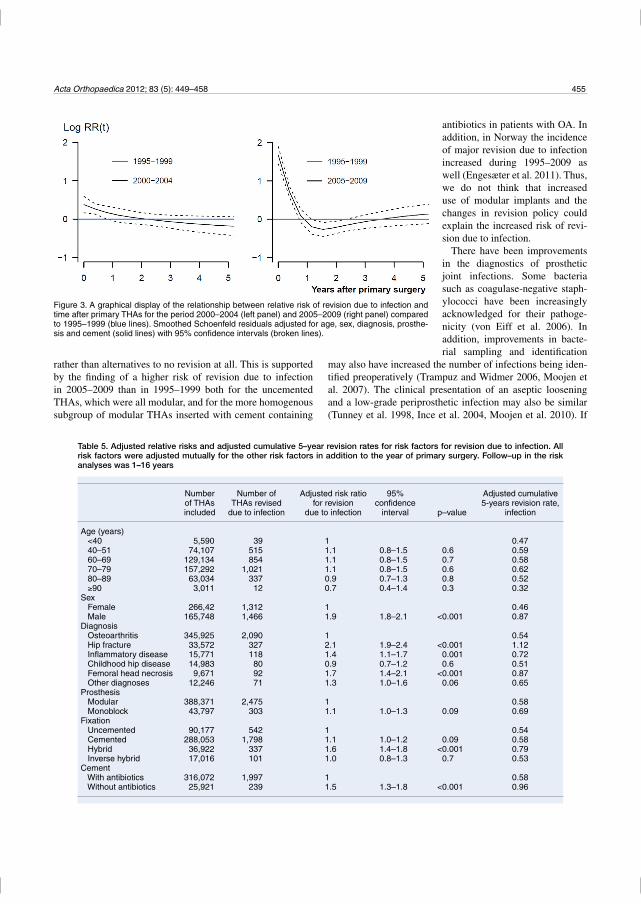
Acta Orthopaedica 2012; 83 (5): 449–458 455
rather than alternatives to no revision at all. This is supported by the finding of a higher risk of revision due to infection in 2005–2009 than in 1995–1999 both for the uncemented THAs, which were all modular, and for the more homogenous subgroup of modular THAs inserted with cement containing
may also have increased the number of infections being iden-tified preoperatively (Trampuz and Widmer 2006, Moojen et al. 2007). The clinical presentation of an aseptic loosening and a low-grade periprosthetic infection may also be similar (Tunney et al. 1998, Ince et al. 2004, Moojen et al. 2010). If
Figure 3. A graphical display of the relationship between relative risk of revision due to infection and time after primary THAs for the period 2000–2004 (left panel) and 2005–2009 (right panel) compared to 1995–1999 (blue lines). Smoothed Schoenfeld residuals adjusted for age, sex, diagnosis, prosthe-sis and cement (solid lines) with 95% confidence intervals (broken lines).
antibiotics in patients with OA. In addition, in Norway the incidence of major revision due to infection increased during 1995–2009 as well (Engesæter et al. 2011). Thus, we do not think that increased use of modular implants and the changes in revision policy could explain the increased risk of revi-sion due to infection.
There have been improvements in the diagnostics of prosthetic joint infections. Some bacteria such as coagulase-negative staph-ylococci have been increasingly acknowledged for their pathoge-nicity (von Eiff et al. 2006). In addition, improvements in bacte-rial sampling and identification
Table 5. Adjusted relative risks and adjusted cumulative 5–year revision rates for risk factors for revision due to infection. All risk factors were adjusted mutually for the other risk factors in addition to the year of primary surgery. Follow–up in the risk analyses was 1–16 years
Number Number of Adjusted risk ratio 95% Adjusted cumulative of THAs THAs revised for revision confidence 5-years revision rate, included due to infection due to infection interval p–value infection
Age (years) <40 5,590 39 1 0.47 40–51 74,107 515 1.1 0.8–1.5 0.6 0.59 60–69 129,134 854 1.1 0.8–1.5 0.7 0.58 70–79 157,292 1,021 1.1 0.8–1.5 0.6 0.62 80–89 63,034 337 0.9 0.7–1.3 0.8 0.52 90 3,011 12 0.7 0.4–1.4 0.3 0.32Sex Female 266,42 1,312 1 0.46 Male 165,748 1,466 1.9 1.8–2.1 <0.001 0.87Diagnosis Osteoarthritis 345,925 2,090 1 0.54 Hip fracture 33,572 327 2.1 1.9–2.4 <0.001 1.12 Inflammatory disease 15,771 118 1.4 1.1–1.7 0.001 0.72 Childhood hip disease 14,983 80 0.9 0.7–1.2 0.6 0.51 Femoral head necrosis 9,671 92 1.7 1.4–2.1 <0.001 0.87 Other diagnoses 12,246 71 1.3 1.0–1.6 0.06 0.65Prosthesis Modular 388,371 2,475 1 0.58 Monoblock 43,797 303 1.1 1.0–1.3 0.09 0.69Fixation Uncemented 90,177 542 1 0.54 Cemented 288,053 1,798 1.1 1.0–1.2 0.09 0.58 Hybrid 36,922 337 1.6 1.4–1.8 <0.001 0.79 Inverse hybrid 17,016 101 1.0 0.8–1.3 0.7 0.53Cement With antibiotics 316,072 1,997 1 0.58 Without antibiotics 25,921 239 1.5 1.3–1.8 <0.001 0.96
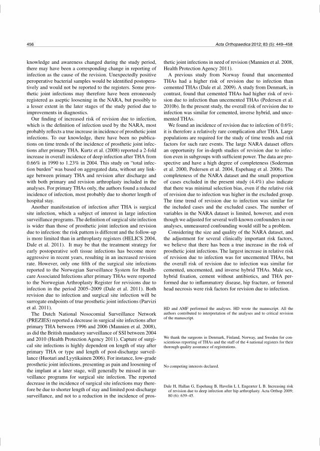
456 Acta Orthopaedica 2012; 83 (5): 449–458
thetic joint infections in need of revision (Mannien et al. 2008, Health Protection Agency 2011).
A previous study from Norway found that uncemented THAs had a higher risk of revision due to infection than cemented THAs (Dale et al. 2009). A study from Denmark, in contrast, found that cemented THAs had higher risk of revi-sion due to infection than uncemented THAs (Pedersen et al. 2010b). In the present study, the overall risk of revision due to infection was similar for cemented, inverse hybrid, and unce-mented THAs.
We found an incidence of revision due to infection of 0.6%; it is therefore a relatively rare complication after THA. Large populations are required for the study of time trends and risk factors for such rare events. The large NARA dataset offers an opportunity for in-depth studies of revision due to infec-tion even in subgroups with sufficient power. The data are pro-spective and have a high degree of completeness (Soderman et al. 2000, Pedersen et al. 2004, Espehaug et al. 2006). The completeness of the NARA dataset and the small proportion of cases excluded in the present study (4.4%) also indicate that there was minimal selection bias, even if the relative risk of revision due to infection was higher in the excluded group. The time trend of revision due to infection was similar for the included cases and the excluded cases. The number of variables in the NARA dataset is limited, however, and even though we adjusted for several well-known confounders in our analyses, unmeasured confounding would still be a problem.
Considering the size and quality of the NARA dataset, and the adjustment for several clinically important risk factors, we believe that there has been a true increase in the risk of prosthetic joint infections. The largest increase in relative risk of revision due to infection was for uncemented THAs, but the overall risk of revision due to infection was similar for cemented, uncemented, and inverse hybrid THAs. Male sex, hybrid fixation, cement without antibiotics, and THA per-formed due to inflammatory disease, hip fracture, or femoral head necrosis were risk factors for revision due to infection.
HD and AMF performed the analyses. HD wrote the manuscript. All the authors contributed to interpretation of the analyses and to critical revision of the manuscript.
We thank the surgeons in Denmark, Finland, Norway, and Sweden for con-scientious reporting of THAs and the staff of the 4 national registers for their thorough quality assurance of registrations.
No competing interests declared.
Dale H, Hallan G, Espehaug B, Havelin L I, Engeæter L B. Increasing risk of revision due to deep infection after hip arthroplasty. Acta Orthop 2009; 80 (6): 639–45.
knowledge and awareness changed during the study period, there may have been a corresponding change in reporting of infection as the cause of the revision. Unexpectedly positive peroperative bacterial samples would be identified postopera-tively and would not be reported to the registers. Some pros-thetic joint infections may therefore have been erroneously registered as aseptic loosening in the NARA, but possibly to a lesser extent in the later stages of the study period due to improvements in diagnostics.
Our finding of increased risk of revision due to infection, which is the definition of infection used by the NARA, most probably reflects a true increase in incidence of prosthetic joint infections. To our knowledge, there have been no publica-tions on time trends of the incidence of prosthetic joint infec-tions after primary THA. Kurtz et al. (2008) reported a 2-fold increase in overall incidence of deep infection after THA from 0.66% in 1990 to 1.23% in 2004. This study on “total infec-tion burden” was based on aggregated data, without any link-age between primary THA and revision after discharge and with both primary and revision arthroplasty included in the analyses. For primary THAs only, the authors found a reduced incidence of infection, most probably due to shorter length of hospital stay.
Another manifestation of infection after THA is surgical site infection, which a subject of interest in large infection surveillance programs. The definition of surgical site infection is wider than those of prosthetic joint infection and revision due to infection: the risk pattern is different and the follow-up is more limited than in arthroplasty registers (HELICS 2004, Dale et al. 2011). It may be that the treatment strategy for early postoperative soft tissue infections has become more aggressive in recent years, resulting in an increased revision rate. However, only one fifth of the surgical site infections reported to the Norwegian Surveillance System for Health-care Associated Infections after primary THAs were reported to the Norwegian Arthroplasty Register for revisions due to infection in the period 2005–2009 (Dale et al. 2011). Both revision due to infection and surgical site infection will be surrogate endpoints of true prosthetic joint infections (Parvizi et al. 2011).
The Dutch National Nosocomial Surveillance Network (PREZIES) reported a decrease in surgical site infections after primary THA between 1996 and 2006 (Mannien et al. 2008), as did the British mandatory surveillance of SSI between 2004 and 2010 (Health Protection Agency 2011). Capture of surgi-cal site infections is highly dependent on length of stay after primary THA or type and length of post-discharge surveil-lance (Huotari and Lyytikainen 2006). For instance, low-grade prosthetic joint infections, presenting as pain and loosening of the implant at a later stage, will generally be missed in sur-veillance programs for surgical site infection. The reported decrease in the incidence of surgical site infections may there-fore be due to shorter length of stay and limited post-discharge surveillance, and not to a reduction in the incidence of pros-
Acta Orthopaedica 2012; 83 (5): 449–458 457
Dale H, Skramm I, Lower H L, Eriksen H M, Espehaug B, Furnes O, Skjeldestad F E, Havelin L I, Engesaeter L B. Infection after primary hip arthroplasty. Acta Orthop 2011; 82 (6): 646–54.
Danaei G, Finucane M M, Lu Y, Singh G M, Cowan M J, Paciorek C J, Lin J K, Farzadfar F, Khang Y H, Stevens G A, Rao M, Ali M K, Riley L M, Robinson C A, Ezzati M. National, regional, and global trends in fasting plasma glucose and diabetes prevalence since 1980: systematic analysis of health examination surveys and epidemiological studies with 370 country-years and 2.7 million participants. Lancet 2011; 378 (9785): 31–40.
Doak C M, Wijnhoven T M, Schokker D F, Visscher T L, Seidell J C. Age standardization in mapping adult overweight and obesity trends in the WHO European Region. Obes Rev 2012; 13 (2): 174–91.
Engesæter L B, Dale H, Schrama J C, Hallan G, Lie S A. Surgical proce-dures in the treatment of 784 infected THAs reported to the Norwegian Arthroplasty Register. Acta Orthop 2011; 82 (5): 530–7.
Espehaug B, Furnes O, Havelin L I, Engesæter L B, Vollset S E, Kindseth O. Registration completeness in the Norwegian Arthroplasty Register. Acta Orthop 2006; 77 (1): 49–56.
Fevang B T, Lie S A, Havelin L I, Engesaeter L B, Furnes O. Improved results of primary total hip replacement. Acta Orthop 2010; 81 (6): 649–59.
Gillam M H, Ryan P, Graves S E, Miller L N, de Steiger R N, Salter A. Com-peting risks survival analysis applied to data from the Australian Orthopae-dic Association National Joint Replacement Registry. Acta Orthop 2010; 81 (5): 548–55.
Hastie T J, Tibshirani R J. Generalized additive models. Chapman & Hall, London 1990.
Havelin L I, Engesæter L B, Espehaug B, Furnes O, Lie S A, Vollset S E. The Norwegian Arthroplasty Register: 11 years and 73,000 arthroplasties. Acta Orthop Scand 2000; 71 (4): 337–53.
Havelin L I, Fenstad A M, Salomonsson R, Mehnert F, Furnes O, Over-gaard S, Pedersen A B, Herberts P, Karrholm J, Garellick G. The Nordic Arthroplasty Register Association: a unique collaboration between 3 national hip arthroplasty registries with 280,201 THRs. Acta Orthop 2009; 80 (4): 393–401.
Haverkamp D, Klinkenbijl M N, Somford M P, Albers G H, van der Vis H M. Obesity in total hip arthroplasty--does it really matter? A meta-analysis. Acta Orthop 2011; 82 (4): 417–22.
Health Protection Agency. Sixth report of the mandatory surveillance of surgical site infection in orthopaedic surgery: April 2004 to March 2010. 2011. Available from http://www.hpa.org.uk/webc/HPAwebFile/HPAweb_C/1287147699571
HELICS. Hospital in Europe Link for Infection Control through Surveillance Surgical Site Infection, Surveillance of Surgical Site Infections, Protocol. European Centre for Disease Prevention and Control, 2004. Available from http://helics.univ-lyon1.fr/
Herberts P, Malchau H. Long-term registration has improved the quality of hip replacement: a review of the Swedish THR Register comparing 160,000 cases. Acta Orthop Scand 2000; 71 (2): 111–21.
Herberts P, Ahnfelt L, Malchau H, Stromberg C, Andersson G B. Multicenter clinical trials and their value in assessing total joint arthroplasty. Clin Orthop 1989; (249): 48–55.
Huotari K, Lyytikainen O. Impact of postdischarge surveillance on the rate of surgical site infection after orthopedic surgery. Infect Control Hosp Epide-miol 2006; 27 (12): 1324–9.
Ince A, Rupp J, Frommelt L, Katzer A, Gille J, Lohr J F. Is “aseptic” loosen-ing of the prosthetic cup after total hip replacement due to nonculturable bacterial pathogens in patients with low-grade infection? Clin Infect Dis 2004; 39 (11): 1599–603.
Iorio R, Williams K M, Marcantonio A J, Specht L M, Tilzey J F, Healy W L. Diabetes mellitus, hemoglobin A1C, and the incidence of total joint arthroplasty infection. J Arthroplasty 2012; 27 (5): 726–9.
Kerttula A M, Lyytikainen O, Karden-Lilja M, Ibrahem S, Salmenlinna S, Virolainen A, Vuopio-Varkila J. Nationwide trends in molecular epidemi-ology of methicillin-resistant Staphylococcus aureus, Finland, 1997–2004. BMC Infect Dis 2007; 7: 94.
Kurtz S M, Lau E, Schmier J, Ong K L, Zhao K, Parvizi J. Infection Burden for Hip and Knee Arthroplasty in the United States. J Arthroplasty 2008; 23 (7): 984–91.
Lie S A, Engesæter L B, Havelin L I, Gjessing H K, Vollset S E. Dependency issues in survival analyses of 55,782 primary hip replacements from 47,355 patients. Stat Med 2004; 23 (20): 3227–40.
Liu S S, Della Valle A G, Besculides M C, Gaber L K, Memtsoudis S G. Trends in mortality, complications, and demographics for primary hip arthroplasty in the United States. Int Orthop 2009; 33 (3): 643–51.
Lucht U. The Danish Hip Arthroplasty Register. Acta Orthop Scand 2000; 71 (5): 433–9.
Lutro O, Langvatn H, Schrama J, Hallan G, Dale H, Espehaug B, Sjursen H, Engesæter L. Resistance of staphylococci isolated from infected hip arthroplasties in Norway. NOF Congress 2010 . 2010. Available from http://www.centraloffice-europe.com/nof2010/detail.asp?id=109
Malchau H, Garellick G, Eisler T, Karrholm J, Herberts P. Presidential guest address: the Swedish Hip Registry: increasing the sensitivity by patient outcome data. Clin Orthop 2005; (441): 19–29.
Mannien J, van den H S, Muilwijk J, van den Broek P J, van Benthem B, Wille J C. Trends in the incidence of surgical site infection in the Netherlands. Infect Control Hosp Epidemiol 2008; 29 (12): 1132–8.
Moojen D J, Spijkers S N, Schot C S, Nijhof M W, Vogely H C, Fleer A, Ver-bout A J, Castelein R M, Dhert W J, Schouls L M. Identification of ortho-paedic infections using broad-range polymerase chain reaction and reverse line blot hybridization. J Bone Joint Surg (Am) 2007; 89 (6): 1298–305.
Moojen D J, van H G, Vogely H C, Burger B J, Walenkamp G H, Tulp N J, Schreurs B W, de Meulemeester F R, Schot C S, van d P, I, Fujishiro T, Schouls L M, Bauer T W, Dhert W J. Incidence of low-grade infection in aseptic loosening of total hip arthroplasty. Acta Orthop 2010; 81 (6): 667–73.
Mraovic B, Suh D, Jacovides C, Parvizi J. Perioperative hyperglycemia and postoperative infection after lower limb arthroplasty. J Diabetes Sci Tech-nol 2011; 5 (2): 412–8.
National Institute of Health and Clinical Excellence (NICE). Surgical site infection, prevention and treatment of surgical site infection, clinical guideline . CG74. 2008. Available from www.nice.org.uk/nicemedia/pdf/CG74FullGuideline.pdf
Parvizi J, Zmistowski B, Berbari E F, Bauer T W, Springer B D, Della Valle C J, Garvin K L, Mont M A, Wongworawat M D, Zalavras C G. New defini-tion for periprosthetic joint infection: from the Workgroup of the Musculo-skeletal Infection Society. Clin Orthop 2011; (469) (11): 2992–4.
Pedersen A, Johnsen S, Overgaard S, Soballe K, Sorensen H T, Lucht U. Reg-istration in the Danish Hip Arthroplasty Registry: completeness of total hip arthroplasties and positive predictive value of registered diagnosis and postoperative complications. Acta Orthop Scand 2004; 75 (4): 434–41.
Pedersen A B, Mehnert F, Johnsen S P, Sorensen H T. Risk of revision of a total hip replacement in patients with diabetes mellitus: a population-based follow up study. J Bone Joint Surg (Br) 2010a; 92 (7): 929–34.
Pedersen A B, Svendsson J E, Johnsen S P, Riis A, Overgaard S. Risk fac-tors for revision due to infection after primary total hip arthroplasty. A population-based study of 80,756 primary procedures in the Danish Hip Arthroplasty Registry. Acta Orthop 2010b; 81 (5): 542–7.
Pulido L, Ghanem E, Joshi A, Purtill J J, Parvizi J. Periprosthetic joint infec-tion: the incidence, timing, and predisposing factors. Clin Orthop 2008; (466) (7): 1710–5.
Puolakka T J, Pajamaki K J, Halonen P J, Pulkkinen P O, Paavolainen P, Nevalainen J K. The Finnish Arthroplasty Register: report of the hip regis-ter. Acta Orthop Scand 2001; 72 (5): 433–41.
Ranstam J, Robertsson O. Statistical analysis of arthroplasty register data. Acta Orthop 2010; 81 (1): 10–4.
Ranstam J, Karrholm J, Pulkkinen P, Makela K, Espehaug B, Pedersen A B, Mehnert F, Furnes O. Statistical analysis of arthroplasty data. II. Guide-lines. Acta Orthop 2011; 82 (3): 258–67.
Ridgeway S, Wilson J, Charlet A, Kafatos G, Pearson A, Coello R. Infection of the surgical site after arthroplasty of the hip. J Bone Joint Surg (Br) 2005; 87 (6): 844–50.

458 Acta Orthopaedica 2012; 83 (5): 449–458
Soderman P, Malchau H, Herberts P, Johnell O. Are the findings in the Swed-ish National Total Hip Arthroplasty Register valid? A comparison between the Swedish National Total Hip Arthroplasty Register, the National Dis-charge Register, and the National Death Register. J Arthroplasty 2000; 15 (7): 884–9.
Stefansdottir A, Johansson D, Knutson K, Lidgren L, Robertsson O. Micro-biology of the infected knee arthroplasty: report from the Swedish Knee Arthroplasty Register on 426 surgically revised cases. Scand J Infect Dis 2009a; 41 (11–12): 831–40.
Stefansdottir A, Robertsson O, Dahl A, Kiernan S, Gustafson P, Lidgren L. Inadequate timing of prophylactic antibiotics in orthopedic surgery. We can do better. Acta Orthop 2009b; 80 (6): 633–8.
The Norwegian Arthroplasty Register. Annual Report 2010. The Norwegian Arthroplasty Register, 2010. Available from http://nrlweb.ihelse.net/eng/Report_2010.pdf
Trampuz A, Widmer A F. Infections associated with orthopedic implants. Curr Opin Infect Dis 2006; 19 (4): 349–56.
Tunney M M, Patrick S, Gorman S P, Nixon J R, Anderson N, Davis R I, Hanna D, Ramage G. Improved detection of infection in hip replacements. A currently underestimated problem. J Bone Joint Surg (Br) 1998; 80 (4): 568–72.
von Eiff C, Arciola C R, Montanaro L, Becker K, Campoccia D. Emerging Staphylococcus species as new pathogens in implant infections. Int J Artif Organs 2006; 29 (4): 360–7.
Copyright © 2022 FDOKUMEN