Infection after primary hip arthroplasty - Bergen Open Research ...
Upload
independentCategory
view
0download
0
15
Evidence Linking Elevated Oxidative Stress and Aseptic Loosening
of Hip Arthroplasty Plamen Kinov and Peter Tivchev
Queen Giovanna – ISUL University Hospital, Department of Orthopedics and Traumatology, Medical University of Sofia
Bulgaria
1. Introduction Aseptic loosening is still the most common late complication after total hip arthroplasty (THA) and one of the main reasons for its failure. Artificial joints are made of metallic, polymeric and ceramic components. In the process of prosthesis functioning in the recipient’s body implant materials are subject to wear and fretting as well as of influence of aggressive biological fluids. Wear debris particles, corrosion products and metal ions from the bearing and contact surfaces of the implant are released in the periprosthetic tissues. As a result of the processes taking part at the implant-bone interface osteolysis develops with subsequent loosening of the implant. Today, the most widely used bearing surface is a metal femoral head made of cobalt chromium molybdenum alloy coupled with a polymeric inlay fabricated from ultra-high molecular weight polyethylene (UHMWPE). For decades in clinical use, metal on polyethylene (MoPE) bearings in total hip arthroplasty provided consistent results. Despite the widespread use of UHMWPE as a bearing surface its wear is the main obstacle restricting the longevity of the artificial joint. With an average rate of polyethylene (PE) wear of 0.1mm per year, 100 million UHMWPE particles (assumed diameter of 1 μm) are liberated into the joint space on a daily basis (Muratoglu & Kurtz, 2002). It is now well established that cyclic mechanical loading, production of wear particles, and the ensuing cascade of adverse tissue response are all significant contributors to local osteolysis at the prosthesis–bone interface and in certain cases loosening of the prosthesis (Aspenberg & Herbertsson, 1996, Goldring et al., 1986, Schmalzried et al., 1992, Willert & Semtlitsch, 1977). Deterioration of clinical results with time and eventually revision of the arthroplasty were a very strong impetus for the search for the “ideal bearing” (Muratoglu & Kurtz, 2002). Improving longevity of the total hip arthroplasty by engineering of new bearing couples with improved biomechanical characteristics and lower rate of wear has been the main line of ongoing research in the orthopedic community. Today, orthopedic surgeons have a wide choice of implants and bearing couples for a particular patient. The new generation of joint bearings provides significantly lower wear rates and is anticipated to diminish the incidence of osteolysis and subsequent revisions.

Recent Advances in Arthroplasty
296
Next to development of new bearing surfaces, the second line of research focuses on understanding of the underlying mechanisms of the process aseptic loosening of THA. Aseptic loosening is characterized with osteolysis and formation of thick membrane around the prostheses that eventually leads to its loosening. Currently, it is well established fact that loosening is a result of host response to wear debris and corrosion products of implant materials. Particles can readily be detected in the periprosthetic tissues as well as at remote locations such as lymph nodes, liver, spleen or bone marrow. Wear debris induce inflammation in the periprosthetic tissues that is sustained through the functioning of the implant as long as wear particles are produced. Metal wear debris, degradation and corrosion products, such as ions and reactive oxygen species (ROS), has been considered to be crucial factors in the process of loosening leading to the failure of metal implants (Tsaryk, 2009). Metal debris could induce inflammatory responses mediated by neutrophils, macrophages, fibroblasts and other cells. Metal ions and corrosion products are potentially toxic, can cause allergic reactions of hypersensitivity, chromosomal aberrations, and eventually malignancy (Keegan et al., 2007). Elevated oxidative stress has been proposed to be a causative factor in many inflammatory and degenerative disorders with tissue damage and fibrosis in different organs and systems (Hogg, 1998, Park et al., 2001). In addition, recent in vitro studies showed the combined effect of particles and macrophage and osteoclast activation on the increase of oxidative stress (Fleury et al., 2006, Petit et al., 2005, Wei et al., 2009). This suggests involvement of reactive oxygen species (ROS) in the mechanism of aseptic loosening of hip arthroplasty. The chronic inflammation state with the elevated oxidative stress results in extensive formation of granulation tissue and fibrous capsule, periprosthetic bone resorption due to osteoclast activation by inflammatory stimuli and finally aseptic loosening of the implant (Tsaryk, 2009). In support of this hypothesis we proved the involvement of ROS in excessive fibrosis around loose hip prostheses (Kinov et al., 2006). This suggests involvement of reactive oxygen species in the mechanism of aseptic loosening of hip arthroplasty leading to formation of the fibrous pseudocapsule that typically consists of a combination of fibrous tissue and macrophages. However, the mechanisms of involvement of ROS in aseptic loosening of THA are still to be elucidated. Some researchers further implicated that free radicals may be involved in the induction and maintenance of chronic inflammation with resulting periprosthetic bone resorption. In support of this hypothesis, recent studies on osteoporosis (Hamel et al., 2008, Li et al., 2009) show that elevated oxidative stress is involved in inhibiting osteoblastic differentiation and stimulating osteoclastogenesis. In addition, in vitro study showed the combined effect of particles and macrophage and osteoclast activation on release of reactive oxygen and nitrogen species (Wang et al., 2002). Different studies investigate the mechanisms of action of oxidative stress on bone formation (Bai et al., 2004, 2005, Chen et al., 2010, Kim et al., 2010, Mazière et al., 2010, Rached et al., 2010). However, the exact mechanism and actions of ROS on inhibition of osteoblasts are still largely unknown. Considering the fact that elevated oxidative stress induces bone loss in postmenopausal osteoporosis (Lean et al., 2005) whereas antioxidants suppress osteoclast activity and enhance differentiation of osteoblasts (Aitken et al., 2004, Mody et al., 2001) it is possible that ROS are involved in aseptic loosening of total hip arthroplasty. In support of this, in two previous studies, we have shown direct evidence for involvement of elevated oxidative stress in aseptic loosening of THA (Kinov et al., 2006, 2010).

Evidence Linking Elevated Oxidative Stress and Aseptic Loosening of Hip Arthroplasty
297
2. Materials for bearings of hip prostheses Materials are very important for the long-term success of THA. Since the introduction of low friction arthroplasty (LFA) by Sir John Charnley in the 1960’s, much has been learned about the durability and biocompatibility of materials used in joint replacement. Concerns about higher rates of aseptic loosening and subsequent prosthesis revision among young and active patients have lead to the development of new implant designs and alternative bearings (Muratoglu & Kurtz, 2002). Strength and endurance, friction and wear properties, inertness and biocompatibility of the materials should be optimized in order to eliminate or diminish to a negligible extent the reaction of the organism to wear debris.
2.1 Brief historical remarks Aseptic loosening has been observed since the beginning of hip replacement. Metal bearings were first introduced by Wiles in the 1930s (Wiles, 1957), but they received wider application in the 1950s and 1960s with the pioneer works of McKee-Farrar and Ring (McKee & Watson-Farrar, 1966, Ring, 1967). The earlier prototypes were manufactured of stainless steel and fracture of the prostheses was a frequent complication (McKee & Watson-Farrar, 1966, Ring, 1967). To solve this problem the cobalt chromium molybdenum (CoCrMo/CoCrMo) articulation was developed. However, the metal-on-metal (MoM) bearing was eventually abandoned in the 1970’s in favor of the Charnley’s low friction arthroplasty. Next to biomechanical factors associated with the joint center and surgical implementation technique, two main reasons for shift from MoM bearing were manufacturing problems and long-term concerns associated with metal toxicity (Muratoglu & Kurtz, 2002). In 1958, Charnley introduced the “low friction arthroplasty” in which the initial bearing material was polytetrafluoroethylene (Charnley, 1979). Because of high rate of wear and “intense foreign-body reaction”, in 1962, polytetrafluoroethylene was replaced with ultra-high molecular weight polyethylene. After use of UHMWPE the rate of wear and the need for revision decreased tremendously. In the cases that required revision the implant-bone interface was surrounded by granulomatous tissue rich of inflammatory cells. Charnley believed that those findings were a result of infection (Charnley et al., 1968). A benign, noninflammatory adverse tissue response was suggested (Harris et al., 1976). Willert and Semlitsch proposed that aseptic loosening resulted from reaction to wear debris ingested by the macrophages in the periprosthetic tissue (Willert & Semlitsch, 1977). Their findings were supported by Mirra et al. (Mirra et al., 1976), and Goldring et al. demonstrated that the periprosthetic membranes were capable of producing collagenase and prostaglandin E2, a powerful stimulator of bone resorption in vivo (Goldring et al., 1986). Polymethylmethacylate (PMMA) was proposed as a cause for osteolysis and loosening and the term “cement disease” was introduced (Jones & Hungerford, 1987). However, the problems of osteolysis and aseptic loosening persisted after the implementation of improved cementing techniques and cementless implants. This led researchers and clinicians to propose other causes for osteolysis and subsequent loosening such as polyethylene and metal debris. The first ceramic-on-ceramic (CoC) total hip arthroplasty was developed by Boutin in 1970 (Boutin, 1971, Boutin & Blanquaert, 1981). The main advantages of ceramics are its superior wear characteristics and biocompatibility, along with better corrosion resistance compared to metallic alloys. Initially, application of ceramics in total hip arthroplasty

Recent Advances in Arthroplasty
298
exclusively included use of alumina (Al2O3) (Boutin & Blanquaert, 1981). In the late 1980s, alumina was replaced with zirconia (ZrO2) due to its superior strength and toughness as compared with alumina (Willmann, 1998). Drawback of ceramic materials used for bearing couples is their inherently lower strength and toughness under tension and bending. Fracture of the ceramic bearing component is the main mode of failure that occurs even with modern ceramic composites.
2.2 Ultra-high molecular weight polyethylene Since 1962, ultra-high molecular weight polyethylene has been successfully used in hip replacement for four decades. UHMWPE is a polymer with outstanding chemical and mechanical characteristics. The chemical composition of polyethylene is simple, consisting only of carbon and hydrogen. However, at molecular level the polymer is a complex crystalline structure and at super-molecular level it is in the form of powder (also known as resin) than must be consolidated by melting or pressure to form a solid body. By further processing complexity could be added to the chemical structure of the polymer. In joint replacement, of special importance are its chemical inertness, low friction, lubricity, impact resistance, and abrasion resistance (Muratoglu & Kurtz, 2002, Kurtz, 2004). After recognition of the problem of wear of the artificial bearing and the ensuing adverse tissue reaction to wear debris the issue is a focus of ongoing research. Alternative bearings were proposed with the goal of elongating longevity of the hip arthroplasty. Polyethylene is a crystalline polymer and its mechanical properties are dependent on its molecular weight and crystalinity. In its solid state, UHMWPE is a two-phase material with crystalline domains embedded within an amorphous phase. The complexities in the microstructure of UHMWPE give rise to a range in mechanical behavior depending upon the processing, thermal and radiation exposure, storage, and prior mechanical history of the polymer (Muratoglu & Kurtz, 2002). In the 1990s, radiation crosslinking combined with thermal treatment has emerged as a technology to improve the wear and oxidation resistance of UHMWPE acetabular components (McKellop et al., 1999, Muratoglu et al., 2001). The new bearing showed reduced wear rate in numerous in vitro and clinical studies (McKellop et al., 1999, Muratoglu et al., 2001, Campbell et al., 2010, Kurtz et al., 2010). The wear rate of the alternative bearing of highly cross-linked polyethylene and CoCr was significantly lower compared to conventional articulation (UHMWPE/CoCr) (McKellop et al., 1999). The reduced wear rates and enhanced strength allow wider clinical application of the highly cross-linked polyethylene. There is an opportunity to enlarge the diameter of the femoral head used in total hip arthroplasty. This will allow increased range of motion of the joint, increased activities in daily living, greater stability of the joint and reduced incidence of subluxation and dislocation, and less frequent impingement.
2.3 Metal-on-metal bearings in hip arthroplasty Metal-on-metal hip prostheses made of cobalt-chromium-molybdenum alloys represent an alternative to metal-on-polyethylene bearings because of their substantially lower wear rates (Kurtz, 2004). MoM bearings were proposed in an effort to eliminate wear-induced osteolysis. However, the size, the shape, the number and the chemical characteristics of ion particles are different from the polyethylene particles (Sieber et al., 1999). Because of the difference in particle size, metal-on-metal bearings have been estimated to produce about

Evidence Linking Elevated Oxidative Stress and Aseptic Loosening of Hip Arthroplasty
299
100 times more wear debris particles than CoCr/UHMWPE bearings (Doorn et al., 1999, Firkins et al., 2001). As a result of increased biological activity of the smaller metal particles different problems from those of MoPE bearings emerged. Adverse reactions to metal debris have been reported to be a cause of pain in metal-on-metal hip arthroplasty (Wynn-Jones et al., 2011) and potential carcinogenesis raised concerns (Keegan et al., 2007). Two important features determine the survivorship of each type of metal implant: metallurgy and implant design. They are interrelated and determine the biological response to an implant and the survivorship of THA. The properties of the bearing surface are dependent on the manufacturing process. This will result in different wear pattern and metal ion release (Catelas et al., 2003). Large femoral heads used with MoM bearing have certain advantages: allow accelerated rehabilitation, good range of motion, lesser possibility for impingement, greater intrinsic stability. However, MoM bearing is very sensitive to improper surgical technique. Suboptimal or improper positioning of the components of the arthroplasty leads to impingement, edge loading, reduced clearance that subsequently results in elevated production of metal wear debris.
2.4 Ceramics in hip arthroplasty After the drawback of implant fracture in the 1970’s and early 1980’s, the very low wear rate of ceramic materials renewed interest in developing new designs of CoC bearings for clinical use in the 1990’s. Ceramics are brittle, polycrystalline hard bodies, characterized with high hardness and friction endurance. Particles produced by CoC articulations are considered biologically inert and could reduce the rate of osteolysis observed with conventional PE bearings. Despite their brittleness, ceramic materials have several tribological properties, including hardness, which contribute to wear and scratch resistance (Kurtz, 2004). There are three types of ceramics that are of interest in THA, including alumina, zirconia, and alumina matrix composites (Kurtz, 2004). The strength of the ceramics depends on the size of the alumina or zirconia powder grains and the distribution of internal defects, as well as on its composition (i.e., percentage of alumina versus zirconia). Advances in technology with diminishing of grain size have resulted in improved strength. However, the survival of the CoC hip arthroplasty is highly dependent on surgical implantation technique. This articulation is less forgiving than conventional UHMWPE bearing to improper positioning of the components with a subsequent risk of fracture. Although fracture risk is low, it continues to be an issue of debate among orthopedic surgeons. Another potential problem is chipping of the liner that occurs with impingement or during insertion with improper placement. Biomechanical studies show excellent wear resistance of ceramic bearings. However, clinical studies do not show significant advantage of ceramics compared to polyethylene. In two randomized studies, an alumina-on-alumina bearing was compared with cobalt-chrome-on-polyethylene bearing (Bierbaum et al., 2002, Capello et al., 2005). There was no significant difference in clinical outcome between CoC and MoPE bearings.
3. Wear debris and aseptic loosening of total hip arthroplasty Osteolysis and subsequent loosening remain the most frequent complication after THA and the main cause for its failure. Wear-induced particle debris and the associated host response

Recent Advances in Arthroplasty
300
is principle factor in this process. Abundant evidence show that the macrophages play a key role in wear debris induced periprosthetic osteolysis (Brooks et al., 2002, Park et al., 2005, Purdue et al., 2006, Sabokbar et al., 1997, Wang et al., 2002). Phagocytosis of wear particles induces secretion of various proinflammatory cytokines such as tumor necrosis factor alpha (TNF-), IL-1 and PGE2 (Goldring, Lam et al., 2000, Yao et al., 2008). This inflammatory response is modulated by various factors including chemical composition, size, shape, and volume of the particles (Sieber et al., 1999, Yang et al., 2002). The prostheses-bone interface could be also influenced by other factors such as endotoxins (Kido et al., 2004), matrix metaloproreinases secreted from activated macrophages that directly resorb bone (Brooks et al., 2002), and mechanical factors such as high fluid pressure (Aspenberg & Van der Vis, 1998, Van der Vis et al., 1998). Although the inflammatory response to wear debris is central to the process of aseptic loosening, the detailed nature of the local response may vary based upon several parameters, including prosthetic type and material, patterns of wear, and patient-related factors (Purdue et al., 2006). Similar to polyethylene debris, metallic debris migrate in the periprosthetic tissues, and because of its smaller size is easily phagocytosed by histiocytes (Doorn et al., 1999). Metal ions can be involved in various cellular, local and systemic biological reactions. However, the mechanism of metal toxicity is not fully understood today. It is well known fact that metals are involved in production of reactive oxygen species (ROS), such as superoxide ions (O2·–), hydrogen peroxide (H2O2), hydroxyl radical (OH·), and nitrogen oxide (NO·) via Fenton/Haber-Weiss chemistry (Sawyer, 1990). Free radicals may damage purine and pyrimidine bases of DNA (Valko et al., 2006). Moreover, direct binding of Cr to DNA is reported (Wolf et al., 1989) that may inhibit the process of DNA repair (Witkiewicz-Kucharczyk & Bal, 2006). These gene modifications and eventual mutations can lead to carcinogenesis. Metal ions have been also associated with a delayed immune reaction (hypersensitivity) (Hallab et al., 2001). It was suggested that a vicious circle of particle phagocytosis, cellular lysis and subsequent release of particles by the involved cells may play role in delayed hypersensitivity reaction mediated by T-lymphocytes (Hallab et al., 2005). In addition to systemic and cellular reactions various local adverse reactions such as extensive necrosis (Ollivere et al., 2009), periprosthetic osteolysis (Amstutz et al., 2011) and pseudotumour reactions (either cystic or solid) (De Haan et al., 2008, Wynn-Jones et al., 2011) have been reported with MoM bearings. Perivascular accumulation of activated macrophages and T-lymphocytes has also been associated with periprosthetic osteolysis (Park et al., 2005). In addition, direct toxicity may also be involved in bone loss (Fleury et al., 2006, McKay et al., 1996). Cells in synovial membrane of the artificial hip joint generate synovial fluid that is called pseudosynovial fluid and secrete the mediators of inflammation into it. Schmalzried et al. hypothesized that wear debris is dispersed into the joint fluid (Schmalzried et al., 1992). Access to the joint fluid for the wear particles is dependent on the contact between implant and bone. Wear debris activates macrophages, which activate osteoclasts or become osteoclasts themselves and initiate bone resorption (Sabokbar et al., 1997). The resulting bone loss will enlarge the interface and ease the flow of joint fluid, resulting in higher transportation capacity of the debris and gradual loosening of the implant. This concept is in concordance with the high pressure theory that was suggested by Aspenberg and Van der Vis (Aspenberg & Van der Vis, 1998).

Evidence Linking Elevated Oxidative Stress and Aseptic Loosening of Hip Arthroplasty
301
4. Oxidative stress Oxidative stress is a condition when the balance of formation of oxidants exceeds the rate of metabolism and the ability of antioxidant systems to remove ROS. High levels of ROS can damage proteins, lipids, and DNA, and eventually cause cell death. Alternatively, oxidative stress can trigger activation of specific physiologic signaling pathways (Rached et al., 2010). In several studies, reactive oxygen species have been demonstrated as one of the key factors in inflammation (Kamikawa et al., 2001, Wang et al., 2002, Tsaryk, 2009). Because of the inflammatory nature of aseptic loosening of total hip replacement, arthroplasty, it is likely that free radicals play a major role in this condition as well. In the periprosthetic tissues, detection of reactive oxygen species provides evidence for the formation and activity of free radicals (Windhager et al., 1998, Kinov et al., 2006). Under physiological conditions, ROS are part of normal regulatory circuits, and the cellular redox state is tightly controlled by antioxidants. However, increased concentrations of ROS and loss of cellular redox homeostasis following extensive particulate challenge can lead to up-regulation of inflammatory processes in the interface membrane. The interface between implant and bone is rich of transitional metals from the alloy of the implant. Transition elements like vanadium (V), chromium (Cr), manganese (Mn), iron (Fe), cobalt (Co), nickel (Ni), molybdenum (Mo), so-called d-block elements, show variable valence, which allow them to undergo changes in oxidation state involving one electron. If free radicals have a causative role in aseptic loosening than transition metals would have a strong promotional effect (Windhager et al., 1998). Via the Fenton reaction they would greatly stimulate inflammation and loosening. Iron i.e., exerts its toxicity through a series of reactions with reactive oxygen species called modified Haber-Weiss or Fenton reaction (Fe2+ + H2O2 → Fe3+ [H2O2-] → .OH + -OH), generating the highly toxic hydroxyl radical (.OH) (Lubec, 1996). The generation of hydroxyl radicals via Fenton chemistry represents one of the most important mechanisms in various pathologic conditions. Hydroxyl radicals can lead to DNA and protein damage and impairment of normal DNA and protein synthesis and cell proliferation and thus has been thought to be casually involved in the multistep process of loosening (Wang et al., 2002). Furthermore, ferrous/ferric ion has a decisive function in lipid peroxidation process by direct reaction with unsaturated fatty acids or reaction with preformed lipid hydroperoxides to form chain-carrying alkoxyl and peroxyl radicals, leading to severe damage of cellular integrity (Lin & Girotti, 1993, Minotti & Aust, 1992, Schaich, 1992). The effects of the metal wear particles on oxidative stress are not augmented by polyethylene and cement wear debris, originating from materials used for implant fixation. In an in vitro study, Petit et al. compared the effects of different wear products from hip prostheses on the nitration of proteins (an evidence for oxidative damage) in macrophages (Petit et al., 2005). The effect of both Co(2+) and Cr(3+) ions was inhibited by glutathione monoethyl-ester that provides protection against oxidative stress. However, ultra-high molecular weight-polyethylene and alumina ceramic particles had no significant effect on the nitration of proteins. Because of their excellent mechanical properties, titanium and titanium alloys are widely used in orthopedic implants. Moreover, superb corrosion resistance and biocompatibility are characteristic for titanium and are the main reasons for its wide use in various biomaterials. However, as a result of wear and corrosion, titanium ions are released in the periprosthetic tissues and can be found in systemic circulation. Titanium may be directly involved in ROS production interacting with H2O2 leading to formation of hydroxyl radicals
Recent Advances in Arthroplasty
302
(Lee et al., 2005). ROS production may exceed physiological protection mechanisms and can thus be referred to as oxidative stress (Tsaryk, 2009). CoCr alloys have higher corrosion rate compared to titanium and titanium alloys and release toxic Co and Cr ions. Furthermore, Co ions mediate oxidative stress and could increase up to eight times oxidative stress in the cell (Limbach et al., 2007). It proves that elevated oxidative stress in the setting of aseptic loosening is a local phenomenon. Recent studies showed that increased levels of Co and Cr ions are not connected with elevation of the level of oxidative stress in the blood of patients (Antoniou et al., 2008, Tkaczyk et al., 2010).
4.1 Response to oxidative stress - oxidative stress and bone Excessive amounts of ROS are toxic to the organism and cells have specific protection mechanisms against oxidative stress. In ROS deactivation, superoxide dismutase (SOD), catalase and gluthatione (GSH-GSSG) system play central role. In one of the most important systems, glutathione peroxidases detoxifies peroxides with GSH acting as an electron donor in the reduction reaction, producing GSSG as an end product (Townsend et al., 2003). Hence, the balance between reduced (GSH) and oxidized gluthatione (GSSG) is very important for protection against oxidative stress. A deficiency of GSH puts the cell at risk for oxidative damage. It is not surprising that an imbalance of protection mechanisms against oxidative stress is observed in wide range of pathologies including inflammatory and degenerative disorders with tissue fibrosis. Elevated oxidative stress was associated with low bone mineral density (Ozgocmen et al., 2007, Basu et al., 2001) and gene polymorphisms in antioxidant enzymes were also associated with low bone mineral density (Mlakar et al., 2010). Further research elucidated oxidative stress as a potential modulator of osteogenesis in different skeletal diseases (Liu et al., 2010). ROS have been involved in osteoporosis by causing cellular death and by inhibiting osteoblast proliferation and stimulating osteoclast differentiation (Hamel et al., 2008, Weitzmann & Pacifici, 2006). It was proven that H2O2 inhibits osteoblast proliferation time- and dose-dependently (Li et al., 2009) and that decreasing oxidative stress normalizes bone formation and bone mass in mice (Rached et al., 2010). Although extensively studied (Bai et al., 2005, Basu et al., 2001, Mody et al., 2001, Rached et al., 2010)., the mechanisms of action of ROS on bone formation are not completely understood. (Bai et al., 2005, Basu et al., 2001, Mody et al., 2001, Rached et al., 2010).
5. Material and methods 5.1 Patients In two studies, we investigated 58 total hip arthroplasties revised for aseptic loosening or high rate of wear of the polyethylene (40 hips) and osteolysis (12 hips) in order to clarify the involvement of ROS in the process of aseptic loosening. Between August 1999 and October 2002, periprosthetic tissues were consecutively obtained at revision of 40 primary THAs at the Department of Orthopedic Surgery, Medical University of Graz. Group I consisted of 8 men and 20 women, with mean age 66 years (range, 32–88 years) at the time of revision. The mean interval between primary THA and revision was 126 months (range, 11–320 months). In Group II, there were three men and nine women with mean age 69 years (range, 54–84 years). In this group, the

Evidence Linking Elevated Oxidative Stress and Aseptic Loosening of Hip Arthroplasty
303
mean interval between primary THA and revision was 97 months (range, 14–157 months) (p=0.405). As a control group, 16 samples of fascia lata were obtained from 16 patients during primary THA. In a second study, periprosthetic tissues and pseudosynovial fluid were obtained at revision of 18 consecutive primary THA performed at the Department of Orthopedics and Traumatology, Medical University of Sofia. The eight men and 10 women in the series had mean age 63.2 years (range, 52 to 78 years) at the time of revision. The mean interval between primary THA and revision was 10.8 years (range, 2.1 to 22.3 years). The pseudosynovial fluid was immediately deep frozen at –80°C until analysis. The periprosthetic samples were fixed in 10% formalin until being processed. Patients with multiple revisions and infections were excluded from the studies. As a control group, 18 samples of joint fluid were obtained from 18 patients during primary TKA.
5.2 Radiographic analysis Prostheses fixation was graded according to the criteria of Engh et al. (Engh et al., 1989) for the cementless and Harris & Penenberg (Harris & Penenberg, 1987) for the cemented components. Osteolysis was graded according to Paprosky (Paprosky & Burnett, 2002). Annual polyethylene wear was measured as described by Livermore et al. (Livermore et al., 1990) and corrected for magnification.
5.3 Histological examination A portion of each specimen was embedded in paraffin, processed with xylene, cut into 5-mm thick sections, and stained with hematoxylin and eosin. All sections were studied blindly at a maximum magnification of 600x and were graded in a semiquantitative fashion for cellular constituents and particulate debris according to Mirra et al. (Mirra et al., 1976). Tissue necrosis was recorded as present or absent.
5.4 Electron microscopic examination Evaluation of the ultrastructure of collagen was obtained by examination of ten representative cases with electron microscopy. These samples were fixed in glutharaldehyde and were processed with standard techniques.
5.5 GSH and GSSG determination Reduced glutathione and oxidized glutathione was measured by spectrophotometry (Beckman Instruments, Fullerton, CA) according to the method of Tietze (Tietze, 1969). Measurements with GSH concentrations bellow 200 μmol/g wet weight were excluded from calculation because of high possibility for error. Results were then weighted for hydroxyproline content and expressed as μmol/mg. Samples were run in duplicates and run according to supplier’s instructions (OxisResearch, Portland, OR).
5.6 Malondialdehyde determination 5.6.1 Malondialdehyde determination in periprosthetic tissue Tissue malondialdehyde levels were determined by Khoschsorur’s method (Khoschsorur et al., 2000). The samples were chromatographed on a high performance liquid chromatographer (HPLC) (Spectrochrom, Brackley, UK) interfaced to a LiChrosorb RP18 column. Fluorometric detection was performed with excitation at 527 nm and emission at 551 nm.

Recent Advances in Arthroplasty
304
Arbitrary values obtained were compared with a series of standard solutions (Sigma-Aldrich, St. Louis, MO). Results were expressed as nmol/mg hydroxyproline.
5.6.2 Malondialdehyde determination in pseudojoint fluid Joint fluid malondialdehyde levels were determined by the modified method of Yagi (Yagi, 1982). The samples were read with fluorometric detection at 515/553 nm. As a standard solution Tetraetoxypropane in concentration of 0.1 μmol/L was used. Results were expressed as nmol/L.
5.7 Collagen determination Hydroxyproline content was evaluated according to the method of Reddy & Enwemeka (Reddy & Enwemeka, 1996). Absorbance of each sample was read at 550 nm using HPLC (Spectrochrom). Serial dilutions of commercial pure hydroxyproline (Sigma-Aldrich) were used as standard. All samples were assayed in duplicates. Results were expressed as mg/g wet tissue.
5.8 Metal particle analysis Elemental concentrations were determined by Inductively Coupled Plasma-Mass Spectrometry (HP-4500; Agilent Technologies, Waldbronn, Germany). Standard reference material was obtained from NIST (RF 1577b, NIST, Gaithersburg, MD). Values were measured in μg/g wet weight.
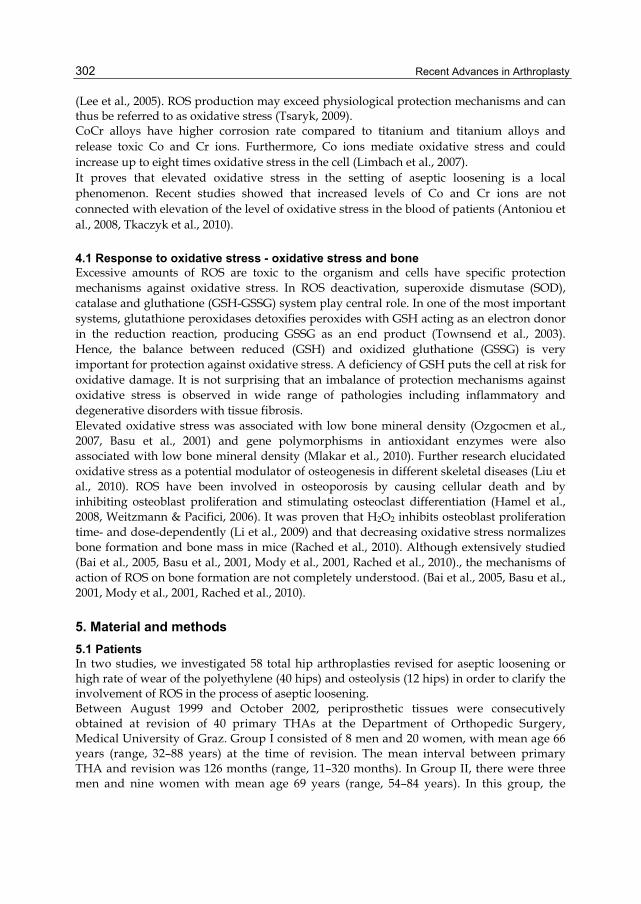
6. Results 6.1 Histological examination Histological examination of the periprosthetic tissues showed large amounts of metal and polyethylene debris and a nonspecific chronic inflammatory reaction. Wear debris, macrophages that had phagocytosed small metal and polyethylene particles, fibroid necrosis, and proliferation of capillaries were seen more commonly in granulomas (Fig. 1).
Fig. 1. Polyethylene and metal particles in the interstitium, giant cells and macrophages (Perle's iron stain, x500)

Evidence Linking Elevated Oxidative Stress and Aseptic Loosening of Hip Arthroplasty
305
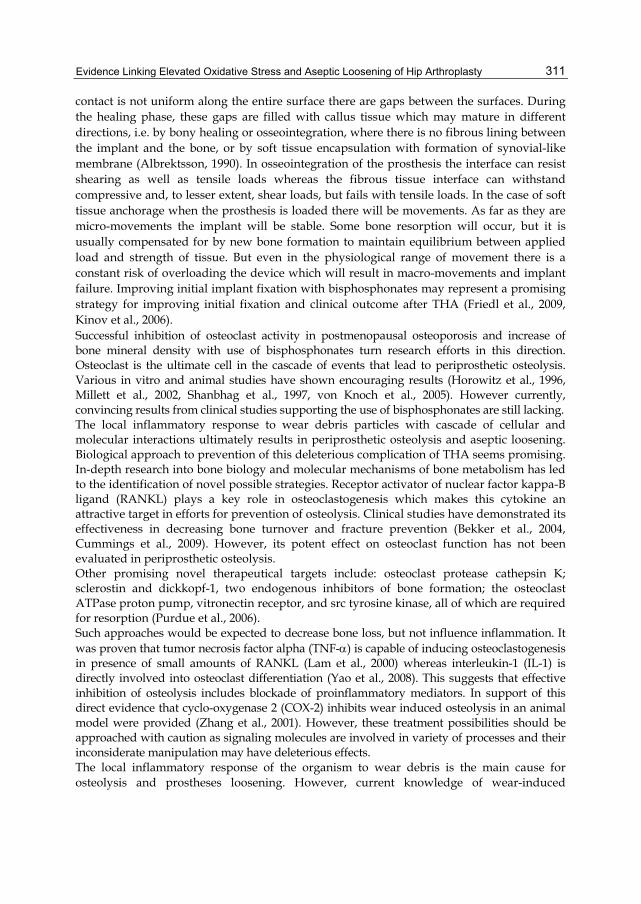
In contrast, stroma rich of connective tissue, abundance of fibroblasts with less frequent macrophages were more prevalent in the fibrous pseudocapsule (Fig. 2).
Fig. 2. Periprosthetic tissue from 72-year-old male, 9 years after implantation of cementless prosthesis. Adjacent to the implant synovia-like membrane with 1-2 cell layers (arrow), interstitial matrix rich of fibrous tissue with abundance of fibroblasts and macrophages digested wear debris (open arrow) (haematoxylene staining, x500).
6.2 Electron microscopic examination Analysis of selected sections from ten representative cases by electron microscopy established damaged collagen fibers and presence of collagen cross-links. Their relative
Fig. 3. Electron micrograph (x39 000) showing periprosthetic tissue taken from stable cementless hip replacement 86 months after implantation. The picture shows damaged collagen fibers (open arrows) with numerous cross-links (black arrows).

Recent Advances in Arthroplasty
306
abundance compared to control samples taken from fascia lata supported the proposal that these findings were a result of ROS damage (Fig. 3). Wear debris inclusions were demonstrated in the various cells (Fig. 4).
Fig. 4. Electron micrograph (x12 000) showing giant cell with ingested polyethylene (open arrow) and metal (black arrow) particles.
6.3 Glutathione determination Level of reduced and oxidized glutathione was elevated in both groups compared to controls. However, a lower statistically significant GSH/GSSG ratio was detected only in Group II compared to Group I and controls (p=0.005 and p=0.024, respectively). GSH and GSSG concentrations and GSH/GSSG ratio in the two groups and controls are given in Table 1.
Analyzed parameter Group I (N 28) Group II (N 12) Controls (N 16)
GSH 74.1 67.3 * 58.7 35 * 18.9 20.9
GSSG 19.3 26.4 * 21.8 12.6 *’*** 4.3 4.4
GSH/GSSG 5.4 3.6 3.1 1.4 **’**** 4.4 1.5
* p<0.0001, ** p<0.024 versus the control group; *** p<0.045, **** p<0.005 versus Group I
Table 1. The levels of GSH, GSSG, as well as GSH/GSSG ratio in periprosthetic tissues and controls. Values measured in µmol/mg hydroxyproline. Data represented as mean SD.
6.4 Malondialdehyde determination 6.4.1 Malondialdehyde determination in periprosthetic tissues Determination of oxidative stress assessed by level of lipid peroxidation product MDA in periprosthetic tissues obtained from 40 primary THA, revised for loosening or high rate of

Evidence Linking Elevated Oxidative Stress and Aseptic Loosening of Hip Arthroplasty
307
wear, showed higher oxidative stress in the two groups. The mean MDA value of the 28 patients in Group I was 0.052 nmol/mg (±0.09 SD), and of the 12 patients with high rate of wear and osteolysis in Group II, 0.031 nmol/mg (±0.014 SD) (p=0.74). MDA levels in the control tissues (0.009 nmol/mg, ±0.0093 SD) were significantly lower than those in Group I (p<0.0001) and Group II (p<0.0001). Figure 5 shows the comparisons between the two groups and controls in graphical form.
*p<0.0001 in comparison with controls.
Fig. 5. The concentration of lipid peroxidation product, malondialdehyde (MDA), in periprosthetic tissues and controls. Values were measured in nmol/mg hydroxyproline. The lower and upper lines in the boxes represent the 25th and 75th percentiles, respectively, with the median marked in the box.
6.4.2 Malondialdehyde determination in pseudosynovial fluid Determination of oxidative stress assessed by level of lipid peroxidation product MDA in pseudojoint fluid showed higher oxidative stress in revision cases. The mean MDA value of the 18 patients with loose hip prostheses was 27.5 nmol/L (±17.6 SD, range 13.5 to 82.9). MDA level in the pseudojoint fluid from controls was significantly lower – 14.9 nmol/L (±4.5 SD, range 10.7 to 28.9) (p=0.001). Figure 6 shows the comparison between patients and controls in graphical form. Although not significantly, MDA level correlated moderately with linear polyethylene wear and grade of femoral osteolysis (Spearman's rho=0.321 and rho=0.315, respectively). Oxidative stress measured by MDA level in pseudosynovial fluid did not correlate with pelvic osteolysis or time elapsed from previous surgery.

Recent Advances in Arthroplasty
308
6.5 Hydroxyproline determination Mean value of hydroxyproline in both groups was 13.9 mg/g (±10.2 SD), and in controls, 55.6 mg/g (±38.6 SD) ( p<0.013).
p<0.001
Fig. 6. The concentration of lipid peroxidation product - malondialdehyde in joint fluid. Values were measured in nmol/L. The lower and upper lines in the boxes represent the 25th and 75th percentiles, respectively, with the median marked in the box.
6.6 Metal particle analysis The tissue concentrations of the different metals for the Co-Cr and Ti-alloy prostheses are shown in Table 2. The mean level of MDA in the 23 implants made of titanium alloy was 0.059 µg/mg, and of the 9 implants made of Co-Cr alloy, 0.023 µg/mg. However, this difference was not significant (p=0.145). Composition Stem/Cup
Ti V Fe Cr Co
Co-Cr / Co-Cr alloy(N 9)
na na 13.5 11.2 75.3 160 * 33.2 34.5
Ti / Ti alloy (N 23)
70.1 189.5 4.4 8.7 na na na
Co-Cr / Ti alloy (N 8)
381.8 911.9 20.6 46.5 11.2 12.2 2.7 4 * 4.1 5.6
* p<0.013 in comparison between groups
Table 2. Concentrations of various metals in periprosthetic tissues from Co-Cr alloy and titanium alloy prostheses. Values measured in µg/g wet tissue. Data represented as mean ±SD.

Evidence Linking Elevated Oxidative Stress and Aseptic Loosening of Hip Arthroplasty
309
6.7 Correlations In the 40 hips, levels of MDA correlated with levels of GSH and GSSG (rho=0.509, p<0.0001, and rho=0.421, p<0.001, respectively). Because oxidative stress is a significant contributor to tissue fibrosis, a second set of correlations was calculated that measured the correlation between oxidative stress and hydroxyproline content. There was a correlation between GSH, GSSG, as well as MDA levels and periprosthetic collagen content. GSH/GSSG ratio and MDA level correlated with degree of osteolysis (rho=0.337, p=0.007, and rho=0.374, p=0.017, respectively). GSSG and MDA levels were higher in hips with greater annual wear of the polyethylene.
7. Rationale of oxidative stress and aseptic loosening Three different mechanisms are mainly responsible for osteolysis and loosening: exacerbated inflammation caused by ROS production in the periprosthetic tissue; cascade of cellular and molecular interactions ultimately resulting in osteoclasts activation; and, compromised bone formation resulting from increased cytotoxicity on mesenchymal osteoprogenitors. Wear debris such as polyethylene, PMMA and metal particles, metal degradation products and ions may be exposed to the ROS produced by the inflammatory cells in the periprosthetic tissues. Mediators such as H2O2, NO and ONOO− are released by the macrophages and the inflammatory cells. Metal debris and metal degradation products could react with the free radicals resulting in elevation of oxidative stress. H2O2 in the cells can also undergo the Fenton reaction in the presence of metal ions with subsequent formation of highly toxic hydroxyl radical (Lubec, 1996, Sawyer, 1990). Various studies have reported that ROS are connected with tissue damage and fibrosis (Park et al., 2001, Riedle & Kerjaschki, 1997, Wang et al., 2002, Windhager et al., 1998). On the other hand, the effect of submicron wear debris on macrophage production of reactive oxygen species is largely unexplored. Thus, these facts led to the hypothesis that ROS play a role in aseptic loosening and formation of fibrous pseudocapsule around hip implants. Our results demonstrated in vivo elevated oxidative stress in periprosthetic tissues and pseudosynovial fluid from loose hip prostheses and hip implants with high rate of wear. This added further insight into the mechanism of aseptic loosening of hip arthroplasty. When the cells are exposed to a large amount of oxidants, the capacity of the regulatory mechanisms of cellular response to oxidative stress may be exceeded by the rate of ROS production, resulting in a condition of “oxidative stress”. In the present study, we found increased levels of markers of oxidative stress in periprosthetic tissues and pseudosynovial fluid from loose THAs. Our results are difficult to compare as the available studies on the possible role of oxidative stress in aseptic loosening of THA are in vitro experiments. We believe that, in the early stage of aseptic loosening, the exposure to wear debris could be responsible for the increase in the values of markers of oxidative stress in total hip arthroplasties with high rate of wear and osteolysis. Moreover, free radicals may be involved in sustaining the foreign-body reaction to wear debris. Later on, chronic exposure would result in a triggering of compensatory mechanisms leading to progressive increase in antioxidants and low oxidative stress as observed in the beginning of loosening (Table 1). This is consistent with the conception of oxidative stress regulation. However, other mechanisms may play role, too.

Recent Advances in Arthroplasty
310
Lipids of cell membranes are a prominent target for free radicals generated in a complex series of oxygen-dependant reactions via Fenton’s chemistry (Sawyer, 1990). Iron-induced lipid peroxidation has been demonstrated in various studies (Marmunti et al., 2004, Lim & Vaziri, 2004). However, we could not establish correlation between levels of MDA and iron. As metal-catalyzed lipid peroxidation is dependent on metal as a catalyst, there might be little influence of metal concentrations, and many other factors might also contribute to MDA formation. On the opposite, we found elevated MDA level in hips with prostheses made of titanium alloy. This finding might be explained by higher rate of wear of titanium implants compared to Co-Cr implants (Table 2) (Wang et al., 2002). We hypothesized that the process of wear debris-mediated loosening leads to elevation of oxidative stress. In support of this, Wang et al. found that exposure to particles stimulates superoxide production by macrophages and osteoclasts (Wang et al., 2002). Furthermore, it was observed that the increase of free radicals on polyethylene correlated with the degree of inflammation of synovial cells in culture (Fiorito et al., 2001). The correlation between markers of oxidative stress and hydroxyproline levels suggest, first of all, that the increase of collagen in periprosthetic tissues in the presence of wear debris is due to elevated oxidative stress. Connective tissue metabolism is normally characterized by equilibrium between degradation and synthesis of extracellular matrix. Deviation from the equilibrium may lead to the replacement of extracellular matrix by fibrous tissue (Park et al., 2001, Riedle & Kerjaschki, 1997, Wang et al., 2002, Windhager et al., 1998). The fibrous pseudocapsule formed is probably related to high intraarticular pressure and expansion of the effective joint space as well as production of inflammatory substances and subsequent loosening of the implant (El-Warrak et al., 2004, Van der Vis et al., 1998). Most of the studies elucidating the mechanism of loosening of hip arthroplasty are performed on animal models or are in vitro studies. However, the results from in vitro or animal studies could not be easily translated to humans. The findings of our in vivo studies provide further insight into the possible mechanisms of aseptic loosening. The number of cases in the studies was relatively small but the differences in measurements of markers of oxidative stress between patients and controls were high which compensated for small sample size.
8. Future perspectives Clinical results of total hip arthroplasty deteriorate with time with aseptic loosening being the main reason for its failure. Clinical and experimental studies show that aseptic loosening is a failure of fixation of the implant secondary to wear of the components of the prostheses (Bechtold et al., 2002, El-Warrak et al., 2004, Willert & Semlitsch, 1977). Improvements in bearing surfaces may diminish implant wear and associated osteolysis. For forty years, MoPE is the bearing of choice in THA. After recognition of the problem of wear alternative bearings were proposed. Metal-on-high cross-linked polyethylene, ceramic-on-polyethylene, ceramic-on-ceramic, and metal-on-metal are becoming increasingly popular. However, reducing particle size may increase its biological activity (Sieber et al., 1999, Yang et al., 2002). Moreover, some of the new alternative bearings are very sensitive to improper surgical technique. The initial bone-implant interface is of paramount importance for the longevity of the joint reconstruction. After implantation the prosthesis is in contact with the bone, but as the
Evidence Linking Elevated Oxidative Stress and Aseptic Loosening of Hip Arthroplasty
311
contact is not uniform along the entire surface there are gaps between the surfaces. During the healing phase, these gaps are filled with callus tissue which may mature in different directions, i.e. by bony healing or osseointegration, where there is no fibrous lining between the implant and the bone, or by soft tissue encapsulation with formation of synovial-like membrane (Albrektsson, 1990). In osseointegration of the prosthesis the interface can resist shearing as well as tensile loads whereas the fibrous tissue interface can withstand compressive and, to lesser extent, shear loads, but fails with tensile loads. In the case of soft tissue anchorage when the prosthesis is loaded there will be movements. As far as they are micro-movements the implant will be stable. Some bone resorption will occur, but it is usually compensated for by new bone formation to maintain equilibrium between applied load and strength of tissue. But even in the physiological range of movement there is a constant risk of overloading the device which will result in macro-movements and implant failure. Improving initial implant fixation with bisphosphonates may represent a promising strategy for improving initial fixation and clinical outcome after THA (Friedl et al., 2009, Kinov et al., 2006). Successful inhibition of osteoclast activity in postmenopausal osteoporosis and increase of bone mineral density with use of bisphosphonates turn research efforts in this direction. Osteoclast is the ultimate cell in the cascade of events that lead to periprosthetic osteolysis. Various in vitro and animal studies have shown encouraging results (Horowitz et al., 1996, Millett et al., 2002, Shanbhag et al., 1997, von Knoch et al., 2005). However currently, convincing results from clinical studies supporting the use of bisphosphonates are still lacking. The local inflammatory response to wear debris particles with cascade of cellular and molecular interactions ultimately results in periprosthetic osteolysis and aseptic loosening. Biological approach to prevention of this deleterious complication of THA seems promising. In-depth research into bone biology and molecular mechanisms of bone metabolism has led to the identification of novel possible strategies. Receptor activator of nuclear factor kappa-B ligand (RANKL) plays a key role in osteoclastogenesis which makes this cytokine an attractive target in efforts for prevention of osteolysis. Clinical studies have demonstrated its effectiveness in decreasing bone turnover and fracture prevention (Bekker et al., 2004, Cummings et al., 2009). However, its potent effect on osteoclast function has not been evaluated in periprosthetic osteolysis. Other promising novel therapeutical targets include: osteoclast protease cathepsin K; sclerostin and dickkopf-1, two endogenous inhibitors of bone formation; the osteoclast ATPase proton pump, vitronectin receptor, and src tyrosine kinase, all of which are required for resorption (Purdue et al., 2006). Such approaches would be expected to decrease bone loss, but not influence inflammation. It was proven that tumor necrosis factor alpha (TNF-) is capable of inducing osteoclastogenesis in presence of small amounts of RANKL (Lam et al., 2000) whereas interleukin-1 (IL-1) is directly involved into osteoclast differentiation (Yao et al., 2008). This suggests that effective inhibition of osteolysis includes blockade of proinflammatory mediators. In support of this direct evidence that cyclo-oxygenase 2 (COX-2) inhibits wear induced osteolysis in an animal model were provided (Zhang et al., 2001). However, these treatment possibilities should be approached with caution as signaling molecules are involved in variety of processes and their inconsiderate manipulation may have deleterious effects. The local inflammatory response of the organism to wear debris is the main cause for osteolysis and prostheses loosening. However, current knowledge of wear-induced

Recent Advances in Arthroplasty
312
mechanism of aseptic loosening is not complete. Understanding of the mechanisms of wear-induced osteolysis will help control undesirable processes at the implant-bone interface and extend longevity of hip arthroplasty.
9. Conclusion Our study provided further insight into the mechanism of aseptic loosening of hip arthroplasty. The most interesting aspect of this study is the evidence of elevated production of ROS, which are known to cause tissue fibrosis and may help explain the initiation of aseptic loosening. We feel that the thick pseudocapsule around loose prostheses is not an adaptive but a reactive tissue as a result of decreased degradation and increased formation of collagen matrix and proliferation of fibroblasts due to high oxidative stress. Furthermore, we demonstrated that elevated oxidative stress is associated with aseptic loosening of hip arthroplasty, suggesting that oxidative stress might induce periprosthetic osteolysis and subsequent loosening. Research in this line may help introducing new strategies for therapeutic prevention and/or treatment of osteolysis and subsequent aseptic loosening of total hip arthroplasty.
10. References Aitken, C., Hodge, J., Nishinaka, Y., Vaughan, T., Yodoi, J., Day, C., Morrison, N. &
Nicholson, G. (2004). Regulation of human osteoclast differentiation by thioredoxin binding protein-2 and redoxsensitive signaling, J Bone Miner Res Vol.19:2057–2064.
Albrektsson, T. (1990). Biological factors of importance for bone integration of implanted devices, in Older MWJ (ed.), Implant bone interface, Springer-Velag, London.
Amstutz, H., Wisk, L. & Le Duff, M. (2011). Sex as a patient selection criterion for metal-on-metal hip resurfacing arthroplasty, J Arthroplasty Vol.26:198-208.
Antoniou, J., Zukor, D.J., Mwale, F., Minarik, W., Petit, A. & Huk, O.L. (2008). Metal ion levels in the blood of patients after hip resurfacing: a comparison between twenty-eight and thirty-six-millimeter-head metal-on-metal prostheses, J Bone Joint Surg Am Vol.90(Suppl. 3):142-8.
Aspenberg, P. & Herbertsson, P. (1996). Periprosthetic bone resorption. Particles versus movement, J Bone Joint Surg Br Vol.78:641–646.
Aspenberg, P. & Van der Vis, H. (1998). Fluid pressure may cause periprosthetic osteolysis. Particles are not the only thing, Acta Orthop Scand Vol.69(No.1):1-4.
Bai, X.C., Lu, D., Bai, J., Zheng, H., Ke, Z.Y., Li, X.M. & Luo, S.Q. (2004). Oxidative stress inhibits osteoblastic differentiation ofbone cells by ERK and NF-kB, Biochem Biophys Res Commun Vol.314:197–207.
Bai, X.C., Lu, D., Liu, A.L., Zhang, Z.M., Li, X.M., Zou, Z.P., Zeng, W.S., Cheng, B.L. & Luo, S.Q. (2005). Reactive oxygen species stimulates receptor activator of NF-kappaB ligand expression in osteoblast, J Biol Chem Vol.280:17497–17506.
Basu, S., Michaëlsson, K., Olofsson, H., Johansson, S. & Melhus, H. (2001). Association between oxidative stress and bone mineral density, Biochem Biophys Res Commun Vol.288(No.1):275-9.
Bechtold, J.E., Kubic, V. & Sobale, K. (2002). Bone ingrowth in the presence of particulate polyethylene, Bone Joint Surg Br Vol.84:915-919.

Evidence Linking Elevated Oxidative Stress and Aseptic Loosening of Hip Arthroplasty
313
Bekker, P.J., Holloway, D.L., Rasmussen, A.S., Murphy, R., Martin, S.W., Leese, P.T., Holmes, G.B., Dunstan, C.R. & DePaoli, A.M. (2004). A single-dose placebo-controlled study of AMG 162, a fully human monoclonal antibody to RANKL, in postmenopausal women, J Bone Miner Res Vol.19(No.7):1059-66.
Bierbaum, B.E., Nairus, J., Kuesis, D., Morrison, J.C. & Ward, D. (2002). Ceramic-on-ceramic bearings in total hip arthroplasty, Clin Orthop Relat Res Vol.405:158-63.
Boutin, P. (1971). [Alumina and its use in surgery of the hip. (Experimental study)], Presse Med; Vol.79(No.14):639–640.
Boutin, P.& Blanquaert, D. (1981). A study of the mechanical properties of alumina-on-alumina total hip prosthesis, Rev Chir Orthop Reparatrice Appar Mot Vol.67(No.3):279-87.
Brooks, R.A., Wimhurst, J.A. & Rushton, N. (2002) Endotoxin contamination of particles produces misleading inflammatory cytokine responses from macrophages in vitro, J Bone Joint Surg Br Vol.84:295–299.
Campbell, D.G., Field, J.R. & Callary, S.A. (2010) Second-generation highly cross-linked X3™ polyethylene wear: a preliminary radiostereometric analysis study, Clin Orthop Relat Res Vol.468(No.10):2704-9.
Capello, W.N., Dantonio, J.A., Feinberg, J.R. & Manley, M.T. (2005). Alternative bearing surfaces: alumina ceramic bearings for total hip arthroplasty, Instr Course Lect Vol.54:171-6.
Catelas, I., Bobyn, J.D., Medley, J.B., Krygier, J.J., Zukor, D.J. & Huk, O.L. (2003). Size, shape, and composition of wear particles from metal-metal hip simulator testing: effects of alloy and number of loading cycles, J Biomed Mater Res A Vol.67: 312-327.
Charnley, J., Follacci, F.M. & Hammond, B.T. (1968). The long-term reaction of bone to self-curing acrylic cement, J Bone Joint Surg Br Vol.50:822–829.
Charnley, J. (1979). Low Friction Arthroplasty of the hip, Springer-Verlag, Berlin-Heidelberg-New York.
Chen, R.M., Lin, Y.L. & Chou, C.W. (2010). GATA-3 transduces survival signals in osteoblasts through upregulation of bcl-x(L) gene expression, J Bone Miner Res Vol.25(No.10):2193-204.
Cummings, S.R., San Martin, J., McClung, M.R., Siris, E.S., Eastell, R., Reid, I.R., Delmas, P., Zoog, H.B., Austin, M., Wang, A., Kutilek, S., Adami, S., Zanchetta, J., Libanati, C., Siddhanti, S. & Christiansen, C. (2009). FREEDOM Trial. Denosumab for prevention of fractures in postmenopausal women with osteoporosis, N Engl J Med Vol.361(No.8):756-65.
De Haan, R., Campbell, P.A., Su, E.P. & De Smet, K.A. (2008). Revision of metal-on-metal resurfacing arthroplasty of the hip: the influence of malpositioning of the components, J Bone Joint Surg Br Vol.90:1158-1163.
Doorn, P.F., Campbell, P. & Amstutz, H. (1999). Particle disease in metal-on-metal total hip replacements. in: Rieker, C.B., Windler, M. & Wyss, U. (eds.), Metasul: A Metal-on-Metal Bearing, Huber, Bern, pp. 113–119.
El-Warrak, A.O., Olmstead, M., Schneider, R., Meinel, L., Bettschart-Wolfisberger, R., Akens, M.K., Auer, J. & von Rechenberg, B. (2004). An experimental animal model of aseptic loosening of hip prostheses in sheep to study early biochemical changes at the interface membrane, BMC Musculoskelet Disord 5:7.
Recent Advances in Arthroplasty
314
Engh, C.A., Massin, P. & Suthers, K.E. (1989). Roentgenographic assessment of the biologic fixation of porous-surfaced femoral components, Clin Orthop Relat Res Vol.257:107–128.
Fiorito, S., Goze, C., Adrey, J., Magrini, L., Goalard, C. & Bernier, P. (2001). Increase in free radicals on UHMWPE hip prostheses components due to inflamed synovial cell products, J Biomed Mater Res Vol.57:35–40.
Firkins, P.J., Tipper, J.L., Saadatzadeh, M.R., Ingham, E., Stone, M.H., Farrar, R. & Fisher, J. (2001). Quantitative analysis of wear and wear debris from metal-on-metal hip prostheses tested in a physiological hip joint simulator, Biomed Mater Eng Vol.11(No.2):143–157.
Fleury, C., Petit, A., Mwale, F., Antoniou, J., Zukor, D.J., Tabrizian, M. & Huk, O.L. (2006). Effect of cobalt and chromium ions on human MG-63 osteoblasts in vitro: morphology, cytotoxicity, and oxidative stress, Biomaterials Vol.27:3351-3360.
Friedl, G., Radl, R., Stihsen, C., Rehak, P., Aigner, R. & Windhager, R. (2009). The effect of a single infusion of zoledronic acid on early implant migration in total hip arthroplasty. A randomized, double-blind, controlled trial, J Bone Joint Surg Am Vol.91(No.2):274-81.
Goldring, S.R., Jasty, M., Roueke, C.M., Bringhurst, F.R. & Harris, W.H. (1986). Formation of a synovial-like membrane at the bone-cement interface. Its role in bone resorption and implant loosening after total hip replacement, Arthritis Rheum Vol.29(No.7):836–842.
Hallab, N., Merritt, K. & Jacobs, J.J. (2001). Metal sensitivity in patients with orthopaedic implants, J Bone Joint Surg Am Vol.83(No.3):428-36.
Hallab, N.J., Anderson, S., Stafford, T., Glant, T. & Jacobs, J.J. (2005). Lymphocyte responses in patients with total hip arthroplasty, J Orthop Res Vol.23(No.2):384-91.
Hamel, P., Abed, E., Brissette, L. & Moreau, R. (2008). Characterization of oxidized low-density lipoprotein-induced hormesis-like effects in osteoblastic cells, Am J Physiol Cell Physiol Vol.294(No.4):C1021-33.
Harris, W.H., Schiller, A.L., Scholler, J.M., Friberg, R.A. & Scott, R. (1976). Extensive localized bone resorption in the femur following total hip replacement, J Bone Joint Surg Am 58:612–618.
Harris, W.H. & Penenberg, B.L. (1987). Further follow-up on socket fixation using a metal-backed acetabular component for total hip replacement, J Bone Joint Surg Am Vol.69:1140–1143.
Hogg, N. (1998). Free Radicals in Disease, Semin Reprod Endocrin Vol.16:241-248. Horowitzm S.M., Algan, S.A. & Purdon, M.A. (1996). Pharmacologic inhibition of
particulate-induced bone resorption, J Biomed Mater Res Vol.31:91–96. Jones, L.C. & Hungerford, D.S. (1987). Cement disease, Clin Orthop Relat Res Vol.225:192–
206. Kamikawa, K., Harada, Y., Nagata, K. & Moriya, H. (2001). Differential effects of oxidised
and non-oxidised polyethylene particles on human monocyte/macrophages in vitro, J Bone Joint Surg Br Vol.83:593-7.
Keegan, G.M., Learmonth, I.D. & Case, C.P. (2007). Orthopaedic metals and their potential toxicity in the arthroplasty patient: A review of current knowledge and future strategies, J Bone Joint Surg Br Vol.89(No.5):567-73.

Evidence Linking Elevated Oxidative Stress and Aseptic Loosening of Hip Arthroplasty
315
Khoschsorur, G.A., Winklhofer-Roob, B.M., Rabl, H., Auer, T., Peng, Z. & Schaur, R. J. (2000). Evaluation of a sensitive HPLC method for the determination of malondialdehyde, and application of the method different biological materials, Chromatographia Vol.52:181–184.
Kido, A., Pap, G., Nagler, D.K., Ziomek, E., Ménard, R., Neumann, H.W. & Roessner A. (2004). Protease expression in interface tissues around loose arthroplasties. Clin Orthop Relat Res Vol.425:230–236.
Kim, W.K., Meliton, V., Bourquard, N., Hahn, T.J. & Parhami, F. (2010). Hedgehog signaling and osteogenic differentiation in multipotent bone marrow stromal cells are inhibited by oxidative stress, J Cell Biochem Vol.111(No.5):1199-209.
Kinov, P., Leithner, A., Radl, R., Bodo, K., Khoschsorur, G. A., Schauenstein, K. & R. Windhager. (2006). Role of free radicals in aseptic loosening of hip arthroplasty, J Orthop Res Vol.24:55-62.
Kinov, P., Tivchev, P., Doukova, P. & Leithner, A. (2006). Effect of risedronate on bone metabolism after total hip arthroplasty: a prospective randomised study, Acta Orthop Belg Vol.72(No.1):44-50.
Kurtz, S.M. (2004). The UHMWPE handbook: ultra-high molecular weight polyethylene in total joint replacement, Elsevier, Boston.
Kurtz, S.M., Austin, M.S., Azzam, K., Sharkey, P.F., MacDonald, D.W., Medel, F.J. & Hozack, W.J. (2010). Mechanical properties, oxidation, and clinical performance of retrieved highly cross-linked crossfire liners after intermediate-term implantation, J Arthroplasty Vol.25(No.4):614-23.
Lam, J., Takeshita, S., Barker, J.E., Kanagawa, O., Ross, F.P. & Teitelbaum, S.L. (2000). TNF-alpha induces osteoclastogenesis by direct stimulation of macrophages exposed to permissive levels of RANK ligand, J Clin Invest Vol.106(No.12):1481-8.
Lean, J.M., Jagger, C.J., Kirstein, B., Fuller, K. & Chambers, T.J. (2005). Hydrogen peroxide is essential for estrogendeficiency bone loss and osteoclast formation, Endocrinology Vol.146:728–735.
Lee, M.C., Yoshino, F., Shoji, H., Takahashi, S., Todoki, K., Shimada, S. & Kuse-Barouch, K. (2005). Characterization by electron spin resonance spectroscopy of reactive oxygen species generated by titanium dioxide and hydrogen peroxide, J Dent Res Vol.84:178-82.
Li, M., Zhao, L., Liu, J., Liu, A.L., Zeng, W.S., Luo, S.Q. & Bai, X.C. (2009). Hydrogen peroxide induces G2 cell cycle arrest and inhibits cell proliferation in osteoblasts, Anat Rec (Hoboken) Vol.292(No.8):1107-13.
Lim, C.S. & Vaziri, N.D. (2004). Iron and oxidative stress in renal insufficiency, Am J Nephrol Vol.24:569–575.
Limbach, L.K., Wick, P., Manser, P., Grass, R.N., Bruinink, A. & Stark, W.J. (2007). Exposure of engineered nanoparticles to human lung epithelial cells: influence of chemical composition and catalytic activity on oxidative stress, Environ Sci Technol Vol.41(No.11):4158-63.
Lin, F. & Girotti, A.W. (1993). Photodynamic action of merocyanine 540 on leukemia cells: iron-stimulated lipid peroxidation and cell killing, Arch Biochem Biophys Vol.300(No.2):714-23.

Recent Advances in Arthroplasty
316
Liu, H., Sun, J.C., Zhao, Z.T., Zhang, J.M., Xu, H. & Li, G.S. (2010). Fluoride-induced oxidative stress in three-dimensional culture of os732 cells and rats, Biol Trace Elem Res doi: 10.1007/s12011-010-8881-0 [Epub ahead of print]
Livermore, J., Ilstrup, D. & Morrey, B. (1990). Effect of femoral head size on wear of the polyethylene acetabular component, J Bone Joint Surg Am Vol.72:518–528.
Lubec, G. (1996). The hydroxyl radical: from chemistry to human disease, J Investig Med Vol.44(No.6):324-46.
Marmunti, M., Gavazza, M. & Catala, A. (2004). Nonenzymatic and enzymatic lipid peroxidation of microsomes and nuclei obtained from rat liver, Mol Cell Biochem Vol.265:1–9.
Mazière, C., Savitsky, V., Galmiche, A., Gomila, C., Massy, Z. & Mazière, J.C. (2010). Oxidized low density lipoprotein inhibits phosphate signaling and phosphate-induced mineralization in osteoblasts. Involvement of oxidative stress, Biochim Biophys Acta Vol.1802(No.11):1013-9.
McKay, G.C., Macnair, R., MacDonald, C. & Grant, M.H. (1996). Interactions of orthopaedic metals with an immortalized rat osteoblast cell line, Biomaterials Vol.17:1339-1344.
McKee, G.K. & Watson-Farrar, J. (1966). Replacement of arthritic hips by the McKee-Farrar prosthesis, J Bone Joint Surg Br Vol.48(No.2):245–259.
McKellop, H., Shen, F.-W., Lu, B., Campbell, P. & Salovey, R. (1999). Development of an extremely wear resistant ultra-high molecular weight polyethylene for total hip replacements, J Orthop Res Vol.17(No.2):157–167.
Millett, P.J., Allen, M.J. & Bostrom, M.P. (2002). Effects of alendronate on particle-induced osteolysis in a rat model, J Bone Joint Surg Am Vol.84:236-49.
Minotti, G. & Aust, S.D. (1992). Redox cycling of iron and lipid peroxidation, Lipids Vol.27(No.3):219-26.
Mirra, J.M., Amstutz, H.C., Matos, M. & Gold, R. (1976). The pathology of the joint tissues and its clinical relevance in prosthesis failure. Clin Orthop Relat Res Vol.117:221–240.
Mlakar, S.J., Osredkar, J., Prezelj, J. & Marc, J. (2010). The antioxidant enzyme GPX1 gene polymorphisms are associated with low BMD and increased bone turnover markers, Dis Markers Vol.29(No.2):71-80.
Mody, N., Parhami, F., Sarafian, T.A. & Demer, L.L. (2001). Oxidative stress modulates osteoblastic differentiation of vascular and bone cells, Free Radic Biol Med Vol.31:509–519.
Muratoglu, O.K., Bragdon, C.R., O’Connor, D.O., Jasty, M. & Harris, W.H. (2001). A novel method of crosslinking UHMWPE to improve wear, reduce oxidation and retain mechanical properties, J Arthrop Vol.16(No.2):149–160.
Muratoglu, O.K. & Kurtz, S.M. (2002). Alternate bearing surfaces in hip replacement, in Sinha, R.K. (ed.), Hip replacement. Current trends and controversies, Marcel Dekker, New York, pp. 1-46.
Ollivere, B., Darrah, C., Barker, T., Nolan, J. & Porteous, M.J. (2009). Early clinical failure of the Birmingham metal-onmetal hip resurfacing is associated with metallosis and softtissue necrosis, J Bone Joint Surg Br Vol.91:1025-1030.
Ozgocmen, S., Kaya, H., Fadillioglu, E., Aydogan, R. & Yilmaz, Z. (2007). Role of antioxidant systems, lipid peroxidation, and nitric oxide in postmenopausal osteoporosis, Mol Cell Biochem Vol.295(No.1-2):45-52.

Evidence Linking Elevated Oxidative Stress and Aseptic Loosening of Hip Arthroplasty
317
Paprosky, W.G. & Burnett, R.S. (2002). Assessment and classification of bone stock deficiency in revision total hip arthroplasty, Am J Orthop Vol.31:459–464.
Park, S.K., Kim, J., Seomun, Y., Choi, J., Kim, D.H., Han, I.O., Lee, E.H., Chung, S.K. & Joo, C.K. (2001). Hydrogen peroxide is a novel inducer of connective tissue growth factor, Biochem Biophys Res Comm Vol.284:966–971.
Park, Y.S., Moon, Y.W., Lim, S.J., Yang, J.M., Ahn, G. & Choi, Y.L. (2005). Early osteolysis following second-generation metal-on-metal hip replacement, J Bone Joint Surg Am Vol.87:1515-1521.
Petit, A., Mwale, F., Tkaczyk, C., Antoniou, J., Zukor, D.J. & Huk O.L. (2005). Induction of protein oxidation by cobalt and chromium ions in human U937 macrophages, Biomaterials Vol.26:4416-4422.
Purdue, P.E., Koulouvaris, P., Nestor, B.J. & Sculco, T.P. (2006). The central role of wear debris in periprosthetic osteolysis, HSS J Vol.2(No.2):102-13.
Rached, M.T., Kode, A., Xu, L., Yoshikawa, Y., Paik, J.H., Depinho, R.A. & Kousteni, S. (2010). FoxO1 is a positive regulator of bone formation by favoring protein synthesis and resistance to oxidative stress in osteoblasts. Cell Metab Vol.11(No.2):147-60.
Reddy, G.K. & Enwemeka, C.S. (1996). A simplified method for the analysis of hydroxyproline in biological tissues, Clin Biochem Vol.29:225–229.
Riedle, B. & Kerjaschki, D. (1997). Reactive oxygen species cause direct damage of Engelbreth-Holm-Swarm matrix, Am J Pathol Vol.151:215–231.
Ring PA. (1967). Total hip replacement, Proc R Soc Med Vol.60(No.3):281-4. Sabokbar, A., Fujikawa, Y., Neale, S., Murray, D.W. & Athanasou, N.A. (1997). Human
arthroplasty derived macrophages differentiate into osteoclastic bone resorbing cells. Ann Rheum Dis Vol.56:414-420.
Sawyer, D.T. (1990). Iron induced activation of HOOH, J Exp Pathol Vol.31:116–131. Schaich, K.M. (1992). Metals and lipid oxidation. Contemporary issues, Lipids
Vol.27(No.3):209-18. Schmalzried, T.P., Jasty, M. & Harris, W.H. (1992). Periprosthetic bone loss in total hip
arthroplasty. Polyethylene wear debris and the concept of the effective joint space, J Bone Joint Surg Am Vol.74:849–863.
Shanbhag, A.S., Hasselman, C.T. & Rubash, H.E. (1997). Inhibition of wear debris mediated osteolysis in a canine total hip arthroplasty model, Clin Orthop Relat Res Vol.344:33-43.
Sieber, H.P., Rieker, C.B. & Köttig, P. (1999). Analysis of 118 second-generation metal-on-metal retrieved hip implants, J Bone Joint Surg Br Vol.81:46-50.
Tietze, F. (1969). Enzymic method for quantitative determination of nanogram amounts of total and oxidized glutathione: applications to mammalian blood and other tissues, Anal Biochem Vol.27:502–522.
Tkaczyk, C., Petit, A., Antoniou, J., Zukor, D.J., Tabrizian, M. & Huk, O.L. (2010). Significance of Elevated Blood Metal Ion Levels in Patients with Metal-on-Metal Prostheses: An Evaluation of Oxidative Stress Markers, Open Orthop J Vol.4:221-7.
Townsend, D.M., Tew, K.D. & Tapiero, H. (2003). The importance of glutathione in human disease, Biomed Pharmacother Vol.57(No.3-4):145-55.
Tsaryk, R. (2009). Effects of metal-induced oxidative stress on endothelial cells in vitro, Dissertation, Mainz.

Recent Advances in Arthroplasty
318
Valko, M., Rhodes, C.J., Moncol, J., Izakovic, M. & Mazur, M. (2006). Free radicals, metals and antioxidants in oxidative stress-induced cancer, Chem Biol Interact Vol.160:1-40.
Van der Vis, H.M., Aspenberg, P., Marti, R.K., Tigchelaar, W. & Van Noorden, C.J. (1998). Fluid pressure causes bone resorption in a rabbit model of prosthetic loosening, Clin Orthop Relat Res Vol.350:201–208.
von Knoch, M., Wedemeyer, C. & Pingsmann, A. (2005). The decrease of particle-induced osteolysis after a single dose of bisphosphonates, Biomaterials Vol.26:1803–1808.
Wang, M.L., Hauschka, P.V., Tuan, R.S. & Steinbeck, M.J. (2002). Exposure to particles stimulates superoxide production by human THP-1 macrophages and avian HD-11EM osteoclasts activated by tumor necrosis factor-alpha and PMA, J Arthroplasty Vol.17:335–346.
Wei, X., Zhang, X., Flick, L.M., Drissi, H., Schwarz, E.M. & O'Keefe, R.J. (2009). Titanium particles stimulate COX-2 expression in synovial fibroblasts through an oxidative stress-induced, calpain-dependent, NF-kappaB pathway, Am J Physiol Cell Physiol Vol.297:C310-320.
Weitzmann, M.N. & Pacifici, R. (2006). Estrogen deficiency and bone loss: an inflammatory tale, J Clin Invest Vol.116:1186–1194.
Wiles, P. (1957). The surgery of the osteo-arthritic hip. Br J Surg Vol.45:488–497. Willert, H.G., Semlitsch, M. (1977). Reactions of the articular capsule to wear products of
artificial joint prostheses. J Biomed Mater Res Vol.11:157–164. Willmann, G., (1998). Ceramics for total hip replacement—What a surgeon should know,
Orthopedics Vol.21(No.2):173–177. Windhager, R., Nemethova, M., Mutsaers, S., Lang, S., Kotz, R., Kitzmueller, E. & Lubec G.
(1998). Evidence for the involvement of the hydroxyl radical in the pathogenesis of excessive connective tissue proliferation in patients with tumorendoprostheses, Life Sci Vol.62:1261–1269.
Witkiewicz-Kucharczyk, A. & Bal, W. (2006). Damage of zinc fingers in DNA repair proteins, a novel molecular mechanism in carcinogenesis, Toxicol Lett Vol.162:29-42.
Wolf, T., Kasemann, R. & Ottenwälder, H. (1989). Molecular interaction of different chromium species with nucleotides and nucleic acids, Carcinogenesis Vol.10:655-659.
Wynn-Jones, H., Macnair, R., Wimhurst, J., Chirodian, N., Derbyshire, B., Toms, A. & Cahir, J. (2011). Silent soft tissue pathology is common with a modern metal-on-metal hip arthroplasty, Acta Orthop Vol.82(No.3):301-7.
Yagi, K. (1982) Assay for serum lipid peroxide level and its clinical significance, Biochem Med; Vol.30: 223-241.
Yang, S.Y., Ren, W., Park, Y., Sieving, A., Hsu, S., Nasser, S. & Wooley, P.H. (2002). Diverse cellular and apoptotic responses to variant shapes of UHMWPE particles in a murine model of inflammation, Biomaterials Vol.23:3535–3543.
Yao, Z., Xing, L., Qin, C., Schwarz, E.M. & Boyce, B.F. (2008). Osteoclast precursor interaction with bone matrix induces osteoclast formation directly by an interleukin-1-mediated autocrine mechanism, J Biol Chem Vol.283(No.15):9917-24.
Zhang, X., Morham, S.G., Langenbach, R., Young, D.A., Xing, L., Boyce, B.F., Puzas, E.J., Rosier, R.N., O'Keefe, R.J. & Schwarz, E.M. (2001). Evidence for a direct role of cyclo-oxygenase 2 in implant wear debris-induced osteolysis, J Bone Miner Res Vol.16(No.4):660-70.
Copyright © 2022 FDOKUMEN