Human pancreatic glucokinase - Bergen Open Research Archive
91
Human pancreatic glucokinase Structural and physico-chemical studies related to catalytic activation, kinetic cooperativity and GCK-diabetes Janne Molnes Dissertation for the degree philosophiae doctor (PhD) at the University of Bergen 2012
-
Upload
khangminh22 -
Category
Documents
-
view
0 -
download
0
Transcript of Human pancreatic glucokinase - Bergen Open Research Archive

Human pancreatic glucokinase Structural and physico-chemical studies related to catalytic
activation, kinetic cooperativity and GCK-diabetes
Janne Molnes
Dissertation for the degree philosophiae doctor (PhD)
at the University of Bergen
2012

1
Contents
Scientific environment ............................................................................................................... 4
Acknowledgements ................................................................................................................... 5
Abbreviations ............................................................................................................................ 6
Abstract ..................................................................................................................................... 8
List of publications .................................................................................................................. 10
1. INTRODUCTION ............................................................................................................ 11
1.1. BACKGROUND ................................................................................................................. 11
1.2. MONOGENIC DIABETES ............................................................................................... 12
1.3. GLUCOKINASE GLYCEMIC DISEASES .................................................................... 14
1.3.1. GCK-MODY .................................................................................................................... 16
Pathophysiology ............................................................................................................... 16 Clinical features ............................................................................................................... 17 Diagnosis and treatment .................................................................................................. 17 Prevalence ........................................................................................................................ 17
1.3.2. GCK-PNDM ..................................................................................................................... 18 1.3.3. GCK-HI ............................................................................................................................ 19
1.4. THE ROLE OF PANCREATIC -CELL GK IN GLUCOSE-STIMULATED INSULIN SECRETION AND GLYCEMIC DISEASE .................................................. 20
1.5. NATURALLY OCCURRING GCK MUTATIONS AND THE MECHANISM OF THEIR ASSOCIATED DISEASE .................................................................................... 22
1.6. GK – A MEMBER OF THE HEXOKINASE FAMILY OF ENZYMES WITH UNIQUE PROPERTIES ................................................................................................... 23
1.6.1. Kinetic properties of GK ............................................................................................... 24
1.6.2. Gene structure and tissue-specific gene regulation ................................................... 25
1.7. THE 3D STRUCTURE OF GK AND THE GLUCOSE-INDUCED
CONFORMATIONAL CHANGE .................................................................................... 26 1.7.1. The active site ................................................................................................................. 28
Glucose-binding site ......................................................................................................... 28
ATP-binding site ............................................................................................................... 29

2
1.7.2. The C-terminal -helix ................................................................................................. 30
1.7.3. The allosteric activator site and pharmacological GK activator drugs ................... 32
1.7.4. Cysteine residues at the active site of GK .................................................................. 33
1.8. KINETIC MODELS OF POSITIVE COOPERATIVITY ............................................. 34
1.9. REGULATION OF hGK ACTIVITY .............................................................................. 37
1.9.1. Post-translational regulation of hGK ........................................................................... 38
Covalent post-translational modifications ....................................................................... 40
The ubiquitin conjugating system and ubiquitin-mediated proteolytic pathway .... 41
Role of the ubiquitin-proteasome pathway in -cell dysfunction and
hyperglycemia ......................................................................................................... 43
2. AIMS OF THE PRESENT STUDY ............................................................................... 45
3. SUMMARY OF RESULTS ............................................................................................. 46
3.1. PAPER I .............................................................................................................................. 46
3.2. PAPER II ............................................................................................................................. 47
3.3. PAPER III ........................................................................................................................... 48
4. METHODS AND METHODOLOGICAL ASPECTS ................................................. 49
4.1. FUNCTIONAL COMPARISON OF GST-hGK AND NON-TAGGED hGK .............. 49
4.2. HELIX NOMENCLATURE ............................................................................................. 50
4.3. ANALYSES OF PROTEIN DYNAMICS ........................................................................ 50
4.3.1. Intrinsic fluorescence spectroscopy ............................................................................. 51
4.3.2. Extrinsic fluorescence spectroscopy............................................................................ 53
4.3.3. Computational methods ................................................................................................ 54
5. GENERAL DISCUSSION ............................................................................................... 55
5.1. GK PROTEIN STABILITY .............................................................................................. 55 5.2. THE MULTIPHASIC GLOBAL CONFORMATIONAL TRANSITION AND
PERTURBATIONS OF CONFORMATIONAL EQUILIBRIA ................................... 57 5.3. ALLOSTERIC EFFECTORS OF hGK ........................................................................... 62
5.3.1. Glucokinase activators (GKAs) ................................................................................... 63
5.3.2. Glucokinase regulatory protein (GKRP) .................................................................... 64

3
5.3.3. The bifunctional enzyme (PFK2/FBPase2) ................................................................ 65
5.3.4. Polyubiquitin .................................................................................................................. 65
5.4. CATALYTIC MECHANISM ........................................................................................... 66
5.5. MOLECULAR MECHANISMS OF DISEASE .............................................................. 68
6. FUTURE PERSPECTIVES ............................................................................................ 70
REFERENCES ................................................................................................................... 72
PAPERS I-III

4
Scientific environment
This work was carried out at the Section for Pediatrics, Department of Clinical
Medicine, University of Bergen, at the Department of Biomedicine, University of
Bergen, and at the Center for Medical Genetics and Molecular Medicine, Haukeland
University Hospital. The financial support for this project was mainly obtained from
Helse-Vest, but additional funding was provided by the University of Bergen, the
Meltzer Foundation, the Norwegian Diabetes Association, the Research Council of
Norway and the Novo Nordisk Foundation.

5
Acknowledgements
Many people have made valuable contributions to this thesis. First of all, I would like to
express my gratitude to my main supervisor professor Pål R. Njølstad for giving me the
opportunity to work with this interesting project, for sharing his expertise in diabetes
genetics, and for his encouragement and support throughout these years. A special thanks
goes to my co-supervisor professor emeritus Oddmund Søvik for his valuable help,
guidance and for always taking interest in this work. My sincere gratitude goes to my co-
supervisor professor emeritus Torgeir Flatmark for his invaluable contribution to this
project, for sharing his great knowledge in biochemistry and enzymology, and for his
encouragement and constant enthusiasm. Special thanks also to Lise Bjørkhaug
Gundersen (co-supervisor) and Ingvild Aukrust for their friendship and contribution to an
inspiring scientific environment, as part of the GK team. I have been privileged to work in
the Bergen Diabetes Research Group, which consists of a number of positive and very
skillful people. Thanks to all of you. Furthermore, I would like to thank Jørn Sagen for
being a good colleague and friend over the years, to Bente B. Johansson for being such a
nice office-mate and for her encouragement during the preparation of this thesis, Maria
Negahdar for the late evening pep talks, and Christine Andersen for listening and for her
good sense of humor. Also thanks to all the people at Forskningslabben and the Center for
Medical Genetics and Molecular Medicine for creating such a friendly working
environment. Moreover, thanks to all the co-authors for their contributions to the papers.
In addition, I would like to thank Knut Teigen for reading the methodological section and
for answering all my questions regarding MD simulations.
Sincere thanks go to my parents Guri and Sverre-Jan, for their encouragement and for
always believing in me. I also thank my wonderful friends and my parents-in-law Jorunn
& Ingmar for their support and interest in my work.
Finally, my warmest gratitude goes to my dear Roald for his love and constant support,
and to Hannah and Sander for their encouragement, for inspiring me with their smiles and
making me laugh. You’re the best!
Bergen, 2012 Janne Molnes

6
Abbreviations
ANS 8-anilino-1-naphthalenesulfonate AdN adenine nucleotide AMP-PNP adenosine-5 -( , -imido)triphosphate ATP adenosine-5'-triphosphate CHI congenital hyperinsulinism of infancy CD circular dichroism DTT dithiothreitol DUB deubiquitinating enzymes EC50 half maximal effective concentration GKA glucokinase activator GKRP glucokinase regulatory protein GSIS glucose-stimulated insulin secretion G6P glucose-6-phosphate GST glutathione-S-transferase HbA1c glycosylated hemoglobin H-bond hydrogen bond hGK human glucokinase HI hyperinsulinism of infancy ITF intrinsic tryptophan spectroscopy L-domain large domain LIST ligand-induced slow transition Lys lysine [L]0.5 ligand concentration at half-maximal effect MD molecular dynamics MODY maturity-onset diabetes of the young NDM neonatal diabetes mellitus nH hill coefficient NMR nuclear magnetic resonance NOS nitric oxide synthase OGTT oral glucose tolerance test PDB protein data bank PFK2/FBPase2 phosphofructokinase-2/fructose-2,6-bisphosphatase Phe phenylalanine PHHI persistent hyperinsulinemic hypoglycemia of infancy PNDM permanent neonatal diabetes mellitus PQC protein quality control PTM post-translational modification RRL rabbit reticulocyte lysate SAXS small angle x-ray scattering S-domain small domain SDS-PAGE sodium dodecyl sulfate polyacrylamide gel electrophoresis [S]0.5 substrate concentration at half-maximal effect T1D type 1 diabetes mellitus
7
T2D type 2 diabetes mellitus Trp tryptophan Tyr tyrosine Ub ubiquitin UBA Ub-association UBD ubiquitin-binding domain UbL Ub-like protein UIM ubiquitin-interacting motif WT wild type

8
Abstract
Glucokinase (GK) functions as a glucose sensor in insulin-producing pancreatic -cells
and as a regulator of hepatic glycolysis, glycogen synthesis and gluconeogenesis. Its
key role in glucose homeostasis is evidenced by naturally occurring GK gene
mutations causing monogenic diabetes and hyperinsulinemic hypoglycemia and by the
discovery of allosteric GK activators (GKA) that hold promise as new antidiabetic
agents.
GK catalyzes the first step in glucose metabolism, i.e. the conversion of -D-glucose
to glucose-6-phosphate (G6P), using MgATP2- as the phosphoryl donor. Glucose
activates GK on its binding to the active site by inducing a global conformational
change. Using intrinsic tryptophan fluorescence (ITF) spectroscopy as a probe on the
glucose-induced conformational change, we identified key residues in this process.
The glucose-induced fluorescence increase was primarily determined by W99 and
W167, and little affected by W257. Based on results from functional mutagenesis and
structural dynamic analyses, we have proposed that three active site residues (N204,
N231 and E256) in the L-domain function as primary contact residues for glucose
binding to the super-open form. Moreover, local torsional stresses at N204 and D205
of the highly flexible connecting region II was important for the subsequent
propagation of the conformational transition towards cleft closure.
No structural data have been available on ATP binding to the apoenzyme and how it
possibly affects its conformation. Here, we provide the first experimental evidence for
an equilibrium binding of ATP and its analogue AMP-PNP to the ligand-free enzyme.
Moreover, ITF quenching analyses and molecular dynamics (MD) simulations
indicated a significant conformational change upon nucleotide binding. This finding
was supported by the protective effect of ATP on binding of the extrinsic fluorescence
probe ANS and on limited proteolysis with trypsin. Furthermore, the modeled
structure of the GK-ATP binary complex provided insight into the active site contact
residues involved in the interaction with ATP.

9
The knowledge on covalent modifications of human GK (hGK) and their possible
regulatory functions are limited, and the molecular and cellular mechanisms involved
in its degradation/turnover are also poorly understood. Using the rabbit reticulocyte
lysate (RRL) as an in vitro model system, we demonstrated that pancreatic -cell
(isoform 1) and liver (isoform 2) hGK are substrates for the ubiquitin-conjugating
enzyme system, and that both isoforms are polyubiquitinated on at least two lysine
residues. A putative ubiquitin interacting motif (UIM) site at the C-terminal end was
identified by 3D structural analysis, and associated with polyubiquitination at one of
the sites. Moreover, our results supported that poly/multiubiquitination of recombinant
pancreatic hGK in vitro target the newly synthesized enzyme for proteasomal
degradation. Interestingly, purified free pentaubiquitin chains were demonstrated to
interact with and allosterically activate (~1.4-fold) recombinant hGK, assigned to their
equilibrium binding to the UIM site. Both these ubiquitin-mediated processes
represent potential physiological regulatory mechanisms of GK.

10
List of publications
Paper I Molnes J, Bjørkhaug L, Søvik O, Njølstad PR and Flatmark T (2008)
Catalytic activation of human glucokinase by substrate binding: residue contacts
involved in the binding of D-glucose to the super-open form and conformational
transitions. FEBS J. 275 (10): 2467-2481.
Paper II Molnes J, Teigen K, Aukrust I, Bjørkhaug L, Søvik O, Flatmark T and
Njølstad PR. (2011) Binding of ATP at the active site of human glucokinase –
nucleotide-induced conformational changes with possible implications for its kinetic
cooperativity. FEBS J. 278 (13): 2372-2386.
Paper III Bjørkhaug L, Molnes J, Søvik O, Njølstad PR and Flatmark T (2007)
Allosteric activation of human glucokinase by free polyubiquitin chains and its
ubiquitin-dependent cotranslational proteasomal degradation. J Biol Chem. 282 (31):
22757-22764.
Related articles not included in the thesis Negahdar M, Johansson BB, Aukrust I, Molnes J, Molven A, Matschinsky FM, Søvik
O, Kulkarni RN, Flatmark T, Njølstad PR and Bjørkhaug L (2012) GCK-MODY
diabetes associated with protein misfolding, cellular self-association and degradation.
Submitted for publication in Biochim Biophys Acta.
Negahdar M, Molnes J, Aukrust I, Johansson BB, Sagen J, Dahl-Jørgensen K,
Kulkarni RN, Søvik O, Flatmark T, Bjørkhaug L and Njølstad PR (2012) GCK-
MODY diabetes as a protein misfolding disease: The mutation R275C promotes
protein misfolding, self-association and cellular degradation. Manuscript to be
submitted.

11
1. Introduction
1.1. Background
Glucose is the primary source of energy in the cell and is essential for life. An
adequate supply of glucose is for instance necessary for a normal function of the brain,
and low blood glucose (hypoglycemia) is therefore associated with loss of
consciousness, seizures and in the most severe cases - death. On the other side, chronic
hyperglycemia, the key element of diabetes mellitus, is associated with dysfunction of
organs like the cardiovascular system, kidneys and eyes. Normally, the blood glucose
concentration varies within a narrow physiological range (4-8 mmol/l). This steady-
state concentration is mediated by coordinated homeostatic mechanisms, involving the
endocrine pancreas (insulin and glucagon), liver (glucose stores) and peripheral tissues
(glucose stores and energy expenditure), as well as a balanced secretion of other
hormonal effectors, for instance from the gut (incretins). The concept of a glucose
sensor component in this homeostatic feedback loop originated already in the late
1960s [1]. Over the past three decades, the central role of glucokinase (GK) as a
glucose sensor in the pancreatic -cell and its impact on whole body glucose
homeostasis has become increasingly evident and is today widely accepted (Table 1)
[2-6]. GK plays a key role in glucose-stimulated insulin secretion in pancreatic –cells
and in the liver hepatocytes where it stimulates glucose uptake and glycogen synthesis
[7-9]. In the 1990s and early 2000s, it was discovered that naturally occurring GK
mutations can cause different forms of glycemic disorders. This new knowledge gave
a considerable boost to the GK glucose sensor concept. The perception of the essential
role of human GK (hGK) in glucose homeostasis culminated in 2003 with the
discovery of a class of small synthetic organic compounds as potent allosteric
activators of GK [10-13]. Recently, it has been demonstrated a potential application of
these compounds in the treatment of type 2 diabetes mellitus (T2D) [9, 14]. In 2004,
human liver GK was successfully crystallized in the unliganded and glucose-bound
conformation [15]. The 3D structures represented a breakthrough in the research on
GK and GK-linked glycemic disorders, opening up new and intriguing approaches to

12
Table 1. Historical milestones in GK research
Year
Discovery
References
1963 GK identified in rat liver [16-19] 1968 GK identified in mouse pancreatic islets [20] 1975/76 Sigmoidal glucose dependency [21, 22] 1977/80 Mnemonic and slow transition mechanistic models
of GK cooperativity [21, 22]
1984/86 The GK glucose sensor paradigm [2, 3] 1986 Differential regulation of GK activity in liver and
pancreatic -cells [23]
1986 Detection of GK in human islets [24] 1989 GKRP identified [25, 26] 1989/91 Cloning of rat and human liver GK cDNA [27, 28] 1992 GK linkage to MODY [29, 30] 1998 GCK-HI described for the first time [31] 2001 GCK-PNDM described for the first time [32] 2001/03 First reports on GKA [10-13] 2004 Crystal structure of hGK solved and deposited to
the PDB [15]
2008/10 First reports on use of GKA in diabetic patients [14, 33]
GKRP, GK regulatory protein; GKA, GK activator; PDB, Protein Data Bank. The table is modified from [34], with kind permission from Springer Science and Business Media © 2011.
the functional characterization of the wild type (WT) enzyme, including its dynamic,
catalytic and regulatory properties, and in particular to investigate disease-associated
mutant forms. The 3D structures of hGK represent the fundamental basis of the present
work.
1.2. Monogenic diabetes
Diabetes mellitus is a metabolic disorder of multiple etiologies. It is characterized by
chronic hyperglycemia resulting from defects in insulin secretion, insulin action, or
both [35]. Whereas, type 1 diabetes (T1D) and T2D, the two major forms of diabetes,
are multi-factorial complex diseases caused by a combination of environmental, life-

13
style related and genetic risk factors, some rare forms of diabetes are monogenic, i.e.
resulting from single-gene defects. About 1-3 % of all diabetes cases can be attributed
to monogenic causes [36-38]. In recent years, there has been a substantial progress in
defining the etiological genes for monogenic diabetes. So far, some 20 genes have
been identified, involved in pancreas development/differentiation or normal -cell
physiology [39]. Identifying these genes has enabled a better understanding of the
genetic and biochemical basis of -cell function and, moreover, improved patient
management in terms of precise diagnosis, appropriate treatment and prognostic
information as well as genetic counseling and follow-up of family members [40].
Genetic testing for monogenic diabetes in Norway has so far enabled the diagnosis of
the exact subtype in about 60% of the cases (Figure 1). Thus, there are still many cases
in which a monogenic cause is suspected but a genetic diagnosis have not been made
(MODYX), presumably due to the presence of mutations in as-yet-undetermined
genes.
Monogenic diabetes is a clinically heterogeneous disease. It can be subdivided into
two predominant forms, neonatal diabetes mellitus (NDM) and maturity-onset diabetes
of the young (MODY). NDM is defined as insulin-requiring hyperglycemia that
develops within the first 6-12 months of life [41] and occurs in ~1 of every 100.000
live births. The condition is associated with intrauterine growth retardation and low
birth weight as a consequence of low fetal insulin levels [42]. NDM can follow one of
two clinical courses, which differ in the duration of insulin dependence early in the
disease. About 50-60% of the cases are transient (transient neonatal diabetes mellitus
or TNDM (OMIM #601410)) and resolve within the first 18 months of life, but then
frequently relapse as T2D later in life [43]. In the remaining cases of NDM, the
condition is permanent (permanent neonatal diabetes mellitus or PNDM (OMIM
#606176)) and require lifelong medical treatment. MODY is the most common form
of monogenic diabetes. It is an autosomal, dominantly inherited disease that is
characterized by an early age of onset (at least one affected family member with an
onset before 25 years of age), non-ketotic diabetes mellitus and primary pancreatic –
cell dysfunction [44]. Hence, the majority of patients typically presents with

14
Figure 1. The relative prevalence of monogenic diabetes subgroups in Norway based on genetic analysis of 314 probands referred to the Norwegian MODY registry. Only the individuals that fulfill the conventional MODY/monogenic criteria are included. In the Norwegian MODY registry, GCK-MODY has a relative prevalence of approximately 15%. MODYX refers to the individuals that have tested negative for the known causes of monogenic diabetes. The data (unpublished) are based on numbers extracted from the Norwegian MODY Registry per September 2011.
hyperglycemia in childhood or adolescence and have a strong family history of
diabetes.
1.3. Glucokinase glycemic diseases
GK was discovered almost 50 years ago, first as an enzyme of rat liver (1963) [16-19]
and subsequently in mouse pancreatic islets (1968) [20]. With its central role in insulin
secretion, it was early an obvious candidate gene for diabetes [45], and the GK-
encoding gene (GCK) was in fact the first MODY gene to be identified [29, 30, 46-
Probands with MODY referred for
genetic testing
30%HNF1A
15%GCK
5%HNF4A
2%HNF1B
<1%CEL
2%MTTL1
2%KCNJ11
1%INS
<0.5%ABCC8
~40%MODYX
<0.5%NEUROD1

15
48]. Alterations in the catalytic activity of GK are associated with abnormal glycemia.
Three different glycemic diseases, due to naturally occurring GCK mutations, have
been described; GCK-MODY, GCK-PNDM and congenital hyperinsulinism. Hence,
GK-associated diseases constitute a broad spectrum of clinical phenotypes, accounted
for by the different nature of the causative mutations (summarized in Figure 2).
Furthermore, common genetic variants of the GCK gene are associated with elevated
fasting plasma glucose levels and T2D risk [49-51]. Altogether, these facts emphasize
the significant role of GK in maintaining normoglycemia.
Figure 2. Schematic representation of the spectrum of clinical phenotypes associated with mutations in the GCK gene. See text for details.
Clinical severity Clinical severity
Catalytic efficiency
Normal
Severe
PNDM
GCK-MODY
T2D risk
Mild Hypoglycemia
Severe Hypoglycemia

16
1.3.1. GCK-MODY
Inactivating heterozygous mutations in GCK that impair either the function or level of
expression of the enzyme cause a subtype of MODY, formerly known as MODY2 and
today more preferably designated GCK-MODY or GCK-monogenic diabetes (OMIM
#125851) [29, 30].
Pathophysiology The pathophysiological mechanism of the hyperglycemia in GCK-MODY patients is
primarily related to a –cell dysfunction characterized by reduced GK activity and an
increased threshold for glucose-stimulated insulin secretion (GSIS) [52, 53]. The
glucose threshold value is generally shifted from ~5 to 7 mmol/l (mM), consistent with
a defect in glucose sensing by pancreatic –cells (see section 1.4). In normal
physiology, the liver contributes to the control of blood-glucose homeostasis by net
glucose uptake and storage after meals when postprandial blood glucose levels are
high, and by net glucose production in fasted state [54, 55]. GK is central in this
process, serving as a metabolic switch to shift hepatic carbohydrate metabolism
between fed and fasting states. The hepatocytes of the liver contain ~99% of the
body’s total GK content [5]. Moreover, GK provides ~95% of the glucose-
phosphorylating (hexokinase) activity in these cells. Clinical investigations in patients
with GCK-MODY have revealed disorders of liver metabolism, in addition to the
altered –cell function, such as reduced net hepatic glycogen synthesis and abnormal
regulation of hepatic glucose output [56, 57]. Extensive studies on tissue-specific and
global GCK knock-out mice, as well as models of GCK overexpression, have
confirmed the findings in humans [58-64]. Furthermore, the mechanisms of action and
therapeutic applications of small molecule GK activators (GKAs), also support a role
of liver GK in the pathophysiology of this disease (see section 1.7.3.). Mice lacking
hepatic GK are mildly hyperglycemic. In contrast, a -cell specific knock-out of GK
results in severe hyperglycemia and death shortly after birth [27, 60]. Taken together,
even though the reduced GK activity in the –cell and the subsequent impairment in
GSIS seem to be the dominant physiological effects, abnormalities in hepatic glucose

17
metabolism seems to be a contributing factor in the pathogenesis of GCK-MODY
hyperglycemia.
Clinical features The clinical phenotype of GCK-MODY is very homogenous, despite a large number
of naturally occurring patient mutations with varying effects on enzyme kinetics (see
section 1.5) [65]. This has, at least in part, been explained by a compensatory up-
regulation of the remaining WT allele [66]. GCK-MODY is distinct from the other
MODY subtypes. The disease is characterized by (i) mildly elevated fasting
hyperglycemia of 5.5-8 mM (from a normal basal concentration of 4-5 mM), (ii)
glycosylated hemoglobin (HbA1c) that is just above the upper limit of normal and
rarely exceeds 7.5%, and (iii) a small 2-hour plasma glucose increment ( 4.6 mM)
during an oral glucose tolerance test (OGTT) [67, 68]. Accordingly, many GCK
mutation carriers do not develop manifest diabetes, but are diagnosed with impaired
glucose tolerance or increased fasting plasma glucose. The fasting hyperglycemia is
present from birth [69] and deteriorates only slightly with age. Due to the mild
hyperglycemia patients are often asymptomatic and the hyperglycemia can easily be
overlooked during childhood and young adulthood.
Diagnosis and treatment Due to the mild nature of the disease, the diagnosis of GCK-MODY is most often
made during routine testing, for instance in pregnancy. GCK mutations have been
found in 2-5% of women diagnosed with gestational diabetes [70-73]. Except during
pregnancy, GCK-MODY patients rarely require pharmacological treatment. The risk
of developing diabetes-related chronic complications like cardiovascular disease,
kidney failure, retinopathy and neuropathy is low [74, 75].
Prevalence It is difficult to assess the true prevalence of GCK-MODY. In UK, about 2% of
Caucasian pregnant women were diagnosed with gestational diabetes, and of these
about 2-5% had a mutation in the GCK gene, suggesting a population prevalence of

18
GCK-MODY of 0.04-0.10% [72]. As the disease has a mild expression, patients are
frequently not diagnosed and, hence, GCK-MODY is probably underestimated.
Moreover, the estimated prevalence of the disease in European Caucasian MODY
families varies considerably. Whereas mutations in the GCK gene seems to be the
second most common cause of MODY in Northern Europe, including Norway (Figure
1) [75-79], it seems to be the most prevalent MODY subtype in countries of Southern
Europe like France, Italy and Spain [48, 80-85]. This discrepancy is probably caused
not only by the variations in the genetic background, but also, at least in part, by
differences in the recruitment criteria [65]. To assess the exact prevalence of this
disease, large-scale population studies are required.
1.3.2. GCK-PNDM
Mutations in the GCK gene are an infrequent cause of PNDM in the European
population [86, 87]. GCK-PNDM is caused by complete deficiency of GK activity due
to homozygous or compound heterozygous inactivating mutations in GCK [65, 88].
The condition should be considered primarily in cases of neonatal diabetes presenting
with a family history of mild hyperglycemia or GCK-MODY, especially when
consanguinity is suspected. The first case of GCK-PNDM was described in 2001 [32],
and later 11 new cases have been reported [89-93] in addition to at least one
unpublished case (Eltonbary et al, unpublished). Almost all cases are attributed to
consanguinity. Clinically, the disease is characterized by severe intrauterine growth
retardation, low birth weight, pronounced hyperglycemia and ketoacidocis within the
first days of life [32, 89]. This is a life-threatening disease that requires insulin
treatment. However, recent studies have presented evidence for the potential use of
sulfonylureas (in addition to insulin) to augment improved glycemic control [92, 93].
It was further demonstrated that use of sulphonylureas stimulated endogenous insulin
secretion from -cells (albeit at very low levels) [92], and the combined treatment with
sulfonylurea allowed management on a reduced insulin dose. It has therefore been
speculated that PNDM cases due to mild GCK mutations may cause a less severe

19
phenotype that respond better to sulfonylureas [90]. The mechanism of sulfonylurea
action in these patients is unclear.
1.3.3. GCK-HI
Hyperinsulinism of infancy (HI), with an estimated incidence of 1 in 40.000-50.000
live births [94, 95], is the most common cause of persistent hypoglycemia in early
infancy [96]. The condition is also known as congenital hyperinsulinism of infancy
(CHI), persistent hyperinsulinemic hypoglycemia of infancy (PHHI) and
nesidioblastosis. HI is characterized by recurrent episodes of hyperinsulinemic
hypoglycemia due to inappropriate secretion of insulin [94]. This is a life-threatening
disorder where early diagnosis and treatment are essential to prevent potentially acute
and severe complications like seizures, coma and, at worst, death. Several genetic
causes of HI have been identified [97]. In 1998, activating heterozygous mutations in
GCK were recognized as a cause of neonatal hypoglycemia [31]. GCK-HI (OMIM
#602485) is a rare subtype, estimated to ~1.2 % of all HI cases [98]. At least 15
activating GCK mutations have been reported to date [65, 99-101]. The primary
mechanism in GCK-HI is a lowered threshold for GSIS, leading to a failure to
suppress insulin secretion in the presence of hypoglycemia. The clinical phenotype
covers a broad spectrum, even within the same family harboring the same mutation,
both with regard to age of onset (from first day of life to adulthood), severity of the
hyperinsulinism (from asymptomatic to unconsciousness and seizures), clinical course
(a few cases have progressed to insulin resistance), as well as responsiveness to
pharmacological treatment [102-104]. Even though a few cases with severe, medically
unresponsive GCK-HI have been reported, most patients present with mild fasting
hypoglycemia responsive to the sulfonylurea receptor agonist diazoxide. In two of the
severe (medical unresponsive) cases of GCK-HI, pancreatic histology revealed
abnormally large islets with some -cells containing large nuclei [100, 105], which
could be due to increased intracellular glucose flux and -cell proliferation [100].
Thus, a complex mechanism for GK regulation is implicated in GCK-HI, and, to some

20
extent, the severity of hypoglycemia seems to correlate with the severity of the
mutation/enzyme defect [65, 104].
1.4. The role of pancreatic -cell GK in glucose-stimulated insulin secretion and glycemic disease The body’s sole glucose-lowering hormone, insulin, is produced, stored and secreted
by the pancreatic -cells of the islets of Langerhans [106]. To maintain
normoglycemia, these cells correlate their insulin secretion with changes in plasma
glucose concentration. A postprandial fall in blood glucose leads to a decrease in
insulin secretion. However, the -cells maintain a basal insulin secretion which allows
the cells of the body to utilize glucose also between meals and during night.
Furthermore, the -cells respond quickly (within minutes) to postprandial spikes in
blood glucose by secreting increased amounts of insulin. Insulin subsequently acts to
reduce blood glucose levels by stimulating glucose uptake in insulin-sensitive cells of
liver, muscle and fat, and by inhibiting glucose production in the liver [107].
In healthy individuals, the physiological threshold for GSIS in the pancreatic –cell is
a result of a finely tuned coordinated interaction between glucose transport, oxidative
glucose metabolism, the KATP-channel and the Ca2+-channel (Figure 3). GK is a key
component in this glucose-sensing machinery and is frequently referred to as the
glucose sensor of the pancreatic –cell [108]. The activity of GK is directly coupled to
the blood glucose concentration, and rises and falls accordingly. Initially, glucose
enters the -cells via the GLUT family of transporters [110]. The elevated intracellular
concentration of glucose, in turn, activates GK, which catalyzes the phosphorylation of
glucose to G6P. Glycolysis and further energy production in the Krebs cycle and by
oxidative phosphorylation in mitochondria generate ATP. The resulting increase in
ATP/MgADP ratio stimulates the closure of the ATP-sensitive potassium (KATP)
channels, depolarization of the plasma membrane, subsequent opening of the L-type

21
Figure 3. A schematic model of the pancreatic –cell illustrating the main sequence of cellular events and the role of GK in glucose-stimulated insulin secretion (GSIS). Blood glucose levels are the major determinant of the rate of insulin secretion. As the plasma glucose level rises above basal levels (~5 mM), glucose is transported into the -cell via a GLUT carrier. Increased intracellular glucose levels activate GK which, in turn, catalyzes the phosphorylation of glucose to G6P. Metabolism of glucose leads to an increase in the cellular ATP/MgADP ratio, which triggers insulin secretion. An inactivating mutation in the GCK gene, the cause of GCK-MODY, leads to an impairment of GK enzymatic activity or protein stability, resulting in decreased glucose phosphorylation and an increased threshold for GSIS, causing mildly elevated fasting blood glucose levels (6-8 mM). In the case of GCK-PNDM, both alleles are affected, resulting in severe insulin deficiency of infancy due to a complete inactivation of GK activity. In contrast, in GCK-HI, activating mutations that increase the catalytic efficiency of GK cause a reduction in the threshold for GSIS (in some cases to as low as 1.5 mM). Thus, in both GCK-MODY and GCK-HI the GSIS is still intact, however induced from a shifted glucose threshold [109]. The figure is reprinted from [44], with permission from Massachusetts Medical Society © 2001.

22
voltage-dependent calcium (Ca2+) channels, influx of extracellular calcium and
mobilization of intracellular calcium stores. The following rise in intracellular calcium
triggers the exocytosis of insulin from granules docked at the plasma membrane
(Figure 3) [111]. In humans, the physiological –cell glucose threshold for GSIS is
maintained close to 5 mM. As glucose phosphorylation is a key point of control for
glycolytic flux in the -cell [112, 113], small changes in GK activity or stability can be
physiologically significant since it will directly affect the threshold for GSIS. This is
the mechanism by which many naturally occurring mutations in GCK cause glycemic
disease in humans (see figure 3 legends). Mathematical modeling of the effect of
naturally occurring GCK mutations on GSIS has indeed demonstrated a tight
relationship between GK activity and the threshold for GSIS [114].
1.5. Naturally occurring GCK mutations and the mechanism of their associated disease The strongest evidence for the GK glucose-sensor concept derives from the functional
consequences of GK gene mutations in humans. From the first identified mutations in
1992 until today, > 630 GCK mutations in more than 1440 families have been reported
[65]. Missense, nonsense, small deletion/insertion (frameshift) and splice site
mutations have been identified, and are distributed throughout the gene [65]. There are
no mutational “hot spots” in the GCK gene. Recently, partial and whole gene deletions
have been identified, but are probably a rare cause of GCK-MODY [115, 116].
Of the ~630 GCK mutations described, around 65% are missense mutations, of which
most (~97%) are inactivating mutations causing hyperglycemia [65]. Many of the
missense mutations have only been described in a single family [65]. In many cases
co-segregation has not been established. Despite the importance of ascribing
pathogenicity to these variants, less than 20% have been functionally characterized
[65, 114], most of them as E. coli expressed recombinant pancreatic hGK-glutathione-
S-transferase (GST) fusion proteins. The vast majority of the functionally
characterized GK mutants demonstrate alteration in one or more enzyme kinetic

23
parameters (kcat, glucose S0.5, nH, Km for MgATP2-). For inactivating mutations
(MODY, PNDM), the mutant proteins show reduced catalytic efficiency due to
decreased catalytic activity (kcat) and/or increased glucose S0.5. The Hill coefficient (nH)
and the MgATP2- Km can be increased or decreased [65]. For GK-activating mutations
(HI), the opposite effect on the kinetic parameters is seen, with an overall increase in
catalytic efficiency, mainly due to a reduction in glucose S0.5 and/or increased kcat. The
Hill coefficient is usually decreased [65]. Furthermore, it has been shown that in some
cases other mutational mechanisms are in play. For instance, some GK mutants
demonstrate altered affinity for known interaction partners (see section 1.9.) or
reduced thermostability [65]. Interestingly, careful kinetic analyses of recombinant GK
enzymes have uncovered several missense variants not detected in healthy individuals,
that demonstrate normal or near normal enzyme kinetic parameters as well as normal
interaction with known regulatory proteins. These are potentially interesting since
novel mutational mechanisms may be involved. In these cases, co-segregation with
fasting hyperglycemia in the family will strengthen the interpretation of the variants as
pathogenic. The importance of combining functional studies and segregation analyses
has recently been emphasized [117].
1.6. GK - a member of the hexokinase family of enzymes with unique properties GK belongs to the hexokinase family of enzymes as one of four glucose-
phosphorylating isozymes (ATP:D-hexose 6-phosphotransferase (EC 2.7.1.1)) found
in mammalian tissues, designated hexokinase I-IV [118] or A-D [119]. The
hexokinases represent key metabolic enzymes, catalyzing the MgATP-dependent
phosphorylation of glucose to G6P, which is the first and rate-limiting step in
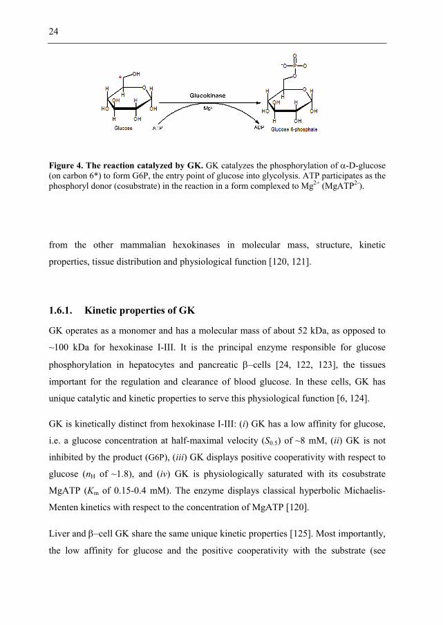
glycolysis (Figure 4).
Glucokinase (hexokinase IV or D) has derived its name from its preference for -D-
glucose as a substrate under physiological conditions. The enzyme is distinguished
24
Figure 4. The reaction catalyzed by GK. GK catalyzes the phosphorylation of -D-glucose (on carbon 6*) to form G6P, the entry point of glucose into glycolysis. ATP participates as the phosphoryl donor (cosubstrate) in the reaction in a form complexed to Mg2+ (MgATP2-).
from the other mammalian hexokinases in molecular mass, structure, kinetic
properties, tissue distribution and physiological function [120, 121].
1.6.1. Kinetic properties of GK
GK operates as a monomer and has a molecular mass of about 52 kDa, as opposed to
~100 kDa for hexokinase I-III. It is the principal enzyme responsible for glucose
phosphorylation in hepatocytes and pancreatic –cells [24, 122, 123], the tissues
important for the regulation and clearance of blood glucose. In these cells, GK has
unique catalytic and kinetic properties to serve this physiological function [6, 124].
GK is kinetically distinct from hexokinase I-III: (i) GK has a low affinity for glucose,
i.e. a glucose concentration at half-maximal velocity (S0.5) of ~8 mM, (ii) GK is not
inhibited by the product (G6P), (iii) GK displays positive cooperativity with respect to
glucose (nH of ~1.8), and (iv) GK is physiologically saturated with its cosubstrate
MgATP (Km of 0.15-0.4 mM). The enzyme displays classical hyperbolic Michaelis-
Menten kinetics with respect to the concentration of MgATP [120].
Liver and –cell GK share the same unique kinetic properties [125]. Most importantly,
the low affinity for glucose and the positive cooperativity with the substrate (see

25
section 1.8) allow GK to maintain a high sensitivity and responsiveness to variations in
the glucose level within the physiological concentration range (4-10 mM).
1.6.2. Gene structure and tissue-specific gene regulation
The tissue distribution of GK and the ‘low-Km’ (20-130 M) hexokinases are different.
Hexokinases I-III are involved in energy metabolism in most tissues, as ubiquitous
housekeeping enzymes. In contrast, GK is expressed with stringent tissue specificity in
accordance with its highly specialized physiological functions.
The GCK gene is located on chromosome 7p15.3-15.1 and consists of 12 exons that
span a region of 45,168 bps. GCK transcription is regulated by two alternative tissue
specific promoters which enable differential regulation of GK in –cells and liver in a
manner consistent with the different function of these two tissues. Whereas the
downstream promoter operates exclusively in the hepatocytes, the upstream,
neuroendocrine promoter is functional in pancreatic –cells, but also in pancreatic –
cells and some rare glucose-sensing neuroendocrine cells of the brain (hypothalamus
and anterior pituitary gland) and enteroendocrine cells of the gut [28, 126-132]. Thus,
there is a complex network of GK-expressing glucose-sensing cells in the body,
important for maintenance of whole body glucose homeostasis.
Three different GK transcripts are produced by alternative splicing, encoding isoform
1 (neuroendocrine) and isoform 2 and 3 (liver). The isoforms differ in the first 14-16
N-terminal amino acids which constitute exon 1 (a, b, c) of the protein. Exon 1a is
expressed in the pancreatic –cell whereas exons 1b and 1c are expressed in the liver
[133, 134]. Exons 2-10 are identical in the three isoforms.

26
1.7. The 3D structure of hGK and the glucose-induced conformational change
GK is a monomeric enzyme composed of two domains; a small (S) and a large (L)
domain, linked by three connecting regions (Figure 5). The active site resides in a cleft
between the two domains [15, 135-137]. GK is highly conserved with 95% sequence
similarity between human and rat (HomoloGene). Before the crystal structure of hGK
was solved, structural predictions were based on homology modeling. The initial
models were made using the X-ray crystal structure of yeast hexokinase B [135, 136,
138, 139]. However, the accuracy of these models was limited by the rather low amino
acid sequence similarity (~30%) to hGK. Models of human brain hexokinase I
(sequence similarity of 54%) provided the basis for more accurate structural
predictions of GK with its substrates, and have been used to locate some of the
disease-causing GK mutations and to interpret their functional implications [137, 140-
147]. Due to its high structural flexibility, GK has proved difficult to crystallize. In
2004, Kamata et al overcame this problem by deleting the 11-15 N-terminal residues
of hGK. These deletion mutants demonstrated similar in vitro kinetic properties as WT
hGK [15]. Two high resolution crystal structures of liver hGK were obtained, one in
its unliganded apo form (3.4 Å, PDB entry 1v4t) and the other in a complex with
glucose and a synthetic GK activator compound (N-thiazol-2-yl-2-amino-4-fluoro-5-
(1-methylimidazol-2-yl)thiobenzamide) (2.3 Å, PDB entry 1v4s) [15]. It should be
noted that residues 157–179 were unassigned in the electron density map of the
unliganded hGK due to a disorder of this region. According to these structures [15],
the sequences 1–64 and 206–439 constitute the L-domain, the sequences 72–201 and
445–465 constitute the S-domain, and the sequences 65–71, 202–205 and 440–444
represent the connecting regions I-III, respectively.
Structural analyses of the two crystallized conformations of hGK confirmed previous
biochemical and biophysical studies [150, 151], providing structural evidence that
hGK undergoes a reversible, large-scale global conformational transition
(isomerization) upon binding glucose, from a ‘super-open’ (inactive) conformation to
a ‘closed’ (active) conformation (Figure 5). The spatial arrangement of the S- and L-

27
Figure 5. The C backbone structure of hGK in the super-open and closed conformation, showing the glucose-induced conformational domain motion and closure of the active site cleft. The dynamic domains, interdomain bending residues and the changes in the interdomain cleft angle were identified with the DYNDOM program [148] using PDB entries 1v4t and 1v4s. The image was created by PyMOL [149].
domains in the apo form of hGK is different (more extended) from the open form of
hexokinase I, and, hence, the structure of apo GK has been referred to as the ‘super-
open’ form [15]. Although the structure and spatial relationship of the closed
conformation of GK and the C-terminal half of the closed form of hexokinase I are
very similar, GK undergoes a much larger domain motion. This is in part due to a
more flexible structure of the interconnecting region I in GK, which facilitates the
movement of the C-terminal helix (see section 1.7.2) [15]. The glucose-induced
conformational rearrangement of GK involves a large-scale hinge bending/sliding

28
motion, which requires the breakage and reformation of numerous interactions. The
final result is a reorientation of the S-domain, involving a ~104 rotation (as compared
to ~12 for hexokinase I) toward the L-domain, which remains largely stationary
(Figure 5) [15]. Hence, the enzyme adopts a more compact (‘closed’) structure
involving a 96% closure of the active site cleft.
1.7.1. The active site The active site of GK resides in a channel-shaped cleft between the S- and L-domain,
and provides a favorable microenvironment for the phosphorylation of the substrate –
D-glucose. The large-scale domain movement induced by binding of glucose (Figure
5), closes the active site cleft and creates the stereochemical environment for binding
of the cosubstrate MgATP. In the ternary GK-glucose-MgATP catalytic complex, the
ATP -phosphate is in close spatial proximity to the 6-hydroxyl group of glucose
[152].
Glucose-binding site In the super-open conformation of GK, the glucose-binding site is exposed to the
solvent and important residues for substrate interaction and catalytic activity are
missing from the site (Figure 6) [15]. Hence, even if glucose is able to bind at this site
with low affinity, the super-open state of the enzyme is considered catalytically
inactive. Glucose binding induces closure of the interdomain cleft and displacement of
the flexible S151-C181 loop structure from the protein surface (Paper III). The loop,
which is part of the S-domain, closes over the incomplete active site as a lid (Figure 5),
protecting the active site from solvent. This active site loop forms one rim of the
glucose-binding site, and contributes to the catalytic environment in the hGK ternary
complex.
The residues that form hydrogen (H) bonds with the oxygen atoms of glucose have
been defined crystallographically [15], and consist of T168 and K169 in the S-domain
and N204, D205, E256 and E290 in the L-domain (Figure 6). The residues N204 and

29
Figure 6. Structure of the glucose-binding site. (A) The glucose-binding site in the closed, binary GK-glucose complex. E256 and E290 of the L-domain (blue stick model), T168 and K169 of the S-domain (cyan stick model), and N204 and D205 of connecting region II (green stick model) form hydrogen bonds with glucose (pink stick model). (B) The glucose-binding site in the super-open, apo form of hGK is exposed to solvent. The residues T168 and K169 of the S-domain (see A) are part of a disordered structure, and are not assigned in the electron density map of the super-open structure (1v4t). The color coding is as described in (A). The figures are reprinted from [15], with permission from Elsevier © 2004.
D205 are part of connecting region II. Mutational analyses have confirmed the
importance of several of these amino acids [138, 146, 153]. In general, mutations in
glucose binding residues have a dramatic effect on the catalytic activity [137]. Most of
these residues have been found mutated in GCK-MODY patients, emphasizing their
critical role for enzyme function [65].
It should be mentioned that in 2004 the structure of hGK with glucose alone (i.e.
without activator) was not solved and, hence, it was not clear whether and to what
extent the active conformation of GK as well as the structure and interactions around
the glucose-binding site are altered by binding of the activator compound.
ATP-binding site Yeast hexokinase and hGK have a low ATPase activity representing 1/10000 and
1/2000 of its kinase activity, respectively [136]. The higher ATPase activity of GK

30
indicated that water molecules are more accessible to the -phosphate of ATP in the
absence of glucose [136]. Interestingly, an ATPase domain with the same basic
structural fold is shared between hexokinases from bacteria, yeast and plants, to
humans and other vertebrates, as well as other less related protein families as actin and
the hsp70 family of heat shock proteins [154, 155]. This actin fold consists of five well
conserved sequence motifs involved in the interaction with ATP; phosphate 1, connect
1, phosphate 2, adenosine and connect 2 (Figure 7) [154].
At the time of conducting this work no crystal structure of a ternary GK-glucose-ATP
complex was available and the most accurate structural predictions on the ATP
binding site were based on homology modeling using human hexokinase type I as
template [137, 140, 141, 143, 146, 147]. Based on these models and ATPase domain
sequence conservation, the residues T82, R85, T228, K296, S336, S411 and K414
were predicted to form H-bond interactions with the ribose moiety or phosphate
oxygens of ATP, placing the nucleotide in the correct orientation and conformation to
interact with glucose. GK mutations that perturb or eliminate the interaction with ATP
generally lead to enzymes with reduced affinity for ATP and severely impaired
catalytic activity [137, 146]. Most of these critical contact residues have been found
mutated in GCK-MODY patients [65].
1.7.2. The C-terminal -helix
The C-terminal -helix (residues 447-460 (helix 17) in the super-open conformation
and residues 443-461 (helix 19) in the closed conformation) plays a crucial role in the
global conformational transition of hGK [15, 157]. Upon domain closure, the C-
terminal helix (referred to as ‘ 13’ in the literature), which is located adjacent to
connecting region III (residues 440-444), moves from a solvent exposed interdomain
position in the super-open form to a sequestered internal location within the S-domain
in the closed conformation [15, 157, 158]. The helix contains several small residues
(glycine and alanine) which assist with the movement around the connecting region. In
both conformations, the C-terminal helix specifically interacts with helix 6 ( 5) of the

31
Figure 7. The five sequence ATP binding motifs of hGK. Multiple alignment of hGK isoform 1 and yeast hexokinases isozymes PI and PII was created using ClustalX v.2 [156] and the reference sequences P35557, P04806 and P04807 (UniProt), respectively. (*) indicates positions which have a single, fully conserved residue (:) indicates conservation between groups of strongly similar properties, and (.) indicates conservation between groups of weakly similar properties. Yellow boxes denote the most evolutionary conserved residues. Residue numbers and secondary structures of the motifs are given (arrow, beta strand; cylinder, helix; half circle, helix-turn or bend). Annotations of the secondary structure elements are derived from the Protein Data Bank using the PDB entry 1v4t.

32
L-domain [15]. During the conformational transition, changes in the domain interface
take place, with the relative orientation of the helices changing from a parallel to a
perpendicular orientation with a subsequent change in main residue interactions [15].
Recent MD simulations support a model in which the ‘release’ of the C-terminal helix
from the S-domain is the final step in the slow conformational transition from the
closed to the super-open state [158]. Moreover, evidence has been presented that this
substrate-induced repositioning of the C-terminal helix is essential for GK kinetic
cooperativity [157]. Interestingly, several GK activating mutations map to this helix or
neighboring interacting structures [15, 65, 157-160].
1.7.3. The allosteric activator site and pharmacological GK activator drugs
The crystal structure of hGK complexed with glucose and a small molecule synthetic
GK activator (GKA), revealed a hydrophobic binding pocket for the GKA molecule at
the domain interface, 20 Å remote from the glucose and ATP binding sites [15]. The
surface of the activator binding site is formed by the flexible loop of connecting region
I (V62-G72), helix 6 of the L-domain (D205-Y215) and the C-terminal helix (E443-
C461) of the S-domain [15, 161, 162]. The search for small molecules that were
capable of activating GK began already in the 1990s [5, 13], and today a wide range of
GK activator compounds have been discovered as a result of intensive medicinal
chemistry efforts. The GKAs have diverse chemical structures, but display a similar
pharmacophore [34, 163]. For some of the activators the binding site and its contact
residues have been defined crystallographically [15, 162, 164-167]. In the unliganded
(glucose-free) form of hGK, the loop of connecting region I is occluded and the C-
terminal helix is released from the small domain, thus, the allosteric pocket is distorted
or absent.
GKAs bind close to one of the hinge regions involved in interdomain communication
and propagation of conformational transitions. By binding to this regulatory site GKAs
are able to allosterically enhance GK activity [13]. In vitro, these molecules increase
the catalytic activity of GK, predominantly by lowering the [S]0.5 value for glucose (5-
10 fold). Many GKAs increase (up to twofold) the turnover rate (kcat) of the enzyme,

33
but in a few instances the kcat is slightly decreased. The Hill coefficient (nH) is lowered
to a varying extent, and in some cases approaching unity [34]. The potential
application of GKAs in the treatment of T2D is currently under investigation [9, 34,
168, 169]. So far, the results from clinical trials (and animal studies) have
demonstrated a dual mechanism of the activator molecules by potentially acting on
both the -cell, by improving GSIS, and the liver, by reducing uncontrolled glucose
output and restoring postprandial glucose uptake and glycogen synthesis [34]. Some
GKAs are believed to carry a risk of inducing hypoglycemia. However, perhaps of
greatest concern are the potential long-term adverse effects of GKAs. As 99% of the
body’s GK is present in the liver, it is possible that enhanced GK activity may lead to
increased de novo lipogenesis and plasma triglyceride levels [9, 170, 171]. Moreover,
the potential effect of GKAs on the function of other GK-expressing cells and organs
must be assessed.
The existence of a physiological, endogenous GKA molecule has been postulated.
Interestingly, the functional effects of synthetic GKAs mimic the kinetic effects of
naturally occurring activating mutations causing HI [31, 65, 98¸ Barbetti, 2009 #177,
101, 103, 105, 144, 172]. In contrast to inactivating GCK mutations, which are
distributed throughout the 3D structure of hGK, almost all the HI-associated mutations
colocalize to a common region in the 3D structure, which coincides with the binding
site of the GKAs [15]. The overlap of these sites may suggest that common molecular
mechanisms of activation are involved.
1.7.4. Cysteine residues at the active site of GK
GK contains 12 cysteine residues, including five that appear as a conserved ring motif
close to the active centre [135, 153]. The close vicinity and spatial localization of the
Cys residues, together with the high structural flexibility of GK, makes the enzyme
very vulnerable towards oxidative formation of intrachain disulfide bridges, and a
concomitant inactivation of enzymatic activity [173-175]. GK is one of the most
sensitive thiol enzymes in the pancreatic -cell, demonstrating high sensitivity towards

34
SH group oxidizing compounds such as the glucose analogue alloxan [176-179].
Glucose protects the enzyme against alloxan-induced inhibition [175]. The inhibitory
effect of alloxan on GK catalytic activity can be prevented and frequently reversed by
sulfhydryl group reducing agents, such as dithiothreitol (DTT), and the presence of
thiol agents has demonstrated to be mandatory during purification of the enzyme
[173]. In the absence of thiol agents, GK experienced a constant decay in activity
[173]. When recombinant GK proteins are subjected to non-reducing SDS-PAGE, a
double-band pattern can be observed, corresponding to the 52 kDa native GK protein
and a faster migrating, more compact, oxidized form of ~49 kDa [176, 180]. The
intensity of the 49 kDa band increased significantly upon alloxan treatment [181].
Moreover, freshly purified recombinant GK also displays a characteristic
electrophoretic double-band pattern, indicating that the enzyme naturally exists in at
least two different conformations [181], dependent upon the redox status of the
sulfhydryl groups. The oxidized form represents ~1-2% of the total protein (Molnes et
al, unpublished observations). No disulfide bonds have been observed in the crystal
structure of pancreatic hGK [152]. In summary, these aspects emphasize that GK, with
its high susceptibility to sulfhydryl oxidation at low glucose concentrations, is very
vulnerable towards oxidative stress, especially in the pancreatic -cell having low
enzymatic antioxidative defense mechanisms [179].
1.8. Kinetic models of positive cooperativity
The positive kinetic cooperativity of GK with respect to glucose was discovered 35
years ago [182, 183]. This property is unique since GK functions exclusively as a
monomer [184] with a single substrate binding site [15]. Two models have appeared
particularly appropriate to explain the observed cooperative kinetics of GK; the
mnemonic and ligand-induced slow transition (LIST) models [21, 185-191]. Both
models attribute the positive cooperativity to a slow, reversible, glucose-dependent
conformational transition between a low affinity and a high affinity form of GK, and
the conversion between the two conformations is slower than the catalytic cycle (kcat).
Accordingly, the existence of different conformational states of the enzyme, combined

35
with their failure to reach conformational equilibria during the course of catalysis, is
the fundamental basis for the sigmoidicity in the reaction rate profile of GK [22, 192,
193].
The mnemonic mechanism [186] is based on the concept that the enzyme retains a
“memory” of the active conformation for some time after product conversion and
release. According to this model, the unliganded enzyme alternates between two
distinct conformational species, a low affinity state (E*) and a high-affinity state (E)
(Figure 8A). Only the low-affinity conformation is postulated to exist in the absence of
glucose. Both forms can bind glucose to form the same binary (E·G) complex, which
reacts rapidly with MgATP to generate the ternary complex. Catalysis occurs from the
high-affinity E·G state. If glucose is abundant, the high-affinity form (E) can rapidly
go through another catalytic cycle, whereas if glucose levels are low, the enzyme
slowly relaxes back to the low-affinity state (E*). Thus, the mnemonic model
postulates the existence of a thermodynamically favored enzyme conformation in the
absence of glucose and involves one catalytically active enzyme species [187]. In
contrast, the LIST mechanism postulates the existence of two catalytic cycles. The
model assumes that a pre-existing equilibrium exists between the two conformational
states (E* and E) in the absence of glucose, and that each can accomplish a separate
catalytic cycle, involving different enzyme states (E·G or E*·G) and kinetics (Figure
8B) [21, 189, 194]. The overall steady-state rate is the sum of the rates for the two
cycles. Furthermore, the equilibrium between the low-affinity (E*) vs. the high-
affinity (E) form is governed by the concentration of glucose. Thus, as the glucose
level increases the equilibrium is shifted towards the high-affinity form. Evidence in
support for both mechanistic models can be found within the experimental data
collected on GK over the last 35 years [21, 120, 150, 151, 182, 187, 188, 194, 195].
Initial support for the existence of a slow glucose-induced isomerization involving
different conformational states of GK came from various observations, e.g. the lag-
phase (~5-10 min) observed in kinetic experiments when rat liver GK was
stored/preincubated at glucose concentrations lower than in the assay [194], and the

36
Figure 8. Alternative mechanisms proposed to explain the positive cooperativity of monomeric GK. (A) The mnemonic and (B) LIST models of positive cooperativity. E and E* represent two conformations of the enzyme. Red arrows represent the slow conformational transition (isomerization) between conformational states. (C) The “Kamata” model based on the crystallographic structures and analysis of domain movement [15]. See the text for details. The figures are reprinted from [15, 193], with permission form Elsevier © 2004/2012.
time-dependent increase in intrinsic tryptophan fluorescence (ITF) of rat liver GK
upon addition of glucose [151]. Structural evidence for the large-scale conformational
alterations that accompany glucose binding was provided by the crystallization of the
unliganded and glucose-bound liver hGK in 2004 [15]. Moreover, based on analysis of
domain movement, Kamata et al predicted the occurrence of an intermediate “open”
conformational form of hGK, in addition to the structurally solved “super-open” and
“closed” forms, and, accordingly, devised a kinetic model that can account for the
cooperative properties of GK (Figure 8C) [15]: (i) The super-open form is
thermodynamically favorable in the absence of glucose; (ii) Upon binding glucose, a
C

37
slow transition from the “super-open” (low-affinity, inactive) form to the “closed”
(high-affinity, active) form is triggered, that is slower than the catalytic reaction; (iii)
After glucose phosphorylation, a fast “closed—open” transition takes place. A large
proportion of the enzyme stays in the “open” conformation for some time, and if
glucose binds during this time, the enzyme re-enters the catalytic cycle. This “fast
cycle” is favored if the glucose concentration is sufficiently high. At low glucose
concentrations, GK will return to the “super-open” form; (iv) GK has two catalytic
cycles, a slow “super-open—closed” cycle and a fast “open—closed” cycle. The ratio
between these two catalytic cycles, which is glucose-dependent, is responsible for the
characteristic sigmoidal nature of GK and its low affinity for glucose [15].
1.9. Regulation of hGK activity
GK is regulated both at the transcriptional and post-translational level by a complex
network of mechanisms which are fundamentally different in the hepatocytes and
pancreatic –cells [6, 34, 196]. As already mentioned (Section 1.6.2), GK gene
transcription is driven by two tissue-specific promoters [133, 134, 197]. The hepatic
promoter, which is primarily controlled by insulin levels, operates exclusively in the
liver, whereas the upstream or neuroendocrine promoter controls GK expression in
pancreatic –cells as well as in glucose sensor cells of the gut, hypothalamus and
anterior pituitary gland [5, 6, 127, 129-132, 196, 198]. In -cells, GCK expression is
constitutive at a relatively low basal activity but subject to direct regulation by glucose
levels [23, 112, 124, 199, 200]. However, some studies suggest that the glucose-
stimulated up-regulation of -cell GCK transcription is mediated, in part, via insulin
secreted in response to glucose [201, 202].
The adaptive response of GK to glucose also occurs at a post-translational level by
means of substrate activation and stabilization [200]. The focus in the rest of this
section and in the discussion will be on post-translational regulatory mechanisms of
GK.

38
1.9.1. Post-translational regulation of hGK
In the pancreatic -cell, the GK protein levels show only marginal variations under
physiological conditions, and the enzyme is considered to be regulated at the post-
translational level [196]. However, post-translational regulation of GK in both -cells
and hepatocytes is complex and only partially understood. It is established that GK
activity, its subcellular localization and cellular stability are regulated by a spectrum of
non-covalent GK-protein interactions that are different in –cells and hepatocytes. In
the hepatocytes, the interaction with the GK regulatory protein (GKRP) is a key short-
term regulatory mechanism of GK activity [203, 204]. The 68 kDa GKRP, first
discovered in rat liver, is an allosteric inhibitor of GK that binds preferentially to the
super-open form of GK which predominates when glucose is low [15, 25, 203]. GKRP
binds GK competitively with respect to glucose, and glucose binding releases GK
from the GK-GKRP complex by inducing a conformational transition to the closed
form. Physiologically, the GK-GKRP interaction and the subsequent inhibition of GK
activity are promoted by fructose-6-phosphate, and suppressed by fructose-1-
phosphate, compounds that bind to GKRP. Thus, the GK-GKRP interaction is
modulated in response to fasting and feeding states [205, 206]. Importantly, GKRP
provides a regulated translocation of GK between the cytosol and nucleus. As the
glucose supply declines during periods of fasting, GKRP binds free cytoplasmic GK
and transports it to the nucleus where GK is sequestered in an inactive state [207, 208].
Postprandially elevated glucose levels dissociate the GK-GKRP complex, and the
active form of GK is translocated to the cytosol. This enables a rapid increase in GK
activity and stimulation of glucose phosphorylation. Moreover, the GKRP-mediated
compartmental redistribution of GK to the nucleus may serve to maintain a functional
reserve of GK that can be quickly mobilized after a meal, in addition to stabilize and
protect the enzyme from degradation by cytoplasmic proteolytic mechanisms [209,
210]. In GKRP deficient mice, the disruption of this regulation and the subsequent
decrease in GK activity leads to altered glucose metabolism and impaired glycemic
control [209]. Furthermore, functional studies on recombinant hGK enzymes have
demonstrated that some GCK mutations cause a loss of regulation by GKRP which

39
may contribute to glucose intolerance in patients with GCK-MODY [65, 211-213].
The GKRP is not present or detectable in the pancreatic islets [214-216]. However, the
presence of an inhibitory protein distinct from the liver GKRP has been suggested
[215].
A second important regulator of GK activity is the bifunctional enzyme
phosphofructokinase-2/fructose-2,6-bisphosphatase (PFK2/FBPase2). The enzyme is
expressed in both hepatocytes and -cells where it is involved in the regulation of the
glycolytic ( -cell and hepatocytes) and gluconeogenetic (hepatocytes) pathways [217].
PFK-2/FBPase-2 is a cytoplasmic binding partner of GK, and in insulin-producing
cells this interaction has been demonstrated to enhance the catalytic activity of GK
[217-219]. This effect may be, at least in part, due to stabilization of a catalytically
favorable (closed) enzyme conformation at elevated glucose concentrations [218-220].
Beside its localization in the cytosol (hepatocytes and -cells) and in the nucleus in
complex with GKRP (hepatocytes), GK has also been found to bind to subcellular
structures such as mitochondria (hepatocytes and -cells) [221-223] and insulin-
containing secretory granules ( -cells) [216, 224, 225]. In mitochondria, GK is part of
a regulatory multiprotein complex, and association of this complex with the outer
mitochondrial membrane is dependent on the presence of the proapoptotic protein
BAD [221, 223, 226]. Moreover, the phosphorylation status of BAD helps regulate the
catalytic activity of GK. The detailed molecular basis for the GK/BAD interaction and
its physiological significance for glucose metabolism, GSIS and apoptosis are, so far,
not fully understood. Another potential mechanism for post-translational modulation
of GK function, specific for the pancreatic -cell, arose from the observations that GK
is part of the outer structure of insulin secretory granules in islet -cells and insulin-
secreting -cell lines [216, 224, 225, 227, 228]. The granule cytoplasm translocation
of GK is regulated by insulin and, moreover, the release of GK from the granule-
bound state was accompanied by an increase in enzyme activity [228]. Hence, it was
suggested that changes in GK activity induced by association/dissociation from insulin
granules may be implicated in the regulation of GSIS in pancreatic -cells [216].

40
Covalent post-translational modifications
Reversible post-translational modifications (PTMs) are used to dynamically modulate
protein activity and stability. PTMs can occur at any step in the life-cycle of a protein
serving various purposes, e.g. to mediate proper folding, cellular stability/turnover,
subcellular localization, allosteric activation/inactivation, alter protein-protein
interactions as well as to target a protein for degradation. There are > 200 different
PTMs of which the majority occur by enzyme-mediated covalent attachment of a small
chemical group, sugar, lipid or protein to one or more of the amino acid side chains in
a target protein (e.g. glycosylation, acetylation, methylation, phosphorylation, S-
nitrosylation, oxidation, ubiquitination, SUMOylation etc.). As already described,
post-translational processes are important in the regulation of the cellular activity and
stability of GK in hepatocytes and -cells, but the knowledge of covalent PTMs of GK
in target cells and their possible regulatory functions has been very limited. However,
studies on cultured -cells have demonstrated that the association of GK with insulin
granules [216, 225] was dependent on its interaction with nitric oxide synthase (NOS)
and that the localization and activity of GK was regulated by post-translational S-
nitrosylation of the enzyme [216, 229]. Furthermore, it was suggested that defects in
site-specific cysteine S-nitrosylation of GK are associated with naturally occurring
GCK-MODY mutations in humans [230].
Ubiquitin (Ub) is a 76-amino acid globular protein (~8.5 kDa) that is highly conserved
in eukaryotic cells. Ubiquitination (or ubiquitylation) is a reversible cellular process
that involves the covalent attachment of one or several Ub proteins to a target protein
[120, 231, 232]. Protein ubiquitination is an elegant example of how a single protein
can regulate an array of diverse cellular processes such as cell cycle progression,
regulated cell proliferation, cellular differentiation, apoptosis, transcriptional
regulation and protein quality control (PQC) [233-236]. Given the central role of Ub in
these processes, dysregulation of the ubiquitination machinery has been found
associated (directly or indirectly) with the pathogenesis of many human diseases [237-
241]. In addition to Ub, there is a growing family of Ub-like proteins (UbLs) which,

41
similar to Ub, covalently modify proteins on lysines by related enzymatic pathways,
but with distinct functional implications [242].
The ubiquitin conjugating system and ubiquitin-mediated proteolytic pathway
Conjugation of Ub to a protein substrate or to itself usually involves the sequential
action of three enzymatic reactions (Figure 9) [237, 243]. In the first step, Ub is
activated for transfer by ubiquitin-activating enzyme E1 in an ATP-dependent
reaction: E1 catalyzes the adenylation of Ub at the C-terminal glycine residue (G76),
followed by the formation of an E1-S-Ub thioester intermediate [244]. In the second
(ubiquitin-conjugating) step, activated Ub is transiently transferred from E1 onto an
active site cysteine of the E2 enzyme via a trans(thio)esterification process to form
another thioester bond [245]. To complete the enzymatic sequence, E2-S-Ub interacts
with the E3 ubiquitin-protein ligase, which recognizes and associates with the
substrate, promoting the transfer and conjugation of Ub [246, 247]. The Ub moiety is
generally conjugated to target proteins through the formation of an isopeptide bond
between a lysine ( -amino group) on the protein and the C-terminal carboxyl group of
Ub. The completion of one ubiquitination cycle results in a monoubiquitinated
substrate. However, most often the cycle is repeated to form polyubiquitinated or
multiubiquitinated substrates. The ubiquitin-chain is lengthened by the E3 ligase in
collaboration with E1 and E2, sometimes with the help of an additional conjugating
factor E4, specifically required for efficient multiubiquitin chain extension [248-250].
Eukaryotes are today estimated to have two E1 enzymes, ~40 E2 enzymes and > 600
E3s or E3 multiprotein complexes [251]. The E2-E3 pair is the primary determinant of
substrate specificity [243]. Ubiquitination is reversible through the action of a large
family of deubiquitinating enzymes (DUBs) (isopeptidases) which releases and
disassembles polyubiquitin chains, enabling recycling of ubiquitin [252-254]. The
human genome is predicted to encode ~95 DUBs which fall into at least five different
classes [255].

42
Several ubiquitin-binding domains (UBDs), including UIM (Ub-interacting motifs)
and UBA (Ub-association), can form transient, non-covalent interactions with either
mono-ubiquitin or polyubiquitin chains [249, 250]. Cellular proteins containing one or
several UBDs (often called ubiquitin receptors) are the immediate decoders of
ubiquitination, being responsible for transmitting specific Ub signals to downstream
cellular events. Importantly, the selective preferences of UBDs for ubiquitin chains of
specific length and linkage are central to the versatile functions of Ub.
The topology of the polyubiquitin chain appears to be a function of specific E2s and
the E2-E3 combinations involved [251-253]. All seven internal Lys residues (K6, K11,
K27, K29, K33, K48 and K63) of Ub are believed to contribute to the assembly of
polyubiquitin chains [254, 255]. This ability to form a variety of structures with
diverse lengths and linkages distinguishes ubiquitination from UbL modification of
proteins. It is generally assumed that the formation of polyubiquitin chains of different
linkage types provides functional specificity that determines the fate of the modified
protein [254].
Ub is best known for its function in targeting proteins for controlled degradation by the
26S proteasome [256, 257]. Most proteins degraded by the ubiquitin-proteasome
pathway are linked to a polyUb chain in which the Ubs are coupled through isopeptide
Figure 9. The ubiquitin-proteasome degradation pathway. See text for details. The figure is reprinted from [248], with permission from Nature Publishing Group, a division of Macmillan Publishers Ltd © 2010.

43
linkages to K48 (or in some cases K11 or K29) on the preceding Ub. At least four
sequentially added Ub moieties are believed to be required for efficient recognition
and binding of the modified target protein to the proteasome [258]. In contrast,
monoubiquitination and K63-linked polyubiquitin chains serve non-proteolytic
functions in various intracellular pathways [234-236, 254, 259, 260]. Once a target
protein is marked by K48-linked polyubiquitination, it appears to have a short half-life
in cells, as it is rapidly degraded by the proteasome. Proteins can be targeted directly
to the proteasome through recognition of polyubiquitin by the 26S complex or,
alternatively, it can occur indirectly, mediated by various proteasomal shuttle factors
[255]. The proteasome is a large (26S or ~2400 kDa) ATP-dependent multicatalytic
protease that is present both in the nucleus and cytosol of eukaryotic cells [237, 261].
The 26S proteasome is composed of two subcomplexes, a 20S proteolytic core that
provides the proteolytic activity needed to degrade modified substrates, and a 19S
regulatory complex that confers the ability to recognize and unfold polyubiquitinated
protein substrates and insert them into the proteolytic chamber [261]. The degradation
through the proteasome is an irreversible process and the proteins are degraded to
small peptides, most of which are hydrolyzed to amino acids by the sequential action
of endo- and exopeptidases in the cytosol and nucleus. Proteins destined for
degradation need to be deubiquitinated to ensure efficient substrate degradation as well
as recycling of Ub, and tightly regulated deubiquitination is accomplished by
proteasome-associated DUBs [262, 263]. The main steps in the ubiquitin-proteasome
pathway are illustrated in Figure 9.
Role of the ubiquitin-proteasome pathway in –cell dysfunction and hyperglycemia
Chronic hyperglycemia has been associated with oxidative stress and subsequent
defective insulin secretion [264]. As previously mentioned, the pancreatic -cell is
particularly vulnerable towards oxidative stress [179], which in turn may trigger the
ubiquitin-proteasome pathway [265], suggesting a potential involvement of this
pathway in hyperglycemia and -cell dysfunction [266]. Interestingly, the ubiquitin-
proteasome degradation pathway has been ascribed a regulatory role in the pancreatic

44
-cell in glucose-stimulated (pro)-insulin synthesis, biogenesis and surface expression
of the K+ATP channel and in maintaining the normal function of the Ca2+ channel – all
essential components of GSIS [267-269]. However, little is known about covalent
post-translational modifications of GK and their possible regulatory functions in the
target cells, and the molecular and cellular mechanisms involved in the
degradation/turnover of GK are also poorly understood. The possible implications of
Ub (or UbLs) and the ubiquitin-proteasome pathway in this regard have not yet been
subject for investigation and, hence, remain to be elucidated.

45
2. AIMS OF THE PRESENT STUDY
Two 3D structures of hGK were solved in 2004 [15]. This represented a milestone in
the research on hGK and hGK-linked glycemic disorders opening up new and
intriguing possibilities for structure-function studies of WT hGK and its disease-
associated mutant forms. The two structures also represented the starting point of the
present study, with the main objective to characterize structural, dynamic, catalytic and
regulatory properties of WT hGK as a basis for studies on disease-related mutant
forms in which the molecular mechanism(s) of the disease remained to be established.
The specific aims were: 1. To characterize steady-state kinetic properties of WT hGK and some selected
mutant forms.
2. To study the reversible glucose-induced global conformational transitions in WT
hGK and Trp mutant forms using ITF spectroscopy.
3. To provide new insight into the mechanism of substrate (glucose) activation:
a. Identify the active site residues involved in the initial recognition and
binding of glucose to the super-open (apoenzyme) conformational form.
b. Determine the site of initiation of the glucose-induced conformational
transition.
4. To provide new insight into the interaction of the cosubstrate (Mg)ATP with hGK:
a. Investigate whether the cosubstrate can bind to the apoenzyme and the
possibility of a related conformational change.
b. Identify active site residues involved in ATP recognition and binding to the
super-open (apoenzyme) conformational form as well as related structural
changes.
5. To gain further insight into the catalytic function of WT hGK and its multiple
disease-related mutant forms.
6. To examine ubiquitination as a potential regulatory mechanism of hGK, in
particular with respect to enzyme activity and stability (degradation/ turnover).

46
3. SUMMARY OF RESULTS
3.1. Paper I
In this work, we have studied binding of -D-glucose and the associated glucose-
induced global conformational transition of pancreatic hGK in real-time by ITF
spectroscopy. By a combined approach using functional mutagenesis and structural
dynamic analyses we aimed to identify key residues involved in this process. A
hyperbolic binding isotherm for -D-glucose was observed at 25 C with a Kd value of
4.8 0.1 mM. Single-site W F mutation analyses revealed a contribution of all three
tryptophans in the overall glucose-induced ITF enhancement, with major contributions
of W99 and W167, which both are located in highly flexible loop structures. The
measured biphasic time-course of the fluorescence enhancement is influenced to a
variable extent by all three tryptophans. To identify the residues involved in the initial
binding of glucose to the unliganded “super-open” conformation and subsequent
conformational transitions, all interacting residues defined in the 3D structure of the
closed conformation were individually mutated. The mutations N204A, D205A and
E256A/K of the L-domain resulted in enzyme forms which did not bind -D-glucose
at 200 mM as measured by ITF and they were essentially catalytically inactive. In
contrast, in the mutants of the S-domain (T168G and K169N), glucose induced a
significant fluorescence enhancement, but with increased Kd values. Overall, our data
support a molecular dynamic model in which initial binding of -D-glucose to residues
N204, N231 and E256 of the L-domain and subsequent interaction with D205 trigger
local structural changes that propagate in a concerted fashion toward a closed
conformation. This involves the highly flexible interdomain connecting region II
(R192-N204), helix 5 (V181-R191), helix 6 (D205-Y215) and the C-terminal helix 17
(R447-K460).
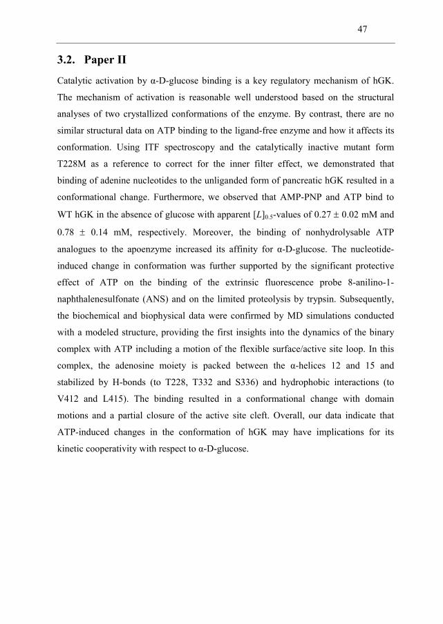
47
3.2. Paper II
Catalytic activation by -D-glucose binding is a key regulatory mechanism of hGK.
The mechanism of activation is reasonable well understood based on the structural
analyses of two crystallized conformations of the enzyme. By contrast, there are no
similar structural data on ATP binding to the ligand-free enzyme and how it affects its
conformation. Using ITF spectroscopy and the catalytically inactive mutant form
T228M as a reference to correct for the inner filter effect, we demonstrated that
binding of adenine nucleotides to the unliganded form of pancreatic hGK resulted in a
conformational change. Furthermore, we observed that AMP-PNP and ATP bind to
WT hGK in the absence of glucose with apparent [L]0.5-values of 0.27 0.02 mM and
0.78 0.14 mM, respectively. Moreover, the binding of nonhydrolysable ATP
analogues to the apoenzyme increased its affinity for -D-glucose. The nucleotide-
induced change in conformation was further supported by the significant protective
effect of ATP on the binding of the extrinsic fluorescence probe 8-anilino-1-
naphthalenesulfonate (ANS) and on the limited proteolysis by trypsin. Subsequently,
the biochemical and biophysical data were confirmed by MD simulations conducted
with a modeled structure, providing the first insights into the dynamics of the binary
complex with ATP including a motion of the flexible surface/active site loop. In this
complex, the adenosine moiety is packed between the -helices 12 and 15 and
stabilized by H-bonds (to T228, T332 and S336) and hydrophobic interactions (to
V412 and L415). The binding resulted in a conformational change with domain
motions and a partial closure of the active site cleft. Overall, our data indicate that
ATP-induced changes in the conformation of hGK may have implications for its
kinetic cooperativity with respect to -D-glucose.

48
3.3. Paper III
In this study, we investigated the role of ubiquitination as a potential post-translational
regulatory mechanism of hGK. Using the rabbit reticulocyte lysate (RRL) as an in
vitro model system, we demonstrated that pancreatic -cell (isoform 1) and liver
(isoform 2) hGK serve as substrates for the ubiquitin-conjugating enzyme system.
Moreover, we found that both isoforms are polyubiquitinated on at least two lysine
residues. Mutational analysis was used to possibly detect the target residue(s), and the
results indicated that multiple lysine residues function as redundant ubiquitin acceptor
sites. Deletion of the C-terminal -helix, containing a putative ubiquitin-interacting
motif (UIM), resulted in loss of polyubiquitination at one site, suggesting that one of
the target lysines is associated with the UIM motif. Proteasome-dependent degradation
of hGK was studied in a proteasome and ATP-enriched RRL system and demonstrated
that poly-/multi-ubiquitination of hGK in vitro, at least in part, served as a signal for
proteasomal degradation of the newly synthesized protein. Moreover, we demonstrated
that free polyubiquitin chains (Ub5, K48-linked) interacted with and allosterically
activated recombinant hGK at low nanomolar concentrations (1.4 fold at ~100 nM
Ub5), assigned to their equilibrium binding to the C-terminal UIM site. Furthermore,
the affinity of Ub5 binding to hGK was regulated by the glucose-dependent
conformational state of the site. We suggest that both these ubiquitin-mediated
processes represent potential physiological regulatory mechanisms for hGK as a
glucose sensor in pancreatic -cells.

49
4. METHODS AND METHODOLOGICAL ASPECTS
4.1. Functional comparison of GST-hGK and non-tagged hGK
For detailed biochemical and physico-chemical studies on hGK, milligram quantities
of highly purified recombinant enzyme are imperative. In this study, efficient and
high-level expression of recombinant pancreatic hGK was provided by expression in
E. coli as GST fusion proteins [270]. Rapid and gentle purification by affinity
chromatography ensured the recovery of pure (>98% as determined by SDS-PAGE)
and enzymatically active, full-length GST-hGK (76 kDa). After cleavage of the fusion
protein at the restriction site by factor Xa, non-tagged hGK (52 kDa) was isolated with
a recovery of ~50 % and no detectable loss in enzyme activity. The relative low yield
of isolated, tag-free hGK can be explained by incomplete cleavage and loss in the final
repurification step.
Prior to the outset of this project, the GST fusion proteins of hGK were mostly used in
the kinetic characterization of mutant forms, due to the potentially stabilizing effect of
the fusion partner. In order to validate the utility of GST-hGK in kinetic and
fluorescence studies, we compared the functional properties of tagged and non-tagged
WT enzyme. They demonstrated similar steady-state kinetic parameters and
comparable apparent binding constants for glucose (Paper I) and AMP-PNP (Paper II)
in equilibrium binding studies by ITF spectroscopy. These findings, in good agreement
with other reports on the subject [271-274], substantiate that the fusion partner at the
N-terminal does not affect the kinetics of WT hGK or perturb the substrate-induced
conformational changes. Moreover, the increase in ITF signal response when glucose
is added at saturating concentrations was comparable without (Feq-Fo ~33) and with
(Feq-Fo~30) the fusion partner (Paper I), considering the experimental error in
determining the absorption coefficients at 280 nm for the two proteins. However, the
relative fluorescence increase ( Fmax,eq/Fo) of WT GST-hGK was ~27%, as compared
with a ~60% increase for WT hGK. This is explained by GST containing four Trp
residues [275], contributing to the background fluorescence (Fo). These quantitative
differences in the ITF response to glucose between non-tagged and tagged enzyme

50
were also observed in the binding studies with AdNs, although the ( Fmax,eq/Fo) value
of the AdN-induced fluorescence quenching was much smaller.
Concurrent studies of more than 30 mutant forms of hGK have provided a detailed
comparison of the kinetic parameters of tagged and non-tagged hGK, which further
support a limited interference of the GST tag [273, 274]. However, care should be
taken when characterizing more mutant proteins since the GST moiety may not always
be inert [273]. In fact, recent data are consistent with GST acting as a transiently
stabilizing partner for some mutant forms (Negahdar et al, unpublished and [273]).
This finding is not surprising since recombinant, non-tagged WT hGK has a marginal
thermal stability and a propensity to aggregate at temperatures 40 C (Paper II and
[276]).
4.2. Helix nomenclature
The helices in hGK are designated according to the crystal structures resolved in 2004
[15]. A total of 17 helices were identified in the unliganded, super-open state (PDB
identity: 1v4t) versus 19 helices in the glucose and allosteric activator bound, closed
state (PDB identity: 1v4s) (see Paper I). In this study, we have numbered the helices
successively irrespective of helix type ( - or 310 helix). This differs from Kamata et al
who generally refer only to -helices (one exception). Thus, in hGK the C-terminal
helix corresponds to helix 17/19 or 13. The information on secondary structure
elements were generally retrieved from the MolMovDB of the Yale Morph Server
[277, 278].
4.3. Analyses of protein dynamics
Although the static 3D crystal structures of GK have been determined for the
apoenzyme and different complexes with substrate(s) and low molecular weight
activators (see Section 1.7), the function of the protein in solution at physiological
temperatures is governed by its dynamic properties. In the present study, we have used

51
fluorescence spectroscopy and computational methods to gain insights into different
conformational states and dynamics of hGK.
4.3.1. Intrinsic fluorescence spectroscopy
Intrinsic fluorescence spectroscopy is a highly sensitive biophysical tool for the
structural and dynamic characterization of proteins. The method relies on the natural
(intrinsic) fluorescence of proteins in the near UV region, which mainly arises from
the aromatic amino acids tryptophan (Trp) and tyrosine (Tyr). Trp is by far the most
frequently used fluorescence probe due to its high extinction coefficient ( ), large
Stokes’ shift (h EX-h EM) and quantum yield ( F), coupled with the unique
environmental sensitivity of its indole ring. Moreover, Trp is a relatively rare amino
acid in proteins, and therefore more specific information on the local conformation and
environment of individual Trp residues can be attained. Its fluorescence is
characterized by a high signal to noise ratio with need of relatively low concentrations
( M) of pure protein when reagents of high analytical grade are used. In intrinsic
tryptophan fluorescence (ITF) spectroscopy the fluorescence from Trp is selectively
measured using excitation wavelengths in the range 295-305 nm. Both the
fluorescence emission intensity and maximum emission wavelength ( max) vary
depending upon the polarity and dynamics of the molecular environment, which make
ITF a useful conformational probe. The max of Trp residues in proteins is highly
related to their degree of solvent exposure. In general, the max of the indole ring is
blue-shifted if the group is buried within the hydrophobic core of the native protein,
and its emission shifts to longer wavelengths (red shift) upon exposure to polar amino
acid groups or solvent water (following, for instance, ligand-induced conformational
changes). Trp residues buried in apolar core regions of a protein have a blue emission
maximum, as low as ~310 nm, whereas Trp fully exposed to an aqueous environment
can have a red emission maximum of ~355 nm. The fluorescence spectra of multi-Trp
proteins are generally broader than that of single-Trp proteins due to the contribution
of multiple emitters with different, but overlapping spectra.

52
GK has three Trp residues (W99, W167 and W257), and the 3D structures of the
protein have provided valuable information on their backbone conformation and
microenvironment in different conformational states. Thus, ITF has been found to be a
sensitive indicator of even minor structural changes of the enzyme and its dynamic
properties on the ms-to-min time scale. As first reported by Lin and Neet [151],
binding of glucose results in an increase in the ITF signal response and a shift in max
of the emission spectrum. In order to assess the contribution of the individual Trp
residues to the glucose-induced fluorescence increase, we made (Paper I) single-Trp
mutants (retaining two Trp residues) by site-directed mutagenesis, where the Trp
residues were substituted one at the time. Trp was replaced by phenylalanine (Phe),
which does not contribute to the fluorescence when excited at 295 nm. The mutations
did not significantly affect the backbone structure or conformational stability of the
enzyme (Paper I and [273]). However, the mutations affected the steady-state kinetic
parameters of the enzyme. The W167F mutant in the active site loop showed a
reduction in catalytic efficiency (kcat/[S]0.5) and kinetic cooperativity with respect to
glucose, which probably can be explained by its localization in the glucose binding
pocket adjacent to the glucose-interacting residues T168 and K169. The W257F
mutant revealed a ~50% reduction in catalytic efficiency, which may be due to its
position in a rigid backbone structure near the glucose binding site, conferring minor
local structural perturbations affecting the glucose affinity. Interestingly, the W99F
mutant in a highly flexible loop structure displayed a small increase in substrate
affinity, and thus, catalytic efficiency. W99 is located in close vicinity to connecting
region I and the GK activator (GKA) binding site, and it has been suggested that
mutations at this site may stabilize the closed form of the enzyme [273].
In order to determine the contribution of the three Trp residues to the glucose-induced
fluorescence enhancement of hGK, their +glucose/-glucose fluorescence difference
spectra were generated. Mutations of W99 and W167 resulted in a large decrease (~70
and ~80 %, respectively) in the fluorescence response to glucose, whereas mutation of
the “buried” W257 gave only a moderate decrease of ~35%. Furthermore, the W99F
mutant demonstrated a ~11 nm blue shift in max in the difference spectrum compared
53
to WT hGK, whereas only a ~2 nm shift was observed for W167F and W257F
(towards the blue and red, respectively). This indicates that W99 is responsible for the
more red emission of WT hGK, in agreement with its high degree of exposure to
aqueous solvent in both the super open (27%) and closed state (45%) of hGK. The
fluorescence spectrum of WT hGK apoenzyme (pH 7.0) displayed an emission
maximum of ~340 nm. Upon binding of glucose a ~2 nm blue shift was observed. This
shift is most likely explained by changes in the fluorescence of W167, experiencing a
large reduction in solvent accessibility upon cleft closure and desolvation of the active
site (Paper I). Recently, the relative solvent accessibility of the Trp residues was
determined by acrylamide quenching analyses (W99 (11.6/13.1 %), W167 (14.4/5.8
%) and W257 (6.2/5.2 %)), providing experimental evidence that the solvation of
W167 is significantly decreased by glucose [279]. Thus, the magnitude of the glucose-
induced fluorescence increase is primarily determined by W99 and W167, and little
affected by W257. Complementary studies on double-Trp mutants (retaining only one
single Trp residue) have recently been performed [273, 279], and the results are
consistent with the findings presented herein for the single-Trp mutants.
4.3.2. Extrinsic fluorescence spectroscopy
A number of polyaromatic hydrocarbons or heterocycles have been used as extrinsic
fluorescent probes in the study of protein structure and ligand-induced conformational
changes. Here, we have used 8-anilinonaphthalene-1-sulfonate (ANS) as an extrinsic
fluorophore with affinity for hydrophobic clusters in GK. The weak fluorescence of
ANS was greatly enhanced upon binding to ligand-free WT hGK. Both ATP and
glucose binding significantly reduced the ANS fluorescence signal (glucose>ATP),
compatible with a decrease in accessible hydrophobic clusters as compared with the
ligand-free enzyme.

54
4.3.3. Computational methods
X-ray-diffraction data on GK contains information not only about the average 3D
structure but also about the spatial distribution around this state. This mean-square
atomic displacement [280] is commonly expressed as the B-factor or temperature
factor. The isotropic B-factor values calculated for the C carbons (Paper I)
demonstrated the freedom and restriction for various sites, with low values ( 30 A2)
for the glucose-interacting residues in the unliganded state, except for T168 and K169.
Complementary global mode analysis by the Gaussian Network Model (GNM) [281,
282] revealed similar sites (minima) of low translation mobility.
Among the different theoretical methods available for description of protein flexibility,
molecular dynamics (MD) is probably the most powerful [283-285]. In this study
(Paper II), we have used state of the art atomistic MD simulations, close to the
physiological conditions, by the widely used AMBER program package [286]. Based
on the atomic coordinates for unliganded hGK as a starting point, MD simulations (ps-
to-2 ns time scale) of the binary complexes with ATP and glucose were carried out in
explicit water (that is, individual water molecules were included in the simulations).
The simulations indicated a significant conformational change of hGK upon ATP
binding, including motion of the flexible surface/active site loop and partial closure of
the active site cleft, and also showed the residues directly contacting the nucleotide at
the active site. In contrast, when the MD simulations were performed with glucose in
the super-open conformation, there was no measurable closure of the active site cleft
during the 2-ns MD simulations. In this case, the simulation time was too short to
observe the large glucose-induced conformational change of hGK, characteristic of a
hysteretic enzyme, which occurs on a ms-to-min time scale in the ITF studies (Paper I
and [157, 287]).
55
5. GENERAL DISCUSSION
5.1. GK protein stability
GK plays a central role in maintaining glucose homeostasis, serving as a glucose
sensor in the pancreatic -cells and as a regulator of hepatic glucose uptake and
glycogen synthesis [7]. Alterations in GK activity and regulation are associated with
abnormal glycemia, as illustrated by naturally occurring GCK mutations causing
diabetes or hyperinsulinemic hypoglycemia. This emphasizes the importance of
preserving the stability and activity of this enzyme, also under conditions of cellular
stress.
Recombinant GK without a fusion partner is only marginally stable under
physiological conditions. It has a low intrinsic thermal stability (apparent Tm = 42.4 ±
0.2 C) and a propensity to aggregate at Tp 40 C (Paper II and [276]). Glucose and
ATP stabilize the enzyme through a ligand-induced conformational change to a more
compact conformation, as demonstrated by the protective effect of the ligands (glucose
> ATP) on limited proteolysis (Paper II). A stabilizing effect of glucose on the thermal
stability of GK has also been observed in ITF spectroscopy [279] and differential
scanning calorimetry studies [279, 287]. Moreover, glycerol has been demonstrated to
increase the thermostability of GK and promote glucose binding (lowering its Kd)
presumably by reducing/removing water interactions with the protein [279]. However,
the stabilization by glucose and glycerol was limited as they were unable to prevent
irreversible heat denaturation above 40 C [279]. Interestingly, in contrast to
guanidine-Cl (Paper II), urea prevented irreversible aggregation and preserved
reversibility of the thermal unfolding process, probably explained by its ability to bind
directly to the protein backbone and the hydrophobic side chains of GK [279]. Overall,
these studies have contributed to an increased understanding of the biophysical basis
of WT GK thermostability and established a foundation for studies on disease-
associated mutant forms.

56
In silico calculation of the free energy of folding/unfolding ( G) [288, 289],
predict that several misfolded missense GCK-MODY mutations harbor a reduced
thermal stability that could be related to an increased rate of degradation when
expressed in mammalian cells (Negahdar et al, unpublished). To date, ~15 GCK-
MODY mutations have been identified with an apparently reduced in vitro thermal
stability of the recombinant enzymes as fusion proteins, and has been proposed to
explain (at least in part) their “loss-of-function” [114, 139, 211, 212, 271, 272, 290-
295]. However, it can be anticipated that many more of the >600 GCK mutations
identified so far will demonstrate reduced in vitro stability. Although such in vitro
stability studies can provide valuable information on mutant forms, the kinetic and
thermodynamic parameters for folding-unfolding processes in the cell are different
from the “ideal” in vitro conditions. For instance, macromolecular crowding affects
the structure and function of proteins under physiological conditions and favors
intermolecular interactions with proteins and other cellular components [296-299].
Recently, we have investigated the effect of the catalytically near-normal mutations
S263P, G264S and R275C on the cellular stability of GK (Negahdar et al,
unpublished). Evidence was provided that their “loss-of-function” is, at least in part, a
result of protein misfolding and destabilization, causing dimerization/aggregation and
an enhanced rate of cellular degradation.
A complex network of mechanisms is involved in the regulation of GK catalytic
activity in pancreatic –cells and hepatocytes. However, little is known regarding the
cellular protein quality control (PQC) machinery involved in the recognition and
targeted degradation of GK and its many disease-associated mutant forms [65]. In
Paper III, we have demonstrated that recombinant pancreatic and liver WT hGK are
covalently modified by ubiquitination, and that poly/multi-ubiquitination of hGK in
vitro can serve as a signal for proteasomal cotranslational degradation of the newly
synthesized protein. Protein synthesis, folding and degradation are closely coupled
processes, and a significant fraction of newly synthesized proteins are degraded
cotranslationally [300-302]. This may represent an early recognition by the PQC
machinery of misfolded nascent proteins [301, 303]. Furthermore, recombinant hGK

57
was found to interact with and to be allosterically activated up to 1.4-fold by purified
free polyubiquitin chains at low nanomolar concentrations, assigned to their
equilibrium binding to a C-terminal UIM site (Paper III). Together, these findings
suggest a potential role of ubiquitination in the cellular regulation of GK catalytic
activity and stability/turnover, as well as in the interaction with other cellular proteins,
including ubiquitin-conjugated proteins and free/conjugated poly-ubiquitin chains.
Because ubiquitination of misfolded proteins associated with cytoplasmic chaperones
are mostly degraded through the ubiquitin-proteasome system [304], further studies
are warranted to investigate this possibility for selected loss-of-function mutants of
GK.
5.2. The multiphasic global conformational transitions and perturbations of conformational equilibria
In steady-state kinetics, GK shows non-hyperbolic (sigmoidal) dependence on glucose
concentration [150, 187], and the underlying mechanism for the cooperative behavior
of GK has been of primary interest in the study of this enzyme. The classical concerted
model of cooperative kinetics [305] does not apply to the monomeric GK with a single
substrate binding site. The possibility of two glucose binding sites in hGK has been
proposed [147], but only a single glucose binding site was observed in the crystal
structure [15, 152]. Furthermore, equilibrium binding of glucose to unliganded WT
hGK displays a hyperbolic (non-cooperative) binding isotherm (Kd = 4.9 ± 0.1 mM)
(Paper I and [273, 287]), consistent with a single binding site. It is now widely
accepted that the positive cooperativity of GK with respect to glucose is kinetic in
origin, and monomeric GK has emerged as a model system for understanding this type
of cooperative response.
GK is activated by binding of glucose, and this process has been described as a
reversible, slow transition from a super-open, inactive (low affinity) state to a closed,
high activity (high affinity) state [151, 287]. Crystal structure analyses of the
unliganded and glucose-bound hGK [15, 152] have confirmed these observations by

58
demonstrating that binding of glucose at the active site induces a large-scale domain
movement that closes the active site cleft and creates the stereochemical environment
for binding of the cosubstrate MgATP2- and catalysis. The three residues N204, N231
and E256 in the L-domain was proposed to function as primary contact residues (Paper
I and [15]). The hGK-glucose association is driven by a favorable entropy change ( S
= 150 ± 10 J mol-1 K-1) (Paper I and [279]), in keeping with the fact that an increase in
protein dynamics plays a dominant role in the interaction. In the closed conformation,
precise alignment of additional substrate interacting residues (notably T168 and K169
in the flexible surface/active site loop of the S-domain) and the subsequent higher
affinity for glucose, efficiently accelerate the chemical reaction on binding of
MgATP2-.
Recent results from x-ray crystallography, high-resolution NMR, targeted MD
simulations, pre-steady state and steady-state glucose binding studies have shown that
hGK is an intrinsically mobile enzyme that can sample multiple distinct
conformational states both in its unliganded and binary enzyme-glucose complex [151,
158, 160, 276, 287, 306-308]. Furthermore, its positive kinetic cooperativity with
respect to glucose is mediated by the glucose-induced conformational transition, which
probably includes interconvertible intermediate states exhibiting variable degree of
cleft closure and different affinities for glucose. Interestingly, Liu et al [309] have
assessed conformations of GK in solution by small angle x-ray scattering (SAXS)
experiments, demonstrating that glucose in solution dose-dependently converts GK
from an apo (super-open) conformation to an intermediate active open conformation,
distinct from the active, closed conformation observed in crystals [15]. The glucose-
bound open conformation was predicted with a cleft opening angle of ~17 relative to
the closed conformation obtained for the crystal structure [309]. The authors suggest
that the active open conformation represents a physiological intermediate in the
reaction cycle of GK, in agreement with a previous proposal [15].
Increasing evidence substantiate that also the ligand-free enzyme in solution is in a
preexisting equilibrium between at least two conformers, i.e. the super-open
conformation and an alternative (presumably less open) conformation with a higher

59
affinity for glucose [276, 306-308]. However, the apoenzyme is considered to be
dominated by the thermodynamically favored super-open conformation [158, 306,
307, 309], as suggested based on 3D structure analyses [15, 309]. In order to gain
insight into the free-energy landscape of proteins, temperature perturbations can be
used to shift the population equilibrium [310]. In unliganded WT hGK the observed
temperature induced (1 C to 39 C) reversible quenching of the ITF fluorescence is
consistent with a slow conformational isomerization (thermal hysteresis), reminiscent
of the global glucose-induced conformational change (Paper I). Moreover, the
observed biphasic time course suggests the presence of a relatively stable intermediate
in the thermal transition, and gives support to the existence of an equilibrium between
conformational substates of hGK in the absence of ligand.
In Paper II, we have provided the first experimental evidence for an equilibrium
binding of ATP and the analogue AMP-PNP to the ligand-free enzyme and a
subsequent nucleotide-dependent conformational change, including motion of the
flexible surface/active site loop and partial closure of the active site cleft. In addition,
MD simulations provided insights into the active site contact residues involved in
binding ATP. The ligand-free and glucose-bound enzymes are dynamic entities that
can sample multiple conformational substates [276, 308], and we hypothesize that
nucleotide binding may shift the equilibrium between different conformations (see
Figure 10), as previously demonstrated for glucose (Paper I and [151, 160, 287, 306,
307]) and allosteric effectors (see below). In fact, results from steady-state kinetic
analyses (Paper II) gave support to a certain conformational control of GK catalytic
activity by binding of (Mg)ATP, with possible implications for the kinetic
cooperativity with respect to glucose. Moreover, using ITF fluorescence titrations it
was demonstrated that binding of ATP analogues to the ligand-free enzyme resulted in
an increased equilibrium binding affinity for glucose, both in the WT enzyme and the
low affinity GCK-MODY mutant L146R (Paper II). We speculate that these effects
may be related to a partial catalytic activation of GK following ATP binding, and that
similar or possible larger effects of ATP may occur in other GK disease-associated
mutant forms.

60
Figure 10. Schematic representation of the standard free energy landscape for the catalytic network of the GK reaction. Conformational changes occur along both axes. The changes occurring along the Reaction Coordinate axis correspond to the environmental reorganization at the active site that facilitates the chemical reaction (Papers I and II, and references therein). It includes molecular motions related to substrate binding (Section 4 and [158]). In contrast, the conformational changes occurring along the Ensemble Conformations axis represent the ensembles of conformational substates existing at all stages along the reaction coordinate [276, 307, 308], possibly leading to a number of parallel catalytic pathways [310]. A state is defined as a minimum in the energy surface, whereas a transition state is the maximum between the wells [311]. A change in the system (ligand binding, mutation) shift equilibrium between states. The figure and portions of the caption are adapted from a recent model on the catalytic network of the enzyme dihydrofolate reductase [312].
It has been suggested that the allosteric GK regulators GKA and GKRP modulate the
activity of GK by finely tuning its conformational landscape [307]. In other words,
they act as reversible conformational traps, by binding and stabilizing a specific GK
conformation [307]. According to this model, GKRP stabilizes open GK conformers
that prevail at low glucose concentrations, whereas increased glucose concentration
and/or addition of GKAs shift the equilibrium towards the closed conformers and
induce a dissociation of the GK-GKRP complex. SAXS experiments have
demonstrated that the GKAs shift the conformational equilibrium of GK to the active
conformation at lower glucose concentrations [309]. The SAXS profiles revealed
small conformational differences between the various GK-glucose-GKA complexes,

61
which was consistent with the inherent ability of the individual GKA to activate GK
(GKAs that suppressed [S]0.5 for glucose mildly gave a GK-glucose-GKA complex
that was less compact than those GKAs with a more dramatic impact on the [S]0.5 -
value) [309].
Similarly, it has been proposed that mutations in hGK may shift its conformational
equilibria by stabilizing or destabilizing one conformer, or facilitate or slow the super-
open closed transition. In the same way as for GKAs, activating GK mutations, most
of which occur at or near the allosteric GKA site, are predicted to shift the enzyme
towards the active conformation [158, 160, 309]. This can be exemplified by the
activating mutations Y214C/S, V455M and A456V, which have been predicted to
perturb the electrostatic and hydrophobic interactions between helix 6 (D205-Y215)
and the C-terminal helix (helix 17, R447-K460) which stabilize the inactive, super-
open conformation (Paper I). These helices are structurally important in the allosteric
regulation of GK. As a result, the structural perturbation of the apoenzyme favor
glucose binding and the transition to the active, closed conformation (Paper I and [158,
160, 309]). Furthermore, transient and steady-state kinetic studies and/or SAXS
analyses on the mutations Y214C/S, Y215A and A456V have demonstrated that the
mutated enzymes seem to adopt a more compact conformation in the apo form, being
favorable for glucose binding but perturbing the allosteric regulation of the enzyme by
GKA and, in some cases, also by GKRP [160, 273, 274, 309] (see also section 5.5).
Further mutational studies, notably on GCK-MODY associated mutations, may
contribute to a better insight into the mechanistic and functional implications of the
multiple conformational equilibria and ligand-induced conformational transitions of
hGK.
To summarize, both substrate binding and catalysis are associated with substantial
conformational changes in GK. The catalytic pathway of the enzyme involves a
glucose-induced cooperative conformational transition and multiple intermediates and
conformations along the reaction coordinate (Figure 10) [151, 158, 160, 276, 287, 306-
309]. Furthermore, the binding of various ligands and effectors (glucose, ATP, GKA,
GKRP etc.) as well as mutations in GK may modulate the enzyme activity by shifting

62
the conformational equilibria. We propose a simplified reaction scheme for
mammalian GK (Scheme 1), based on the existence of a pre-existing equilibrium
between at least two (a super-open (GK‡) and a presumably less open (GK ))
conformers with different affinity for glucose (low and higher affinity, respectively).
Scheme 1: Reaction scheme for mammalian GK. GK‡ and GK are the proposed apo conformers in a two-state conformational model, whereas GK* is the active, closed conformation of the enzyme (Paper II).
5.3. Allosteric effectors of hGK
Allostery is the regulation of protein activity by the reversible binding of an effector
molecule at a site other than the active site [313, 314]. The mechanism(s) of this site-
to-site communication is of great interest, especially in the field of drug design [313].
Allosteric effectors are commonly small molecules or proteins. However, mutations
and covalent modifications such as phosphorylation and formation/breaking of
disulfide bonds, can also act as allosteric triggers [314].
In addition to the basic kinetic cooperativity of GK with respect to glucose, the
catalytic activity of GK is modulated by its interaction with physiological and
pharmacological allosteric effectors, including proteins and small molecules. The
discovery of small molecules as GK activators, and the identification of their binding
site in the 3D structure [15, 152], represents a promising development in the treatment
of T2D [9, 13, 34, 168, 169, 315-318]. Against this background it has become
important to fully understand the complex allosteric regulation of the enzyme and the
possible cross-talk between the allosteric effectors.

63
The crystal structures of GK have revealed that its surface properties are very
dissimilar in the open and closed conformations [15, 152]. Moreover, a number of
related algorithms have revealed that the location of potential binding sites of small
molecules in surface motifs (pockets) is very different in the two conformational states
[319]. This difference explains the biochemical finding that the function of the
allosteric effectors is dependent on the conformational state of the enzyme.
5.3.1. Glucokinase activators (GKAs)
The small molecule GKAs have diverse chemical structures, and varies in their
activating potency depending on how they modulate the kinetic parameters ([S]0.5 for
glucose, Vmax and nH) of GK, and thus their overall effect on its catalytic efficiency [9,
34, 309]. Today, the binding site of several GKAs has been defined
crystallographically [15, 152, 162, 164-167], and they all bind to a common
hydrophobic pocket [9, 15, 34, 152, 161, 162, 318]. The site is located near the hinge
region between the S- and L-domains in the closed (active) conformation, but is in
general inaccessible in apo GK (even though some GKAs have been reported to bind
with low affinity to the unliganded form) [15, 152, 307].
In general, many allosteric effector molecules exert their effect by causing a
conformational change in the target protein that affects its ability to bind, or properly
interact with, the substrate(s) [314]. However, no conformational changes were
observed in the crystal structure of GK upon binding of GKA, and no apparent effect
on the molecular flexibility could be detected [152]. Therefore, the authors proposed
an allosteric mechanism in which GKAs, on binding to the closed form of GK, cause a
shift in the equilibrium of conformational ensembles (Section 5.2) towards the active
closed (high-affinity) form, also supported by kinetic studies [307]. However, this
allosteric mechanism does not sufficiently account for the variable effects of GKAs
upon enzyme kinetics. The allosteric activator site includes the connecting region I and
the C-terminal helix, involved in propagation of the glucose-induced conformational
change. It is therefore reasonable to hypothesize that structural changes are closely
linked also to the allosteric modulation of GK activity, but presumably too small to be
64
seen in the crystal structures [313]. In that case, a fundamental question is how the
allosteric effect/signal is communicated from the allosteric to the active site 20 Å
away. Recently, Larion and Miller have proposed a model for the structural coupling
of the two sites, involving a network of interconnected amino acids that may have
significant impact on glucose binding and/or catalysis [193]. According to their model,
helix 6 is involved in this site-to-site communication, with the glucose interacting
residues N204 and D205 (catalytic base) located at its N-terminal end and the residues
Y214 and Y215, forming hydrophobic interactions with the activator, at the C-terminal
end of the helix [193].
5.3.2. Glucokinase regulatory protein (GKRP)
One of the endogenous modifiers of GK is the GKRP, an allosteric inhibitor that
modulate GK activity in hepatocytes. GKRP binds preferentially to the super-open
conformation, and sequesters the inhibited enzyme in the nucleus [15, 25, 203, 204].
The crystal structure of GKRP has not been solved, and therefore the current
knowledge on the structural interface of the GK-GKRP interaction is not established.
Several motifs of GK have been proposed to be involved, including two asparagine-
leucine motifs, located in the hinge region near the substrate binding site (L58/N204)
and in the large domain (L355/N350) [320]. Interestingly, L58 is part of an in silico
predicted disordered region in GK [321]. Moreover, the L58/N204 motif is partially
solvent exposed in the super-open conformation, but inaccessible in the closed
glucose-bound form (data not shown), consistent with the finding that the interaction
of GKRP with GK is competitive with glucose [203]. Recently, a common allosteric
regulator region with non-overlapping binding sites has been proposed for GKAs and
GKRP [274], that encompass different residues than the previous proposal. Two
separate contact patches for GKRP (E51 and E52 & K140-L144 and M197) were
proposed, also consistent with a previous study [322]. A concerted motion of the GKA
and GKRP subdomains was proposed, in which glucose binding causes the two GKRP
binding regions to separate (~ 20 Å 40 Å), causing dissociation of the GK-GKRP
complex, and opening of the GKA binding site [274, 309]. This model is consistent

65
with the view that binding of GKA and GKRP are mutually exclusive, and that GKA
binding is glucose-dependent and GKRP inhibition of GK is competitive with glucose
[274].
5.3.3. The bifunctional enzyme (PFK2/FBPase2)
In contrast to GKRP, the bifunctional enzyme is expressed in both pancreatic -cells
and hepatocytes, and it acts as an allosteric activator of GK [217-220]. The activation
increases the Vmax value of GK, presumably by binding to and stabilizing its active
closed form [218]. The interaction between GK and PFK2/FBPase2 is mediated by a
binding motif in the bisphosphatase domain [217], whereas the interaction site on GK
is currently not known.
5.3.4. Polyubiquitin
In Paper III, we have presented a potential novel mechanism for the allosteric
regulation of GK. It was demonstrated that recombinant hGK interacts with and is
allosterically activated by purified free pentaubiquitin chains (with an apparent EC50 of
93 nM), and possibly also by unidentified polyubiquitinated proteins. The interaction
with pentaubiquitin affected mainly Vmax (>40 % enhancement at ~100 nM Ub5), but
with only a modest increase in the affinity for glucose. By 3D structural analysis we
identified a classical UIM site in the highly mobile C-terminal part of GK including
the C-terminal helix, and mutational removal of this site almost abolished the binding
of free polyubiquitin to GST-hGK. Moreover, polyubiquitination of GK appeared to
be coupled to this site. The UIM site (residues 442-464) interacts specifically with
helix 6 in both the super-open and closed conformations. Interestingly, in the closed
conformation three residues of the C-terminal helix (V452, V455 and A456) form one
face of the GKA binding site, whereas residues M210, I211, Y214 and Y215 in helix 6
forms another face of the site. Thus, the proximity of the UIM and GKA site is
striking. Moreover, the maximum stimulation of Vmax by pentaubiquitin was in the
same order of magnitude as that reported for many GKAs (~1.5-fold) [9, 34, 323]. But

66
in contrast to GKAs, polyubiquitin binds preferentially to GST-hGK in the absence of
glucose, explained by a high accessibility of key interacting residues of the UIM site in
the 3D structure of its unliganded form, whereas the accessibility of some contact
residues is lower in the glucose-bound form (Paper III). As far as the molecular
mechanism of the catalytic activation is concerned, the binding at the UIM site may
perturb the equilibria between conformational substates, as has been proposed for
GKAs and GKRP (see Section 5.2).
5.4. Catalytic mechanism
Enzyme kinetics and crystallographic studies have revealed that GK and other
hexokinases share a common catalytic strategy that involves the initial activation of
the 6 -OH group of glucose, followed by a nucleophilic attack on the -phosphate of
ATP [152, 309, 324]. A conserved aspartate, whose side-chain carboxylate group is in
close proximity to the 6 -OH group of glucose at the active site, serves as a general
base catalyst, extracting a proton, thereby activating the 6 -oxygen to act as a
nucleophile in the reaction. In GK, this catalytic residue is D205 [15, 152, 325],
located in helix 6 of the L-domain and part of a classical ATP-binding motif (Connect
1 site) [154]. In the super-open conformation (PDB i.d. 1v4t), the D205 carboxylate is
locked into a position outside the active site by forming a salt-bridge with the side-
chain of R447 in the C-terminal helix (Paper I and [152]). In the glucose-bound closed
conformer (PDB i.d. 1v4s), the D205 carboxylate group is rotated 177.7 about the
C C bond to form a H-bond to glucose (Paper I). This repositioning of the catalytic
base is coordinated with the glucose-induced closure of the active site cleft [15]. On
binding MgATP and formation of the ternary complex, D205 forms a H-bond
interaction also with Mg2+ (Figure 11), as part of the hexahedral coordination of the
ion in the catalytic complex [152]. Moreover, K169 in the active site (activation) loop
plays an essential role in both glucose and ATP binding as well as in catalysis (Paper I
and [152, 325]). In the closed binary GK-glucose enzyme, K169 forms H-bond
interaction with glucose, and hence, contributes to the stabilizing of this complex. In

67
Figure 11. The catalytic scaffold of GK. The figure highlights the most important H-bond interactions involved in the stabilization of the catalytic complex. See text for details. The figure is based on the coordinates given in [152].
the catalytic ternary complex, K169 interacts with the -phosphate of AMP-PNP
(Figure 11), and is predicted to directly participate in glucose phosphorylation by
acting as a general acid catalyst, providing a proton to protonate the O 3 atom of the
nucleotide [152, 325]. Moreover, K169N is a naturally occurring mutation in the GCK
gene, associated with familial mild fasting hyperglycemia [80]. Consistent with its
critical role in both ligand binding (glucose and ATP) and catalysis, the mutation of
lysine results in a partial loss of glucose binding and an almost complete loss of
catalytic activity (Paper I). N169 is not able to act as a general acid catalyst because its
side-chain is incapable of providing a proton to ATP [325].
In the crystal structure of the ternary complex the 6 -OH group of glucose forms an
important H-bond interaction with the O 3 atom of AMP-PNP (Figure 11), and
additional H-bonds between GK and its substrates stabilize the catalytic complex.
T228 is a highly conserved residue at the active site of the hexokinase family of
enzymes, and mutations of this residue (T228M) are associated with GCK-MODY in
68
the heterozygous state [46], and with GCK-PNDM in the homozygous state [32]. The
catalytic activity of GK is also dependent upon Mg2+, which coordinate the - and -
phosphates of ATP (Figure 11). According to the crystal structure, the ion is held in a
hexahedral coordination, directly interacting with the side chain of D205, and the
oxygens of the AMP-PNP -phosphate (O1 and O2) and -phosphate (O3), and
indirectly with the side chains of S151 and D78, mediated through water molecules
[152].
5.5. Molecular mechanisms of disease
Functional characterization of recombinant mutant enzymes has demonstrated that GK
mutations can be divided into groups of different molecular mechanisms, or a
combination:
1. Mutations that affect one or more enzyme kinetic parameters (kcat, [S]0.5 for
glucose, nH, Km for MgATP2-). The majority of GCK mutations can be explained
by an alteration in one or more kinetic parameters of the recombinant enzymes.
This includes mutations that perturb the interactions with glucose/ATP or transmit
structural changes to the substrate/cosubstrate binding site, and mutations that
affect the activator (GKA) binding site or the connecting regions linking the L- and
S-domains (Paper I and [65, 138, 146, 153, 325]). A subgroup of (1) is
2. Mutations that affect the mechanism of GK activation, either by promoting
glucose binding to the apoenzyme (T65I, Y214C and A456V) or by facilitating
enzyme isomerization to the active form (W99R, Y214C and V455M) [160]. It is
possible that this form of structural perturbation, that alters the kinetic basis of
enzyme activation, is applicable also to inactivating GK mutations, but with an
opposite effect on catalytic efficiency. Some of these mutations also seem to cause
a structural compaction and partial closure of the apoenzyme, which mimics the
activated (closed) form of hGK [160, 274, 309].

69
3. Mutations that shift the equilibrium between conformational substates of GK.
See Section 5.2 for details.
4. Mutations that interfere with the normal interactions of GK with known
regulatory proteins. See Section 1.9 for details.
5. Mutations that cause an increased susceptibility to protein misfolding and
destabilization. Enzyme kinetics alone cannot always explain the clinical and
metabolic phenotypes of GK mutations. In fact, a number of mutations have been
identified that co-segregate with GCK-MODY in the family, and are regarded as
pathogenic, but when expressed as recombinant proteins they demonstrate near-
normal or even slightly increased catalytic efficiency [65, 114]. Instead, a reduced
protein stability can be observed in vitro (e.g. thermal instability, aggregation and
increased susceptibility to limited proteolysis) and/or in vivo (e.g.
oligomerization/aggregation, a susceptibility to inactivation by oxidation [326] and
increased turnover). See Section 5.1 for details.
6. Mutations in the regulatory region of pancreatic GCK. Up to date, only a single
mutation has been reported in the regulatory region of GCK. The mutation -
71G>C, located in the -cell promoter, was demonstrated to significantly reduce
gene expression in vitro through a loss of regulation by the transcription factor Sp1
[327].
This classification highlights the variety of molecular mechanisms that contribute to
the reduced (MODY, PNDM) or increased (HI) phosphorylation capacity of GK, and
emphasizes the importance of combining biochemical, biophysical and structural
approaches in the study of naturally occurring GCK mutations. Moreover, cell-
biological approaches are warranted to fully characterize the mutant enzymes and to
gain a deeper knowledge of the complex molecular regulatory mechanisms of GK.

70
6. FUTURE PERSPECTIVES
The 3D structures of hGK [15] represented the starting point of the present study
(Papers I-III), with the main objective to study structure-function relationships and
regulatory properties of WT hGK. The unique kinetic properties of WT GK studied in
Papers I and II represent the basis for its physiological role as a glucose sensor in
pancreatic -cells. Further kinetic, biophysical and molecular dynamics studies are,
however, needed to fully understand the basis of its positive kinetic cooperativity and
the molecular mechanism of the glucose-induced conformational changes. Moreover,
the extent of conformational heterogeneity of GK in its unliganded and complexed
states is only partly understood (see Section 5.2).
The new insights obtained in the current study with respect to the function of WT GK
constitute a valuable basis for further studies on disease-related mutant forms.
Mutations in GCK are the second most frequent cause of MODY in Norway. Except
during pregnancy, patients with GCK-MODY rarely need pharmacological treatment.
Recognizing GCK-MODY in these patients is therefore important since a correct
diagnosis may change management [75]. Moreover, identification and functional
studies on naturally occurring GCK mutations have proven important to provide
insight into the biochemical basis of glucose sensor regulation, and a variety of cellular
and molecular regulatory mechanisms are involved in the control of GK activity and
stability (summarized in Section 5.5). WT hGK is only marginally stable under
physiological conditions (Paper II), and we recently demonstrated that the catalytically
near-normal mutations S263P, G264S and R275C cause protein misfolding,
aggregation and reduced protein stability when expressed in HEK293 and MIN6 -
cells (Negahdar et al, unpublished). The possibility that this also may induce a cellular
stress in -cells, with its low antioxidative defense system, should be studied. It has
recently been demonstrated that some GCK-MODY associated mutants show an
increased susceptibility towards oxidative stress and/or protein instability [326].
The discovery that small molecule glucokinase activators (GKAs) allosterically
activate GK by increasing its substrate affinity and maximal catalytic rate, represent a

71
promising development in the treatment of T2D (www.ClinicalTrials.gov). In clinical
trials the GKAs favorably influence glucose homeostasis by a dual action, affecting
GK activity in both the liver and pancreatic -cells [9, 34]. Endogenous compounds
have been proposed to play a similar activating/regulatory role, but they remain to be
discovered. Some GKAs might induce hypoglycemia, which may narrow the
therapeutic window. In order to reduce this risk, current efforts are focused on partial
GK agonists that reduce [S]0.5 for glucose moderately, i.e. mimicking mild activating
GK mutations, and hepatoselective compounds [9, 34]. As summarized in Section 5.3,
the molecular mechanisms of allosteric regulation of GK are complex and only partly
understood, and studies on a possible cross-talk between the different allosteric
effectors should have a high priority. In paper III, we demonstrated that polyubiquitin
allosterically enhances the catalytic activity of recombinant GK. We also found that
WT hGK is polyubiquitinated in vitro, and evidence was presented that this may serve
as a signal for proteasomal degradation of the newly synthesized protein. Cellular
studies are warranted to examine whether these ubiquitin-mediated processes represent
potential physiological regulatory mechanisms affecting the role of GK in glucose
metabolism and insulin secretion. Moreover, a 3D structure of the GK (Ub)n complex
would define the molecular mechanism of the catalytic activation.

72
References
1. Matschinsky FM, Landgraf R, Ellerman J & Kotler-Brajtburg J (1972) Glucoreceptor mechanisms in islets of Langerhans. Diabetes 21, 555-569. 2. Meglasson MD & Matschinsky FM (1984) New perspectives on pancreatic islet glucokinase. Am J Physiol 246, E1-13. 3. Meglasson MD & Matschinsky FM (1986) Pancreatic islet glucose metabolism and regulation of insulin secretion. Diabetes Metab Rev 2, 163-214. 4. Matschinsky F, Liang Y, Kesavan P, Wang L, Froguel P, Velho G, Cohen D, Permutt MA, Tanizawa Y, Jetton TL, et al. (1993) Glucokinase as pancreatic beta cell glucose sensor and diabetes gene. J Clin Invest 92, 2092-2098. 5. Matschinsky FM, Magnuson MA, Zelent D, Jetton TL, Doliba N, Han Y, Taub R & Grimsby J (2006) The network of glucokinase-expressing cells in glucose homeostasis and the potential of glucokinase activators for diabetes therapy. Diabetes 55, 1-12. 6. Iynedjian PB (2009) Molecular physiology of mammalian glucokinase. Cell Mol Life Sci 66, 27-42. 7. Matschinsky FM (1990) Glucokinase as glucose sensor and metabolic signal generator in pancreatic beta-cells and hepatocytes. Diabetes 39, 647-652. 8. Agius L (2008) Glucokinase and molecular aspects of liver glycogen metabolism. Biochem J 414, 1-18. 9. Matschinsky FM (2009) Assessing the potential of glucokinase activators in diabetes therapy. Nat Rev Drug Discov 8, 399-416. 10. Cuesta-Munoz AL, Boettger CW, Davis E, Shiota C, Magnuson MA, Grippo JF, Grimsby J & Matschinsky FM (2001) Novel pharmacological glucokinase activators partly or fully reverse the catalytic defects of inactivating glucokinase missense mutants that cause MODY-2 (Abstract 436-P; 61st ADA Meeting Philadelphia). Diabetes 50 (Suppl 2), A109. 11. Doliba N, Vatamaniuk M, Najafi H, Buettger C, Collins H, Sarabu R, Grippo JF, Grimsby J & Matschinsky FM (2001) Novel pharmacological glucokinase activators enhance glucose metabolism, respiration and insulin release in isolated pancreatic islets demonstrating a unique therapeutic potential (Abstract 1495-P; 61st ADA Meeting Philadelphia). Diabetes 50 (Suppl 2), A359. 12. Grimsby J, Sarabu R, Bizzarro FT, Coffey JW, Chiu C-A, Corbett WL, Dvorozniak MT, Guertin KR, Haynes N-E, Hilliard DW, et al. (2001) Allosteric activation of islet and hepatic glucokinase: a potential new approach to diabetes therapy (Abstract 460-P; 61st ADA Meeting Philadelphia). Diabetes 50 (Suppl 2), A115. 13. Grimsby J, Sarabu R, Corbett WL, Haynes NE, Bizzarro FT, Coffey JW, Guertin KR, Hilliard DW, Kester RF, Mahaney PE, et al. (2003) Allosteric activators of glucokinase: potential role in diabetes therapy. Science 301, 370-373. 14. Bonadonna RC, Heise T, Arbet-Engels C, Kapitza C, Avogaro A, Grimsby J, Zhi J, Grippo JF & Balena R (2010) Piragliatin (RO4389620), a novel glucokinase activator, lowers plasma glucose both in the postabsorptive state and after a glucose challenge in patients with type 2 diabetes mellitus: a mechanistic study. J Clin Endocrinol Metab 95, 5028-5036. 15. Kamata K, Mitsuya M, Nishimura T, Eiki J-i & Nagata Y (2004) Structural basis for allosteric regulation of the monomeric allosteric enzyme human glucokinase. Structure 12, 429-438. 16. Vinuela E, Salas M & Sols A (1963) Glucokinase and hexokinase in liver in relation to glycogen synthesis. J Biol Chem 238, 1175-1177. 17. Sharma C, Manjeshwar R & Weinhouse S (1964) Hormonal and dietary regulation of hepatic glucokinase. Adv Enzyme Regul 2, 189-200.

73
18. Sols A, Salas M & Vinuela E (1964) Induced biosynthesis of liver glucokinase. Adv Enzyme Regul 2, 177-188. 19. Walker DG & Rao S (1964) The role of glucokinase in the phosphorylation of glucose by rat liver. Biochem J 90, 360-368. 20. Matschinsky FM & Ellerman JE (1968) Metabolism of glucose in the islets of Langerhans. J Biol Chem 243, 2730-2736. 21. Neet KE & Ainslie Jr GR (1980) Hysteretic enzymes. Methods Enzymol 64, 192-226. 22. Cornish-Bowden A & Cárdenas ML (2004) Glucokinase: A monomeric enzyme with positive cooperativity. In Glucokinase and Glycemic Disease: From Basics to Novel Therapeutics (Matschinsky FM & Magnuson MA, eds), pp. 125-134. Karger, Basel. 23. Bedoya FJ, Matschinsky FM, Shimizu T, O'Neil JJ & Appel MC (1986) Differential regulation of glucokinase activity in pancreatic islets and liver of the rat. J Biol Chem 261, 10760-10764. 24. Bedoya FJ, Wilson JM, Ghosh AK, Finegold D & Matschinsky FM (1986) The glucokinase glucose sensor in human pancreatic islet tissue. Diabetes 35, 61-67. 25. Vandercammen A & Van Schaftingen E (1990) The mechanism by which rat liver glucokinase is inhibited by the regulatory protein. Eur J Biochem 191, 483-489. 26. Vandercammen A & Van Schaftingen E (1991) Competitive inhibition of liver glucokinase by its regulatory protein. Eur J Biochem 200, 545-551. 27. Postic C, Shiota M & Magnuson MA (2001) Cell-specific roles of glucokinase in glucose homeostasis. Recent Prog Horm Res 56, 195-217. 28. Iynedjian PB (2004) Molecular Biology of Glucokinase Regulation. In Glucokinase and Glycemic Disease: From Basics to Novel Therapeutics (Matschinsky FM & Magnuson MA, eds), pp. 155-168. Karger, Basel. 29. Froguel P, Vaxillaire M, Sun F, Velho G, Zouali H, Butel MO, Lesage S, Vionnet N, Clement K, Fougerousse F, et al. (1992) Close linkage of glucokinase locus on chromosome 7p to early-onset non-insulin-dependent diabetes mellitus. Nature 356, 162-164. 30. Hattersley AT, Turner RC, Permutt MA, Patel P, Tanizawa Y, Chiu KC, O'Rahilly S, Watkins PJ & Wainscoat JS (1992) Linkage of type 2 diabetes to the glucokinase gene. Lancet 339, 1307-1310. 31. Glaser B, Kesavan P, Heyman M, Davis E, Cuesta A, Buchs A, Stanley CA, Thornton PS, Permutt MA, Matschinsky FM, et al. (1998) Familial hyperinsulinism caused by an activating glucokinase mutation. N Engl J Med 338, 226-230. 32. Njølstad PR, Søvik O, Cuesta-Muñoz A, Bjørkhaug L, Massa O, Barbetti F, Undlien DE, Shiota C, Magnuson MA, Molven A, et al. (2001) Neonatal diabetes mellitus due to complete glucokinase deficiency. N Engl J Med 344, 1588-1592. 33. Zhi J, Zhai S, Mulligan ME, Grimsby J, Arbet-Engels C, Boldrin M & Balena R (2008) A novel glucokinase activator RO4389620 improved fasting and postprandial plasma glucose in type 2 diabetic patients. Diabetologia 51 (Suppl 1), S23 (Abstract # 42). 34. Matschinsky FM, Zelent B, Doliba NM, Kaestner KH, Vanderkooi JM, Grimsby J, Berthel SJ & Sarabu R (2011) Research and development of glucokinase activators for diabetes therapy: theoretical and practical aspects. Handb Exp Pharmacol 203, 357-401. 35. AmericanDiabetesAssociation (2011) Diagnosis and classification of diabetes mellitus. Diabetes Care 34 (Suppl 1), S62-69. 36. Ledermann HM (1995) Is maturity onset diabetes at young age (MODY) more common in Europe than previously assumed? Lancet 345, 648. 37. Frayling TM, Evans JC, Bulman MP, Pearson E, Allen L, Owen K, Bingham C, Hannemann M, Shepherd M, Ellard S, et al. (2001) Beta-cell genes and diabetes: molecular and clinical characterization of mutations in transcription factors. Diabetes 50 (Suppl 1), S94-100.

74
38. Eide SA, Ræder H, Johansson S, Midthjell K, Søvik O, Njølstad PR & Molven A (2008) Prevalence of HNF1A (MODY3) mutations in a Norwegian population (the HUNT2 Study). Diabet Med 25, 775-781. 39. Molven A & Njølstad PR (2011) Role of molecular genetics in transforming diagnosis of diabetes mellitus. Expert Rev Mol Diagn 11, 313-320. 40. Murphy R, Ellard S & Hattersley AT (2008) Clinical implications of a molecular genetic classification of monogenic beta-cell diabetes. Nat Clin Pract Endocrinol Metab 4, 200-213. 41. Rubio-Cabezas O, Flanagan SE, Damhuis A, Hattersley AT & Ellard S (2011) K(ATP) channel mutations in infants with permanent diabetes diagnosed after 6 months of life. Pediatr Diabetes 13, 315-318. 42. Greeley SA, Tucker SE, Naylor RN, Bell GI & Philipson LH (2010) Neonatal diabetes mellitus: a model for personalized medicine. Trends Endocrinol Metab 21, 464-472. 43. Aguilar-Bryan L & Bryan J (2008) Neonatal diabetes mellitus. Endocr Rev 29, 265-291. 44. Fajans SS, Bell GI & Polonsky KS (2001) Molecular mechanisms and clinical pathophysiology of maturity-onset diabetes of the young. N Engl J Med 345, 971-980. 45. Niki A & Niki H (1975) Letter: Is diabetes mellitus a disorder of the glucoreceptor? Lancet 2, 658. 46. Stoffel M, Froguel P, Takeda J, Zouali H, Vionnet N, Nishi S, Weber IT, Harrison RW, Pilkis SJ, Lesage S, et al. (1992) Human glucokinase gene: isolation, characterization, and identification of two missense mutations linked to early-onset non-insulin-dependent (type 2) diabetes mellitus. Proc Natl Acad Sci USA 89, 7698-7702. 47. Vionnet N, Stoffel M, Takeda J, Yasuda K, Bell GI, Zouali H, Lesage S, Velho G, Iris F, Passa P, et al. (1992) Nonsense mutation in the glucokinase gene causes early-onset non-insulin-dependent diabetes mellitus. Nature 356, 721-722. 48. Froguel P, Zouali H, Vionnet N, Velho G, Vaxillaire M, Sun F, Lesage S, Stoffel M, Takeda J, Passa P, et al. (1993) Familial hyperglycemia due to mutations in glucokinase. Definition of a subtype of diabetes mellitus. N Engl J Med 328, 697-702. 49. Weedon MN, Frayling TM, Shields B, Knight B, Turner T, Metcalf BS, Voss L, Wilkin TJ, McCarthy A, Ben-Shlomo Y, et al. (2005) Genetic regulation of birth weight and fasting glucose by a common polymorphism in the islet cell promoter of the glucokinase gene. Diabetes 54, 576-581. 50. Weedon MN, Clark VJ, Qian Y, Ben-Shlomo Y, Timpson N, Ebrahim S, Lawlor DA, Pembrey ME, Ring S, Wilkin TJ, et al. (2006) A common haplotype of the glucokinase gene alters fasting glucose and birth weight: association in six studies and population-genetics analyses. Am J Hum Genet 79, 991-1001. 51. Dupuis J & Langenberg C & Prokopenko I & Saxena R & Soranzo N & Jackson AU & Wheeler E & Glazer NL & Bouatia-Naji N & Gloyn AL, et al. (2010) New genetic loci implicated in fasting glucose homeostasis and their impact on type 2 diabetes risk. Nat Genet 42, 105-116. 52. Velho G, Froguel P, Clement K, Pueyo ME, Rakotoambinina B, Zouali H, Passa P, Cohen D & Robert JJ (1992) Primary pancreatic beta-cell secretory defect caused by mutations in glucokinase gene in kindreds of maturity onset diabetes of the young. Lancet 340, 444-448. 53. Byrne MM, Sturis J, Clement K, Vionnet N, Pueyo ME, Stoffel M, Takeda J, Passa P, Cohen D, Bell GI, et al. (1994) Insulin secretory abnormalities in subjects with hyperglycemia due to glucokinase mutations. J Clin Invest 93, 1120-1130. 54. Cherrington AD (1999) Banting Lecture 1997. Control of glucose uptake and release by the liver in vivo. Diabetes 48, 1198-1214.

75
55. Clark C & Newgard CB (2007) Hepatic regulation of fuel metabolism. In Mechanisms of insulin action (Saltiel AR & Pessin JE, eds), pp. 90-109. Landes Bioscience and Springer Science, Basel. 56. Velho G, Petersen KF, Perseghin G, Hwang JH, Rothman DL, Pueyo ME, Cline GW, Froguel P & Shulman GI (1996) Impaired hepatic glycogen synthesis in glucokinase-deficient (MODY-2) subjects. J Clin Invest 98, 1755-1761. 57. Tappy L, Dussoix P, Iynedjian P, Henry S, Schneiter P, Zahnd G, Jequier E & Philippe J (1997) Abnormal regulation of hepatic glucose output in maturity-onset diabetes of the young caused by a specific mutation of the glucokinase gene. Diabetes 46, 204-208. 58. Ferre T, Pujol A, Riu E, Bosch F & Valera A (1996) Correction of diabetic alterations by glucokinase. Proc Natl Acad Sci U S A 93, 7225-7230. 59. Niswender KD, Shiota M, Postic C, Cherrington AD & Magnuson MA (1997) Effects of increased glucokinase gene copy number on glucose homeostasis and hepatic glucose metabolism. J Biol Chem 272, 22570-22575. 60. Postic C, Shiota M, Niswender KD, Jetton TL, Chen Y, Moates JM, Shelton KD, Lindner J, Cherrington AD & Magnuson MA (1999) Dual roles for glucokinase in glucose homeostasis as determined by liver and pancreatic beta cell-specific gene knock-outs using Cre recombinase. J Biol Chem 274, 305-315. 61. Jackerott M, Baudry A, Bucchini D, Jami J & Joshi RL (2002) Improved metabolic disorders of insulin receptor-deficient mice by transgenic overexpression of glucokinase in the liver. Diabetologia 45, 1292-1297. 62. Grupe A, Hultgren B, Ryan A, Ma YH, Bauer M & Stewart TA (1995) Transgenic knockouts reveal a critical requirement for pancreatic beta cell glucokinase in maintaining glucose homeostasis. Cell 83, 69-78. 63. Terauchi Y, Sakura H, Yasuda K, Iwamoto K, Takahashi N, Ito K, Kasai H, Suzuki H, Ueda O, Kamada N, et al. (1995) Pancreatic beta-cell-specific targeted disruption of glucokinase gene. Diabetes mellitus due to defective insulin secretion to glucose. J Biol Chem 270, 30253-30256. 64. Magnuson MA & Kim K-A (2004) Mouse models of altered glucokinase gene expression. In Glucokinase and Glycemic Disease: From Basics to Novel Therapeutics (Matschinsky FM & Magnuson MA, eds), pp. 289-300. Karger, Basel. 65. Osbak KK, Colclough K, Saint-Martin C, Beer NL, Bellanné-Chantelot C, Ellard S & Gloyn AL (2009) Update on mutations in glucokinase (GCK), which cause maturity-onset diabetes of the young, permanent neonatal diabetes, and hyperinsulinemic hypoglycemia. Hum Mutat 30, 1512-1526. 66. Sreenan SK, Cockburn BN, Baldwin AC, Ostrega DM, Levisetti M, Grupe A, Bell GI, Stewart TA, Roe MW & Polonsky KS (1998) Adaptation to hyperglycemia enhances insulin secretion in glucokinase mutant mice. Diabetes 47, 1881-1888. 67. Stride A, Vaxillaire M, Tuomi T, Barbetti F, Njølstad PR, Hansen T, Costa A, Conget I, Pedersen O, Søvik O, et al. (2002) The genetic abnormality in the beta cell determines the response to an oral glucose load. Diabetologia 45, 427-435. 68. Ellard S, Bellanné-Chantelot C & Hattersley AT (2008) Best practice guidelines for the molecular genetic diagnosis of maturity-onset diabetes of the young. Diabetologia 51, 546-553. 69. Velho G, Blanche H, Vaxillaire M, Bellanné-Chantelot C, Pardini VC, Timsit J, Passa P, Deschamps I, Robert JJ, Weber IT, et al. (1997) Identification of 14 new glucokinase mutations and description of the clinical profile of 42 MODY-2 families. Diabetologia 40, 217-224.

76
70. Stoffel M, Bell KL, Blackburn CL, Powell KL, Seo TS, Takeda J, Vionnet N, Xiang KS, Gidh-Jain M, Pilkis SJ, et al. (1993) Identification of glucokinase mutations in subjects with gestational diabetes mellitus. Diabetes 42, 937-940. 71. Saker PJ, Hattersley AT, Barrow B, Hammersley MS, McLellan JA, Lo YM, Olds RJ, Gillmer MD, Holman RR & Turner RC (1996) High prevalence of a missense mutation of the glucokinase gene in gestational diabetic patients due to a founder-effect in a local population. Diabetologia 39, 1325-1328. 72. Ellard S, Beards F, Allen LI, Shepherd M, Ballantyne E, Harvey R & Hattersley AT (2000) A high prevalence of glucokinase mutations in gestational diabetic subjects selected by clinical criteria. Diabetologia 43, 250-253. 73. Kousta E, Ellard S, Allen LI, Saker PJ, Huxtable SJ, Hattersley AT & McCarthy MI (2001) Glucokinase mutations in a phenotypically selected multiethnic group of women with a history of gestational diabetes. Diabet Med 18, 683-684. 74. Timsit J, Bellanné-Chantelot C, Dubois-Laforgue D & Velho G (2005) Diagnosis and management of maturity-onset diabetes of the young. Treat Endocrinol 4, 9-18. 75. Sagen JV, Bjørkhaug L, Molnes J, Ræder H, Grevle L, Søvik O, Molven A & Njølstad PR (2008) Diagnostic screening of MODY2/GCK mutations in the Norwegian MODY Registry. Pediatr Diabetes 9, 442-449. 76. Lehto M, Wipemo C, Ivarsson SA, Lindgren C, Lipsanen-Nyman M, Weng J, Wibell L, Widen E, Tuomi T & Groop L (1999) High frequency of mutations in MODY and mitochondrial genes in Scandinavian patients with familial early-onset diabetes. Diabetologia 42, 1131-1137. 77. Lindner TH, Cockburn BN & Bell GI (1999) Molecular genetics of MODY in Germany. Diabetologia 42, 121-123. 78. Thomson KL, Gloyn AL, Colclough K, Batten M, Allen LI, Beards F, Hattersley AT & Ellard S (2003) Identification of 21 novel glucokinase (GCK) mutations in UK and European Caucasians with maturity-onset diabetes of the young (MODY). Hum Mutat 22, 417. 79. Johansen A, Ek J, Mortensen HB, Pedersen O & Hansen T (2005) Half of clinically defined maturity-onset diabetes of the young patients in Denmark do not have mutations in HNF4A, GCK, and TCF1. J Clin Endocrinol Metab 90, 4607-4614. 80. Massa O, Meschi F, Cuesta-Munoz A, Caumo A, Cerutti F, Toni S, Cherubini V, Guazzarotti L, Sulli N, Matschinsky FM, et al. (2001) High prevalence of glucokinase mutations in Italian children with MODY. Influence on glucose tolerance, first-phase insulin response, insulin sensitivity and BMI. Diabetes Study Group of the Italian Society of Paediatric Endocrinology and Diabetes (SIEDP). Diabetologia 44, 898-905. 81. Barrio R, Bellanné-Chantelot C, Moreno JC, Morel V, Calle H, Alonso M & Mustieles C (2002) Nine novel mutations in maturity-onset diabetes of the young (MODY) candidate genes in 22 Spanish families. J Clin Endocrinol Metab 87, 2532-2539. 82. Mantovani V, Salardi S, Cerreta V, Bastia D, Cenci M, Ragni L, Zucchini S, Parente R & Cicognani A (2003) Identification of eight novel glucokinase mutations in Italian children with maturity-onset diabetes of the young. Hum Mutat 22, 338. 83. Pruhova S, Ek J, Lebl J, Sumnik Z, Saudek F, Andel M, Pedersen O & Hansen T (2003) Genetic epidemiology of MODY in the Czech republic: new mutations in the MODY genes HNF-4alpha, GCK and HNF-1alpha. Diabetologia 46, 291-295. 84. Estalella I, Rica I, Perez de Nanclares G, Bilbao JR, Vazquez JA, San Pedro JI, Busturia MA & Castano L (2007) Mutations in GCK and HNF-1alpha explain the majority of cases with clinical diagnosis of MODY in Spain. Clin Endocrinol (Oxf) 67, 538-546. 85. Lorini R, Klersy C, d'Annunzio G, Massa O, Minuto N, Iafusco D, Bellanné-Chantelot C, Frongia AP, Toni S, Meschi F, et al. (2009) Maturity-onset diabetes of the young in

77
children with incidental hyperglycemia: a multicenter Italian study of 172 families. Diabetes Care 32, 1864-1866. 86. Gloyn AL, Ellard S, Shield JP, Temple IK, Mackay DJ, Polak M, Barrett T & Hattersley AT (2002) Complete glucokinase deficiency is not a common cause of permanent neonatal diabetes. Diabetologia 45, 290. 87. Vaxillaire M, Samson C, Cave H, Metz C, Froguel P & Polak M (2002) Glucokinase gene mutations are not a common cause of permanent neonatal diabetes in France. Diabetologia 45, 454-455. 88. Hussain K (2010) Mutations in pancreatic ß-cell Glucokinase as a cause of hyperinsulinaemic hypoglycaemia and neonatal diabetes mellitus. Rev Endocr Metab Disord 11, 179-183. 89. Njølstad PR, Sagen JV, Bjørkhaug L, Odili S, Shehadeh N, Bakry D, Sarici SU, Alpay F, Molnes J, Molven A, et al. (2003) Permanent neonatal diabetes caused by glucokinase deficiency: inborn error of the glucose-insulin signaling pathway. Diabetes 52, 2854-2860. 90. Porter JR, Shaw NJ, Barrett TG, Hattersley AT, Ellard S & Gloyn AL (2005) Permanent neonatal diabetes in an Asian infant. J Pediatr 146, 131-133. 91. Rubio-Cabezas O, Diaz Gonzalez F, Aragones A, Argente J & Campos-Barros A (2008) Permanent neonatal diabetes caused by a homozygous nonsense mutation in the glucokinase gene. Pediatr Diabetes 9, 245-249. 92. Turkkahraman D, Bircan I, Tribble ND, Akcurin S, Ellard S & Gloyn AL (2008) Permanent neonatal diabetes mellitus caused by a novel homozygous (T168A) glucokinase (GCK) mutation: initial response to oral sulphonylurea therapy. J Pediatr 153, 122-126. 93. Bennett K, James C, Mutair A, Al-Shaikh H, Sinani A & Hussain K (2011) Four novel cases of permanent neonatal diabetes mellitus caused by homozygous mutations in the glucokinase gene. Pediatr Diabetes 12, 192-196. 94. Arnoux JB, Verkarre V, Saint-Martin C, Montravers F, Brassier A, Valayannopoulos V, Brunelle F, Fournet JC, Robert JJ, Aigrain Y, et al. (2011) Congenital Hyperinsulinism: Current Trends in Diagnosis and Therapy. Orphanet J Rare Dis 6, 63. 95. Senniappan S, Shanti B, James C & Hussain K (2012) Hyperinsulinaemic hypoglycaemia: genetic mechanisms, diagnosis and management. J Inherit Metab Dis. Epub ahead of print. PMID: 22231386. 96. Stanley CA (1997) Hyperinsulinism in infants and children. Pediatr Clin North Am 44, 363-374. 97. Marquard J, Palladino AA, Stanley CA, Mayatepek E & Meissner T (2011) Rare forms of congenital hyperinsulinism. Semin Pediatr Surg 20, 38-44. 98. Christesen HB, Tribble ND, Molven A, Siddiqui J, Sandal T, Brusgaard K, Ellard S, Njolstad PR, Alm J, Brock Jacobsen B, et al. (2008) Activating glucokinase (GCK) mutations as a cause of medically responsive congenital hyperinsulinism: prevalence in children and characterisation of a novel GCK mutation. Eur J Endocrinol 159, 27-34. 99. Barbetti F, Cobo-Vuilleumier N, Dionisi-Vici C, Toni S, Ciampalini P, Massa O, Rodriguez-Bada P, Colombo C, Lenzi L, Garcia-Gimeno MA, et al. (2009) Opposite clinical phenotypes of glucokinase disease: Description of a novel activating mutation and contiguous inactivating mutations in human glucokinase (GCK) gene. Mol Endocrinol 23, 1983-1989. 100. Kassem S, Bhandari S, Rodriguez-Bada P, Motaghedi R, Heyman M, Garcia-Gimeno MA, Cobo-Vuilleumier N, Sanz P, Maclaren NK, Rahier J, et al. (2010) Large islets, beta-cell proliferation, and a glucokinase mutation. N Engl J Med 362, 1348-1350. 101. Beer NL, van de Bunt M, Colclough K, Lukacs C, Arundel P, Chik C, Grimsby J, Ellard S & Gloyn AL (2011) Discovery of a novel site regulating glucokinase activity following characterisation of a new mutation causing hyperinsulinaemic hypoglycaemia in humans. J Biol Chem 286, 19118-19126.

78
102. Christesen HB, Herold K, Noordam K & Gloyn AL (2004) Glucokinase-linked hypoglycemia. Clinical aspects of activating glucokinase mutations. In Glucokinase and Glycemic Disease: From Basics to Novel Therapeutics (Matschinsky FM & Magnuson MA, eds), pp. 75-91. Karger, Basel. 103. Wabitsch M, Lahr G, Van de Bunt M, Marchant C, Lindner M, von Puttkamer J, Fenneberg A, Debatin KM, Klein R, Ellard S, et al. (2007) Heterogeneity in disease severity in a family with a novel G68V GCK activating mutation causing persistent hyperinsulinaemic hypoglycaemia of infancy. Diabet Med 24, 1393-1399. 104. Sayed S, Langdon DR, Odili S, Chen P, Buettger C, Schiffman AB, Suchi M, Taub R, Grimsby J, Matschinsky FM, et al. (2009) Extremes of clinical and enzymatic phenotypes in children with hyperinsulinism caused by glucokinase activating mutations. Diabetes 58, 1419-1427. 105. Cuesta-Muñoz AL, Huopio H, Otonkoski T, Gomez-Zumaquero JM, Nanto-Salonen K, Rahier J, Lopez-Enriquez S, Garcia-Gimeno MA, Sanz P, Soriguer FC, et al. (2004) Severe persistent hyperinsulinemic hypoglycemia due to a de novo glucokinase mutation. Diabetes 53, 2164-2168. 106. Henquin JC (2011) The dual control of insulin secretion by glucose involves triggering and amplifying pathways in beta-cells. Diabetes Res Clin Pract 93 (Suppl 1), S27-31. 107. Elmendorf JS (2009) The Endocrine Pancreas. In Medical Physiology: Principles for Clinical Medicine (Rhoades RA & Bell DR, eds), pp. 641-655. Lippincott Williams & Wilkins, Philadelphia. 108. Matschinsky FM, Glaser B & Magnuson MA (1998) Pancreatic beta-cell glucokinase: closing the gap between theoretical concepts and experimental realities. Diabetes 47, 307-315. 109. Matschinsky FM (2002) Regulation of pancreatic beta-cell glucokinase: from basics to therapeutics. Diabetes 51, S394-404. 110. McCulloch LJ, van de Bunt M, Braun M, Frayn KN, Clark A & Gloyn AL (2011) GLUT2 (SLC2A2) is not the principal glucose transporter in human pancreatic beta cells: Implications for understanding genetic association signals at this locus. Mol Genet Metab. 111. Tarasov A, Dusonchet J & Ashcroft F (2004) Metabolic regulation of the pancreatic beta-cell ATP-sensitive K+ channel: a pas de deux. Diabetes 53 (Suppl 3), S113-122. 112. Liang Y, Najafi H, Smith RM, Zimmerman EC, Magnuson MA, Tal M & Matschinsky FM (1992) Concordant glucose induction of glucokinase, glucose usage, and glucose-stimulated insulin release in pancreatic islets maintained in organ culture. Diabetes 41, 792-806. 113. Wang H & Iynedjian PB (1997) Modulation of glucose responsiveness of insulinoma beta-cells by graded overexpression of glucokinase. Proc Natl Acad Sci U S A 94, 4372-4377. 114. Gloyn AL, Odili S, Buettger C, Njølstad PR, Shiota C, Magnuson MA & Matschinsky FM (2004) Glucokinase and the Regulation of Blood Sugar. A Mathematical Model Predicts the Threshold for Glucose Stimulated Insulin Release for GCK Gene Mutations that cause Hyper- and Hypoglycemia. In Glucokinase and Glycemic Disease: From Basics to Novel Therapeutics (Matschinsky FM & Magnuson MA, eds), pp. 92-109. Karger, Basel. 115. Ellard S, Thomas K, Edghill EL, Owens M, Ambye L, Cropper J, Little J, Strachan M, Stride A, Ersoy B, et al. (2007) Partial and whole gene deletion mutations of the GCK and HNF1A genes in maturity-onset diabetes of the young. Diabetologia 50, 2313-2317. 116. Garin I, Rica I, Estalella I, Oyarzabal M, Rodriguez-Rigual M, San Pedro JI, Perez-Nanclares G, Fernandez-Rebollo E, Busturia MA, Castano L, et al. (2008) Haploinsufficiency at GCK gene is not a frequent event in MODY2 patients. Clin Endocrinol (Oxf) 68, 873-878.

79
117. Steele AM, Tribble ND, Caswell R, Wensley KJ, Hattersley AT, Gloyn AL & Ellard S (2011) The previously reported T342P GCK missense variant is not a pathogenic mutation causing MODY. Diabetologia 54, 2202-2205. 118. Katzen HM & Schimke RT (1965) Multiple forms of hexokinase in the rat: tissue distribution, age dependency, and properties. Proc Natl Acad Sci U S A 54, 1218-1225. 119. Gonzalez C, Ureta T, Sanchez R & Niemeyer H (1964) Multiple molecular forms of ATP:hexose 6-phosphotransferase from rat liver. Biochem Biophys Res Commun 16, 347-352. 120. Pollard-Knight D & Cornish-Bowden A (1982) Mechanism of liver glucokinase. Mol Cell Biochem 44, 71-80. 121. Cardenas ML (1997) Kinetic behaviour of vertebrate hexokinases with emphasis on hexokinase D (IV). Biochem Soc Trans 25, 131-135. 122. Reyes A & Cardenas ML (1984) All hexokinase isoenzymes coexist in rat hepatocytes. Biochem J 221, 303-309. 123. Schuit F, Moens K, Heimberg H & Pipeleers D (1999) Cellular origin of hexokinase in pancreatic islets. J Biol Chem 274, 32803-32809. 124. Matschinsky FM (1996) Banting Lecture 1995. A lesson in metabolic regulation inspired by the glucokinase glucose sensor paradigm. Diabetes 45, 223-241. 125. Cárdenas ML (2004) Comparative Biochemistry of Glucokinase. In Glucokinase and Glycemic Disease: From Basics to Novel Therapeutics (Matschinsky FM & Magnuson MA, eds), pp. 31-41. Karger, Basel. 126. Bedoya FJ, Oberholtzer JC & Matschinsky FM (1987) Glucokinase in beta-cell-depleted islets of Langerhans. J Histochem Cytochem 35, 1089-1093. 127. Jetton TL, Liang Y, Pettepher CC, Zimmerman EC, Cox FG, Horvath K, Matschinsky FM & Magnuson MA (1994) Analysis of upstream glucokinase promoter activity in transgenic mice and identification of glucokinase in rare neuroendocrine cells in the brain and gut. J Biol Chem 269, 3641-3654. 128. Heimberg H, De Vos A, Moens K, Quartier E, Bouwens L, Pipeleers D, Van Schaftingen E, Madsen O & Schuit F (1996) The glucose sensor protein glucokinase is expressed in glucagon-producing alpha-cells. Proc Natl Acad Sci U S A 93, 7036-7041. 129. Spyer G, Hattersley AT, Mitchell K, Ayres S, Amiel S & Macleod K (2000) Is glucokinase the hypothalamic glucose sensor? Diabet Med 17, A77. 130. Schuit FC, Huypens P, Heimberg H & Pipeleers DG (2001) Glucose sensing in pancreatic beta-cells: a model for the study of other glucose-regulated cells in gut, pancreas, and hypothalamus. Diabetes 50, 1-11. 131. Zelent D, Golson ML, Koeberlein B, Quintens R, van Lommel L, Buettger C, Weik-Collins H, Taub R, Grimsby J, Schuit F, et al. (2006) A glucose sensor role for glucokinase in anterior pituitary cells. Diabetes 55, 1923-1929. 132. Sorenson RL, Stout LE, Brelje TC, Jetton TL & Matschinsky FM (2007) Immunohistochemical evidence for the presence of glucokinase in the gonadotropes and thyrotropes of the anterior pituitary gland of rat and monkey. J Histochem Cytochem 55, 555-566. 133. Magnuson MA (1990) Glucokinase gene structure. Functional implications of molecular genetic studies. Diabetes 39, 523-527. 134. Iynedjian PB (1993) Mammalian glucokinase and its gene. Biochem J 293 (Pt 1), 1-13. 135. St Charles R, Harrison RW, Bell GI, Pilkis SJ & Weber IT (1994) Molecular model of human beta-cell glucokinase built by analogy to the crystal structure of yeast hexokinase B. Diabetes 43, 784-791. 136. Xu LZ, Weber IT, Harrison RW, Gidh-Jain M & Pilkis SJ (1995) Sugar specificity of human beta-cell glucokinase: correlation of molecular models with kinetic measurements. Biochemistry 34, 6083-6092.
80
137. Mahalingam B, Cuesta-Muñoz A, Davis EA, Matschinsky FM, Harrison RW & Weber IT (1999) Structural model of human glucokinase in complex with glucose and ATP: implications for the mutants that cause hypo- and hyperglycemia. Diabetes 48, 1698-1705. 138. Xu LZ, Zhang W, Weber IT, Harrison RW & Pilkis SJ (1994) Site-directed mutagenesis studies on the determinants of sugar specificity and cooperative behavior of human beta-cell glucokinase. J Biol Chem 269, 27458-27465. 139. Miller SP, Anand GR, Karschnia EJ, Bell GI, LaPorte DC & Lange AJ (1999) Characterization of glucokinase mutations associated with maturity-onset diabetes of the young type 2 (MODY-2): different glucokinase defects lead to a common phenotype. Diabetes 48, 1645-1651. 140. Aleshin AE, Zeng C, Bartunik HD, Fromm HJ & Honzatko RB (1998) Regulation of hexokinase I: crystal structure of recombinant human brain hexokinase complexed with glucose and phosphate. J Mol Biol 282, 345-357. 141. Aleshin AE, Zeng C, Bourenkov GP, Bartunik HD, Fromm HJ & Honzatko RB (1998) The mechanism of regulation of hexokinase: new insights from the crystal structure of recombinant human brain hexokinase complexed with glucose and glucose-6-phosphate. Structure 6, 39-50. 142. Rosano C, Sabini E, Rizzi M, Deriu D, Murshudov G, Bianchi M, Serafini G, Magnani M & Bolognesi M (1999) Binding of non-catalytic ATP to human hexokinase I highlights the structural components for enzyme-membrane association control. Structure 7, 1427-1437. 143. Aleshin AE, Kirby C, Liu X, Bourenkov GP, Bartunik HD, Fromm HJ & Honzatko RB (2000) Crystal structures of mutant monomeric hexokinase I reveal multiple ADP binding sites and conformational changes relevant to allosteric regulation. J Mol Biol 296, 1001-1015. 144. Christesen HB, Jacobsen BB, Odili S, Buettger C, Cuesta-Munoz A, Hansen T, Brusgaard K, Massa O, Magnuson MA, Shiota C, et al. (2002) The second activating glucokinase mutation (A456V): implications for glucose homeostasis and diabetes therapy. Diabetes 51, 1240-1246. 145. Gloyn AL (2003) Glucokinase (GCK) mutations in hyper- and hypoglycemia: maturity-onset diabetes of the young, permanent neonatal diabetes, and hyperinsulinemia of infancy. Hum Mutat 22, 353-362. 146. Harrison RW & Weber IT (2004) Molecular Models of Human Glucokinase and the Implications for Glycemic Diseases. In Glucokinase and Glycemic Disease: From Basics to Novel Therapeutics (Matschinsky FM & Magnuson MA, eds), pp. 135-144. Karger, Basel. 147. Marotta DE, Anand GR, Anderson TA, Miller SP, Okar DA, Levitt DG & Lange AJ (2005) Identification and characterization of the ATP-binding site in human pancreatic glucokinase. Arch Biochem Biophys 436, 23-31. 148. Hayward S & Berendsen HJ (1998) Systematic analysis of domain motions in proteins from conformational change: new results on citrate synthase and T4 lysozyme. Proteins 30, 144-154. 149. Schrödinger LLC (2007) The PyMOL Molecular Graphics system. San Francisco. 150. Cornish-Bowden A & Storer AC (1986) Mechanistic origin of the sigmoidal rate behaviour of rat liver hexokinase D ('glucokinase'). Biochem J 240, 293-296. 151. Lin S-X & Neet KE (1990) Demonstration of a slow conformational change in liver glucokinase by fluorescence spectroscopy. J Biol Chem 265, 9670-9675. 152. Petit P, Antoine M, Ferry G, Boutin JA, Lagarde A, Gluais L, Vincentelli R & Vuillard L (2011) The active conformation of human glucokinase is not altered by allosteric activators. Acta Crystallogr D Biol Crystallogr 67, 929-935. 153. Pilkis SJ, Weber IT, Harrison RW & Bell GI (1994) Glucokinase: structural analysis of a protein involved in susceptibility to diabetes. J Biol Chem 269, 21925-21928.

81
154. Bork P, Sander C & Valencia A (1992) An ATPase domain common to prokaryotic cell cycle proteins, sugar kinases, actin, and hsp70 heat shock proteins. Proc Natl Acad Sci USA 89, 7290-7294. 155. Kabsch W & Holmes KC (1995) The actin fold. FASEB J 9, 167-174. 156. Larkin MA, Blackshields G, Brown NP, Chenna R, McGettigan PA, McWilliam H, Valentin F, Wallace IM, Wilm A, Lopez R, et al. (2007) Clustal W and Clustal X version 2.0. Bioinformatics 23, 2947-2948. 157. Larion M & Miller BG (2009) 23-Residue C-terminal alpha-helix governs kinetic cooperativity in monomeric human glucokinase. Biochemistry 48, 6157-6165. 158. Zhang J, Li C, Chen K, Zhu W, Shen X & Jiang H (2006) Conformational transition pathway in the allosteric process of human glucokinase. Proc Natl Acad Sci USA 103, 13368-13373. 159. Pedelini L, Garcia-Gimeno MA, Marina A, Gomez-Zumaquero JM, Rodriguez-Bada P, Lopez-Enriquez S, Soriguer FC, Cuesta-Munoz AL & Sanz P (2005) Structure-function analysis of the alpha5 and the alpha13 helices of human glucokinase: description of two novel activating mutations. Protein Sci 14, 2080-2086. 160. Heredia VV, Carlson TJ, Garcia E & Sun S (2006) Biochemical basis of glucokinase activation and the regulation by glucokinase regulatory protein in naturally occuring mutations. J Biol Chem 281, 40201-40207. 161. Grimsby J, Matschinsky FM & Grippo JF (2004) Discovery and actions of glucokinase activators. In Glucokinase and Glycemic Disease: From Basics to Novel Therapeutics (Matschinsky FM & Magnuson MA, eds), pp. 360-379. Karger, Basel. 162. Efanov AM, Barrett DG, Brenner MB, Briggs SL, Delaunois A, Durbin JD, Giese U, Guo H, Radloff M, Gil GS, et al. (2005) A novel glucokinase activator modulates pancreatic islet and hepatocyte function. Endocrinology 146, 3696-3701. 163. Grimsby J, Berthel SJ & Sarabu R (2008) Glucokinase activators for the potential treatment of type 2 diabetes. Curr Top Med Chem 8, 1524-1532. 164. Bebernitz GR, Beaulieu V, Dale BA, Deacon R, Duttaroy A, Gao J, Grondine MS, Gupta RC, Kakmak M, Kavana M, et al. (2009) Investigation of functionally liver selective glucokinase activators for the treatment of type 2 diabetes. J Med Chem 52, 6142-6152. 165. Mitsuya M, Kamata K, Bamba M, Watanabe H, Sasaki Y, Sasaki K, Ohyama S, Hosaka H, Nagata Y, Eiki J, et al. (2009) Discovery of novel 3,6-disubstituted 2-pyridinecarboxamide derivatives as GK activators. Bioorg Med Chem Lett 19, 2718-2721. 166. Nishimura T, Iino T, Mitsuya M, Bamba M, Watanabe H, Tsukahara D, Kamata K, Sasaki K, Ohyama S, Hosaka H, et al. (2009) Identification of novel and potent 2-amino benzamide derivatives as allosteric glucokinase activators. Bioorg Med Chem Lett 19, 1357-1360. 167. Takahashi K, Hashimoto N, Nakama C, Kamata K, Sasaki K, Yoshimoto R, Ohyama S, Hosaka H, Maruki H, Nagata Y, et al. (2009) The design and optimization of a series of 2-(pyridin-2-yl)-1H-benzimidazole compounds as allosteric glucokinase activators. Bioorg Med Chem 17, 7042-7051. 168. Coghlan M & Leighton B (2008) Glucokinase activators in diabetes management. Expert Opin Investig Drugs 17, 145-167. 169. Matschinsky FM & Porte D (2010) Glucokinase activators (GKAs) promise a new pharmacotherapy for diabetics. F1000 Med Rep 2. 170. Peter A, Stefan N, Cegan A, Walenta M, Wagner S, Konigsrainer A, Konigsrainer I, Machicao F, Schick F, Haring HU, et al. (2011) Hepatic glucokinase expression is associated with lipogenesis and fatty liver in humans. J Clin Endocrinol Metab 96, E1126-1130.

82
171. Nissim I, Horyn O, Daikhin Y, Wehrli SL, Yudkoff M & Matschinsky FM (2012) Effects of a Glucokinase Activator on Hepatic Intermediary Metabolism: Study With 13C Isotopomer-Based Metabolomics. Biochem J. 172. Gloyn AL, Noordam K, Willemsen MA, Ellard S, Lam WW, Campbell IW, Midgley P, Shiota C, Buettger C, Magnuson MA, et al. (2003) Insights into the biochemical and genetic basis of glucokinase activation from naturally occurring hypoglycemia mutations. Diabetes 52, 2433-2440. 173. Tippett PS & Neet KE (1983) Interconversions between different sulfhydryl-related kinetic states in glucokinase. Arch Biochem Biophys 222, 285-298. 174. Meglasson MD, Burch PT, Berner DK, Najafi H & Matschinsky FM (1986) Identification of glucokinase as an alloxan-sensitive glucose sensor of the pancreatic beta-cell. Diabetes 35, 1163-1173. 175. Tiedge M, Krug U & Lenzen S (1997) Modulation of human glucokinase intrinsic activity by SH reagents mirrors post-translational regulation of enzyme activity. Biochim Biophys Acta 1337, 175-190. 176. Lenzen S & Panten U (1988) Alloxan: history and mechanism of action. Diabetologia 31, 337-342. 177. Lenzen S, Freytag S & Panten U (1988) Inhibition of glucokinase by alloxan through interaction with SH groups in the sugar-binding site of the enzyme. Mol Pharmacol 34, 395-400. 178. Tiedge M, Richter T & Lenzen S (2000) Importance of cysteine residues for the stability and catalytic activity of human pancreatic beta cell glucokinase. Arch Biochem Biophys 375, 251-260. 179. Lenzen S (2008) Oxidative stress: the vulnerable beta-cell. Biochem Soc Trans 36, 343-347. 180. Lenzen S, Brand FH & Panten U (1988) Structural requirements of alloxan and ninhydrin for glucokinase inhibition and of glucose for protection against inhibition. Br J Pharmacol 95, 851-859. 181. Tiedge M, Baltrusch S & Lenzen S (2004) Role of Sulfhydril Groups in GK Catalysis for GK Function. In Glucokinase and Glycemic Disease: From Basics to Novel Therapeutics (Matschinsky FM & Magnuson MA, eds), pp. 275-288. Karger, Basel. 182. Niemeyer H, de la Luz Cardenas M, Rabajille E, Ureta T, Clark-Turri L & Penaranda J (1975) Sigmoidal kinetics of glucokinase. Enzyme 20, 321-333. 183. Storer AC & Cornish-Bowden A (1976) Kinetics of rat liver glucokinase. Co-operative interactions with glucose at physiologically significant concentrations. Biochem J 159, 7-14. 184. Cardenas ML, Rabajille E & Niemeyer H (1978) Maintenance of the monomeric structure of glucokinase under reacting conditions. Arch Biochem Biophys 190, 142-148. 185. Ainslie GR, Jr., Shill JP & Neet KE (1972) Transients and cooperativity. A slow transition model for relating transients and cooperative kinetics of enzymes. J Biol Chem 247, 7088-7096. 186. Ricard J, Meunier JC & Buc J (1974) Regulatory behavior of monomeric enzymes. 1. The mnemonical enzyme concept. Eur J Biochem 49, 195-208. 187. Storer AC & Cornish-Bowden A (1977) Kinetic evidence for a 'mnemonical' mechanism for rat liver glucokinase. Biochem J 165, 61-69. 188. Gregoriou M, Trayer IP & Cornish-Bowden A (1981) Isotope-exchange evidence for an ordered mechanism for rat-liver glucokinase, a monomeric cooperative enzyme. Biochemistry 20, 499-506.

83
189. Cárdenas ML, Rabajille E & Niemeyer H (1984) Suppression of kinetic cooperativity of hexokinase D (glucokinase) by competitive inhibitors. A slow transition model. Eur J Biochem 145, 163-171. 190. Pettersson G (1986) Mechanistic origin of the sigmoidal rate behaviour of glucokinase. Biochem J 233, 347-350. 191. Monasterio O & Cárdenas ML (2003) Kinetic studies of rat liver hexokinase D ('glucokinase') in non-co-operative conditions show an ordered mechanism with MgADP as the last product to be released. Biochem J 371, 29-38. 192. Neet KE (2009) Mechanisms for enzyme cooperativity. In Contemporary enzyme kinetics and mechanism: Reliable lab solutions (Purich DL, ed), pp. 235-252. Elsevier. 193. Larion M & Miller BG (2012) Homotropic allosteric regulation in monomeric mammalian glucokinase. Arch Biochem Biophys 519, 103-111. 194. Neet KE, Keenan RP & Tippett PS (1990) Observation of a kinetic slow transition in monomeric glucokinase. Biochemistry 29, 770-777. 195. Pollard-Knight D & Cornish-Bowden A (1987) Kinetics of hexokinase D ('glucokinase') with inosine triphosphate as phosphate donor. Loss of kinetic co-operativity with respect to glucose. Biochem J 245, 625-629. 196. Baltrusch S & Tiedge M (2006) Glucokinase Regulatory Network in Pancreatic -Cells and Liver. Diabetes 55, S55-S64. 197. Magnuson MA (1992) Tissue-specific regulation of glucokinase gene expression. J Cell Biochem 48, 115-121. 198. Massa ML, Gagliardino JJ & Francini F (2011) Liver glucokinase: An overview on the regulatory mechanisms of its activity. IUBMB Life 63, 1-6. 199. Liang Y, Najafi H & Matschinsky FM (1990) Glucose regulates glucokinase activity in cultured islets from rat pancreas. J Biol Chem 265, 16863-16866. 200. Zelent D, Najafi H, Odili S, Buettger C, Weik-Collins H, Li C, Doliba N, Grimsby J & Matschinsky FM (2005) Glucokinase and glucose homeostasis: proven concepts and new ideas. Biochem Soc Trans 33, 306-310. 201. Leibiger B, Leibiger IB, Moede T, Kemper S, Kulkarni RN, Kahn CR, de Vargas LM & Berggren PO (2001) Selective insulin signaling through A and B insulin receptors regulates transcription of insulin and glucokinase genes in pancreatic beta cells. Mol Cell 7, 559-570. 202. Leibiger B, Berggren PO & Leibiger IB (2004) Regulation of beta-cell GK gene transcription by insulin. In Glucokinase and Glycemic Disease: From Basics to Novel Therapeutics (Matschinsky FM & Magnuson MA, eds), pp. 249-261. Karger, Basel. 203. Van Schaftingen E (1989) A protein from rat liver confers to glucokinase the property of being antagonistically regulated by fructose 6-phosphate and fructose 1-phosphate. Eur J Biochem 179, 179-184. 204. Van Schaftingen E, Detheux M & Veiga da Cunha M (1994) Short-term control of glucokinase activity: role of a regulatory protein. FASEB J 8, 414-419. 205. Van Schaftingen E & Vandercammen A (1989) Stimulation of glucose phosphorylation by fructose in isolated rat hepatocytes. Eur J Biochem 179, 173-177. 206. Davies DR, Detheux M & Van Schaftingen E (1990) Fructose 1-phosphate and the regulation of glucokinase activity in isolated hepatocytes. Eur J Biochem 192, 283-289. 207. Shiota C, Coffey J, Grimsby J, Grippo JF & Magnuson MA (1999) Nuclear import of hepatic glucokinase depends upon glucokinase regulatory protein, whereas export is due to a nuclear export signal sequence in glucokinase. J Biol Chem 274, 37125-37130. 208. Van Schaftingen E & Veiga da Cunha M (2004) Discovery and role of glucokinase regulatory protein. In Glucokinase and Glycemic Disease: From Basics to Novel Therapeutics (Matschinsky FM & Magnuson MA, eds), pp. 193-207. Karger, Basel.

84
209. Farrelly D, Brown KS, Tieman A, Ren J, Lira SA, Hagan D, Gregg R, Mookhtiar KA & Hariharan N (1999) Mice mutant for glucokinase regulatory protein exhibit decreased liver glucokinase: a sequestration mechanism in metabolic regulation. Proc Natl Acad Sci U S A 96, 14511-14516. 210. Grimsby J, Coffey JW, Dvorozniak MT, Magram J, Li G, Matschinsky FM, Shiota C, Kaur S, Magnuson MA & Grippo JF (2000) Characterization of glucokinase regulatory protein-deficient mice. J Biol Chem 275, 7826-7831. 211. Gloyn AL, Odili S, Zelent D, Buettger C, Castleden HA, Steele AM, Stride A, Shiota C, Magnuson MA, Lorini R, et al. (2005) Insights into the structure and regulation of glucokinase from a novel mutation (V62M), which causes maturity-onset diabetes of the young. J Biol Chem 280, 14105-14113. 212. Sagen JV, Odili S, Bjørkhaug L, Zelent D, Buettger C, Kwagh J, Stanley C, Dahl-Jørgensen K, de Beaufort C, Bell GI, et al. (2006) From clinicogenetic studies of maturity-onset diabetes of the young to unraveling complex mechanisms of glucokinase regulation. Diabetes 55, 1713-1722. 213. Arden C, Trainer A, de la Iglesia N, Scougall KT, Gloyn AL, Lange AJ, Shaw JA, Matschinsky FM & Agius L (2007) Cell biology assessment of glucokinase mutations V62M and G72R in pancreatic beta-cells: evidence for cellular instability of catalytic activity. Diabetes 56, 1773-1782. 214. Brown KS, Kalinowski SS, Megill JR, Durham SK & Mookhtiar KA (1997) Glucokinase regulatory protein may interact with glucokinase in the hepatocyte nucleus. Diabetes 46, 179-186. 215. Tiedge M, Steffeck H, Elsner M & Lenzen S (1999) Metabolic regulation, activity state, and intracellular binding of glucokinase in insulin-secreting cells. Diabetes 48, 514-523. 216. Miwa I, Toyoda Y & Yoshie S (2004) Glucokinase in -cell insulin-secretory granules. In Glucokinase and Glycemic Disease: From Basics to Novel Therapeutics (Matschinsky FM & Magnuson MA, eds), pp. 350-359. Karger, Basel. 217. Baltrusch S, Lenzen S, Okar DA, Lange AJ & Tiedge M (2001) Characterization of glucokinase-binding protein epitopes by a phage-displayed peptide library. Identification of 6-phosphofructo-2-kinase/fructose-2,6-bisphosphatase as a novel interaction partner. J Biol Chem 276, 43915-43923. 218. Massa L, Baltrusch S, Okar DA, Lange AJ, Lenzen S & Tiedge M (2004) Interaction of 6-phosphofructo-2-kinase/fructose-2,6-bisphosphatase (PFK-2/FBPase-2) with glucokinase activates glucose phosphorylation and glucose metabolism in insulin-producing cells. Diabetes 53, 1020-1029. 219. Baltrusch S, Wu C, Okar DA, Tiedge M & Lange AJ (2004) Interaction of GK with the Bifunctional enzyme 6-Phosphofructo-2-Kinase/Fructose-2,6-Biphosphatase (6PF2K/F26P2ase). In Glucokinase and Glycemic Disease: From Basics to Novel Therapeutics (Matschinsky FM & Magnuson MA, eds), pp. 262-274. Karger, Basel. 220. Langer S, Kaminski MT, Lenzen S & Baltrusch S (2010) Endogenous activation of glucokinase by 6-phosphofructo-2-kinase/fructose-2,6-bisphosphatase is glucose dependent. Mol Endocrinol 24, 1988-1997. 221. Danial NN, Gramm CF, Scorrano L, Zhang CY, Krauss S, Ranger AM, Datta SR, Greenberg ME, Licklider LJ, Lowell BB, et al. (2003) BAD and glucokinase reside in a mitochondrial complex that integrates glycolysis and apoptosis. Nature 424, 952-956. 222. Arden C, Baltrusch S & Agius L (2006) Glucokinase regulatory protein is associated with mitochondria in hepatocytes. FEBS Lett 580, 2065-2070. 223. Danial NN, Walensky LD, Zhang CY, Choi CS, Fisher JK, Molina AJ, Datta SR, Pitter KL, Bird GH, Wikstrom JD, et al. (2008) Dual role of proapoptotic BAD in insulin secretion and beta cell survival. Nat Med 14, 144-153.

85
224. Stubbs M, Aiston S & Agius L (2000) Subcellular localization, mobility, and kinetic activity of glucokinase in glucose-responsive insulin-secreting cells. Diabetes 49, 2048-2055. 225. Arden C, Harbottle A, Baltrusch S, Tiedge M & Agius L (2004) Glucokinase is an integral component of the insulin granules in glucose-responsive insulin secretory cells and does not translocate during glucose stimulation. Diabetes 53, 2346-2352. 226. Liu S, Okada T, Assmann A, Soto J, Liew CW, Bugger H, Shirihai OS, Abel ED & Kulkarni RN (2009) Insulin signaling regulates mitochondrial function in pancreatic beta-cells. PLoS One 4, e7983. 227. Toyoda Y, Yoshie S, Shironoguchi H & Miwa I (1999) Glucokinase is concentrated in insulin-secretory granules of pancreatic beta-cells. Histochem Cell Biol 112, 35-40. 228. Rizzo MA, Magnuson MA, Drain PF & Piston DW (2002) A functional link between glucokinase binding to insulin granules and conformational alterations in response to glucose and insulin. J Biol Chem 277, 34168-34175. 229. Rizzo MA & Piston DW (2003) Regulation of beta cell glucokinase by S-nitrosylation and association with nitric oxide synthase. J Cell Biol 161, 243-248. 230. Ding SY, Tribble ND, Kraft CA, Markwardt M, Gloyn AL & Rizzo MA (2010) Naturally occurring glucokinase mutations are associated with defects in posttranslational S-nitrosylation. Mol Endocrinol 24, 171-177. 231. Ciechanover A, Heller H, Elias S, Haas AL & Hershko A (1980) ATP-dependent conjugation of reticulocyte proteins with the polypeptide required for protein degradation. Proc Natl Acad Sci U S A 77, 1365-1368. 232. Hershko A, Ciechanover A, Heller H, Haas AL & Rose IA (1980) Proposed role of ATP in protein breakdown: conjugation of protein with multiple chains of the polypeptide of ATP-dependent proteolysis. Proc Natl Acad Sci U S A 77, 1783-1786. 233. Pickart CM (2001) Mechanisms underlying ubiquitination. Annu Rev Biochem 70, 503-533. 234. Pickart CM (2001) Ubiquitin enters the new millennium. Mol Cell 8, 499-504. 235. Weissman AM (2001) Themes and variations on ubiquitylation. Nat Rev Mol Cell Biol 2, 169-178. 236. Johnson ES (2002) Ubiquitin branches out. Nat Cell Biol 4, E295-298. 237. Glickman MH & Ciechanover A (2002) The ubiquitin-proteasome proteolytic pathway: destruction for the sake of construction. Physiol Rev 82, 373-428. 238. Ciechanover A & Schwartz AL (2004) The ubiquitin system: pathogenesis of human diseases and drug targeting. Biochim Biophys Acta 1695, 3-17. 239. Patterson C, Ike C, Willis PW, Stouffer GA & Willis MS (2007) The bitter end: the ubiquitin-proteasome system and cardiac dysfunction. Circulation 115, 1456-1463. 240. Tai HC & Schuman EM (2008) Ubiquitin, the proteasome and protein degradation in neuronal function and dysfunction. Nat Rev Neurosci 9, 826-838. 241. Kirkin V & Dikic I (2011) Ubiquitin networks in cancer. Curr Opin Genet Dev 21, 21-28. 242. Kerscher O, Felberbaum R & Hochstrasser M (2006) Modification of proteins by ubiquitin and ubiquitin-like proteins. Annu Rev Cell Dev Biol 22, 159-180. 243. Hershko A & Ciechanover A (1998) The ubiquitin system. Annu Rev Biochem 67, 425-479. 244. Schulman BA & Harper JW (2009) Ubiquitin-like protein activation by E1 enzymes: the apex for downstream signalling pathways. Nat Rev Mol Cell Biol 10, 319-331. 245. Ye Y & Rape M (2009) Building ubiquitin chains: E2 enzymes at work. Nat Rev Mol Cell Biol 10, 755-764. 246. Deshaies RJ & Joazeiro CA (2009) RING domain E3 ubiquitin ligases. Annu Rev Biochem 78, 399-434.

86
247. Rotin D & Kumar S (2009) Physiological functions of the HECT family of ubiquitin ligases. Nat Rev Mol Cell Biol 10, 398-409. 248. Hymowitz SG & Wertz IE (2010) A20: from ubiquitin editing to tumour suppression. Nat Rev Cancer 10, 332-341. 249. Hurley JH, Lee S & Prag G (2006) Ubiquitin-binding domains. Biochem J 399, 361-372. 250. Dikic I, Wakatsuki S & Walters KJ (2009) Ubiquitin-binding domains - from structures to functions. Nat Rev Mol Cell Biol 10, 659-671. 251. Kim HT, Kim KP, Lledias F, Kisselev AF, Scaglione KM, Skowyra D, Gygi SP & Goldberg AL (2007) Certain pairs of ubiquitin-conjugating enzymes (E2s) and ubiquitin-protein ligases (E3s) synthesize nondegradable forked ubiquitin chains containing all possible isopeptide linkages. J Biol Chem 282, 17375-17386. 252. David Y, Ziv T, Admon A & Navon A (2010) The E2 ubiquitin-conjugating enzymes direct polyubiquitination to preferred lysines. J Biol Chem 285, 8595-8604. 253. David Y, Ternette N, Edelmann MJ, Ziv T, Gayer B, Sertchook R, Dadon Y, Kessler BM & Navon A (2011) E3 ligases determine ubiquitination site and conjugate type by enforcing specificity on E2 enzymes. J Biol Chem 286, 44104-44115. 254. Pickart CM & Fushman D (2004) Polyubiquitin chains: polymeric protein signals. Curr Opin Chem Biol 8, 610-616. 255. Grabbe C, Husnjak K & Dikic I (2011) The spatial and temporal organization of ubiquitin networks. Nat Rev Mol Cell Biol 12, 295-307. 256. Hough R, Pratt G & Rechsteiner M (1987) Purification of two high molecular weight proteases from rabbit reticulocyte lysate. J Biol Chem 262, 8303-8313. 257. Waxman L, Fagan JM & Goldberg AL (1987) Demonstration of two distinct high molecular weight proteases in rabbit reticulocytes, one of which degrades ubiquitin conjugates. J Biol Chem 262, 2451-2457. 258. Thrower JS, Hoffman L, Rechsteiner M & Pickart CM (2000) Recognition of the polyubiquitin proteolytic signal. EMBO J 19, 94-102. 259. Hicke L (2001) Protein regulation by monoubiquitin. Nat Rev Mol Cell Biol 2, 195-201. 260. Sun L & Chen ZJ (2004) The novel functions of ubiquitination in signaling. Curr Opin Cell Biol 16, 119-126. 261. Pickart CM & Cohen RE (2004) Proteasomes and their kin: proteases in the machine age. Nat Rev Mol Cell Biol 5, 177-187. 262. Reyes-Turcu FE, Ventii KH & Wilkinson KD (2009) Regulation and cellular roles of ubiquitin-specific deubiquitinating enzymes. Annu Rev Biochem 78, 363-397. 263. Ventii KH & Wilkinson KD (2008) Protein partners of deubiquitinating enzymes. Biochem J 414, 161-175. 264. Robertson RP (2004) Chronic oxidative stress as a central mechanism for glucose toxicity in pancreatic islet beta cells in diabetes. J Biol Chem 279, 42351-42354. 265. Shang F & Taylor A (2011) Ubiquitin-proteasome pathway and cellular responses to oxidative stress. Free Radic Biol Med 51, 5-16. 266. Lopez-Avalos MD, Duvivier-Kali VF, Xu G, Bonner-Weir S, Sharma A & Weir GC (2006) Evidence for a role of the ubiquitin-proteasome pathway in pancreatic islets. Diabetes 55, 1223-1231. 267. Kitiphongspattana K, Mathews CE, Leiter EH & Gaskins HR (2005) Proteasome inhibition alters glucose-stimulated (pro)insulin secretion and turnover in pancreatic B-cells. J Biol Chem 280, 15727-15734.
87
268. Yan FF, Lin CW, Cartier EA & Shyng SL (2005) Role of ubiquitin-proteasome degradation pathway in biogenesis efficiency of B-cell ATP-sensitive potassium channels. Am J Physiol Cell Physiol 289, C1351-1359. 269. Kawaguchi M, Minami K, Nagashima K & Seino S (2006) Essential role of ubiquitin-proteasome system in normal regulation of insulin secretion. J Biol Chem 281, 13015-13020. 270. Smith DB & Johnson KS (1988) Single-step purification of polypeptides expressed in Escherichia coli as fusions with glutathione S-transferase. Gene 67, 31-40. 271. Liang Y, Kesavan P, Wang LQ, Niswender K, Tanizawa Y, Permutt MA, Magnuson MA & Matschinsky FM (1995) Variable effects of maturity-onset-diabetes-of-youth (MODY)-associated glucokinase mutations on substrate interactions and stability of the enzyme. Biochem J 309 (Pt 1), 167-173. 272. Davis EA, Cuesta-Muñoz A, Raoul M, Buettger C, Sweet I, Moates M, Magnuson MA & Matschinsky FM (1999) Mutants of glucokinase cause hypoglycaemia- and hyperglycaemia syndromes and their analysis illuminates fundamental quantitative concepts of glucose homeostasis. Diabetologia 42, 1175-1186. 273. Zelent B, Odili S, Buettger C, Shiota C, Grimsby J, Taub R, Magnuson MA, Vanderkooi JM & Matschinsky FM (2008) Sugar binding to recombinant wild-type and mutant glucokinase monitored by kinetic measurement and tryptophan fluorescence. Biochem J 413, 269-280. 274. Zelent B, Odili S, Buettger C, Zelent DK, Chen P, Fenner D, Bass J, Stanley C, Laberge M, Vanderkooi JM, et al. (2011) Mutational analysis of allosteric activation and inhibition of glucokinase. Biochem J 440, 203-215. 275. Rufer AC, Thiebach L, Baer K, Klein HW & Hennig M (2005) X-ray structure of glutathione S-transferase from Schistosoma japonicum in a new crystal form reveals flexibility of the substrate-binding site. Acta Crystallogr Sect F Struct Biol Cryst Commun 61, 263-265. 276. Larion M, Salinas RK, Bruschweiler-Li L, Bruschweiler R & Miller BG (2010) Direct evidence of conformational heterogeneity in human pancreatic glucokinase from high-resolution nuclear magnetic resonance. Biochemistry 49, 7969-7971. 277. Gerstein M & Krebs W (1998) A database of macromolecular motions. Nucleic Acids Res 26, 4280-4290. 278. Krebs WG & Gerstein M (2000) The morph server: a standardized system for analyzing and visualizing macromolecular motions in a database framework. Nucleic Acids Res 28, 1665-1675. 279. Zelent B, Buettger C, Grimsby J, Sarabu R, Vanderkooi JM, Wand AJ & Matschinsky FM (2012) Thermal stabilty of glucokinase (GK) as influenced by the substrate glucose, an allosteric glucokinase activator drug (GKA) and the osmolytes glycerol and urea. Biochim Biophys Acta 1824, 769-784. 280. Frauenfelder H, Petsko GA & Tsernoglou D (1979) Temperature-dependent X-ray diffraction as a probe of protein structural dynamics. Nature 280, 558-563. 281. Yang LW & Bahar I (2005) Coupling between catalytic site and collective dynamics: a requirement for mechanochemical activity of enzymes. Structure 13, 893-904. 282. Yang LW, Rader AJ, Liu X, Jursa CJ, Chen SC, Karimi HA & Bahar I (2006) oGNM: online computation of structural dynamics using the Gaussian Network Model. Nucleic Acids Res 34, W24-31. 283. McCammon JA, Gelin BR & Karplus M (1977) Dynamics of folded proteins. Nature 267, 585-590. 284. Karplus M & McCammon JA (2002) Molecular dynamics simulations of biomolecules. Nat Struct Biol 9, 646-652.

88
285. Durrant JD & McCammon JA (2011) Molecular dynamics simulations and drug discovery. BMC Biol 9, 71. 286. Case DA, Darden TA, Cheatham TE, Simmerling CL, Wang J, Duke RE, Luo R, Crowley M, Walker RC, Zhang W, et al. (2008) AMBER 10. University of California, San Francisco. 287. Heredia VV, Thomson J, Nettleton D & Sun S (2006) Glucose-induced conformational changes in glucokinase mediate allosteric regulation: transient kinetic analysis. Biochemistry 45, 7553-7562. 288. Parthiban V, Gromiha MM & Schomburg D (2006) CUPSAT: prediction of protein stability upon point mutations. Nucleic Acids Res 34, W239-242. 289. Benedix A, Becker CM, de Groot BL, Caflisch A & Böckmann RA (2009) Predicting free energy changes using structural ensembles. Nat Methods 6, 3-4. 290. Kesavan P, Wang L, Davis E, Cuesta A, Sweet I, Niswender K, Magnuson MA & Matschinsky FM (1997) Structural instability of mutant beta-cell glucokinase: implications for the molecular pathogenesis of maturity-onset diabetes of the young (type-2). Biochem J 322 (Pt 1), 57-63. 291. Galan M, Vincent O, Roncero I, Azriel S, Boix-Pallares P, Delgado-Alvarez E, Diaz-Cadorniga F, Blazquez E & Navas MA (2006) Effects of novel maturity-onset diabetes of the young (MODY)-associated mutations on glucokinase activity and protein stability. Biochem J 393, 389-396. 292. Garcia-Herrero CM, Galan M, Vincent O, Flandez B, Gargallo M, Delgado-Alvarez E, Blazquez E & Navas MA (2007) Functional analysis of human glucokinase gene mutations causing MODY2: exploring the regulatory mechanisms of glucokinase activity. Diabetologia 50, 325-333. 293. Pino MF, Kim KA, Shelton KD, Lindner J, Odili S, Li C, Collins HW, Shiota M, Matschinsky FM & Magnuson MA (2007) Glucokinase thermolability and hepatic regulatory protein binding are essential factors for predicting the blood glucose phenotype of missense mutations. J Biol Chem 282, 13906-13916. 294. Fenner D, Odili S, Hong HK, Kobayashi Y, Kohsaka A, Siepka SM, Vitaterna MH, Chen P, Zelent B, Grimsby J, et al. (2011) Generation of ENU diabetes models in the mouse demostrates genotype-specific action of glucokinase activators. J Biol Chem 286, 39560-39572. 295. Valentinova L, Beer NL, Stanik J, Tribble ND, van de Bunt M, Huckova M, Barrett A, Klimes I, Gasperikova D & Gloyn AL (2012) Identification and functional characterisation of novel glucokinase mutations causing maturity-onset diabetes of the young in Slovakia. PLoS One 7, e34541. 296. Ellis RJ (2001) Macromolecular crowding: an important but neglected aspect of the intracellular environment. Curr Opin Struct Biol 11, 114-119. 297. Minton AP (2005) Influence of macromolecular crowding upon the stability and state of association of proteins: predictions and observations. J Pharm Sci 94, 1668-1675. 298. Ellis RJ & Minton AP (2006) Protein aggregation in crowded environments. Biol Chem 387, 485-497. 299. Munishkina LA, Ahmad A, Fink AL & Uversky VN (2008) Guiding protein aggregation with macromolecular crowding. Biochemistry 47, 8993-9006. 300. Sato S, Ward CL & Kopito RR (1998) Cotranslational ubiquitination of cystic fibrosis transmembrane conductance regulator in vitro. J Biol Chem 273, 7189-7192. 301. Turner GC & Varshavsky A (2000) Detecting and measuring cotranslational protein degradation in vivo. Science 289, 2117-2120. 302. Gandin V, Brina D, Marchisio PC & Biffo S (2010) JNK inhibition arrests cotranslational degradation. Biochim Biophys Acta 1803, 826-831.

89
303. Chuang SM, Chen L, Lambertson D, Anand M, Kinzy TG & Madura K (2005) Proteasome-mediated degradation of cotranslationally damaged proteins involves translation elongation factor 1A. Mol Cell Biol 25, 403-413. 304. Chen B, Retzlaff M, Roos T & Frydman J (2011) Cellular strategies of protein quality control. Cold Spring Harb Perspect Biol 3, a004374. 305. Monod J, Wyman J & Changeux JP (1965) On the Nature of Allosteric Transitions: A Plausible Model. J Mol Biol 12, 88-118. 306. Kim YB, Kalinowski SS & Marcinkeviciene J (2007) A pre-steady state analysis of ligand binding to human glucokinase: evidence for a preexisting equilibrium. Biochemistry 46, 1423-1431. 307. Antoine M, Boutin JA & Ferry G (2009) Binding kinetics of glucose and allosteric activators to human glucokinase reveal multiple conformational states. Biochemistry 48, 5466-5482. 308. Larion M & Miller BG (2010) Global Fit Analysis of Glucose Binding Curves Reveals a Minimal Model for Kinetic Cooperativity in Human Glucokinase. Biochemistry 49, 8902-8911. 309. Liu S, Ammirati MJ, Song X, Knafels JD, Zhang J, Greasley SE, Pfefferkorn JA & Qiu X (2012) Insights into Mechanism of Glucokinase Activation: OBSERVATION OF MULTIPLE DISTINCT PROTEIN CONFORMATIONS. J Biol Chem 287, 13598-13610. 310. Hammes GG, Benkovic SJ & Hammes-Schiffer S (2011) Flexibility, diversity, and cooperativity: pillars of enzyme catalysis. Biochemistry 50, 10422-10430. 311. Henzler-Wildman K & Kern D (2007) Dynamic personalities of proteins. Nature 450, 964-972. 312. Benkovic SJ, Hammes GG & Hammes-Schiffer S (2008) Free-energy landscape of enzyme catalysis. Biochemistry 47, 3317-3321. 313. Goodey NM & Benkovic SJ (2008) Allosteric regulation and catalysis emerge via a common route. Nat Chem Biol 4, 474-482. 314. Laskowski RA, Gerick F & Thornton JM (2009) The structural basis of allosteric regulation in proteins. FEBS Lett 583, 1692-1698. 315. Sarabu R & Grimsby J (2005) Targeting glucokinase activation for the treatment of type 2 diabetes--a status review. Curr Opin Drug Discov Devel 8, 631-637. 316. Guertin KR & Grimsby J (2006) Small molecule glucokinase activators as glucose lowering agents: a new paradigm for diabetes therapy. Curr Med Chem 13, 1839-1843. 317. Johnson D, Shepherd RM, Gill D, Gorman T, Smith DM & Dunne MJ (2007) Glucokinase activators: molecular tools for studying the physiology of insulin-secreting cells. Biochem Soc Trans 35, 1208-1210. 318. Kumari V & Li C (2008) Comparative docking assessment of glucokinase interactions with its allosteric activators. Curr Chem Genomics 2, 76-89. 319. Coleman RG & Sharp KA (2010) Protein pockets: inventory, shape, and comparison. J Chem Inf Model 50, 589-603. 320. Baltrusch S, Francini F, Lenzen S & Tiedge M (2005) Interaction of glucokinase with the liver regulatory protein is conferred by leucine-asparagine motifs of the enzyme. Diabetes 54, 2829-2837. 321. Linding R, Jensen LJ, Diella F, Bork P, Gibson TJ & Russell RB (2003) Protein disorder prediction: implications for structural proteomics. Structure 11, 1453-1459. 322. Veiga-da-Cunha M, Courtois S, Michel A, Gosselain E & Van Schaftingen E (1996) Amino acid conservation in animal glucokinases. Identification of residues implicated in the interaction with the regulatory protein. J Biol Chem 271, 6292-6297. 323. Sarabu R, Taub R & Grimsby J (2007) Glucokinase activation – a strategy for T2D therapy: recent developments. Drug Discovery Today: Therapeutic Strategies 4, 111-115.

90
324. Lange AJ, Xu LZ, Van Poelwijk F, Lin K, Granner DK & Pilkis SJ (1991) Expression and site-directed mutagenesis of hepatic glucokinase. Biochem J 277, 159-163. 325. Zhang J, Li C, Shi T, Chen K, Shen X & Jiang H (2009) Lys169 of human glucokinase is a determinant for glucose phosphorylation: implication for the atomic mechanism of glucokinase catalysis. PLoS One 4, e6304. 326. Cullen KS, Matschinsky FM, Agius L & Arden C (2011) Susceptibility of Glucokinase-MODY Mutants to Inactivation by Oxidative Stress in Pancreatic beta-Cells. Diabetes 60, 3175-3185. 327. Gasperikova D, Tribble ND, Stanik J, Huckova M, Misovicova N, van de Bunt M, Valentinova L, Barrow BA, Barak L, Dobransky R, et al. (2009) Identification of a novel beta-cell glucokinase (GCK) promoter mutation (-71G>C) that modulates GCK gene expression through loss of allele-specific Sp1 binding causing mild fasting hyperglycemia in humans. Diabetes 58, 1929-1935.