The Uncemented Cup in Total Hip Arthroplasty - DIVA
109
The Uncemented Cup in Total Hip Arthroplasty Stability, Wear and Osteolysis Volker Otten Department of Surgical and Perioperative Sciences, Orthopaedics Umeå 2019
-
Upload
khangminh22 -
Category
Documents
-
view
1 -
download
0
Transcript of The Uncemented Cup in Total Hip Arthroplasty - DIVA

The Uncemented Cup in Total Hip Arthroplasty
Stability, Wear and Osteolysis
Volker Otten
Department of Surgical and Perioperative Sciences, Orthopaedics Umeå 2019

This work is protected by the Swedish Copyright Legislation (Act 1960:729) Dissertation for PhD ISBN: 979-91-7855-038-8 ISSN: 0346-6612 New Series No 2019 Photos and illustrations: ©Pixelnorth AB Cover: 25 uncemented cups. Do you know the names? Reveal the answer on the last page of the book. Electronic version available at: http://umu.diva-portal.org/ Printed by: CityPrint i Norr AB Umeå, Sweden 2019

Learn from yesterday, live for today, hope for tomorrow. The important thing is not to stop questioning.
Albert Einstein

i
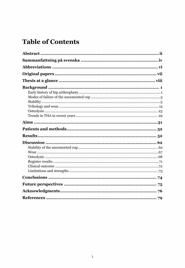
Table of Contents
Abstract ........................................................................................... ii Sammanfattning på svenska ........................................................... iv Abbreviations ................................................................................. vi Original papers .............................................................................. vii Thesis at a glance .......................................................................... viii Background ..................................................................................... 1
Early history of hip arthroplasty ...................................................................................... 1 Modes of failure of the uncemented cup .........................................................................3 Stability ............................................................................................................................. 5 Tribology and wear ......................................................................................................... 15 Osteolysis ....................................................................................................................... 23 Trends in THA in recent years ...................................................................................... 29
Aims ............................................................................................... 31 Patients and methods ..................................................................... 32 Results ........................................................................................... 52 Discussion ..................................................................................... 62
Stability of the uncemented cup.................................................................................... 62 Wear ................................................................................................................................ 67 Osteolysis ....................................................................................................................... 68 Register results ............................................................................................................... 71 Clinical outcome ............................................................................................................. 72 Limitations and strengths .............................................................................................. 73
Conclusions ................................................................................... 74 Future perspectives ....................................................................... 75 Acknowledgments .......................................................................... 76 References ..................................................................................... 79

ii
Abstract
Background: Artificial hip joint replacement has undergone tremendous development in the past 100 years. In the beginning, complications, such as infection and early loosening, were the rule rather than the exception. Today, complications of any sort are rare during the first decade after the operation. Artificial hip joint replacement has been chosen as the "Operation of the Century" and has dramatically improved the quality of life of millions of patients. Unfortunately, in the long-term, prosthesis loosening due to pathological bone resorption (osteolysis) around the prosthesis is still common. Traditionally, the prosthesis is anchored in the bone with bone cement (Plexiglas). However, since this cementation method was suspected to cause late loosening, alternative methods, such as the implantation of so-called uncemented prostheses, have been developed and are being increasingly applied. Because the early movement of a prosthesis (migration) increases the risk of loosening, uncemented cups are often augmented with additional screws. The mechanisms regulating the early and late loosening of uncemented cups are not fully established. Wear particles from the artificial joint and intermittent fluid pressure on the bone appear to accelerate or even cause bone loss and can eventually lead to loosening of the prosthesis. Therefore, screw holes in the uncemented cup have been suspected to be a risk factor.
Aims: We have studied whether the additional augmentation of modern uncemented cups with screws, pegs or hydroxyapatite increases the long-term stability, affects the wear rate, influences the development of osteolysis, or has any impact on the risk of cup revision. Furthermore, we investigated whether computed tomography (CT), which is needed to detect osteolysis around the prosthesis, could also be used in the follow-up of migration studies without losing significant precision compared to radiostereometry (RSA), which is the gold standard for these measurements.
Patients and Methods: In studies I-III, we evaluated 48 hips (45 patients) randomized to receive cups with or without augmentation. As part of the 14-year follow-up with conventional radiographs of the pelvis, two pairs of stereo radiographs and a CT scan were obtained. Migration and wear were measured by RSA. The volume and type of osteolysis were determined on CT. Furthermore, we calculated the precision and limit of agreement of RSA and CT to compare these two modalities as tools for migration measurements.
In study IV, we compared the risk of cup revision between 10,371 uncemented cups with and 12,354 without screw holes, using data from the Swedish Hip Arthroplasty Register.
iii
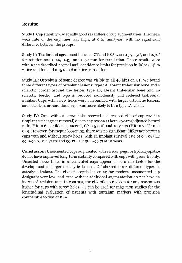
Results:
Study I: Cup stability was equally good regardless of cup augmentation. The mean wear rate of the cup liner was high, at 0.21 mm/year, with no significant difference between the groups.
Study II: The limit of agreement between CT and RSA was 1.15°, 1.51°, and 0.70° for rotation and 0.46, 0.43, and 0.52 mm for translation. These results were within the described normal 99% confidence limits for precision in RSA: 0.3° to 2° for rotation and 0.15 to 0.6 mm for translation.
Study III: Osteolysis of some degree was visible in all 48 hips on CT. We found three different types of osteolytic lesions: type 1A, absent trabecular bone and a sclerotic border around the lesion; type 1B, absent trabecular bone and no sclerotic border; and type 2, reduced radiodensity and reduced trabecular number. Cups with screw holes were surrounded with larger osteolytic lesions, and osteolysis around these cups was more likely to be a type 1A lesion.
Study IV: Cups without screw holes showed a decreased risk of cup revision (implant exchange or removal) due to any reason at both 2 years (adjusted hazard ratio, HR: 0.6, confidence interval, CI: 0.5-0.8) and 10 years (HR: 0.7, CI: 0.5-0.9). However, for aseptic loosening, there was no significant difference between cups with and without screw holes, with an implant survival rate of 99.9% (CI: 99.8-99.9) at 2 years and 99.1% (CI: 98.6-99.7) at 10 years.
Conclusion: Uncemented cups augmented with screws, pegs, or hydroxyapatite do not have improved long-term stability compared with cups with press-fit only. Unsealed screw holes in uncemented cups appear to be a risk factor for the development of larger osteolytic lesions. CT showed three different types of osteolytic lesions. The risk of aseptic loosening for modern uncemented cup designs is very low, and cups without additional augmentation do not have an increased revision rate. In contrast, the risk of cup revision for any reason was higher for cups with screw holes. CT can be used for migration studies for the longitudinal evaluation of patients with tantalum markers with precision comparable to that of RSA.

iv
Sammanfattning på svenska
Ledproteser är ett av de stora kirurgiska framstegen under 1900-talet som har givit många miljoner människor tillbaka en smärtfri och mobil vardag. Varje år utförs ca 18 000 primära höftprotesoperationer i Sverige, ca 5 procent av alla invånare över 50 år har minst en höftprotes idag. Allt yngre patienter opereras och de lever allt aktivare och längre med sina proteser. Sedan 1960-talet har majoriteten av proteserna fästs mot benet med metylmetakrylat (bencement). Det har dock visat sig att denna bencement lossnar med tiden från det omgivande benet. Trots alla framgångar inom proteskirurgin måste i Sverige mer än 2 000 höftproteser opereras om varje år. Vanligast pga aseptisk (icke infektions-betingad) lossning av protesen som beror på osteolys (benförlust kring protesen). På 1970 och 80-talet ansåg man att slitagepartiklar ifrån bencementen är den viktigaste orsaken till osteolys. Därför har man utvecklat alternativa, cementfria, sätt att fästa protesen mot benet.
Under de senaste 20 åren har andelen ocementerade proteser ökat kraftigt. Protesen måste direkt vara tillräckligt stabil så att det omgivande benet kan växa på dess yta och därmed ge långsiktig stabilitet. I avsikt att förbättra den initala stabiliteten så används ofta skruvar, pinnar eller ytbeläggningar med hydroxyapatit (HA) på den ocementerade höftskålen. Det är oklart om dessa förstärkningar fortfarande, med dagens protesdesign och material, ger några fördelar, eller om de till och med innebär risker i det långa loppet, som benförlust. Det är svårt att se med vanlig röntgen, men datortomografi kan identifiera benförlusten och dessutom mäta dess omfattning.
Proteser med ökad rörelse i förhållande till benet (migration) inom 1–2 år efter operationen har en ökad risk för lossning. För att tidigt upptäcka hur mycket nya proteser slits och eller hur lätt de lossnar behövs mätmetoder med hög precision. Radiostereometri (RSA) har varit guldstandard, men kräver speciella röntgenlaboratorier och är bara tillgänglig på några få forskningscentra. Olivecrona och medarbetare har vidareutvecklat RSA-principerna med datortomografi, med hög precision. Men den nya metoden var ännu inte validerad mot RSA. I Umeå har vi använt RSA sedan 80-talet och ett av de mest använda dataprogrammen för RSA utvecklades här. Det var angeläget att jämföra de två metoderna, eftersom den nya tekniken kan göras rutinmässigt i vården.
Följande frågor belyses i denna avhandling:
1. Påverkar användning av skruvar, pinnar och HA stabiliteten av höftskålen eller kliniska resultat på lång sikt?

v
2. Ökar skruvhål i höftskålen risken för benförlust?
3. Kan RSA migrationsstudier följas upp med datortomografi utan att förlora precision?
4. Visar registerdata skillnader i risken för omoperation på kort och eller lång sikt för höftskålar med eller utan skruvhål?
Vi har efterundersökt 48 höfter (45 patienter) ifrån en prospektiv randomiserad studie 14 - 17 år efter operation. Migration, slitage och benförlust bedömdes på konventionell röntgen, RSA och datortomografi.
I första studien visar mätningar med RSA att skruvar, pinnar och HA inte förbättrar stabiliteten av höftskålen på lång sikt och inte heller påverkar slitaget.
I andra studien jämföres precisionen av upprepade RSA undersökningar och mellan RSA och datortomografiundersökningar för migrationsmätning. Datortomografi och RSA har liknande reliabiliet och datortomografi kan därför ersätta RSA i migrationsstudier.
I tredje studien har vi närmare undersökt förekomsten av osteolys kring höftskålen. På konventionell slätröntgen kunde tydliga osteolytiska förändringar ses i 7/48 fall. Datortomografi visade osteolys i samtliga fall, och vi kunde urskilja tre olika typer av bendefekter. Runt cupar med skruvhål föreföll benförlusten större än runt cupar utan hål.
I fjärde studien vidgade vi perspektivet med en registerstudie för att kunna studera risken för omoperation. Det visade sig att risken för aseptisk lossning av moderna ocementerade cupar var mycket låg och att skruvfixation inte gav några fördelar vid standardoperationer utan snarare föreföll öka risken för omoperation av andra orsaker.
Sammanfattningsvis fann vi att förstärkning med skruvar, pinnar eller HA inte ökade cupstabiliteten. Vi kunde demonstrera att migrations och slitagemätningar på patienter som ingår i en RSA studie kan göras med hög precision även med hjälp av datortomografi. På datortomografi går det dessutom att särskilja 3 typer av osteolys, och osteolysen kring höftskålar med skruvhål är något större än de utan skruvhål. Slutligen visar data ifrån svenska höftprotesregistret att användning av cementfria höftskålar utan möjlighet till skruvfixation inte ökar risken för omoperation.

vi
Abbreviations
ADL Activities of Daily Living ANOVA Analysis Of VAriance AP Antero-Posterior CI Confidence Interval Cl Confidence limit CoC/CoM Ceramic-on-Ceramic/Ceramic-on-Metal CoP Ceramic-on-Polyethylene CT Computed Tomography DAIR Debridement Antibiotics and Implant Retention DMC Dual-Mobility Cup Dnr Diarienumber EtO Ethylene Oxide GUR Granula UHMWPE Ruhrchemie HA Hydroxyapatite HHS Harris Hip Score HMWPE High-Molecular-Weight PolyEthylene HOOS Hip dysfunction and Osteoarthritis Outcome Score HR Hazard Ratio HXLPE Highly Cross-Linked Polyethylene IL Interleukin ME/MERBF Mean Error/Mean Error of the Rigid-Body Fitting MoM Metal-on-Metal MoP Metal-on-Polyethylene MTPM Maximal Total Point Motion NARA Nordic Arthroplasty Register Association NF Nuclear Factor OA Osteoarthritis OPG Osteoprotegerin PE PolyEthylene PF Press-Fit PMMA PolyMethyl MethAcrylate PROM Patient-Reported Outcome Measure PTFE PolyTetraFluorEthylene QOL Quality Of Life RANKL Receptor Activator of Nuclear factor Kappa-B Ligand RCT Randomized Controlled Trial RLL RadioLucent Line RSA Roentgen Stereophotogrammatic Analysis, Radiostereometry SD Standard Deviation SHAR Swedish Hip Arthroplasty Register THA Total Hip Arthroplasty TMT Trabecular Metal Tantalum TNF Tumor Necrosis Factor TT Trabecular Titanium UHMWPE Ultra-High-Molecular-Weight PolyEthylene

vii
Original papers
I. Otten V, Crnalic S, Röhrl S, Nivbrant B and Nilsson K. "Stability of Uncemented Cups—Long-Term Effect of Screws, Pegs and HA Coating: A 14-Year RSA Follow-Up of Total Hip Arthroplasty." The Journal of Arthroplasty 2016; 31(1): 156-161
II. Otten V, Maguire Jr G, Noz M, Zeleznik M, Nilsson K and Olivecrona H (2017). "Are CT Scans a Satisfactory Substitute for the Follow-Up of RSA Migration Studies of Uncemented Cups? A Comparison of RSA Double Examinations and CT Datasets of 46 Total Hip Arthroplasties." BioMed Research International 2017: Article ID 3681458, 11 pages, 2017. https://doi.org/10.1155/2017/3681458
III. Otten V, Stamenkov R, Callary S, Howie D, Crnalic S, Nilsson K “Osteolysis around uncemented cups with and without screw holes. Analysis of osteolytic lesions on CT images at 14-year follow up in 48 hips” Manuscript
IV. Otten V, Mukka S, Nilsson K, Crnalic S, Kärrholm J “Uncemented cups with and without screw holes. A Swedish Hip Arthroplasty Register study with 22725 hips.” In Press, Acta Orthopaedica 2019.
Previously published papers were reproduced with the kind permission of the publishers of The Journal of Arthroplasty and BioMed Research International.

viii
Thesis at a glance
Study I: Equally good long-term stability of porous-coated cups with or without additional augmentation
Patients and methods: A 14-year follow-up with RSA in a retrospective cohort study with 48 hips (45 patients).
Conclusion: Screws, pegs or a hydroxyapatite (HA) coating did not increase the long-term stability of porous-coated uncemented cups compared to press-fit fixation only.
Study II: CT can be used in the follow-up of RSA migration studies without losing significant precision
Patients and methods: CT and double RSA examinations of 46 hips.
Conclusion: The limit of agreement between CT and RSA lies within the limits of what is described as normal precision for RSA. CT can be used in the follow-up of marker-based RSA migration studies.

ix
Study III: Larger osteolytic lesions around cups with screw holes
Patients and methods: CT of 48 hips 14-17 years after primary hip arthroplasty.
Conclusion: Osteolytic lesions were larger around cups with screw holes than cups without screw holes. Unsealed screw holes seem to be a risk factor for the development of extended osteolytic lesions.
Study IV: No difference in risk of revision due to aseptic loosening between cups with and without screw holes
Patients and methods: Register study of 12,354 cups without screw holes and 10,371 cups with screw holes.
Conclusion: We found no difference in the risk of either early (2 years) or midterm (10 years) revision for aseptic loosening between cups with and without screw holes in patients with primary osteoarthritis. In contrast, the risk of revision for any reason was increased for cups with screw holes.


1
Background
Total hip arthroplasty (THA) is one of the most successful surgical procedures in history and has been called “The Operation of the Century” (Learmonth et al. 2007). Nevertheless, approximately 10% of all patients who undergo THA require reoperation at least once during their lifetime (Kärrholm et al. 2017). With approximately 1,000,000 primary THA procedures performed per year worldwide, the revision burden is growing. The most common overall cause of failure is aseptic loosening of the components, i.e., loosening without infection, caused by osteolysis and wear (Kärrholm et al. 2017). However, there are major differences in both the failure rate and the failure pattern between different prosthesis designs and materials. Unfortunately, new designs and new materials have not always been successful, and some have performed much worse than older concepts (Anand et al. 2011). Sweden has a lower frequency of adverse events after THA, largely because of a national register that tracks almost all THAs performed since 1979.
This introduction provides a short summary of the development, materials, tribology, wear, migration and failure modes of the prostheses used in THA, with special reference to the uncemented cup.
Early history of hip arthroplasty The journey toward artificial joint replacement started at the end of the 19th century. The Czech surgeon Vitezlav Chlumsky started experimenting with a variety of interpositional materials in joints, including silver plates, rubber struts, magnesium, zinc, and glass, among others (Gomez et al. 2005). The first to perform a total hip replacement in a human was the German surgeon Themistocles Gluck in 1890. He inserted 14 ivory joint replacements, including one total hip, in patients with tuberculosis. Although he presented these as successful over the short term, all patients suffered from chronic infection, and he later stated that joint infection was a contraindication to joint arthroplasty (Brand et al. 2011). At the beginning of the 20th century, the first successful interpositional hip arthroplasties with good functional long-term results were reported (Gomez et al. 2005). In 1923, Smith-Petersen presented resurfaced cups for the femoral head, initially made of glass and later of Vitallium (Smith-Petersen 1939). This prosthesis still holds the record today for the longest lasting hip replacement (Northover et al. 2008, World Record Academy 2016). In the 1930s, Wiles operated on a series of patients with rheumatic disease and used the first uncemented stainless steel prosthesis for THA with metal-on-metal (MoM) articulation (Wiles 1958). The cup was held in place by a couple of screws. On the femoral side, the prosthesis replaced half of the femoral head and was held in

2
place by a bolt inside the column that was secured by a lateral plate. Bone resorption, soft tissue reactions and early aseptic loosening were the main issues. Judet introduced an acrylic head prosthesis in 1947 (Judet et al. 1950). Poly(methyl methacrylate) (PMMA) is well tolerated by human tissue but does not show good wear resistance and carries a high risk of neck fracture. Today, PMMA is used as bone cement but no longer as an articular surface. Moore described bone formation around a stemmed Vitallium prosthesis in 1940 and started developing an uncemented metal hip prosthesis (Moore et al. 2006). The uncemented Austin Moore hip prosthesis, launched in 1952, with only minor modifications, is still on the market today (Moore 1952, Moore 1957, Naser et al. 2018).
John Charnley started to work on the low-friction arthroplasty concept during the 1950s. He used polytetrafluorethylene (PTFE) as a material with a very low friction coefficient; unfortunately, it also caused a vehement tissue reaction after a few years. Despite encouraging early results with this material as an articular surface in THA, all (approximately 300) implanted prostheses had to be revised within the first few years because of wear and the formation of granulomatous masses around the joint (Gomez et al. 2005). Mr. Charnley thought that this was the end of THA. To his surprise, his patients said that the few years of pain relief were acceptable and agreed to reoperation, which Mr. Charnely performed personally. He continued to experiment with different materials and solved the main issues of THA at that time within a few years. In 1962, he inserted the first prosthesis with a cemented cup made of high-molecular-weight polyethylene (HMWPE) and a cemented stainless steel monobloc stem with a 22-mm head. He had also tested an uncemented version of the cup with a polished metal back. However, the uncemented version had a higher revision rate and was no longer used. HMWPE had a relatively low coefficient of friction that was further reduced in conjunction with synovial fluid. HMWPE had a high wear resistance and did not produce, at that time, an identifiable tissue reaction. The small head reduced the contact area and the force introduced into the joint during motion, leading to reduced wear. Using polymethylmethacrylate (PMMA) to cement both the cup and the stem reduced the risk of early loosening and yielded reliable and very good long-term results. Furthermore, Charnley reduced the infection rate by performing the surgery in ultra-clean operation theaters. This was the starting point for the real success story of THA (Learmonth et al. 2007). The revision rate of THA, following Charnley’s principles, was below 10% within 10-15 years (Eftekhar 1987, McCoy et al. 1988). Older patients who underwent cemented THA were unlikely to require subsequent arthroplasty.
During the 1980s, the first reports of periprosthetic osteolysis and tissue reactions around stable cemented THA prostheses were published (Jasty et al. 1986). Histological examination of the periprosthetic tissues showed more

3
macrophages and giant cells in patients with loose cemented implants than those with loose uncemented implants (Lennox et al. 1987). Hence, osteolysis was considered a cement-related disease, and it was therefore thought that cemented prostheses were not suitable for young patients (Jones et al. 1987). Concerns regarding cement-related disease and the need for better long-term solutions for younger patients drove the industry toward developing new components that could be fixed to the bone without cement.
The retrospective long-term follow-up of matched patients with either cemented or uncemented acetabular components showed less loosening for uncemented cups (Clohisy et al. 2001).
Modes of failure of the uncemented cup Uncemented THA carries a higher risk of early revision due to aseptic loosening, infection, dislocation and periprosthetic fracture than cemented THA (Troelsen et al. 2013, Phedy et al. 2017, Kärrholm et al. 2018). However, comparing data from follow-up periods of more than 7 years, the overall risk of revision is lower for uncemented THA among younger patients (Kärrholm et al. 2018). Here, we focus on the mechanisms of aseptic loosening, osteolysis and wear. Infection, dislocation and fracture are indeed important complications but are outside the scope of this thesis. Therefore, only some examples of how the failure of uncemented cups may differ from the failure of cemented cups are mentioned in this regard.
Infection
Although the infection rate in THA is low today compared to that in the early days of hip arthroplasty, the incidence of infection early after primary surgery has been increasing in recent years (Kärrholm et al. 2018). DAIR (debridement, antibiotics and implant retention) has shown encouraging results (Anagnostakos et al. 2014). During these procedures, the liner of uncemented cups is usually changed, while cemented cups are not revised. This may partly explain the somewhat higher rate of early revision for uncemented cups caused by infection (Kärrholm et al. 2018). The overall risk of revision due to infection is not significantly different between cemented and uncemented THA (Hailer et al. 2010).
Dislocation
Although the risk of dislocation is mostly dependent on the surgeon, surgical technique and patient-related factors, it seems to also be related to the implant design to some degree. In particular, trabecular metal cups have shown a higher risk of dislocation (Hailer 2018). The cup and liner design, as well as difficult cup

4
positioning of cups with high-friction surfaces, may contribute to an increased risk of dislocation.
Intraprosthetic dislocation, i.e., separation of the liner from the shell, has been a problem with early liner locking mechanisms (Niggemeyer et al. 2002) but is rare with modern modular cups. However, the rate of a new form of intraprosthetic dislocation, i.e., between the mobile liner and the head, has been described to reach up to 3.6% for dual-mobility cups (Philippot et al. 2009).
Fracture
Two completely different types of fracture have been distinguished as causes of uncemented cup revision: fracture of the acetabular bone (periprosthetic fracture) and fracture of the components themselves.
Periprosthetic fractures can occur in surgery during reaming or impaction of the shell (Benazzo et al. 2015). Risk factors include extensive underreaming, oversized cups and poor bone quality. Postoperative, traumatic fractures are rare in the absence of osteolysis.
The combination of a small cup size with a large head size leads to a reduced liner thickness, which can occasionally result in liner fracture (Ast et al. 2014).
Aseptic loosening, wear and osteolysis
The main problems of early uncemented cups were insufficient initial stability, the lack of bony growth into the implant surface and wear of the articulating surface (Havelin et al. 1994). Further development of the implant design, surface and material has led to improved early stability and reduced wear. However, aseptic loosening is still the main overall long-term cause of THA revision (Kärrholm et al. 2017).

5
Stability
Implant design
There are two basic design shapes for the uncemented cup, hemispherical and conical (Figure 1). Many of the early cups had a conical shape, as this provides a larger surface area between the bone and implant and theoretically greater resistance to cup tilting. Conical cups are mostly used as either threaded cups or cups with a dome stem. During the 1960s and 1970s, the ring prosthesis, a conical prosthesis with a threaded stem that was either all poly or metal, was one of the most popular uncemented cups (Patterson 1987, Mok et al. 1989). These types of cups require the removal of a larger portion of bone, as the natural form of the acetabulum is spherical. In cases with large bone defects, as in some tumor cases, conical acetabular cups with a dome stem are still used (Bus et al. 2017). There are many different existing designs of both threaded conical and ring-shaped cups, such as the conical Zweimüller cup, which has good results (Garcia-Cimbrelo et al. 2003), and the ring-shaped Mecring, which has a high failure rate (Mueller et al. 2016); in addition, modern spherical threaded cups have results comparable to those of modern press-fit cups (Ellenrieder et al. 2016). Although threaded cups have shown greater initial stability in experimental studies than press-fit cups (Litsky et al. 1994), in registry data, threaded cups show an increased risk of aseptic loosening (Eskelinen et al. 2005). This difference was even greater, with a disadvantage for threaded cups, with revisions for any reason as the endpoint. Cup breakage, local strain leading to osteonecrosis, and soft tissue damage, including nerve palsy and acetabular fracture, are some of the complications that are more frequently reported for threaded cups (Effenberger et al. 2004).
Hemispherical cups are available in single- and dual-geometry designs. In the single-geometry design, the radius of the outer surface of the cup remains the same throughout the entire hemisphere. In the dual-radius design, the radius is larger at the rim than the dome, either with a clear step between the two or with a gradual transition between the larger radius at the rim and the smaller radius at the pole (hemi-ellipsoid). The TMT cup is an example of this design in use today. On the one hand, the hemi-ellipsoid design provides a better press fit at the rim where the bone normally is denser and might therefore enhance the primary stability. On the other hand, this design transfers less load through the dome and might therefore cause stress shielding.
In addition to these main fixation principles, some more exotic methods have been tested. One example is the CLS Spotorno expansion cup, in which the shell is slotted and thus highly flexible. This cup is compressed before insertion and then expands inside the acetabulum (Cech et al. 2001).

6
Today, the dominant principal design of the uncemented acetabular component is the hemispherical single-geometry press-fit cup.
Figure 1: Conical shaped threaded ceramic cup (Mittelmeier, Smith & Nephew) and hemispherical porous-coated titanium cup with hydroxyapatite (HA) (DeltaMotion, DePuy).
Fixation surface of the uncemented cup
The surface of the shell must serve two functions. It must provide sufficient initial friction for primary stability and enable bone on- or ingrowth to achieve secondary stability.
The fixation surface of the cup is defined by the material, roughness and porosity.
The roughness of a surface can be expressed in different ways. Ra denotes the arithmetic mean deviation of the assessed profile; this measurement is two dimensional, is presented in μm, and is probably the most used. Shalabi et al. concluded in a systematic review of animal studies on surface roughness and bone healing that both the bone-to-implant contact and pull-out and torque resistance of metal implants increase with the roughness of the surface (Shalabi et al. 2006).
Porosity is a measure of empty space in a material, the fraction of the volume of voids over the total volume. Porosity is expressed in %. Early designs had a smooth surface with inferior stability and clinical results (Engh et al. 1990). Coating with wire mesh or large beads (approximately 500 μm) increased the porosity to approximately 30-40% and achieved better bone ingrowth. Sintered, smaller beads (approximately 250 μm) resulted in a porosity of up to 50% and further enhanced bone ingrowth in a retrieval study (Swarts et al. 2015). The porosity of the latest generation of metal surfaces, trabecular metal, reaches up to 80%.
Frosch et al. found in an in vitro study that a pore size of 600 μm leads to the fastest ingrowth of bone into titanium implants (Frosch et al. 2004). Modern production processes, such as 3D printing, make it possible to produce almost every imaginable metal surface structure (Figure 2).

7
Figure 2: Smooth surface of explanted titanium screw cup without signs of bone ingrowth (A). Explanted cup with titanium fiber mesh surface with deep bone ingrowth (B). Porous surface with 250-μm sintered titanium beads and thin HA coating (C). Trabecular metal tantalum cup surface (D). 3D-printed trabecular titanium (E). Layered porous coating with 300-μm sintered beads coated with an extra layer of irregularly shaped pure titanium pieces (F). Images C-F are from unused cups.
A B
C D
E F

8
Hydroxyapatite (HA) coating
The bone matrix consists of 30% organic components, mainly collagen, which provides tensile strength, and 70% inorganic components, i.e., bone mineral, which provides compressive strength. The main component of bone mineral consists of calcium and phosphate salts, primarily HA, (Ca10(PO4)6(OH)2).
Animal studies during the 1970s showed good biocompatibility and direct bone bonding to porous bulk HA (Nery et al. 1975, Jarcho et al. 1977). Coating metal implants with HA increased bone ingrowth and the mechanical strength of the interface (Ducheyne et al. 1980). Bone bridge formations were even observed over several-mm-long gaps between host bone and HA-coated titanium implants in a canine model (Stephenson et al. 1991). Micromotion inhibited bony ingrowth within the first 4 weeks, but HA-coated implants showed a higher shear strength under these circumstances (Søballe et al. 1992). Additionally, despite a continued micromotion of 150 µm, bone bridges were observed histologically around HA-coated implants within 16 weeks in a dog model. In implants without an HA coating, only fibrous ingrowth was observed, as long as the micromotion continued (Soballe et al. 1993). Trabecular bone formation between host bone and HA-coated implants was found within weeks in postmortem specimens (Furlong et al. 1991). In a high-wear-rate sheep model, HA-coated cups showed significantly more bone contact and thinner fibrous tissue at the bone-implant interface than both uncemented cups without an HA coating and cemented cups (Coathup et al. 2005). In an autopsy retrieval study, cases with radiologically good osseointegration also had histologically good bone ingrowth (Geesink 1993). The HA coating is slowly resorbed in vivo. Most of this resorbed material is replaced by newly formed bone. Micromotion accelerates the resorption process (Søballe et al. 1999). At the 5-year follow-up of patients included in studies I-III in this thesis, HA-coated cups displayed fewer radiolucent lines (RLLs) than cups without an HA coating (Röhrl et al. 2004). In a clinical retrieval study, HA particles were found both in osteolytic lesions and incorporated into the polyethylene (PE) of the liner (Bloebaum et al. 1994). Third-body wear due to detached HA particles in the joint space may produce high wear rates, leading to severe osteolysis and eventually component loosening (Morscher et al. 1998). Several different coating techniques are used. Plasma-spraying, in which HA particles are accelerated to a high velocity, heated to temperatures of approximately 800°C, and then driven against the surface of the implant, is currently the most commonly used method. Important factors of the quality of the HA coating are the chemical composition (purity), Ca/P ratio (HA normally has a ratio of 1.67), crystallinity, microstructure (density), adhesive strength and coating thickness (Søballe 1993). All of these parameters differ among companies and might considerably influence the long-term outcomes. Registry data of primary THA patients under 70 in Denmark show no difference in the risk of

9
aseptic loosening between cups with and without an HA coating (Paulsen et al. 2007). In an analysis of three somewhat older cup models (Harris-Galante, Romanus and Trilogy) with data from the Swedish Hip Arthroplasty Register (SHAR), the risk of revision due to aseptic loosening was higher for cups with an HA coating (Lazarinis et al. 2010). In the largest study thus far, analysis of the influence of an HA coating on the risk of cup revision using data from the Nordic Arthroplasty Register Association (NARA) database showed no difference in terms of revision due to aseptic loosening but more revisions due to infection for HA-coated cups (Lazarinis et al. 2017).
Supplemental fixation of the uncemented spherical press-fit cup
More than 1 mm of uncemented cup migration within two years has been considered to increase the risk of late revision because of aseptic loosening (Stocks et al. 1995, Krismer et al. 1996). Pijls found in a meta-analysis indirectly comparing RSA studies and survival studies that even an early migration between 0.2 mm and 1 mm increased the risk of late revision to more than 5% at 10 years (considered a normal revision risk). Every mm of migration within 2 years increased the revision rate by 10% (Pijls et al. 2012).
Many different supplemental fixation methods have been used to enhance the primary stability of uncemented cups. Spikes and fins can enhance, at least in vitro, the primary stability (Baleani et al. 2001). These features can be integrated directly into the cup surface and do not necessitate any additional steps during the operation. Pegs and screws are normally inserted through the shell during the operation. This approach provides greater flexibility and is currently the most commonly used method for the supplemental fixation of uncemented cups.
Using screws has some disadvantages and potential risks. Both a prolonged operation time (Pepe et al. 2017) and increased bleeding (Colacchio et al. 2017) have been reported in recent studies. Inserting screws into the acetabulum might even pose a risk of damaging intrapelvic vessels (Ohashi et al. 2017).
The screw fixation of uncemented cups does increase stability in simulation models (Hsu et al. 2007) and cadaver studies (Won et al. 1995). However, this requires the cups to be manufactured with screw holes. Screw holes in acetabular cups have been discussed as potential routes for synovial fluid containing cells and substances with osteolytic potential to gain access to the bone tissue and the bone-implant interface (Schmalzried et al. 1999). Ni et al. could not find any significant difference in revision, migration or osteolysis between cups supplemented with screws and press-fit only cups in a meta-analysis of 5 studies with a total of 1130 patients and a follow-up of 2-5 years (Ni et al. 2013). There is still a lack of studies with longer follow-up periods and larger patient groups comparing cups with and without screw holes. Furthermore, most of the relevant

10
studies have compared one or more cup designs with screws to totally different cup designs without screws. In some studies, cups with screw holes were used in both the groups with cups with and without screws (Iorio et al. 2010).
Materials in THA with uncemented cups
Ultra-high-molecular-weight polyethylene (UHMWPE) The German company Ruhrchemie invented UHMWPE and presented this new material in 1955 at the K-fair in Düsseldorf. The original polymer resin was named GUR (Granular UHMWPE Ruhrchemie) resin. GUR exists in many different versions, and the name is often supplemented with numbers. The first number indicates where the resin is produced (Germany (1), USA (4)), the second number indicates the presence (1) or absence (0) of calcium stearate, and the third number indicates a molecular weight of either 2 million (2) or 5 million (5). The higher the molecular weight, the higher the viscosity.
The resin is produced as a powder. To create a solid material, the powder must be compressed and heated. Three different production methods for the final implant are in use, as follows: 1. UHMWPE resin is pushed with a ram into a heated cylinder, forming a cylindrical bar. These bars are then machined into the final product (ram extrusion and machining). 2. Large sheets of solid UHMWPE are molded under pressure in a preheated rectangular container. These sheets are then machined into the final product (sheet compression molding and machining). 3. UHMWPE resin is directly compressed and molded into the shape of the final product (direct compression molding) (Berry et al. 2012). Direct-molded products have a very smooth surface finish and are reported to have a lower clinical wear rate (Bankston et al. 1995) but are the most expensive to produce.
Sterilization of the UHMWPE component is most often performed with plasma gas, ethylene oxide (EtO) or gamma irradiation (25-40 KGy).
Gas plasma sterilization was introduced during the 1990s. In this method, ionized gas is used at low temperatures (<50°C) to deactivate pathogens on the implant surface. This is currently the fastest and most inexpensive method used for sterilizing implants.
EtO gas has been used since the 1970s for sterilization. It is highly toxic, forms irreversible chemical bonds with bacteria, spores and viruses, and can sterilize deep within the material, even in small cracks or recesses. The high toxicity requires that all EtO is removed before implantation and might also be an environmental problem.

11
Gamma irradiation creates free radicals in the material. These free radicals can lead to chain breakage in the polymer and oxidation but also to cross-linking between polymer molecules in the absence of oxygen. EtO-sterilized UHMWPE components show a lower risk of oxidation (Costa et al. 1998) but are less resistant to wear than gamma-irradiated UHMWPE components (Affatato et al. 2002) in laboratory tests. Gamma irradiation in a low-oxygen environment shows reduced oxidation and thereby increases wear resistance (McKellop et al. 2000).
The amount of cross-linking and thus wear resistance increases with increasing radiation dose (McKellop et al. 1999). At the same time, the number of free radicals and therefore the amount of oxidation also increases.
In the production of highly cross-linked PE (HXLPE), sheets or bars of UHMWPE are irradiated with 50 to 100 KGy and then machined into the final implant. Heating the PE sheets or bars before machining reduces free radicals (McKellop et al. 1999). Two heating methods are used: annealing, in which the irradiated UHMWPE is thermally treated with temperatures below the melting point; and remelting, in which the UHMWPE is heated to temperatures above the melting point (Table 1). Remelting will decrease the fatigue strength, while annealing will leave some residual radicals. Another way to reduce free radicals is to add the antioxidant vitamin E, either by blending it into the resin powder before consolidation and irradiation or by doping the UHMWPE afterward. The first method can reduce cross-linking during irradiation, and with the second method, only a limited concentration of vitamin E can penetrate the UHMWPE (Lambert et al. 2018). Several randomized controlled trials (RCTs) have shown reduced femoral head penetration in liners with vitamin E than liners consisting of thermally treated HXLPE (Lambert et al. 2018), at least in the early phase after the operation; this difference is not as clear for the later annual wear rate (Salemyr et al. 2015).
Although there are some examples of all-PE uncemented press-fit cups, PE is commonly used as a liner inside an uncemented metal shell or in cemented cups.

12
Table 1: Examples of commercially available versions of highly cross-linked UHMWPE (Berry et al. 2012) (and producers’ homepages).
PE name Resin Production method
Irradiation Radical reduction
Sterilization
Arcom XL (Biomet)
1050 GUR
Extrusion and machining
50 kGy Annealing (two times to 130°C)
Gas plasma
Marathon (DePuy)
1050 GUR
Ram-extrusion and machining
50 kGy (gamma)
Remelting (150°C for 24 h)
Gas plasma
AltrX (DePuy)
1020 GUR
Ram-extrusion and machining
75 kGy (gamma)
Remelting (155°C for 24 h) plus annealing (120°C for 24 h)
Gas plasma
Longevity (Zimmer)
1050 GUR
Extrusion and machining
100 kGy (electron beam)
Remelting (>150°C for 6 h)
Ethylene oxide
Durasul (Zimmer)
1050 GUR
Extrusion and machining
95 KGy (electron beam)
Remelting (>150°C for 2 h)
Ethylene oxide
XLPE (Smith & Nephew)
1050 GUR
Extrusion and machining
100 kGy (electron beam)
Remelting (>150°C)
Ethylene oxide
Crossfire (Stryker)
1050 GUR
Extrusion and machining
75 kGy (gamma)
Annealing 25 KGy gamma radiation
X3 (Stryker)
1050 GUR
Extrusion and machining
3 x 30 kGy Annealing (130°C, 3 times)
Gas plasma
X-LINKed (LINK)
1020 GUR
Sheet compression molding and machining
75 kGy Remelting (150°C) and shaving of 5 mm of outer layer of bar
Ethylene oxide
Titanium In 1791, the German chemist Martin Heinrich Klaproth found the element titanium in ore rutile, which consists mainly of titanium dioxide (TiO2). Because of its high strength, titanium became often used in the military since the early 1950s and later even in many other industries. Animal studies during the 1940s showed outstanding tissue compatibility (Wang 1996). The high resistance to corrosion even in the harsh environment of the human body is a result of the protective oxide film on the surface of titanium that forms naturally in the presence of oxygen. The mechanical abrasion of this film leads to an increased rate of corrosion. During the 1950s and 1960s, titanium alloy medical devices began to be used. For implants, titanium is most often alloyed with approximately 6% aluminum and 4% vanadium (Ti6AI4V). During the 1970s, these titanium alloys were increasingly used in both cemented and uncemented hip arthroplasties. During the 1980s, Ti6Al4V was also used for articular surfaces,

13
such as modular heads, but the relatively poor wear resistance rendered this material less suitable as an articular surface. However, the excellent biocompatibility and relatively low Young’s modulus make titanium and the titanium alloy Ti6Al4V most suitable as surfaces for bone ingrowth in uncemented THA.
Tantalum Tantalum, element number 73 in the periodic table, was discovered in 1805 by the Swedish chemist Anders Ekeberg. It has a high melting point of 3017°C and mostly exists in two crystalline phases, ductile (significant plastic deformation before rupture), somewhat softer α-Ta and hard, brittle β-Ta. Burke suggested tantalum as early as 1940 as a material for orthopedic implants because of its very high corrosion resistance and biological inertness (Burke 1940). Today, tantalum is used in THA mostly in cups or for augmentation in the form of trabecular metal. Trabecular metal has a porosity of 80% and reduces the Young’s modulus to 2.5-3.9 GPa, which is close to that of cancellous bone. In a solid form, tantalum is used, for example, as marking beads for RSA.
Cobalt chrome (CoCr) Elwood Haynes, an American inventor, discovered the CoCr alloy during the search for corrosion-resistant material for the car industry in the early 1900s. Smith-Petersen used variations of this alloy for his hip cup since the 1930s. Today, cobalt-based alloys most commonly contain approximately 28 weight % chromium and 6 weight % molybdenum. CoCrMo can be molded, forged or machined into the shape of the final implant. The alloy has a high corrosion resistance and high hardness. This is a strong metal alloy with a yield strength, i.e, limit of elastic behavior, of approximately 600 MPa and a tensile strength, i.e., breaking point, of approximately 800 MPa.
CoCr has been tested as a material for uncemented components but has shown a higher failure rate because of loosening than titanium alloys (Kubo et al. 2001, Dickinson et al. 2012). This is probably caused by the high Young’s modulus; CoCr alloys are approximately 10-15 times stiffer than cortical bone. In uncemented THA, CoCr is mainly used as a material for the articular surface, mostly as the head on the femoral side but also on the acetabular side in MoM articulation and dual-mobility cups.
Ceramics Aluminum oxide, Al2O3, also called alumina, is the most common ceramic. Because of its extreme hardness (Table 2), it is often used as an industrial diamond and in sandpaper. In its purest crystalline form, it is the main component of both rubies and sapphires. Biolox®forte consists of high-purity

14
alumina containing a small amount of magnesium oxide to control grain growth and achieve the highest possible density. The greatest problem with Biolox®forte is the risk of breakage.
Zirconium dioxide, ZrO2, also called zirconia, was identified by the German chemist Martin Heinrich Klaproth in 1789. This ceramic material is very tough, resistant to wear and thermal shock and shows low corrosion (Piconi et al. 1999). Zirconia exists in three different phases, monoclinic, tetragonal and cubic. The cubic face is stable but brittle and is used as a diamond simulant in jewelry. In the tetragonal phase, zirconia is tough but unstable. Yttrium oxide can be used to stabilize zirconia in the tetragonal phase. When zirconia transforms into the monoclinic phase, the volume increases (Macdonald et al. 2014). This phenomenon leads to a change in the surface roughness of pure zirconia heads over time, leading to a faster wear rate (Haraguchi et al. 2001). In 2001, the FDA issued a warning about this problem, and since then, the use of pure zirconia heads has ceased (Lachiewicz et al. 2018). Oxinium® is a further developed material consisting of a zirconium-niobium alloy core and zirconia surface. Unfortunately, this new material seems to be easily damaged, according to a case report (Kop et al. 2007).
The further development of ceramics seeks to combine the crack resistance of zirconia and the hardness and smoothness and thereby wear resistance of alumina. Accordingly, Biolox®delta was introduced for use in hip arthroplasty in 2003; this ceramic consists of Al2O3 and ZrO2 as the main components and SrO, Y2O3 and Cr2O3 as additives (Figure 3).
Figure 3: Femoral heads in CoCr, zirconia, alumina and zirconia-reinforced alumina (with adhesive titanium wear stripes).
Howard et al. analyzed 212,296 THAs performed with at least one ceramic component registered in the National Joint Registry for England, Wales,
15
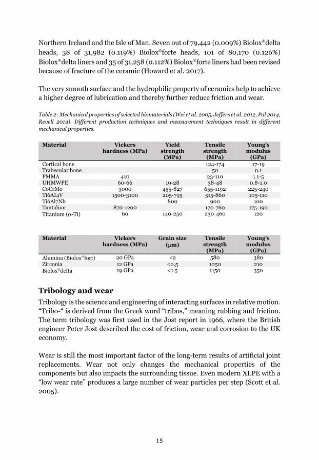
Northern Ireland and the Isle of Man. Seven out of 79,442 (0.009%) Biolox®delta heads, 38 of 31,982 (0.119%) Biolox®forte heads, 101 of 80,170 (0.126%) Biolox®delta liners and 35 of 31,258 (0.112%) Biolox®forte liners had been revised because of fracture of the ceramic (Howard et al. 2017).
The very smooth surface and the hydrophilic property of ceramics help to achieve a higher degree of lubrication and thereby further reduce friction and wear.
Table 2: Mechanical properties of selected biomaterials (Wei et al. 2005, Jeffers et al. 2012, Pal 2014, Revell 2014). Different production techniques and measurement techniques result in different mechanical properties.
Material Vickers hardness (MPa)
Yield strength
(MPa)
Tensile strength
(MPa)
Young’s modulus
(GPa) Cortical bone 124-174 17-19 Trabecular bone 50 0.1 PMMA 410 23-110 1.1-5 UHMWPE 60-66 19-28 38-48 0.8-1.0 CoCrMo 3000 455-827 655-1192 225-240 Ti6AI4V 1500-3100 205-795 515-860 105-110 Ti6Al7Nb 800 900 100 Tantalum 870-1200 170-760 175-190 Titanium (α-Ti) 60 140-250 230-460 120
Material Vickers hardness (MPa)
Grain size (μm)
Tensile strength
(MPa)
Young’s modulus
(GPa) Alumina (Biolox®fort) 20 GPa <2 580 380 Zirconia 12 GPa <0.5 1050 210 Biolox®delta 19 GPa <1.5 1150 350
Tribology and wear Tribology is the science and engineering of interacting surfaces in relative motion. “Tribo-“ is derived from the Greek word “tribos,” meaning rubbing and friction. The term tribology was first used in the Jost report in 1966, where the British engineer Peter Jost described the cost of friction, wear and corrosion to the UK economy.
Wear is still the most important factor of the long-term results of artificial joint replacements. Wear not only changes the mechanical properties of the components but also impacts the surrounding tissue. Even modern XLPE with a “low wear rate” produces a large number of wear particles per step (Scott et al. 2005).
16
For a basic understanding of tribology, the definition of some essential terms is necessary.
A tribosystem consists of four elements: a body, a counterbody, an interfacial medium and the environment. In an artificial hip joint, the liner is the body, and the head is the counterbody. Synovial fluid is the interfacial medium but also influences the environment. Other important environmental factors are the temperature and the oxygen content in the joint. The wear of this system is influenced by many factors. Relative kinematics, contact load and loading profile are important input variables that are both influenced by the implant design, component positioning and patient’s activities. Wear in response to these input variables depends mainly on the surface characteristics and lubrication. The surface characteristics, e.g., roughness and hardness, depend on both the surface material and machining techniques. Although the surfaces of THA prostheses can appear perfectly smooth, contact between articulating surfaces is established only at very small points. The proportion of this real contact area to the apparent contact area describes the roughness.
Friction is the force resisting the relative motion between the body and the counterbody. The friction coefficient describes the relationship between the force of friction and the normal force of the objects. The friction depends on the roughness of the surfaces, the real contact area and the contact load. Most of the energy that is introduced into the tribosystem to achieve relative motion is transformed into heat. A smaller portion of the energy is transformed into elastic or plastic deformation and leads to cracks and wear. In rare cases, such as some ceramic-on-ceramic (CoC) prostheses, the introduced energy can even be transformed into sound.
Larger head sizes normally increase the contact area and lead to higher friction and thereby increased wear. Lubrication can reverse this effect by separating the body and counterbody with an interfacial medium, which is called lubricant. In 1902, the German engineer Richard Stribeck introduced the lambda ratio (λ), which represents the thickness of the lubricant in relation to the surface roughness; λ<1 describes boundary lubrication where the real contact area remains unchanged, and the lubricant fills only the space between points of contact, which reduces resistance to relative motion by chemical and physical processes, e.g., heat dissipation. In a case of mixed lubrication, with λ between 1 and 3, the real contact area between the body and counterbody is reduced, and the friction coefficient reaches the lowest value. A value of λ>3 indicates complete separation of the articulating surfaces. In this scenario, the friction coefficient is slightly higher again, as the viscosity of the lubricant has to be higher to support the load. Wear is reduced to a minimum. Resurfaced prostheses were developed

17
over the past few decades and reached λ>3 in laboratory tests; unfortunately, clinical results showed that this theoretically perfect prosthesis design was not as perfect when applied in the human body.
Wear
Wear describes the loss of material in the form of debris and should not be confused with signs of surface damage, such as scratches, that can occur without any material loss.
In the artificial hip joint, there are four predominant wear modes (McKellop 2007):
1. Sliding wear occurs in normal motion when the load-bearing surfaces articulate as intended by the implant design. This wear mode produces the fewest wear particles.
2. Impaction or impingement wear occurs when a load-bearing surface articulates against a non-load-bearing surface. This may, for example, occur during dislocation and can lead to massive wear.
3. Third-body wear occurs when an abrasive third-body particle (e.g., PMMA, metal, ceramic, HA or bone fragments) is interposed between the articulating surfaces. This mode can lead to massive wear in a short period of time.
4. Fretting wear or backside wear occurs when two surfaces, not intended for articulation, are moving against each other (e.g., a loose head against the stem taper or the liner against the shell).
Three main wear mechanisms can be distinguished:
• Abrasion, where the roughness of the articulating surface of the body cause wear on the counterbody, or vice versa. This can be both a roughening and polishing process and thereby does not necessarily lead to noticeable surface damage. Abrasive wear is reduced by smoothening the bearing surfaces and by lubrication.
• Adhesion, where material is transferred from one surface to another. This can be a mechanical or chemical process. Additionally, adhesive wear can lead to both an increase and decrease in surface roughness. Adhesive wear is reduced by the choice of material combination and by lubrication.
• Fatigue, where the surface material is weakened by cyclical loading. Wear particles are detached when microcracks occur or grow. Fatigue wear is reduced by choosing a material that is resistant to both the applied load and environmental influences in the joint.

18
When the wear resistance of a prosthesis is studied in the laboratory, it is important that testing simulates not only a normal load and regular motion but also environmental factors and that all possible wear modes and wear mechanisms are taken into account.
In simulation studies, wear can be measured directly on the components and then reported as linear or volumetric wear per million cycles.
In vivo, wear is measured as penetration of the femoral head into the liner/cup. The initial movement of the head into the liner can be caused by creep, a plastic deformation of the PE without material loss. This should not be confused with wear and does not cause any biological response, as no free wear particles are created. This initial phase is also called the bedding-in phase and is usually completed within the first year. However, for some cup designs, such as the Macring, for instance, creep could go on for a longer time, develop into severe deformity and cause mechanical problems.
In clinical studies, wear is most commonly specified as either cranial or cranio-medial linear wear or linear wear in a 3D space. The results are stated in mm, either as total wear or annual wear rate. Volumetric wear can be estimated from the linear wear when the wear direction, linear wear length and head diameter are known (Kabo et al. 1993). The wear volume normally increases with increasing head diameter, but the lubrication effect can reverse this basic rule.
The tissue reaction to wear particles depends on the amount of wear, the size and kind of particles and the patient itself. PE particles have a mean size of approximately 0.5 μm, and 90% of these particles are less than 1 μm in size (Schwarz et al. 2000).
Sochart et al. found in a follow-up study of 235 Charnley prostheses with 22-mm CrCo heads and cemented UHMWPE cups that prostheses with an annual wear rate of less than 0.1 mm had a twenty-five-year survivorship of over 90% (Sochart 1999). However, none of the prostheses in their study with a wear rate of more than 0.2 mm per year survived for 25 years.
Metal debris is most often found in cases of MoM articulation but can also occur due to motion between the metal taper of the prosthesis stem and the head. When different metals are combined, such as a titanium alloy stem and a CrCo alloy head, the risk of corrosion is increased. Metal wear particles are much smaller (nanometer instead of micrometer) than PE wear particles. Therefore, MoM articulation can produce smaller wear volumes than metal-on-polyethylene (MoP) articulation, with significantly more particles.

19
Bearing surfaces
Figure 4: Articular surface combinations used in Sweden for primary THA between 1999 and 2017. A peak of hard-on-hard articulation was observed in 2008, with 2.5% of all primary THAs consisting mainly of MoM resurfaced prostheses. However, hard-on-soft combinations (including DMC) dominate by far. (Data from the SHAR.)
Hard-on-soft articular combinations
The combination of a hard metal head and a soft PE cup (i.e., MoP) was popularized by Charnley in the 1960s and is still by far the most commonly used articular combination (Figure 4). In Sweden, almost 80% of all primary THAs in 2017 used a MoP combination. Another popular hard-on-soft articular combination is that of a ceramic head and PE cup or liner (i.e., CoP). In 2017, approximately 20% of all primary THAs in Sweden used this combination (Figure 5). The greatest advantages of MoP and CoP are the low coefficients of friction and relatively low wear rates. Early mechanical failures, such as liner or head breakage and interprosthetic dislocations, are now rare. Although, the metal shell

20
can be deformed slightly during press-fit insertion, and modern liners can be quite thin; the fracture of PE liners is highly unlikely (Ong et al. 2009). Malpositioning of the components can lead to interprosthetic impingement, a higher wear rate and a higher risk of dislocation. Nevertheless, hard-on-soft combinations are relatively forgiving regarding operation-related flaws. CoP results in a lower wear rate (Dahl et al. 2013). The fracture risk of ceramic heads has been drastically reduced since the introduction of zirconia-reinforced alumina (Howard et al. 2017). For MoP combinations with HXLPE, the annual wear rate has been reported to be <0.06 mm (Callary et al. 2015).
Hard-on-hard articular combinations
Metal-on-Metal (MoM) articular combinations have been used since the 1940s. After initial problems with a high early failure rate of stainless steel prostheses, McKee and Farrar developed a version made of CoCr with promising early results (McKee et al. 1966). CoCr is both very hard and scratch resistant, resulting in a low wear rate compared to that of other articulation combinations despite the substantially higher coefficient of friction. The long-term follow-up of these early MoM prostheses showed high failure rates because of loosening. During the 1990s, a new concept of MoM hip arthroplasty was developed and involved a large resurfacing head and a thin-walled cup. In vitro studies indicated that high-quality manufacturing would reproducibly lead to very low wear rates due to lubrication (Chan et al. 1999). Unfortunately, after the rapid and broad introduction of this new type of resurfaced hip prostheses, the clinical results were disappointing, with high failure rates because of adverse tissue reactions to metal debris, loosening and fractures (Shimmin et al. 2005, Drummond et al. 2015).
Ceramic-on-Ceramic (CoC) articular combinations were introduced to THA in the 1970s, both as cemented alumina bulk sockets and as uncemented threaded alumina bulk cups. The alumina-alumina combinations showed excellent low-wear performance (Table 3). Ceramic wear particles could be found in macrophages in the joint but without the inflammatory reaction seen with MoP combinations (Boutin et al. 1988). Aseptic loosening was the main problem of these early alumina bulk sockets (Jeffers et al. 2012). During the 1990s, CoC combinations were used more frequently with porous-coated titanium shells and a ceramic liner. This reduced the risk of aseptic loosening dramatically but introduced a new problem of liner dissociation or canting when the liner was not seated properly during insertion. The fracture of ceramic components has always been a problem. While the risk of head fracture was greatly reduced with new materials, the risk of liner fracture is approximately the same for zirconia-reinforced alumina as for pure alumina liners (Howard et al. 2017). Squeaking, an audible noise from the artificial joint during motion, has been reported in 0-

21
31% of cases. For the majority of patients, this squeaking occurs only occasionally, and persistent squeaking with every step is relatively rare. Some prosthesis design combinations have a higher risk for squeaking than others (Salo et al. 2017).
Figure 5: Distribution of different soft-on-hard bearings between 1999 and 2017 according to data from the SHAR. HXLPE has replaced standard PE (UHMWPE) and has been used in more than 90% of primary THAs in recent years, either in combination with ceramic heads (CoP) or CoCr heads (MoP). The numbers in the graph represent the numbers of cups registered for primary THA (cemented and uncemented) in each year and for each type of bearing.

22
Table 3: Normal clinical annual head penetration and coefficient of friction for different combinations of bearing surfaces (Revell 2014).
Material Annual head penetration
(mm/year)
Coefficient of friction Liner Head
UHMWPE Metal 0.1-0.2 0.06-0.08
HXLPE Metal/ceramic 0.01-0.02 0.06-0.08
Metal Metal 0.005-0.025 0.20-0.27
Ceramic Ceramic <0.01 0.002-0.07

23
Osteolysis “The pathological destruction or disappearance of bone tissue.” (Oxford English Dictionary 2018)
“The process of progressive destruction of periprosthetic bony tissue, characterized on serial radiographs as progressive radiolucent lines and/or cavitation at the implant-bone or cement-bone interface.” (Saleh et al. 2004)
In a healthy adult, approximately 10% of the skeleton is remodeled each year in a lifelong process in which bone is removed (resorption) by osteoclasts and rebuilt (ossification) by osteoblasts. Bone is constantly adjusted to the applied load. An imbalance between resorption and ossification with greater osteoclast activity than osteoblast activity results in a net loss of bone. The continuous loss of bone around an implant, called osteolysis, may lead to loosening of the prosthesis. Since, according to Wolf’s law, form follows function, any implant causes an nonphysiological load, which affects bone remodeling.
Various theories of driving forces for the development of periprosthetic osteolysis have been described over the past 40 years. In 1976, Harris et al. reported four cases of THA with aseptic loosening, massive bone loss and sheets of macrophages and giant cells around the implants (Harris et al. 1976). In 1977, Willert et al. described the relationship between wear particles and the development of osteolysis (Willert et al. 1977). This is still the prevailing theory of the development of periprosthetic bone loss and is the best researched theory. However, there is evidence that other mechanisms can trigger the development of osteolysis, and it is most likely that this process is multifactorial.
During the 1980s, it became clear that in addition to PMMA particles, other wear particles can trigger the process of osteolysis by stimulating the immune system and osteoclasts (Maloney et al. 1993). During the 1990s, the importance of stress shielding, micromotion, fluid pressure and endotoxins was further examined (Sundfeldt et al. 2006).
Acetabular osteolysis can appear in different ways (Figure 6). Linear osteolysis along the surface of the cup can lead to aseptic loosening without a large osteolytic volume. This form of osteolysis is more often observed around cemented cups (Schmalzried et al. 1992, Dumbleton et al. 2002). Localized osteolysis, e.g., osteolysis ballooning out of screw holes or from the rim of the cup, can lead to large osteolytic volumes without cup loosening (Schmalzried et al. 1992) and is more often seen around uncemented cups (Dumbleton et al. 2002). Localized osteolysis can reach large volumes without any clinical symptoms, as long as the cup is stable (Schmalzried et al. 1994).

24
Figure 6: Typical locations (orange and red) and causes (blue) of osteolysis around the uncemented cup.

25
Wear-induced osteolysis
Activated macrophages produce numerous cytokines and inflammatory mediators (Ollivere et al. 2012). These cytokines both recruit new macrophages and stimulate surrounding cells, such as osteoblasts and stromal cells, to release receptor activator of nuclear factor kappa-B ligand (RANKL) (Goodman et al. 2012). Nuclear factor kappa-B (NF-κB) is a transcription factor that regulates many inflammatory processes. The membrane-bound receptor RANK is predominantly found on the surface of osteoclasts. The binding of RANKL with RANK initiates the differentiation of osteoclasts or osteoclast precursor cells into active bone-resorbing cells. Osteoprotegerin (OPG), produced by osteoblasts, binds to RANKL and prevents interaction with RANK, thereby inhibiting osteoclast differentiation. Wear debris results in an increased RANKL/OPG ratio (Ollivere et al. 2012).
This complex and as yet poorly understood crosstalk between cells surrounding the implant in response to wear particles is illustrated in Figure 7.
Wear particles are considered to be one of the main causes of periprosthetic osteolysis (Willert et al. 1977, Sochart 1999, Oparaugo et al. 2001, Orishimo et al. 2003, Otto et al. 2006). Particles in the joint space smaller than 7-10 μm are phagocytosed by macrophages and giant cells (Goodman et al. 2012, Ollivere et al. 2012). The activity of macrophages in response to wear debris depends on the material, concentration and size of the particles. A size of approximately 1 μm, similar to most UHMWPE wear particles, appears to be the most stimulatory (Goodman et al. 2012). Particles larger than 2 μm seem more likely to cause cell death. All types of wear particles (PE, ceramic or metal) can cause osteolysis, but it seems that different materials cause different responses, e.g., ceramic particles generate less of an inflammatory response than PE (Catelas et al. 1998).
Dumbleton reviewed the risk of osteolysis and found that it was associated with high UHMWPE wear. Osteolysis is rare with a linear wear rate of <0.1 mm/year and almost absent when the wear rate is less than 0.05 mm/year. Linear wear of 0.1 mm corresponds to a wear volume of approximately 38 mm3 for 22-mm heads, 62 mm3 for 28-mm heads and 80 mm3 for 32-mm heads (Dumbleton et al. 2002).

26
Figure 7: Simplified schematic representation of the wear-induced osteolysis cascade. Green and red arrows show upregulation and downregulation of the osteolytic process.
Redrawn according to Goodman et al. (2013).
27
In addition to causing a nonspecific foreign body reaction via macrophages, as described above, metal debris can also cause an aseptic lymphocytic vasculitis-associated lesion (ALVAL) or adverse reaction to metal debris (ARMD), which in some cases, can lead to the formation of pseudotumors. Furthermore, metals, especially cobalt and chromium, are toxic. Thus, all of these responses to metal particles can lead to osteolysis.
Micromotion between the taper and head or liner and shell leads to the wear of nonarticular surfaces. Backside wear in conjunction with screw holes has been discussed as a potential risk for the development of acetabular osteolysis (Dumbleton et al. 2002). To reduce backside wear, liner fixation mechanisms have been improved, and the inner surface of the metal shell is therefore now mostly polished.
Stress shielding
As mentioned above, orthopedic implants disrupt the physiological load and mechanical function of the skeleton. Titanium alloys used for the metal shell of the uncemented cup are approximately 6 times stiffer than the cortical bone of the acetabulum (Table 2, page 15). Many cups are designed to transfer a larger part of the load to the acetabular rim. Underreaming the cup further increases the load at the rim and reduces the load at the dome. According to Wolff’s law, the bone will adapt to the applied load (Wolff 1893). This phenomenon around an implant, leading to the loss of bone density at the dome of the cup, is called stress shielding. The amount of bone loss depends on the stiffness of the cup, among other factors (Meneghini et al. 2010).
Fluid pressure-induced osteolysis
In the early 1990s, the distribution of joint fluid around the artificial joint was examined more closely. Schmalzried showed in a retrieval study that macrophages with intracellular debris could be found in osteolytic areas behind the cup, away from the articular surfaces. This finding led to the concept of the effective joint space, which includes areas of the prosthesis-bone interface reached by the joint fluid during motion (Schmalzried et al. 1992). Periarticular bone cysts can occur even without implants and resemble localized sites of osteolysis around artificial joints. Schmalzried discussed whether cysts might be caused by synovial fluid that enters the bone through degraded articular cartilage (Schmalzried et al. 1997). Van der Vis showed in a rabbit model that fluid pressure alone can cause osteolysis (van der Vis et al. 1998, van der Vis et al. 1998). It is possible that fluid pressure is also an initiator of periprosthetic osteolysis (Aspenberg et al. 1998).

28
Osteolysis seems to be larger and more commonly found in the retroacetabular region (Charnley zone II) around cups with screw holes than cups without holes (Claus et al. 2001). Joint fluid is presumably pumped through screw holes in uncemented cups, creating pressure in the bone surrounding the holes (Walter et al. 2004). Different pumping mechanisms have been described, depending on the form and stability of the cup-liner combination (Walter et al. 2005).
Endotoxins
Lipopolysaccharide (LPS), an endotoxin, is a component of the bacterial cell membrane. When attached to wear particles, LPS molecules can significantly increase the bioactivity of the particles. Three potential sources of endotoxins in patients have been described. First, implants, although sterilized, can still contain endotoxins as bacterial remnants. Second, in the body, circulating endotoxins might accumulate on wear particles due to the high affinity of wear particles for endotoxins. Third, and likely the most important, subclinical bacterial biofilm formation on implants might be a source of endotoxins (Greenfield et al. 2005).
Patient-specific variations
There are also wide variations in individual patients’ reactions to implants. Both the wear rate and the development of osteolysis vary quite significantly among individual patients even when comparing patients who received the same type of prosthesis (Callary et al. 2015). The annual wear rates also depend on the cup positioning, with a steeper abduction angle leading to a higher wear rate (Tian et al. 2017). Active patients are theoretically more likely to have a higher wear rate, but this correlation has been shown to be relatively poor (Goldsmith et al. 2001). Body weight and different biomechanical configurations of the hip will influence the amount of wear produced per step. Additionally, the risk of third-body wear may explain why some individual patients show immensely increased wear rates. Comparing patients with a similar amount of wear, the amount of developed periprosthetic osteolysis, can vary widely for some individuals, even though there is an overall correlation (Looney et al. 2002). Mathews et al. performed in vitro studies showing that the cytokine release response to PE wear particles could differ up to 20-fold among cells from different human individuals (Matthews et al. 2000, Matthews et al. 2000). These differences might be explained by genetic factors. Specific variants of single-nucleotide polymorphisms (SNPs) for some cytokines might have an impact on the risk of periprosthetic osteolysis (Perkins et al. 2018).

29
Trends in THA in recent years The number of THA operations per year performed in Sweden is constantly increasing. During 2017, for the first time in the 39-year history of the SHAR, the number of hips treated by THA reached more than 18,000. At the end of 2002, 82,481 people in Sweden underwent at least one THA, which corresponds to a prevalence of 1.8% in Sweden’s population over the age of 40 years. This prevalence increased to 3.4% at the end of 2017, which corresponds to 175,159 persons. Of these, 27% received bilateral hip prostheses (Kärrholm et al. 2018).
Cemented prostheses have traditionally dominated in Sweden. Throughout the 1960s and 1970s, no more than 400 uncemented cups were used in Sweden for primary THA, compared to more than 24,000 cemented cups during the same time period (Herberts et al. 1998). In the 1980s, interest in uncemented cups began to increase. From 1979 to 1999, uncemented cups were used in approximately 6.5% of all primary THAs in Sweden; 5840 reported to the SHAR were in combination with uncemented stems, and 5701 were in combination with cemented stems (Herberts et al. 2000). The 10-year survival of uncemented THA improved from approximately 80% in the 1980s to 87.8% in the end of the 1990s but was still far behind the results of cemented THA, at 95.3% in 1999 (Herberts et al. 2000).
During the last two decades, due to the development of ingrowth surfaces and improvements in material, liner locking mechanisms, and designs, the confidence in the performance of uncemented cups has vastly increased. While the number of cemented cups per year used for primary THA in Sweden increased by 37%, the number of uncemented cups used in 2017 was approximately 7.5 times higher than that in 1999 (Figure 8). Today, an uncemented cup is used in approximately 30% of all primary THAs.

30
Figure 8: Distribution of cemented and uncemented cups registered for primary THA in the SHAR from 1999 to 2017. Digits inside the graph represent the number of cups registered during each year.

31
Aims
The overall aim was to investigate the pros and cons of using screws, pegs or HA for uncemented cups and to explore alternative methods to RSA for following implant migration.
The specific aims of papers I-IV were as follows:
I. To evaluate whether the additional augmentation of uncemented cups with screws, pegs or HA increases the long-term stability, affects the wear rate or influences the development of osteolysis.
II. To test whether CT could be used in the follow-up of RSA migration and wear studies without losing significant precision.
III. To describe the appearance of osteolysis around uncemented cups with and without screw holes.
IV. To analyze and compare the risk of revision for cups with and without screw holes.

32
Patients and methods
Studies I-III
For the first three studies of this thesis, 45 patients (48 hips) from a previous RCT were followed 14-17 years after primary THA. For the initial RCT, 63 patients (68 hips) were recruited from 333 consecutive THA patients treated during 1995 and 1997 at the Department of Orthopedics, Umeå University Hospital. The exclusion criteria were as follows: age >70 years, rheumatoid arthritis, osteoporosis, Paget’s disease, malignancy or grossly abnormal hip anatomy. In all patients, a hemispherical porous-coated titanium alloy cup (Reflection, Smith & Nephew, Memphis, TN, USA) was used. The coating layer was 2-3 beads thick and had an average porosity of 20-40% and an average pore size of 170 microns. During the operation, the patients were randomized to one of four groups (17 hips in each group): PF, press-fit only; PF+screws, press-fit and screws; PF+pegs, press-fit and pegs; or PF+HA, press-fit and HA coating. The HA coating had a thickness of 40±10 μm. All cups had one central hole for the cup inserter, and the cups intended for screw or peg fixation had an additional 5 screw holes. Three screws or 3 pegs were used per cup, and 2 screw holes were left unsealed. Cups in the PF and PF+HA groups were defined as cups without screw holes (or sealed cups), and cups in the PF+screws and PF+pegs groups were defined as cups with screw holes. In all hips, a 28-mm zirconia head and an EtO-sterilized, lipped PE liner made of ram-extruded GUR 1050 were used.
During the follow-up period, 8 patients (9 hips) died, 5 patients (6 hips) underwent revision, and 5 patients (5 hips) were too ill or refused to complete the follow-up. Forty-eight hips (45 patients) were included in the remainder of the studies: 13 in the PF group; 9 in the PF+screws group; 14 in the PF+pegs group; and 12 in the PF+HA group. The clinical data of the remaining 45 patients (48 hips) can be found in Table 1 in study I.
Study IV
In the fourth study, data from the SHAR on primary THA with uncemented cups performed between January 1st, 2000 and December 31st, 2017 were analyzed. During this time period, 46,047 procedures using an uncemented cup were registered. In this study, we classified cups without any holes and cups with one central hole, used for the cup impactor, as “cups without screw holes.” Cups with holes placed in a sector of the shell or over the whole cup area (multihole cups) were classified as “cups with screw holes,” regardless of whether the holes were intended for screws or pegs, as long as the holes penetrated the entire thickness of the shell. The exclusion criteria were as follows: cup designs registered less

33
than 500 times or less than 100 times as with screw holes or less than 100 times as without screw holes (this excluded all resurfaced and dual-mobility cups); liner materials other than HXLPE; head sizes other than 28 mm, 32 mm or 36 mm; and all diagnoses other than primary osteoarthritis. After applying these exclusion criteria, 22,725 cases comprising 10,371 cups with screw holes and 12,354 cups without screw holes were included in the analysis. The baseline characteristics of the study population can be found in Table 1 in the manuscript of study IV. Eight different cup designs were included, all of which are still available on the market today. All these cups are hemispherical press-fit cups; 7 are made entirely of titanium alloy, and one consists of a titanium core and a tantalum coating.
Ethical considerations
The group of patients included in studies I-III all underwent THA with an articulation combination of pure zirconia and EtO-sterilized UHMWPE. This combination of materials appeared promising at the time when the original RCT was planned but was later shown to have a high wear rate; therefore, a high risk of periprosthetic osteolysis was expected. Around stable uncemented cups, osteolytic lesions can asymptomatically grow into large defects. In some cases, this can lead to insufficiency fractures and difficult conditions for revision surgery. Hence, it was important to offer these patients a thorough clinical and radiological follow-up before these complications would occur. The results of all examinations, including measurements of migration, wear and osteolysis, could directly be used to plan the optimal treatment needed for each individual patient. The overarching project in which these studies are included was approved by The Central Ethical Review Board of Umeå University (Dnr 2010-100-31)
The data for study IV were obtained from the SHAR. The treatment of the studied patients was not altered in any way, and it is not possible to identify any personal data in the report. All patients were informed before registration to the SHAR. Participation is optional, and all patients are free to withdraw their data from the register at any time.
The study was approved by the Regional Ethics Committee of Gothenburg (Dnr 348-17).

34
Conventional radiography (studies I and III)
Antero-posterior (AP), lateral and pelvic radiographs were obtained within one week postoperatively and after 2, 5 and 14 years for all patients included in studies I-III.
RLLs were defined as dark lines at least 1 mm wide between the cup surface and the bone (DeLee et al. 1976). In each of the DeLee-Charnley zones, we measured the length of the RLL in correlation to the length of the respective zone.
Measurement of osteolysis on conventional radiographs Osteolytic lesions were defined as areas of bone that were darker or less dense than the surrounding bone, had a sclerotic border and were more than 3 mm in diameter on the AP pelvic view (Maloney et al. 1999, Claus et al. 2001, Engh et al. 2002, Kitamura et al. 2006).
Unfortunately, neither the preoperative nor the direct postoperative conventional radiographs were available at the time of the 14-year follow-up. Therefore, we used postoperative RSA radiographs to determine potential preexisting cysts, which were defined as described above.

35
RSA (studies I and II)
Roentgen stereophotogrammetric analysis, or RSA, was invented by the Swedish mathematician and anatomist Göran Selvik during the 1970s (Selvik 1989).
The main principle is to project the object of interest with two simultaneously firing X-ray sources (roentgen foci) onto two separate roentgen films (or digital detectors). When the exact position of the X-ray sources and the roentgen film is known, the position of the object can be calculated. The use of a calibration cage allows for reconstruction of the position of the two roentgen foci in relation to each other, to the roentgen films and to the object of interest. At the same time, the calibration cage is used to construct a 3D coordinate system. Selvik used an array of small tantalum markers inside the object of interest to construct a rigid body. Based on the principles of rigid-body kinematics, the motion of an object can be described as translation along or rotation around the orthogonal axes of the coordinate system. Alternatively, the relative motion between two rigid bodies can be measured and described in the same way. Selvik reached a precision of 10-40 μm in translation and 0.001-0.003° in rotation and an accuracy of 10-50 μm (i.e., one standard deviation of the error) in laboratory tests (Kärrholm et al. 2006). The precision of this marker-based method in clinical migration studies of THA varies between 0.15 and 0.6 mm for translation and 0.3° and 2° for rotation, corresponding to the 99% Cl (Kärrholm et al. 1997). In comparison, Malchau et al. concluded that cup migration must exceed 3-4 mm to be detected on conventional radiographs (Malchau et al. 1995). RSA is currently considered the gold standard for evaluating implant migration in vivo (Kärrholm et al. 1997, Kärrholm et al. 2006).
Five essential steps are needed for marker-based RSA studies of the hip:
1. Tantalum bead (marker) insertion 2. Radiographic examination 3. Marking and naming of the tantalum beads on the radiographs in the
RSA measuring software 4. Calculation of the 3D position of the markers 5. Measurement of the relative motion between segments (Kärrholm et al.
1997)
Tantalum beads 0.5, 0.8 and 1.0 mm in size are normally used as markers. The prostheses and the surrounding bone are marked separately, where markers on the bone represent the reference segment, and markers on the prosthesis represent the target segment. At least three well-distributed and stable markers are necessary to build a rigid body for each segment. As some of the markers can be obscured by the components of the prosthesis, 5-9 markers are normally
36
inserted per segment. For the RSA measurements in studies I and II, 0.8-mm tantalum beads were inserted into both the acetabulum and the rim of the PE liner during surgery.
For the radiographic examinations in our studies, a ceiling stand (Aristos VX, Siemens, Munich, Germany) was combined with a mobile digital X-ray unit (Mobilett, Siemens, Munich, Germany), both with resolution of 10 pixels per mm, to obtain two simultaneous exposures of the hip joint. Patients were placed supine on the examination table with the planes of the two X-ray tubes angled at approximately 40° relative to each other combined with a uniplanar calibration cage (No. 43, RSA Biomedical, Umeå, Sweden). Tantalum beads in the baseplate of the calibration cages, so-called fiducial markers, define the coordinate system. Control markers placed on a wall perpendicular to the baseplate are used to calculate the position of the roentgen foci (Kärrholm 1989). Especially for the reference examination, it is important to align the patient carefully to the calibration cage, as this will determine the coordinate system in which migration and wear are later described. In our studies, the reference examination was performed 2-4 d postoperatively. Subsequent examinations were performed at 2 months and 1, 2, 5 (Röhrl et al. 2004), and 14 years postoperatively. For the 14-year follow-up, two image pairs (a double examination: 4 X-ray images) were obtained with slightly different patient positions. This double examination is needed for validation of the setup’s precision. In some cases, an additional one or two extra image pairs were needed until two image pairs with optimal visualization of the tantalum markers on both foci were achieved. The median (range) effective radiation dose per complete examination was calculated using PCXMC version 1.0 (Servomaa et al. 1998) as 1.9 (1.9-3.8) mSv for a fully digitized acquisition at 120 kV and 150 mAs.
Marking and naming of the tantalum beads, calculation of the coordinates of each marker and analysis of the relative motion was performed using UmRSA software 6.0 (RSA Biomedical AB, Umeå, Sweden).
Each tantalum bead of the calibration cage, bone and prosthesis segment was marked on both focus images via a semiautomated process. When a point is selected on one focus image, the program suggests a corresponding point on the second focus image and calculates the center of the bead and corresponding coordinates. For the calculation of wear, the head of the prosthesis is registered as one point by detecting the periphery of the sphere and then computing the center of the sphere as one marker. In the next step, the software orients the coordinate system of the current examination such that the rigid body of the reference segment fits as well as possible to the rigid body of the reference segment in the reference examination. The mean error of the rigid-body fitting (MERBF or ME) is calculated from the errors of tantalum beads in the

37
segment. If a bead is loose or marked incorrectly, this marker can be excluded to decrease the mean error. In some cases, when different beads on the two focus images are incorrectly marked as the same, remarking can improve the error.
Furthermore, the configuration of the markers on each segment is described by calculating the condition number; a closely spaced pile of markers or markers lying in a straight line gives a high value, while well-scattered beads give a low value. The lower the condition number, the better. In study I, we accepted ME≤0.25 with a condition number <90 or ME≤0.35 if the condition number was <80. Condition numbers up to 130 were tolerated in a few cases if the ME was less than 0.2.
Measurement of migration and wear The relative motion over time between two rigid bodies, in the case of the uncemented cup, the shell and the surrounding acetabular bone, is called migration. The motion of a rigid body can be presented as either rotation or translation. For translation, a point of measurement in the moving body, for example, the cup, must be defined. This can be either the center of gravity (centroid) or another selected single point of the rigid body. In cases of repeated analyses performed over time, this point must be the same for the entire study period. The measurement and presentation of orthogonal translation and rotation requires a coordinate system. The orientation of this global coordinate system differs between different examination methods. For plain radiographs, it is defined by the orientation of the X-ray film or detector plate. In RSA, the calibration cage defines the orientation of the coordinate system, and for CT, it is defined by the orientation of the detector ring. Furthermore, the orientation of the x, y and z axis of the coordinate system in relation to anatomical body planes is altered for different methods, and the direction of the translation and rotation is different on the right and left sides of the body (Figure 9).
The position of the reference rigid body, e.g., the acetabular bone, within the coordinate system is determined in the first postoperative examination. For all subsequent evaluations, the dataset must be transformed to fit the initial orientation.
Migration of the moving body, i.e., implant, in relation to the reference segment, is calculated using the RSA program segment motion, which measures rotation of the implant as well as translation of the centroid of the markers spaced out on the implant. Point motion measures the translation of individual beads/markers on the implant. Liner wear is normally calculated using point motion.

38
Maximal total point motion (MTPM) is a special form of point motion defined as the length of the 3D translation vector of the bead/marker that moves the most. This can be either an actual bead or a virtual point. When using actual beads, one should be aware that the position of the point will differ between patients and implants and thereby influence the outcome significantly. This risk is reduced by using virtual beads/markers.
Figure 9: Illustration of the coordinate system of RSA compared with that of CT. Rotation is defined as anterior (+) or posterior tilt (-) around the lateral-medial axis, adduction (+) or abduction around the posterior-anterior axis and internal (+) or external (-) rotation around the distal-proximal axis.

39
Precision
Precision is the “closeness of agreement between independent test results obtained under stipulated conditions” (Ranstam et al. 2000). The difference between two measurements in a sufficient number of patients (in our study, all patients) is analyzed separately for all 6 degrees of freedom in the coordinate system. The mean, either the true mean or the mean of the absolute values, and the standard deviation (SD) are calculated.
Precision is presented in different ways in the literature:
• 95% Confidence limit (Cl): mean of absolute differences + 1.96 x SD 99% Cl: mean of absolute differences + 2.575 x SD (Kärrholm et al. 1997)
• Separately as the true mean and median of the differences with the standard deviation and 95% confidence interval (CI) (Valstar et al. 2005)
Assuming a stable component between 2 examinations spaced some minutes apart (i.e., a double examination), the relative motion between the segments (e.g., the cup and acetabulum) should be zero. The calculated motion between these double examinations describes the precision of the setup.
40
CT (studies II and III)
All CT examinations were performed using a multidetector CT system (GE LightSpeed VCT scanner, GE Healthcare, Milwaukee, WI, USA). Noncontrast helical CT scans (100-650 mA, 120 kVp, 800 ms) were obtained in a supine position from 6-10 cm above the acetabular cup to the level just below the lesser trochanter. Standard and Bone Plus soft algorithms were applied, resulting in 225 to 366 slices at 0.625-mm increments with a matrix size of 512×512 and a pixel resolution of 0.78×0.78 mm in the axial (𝑥𝑦) plane. The contralateral hip was always included in the reconstruction. The median (range) effective radiation dose was 6.9 (3.2-21.1) mSv.
CT-RSA To determine whether subsequent RSA images can be substituted with CT images in a marker-based RSA migration study, we needed to create datasets for which the limit of agreement between CT and RSA could be calculated in the same way as precision is calculated from double RSA examinations, as described above.
Six main steps were necessary to produce these datasets and calculate the limit of agreement (precision):
1. Assigning bone and prosthetic markers in CT consistent with the marker numbers in RSA
2. Importing landmark coordinates from RSA into a copy of the CT volume 3. Registering the target volume (CT) to the reference volume (RSA) based
on the bone landmark sets using rigid-body transformation 4. Transforming the target prosthesis landmark set into the registered
coordinate system 5. Rotating and translating both volumes and associated landmarks into a
standard anatomical orientation 6. Computing the rigid-body transformation, moving the resulting target
prosthesis landmark sets into spatial alignment with the reference set For the first step, both focus images of the RSA examination were displayed on one monitor. (In study II, we always used the first of the two RSA image pairs to calculate the limit of agreement between CT and RSA.) On a second monitor, the corresponding CT volumes were opened in a previously described and validated 3D volume fusion (spatial registration) software (Noz et al. 2001, Olivecrona et al. 2002, Scheerlinck et al. 2016). The isosurface levels were set such that only the metal of the prosthesis and the markers were visible. The same CT volume was displayed in two separate windows on the second monitor and rotated to mirror focus 1 and focus 2 of the RSA images. Using the pointing device in the fusion software, the tantalum beads in the bone were designated and assigned the same

41
marker numbers as in RSA (Figure 10). When a bead was designated, the fusion tool automatically found a best-fit center and recorded this as a 3D landmark. This created a set of “CT-based bone landmarks” in the CT data coordinate system. Markers that had not received a number on the RSA images were also not assigned any number on the CT images (red arrows in Figure 10).
Figure 10: The isosurface level for the CT volumes is set such that only metal is visible. The image of the 3D CT volume is rotated until it matches focus 1 and focus 2 of the RSA images.
The 3D CT volume made it obvious when markers on the RSA images were marked incorrectly. For example, Figure 11 shows how marker number 6 on focus 1 is marked as number 7 on focus 2, and vice versa. The result is that at the

42
coordinates of both markers, no tantalum bead exists. These markers are eliminated before further calculations.
a b c
Figure 11: The process of eliminating inappropriate landmarks is illustrated. Two landmarks derived from the RSA data file visualized on the CT volume. The CT volume can be freely rotated. In the rotation corresponding to focus 1 (a) and 2 (b), the landmarks seem to correspond to the marker beads. Rotation of the 3D CT volume reveals that the points have been interchanged on the RSA radiographs (c).
In the second step, a copy of the CT volume is created. The coordinate system defined by the RSA calibration cage and the CT coordinate system differs both in scale and in orientation of the axes (Figure 9, page 38). Therefore, coordinate data from the RSA dataset were first transformed to match the CT coordinate system (Ericson et al. 2007). Then, RSA landmark coordinates were imported into the CT volume copy. This step resulted in two CT volume copies, one with RSA landmark coordinates and one with CT landmark coordinates.
All of the following steps are essentially the same as those in a classic marker-based radiostereometric analysis and could also be executed in RSA software in the same way. We continued to use the CT fusion software to perform the subsequent steps.
In the third step, the CT volume containing the “RSA-based bone landmarks” was used as the “reference” volume, while the CT volume containing the “CT-based bone landmarks” was used as the “target” volume. The fusion software then registered all bone landmarks from the target volume with the corresponding landmarks in the reference volume. This rigid-body transformation rotates, translates and scales the two CT volumes until the combined distance between all

43
corresponding “CT-based bone landmarks” and “RSA-based bone landmarks” is as small as possible.
In the fourth step, the exact same amount of rotation, translation and scaling as in step three is applied to the prosthesis landmarks of the target volume. After this step, the coordinate system in each CT volume corresponds to the other, and the “CT-based bone landmarks” and “RSA-based bone landmarks” have basically the same coordinates.
In the fifth step, both CT volumes are simultaneously rotated to a standard anatomical orientation.
In the final step, a rigid-body transformation is computed in which the CT-based prosthesis landmarks are registered to the RSA-based prosthesis landmarks. The amount of translation along and rotation around the axes of the coordinate system equals the limit of agreement (precision).

44
Measurement of osteolysis on CT
There is no consensus regarding how to define osteolysis on CT images. Instead, there is a variety of more or less strict descriptions. Osteolytic lesions can be defined as “demarked nonlinear lytic lesions measuring >3 mm in diameter” (Howie et al. 2007), as areas with a “well-defined sclerotic border” that did “not exist before hip arthroplasty” and with “clear communication between the defect and the joint space” (Kitamura et al. 2005) or just as “any sharply demarcated area adjacent to the socket or screw without osseous trabeculae” (Puri et al. 2002).
For study III, the volume of osteolytic lesions was measured on a Vitrea Workstation (Canon Medical Systems ANZ Pty Limited) using Vitrea Software Version 6.7.3 (Vital Images, Inc., Corporate Headquarters 5850 Opus Parkway, Suite 300, Minnetonka, MN 55343 USA).
The CT volumes were analyzed slice by slice, usually in the axial plane and occasionally in the sagittal and coronal planes, in two different ways.
In the first analysis, lesions were defined as areas around the acetabular cup with clearly less density than the surrounding bone. The volume was measured by two independent observers.
During analysis of the CT scans, it became evident that the observed lesions had three somewhat different types of appearances. Therefore, in the second analysis of the CT scans, all lesions were first classified into one of three groups (Figure 12); thereafter, the volume of each lesion was measured separately according to this classification. The classification was performed by two independent observers, one of whom classified the lesions twice. The osteolytic volume was measured twice, 3 weeks apart, by the same observer.

45
Type 1A lesions: A single or series of interconnecting radiolucent lesions devoid of trabeculae with a well-defined sclerotic border, a diameter greater than 3 mm, clear communication between the defect and the component; a defect that was not evident on the early postoperative radiograph indicative of a pre-existing cyst.
Type 1B lesions: A single or series of interconnecting radiolucent lesions devoid of trabeculae without a well-defined sclerotic border and with a diameter greater than 3 mm and clear communication between the defect and the component; a defect that was not evident on the early postoperative radiograph indicative of a pre-existing cyst.
Type 2 lesions: A single or series of interconnecting areas with reduced radiodensity, reduced trabecular number, a diameter greater than 3 mm, and clear communication between the defect and the component; a defect that was not evident on the early postoperative radiograph indicative of a pre-existing cyst.
Figure 12: Classification of osteolytic lesion types.

46
Clinical outcome measurements
In study I, the clinical outcome was measured with the Harris Hip Score (HHS), EQ-5D 3L and Hip dysfunction and Osteoarthritis Outcome Score (HOOS) (Nilsdotter et al. 2003).
For calculation of the EQ-5D index, the UK tariff for the time trade-off method was used (Burstrom et al. 2001).
HOOS consists of 5 subscales: pain, other symptoms, function in daily living (ADL), function in sport and recreation (Sport/Rec) and hip-related quality of life (QOL). The last week is taken into consideration when answering the questions. Standardized answer options are given (5 Likert boxes), and each question is scored from 0 to 4. A normalized score (100-0, best to worst) is calculated for each subscale.
SHAR
The SHAR was founded by Peter Herberts and Gunnar Andersson in 1979. Reoperations including revisions have been registered on a patient basis since its establishment, but until 1991, the number of primary arthroplasties and the number and type of prostheses were registered per clinic. Since 1992, the registration of primary procedures has been patient-based, and information regarding sex, age, diagnosis, implant type and fixation type has been included. The coverage of primary prosthetic placement is approximately 95-100% (Herberts et al. 2000). Since 1999, registration has been electronic, and every implant is registered with its item number, which provides much more detailed information about the individual implant (e.g., number of screw holes, additional coating, etc.)
From 2005 onward, in addition to THAs, hemiarthroplasties have also been registered, which are mainly used for hip fracture patients (Kärrholm et al. 2007). Today, detailed data about the implant and operation (e.g., surgical approach, operation theater environmental factors, antibiotics, and thrombosis prophylaxis, etc.) are collected in the register, as well as Patient-Reported Outcome Measure (PROM) data preoperatively and 1, 6 and 10 years postoperatively (which was successively introduced from 2002-2008) (Kärrholm et al. 2018).

47
Statistics
Statistical analysis in all four studies was performed using the software IBM® SPSS® Statistics version 21-24 (IBM, New York, USA).
The following statistical terms and tests have been used throughout the thesis:
Standard deviation Population standard deviation:
𝜎 = ∑ (𝑥 − 𝑥)𝑛 x = each value in the data set 𝑥 = mean of all values in the data set n = total number of values from the population
Sample standard deviation: 𝑠 = ∑ ( ) n = sample size
Confidence interval (CI) Definition: There is a 95% probability that the calculated 95% CI of the sample mean will include the real mean of the population. (Note: it is not the probability that the real mean lies within the CI, as the real mean is fixed.)
When the standard deviation of the population 𝜎 is known, the CI can be calculated as follows: 95% CI = 𝑥 ∓ 1.96 𝜎 and 99% CI = 𝑥 ∓ 2.576 𝜎. T-test The most commonly used t-test is Student’s t-test, which is used to determine whether the mean of continuous values from two independent samples differs significantly. For this test, it is assumed that the variance and sample size of both samples are equal and that the measured values are normally distributed. Student’s t-test is relatively robust even if one of these assumptions is violated, but when both the variance and sample size of the two samples are different, Student’s t-test might either overestimate or underestimate the p-value.
Welch’s t-test does not assume equal sample size or equal variance and can be used instead.

48
Kolmogorov-Smirnov test This nonparametric test is used to compare the distribution of a sample to a reference. Throughout the thesis, normality of the dataset was tested by comparing the distribution in the study sample to a standard normal distribution. For nonnormally distributed datasets, nonparametric tests were used.
One-way analysis of variance (ANOVA) One-way ANOVA compares the mean of two or more samples under the assumption of normal distributions, equal variances and independent groups.
Mann-Whitney U test The Mann-Whitney U test compares the distribution of ordinal variables from two independent samples, similar to the t-test, but without the assumption of a normal distribution.
Kruskal-Wallis test This is the equivalent of one-way ANOVA for comparing ordinal variables from three or more independent samples without the assumption of a normal distribution.
Wilcoxon signed-rank test The Wilcoxon signed-rank test is a non-parametric test used to compare the mean of continuous variables in two related samples, two matched samples or two repeated measurements on the same individuals.
Pearson correlation Pearson’s r is a measure of the linear correlation between two continuous variables and ranges from -1 (perfect negative correlation) to 1 (perfect positive correlation). A result of r=0 indicates that there is no linear correlation but does not exclude other forms of correlations.
Spearman’s rank correlation coefficient Spearman’s ρ (rho) is a nonparametric measure of rank correlation and describes the monotonic (not only linear) relationship between two variables. As the calculation is based on ranks, Spearman’s correlation is suitable for both continuous variables and ordinal variables, such as Likert scales.
Chi-square test The chi-square test, the most common of which is Pearson’s chi-square test, assesses the association between two categorical variables. The Pearson chi-square test is not suitable for samples smaller than 500 or when any combination

49
of the variables is found in fewer than 5 cases. For small samples, Fisher’s exact test is used instead.
Limit of agreement The 95% limit of agreement describes an interval within which 95% of the differences between measurements by two methods are expected to lie. It is calculated by the mean of the differences ∓ 1.96 standard deviation of the differences.
Kappa statistics The kappa coefficient (κ) describes the inter- and intraobserver agreement. If the agreement is perfect, κ=1, while no agreement other what would be expected by chance corresponds to κ=0. In cases where the agreement is less than what would be expected by chance, κ can even be negative. The interpretation of κ between 0 and 1 depends on the number of codes the observer can choose from and the distribution of the codes in the sample. The higher the number of codes, the higher will κ be. With a small number of codes and a relatively even distribution between the codes in the sample, the following interpretation of κ can be used for the inter- or intraobserver agreement:
0-0.2=poor; 0.21-0.4=fair; 0.41-0.6=moderate; 0.61-0.8=good and 0.81-1=very good.
Regression model In regression analysis, the influence of one (simple) or several (multiple regression) independent variables on the outcome variable is tested to predict the outcome. Linear regression models the relationship between a continuous independent variable or variables and a continuous outcome variable. In a logistic regression model, a dichotomous outcome variable is predicted from either categorical or continuous independent variables.
Survival analysis Life tables represent the survivorship of cases at specific time intervals between an initial event (e.g., primary operation) and a secondary event (e.g., revision). The Kaplan-Meier procedure is used to estimate the survival rate at each time point in the presence of censored cases (e.g., cases lost to follow-up) under the assumption that the probability of the secondary event depends only on time, that there are no systemic differences between censored and uncensored cases and that the probability of the secondary event is the same for cases recruited early and cases recruited late in the study. The log-rank test compares the survival of two samples under the same assumption as for the Kaplan-Meier test. The Cox proportional hazard regression analysis extends the log-rank test by allowing the
50
inclusion of several covariates, both categorical and continuous. The hazard ratio (HR) describes the proportional probability of an event occurring within a specific time in one group compared to another group.
E-value The calculation of an E-value is a relatively new concept for determining the extent to which unknown or unmeasured confounding factors could influence the outcome of observational studies, such as register-based studies. 𝐸 − 𝑣𝑎𝑙𝑢𝑒 = 𝐻𝑅 + (𝐻𝑅 × (𝐻𝑅 − 1))
The HR for the group with the higher risk of an event must be used.
For example, an E-value of 3 indicates that an unknown confounder would need to increase the risk of the event 3 times and be 3 times more common in the group with the higher HR to explain the calculated HR.
Sample size
The sample size needed to be able to investigate the aim of the study depends on four factors (Swinscow et al. 2002, Naing et al. 2006):
1. Type I error, α: the rejection of a true null hypothesis 2. Type II error, β: the failure to reject a false null hypothesis 3. Standard deviation, σ: variability of the measured data in the population 4. Effect size, d: difference in outcome that is clinically relevant
For a two-group parallel trial with a continuous outcome, the required sample
size per group n can be calculated with the formula: 𝑛 = ( ) , where Z is
a constant for type I and type II error, as indicated.
Accepting type I error up to 5% and type II error up to 20%, the formula can be
simplified to 𝑛 ≅ .

51
Post hoc calculation of sample size
For none of the studies was the sample size calculated beforehand. The nature of the retrospective follow-up of an earlier randomized group of patients made it impossible to increase the number of patients who were included in studies I-III. In this chapter, the needed sample size was calculated for studies I-III in retrospect.
For study I, the standard deviation of 3D migration over a 14- to 17-year period for the combined group was 0.7 mm. Assuming that migration differences of less than 1 mm during a 14- to 17-year follow-up are not clinically relevant, a sample size of 8 patients per group is sufficient to compare cups with and without screw holes or cups with or without augmentation.
For study II, the standard deviation of rotation and translation measurements made on CT compared to RSA was 0.31° to 0.64° and 0.16-0.22 mm, respectively. Assuming that rotation differences of less than 1° and translation differences less than 0.2 mm are not clinically relevant and using the “worst case” standard deviation for the calculation, the needed sample size is 16 patients for rotation measurements and 19 patients for translation measurements.
For study III, the standard deviation of volume measurements of 1A lesions was 1.2 and 10.05 for volume measurements without classification. Assuming that a difference of more than 1 cm3 is clinically relevant, a sample size of 23 per group is necessary to identify differences in type 1A lesions between cups with and without screw holes, and for unclassified lesions, a sample size of 1616 patients would be needed to identify differences between these groups in a reliable way.

52
Results
Study I
High wear rate of EtO-sterilized PE when combined with zirconia heads The median 3D linear wear at the 14-year follow-up was 3.13 mm (range: 0.97-5.54). There were no significant differences among the four cup groups (p=0.303) or between cups with and without screw holes (p=0.724).
Men had a higher median 3D wear than women, at 3.81 mm (range: 1.42-5.54) and 2.76 mm (0.97-4.52), respectively (p=0.05). Patients with bilateral hip disease showed lower wear than those with unilateral hip disease, at 2.54 mm (0.97-4.52) and 3.53 (1.42-5.54), respectively (p=0.032).
The median annual 3D wear rate between the 1- and 5-year follow-ups was 0.16 mm (range: 0.05-0.40) and increased to a median annual wear rate of 0.22 mm (0.04-0.41) for the years between the 5- and 14-year follow-ups (p=0.043) (Figure 13).
Figure 13: Boxplot of the annual 3D wear rate comparing the 1- to 5-year follow-up data to the 5- to 14-year follow-up data accordingly to the four groups of augmentation. PF (press-fit only), PF+screws (press-fit plus 3 screws), PF+pegs (press-fit plus 3 pegs), PF+HA (press-fit plus HA coating). In the published article, the 1- to 5-year annual wear rate calculation included hips that had been revised before the 14-year follow-up.
follow-up time5-14 years1-5 years
mm
/ y
ear
.40
.30
.20
.10
.00
Annual 3D wear
PF+HAPF+pegsPF+screwsPFGroups

53
No difference in long-term stability between cups with and without augmentation (screws, pegs or HA coating) or between cups with and without screw holes The cranial cup migration for all cups was 0.15 mm (range: 0.01-1.40) at the 2-year follow-up and slowly increased to 0.27 mm (0.1-1.38) at the 14-year follow-up. There were no significant differences among the four groups of augmentation at 2 years (p=0.275) or 14 years (p=0.616) or between cups with or without screw holes (p=0.296, p=0.875) (Figure 14).
Figure 14: 3D cup migration at 2 months and 1, 2, 5 and 14 years postoperatively. Data are presented as the mean and 95% CI.
Surgeons’ peroperative judgment of stability does not predict long-term stability The median 3D migration at 2 months was significantly higher in hips where the surgeon had judged the intraoperative quality of fixation as “doubtful,” at 0.33 mm (range: 0.22-1.76), than in hips where the intraoperative stability was judged as “good,” at 0.27 mm (0.07-0.87), or “optimal,” at 0.14 (0.04-0.44) (p=0.046). At the 14-year follow-up, there was no such difference; the group of cups initially judged as “optimal” had migrated 0.95 mm (0.14-4.35); “good,” 0.85 mm (0.24-3.42); and “doubtful,” 0.62 mm (0.31-1.84) (p=0.572). In 10 cases (21%), information regarding the surgeon’s judgment is missing.
No differences in clinical outcome among augmentation groups There were no significant differences in the clinical outcome measured with the HOOS, HHS and EQ-5D among the four groups of cup augmentation.
Years
14131211109876543210
mm
2.00
1.50
1.00
0.50
0.00
3 D migration
Error Bars: 95% CI
cups without screw holescups with screw holes

54
Study II
Visualization of tantalum markers is easier on CT, but neighboring metals can be a problem On CT, all markers, both in the acetabular bone and in the liner of the prosthesis, could easily be visualized, even markers that had been obscured by the femoral head on RSA. By freely rotating the CT images, the detection of markers that were incorrectly marked on RSA image pairs was effortless. In the vast majority of cases, all markers could correctly be detected via the automated process. For one marker that was located less than 1 mm from the cup, the software was not able to find the correct center of the bead on the CT images, whereas this was possible on the RSA images.
Overlapping CI for landmark errors comparing CT to RSA and double RSA examinations even without the automatic exclusion of high-error CT markers In RSA, any bone marker that raises the ME above 0.35 is automatically excluded by the software, as this indicates that the marker was incorrectly marked or loose. For CT, no such automatic exclusion was performed, as wrongly marked beads could easily be visualized. After rigid-body fitting, the mean landmark error of bone markers was 0.27 mm (95% CI: 0.00-0.61) for the comparison between CT and RSA and 0.09 mm (0.00-0.20) for the double RSA examinations. For the prosthesis markers, the mean landmark error was 0.22 mm (0.00-0.53) and 0.11 mm (0.00-0.24), respectively. “Double balls,” i.e., two tantalum beads closer than 1 mm to each other, led to interference on the CT images resulting in outliers with high landmark errors. These landmarks were not excluded from further calculations. The reason for this decision will be explained in the discussion section.

55
Limit of agreement between CT and RSA lies within the limits of what is described as normal precision for RSA The relative prosthetic movement expressed in 6 degrees of freedom and MTPM was calculated between RSA double examinations and between CT and RSA examinations. Translation is expressed with respect to the centroid of the markers on the prosthesis. The relative movement between the examinations is theoretically zero; hence, the calculated movement presents the precision/limit of agreement of the setup. The 99% Cl of the limit of agreement between CT and RSA was 0.7-1.51° for rotation and 0.43-0.52 mm for translation (Table 4) and is within the limits of what is described as normal precision for RSA setups, i.e., 0.3-2° for rotation and 0.15-0.6 mm for translation (Kärrholm et al. 1997). (Unfortunately, as shown in Table 2c in the published article for study II, the standard deviation of signed values, instead of the standard deviation of absolute values, was incorrectly used in the calculation of the Cl.)
Table 4: Relative movement of prosthesis expressed in 6 degrees of freedom. The Cl is calculated as the “mean of absolute values + 1.96 * SD” for the 95% Cl and the “mean of absolute values + 2.575 * SD” for the 99% Cl.
Rotation (degrees) Translation (mm) X
(Transverse axis)
Y (Longitudinal
axis)
Z (Sagittal
axis)
X (Transverse
axis)
Y (Longitudinal
axis)
Z (Sagittal
axis)
MTPM
CT to RSA comparison 95% Cl 0.96 1.27 0.59 0.38 0.36 0.44 0.61 99% Cl 1.15 1.51 0.70 0.46 0.43 0.52 0.71 RSA double examination 95% Cl 0.96 0.88 0.41 0.30 0.11 0.22 0.37 99% Cl 1.19 1.06 0.50 0.37 0.13 0.26 0.45

56
Study III
Osteolysis around the cup is underestimated on conventional radiographs We found osteolysis in only 7 of 48 cases (15%) on conventional radiographs; none in the PF group, 1 in the PF+screws group, 3 in the PF+pegs group, and 3 in the PF+HA group. In all 48 cases, areas with reduced bone density were found around the cups on CT. In 24 cases, there were radiolucent lesions completely devoid of trabeculae. In the other 50%, there were still some trabeculae inside the lesions.
Higher overall osteolytic volume around cups with screw holes Cups without screw holes (PF and PF+HA groups) were surrounded by smaller osteolytic lesions (median volume: 3.48 cm3) than cups with screw holes (median volume: 5.96 cm3) (Table 5). When excluding HA-coated cups, the difference became even larger.
Table 5: Volume (cm3) of osteolytic lesions around the cup.
Median Range P-value Group1 PF and PF+HA 3.48 1.21-50.42 0.032a
PF+screws and PF+pegs 5.96 1.75-45.66 PF 2.92 1.83-7.03 0.010a PF+screws and PF+pegs 5.96 1.75-45.66 All
4.04 1.21-50.42
1PF (press-fit only), PF+screws (press-fit+3 screws), PF+pegs (press-fit+3 pegs), and PF+HA (press-fit+hydroxyapatite coating) aMann-Whitney U Test
Poor correlation between 3D wear and overall osteolytic volume There was a poor positive correlation between 3D wear measured by RSA and the volume of the osteolytic lesion (Spearman ρ=0.201, p=0.197).

57
Three different types of bone lesions During analysis of the CT scans, it became evident that the observed lesions had different types of appearances. Three different types of osteolytic lesions were defined: type 1A, absent trabecular bone and a sclerotic border; type 1B, absent trabecular bone without a sclerotic border; and type 2, reduced radiodensity and trabecular number (Figure 12, page 45).
Very good interobserver and intraobserver agreement for type 1A lesions but poor agreement for type 2 lesions The interobserver and intraobserver agreement when classifying bone lesions was good to very good for type 1A lesions (κ=0.69-0.83), while the interobserver agreement for type 2 lesions was poor, with κ=-0.17 to -0.46, hence, less than what would be expected by chance (Table 6). The correlation between repeated volume measurements of type 1A lesions was near perfect (R2=0.98, p<0.001).
Table 6: Strength of interobserver and intraobserver agreement in the classification of bone lesions calculated with kappa statistics.
Interobserver agreement 1
Interobserver agreement 2
Intraobserver agreement
Agreement on:
Type 1A lesions 0.83* 0.68* 0.69* Type 1B lesions 0.78* 0.29* 0.14x
Type 2 lesions -0.17* -0.46* 0.48* Overall, all types of lesions and combinations -0.25* -0.28* 0.54*
*Kappa value reached statistical relevance (p<0.05), xKappa value did not reach statistical relevance
No significant correlation between wear or migration and the size of type 1A lesions There was no significant correlation between liner wear at the 14-year follow-up and the volume of type 1A lesions (R2=0.007, p=0.586). The median (range) 3D wear at the 14-year follow-up was 3.20 mm (1.45-4.92) in cups where we could detect type 1A lesions in the surrounding bone and 3.12 (0.97-5.54) in cups without these findings.

58
Type 1A lesions are more common around cups with screw holes We found type 1A lesions around cups with screw holes in 57% (13/23) of the cases and around cups without screw holes in 28% (7/25) (p=0.078).
Table 7: Distribution of the different lesion types in the four groups of cup fixation and cups with and without screw holes for the 48 cases.
Type of lesion
1A 1A + 2 1B + 2 2 P-value n 7 (15%) 13 (27%) 4 (8%) 24 (50%)
Cup fixation1
PF 13 1 2 1 9 0.060a
PF+screws 9 4 2 0 3
PF+pegs 14 1 6 0 7
PF+HA 12 1 3 3 5
Screw holes2
Cups without screw holes 25 2 5 4 14 0.087a
Cups with screw holes 23 5 8 0 10
1PF (press-fit only), PF+screws (press-fit+3 screws), PF+pegs (press-fit+3 pegs), and PF+HA (press-fit+hydroxyapatite coating); 2PF and PF+HA are cups without screw holes and PF+screws and PF+pegs are cups with screw holes a Pearson’s chi-square test

59
Study IV
Very low risk of early revision due to aseptic loosening for both cups with and without screw holes Out of the 22,725 procedures included in this study, 293 cups were revised within 2 years after the primary operation. These early revisions were caused by infection in 58%, dislocation in 27%, and aseptic loosening in 9% of the cases. Other complications, such as fracture, implant failure or pain, caused less than 6% of early cup revisions. The survival rate of aseptic loosening was 99.9% (CI: 99.8-99.9), with no significant difference between cups with and without screw holes (p=0.550) (Table 8). Cups without screw holes had a lower risk of revision due to any reason, with an adjusted HR of 0.6 (CI: 0.5-0.8, p=0.002) and an E-value of 2.7 (CI: 1.8-3.4).
Table 8: Survival after 2 years and HR for revision within 2 years after primary operation. The survival of cups with and without screw holes is compared with the log-rank test. The HR is adjusted in a Cox regression model with sex, age, surgical approach, type of stem fixation, cup coating, head size, head material and cup design as covariates.
Two-year survival (95% CI)
Crude HR (95% CI)
Adjusted HR (95% CI)
Endpoint Cup revision for aseptic loosening
No screw holes 99.9 (99.8-99.9) p=0.550
0.8 (0.4-1.7) p=0.551
0.6 (0.2-1.8) p=0.383 With screw
holes 99.9
(99.8-99.9) 1.0 (ref) 1.0 (ref)
Cup revision for any reason No screw holes 98.6
(98.4-98.8) p=0.093
0.8 (0.7-1.0) p=0.094
0.6 (0.5-0.8) p=0.002 With screw
holes 98.4
(98.2-98.7) 1.0 (ref) 1.0 (ref)
Revision (cup or stem) for any reason No screw holes 98.0
(97.7-98.2) p=0.092
0.9 (0.7-1.0) p=0.092
0.6 (0.5-0.8) p=0.001 With screw
holes 97.7
(97.4-98.0) 1.0 (ref) 1.0 (ref)

60
Cause of revision differs significantly among cup designs The proportion of the various reasons for early revision did not differ significantly between cups with and without screw holes (p=0.15) but differed significantly among cup designs (p=0.03) (Figure 15). For example, all revisions of the Tritanium cup were performed due to infection, while the main reason for revision of the Continuum cup was dislocation (47%).
Figure 15: Proportion of causes of revision within 2 years after the operation for every included implant design. The numbers represent the actual count of revisions.
Cups with screw holes have a slightly higher risk of cup revision at 10 years when all reasons for revision were included At 10 years, the adjusted HR for cup revision for any reason was 0.7 (CI: 0.5-0.9, p=0.004), with an E-value of 2.2 (CI: 1.5-3.4) for cups without screw holes (Table 9). Between the 2- and 10-year follow-ups, 152 cup revisions were registered. Infection (41%) and dislocation (26%) were still the main causes of revision. However, the proportion of revisions caused by aseptic loosening increased to 20%. Out of 1846 cups still at risk at 10 years, 322 were Trident AD cups, and 1473 were Trilogy cups (Figure 16).
Cup Design
ContinuumTrident ADPinnacle Gription
TrilogyPinnacleTrident Hemi
RegenerexTritanium
100.0%
80.0%
60.0%
40.0%
20.0%
0.0%41353
34711
206
2
31
619
4426
2512
7
3
4
39331
1
Distr ibution of revision causes for different cup designs
others: fracture, pain etc.DislocationInfectionAseptic loosening
Cause of revision
Number of cups included in the study547 732 3305 2751 6702 3364 1815 3509

61
Table 9: Survival after 10 years and HR for revision within 10 years after the primary operation. The survival of cups with and without screw holes is compared with the log-rank test. The HR is adjusted in a Cox regression model with sex, age, surgical approach, type of stem fixation, cup coating, head size, head material and cup design as covariates.
Ten-year survival (95% CI)
Crude HR (95% CI)
Adjusted HR (95% CI)
Endpoint Cup revision for aseptic loosening
No screw holes 99.1 (98.6-99.5) p=0.464
1.2 (0.7-2.1) p=0.465
0.9 (0.5-1.8) p=0.793 With screw
holes 99.5
(99.3-99.7) 1.0 (ref) 1.0 (ref)
Cup revision for any reason No screw holes 96.5
(95.8-97.2) p=0.735
1.0 (0.8-1.2) p=0.736
0.7 (0.5-0.9) p=0.004 With screw
holes 96.8
(96.3-97.2) 1.0 (ref) 1.0 (ref)
Revision (cup or stem) for any reason No screw holes 95.0
(94.1-95.9) p=0.638
1.0 (0.8-1.1) p=0.638
0.7 (0.6-0.8) p≤0.001 With screw
holes 95.4
(94.9-95.9) 1.0 (ref) 1.0 (ref)
Figure 16: Kaplan-Meier survival curve (±95% CI) for cup revision due to any reason comparing cups with and without screw holes, illustrated for the Trident AD and Trilogy cup subgroups.
Tr ident AD Tr ilogy

62
Discussion
Stability of the uncemented cup In the first study of this thesis, we showed that uncemented porous-coated cups were equally stable during the 14-year follow-up, regardless of whether they were augmented with screws, pegs, or HA or were without any additional augmentation. In fact, none of the cups in our study had loosened.
Screws and screw holes The screw fixation of uncemented cups increases the initial stability in cadaver studies (Clarke et al. 1991, Perona et al. 1992, Won et al. 1995) and simulation models (Hsu et al. 2007). However, this does not necessarily mean that screw fixation also reduces migration in vivo. We did not find any significant differences in migration between cups augmented with screws or pegs and cups without these types of augmentations. This is in accordance with all 7 studies that have, to the best of our knowledge, been published on this topic during the past two decades (Table 10).
Table 10: List of studies comparing cups of the same design with and without screw holes.
Study Cup design Sample size (with/without screws)
Follow-up time
Study design
Outcome (measurement method)
Thanner et al. 2000
Trilogy 30/34 2 years RCT Migration (RSA)
Röhrl et al. 2004
Reflection 44/43 5 years RCT Migration (RSA)
Roth et al. 2006
Duraloc 110/101 5 years Cohort RLL and osteolysis (conventional X-ray)
Iorio et al 2010
PFC Duraloc Pinnacle
73/70 354/157 82/39
≥2 years Cohort Revision, migration and osteolysis (conventional X-ray)
Pakvis et al. 2012
RM classic 19/18 2 years RCT Migration (RSA)
Minten et al. 2016
RM all poly 15/12 6.5 years RCT Migration (RSA)
Pepe et al. 2017
Exceed ABT
16/14 1 year Cohort Operation time, migration (conventional X-ray)
Altogether, 8 different cup designs have been included in these studies; among them, 7 have a porous titanium coating (Thanner et al. 2000, Röhrl et al. 2004, Roth et al. 2006, Iorio et al. 2010, Pakvis et al. 2012, Pepe et al. 2017), and one is an all-poly cup (Minten et al. 2016). None of these studies reported a significant difference in migration between cups with and without screws. In four of them, cup migration was measured by RSA as in study I of this thesis. Pakvis et al.
63
(2012) reported a cranial migration of 0.06 mm and 0.27 mm within 2 years for cups with and without screws, respectively, but judged this difference as not clinically relevant. The median cranial cup migration in our study was 0.15 mm at two years, with no significant difference between the augmentation groups. A number of studies have endeavored to find a correlation between the initial migration of the cup and late failure. A threshold of 1 mm for cranial cup migration within 2 years (Stocks et al. 1995, Krismer et al. 1996) and even of 0.2 mm has been suggested (Pijls et al. 2012); the majority of the cups in our study were well below these thresholds. Interestingly, we found greater initial migration in those cases in which the surgeon had judged the intraoperative quality of fixation to be “doubtful.” Nevertheless, these cups proved to be stable at the 14-year follow-up. In a systematic review and meta-analysis, Pijls (2012) concluded that a migration of more than 0.2 mm during the first 2 years increases the risk of late failure. This meta-analysis included 13 different cup designs, 7 cemented and 6 uncemented. Only two were porous-coated, the HG1 cup and the HG2 cup. Engh et al. (1990) reported a lower risk of early failure for porous-coated uncemented cups than for cups with a smooth surface. Reflection® cups, used in our study, have a porous coating with an average pore size of 170 μm and a porosity of 20-40%. The roughness, porosity and pore size of the surface not only influence the initial stability by changing the surface friction but also significantly influence how rapidly and deeply bone can grow into the implant surface (Frosch et al. 2004). This might be the reason why even cups that showed slightly greater initial migration within 2 months in our study were still perfectly stable after 14 years.
It is very unlikely that a cemented cup will ever be more stable than just after the cement has hardened. On the other hand, it is not self-evident that the same applies to uncemented cups. Here, the initial connection between the prosthesis surface and surrounding bone needs only to be stable enough to allow bone growth on or into the implant surface. Slight migration within the first weeks might be tolerated. Our results show that an experienced surgeon was able to judge the intraoperative stability correctly. However, this only predicted migration within the first few weeks and did not predict long-term stability. The tolerable initial movement may be different for different prosthesis designs and surfaces. For uncemented porous-coated cups, migration within the first weeks should probably not be included in “early migration” when predicting late failure. In the largest of the 7 studies listed in Table 10, a retrospective cohort study with 775 hips and a follow-up time of 2-11 years reported two revisions, one cup with screws and one without (Iorio et al. 2010). In study IV of this thesis, we report 22,725 primary THAs with a median follow-up time of 3.4 years (range: 0-18). We included 12,354 cups with and 10,371 cups without screw holes spread over 8 different cup designs. Thus, the substantially larger number of implants allowed us to analyze and compare the revision risk of cups with and without screw holes.

64
We found a very low risk of revision within 2 years due to aseptic loosening for uncemented cups, with no difference between cups with and without screw holes. This finding confirms the results of our study I and the migration studies listed above (Table 10). All cups included in the studies in this thesis were cups with either a porous-coated or trabecular surface. The results of our studies I and IV show that the potential increase in primary stability due to cup augmentation does not result in a reduction in migration or the risk of revision. Thus, the micromotion that may occur directly after the operation, when primary stability is based exclusively on the press-fit principle, does not seem to stop bone growth into porous-coated or trabecular surfaces.
HA coating Unadjusted data from our register-based study IV show a slight advantage for cups with an HA coating, but when adjusting for potential confounders, including cup design, there is no significant difference in the risk of revision. There is still no consensus on how an HA coating influences the risk of cup revision. Two studies have reported an increased revision rate for HA-coated cups (Stilling et al. 2009, Lazarinis et al. 2010). The main reason for the higher revision rate of cups with an HA coating seems to be liner failure (Lazarinis et al. 2012). Older cup designs have more often been used with an HA coating and, at the same time, with standard, not cross-linked, PE liners. After adjusting for potential confounders, including the liner type, in a larger register-based study, Lazarinis et al. (2017) found a similar risk of aseptic loosening for cups with or without an HA coating.
At the 5-year follow-up of the patients included in our studies I-III, we found that HA-coated cups displayed fewer RLLs (Röhrl et al. 2004). Stephenson et al. (1991) showed in a canine model that an HA coating improves the ability of new bone formation to bridge gaps between the host bone and implant. Furthermore, Søballe et al. (1993) found in a dog model that bone bridges to the implant were formed despite a micromotion of 150 μm. However, in our studies, the correlation between the number and length of RLLs at the 5-year follow-up and the migration of the cups at the 14-year follow-up was weak and not statistically significant. We conclude that the measurement of small RLLs around uncemented cups is not predictive of long-term migration.
Importance of migration studies
Havelin et al. (1995) found in a register-based study of 4352 uncemented cups with 11 different designs a revision rate of 7.1% after 6 years with cup revision due to aseptic loosening as the endpoint. In comparison, we found a 10-year survival rate of 99.1% for cups without screw holes and 99.5% for cups with screw holes. Our results indicate that modern cup surfaces have improved cup stability

65
significantly compared to uncemented cups from the 1980s. Unfortunately, not all new implants are better than their predecessors. Relatively often, new implants can even be worse than the older proven ones (Anand et al. 2011). Register data eventually reveal underperforming implants, but this often requires a long observation time and the treatment of many patients with the new prosthesis.
Measurement of migration RSA is currently the gold standard for measuring prosthesis migration. With the high precision of RSA, RCT migration studies with 20 to 30 cases per group and a follow-up of 2 years are often sufficient for detecting underperforming prostheses (Kärrholm 2012). RSA requires radiology facilities with two X-ray machines and trained personnel for the simultaneous acquisition of two X-ray images. Unfortunately, the number of hospitals capable of this analysis is limited.
Detection of markers As described in the methods section, RSA is based on the principles of rigid-body kinematics. In marker-based RSA, tantalum markers are used to build rigid bodies for each segment (e.g., the prosthesis and bone segments). We found in study II that it was possible to detect all tantalum markers on CT images of both the bone and prosthesis segments, while some markers, especially in the prosthesis segment, were obscured by the prosthesis on RSA images. We excluded 2 hips from the migration measurements at 14 years because only 2 stable markers were visible on both RSA images. This could have been avoided with CT. Furthermore, if all markers can be included in all follow-ups, the point of gravity will automatically stay the same, and the evaluation of translation will be more consistent. In our study, however, we excluded all markers that were not detectable on RSA, as we wanted to study how CT-derived measurements are suitable for the follow-up of RSA studies.
Landmark errors—ME/MERBF The ME/MERBF, calculated from the landmark errors of a segment, describes the stability of the rigid body. An increasing ME decreases the precision of RSA (Ryd et al. 2000). In clinical RSA trials, ME values of 0.10-0.25 mm are typical (Kärrholm et al. 1997).
There are two main reasons for high landmark errors in RSA. The first reason is an incorrectly named marker. For example, one marker can be named “marker 6” in focus 1 but “marker 7” in focus 2, while a second marker would be named “marker 7” in focus 1 and “marker 6” in focus 2 (Figure 11, page 42). Switching marker names can eliminate the error, which can be done in the software; however, usually not all incorrectly named markers can be corrected automatically. The second common reason for high landmark errors is instability
66
of the marker itself. The RSA software used in our studies (UmRSA 6.0, RSA Biomedical, Umeå, Sweden) allows the user to define a maximal tolerated ME. All landmarks that would increase the ME to a value above this limit will automatically be excluded. Thus, all markers included in our RSA measurements were judged to be sufficiently stable. Artifacts around and interference between metal objects are more pronounced on CT images than RSA radiographs. We found that the landmark error was higher when two markers were placed in the immediate vicinity of each other or very close to the prosthesis. However, there was only one marker, a bone marker placed less than 1 mm from the cup, in the whole series that could not be detected by the software on CT and had to be excluded.
The ME between RSA and CT was higher than that between double RSA examinations. Excluding double markers or markers with higher landmark error in general from CT would have reduced this error. However, that would have led to the exclusion of further cases, which might be a problem when using CT in the follow-up of RSA studies. We found, even without excluding any landmarks due to higher landmark errors on CT, that the CIs for the ME overlapped widely.
Precision—limit of agreement The precision of RSA in a clinical setting, calculated from double examinations, has been shown to vary from 0.15-0.60 mm for translation and from 0.3-2° for rotation at a 99% Cl (Kärrholm et al. 1997). We calculated the limit of agreement between RSA and CT in the same way as precision for double RSA examinations. Our results for the limit of agreement are comparable to those expected for precision in an RSA study. Hence, the follow-up of RSA studies with measurements on CT images is possible without losing clinically relevant precision.
Radiation exposure The median (range) effective radiation dose in our study was 6.9 (3.2-21.1) mSv for CT and 1.9 (1.9-3.8) mSv for RSA. Thus, CT examinations exposed the patient to higher radiation levels than the RSA examinations. However, these CT volumes were primarily obtained to analyze osteolysis around the cup and were not optimized in any way for simply locating the tantalum beads. Sandgren et al. (2016) showed that reducing the radiation dose to 0.7 mSv in CT is possible for examination of the pelvis. Reducing the radiation dose did not significantly reduce the precision (Olivecrona et al. 2016). In a phantom study, Brodén et al. (2016) found comparable precision and accuracy between standard RSA and low-dose CT that would give an effective radiation dose of less than 0.4 mSv when applied to patients. Consequently, it would be possible to use CT for the follow-up of marker-based RSA studies without losing significant precision and with exposing patients to even lower radiation doses than with RSA.

67
Wear
Increased wear with a combination of zirconia and standard PE We found extensive liner wear at the 14-year follow-up without any significant differences among the 4 augmentation groups. The annual wear rate up to the 5-year follow-up was already at the upper limit of what would be expected from a standard poly liner (Röhrl et al. 2004). This was to be expected as the combination of first-generation zirconium ceramic and EtO-sterilized PE is known to produce a great amount of wear (Allain et al. 1999, Haraguchi et al. 2001). Between the 5- and 14-year follow-ups, the annual wear rate increased further. A continuous transformation of zirconia from the tetragonal phase to the monoclinic phase (see section on ceramics, page 13) may have further increased the roughness of the head surface over the years (Haraguchi et al. 2001). Pure zirconia seems to be inappropriate as a material for articular surfaces. The aging of standard PE may also contribute to the increasing wear rate.
Patient-specific factors that influence the wear rate Less wear was found in patients with bilateral hip disease than in those with unilateral hip disease at the time of the index operation; less wear was also found in women than in men. Our results support previous studies suggesting that apart from the articulating material, the activity of the patient is probably the most important factor influencing the wear (Schmalzried et al. 2000).
Consequences of wear The “wear-through” and fracture of the PE liner were described in the 1990s (Berry et al. 1994). Liner instability can lead to massive wear and further to dislocation of the liner (Niggemeyer et al. 2002). These problems seem to be design specific. In our study, none of the cups or liners had to be revised due to “wear-through,” liner fracture or liner dislocation. In all cases of revision, both before and after the 14-year follow-up, the liner proved to be well fixed to the shell despite the high wear rate.
The main concern with the increased wear rate today is the development of peri-prosthetic osteolysis and consequent loosening of the prosthesis (Dumbleton et al. 2002). In our study, none of the cups showed signs of loosening, and there was no significant correlation between the 3D wear and the 3D migration at the 14-year follow-up (p=0.234).

68
Osteolysis
Detection of osteolysis In clinical practice, a standard radiograph of the pelvis or hip is usually used for the diagnosis of osteolysis. Osteolysis was defined as areas of localized bone loss not existing on direct postoperative radiographs and with a sclerotic border around the lesion clear enough to be marked on the images (Claus et al. 2001). We found osteolysis in only 7 cases (15%) on conventional radiographs. When analyzing CT images instead, we found areas with clearly reduced bone density in all 48 cases. In 20 cases (42%), these lesions had sclerotic borders and a complete absence of trabecular bone. Our findings confirm that osteolytic lesions can be detected on CT to a greater degree than on standard digital radiographs (Sandgren et al. 2013).
Influence of screw holes on osteolysis Analysis of the CT images improved our understanding of the osteolytic lesions. For measurement of the osteolytic volume, we defined osteolytic lesions as demarcated nonlinear lytic lesions measuring >3 mm in diameter (Howie et al. 2007), which allowed us to measure the volume in a repeatable way (Stamenkov et al. 2003). We found larger volumes of osteolysis around cups with screw holes than around cups without screw holes. Unsealed screw holes have been suspected to be associated with osteolysis. Table 11 summarizes published studies on this topic. Schmalzried et al. (1999) compared the occurrence of osteolysis on conventional radiographs around cups with and without screw holes and with and without screw fixation. In his retrospective cohort study, 5 different cup designs, including resurfaced cups, were included, and the follow-up time varied between 40 and 108 months. Although he found osteolysis more often around cups without screw holes and without screw fixation, Schmalzried concluded that there was no direct relationship between holes and/or screws and the development of periprosthetic osteolysis but that the differences he found were more dependent on cup design (Schmalzried et al. 1999). However, in our study, we compared cups with and without screw holes of the same cup design and could therefore exclude design-dependent differences. The follow-up time was similar for all our patients. We used a more osteolysis-sensitive method, CT, and measured the volume of the osteolytic lesions. Schmalzried et al. (1999) argued that joint fluid, including wear particles, can reach the periprosthetic bone via the peripheral bone-implant interface. Indeed, we found that approximately one-third of all osteolytic lesions had a direct connection to the rim of the cup. Stamenkov et al. (2010) found a higher risk of rim-related osteolysis for porous-coated anatomic (PCA) cups without screw holes than for HG-1 cups with screw holes. We found a similar risk of rim-related osteolysis for cups with and without screw holes. In the Stamenkov study, two different cup designs were compared (Stamenkov et al. 2010). For example, differences in the stiffness of the rim between the CoCr PCA
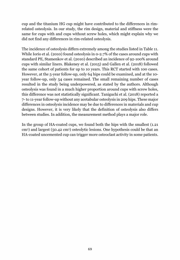
69
cup and the titanium HG cup might have contributed to the differences in rim-related osteolysis. In our study, the rim design, material and stiffness were the same for cups with and cups without screw holes, which might explain why we did not find any differences in rim-related osteolysis.
The incidence of osteolysis differs extremely among the studies listed in Table 11. While Iorio et al. (2010) found osteolysis in 0-2.7% of the cases around cups with standard PE, Stamenkov et al. (2010) described an incidence of 92-100% around cups with similar liners. Blakeney et al. (2015) and Gallen et al. (2018) followed the same cohort of patients for up to 10 years. This RCT started with 100 cases. However, at the 5-year follow-up, only 64 hips could be examined, and at the 10-year follow-up, only 54 cases remained. The small remaining number of cases resulted in the study being underpowered, as stated by the authors. Although osteolysis was found in a much higher proportion around cups with screw holes, this difference was not statistically significant. Taniguchi et al. (2018) reported a 7- to 11-year follow-up without any acetabular osteolysis in 209 hips. These major differences in osteolysis incidence may be due to differences in materials and cup designs. However, it is very likely that the definition of osteolysis also differs between studies. In addition, the measurement method plays a major role.
In the group of HA-coated cups, we found both the hips with the smallest (1.21 cm3) and largest (50.42 cm3) osteolytic lesions. One hypothesis could be that an HA-coated uncemented cup can trigger more osteoclast activity in some patients.

70
Table 11: Studies investigating the incidence of osteolysis around cups with and without screw holes.
Study Cup design Sample size (with/without screw holes)
Follow-up time
(years)
Osteolysis measurement
method
Incidence of acetabular osteolysis
Schmalzried et al. 1999
ARC HGP HGP AML CCD Dual Geometry
0/50 83/0 0/122 0/115 78/35 30/0
5-8 5-7
4-5.5 6-9
3.5-8 4-6
Conventional X-ray
4% 1.2% 0% 14%
12%/29% 6.7%
Claus et al. 2001
Arthropor AML
112/0 0/126 10 Conventional
X-ray
47.3% 47.3%
Iorio et al. 2010
PFC Duraloc Pinnacle
73/70 354/157 82/39
2-11 Conventional X-ray
2.7%/0% 1.7%/1.3%
0% Stamenkov et al. 2010
HG-1 PCA
38/0 0/19
10-19 11-18 CT 92%
100% Blakeney et al. 2015* Reflection 40/24 5 CT 37.5%/29%
Gallen et al. 2018*
Reflection 32/22 10 CT 22%/4.5%
Taniguchi 2018
Trilogy Trident AD
100/13 82/14 7-11 CT 0%
0% *Studies investigated the same cohort
Reduction of the particle load According to a literature review, osteolysis is rarely observed when the PE wear rate is less than 0.1 mm/year, and the authors suggested that a wear rate of 0.05 mm/year would eliminate osteolysis (Dumbleton et al. 2002). Callary et al. (2015) found in their review that the mean proximal wear rate of HXLPE ranges from 0.00 and 0.06 mm/year, according to RSA measurements with up to 10 years of follow-up. This would tempt one to conclude that no osteolysis will occur with XLPE. In a cohort of 513 hips (304 with standard PE liners and 209 with HXLPE liners) with a minimum of 10 years of follow-up, Lachiewicz et al. (2016) found a reduction in osteolysis from 24% around cups with standard PE liners to 13% around cups with HXLPE liners. Although HXLPE appears to have a greatly reduced risk of osteolysis, the desired complete elimination of osteolysis appears to be yet unachieved. It is most likely that the mechanism of periprosthetic osteolysis is multifactorial.
Are there different types of osteolysis? We found three different types of osteolytic lesions on the CT images and defined these as types 1A, 1B and 2 (Figure 12, page 45). Several classifications of the extent and location of osteolysis in relation to the acetabular cup have been published (Paprosky et al. 1994, Garbuz et al. 1996, Rubash et al. 1999, Saleh et

71
al. 2001, Sandgren et al. 2013). To the best of our knowledge, there are no classification systems for the different types of osteolysis around the acetabular cup. Better metal artifact reduction on CT and overall better image quality enhance the possibilities for distinguishing between different types of osteolytic lesions. The lesions with a sclerotic border and the complete absence of trabecular bone, type 1A lesions in our study, were most reliably detected. These lesions follow the often-used definition of osteolysis and can be described as classical osteolytic lesions. The sclerotic border might be a sign of a bony reaction to an osteolytic process (Maloney et al. 1999, Claus et al. 2001, Engh et al. 2002, Kitamura et al. 2006). On the other hand, there is a risk of underestimating the extent of osteolysis since the transition between osteolytic lesions and normal bone can be smooth, and even large defects could be judged as normal solely because of the missing sclerotic border. Additionally, the demand for a sclerotic border is difficult to fulfill when the cortical bone walls are included in the osteolytic lesion, and as a consequence, there is no lesion border in that direction.
Type 1A lesions were more common around cups with screw holes than around sealed cups, although this difference was not statistically significant. Type 1B lesions were more often found around sealed cups.
Type 1A and 1B lesions are probably the most clinically relevant lesions since the complete absence of trabecular bone indicates a complete reduction of mechanical strength and thereby a risk of prosthesis loosening. Type 2 lesions might be caused by a different bone remodeling process, such as stress shielding, or might be an earlier phase of the development of osteolysis. A future longitudinal follow-up study with repeated CT scans of a sufficient number of patients with type 2 lesions may be able to address this issue.
Causes of osteolysis The correlation between 3D wear and osteolytic lesion volume was poor and not statistically significant in our study. The various shapes, sizes and locations of osteolysis are difficult to explain only by the presence of wear particles. Our findings may support those of previous studies suggesting that fluid pressure through unsealed screw holes or along the rim of the cup, in combination with wear particles, contributes to the development of osteolysis (Fahlgren et al. 2010, Johansson et al. 2011). It is possible that different causes of osteolysis result in different types of osteolysis.
Register results The most common reason for THA revision is still aseptic loosening caused by peri-prosthetic bone resorption. Additionally, in recent decades, the number of revisions due to deep infection has increased, and infection is now the number
72
one cause of early revision (Kärrholm et al. 2018). We found that the cause of cup revision differed significantly among cup designs. For example, trabecular metal cups in our cohort had a higher risk of revision due to dislocation. This finding is concurrent with those of earlier register reports (Laaksonen et al. 2018). We could not confirm earlier findings that cups made of tantalum would have a lower risk of infection (Tokarski et al. 2015). At both the 2- and 10-year follow-up, cups with screw holes had a higher risk of revision due to any reason, which was somewhat surprising. Perhaps surgeons chose cups with the possibility for augmentation with screws in more difficult cases. Excluding all diagnoses other than primary osteoarthritis reduces the variety of some of these factors. A comparison of the included hospitals showed significant differences in the use of cups with screw holes. It is unlikely that these differences among hospitals can fully be explained by the mix of cases. Furthermore, the calculated E-value of 2.7 at the 2-year and 2.2 at the 10-year follow-up indicates that unmeasured confounders would have to increase the risk of revision by 2.7/2.2 times and would have to be 2.7/2.2 times more common when cups with screw holes are used to explain the differences we found in our study.
Clinical outcome The PROM data and clinical function results obtained in study I were similar among the four augmentation groups. This was expected as there was no significant difference in migration or wear. A subanalysis revealed a statistically significant difference in symptoms and activities of daily living (ADL) between cups with and without screw holes. However, this difference was very small and would not have any clinical impact.

73
Limitations and strengths The main limitation of study I is the small number of patients. Consequently, there is a risk that we may have missed existing differences between the groups. However, the use of RSA, as a precise method for measuring the migration of implants in vivo, allows the detection of migration differences even in small groups. Although this was a retrospective cohort study, patients were randomized to the treatment groups. The strength of the study is that we have a complete record of each patient over a relatively long follow-up period of 14 years and that we can show the long-term results of a cup design that is still commonly used.
The main limitation of study II is that we only had the results of one CT examination for each patient. Therefore, we could not analyze the precision of CT migration measurements and could measure only the limit of agreement between CT and RSA. The strength of the study is that all measurements in this series were performed using CT images that were obtained in a normal clinical setting and therefore can be reproduced easily in any hospital that has access to a reasonably modern CT.
The main limitation of study III was the small number of patients and the lack of preoperative or immediately postoperative CT scans to detect preexisting bone cysts that might be judged as osteolytic lesions. A series of CT examinations would be needed to determine whether the bone lesions were progressing. The strengths of this study are the relatively long follow-up time and the fact that all cups, aside from the screw holes and coating, had the same design and were made from the same material.
The main limitation of study IV is the lack of detailed patient- or surgeon-related information that might have influenced the decision to use screws. Register data do not provide any information regarding the reason why a specific implant was chosen in an individual case. The strength of the study is the large number of cases, at least for the 2-year follow-up, which represents a large portion of the whole population of patients who underwent THA during the observation time.
The strength of the thesis is the combination of studies with very detailed information about each patient and a register-based study analyzing the effect of screws and screw holes in uncemented cups in a large setting. Studies I, III and IV allowed us to examine the different aspects of cup augmentation with different instruments: migration and wear with RSA, osteolysis with CT and the risk of reoperation with the national register.

74
Conclusions
I. Augmentation of the uncemented porous-coated cup with screws, pegs or an HA coating does not improve long-term stability. Press-fit porous-coated cups without any additional augmentation seem to be the best choice for primary hip arthroplasty in patients with a normal anatomy and good bone quality.
II. CT scans can be used for marker-based migration measurements in longitudinal evaluations of implants without losing clinically significant precision compared to RSA. The visualization of tantalum markers is easier on CT than on RSA. This may be an advantage in the follow-up period as it reduces the risk of excluding cases where otherwise too few markers would be visible. Placing markers in the direct vicinity of other markers or the metal implant should be avoided as this can increase the landmark error on CT.
III. Osteolysis around uncemented cups is underestimated on conventional radiographs. Three different types of osteolytic lesions could be distinguished on CT. We found unsealed screw holes to be a possible risk factor for the development of extended osteolytic lesions.
IV. The revision rate for modern uncemented porous-coated cups is very low, and cup revision due to aseptic loosening within 2 years is extremely rare. There is no significant difference in the risk of cup revision due to aseptic loosening between cups with and without screw holes when used in standard cases. In contrast, the risk of cup revision for any reason was higher for cups with screw holes.

75
Future perspectives
The development of uncemented cups has come a long way. The results of this thesis show that failure due to early aseptic loosening is extremely rare. There is no need for the routine additional augmentation of the porous-coated cup with screws in standard cases. Osteolysis, on the other hand, is still a major problem, especially around older cups that were inserted many years ago, but even around modern cups with XLPE liners. When detected early, osteolysis can be treated without implant removal. A 10-year follow-up with CT of patients with standard PE liners and a 15-year follow-up of patients with XLPE liners would be advisable to avoid excessive osteolysis, prosthesis loosening and fracture.
As the performance of modern uncemented cups is very good, there is a substantial risk that future newer implants might perform worse than existing implants. New prostheses must therefore be thoroughly tested in small patient groups by migration, wear and osteolysis studies. In most RSA studies, patients are only followed for the first few years. It would be important to extend the observation time for at least some of these studies and further examine the development of osteolysis. CT examinations make it possible to study both migration and wear with high precision, as well as the development of osteolysis. For all osteolysis studies, it is essential that the type of osteolysis measured is described carefully. Otherwise, it is not possible to compare the results of different studies.
The development of osteolysis around cups with XLPE liners is a late complication. To determine the long-term effect of screw holes in the cup on the revision risk, a new analysis of the register data will be necessary when a sufficient number of cases with several different cup designs have been followed for more than 10-15 years.

76
Acknowledgments
After many years, it is now finally time to conclude this thesis. This would not have been possible without the help of my mentors, colleagues, friends and family.
Thank YOU ALL!!
I especially want to thank the following:
Kjell Nilsson, Professor at the Department of Orthopedics, Head of the Joint Replacement Unit in Umeå, my tutor throughout this thesis and my friend, for introducing me to research in general, to joint replacement studies in particular, and for helping me on my way in both research and clinical work. You are the reason why I came to Umeå, and you are the reason why I stayed in Umeå. Working with you and learning from you is a pleasure. You not only showed me the world of research but also the world as such. I will never forget our common travels to research conferences in Italy, the Czech Republic, England, the USA and Australia, to mention just a few.
Sead Crnalic, my tutor throughout this thesis, colleague and friend, for helping me with unsurpassed patience throughout the years. Whenever I felt stuck, you showed me a way forward. You are an amazing pedagogue. You are the port in the storm that allows me to expand the boundaries in both clinical work and research. You have always been there to discuss ideas, to develop the good ones and to reject those that are not so good. I am looking forward to all the things we can and will achieve together in the years to come.
Olle Svensson, Professor emeritus at the Department of Orthopedics in Umeå, for always encouraging me during the process of this thesis and your outstanding help to turn long, hard-to-read text into short, comprehensible messages.
Johan Kärrholm, my coauthor, for introducing me to register-based research, providing me with all the data for study IV and all the register data for this book, and helping me to understand and interpret these data. There could not have been a better teacher for these tasks.
Sebastian Mukka, my coauthor, colleague and friend, who is always full of ideas and the energy implement them. Thank you for your help. I am looking forward to all of our shared adventures to come.

77
Henrik Olivecrona and Marilyn Noz, my coauthors, for introducing me to the world of CT-based migration measurement. You are brilliant at explaining the most technical, complex things in an easily understandable way.
Roumen Stamenkov, Stuart Callary and Donald Howie, my coauthors, for introducing me to and helping me with CT-based bone lesion measurements, as well as drawing my attention to the complexity of osteolysis research.
Stephan Röhrl and Bo Nivbrant, my coauthors, for performing the surgeries, providing me with all background data for the first three studies, and helping me to sort out all the files.
Leif Nyström, for always taking the time to answer all my technical questions about RSA and offering your outstanding support for both UmRSA and MDesk.
Anna Kristofferson, for organizing the consultation hours with the patients from all the different studies. You are a key player in the organization of our clinical trials and in the contact with the study participants.
Per Söderlund, my colleague and friend, for helping me to learn the practical steps involved in RSA measurements and having my back at the clinic.
Bernhardt Kickinger, my friend and colleague, for helping me with so many things—everything from working extra in the clinic to relieve me to taking care of my children when needed. You and your family play an important role in my life.
Åke Hamberg, my colleague and friend, for coming to Umeå again and again to replace me in the clinic so that I could concentrate on the work for this thesis and for all of our discussions about orthopedics, research and life in general.
Hans-Olov Larsson, my mentor and colleague during my residency, for welcoming me so warmly to my first job in Sweden and for introducing me to joint arthroplasty surgery.
Lars Wallinder, my mentor in clinical work, for teaching me so much about revision and advanced primary hip arthroplasty.
Ulrica Bergström, head of the Department of Orthopedics, for creating the best possible research conditions, despite a financially demanding situation.
All my colleagues at the orthopedic department (including the orthopedic section of COP 1), for your support and tolerance during my absence from clinical work.
78
Representatives of DePuy, Link, Smith & Nephew, Stryker and ZimmerBiomet, for always answering my detailed questions about your products and lending me all the prosthesis so that I could make the pictures for this book.
Danke auch an meinem Schwiegervater Ernst Diebel, der mich immer wieder ermuntert hat auf meinem Weg. Thanks to my mother, Heide Otten, who has encouraged me to be a researcher since I was a little boy.
Last, but absolutely not least, I would like to thank my family, my wife Julia, and my children Jonathan, Hanna and Sofia, for all your support, your understanding of my absence and your love that encouraged me to go on. Jonathan, thank you for all your tips on how to make the text easier to understand, and thank you for watching your younger sisters. Hanna, tack för din kärlek och dina kramar. Sofia, Danke für Deine viele positive Energie und unsere tollen gemeinsamen Ausflüge um mal von der Arbeit wegzukommen. You are the most important thing in my life.

79
References
Affatato S, Bordini B, Fagnano C, Taddei P, Tinti A, Toni A. Effects of the Sterilisation Method on the Wear of Uhmwpe Acetabular Cups Tested in a Hip Joint Simulator. Biomaterials. 2002;23(6):1439-46.
Allain J, Le Mouel S, Goutallier D, Voisin MC.
Poor Eight-Year Survival of Cemented Zirconia-Polyethylene Total Hip Replacements. The Journal of Bone & Joint Surgery British Volume. 1999;81(5):835-42.
Anagnostakos K, Schmitt C. Can
Periprosthetic Hip Joint Infections Be Successfully Managed by Debridement and Prosthesis Retention? World Journal of Orthopedics. 2014;5(3):218.
Anand R, Graves SE, de Steiger RN, Davidson
DC, Ryan P, Miller LN, Cashman K. What Is the Benefit of Introducing New Hip and Knee Prostheses? The Journal of Bone & Joint Surgery American Volume. 2011;93(Supplement 3):51-4.
Aspenberg P, van der Vis H. Fluid Pressure
May Cause Periprosthetic Osteolysis. Particles Are Not the Only Thing. Acta Orthopaedica. 1998;69(1):1-4.
Ast MP, John TK, Labbisiere A, Robador N,
Della Valle AG. Fractures of a Single Design of Highly Cross-Linked Polyethylene Acetabular Liners: An Analysis of Voluntary Reports to the United States Food and Drug Administration. The Journal of Arthroplasty. 2014;29(6):1231-5.
Baleani M, Fognani R, Toni A. Initial Stability
of a Cementless Acetabular Cup Design:
Experimental Investigation on the Effect of Adding Fins to the Rim of the Cup. Artificial Organs. 2001;25(8):664-9.
Bankston AB, Keating EM, Ranawat C, Faris
PM, Ritter MA. Comparison of Polyethylene Wear in Machined Versus Molded Polyethylene. Clinical Orthopaedics and Related Research. 1995(317):37-43.
Benazzo F, Formagnana M, Bargagliotti M,
Perticarini L. Periprosthetic Acetabular Fractures. International Orthopaedics. 2015;39(10):1959-63.
Berry DJ, Barnes CL, Scott RD, Cabanela ME,
Poss R. Catastrophic Failure of the Polyethylene Liner of Uncemented Acetabular Components. The Journal of Bone & Joint Surgery British Volume. 1994;76(4):575-8.
Berry DJ, Lieberman J. Surgery of the Hip, E-
Book. Elsevier Health Sciences 2012. Blakeney WG, Khan H, Khan RJK. Cluster
Hole Versus Solid Cup in Total Hip Arthroplasty: A Randomized Control Trial. The Journal of Arthroplasty. 2015;30(2):223-9.
Bloebaum RD, Beeks D, Dorr LD, Savory CG,
Dupont JA, Hofmann AA. Complications with Hydroxyapatite Participate Separation in Total Hip Arthroplasty. Clinical Orthopaedics and Related Research. 1994;298:19-26.
Boutin P, Christel P, Dorlot JM, Meunier A,
De Roquancourt A, Blanquaert D, Herman S, Sedel L, Witvoet J. The Use of Dense Alumina–Alumina Ceramic Combination in Total Hip Replacement. Journal of Biomedical Materials Research. 1988;22(12):1203-32.

80
Brand RA, Mont MA, Manring MJCO,
Research® R. Biographical Sketch: Themistocles Gluck (1853–1942). Clinical Orthopaedics. 2011;469(6):1525-7.
Brodén C, Olivecrona H, Maguire GQ, Noz
ME, Zeleznik MP, Sköldenberg O. Accuracy and Precision of Three-Dimensional Low Dose Ct Compared to Standard Rsa in Acetabular Cups: An Experimental Study. BioMed Research International. 2016;2016.
Burke GL. The Corrosion of Metals in Tissues;
and an Introduction to Tantalum. Canadian Medical Association Journa. 1940;43(2):125.
Burstrom K, Johannesson M, Diderichsen F.
Swedish Population Health-Related Quality of Life Results Using the Eq-5d. Quality of Life Research. 2001;10(7):621-35.
Bus MP, Szafranski A, Sellevold S, Goryn T,
Jutte PC, Bramer JA, Fiocco M, Streitbürger A, Kotrych D, van de Sande MA. Lumic® Endoprosthetic Reconstruction after Periacetabular Tumor Resection: Short-Term Results. Clinical Orthopaedics and Related Research. 2017;475(3):686-95.
Callary SA, Solomon LB, Holubowycz OT,
Campbell DG, Munn Z, Howie DW. Wear of Highly Crosslinked Polyethylene Acetabular Components: A Review of Rsa Studies. Acta Orthopaedica. 2015;86(2):159-68.
Catelas I, Huk OL, Petit A, Zukor DJ,
Marchand R, Yahia LH. Flow Cytometric Analysis of Macrophage Response to Ceramic and Polyethylene Particles: Effects of Size, Concentration, and
Composition. Journal of Biomedical Materials Research. 1998;41(4):600-7.
Cech O, Dzupa V, Svatos F. The Uncemented
Cup of the Cls Spotorno Joint Endoprosthesis--5-Years' Results. Acta Chirurgiae, Orthopaedica et Traumatologiae Cechoslovaca. 2001;68(1):10-7.
Chan FW, Bobyn JD, Medley JB, Krygier JJ,
Tanzer M. Wear and Lubrication of Metal-on-Metal Hip Implants. Clinical Orthopaedics and Related Research. 1999;369:10-24.
Clarke HJ, Jinnah RH, Warden KE, Cox QG,
Curtis MJ. Evaluation of Acetabular Stability in Uncemented Prostheses. The Journal of Arthroplasty. 1991;6(4):335-40.
Claus AM, Sychterz CJ, Hopper RH, Jr., Engh
CA. Pattern of Osteolysis around Two Different Cementless Metal-Backed Cups: Retrospective, Radiographic Analysis at Minimum 10-Year Follow-Up. The Journal of Arthroplasty. 2001;16(8 Suppl 1):177-82.
Clohisy JC, Harris WH. Matched-Pair
Analysis of Cemented and Cementless Acetabular Reconstruction in Primary Total Hip Arthroplasty. The Journal of Arthroplasty. 2001;16(6):697-705.
Coathup MJ, Blackburn J, Goodship AE,
Cunningham JL, Smith T, Blunn GW. Role of Hydroxyapatite Coating in Resisting Wear Particle Migration and Osteolysis around Acetabular Components. Biomaterials. 2005;26(19):4161-9.
Colacchio ND, Cleary MX, Reid D, Trofa D,
Pevear ME, Smith ELJCop. Direct Anterior Approach Total Hip Arthroplasty

81
Requires Less Supplemental Acetabular Screw Fixation and Fewer Blood Transfusions Than Posterior Approach. Current Orthopaedic Practice. 2017;28(4):404-8.
Costa L, Luda M, Trossarelli L, Del Prever EB,
Crova M, Gallinaro P. Oxidation in Orthopaedic Uhmwpe Sterilized by Gamma-Radiation and Ethylene Oxide. Biomaterials. 1998;19(7-9):659-68.
Dahl J, Snorrason F, Nordsletten L, Rohrl
SM. More Than 50% Reduction of Wear in Polyethylene Liners with Alumina Heads Compared to Cobalt-Chrome Heads in Hip Replacements. Acta Orthopaedica. 2013;84(4):360-4.
DeLee JG, Charnley J. Radiological
Demarcation of Cemented Sockets in Total Hip Replacement. Clinical Orthopaedics and Related Research. 1976(121):20-32.
Dickinson A, Taylor A, Browne M. The
Influence of Acetabular Cup Material on Pelvis Cortex Surface Strains, Measured Using Digital Image Correlation. Journal of Biomechanics. 2012;45(4):719-23.
Drummond J, Tran P, Fary C. Metal-on-
Metal Hip Arthroplasty: A Review of Adverse Reactions and Patient Management. Journal of Functional Biomaterials. 2015;6(3):486-99.
Ducheyne P, Hench L, Kagan Ad, Martens M,
Bursens A, Mulier J. Effect of Hydroxyapatite Impregnation on Skeletal Bonding of Porous Coated Implants. Journal of Biomedical Materials Research. 1980;14(3):225-37.
Dumbleton JH, Manley MT, Edidin AA. A
Literature Review of the Association between Wear Rate and Osteolysis in Total
Hip Arthroplasty. The Journal of Arthroplasty. 2002;17(5):649-61.
Effenberger H, Imhof M, Richolt J, Rehart S.
Cement-Free Hip Cups. Current Status. Der Orthopäde. 2004;33(6):733-50; quiz 51.
Eftekhar NS. Long-Term Results of
Cemented Total Hip Arthroplasty. Clinical Orthopaedics and Related Research. 1987(225):207-17.
Ellenrieder M, Bader R, Bergschmidt P,
Mittelmeier W. Press-Fit Versus Threaded Acetabular Cups in Total Hip Arthroplasty: Functional and Radiological Results after Five Years. Acta Orthopaedica Belgica. 2016;82:60-7.
Engh CA, Griffin WL, Marx CL. Cementless
Acetabular Components. The Journal of Bone & Joint Surgery British Volume. 1990;72(1):53-9.
Engh CA, Jr., Sychterz CJ, Young AM, Pollock
DC, Toomey SD, Engh CA, Sr. Interobserver and Intraobserver Variability in Radiographic Assessment of Osteolysis. The Journal of Arthroplasty. 2002;17(6):752-9.
Ericson A, Arndt A, Stark A, Noz ME, Maguire
GQ, Jr., Zeleznik MP, Olivecrona H. Fusion of Radiostereometric Analysis Data into Computed Tomography Space: Application to the Elbow Joint. Journal of Biomechanics. 2007;40(2):296-304.
Eskelinen A, Remes V, Helenius I, Pulkkinen
P, Nevalainen J, Paavolainen P. Total Hip Arthroplasty for Primary Osteoarthrosis in Younger Patients in the Finnish Arthroplasty Register. Acta Orthopaedica. 2005;76(1):28-41.

82
Fahlgren A, Bostrom MP, Yang X, Johansson L, Edlund U, Agholme F, Aspenberg P. Fluid Pressure and Flow as a Cause of Bone Resorption. Acta Orthopaedica. 2010;81(4):508-16.
Frosch KH, Barvencik F, Viereck V, Lohmann
CH, Dresing K, Breme J, Brunner E, Sturmer KM. Growth Behavior, Matrix Production, and Gene Expression of Human Osteoblasts in Defined Cylindrical Titanium Channels. Journal of Biomedical Material Research Part A. 2004;68(2):325-34.
Furlong R, Osborn J. Fixation of Hip
Prostheses by Hydroxyapatite Ceramic Coatings. The Journal of Bone & Joint Surgery British Volume. 1991;73(5):741-5.
Gallen RA, Khan RJK, Haebich SJ,
Karamfiles SH, Khan H. Solid Cup Vs Cluster Hole in Total Hip Arthroplasty: A 10-Year Randomized Control Trial. The Journal of Arthroplasty. 2018;33(4):1113-9.
Garbuz D, Morsi E, Mohamed N, Gross AE.
Classification and Reconstruction in Revision Acetabular Arthroplasty with Bone Stock Deficiency. Clinical Orthopaedics and Related Research. 1996(324):98-107.
Garcia-Cimbrelo E, Cruz-Pardos A, Madero
R, Ortega-Andreu M. Total Hip Arthroplasty with Use of the Cementless Zweymüller Alloclassic System: A Ten to Thirteen-Year Follow-up Study. The Journal of Bone & Joint Surgery. 2003;85(2):296-303.
Geesink R. Clinical, Radiological and Human
Histological Experience with Hydroxyapatite Coatings in Orthopaedic
Surgery. Acta Orthopaedica Belgica. 1993;59:160-4.
Goldsmith A, Dowson D, Wroblewski B, Siney
P, Fleming P, Lane J. The Effect of Activity Levels of Total Hip Arthroplasty Patients on Socket Penetration. The Journal of Arthroplasty. 2001;16(5):620-7.
Gomez PF, Morcuende JA. Early Attempts at
Hip Arthroplasty-1700s to 1950s. The Iowa Orthopaedic Journal. 2005;25:25.
Gomez PF, Morcuende JA. A Historical and
Economic Perspective on Sir John Charnley, Chas F. Thackray Limited, and the Early Arthoplasty Industry. The Iowa Orthopaedic Journal. 2005;25:30-7.
Goodman S, Ma T. Biological Responses to
Particle Debris. In: Surgery of the Hip. Elsevier Health Sciences; 2012. p. 157-70.
Goodman SB, Gibon E, Yao Z. The Basic
Science of Periprosthetic Osteolysis. Instructional Course Lectures. 2013;62:201-6.
Greenfield EM, Bi Y, Ragab AA, Goldberg
VM, Nalepka JL, Seabold JM. Does Endotoxin Contribute to Aseptic Loosening of Orthopedic Implants? Journal of Biomedical Materials Research Part B: Applied Biomaterials. 2005;72(1):179-85.
Hailer N. 20 Years of Porous Tantalum in
Primary and Revision Hip Arthroplasty-Time for a Critical Appraisal. Acta Orthopaedica. 2018;89(3):254-5.
Hailer NP, Garellick G, Karrholm J.
Uncemented and Cemented Primary Total Hip Arthroplasty in the Swedish Hip Arthroplasty Register. Acta Orthopaedica. 2010;81(1):34-41.

83
Haraguchi K, Sugano N, Nishii T, Miki H, Oka
K, Yoshikawa H. Phase Transformation of a Zirconia Ceramic Head after Total Hip Arthroplasty. The Journal of Bone & Joint Surgery British Volume. 2001;83(7):996-1000.
Harris W, Schiller A, Scholler J, Freiberg R,
Scott RJJ. Extensive Localized Bone Resorption in the Femur Following Total Hip Replacement. The Journal of Bone & Joint Surgery. 1976;58(5):612-8.
Havelin LI, Espehaug B, Vollset SE,
Engesaeter LBJAOS. Early Failures among 14,009 Cemented and 1,326 Uncemented Prostheses for Primary Coxarthrosis: The Norwegian Arthroplasty Register, 1987-1992. Acta Orthopaedica. 1994;65(1):1-6.
Havelin LI, Vollset SE, Engesaeter LB.
Revision for Aseptic Loosening of Uncemented Cups in 4,352 Primary Total Hip Prostheses. A Report from the Norwegian Arthroplasty Register. Acta Orthopaedica. 1995;66(6):494-500.
Herberts P, Malchau H. Swedish Hip
Arthroplasty Register: Annual Report 1997. Gothenburg; 1998.
Herberts P, Malchau H, Söderman P. Swedish
Hip Arthroplasty Register: Annual Report 1999. Gothenburg; 2000.
Howard D, Wall P, Fernandez M, Parsons H,
Howard PJTb, journal j. Ceramic-on-Ceramic Bearing Fractures in Total Hip Arthroplasty: An Analysis of Data from the National Joint Registry. The Bone & Joint Journal. 2017;99(8):1012-9.
Howie DW, Neale SD, Stamenkov R, McGee
MA, Taylor DJ, Findlay DM. Progression
of Acetabular Periprosthetic Osteolytic Lesions Measured with Computed Tomography. The Journal of Bone & Joint Surgery American Volume. 2007;89(8):1818-25.
Hsu J-T, Chang C-H, Huang H-L, Zobitz ME,
Chen W-P, Lai K-A, An K-N. The Number of Screws, Bone Quality, and Friction Coefficient Affect Acetabular Cup Stability. Medical Engineering & Physics. 2007;29(10):1089-95.
Iorio R, Puskas B, Healy WL, Tilzey JF,
Specht LM, Thompson MS. Cementless Acetabular Fixation with and without Screws: Analysis of Stability and Migration. The Journal of Arthroplasty. 2010;25(2):309-13.
Iorio R, Puskas B, Healy WL, Tilzey JF,
Specht LM, Thompson MS. Cementless Acetabular Fixation with and without Screws: Analysis of Stability and Migration. The Journal of Arthroplasty. 2010;25(2):309-13.
Jarcho M, Kay JF, Gumaer KI, Doremus RH,
Drobeck HP. Tissue, Cellular and Subcellular Events at a Bone-Ceramic Hydroxylapatite Interface. Journal of Bioengineering. 1977;1(2):79-92.
Jasty MJ, Schiller A, Goldring S, Harris
WJTJob, volume jsA. Localized Osteolysis in Stable, Non-Septic Total Hip Replacement. The Journal of Bone & Joint Surgery American Volume. 1986;68(6):912-9.
Jeffers J, Walter W. Ceramic-on-Ceramic
Bearings in Hip Arthroplasty State of the Art and the Future. The Journal of Bone & Joint Surgery British Volume. 2012;94(6):735-45.

84
Johansson L, Edlund U, Fahlgren A, Aspenberg P. Fluid-Induced Osteolysis: Modelling and Experiments. Computer Methods in Biomechanics and Biomedical Engineering. 2011;14(4):305-18.
Jones LC, Hungerford DS. Cement Disease.
Clinical Orthopaedics and Related Research. 1987(225):192-206.
Judet J, Judet R. The Use of an Artificial
Femoral Head for Arthroplasty of the Hip Joint. The Journal of Bone & Joint Surgery British Volume. 1950;32(2):166-73.
Kabo J, Gebhard J, Loren G, Amstutz H. In
Vivo Wear of Polyethylene Acetabular Components. The Journal of Bone & Joint Surgery British Volume. 1993;75(2):254-8.
Kärrholm J. Roentgen
Stereophotogrammetry. Review of Orthopedic Applications. Acta Orthopaedica. 1989;60(4):491-503.
Kärrholm J. Radiostereometric Analysis of
Early Implant Migration - a Valuable Tool to Ensure Proper Introduction of New Implants. Acta Orthopaedica. 2012;83(6):551-2.
Kärrholm J, Garellick G, Herberts P. Swedish
Hip Arthroplasty Register: Annual Report 2006. Gothenburg; 2007.
Kärrholm J, Gill RH, Valstar ER. The History
and Future of Radiostereometric Analysis. Clinical Orthopaedics and Related Research. 2006;448:10-21.
Kärrholm J, Herberts P, Hultmark P,
Malchau H, Nivbrant B, Thanner J. Radiostereometry of Hip Prostheses. Review of Methodology and Clinical
Results. Clinical Orthopaedics and Related Research. 1997(344):94-110.
Kärrholm J, Lindahl H, Malchau H,
Mohaddes M, Nemes S, Rogmark C, Rolfson O. The Swedish Hip Arthroplasty Register: Annual Report 2016. 2017.
Kärrholm J, Mohaddes M, Odin D, Vinblad J,
Rogmark C, Rolfson O. Swedish Hip Arthroplasty Register: Annual Report 2017. Gothenburg; 2018.
Kitamura N, Leung SB, Engh CA, Sr.
Characteristics of Pelvic Osteolysis on Computed Tomography after Total Hip Arthroplasty. Clinical Orthopaedics and Related Research. 2005;441:291-7.
Kitamura N, Pappedemos PC, Duffy PR, 3rd,
Stepniewski AS, Hopper RH, Jr., Engh CA, Jr., Engh CA. The Value of Anteroposterior Pelvic Radiographs for Evaluating Pelvic Osteolysis. Clinical Orthopaedics and Related Research. 2006;453:239-45.
Kop AM, Whitewood C, Johnston DJL.
Damage of Oxinium Femoral Heads Subsequent to Hip Arthroplasty Dislocation: Three Retrieval Case Studies. The Journal of Arthroplasty. 2007;22(5):775-9.
Krismer M, Stöckl B, Fischer M, Bauer R,
Mayrhofer P, Ogon M. Early Migration Predicts Late Aseptic Failure of Hip Sockets. The Journal of Bone & Joint Surgery British Volume. 1996;78(3):422-6.
Kubo T, Inoue S, Maeda T, Arai Y, Hirakawa
K, Wu Y, Suehara H, Ogura T, Hirasawa Y. Cementless Lord Total Hip Arthroplasty: Cup Loosening Common after Minimum

85
10-Year Follow-up of 103 Hips. Acta Orthopaedica. 2001;72(6):585-90.
Laaksonen I, Lorimer M, Gromov K,
Eskelinen A, Rolfson O, Graves SE, Malchau H, Mohaddes M. Trabecular Metal Acetabular Components in Primary Total Hip Arthroplasty: Higher Risk for Revision Compared with Other Uncemented Cup Designs in a Collaborative Register Study Including 93,709 Hips. Acta Orthopaedica. 2018:1-6.
Lachiewicz PF, Kleeman LT, Seyler T. Bearing
Surfaces for Total Hip Arthroplasty. Journal of the American Academy of Orthopaedic Surgeons. 2018;26(2):45-57.
Lachiewicz PF, Soileau ES. Highly Cross-
Linked Polyethylene Provides Decreased Osteolysis and Reoperation at Minimum 10-Year Follow-Up. The Journal of Arthroplasty. 2016;31(9):1959-62.
Lambert B, Neut D, van der Veen HC, Bulstra
SK. Effects of Vitamin E Incorporation in Polyethylene on Oxidative Degradation, Wear Rates, Immune Response, and Infections in Total Joint Arthroplasty: A Review of the Current Literature. International Orthopaedics. 2018.
Lazarinis S, Karrholm J, Hailer NP. Increased
Risk of Revision of Acetabular Cups Coated with Hydroxyapatite. Acta Orthopaedica. 2010;81(1):53-9.
Lazarinis S, Karrholm J, Hailer NP. Effects of
Hydroxyapatite Coating of Cups Used in Hip Revision Arthroplasty. Acta Orthopaedica. 2012;83(5):427-35.
Lazarinis S, Mäkelä KT, Eskelinen A, Havelin
L, Hallan G, Overgaard S, Pedersen AB, Kärrholm J, Hailer NP. Does
Hydroxyapatite Coating of Uncemented Cups Improve Long-Term Survival? An Analysis of 28,605 Primary Total Hip Arthroplasty Procedures from the Nordic Arthroplasty Register Association (Nara). Osteoarthritis and Cartilage. 2017;25:1980-7.
Learmonth ID, Young C, Rorabeck C. The
Operation of the Century: Total Hip Replacement. The Lancet. 2007;370(9597):1508-19.
Lennox DW, Schofield BH, McDonald DF,
Riley LH, Jr. A Histologic Comparison of Aseptic Loosening of Cemented, Press-Fit, and Biologic Ingrowth Prostheses. Clinical Orthopaedics and Related Research. 1987(225):171-91.
Litsky AS, Pophal SG. Initial Mechanical
Stability of Acetabular Prostheses. Orthopedics. 1994;17(1):53-7.
Looney RJ, Boyd A, Totterman S, Seo G-S,
Tamez-Pena J, Campbell D, Novotny L, Olcott C, Martell J, Hayes FA. Volumetric Computerized Tomography as a Measurement of Periprosthetic Acetabular Osteolysis and Its Correlation with Wear. Arthritis research. 2002;4(1):59-63.
Macdonald N, Bankes M. Ceramic on Ceramic
Hip Prostheses: A Review of Past and Modern Materials. Archives of Orthopaedic and Trauma Surgery. 2014;134(9):1325-33.
Malchau H, Karrholm J, Wang YX, Herberts
P. Accuracy of Migration Analysis in Hip Arthroplasty. Digitized and Conventional Radiography, Compared to Radiostereometry in 51 Patients. Acta Orthopaedica. 1995;66(5):418-24.

86
Maloney WJ, Galante JO, Anderson M, Goldberg V, Harris WH, Jacobs J, Kraay M, Lachiewicz P, Rubash HE, Schutzer S, Woolson ST. Fixation, Polyethylene Wear, and Pelvic Osteolysis in Primary Total Hip Replacement. Clinical Orthopaedics and Related Research. 1999(369):157-64.
Maloney WJ, Peters P, Engh CA, Chandler H.
Severe Osteolysis of the Pelvic in Association with Acetabular Replacement without Cement. The Journal of Bone & Joint Surgery American Volume. 1993;75(11):1627-35.
Matthews JB, Besong AA, Green TR, Stone
MH, Wroblewski BM, Fisher J, Ingham E. Evaluation of the Response of Primary Human Peripheral Blood Mononuclear Phagocytes to Challenge with in Vitro Generated Clinically Relevant Uhmwpe Particles of Known Size and Dose. Journal of Biomedical Materials Research Part A. 2000;52(2):296-307.
Matthews JB, Green TR, Stone MH,
Wroblewski BM, Fisher J, Ingham E. Comparison of the Response of Primary Human Peripheral Blood Mononuclear Phagocytes from Different Donors to Challenge with Model Polyethylene Particles of Known Size and Dose. Biomaterials. 2000;21(20):2033-44.
McCoy T, Salvati E, Ranawat CS, Wilson
JPJTOcoNA. A Fifteen-Year Follow-up Study of One Hundred Charnley Low-Friction Arthroplasties. The Orthopedic Clinics of North America. 1988;19(3):467-76.
McKee G, Watson-Farrar J. Replacement of
Arthritic Hips by the Mckee-Farrar Prosthesis. The Journal of Bone & Joint Surgery British Volume. 1966;48(2):245-59.
McKellop H, Shen F-w, Lu B, Campbell P,
Salovey R. Effect of Sterilization Method and Other Modifications on the Wear Resistance of Acetabular Cups Made of Ultra-High Molecular Weight Polyethylene: A Hip-Simulator Study. The Journal of Bone & Joint Surgery. 2000;82(12):1708-25.
McKellop H, Shen Fw, Lu B, Campbell P,
Salovey R. Development of an Extremely Wear‐Resistant Ultra High Molecular Weight Polythylene for Total Hip Replacements. Journal of Orthopaedic Research. 1999;17(2):157-67.
McKellop HA. The Lexicon of Polyethylene
Wear in Artificial Joints. Biomaterials. 2007;28(34):5049-57.
Meneghini RM, Ford KS, McCollough CH,
Hanssen AD, Lewallen DG. Bone Remodeling around Porous Metal Cementless Acetabular Components. The Journal of Arthroplasty. 2010;25(5):741-7.
Minten MJ, Heesterbeek PJ, Spruit M. No
Effect of Additional Screw Fixation of a Cementless, All-Polyethylene Press-Fit Socket on Migration, Wear, and Clinical Outcome: A 6.5-Year Randomized Radiostereometric Analysis Follow-up Report. Acta Orthopaedica. 2016;87(4):363-7.
Mok D, Bryant K. Ring Uncemented Plastic
on Metal Hip Replacements-Results from an Independent Unit. Journal of the Royal Society of Medicine. 1989;82(3):142-4.
Moore AT. Metal Hip Joint; a New Self-
Locking Vitallium Prosthesis. Southern Medical Journal. 1952;45(11):1015-19.

87
Moore AT. The Self-Locking Metal Hip Prosthesis. The Journal of Bone & Joint Surgery American Volume. 1957;39-a(4):811-27.
Moore AT, Bohlman HR. The Classic: Metal
Hip Joint: A Case Report. Clinical Orthopaedics and Related Research. 2006;453:22-4.
Morscher EW, Hefti A, Aebi U. Severe
Osteolysis after Third-Body Wear Due to Hydroxyapatite Particles from Acetabular Cup Coating. The Journal of Bone & Joint Surgery British Volume. 1998;80(2):267-72.
Mueller U, Lee C, Heisel C, Thomsen M,
Bitsch RG, Kretzer JP. Failure of Polyethylene Inlays in Cementless Total Hip Arthroplasty: A Retrieval Analysis. BioMed Research International. 2016;2016.
Naing L, Winn T, Rusli B. Practical Issues in
Calculating the Sample Size for Prevalence Studies. Archives of Orofacial Science. 2006;1:9-14.
Naser MA, Pathak R, Ahmad A. Superiority of
Fixed Stem Bipolar Prosthesis over Austin Moore Prosthetic in Fracture Neck Femur. International Journal of Research in Orthopaedics. 2018.
Nery E, Lynch K, Hirthe W, Mueller K.
Bioceramic Implants in Surgically Produced Infrabony Defects. Journal of Periodontology. 1975;46(6):328-47.
Ni SH, Guo L, Jiang TL, Zhao J, Zhao YG.
Press-Fit Cementless Acetabular Fixation with and without Screws. International Orthopaedics. 2013.
Niggemeyer O, Fink B, Rüther W. Dislocation of the Polyethylene Liner in Hip Arthroplasty. Acta Orthopaedica. 2002;73(4):403-6.
Nilsdotter AK, Lohmander LS, Klassbo M,
Roos EM. Hip Disability and Osteoarthritis Outcome Score (Hoos)--Validity and Responsiveness in Total Hip Replacement. BMC Musculoskelet Disorders. 2003;4:10.
Northover JR, Maqsood M. 58-Year Follow-
up of a Vitallium Mold Hip Arthroplasty: A Case Report and Literature Review. Acta Orthopaedica. 2008;79(6):731-3.
Noz ME, Maguire Jr GQ, Zeleznik MP,
Kramer EL, Mahmoud F, Crafoord J. A Versatile Functional–Anatomic Image Fusion Method for Volume Data Sets. Journal of Medical Systems. 2001;25(5):297-307.
Ohashi H, Kikuchi S, Aota S, Hakozaki M,
Konno SJJoOS. Surgical Anatomy of the Pelvic Vasculature, with Particular Reference to Acetabular Screw Fixation in Cementless Total Hip Arthroplasty in Asian Population: A Cadaveric Study. Journal of Orthopaedic Surgery (Hong Kong). 2017;25(1):2309499016685520.
Olivecrona H, Maguire GQ, Noz ME, Zeleznik
MP, Kesteris U, Weidenhielm L. A Ct Method for Following Patients with Both Prosthetic Replacement and Implanted Tantalum Beads: Preliminary Analysis with a Pelvic Model and in Seven Patients. Journal of Orthopaedic Surgery and Research. 2016;11(1):1.
Olivecrona L, Crafoord J, Olivecrona H, Noz
ME, Maguire GQ, Zeleznik MP, Svensson L, Weidenhielm L. Acetabular Component Migration in Total Hip Arthroplasty Using

88
Ct and a Semiautomated Program for Volume Merging. Acta Radiologica. 2002;43(5):517-27.
Ollivere B, Wimhurst J, M. Clark I, Donell
SJTJob, volume jsB. Current Concepts in Osteolysis. The Journal of Bone & Joint Surgery British Volume. 2012;94(1):10-5.
Ong KL, Rundell S, Liepins I, Laurent R,
Markel D, Kurtz SM. Biomechanical Modeling of Acetabular Component Polyethylene Stresses, Fracture Risk, and Wear Rate Following Press‐Fit Implantation. Journal of Orthopaedic Research. 2009;27(11):1467-72.
Oparaugo PC, Clarke IC, Malchau H,
Herberts P. Correlation of Wear Debris-Induced Osteolysis and Revision with Volumetric Wear-Rates of Polyethylene: A Survey of 8 Reports in the Literature. Acta Orthopaedica. 2001;72(1):22-8.
Orishimo KF, Claus AM, Sychterz CJ, Engh
CA. Relationship between Polyethylene Wear and Osteolysis in Hips with a Second-Generation Porous-Coated Cementless Cup after Seven Years of Follow-Up. The Journal of Bone & Joint Surgery American Volume. 2003;85-A(6):1095-9.
Otto M, Kriegsmann J, Gehrke T, Bertz S.
Wear Particles: Key to Aseptic Prosthetic Loosening? Der Pathologe. 2006;27(6):447-60.
Oxford English Dictionary O. Oxford
University Press; 2018. Pakvis D, Luites J, van Hellemondt G, Spruit
M. A Cementless, Elastic Press-Fit Socket with and without Screws. Acta Orthopaedica. 2012;83(5):481-7.
Pal S. Mechanical Properties of Biological Materials. In: Design of Artificial Human Joints & Organs. Springer; 2014. p. 23-40.
Paprosky WG, Perona PG, Lawrence JM.
Acetabular Defect Classification and Surgical Reconstruction in Revision Arthroplasty. A 6-Year Follow-up Evaluation. The Journal of Arthroplasty. 1994;9(1):33-44.
Patterson M. Ring Uncemented Hip
Replacements. The Results of Revision. The Journal of Bone & Joint Surgery British Volume. 1987;69(3):374-80.
Paulsen A, Pedersen AB, Johnsen SP, Riis A,
Lucht U, Overgaard S. Effect of Hydroxyapatite Coating on Risk of Revision after Primary Total Hip Arthroplasty in Younger Patients: Findings from the Danish Hip Arthroplasty Registry. Acta Orthopaedica. 2007;78(5):622-8.
Pepe M, Kocadal O, Erener T, Ceritoglu K,
Aksahin E, Aktekin CNJWjoo. Acetabular Components with or without Screws in Total Hip Arthroplasty. World Journal of Orthopedics. 2017;8(9):705.
Perkins M, Butler R, Tidwell L, Gu W,
Mihalko W. Genetic Links to Total Joint Arthroplasty Outcomes: A Systematic Review. In: Beyond the Implant: Retrieval Analysis Methods for Implant Surveillance. ASTM International; 2018. p. 261-76.
Perona PG, Lawrence J, Paprosky WG,
Patwardhan AG, Sartori M. Acetabular Micromotion as a Measure of Initial Implant Stability in Primary Hip Arthroplasty. An in Vitro Comparison of Different Methods of Initial Acetabular

89
Component Fixation. The Journal of Arthroplasty. 1992;7(4):537-47.
Phedy P, Ismail HD, Hoo C, Djaja YP. Total
Hip Replacement: A Meta-Analysis to Evaluate Survival of Cemented, Cementless and Hybrid Implants. World Journal of Orthopedics. 2017;8(2):192-207.
Philippot R, Camilleri JP, Boyer B, Adam P,
Farizon F. The Use of a Dual-Articulation Acetabular Cup System to Prevent Dislocation after Primary Total Hip Arthroplasty: Analysis of 384 Cases at a Mean Follow-up of 15 Years. International Orthopaedics. 2009;33(4):927-32.
Piconi C, Maccauro G. Zirconia as a Ceramic
Biomaterial. Biomaterials. 1999;20(1):1-25.
Pijls BG, Nieuwenhuijse MJ, Fiocco M,
Plevier JW, Middeldorp S, Nelissen RG, Valstar ER. Early Proximal Migration of Cups Is Associated with Late Revision in Tha: A Systematic Review and Meta-Analysis of 26 Rsa Studies and 49 Survivalstudies. Acta Orthopaedica. 2012;83(6):583-91.
Puri L, Wixson RL, Stern SH, Kohli J,
Hendrix RW, Stulberg SD. Use of Helical Computed Tomography for the Assessment of Acetabular Osteolysis after Total Hip Arthroplasty. The Journal of Bone & Joint Surgery American Volume. 2002;84-A(4):609-14.
Ranstam J, Ryd L, Önsten I. Accurate
Accuracy Assessment. Acta Orthopaedica. 2000;71(1):106-8.
Revell PA. Joint Replacement Technology.
Elsevier 2014.
Röhrl SM, Nivbrant B, Strom H, Nilsson KG. Effect of Augmented Cup Fixation on Stability, Wear, and Osteolysis: A 5-Year Follow-up of Total Hip Arthroplasty with Rsa. The Journal of Arthroplasty. 2004;19(8):962-71.
Roth A, Winzer T, Sander K, Anders J,
Venbrocks R-A. Press Fit Fixation of Cementless Cups: How Much Stability Do We Need Indeed? Archives of Orthopaedic and Trauma Surgery. 2006;126(2):77-81.
Rubash HE, Sinha RK, Paprosky W, Engh CA,
Maloney WJ. A New Classification System for the Management of Acetabular Osteolysis after Total Hip Arthroplasty. Instructional Course Lectures. 1999;48:37-42.
Ryd L, Yuan X, Lofgren H. Methods for
Determining the Accuracy of Radiostereometric Analysis (Rsa). Acta Orthopaedica. 2000;71(4):403-8.
Saleh KJ, Holtzman J, Gafni A, Saleh L, Davis
A, Resig S, Gross AE. Reliability and Intraoperative Validity of Preoperative Assessment of Standardized Plain Radiographs in Predicting Bone Loss at Revision Hip Surgery. The Journal of Bone & Joint Surgery American Volume. 2001;83-a(7):1040-6.
Saleh KJ, Thongtrangan I, Schwarz EM.
Osteolysis: Medical and Surgical Approaches. Clinical Orthopaedics and Related Research. 2004(427):138-47.
Salemyr M, Muren O, Ahl T, Bodén H,
Chammout G, Stark A, Sköldenberg O. Vitamin-E Diffused Highly Cross-Linked Polyethylene Liner Compared to Standard Liners in Total Hip Arthroplasty. A Randomized, Controlled Trial.

90
International Orthopaedics. 2015;39(8):1499-505.
Salo P, Honkanen P, Ivanova I, Reito A,
Pajamäki J, Eskelinen A. High Prevalence of Noise Following Delta Ceramic-on-Ceramic Total Hip Arthroplasty. The Bone & Joint Journal. 2017;99(1):44-50.
Sandgren B, Crafoord J, Garellick G, Carlsson
L, Weidenhielm L, Olivecrona H. Computed Tomography Vs. Digital Radiography Assessment for Detection of Osteolysis in Asymptomatic Patients with Uncemented Cups: A Proposal for a New Classification System Based on Computer Tomography. The Journal of Arthroplasty. 2013;28(9):1608-13.
Sandgren B, Skorpil M, Nowik P, Olivecrona
H, Crafoord J, Weidenhielm L, Persson A. Assessment of Wear and Periacetabular Osteolysis Using Dual Energy Computed Tomography on a Pig Cadaver to Identify the Lowest Acceptable Radiation Dose. Bone and Joint Research. 2016;5(7):307-13.
Scheerlinck T, Polfliet M, Deklerck R, Van
Gompel G, Buls N, Vandemeulebroucke J. Development and Validation of an Automated and Marker-Free Ct-Based Spatial Analysis Method (Ctsa) for Assessment of Femoral Hip Implant Migration in Vitro Accuracy and Precision Comparable to That of Radiostereometric Analysis (Rsa). Acta Orthopaedica. 2016;87(2):139-45.
Schmalzried TP, Akizuki KH, Fedenko AN,
Mirra J. The Role of Access of Joint Fluid to Bone in Periarticular Osteolysis. A Report of Four Cases. The Journal of Bone & Joint Surgery American Volume. 1997;79(3):447-52.
Schmalzried TP, Brown IC, Amstutz HC, Engh CA, Harris WH. The Role of Acetabular Component Screw Holes and/or Screws in the Development of Pelvic Osteolysis. Proceedings of the Institution of Mechanical Engineers Part H, Journal of Engineering in Medicine. 1999;213(2):147-53.
Schmalzried TP, Guttmann D, Grecula M,
Amstutz HC. The Relationship between the Design, Position, and Articular Wear of Acetabular Components Inserted without Cement and the Development of Pelvic Osteolysis. The Journal of Bone & Joint Surgery American Volume. 1994;76(5):677-88.
Schmalzried TP, Jasty M, Harris WH.
Periprosthetic Bone Loss in Total Hip Arthroplasty. Polyethylene Wear Debris and the Concept of the Effective Joint Space. The Journal of Bone & Joint Surgery American Volume. 1992;74(6):849-63.
Schmalzried TP, Kwong LM, Jasty M,
Sedlacek RC, Haire TC, O'Connor DO, Bragdon CR, Kabo JM, Malcolm AJ, Harris WH. The Mechanism of Loosening of Cemented Acetabular Components in Total Hip Arthroplasty: Analysis of Specimens Retrieved at Autopsy. Clinical Orthopaedics and Related Research. 1992;274:60-78.
Schmalzried TP, Shepherd EF, Dorey FJ,
Jackson WO, dela Rosa M, McKellop HA, McClung CD, Martell J, Moreland JR, Amstutz HC. Wear Is a Function of Use, Not Time. Clinical Orthopaedics and Related Research. 2000;381:36-46.
Schwarz EM, Lu AP, Goater JJ, Benz EB,
Kollias G, Rosier RN, Puzas JE, O'Keefe RJ. Tumor Necrosis Factor-
91
Alpha/Nuclear Transcription Factor-Kappab Signaling in Periprosthetic Osteolysis. Journal of Orthopaedic Research. 2000;18(3):472-80.
Scott M, Morrison M, Mishra S, Jani S.
Particle Analysis for the Determination of Uhmwpe Wear. Journal of Biomedical Materials Research Part B: Applied Biomaterials. 2005;73(2):325-37.
Selvik G. Roentgen Stereophotogrammetry. A
Method for the Study of the Kinematics of the Skeletal System. Acta Orthopaedica. 1989;232:1-51.
Servomaa A, Tapiovaara M. Organ Dose
Calculation in Medical X Ray Examinations by the Program Pcxmc. Radiation Protection Dosimetry. 1998;80(1-3):213-9.
Shalabi M, Gortemaker A, Hof MVt, Jansen J,
Creugers N. Implant Surface Roughness and Bone Healing: A Systematic Review. Journal of Dental Research. 2006;85(6):496-500.
Shimmin A, Back D. Femoral Neck Fractures
Following Birmingham Hip Resurfacing: A National Review of 50 Cases. The Journal of Bone & Joint Surgery British Volume. 2005;87(4):463-4.
Smith-Petersen M. Arthroplasty of the Hip.
The Journal of Bone & Joint Surgery. 1939;21(2):269-88.
Søballe K. Hydroxyapatite Ceramic Coating
for Bone Implant Fixation. Acta Orthopaedica. 1993;64(s255):1-58.
Soballe K, Hansen E, Brockstedt-Rasmussen
H, Bunger C. Hydroxyapatite Coating Converts Fibrous Tissue to Bone around Loaded Implants. The Journal of Bone &
Joint Surgery British Volume. 1993;75(2):270-8.
Søballe K, Hansen ES, B.‐Rasmussen H,
Jørgensen PH, Bünger C. Tissue Ingrowth into Titanium and Hydroxyapatite‐Coated Implants During Stable and Unstable Mechanical Conditions. Journal of Orthopaedic Research. 1992;10(2):285-99.
Søballe K, Overgaard S, Hansen ES,
Brokstedt-Rasmussen H, Lind M, Bünger C. A Review of Ceramic Coatings for Implant Fixation. Journal of Long-Term Effects of Medical Implants. 1999;9(1-2):131-51.
Sochart DH. Relationship of Acetabular Wear
to Osteolysis and Loosening in Total Hip Arthroplasty. Clinical Orthopaedics and Related Research. 1999(363):135-50.
Stamenkov R, Howie D, Taylor J, Findlay D,
McGee M, Kourlis G, Carbone A, Burwell M. Measurement of Bone Defects Adjacent to Acetabular Components of Hip Replacement. Clinical Orthopaedics and Related Research. 2003(412):117-24.
Stamenkov RB, Howie DW, Neale SD, McGee
MA, Taylor DJ, Findlay DM. Distribution of Periacetabular Osteolytic Lesions Varies According to Component Design. The Journal of Arthroplasty. 2010;25(6):913-9.
Stephenson P, Freeman M, Revell P, Germain
J, Tuke M, Pirie C. The Effect of Hydroxyapatite Coating on Ingrowth of Bone into Cavities in an Implant. The Journal of Arthroplasty. 1991;6(1):51-8.
Stilling M, Rahbek O, Søballe K. Inferior
Survival of Hydroxyapatite Versus Titanium-Coated Cups at 15 Years. Clinical

92
Orthopaedics and Related Research. 2009;467(11):2872-9.
Stocks G, Freeman M, Evans S. Acetabular
Cup Migration. Prediction of Aseptic Loosening. The Journal of Bone & Joint Surgery British Volume. 1995;77(6):853-61.
Sundfeldt M, Carlsson LV, Johansson CB,
Thomsen P, Gretzer C. Aseptic Loosening, Not Only a Question of Wear: A Review of Different Theories. Acta Orthopaedica. 2006;77(2):177-97.
Swarts E, Bucher TA, Phillips M, Yap FHX.
Does the Ingrowth Surface Make a Difference? A Retrieval Study of 423 Cementless Acetabular Components. The Journal of Arthroplasty. 2015;30(4):706-12.
Swinscow TDV, Campbell MJ. Statistics at
Square One. British Medical Journal: London 2002.
Taniguchi N, Jinno T, Takada R, Koga D,
Ando T, Okawa A, Haro H. Do Screws and Screw Holes Affect Osteolysis in Cementless Cups Using Highly Crosslinked Polyethylene? A 7 to 10-Year Follow-up Case-Control Study. Orthopaedics & Traumatology, Surgery & Research. 2018;104(3):307-15.
Thanner J, Karrholm J, Herberts P, Malchau
H. Hydroxyapatite and Tricalcium Phosphate-Coated Cups with and without Screw Fixation: A Randomized Study of 64 Hips. The Journal of Arthroplasty. 2000;15(4):405-12.
Tian JL, Sun L, Hu RY, Han W, Tian XB.
Correlation of Cup Inclination Angle with Liner Wear for Metal-on-Polyethylene in
Hip Primary Arthroplasty. Orthopaedic Surgery. 2017.
Tokarski AT, Novack TA, Parvizi J. Is
Tantalum Protective against Infection in Revision Total Hip Arthroplasty? The Bone & Joint Journal. 2015;97-B(1):45-9.
Troelsen A, Malchau E, Sillesen N, Malchau
H. A Review of Current Fixation Use and Registry Outcomes in Total Hip Arthroplasty: The Uncemented Paradox. Clinical Orthopaedics and Related Research. 2013;471(7):2052-9.
Valstar ER, Gill R, Ryd L, Flivik G, Börlin N,
Kärrholm J. Guidelines for Standardization of Radiostereometry (Rsa) of Implants. Acta Orthopaedica. 2005;76(4):563-72.
van der Vis H, Aspenberg P, de Kleine R,
Tigchelaar W, van Noorden CJ. Short Periods of Oscillating Fluid Pressure Directed at a Titanium-Bone Interface in Rabbits Lead to Bone Lysis. Acta Orthopaedica. 1998;69(1):5-10.
van der Vis HM, Aspenberg P, Marti RK,
Tigchelaar W, Van Noorden CJ. Fluid Pressure Causes Bone Resorption in a Rabbit Model of Prosthetic Loosening. Clinical Orthopaedics and Related Research. 1998(350):201-8.
Walter WL, Clabeaux J, Wright TM, Walsh W,
Walter WK, Sculco TP. Mechanisms for Pumping Fluid through Cementless Acetabular Components with Holes. The Journal of Arthroplasty. 2005;20(8):1042-8.
Walter WL, Walter WK, O’Sullivan M. The
Pumping of Fluid in Cementless Cups with Holes. The Journal of Arthroplasty. 2004;19(2):230-4.

93
Wang K. The Use of Titanium for Medical
Applications in the USA. Materials Science Engineering: A. 1996;213(1-2):134-7.
Wei G, Bhushan B, Ferrell N, Hansford D.
Microfabrication and Nanomechanical Characterization of Polymer Microelectromechanical System for Biological Applications. Journal of Vacuum Science Technology A: Vacuum, Surfaces, Films. 2005;23(4):811-9.
Wiles P. The Surgery of the Osteoarthritic
Hip. The British Journal of Surgery. 1958;45(193):488-97.
Willert HG, Semlitsch M. Reactions of the
Articular Capsule to Wear Products of Artificial Joint Prostheses. Journal of Biomedical Materials Research. 1977;11(2):157-64.
Wolff J. Das Gesetz Der Transformation Der
Knochen. Deutsche Medizinische Wochenschrift. 1893;19(47):1222-4.
Won CH, Hearn T, Tile M. Micromotion of
Cementless Hemispherical Acetabular Components. Does Press-Fit Need Adjunctive Screw Fixation? The Bone & Joint Journal. 1995;77(3):484-9.
World Record Academy. Guinness World
Records. 2016.

Cups on the cover page
Mittelmeier
Smith&Nephew
Screw-in Cup, type V
Link
Screw-in Cup, type K
Link
ABG II
Stryker
Press-fit cup
Link
HGP-2
Zimmer
Trilogy
HA/TCP multihole shell
Zimmer
Pinnacle
Multi-Hole Gription Coating
DePuySynthes
Trident
Tritanium Hemispherical
Multihole
Stryker
DELTA-ONE-TT
LimaCorporate
Reflection
5-hole
Smith&Nephew
Trilogy
HA/TCP multihole shell
Zimmer
Trident
Hemispherical HA Multihole
Stryker
Continuum
Cluster Hole
Zimmer
R3
Smith&Nephew
MobileLink
Link
DELTA-PF
LimaCorporate
DELTA-TT
LimaCorporate
T.O.P. cup
Link
Trident
Hemispherical Solid Back
Stryker
Reflection
HA No Hole
Smith&Nephew
Continuum
Uni Hole
Zimmer
Trident
PSL HA Solid Back
Stryker
Avantage
Reload
Biomet
DeltaMotion
DePuySynthes