No difference in early cellular response of the pseudo-synovial membrane after total hip...
11
402 Acta Orthopaedica 2006; 77 (3): 402–412 No difference in early cellular response of the pseudo- synovial membrane after total hip arthroplasty Comparison of 3 combinations of bearing materials Marianne Nygaard 1 , Folmer Elling 2 , Lone Bastholm 2 , Kjeld Søballe 3 and Arne Borgwardt 1 1 Department of Orthopaedic Surgery, Frederiksberg University Hospital, Frederiksberg, 2 Institute of Molecular Pathology, University of Copenhagen, Copenhagen, 3 Department of Orthopaedic Surgery, Aarhus University Hospital, Aarhus, Denmark. Correspondence MN: [email protected] Submitted 04-08-12. Accepted 05-05-12 Copyright© Taylor & Francis 2006. ISSN 1745–3674. Printed in Sweden – all rights reserved. DOI 10.1080/17453670610046325 Background Wear-resistant bearing materials may hypothetically reduce chronic inflammation in the pseu- dosynovial membrane as compared to less wear-resistant bearing materials such as polyethylene. We assessed the foreign body response in the pseudosynovial membrane in vivo after total hip replacement. Methods 37 patients from a larger prospective randomized trial of 225 patients had biopsies taken arthroscopically from the artificial hip joint (i.e. the pseudosynovial membrane) 1 year after insertion of the implant. All patients had an identical hip prosthesis (Bimetric-RingLoc) except for the bearing materials, which consisted of polyethylene on zirconia, CoCr on CoCr, or alumina on alumina. Histological quantification was performed on 2-µm- thick semi-thin plastic sections or paraffin sections by point counting technique to compare the volume frac- tion of macrophages, granulomas and endothelial cells in biopsies of the pseudosynovial membrane. Results The median macrophage volume fractions for polyethylene-on-zirconia bearing material (n = 15), CoCr-on-CoCr (n = 9), and alumina-on-alumina (n = 11) were 0.02, 0.04, and 0.004, respectively. The median granuloma volume fractions for polyethylene-on-zirco- nia (n = 13), CoCr-on-CoCr (n = 9), and alumina-on- alumina (n = 13) were 0.02, 0.04, and 0.02, respectively. The median endothelial cell volume fractions for poly- ethylene-on-zirconia (n = 15), CoCr-on-CoCr (n = 9), and alumina-on-alumina (n = 11) were 0.03, 0.02, and 0.05, respectively. Statistical analysis showed no significant differences between the three groups with the different bearings with respect to volume fraction of macrophages, granu- lomas and endothelial cells. Interpretation Our study demonstrated that a granulomatous inflammation is a common finding in non-loose implants as early as 1 year after the opera- tion not demonstrating a difference in macrophages and granuloma formation with the various bearing materi- als. Thus a high volume fraction of macrophages was found in the osteoarthritis control group compared to the operated group. ■ Chronic inflammation around aseptically loosened hip arthroplasties is a common finding and a major long-term complication (Santavirta et al. 1990a, b, Korovessis and Repanti 1994). Histological findings have included particles, sheets of macro- phages, and granulomatous inflammation in tissue surrounding the aseptically loose implants (Chris- tel 1992, Lerouge et al. 1997, Bos and Willmann 2001, Mochida et al. 2001, Hatton et al. 2002). The severity of the granulomatous response has been correlated with the degree of peripros- thetic bone resorption and to the degree of wear debris (Jasty et al. 1994). Thus, there is reason to believe that the interaction between particles Acta Orthop Downloaded from informahealthcare.com by 197.255.254.237 on 05/20/14 For personal use only.
-
Upload
independent -
Category
Documents
-
view
0 -
download
0
Transcript of No difference in early cellular response of the pseudo-synovial membrane after total hip...

402 Acta Orthopaedica 2006; 77 (3): 402–412
No difference in early cellular response of the pseudo-synovial membrane after total hip arthroplastyComparison of 3 combinations of bearing materials
Marianne Nygaard1, Folmer Elling2, Lone Bastholm2, Kjeld Søballe3 and Arne Borgwardt1
1Department of Orthopaedic Surgery, Frederiksberg University Hospital, Frederiksberg, 2Institute of Molecular Pathology, University of Copenhagen, Copenhagen, 3Department of Orthopaedic Surgery, Aarhus University Hospital, Aarhus, Denmark. Correspondence MN: [email protected] 04-08-12. Accepted 05-05-12
Copyright© Taylor & Francis 2006. ISSN 1745–3674. Printed in Sweden – all rights reserved.DOI 10.1080/17453670610046325
Background Wear-resistant bearing materials may hypothetically reduce chronic inflammation in the pseu-dosynovial membrane as compared to less wear-resistant bearing materials such as polyethylene. We assessed the foreign body response in the pseudosynovial membrane in vivo after total hip replacement.
Methods 37 patients from a larger prospective randomized trial of 225 patients had biopsies taken arthroscopically from the artificial hip joint (i.e. the pseudosynovial membrane) 1 year after insertion of the implant. All patients had an identical hip prosthesis (Bimetric-RingLoc) except for the bearing materials, which consisted of polyethylene on zirconia, CoCr on CoCr, or alumina on alumina.
Histological quantification was performed on 2-µm-thick semi-thin plastic sections or paraffin sections by point counting technique to compare the volume frac-tion of macrophages, granulomas and endothelial cells in biopsies of the pseudosynovial membrane.
Results The median macrophage volume fractions for polyethylene-on-zirconia bearing material (n = 15), CoCr-on-CoCr (n = 9), and alumina-on-alumina (n = 11) were 0.02, 0.04, and 0.004, respectively. The median granuloma volume fractions for polyethylene-on-zirco-nia (n = 13), CoCr-on-CoCr (n = 9), and alumina-on-alumina (n = 13) were 0.02, 0.04, and 0.02, respectively. The median endothelial cell volume fractions for poly-ethylene-on-zirconia (n = 15), CoCr-on-CoCr (n = 9), and alumina-on-alumina (n = 11) were 0.03, 0.02, and 0.05, respectively.
Statistical analysis showed no significant differences between the three groups with the different bearings with respect to volume fraction of macrophages, granu-lomas and endothelial cells.
Interpretation Our study demonstrated that a granulomatous inflammation is a common finding in non-loose implants as early as 1 year after the opera-tion not demonstrating a difference in macrophages and granuloma formation with the various bearing materi-als. Thus a high volume fraction of macrophages was found in the osteoarthritis control group compared to the operated group.
■
Chronic inflammation around aseptically loosened hip arthroplasties is a common finding and a major long-term complication (Santavirta et al. 1990a, b, Korovessis and Repanti 1994). Histological findings have included particles, sheets of macro-phages, and granulomatous inflammation in tissue surrounding the aseptically loose implants (Chris-tel 1992, Lerouge et al. 1997, Bos and Willmann 2001, Mochida et al. 2001, Hatton et al. 2002).
The severity of the granulomatous response has been correlated with the degree of peripros-thetic bone resorption and to the degree of wear debris (Jasty et al. 1994). Thus, there is reason to believe that the interaction between particles
Act
a O
rtho
p D
ownl
oade
d fr
om in
form
ahea
lthca
re.c
om b
y 19
7.25
5.25
4.23
7 on
05/
20/1
4Fo
r pe
rson
al u
se o
nly.

Acta Orthopaedica 2006; 77 (3): 402–412 403
and macrophages contributes to the loosening of the implants. In theory, wear-resistant bearing materials may reduce the chronic inflammation in the pseudosynovial membrane as compared to less wear-resistant bearing materials such as polyethylene. Consequently, different materials that produce less particles, such as CoCr on CoCr and alumina on alumina, have been investigated (Santavirta et al. 2003). In vitro, however, mac-rophages have demonstrated sensitivity to param-eters such as the composition, size, and concen-tration of particles (Catelas et al. 1998, Green et al. 1998, 2000). Independently of the amount of wear, these parameters may contribute to an accelerated cellular response—as may the indi-vidual host reaction.
It is not clear when the granulomatous inflam-mation begins, but macrophages with intracellular particles of ceramic and polyethylene have been demonstrated in the pseudosynovial membrane as early as 7 months postoperatively (Mochida et al. 2001). Such implants had a history of loosening or impingement. Foreign body granulomas have been demonstrated in fully developed pseudosynovial membranes 4 months to 25 years after insertion of an implant (Korovessis and Repanti 1994, Boynton et al. 1995, Doorn et al. 1996).
Previously published histological studies of the pseudosynovial membrane are difficult to compare due to different experimental designs. Most pseu-dosynovial tissue that has been evaluated has been derived from failed hip arthroplasties, either those that have not become loose but which have techni-cal problems or those showing aseptic loosening with or without osteolysis (Christel 1992, Lerouge
et al. 1997, Bos and Willmann 2001, Mochida et al. 2001, Hatton et al. 2002).
In a subset of 37 patients from a prospec-tive randomized study of 275 patients with hip arthroplasties that were not loose, we examined the cellular response in biopsies from the pseudosyno-vial membrane 1 year after surgery. Three com-monly used bearing materials had been implanted. The study was designed to demonstrate possible differences in the host response to the three differ-ent bearings, in order to determine any differences in host reaction.
Patients and methods
Study design
Biopsies were taken arthroscopically from the arti-ficial hip joint (i.e. the pseudosynovial membrane) of 37 patients, 1 year after insertion of the implant. These patients were from a larger prospective ran-domized trial of 225 patients who had had a primary hybrid total hip replacement during the period Jan-uary 2001 through January 2003 at the University Orthopedic Clinic, Frederiksberg Hospital. The patients are still participating in an ongoing long-term survival study of the hip arthroplasties with the three different bearings (Figure 1).
The patients participating in the trial had primary osteoarthritis or avascular necrosis of the femoral head. All subjects enrolled in the larger study and the present study read and signed an informed con-sent form. The studies were approved by the local Human Research Ethics Committee (KF) (ref. 01-355/98).
Patients asked to join the study(n = 834)
Bearing material study(n = 225)
Side study (not presented)(n = 75)
Included and randomised(n = 398)
BIOPSY STUDY(N = 37)
Excluded peroperatively (n = 98)
Figure 1. Flow diagram of study popu-lation. A prospective randomised study of 300 patients having primary hip arthroplasty was performed to evalu-ate four different bearings. A total of 225 patients had an identical hip prosthesis (Bimetric-RingLoc) and was included in the present study. In this group 37 patients had a biopsy taken. The fourth group had an (Bimetric-Asian cup) and was not included in the present study. The patients are all participating in an ongoing long-term survival study of the hip arthroplasties with the four different groups.
Act
a O
rtho
p D
ownl
oade
d fr
om in
form
ahea
lthca
re.c
om b
y 19
7.25
5.25
4.23
7 on
05/
20/1
4Fo
r pe
rson
al u
se o
nly.
404 Acta Orthopaedica 2006; 77 (3): 402–412
Exclusion criteria included age below 18 years, dementia, active infection, revision arthroplasty, marked bone loss which could prevent adequate fixation of the device, severe vascular insuffi-ciency of the affected limb, severe instability or deformity, and abnormal gait for other reasons, e.g. poliomyelitis.
After the operation, all the patients were informed about the procedure and the bearing materials used. 1 year after the total hip arthroplasty, the patients were asked to participate in the present biopsy study. All patients were DEXA-scanned and had a radiograph taken of the operated hip immediately postoperatively and after 1 year (Nygaard et al. 2004).
Surgery
All patients were operated according to the BiMet-ric and RingLoc manual. 7 surgeons participated. Preoperatively, all hips were templated. A distal hand reamer was used to ream the medullary canal to 2 mm above the stem diameter. Then a broach was used to ream the proximal part to the same size.
Stem and acetabular components
The cementless acetabular components were Uni-versal RingLoc, plasma-sprayed and porous-coated with seven screw holes (Ti-alloy; Biomet, Warsaw, IN). The acetabular components were inserted by press-fit. Screws were used when needed. The stem used was the Bimetric collarless stem (Ti-alloy; Biomet) cemented with Palacos R-40 with genta-micin (Merck) using modern cementing technique.
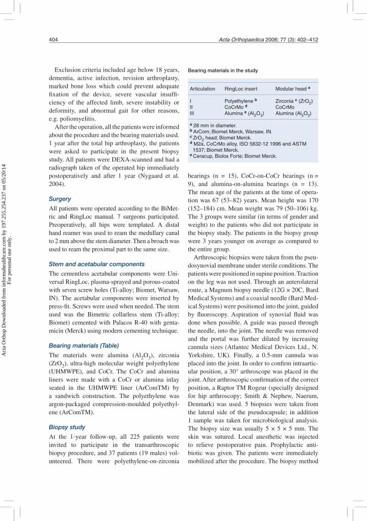
Bearing materials (Table)
The materials were alumina (Al2O3), zirconia (ZrO2), ultra-high molecular weight polyethylene (UHMWPE), and CoCr. The CoCr and alumina liners were made with a CoCr or alumina inlay seated in the UHMWPE liner (ArComTM) by a sandwich construction. The polyethylene was argon-packaged compression-moulded polyethyl-ene (ArComTM).
Biopsy study
At the 1-year follow-up, all 225 patients were invited to participate in the transarthroscopic biopsy procedure, and 37 patients (19 males) vol-unteered. There were polyethylene-on-zirconia
bearings (n = 15), CoCr-on-CoCr bearings (n = 9), and alumina-on-alumina bearings (n = 13). The mean age of the patients at the time of opera-tion was 67 (53–82) years. Mean height was 170 (152–184) cm. Mean weight was 79 (50–106) kg. The 3 groups were similar (in terms of gender and weight) to the patients who did not participate in the biopsy study. The patients in the biopsy group were 3 years younger on average as compared to the entire group.
Arthroscopic biopsies were taken from the pseu-dosynovial membrane under sterile conditions. The patients were positioned in supine position. Traction on the leg was not used. Through an anterolateral route, a Magnum biopsy needle (12G × 20C, Bard Medical Systems) and a coaxial needle (Bard Med-ical Systems) were positioned into the joint, guided by fluoroscopy. Aspiration of synovial fluid was done when possible. A guide was passed through the needle, into the joint. The needle was removed and the portal was further dilated by increasing cannula sizes (Atlantec Medical Devices Ltd., N. Yorkshire, UK). Finally, a 0.5-mm cannula was placed into the joint. In order to confirm intraartic-ular position, a 30° arthroscope was placed in the joint. After arthroscopic confirmation of the correct position, a Raptor TM Rogeur (specially designed for hip arthroscopy; Smith & Nephew, Naerum, Denmark) was used. 5 biopsies were taken from the lateral side of the pseudocapsule; in addition 1 sample was taken for microbiological analysis. The biopsy size was usually 5 × 5 × 5 mm. The skin was sutured. Local anesthetic was injected to relieve postoperative pain. Prophylactic anti-biotic was given. The patients were immediately mobilized after the procedure. The biopsy method
Bearing materials in the study
Articulation RingLoc insert Modular head a
I Polyethylene b Zirconia c (ZrO2)II CoCrMo d CoCrMoIII Alumina e (Al2O3) Alumina (Al2O3)
a 28 mm in diameter.b ArCom; Biomet Merck, Warsaw, IN.c ZrO2 head; Biomet Merck.d M2a, CoCrMo alloy, ISO 5832-12 1996 and ASTM 1537; Biomet Merck.e Ceracup, Biolox Forte; Biomet Merck.
Act
a O
rtho
p D
ownl
oade
d fr
om in
form
ahea
lthca
re.c
om b
y 19
7.25
5.25
4.23
7 on
05/
20/1
4Fo
r pe
rson
al u
se o
nly.
Acta Orthopaedica 2006; 77 (3): 402–412 405
was simple and fast, with a 5-min operation time. 1 patient developed a 3-cm hematoma around the portal. No postoperative infections developed. All patients could leave the hospital within hours of the procedure.
Preparation of the biopsies
Histology. 1 of the biopsies was immediately cut into 4–5 blocks and fixed in 2.5% glutaraldehyde in 0.1 M phosphate buffer, pH 7.2, for 2 h at 4°C. The samples were rinsed in 0.1 M phosphate buffer, pH 7.2, and left in phosphate buffer containing 0.01% sodium azide at 4°C. The post-fixation was done in 2% osmium tetroxide for 2 h. The samples were rinsed 2 × 5 min in cacodylate buffer and once for 5 min in distilled water. They were then dehydrated in 70% ethanol 2 × 15 min, 93% ethanol 2 × 15 min, 99% ethanol 3 × 15 min, and acetone 3 × 10 min, at room temperature. The samples were embedded in epon, arbitrarily orientated: epon:acetone 1:1 for 1 h, then epon:acetone 2:1 for 1 h, and finally 100% epon overnight (Epon Research Industries, Burlington, VT). The samples were then embed-ded into new epon and polymerized at 60°C for 24 h at room temperature, continuously stirred. Each of these blocks was cut in consecutive 2-µm-thick sections (with 16–35 sections per patient). Finally, the sections were stained with 0.1% toluidine blue in phosphate buffer, pH 7.4, at room temperature.
Immunohistochemistry. Another biopsy were embedded in paraffin and 2-µm paraffin sections were cut, mounted on ChemMate slides (DAKO, Denmark), and dried for 1 h at 60°C. The sec-tions were deparaffinized and rehydrated. Antigen unmasking was performed by microwave heating in a closed container in Tris-EGTA, pH 9.0, for 25 min concentrations of Tris 10 mM and EGTA 0.5 mM, followed by cooling in TBS (50 mM Tris, 150 mM NaCl, pH 7.6). Incubation with the primary antibody was done in DAKO ChemMate Antibody Diluent (cat. no. S2022) for 60 min, followed by rinsing in TBS for 3 × 3 min. Endogenous peroxi-dase blocking was done by incubation in metha-nol and H2O2 for 10 min. The primary antibody was detected by incubation for 30 min with DAKO horseradish peroxidase Envision polymer (cat. no. K4001) followed by rinsing in TBS for 3 × 3 min. Visualization was done with the chromogen diami-nobenzidine tetrahydrochloride (DAB) mounted
with Pertex (cat. no. 00801; Histolab, Gothenburg, Sweden) for 15 min followed by rinsing in TBS for 2 min. The reaction was enhanced by incubation in a CuSO4 solution (cat. no. C-7631; Sigma, St. Louis, MO) for 5 min. Finally, the sections were counterstained with hematoxylin, dehydrated and mounted with Pertex (cat. no. 00801; Histolab, Västra Frölunda, Sweden). All immunohistochem-ical staining was performed using an automated immunostainer (Techmate 500; DAKO, Denmark). Positive controls were added for each stain.
The following mouse monoclonal IgGs were used as primary antibodies: (1) anti-human CD68 for detection of macrophages/monocytes (cat. no. M0814; DAKO) (1:500 dilution), and (2) anti-human CD34 for detection of endothelial cells (cat. no. M7165; DAKO) (1:200 dilution).
Stereology
The unbiased estimation of volume fractions was performed on all histological sections by the point counting technique (Gundersen et al. 1988, Cogge-shall and Lekan 1996, Larsen 1998). See Figures 2 A–C for further details.
A profile of granuloma was defined as a spheri-cal lesions with or without surrounding collagen fibres. The border of the granuloma was defined as the outer border of the collagen or the most periph-eral cell of the granuloma (Figure 2c). A profile of macrophage or endothelial cells was defined as positive stained profiles by immunohistochemistry were counted as macrophages or endothelial cells (Figures 3a, b).
The total surface area of the pseudosynovial tissue was estimated. The membrane was geo-metrically compared with a half-sphere. The mean diameter of an acetabular cup used was 56 mm. The total surface area was estimated to be 4,925 mm2. The mean surface area A of a biopsy was 25 mm2. The observed tissue was roughly estimated to be 1/200 of the total surface area of the pseudo-synovial membrane.
Regional variance study
In 2 patients, 4 extra transarthroscopic biopsies were taken from arbitrary areas in the pseudosy-novial membrane. These biopsies were used to evaluate the variance between different parts of the capsule.
Act
a O
rtho
p D
ownl
oade
d fr
om in
form
ahea
lthca
re.c
om b
y 19
7.25
5.25
4.23
7 on
05/
20/1
4Fo
r pe
rson
al u
se o
nly.

406 Acta Orthopaedica 2006; 77 (3): 402–412
dx
dy
(a)
(b)
(c)
Preparation of the biopsy forhistology
Biopsy in 3-5 sections
0.05
mm
Figure 2. Point counting technique. The section was initially delineated at low magnification (a). The microscope stage was programmed to move in well-defined steps of lengths dx and dy within the delineated area. The step length was 200x200 (b). Using an oil immersion lens x60 the section was observed on a computer screen at a final magnifica-tion X8216 mm2. A point grid with 16 points was superim-posed on the high magnification field of vision. Area per point 513.5 mm2 (c). The broken line illustrates the defined border of the granuloma. The ratio of the total number of points hitting the granuloma is divided by the total number of points hitting the reference tissue, P, and is an estimate of the volume fraction. The points that hit granuloma are also included in the reference tissue. In the shown field of vision the reference tissue is 15 and granuloma profile is 13. The short arrow indicates the point that does not hit any tissue. The actual counting was performed using an Olym-pus BX51 light microscope equipped with a computer, that allowed for systematic, random sampling in well-defined steps in the x- and y-axes (C.A.S.T.-GRID software, Olym-pus Denmark). Toluidin blue.
a
0.1
mm
b
0.1
mm
Figure 3. Immunohistochemical staining CD68, macro-phage/monocytes (a); and CD34, endothelial cells (b). Arrows indicates positive stained cells.
Estimator variance study
Double measurements were performed of granu-lomas, macrophages, and endothelial cells on biopsies in order to calculate coefficient of error. 6 biopsies, randomized by computer, were counted in two separate occasions.
Control group
For controls, synovial membrane was preserved from 8 patients during insertion of a primary hip arthroplasty. Indication for operation was osteo-arthritis in all cases.
Statistics
Statistical comparison of the groups was per-formed with Mann-Whitney test for independent samples of non-parametric data (using SPSS for Windows).
Act
a O
rtho
p D
ownl
oade
d fr
om in
form
ahea
lthca
re.c
om b
y 19
7.25
5.25
4.23
7 on
05/
20/1
4Fo
r pe
rson
al u
se o
nly.

Acta Orthopaedica 2006; 77 (3): 402–412 407
from the other two groups were not discolored.
Light microscopy
Acute inflammation was absent and connective tissue was abundant in all specimens. The collagen was either densely packed and well organized or more irregular. Shape or number of particles could not be determined in the light microscope. Capil-laries were frequently observed, but compared to the metal-on-metal group, highly vascular areas were not found in the alumina-on-alumina and polyethylene-on-zirconia groups.
In the polyethylene-on-zirconia group, polar-ized light microscopy did not reveal birefringent material such as polyethylene particles. Small dark particles were observed in the cytoplasm of some macrophages. The granulomas were paler at the center than in the CoCr-on-CoCr group. 2 biop-sies from the polyethylene-on-zirconia group were excluded because of failed preparation. 1 patient had 5 incidents of dislocation. Statistical calcula-tions excluding this patient had no effect on the results. The other patients had no complications. In the CoCr-on-CoCr group, the dark particles were observed as accumulated black intracellular gran-ules in macrophages or as extracellular foam-like structures. The foam-like structures were found in acellular areas or in perivascular zones, but rarely in the granulomas (Figure 4).
In the alumina-on-alumina group, particle-like structures were demonstrated on a few occasions (Figure 5).
Quantification of the cellular response
The cellular response in the biopsies was quanti-
b
0.1
mm
c
0.1
mm
Figure 4. a. Metal-on-metal bearing surfaces. Phagocytes with intracytoplasmatic structures (arrows). Toluidin blue. b. Metal-on-metal bearing surfaces. Extracellular foam-like structures (arrows). Toluidin blue. c. Metal-on-metal bearing surfaces. Multiple close laying vessels (arrows). Toluidin blue.
Results
Gross pathology
All implants were well fixated radiologically and clinically. The biopsies taken from patients in the CoCr-on-CoCr group were dark-colored. Biopsies
0.05 mma
0.1
mm
0.05
mm
Figure 5. Alumina-on-alumina bearing surfaces. Dark par-ticle-like structures (arrows). Toluidin blue.
Act
a O
rtho
p D
ownl
oade
d fr
om in
form
ahea
lthca
re.c
om b
y 19
7.25
5.25
4.23
7 on
05/
20/1
4Fo
r pe
rson
al u
se o
nly.

408 Acta Orthopaedica 2006; 77 (3): 402–412
Volume fraction of granulomas
0.0
0.1
0.2
0.3
0.4
0.5
0.6
(n = 13) (n = 9) (n = 13)
Case 2Case 3 Case 4
ControlOstearthrosis
(n = 7)
Case 1
(a)A B C
Figure 6. Volume fraction of granulomas (a), single macro-phages (b) and endothelial cells (c) in the pseudosynovial membrane biopsies collected one year after implantation of polyethylene-on-zirconia (●), metal-on-metal (●) or alu-mina-on-alumina (▼) bearings in hip arthroplasties. Con-trol group with osteoarthrosis (■). All hip arthroplasties were non-loose and painless. The horizontal lines indicate median.
Volume fraction of single macrophages
0.0
0.1
0.2
0.3
0.4
0.5
0.6
(b) (n = 15) (n = 9) (n = 11)Control
Osteoarthrosis(n = 8)
A B C
Volume fraction of endothelial cells
0.0
0.1
0.2
0.3
0.4
0.5
0.6
(c) (n = 15) (n = 9) (n = 11)Control
Osteoarthrosis(n = 8)
A B C
Volume fraction of granulomas
0.00
0.05
0.10
0.15
0.20
Volume fraction CD-68
0.00
0.05
0.10
0.15
0.20
Volume fraction CD-34
0.00
0.05
0.10
0.15
0.20
Figure 7A. Estimator variance. Biopsies from 6 patients (x-row) were quantified in two seperate occations. Y-row demonstrates the result from the two quantifications in each patient.
Act
a O
rtho
p D
ownl
oade
d fr
om in
form
ahea
lthca
re.c
om b
y 19
7.25
5.25
4.23
7 on
05/
20/1
4Fo
r pe
rson
al u
se o
nly.

Acta Orthopaedica 2006; 77 (3): 402–412 409
fied by volume fraction. Differences in the host reaction could not be demonstrated. Granuloma formation was observed in 33 of the 37 biopsies.
Granulomas. The median volume fraction of granulomas was: polyethylene-on-zirconia, 0.02; CoCr-on-CoCr, 0.04; and alumina-on-alumina, 0.02 (Figure 6A). The differences were not signifi-cant whether or not the patient with dislocations was excluded.
In the polyethylene-on-zirconia group, 2 patients (cases 1 and 2) had an unusually large granuloma volume fraction of 0.13 and 0.15, respectively. In the CoCr-on-CoCr group, 1 patient (case 3) had a volume fraction of 0.17. 1 patient in the alumina-on-alumina group (case 4) had a volume fraction of 0.18.
Case 1 had dislocated 5 times. The pseudosyno-vial membrane of case 1 contained small areas with accumulated macrophages and granulomas in the connective tissue, but particles were not observed. In cases 2–4, granulation tissue, chronic inflamma-tion and lipocytes could be found. There was an abundance of small vessels, especially in case 3, and macrophages and mast cells were found in the connective tissue. 2 biopsies were not quantified because of unsuccessful staining.
Macrophages. The median volume fraction was: polyethylene-on-zirconia, 0.02; CoCr-on-CoCr,
0.04; and alumina-on-alumina, 0.004 (Figure 6B). The differences were not significant. Two biopsies were not quantified because of unsuccessful stain-ing.
Endothelial cells. The median volume fraction of endothelial cells was: polyethylene-on-zirconia, 0.03; CoCr-on-CoCr, 0.02; and alumina-on-alu-mina, 0.05 (Figure 6C). The differences were not significant. 2 biopsies were not quantified because of unsuccessful staining.
Estimator variance. For the biopsies quantified on two occasions, the results are shown in (Figure 7A).
Regional variance. In 2 patients, 5 biopsies were taken from different areas of the pseudosynovial membrane. The volume fractions of granulomas were quantified in each biopsy and the results plot-ted (Figure 7B).
Discussion
In the 37 patients with well-fixated implants, a host response was verified but no statistical differences could be detected in the histological analysis 1 year after the operation. The annual wear rate of ceramic-ceramic and CoCr-CoCr bearings is sig-nificantly less than with polyethylene (Semlitsch and Willert 1997, Willmann 1998, 2000). This, however, was not reflected in the granulomatous response in the present study. In a previous study, no differences in failure rate were found 6.4 years after arthroplasties with different bearings (Kim et al. 2001). The study compared 70 patients with bilateral simultaneous total hip arthroplasties. The bearings were cobalt-chrome and zirconia heads, articulating with a polyethylene liner. Acetabular osteolysis was found in 6 patients with a 28-mm cobalt-chrome femoral head and 6 patients with a 28-mm zirconia head.
The pathogenesis of the granulomatous inflam-mation has been related to 1. the individual host response, 2. particle characteristics, 3. particle con-centration, and 4. the length of particle exposure. These will be considered in turn below.
1. We found a few cases with a volume fraction of granulomas that was higher than the median of the entire group. So far, we have not been able to explain this reaction. It cannot be excluded that
Volume fraction of granulomas
0.00
0.05
0.10
0.15
0.20
Patient 1 Patient 2
Figure 7B. Regional variance. Volume fraction of granu-lomas in the pseudosynovial membrane from 5 different areas (i.e. different biopsies) within the same patient. The horizontal lines indicate median. CV=SD/mean x 100. Patient 1 (CV 66%), patient 2 (CV 143%).
Act
a O
rtho
p D
ownl
oade
d fr
om in
form
ahea
lthca
re.c
om b
y 19
7.25
5.25
4.23
7 on
05/
20/1
4Fo
r pe
rson
al u
se o
nly.

410 Acta Orthopaedica 2006; 77 (3): 402–412
these patients have a high wear rate. Another pos-sibility is that these patients are more sensitive to implants. If so, the response in these patients may be independent of wear rate and type of wear. Gene mapping of these patients in comparison with the others in this cohort may elucidate basic biologi-cal differences, and may thus enable us to identify potential fast rejectors.
2. The response to different particle characteris-tics varies. As early as in 1977, Willert examined the tissue response in the pseudosynovial membrane from failed implants with various bearing materi-als. The CoCr-on-CoCr bearings mainly caused a macrophage foreign-body reaction whereas poly-ethylene-on-metal (stainless steel and CoCr) bear-ings mainly induced foreign body giant-cell for-mation. Though the concentration of particles was higher in the polyethylene group and the time of implantation was not reported, Willert suggested that “a typical reaction exists for each of the mate-rials from which the prosthesis is made” (Willert 1977). The exact reason for this is not clear, but the particle size is known to have an influence on the foreign body response (Pazzaglia et al. 1987). Lerouge et al. (1997) could not demonstrate any differences in the cellular reaction of the inter-face membrane from loose ceramic-on-ceramic or metal-on-polyethylene hip arthroplasties. The study was retrospective, and the mean implanta-tion time was 8.7 and 10.3 years, respectively. The interface membranes from both groups all had severe granulomatous formation, graded a. m. (Willert 1977). Lerouge et al. (1997) suggested that the severe granuloma formation was caused by cement particles, rather than by wear particles from the bearings.
3. Particles may accumulate, inducing a more aggressive reaction. The particles may accumulate as a result of high particle production, or a reduced clearance. It was suggested that particles gener-ated at the bearing surface would be cleared from the joint cavity via the lymphatics (Willert 1977). If production of wear particles is excessive, the local tissue may not be able to transport the par-ticles to the lymph nodes in sufficient quantities. Willert suggested that when the transportation of accumulated particles away from the joint tissue and capsule tissue fails, the granulomatous inflam-mation may become extended to involve adjacent
tissue, inducing local bone resorption along the implant.
Most likely, the patients examined in our study may all have been below the threshold particle con-centration required to activate a more aggressive response, since the biopsies were taken one year after operation. Correlation of the granulomatous response in our study with the annual volumetric wear rate in each patient would be interesting. Thus, radiographic findings do not allow evalua-tion of the wear rate after 1 year. A valid wear rate of polyethylene bearings cannot be measured until 2 years after implantation (Hui et al. 2003), and a valid wear rate may be even more difficult to deter-mine in hard-on-hard bearing materials.
4. Elevated host response have been related to the implantation time (Bos 2001). What is the dif-ference in granulomatous inflammation in loose and non-loose implants? Our findings indicate that granulomatous inflammation is a common finding in non-loose implants. This is in line with previ-ous studies which semi-quantified macrophages in the pseudosynovial membrane from well-fix-ated implants retrieved at autopsy (Bos and Will-mann 2001). The percentage of macrophages was higher in the polyethylene-ceramic and polyethyl-ene metal groups (40–60%) than in the ceramic-ceramic group (20–40%).
Pseudosynovial membranes were compared from loose uncemented and non-loose implants retrieved at autopsy (Mochida et al. 2001). Macro-phages with intracytoplasmic particles were found in both groups involving 11 patients with alumina-on-alumina bearings (7–60 months) and 5 patients with alumina-on-polyethylene bearings (27–54 months). The study did not quantify the findings, but verified that both combinations of bearings were capable of inducing a biological response and that a response could be found in both loose and non-loose implants (Mochida et al. 2001).
The biological response to CoCr-on-CoCr bearings in the pseudocapsule was investigated in 9 patients by Doorn et al. (1996), 5 loose hip prostheses (7 months to 24 years) and 4 non-loose (10–27 months). Both groups had either cemented or cementless prosthe-ses. Macrophages with metal particles and giant cells were found in both groups. These results corre-spond to our study by not demonstrating differences in macrophages and granuloma formation with the
Act
a O
rtho
p D
ownl
oade
d fr
om in
form
ahea
lthca
re.c
om b
y 19
7.25
5.25
4.23
7 on
05/
20/1
4Fo
r pe
rson
al u
se o
nly.

Acta Orthopaedica 2006; 77 (3): 402–412 411
various bearing materials. However, both combina-tions could induce a host reaction.
Despite the high quantity of macrophages with intracellular particles and necrosis in the peripros-thetic tissue, the implants were well fixated. The nature of the response is reminiscent (in a qualita-tive sense) of the response seen in tissue from asep-tic loose implants, although less aggressive. A more aggressive response may develop when particles accumulate in the periprosthetic area, as suggested by (Willert 1977). With time, the groups may show significant differences in biological response—and it will be most interesting to evaluate our patients in a long-term follow-up study. The results of our study will provide an important baseline for well-fixated implants.
We cannot exclude the possibility that backside wear between the acetabular cup and the liners may have influenced the biological response in our study. All liners used had polyethylene lying against metal (possible mode 4 particle genera-tion). It is not clear to what extent this may have affected the result, but we estimated the backside wear to be identical in the 3 groups.
We found a higher volume fraction of macro-phages in the control group than in the operated group. Activated macrophages have been sug-gested to be a mediator of osteoarthritis. This could explain the high number of macrophages in the control group (Gardner 1994). In contrast, we found a very low volume fraction of granulomas in the osteoarthritis group. This finding suggests that the granuloma is a more reliable parameter for comparing host responses to implant materials, than are single macrophages. These findings justify further investigations into finding ways of reducing foreign body reactions.
Contributions of authors LB and FE contributed in the evaluation of the pathology and in the preparation of the manuscript. MN contributed in including the patients, the biopsy procedure, evaluation of the pathology, and in the preparation of the manuscript. KS and AB preparation of the manuscript.
The authors thank the orthopedic surgeons, the staff at the theatre, the ward, and outpatient department of Orthopae-dic Surgery, Frederiksberg University Hospital, Frederiks-berg, Denmark; also, Jytte Overgaard Larsen Ph.D., Institute of Medical Anatomy, University of Copenhagen; Hakon
Kofoed, Department of Orthopaedic Surgery; Frederiksberg University Hospital; Klaus Qvortrup Ph.D., Department of Medical Anatomy, University of Copenhagen; Ditte Hansen, laboratory technician, Department of Pathology, Hvidovre Hospital; Torben Steiniche D.M.Sc., Institute of Pathologi-cal Anatomy, Aarhus University Hospital; Ellen-Margrethe Hauge Ph.D., Department of Rheumatology, Aarhus Uni-versity Hospital, for having provided many useful ideas and constructive dialogs.
This study was supported by the Biomet Merck Company. The company did not influence the methods of evaluation, interpretation of the results or writing of the report.
Bos I. Tissue reactions around loosened hip joint endopros-theses. A histological study of secondary capsules and interface membranes. Orthopade 2001; 30: 881-9.
Bos I, Willmann G. Morphologic characteristics of peripros-thetic tissues from hip prostheses with ceramic-ceramic couples: a comparative histologic investigation of 18 revi-sion and 30 autopsy cases. Acta Orthop Scand 2001; 72: 335-42.
Boynton E L, Henry M, Morton J, Waddell J P. The inflam-matory response to particulate wear debris in total hip arthroplasty. Can J Surg 1995; 38: 507-15.
Catelas I, Huk O L, Petit A, Zukor D J, Marchand R, Yahia L. Flow cytometric analysis of macrophage response to ceramic and polyethylene particles: effects of size, con-centration, and composition. J Biomed Mater Res 1998; 41: 600-7.
Christel P S. Biocompatibility of surgical-grade dense poly-crystalline alumina. Clin Orthop 1992; (282): 10-8.
Coggeshall R E, Lekan H A. Methods for determining num-bers of cells and synapses: a case for more uniform stan-dards of review. J Comp Neurol 1996; 364: 6-15.
Doorn P F, Mirra J M, Campbell P A, Amstutz H C. Tissue reaction to metal on metal total hip prostheses. Clin Orthop 1996; (329 Suppl): S187-S205.
Gardner D L. Problems and paradigms in joint pathology. J Anat 1994; 184: 465-76.
Green T R, Fisher J, Stone M, Wroblewski B M, Ingham E. Polyethylene particles of a ’critical size’ are necessary for the induction of cytokines by macrophages in vitro. Biomaterials 1998; 19: 2297-302.
Green T R, Fisher J, Matthews J B, Stone M H, Ingham E. Effect of size and dose on bone resorption activity of mac-rophages by in vitro clinically relevant ultra high molecu-lar weight polyethylene particles. J Biomed Mater Res 2000; 53: 490-7.
Gundersen H J, Bendtsen T F, Korbo L, Marcussen N, Moller A, Nielsen K, Nyengaard J R, Pakkenberg B, Sorensen F B, Vesterby A. Some new, simple and efficient stereo-logical methods and their use in pathological research and diagnosis. APMIS 1988; 96: 379-94.
Act
a O
rtho
p D
ownl
oade
d fr
om in
form
ahea
lthca
re.c
om b
y 19
7.25
5.25
4.23
7 on
05/
20/1
4Fo
r pe
rson
al u
se o
nly.

412 Acta Orthopaedica 2006; 77 (3): 402–412
Hatton A, Nevelos J E, Nevelos A A, Banks R E, Fisher J, Ingham E. Alumina-alumina artificial hip joints. Part I: a histological analysis and characterisation of wear debris by laser capture microdissection of tissues retrieved at revision. Biomaterials 2002; 23: 3429-40.
Hui A J, McCalden R W, Martell J M, MacDonald S J, Bourne R B, Rorabeck C H. Validation of two and three-dimensional radiographic techniques for measuring poly-ethylene wear after total hip arthroplasty. J Bone Joint Surg (Am) 2003; 85: 505-11.
Jasty M, Bragdon C, Jiranek W, Chandler H, Maloney W, Harris W H. Etiology of osteolysis around porous-coated cementless total hip arthroplasties. Clin Orthop 1994; (308): 111-26.
Kim Y H, Kim J S, Cho S H. A comparison of polyethylene wear in hips with cobalt-chrome or zirconia heads. A pro-spective, randomised study. J Bone Joint Surg (Br) 2001; 83: 742-50.
Korovessis P, Repanti M. Evolution of aggressive granuloma-tous periprosthetic lesions in cemented hip arthroplasties. Clin Orthop 1994; (300): 155-61.
Larsen J O. Stereology of nerve cross sections. J Neurosci Methods 1998; 85: 107-18.
Lerouge S, Huk O, Yahia L, Witvoet J, Sedel L. Ceramic-ceramic and metal-polyethylene total hip replacements: comparison of pseudomembranes after loosening. J Bone Joint Surg (Br) 1997; 79: 135-9.
Mochida Y, Boehler M, Salzer M, Bauer T W. Debris from failed ceramic-on-ceramic and ceramic-on-polyethylene hip prostheses. Clin Orthop 2001; (389): 113-25.
Nygaard M, Zerahn B, Bruce C, Soballe K, Borgwardt A. Early periprosthetic femoral bone remodelling using different bearing material combinations in total hip arthroplasties: a prospective randomised study. Eur Cell Mater 2004; 8: 65-75.
Pazzaglia U E, Dell’Orbo C, Wilkinson M J. The foreign body reaction in total hip arthroplasties. A correlated light-microscopy, SEM, and TEM study. Arch Orthop Trauma Surg 1987; 106: 209-19.
Santavirta S, Hoikka V, Eskola A, Konttinen Y T, Paavilainen T, Tallroth K. Aggressive granulomatous lesions in cementless total hip arthroplasty. J Bone Joint Surg (Br) 1990a; 72: 980-4.
Santavirta S, Konttinen Y T, Bergroth V, Eskola A, Tallroth K, Lindholm T S. Aggressive granulomatous lesions asso-ciated with hip arthroplasty. Immunopathological studies. J Bone Joint Surg (Am) 1990b; 72: 252-8.
Santavirta S, Böhler M, Harris W H, Konttinen Y T, Lap-palainen R, Muratoglu O, Rieker C, Salzer M. Alternative materials to improve total hip replacement tribology. Acta Orthop Scand 2003; 74: 380-8.
Semlitsch M, Willert H G. Clinical wear behaviour of ultra-high molecular weight polyethylene cups paired with metal and ceramic ball heads in comparison to metal-on- metal pairings of hip joint replacements. Proc Inst Mech Eng [H ] 1997; 211: 73-88.
Willert H G. Reactions of the articular capsule to wear prod-ucts of artificial joint prostheses. J Biomed Mater Res 1977; 11: 157-64.
Willmann G. Ceramics for total hip replacement--what a surgeon should know. Orthopedics 1998; 21: 173-7.
Willmann G. The evolution of ceramics in total hip replace-ment. Hip International 2000; 10: 193-203.
Act
a O
rtho
p D
ownl
oade
d fr
om in
form
ahea
lthca
re.c
om b
y 19
7.25
5.25
4.23
7 on
05/
20/1
4Fo
r pe
rson
al u
se o
nly.