Evaluation of Sentinel Lymph Node Assisted Neck Dissection ...
83
A Dissertation on Evaluation of Sentinel Lymph Node Assisted Neck Dissection Using Methylene Blue Dye in Oral Cavity Cancers Submitted to The Tamilnadu Dr.M.G.R Medical University in partial fulfillment of the requirement for the award of degree of M.Ch. (SURGICAL ONCOLOGY) BRANCH VII KILPAUK MEDICAL COLLEGE THE TAMILNADU Dr.M.G.R. MEDICAL UNIVERSITY CHENNAI, TAMILNADU AUGUST 2013
-
Upload
khangminh22 -
Category
Documents
-
view
4 -
download
0
Transcript of Evaluation of Sentinel Lymph Node Assisted Neck Dissection ...

A Dissertation on
Evaluation of Sentinel Lymph Node Assisted Neck Dissection Using
Methylene Blue Dye in Oral Cavity Cancers
Submitted to
The Tamilnadu Dr.M.G.R Medical University
in partial fulfillment of the requirement
for the award of degree of
M.Ch. (SURGICAL ONCOLOGY)
BRANCH VII
KILPAUK MEDICAL COLLEGE
THE TAMILNADU Dr.M.G.R. MEDICAL UNIVERSITY
CHENNAI, TAMILNADU
AUGUST 2013

BONAFIDE CERTIFICATE
This is to certify that Dr. P. Kathirvel kumaran, bonafide student of M.Ch.
Surgical Oncology (August 2010 to August 2013) in the Department of Surgical
Oncology, Government Royapettah Hospital, Chennai – 600 014 has done this
dissertation on “Evaluation of Sentinel Lymph Node Assisted Neck Dissection
Using Methylene Blue Dye in Oral Cavity Cancers” under my guidance and
supervision in partial fulfillment of the regulations laid down by The Tamilnadu Dr.
M.G.R. Medical University, Chennai for M.Ch. Surgical Oncology Examination to be
held in August 2013.
,
.
Prof. P. Ramakrishnan MD., DLO.,
Dean
Kilpauk Medical College, Chennai
Prof. R .Rajaraman MS., MCh.,
Professor & Head
Department of Surgical Oncology
Govt. Royapettah Hospital &
Kilpauk Medical College, Chennai

ACKNOWLEDGEMENT
It is my pleasure and privilege to record my deep sense of gratitude to Prof.
Dr. R. Rajaraman M.S., M.Ch, Professor & Head of the Department, Department of
Surgical Oncology, Government Royapettah Hospital, Kilpauk Medical College,
Chennai, for his constant encouragement, motivation and guidance given to me in
bringing forth this piece of work.
Special gratitude is due to the Assistant Professors of our department,
Dr. S. Subbiah M.S., M.Ch, Dr. M. P. Viswanathan M.S., M.Ch, Dr. A.
Balasubramanian M.S., M.Ch, Dr. D.Jeyakumar M.S., Mch, and for the help and
kindness rendered.
I am also thankful to Professors of Pathology at Government Royapettah
Hospital, present and past for their generous help and co-operation rendered towards
the study.
I thank my fellow post graduates, technical staff, and paramedical staff of our
department for their generous assistance throughout this study. I owe my gratitude to
all the patients who participated in the work with kind cooperation.

CONTENTS Page No
1 INTRODUCTION 1
2 AIM OF THE STUDY 3
3 REVIEW OF LITERATURE 4
4 PATIENTS AND METHODS 29
5 STATISTICAL ANALYSIS 34
6 RESULTS & OBSERVATIONS 35
7 DISCUSSION 53
8 SUMMARY OF RESULTS 63
9 CONCLUSION 65
10 APPENDICES
i) BIBILIOGRAPHY
ii) ABBREVATIONS
iii) PROFORMA
iv) CONSENT FORM
v) ETHICAL COMMITTEE
APPROVAL
vi) MASTER CHART
vii) SIMILARITY CHECK

Sentinel Lymph Node Biopsy in Oral Cancers
1
INTRODUCTION
Carcinoma of the oral cavity is the second most common cancer in India and
fourth most common in Madras Metropolitan Tumor Registry (1)
. Cervical nodes form
the first echelon of metastases in cancers of the oral cavity. The frequency of cervical
nodal metastases varies depending on the subsite, tumour size, depth of invasion,
tumour grade, etc. The treatment of cervical nodal metastases from oral cancers
depends on the number, size, and level of nodal spread; it can be surgery, radiation,
chemotherapy or their combinations. Clinically node negative (N0) patients form a
specific subset among oral cancers for whom the treatment of neck is not well
standardised. Treatment options include observation, elective neck dissection or
elective neck irradiation and depends on various factors like risk category, patient
preference, treatment availability, treatment for the primary, physician preference etc.
The chance of occult metastases in clinically N0 disease can be up to 30 percent (2)
.
Elective treatment of neck is recommended in high risk groups defined as those
patients with risk of lymph node metastasis risk more than 20 percent (3).
If the neck is
not addressed while adequately treating the primary, there is a high chance of nodal
recurrence in those patients who harbor metastases (3).
Clinical examination with imaging modalities like ultrasound, computed
tomography (CT) scan, magnetic resonance imaging (MRI) and positron emission
tomography (PET) scan are not sensitive enough to exclude occult neck nodes in
clinically N0 patients(3)
.
Sentinel lymph node is the first echelon node and often first site of metastases.
Histological status of sentinel node may predict micro-metastases in the reminder of

Sentinel Lymph Node Biopsy in Oral Cancers
2
lymphatic basin. The technique of Sentinel lymph-node biopsy (SLNB) has been
successfully applied in carcinoma of breast and melanoma. Extrapolating these
concepts, it would be ideal to do a complete neck dissection only in patients with
positive sentinel nodes. This could spare the morbidity of the neck dissection in up to
70 percent of patients (4)
. With this background we tried to evaluate sentinel lymph
node using methylene blue dye in clinically node negative oral cavity cancers at
Government Royapettah Hospital.

Sentinel Lymph Node Biopsy in Oral Cancers
3
AIM OF THE STUDY
Primary Aim:
The primary aim of this study is to evaluate the feasibility and efficacy of
sentinel lymph node using methylene blue dye in clinically node negative early stage
oral cavity cancers in avoiding morbidity of neck dissection.
Secondary Aims:
(1) To identify most common site of sentinel lymph node for oral cancers.
(2) Usefulness of USG imaging in assessing neck metastasis not apparent
clinically.
(3) Correlation of sentinel node with non-sentinel node metastases
(4) Assessing possibility of level IIB, IV and V sparing neck dissections in
oral cancers N0 neck

Sentinel Lymph Node Biopsy in Oral Cancers
4
REVIEW OF LITERATURE
Introduction:
Carcinoma of oral cavity is more prevalent in the areas of tobacco abuse. It is
more common in men, with a male to female ratio of 2.3:1(5)
. The risk of nodal
metastases increases with increasing “T’ status and grade of the tumor and specific
subsites like tongue, FOM and alveolus. The presence of metastatic neck node is the
single most important adverse prognostic factor. The 5-year survival rate decreases to
below 50 percent when cervical nodal metastases are present (6,7,8)
. The lymphatic
metastases from head and neck cancers follow a well-defined pattern. In the absence
of metastasis to levels I, II and III, the involvement of levels, IV and V are rare
(exception being tongue) (7).
Lymphatic Anatomy:
The prime function of the lymphatic system is the return of proteins,
interstitial fluid and immune cells back to the bloodstream. These elements initially
enter the lymphatic capillary vessels which are lined by non-gapped continuous
endothelial cells. Lymphatic fluid flows in smaller collecting vessels (2 layered) and
then into larger collecting vessels (3 layered), which finally drain into lymph nodes (9)
(fig-1). Afferent lymphatic vessels enter the convex surface of lymph node and drain
into the marginal sinus. Medullary sinuses receive lymph from the marginal sinus and
penetrate the medulla of the lymph node. Efferent lymphatic vessels formed by
coalescence of lymphatic channels in medulla exit via the hilum. Lymph flows
through the node in a unidirectional manner. Afferent lymphatics may at times bypass
certain lymph nodes in their path (10).

Sentinel Lymph Node Biopsy in Oral Cancers
5
Variations in anatomy of lymphatics have been described by Ludwig (11)
(Figure 2).
Figure 1 Lymph Node Anatomy
Figure 2 Types of Lymphatic Drainage

Sentinel Lymph Node Biopsy in Oral Cancers
6
Lymphatic Flow Pattern in Head and Neck:
The head and neck region has a dense and complicated network of lymphatic
channels. It accounts for a third of the total number of lymph nodes in the human
body (approximately 200 to 350 lymph nodes). Lymph nodes of the head and neck
are divided into superficial and deep groups. The superficial lymphatics and nodes lie
between the skin and the superficial fascia. Deep cervical nodes are present deep to
the sternocleidomastoid muscle, along the internal jugular vein from the base of skull
to the brachiocephalic junction. Efferent lymphatics drain into the venous system at
that junction.
Lymphatic drainage from specific regions of the Upper-Aero-Digestive-Tract
(UADT) (12,13)
follow a predictable pattern according to the preferential pathways of
natural lymph flow (Werner and Davis(14)
). Lymphatics of the neck have been
organized into four functional drainage pathways 1). The main lymphatic pathway,
2).The posterior pathway, 3). The anterior lymphatic pathway and 4). The superficial-
lateral pathway. The works of Lindberg, Byers et al. (15)
(1988), and Shah et al. (16)
(1990) were among the many studies that were vital in understanding the lymphatic
basins at risk of metastasis from Squamous Cell Carcinoma (SCC) arising from
specific sub-sites of the UADT.
However, it is important to emphasize that, inspite of these widely accepted
generalized patterns of lymphatic drainage; there is wide variability in the lymphatic
flow of the head and neck region (17).
This variability is confirmed by studies
performed at the Moffit Cancer Center, Miami, which has shown as much as a 63
percent discordance between a patient’s predicted lymphatic drainage based on classic

Sentinel Lymph Node Biopsy in Oral Cancers
7
pathways as opposed to the patient’s actual lymphatic anatomy imaged by a
lymphoscintigram(17)
In 2004, American Head and Neck Society agreed upon a classification
system of six lymph node levels proposed by Robbins et al., 2000(18)
which has been
widely accepted. Knowledge of the primary tumor site and understanding of the
pathways of lymphatic tumor spread helps to predict the region of the neck at highest
risk for metastatic disease.
Lymphatic drainage of oral cavity (19)
:
Lymph from the central part of the lower lip drains to the submental lymph
nodes. Lateral parts of the lower lip, upper lip and the mucous membrane of the cheek
drain to the submandibular lymph nodes. Both surfaces of the lower gingivae and the
outer surface of the upper gingivae drain into submandibular lymph nodes. The inner
surface of the upper gingiva is drained by the vessels of the hard palate to the upper
deep cervical lymph nodes. The floor of the mouth drains anteriorly via lymphatics
that pierce the mylohyoid muscle to reach the submental nodes or posteriorly to the
submandibular nodes. The submandibular and submental nodes drain into the deep
cervical nodes from where the lymph finally empty in the jugular trunk.
Types of Neck Dissections (20)
:
Treatment options of the neck in clinically node positive patients (N+) mostly
depend on the treatment of the primary lesion. Radiation or chemo radiation to the
neck is given if primary lesion is treated by radiation, and neck dissection should be
performed if primary lesion is managed by surgery. An academic classification of
neck dissections is shown in Table 1 and the types are described below.

Sentinel Lymph Node Biopsy in Oral Cancers
8
Table 1 Classification of Neck Dissections
Neck dissections classification
Comprehensive Neck Dissections Selective Neck Dissections
Radical neck dissection. Supraomohyoid neck dissection (level I-III).
Modified radical neck dissection Posterolateral neck dissection
(level II, III, IV, V)
Extended radical neck dissection Lateral neck dissection (Level II-IV)
Anterior neck dissection (VI)
1. Radical neck Dissection:
In radical neck dissection, all cervical lymph node groups from levels I to V were
removed as enbloc with the Spinal Accessary Nerve, Internal jugular vein, and
Sternocliedomastoid muscle on one side.
2. Modified Radical Necks Dissection:
In modified radical neck dissection, all lymph nodes routinely removed by the
radical neck dissection are removed with preservation of one or more the important
non lymphatic structures i.e. Spinal Accessary Nerve, Internal jugular vein, and
Sternocliedomastoid muscle.
3. Extended Neck Dissection:
Extended neck dissection refers to the removal of one of more additional
lymph node groups’ like retropharyngeal, superior mediastinal, buccinators and para
tracheal lymph nodes and/or non lymphatic structures like the hypoglossal nerve,
vagus nerve, paraspinal muscles, skin, external carotiod, etc.

Sentinel Lymph Node Biopsy in Oral Cancers
9
4. Selective Neck Dissection:
A selective neck dissection refers to dissection of lymph node groups which
are at high risk of involvement based on the lymphatic anatomy of the primary. For
oral cavity cancers, the lymph nodes at greatest risk are located in levels I, II, and III.
The lymph nodes at risk for oropharyngeal, hypopharyngeal, and laryngeal cancers
are located in levels II, III, and IV, for thyroid cancer the lymph nodes are level VI, II,
III, IV, V. (Table 1)
If a neck dissection is carried out when there is no evidence of neck disease it
is termed an "elective" neck dissection (END). Some authors use the word
"prophylactic" instead of "elective" to denote the same procedure. If the neck
dissection is undertaken for metastatic disease in the neck it is called a "therapeutic"
neck dissection
Clinically Node Negative Patients (N0):
Though the treatment of neck in N+ patients has been well studied and
established, the treatment of neck in clinically N0 patients remains controversial.
Clinically N0 patients harbor a risk of having occult metastases in the neck in upto 30
percent. Patients with high risk for occult metastasis,(6,7,8)
are identified by
characteristics of the primary lesion thickness of >4 mm and size >2 cm, anatomic
location, lympho vascular invasion, perineural infiltration, poorly differentiated
histology and immunosuppression.(Table 2) Cervical node metastasis is the single
most important adverse prognostic factor with drop in overall 5 years survival from
82 to 53 percent. Hence it seems logical to intervene early rather than watchful
waiting, as delayed resection in clinically evident macroscopic disease have poor
prognosis (21)
.

Sentinel Lymph Node Biopsy in Oral Cancers
10
Table 2 Risk of Occult Neck Node Metastases in Oral Cancers
Group
Risk of occult
metastases
Stage Primary site
High risk >30%
T1-4
Nasopharynx, pyriform sinus,
base of tongue
T2-4 Soft palate, pharyngeal wall, supraglottic
larynx, tonsil
T3-4 Anterior 2/3
rd tongue , FOM, RMT,
Gingiva, Hard palate, Buccal mucosa
Intermediate
risk 20-30%
T1
Anterior 2/3rd
tongue, soft palate,
pharyngeal wall, supraglottic larynx,
tonsils
T2
Anterior 2/3rd
tongue , FOM, RMT,
Gingiva, Hard palate, Buccal mucosa
Low risk <20% T1 FOM, RMT, Gingiva, Hard palate,
Buccal mucosa
Modalities for treatment of N0 neck: (22,23)
The management options for the clinically N0 neck include (1) selective neck
dissection with the rationale of regional staging and elective treatment, (2) irradiation
of the neck as elective treatment, and (3) observation- clinical follow-up with option
of therapeutic neck dissection or irradiation if patients develop detectable neck nodal
disease.

Sentinel Lymph Node Biopsy in Oral Cancers
11
Elective irradiation of neck has several limitations. It is more morbid and
provides no staging information to estimate prognosis or guide further management.
Few treatment alternatives exist in those who develop second primary tumors which
occurs in about 2-4%/year (23)
.
Selective neck dissection (I-III) offers comparable local control rates with less
morbidity when compared with radiotherapy and other types of neck dissection in
SCC of oral cavity. Sometimes close observation is sufficient if the primary lesion is
of T1 stage.
More precise staging before treatment is mandatory to prevent the
consequences of inappropriately selected management strategies for the clinically N0
neck in oral cancer. The concept of sentinel node biopsy may fulfill the requirement.
The surgeon should have experience with a minimum of 10 cases before undertaking
SLNB as a staging tool.
Methods to identify occult lymph node metastases:
1. Noninvasive methods:
The radiological assessment of neck in clinically node negative patients has
improved with recent advances in imaging techniques. Contrast-enhanced CT and
MRI are the common imaging modalities used to evaluate the neck in oral cancers.
Radiological criteria for nodal involvement include size (Levels I, II ≥ 1.5 cm, Levels
III – VI ≥ 1.2 cm), number of nodes (3 lymph nodes > 8 mm), central necrosis,
irregular enhancement and poorly defined or irregular capsules (24,25)
. PET scan has
poor sensitivity in detecting micro-metastases in the neck (26)
. The combined use of
ultrasound with fine needle aspiration may also identify patients requiring neck

Sentinel Lymph Node Biopsy in Oral Cancers
12
dissection (27)
. However the available methods reach only 80–85% sensitivity and
require experienced and skilled operators (27)
. (Table 3)
Table 3: Comparison of imaging modalities in Occult Metastases in N0 Neck
Modality Sensitivity Specificity
Ultrasound (24)
50-58% 75-82%
CT(24)
40-68% 78-92%
MRI(24)
55-93% 82-95%
PET(26)
87-90% 80-93%
CT-PET(26)
96% 98.5%
2. Invasive methods:
1. Pre-operative dynamic / static scintigraphy with or without SPECT
2. Blue dye technique
3. Hand held Gamma probe aided detection of SLNB.
4. Combination of two or more techniques.
Sentinel lymph node biopsy - Historical perspective
Regional lymph node dissection (RLND) is based on existing knowledge of
tumour spread through lymphatics. The varied frequency of lymph node metastases in
various cancers depending on the primary tumour characteristics challenges the role
for routine RLND or its modifications in all patients with nodal disease. Sentinel node
biopsy may offer a solution to this dilemma.
In 1955, Seaman and Powers (28)
laid the groundwork for lymphoscintigraphy
and lymphatic mapping. Gould et al (29)
independently observed that the routine

Sentinel Lymph Node Biopsy in Oral Cancers
13
excision of a ‘‘sentinel node’’ found at the origin of the common facial vein at the
time of parotidectomy offered diagnostic significance. The “'Cabanas approach (30)
'
introduced in 1977 is less reliable because of the relatively crude localization
techniques based on anatomy only. In 1992 Morton (31)
et al used cutaneous
lymphoscintigraphy as a method to identify the nodal basins at risk of metastases in
melanomas located in ambiguous sites. Alex and Krag (32)
in 1996 reported the first
successful sentinel node biopsy in a case of supraglottic cancer. While
lymphoscintigraphy improved and assay techniques increased in sensitivity,
preferential drainage to one or two nodes in the lymph nodal basin was consistently
demonstrated which made application of SNB technique more common (33)
.
Sentinel Lymph Node Biopsy – Concept and Principles
Tumour cell progression within the lymphatic system follows an orderly
pattern. Primary or the draining lymph nodes possess the structural and functional
capability to filter and entrap tumour cells efficiently. Thus removing uninvolved
lymph nodes may be harmful to the patient from an immunological point of view.
Sentinel node biopsy is a minimally invasive, diagnostic procedure which can
accurately predict the presence/ absence of nodal metastases. It offers a reliable
method to avoid lymphadenectomy. The success of SLNB depends on two factors:
the accuracy of the localisation technique to identify the sentinel lymph node, and its
intraoperative feasibility. The finding of gross cancer involvement contraindicates
sentinel node biopsy and mandates formal lymphadenectomy.

Sentinel Lymph Node Biopsy in Oral Cancers
14
Figure 3: Concept of Sentinel Lymph node
The three essential principles of SLNB are: (34)
1. A predictable and orderly pattern of tumour spread from the primary to
regional lymph nodes.
2. The lymph node effectively filters the afferent lymph whereby the tumour
cells get entrapped in it.
3. There is a sequential progression of tumour cells from the first echelon to
second echelon nodes.
4. Unidirectional spread due to lymphatic valves.
There is sufficient evidence available to support these three basic principles in
head and neck cancers (35,36)
.
Sentinel Lymph Node Biopsy (SLNB) is applicable for staging the following (37)
:
1. Ipsilateral clinically N0 neck drained by a unilateral primary tumor
2. Bilateral clinically N0 neck drained by a midline tumor or crossing the
midline

Sentinel Lymph Node Biopsy in Oral Cancers
15
3. Contralateral clinically N0 neck drained by a midline tumor or tumor crossing
the midline in the presence of a clinically positive ipsilateral neck.
De Boer et al(38)
showed a statistically significant difference in disease free
survival (DFS) in head and neck cancers between SLN-negative and patients with
micro metastases or ITCs. Pitman KT (39) et al
showed that patients with micro
metastases had an intermediate prognosis between patients who are node-negative
and those with macro metastases. Sentinel lymph nodes identified in the context of
elective neck dissection is called as SNB-assisted elective neck dissection (39)
.
Additional pathologic methods of serial sectioning and immunohistochemical
analysis of sentinel nodes have upstaged up to 8% of neck dissection specimens in
patients with head and neck (40)
.
To date, SLNB can be considered as the most accurate way to stage the cN0
neck(41)
.The SLN with the highest radioactive count may be the most likely to harbor
tumor cells. Studies in patients with breast cancer reported a considerable false-
negative rate of SLNB (9% to 29%) if only the hottest SLN was removed.42
Atula et
al43
demonstrated that, in Oropharyngeal SCC it was sufficient to dissect the 3 hottest
lymph nodes to detect occult metastases in all patients.
Sentinel Lymph Node Biopsy - Physiology of Lymphoscintigraphy: (44-46)
Lymphoscintigraphy (LS) is the study used for localization of lymph nodes
draining a specific anatomic area. It uses after intraepithelial, subepithelial, or intra-
parenchymal injection of identifiable particles of appropriate size into the interstitium.
The particles enter lymphatic capillaries and flow to the lymph nodes where they are
phagocytosed by macrophages. Successful SLNB requires patent lymphatic channels
as well as normally functioning lymph nodes. It also requires that the percentage of

Sentinel Lymph Node Biopsy in Oral Cancers
16
particles trapped in the more proximal nodes is greater than the percentage of
particles that flow to distal lymph nodes. 99mTc sulfur colloids are common for
lymphoscintigraphy. The advantages are:
1. They emit only gamma rays and have low radiation exposure
2. Half-life is only 6 hr and peak energy emission peak of 140 keV, within the
detection range of most handheld gamma probes.
Particle size is the primary factor that determines the rate of uptake and filtration
within the sentinel node. The optimal particle size of radioisotopes is between 5 and
10nm. Particles smaller than 5nm are absorbed by the vascular system.
SLNB usually uses a triple diagnostic approach. Twenty four hours prior to
operation, radiocolloid is injected around the primary tumor site and static
lymphoscintigraphy is used to identify the location of the sentinel node(s).
Intraoperative identification is done using both blue dye and hand-held gamma probe.
Sentinel Lymph Node Biopsy - Lymphoscintigraphy Technique:
Static and dynamic imaging, are two modes of obtaining lymphoscintigraphy.
Optimum pre-operative information is obtained by dynamic imaging, due to the short
distance from primary site to sentinel node, complexity of the anatomy and variable
pattern of lymphatic drainage.
Early imaging (<30 min post injection)
i) SLNB- Hotspots with evident uptake
ii) 2nd
echelon - Caudal hotspot with clearly visible connecting lymphatic
vessel from a cranial hotspot, not increasing in time
iii) Caudal hotspot with low uptake not increasing in time

Sentinel Lymph Node Biopsy in Oral Cancers
17
Late static imaging (2–4 hr post injection)
i) SLNB-New hotspots visualized ipsilaterally or contralaterally.
ii) New hotspots visualized between a hotspot already identified during early
imaging and the injection site.
iii) Caudal hotspots with a previously low uptake, but now much more
intense.
iv) Newly visualized hotspot also considered to be a sentinel node.
Tumors in the oral cavity other than mobile tongue and FOM tumors seem to
have slower lymphatic drainage; an early lymphatic imaging was able to identify
hotspots in only 29% of patients with these tumors (44)
. Heuveling et al (44)
believes
that late lymphoscintigraphic imaging should be considered for these tumors to
minimize the risk of false-negative results. The same is true for paramedian and
midline tumors, for which bilateral drainage was observed in the majority (83%) of
tumors, half of them visible only during late imaging (44)
.
SLNB may also prove useful for those patients with clinically established
ipsilateral neck disease, with an N0 contra-lateral neck clinically but are at risk of
contra-lateral neck involvement (44)
. In such patients lymphoscintigraphy may
establish the presence of bilateral drainage, and sentinel node biopsy can spare them
the morbidity of elective treatment to opposite neck. SPECT/CT offers better
anatomical localisation than planar lymphoscintigraphy but may not improve the
outcome of the sentinel node procedure itself (46)
. Acquisition time of SPECT is much
longer compared with that of planar lymphoscintigraphy, and SPECT imaging is
often performed as the final imaging procedure – that is, at a later time point after
injection of 99mTc-Nanocoll – second echelon lymph nodes containing radiocolloid
Sentinel Lymph Node Biopsy in Oral Cancers
18
may become visible on SPECT images (46)
. Furthermore, additional hotspots detected
next to the hotspot that was identified on planar LS should also be detected
intraoperative by the gamma probe. Haerle et al (46)
believe that SPECT-CT imaging is
a helpful additional tool for detailed localization of a hotspot, but not for
identification of true SNs.
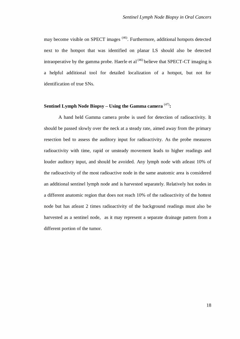
Sentinel Lymph Node Biopsy – Using the Gamma camera (47)
:
A hand held Gamma camera probe is used for detection of radioactivity. It
should be passed slowly over the neck at a steady rate, aimed away from the primary
resection bed to assess the auditory input for radioactivity. As the probe measures
radioactivity with time, rapid or unsteady movement leads to higher readings and
louder auditory input, and should be avoided. Any lymph node with atleast 10% of
the radioactivity of the most radioactive node in the same anatomic area is considered
an additional sentinel lymph node and is harvested separately. Relatively hot nodes in
a different anatomic region that does not reach 10% of the radioactivity of the hottest
node but has atleast 2 times radioactivity of the background readings must also be
harvested as a sentinel node, as it may represent a separate drainage pattern from a
different portion of the tumor.

Sentinel Lymph Node Biopsy in Oral Cancers
19
Table 3A : Comparison between techniques for localization:
Study No. of
patients
SLN
identified
True
positive
False
negative
Sensitivity
(%)
Shoaib et al 35
13-blue
13-radio
5
12
0
7
3
0
41
100
Civantos 65
(two)
except blue
43 43 18 2 90
Stoeckli 67
(triple)
79 78 29 2 94
Ross 89
(triple) 134 125 42 3 93
Sentinel Lymph Node Biopsy – Drawbacks (48,49)
:
The disadvantages of SLNB are
1. Skip metastasis alter management predictions.
2. Learning curve
3. Radionuclide retention in tissues.
4. Failure to identify sentinel nodes
5. Free soft tissue disease not taken into account
Frozen sections are unreliable to detect micro-metastasis and hence occult
metastasis (50)
. Sentinel lymph node biopsy is an acceptable procedure for microscopic
disease; however clinical disease must be ruled out with clinical examination after
induction of anesthesia.

Sentinel Lymph Node Biopsy in Oral Cancers
20
For oral cancer, it appears that the sentinel node is an excellent tool for
assessment of microscopic disease, but subclinical gross disease must be sought by
radiologic studies and intraoperative palpation (51)
.
Sentinel Lymph Node Biopsy - Failure to Identify Sentinel Nodes: (52-53)
For T3 and T4 primary tumors difficulty in injecting the dye around tumor
may alter the pickup rate requiring large volumes for these tumor, other reason for
failure of SLNB technique include excessive uptake of radioactivity, potential for
false-negatives caused by incomplete injection, and technical futility of removing a
large number of nodes in piecemeal fashion. Bulky or deeply infiltrative primary
tumors that invade adjacent anatomic subsites clearly pose technical difficulties for
peritumoral injection.
Clinical node positivity predicts failure of the technique to identify sentinel
node, hence anterior tongue and FOM are subsites with reduced success rate due to
high risk of node positivity (20-30%).
FOM tumors lesions are the most difficult sub site for SLNB (53)
. The lymph
from the floor of mouth drains through nodes along lingual nerve (lingual nodes) to
cervical nodes. Uptake in the lingual nodes and level I nodes overlap with the
injection site uptake and SLNB may be missed. It is referred as the ‘‘shine through’’
phenomenon (53)
.
The mandible interferes with the gamma camera probe angulation. The
technical difficulty may be overcome with several solutions like software masking,
lead shields, removing the primary tumor prior to SLNB identification, and elective
dissection of level I nodes can be useful but sentinel nodes for FOM remains
problematic. Higher successful harvest rate is seen in patients with a positive

Sentinel Lymph Node Biopsy in Oral Cancers
21
preoperative lymphoscintigraphy (94%) compared with patients with a negative
preoperative lymphoscintigraphy (79%)
Delayed sentinel lymph node biopsy (D-SLNB) is defined as any SLNB
procedure that is carried out after a previous wide local excision of the primary. A
delay or more than 90 days, or cicatrix after Wide Local Excision alters lymph
draining channels rendering the procedure unreliable and SLNB non-representative of
the original lymphangiosome.
Sentinel Lymph Node Biopsy - Enhanced pathological review (54-60)
:
A successful procedure relies on efficient screening by the pathologist. Extent
to which SLNB upstages the neck by traditional pathologic methods seems to be
similar to that with elective neck dissection. The use of additional pathologic methods
results in perhaps an even greater level of detection of disease. The negative
predictive value of frozen section ranges from 83 to 99% (55)
depending on the slicing
technique used and is higher for multi-slice technique (55)
. Unlike frozen section,
imprint cytology can also be used where concern about loss of tissue during
processing does not exist (55)
.
Serial sectioning has to be done with more attention than usual. Tumor cells
enter the node through the opposite side of hilum. SLNs less than 0.5 cm are
processed entirely, nodes up to 1 cm are halved and sentinel lymph node more than
1cm are downsized to 0.5 cm and step sections are made if first section is negative.
While cutting the step ribbons, one portion is retained for IHC and other for routine
HPE staining (57)
. Without step sections and immunohistochemistry, up to 15 to 20%
of metastases may be missed (76)
. Therefore, the theoretical sensitivity for detection of
metastases by single HE frozen section analysis is (under optimal conditions) not

Sentinel Lymph Node Biopsy in Oral Cancers
22
higher than 80 to 85% (58)
. These small deposits of occult disease are likely to be
overlooked by the routine histological assessment of the large number of nodes in a
neck dissection specimen.
Sentinel Lymph Node Biopsy - Rapid Molecular Detection as an Intraoperative
Adjunct (60)
:
Frozen section results vary with pathologist and technical staff. Since
sensitivity is not very high, molecular detection came in to practice for the last 2
years. It is quick, has high negative predictive value and facilitates one time surgery
(60). Use of qRT-PCR (Reverse Transcriptase associated Polymerase Chain Reaction)
permits quick and reliable assessment and can be useful tool in decision making. Per
operative molecular staging with PCR amplification of marker genes could spare 60-
70% of pN0 patients from unnecessary surgery (60)
.
Sentinel Lymph Node Biopsy - Classification and Staging (61)
:
UICC definitions of metastatic deposits divided into macrometastasis (>2 mm), micro
metastasis (0.2-2 mm) and even small tumour cells or small clusters <0.2 mm
(isolated tumour cells, ITC).

Sentinel Lymph Node Biopsy in Oral Cancers
23
Sentinel Lymph Node Biopsy - Outcomes:
Three prospective studies and good quality of SLNB are in progress namely
SENT (62)
, DAHANCA 22 (63)
, Brazilian Head & Neck Group (64)
. Recently the
prospective multi-institutional clinical trial by ACOSOG Head and Neck Committee
has shown encouraging results (65)
. The Efficacy of Sentinel Node Biopsy in Head and
Neck Cancers in various studies are shown in Table 4.

Sentinel Lymph Node Biopsy in Oral Cancers
24
Table 4 Efficacy of Sentinel Node Biopsy in Head and Neck Cancers
Author No. of
Patients (n)
SLNB
Identification
rate
NPV Occult
Metastases (n)
Ross et al
(2005) (66)
134 125/134 96% 39 (33%)
Stoeckli et
al.(2008) (67)
79 79/79 100% 26 (32%)
Alkusheri et al
(2010) (68)
134 125/134 94.6% 42 (34%)
Melakane et al
(2012) (69)
166 154/166 95.2% 42 (25%)
Antanio et al
(2012) (70)
209 183/209 94.9% 54 (29.5%)
Sentinel Lymph Node Biopsy – the consensus in literature (71)
:
1) Negative predictive value is between 90% and 100%.
2) Step serial sectioning and IHC are essential parts of the procedure.
3) IHC and step serial sectioning yields better results and significantly improve the
negative predictive value of this technique.
4) Compared to Lymphadenectomy SLNB significantly upstages nodal stages.
5) Unexpected patterns of lymphatic drainage can indeed occur, including
unanticipated contralateral drainage to nodes that might be missed in standard
lymphadenectomy

Sentinel Lymph Node Biopsy in Oral Cancers
25
6) Frozen section, imprint cytology, and molecular biology based technique permits
quick and reliable assessment of nodes facilitating the neck dissection at the same
sitting if appropriate.
Following are significant concerns. Staged surgery is required in a small
group when positive nodes are found after formal processing at later date.
The best treatment of neck with single micro metastasis or even isolated tumor
cells is elusive (72)
. It has been proposed that removal of the sentinel node alone may
be sufficient in these patients but without strong evidence (73)
.
Selective Neck Dissections and post-operative shoulder dysfunction syndrome:
Subclinical spinal accessory nerve impairment can be observed even after
selective neck dissections (levels I-III) due to routine clearance of sublevel II B (74)
.
The most common morbidity associated with selective neck dissection (SND-I-III) is
spinal accessory nerve dysfunction and related shoulder disability (75,76)
. Nerve
morbidity is mainly due to stretching while clearing level IIB nodes. If these nodes
are not dissected the above mentioned morbidity can be prevented.
Even after careful dissection of the posterior triangle, unavoidable trauma to
the accessory nerve (SAN) does occur. Potential reason for the nerve dysfunction
includes traction injury, use of diathermy and vascular injury. Postoperative
hemorrhage, infection and scarring may affect nerve function. Apart from injury to
SAN, connections with the cervical plexus are damaged invariably during dissection
of Level V. All these factors lead to shoulder syndrome. This syndrome is
characterized by shoulder pain, weak abduction, and winging scapula. These
symptoms may lead to significant restrictions in patient’s professional and every day

Sentinel Lymph Node Biopsy in Oral Cancers
26
activity (77)
. Pinsolle et al (1997) (77)
found in a series of 41 patients following
supraomohyoid neck dissection, 32% to have minor, 5% moderate, and 2.5% severe
shoulder problems. Paul van et al (2003) (78)
in a study of 58 patients showed 14
(28%) shoulder dysfunction.
Is Level II B removal necessary?
Involvement of IIb nodes is rare in clinically N0 oral cavity cancers (79-82)
.
This has led to the idea of preserving them to avoid trauma to Spinal Accessory
Nerve. This is anticipated to have a better functional out come and reduced morbidity
(80, 81). The review of literature (Table 5) suggests that metastatic involvement of level
IIb is rare based on from both elective and therapeutic neck dissection.
Table 5 Involvement of Level IIB Nodes
Author No. of Neck
Dissections
Level IIb
no. and (%)
Exclusive Level IIb
involvement
Lim et al [2004]
Elsheikh et al [2005] (79)
Lim et al [2006] (80)
Villaret et al [2007] (81)
Manola et al 2011 (82)
74
48
125
43
34
4(6.7)
1(2)
8(6.4)
26(5.6)
0
0
1
0
2
0
It shows Level IIb involvement ranges between 2- 6.5% in cN0 neck. But in
presence of clinically palpable nodes and level II involvement metastasis increase
upto 22—36% (106)
. Hence, routine clearance of IIb is not necessary in N0 neck.

Sentinel Lymph Node Biopsy in Oral Cancers
27
Is Level IV and V clearance necessary? :
In clinically N0 neck of oral cavity cancers, level IV and V nodes are not
first echelon nodes. Skip metastasis to level IV & V are rare. Shah JP et al (1990) (83)
found that in oral cavity cancers occurrence of level IV metastases was 3% in N0
neck and 17% in node positive neck and the prevalence of neck metastases in level V
is 0.5% in N0 and 3% in node positive. Davidson et al (1993) (84)
in a review of 1123
patients who underwent a neck dissection for squamous cell carcinoma of the head
and neck and reported a 3% incidence of histologically positive nodes at level V, with
1% for N0 and 5% for node positive patients.
Dissection of level IV is associated with two important complications injury
to thoracic duct (left), and injury to phrenic nerve. Dissection of Level V can cause
injury to spinal accessory nerve, transverse scapular vessels, cervical plexus and
brachial nerve plexus. Dissection of level V area is also associated with shoulder
dysfunction syndrome. Hence sparing these groups of lymph nodes in elective neck
dissection of clinically negative neck can decrease the morbidity without
compromising oncologic cure (85)
.
Rationale for the present study:
There is continuing debate over management of clinically negative neck.
Since occult neck node metastases is about 30% there is high probability that neck
can be under staged if only clinical examination is used to stage the neck. Imaging
modalities like ultrasound CT scan and MRI have contributed in detecting metastatic
nodes in clinically negative neck, but no imaging modality is capable of diagnosing
micro-metastases in less than 5mm node.

Sentinel Lymph Node Biopsy in Oral Cancers
28
Sentinel lymph node biopsy is a promising tool slowly gaining popularity in
Head neck cancers. It is reasonably accurate in predicting nodal metastases. Apart
from conventional histopathological techniques serial sectioning, routine use of
immuno-histochemistry and more recently use of molecular markers have improved
the detection of nodal metastases. Combination of these two tools may give even
superior and accurate staging information.

Sentinel Lymph Node Biopsy in Oral Cancers
29
MATERIAL AND METHODS
Study design:
This prospective pilot study was carried out in Department of Surgical
oncology with collaboration of Department of pathology and Radiology, Government
Royepettah Hospital, Chennai. Patients of oral cancer with clinically negative neck
are included in the study, after obtaining informed consent. Thirty two patients with
oral cancers with stages T1-T3, N0 were included in the study.
Inclusion criteria:
1. Patients with oral cancer clinically negative neck nodes.
2. Patients above 18 years age with ability to give consent.
Exclusion Criteria:
1. Patient with T4 tumour
2. Patients primarily treated by radiotherapy.
3. Patients who had previous surgery in neck.
3. Patients with palpable nodes.
4. Patients who are not medically fit to undergo surgery.
5. Histology other than Squamous cell carcinomas.
6. Patients not willing to participate in the study.

Sentinel Lymph Node Biopsy in Oral Cancers
30
Clinical Evaluation:
Comprehensive history was taken and through clinical examination carried out.
Details about age, sex, religion, any family history of head and neck cancer were
recorded. History of tobacco chewing, pan, quid, smoking and alcohol were enquired.
Symptoms like ulcer, pain bleeding, hyper salivation, etc. elaborated in detail. Oral
cavity examined for any pre-malignant conditions like leukoplakia, sub-mucus
fibrosis. Side, T stage of the tumor and type of tumor noted. Neck is examined for
palpable lymph nodes. Only patients who were clinically negative for lymph nodes
were taken in for the study.
Ultrasound Examination:
Ultrasound examination is done with real time scanner with probe head of 7.5
MHz frequency transducer. The neck is examined longitudinal and transversely in
continuous sweep technique covering from the thoracic inlet to the sub mentum. If
any found on ultrasound the level, size, echogenicity and other characteristics is
noted.
Sentinel lymph node biopsy (SLNB):
Patients in the study were injected with 2 ml of methylene blue dye
peritumorally at 3, 6, 9 and 12’ clock position (0.5 ml in each quadrant) in the
operating room after anesthesia. Stop clock is started to set the maximum limit as 20
minutes. Lymph nodes were harvested between 10 – 20 minutes and if dissection
exceeds beyond 20 min, the patient was excluded from the study.
Neck incision is marked as per the convenience of surgeon (crile’s, hemi-
apron). Tumescent is injected along the marked incision and sub platysmal flaps are
raised superiorly and inferiorly.

Sentinel Lymph Node Biopsy in Oral Cancers
31
Then visual inspection of the draining area is done for the blue colored node,
before removal of any lymphatic tissue. Any blue node found is dissected out
separately and sent for Frozen & histopathological examination. If Lymph node has
not taken the stain then blue stained lymphatics were sought and traced till the
draining lymph node and the same node is treated as sentinel node. Presence or
absence of blue nodes, number of nodes and the level of nodes were noted.
Selective neck dissection is completed from Level Ia, Ib, IIa, IIb, III, IV
separately.
Frozen section and serial sections of the sentinel lymph node is prepared with
hematoxylin and eosin stain and examined under microscope for macro or micro
metastasis or isolated tumor cells. Immunohistochemistry using pancytokeratin
marker was done at a later date to detect occult disease.
Neck dissection:
All patients are subjected to selective neck dissection. Lymph nodes are
retrieved from all levels and sublevels separately. Lymph nodes are labeled as Level
Ia, Level Ib, Level IIa, Level IIb, Level III, Level IV. Type of neck dissection,
number of lymph nodes in each dissection is recorded. The number of lymph nodes
from each level and sub level noted separately. Histopathological examination is done
with routine hematoxylin and eosin staining. Involvement of Level IIb, Level IV was
specifically recorded.
Postoperative Histopathology:
1) Primary tumor: Post-operative specimen of primary tumor is examined under
hematoxylin and eosin stain after preparing paraffin sections. Tumor Grade,

Sentinel Lymph Node Biopsy in Oral Cancers
32
margin, tumor thickness, vascular invasion, peri-neural invasion, Lymphatic
invasion and muscle invasion are noted.
2) Lymph nodes of neck:
Number of nodes harvested at each level and sub-level is noted. Lymph nodes are
examined after fixing and staining for metastasis, extra capsular spread.
Follow up:
All patients are followed up and advised adjuvant treatment as appropriate.
They are examined regularly for any evidence of loco-regional and distant metastases
monthly for the first 2 years, when there is highest chance of recurrence. For the next
year every second monthly review is done.
Outcome Measures:
The primary outcome of the study was to analyze the feasibility of sentinel
lymph node assisted selective neck dissections in clinically N0 patients using blue
dye. The secondary outcome measures were- i) analyses the incidence and level of
involvement of lymph nodes in carcinoma oral cavity. ii) Correlation with tumor
stage and grade. iii) Comparison of accuracy of neck staging with SLNB to neck
dissection. iv) Identification of factors predicting the node positivity in oral cavity
cancers. v) Evaluation of the necessity of level IIB, IV clearance in neck dissections.

Sentinel Lymph Node Biopsy in Oral Cancers
33
Table 6 Boundaries of Neck Dissection (ref- as defined by MSKCC group)

Sentinel Lymph Node Biopsy in Oral Cancers
34
STATISTICAL ANALYSIS
Statistical analysis was done with SPSS for Windows, 16.0 version (® SPSS
Inc, USA). Quantitative data are described as mean and standard deviations.
Categorical data are shown as Proportions. Data are also presented graphically with
bar diagrams and pie charts. Data were explored for any outliers, typing errors and
missing values. Comparison of groups was carried out for various categorical
variables using Chi-square test of association. A p-value (two-tailed) < 0.05 was
taken as significant.

Sentinel Lymph Node Biopsy in Oral Cancers
35
RESULTS AND OBSERVATIONS
In our study of 32 patients of whom 24 (75%) were male and 8(25%) with
mean age 43 (26-70) years, right (n=16) and left (n=16) sided tumor were equal.
Clinical Tumour status T1, T2 and T3 in this study were 16 (50%), 14 (43.7%), 2
(6.3%) respectively. 19 (59.7%) patients were smokers, 10 (31.2%) were alcoholic,
9(28%) patients used tobacco quid and others forms tobacco usage occurred in7
(21%) (Table 7)
Table 7: General Patient Characteristics
General Patient characteristics: No. of patients Percentage
Age distribution
26-70 (mean 43 years)
Less than 50 years 14 43.8%
50 years & above 18 56.3%
Sex distribution Male 24 75%
Female 8 25%
Addictions Smoking( cigar, beedi) 19 59.4%
Smokeless tobacco 16 50 %
Alcohol 10 31.3%
Laterality Left 16 50%
Right 16 50%
Clinical tumour staging T1 16 50.0%
T2 14 43.8%
T3 2 6.3%

Sentinel Lymph Node Biopsy in Oral Cancers
36
Tongue was the most common site involved in a 18 (56%), followed by
buccal mucosa 7 (22%) then hard palate 2(6%), retro-molar trigone 2(6%), hard
palate 2 (6%) floor of mouth 1(3%) (Figure-4) Morphologically ulcero-proliferative
type is the most common occurring in 16 (50%) patients and other types include
ulcerative, infiltrative and verrucous type in 8 (25%), 6 (18.8%), 2 (6.3%)
respectively (figure-5).
Symptoms like ulcer occurred in 18 (56.3%) patients, or ulcer with pain
12(37.5%), hyper salivation, difficulty in swallowing and referred otalgia are less
common mode of presentation in our early tumors (one patient each) (figure-6).
Patients presented within and after 4 months were 21 (65.6%) and 11 (34.4%)
respectively. Earliest reported was 15 days and longest was 11 months. Correlation of
duration with T stage and grade was not significant (table 8 and 9)
Table 8 Duration of Symptoms vs. T stage
Duration T1(n=16) T2(n=14) T3(n=2) Total(n=32)
2 months or less 2 (50%) 2 (50%) 0 4
3-4 months 10 (58.8%) 6 (35.3%) 1 (5.9%) 17
More than 4 months 4 (36.4%) 6 (54.5%) 1 (9.1%) 11
Total 16 14 2 32
Chi-square value: 1.934; P= 0.748

Sentinel Lymph Node Biopsy in Oral Cancers
37
Figure 4: Tumour Subsite
Figure 5: Morphology of Tumour
Figure 6: Frequency of symptoms
2 (6%)
7 (22%)
1 (3%)
2 (6%)
2 (6%)
18 (56%)
Tumour Subsite
Alveolus
Buccal mucosa
Floor of mouth
Hard palate
Retromolar trigone
Tongue
8 (25%)
16 (50%)
6 (19%)
2 (6%)
Morphology of Tumour
Ulcer
Ulceroproliferative
Infiltrative
Verrucous
Ulcer
Ulcer + pain
Ulcer, referred otalgia, hypersalivation
Ulcer, pain + dysphagia
0 5 10 15 20
Frequency of symptoms

Sentinel Lymph Node Biopsy in Oral Cancers
38
Nineteen patients (59.3%) had well differentiated, 12(37.5%) moderately
differentiated and 1(3.2%) poorly differentiated tumours. Correlation of grade with T
stage and duration of symptoms was not significant (table9 and 10).
Table 9 Duration of Symptoms vs. Grade of tumour
Duration Grade 1 Grade 2 Grade 3 Total
2 months or less 2 (50%) 2 (50%) 0 4
3-4 months 11 (64.7%) 6 (35.3%) 0 17
More than 4 months 6 (54.5%) 4 (36.4%) 1 (9.1%) 11
Total 19 12 1 32
Chi-square value: 1.666; P= 0.435
Table 10 Grade of tumour Vs Pathological tumour staging
Grade of tumour
Pathological tumour staging
Total
T1 T2 T3
Grade 1 12 (63.2%) 5 (26.3%) 2 (10.5%) 19
Grade 2 4 (33.3%) 8 (66.7%) 0 12
Grade 3 1 (100%) 0 0 1
Total 17 (53.1%) 13 (40.6%) 2 (6.3%) 32
Chi-square value: 7.356 (P=0.118)
Correlation with the smoking with subsite, stage and grade of tumor (table
11,12,13) has revealed smokers found to have carcinoma tongue twice likely than
nonsmokers. Floor of mouth and retromolar tumors occurred exclusively in smokers
and alveolus and hard palate in nonsmokers only. Correlation of smoking with T
stage and grade was not significant (table 12 and 13)

Sentinel Lymph Node Biopsy in Oral Cancers
39
Table 11 Sub-site distribution among smokers & non smokers
Site Smoker Non- smoker Total
Alveolus 0 2 (100%) 2
Buccal mucosa 4 (57.1%) 3 (42.9%) 7
Floor of mouth 1 (100%) 0 1
Hard palate 0 2 (100%) 2
RMT 2 (100%) 0 2
tongue 12 (66.7%) 6 (33.3%) 18
Total 19 (59.4%) 13 (40.6%) 32
Chi-square value: 10.754; P= 0.056
Table 12 Smoking Vs. tumour staging
Smoking habit
Pathological tumour staging
Total
T1 T2 T3
Non smoker 8 (61.5%) 4 (30.8%) 1 (7.7%) 13
Smoker 9 (47.4%) 9 (47.4%) 1 (5.3%) 19
Total 17 (53.1%) 13 (40.6%) 2 (6.3%) 32
Chi-square value: 0.901 (P=0.924)
Table 13 Grade Vs Smoking
Smoking
status
Grade 1 Grade 2 Grade 3 Total
Smoker 10 (52.6%) 8 (42.1%) 1 (5.3%) 19
Non-smoker 9 (69.2%) 4 (30.8%) 0 13
Total 19 12 1 32
Chi-square value: 1.666; P= 0.435

Sentinel Lymph Node Biopsy in Oral Cancers
40
Alcoholic and tobacco users were analysed for any significant correlation
between “T” status and grade none were statistically significant (Tables 14, 15).
Table 14 Alcohol Vs tumour staging
Alcohol intake
Pathological tumour staging
Total
T1 T2 T3
Non alcoholic 13 (59.1%) 8 (36.4%) 1 (4.5%) 22
Alcoholic 4 (40%) 5 (50%) 1 (10%) 10
Total 17 (53.1%) 13 (40.6%) 2 (6.3%) 32
Chi-square value: 1.103 (P=0.576)
Table 15: Smokeless Tobacco usage Vs tumour staging
Tobacco usage
Pathological tumour staging
Total
T1 T2 T3
Non tobacco user 10 (43.5%) 11 (47.8%) 2 (8.7%) 23
Smokeless Tobacco User 7 (77.8%) 2 (22.2%) 0 9
Total 17 (53.1%) 13 (40.6%) 2 (6.3%) 32
Chi-square value: 3.827 (P=0.148)
Table 16: Distribution of addictions
Addictions Frequency Percentage
No addiction 7 21.9%
Smoker (or) Alchol (or) smokeless tobacco 11 34.4%
Any 2 addictions 8 25%
All 3 addictions 6 18.8%
Total 32 100%

Sentinel Lymph Node Biopsy in Oral Cancers
41
Overview of Sentinel Lymph Node Biopsy
In 29 SLNB identified neck dissections, 50 sentinel nodes were harvested with
mean 1.56 nodes.
Table 17: Overview of Sentinel Lymph Node Biopsy
Total No. of patients 32
No. of patients from whom SLN were harvested 29
Sentinel lymph node localization rate 90.6%
No. of patients in whom SLN was positive by Frozen section 5
No. of false positive by Frozen section 1
No. of false negative by Frozen section 1
No. of patients in whom nodes were positive by enhanced pathological review 1
No. of patients in whom nodes were positive by Cytokeratin 3
There is considerable variation in the mean number of sentinel node harvest
with retromolar trigone having the highest yield 2.5 per patient hardpalate has least
with 1 node per patient. Distribution of total number sentinel lymphnode dissected in
hard palate, buccal mucosa, tongue, alveolus, floor of mouth and RMT were (2, 9, 28,
4, 2, 5) and mean is (1, 1.28, 1.56, 2, 2, 2.5) respectively (table 17,18).
Out of 8 positive nodes, level IB was positive in two patients, IIA positive in
four patients and Level III was positive in two patients. In the five positive neck 3
nodes(IB, IIA,III) were harvested in one patient, two patients with two nodes (IIA,III
& IB,IIA) and two patients has one node each in IIA level. In these patients except
two node patients (IIA, III) who had only level III positive, all the other patients had

Sentinel Lymph Node Biopsy in Oral Cancers
42
all sentinel nodes were positive. In the positive neck patient out of 9 nodes harvested
8 were positive for occult metastasis (table 19).
Table 18: Distribution of tumour and sentinel nodes
Site of tumour
No. of
patients
No. of SLN dissected
Mean no. of nodes per
patient
Alveolus 2 4 2
Buccal mucosa 7 9 1.28
FOM 1 2 2
Hardpalate 2 2 1
RMT 2 5 2.5
Tongue 18 28 1.56
Total 32 50 1.56
Table 19: Analysis of 5 SLNB positive patients
S NO Level of SLNB No of positivity
1 IB,IIA,III IB, IIA, III positive
2 IIA IIA positive
3 IIA, III III positive
4 IB,IIA IB,IIA positive
5 IIA IIA positive
When analyzing the factors affecting the nodal positivity all the 5 positive
patients were carcinoma tongue. Patients with T1, T2 were involved in 3 (21.4%), 2
(15.4%). Well, moderately, poorly differentiated were involved in 3 (17.6%), 1
(9.1%), 1 (100%) respectively. None of factors were found to have statistically
significant (table-20).

Sentinel Lymph Node Biopsy in Oral Cancers
43
Table 20: Factors affecting sentinel node positivity
Patient characteristics Sentinel node frozen section Total significance
Positive Negative
Site of
tumour
Non tongue 0 13 (100%) 13 P=0.029
Tongue 5 (31.2%) 11 (68.8%) 16
Total 5 (17.2%) 24 (82.8%) 29
Duration 2 months or
less
1 (33.3%) 2 (66.7%) 3 P= 0.678
3-4 months 2 (12.5%) 14 (87.5%) 16
>4 months 2 (20%) 8 (80%) 10
Total 5 (17.2%) 24 (82.8%) 29
“T” status T1 3 (21.4%) 11 (78.6%) 14 P= 0.621
T2 2 (15.4%) 11 (84.6%) 13
T3 0 2 (100%) 2
Total 5 (17.2%) 24 (82.8%) 29
Grade Grade 1 3 (17.6%) 14 (82.4%) 17 P= 0.128
Grade 2 1 (9.1%) 10 (90.9%) 11
Grade 3 1 (100%) 0 1
Total 5 (17.2%) 24 (82.8%) 29

Sentinel Lymph Node Biopsy in Oral Cancers
44
FACTORS AFFECTING SENTINEL NODE IDENTIFICATION:
Sentinel lymph node localization rate 90.6% and 3 patients who were not
identified are >50 years and male. Tongue, T1 tumors and well differentiated tumors
were the factors for non-identification in two out of three patients (table 21).
Table 21: Factors Affecting Sentinel Node Identification
Patient Characteristics Sentinel node status Total P Value
Identified Not identified
Age
group
< 50 years 14 (100%) 0 14 0.114
≥50 years 15 (83.3%) 3 (16.7%) 18
Total 29 (90.6%) 3 (9.4%) 32
Sex Male 21 (87.5%) 3 (12.5%) 24 0.555
Female 8 (100%) 0 8
Total 29 (90.6%) 3 (9.4%) 32
Subsite Alveolus 2 0 2 0.9
Buccal mucosa 6 (85.7%) 1 (14.3%) 7
FOM 1 0 1
Hardpalate 2 0 2
RMT 2 0 2
Tongue 16 (88.9%) 2 (11.1%) 18
Total 29 (90.6%) 3 (9.4%) 32
Tumour
Stage
T1 14 2 16 0.722
T2 13 1 14
T3 2 0 2
Total 29 (90.6%) 3 (9.4%) 32
Grade of
tumour
Grade 1 17 (89.5%) 2 (10.5%) 19 0.886
Grade 2 11 (91.7%) 1 (8.3%) 2
Grade 3 1 (100%) 0 1
Total 29 (90.6%) 3 (9.4%) 32

Sentinel Lymph Node Biopsy in Oral Cancers
45
Sentinel lymphnode commonly harvested in level IIA and IB next common
sites include level III and IA. In level IIB and IV none of the SLNB was identified. In
32 neck dissections 13 (1.8%) nodes were positive out of 707 (mean 22) nodes
harvested with maximum yield of nodes in level III and IIA.
Table 22: Distribution of metastasis in the neck dissection
Level
Total no. of LN
harvested
Mean no. of LN
harvested
LN’s positive on
HPE
Distribution of
Sentinel node
Ia 89 2.78 (0-6) 0 4
IB 104 3.25 (1-8) 4 20
IIa 145 4.53 (2-10) 6 21
IIB 98 3.06 (1-6) 0 0
III 149 4.66 (1-12) 3 5
IV 114 3.56 (0-12) 0 0
V 22 4.4 (0-6) 0 0
Total 707 22.09 13 50
When analyzing the factors contributing to nodal positivity it was found all
were non tobacco users. Non alcoholic and non smokers are at high risk than
alcoholic and smokers (3 vs 2). Comparing the “T” status T1, T2, and grade 1, 2, 3
were involved in 12.5%, 21.4%, 10.5%, 16.7%, and 100% respectively. However
none of the factors turned out to be significant enough to make a difference.

Sentinel Lymph Node Biopsy in Oral Cancers
46
FACTORS AFFECTING PATHOLOGICAL NODE POSITIVITY
Table 23: Factors affecting pathological node positivity
Patient Characteristics Pathological node status Total P Value
Positive Negative
Tobacco
usage
Non tobacco user 5 (21.7%) 18 (78.3%) 23 0.134
Tobacco user 0 9 (100%) 9
Total 5 (15.6%) 27 (84.4%) 32
Smoking Non smoker 3 (23.1%) 10 (76.9%) 13 0.345
Smoker 2 (10.5%) 17 (89.5%) 19
Total 5 (15.6%) 27 (84.4%) 32
Alcohol
intake
Non alcoholic 3 (13.6%) 19 (86.4%) 22 0.651
Alcoholic 2 (20%) 8 (80%) 10
Total 5 (15.6%) 27 (84.4%) 32
Tumour
Stage
T1 2 (12.5%) 14 (87.5%) 16 0.568
T2 3 (21.4%) 11 (78.6%) 14
T3 0 2 (100%) 2
Total 5 (15.6%) 27 (84.4%) 32
Grade Grade 1 2 (10.5%) 17 (89.5%) 19 P=0.126
Grade 2 2 (16.7%) 10 (83.3%) 12
Grade 3 1 (100%) 0 1
Total 5 (15.6%) 27 (84.4%) 32
Immunohistochemistry using Pan-Cytokeratin analysis showed 8 out of 29
were positive (figure-7). This protocol sentinel node assisted neck dissection

Sentinel Lymph Node Biopsy in Oral Cancers
47
converted 5 patients to MRND-1 out of 32 patients (figure - 8). Surgical procedures
performed in this study were enumerated in the diagram (figure - 9). Out 14patients
who underwent reconstruction are as in (figure- 10). Post treatment adjuvant
treatments given to these patients were as in (figure - 11).
Out of 32 patients 8(25%) patients harbor metastasis in the lymphnodes. These
patients were upstaged, considering the factors affecting stage migration, stage
subsite were taken into account. Of these factors increasing “T’ status increases
chance of migration T1 vs T3 ( 25% vs 50%). Tongue is the most common site to be
upstaged and followed by alveolus and buccal mucosa. Among the grade of lesion
poorly differentiated tumours were upstaged commonly rather than well differentiated
tumours (100% vs 31%). (Table 24)

Sentinel Lymph Node Biopsy in Oral Cancers
48
Figure -7: Cytokeratin analysis of sentinel nodes:
Figure – 8: Types of neck dissection performed:
Figure -9: Types of procedure performed
Positive: 8 (28%)
Negative: 21 (72%)
Supra-omohyoid
dissection: 27 (84%)
Modified radical neck dissection: 5
(16%)
Hemiglossectomy: 14 (44%)
Wide local excision: 9
(28%)
Palatoalveolar resection: 4
(12%)
Wide local excision+ Marginal
mandibulectomy: 4 (13%)
Wide local excision+
Periosteum: 1 (3%)

Sentinel Lymph Node Biopsy in Oral Cancers
49
Figure- 10: Reconstruction n= 14
Figure -11: Adjuvant treatment in 32 patients:
Nasolabial reconstruction, 1
Obturator reconstruction, 4 Split skin graft, 4
Local reconstruction, 5
Nasolabialreconstruction
Obturatorreconstruction
Split skin graft Local reconstruction
0
1
2
3
4
5
6
5
27
0
5
10
15
20
25
30
Yes No

Sentinel Lymph Node Biopsy in Oral Cancers
50
Table 24: Factors affecting Stage migration
Patient characteristics N Frozen HPE Cytokeratin
Clinical
Stage
T1 16 3 (18.8%) 2 (12.5%) 4 (25%)
T2 14 2 (14.3%) 3 (21.4%) 3 (21.4%)
T3 2 0 0 1 (50%)
Subsite RMT 2 0 0 0
FOM 1 0 0 0
Alveolus 2 0 0 1 (50%)
Hardpalate 2 0 0 0
Buccal mucosa 7 0 1 (14.3%) 1 (14.3%)
Tongue 18 5 (27.8%) 4 (22.2%) 6 (33.3%)
Grade Grade 1 19 3 (15.8%) 2 (10.5%) 6 (31.3%)
Grade 2 12 1 (8.3%) 2 (16.7%) 1 (8.3%)
Grade 3 1 1 (100%) 1 (100%) 1 (100%)
EVALUATION OF FROZEN SECTION OF SLN AS A SCREENING TEST:
Table 25: Lymph node positivity based on technique
Nodal identification
technique
SLN identified n=29 SLN Non
identification n=3 Positive Negative
Frozen section (n=29) 5 24 00
HPE (n=32) 4 1 negative
Cytokeratin (n=32) 4 4 negative

Sentinel Lymph Node Biopsy in Oral Cancers
51
Out of 32 patients sentinel lymphnode identified in 29 patients using frozen
section, which on later date using serial section and study found one false positive and
false negative which was confirmed by cytokeratin in addition to three patients with
metastasis. When frozen, HPE and cytokeratin were used as screening test its
sensitivity, specificity, false negative, false positive, positive predictive value and
negative predictive value as given in tables 26, 27.28
Table26: Frozen section positivity Vs Pathological node status
Frozen
section SLN
pN Status Total
Efficacy of frozen section
Positive Negative
Positive 4 1 5 Sensitivity: 80% PPV: 80%
Negative 1 23 24 Specificity: 95.8% NPV: 95.8%
Total 5 24 29 False (+) : 4.2% False (-) : 20%
Table 27: Frozen section of SLN Vs Cyokeratin positivity
Frozen section
SLN
Cytokeratin Total
Efficacy of Cyokeratin
Positive Negative
Positive 4 1 5 Sensitivity: 50% PPV: 80%
Negative 4 20 24 Specificity: 95.2% NPV: 83.3%
Total 8 21 29 False (+) : 4.8% False (-) : 50%

Sentinel Lymph Node Biopsy in Oral Cancers
52
Table 28: Evaluation of HPE (Pn) against Cytokeratins
HPE Cytokeratin results
Total Efficacy of HPE
Positive Negative
Positive 4 1 5 Sensitivity: 50% PPV: 80%
Negative 4 20 24 Specificity: 95.2% NPV: 83.3%
Total 8 21 29 False (+) : 4.8% False (-) : 50%
Sensitivity of sentinel node biopsy of using frozen section was 80% and
specificity of 95.8% with negative predictive value of 95.8% when enhanced
pathological review are considered as standard.

Sentinel Lymph Node Biopsy in Oral Cancers
53
DISCUSSION
We conducted a pilot study to evaluate the feasibility and efficacy of sentinel
lymph node assisted neck dissection using methylene blue dye in clinically node
negative early stage oral cavity cancers at Government Royapettah Hospital. The
results were as follows
Patient Demographics:
Age & sex: In our study, we had 32 patients of whom 24 (75%) were male and
8(25%) were female. The age distribution is 26-70 (mean 43 years). Stoeckli (86)
et al
studied 79 patients the mean age was 57 years. The male: female ratio was 53:26.
Agarwal (87)
et al (2011) in their study of 111 patients presenting with oral cavity
cancer,90 (81%) were male and 21 (19%) were female.
Our patients were 10 years younger than what is reported in literature.
Site & distribution: In the present study, tongue was the most common site involved
in 18(56.3%), followed by buccal mucosa 7 (21.9%) hard palate, retro-molar trigone,
RMT and the least common is the floor of mouth. In a study by Stoeckli (86)
et al oral
tongue was the most common subsite and buccal mucosa was the least common
subsite. Shenoi (88)
et al reported in 2012 the buccal mucosa subsite distribution as
23.73%, mandibular alveolus 45.76%, tongue 18.31%, lips 3.05%, FOM 2.03% and
palate 1.36%. Agarwal (87)
et al (2011) in their 111 patient’s found to be buccal
mucosa (41%) to be the commonest.
In contrast to literature of western world where buccal mucosa is the least
common which forms the second most common sub site in our population probably
due to increased use of non-smoke tobacco products.

Sentinel Lymph Node Biopsy in Oral Cancers
54
T status & grade: Stoeckli (86)
et al studied 79 patients with early stage T1&T2 oral
cavity squamous cell cancer without clinical and radiological evidence of cervical
lymph nodes. One hundred and eleven patients studied by Agarwal (87) et
al 51 each
(46%) were well differentiated and moderately differentiated, whereas, 9 (8%) were
poorly differentiated cancers 39%) presented with early stage disease (i.e. stage I and
II).
In our study clinical Tumour status among our patients was T1-16 (50%), T2-
14 (43.7%), and T3-2 (6.3%). Well differentiated, moderately differentiated and
poorly differentiated occurred in 19(59.3%), 12(37.5%), 1(3.2%) respectively which
is consistent with the reported literature. Distribution of grade was in par with
literature however most of our patients presented with advanced stage. In early stage
T1 and T2 were in equal distribution.
Patient Presentation:
Duration & symptoms: Shoenoi (88)
et al 2012 showed 68.14% presented within 6
months and ulcer with mass is the most common presenting symptom. In another
study by Agarwal (87)
et al (2011) the most common presenting symptom was a
mass/ulcer in the oral cavity, followed by pain, dysphagia and trismus.
In this study 17 (53.12%) patients had 3-4 month of symptoms before they
sought medical attention and 4 (12.5%) presented within two months, however 11
(34.38%) patients reported after >4 months of symptoms Most of our patients
presented with ulcer 18 (56.3%), followed by ulcer with pain 12(37.5%). Other
symptoms like hyper salivation, difficulty in swallowing and referred otalgia were
less common

Sentinel Lymph Node Biopsy in Oral Cancers
55
Morphology: In Agarwal (87)
et al’s 111 patient’s tumour mass was exophytic/
proliferative in appearance in 99 patients and ulcerative/infiltrative in the rest.
In our study Ulcero-proliferative type of growth was the most common
morphology observed in 16 (50%) patients and the other types ulcerative, infiltrative
and verrucous seen in 8 (25%), 6 (18.8%) and 2 (6.3%) respectively. Right and left
sided lesions were in equal distribution.
Risk factors:
Major risk factor includes tobacco chewing, smoking and alcohol intake alone
or in combinations and viral infection have been implicated in head and neck
carcinogenesis.
Single vs. multiple: Shenoi (88)
et al (2012) reported tobacco chewing alone in
31.86%, smoking alone in 15.93% and alcohol alone in 5.42%. Tobacco chewing
with smoking or alcohol occurred in 18.6% and 7.12% respectively, all the three
occurred in 15.93%. A wide variety of tobacco habits like smoking, chewing,
snuffing, using burnt tobacco as powder or paste are prevalent in India, which is
more so in the rural population than in their urban counterparts (National Sample
Survey Organization, 1998). Smoking is most common form of tobacco consumption
among males and smokeless tobacco among females (National Sample Survey
Organization, 1998). The same Survey has shown that the tobacco consumption has
decreased in both urban and rural males and females over the period 1987-94.
Contrary to the popular belief that the tobacco consumption is increasing, this data
shows that it has decreased in all sectors.
In our study 19 patients were smokers, 10 were alcoholic and 9 patients used
tobacco quid. 21.9% of our patient did not have any addiction. In this study 34.4%,

Sentinel Lymph Node Biopsy in Oral Cancers
56
25%, 18.8% were addicted to one, two and all the three habits respectively.
Carcinoma of oral tongue was the most common cancer among the smokers (67% p=
0.056) in our study. However alcohol, tobacco quid or other addictions did not reach
significance level as causative agent in our study.
Non-invasive imaging:
Imaging modalities which rely on morphological parameters like Ultrasound
and CT scan have been used in detecting metastasis with varying results (24-27)
.
Debate persists over relative merits of imaging in evaluation of N0 neck (24-27).
Studies that correlate radiological and histopathological findings show that early
microscopic metastasis can be present in lymphnode smaller than 10mm. These nodes
do not show central necrosis or extracapsular disease. We used ultrasound of the neck
in all our patients before they were subjected to SLNB. The ultrasound of neck was
not able to identify any metastatic node in the study patient.
Sentinel lymphnode biopsy:
Identification rate, factors affecting them: In a Multi- Institutional Prospective
Study, Ross (89)
et al reviewed the data from 22 centers and 316 patients with
clinically N0 neck. The SLNB identification rate was 95% with a overall sensitivity
of 90%. The sensitivity increased to 94% after exclusion of data from low volume
centers (<10 patients). Alkureishi (90)
reported an identification rate of (93%, 125/134
patients), with lower rates for FOM (88% vs. 96%, P = 0.14). Overall detection rates
for sentinel neck nodes are greater than 95% and there is also a negative predictive
value of 95% for SLNB(91,92)
.

Sentinel Lymph Node Biopsy in Oral Cancers
57
In our study, 29 (90.6%) patients sentinel lymphnode were identified and in
three patients (9.4%) sentinel nodes could not be demonstrated which is low
compared to reported literature. All the 3 patients were males above 50 years. Two
out of three where T1, well differentiated and tongue tumor.
Sentinel node positivity: Out of 32 neck dissection, 50 sentinel nodes was harvested
with a mean of 1.56 nodes which is less than that reported in literature. In the 50
harvested nodes 8 (16%) showed metastasis. Out of 29 patients, 5 patients (17.2%)
were positive for occult metastasis on frozen section. On further histopathological
evaluation using step sectioning, one was false positive and the other was false
negative. Three patients (10.3%) found to have micro metastasis using cytokeratin
after histology reported benign disease. Out of 5 positive samples, level IB showed
sentinel node in 2 patients, level IIA positive in 4 patients and Level III in two
patients. Three level of nodes(IB, IIA,III) were harvested in one patient, two level of
nodes in two patients (IIA,III & IB,IIA) and the next two patients had one node each
in IIA level. When tumor stage and grade were compared no significant correlation
could be seen (table-20).
Stage migration: In study by Alkureishi (90)
et al 42 patients were upstaged by SNB
(34%), with 10 patients having micro metastatic disease detectable only by SSS (n =
2) or IHC (n = 8).
In our study 8 out 32 patients (25 %) where upstaged using enhanced
pathological review and IHC. T1 tumors (25%) and T3 tumors (50%) likely to
undergo stage migration. Carcinoma tongue is the most common site to be upstaged;
others include alveolus and buccal mucosa. Poorly differentiated tumors as compared
to well differentiated tumors are more likely to undergo stage migration.

Sentinel Lymph Node Biopsy in Oral Cancers
58
Intra-operative Frozen section Vs. Enhanced Pathological review Vs. IHC:
Serial sectioning, immunohistochemistry (IHC), and reverse transcriptase
polymerase chain reaction (RT-PCR) may identify metastases missed by standard
processing (73-80)
. Although intraoperative frozen section has been evaluated for SNB,
its sensitivity is not very high (60%), and an inherent problem with intraoperative
frozen section is sampling error, because even one or two 5-mm frozen sections of a
1-cm LN only corresponds to 0.1%, which is visually analyzed and therefore may
miss metastasis(73-80)
. This is particularly true if the metastatic deposit is located near
the capsule, as opposed to the mid-portion when the node is bivalved into two
sections. Frozen-section histology is also subject to technical difficulties in sample
and section preparation, including interpretive challenges, all of which are heavily
dependent on the skill and experience of the pathologist and support staff. Even
though final assessment of step-sectioned formalin-fixed, paraffin embedded sections
is the gold standard and samples a larger percentage of the node. It can underestimate
the tumor burden because it may not find micro metastatic deposits (<2 mm and >0.2
mm), and ITCs (tumor clusters <0.2 mm). This fact is indirectly suggested by “7% to
10% of pN0 patients after END experience recurrent disease in the neck”. Hence, a
method of evaluating a large portion of SLN for metastasis that is both rapid and
accurate would be of great benefit.
Sensitivity of sentinel node biopsy of using frozen section was 80% and
specificity of 95.8% with negative predictive value of 95.8% when enhanced
pathological review is considered as standard. However if micro-metastasis are
identified using IHC was considered as standard Frozen section sensitivity drops to
50% but has specificity 95.2 % and negative predictive value was 83.3%. In the 3

Sentinel Lymph Node Biopsy in Oral Cancers
59
patients whom SLN were not identifiable, routine histopathological examination of
the extended supra-omohyoid specimen were negative as well. In our study we had
one false positive (Frozen positive HPE Negative) and one false Negative patient
(Frozen negative, HPE Negative). Cytokeratin analysis revealed additional 3 patients
harboring micro metastases in the sentinel node which was not detected by frozen
section and enhanced pathological review.
Differential spread of tumors:
Several forms of differential tumor spread to lymphatics have been
documented. In overflow phenomenon, advanced metastatic burden in first echelon
nodes, resulting in embolic spread to lower anatomical levels to create inverted cone
effect. . Fast tract nodes are those in which have primary drainage to level III and IV
rather than upper levels. Peppering is isolated tumour cell(s)/ micro metastasis in
nodes at multiple levels in the absence of macro metastasis. Soft tissue deposit is
defined as metastasis with in area of lymphatic drainage but without any obvious
nodal structure. Extracapsular disease occurring in the lymphnode can be
macroscopic, microscopic and early microscopic.
In the study by Byers et al (93)
15.8% had either level IV metastasis as the only
manifestation of disease in the neck or the level III node was the only node present
without disease in level I-II. Skip metastases occur in carcinoma tongue and FOM
which has been supported by anatomical studies (94)
. Based on this extended
supraomohyoid dissection became treatment of choice (93,94).
In our study we did not have any skip metastasis, extracapsular disease and
soft tissue deposit or overflow phenomenon. Peppering of nodes occurred in 2/5
(40%) patients. Fast tract node occurred in one patient. Three patients (60%) had only

Sentinel Lymph Node Biopsy in Oral Cancers
60
one sentinel node as metastatic disease in the neck. If more than one sentinel lymph
node positive, it is likely that other lymph node(s) are involved.
Occult metastasis:
Occult metastases are those that are identified by enhanced techniques after
being missed on a standard evaluation. They may be micrometastases that are
identified only by serial sectioning, immunostaining, or RT-PCR. The table 28 shows
the evidence from the available literature on occult metastasis.
Table 28: Occult Metastases
Study No. of Patients Occult Metastases
Beyers et al (1999)(93)
227 SOHND 42%
Mira et al (2002) (95)
126 SOHND 11%
De zinis et al (2006) (96)
89 (SOHND+RND) 52%
Iype et al(2008) (97)
184 SOHND 24.3 %
Legoux et al (98)
77 ( 62 SOHND+ 15 RND) 32.5%
Our data has shown 4/32 (12.5%) patients had occult disease which is in par
with the recent literature. However none of our patients had extra capsular disease.
Neck dissection:
Pitman KT et al (99)
comparing the modified radical neck and selective neck
dissection for clinically N0 neck in 436 patients, showed regional recurrence rate of
5.8% for MRND and 3.5 % for selective neck dissection. The Brazilian Head and
Neck Cancer Study Group (100)
compared neck recurrence and survival for selective
and modified radical neck dissection. In that study the regional control and overall 5-
year survival rates were 87.5% and 67% in the selective neck dissection group and
89.3 % and 63% for MRND group respectively. This data suggest that the selective

Sentinel Lymph Node Biopsy in Oral Cancers
61
neck dissection is oncologically comparable to conventional MRND for management
of N0 neck
In our study sentinel node assisted neck dissection study out of the 32
patients 5 patients underwent MRND-1 and 27 patients had extended Supra Omo-
Hyoid (SOHD) neck dissection.
Necessity of Level IIB, IV and V clearance:
The necessity of Level IIB clearance in oral cavity cancers with N0 disease is
still debated due to the associated increased chance of injury to spinal accessory nerve
and subsequent shoulder syndrome, since the risk of level IIB involvement is 2-6%
(102-105). In our study, none of sentinel nodes were identified in the level IIB. Moreover
out of 98 nodes dissected in the level IIB in 32 patients with a mean of 3 nodes per
neck dissection none revealed metastasis. Hence level IIB sparing neck dissection is
sufficient if SLNB is negative.
Similarly, in clinically N0 neck involvement of level IV and V are rare (107,108)
.
In our study we have dissected 114 nodes with a mean of 3 nodes per patient in level
IV. In level V a total of 22 nodes were harvested but all were histologically benign.
Recurrence:
A study by Carvalho AL(101)
et al found that elective neck dissection is often
used as a staging procedure, but recurrence has been found in 4.5% of the cases and
this occurred either in the area of neck dissection (57.1%) or beyond (42.9%).
Considering the six hypothetic scenarios: "only SLN removal", "SLN level
dissection", "neck dissection from the tumour site to SN level", "selective neck
dissection of three levels (SOHND)", "dissection from level I to IV"(extended

Sentinel Lymph Node Biopsy in Oral Cancers
62
SOHND) and "comprehensive I-V dissection"(MRND), neck recurrences could be
expected in 6.5%, 3.8%, 2.18%, 2.73%, 1.09% and 1.09% of cases, respectively
In our study 2 patient developed recurrences, both occurred in the
contralateral neck. The first patient developed recurrence after 2 months and second
patient after 4 months. One had T3 disease and the other had a 3.5 cm tumour. Both
presented after 8 months of symptom and had carcinoma tongue. They had sentinel
nodes in ipsilateral IB, IIA which was negative on frozen section and histo-
pathological examination. Adequate nodes were sampled (16 & 31) in the two
patients and all were negative. But on cytokeratin IHC one patient (T3) was positive
for micro metastases. This is due to not sampling the contralateral side because of
non-utilisation of radio colloid technique.
Disadvantages of our study:
1. Need to raise subplatysmal flap to identify sentinel lymph node.
2. Contralateral drainage cannot be identified.
3. Blue dye stains peritumoral areas making resection margin determination
difficult
4. Difficulty in injecting 2 ml in case of hard palate and alveolar subsites(bony).
5. Blue dye tracking from the peritumoral areas to neck level from FOM lesion
makes identification of SLN difficult.
6. Small sample size.

Sentinel Lymph Node Biopsy in Oral Cancers
63
SUMMARY OF RESULTS:
In our study of 32 patients we had male: female 75%:25% ratio with mean
age of 43.15 yrs (range: 26-70). Tongue (56.3%), buccal mucosa are the two
common sub-sites, FOM is the least common. In western literature, buccal mucosa
is the least common subsite as opposed to Indian population. This disparity is due
to peculiar habit of using smokeless tobacco among our people. Our study included
19 smokers, 9 using tobacco quid and 10 were alcoholics. Carcinoma of oral
tongue was the most common cancer among the smokers (p= 0.056). Our patients
presented with ulcer (56.3%), or ulcer with pain (37.5%) and most have ulcero-
proliferative (50%) type of tumor morphology. Right and left sided lesions are in
equal distribution. 53.12% of patients reported for treatment after 3-4 month of
symptoms and < 12.5% of patients presented within two months.
In this SLNB study, 50%, 43.7%, 6.3% are T1, T2, T3 tumors, 59.3%,
37.5% and 3.2% tumors were grade 1, 2 and 3 respectively and USG neck was not
helpful in detecting occult metastatic nodes.
In this pilot study methylene blue dye based SLNB was done (not
standard practice in head and neck cancer) and all patients electively followed with
neck dissection. Sentinel lymphnode identification rate was 90.7% and 50 (mean
1.56) sentinel nodes were identified. Retro-molar trigone had the highest yield of
SLN (mean 2.5) per patient; hard palate had least with (mean 1) node per patient.
Level IIA was the most common level for SLN (17.2%) positivity for occult
metastasis on frozen section. Out of the five patients all were carcinoma tongue
and 60% of them were T1, grade -1 tumor and 40% were T2 tumor and 20% were
grade 2, 3 tumor. In these patients 88% of SLN were positive for occult metastasis.

Sentinel Lymph Node Biopsy in Oral Cancers
64
Enhanced pathological review facilitated to identify false positive and false
negative on HPE, 3 patients were upstaged by IHC. Sensitivity, specificity and
negative predictive value of sentinel node biopsy for using frozen section was
50%,95.2 %,83.3% and 80%, 95.8%, 95.8% when enhanced pathological review
or IHC was used. In all patients on whom SLNB was non identifiable,
histopathological examination of neck dissection specimen were also negative.
Non-smokers (15.6%) and non-alcoholic (9.3%) are at more risk for
nodal metastasis than smokers & alcoholic (6.2%). T1 and T2 tumors have 12.5%
and 21.4% pathological node positivity rates. Increasing grade conferred increased
risk of nodal metastasis. In our study of sentinel node assisted neck dissection,
15.6% underwent MRND-1 and 84.4% SOHD.
In this study no sentinel nodes were identified in the level IIB, nor did any
nodes positive in this level on neck dissection. Hence Level IIB sparing neck
dissection may be recommended if SLNB is negative. In this pilot trial we did not
encounter any skip metastasis, extracapsular disease, soft tissue deposit or
overflow phenomenon. Peppering of nodes occurred in 40% patients and fast tract
node occurred in 3.12%. Sentinel node as the only site of metastatic disease
occurred in 60% and if more than one sentinel lymph node was involved other
node(s) in the same or other level is likely to be involved as well.
Our data had shown occult disease in 12.5% of clinically N0 patients.
However none our patients had extra capsular disease. 6.25% patient developed
recurrences in the contralateral neck. This reveals the disadvantage of not sampling
the contralateral side for these patients since only methylene blue dye was used.
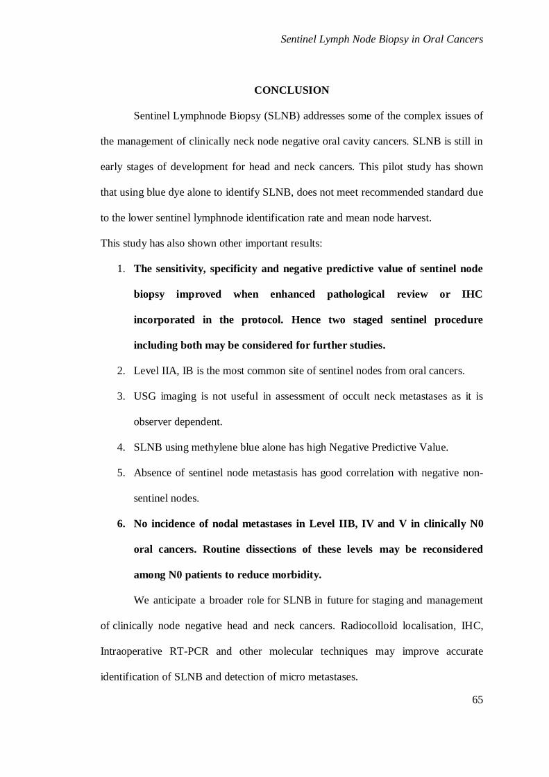
Sentinel Lymph Node Biopsy in Oral Cancers
65
CONCLUSION
Sentinel Lymphnode Biopsy (SLNB) addresses some of the complex issues of
the management of clinically neck node negative oral cavity cancers. SLNB is still in
early stages of development for head and neck cancers. This pilot study has shown
that using blue dye alone to identify SLNB, does not meet recommended standard due
to the lower sentinel lymphnode identification rate and mean node harvest.
This study has also shown other important results:
1. The sensitivity, specificity and negative predictive value of sentinel node
biopsy improved when enhanced pathological review or IHC
incorporated in the protocol. Hence two staged sentinel procedure
including both may be considered for further studies.
2. Level IIA, IB is the most common site of sentinel nodes from oral cancers.
3. USG imaging is not useful in assessment of occult neck metastases as it is
observer dependent.
4. SLNB using methylene blue alone has high Negative Predictive Value.
5. Absence of sentinel node metastasis has good correlation with negative non-
sentinel nodes.
6. No incidence of nodal metastases in Level IIB, IV and V in clinically N0
oral cancers. Routine dissections of these levels may be reconsidered
among N0 patients to reduce morbidity.
We anticipate a broader role for SLNB in future for staging and management
of clinically node negative head and neck cancers. Radiocolloid localisation, IHC,
Intraoperative RT-PCR and other molecular techniques may improve accurate
identification of SLNB and detection of micro metastases.

i
REFERENCES
1. Madras Metropolitan Tumour Registry.
2. Kavita M. Pattani, Joseph Califano, “Long-Term Experience in Sentinel Node Biopsy for Early
Oral and Oropharyngeal Squamous Cell Carcinoma” Ann Surg Oncol (2011) 18:2709–10.
3. Stanley P.L. Leong, Markus Zuber, Robert L. Ferris, Yuko Kitagawa Impact of Nodal Status and
Tumour Burden in Sentinel Lymph Nodes on the Clinical Outcomes of Cancer Patients Journal of
Surgical Oncology 2011;103:518–30
4. Francisco Civantos, Robert Zitsch, Anthony Bared, Ayman Amin, “Sentinel Node Biopsy for
Squamous Cell Carcinoma of the Head and Neck”, J. Surg. Oncol. 2008; 97: 683–90.
5. Sharma MK, Gour N, Pandey A, Wallia D. Epidemiological study of risk factors for oral, laryngeal
and esophageal cancers at a tertiary care hospital in India. Asian Pac J Cancer Prev 2011; 12:1215-
8.
6. Layland MK, Sessions DG, Lenox J. The influence of lymph node metastasis in the treatment of
squamous cell carcinoma of the oral cavity, oropharynx, larynx, and hypopharynx: N0 versus N+.
Laryngoscope 2005; 115:629-39.
7. Leemans CR, Tiwari R, Nauta JJ, van der Waal I, Snow GB. Regional lymph node involvement
and its significance in the development of distant metastases in head and neck carcinoma.Cancer
1993; 71:452-56.
8. Luiz P. Kowalski, Roge ́ rio Bagietto Jose ́ R. L. Lara, Roge´ rio L. Santos, “Prognostic
significance of the distribution of neck node metastasis from oral carcinoma” Head Neck 22: 207–
14, 2000
9. James C. Alex. The Application of Sentinel Node Radio localization to Solid Tumors of the Head
and Neck: A 10-Year Experience, Laryngoscope, 114:2–19, 2004
10. Werner JA, Dunne AA, Myers JN. 2003. Functional anatomy of the lymphatic drainage system of
the upper aerodigestive tract and its role in metastasis of squamous cell carcinoma. Head Neck
25:322–32.
11. Ludwig J. Ueber Kurschlusswege Der. Lymphbahnen and ihre Beziehungen zur lymphogen Krebs
metastasierung. Pathol Microbiol 1992;25:329
12. Yuan Wang, Thomas J. Ow, Jeffrey N. Myers “Pathways for Cervical Metastasis in Malignant
Neoplasms of the Head and Neck Region” Clinical Anatomy 25:54–71 (2012).
13. T. Shoaiba,, D.S. Soutara, D.G. MacDonald, H.W. Gray “The nodal neck level of sentinel lymph
nodes in mucosal head and neck cancer”, British Journal of Plastic Surgery (2005) 58, 790–4
14. Werner JA, Davis RK. (eds.) 2004. Metastases in Head and Neck Cancer. New York: Springer
15. Byers RM, Wolf PF, Ballantine AJ. Rationale for Elective modified neck dissection Head Neck
surg. 1998; 10 (3):160-7.

ii
16. Shah JP, Andersen PE. Evolving role of modifications in neck dissection for oral squamous
carcinoma. Br J OralMaxillofac Surg 1995; 33:3-8
17. Civantos FJ, Moffat FI, and Goodwin WJ: Lymphatic mapping and sentinel lymphadenectomy for
106 head and neck lesions: Contrasts between oral cavity and cutaneous malignancy. Laryngoscpe
2006; 16:1–15.
18. Robbins KT, Clayman G, and Levine PA, et al. Neck dissection classification update: Revision
proposed by American Academy of 1otolaryngeology- Head Neck Surg 2002; 128(7):751-8.
19. Y. Ducic, L. Young, J. Mc Lntyre. Neck dissection: past and present Minerva Chir 2010;65:45-58
20. K Harish Neck dissections: radical to conservative WJSO 2005, 3:21
21. Martina A. Broglie, MD,1 Stephan K. Haerle, MD,2 Gerhard F. Huber, MD, MSc,2 Sarah R. Haile,
“Occult metastases detected by sentinel node biopsy in patients with early oral and oropharyngeal
squamous cell carcinomas: Impact on survival” Head Neck 2012
22. Keski-Santti H, et al. Elective neck treatment versus observation in patients with T1/T2 N0
squamous cell carcinoma of oral tongue. Oral Oncol. 2006;42:96–101.
23. Jochen A. Werner, Anette Ramaswamy, Carsten Dalchow, “The sentinel node concept in head and
neck cancer: Solution for the controversies in the n0 neck?” Head Neck 26: 603–11, 2004.
24. Adams S, Baum RP, Stuchensen T, Bitter K, et al. Prospective comparison 18F-FDG PET with
conventional imaging modalities (CT, MRI, US) in lymph node staging of head and neck cancer.
Eur J Nucl Med 1998;25:1255–60
25. Braams JW, Pruim J, Freling NJM, Nikkels PG, et al. Detection of lymph node metastases of
squamous cell cancer of the head and neck with FDG PET and MRI. J Nucl Med 1995;36: 211–6.
26. Benchaou M, Lehman W, Slosman DO, et al. The role of FDG PET in the pre-operative
assessment of N staging in head and neck cancer. Acta Otolaryngol 1996;116:332–5.
27. Nieuwenhuis EJ, Castelijns JA, Pijpers R, et al. Wait-and see policy for the N0 neck in early stage
oral and oropharyngeal SCC using ultrasound guided cytology: is there a role for identification of
the sentinel node? Head Neck 2002;24:282–289
28. Seaman WB, Powers WE (1955) Studies of the distribution of radioactive colloidal gold in regional
lymph nodes containing cancer. Cancer 8:1044–46
29. Gavilan J, Herranz-Gonzalez JJ, Lentsch EJ. 2003. Cancer of the Neck. In: Myers E, Suen J, Myers
J, Hanna E, editors. Cancer of the Head and Neck. 4th Ed. Philadelphia, PA: W. B. Saunders. P
407–30
30. Cabanas RM. An approach for the treatment of penile carcinoma. Cancer 1997; 39: 456
31. Donald L. Morton and Arden D, Chan “The Concept of Sentinel Node Localization: How It
Started” Seminars in Nuclear Medicine, Vol No 1 (January), 2000: pp 4-10.
32. Alex JC, Krag DN: The gamma-probe-guided resection of radiolabeled primary lymph nodes. Surg
Oncol Clin N Am 1996;5:33.

iii
33. Francisco J. Civantos, Robert P. Zitsch, David E. Schuller, Amit Agrawal Sentinel Lymph Node
Biopsy Accurately Stages the Regional Lymph Nodes for T1-T2 Oral Squamous Cell Carcinomas:
Results of a Prospective Multi-Institutional Trial J Clin Oncol 28:1395-400.
34. Pitman KT, Johnson JT, Brown ML, Myers EN. Sentinel lymph node biopsy in head and neck
squamous cell carcinoma. Laryngoscope 2002; 112:2101– 13
35. Shoaib T, Soutar DS, Prosser JE, et al. A suggested method for sentinel node biopsy in squamous
cell carcinoma of the head and neck. Head Neck 1999; 21:728–33
36. Moni Abraham Kuriakose Nirav P. Trivedi “Sentinel node biopsy in head and neck squamous cell
carcinoma”, Otolaryngology & Head and Neck Surgery 2009, 17:100–10.
37. Remy H. Blanchaert and D. Lee Morris, ‘‘Reliability of sentinel lymph node biopsy with squamous
cell carcinoma of the oral cavity’’ Oral Surg Oral Med Oral Pathol Oral Radiol Endod
2005;99:539-41
38. de Boer M, van Deurzen CH, van Dijck JA, et al. Micrometastases or isolated tumor cells and the
outcome of breast cancer. N Engl J Med 2009; 361:653–63.
39. Pitman KT, Johnson JT, Edington H, et al. Lymphatic mapping with isosulfan blue dye in
squamous cell carcinoma of the head and neck. Arch Otolaryngol Head Neck Surg 1998; 124:790–
93.
40. Stoeckli SJ, Pfaltz M, Steinert H, Schmid S. Histopathological features of occult metastasis
detected by sentinel lymph node biopsy in oral and oropharyngeal squamous cell carcinoma.
Laryngoscope 2002; 112:111–5.
41. Thomas D. Shellenberger, “Sentinel Lymph Node Biopsy in the Staging of Oral Cancer”, Oral
Maxillofacial Surg Clin N Am 18 (2006) 547–63
42. Ross GL, Shoaib T. Role of sentinel node biopsy in the management and staging of the N0 neck.
Odontology 2005; 93:1–6
43. Atula T, Hunter KD, Cooper LA, et al. Micrometastases and isolated tumour cells in sentinel
lymph nodes in oral and ropharyngeal squamous cell carcinoma. Eur J Surg Oncol 2009; 35:532
44. Derrek A. Heuveling, Ge ́ke B. Flach, Annelies van Schie, Stijn van Weert “Visualization of the
sentinel node in early-stage oral cancer: limited value of late static lymphoscintigraphy” Nuclear
Medicine Communications 2012.
45. Keereweer CM. Sterenborg, F. Kerrebijn, A. Van Driel, “Image-guided surgery in head and neck
cancer: current practice and future directions of optical imaging Head Neck” 34: 120–6, 2012
46. Stephan K. Haerle, Thomas F. Hany, Klaus Strobel, Daniel Sidler “Is There an Additional Value of
SPECT/CT Over Planar Lymphoscintigraphy for Sentinel Node Mapping in Oral/Oropharyngeal
Squamous Cell Carcinoma?”, Ann Surg Oncol (2009) 16:3118–24
47. Francisco J. Civantos, Frederick L. Moffat, William J. Goodwin “Lymphatic Mapping and Sentinel
Lymphadenectomy for 106 Head and Neck Lesions: Contrasts Between Oral Cavity and Cutaneous
Malignancy”, Laryngoscope, 116(Suppl. 109):1–15, 2006.

iv
48. James C. Alex. The Application of Sentinel Node Radiolocalization to Solid Tumors of the Head
and Neck: A 10-Year Experience”, Laryngoscope, 114:2–19, 2004
49. L. W. T. Alkureishi, Z. Burak, J. A. Alvarez, J. Ballinger “Joint Practice Guidelines for
Radionuclide Lymphoscintigraphy for Sentinel Node Localization in Oral/Oropharyngeal
Squamous Cell Carcinoma”, Ann Surg Oncol (2009) 16:3190–210.
50. Van den Hoogen FJ, Manni JJ. Value of the supraomohyoid neck dissection with frozen section
analysis as a staging procedure in the clinically negative neck in squamous cell carcinoma of the
oral cavity. Eur Arch Otorhinolaryngol 1992;249:144–48
51. Francisco J. Civantos, Jochen A. Werner, Anthony Bared, “Sentinel node biopsy in cancer of the
oral cavity” Operative Techniques in Otolaryngology (2005) 16, 275-85.
52. Von Buchwald C, Bilde A, Shoaib T, Ross G. Sentinel node biopsy: the technique and the
feasibility in head and neck cancer. ORL J Otorhinolaryngol Spec 2002;64:268–74
53. Marije T. Hornstra, Alkureishi, Gary L. RossTaimur Shoaib, “ Predictive factors for failure to
identify sentinel nodes in head and neck squamous cell carcinoma” Head Neck 30: 858–62, 2008
54. Jose J, Coates worth AP, MacLennan K. Cervical metastases in upper aerodigestive tract squamous
cell carcinoma: histopathologic analysis and reporting. Head Neck 2003; 25:194–197.
55. Bilde A, von Buchwald C, Therkildsen MH, et al. Need for intensive histopathologic analysis to
determine lymph node metastases when using sentinel node biopsy in oral cancer. Laryngoscope
2008; 118:408–14
56. David M. Euhus “Cytokeratin Staining and Other Sentinel Node Controversies” Clinical Breast
Cancer, Vol. 4, Suppl. 1, S49-S54, April 2003
57. Paul J. Van Diest, Hans Torrenga, Sybren Meijer, Chris J.L.M. Meijer, “Pathologic Analysis of
Sentinel Lymph Nodes” Seminars in Surgical Oncology 2001; 20:238–245.
58. Eline J. C. Nieuwenhuis, Isaac van der Waal, C. Rene ́Leemans, Alain Kummer, “Histopathologic
validation of the sentinel node concept in oral and oropharyngeal squamous cell carcinoma” Head
Neck 27: 150– 158, 2005
59. Kenneth O. Devaney, Alessandra Rinaldo, Juan Pablo Rodrigo, Alfio Ferlito, “Sentinel node
biopsy and head and neck tumors—where do we stand today?” Head Neck 28: 1122–1131, 2006.
60. Robert L. Ferris, Patrick Stefanika, Liqiang Xi, William Gooding, “Rapid Molecular Detection of
Metastatic Head and Neck Squamous Cell Carcinoma as an Intraoperative Adjunct to Sentinel
Lymph Node Biopsy”, Laryngoscope, 122:1020–1030, 2012.
61. UICC 7th
edition
62. Philip Sloan, Head and Neck Sentinel Lymph Node Biopsy: Current State of the Art Head and
Neck Pathol (2009) 3:231–237
63. Dahanca. 22 sentinel node European trial by the Danish Head and Neck Cancer Study Group
64. Philip Sloan, Head and Neck Sentinel Lymph Node Biopsy: Current State of the Art Head Neck
Pathol. 2009 September; 3(3): 231–237.

v
65. Civantos FJ, Zitsch RP, Schuller DE, et al. Sentinel lymph node biopsy accurately stages the
regional lymph nodes for T1-T2 oral squamous cell carcinomas: results of a prospective
multiinstitutional trial. J Clin Oncol 2010; 28:1395–1400.
66. Ross GL, Soutar DS, Gordon MacDonald D, Shoaib T, Camilleri I et al, Sentinel node biopsy in
head and neck cancer: preliminary results of a multicenter trial. Ann Surg Oncol. 2004 Jul;
11(7):690-6.
67. Sandro J. Stoeckli, Sentinel node Biopsy for Oral and Oropharyngeal Squamous Cell Carcinoma of
the Head and Neck, Australia - Asian Journal of Cancer, Vol. 7, 2008
68. Alkureishi LW, Ross GL, Shoaib T, Soutar DS, Robertson AG, Thompson R Sentinel node biopsy
in head and neck squamous cell cancer: 5-year follow-up of a European multicenter trial. Ann Surg
Oncol. 2010 Sep;17(9):2459-64
69. Melkane AE, Mamelle G, Wycisk G, Temam S, Janot F, Casiraghi O et al Sentinel node biopsy in
early oral squamous cell carcinomas: a 10-year experience. Laryngoscope. 2012 Aug;122(8):1782-
8.
70. J.K. Antonio, S. Santini, D. Politi, S. Sulfaro, R. Spaziante,. Alberti, et al, Sentinel lymph node
biopsy in squamous cell carcinoma of the head and neck: 10 years of experience Acta
Otorhinolaryngol Ital. 2012 February; 32(1): 18–25
71. L. W. T. Alkureishi, Z. Burak, J. A. Alvarez, J. Ballinger, A. Bilde, A. J. Britten, Joint Practice
Guidelines for Radionuclide Lymphoscintigraphy for Sentinel Node Localization in
Oral/Oropharyngeal Squamous Cell Carcinoma, Ann Surg Oncol (2009) 16:3190–10
72. Canis, Plüquett, Ihler, Matthias, Kron, Steiner. Impact of Elective Neck Dissection vs Observation
on Regional Recurrence and Survival in cN0-Staged Patients With Squamous Cell Carcinomas of
the Upper Aerodigestive Tract. Arch Otolaryngol Head Neck Surg. 2012;138(7):650-655
73. Ferlito A, Silver CE, Rinaldo A. Elective management of the neck in oral cavity squamous
carcinoma: current concepts supported by prospective studies. Br J Oral Maxillofac Surg.
74. Christian Fialka, Gerhard Oberleitner, Paul Stampfl, Werner Brannath “Modification of the
Constant—Murley shoulder score–introduction of the individual relative Constant score Individual
shoulder assessment”, Injury, Int. J. Care Injured (2005) 36, 1159-65.
75. Karin Murer, Gerhard F. Huber, Sarah R. Haile, Sandro J. Stoeckli “Comparison of morbidity
between sentinel node Biopsy and elective neck dissection for Treatment of the n0 neck in patients
with oral Squamous cell carcinoma”, HEAD & NECK—September 2011
76. Franziska Schiefke, Michael Akdemir, Anette Weber, Daniel Akdemir, Susanne Singer, Bernhard
Frerich, “Function, postoperative morbidity, and quality of Life after cervical sentinel node biopsy
and after Selective neck dissection” Head & Neck 31: 503–512, 2009
77. Pinsolle V, Michelet V, Majoufre C et al Branche extreme du nerf spinal et evidements
ganglionnaries cervicaux . Rev Stomatol Chir Maxillofac 1997; 98:138-142

vi
78. Hermanek P, Hutter RV, Sobin LH, et al. International union against cancer classification of
isolated tumor cells and micrometastasis. Cancer. 1999;86:2668–2673
79. Lim YC, Song MH, Kim SC, Kim KM, Choi EC Preserving level IIb lymph nodes in elective
supraomohyoid neck dissection for oral cavity squamous cell carcinoma. Arch Otolaryngol Head
Neck Surg. 2004 Sep;130 (9):1088-91.
80. Elsheikh MN, Mahfouz ME, Elsheikh ELevel IIb lymph nodes metastasis in elective
supraomohyoid neck dissection for oral cavity squamous cell carcinoma: a molecular-based study.
Laryngoscope. 2005 Sep;115(9):1636-40.
81. Villaret AB, Piazza C, Peretti G, et al. Multicentric prospective study on the prevalence of sublevel
IIb metastases in head and neck cancer. Arch Otolaryngol Head Neck Surg 2007;133:897-903.
82. M. Manola, C. Aversa, L. Moscillo, S. Villano “Status of level IIb lymph nodes of the neck in
squamous cell carcinoma of the oral tongue in patients who underwent modified radical neck
dissection and lymph node sentinel biopsy” Acta Oto italica 2011;31:130-4.
83. Shah JP. Patterns of cervical nodal metastasis from squamous cell carcinoma of upper
areodigestive tract. Am J Surg 1990; 160:405-9
84. Davidson BJ, Culkarny V, Delacure MD et al Posterior triangle metastases of squamous cell
carcinoma of upper aerodigestive tract. AM J Surg 1993; 166:395-398
85. LP Kowalski and A Sanabria Elective neck dissection in oral carcinoma: a critical review of the
evidence Acta Otorhinolaryngol Ital. 2007 June; 27(3): 113–117.
86. Sandro J. Stoeckli and Martina A. Broglie Sentinel node biopsy for early oral carcinoma Curr Opin
Otolaryngol Head Neck Surg 2012, 20:103–108
87. Agarwal, A. K.; Sethi, A.; Sareen, D. & Dhingra, S Oral and Oropharyngeal Squamous Cell
Carcinoma in Our Population: The Clinic-Pathological and Morphological Description of 153
Cases, Int. J. Morphol., 29(3):686-693, 2011
88. Shenoi R, Devrukhkar, Chaudhuri, Sharma BK, Sape SB, Chikhale Demographic and clinical
profile of oral squamous cell carcinoma patients: A retrospective study, Indian journal of cancer
2012, 49 (1): 21- 26
89. Ross GL, Shoaib T, Soutar DS, et al. The First International Conference on Sentinel node biopsy in
mucosal head and neck cancer and adoption of a multicenter trial protocol. Ann Surg Oncol 2002;
9:406–410.
90. Lee W. T. Alkureishi, Gary L. Ross, Taimur Shoaib, David S. Soutar, “Sentinel Node Biopsy in
Head and Neck Squamous Cell Cancer:5-Year Follow-Up of a European Multicenter Trial” Ann
Surg Oncol (2010) 17:2459–2464
91. Thomas D. Shellenberger, “Sentinel Lymph Node Biopsy in the Staging of Oral Cancer”, Oral
Maxillofacial Surg Clin N Am 18 (2006) 547–563.
92. Reynolds, et al. 2009. Head & Neck. New York Head and Neck Society

vii
93. Byers RM, Weber RS, Andrews T, McGill D, Kare R, Wolf P. Frequency and therapeutic
implications of "skip metastases’’ in the neck from squamous carcinoma of the oral tongue. Head
Neck 1997; 19:14–19
94. Dias FL, Lima RA, Kligerman J, Farias TP, Soares JR, Manfro G, Relevance of skip metastases for
squamous cell carcinoma of the oral tongue and the floor of the mouth. Otolaryngol Head Neck
Surg. 2006 Mar;134(3):460-5.
95. Mira E, Benazzo M, Rossi V, Efficacy of selective lymph node dissection in clinically negative
neck. Otolaryngeol Head Neck surg. 2002 Oct; 127(4): 279-83
96. De zinis LO, Bolzoni A, Piazza C, et al Prevelance and localization of nodal metastasis in
squamous cell carcinoma of the oral cavity: role and extension of neck dissection. Euro Arch
Otorhinolaryngol. 2006 Dec; 263 (12) 1131-5.
97. Elizabeth Mathew Iype, Paul sebastian, Aleyamma Mathew, et al,The role of selective neck
dissection I –III in the treatment of node negative (N0) neck in oral cancer. Oral Oncology 2008
98. Jegoux F, Caze A, Mohr E , Neck dissection for stage N0 oral cavity cancer. Ann Otolaryngeol
Head Neck surg 2006 Nov; 123 (5):221-6.
99. Pitman KT, Johnson JT, Myers EN. Effectiveness of selective neck dissection for management of
the clinically negative neck. Arch Otolaryngeol Head Neck Surg 1997; 123: 917-22
100. The Brazilian Head and Neck Cancer Study Group
101. Carvalho AL, Kowalski LP, Borges JA, et al. Ipsilater neck cancer recurrences after elective
supraomohyoid neck dissection. Arch Otolaryngol Head Neck Surg 2000;126:410-12
viii
ABBREVIATIONS
DFS Disease free Survival
FOM Floor of Mouth
HPE Histopathological Examination
ITC Isolated Tumour Cells
LN Lymph Node
LS Lymphoscintography
Mi Micrometastases
MRND Modified Radical Neck Dissection
N+ Clinically Node Positive
N0 Clinically Node Negative
RLND Regional Lymph Node Dissection
RMT Retromolar Trigone
RND Radical Neck Dissection
RT-PCR Reverse Transcriptase – Polymerase chain Reactions
SAN Spinal Accessary Nerve
SCC Squamous Cell Carcinoma
SLN Sentinel Lymph Node
SLNB Sentinel Lymph Node Biopsy
SND Selective Neck Dissection
SOHND Supra omohyoid Neck Dissection

ix
PROFOMA
Sr.No………… IP.NO………..
Name……………………. Age……. Sex a) Male b) Female
Date of admission: Date of surgery: Date of discharge:
Address……………………………………………………………. Occupation………….
Patient History:
Religion: a) Hindu b) Christian c) Muslim d) others
Marital status: a) Married b) Unmarried c) Widow
Family H/o Cancer: a) Yes b) No c) Don’t know
Family h/o Head and neck cancer: a) Yes b) No If YES: details:
Any pre- cancerous lesion or condition: ……………………..
Habits: a) Yes b) No If yes: a) tobacco chewing b) pan c) smoking d) alcohol
Duration: a) <1 yr b) 1-5 yr 3) > 5 yr.
Co-morbid factors a.DM b.HT c. IHD d. COPD e. Others: specify………
Previous Neck surgery: a)Yes b)No If Yes – Excluded Previous treatment
of primary …………………………………
Symptoms : Duration of symptoms in months a) 1 b) 2-3 c) 4-6 d) >6
local pain ulcer Bleeding Aspiration
Trismus Dysphagia Referred otalgia Hypersalivation
Voice Change Neck swelling Others
Examination:
Number of lesions a) single b) Multiple.
Side a) Right b) Left c) Midline d) Bilateral e) Crossing midline
Type of growth a)Ulcero-proliferative b)verrucous c)infiltrative
Size of lesion a) Length ……cms b)Breadth……….cms
Extension to adjacent sites a)yes b)no If yes specify……………………….
Clinical Neck Nodes a)Yes b)No If Yes – Excluded
USG Neck ……………………………………………
Diagnosis: Clinical T N0 M0 a)Stage-1 b)Stage-2 c)Stage-3 d)Stage-4
Pre op HPE………………………………… Grade ….....................

x
Management:
Surgical treatment of neck: a) SOHND b) MRND c) RND
Surgical reconstruction: a) Nil b)PMMC c) DP d) Forehead e) others
Sentinel Node Biopsy details
Level No. of Nodes Frozen Section
Positive/Negative
Final HPE Positive/Negative/ Micro
metastases/ ITCs
Cytokeratin
Positive/Negative
IA
IB
IIA
IIB
III
IV
V
Elective Neck Dissection details
Level No. of Nodes Final HPE:Positive/Negative/ Micro metastases/ ITCs
IA
IB
IIA
IIB
III
IV
V
Adjuvant Details: A) Chemotherapy B) Radiotherapy C) Others
Details of Chemo/RT…………………………………………………………
Recurrence in primary
Nodal recurrence
Metastasis
Others
Details of Treatment of recurrence ……………………………………….
xi
INFORMED CONSENT
SL NO …….. IP NO………….
Name………………………….. Age ………. Sex…………….
Date of Admission ……………… Date of surgery……………..
Address……………………………………………………………………..
I’m suffering from oral cavity cancer. This cancer frequently affects lymphnodes and later it
spreads to distant sites. Multimodality approach like Surgery, Radiotherapy and Chemotherapy are
used to treat such cancer. Neck dissection surgery is treatment for cancer involvement of neck
nodes in which lymphatic tissues are removed as a part of treatment. Neck dissection may lead to
minor complication like seroma formation or at times major complications like bleeding and it can
be acute , sub-acute or chronic. Few of which are bleeding at the time of surgery or delayed, air-
way obstruction, nerve damage, chylous fistulae, infection, flap necrosis etc…
Alternative treatment includes Radiotherapy to neck and you have 100% right to refuse to
participate in this project. Department of surgical oncology, GRH, Chennai-14 has started a project
with the aim to diagnose involvement of neck nodes before they become palpable and will facilitate
to identify early spread of cancer to neck and plan accurate treatment. We will first use the blue dye
to identify sentinel node and then proceed to Selective Neck Dissection (SND) /Modified Radical
Neck Dissection (MRND)/. Our research group will be very thankful for your support & co-
operation.
I have studied and under-stood well all the points and I give free consent for my enrolment
in above study whole heartedly.
Date:
Place: Signature
Signature of witness
Name & Contact no

Name age
sex
IP n
o
Re
ligi
on
Occupation
Fam
ily
His
tory
Co
mo
rbid
itie
s
Premalignant
conditions
smo
kin
g
Alc
oh
ol
Tob
acco
ch
ew
ing
Oth
er
Ad
dic
tio
ns
Symptoms
Sym
pto
m D
ura
tio
n
Site
mani 48 M 960025 hindu watchman normal no no 35 yrs no yes hans ulcer 24 buccal mucosathiruvenkadam 55 M 963039 hindu flower vendor normal no no no yes 30 yrs no ulcer 3 hardpalateprema 40 F 962282 hindu cooly normal no no no no no no ulcer, pain 2 tongueviswanathan 55 M 963837 hindu cooly normal ht no 15 yrs 15 yrs no no ulcer 8 tonguebaskar 43 M 964364 hindu cooly grand mother ht leucoplakia no no 20 yr no pain, ulcer 3 tongue
rathiannamal 58 F 964465 christian home maker normal no no no no no no ulcer , otologia, hypersalivation 5 tongueeallamal 70 F 964803 hindu cooly normal no no no no 40 yrs no ulcer 4 buccal mucosa
vijayakumar 32 M 964626 hindu mechanic normal copd no 7 yrs no 5 yrs pan ulcer, pain 5 tongueloganathan 56 M 967687 hindu carpenter normal no no 30 yrs 30 yrs no no ulcer 6 tonguevaradarajan 60 M 966124 hindu agriculturist normal dm no 35 yrs no no no ulcer, pain 3 tonguethangarasu 40 M 967446 hindu electrician normal no no 20 yrs 15 yrs no no ulcer, pain 3 tonguejeyaraj 54 M 968502 christian agriculturist normal no leukoplakia 30 yrs no no no ulcer 3 tongueramakrishnan 49 M 969310 hindu cooly normal no no 25 yrs no no no ulcer 3 buccal mucosalawrence 46 M 972058 christian salesman normal no no no no no no ulcer 2 tonguebaradhan 70 M 972950 hindu cooly normal no no 40 yrs 35 yrs no no ulcer 5 tonguevasantha 55 M 975161 hindu home maker normal no no no no no no ulcer 1 buccal mucosajeyaraman 62 M 976100 hindu gardner normal no no 30 yrs no 10 yrs pan ulcer, pain dysphagia 6 tonguekannumayil 62 F 976016 hindu cooly normal no no no no no no ulcer, pain 8 tonguemarimuthu 51 M 982487 hindu agriculturist normal no no 25 yrs no no no ulcer 4 buccal mucosanagaraj 31 M 982423 hindu cooly normal no no 15 yrs 10 yrs no no ulcer, pain 4 tonguerebel 58 M 985045 christian cooly normal no no 25 yrs no no no ulcer, pain 3 tonguesekar 55 M 983674 hindu cooly normal no no 23 yrs 20 yrs no no ulcer 1 fomvelayutham 42 M 988608 hindu cooly normal no no 20 yrs no no no ulcer. Pain 3 rmtvaralakshmi 43 F 991452 hindu home maker normal no no no no no no ulcer 3 tongueveerasamy 65 M 991956 hindu agriculturist normal no no 36 yrs no no no ulcer 4 buccal mucosaillamalli 55 F 993341 hindu cooly normal no no no no 35 yrs no ulcer 6 alveolusjanakiraman 26 M 999676 hindu mechanic normal no no 12 yrs 10 yrs no pan 10 yrs ulcer pain 4 tonguekaliyaperumal 65 M 998266 hindu agriculturist normal no no 30 yrs 25 yrs no no ulcer pain 5 rmtparthiban 45 M 100674 hindu salesman normal no no 20 yrs 20 yrs no pan 15 yrs ulcer 3 tonguerama 47 F 102479 hindu cooly normal no post cricoid web no no 20 yrs no ulcer 3 alveolusneela 45 F 111091 hindu home maker normal no no no no no no ulcer, pain 6 buccal mucosakrishna moorthy 57 M 111543 hindu agriculturist normal no no no no 30 yrs pan 20 yrs ulcer 3 hardpalate

Name
manithiruvenkadampremaviswanathanbaskar
rathiannamaleallamal
vijayakumarloganathanvaradarajanthangarasujeyarajramakrishnanlawrencebaradhanvasanthajeyaramankannumayilmarimuthunagarajrebelsekarvelayuthamvaralakshmiveerasamyillamallijanakiramankaliyaperumalparthibanrama neelakrishna moorthy
Size
(la
rge
st
dia
me
ter)
type of tumour
Nu
mb
er
Sid
e
cT
His
top
ath
olo
gy
Gra
de
Resection
Re
con
stru
ctio
n
Ne
ck d
isse
ctio
n
Sen
tin
el N
od
e
nu
mb
er
no
. of
Sen
tin
el N
od
es
dis
sect
ed
No
. of
Sen
tin
el N
od
es
Po
siti
ve o
n F
roze
n
No
de
s IA
No
de
s P
osi
tive
IA
No
de
s IB
No
de
s P
osi
tive
IB
No
de
s II
A
No
de
s P
osi
tive
IIA
No
de
s II
B
No
de
s P
osi
tive
IIB
No
de
s II
I
No
de
s P
osi
tive
III
4x3 ulceroproliferative one right 2 scc 1 Wide Local Excision nasolabial sohd Ib, II 2 negative 2 0 1 0 6 0 5 0 1 0ulcer one left 2 scc 1 palatoalveolar resection obturator sohd Ib 1 negative 3 0 3 0 10 0 2 0 3 0
3x2 ulceroproliferative one right 2 scc 2 hemiglossectomy nil mrnd-1 Ib, Iia, III 3 positive 3 0 3 2 3 2 2 0 4 21.5 x 1 infiltrative one left 1 scc 3 hemiglossectomy nil mrnd-1 Iia 1 positive 2 0 5 0 2 0 5 0 8 03.5x 2 ulceroproliferative one left 2 scc 1 hemiglossectomy nil sohd III 1 negative 3 0 3 0 10 0 2 0 10 0
3.5x 2.5 ulceroproliferative one left 2 scc 2 hemiglossectomy nil sohd Ib 1 negative 4 0 3 0 7 0 2 0 9 01.5x1 verrucous one left 1 scc 1 Wide Local Excision ssg sohd IB 1 negative 2 0 1 0 3 0 4 0 8 0
1.5x 1 ulcer one left 1 scc 1 Wide Local Excision nil mrnd-1 Iia III 2 III positive 3 0 4 0 7 0 2 0 10 02x 3 ulceroproliferative one right 2 scc 1 hemiglossectomy nil sohd Ib, Iia 2 negative 4 0 8 0 3 0 2 0 5 02x 1.5 ulcer one right 1 scc 2 hemiglossectomy nil sohd no no no 2 0 6 0 7 0 3 0 5 05x4 infiltrative one right 2 scc 1 hemiglossectomy nil mrnd-1 Ib, II 2 positive 1 0 3 2 5 1 6 0 5 03.2x 1.5 infiltrative one right 2 scc 1 hemiglossectomy nil sohd IIa,III 2 negative 2 0 4 0 2 0 1 0 2 03x2 ulceroproliferative one left 2 scc 2 Wide Local Excision ssg sohd Ia, Ib 2+1 negative 3 0 2 0 4 0 6 0 3 01X1.5 verrucous one right 1 scc 1 Wide Local Excision nil sohd Ib 2 negative 3 0 3 0 4 0 3 0 2 03x 2 ulcer one left 2 scc 1 hemiglossectomy nil sohd no no no 3 0 2 0 4 0 2 0 3 02x2 ulceroproliferative one right 1 scc 1 Wide Local Excision ssg sohd no no no 0 0 2 0 5 0 2 0 4 05x2 infiltrative one left 2 scc 2 hemiglossectomy nil sohd Ib 2 negative 1 0 2 0 2 0 6 0 1 05x 2.5 ulceroproliferative one right 3 scc 1 hemiglossectomy nil sohd Ib, II 2 negative 6 0 4 0 5 0 4 0 4 02x1 ulceroproliferative one left 1 scc 2 Wide Local Excision ssg sohd Iia 1 negative 4 0 3 0 3 0 2 0 3 02x1.5 infiltrative one left 1 scc 1 hemiglossectomy nil sohd Iia 1 negative 4 0 5 0 7 0 5 0 3 02x2 ulcer one left 2 scc 2 Wide Local Excision nil sohd Ib 2 negative 3 0 4 0 3 0 4 0 12 02.5 x 1.5 ulceroproliferative one right 2 scc 2 Wide Local Excision + MM local sohd ib 2 negative 3 0 6 0 6 1 2 0 1 02x1.5 ulcer one right 1 scc 2 Wide Local Excision + MM local sohd Iia 2 negative 6 0 1 0 4 0 1 0 7 02x3 ulceroproliferative one right 1 scc 1 Wide Local Excision nil mrnd-1 Iia 1 positive 3 0 4 0 3 1 3 0 1 03.5x2 ulceroproliferative one left 1 scc 1 Wide Local Excision+ periosteum local sohd Ia 1 negative 0 0 4 0 5 0 2 0 6 02x2 ulceroproliferative one left 1 scc 1 palatoalveolar resection obturator sohd Ib, Iia 2 negative 5 0 2 0 3 0 2 0 7 05x4 infiltrative one right 3 scc 1 hemiglossectomy nil sohd Ia, Iia, III 3 negative 4 0 1 0 3 0 3 0 2 02x1 ulceroproliferative one left 1 scc 2 Wide Local Excision+ MM local sohd Ia, Iia 2+1 negative 0 0 2 0 2 0 2 0 1 03.5x3 ulceroproliferative one right 1 scc 1 hemiglossectomy nil sohd Iia 1 negative 3 0 2 0 4 0 2 0 10 03x3 ulcer one right 1 scc 1 palatoalveolar resection obturator sohd Iia 2 negative 0 0 3 0 6 0 5 0 6 03x2 ulceroproliferative one left 2 scc 2 Wide Local Excision + MM local sohd Ib 1 negative 3 0 7 2 2 0 4 0 1 02x1.5 ulcer one right 1 scc 2 palatoalveolar resection obturator sohd Iia 1 negative 4 0 1 0 5 0 2 0 2 0

Name
manithiruvenkadampremaviswanathanbaskar
rathiannamaleallamal
vijayakumarloganathanvaradarajanthangarasujeyarajramakrishnanlawrencebaradhanvasanthajeyaramankannumayilmarimuthunagarajrebelsekarvelayuthamvaralakshmiveerasamyillamallijanakiramankaliyaperumalparthibanrama neelakrishna moorthy
No
de
s IV
No
de
s P
osi
tive
IV
No
de
s V
No
de
s P
osi
tive
V
Tota
l No
de
s
Tota
l No
de
s P
osi
tive
pT
pN
Po
st o
p M
orb
idit
y
Ad
juva
nt
Tre
atm
en
t
Re
curr
en
ce
tim
e t
o r
ecu
rre
nce
Foll
ow
up
De
tail
s
cyto
kera
tin
5 0 0 0 20 0 1 0 nil no nil no 26 Negative4 0 0 0 18 0 1 0 nil no nil no 26 Negative3 0 3 0 21 4 2 2b nil RT nil no 25 Positive3 0 4 0 29 1 1 1 nil no nil no 24 Positive2 0 0 0 28 0 2 0 nil no nil no 24 Negative
2 0 0 0 27 0 2 0 nil no nil no 25 Negative10 0 0 0 28 0 1 0 nil no nil no 24 Negative
7 0 5 0 38 0 1 0 nil no nil no 23 false positive slnb Negative3 0 0 0 25 0 2 0 nil no nil no 23 Negative0 0 0 0 23 0 2 0 nil no nil no 23 no7 0 6 0 33 1 2 1 nil yes nil no 22 Positive1 0 0 0 12 0 2 0 nil no nil no 22 Positive ITC2 0 0 0 20 0 2 0 nil no nil no 21 Negative3 0 0 0 18 0 1 0 nil no nil no 21 Negative2 0 0 0 16 0 2 0 nil no nil no 20 no2 0 0 0 13 0 1 0 nil no nil no 19 no4 0 0 0 16 0 2 0 nil yes yes 4 19 dead Negative8 0 0 0 31 0 3 0 nil yes yes 2 15 dead Positive ITC5 0 0 0 22 0 1 0 nil no nil no 15 Negative6 0 0 0 27 0 1 0 nil no nil no 15 Negative1 0 0 0 27 0 1 0 nil no nil no 14 Negative
12 0 0 0 31 0 2 0 nil no nil no 14 false negative Negative1 0 0 0 20 0 1 0 nil no nil no 11 Negative6 0 4 0 21 0 1 1 nil no nil no 10 Positive5 0 0 0 22 0 1 0 nil no nil no 9 Positive ITC5 0 0 0 24 0 1 0 nil no nil no 8 Positive ITC3 0 0 0 16 0 3 0 nil yes nil no 7 Negative0 0 0 0 7 0 2 0 nil no nil no 7 Negative2 0 0 0 23 0 1 0 nil no nil no 6 Negative0 0 0 0 20 0 1 0 nil no nil no 5 Negative0 0 0 0 17 2 2 2a nil no nil no 3 Negative0 0 0 0 14 0 1 0 nil no nil no 3 Negative