Predictors of positive axillary lymph nodes in breast cancer patients with metastatic sentinel lymph...
22
S1 Available online http://breast-cancer-research.com/supplements/7/S1 Speaker abstracts Symposium I: Molecular genetics, phenotypes for prognosis and response to treatments S1 The role of gene expression profiling by microarray analysis for prognostic classification of breast cancer MJ van de Vijver Department of Pathology, Netherlands Cancer Institute, Amsterdam, The Netherlands Breast Cancer Research 2005, 7(Suppl 1):S1 (DOI 10.1186/bcr1205) Introduction Prognostic and predictive factors play important roles in the treatment of breast cancer. Genome-wide monitoring of gene expression using DNA microarrays makes it possible to study thousands of genes in a tumour sample in a single experiment. By looking for an association between the gene expression pattern and tumour behaviour, it should be possible to identify new prognostic and predictive factors. Method We used gene expression profiling using two different microarray platforms: one containing 25,000 oligonucleotide probes and one containing 18,000 cDNA probes. To obtain prognostic gene expression profiles, we isolated RNA from tumours from a series of 295 patients younger than 53 years presenting with stage I and II breast cancer treated at our institute between 1984 and 1993. The expression of 25,000 genes was assessed, and using various statistical approaches correlation of gene expression with distant metastasis-free probability and overall survival was assessed [1-3]. In addition, we started studies to obtain gene expression profiles predicting response to specific chemotherapy regimens. Within a single-institution, randomized phase II trial, patients with locally advanced breast cancer received six courses of either AC (n = 24) or AD (n = 24) containing neoadjuvant chemotherapy. Gene expression profiles for 18,000 genes were generated from core needle biopsies obtained before treatment and correlated with the response of the primary tumour to the chemotherapy administered [4]. Additionally, pretreatment gene expression profiles were compared with those in tumours remaining after chemotherapy. Results We previously identified a 70-gene expression profile associated with increased risk for developing distant metastases within 5 years [1,2]. More recently, we studied a Wound Signature in these same tumors [3]. By combining the 70-gene expression profile to subdivide the tumours into ‘good prognosis’ and ‘poor prognosis’ tumours, and the Wound signature to subdivide tumours into ‘activated’ and ‘quiescent’ tumours, subgroups of patients with markedly different prognosis can be identified. Additional gene expression signatures are being tested in this series of tumours to arrive at an optimal prognostic classifier and to obtain improved insight into breast cancer biology. In the study to identify predictive profiles, 10 (20%) of the 48 patients showed (near) pathological complete remission of the primary tumour after treatment [4]. No gene expression pattern correlating with response could be identified for all patients, or for the AC or AD treated groups separately. Conclusion Various gene expression profiles in breast cancer are associated with the propensity of the tumour to develop distant metastases. Gene expression profile predicting the response of primary breast carcinomas to AC or AD based neoadjuvant chemotherapy are most likely to be very subtle and cannot be detected when small series of patients are studied. Genetic tests derived from gene expression profiling studies are likely to become useful as prognostic and predictive tests to guide clinical decision making in the treatment of primary breast cancer. References 1. van’t Veer LJ, Dai HY, van de Vijver MJ, et al.: Gene expression profiling predicts clinical outcome of breast cancer. Nature 2002, 415:530-536. 2. van de Vijver MJ, He YD, ‘t Veer LJ, et al.: A gene-expression signature as a predictor of survival in breast cancer. N Engl J Med 2002, 347:1999-2009. 3. Chang HY, Nuyten DSA, Sneddon JB, et al.: Robustness, scala- bility, and integration of a wound-like gene expression signa- ture in predicting breast cancer survival. Proc Natl Acad Sci USA 2005, 102:3738-3743. 4. Hannemann J, Oosterkamp HM, Bosch CAJ, et al.: Changes in gene expression associated with response to neoadjuvant chemotherapy. J Clin Oncol 2005:in press S2 Gene expression profiles and molecular classification to predict distant metastasis and tamoxifen-resistant breast cancer JGM Klijn 1 , EMJJ Berns 1 , J Martens 1 , MPHM Jansen 1 , D Atkins 2 , JA Foekens 1 , Y Wang 2 1 Daniel den Hoed Cancer Center/Erasmus MC, Rotterdam, The Netherlands; 2 Veridex LLC, Johnson and Johnson, Molecular Diagnostics, San Diego, California, USA Breast Cancer Research 2005, 7(Suppl 1):S2 (DOI 10.1186/bcr1206) Introduction Genome-wide measures of gene expression can identify patterns of gene activity that subclassify tumours and might provide better means than are currently available for individual risk assessment in patients with primary breast cancer and for prediction of tamoxifen resistance. Methods We analyzed, with Affymetrix Human U133a GeneChips, the expression of 22,000 transcripts from total RNA of frozen tumour samples from 286 lymph node negative (LNN) patients who had not received adjuvant systemic treatment. In a separate second study conducted in 112 estrogen receptor (ER)-positive primary breast carcinomas from patients with metastatic disease and clearly defined types of response to first-line treatment with tamoxifen, a 18,000 human cDNA microarray was used to discover gene expression profiles predictive of tamoxifen resistance. Breast Cancer Research Volume 7 Supplement 1, June 2005 Meeting abstracts VI Madrid Breast Cancer Conference: Changes in the treatment of Breast Cancer Madrid, Spain 1–3 June 2005 Received: 28 April 2005 Published: 27 May 2005 © 2005 BioMed Central Ltd
Transcript of Predictors of positive axillary lymph nodes in breast cancer patients with metastatic sentinel lymph...
S1
Available online http://breast-cancer-research.com/supplements/7/S1
Speaker abstracts
Symposium I: Molecular genetics, phenotypes forprognosis and response to treatments
S1The role of gene expression profiling by microarrayanalysis for prognostic classification of breast cancerMJ van de VijverDepartment of Pathology, Netherlands Cancer Institute, Amsterdam,The NetherlandsBreast Cancer Research 2005, 7(Suppl 1):S1 (DOI 10.1186/bcr1205)Introduction Prognostic and predictive factors play important roles inthe treatment of breast cancer. Genome-wide monitoring of geneexpression using DNA microarrays makes it possible to study thousandsof genes in a tumour sample in a single experiment. By looking for anassociation between the gene expression pattern and tumour behaviour,it should be possible to identify new prognostic and predictive factors.Method We used gene expression profiling using two differentmicroarray platforms: one containing 25,000 oligonucleotide probesand one containing 18,000 cDNA probes. To obtain prognostic geneexpression profiles, we isolated RNA from tumours from a series of 295patients younger than 53 years presenting with stage I and II breastcancer treated at our institute between 1984 and 1993. Theexpression of 25,000 genes was assessed, and using variousstatistical approaches correlation of gene expression with distantmetastasis-free probability and overall survival was assessed [1-3]. Inaddition, we started studies to obtain gene expression profilespredicting response to specific chemotherapy regimens. Within asingle-institution, randomized phase II trial, patients with locallyadvanced breast cancer received six courses of either AC (n = 24) orAD (n = 24) containing neoadjuvant chemotherapy. Gene expressionprofiles for 18,000 genes were generated from core needle biopsiesobtained before treatment and correlated with the response of theprimary tumour to the chemotherapy administered [4]. Additionally,pretreatment gene expression profiles were compared with those intumours remaining after chemotherapy.Results We previously identified a 70-gene expression profileassociated with increased risk for developing distant metastases within5 years [1,2]. More recently, we studied a Wound Signature in thesesame tumors [3]. By combining the 70-gene expression profile tosubdivide the tumours into ‘good prognosis’ and ‘poor prognosis’tumours, and the Wound signature to subdivide tumours into‘activated’ and ‘quiescent’ tumours, subgroups of patients withmarkedly different prognosis can be identified. Additional geneexpression signatures are being tested in this series of tumours toarrive at an optimal prognostic classifier and to obtain improved insightinto breast cancer biology.In the study to identify predictive profiles, 10 (20%) of the 48 patientsshowed (near) pathological complete remission of the primary tumourafter treatment [4]. No gene expression pattern correlating with
response could be identified for all patients, or for the AC or ADtreated groups separately.Conclusion Various gene expression profiles in breast cancer areassociated with the propensity of the tumour to develop distantmetastases. Gene expression profile predicting the response of primarybreast carcinomas to AC or AD based neoadjuvant chemotherapy aremost likely to be very subtle and cannot be detected when small seriesof patients are studied. Genetic tests derived from gene expressionprofiling studies are likely to become useful as prognostic andpredictive tests to guide clinical decision making in the treatment ofprimary breast cancer.References1. van’t Veer LJ, Dai HY, van de Vijver MJ, et al.: Gene expression
profiling predicts clinical outcome of breast cancer. Nature2002, 415:530-536.
2. van de Vijver MJ, He YD, ‘t Veer LJ, et al.: A gene-expressionsignature as a predictor of survival in breast cancer. N Engl JMed 2002, 347:1999-2009.
3. Chang HY, Nuyten DSA, Sneddon JB, et al.: Robustness, scala-bility, and integration of a wound-like gene expression signa-ture in predicting breast cancer survival. Proc Natl Acad SciUSA 2005, 102:3738-3743.
4. Hannemann J, Oosterkamp HM, Bosch CAJ, et al.: Changes ingene expression associated with response to neoadjuvantchemotherapy. J Clin Oncol 2005:in press
S2Gene expression profiles and molecular classificationto predict distant metastasis and tamoxifen-resistantbreast cancerJGM Klijn1, EMJJ Berns1, J Martens1, MPHM Jansen1, D Atkins2,JA Foekens1, Y Wang2
1Daniel den Hoed Cancer Center/Erasmus MC, Rotterdam, TheNetherlands; 2Veridex LLC, Johnson and Johnson, MolecularDiagnostics, San Diego, California, USABreast Cancer Research 2005, 7(Suppl 1):S2 (DOI 10.1186/bcr1206)Introduction Genome-wide measures of gene expression can identifypatterns of gene activity that subclassify tumours and might providebetter means than are currently available for individual risk assessmentin patients with primary breast cancer and for prediction of tamoxifenresistance.Methods We analyzed, with Affymetrix Human U133a GeneChips, theexpression of 22,000 transcripts from total RNA of frozen tumoursamples from 286 lymph node negative (LNN) patients who had notreceived adjuvant systemic treatment. In a separate second studyconducted in 112 estrogen receptor (ER)-positive primary breastcarcinomas from patients with metastatic disease and clearly definedtypes of response to first-line treatment with tamoxifen, a 18,000human cDNA microarray was used to discover gene expressionprofiles predictive of tamoxifen resistance.
Breast Cancer Research Volume 7 Supplement 1, June 2005
Meeting abstractsVI Madrid Breast Cancer Conference: Changes in the treatment of Breast CancerMadrid, Spain1–3 June 2005
Received: 28 April 2005 Published: 27 May 2005
© 2005 BioMed Central Ltd

S2
Results In the first single-center study, in a training set of 115 tumors(80 ER+ and 35 ER– tumors) we identified a 76-gene signature (60genes for ER+ and 16 for ER–) for predicting the occurrence of distantmetastasis within 5 years. This signature was successfully validatedwith 93% sensitivity in an independent test set of 171 LNN patients asa whole, irrespective of age or ER status. The 76-gene profile wasstrongly predictive of those patients who will develop a distantmetastasis within 5 years or will remain recurrence free during thatperiod (hazard ratio [HR] 5.67; P < 0.00002) and in multivariateanalysis when corrected for traditional prognostic factors includinggrade (HR 5.55; P < 0.00003). Analogously, the 76-gene expressionprofile strongly predicted overall survival (HR 8.62; P < 0.00002). The76-gene profile was also a strong prognostic factor in the subgroup of79 patients with a tumor size ranging from 10 to 20 mm (HR 14.1;P < 0.00003) and in 84 premenopausal patients (HR 9.60; P < 0.0002)and 87 postmenopausal patients (HR 4.04; P = 0.0017). In thesubgroup of 42 ER– patients in the validation set, even a profile of only16 genes appeared to have a strong prognostic value (HR 8.74;P = 0.012). Recently, our 76-gene expression signature wassuccessfully validated in a separate multicenter European study of 180patients from four institutions (Nijmegen, Munich, Bari, Ljubljana) (HR7.41; P < 0.0001) with similar sensitivity and specificity.In the second study, conducted in 112 patients with metastaticdisease, using a training set of 46 breast cancers 81 genes were foundto be differentially expressed between tamoxifen-responsive and-resistant tumors. From the 81 genes, a predictive signature of 44genes was extracted and validated in an independent set of 66 tumors.This 44-gene signature is significantly superior (odds ratio [OR] 3.16;P = 0.03) to traditional predictive factors in univariate analysis andsignificantly related to longer progression-free survival in univariate aswell as in multivariate analyses (P = 0.03). The predictive value of the44-gene signature was recently confirmed in an extended series of 280patients with advanced disease.Conclusion In the first study, the identified 76-gene signature provides apowerful tool for identification of patients at high or low risk for distantrecurrence or death due to breast cancer, allowing clinicians to adaptchoices of adjuvant systemic therapy. In the second study, the 44-genesignature predicts tamoxifen resistance more accurately than do traditionalpredictive factors. Interestingly, in a third study DNA methylation statusalso appeared to be useful in predicting tamoxifen resistance.References1. Wang Y, Klijn JGM, Zhang Y, et al.: Gene-expression profiles to
predict distant metastasis of lymph-node-negative primarybreast cancer. Lancet 2005, 365:671-679.
2. Wang Y, Klijn JGM, Zhang Y, et al.: Pathway analysis and vali-dation of the 76-gene prognostic signature in lymph-node-negative primary breast cancer. 27th Annual San AntonioBreast Cancer Symposium [abstract]. Breast Cancer Res Treat2004, Suppl 1:103.
3. Jansen MPHM, Foekens JA, Van Staveren IL, et al.: Molecularclassification of tamoxifen-resistant breast cancer by geneexpression profiling. J Clin Oncol 2005, 23:732-740.
4. Martens JWM, Nimmrich I, Koenig T, et al.: Association of DNA-methylation of phosphoserine aminotransferase withresponse to endocrine therapy in patients with recurrentbreast cancer. Cancer Res 2005:in press.
S3Methods for gene expression profiling in clinicaltrials for breast cancerS PaikDivision of Pathology, National Surgical Adjuvant Breast and BowelProject, Pittsburgh, Philadelphia, USABreast Cancer Research 2005, 7(Suppl 1):S3 (DOI 10.1186/bcr1207)High throughput gene expression profiling provides a powerful tool fordiscovery of prognostic and predictive markers for breast cancer [1-4].The main limitation to this approach is the requirement for high-qualityRNA, which is difficult in the multicenter clinical trial setting. One
solution is to use RNAlater, which allows procurement and shipping oftissue specimens at room temperature [5,6]. The National SurgicalAdjuvant Breast and Bowel Project (NSABP) has conducted a pilotstudy to procure pretreatement core biopsy specimens in aneoadjuvant study. Most of the samples in this study provided high-quality RNA, as determined by Bioanalyzer and Affymetrix GeneChipanalyses. When combined with a RNA amplification method, qualitydata could be obtained from 10 ng of total RNA as starting material.NSABP currently has two neoadjuvant trials in which pretreatmentspecimens are procured in RNAlater. However, the typical practicepattern in the USA makes it difficult to procure tissue in the adjuvantsetting even with the use of RNAlater. Therefore, methods that permithigh throughput gene expression profiling of formalin-fixed, paraffin-embedded materials are in great need. Such methods will also allowinterrogation of archived tissue banks with annotation established frompreviously finished trials and will therefore shorten the time for markerdevelopment and validation. Chemical modification by formalin anddegradation during storage make RNA extracted from paraffin a poorsubstrate for gene expression profiling [7]. We have examined bothmicroarray and RT-PCR platforms for this purpose. In generalmicroarray analysis using the Arcturus Paradise system has been adisappointment in our hands, with high rate for assay failure formaterials older than 3 years. However, there are RNA amplification andlabeling methods in development that are not dependent on oligo-dTpriming for cDNA synthesis and may provide better results. Incollaboration with Genomic Health, Inc., we have explored the use ofhigh-throughput real time RT-PCR for discovery and validation ofprognostic markers for node negative and estrogen receptor positivebreast cancer [8]. This has resulted in development of the OncotypeDxassay, which is offered as a commercial reference laboratory test. Thedisadvantage of real-time RT-PCR assays is relatively low throughput(less than 1000 genes, even at industrial scale). DASL assay fromIllumina is a kind of hybrid between PCR and microarray platforms, andmay provide relatively cost-efficient means by which to assay manycandidate genes using degraded RNA obtainable from paraffin blocks[9].References1. Pusztai L, Ayers M, Stec J, et al.: Gene expression profiles
obtained from fine-needle aspirations of breast cancer reliablyidentify routine prognostic markers and reveal large-scalemolecular differences between estrogen-negative and estro-gen-positive tumors. Clin Cancer Res 2003, 9:2406-2415.
2. Sorlie T, Perou CM, Tibshirani R, et al.: Gene expression patternsof breast carcinomas distinguish tumor subclasses with clinicalimplications. Proc Natl Acad Sci USA 2001, 98:10869-10874.
3. van ‘t Veer LJ, Dai H, van de Vijver MJ, et al.: Gene expressionprofiling predicts clinical outcome of breast cancer. Nature2002, 415:530-536.
4. van de Vijver MJ, He YD, van’t Veer LJ, et al.: A gene-expressionsignature as a predictor of survival in breast cancer. N Engl JMed 2002, 347:1999-2009.
5. Grotzer MA, Patti R, Geoerger B, et al.: Biological stability ofRNA isolated from RNAlater-treated brain tumor and neurob-lastoma xenografts. Med Pediatr Oncol 2000, 34:438-442.
6. Mutter GL, Zahrieh D, Liu C, et al.: Comparison of frozen andRNALater solid tissue storage methods for use in RNAexpression microarrays. BMC Genomics 2004, 5:88.
7. Masuda N, Ohnishi T, Kawamoto S, et al.: Analysis of chemicalmodification of RNA from formalin-fixed samples and opti-mization of molecular biology applications for such samples.Nucleic Acids Res 1999, 27:4436-4443.
8. Paik S, Shak S, Tang G, et al.: A multigene assay to predictrecurrence of tamoxifen-treated, node-negative breast cancer.N Engl J Med 2004, 351:2817-2826.
9. Bibikova M, Talantov D, Chudin E, et al.: Quantitative gene expres-sion profiling in formalin-fixed, paraffin-embedded tissues usinguniversal bead arrays. Am J Pathol 2004, 165:1799-1807.
Breast Cancer Research Vol 7 Suppl 1 VI Madrid Breast Cancer Conference

S3
S4Gene expression profile-based predictors ofresponse to chemotherapyL PusztaiAnderson Cancer Center, Houston, Texas, USABreast Cancer Research 2005, 7(Suppl 1):S4 (DOI 10.1186/bcr1208)A molecular test that could help in selecting the most effectivechemotherapy for a particular individual could save patients fromunnecessary toxicity, and the right choice of drugs may save lives,particularly in the adjuvant treatment of breast cancer. Administration ofchemotherapy before surgery provides an attractive opportunity todiscover predictors of response [1]. Pathologic complete eradication ofcancer from the breast and lymph nodes (pCR) represents an extremeform of chemotherapy sensitivity and invariably heralds excellent long-term survival. We adopted pCR as an early surrogate of clinicallymeaningful benefit from therapy and as an outcome that is worthpredicting. There are simple clinical and histological parameters,including grade, estrogen receptor status and tumor size, that can becombined into powerful prediction scores. However, these clinicalvariables do not yield treatment regimen specific predictions, and theycannot be used to select one therapy over another. Assessment oftraditional single gene markers of chemotherapy sensitivity has not yetresulted in clinically useful tests. Gene expression profiling, whichenables simultaneous measurement of thousands of genes, representsa promising new tool that may be applied to this clinical problem. It iscurrently unknown what the best strategy is to discover responsepredictors from high dimensional gene expression data. The simplestapproach may be to search for the single most informative gene that isdifferentially expressed between responders and nonresponders. Thismay lead to new mechanistic insights into the biology of chemotherapyresponse and could yield easy-to-use but moderately powerful singlegene predictive markers [2]. Another approach is to identify geneexpression signatures that are predictive of response, assuming thatthe combined information provided by multiple genes would result inmore accurate predictions than any single gene can do. Several smallstudies have suggested that this is feasible [3]. Large-scale validationof these results is needed and is currently underway. Yet anotherapproach is to recognize the different molecular subtypes of breastcancer and attempt to develop distinct predictors for each subtype [4].This approach assumes that, by focusing on the molecularly morehomogenous subgroups, more accurate predictors could be developedthan by analyzing all breast cancers together. We shall present resultsfrom our own research program, illustrating the successes andlimitations of each of these approaches.References.1. Pusztai L, Rouzier R, Wagner P, Symmans WF: Individualizing
chemotherapy treatment for breast cancer: is it necessary,can it be done? Drug Resistance Updates 2005, 7:325-331.
2. Roman R, Rajan R, Hess KR, et al.: Microtubule associatedprotein tau is a predictive marker and modulator of responseto paclitaxel-containing preoperative chemotherapy in breastcancer. Proc Natl Acad Sci USA 2005:in press.
3. Ayers M, Symmans WF, Stec J, et al.: Gene expression profilespredict complete pathologic response to neoadjuvant pacli-taxel/FAC chemotherapy in breast cancer. J Clin Oncol 2004,22:2284-2293.
4. Rouzier R, Anderson K, Hess KR, et al.: Basal and luminal typesof breast cancer defined by gene expression patternsrespond differently to neoadjuvant chemotherapy [abstract].Breast Cancer Res Treat 2004, 88:S24.
Lecture
S5Present situation and future of genetic profiling forprognosis and treatmentGN HortobagyiDepartment of Breast Medical Oncology, The University of Texas MDAnderson Cancer Center, Houston, Texas, USABreast Cancer Research 2005, 7(Suppl 1):S5 (DOI 10.1186/bcr1209)Classification and staging systems are important in oncology to predictclinical behavior and determine prognosis. In addition, they maycontribute to the selection of optimal treatment strategies. Muchclinical and translational research over the past 30 years was directedat establishing or refining prognostic and predictive factors for breastcancer. Initially, tumor related factors such as size, grade, lymph nodeinvolvement, and hormone receptor status were considered in thedetermination of prognosis. Patient characteristics, such as age,menopausal status and performance status, also contributed to theseestimates. Some factors such as estrogen receptor (ER) status wereshown to be better predictive factors than prognostic factors. Thus,although ER-positive tumors have a slightly better prognosis during theearly years of follow up than do ER-negative ones, the major applicationof ER status is to predict response to endocrine therapy. A variety ofbiochemical and molecular factors were reported to have prognostic orpredictive ability over the past 20 years. These included cathepsin D,HER2, EGFR, p53, UPA, PAI, and many others. Of these, only HER2was consistently validated as a prognostic factor, as well as a predictorof response to the monoclonal antibody trastuzumab (Herceptin).Developing, assessing, and discarding these various putativeprognostic and/or predictive factors was the result of an enormousinvestment of time and effort of many scientists from many countriesaround the world. Considering that only one new prognostic/predictivefactor was universally adopted over the past 25 years (HER2 status), itmust be concluded that this is an enormously inefficient process.The Human Genome Project was a major milestone in the history ofmedicine. Both the genetic information obtained and the technologicaladvances that took place during this large multicenter effort have hadenormous influence over all fields of medicine. For the field ofprognostication and prediction in breast cancer, the major consequencewas the development of technology that led to the simultaneousevaluation of gene expression for hundreds and, more recently, thousandsof genes. In fact, recently launched gene arrays include the entire humangenome. Thus, we have the opportunity to assess, in a small tumorsample, the expression profile of all known human genes. There aremultiple technological platforms under evaluation for this purpose, and theresults obtained with one cannot automatically be substituted for resultsobtained with another platform. Nevertheless, on the basis of severalreports, it can be stated that gene expression profiling of human breastcancer provides valuable information in the following areas:1. Molecular classification of primary breast cancer2. Identification of multiple distinct prognostic subgroups3. Determination of expression level of several genes of interest (ER,
PR, HER2, etc.)4. Identification of genetic networks5. Prediction of response to chemotherapyThe initial reports were based on small patient numbers that presentedsubstantial statistical challenges for adequate estimation of end-pointsand to prevent frequent false-positive or false-negative results. Morerecent analyses have included several dozen and up to a few hundredpatients. These reports provide greater statistical power and greaterreliability. However, these reports still represent retrospective analysesof subsets of patients, and prospective validation is still sorely needed.Reports are beginning to appear comparing the performance ofdifferent platforms on the same tumor samples and considering thesame end-points. The source of tumor material, the manner in which itwas handled before testing, and the amount of tissue needed forreliable testing are all under intense scrutiny. Gene profiling withcurrently available platforms includes a number of genes or gene
Available online http://breast-cancer-research.com/supplements/7/S1

S4
segments of uncertain function (ESTs). These provide an excellentopportunity to assess the functional value of these genes and enrichour understanding of their biological function. Many centers andgroups are assessing the potential of molecular profiling in theprediction of response to therapy. As technology evolves, this type ofinformation will transform the way we think of breast cancer, the waywe assess and stage primary and metastatic breast cancer, and themanner in which we select the best combination and sequence oftherapies to obtain optimal therapeutic results. Today’s costs, whilesubstantial, are rapidly falling and newer technology will make theseassays much more accessible. Furthermore, because multiple relevantmarkers can be determined using a single assay, it is likely that geneexpression profiling will be more cost-effective than currently useddiagnostic and prognostic tests.The major challenges in gene profiling are still in developing and usingthe most appropriate statistical methods for data analysis. The need forhandling tens of thousands or hundreds of thousands of data points,especially originating from a much smaller number of tumors, isdaunting and mistaken conclusions might be reached in the absence ofoptimal analytical techniques.Finally, prospective validation of the clinical utility of gene profiling forclassification, determination of prognosis, and selection of optimaltherapies for individual patients will require large, prospective,multicenter, controlled clinical trials. If successful, these will take usone step closer to individualized medicine.References1. Pusztai L, Ayers M, Stec J, et al.: Gene expression profiles
obtained from fine-needle aspirations of breast cancer reliablyidentify routine prognostic markers and reveal large-scalemolecular differences between estrogen-negative and estro-gen-positive tumors. Clin Cancer Res 2003, 9:2406-2415.
2. Pusztai L, Ayers M, Stec J, Hortobagyi GN: Clinical applicationof cDNA microarrays in oncology. Oncologist 2003, 8:252-258.
3. Symmans WF, Ayers M, Clark EA, et al.: Total RNA yield andmicroarray gene expression profiles from fine-needle aspira-tion biopsy and core needle biopsy samples of breast carci-noma. Cancer 2003, 97:2960-2971.
4. Van Laere SJ, Van Den Eynden GG, Van der Auwera I, et al.:Unsupervised hierarchical clustering of gene expression leveldata obtained by microarray analysis demonstrated a uniqueexpression pattern for inflammatory breast cancer versusnon-inflammatory breast cancer [abstract 5245]. Proc AnnuMeet Am Assoc Cancer Res 2004, 45:1210.
5. Perou CM, Sorlie T, Eisen MB, et al.: Molecular portraits ofhuman breast tumours. Nature 2000, 406:747-752.
6. van de Vijver MJ, He YD, van’t Veer LJ, et al.: A gene-expressionsignature as a predictor of survival in breast cancer. N Engl JMed 2002, 347:1999-2009.
Symposium II: Advances in diagnosis and staging
S6The role of magnetic resonance imaging as animaging tool to assess disease status and residualdisease in locally advanced breast cancerM CristofanilliDepartment of Breast Medical Oncology, The University of Texas, MDAnderson Cancer Center, Houston, Texas, USABreast Cancer Research 2005, 7(Suppl 1):S6 (DOI 10.1186/bcr1210)Magnetic resonance imaging (MRI) is a very sensitive diagnostic toolfor the evaluation of breast cancer. A MRI is more frequently utilized inconjunction with other diagnostic modalities, particularly mammogram,to better assess a breast abnormality or biopsy-proven cancer,including ductal carcinoma in situ (DCIS) [1]. Most recently it has alsobeen demonstrated to be a better screening tool for women at high riskfor developing breast cancer, including women with documentedgenetic predisposition. Because MRI has the advantages of providing athree-dimensional view of the breast, it has been shown to be moreprecise than standard imaging in determining the initial staging and
evaluation of the extension of invasive disease [2]. This information is ofparticular value in patients with locally advanced disease, includinginflammatory breast cancer (IBC) and in classic lobular histology, whichmay exhibit diffuse involvement of the breast at initial presentation andtherefore are frequently treated with primary systemic therapy (PST) [3,4].The management of primary breast cancer has evolved significantly inthe past decade, with the increasing use of preoperative or primarychemotherapy (PST), and most recently also primary hormonal therapyfor both early and locally advanced breast cancers (LABCs). Theadvantages of the early use of systemic therapy are considered: thefeasibility of a more conservative surgery, and the possibility of true invivo testing of the tumor’s drug sensitivity. The amount of residualdisease found following surgical excision represents the pathologicalresponse to the preoperative treatment and remains the most importantprognostic factor.The high staging accuracy of breast MRI makes it an attractive methodfor assessing tumor response to PST. MRI can contribute in severalways to the management of patients receiving preoperative chemo-therapy, including the initial determination of extent of disease forproper staging (baseline evaluation), early identification of poorresponders (intermediate evaluation during treatment), andidentification and description of the presence and extent of residualdisease for surgical planning (preoperative imaging study) [3,4]. MRImeasurements of tumor response may have predictive value fordisease recurrence and responsiveness to novel therapeutics.Comparison of dynamic parameters (e.g. signal enhancement ratio) atbaseline and at subsequent evaluation time points can also contributeinformation on the response to treatment and predict residual disease[4]. A series of prospective trials has been conducted in patients withLABC, including IBC. Most recently a large multicenter trial sponsoredjointly by the American College of Radiology Imaging Network, theCancer and Leukemia Group B, and the National Cancer institute isintegrating serial MRI tumor measurements with serial collection oftissue for evaluation of biomarkers (expression, genomic, protein arrays,as well as specific immunohistochemical markers and fluorescence insitu hybridization). The goal of such studies is to compare tissueassessment of biomarkers with imaging to identify the most appropriatetool for prediction of pathological response to PST. This study couldimpact on the present management of primary breast cancer byallowing the early introduction of novel therapeutics in patients withearly demonstration of poor response to treatment.References1. Kriege M, Brekelmans CTM, Boetes C, et al.: Efficacy of MRI and
mammography for breast cancer screening in women with afamilial or genetic predisposition. N Engl J Med 2004, 351:427-437.
2. Hata T, Takashi H, Watanebe K, et al.: Magnetic resonanceimaging for preoperative evaluation of breast cancer: A com-parative study with mammography and ultrasonography. J AmColl Surg 2004, 198:190-197.
3. Partridge SC, Gibbs JE, Lu Y, et al.: Accuracy of MR imaging forrevealing residual breast cancer in patients who have under-gone neoadjuvant chemotherapy. Am J Roentgenol 2002, 179:1193-1199.
4. Esserman L, Kaplan E, Partridge S, et al.: MRI phenotype isassociated with response to doxorubicin and cyclophos-phamide neoadjuvant chemotherapy in stage III breastcancer. Ann Surg Oncol 2001, 8:549-559.
S7The roles of PET and CT/PET as preoperative studiesRC Delgado-Bolton1, JL Carreras Delgado2
1Instituto PET Focuscan, Madrid, Spain; 2Nuclear MedicineDepartment, Hospital Clínico San Carlos, Madrid, SpainBreast Cancer Research 2005, 7(Suppl 1):S7 (DOI 10.1186/bcr1211)Introduction Positron emission tomography (PET) with 18F-fluoro-deoxyglucose (FDG) is a functional imaging technique that hasdemonstrated advantages over anatomically based imaging modalities
Breast Cancer Research Vol 7 Suppl 1 VI Madrid Breast Cancer Conference

S5
in oncology in the detection of malignant lesions. The recentintroduction of combined computed tomography (CT)/PET systemsallows the coregistration of functional PET and anatomical CT images,which will very likely improve accuracy. The role of PET and CT/PET inbreast cancer is reviewed.Methods In breast cancer, FDG-PET has been used for detection,staging, and response monitoring; one of its main clinical applicationsis defining the extent of recurrent or metastatic disease [1]. We havereviewed the evidence regarding the roles of PET and CT/PET aspreoperative studies in breast cancer.Results The review of the evidence shows that PET and CT/PET cancontribute to patient diagnosis and management as preoperativestudies in breast cancer in the following situations:1. Detection of primary breast cancer. When evaluating suspicious
breast abnormalities, FDG-PET has a sensitivity of 80–100% andspecificity of 75–100%. However, its role in primary tumordetection is not clear when compared with conventional imagingmethods and remains to be determined [1].
2. Prognostic value of FDG uptake in primary tumor. Most studiessuggest that the level of FDG uptake in primary breast tumorscarries clinical and biological information, and that a higher FDGuptake is correlated with more clinically aggressive tumors [1].This information may help to stratify patients according toprognosis and risk for recurrence, and may help to tailortreatments to the individual patient.
3. Axillary node staging. In this situation, FDG-PET had a sensitivityof 57–100% and specificity of 66–100%. FDG-PET under-estimates the number of tumor-involved nodes compared withpathologic evaluation from conventional dissection. Therefore,FDG-PET should not replace axillary node sampling for routinestaging of the axilla because even microscopic nodal involvementmay be important for prognosis and treatment planning [1].However, FDG-PET may be complementary to sentinel lymphnode mapping and other standard axillary procedures in patientswith more advanced tumors or equivocally palpable axillary nodes.
4. Detection of locoregional and distant metastases. Functionalimaging with FDG-PET is more accurate than CT for the detectionof nodal involvement in the mediastinum; the sensitivity of FDG-PET was significantly higher (85%) than CT (50%), with nearlythe same specificity (90% for FDG-PET versus 83% for CT).Regarding the detection of distant metastases, FDG-PET canaccurately detect sites of distant disease with a sensitivity of80–97% and specificity of 75–94% [1].
5. Evaluation of therapy response. In locally advanced breast cancer(LABC), the assessment of response to neoadjuvantchemotherapy with conventional imaging methods is ofteninaccurate or slow because it depends on morphological criteria.Initial studies have shown the utility of FDG-PET in the evaluationof treatment response, specifically in its ability to discriminateresponders from nonresponders more accurately and earlier thanconventional imaging methods [1]. Changes in FDG uptake aftera single course of chemotherapy can predict pathologicalresponse in primary LABC tumors [2,3]. Histopathologicalresponse could be predicted with an accuracy of 88–91% afterthe first and second course of chemotherapy [3]. Other PETtracers may be used in the evaluation of the primary tumor;preliminary results suggest that applying PET in this way may helpto identify physiologic manifestations of drug resistance, whichwould help to tailor systemic therapy [1]. Preliminary data alsosuggest that FDG-PET may be useful in the assessment of sitesof disease other than the primary tumor for monitoring responseto chemotherapy in advanced breast cancer. Initial studiessuggest other possible applications of FDG-PET, such asevaluation of the response of skeletal metastases to therapy, andprediction of the response to antiestrogen therapy in patients withadvanced estrogen receptor positive breast cancer [1].Regarding CT/PET, to date only few studies have been reported,but the advantages of CT/PET compared with PET alone may betaken to indicate that CT/PET may improve the accuracy in the
evaluation of treatment response by directly defining metabolicand morphological changes [4].
6. Future applications. FDG is the most important radiotracer forPET in breast cancer and therefore it is analyzed in most studies.However, in the near future more specific PETradiopharmaceuticals may help to guide treatment, individualizingtherapies to a particular patient depending on the tumor’s biologiccharacteristics [1]. PET may help in management decisions byquantifying the therapeutic target, identifying resistance factors,and measuring early response to therapy.
Conclusion The clinical application of PET and CT/PET in breastcancer will help to predict clinical behavior, and allow one to choosethe appropriate treatment and to tailor local treatment options to theindividual patient. PET and CT/PET are also likely to play key roles inmonitoring systemic therapy and evaluating the response to therapy atan earlier stage than conventional methods. In the future, PET may beapplied with other tracers in addition to FDG, to improvecharacterization of tumor biology and more effectively measureresponse to therapy.References1. Eubank WB, Mankoff DA: Evolving role of positron emission
tomography in breast cancer imaging. Semin Nucl Med 2005,35:84-99.
2. Krak NC, Hoekstra OS, Lammertsma AA: Measuring responseto chemotherapy in locally advanced breast cancer: method-ological considerations. Eur J Nucl Med Mol Imaging 2004,Suppl 1:S103-S111.
3. Biersack HJ, Bender H, Palmedo H: FDG-PET in monitoringtherapy of breast cancer. Eur J Nucl Med Mol Imaging 2004,Suppl 1:S112-S117.
4. Zangheri B, Messa C, Picchio M, et al.: PET/CT and breastcancer. Eur J Nucl Med Mol Imaging 2004, Suppl 1:S135-S142.
S8Minimal residual disease (MRD) in the bone marrowin ER-αα-positive primary breast cancer patientsRC Coombes1,4, MT De Bella1, G Tripuraneni1, A Zaidi1, SAnwer1, DA Stephens2, B Ward1, HD Sinnett3, MJ Slade1,4,5
1Cancer Research UK Laboratories, Department of Medicine, ImperialCollege, London, UK; 2Department of Mathematics, Imperial College,London, UK; 3Breast Clinic, Charing Cross Hospital, Faculty ofMedicine, Imperial College, London, UK; 4,5Joint senior authorsBreast Cancer Research 2005, 7(Suppl 1):S8 (DOI 10.1186/bcr1212)Introduction We conducted a study to determine whether a group ofestrogen-induced genes could be used to detect and monitor formicrometastases in the bone marrow of patients with breast cancer.Methods We data-mined for potential markers of estrogen action,verified their relationship to ER in cell lines and purified cells frompatient biopsies, and checked their estrogen-inducibility afterdeveloping a real-time quantitative PCR assay for each. We thenexamined 99 bone marrow samples obtained over 2 years during thefollow up of good (n = 7) or poor (n = 19) prognosis patients todetermine the expression frequency.Results We discovered that the expression of eight out of 23 genes,identified by data-mining, were estrogen-regulated. We developed real-time quantitative PCR (QPCR) assays for measurement of the genesfor which ESTs were available (ER-α, PR and GATA-3, EEIG-1, EP-3,PS2). We examined their expression in purified breast cancer cellsfrom primary cancers and also from metastases from endocrine-resistant cancers and confirmed that these genes were still expressed.Of these, three were expressed in peripheral blood, excluding them ascandidate markers. We then examined 79 samples of bone marrowfrom 19 poor prognosis patients and 20 from seven good prognosispatients. We found that GATA-3 and ER expressions were significantlyhigher in the bone marrow of poor-prognosis patients.Conclusion GATA-3 and ER appear to be potentially useful markers, inaddition to CK19, for monitoring the effects of treatment in the bonemarrow of patients with ER-positive breast cancer.
Available online http://breast-cancer-research.com/supplements/7/S1

S6
Acknowledgements This work was funded by the Breast CancerResearch Trust (MTDB) (MS) and Cancer Research (UK) (RCC).
S9Circulating tumor cells and novel biomarkers forprognostic and biological of breast cancerM CristofanilliDepartment of Breast Medical Oncology, The University of Texas, MDAnderson Cancer Center, Houston, Texas, USABreast Cancer Research 2005, 7(Suppl 1):S9 (DOI 10.1186/bcr1213)The detection of microscopic disease in breast cancer has beenevaluated in lymph nodes, bone marrow (primary breast cancer), andperipheral blood (metastatic disease) [1,2]. Most of these studiesdemonstrated that the detection of microscopic disease in breastcancer patients contributes prognostic information and, in selectedcases, can predict the efficacy of treatments [1,2]. In primary breastcancer, the detection of microscopic disease in lymph nodes and bonemarrow has led to a better understanding of the role of minimal residualdisease (MRD). In metastatic breast cancer (MBC) reliable detection ofcirculating tumor cells (CTCs) had been obtained by using immuno-magnetic separation and subsequent analysis by the CellSpotter™analyzer (Veridex LLC, a Johnson & Johnson company, Warren, NJ,USA). This technology is becoming a standard tool for the ‘real-time’assessment of prognosis and response to treatment. This is particularlyimportant in the context of advanced disease management, consideringthe incurable status of the disease and the increasing therapeuticoptions available that could at least contribute to improve palliation andimpact on overall survival.In fact, despite years of clinical research, the odds of achievingcomplete response, and hence major survival benefit, for patients withMBC remain extremely low. Only a few patients who achieve acomplete response after chemotherapy remain in this state forprolonged periods of time, with some remaining in remission beyond20 years. There are presently no reliable biological markers that canpredict prognosis and monitor therapy effects in MBC.The detection of CTCs in patients with MBC about to start a new lineof treatment has been shown to predict progression-free survival (PFS)and overall survival (OS). This prognostic value was independent of theline of therapy (e.g. first-line versus second-line or more) [2,3].Moreover, in multivariate analysis CTCs demonstrated superior valuecompared with site of metastasis (e.g. visceral versus soft tissue/bone),type of therapy, and length of time to recurrence after definitive primarysurgery. In recent analysis, detection of CTCs has also been found tobe prognostic in patients with bone-only disease (not measurabledisease). CTCs have been shown to be superior to standard tumormarkers (e.g. Ca27-29) in predicting prognosis. Furthermore, theefficacy or benefit to systemic therapy could be predicted by the levelof CTCs as early as 3-4 weeks after initiation of therapy. Patients withpersistent of ≥5 CTCs demonstrated lack of response or progressivedisease at the time of restaging by standard imaging modalities.Conversely, patients with < 5CTCs showed objective remission. Thesedata clearly suggest that CTCs can be used as an early predictor oftreatment efficacy and be extremely useful in sparing patients fromfutile therapy early in the course of their treatment.Prospective clinical trials are presently being conducted in MBC tovalidate further the prognostic value of CTCs, possibly to use thisdiagnostic tool to better stratify patients with metastatic disease,eventually modifying the current staging system (International Stage IVStratification Study [ISSS]). Patients with metastatic disease could bedivided into the subcategories IVA and IVB, depending on the presenceor absence of CTCs. Additional studies are presently assessing thesurvival benefit of early change in treatment based on the persistenceof CTCs and the possibility of collecting the cells, after sorting forevaluation of biomarkers (RT-PCR, gene profiling). Exploratory studiesin PBC are also being conducted.This technology could be integrated with other new investigation toolsto develop blood-based integrated platforms that will facilitate
screening, diagnosis, prognosis and target discovery. A recentacquisition is represented by the use of glycan arrays [4].Malignant transformation and tumor progression are associated withthe specific changes in the complex surface carbohydrates known astumor-associated carbohydrate antigens (TACAs). Production ofautoantibodies against these abnormal carbohydrates during cancerprogression is expected. A robust printed glycan array was recentlyfabricated that employs a library of over 200 well defined structurescomprising carbohydrate sequences of N-glycans, O-glycans,glycolipids, and glycoproteins. This printed glycan array was used tosimultaneously detect multiple specific antiglycan autoantibodies insera from breast cancer patients.References1. Braun S, Pantel K, Müller P, et al.: Cytokeratin-positive cells in
the bone marrow and survival of patients with stage I, II or IIIbreast cancer. N Engl J Med 2000, 342:525-533.
2. Cristofanilli M, Budd GT, Ellis M, et al.: Circulating tumor cellspredict progression free survival and overall survival inmetastatic breast cancer. N Engl J Med 2004, 351:781-791.
3. Cristofanilli M, Hayes DF, Budd GT, et al.: Circulating tumorcells: a novel prognostic factor for newly diagnosed metasta-tic breast cancer. J Clin Oncol 2005, 23:1420-1430.
4. Blixt O, Head S, Mondala T, et al.: Printed covalent glycan arrayfor ligand profiling of diverse glycan binding proteins. ProcNatl Acad Sci USA 2004, 101:17033-17038.
Symposium III: Advances in local treatment
S10Simultaneous reconstructive surgery for radicalmastectomyG RobbDivision of Plastic Surgery, Department of Surgery, University of TexasMD Anderson Cancer Center, Houston, Texas, USABreast Cancer Research 2005, 7(Suppl 1):S10 (DOI 10.1186/bcr1214)Breast reconstruction following radical mastectomy, if desired, isconsidered vital to the patient’s rehabilitation and is an intrinsic part ofher breast cancer treatment. Immediate reconstruction – especiallyimmediate reconstruction using autologous tissues – has becomemore established since the introduction of the skin-sparing mastectomyin the early 1990s. Now, as the more current therapeutic armamen-tarium has been expanded to feature preoperative tumor shrinking withchemotherapy, accelerated or partial breast radiotherapy, and, inparticular, the increased use of breast conservation surgery for largertumors, immediate breast reconstruction techniques have also furtherevolved to address the radical mastectomy defect with newer micro-surgical techniques and autologous flap tissues, such as the IGAP,gracilis [1], and SIEA flaps, as well as improved silicone and anatomicsaline implant designs [2] with post-operative adjustment capabilitiesdesigned to facilitate longer term symmetrical breast reconstructionoutcomes.The increased use of postmastectomy radiation therapy in patients withearly-stage breast cancer has increased the complexity of planning forimmediate breast reconstruction. Studies have evaluated the outcomesof breast reconstruction performed before radiation therapy, revealing ahigh incidence of complications and poor aesthetic outcomes [3].Moreover, immediate breast reconstruction can interfere with thedelivery of postmastectomy radiation therapy. Multidisciplinary breastconference identification of early breast cancer patients at high risk forradiation therapy has evolved a unique and highly successful ‘delayedimmediate’ reconstruction [4] approach that preserves the aestheticoutcomes of immediate reconstruction and avoids radiation injury to thereconstructive tissues. This is accomplished by utilizing a filledsubpectoral tissue expander to temporarily preserve the breast skinenvelope until the final tissue pathology is confirmed and the patienteither goes on to definitive reconstruction or to radiation therapy withthe expander deflated. A total of 28 high-risk early breast cancerpatients have undergone the delayed immediate approach with 20patients (71%) not ultimately requiring radiation therapy. Nineteen
Breast Cancer Research Vol 7 Suppl 1 VI Madrid Breast Cancer Conference
S7
patients in the non-radiated group (95%) have now completeddefinitive reconstruction, primarily with the use of autologous tissues.The eight patients who required radiation have completed the radiationtherapy and six (75%) have undergone tissue re-expansion and skin-preserving delayed reconstruction designed to be as similar inoutcome to immediate reconstruction as possible. The complicationrate for the initial expander placement at the time of mastectomy was18% for all patients. Five nonradiated patients (25%) hadcomplications in the second stage of definitive reconstruction and onepatient (17%) following radiation therapy had complications in the skin-preserving delayed reconstruction.Finally, following the successful experience of the delayed immediateapproach for early breast cancer patients, 17 advanced stage patientswith planned postoperative radiation therapy also had the opportunityfor skin-preserving tissue expansion prior to radiation therapy uponmultidisciplinary approval. All the patients received neoadjuvantchemotherapy. Five of the patients (29%) had complications in the firststage of expander placement but two patients (12%) have nowcompleted definitive reconstruction following radiation therapy with re-expansion of preserved breast skin and have experienced nocomplications.Immediate reconstruction minimizes incisional scars on the breast andimproves overall breast contour, shape, and appearance. The improvedaesthetic outcomes over delayed reconstruction, achieved as well bythese diverse skin-preserving ‘delayed immediate’ approaches withoutsignificant incidents of complications, has convinced many breastcancer patients to view mastectomy with reconstruction as a viable andpositive treatment choice.References1. Wechselberger G, Schoeller T: The transverse myocutaneous
gracilis free flap: a valuable tissue source in autolgous breastreconstruction. Plast Reconst Surg 2004, 114:69-73.
2. Spear S, Majidian A: Immediate breast reconstruction in twostages using textured, integrated-valve tissue expanders andbreast implants: a retrospective review of 171 consecutivebreast reconstructions from 1989 to 1996. Plast Reconst Surg1998, 101:53-63.
3. Tran NV, Chang DW, Gupta A, et al.: Comparison of immediatefree TRAM flap breast reconstruction in patients receivingpostmastectomy radiation therapy. Plast Reconst Surg 2001,108:78-82.
4. Kronowitz SJ, Hunt KK, Kuerer HM, et al.: Delayed-immediatebreast reconstruction. Plast Reconst Surg 2004, 113:1617-1628.
S11Sentinel node biopsy versus conventional axillarydissection in clinically node-negative breast cancerpatientsHD BearDivision of Surgical Oncology and the Massey Cancer Center, VirginiaCommonwealth University, Richmond, Virginia, USABreast Cancer Research 2005, 7(Suppl 1):S11 (DOI 10.1186/bcr1215)Introduction Lymphatic mapping and biopsy of the sentinel lymphnodes (SLNs) as a method for pathologically staging breast cancerpatients has been extensively evaluated over the past 10 years. Thegoal of this approach is to stage patients accurately in order to makeappropriate decisions about adjuvant treatment, but also to avoid thepotential morbidity of conventional axillary lymph node dissection(ALND). A large number of single center and multicenter trials havebeen reported that indicate the accuracy of several different methods,and the largest prospective randomized trial of SLN biopsy versusALND, conducted by the National Surgical Adjuvant Breast and BowelProject (NSABP), completed accrual last year. Other trials withdifferent designs and objectives have also been completed. A greatdeal of information is now available on the use of this approach tobreast cancer staging, but many questions remain controversial,including technical issues and patient selection parameters.
Methods A review of the literature was performed, with particularattention to recently reported results from the NSABP’s B-32 trial andthe UK ALMANAC trial.Results Single center and multicenter validation trials of sentinel nodebiopsy for breast cancer have demonstrated success rates varyingfrom under 70% to 100%, accuracy rates from 95% to 100%, andfalse-negative (FN) rates from 0% to 19% [1]. The NSABP B-32 studyis a randomized trial comparing SLN biopsy alone versus SLN biopsyplus ALND. Patients with positive SLN by routine histology (withoutimmunohistochemical staining) underwent completion axillary nodedissection. A total of 5611 patients were accrued to this trial, and thetechnical results and accuracy of SLN biopsy were recently reported[2]. At least one SLN was identified in over 97% of the evaluablesubjects, and the SLN was positive for metastases in 26%. The FN ratein the group who also had an ALND was 9.7%. The SLN was the onlypositive node in 61.5% of patients, and only 0.6% of patients had apositive SLN outside of the axilla. SLN identification improved withincreasing surgeon experience, and the FN rate was higher aftersurgical biopsy of the breast versus needle biopsy. In the ALMANACtrial, patients were randomly assigned to SLN biopsy or ALND.Analysis of morbidity demonstrated markedly decreased functionalsequelae after SLN biopsy versus ALND, especially in the incidence ofsensory loss and arm edema [3].Issues that are controversial include technical parameters, such as theuse of a radionuclide or visible dye alone versus the combination, thesites of injection (subareolar, intradermal, or intraparenchymal), andtiming of injection. Several patient selection factors, such as age,obesity, tumor size, and multicentricity, may also impact on the successrate and accuracy of SLN biopsy. Some have advocated routine use ofSLN biopsy in patients with ductal carcinoma in situ (DCIS), but it isnot clear that this impacts on treatment decisions. It is appropriate toconsider SLN biopsy in patients with extensive DCIS diagnosed byneedle biopsy, especially if there is a high risk for finding invasivecancer on definitive excision or if the patient is undergoing a totalmastectomy. The prognostic significance of ‘occult’ micrometastasesfound in SLN by immunohistochemistry is uncertain, but will hopefullybe resolved by the NSABP B-32 trial and the American College ofSurgeons Oncology Group (ACOSOG) Z0010 study. There is alsogreat interest in being able to predict accurately which patients with apositive SLN have no other nodes involved and could therefore avoidcompletion ALND. Finally, there is disagreement about the role andtiming of SLN biopsy in breast cancer patients receiving neoadjuvantchemotherapy. The FN rates for SLN after chemotherapy have beenextremely varied, but in the largest series of patients who underwentSLN biopsy and ALND after chemotherapy (in the NSABP B-27 trial)the FN rate was 10.7% and was not affected by clinical nodal statusprior to treatment [4].Conclusion SLN biopsy, in experienced hands, is a very accuratemethod for assessing lymph node status in women with breast cancerand clinically negative nodes. A surprising array of techniques andpatients selected for the procedure appear to be successful. SLNbiopsy has the potential to reduce drastically the incidence of morbidityrelated to surgical staging of the regional lymph nodes in women withbreast cancer.References1. Kelley MC, Hansen N, McMasters KM: Lymphatic mapping and
sentinel lymphadenectomy for breast cancer. Am J Surg 2004,188:49-61.
2. Julian TB, Krag D, Brown A, et al.: Preliminary technical resultsof NSABP B-32, a randomized phase III clinical trial tocompare sentinel node resection to conventional axillary dis-section in clinically node-negative breast cancer patients[abstract]. Breast Cancer Res Treat 2004, 88:S11-S12.
3. Mansel RE, Goyal A, Fallowfield L, Newcombe RG: Sentinelnode biopsy versus standard axillary treatment: results of therandomized multicenter UK ALMANAC trial [abstract]. BreastCancer Res Treat 2004, 88:S13.
4. Mamounas EP: Sentinel lymph node biopsy after neoadjuvantsystemic therapy. Surg Clin North Am 2003, 83:931-942.
Available online http://breast-cancer-research.com/supplements/7/S1
S8
S12Partial breast irradiation: why and howFA Calvo1, JA Díaz2, Á Montero3, A Álvarez González1
1Hospital General Universitario Gregorio Marañón, Madrid, Spain;2Clínica Universitaria de Navarra, Madrid, Spain; 3Hospital Ramón yCajal, Madrid, SpainBreast Cancer Research 2005, 7(Suppl 1):S12 (DOI 10.1186/bcr1216)Introduction Successful treatment of early breast cancer, with highcure rates and excellent cosmetic results, is a reality that has beenachieved in the past 25 years due in part to the use of post-tumorectomy whole breast radiotherapy [1]. The EORTC randomizedtrial questioned the need for radiation boost to the post-tumorectomysurgical bed, with an evident age-related local control effect [2].Furthermore, examination of the topography of breast cancerrecurrences after breast conservation, whether or not a radiotherapytreatment component was included, revealed that recurrencesdeveloped in the operative area in 90% of cases [1]. These factorshave stimulated emerging interest in exploring partial breast irradiation(PBI) in early breast cancer. Intraoperative radiation therapy (IORT) isan appropriate technical alternative to delivering PBI, together with highdose rate brachytherapy and/or external irradiation precisiontechniques (3DCRT, IMRT).Method IORT implies delivery of a high, single dose of radiation to alimited intrasurgical anatomic area. In the case of post-tumorectomyearly breast cancer, the target volume is the tumor bed, maintaining asafety margin in depth (thickness of tissue to be treated) and laterally.Dosimetrically, electrons and high dose rate brachytherapy are wellsuited to these requirements. Intrabeam (soft X-rays at 50 kV),mammosite 3DCRT and IMRT are alternative technologies that havebeen adopted into clinical radiotherapy practice and have theoreticallyfavourable dose–gradient effects. Target size, normal tissues includedin the radiation fields, and operative/treatment time are variables thatdiffer for each individual patient. The optimal PBI dose is underinvestigation based upon radiobiological dose-effects models. Anefficient therapeutic index in IORT trials has been identified, with boostdoses in the range 10–12.5 Gy (maximum 15 Gy). For IORT singleradiation component, clinical information is scarce [3,4].Clínical trials Limited institutional experience and pilot studies areavailable in the literature describing results with IORT as a boost,hypofractionated HDRB, or external irradiation. There are two ongoingrandomized trials that have been recruiting patients since 2000 usingIntrabeam system (active in UK, Europe, USA and Australia) andNovac-7 (electrons; Milan). Both trials are exploring single dosesaround 20 Gy. In 2005 a multi-institutional randomized trial includingPBI HDR brachytherapy was initiated. Selection criteria for inclusionare strict in these trials, and a highly selected group of breast cancerpatients with good prognosis are apparently being investigated. Anextensive review of clinical research considerations, radiobiologicalimplications, pathology and surgical methodological requirements,physics specifications, and summary of the available literature wasrecently published after a group expert meeting to define the state ofthe art and science of PBI, including all available techniques [5].Discussion Recent randomized trials have questioned the need forsystematic use of whole breast irradiation after lumpectomy in thecontext of selection by age, tumor size, or tamoxifen treatment [6].While the data in PBI consolidate and mature, there is solid evidenceto support moderation in clinical practice modification. ProfessorBartelink [7] has summarized arguments to question the potentialcontribution of PBI, in particular IORT, to change clínical practice in thetreatment of early breast cancer. The most relevant issues to beaddressed, for an sceptical or conservative opinion regarding PBI, areas follows:1. The omission of external beam irradiation without validated tools
for selection of patients according to biological risk mightcompromise local control and survival.
2. The biological effects (both in tumor control and normal tissuetoxicity) of a high single radiation dose, as is used in IORT, oraltered fractionation as is used in other PBI techniques trials are
speculative, with a significant risk for unpredictable late damageto normal tissue.
3. Target volume definition and dosimetric characteristics of the twoongoing randomized clinical trials have major methodological andtechnical differences, which will make local results uncomparable.
Some additional topics will be introduced for discussion in thepresentation, such as influence of PBI in the radiotherapy managementof metastatic axia, modification of scales for cosmetic assessment,treatment planning availability, dosimetric disturbances with the use ofshielding material, and opportunities for prospective testing ofbiological predictive factors on tolerance of normal tissues.The mentioned arguments seem valid and should be influential in thescientific development of PBI for breast cancer. Experts in PBI andprecision radiotherapy for human cancer have been particularlymeticulous in analyzing local effects and topography of recurrences. IfPBI successfully contributes to the treatment of breast cancer, thensurgeons and radiation oncologists should be open minded andchange their clinical practice. Health authorities should facilitate theappropriate technology to ensure that this particular population breastcancer patients receives quality treatment.References1. Veronesi U, Marubini E, Mariani L, et al.: Radiotherapy after
breast-conserving surgery in small breast carcinoma: long-term results of a randomized trial. Ann Oncol 2001, 12:997-1003.
2. Bartelink H, Horviot JC, Poortmans P, et al.: Recurrence ratesafter treatment of breast cancer with standard radiotherapywith or without additional radiation. N Engl J Med 2001, 345:1378-1387.
3. Varidya JS, Tobias JS, Baum M, et al.: Intraoperative radiother-apy for breast cancer. Lancet Oncol 2004, 5:165-173.
4. Veronesi U, Orecchia R, Luini A, et al.: A preliminary report ofintraoperative radiotherapy (IORT) in limited-stage breastcancers that are conservatively treated. Eur J Cancer 2001, 37:2178-2183.
5. Wallner P, Arthur D, Bartelink H, et al.: Workshop on partialbreast irradiation: state of the art and the science. J NatlCancer Inst 2004, 96:175-184.
6. Smith E, Ross GM: Breast radiotherapy after lumpectomy. Nolonger always necessary. N Eng J Med 2004, 351:1021-1023.
7. Baterlink H: Intraoperative radiotherapy for breast cancer: tailwagging the dog? Lancet Oncol 2004, 5:207-208.
8. Arthur DW, Vicini FA: Accelerated partial breast irradiation as apart of breast conservation therapy. J Clin Oncol 2005, 23:1726-1735.
Clinical case discussion: interactive session
S13Simultaneous breast reconstructionF Gómez BravoClinica Ruber, Madrid and Erasmus University Medical Center,Rotterdam, The NetherlandsBreast Cancer Research 2005, 7(Suppl 1):S13 (DOI 10.1186/bcr1217)The timing of breast reconstruction following mastectomy has been anarea of controversy. Simultaneous or immediate breast reconstruction(IBR) allows the patient to adjust to the loss of a breast by restoring herbody image. It also reduces the surgical stages involved and providesan enhanced aesthetic outcome when combined with skin sparingmastectomy techniques. Still, IBR has not been widely accepted due toconcerns about interference with locoregional recurrence control,possible delay in adjuvant chemotherapy application and technicaldifficulties in postmastectomy radiation therapy delivery.During this presentation a clinical case discussion will serve as theintroduction to a thorough literature review regarding the oncologicalsafety and convenience of IBR both for partial mastectomy defectsafter breast conservation surgery as well as for skin sparingmastectomies. Special attention will be given to the main factorsinvolved in the decision making process, including type, stage, and
Breast Cancer Research Vol 7 Suppl 1 VI Madrid Breast Cancer Conference
S9
location of the tumour, the necessity for adjuvant therapy, and thetechniques used for breast reconstruction.All cases should be individually discussed in a multidisciplinary breastteam including pathologists, radiologists, oncologic breast surgeons,reconstructive plastic surgeons, and medical and radiation oncologists,with active participation of the patient.
S14Radiotherapy in early stage invasive breast cancer:current tendenciesJA Diaz-Gonzalez1, KH Shin1, R Martinez-Monge2, M Zelefsky3
1Sloan-Kettering Institute for Cancer Research, Memorial Sloan-Kettering Cancer Center, New York, New York, USA; 2ClinicaUniversitaria, University of Navarra, Pamplona, Spain; 3Department ofRadiation Oncology, Memorial Sloan-Kettering Cancer Center, NewYork, New York, USABreast Cancer Research 2005, 7(Suppl 1):S14 (DOI 10.1186/bcr1218)Introduction Breast-conserving treatment (BCT) constitutes thepredominant approach to local treatment in early stage invasive breastcancer. This modality of treatment includes breast-conserving surgery(BCS), radiotherapy, and systemic treatment. These three pillars ofBCT have been intensively studied during the past few decades todetermine their role and possible variations in their application.Method This panel discusses the state of the art and the currentdevelopments in radiotherapy for patients with early stage invasivebreast cancer treated with BCT. Different radiotherapy options aresystematically presented according to particular clinical scenarios andavailable scientific data.Results Whole breast irradiation (WBI) represents the radiation ‘goldstandard’ after BCS in breast cancer stages I–II. Several randomizedtrials, enrolling thousands of patients and with long-term follow up,have shown a clear improvement in local control when WBI is usedafter BCS. A recent meta-analysis has confirmed a threefold increasein local control rates. In addition, an 8.6% decrease in the risk for deathwas demonstrated [1].Adjuvant chemotherapy and hormone therapy also contribute toincrease local control rates in this group of patients [2]. Substantialefforts have been made to identify a low-risk subgroup of patients whodo not benefit from radiotherapy after BCS. However, this subgrouphas not been yet identified because even low-risk patients (T <2 cm,margin negative, EIC negative, age >70 years) do benefit from adjuvantWBI [3]. Nevertheless, several well known clinical and pathologicalfactors define a profile of lower risk for local relapse in which moreconservative radiotherapy modalities are being explored.In this context accelerated partial breast irradiation (APBI) appears tobe a promising alternative to WBI in selected patients, with possiblesimilar efficacy, a considerable reduction in the treatment length with aresultant improved quality of life, and potential decreased toxicity.Different APBI techniques can be used, such as intraoperativeelectrons, catheter-based interstitial brachytherapy, MammoSiteBalloon brachytherapy, or external-beam partial irradiation. Encouragingresults with adequate recruitment and medium term follow up havebeen published in terms of local control and tolerability, the majority ofthem with the use of catheter-based interstitial brachytherapy [4].However, some concerns remain, particularly regarding potential lateadverse effects and potential differences among techniques. Patientselection, expertise, and high quality technology and assurance are keyelements to the success of this emerging approach. Currentmulticentric randomized trials are ongoing and hopefully will help todefine the ideal criteria for patient selection, the most satisfactorytreatment modality, and the exact role of APBI in terms of outcome andtoxicity.Conclusion Although WBI remains the radiation standard of care inearly-stage invasive breast cancer after BCS, APBI emerges aspromising approach for treating selected patients.Acknowledgement JA Diaz-Gonzalez is supported by a grant fromFundación Ramón Areces.
References1. Vinh-Hung V, Verschraegen C: Breast-conserving surgery with
or without radiotherapy: pooled-analysis for risks of ipsilat-eral breast tumor recurrence and mortality. J Natl Cancer Inst2004, 96:115-121.
2. Fisher B, Jeong JH, Bryant J, et al.: Treatment of lymph-node-negative, oestrogen-receptor-positive breast cancer: long-term findings from National Surgical Adjuvant Breast andBowel Project randomised clinical trials. Lancet 2004, 364:858-868.
3. Hughes KS, Schnaper LA, Berry D, et al.: Lumpectomy plustamoxifen with or without irradiation in women 70 years ofage or older with early breast cancer. N Engl J Med 2004, 351:971-977.
4. Arthur DW, Vicini FA: Accelerated partial breast irradiation as apart of breast conservation therapy. J Clin Oncol 2005, 23:1726-1735.
Lecture
S15Chemoprevention: beyond tamoxifenJ CuzickCancer Research UK Centre for Epidemiology, Mathematics andStatistics, Wolfson Institute of Preventive Medicine, Queen Mary,University of London, London, UKBreast Cancer Research 2005, 7(Suppl 1):S15 (DOI 10.1186/bcr1219)Four trials have now reported on the use of tamoxifen for the preventionof breast cancer and one trial on the use of raloxifene. Overall, morethan 28,000 women have participated in tamoxifen prevention trialsand more than 140,000 women-years of follow up have accrued.Although early reports on the ability of tamoxifen to prevent breastcancer were apparently contradictory, with further follow up aconsensus is now emerging indicating that 30–40% of breast cancerscan be prevented by tamoxifen [1]. The benefit is restricted tooestrogen receptor positive tumours, where it is about 50%, but noreduction in receptor negative tumours has been found. Thrombo-embolic events are emerging as the most important side effects, andendometrial cancers are increased about twofold, although these arealmost all low/intermediate grade, stage I cancers.Raloxifene does not have the gynaecologic problems of tamoxifen, butstill leads to an increase in thromboembolic events. Recent data fromCORE/MORE [2] suggests that this selective oestrogen receptormodulator (SERM) may be more effective in prevention than tamoxifen.Six adjuvant trials have reported on the use of aromatase inhibitors forearly breast cancer. All of them show a marked reduction incontralateral tumours compared with tamoxifen [3]. The drugs are alsobetter tolerated and have fewer side effects than tamoxifen, suggestingthat they are very promising agents for breast cancer prevention. Thesedata will be reviewed and ongoing chemoprevention trials discussed.References1. Cuzick J, Powles T, Veronesi U, et al.: Overview of the main out-
comes in breast cancer prevention trials. Lancet 2003,361:296-300.
2. Martino S, Cauley JA, Barrett-Connor E, et al.: Continuing out-comes relevant to Evista: breast cancer incidence in post-menopausal osteoporotic women in a randomized trial ofraloxifene. J Natl Cancer Inst 2004, 96:1751-1761.
3. Cuzick J: Aromatase inhibitors for breast cancer prevention. JClin Oncol 2005, 23:1636-1643.
Available online http://breast-cancer-research.com/supplements/7/S1

S10
Symposium IV: Primary systemic treatment
S16Preoperative hormonal therapy: results and implicationsM DowsettAcademic Department of Biochemistry, Royal Marsden Hospital,London, UKBreast Cancer Research 2005, 7(Suppl 1):S16 (DOI 10.1186/bcr1220)The development of presurgical endocrine strategies for the treatmentof primary breast cancer was developed initially from the application ofsuch treatments in elderly patients to try to avoid the potentialcomplications of surgery. Although there are few data available, onestudy has indicated that in oestrogen receptor (ER)-positive tumoursclinical responses are as frequent with endocrine therapy as withcytotoxic chemotherapy. The optimal duration of treatment is notestablished, although most trials range between 3 and 4 months. Arandomized trial indicated that the aromatase inhibitor letrozole wassignificantly better than tamoxifen when given as neoadjuvant therapyto patients ineligible for breast conserving surgery (BCS). A similarresult was obtained for anastrozole in another randomized trial(IMPACT) but no greater efficacy than tamoxifen was seen in tumoursin which BCS was possible. These two studies have provided anindication that aromatase inhibitors may be significantly more effectivethan tamoxifen in HER2-positive tumours. In the IMPACT trial thechanges in the proliferation marker Ki67 were predictive of outcome inthe large ATAC adjuvant trial, supporting the concept of using theneoadjuvant scenario to assess new therapeutic agents/ideas prior toinitiating large phase 3 studies. The relatively easy availability of tissuesamples during before and during neoadjuvant trials makes this aparticularly valuable arena for translational research studies with newtargeted agents in combination with hormonal treatment.
S17Primary chemotherapy for operable breast cancer:the NSABP experienceHD BearDivision of Surgical Oncology and the Massey Cancer Center, VirginiaCommonwealth University, Richmond, Virginia, USABreast Cancer Research 2005, 7(Suppl 1):S17 (DOI 10.1186/bcr1221)Introduction Primary or neoadjuvant systemic chemotherapy, initiallydescribed for patients with locally advanced or borderline inoperablebreast cancer, has been increasingly utilized for patients with lessadvanced or operable breast cancer. Theoretically, primary systemictherapy could inhibit the rapid growth of metastases after surgery andmay decrease the emergence of chemoresistant clones of cells. On apractical level, primary systemic therapy has the potential to increasethe use of breast conservation by decreasing tumor size. Beginning in1988, the National Surgical Adjuvant Breast and Bowel Project(NSABP) cooperative group conducted two sequential trials to test thevalue of neoadjuvant chemotherapy and to optimize the treatmentregimen for operable breast cancers.Methods NSABP Protocol B-18 was designed to compare pre-operative chemotherapy with doxorubicin (adriamycin) and cyclophos-phamide (AC) given every 3 weeks for four cycles versus the samechemotherapy treatment given in the adjuvant setting. In protocol B-18,1523 women with operable breast cancer were randomized to receivefour cycles of AC followed by surgery or surgery followed by fourcycles of AC. Women 50 years of age or older also received tamoxifenfor 5 years, starting after chemotherapy.Subsequently, NSABP Protocol B-27 was conducted with the intent todetermine the effect of adding docetaxel (taxotere [T]) after four cyclesof preoperative AC on disease-free survival (DFS) and overall survival(OS) of women with operable breast cancer. A total of 2411 womenwith operable primary breast cancer were randomized to receive eitherfour cycles of preoperative AC followed by surgery (group I) or fourcycles of AC followed by four cycles of T, followed by surgery (group
II), or four cycles of AC followed by surgery and then four cycles of T(group III). Tamoxifen was given to all patients, starting concurrentlywith chemotherapy.Results In protocol B-18, mean tumor size was 3.5 cm. PreoperativeAC produced objective clinical responses in 79% of the treated patientsand clinical complete responses (cCR) in 36%. Pathologic completeresponses (pCR, defined as no invasive cancer in the breast) wereobserved in 13%. OS and DFS were similar in the two randomizedtreatment groups. Preoperative chemotherapy resulted in a statisticallysignificant increase in the rate of breast conserving therapy (BCT), from60% to 68%. This was particularly notable in the patients with tumors>5 cm, in whom BCT was increased from 8% to 22% [1]. Althoughthere was a trend toward increased ipsilateral breast tumor recurrence(IBTR) in preoperative chemotherapy patients who were downstaged tolumpectomy compared with patients treated preoperatively who wereconsidered to be candidates for BCT at the outset (15.9% versus9.9%), this difference was not statistically significant after controlling forpatient age and tumor size [2]. Patients in the preoperativechemotherapy group who experienced a pCR had significantly improvedDFS and OS compared with all other patients in the preoperativechemotherapy group (P < 0.0001). Clinical response was alsoassociated with improved outcomes with long-term follow up [2,3].For protocol B-27, mean tumor size was 4.5 cm; this and other keycharacteristics were evenly balanced among the three treatment arms.The addition of docetaxel preoperatively resulted in significantincreases in cCR and pCR at the time of surgery compared with ACalone (63.6% versus 40.1% and 26.1% versus 13.7%, respectively)[4]. Despite this, addition of docetaxel to AC did not significantlyimpact on survival in this cohort of patients [5]. There was a trendtoward improved DFS in group II patients who received preoperative T,but this was not statistically significant (72% versus 67% DFS at5 years; HR = 0.86, P = 0.10). In an analysis of relapse-free survival(RFS), which did not include second primary cancers, group II had asignificantly better outcome compared with group I (74% versus 69%RFS at 5 years; HR = 0.81, P = 0.03). Group III RFS was notsignificantly different from group I (71% at 5 years; HR = 0.91,P = 0.32). Addition of docetaxel significantly reduced the incidence oflocal recurrences as first events, including IBTR in patients treated withbreast conservation. There were no significant interactions betweentreatment and estrogen receptor status, age, tumor size, or clinicalnodal status. An exploratory analysis of treatment effects in subsets ofpatients according to clinical response to AC suggests that preoperativeT, but not postoperative T, significantly increased DFS in patients whohad a partial clinical response after four cycles of AC (63%, 74%, 65%at 5 years for groups I, II, and III; HR = 0.68 for group II versus group I,P = 0.003). Addition of T did not appear to be beneficial in patients whowere nonresponders after AC nor in those patients who had a cCR afterAC. Pathologic complete response was a highly significant predictor ofDFS and OS in all treatment groups (HR = 0.45, P < 0.0001, andHR = 0.33, P < 0.0001, respectively). In addition, pathologic nodalstatus after chemotherapy was a significant prognostic factor for survival,independent of pathologic response in the breast.Conclusion The B-18 trial did not demonstrate superiority ofneoadjuvant over adjuvant chemotherapy for operable breast cancer,but with equivalent survival and increased BCT, the neoadjuvantapproach can safely be used to offer BCT to more women with breastcancer. This trial also demonstrated a strong association between pCRand improved patient outcomes. In B-27, however, despite a doublingof the pathologic complete response rate in the breast with theaddition of T preoperatively, we have not yet observed a significantimprovement in DFS or OS for the study as a whole. Addition ofpreoperative or postoperative docetaxel decreased the incidence oflocal recurrences. There was a decrease in relapses with the additionof preoperative T, particularly in a subset of patients who had partialclinical responses to AC alone. Post-treatment pathologic response inthe breast and nodal status remained powerful predictors of patientoutcomes. Future studies will examine the value of additional drugsgiven with doctaxel after AC preoperatively and will also be designed to
Breast Cancer Research Vol 7 Suppl 1 VI Madrid Breast Cancer Conference

S11
assess the ability of genomic and molecular profiles of pretreatmenttumor to predict responsiveness to chemotherapy.References1. Fisher B, Brown A, Mamounas E, et al.: Effect of preoperative
chemotherapy on local-regional disease in women with oper-able breast cancer: findings from National Surgical AdjuvantBreast and Bowel Project B-18. J Clin Oncol 1997, 15:2483-2493.
2. Wolmark N, Wang J, Mamounas E, et al.: Preoperativechemotherapy in patients with operable breast cancer: Nine-year results from National Surgical Adjuvant Breast andBowel Project B-18. JNCI Mongraphs 2001, 30:96-102.
3. Fisher B, Bryant J, Wolmark N, et al.: Effect of preoperativechemotherapy on the outcome of women with operablebreast cancer. J Clin Oncol 1998, 16: 2672-2685.
4. Bear HD, Anderson S, Brown A, et al.: The effect on tumorresponse of adding sequential preoperative docetaxel (Tax-otere) to preoperative doxorubicin and cyclophosphamide(AC): preliminary results from National Surgical AdjuvantBreast and Bowel Project (NSABP) Protocol B-27. J ClinOncol 2003, 21:4165-4174.
5. Bear HD, Anderson S, Smith RE, et al.. A randomized trial com-paring preoperative (preop) doxorubicin/cyclophosphamide(AC) to preop AC followed by preop docetaxel and to preopAC followed by postoperative docetaxel in patients with oper-able carcinoma of the breast: results of NSABP B-27[abstract]. Breast Cancer Res Treat 2004, 88:S16.
S18Neoadjuvant treatment: the MD Anderson experienceGN HortobagyiDepartment of Breast Medical Oncology, The University of Texas, MDAnderson Cancer Center, Houston, Texas, USABreast Cancer Research 2005, 7(Suppl 1):S18 (DOI 10.1186/bcr1222)Adjuvant chemotherapy has traditionally been administered in thepostoperative setting. However, numerous studies have evaluated itsuse preoperatively. The potential benefits of neoadjuvant chemotherapy(NACT) include downstaging the primary tumor to allow breast-conserving surgery and assessment of a tumor’s in vivo sensitivity toindividual chemotherapeutic regimens. Our group at the MD AndersonCancer Center initiated clinical trials with neoadjuvant chemotherapy in1974, and over the past three decades we have treated severalthousand patients in prospective clinical trials. Initially, NACT wasreserved for the treatment of patients with locally advanced and/orinflammatory breast cancers. These studies clearly demonstrated thatmost patients had a marked reduction in tumor volume withanthracycline–cyclophosphamide–fluorouracil based regimens.Inoperable tumors became operable for most patients, and laterstudies indicated that even large tumors became candidates for breast-conserving therapies. We described 20 years ago the prognostic valueof pathological complete remission (pCR), and subsequent studiesincluded pCR as a surrogate end-point for long-term efficacy. Thesefindings were later confirmed by the largest study evaluating the impactof neoadjuvant chemotherapy, the NSABP B-18. In this study, 1523women were randomized to receive four cycles of doxorubicin andcyclophosphamide either prior to or after surgical resection. Anotherlarge study of similar design was performed by the EORTC, with similarresults. The timing of chemotherapy did not affect the disease-free oroverall survival for the entire cohort, although more patients whoreceived preoperative therapy were able to undergo breastconservation rather than mastectomy in comparison to those treatedpostoperatively. These studies confirmed the clear correlation ofpathological complete response (pCR) in the breast (absence ofinvasive cancer cells) with survival. Using a single, anthracycline-containing chemotherapy regimen, a pCR rate of about 10–13% canbe obtained. The definition of pCR used by our group includes theabsence of lymph node involvement, in contrast to the definitions usedby NSABP and other groups. The pCR rate has become one of the
most important intermediate trial endpoints in assessing the efficacy ofnew adjuvant chemotherapy regimens. Published studies of anthracycline-based preoperative chemotherapydemonstrate pCR rates of up to 17%. Several recently reportedstudies including the sequential use of anthracycline-based regimensand taxanes have achieved significantly higher pathologic responses,ranging from 25% to 34%. Our studies focused on the sequential useof anthracyclines and taxanes, showing excellent tolerance and efficacyof this strategy. In addition, we demonstrated the therapeuticsuperiority of weekly paclitaxel in this setting. These findings weresubsequently confirmed by much larger, randomized trials conductedby another cooperative group. We used the neoadjuvant strategy forthe initial evaluation of trastuzumab in patients with primary breastcancer. That small randomized trial indicated an almost threefoldincrease in pCR with the addition of trastuzumab to chemotherapy.Currently, we conduct studies with gene profiling in the neoadjuvantsetting to determine predictors of pCR, and therefore long-termprognosis, and to develop individualized medicine for patients withprimary breast cancer.There are multiple remaining questions related to the use of thisstrategy, however. Some pertain to optimal local–regional therapies:when should axillary assessment be performed in relation to NACT,what should be the criteria for administration of postmastectomyradiation therapy following NACT, and how to optimally perform breast-conserving surgery following NACT. The role and relative timing ofneoadjuvant hormone therapy (NAHT) is also under intensiveevaluation at this time. This is solely relevant to the group of patientswith hormone receptor-positive tumors, but has potential impact on thetype and sequence of local, regional and systemic therapies.
Symposium V: Advances in adjuvant treatment
S19Tamoxifen resistance and adjuvant hormone therapyA HowellCRUK Department of Medical Oncology, University of Manchester,Manchester, UKBreast Cancer Research 2005, 7(Suppl 1):S19 (DOI 10.1186/bcr1223)The Oxford Overview of adjuvant endocrine trials [1] indicates that 5years of adjuvant tamoxifen reduces recurrence by 41% and deaths by34% in women with oestrogen receptor (ER)-positive breast cancers.At 5 years, in all patients studied, the recurrence rate was 25.8% incontrols but 13.9% on tamoxifen. There was a substantial ‘carry over’effect of tamoxifen such that even after 15 years of follow up mortalitywas about 30% less in tamoxifen-treated patients. The effect oftamoxifen was greater in patients with ER-positive, PR-positive ascompared with ER-positive PR-negative tumours. These data indicate asubstantial effect of tamoxifen but it is clear that approximately half ofpatients are resistant to tamoxifen de novo (early relapses) or acquireresistance if we assume that women who relapsed later had an initialresponse to tamoxifen. The potential reasons for resistance includeactivated growth factor pathways overriding the inhibitory effects of thedrug either via nuclear or membrane ER. Of ER-positive PR-negativetumours, 30% are HER1/2-positive, as compared with about 10% ofER-positive PR-positive tumours, and this difference may account fortheir lower activity of tamoxifen in PR-negative tumours. Modernaromatase inhibitors (AIs) are more effective in reducing relapsecompared with tamoxifen whether AI treatment is initiated after surgery(ATAC and BIG1-98 trials) or after 2–3 years of tamoxifen (ITA,ARNO/ABCSG). At present, it is difficult to distinguish any differencesin effectiveness between the three agents used in these trials(anastrozole, letrozole and exemestane) but small differences in toxicitypatterns are beginning to emerge. The reason for the greatereffectiveness of AIs is not clear. In randomized studies of neoadjuvantendocrine therapy [2] and in the anastrozole adjuvant trials (ATAC andARNO/ABCSG) [3], the AIs used were particularly more active thantamoxifen in the ER-positive PR-negative subgroup of tumours, but thiswas not seen in the BIG1-98 and IES trials. Studies on letrozole
Available online http://breast-cancer-research.com/supplements/7/S1
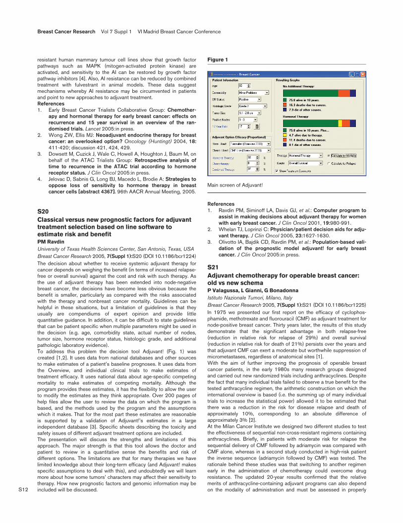
S12
resistant human mammary tumour cell lines show that growth factorpathways such as MAPK (mitogen-activated protein kinase) areactivated, and sensitivity to the AI can be restored by growth factorpathway inhibitors [4]. Also, AI resistance can be reduced by combinedtreatment with fulvestrant in animal models. These data suggestmechanisms whereby AI resistance may be circumvented in patientsand point to new approaches to adjuvant treatment.References1. Early Breast Cancer Trialists Collaborative Group: Chemother-
apy and hormonal therapy for early breast cancer: effects onrecurrence and 15 year survival in an overview of the ran-domised trials. Lancet 2005:in press.
2. Wong ZW, Ellis MJ: Neoadjuvant endocrine therapy for breastcancer: an overlooked option? Oncology (Huntingt) 2004, 18:411-420; discussion 421, 424, 429.
3. Dowsett M, Cuzick J, Wale C, Howell A, Houghton J, Baum M, onbehalf of the ATAC Trialists Group: Retrospective analysis oftime to recurrence in the ATAC trial according to hormonereceptor status. J Clin Oncol 2005:in press.
4. Jelovac D, Sabnis G, Long BJ, Macedo L, Brodie A: Strategies tooppose loss of sensitivity to hormone therapy in breastcancer cells [abstract 4367]. 96th AACR Annual Meeting, 2005.
S20Classical versus new prognostic factors for adjuvanttreatment selection based on line software toestimate risk and benefitPM RavdinUniversity of Texas Health Sciences Center, San Antonio, Texas, USABreast Cancer Research 2005, 7(Suppl 1):S20 (DOI 10.1186/bcr1224)The decision about whether to receive systemic adjuvant therapy forcancer depends on weighing the benefit (in terms of increased relapse-free or overall survival) against the cost and risk with such therapy. Asthe use of adjuvant therapy has been extended into node-negativebreast cancer, the decisions have become less obvious because thebenefit is smaller, particularly as compared with the risks associatedwith the therapy and nonbreast cancer mortality. Guidelines can behelpful in these situations, but a limitation of guidelines is that theyusually are compendiums of expert opinion and provide littlequantitative guidance. In addition, it can be difficult to state guidelinesthat can be patient specific when multiple parameters might be used inthe decision (e.g. age, comorbidity state, actual number of nodes,tumor size, hormone receptor status, histologic grade, and additionalpathologic laboratory evidence).To address this problem the decision tool Adjuvant! (Fig. 1) wascreated [1,2]. It uses data from national databases and other sourcesto make estimates of a patient’s baseline prognoses. It uses data fromthe Overview, and individual clinical trials to make estimates oftreatment efficacy. It uses national data about age-specific competingmortality to make estimates of competing mortality. Although theprogram provides these estimates, it has the flexibility to allow the userto modify the estimates as they think appropriate. Over 200 pages ofhelp files allow the user to review the data on which the program isbased, and the methods used by the program and the assumptionswhich it makes. That for the most part these estimates are reasonableis supported by a validation of Adjuvant!’s estimates in a largeindependent database [3]. Specific sheets describing the toxicity andsafety issues of different adjuvant treatment options are included.The presentation will discuss the strengths and limitations of thisapproach. The major strength is that this tool allows the doctor andpatient to review in a quantitative sense the benefits and risk ofdifferent options. The limitations are that for many therapies we havelimited knowledge about their long-term efficacy (and Adjuvant! makesspecific assumptions to deal with this), and undoubtedly we will learnmore about how some tumors’ characters may affect their sensitivity totherapy. How new prognostic factors and genomic information may beincluded will be discussed.
References1. Ravdin PM, Siminoff LA, Davis GJ, et al.: Computer program to
assist in making decisions about adjuvant therapy for womenwith early breast cancer. J Clin Oncol 2001, 19:980-991.
2. Whelan TJ, Loprinzi C: Physician/patient decision aids for adju-vant therapy. J Clin Oncol 2005, 23:1627-1630.
3. Olivotto IA, Bajdik CD, Ravdin PM, et al.: Population-based vali-dation of the prognostic model adjuvant! for early breastcancer. J Clin Oncol 2005:in press.
S21Adjuvant chemotherapy for operable breast cancer:old vs new schemaP Valagussa, L Gianni, G BonadonnaIstituto Nazionale Tumori, Milano, ItalyBreast Cancer Research 2005, 7(Suppl 1):S21 (DOI 10.1186/bcr1225)In 1975 we presented our first report on the efficacy of cyclophos-phamide, methotrexate and fluorouracil (CMF) as adjuvant treatment fornode-positive breast cancer. Thirty years later, the results of this studydemonstrate that the significant advantage in both relapse-free(reduction in relative risk for relapse of 29%) and overall survival(reduction in relative risk for death of 21%) persists over the years andthat adjuvant CMF can exert a moderate but worthwhile suppression ofmicrometastases, regardless of anatomical sites [1].With the aim of further improving the prognosis of operable breastcancer patients, in the early 1980s many research groups designedand carried out new randomized trials including anthracyclines. Despitethe fact that many individual trials failed to observe a true benefit for thetested anthracycline regimen, the arithmetic construction on which theinternational overview is based (i.e. the summing up of many individualtrials to increase the statistical power) allowed it to be estimated thatthere was a reduction in the risk for disease relapse and death ofapproximately 10%, corresponding to an absolute difference ofapproximately 3% [2].At the Milan Cancer Institute we designed two different studies to testthe effectiveness of sequential non-cross-resistant regimens containinganthracyclines. Briefly, in patients with moderate risk for relapse thesequential delivery of CMF followed by adriamycin was compared withCMF alone, whereas in a second study conducted in high-risk patientthe inverse sequence (adriamycin followed by CMF) was tested. Therationale behind these studies was that switching to another regimenearly in the administration of chemotherapy could overcome drugresistance. The updated 20-year results confirmed that the relativemerits of anthracycline-containing adjuvant programs can also dependon the modality of administration and must be assessed in properly
Breast Cancer Research Vol 7 Suppl 1 VI Madrid Breast Cancer Conference
Figure 1
Main screen of Adjuvant!
S13
designed trials in which the magnitude of the benefits can be weightedagainst potential risks [3].The treatment results observed after sequential adriamycin followed byCMF in a poor risk subset could probably be explained by an increaseddensity of the anthracycline, which was delivered at full dose within thefirst 9 weeks of treatment. The value of dose density was recentlyconfirmed by the National Cancer Institute’s Breast Intergroup INTC9741 trial, in which patients who received the dose-dense regimenshad significantly improved early treatment outcome compared with theircounterparts who did not receive these regimens.The role of sequential non-cross-resistant regimens was tested in manyother trials, both in the adjuvant and neoadjuvant settings. Of note, the3-year joint efficacy analysis of the National Epirubicin Adjuvant Trial(NEAT) and of the Scottish Cancer Trials Breast Group reported ahighly significant benefit in favor of the sequential regimen, supportingthe hypothesis that the sequential administration of single-agentanthracyclines given upfront before CMF can indeed improve treatmentoutcome. Also, the addition of taxanes after delivery of adriamycin andcyclophosphamide contributed to improving therapeutic results over thenontaxane regimen both in the adjuvant and neoadjuvant settings. Bycontrast, in the INT C9741 trial no difference was detected in treatmentoutcome between the concurrent or sequential schedules of adriamycin,cyclophosphamide and paclitaxel. Similarly, the MD Andersonrandomized study failed to detect superiority of the sequential arm.Thirty years ago, treating patients who were free of identifiablemetastatic disease with systemic adjuvant therapy because some ofthem might eventually develop distant disease was a revolutionarydeparture from prior treatment approaches [4]. It has been estimatedthat improvements since the 1970s in the way in which breast canceris managed must have prevented about 25–30% of the breast cancerdeaths in middle-aged women that would otherwise have occurred inthe year 2000.References1. Bonadonna G, Molterni A, Zambetti M, Daidone MG, Pilotti S,
Gianni L, Valagussa P: 30 years’ follow up of randomizedstudies of adjuvant CMF in operable breast cancer: cohortstudy. BMJ 2005, 330:217-220.
2. Early Breast Cancer Trialists’ Collaborative Group. Poly-chemotherapy for early breast cancer: an overview of ran-domised trials. Lancet 1998, 352:930-942.
3. Bonadonna G, Zambetti M, Gianni L, Valagussa P: Clinical rele-vance of different sequencing of doxorubicin and cyclophos-phamide, methotrexate, and fluorouracil operable breastcancer. J Clin Oncol 2004, 22:1614-1620.
4. Fisher B: The evolution of paradigms for the management ofbreast cancer: a personal perspective. Cancer Res 1992,52:2371-2383.
Controversial panel discussion
S22Prognostic studies in patients with high-risk primarybreast cancer (HRPBC) receiving high-dosechemotherapy (HDC)Y NietoClínica Universitaria de Navarra, Pamplona, SpainBreast Cancer Research 2005, 7(Suppl 1):S22 (DOI 10.1186/bcr1226)Introduction After standard-dose chemotherapy (SDC), more than50% of patients with HRPBC (defined as extensive axillary nodeinvolvement or inflammatory disease) experience relapse. Controversyhas surrounded the use of HDC for HRPBC for over a decade. Currentresults from at least 15 randomized trials comparing diverse forms ofHDC and SDC for HRPBC appear contradictory at this point [1]. Inaddition, some studies suggest that younger women might benefitselectively from HDC. An NCI-sponsored meta-analysis is in progress.Regardless of this unsettled issue, it is clear that a substantialpercentage of HRPBC patients still relapse after HDC. Extensiveprognostic studies are required to improve their outcome. Suchinvestigations may allow us to identify adequate patient subsets and
new therapeutic targets for trials of developmental therapeutics.Combinations of HDC, capable of ample cytoreduction, with novelagents that target specific tumor features responsible for post-transplant relapse may hold promise.Methods We analyzed clinical variables in HRPBC patients enrolled atthe University of Colorado in clinical trials of HDC targeting 4–9+, ≥10+nodes, or inflammatory disease. First, we developed a prognostic modelamong 176 patients treated from 1990 to 1997, and validated it in anexternal sample. Subsequently, the model was validated prospectively ina second cohort of 88 patients treated at Colorado since 1997.We hypothesized that intrinsic biologic differences, insurmountable byHDC, existed between the two risk categories identified by the clinicalmodel. Through immunohistochemical analyses of paraffin-embeddedtumor blocks collected from the referring institutions, we studied aseries of putative molecular candidates, related to signal transductionpathways or an angiogenic phenotype, which could be responsible, atleast in part, for those differences.Results At median follow up of more than 7 years, the relapse-freesurvival (RFS) and overall survival (OS) rates for the whole group of264 patients treated at Colorado were 69.8% and 73%, respectively.The median time to relapse was 14 months (63.5% relapses within thefirst 2 years, 6.7% after the 5th year).We identified three clinical variables independently associated withoutcome: nodal ratio (number of involved nodes /number of dissectednodes), pathological tumor size, and hormone receptors [2]. A scoringsystem was constructed with those variables: score = (nodal ratio ×3.05) + (tumor size × 0.15) – (ER/PR × 1.15). In this formula, size isentered in cm, and ER/PR is assigned ‘1’ if positive (estrogen receptor[ER] and/or progesterone receptor [PR] positive), or '0' if negative(both negative). A cutoff score of 2.41 yields the best sensitivity andspecificity. Thus, patients with low (<2.41) and high (≥2.41) scoresbefore transplant presented significant differences in outcome. Thismodel was validated in an external sample of 225 HRPBC patientstreated at Duke University with the same HDC. It was subsequentlyvalidated prospectively in our second patient cohort [3].Overexpression of HER2, identified as an independent predictor ofoutcome, complemented the clinical model, establishing the followingrisk groups: low risk (low score, HER2-negative; 44% patients; 87%RFS), intermediate risk (low score, HER2-positive; 29% patients; 68%RFS), high risk (high score, any HER2; 27% patients; 49% RFS) [4].We detected an independent prognostic effect of EGFR (epidermalgrowth factor receptor), particularly among HER2-positive patients [5],which suggests a synergistic effect through heterodimerization of bothreceptors. In contrast, we did not observe a prognostic effect of p53status [4].Tumor angiogenesis, assessed through CD31-stained microvessel count,was an independent adverse predictor of outcome [6]. In contrast, tumorVEGF (vascular endothelial growth factor) expression lacked prognosticsignificance in our population with locoregionally advanced tumors, incontrast to multiple prior observations in patients with earlier disease [6].Finally, we observed that the presence of tumor cells contaminating theapheresis product, detected through immunocytochemistry for cyto-keratins, was independently associated with post-transplant relapse [7].Conclusions We can now predict which HRPBC patients are most likelyto remain long-term disease free after HDC. Additionally, we identifiedimportant prognostic molecular markers that could constitute relevanttargets for studies combining novel therapeutics with HDC in HRPBC.Acknowledgments Supported by grants 1 R21 CA095762-01 fromthe National Cancer Institute and an American Cancer Society/University of Colorado Cancer Center Research Seed Grant.References1. Nieto Y, Jones RB, Shpall EJ: High-dose chemotherapy for breast
cancer: Is another look warranted? Curr Opin Oncol 2004, 16:114-119.
2. Nieto Y, Cagnoni PJ, Xu X, et al.: Predictive model for relapseafter high-dose chemotherapy with peripheral blood progeni-tor cell support for high-risk primary breast cancer. ClinCancer Res 1999, 5:3425-3431.
3. Nieto Y, Nawaz S, Shpall EJ, et al.: Long-term analysis and
Available online http://breast-cancer-research.com/supplements/7/S1
S14
prospective validation of a prognostic model for patients withhigh-risk primary breast cancer receiving high-dosechemotherapy. Clin Cancer Res 2004, 10:2609-2617.
4. Nieto Y, Cagnoni PJ, Nawaz S, et al.: Evaluation of the predic-tive value of HER2/neu overexpression and p53 mutations inhigh-risk primary breast cancer patients treated with high-dose chemotherapy and autologous stem-cell transplanta-tion. J Clin Oncol 2000, 18:2070-2080.
5. Nieto Y, Nawaz S, Bearman SI, et al.: Overexpression of Epider-mal Growth Factor Receptor (EGFR) adds prognostic value toHER2 status in patients (pts) with high-risk primary breastcancer (HRPBC). Proc Am Soc Clin Oncol 2003, 22:15a.
6. Nieto Y, Woods J, Jones RB, et al.: Prognostic analysis of intra-tumor microvessel density (MVD) and Vascular EndothelialGrowth Factor (VEGF) in high-risk primary breast cancer(HRPBC) patients (PTS) receiving high-dose chemotherapy. JClin Oncol 2005, 23:27s.
7. Nieto Y, Franklin W, Jones RB, et al.: Prognostic significance ofoccult tumor cells in the apheresis products of patients withadvanced breast cancer receiving high-dose chemotherapyand autologous hematopoietic progenitor cell support. BiolBlood Bone Marrow Transplant 2004, 10:415-425.
S23Efficacy of high-dose alkylating chemotherapy in theadjuvant treatment of HER2/neu-negative primarybreast cancer: update of the Dutch randomized trialS Rodenhuis1, M Bontenbal2, LVAM Beex3, WM Smit4, MA Nooij5, EE Voest6, E van der Wall7, P Hupperets8, Harm van Tinteren1, HL Peterse1, MJ van de Vijver1, EGE de Vries9, for the Netherlands Working Party onAutologous Transplantation in Solid Tumors1Netherlands Cancer Institute, Amsterdam, The Netherlands; 2ErasmusMedical Center/Daniel den Hoed Cancer Center, Rotterdam, TheNetherlands; 3University Hospital Nijmegen, The Netherlands;4Medical Hospital Twente, Enschede, The Netherlands; 5UniversityMedical Center, Leiden, The Netherlands; 6University Medical Center,Utrecht, The Netherlands; 7Free University Hospital, Amsterdam, TheNetherlands; 8University Hospital, Maastricht, The Netherlands;9University Hospital Groningen, Groningen, The NetherlandsBreast Cancer Research 2005, 7(Suppl 1):S23 (DOI 10.1186/bcr1227)Introduction The role of high-dose chemotherapy in the adjuvanttreatment of high-dose breast cancer has not been established.Results have been reported from six randomized studies with asymmetrical study design (Table 1). All show a lower relapse rate in thehigh-dose arm, but in only one study was this result statisticallysignificant.Methods Patients below 56 years of age who had undergone surgeryfor stage II or III breast cancer were eligible if they had at least four
tumor-positive axillary lymph nodes. Patients in the conventional dose(CD) arm received five courses of FEC (fluorouracil 500 mg/m2,epirubicin 90 mg/m2 and cyclophosphamide 500 mg/m2; every3 weeks) followed by radiation therapy and tamoxifen. The high-dose(HD) arm was identical, except that high-dose chemotherapy (CTC[cyclophosphamide 6 g/m2, thiotepa 480 mg/m2 and carboplatin1600 mg/m2]) with peripheral blood progenitor cell reinfusion wasgiven instead of the fifth FEC course.Results Between August 1993 and July 1999, 885 patients withprimary breast cancer and four or more tumor-positive lymph nodeswere randomized in 10 Dutch centers in a study of high-dosechemotherapy. The results of this study at 57 months of follow-up havenow been updated at 87 months. In a pathology review, 621 tumorsamples were shown to be HER2/neu-negative (either 0+ atimmunohistochemistry or negative at in situ hybridization). Patients withHER2/neu-negative disease had a 5-year RFS of 72% following HD andof 59% after CD (P = 0.002). Overall survival in the HD group was 78%at 5 years versus 71% for the CD group (P = 0.02). Young age and lowmalignancy grade were associated with a relative benefit for HD (testsfor interactions: P = 0.04 and P = 0.0057, respectively). The treatment-related mortality in the high-dose chemotherapy arm was 1%. An equalnumber of second malignancies were observed in both arms.Conclusion Although the subgroup analysis of HER2/neu-negativedisease was not planned in the original protocol, these findings areconsistent with findings from other studies [4]. The marked efficacy ofHD therapy in HER2/neu-negative breast cancer may have beenmasked in this and in other studies by its disadvantage in theHER2/neu-positive group, which may have benefited from a higherdose of anthracycline-dose in the control arm. High-dose alkylatingchemotherapy is a viable option for high-risk breast cancer patientswith HER2/neu-negative disease.References1. Rodenhuis S, Bontenbal M, Beex LV, et al.: High-dose
chemotherapy with hematopoietic stem-cell rescue for high-risk breast cancer. N Engl J Med 2003, 349:7-16.
2. Peters WP, Rosner GL, Vredenburgh JJ, et al.: Prospective, ran-domized comparison of high-dose chemotherapy with stem-cell support versus intermediate-dose chemotherapy aftersurgery and adjuvant chemotherapy in women with high-riskprimary breast cancer: a report of CALGB 9082, SWOG 9114,and NCIC MA-13. J Clin Oncol 2005, 23:2191-2200.
3. Tallman MS, Gray R, Robert NJ, et al.: Conventional adjuvantchemotherapy with or without high-dose chemotherapy andautologous stem-cell transplantation in high-risk breastcancer. N Engl J Med 2003, 349:17-26.
4. Nieto Y, Nawaz S, Jones RB, et al.: Prognostic model forrelapse after high-dose chemotherapy with autologous stem-cell transplantation for stage IV oligometastatic breast cancer.J Clin Oncol 2002, 20:707-718.
Breast Cancer Research Vol 7 Suppl 1 VI Madrid Breast Cancer Conference
Table 1 (abstract S23)
Randomized studies evaluating the role of high-dose chemotherapy in high-risk breast cancer
Author Patients Selection Conventional arm High-dose arm RFS analysis
Rodenhuis [1] 885 4+ nodes 5×FEC 4×FEC – CTC HD better (P = 0.08)
Peters [2] 785 10+ nodes 4×CAF + ID-CPB 4×CAF + HD-CPB No difference, HD fewer relapses
Tallman [3] 540 10+ nodes 6×CAF 6×CAF + CT No difference, HD fewer relapses
Roché 314 8+ nodes 4×FEC 4×FEC + CMA HD better (P = 0.002)
Tokuda 97 10+ nodes 6×CAF 6×CAF + CT HD better (NS)
Rodenhuis 81 Infraclav biopsy 4×FEC 4×FEC + CTC No difference, HD less relapses
CAF, cyclophosphamide, doxorubicin, fluorouracil; CMA, cyclophosphamide, mitoxantrone and melphalan; CT, cyclophosphamide and thiotepa; CTC,cyclophosphamide, thiotepa and carboplatin; FEC, fluorouracil, epirubicin, cyclophosphamide; nodes, tumor-positive axillary lymph nodes; HD-CPB, high-dosecyclophosphamide, cisplatin and BCNU; ID-CPB, intermediate-dose cyclophosphamide, cisplatin and BCNU.

S15
S24SOLTI (Solid Tumor Intensification) Group experiencewith high-dose chemotherapy treatment for earlybreast carcinomaH Cortés Funes1, A Lluch2, M Climent3, JJ López4, B Ojeda4, J Hornedo1, E Ciruelos1, J Baselga5, on behalf of the SOLTIGroup1Medical Oncology Dpt.,Hospital Universitario 12 de Octubre, Madrid,Spain; 2Medical Oncology Dpt., Hospital Clínico Universitario,Valencia, Spain; 3Medical Oncology Dpt., Instituto Valenciano deOncología, Valencia, Spain; 4Medical Oncology Dpt., Hospital SantaCreu i Sant Pau, Barcelona, Spain; 5Medical Oncology Dpt., HospitalUniversitario Vall d´Hebron, Barcelona, Spain.Breast Cancer Research 2005, 7(Suppl 1):S24 (DOI 10.1186/bcr1228)Introduction Primary carcinoma of the breast is a worldwide public-health problem; despite conventional treatment, long-term prognosis ispoor, especially for large tumors or in cases with axillary involvement. Inan attempt to improve these results, phase II high-dose chemotherapytrials were performed by the SOLTI Group.Method A total of 416 patients were included in three high-dosechemotherapy trials in the adjuvant setting, as follows. The 9301 trialincluded 297 patients with stage II/III breast carcinoma with more than10 axillary lymph nodes involved. After conventional adjuvant chemo-therapy (FEC regimen), high-dose chemotherapy (STAMP V regimen)with peripheral stem cell support was performed. The 9302 trialincluded 66 patients with inflamatory breast carcinoma treated withthree to six cycles of FEC neoadjuvant therapy. Responding patientswere treated with high-dose chemotherapy after surgery with STAMP Vregimen and blood stem cell support. The 9702 trial included 53patients with stage III breast cancer treated with neoadjuvantdoxorubicin and paclitaxel. Patients with pathologic axillary involvementin the surgical specimen were treated with adjuvant STAMP V high-dose chemotherapy.Results In trial 9301, with a median follow up of 63 months, 5-yeardisease-free survival (DFS) was 59% and overall survival (OS) was80%. In trial 9302, median DFS was 30 months and median OS 75months, with 55.3% of patients alive at 5 years. In trial 9702, with amedian follow up of 31 months, the median DFS and OS have not yetbeen determined. No toxic deaths were reported. Most commonnonhematological toxicities were emesis, mucositis, hepatic andalopecia. Neutropenia was easily resolved with G-CSF support; andanemia and trombopenia were frequent (50–60% patients).Conclusion The toxicity of treatment with high-dose chemotherapy isacceptable and similar to that described in other series. Although theresults obtained are promising, their comparison with historic controlsand the information derived from other reported trials do not enable usto recommend high-dose chemotherapy with bone-marrow rescue as aroutine treatment in high-risk breast cancer. It nevertheless remains avalid investigational strategy.
Symposium VI: Molecular targeted agents
Symposium VII: Advances in metastatic disease
S25Oral vinorelbine in metastatic breast cancerPF Conte, S GiovannelliDivision of Medical Oncology, Department of Oncology andHaematology, University Hospital, University of Modena and ReggioEmilia, Modena, ItalyBreast Cancer Research 2005, 7(Suppl 1):S25 (DOI 10.1186/bcr1229)Introduction A new oral formulation of vinorelbine has been introducedin clinical studies since 1994, following increasing interest in thedevelopment of oral chemotherapy, driven by pharmacoeconomicissues, patient convenience and the potential for improved quality oflife. A dose-finding study [1] established that 100 mg/m2 was the MTDdose, limiting toxicities being neutropenia, nausea and vomiting, andneuroconstipation; the recommended dose was then defined at
80 mg/m2 per week. The first phase II studies conducted in chemo-therapy-naive NSCLC and as first-line chemotherapy in advancedbreast cancer (ABC) showed an excessive rate of complicatedneutropenia. This led to the formulation of a new schedule in which alower dose of 60 mg/m2 per week was administered for the first threecourses with escalation to 80 mg/m2, with a safety profile qualitativelycomparable to that of intravenous vinorelbine at standard doses [2].Equivalent blood concentrations were demonstrated between80 mg/m2 oral and 30 mg/m2 intravenous, and between 60 mg/m2 oraland 25 mg/m2 intravenous [3].Studies in metastatic breast cancer (MBC): single agent Amulticenter phase II trial assessed the activity, safety andpharmacokinetic profile of oral navelbine in ABC. Sixty-four patientswere entered to receive oral NVB on a weekly basis for a total of8 weeks unless progression or toxicity occurred. Oral vinorelbine wasgiven at 60 mg/m2 weekly for the first three administrations and wasincreased to 80 mg/m2 for the subsequent administrations if there wasno grade 4 neutropenia or no more than one episode of grade 3neutropenia. Patients with objective response or stable diseasecontinued treatment up to a total of 12 weeks or more. Fifty-eightevaluable patients were included. Four patients (6.9%) had completeresponses and 14 (24.1%) had partial responses, for an overallresponse rate of 31% (95% confidence interval 19–43%). The medianprogression-free survival was 17.4 weeks, and the median overallsurvival was 22.9 months. There were no treatment-related deaths. Themain toxicity was neutropenia: grade 4 in 17.2% of the patients, and1.8% of administrations and associated clinical serious events in fourpatients (6.2%). Grade 3 and 4 nausea and/or vomiting were noted in3.1% and 4.6% of the patients, respectively. Only one patientdeveloped grade 3 neuroconstipation. An analysis of Quality of LifeQuestionnaire C30 forms revealed no significant alteration betweenbaseline and weeks 8 and 16 in global quality of life. Oral navelbine assingle agent in first-line MBC has the same efficacy of intravenousvinorelbine in phase II studies in terms of OR, duration of response,progression-free survival and overall survival, and is well tolerated witha manageable gastrointestinal toxicity (8% of G3-4 N/V withoutprophylactic antiemetic treatment) [5].A second phase II trial is still ongoing. An interim analysis on the first72 patients (median age 63 years) showed a similar toxicity profile witha RR of 30%, a median progression free-survival of 4.6 months and amedian survival of 20.7 months.Studies in MBC: combinations The increasing prevalence ofantracycline and taxane treatment in adjuvant setting led to anexploration of new combinations of non-cross-resistant therapies, inparticular for those patients for which a polychemotherapy might offergreater benefits than single agents. In this setting the results ofintravenous navelbine in combination with other drugs suggested newmodels in which to introduce the oral formulation.Several phase II studies had investigated intravenous vinorelbine inassociation with capecitabine with a RR in second line >50% and witha mild toxicity. Only one phase II trial with this combination has beenconducted as first-line chemotherapy, and it confirmed a good toxicityprofile. The availability of oral formulations of both of these drugs led toinvestigation of their attractive combination, and preliminary results arenow available from an ongoing phase II trial of vinorelbine oral pluscapecitabine ± trastuzumab in MBC as first-line chemotherapy.Capecitabine is administered at the dose of 2000 mg/m2 per day givendays 1 to 14, and vinorelbine at the dose of 60 mg/m2 on days 1 and 8every 3 weeks for the first course, then escalated to 80 mg/m2 from thesecond course. After 81 courses the incidence of G3 nausea/vomitingwas 2.5%, G3 diarrhea 3.7%, grade 3 HFS 1.2%, and G3/4neutropenia 13.5%. After 2 cycles the combination has revealed 4/16CR-PR, 10/16 SD and 1/16 PD; and after 4 cycles 7/15 CR-PR and7/15 SD. From these preliminary results this combination seems to beeffective in the treatment of MBC previously treated with ananthracycline and/or a taxane with low toxicity.Several studies have investigated the combination of intravenousnavelbine and trastuzumab with an OR of 61–75%, demonstrating thatthis can be an effective and well tolerated option for HER2+ MBC. It is
Available online http://breast-cancer-research.com/supplements/7/S1
S16
an important regimen, given the increasing prevalence of antracyclineand taxane treatment in adjuvant setting.Other sequential combinations of oral vinorelbine with paclitaxel ordocetaxel or epirubicin are being explored, suggesting a possible rolefor the oral formulation in a sequential prolonged treatment in MBC.References1. Bonneterre J, Senac I, Variol P, Daniel P: Dose finding study of
weekly oral vinorelbine in patients with advanced breastcancer. Institute de Recherche Pierre Fabre, Castres, France. Iter-national study report PM 259 IN M 156.
2. Depierre A, Freyer G, Jassem J, et al.: Oral vinorelbine:feasibilityand safety profile. Ann Oncol 2001, 12:1677-1681.
3. Variol P, Nguyen L, Tranchand B, et al.: A simultaneousoral/intravenous population pharmacokinetic model forvinorelbine. Eur J Clin Pharmacol 2002, 58:467-476.
4. Marty M, Fumoleau P, Adenis A, et al.: Oral vinorelbine pharma-cokinetics and absolute biovailibility study in patients withsolid tumors. Ann Oncol 2001, 12:1643-1649.
5. Freyer G, delozier T, Lichinister M, et al.: Phase II study of oralvinorelbine in first-line advanced breast cancer chemother-apy. J Clin Oncol 2003, 21:35-40.
6. Anh JH, Kim SB, Lee JS, et al.: Capecitabine and vinorelbine inpatients with metastatic breast cancer previously treated withanthracycline and taxane. J Korean Med Sci 2004, 19:547-553.
7. Ghosn M, Kattan J, Farhat F, et al.: Navelbine capecitabine com-bination: the new first line chemotherapy regimen for meta-static breast cancer. Breast Cancer Res Treat 2002, Suppl1:531.
S26The role of Xeloda in metastasic breast cancerH Cortés-FunesHospital Universitario ‘12 de Octubre’, Jefe de Servicio de Oncología,Madrid, SpainBreast Cancer Research 2005, 7(Suppl 1):S26 (DOI 10.1186/bcr1230)Currently, breast cancer is the primary reason for death due to cancerin Spanish women aged between 35 and 64 years. Nevertheless,despite detecting an increased incidence, it is observed a decrease onmortality rates, in agreement with it is observed in other Europeancountries. This issue is basically due to the development of programsto sift and the use of widespread systemic treatments, both on earlyand advanced stages of the disease. During the past decade breastcancer has benefited from incorporation of new antitumor drugs totreat this kind of cancer. These new drugs are able to control, in aneffective way, the progress of metastatic disease and slow down oreradicate micrometastasis in early stages.Patients with metastatic disease have been useful in the study of newactive agents and their combinations subsequently used in adjuvantregimens. Breast cancer treatment often achieves important objectiveresponses with different durations of treatment, but these durationshave an impact on patient survival. The median of survival in this groupvaries between 18 and 24 months, and the main objective for thetreatment of metastatic breast cancer is palliation by means of thecontrol of disease-related symptoms, with an adequate profile oftoxicity, which provides an improvement in quality of life.There are currently several lines of work on the development of breastcancer treatments at different stages. These are expected to improvesurvival and quality of life parameters. These lines of developmentrange from the incorporation of new active agents (cytotoxic andhormonal) to optimization of current schemes with active agents (oldand new), by means of new combinations used for metastatic diseaseand use in early stages, even before surgery.Breast cancer is one of the most chemo sensible solid tumours, thatrespond to almost every cytotoxic drug used alone or in combination.These active agents include alkylating drugs (e.g. cyclophosphamideand the cisplatins) and antimetabolites (e.g. fluorouracil andmethotrexate), which were used for the design of the first-linepolychemotherapy schemes in solid tumours such as CMF. After this,
the anthracyclines (doxorubicin and epirubicin) were substituted formethotrexate in the latter combination, giving us the FAC and FECschemes, which were classic treatments until relatively recently. Duringthe past decade, many new agents have been incorporated that haveimproved both response rates and patient survival, such as the taxanes(paclitaxel and docetaxel), vinorelbine, caelyx and, recently,gemcitabine; there have been used in new combinations or have evenbeen used as isolated agents as second- and third-line treatments forwith advanced disease.The main problem with these agents and their combinations is thecomplexity of administration, at day hospitals and through intravenousinjections, which seriously impairs patient quality of life. However, thebenefit in terms of survival parameters and symptom reduction offsetthese difficulties.Capecitabine (Xeloda), one of the most recently introduced activeagents into treatment for metastatic breast cancer, has the sameantitumoral activity as current agents but without many of theirinconveniences, which is the reason why its incorporation into standardtreatment for breast cancer patients is becoming increasingly common.It is orally administered, avoiding the difficulties associated withintravenous injection. Its mode of action is similar to that of 5-fluorouracyl in continuous infusion, without the need for infuser orcentral catheters. On the other hand, it has a synergistic action with themajority of cytotoxic drugs, in particular with docetaxel, leading to abetter survival rates. The results of studies conducted in recent yearshave confirmed the important role of capecitabine in the treatment ofadvanced disease, and it have been the base for the studies withcapecitabine on the adjuvant and neoadjuvant setting.
S27Targeting the right chemotherapy for the right patientPF Conte, V GuarneriDepartment of Oncology and Hematology, University of Modena andReggio Emilia, Modena, ItalyBreast Cancer Research 2005, 7(Suppl 1):S27 (DOI 10.1186/bcr1231)In the past few years breast cancer mortality has been declining inmost Western countries as a consequence of better education,implementation of screening programs and more effective therapies.However, a small proportion of patients are metastatic at initialdiagnosis (about 5–7%), and 25–30% of patients develop metastasesfollowing primary treatment. At this stage, the disease is consideredincurable, the median survival ranges from 2 to 4 years, and a limitedproportion of patients (about 20%) survive more than 5 years. In thisscenario, it is important to identify the aims of treatment on the basis ofindividual patient needs. Data from clinical trials, meta-analyses,databases of large institutions, and cancer registries indicate thatchemotherapy can prolong survival, and survival prolongation isassociated with the activity of drugs [1-5]. Moreover, those patientswho achieve a complete response have about 20% chance of survivingbeyond 5 years [6]. Finally, although there are few trials specificallyaddressing symptomatic control and quality of life, it is generally agreedthat tumor shrinkage is associated with better control of symptoms,and that quality of life results from the balance between activity andtolerability of treatments.Metastatic breast cancer patients represent a very heterogeneouspopulation, and several factors are important in determining prognosis:patient characteristics such as motivation, compliance with treatment,age, performance status and comorbidities; tumor characteristics suchas hormonal receptor status and expression of HER2-neu; prioradjuvant therapies and disease-free interval; and site and extension ofmetastatic spread.Treatment options include locoregional treatments, endocrine agents,monoclonal antibodies, bisphosphonates and cytotoxic agents. Theaims of these treatments include symptomatic control, maintenance ofquality of life, tumor shrinkage, prolonging time to progression, andsurvival prolongation.Although the role of radiation therapy in controlling locoregionalrelapses, and of endocrine therapy in the upfront treatment of
Breast Cancer Research Vol 7 Suppl 1 VI Madrid Breast Cancer Conference

S17
endocrine-sensitive tumors is undebatable, the optimal use of cytotoxicchemotherapy remains controversial. The main reasons for the lack ofgeneral consensus are the heterogeneity of the patient population andthe availability of several effective options. In the following paragraphswe discuss the different treatment options in the most common clinicalscenarios.Treatment options based on patient characteristics The majority ofmetastatic breast cancer patients are over 65 years of age, andtherefore a significant proportion are affected by comorbidities such ashypertension, diabetes and respiratory disease [7]. Tolerability ofchemotherapy can be profoundly influenced by these comorbidities aswell as by their specific treatments.In the case of elderly patients with significant comorbidities anddeclining general condition, the aim of treatment is symptomatic controlwith improvement in quality of life (QoL). To date, the availability of oralagents such as capecitabine and navelbine allows for a good balancebetween activity, tolerability, compliance and dosage flexibility. Further-more, weekly administration allows maintenance of the activity ofimportant cytotoxic agents such as taxanes and platinum salts withsubstantial decrease in toxicity. Finally, new antimetabolites(gemcitabine) or new formulations of old drugs (liposome-encapsulateddoxorubicin) are generally associated with reduced toxicity. For thesepatients it is clear that the best choice is the use of single agents thatcan be sequentially administered on the basis of tolerability anddisease control.About 20% of patients present with locoregional relapses (regionallymph nodes, skin metastases). Moreover more sensitive imagingtechniques (Multislice computed tomography [CT], magnetic resonanceimaging [MRI], positron emission tomography [PET] scan) and thedevelopment of methods to detect micrometastatic disease (i.e.circulating tumor cells) permit diagnosis of an increasing proportion ofpatients with oligometastatic disease. A reasonable percentage ofthese patients can be cured or at least rendered disease free forprolonged period of time; a chemotherapy regimen to induce rapid andimportant tumor shrinkage followed by locoregional treatments(radiotherapy, surgery, radiofrequency ablation) is necessary to achievethis goal. In this setting, it is clear that combination regimens should bethe preferred option.Another clinical scenario is represented by younger patients, withoutcomorbid conditions, and massive visceral involvement. Here again,rapid tumor shrinkage is important in preventing life-threatening organfailure. In this setting, combination regimens ensure a higher percentageof objective responses [8,9] and a shorter time to response [10].Treatment options based on tumor characteristics Breast cancertumors are at least as heterogeneous as breast cancer patients.However, the only tumor-related parameters currently utilized in thedecision making process are the disease course in the individualpatient, hormone receptor status, and HER2-neu overexpression.In the case of patients with slowly-growing hormone receptor-negativetumors and predominant bone disease, main aims of treatment aremaintenance of QoL and prolonging time to progression. In thesecases, single-agent chemotherapy provides a better balance betweenactivity and tolerability. More frequently, patients with hormone-sensitive tumors receive several lines of endocrine therapy untildevelopment of hormone resistance. Here again, when chemotherapyis required, the preferred choice is the sequential administration ofsingle agents.Another important tumor characteristic is the HER2-neu status; HER2-neu is overexpressed in 25–30% of breast cancers. As a single agenttrastuzumab, a monoclonal antibody directed against the extra-membrane portion of HER2 receptor, can induce a 30% response ratein HER2-overexpressing tumors. The addition of trastuzumab tochemotherapy as compared with chemotherapy alone is associatedwith a significant improvement in objective response rate, duration ofresponse and overall survival [11].Several cytotoxic agents showed synergism or additive effect whencombined with trastuzumab; clinical trials have shown that trastuzumabcan successfully be combined with both single agent and combinationchemotherapy.
Treatment options based on prior adjuvant therapy Anthracyclinesrepresent the most active agents, and anthracycline-containingregimens are more effective in terms of response rates, completeremission rates, remission duration and survival. However, anthracyclineregimens are increasingly used in the adjuvant setting, and thereforeretreatment with anthracyclines, even if effective, is limited to patientsexposed to low cumulative anthracycline doses and with a relapse-freesurvival after adjuvant chemotherapy longer than 12 months. The mainlimitation to anthracyclines is their dose-dependent cardiac toxicity;patients should not exceed the cumulative dose of 450–550 mg/m2 fordoxorubicin and 800-900 mg/m2 for epirubicin. However, few data areavailable on the efficacy of anthracycline rechallenge after priorexposure to adjuvant anthracycline. A recent report from our group [12]has shown that anthracycline–taxane combinations as first-linetreatment for metastatic breast cancer are effective, regardless ofprevious adjuvant chemotherapy.As more and more patients are receiving taxanes as a component oftheir adjuvant program, it will be important also to have data on theefficacy of taxane rechallenge in metastatic patients.Conclusion Survival prolongation must be the primary goal oftreatment, and this aim can be achieved with the incorporation of newactive agents in the treatment strategy. Both combined and sequentialsingle agents are acceptable options; however, if not contraindicatedby the conditions of the patient and if feasible with an acceptabletoxicity profile, there is no reason to delay the upfront use of activeagents.References1. Fossati R, Confalonieri C, Torri V, et al.: Cytotoxic and hormonal
treatment for metastatic breast cancer: a systematic review ofpublished randomized trials involving 31,510 women. J ClinOncol 1998, 16:3439-3460.
2. Ghersi D, Wilcken N, Simes J, et al.: Taxane containing regi-mens for metastatic breast cancer. The Cochrane Library, Issue3, 2003.
3. Chia SKL, Speers C, Kang A, et al.: The impact of newchemotherapeutic and hormonal agents on the survival ofwomen with metastatic breast cancer (MBC) in a populationbased cohort [abstract]. Proc Am Soc Clin Oncol 2003, 22:22.
4. Andre F, Slimane K, Bachelot T, et al.: Breast cancer with syn-chronous metastases: trends in survival during a 14-yearperiod. J Clin Oncol 2004, 22:3302-3308.
5. Giordano SH, Buzdar AU, Smith TL, et al.: Is breast cancer sur-vival improving? Cancer 2004, 100:44-52.
6. Greenberg PA, Hortobagyi GN, Smith TL, et al.: Long termfollow up of patients with complete remission following com-bination chemotherapy for metastatic breast cancer. J ClinOncol 1996, 14:2197-2205.
7. Yancik R, Wesley M, Ries L, et al.: Effect of age and comorbidityin postmenopausal breast cancer patients. JAMA 2001,285:885-892.
8. Conte PF, Gennari A, Donati S, et al.: Gemcitabine plus epiru-bicin plus taxol (GET) in advanced breast cancer: a phase IIstudy. Breast Cancer Res Treat 2001, 68:171-179.
9. Cappuzzo F, Mazzoni F, Gennari A, et al.: Multicentric phase IItrial of gemcitabine plus epirubicin plus paclitaxel as first-linechemotherapy in metastatic breast cancer. Br J Cancer 2004,90:31-35.
10. Conte PF, Guarneri V, Bruzzi P, et al.: Concomitant versussequential administration of epirubicin and paclitaxel as first-line therapy in metastatic breast carcinoma: results for theGruppo Oncologico Nord Ovest randomized trial. Cancer2004, 101:704-712.
11. Slamon DJ, Leyland-Jones B, Shak S, et al.: Use of chemother-apy plus a monoclonal antibody against HER2 for metastaticbreast cancer that overexpresses HER2. N Engl J Med 2001,344:783-792.
12. Gennari A, Bruzzi P, Orlandini C, et al.: Activity of first line epiru-bicin and paclitaxel in metastatic breast cancer is independentfrom type of adjuvant therapy. Br J Cancer 2004, 90:962-967.
Available online http://breast-cancer-research.com/supplements/7/S1

S18
Symposium VIII: Other advances in metastaticdisease
S28Role of translational studies in optimizing palliativechemotherapyR KramerBreast Care Center, Baylor College of Medicine, Houston, Texas,USA, and Visiting Professor, Hospital Universitario ‘12 de Octubre’,Madrid, SpainBreast Cancer Research 2005, 7(Suppl 1):S28 (DOI 10.1186/bcr1232)Introduction Patients with metastatic breast cancer have a highlyvariable clinical course. Systemic chemotherapy may decreasesymptoms and prolong survival for some patients. However, othersexperience significant toxicity and achieve little benefit. Predictivemarkers are needed to determine which patients will benefit fromparticular chemotherapy agents and which should be offered noveltherapeutics.Methods The English literature related to predictive markers formetastatic cancer was reviewed. Translational studies examining thepotential role of tumor profiles in the selection of optimal chemotherapyagents were identified. Particular attention was paid to studiesexamining sensitivity and resistance to anthracyclines, taxanes,vinorelbine and capecitabine.Conclusion This paper will summarize the rapidly expanding datapublished to data and will discuss clinical trial designs that will facilitatefuture studies.
S29Letrozole efficacy is related to human aromataseCYP19 single nucleotide polymorphisms (SNPs) inmetastatic breast cancerR Colomer1,2, M Monzo3, I Tusquets4, J Rifa5, JM Baena6, A Barnadas7, L Calvo8, F Carabantes9, C Crespo10, M Muñoz11, A Llombart12, A Plazaola13, E Alba14, D Fuster3, M Gilabert15, B Lloveras16
1Institut Catala d’Oncologia, Girona, Spain; 2Hospital 12 de Octubre,Madrid, Spain; 3Universitat de Barcelona, Barcelona, Spain; 4Hospitaldel Mar, Barcelona, Spain; 5Hospital Son Dureta, Palma de Mallorca,Spain; 6Hospital Puerta del Mar, Cadiz, Spain; 7Institut Catalad’Oncologia, Badalona, Spain; 8Hospital Juan Canalejo, Coruña,Spain; 9Hospital carlos Haya, Malaga, Spain; 10Hospital Ramon yCajal, Madrid, Spain; 12Hospital Clinic, Barcelona, Spain; 13InstitutoValenciano de Oncologia, Valencia, Spain; 14Instituto Oncológico deGuipuzcoa, San Sebastian, Spain; 15Hospital Virgen de la Victoria,Malaga, Spain; 16Novartis, Barcelona, Spain; 17Institut Catalad’Oncologia, Barcelona, SpainBreast Cancer Research 2005, 7(Suppl 1):S29 (DOI 10.1186/bcr1233)Introduction The efficacy of aromatase inhibitors varies widely amongpostmenopausal breast cancer patients, but it is not associated witharomatase gene expression. We evaluated whether polymorphisms ofthe aromatase gene CYP19 are related to the efficacy of the aromataseinhibitor letrozole.Methods PCR allelic discrimination was used to examine singlenucleotide polymorphisms (SNPs) in DNA obtained from 67 breastcarcinomas. Postmenopausal patients with hormone receptor positivemetastatic breast cancer were treated with the aromatase inhibitorletrozole. All patients in the study had documented diseaseprogression before receiving letrozole, and had been treated previouslywith tamoxifen or another selective estrogen receptor modulator(SERM). Sixty-five patients were evaluable for efficacy. Three regions ofthe aromatase CYP19 gene were examined: two were localized in the3’ untranslated region (UTR; rs10046 and rs4646) and one in theintronic region (rs727479). Presence of variant gene polymorphismswas correlated with the efficacy end-point of the study, which was timeto treatment progression (TTP).
Results The median age of patients was 62 years, and the mediannumber of metastatic sites was two. Median TTP was 12.1 months.Percentage of cases with allelic SNP variation of rs10046 was 69%, ofrs4646 was 48%, and of rs727479 was 63%. TTP was significantlylonger in patients with the rs4646 variant of CYP19 when compared withnormal CYP19 (17.2 months versus 6.4 months; P = 0.02). A relationshipof TTP with the rs10046 or rs727479 variant was not observed.Conclusion In hormone receptor positive metastatic breast cancerpatients treated with the aromatase inhibitor letrozole, the presence ofa SNP on the 3’-UTR of the CYP19 aromatase gene is associated withimproved treatment efficacy, and may help in the future to selectpatients for antiaromatase therapy.
S30New agents for bone metastasisGN HortobagyiDepartment of Breast Medical Oncology, The University of Texas, MDAnderson Cancer Center, Houston, Texas, USABreast Cancer Research 2005, 7(Suppl 1):S30 (DOI 10.1186/bcr1234)Metastases to osseous structures represent the most commonmetastatic destination for human breast carcinoma. Conversely, breastcancer is one of the most common sources of bone metastases. Bonemetastases produce multiple complications, including pain, patho-logical fractures, hypercalcemia, and spinal cord compression. Some ofthese complications are potentially catastrophic and all reduce thequality of life of patients with metastatic breast cancer.Analgesics, orthopedic surgery, and radiotherapy have been consi-dered the only successful approaches to the management of complica-tions of bone metastases prior to the therapeutic development of bis-phosphonates. Research over the past decade expanded our under-standing of the metastatic process, including the pathophysiology ofbone metastases. Cancer cells play a minor role in the direct processof bone metastases. Rather, cancer cells produce and secrete a varietyof growth factors (transforming growth factors ∀ and ∃, epidermalgrowth factor, granulocyte–macrophage colony-stimulating factor) aswell as a number of cytokines (tumor necrosis factor, IL-1 and IL-6,prostaglandins) and parathyroid hormone-related protein (PTHrP),which recruit osteoclast precursors and activate mature osteoclasts. Itis the excessive osteoclastic activity, uncoupled from matchedosteoblastic activity, that results in excessive bone resorption and bonemetastases. Therefore, efforts at interfering with osteoclast activation,maturation and the recruitment of osteoclast precursors have becomethe main focuses of therapeutic research in the area of bonemetastases.A variety of substances have been shown to inhibit osteoclast activity;calcitonins, gallium nitrate and bisphosphonates have marked thera-peutic effects in the management of hypercalcemia of malignancy,Paget’s disease of bone, and bone metastases. Bisphosphonates areconsidered the standard of care for all these indications. Clodronate,pamidronate, ibandronate, and zoledronic acid have all been shown toreduce bone-related events in randomized trials, including fractures,pain, hypercalcemia, and spinal cord compression. In addition, thesystematic use of bisphosphonates, especially IV aminobisphos-phonates (pamidronate, zoledronic acid), was also shown in placebo-controlled trials to reduce the need for radiotherapy and orthopedicsurgery. Therefore, current practice guidelines indicate initiation ofmonthly intravenous bisphosphonate therapy upon the identification ofbone metastases by imaging, regardless of other anticancer treatmentbeing administered. Bisphosphonate therapy should continueindefinitely, with periodic monitoring of renal function.In patients with primary breast cancer, the use of bisphosphonatetherapy for prevention of bone metastases is not indicated outside of aclinical trial. There are conflicting results from three, relatively small,randomized trials regarding the clinical utility of adjuvantbisphosphonate therapy. Ongoing and planned randomized trials in theadjuvant setting should determine the contribution, if any, ofbisphosphonates to the management of primary breast cancer. In this
Breast Cancer Research Vol 7 Suppl 1 VI Madrid Breast Cancer Conference
S19
regard, NSABP B-34 has completed accrual, and results should beavailable within the next 1–2 years.A different indication for bisphosphonates, independent of thepresence or absence of breast cancer, is for management ofosteopenia or osteoporosis. National and international guidelines existfor postmenopausal patients, and patients with breast cancer shouldbe monitored and treated for osteoporosis in the same manner.Because some of the anticancer treatments used to manage breastcancer result in premature ovarian ablation or suppression resulting inrapid bone loss, early assessment of bone density in these patients isnecessary to determine the role of bisphosphonate therapy to preservebone strength and density.A clearer understanding of signaling pathways involved in osteoclastactivation and uncoupled and unmatched bone resorption associatedwith malignant tumors has led to the development of several newstrategies to manage metastatic bone disease. Osteoprotegerin, andantibodies directed to the ligand or the receptor activator of nuclearfactor-κB, have demonstrated substantial activity in inhibitingosteoclast activation, recruitment and differentiation. Phase II and IIItrials with some of these agents are progressing rapidly.PTHrP is a critical initiating factor in the process of bone resorptionand it is produced by a number of malignant tumor cells, includingbreast cancer. Antibodies against PTHrP are being explored astherapeutic agents in advanced clinical trials.Src inhibitors might have an important role to play in inhibitingosteoclast activation, and several lead compounds are initiating theirclinical evaluation alone and in combination with other relevanttreatments.The management of bone metastases has benefited enormously by ourexpanded understanding of basic biological processes related toosteoclast and osteoblast function. These advances will result inimprove management of established bone metastases and possiblyeffective preventive interventions.
Closing lecture
Posters
P1Cyclo-oxygenase-2 expression is associated withpoor clinical outcome after doxorubicin-basedchemotherapy in node-positive breast cancer:integration of tissue microarraySH Han1, JS Kim2, SS Jung2, BJ Song2
1Department of Surgery, Inje University Sanggye Paik Hospital, Seoul,Korea; 2Department of Surgery, College of Medicine, The CatholicUniversity of Korea, Seoul, KoreaBreast Cancer Research 2005, 7(Suppl 1):P1 (DOI 10.1186/bcr1235)Introduction We performed this study to evaluate the frequency andclinical implications of cyclo-oxygenase (COX)-2 expression in clinicalbreast cancer.Method COX-2 expression was analyzed on tissue microarray (TMA)of 178 node-positive patients treated with doxorubicin-based adjuvantchemotherapy by immunohistochemistry (IHC).Results COX-2 was over-expressed in 70 (39.3%) out of 178 invasivebreast cancers. COX-2 expression was significantly increased inundifferentiated tumor with high S-phase fraction. COX-2 expressionappeared to be increased in HER2-amplified tumors but the differencewas not statistically significant. There was no significant associationbetween COX-2 over-expression and other clinical and biologicprofiles such as tumor size, histologic grade, and oestrogen receptor(ER) expression. Disease-free survival (DFS) and overall survival (OS)of the patients with COX-2 expressing tumor was significantlydecreased compared with the patients with COX-2 negative tumor(P = 0.009 for DFS, P = 0.011 for OS). Cox-2 expression andhistologic grade were significant prognostic factors for DFS and OS inmultivariate analysis.
Conclusion The intimate association of COX-2 expression withincreased S-phase and high histologic grade, together with poorclinical outcomes for COX-2 expressing tumors, indicates that COX-2expression represents a highly aggressive phenotype of breast cancer.
P2Predictors of positive axillary lymph nodes in breastcancer patients with metastatic sentinel lymph nodeIP Callejo1, J Brito2, R Alves1, P Dias1, M Limbert1, N Abecasis1, J Faria1, J Weinholtz1, S André3, C Costa1, O Almeida3, M e Sousa1
1Department of Surgery, Portuguese Institute of Cancer, Lisbon,Portugal; 2Department of Mathematics, Physics and ComputerScience, Ryerson University, Toronto, Canada; 3Department ofPathology, Portuguese Institute of Cancer, Lisbon, PortugalBreast Cancer Research 2005, 7(Suppl 1):P2 (DOI 10.1186/bcr1236)Introduction Breast cancer with metastatic sentinel lymph nodes(SLN) may have clinicopathologic factors associated with the presenceof positive nonsentinel axillary nodes (NSLN). The aim of the presentstudy was to determine factors that predict involvement of NSLN inbreast cancer patients with positive SLN.Method A prospective database search identified 80 patients whounderwent SLN biopsy for invasive breast cancer between January1999 and August 2002. Clinicopathologic data were analyzed todetermine factors that predicted additional positive axillary nodes.Results A total of 23 patients had positive SLN and underwentconventional axillary lymph node dissection. Statistical analysisrevealed that lymphovascular invasion (P ~ 0.00000), SLN metastasis>2 mm (P = 0.002), and the presence of extranodal involvement(P = 0.002), were positive predictors of the metastatic involvement ofNSLN.Conclusion The likelihood of positive NSLN correlates with pathologicparameters such as the presence of lymphovascular invasion, size ofthe SLN metastasis, and extranodal involvement. These data may behelpful with the regard to the decision to undertake axillary dissection inbreast cancer patients with metastatic SLN.
P3Fibroadenoma of breast in Iranian women between1994 and 2004A Abdollahi1, S Shalbaf2
1Consultant Surgeon and Dean of Department and 2GeneralPhysician, Surgery Department, Tehran Medical School, Islamic AzadUniversity, Tehran, IranBreast Cancer Research 2005, 7(Suppl 1):P3 (DOI 10.1186/bcr1237)Introduction Fibroadenomas are the most common benign tumors ofthe female breast and are associated with a slight increase in risk forsubsequent breast cancer. The aim of this study was to make amethodical inventory of risk factors for fibroadenoma in women.Materials and method This study was conducted between 1994 and2004 in cross-section in 4000 women hospitalized in some Iranianhospitals for fibroadenoma. In the study the following factors wereexamined: age, menstrual cycle pattern, age of menarche, menopauseafter 50 years, oral contraceptive use, mastalgia, marriage, number offull-term pregnancies, first pregnancy over age 35 years, history ofbreastfeeding, cigar smoking, the kind of radiology for diagnosis, theplace of tumor, tumor location in breast, diameter of tumor, multipletumor, patient background and family history of fibroadenoma, andpathology report.Results The common age was between 26 and 30 years; 42% hadbleeding for more than 35 days; 57.8% had menarche before 12 yearsold; 2.6% had menopause after 50 years old; 67.3% of patients usedoral contraceptive; 57% had premenstrual mastalgia; 11.9% weresingle; the risk for fibroadenoma decreased with increasing number offull-term pregnancies; 10.2% had first pregnancy at age over 35 years;36.4% had breastfed; 7.9% had smoked cigarettes; 36.9% of patientshad only mammography, 82.1% only had sonography examination, and
Available online http://breast-cancer-research.com/supplements/7/S1

S20
28.9% had both; the left breast was affected slightly more than theright; the most frequent location was the upper-outer quadrant; themost frequent size was between 4 and 5.9 cm; 24.7% had multipletumors; 12.2% had background and 62.9% had family history offibroadenoma; 17.6% had malignancy in their pathology report.Conclusion The results for the studied risk factors for fibroadenomaare similar to those of studies conducted in other countries. In ourstudy, however, we observed some differences in breastfeeding,cigarette smoking, patient background, and malignancy in thepathology report.
P4Nanoparticulate paclitaxel loaded into stericallystabilized mixed phospholipid micelles to improvechemotherapy of breast cancerI Rubinstein, A Krishnadas, LR Peddakota, H ÖnyükselDepartments of Biopharmaceutical Sciences and Medicine, Collegesof Pharmacy and Medicine, University of Illinois at Chicago, Chicago,Illinois, USABreast Cancer Research 2005, 7(Suppl 1):P4 (DOI 10.1186/bcr1238)Active targeting of water-insoluble chemotherapeutic drugs, such aspaclitaxel, to breast cancer is a highly desirable because of itsassociated increase in anticancer efficacy coupled with reducedsystemic drug toxicity. However, rational design of these drug deliveryplatforms should take into account both pathobiological attributes ofbreast cancer, such as enhanced permeability and retentionphenomenon and overexpression of vasoactive intestinal peptide (VIP)receptors, as well as biophysical properties of its ingredients, includingease of preparation, water insoluble drug loading capacity, sterichindrance, nanosize, and scale-up production and storage. To this end,we developed and tested a novel biocompatible and biodegradablenanoparticulate formulation of VIP-conjugated sterically stabilizedphospholipid mixed micelles (SSMM-VIP; size ~14 nm) composed ofdisteraroyl phosphatidylethanolamnine-poly(ethylenglycol-2000) andegg yolk phosphatidylcholine. This construct solubilized 1 mg/mlpaclitaxel (P-SSMM-VIP) and retained its biophysical properties uponlyophylization and reconstitution in saline. Moreover, it exhibited atwofold increase in cytotoxicity to MCF-7 breast cancer cells incomparison with P-SSMM and paclitaxel in DMSO (P < 0.05). Inaddition, the construct targeted VIP receptors overexpressed in methylnitrosurea (MNU)-induced in situ rat breast cancer tissues. There wasa twofold increase in accumulation of intravenously administeredP-SSMM-VIP (1 mg/kg) in MNU-induced rat breast cancer, coupledwith a significantly greater regression of breast cancer in comparisonwith P-SSMM and Taxol (P < 0.05). At the same time there was asignificant reduction in P-SSMM-VIP accumulation in bone marrow,spleen and other organs in comparison with P-SSMM and Taxol(P < 0.05). There was no significant change in systemic arterialpressure during administration of P-SSMM-VIP. Collectively, these dataindicate that actively targeting paclitaxel passively loaded intobiocompatible, biodegradable, long-circulating SSMM to breast cancerthrough VIP receptors improves drug efficacy and reduces its uptake ininjury-prone normal tissues. We suggest that P-SSMM-VIP is anefficacious and safe, actively targeted drug delivery platform to treatbreast cancer.
P56-(1-oxobutyl)-5,8-dimetoxy-1,4-naphthoquinoneinhibits the growth of MCF-7 cells via inhibition ofangiogenesis and hypoxia inducible factor alphaS-H KimDepartment of Oncology, Graduate School of East-West MedicalScience, Kyunghee University, Yongin, South KoreaBreast Cancer Research 2005, 7(Suppl 1):P5 (DOI 10.1186/bcr1239)Introduction Hypoxia induces the transcription of various genes thatare involved in angiogenesis and anaerobic metabolism necessary forthe growth of tumor cells. Hypoxia-inducible factor (HIF)-1α regulates
genes that are involved in the response to hypoxia and promotes neo-angiogenesis in cancer. Thus, 6-(1-oxobutyl)-5,8-dimetoxy-1,4-naphthoquinone (OXO) was synthesized, to develop an anticanceragent with antiangiogenic activity in hypoxic cancer cells.Method The XTT (2,3-bis[2-methoxy-4-nitro-5-sulfophenyl]-2H-tetrazolium-5-carboxanilide) assay for cytotoxicity, ELISA ( enzymelinked immunosorbent assay), RT-PCR and Western blotting analysiswere employed in MCF-7 human breast cancer cells under hypoxicconditions.Results OXO exhibited cytotoxicity against MCF-7 cells, human breastcancer cells with an IC50 of 20 µmol/l. OXO also reduced the levels ofvascular endothelial cell growth factor (VEGF) and HIF-1α in MCF cellsexposed to hypoxia. Similarly, OXO downregulated the expression ofHIF-1 and VEGF by western blotting and RT-PCR. In addition, OXOinhibited the basic fibroblast growth factor (bFGF) inducedproliferation, tube formation of human umbilical vein endothelial cells,and disrupted the neovasularization in bFGF treated Matrigel in vivo.Conclusion Taken together, OXO may exert antitumor and antiangio-genic activity against MCF-7 cells via regulation of HIF-1α and VEGF.
P6Clinical profile and management of bilateral breastcancerSV Suryanarayana Deo1, D Shridhar1, NK Shukla1, S Kumar1, J Purkayastha1, V Raina2, GK Rath3
1Department of Surgical Oncology, Institute Rotary Cancer Hospital,All India Institute of Medical Sciences, New Delhi, India; 2Departmentof Medical Oncology, Institute Rotary Cancer Hospital, All IndiaInstitute of Medical Sciences, New Delhi, India; 3Department ofRadiation Oncology, Institute Rotary Cancer Hospital, All India Instituteof Medical Sciences, New Delhi, IndiaBreast Cancer Research 2005, 7(Suppl 1):P6 (DOI 10.1186/bcr1240)Bilateral breast cancer (BBC) is a rare clinical entity. Unlike unilateralbreast cancer there are no clear treatment guidelines for BBC. Thereare several controversial issues regarding BBC pertaining to thediagnostic criteria, nomenclature, and management policies. To addressthese issues, a retrospective analysis of breast cancer database at atertiary care cancer center was performed and the clinical profile,treatment patterns and outcome of patients with BBC were analyzed.Thirty out of 1100 (2.7%) patients with breast cancer treated between1993 and 2003 had BBC, of whom 20 patients had metachronousand 10 patients had synchronous BBC. Family history of breast cancerwas present in five patients (16%) only. Contralateral breast cancer(CBC) was detected mammographically in three and by clinicalexamination in 27 patients. Most CBC patients had early-stage diseasecompared with the index side (73% versus 27%). Fifty-six out of 60tumors were found to be invasive ductal carcinoma, and none of thepatients had lobular carcinoma. Twenty-three patients had bilateralmastectomy, three had unilateral mastectomy and four had acombination of breast conservation and mastectomy. Sixteen patientshad unilateral and six had bilateral adjuvant radiotherapy. All patientsreceived adjuvant chemotherapy and/or hormonal therapy both forindex and CBC based on the stage and hormone receptor status. At amedian follow up of 31.5 months (3–142 months), 23 (76%) patientswere disease free and seven (24%) patients had disease relapse.Mean overall survival of patients with MBBC was significantly longerthan those with SBBC (30.4 months versus 19.2 months; P = 0.045).BBC is an uncommon clinical entity. These patients requireindividualized treatment planning based on the tumor factors andtreatment factors of the index lesion. Optimal results can be obtainedby using a logical multimodality treatment approach for BBC.
Breast Cancer Research Vol 7 Suppl 1 VI Madrid Breast Cancer Conference

S21
P7Adjuvant radiotherapy after surgery in breast cancer:evaluation of acute toxicityMA Molinaro, C VaccaroUO Radiotherapy and Radiobiology Hospital Pugliese-Ciaccio,Catanzaro, ItalyBreast Cancer Research 2005, 7(Suppl 1):P7 (DOI 10.1186/bcr1241)Introduction Postoperative irradiation of conservatively operatedbreast carcinoma is one of the most common practice in radiationoncology. In this job we analyze the acute toxicity during treatment.Materials and method From 1999 to January 2003, 220 patients aged31–71 (median, 55.8) with breast cancer in stage I and II afterconservative surgery were studied. Menopause was induced in 75 of thepatients with hormonal therapy. Radiation therapy of the breast afterquadrantectomy is based on the use of lateral and medial tangentialportals. With the advent of conformal 3D-treatment planning precisephysical dose localization can be achieved optimizing the dosedistribution. The definition of the volume target and the elaboration of thetreatment plan were executed on computed tomography scans. A totaldose of 50 Gy is given to the whole breast and followed by a 10 Gyboost. In the patients with neoplastic involvement of the lymph nodes,chemotherapy treatment and/or endocrine therapy was instituted. Theanalysis of the acute toxicity was valuated with scale EORTC RTOG.Results and conclusion Postoperative radiotherapy was well toleratedon its own and concomitant with chemotherapy. The cutaneous acutetoxicity in patients managed with single radiotherapy was as follows: G0= 5.6%, G1 = 17.6%, G2 = 4.8% and G3 = 0.6%. For patientsmanaged with OT the cutaneous acute toxicity was as follows: G0 =4.8%, G1 = 22.2%, G2 = 5.8% and G3 = 0.6%. For patients managedwith CT the cutaneous acute toxicity was as follows: G0 = 4.7%, G1 =23.6%, G2 = 0.8% and G3 = 1.6%. Differences in acute cutaneoustoxicity between different outlines of CT did not emerge. Pulmonaryand/or cardiac acute reactions were not found.
P8Breast cancer-screening programme amongstunpriviledged women in Charleroi (Hainaut), BelgiumF Bastin1, M Lejeune2
1Cancer Screening Centre – CPAS Charleroi, Oncology DepartmentCHU Charleroi, Belgium; 2Carolo Prévention Santé, Charleroi,BelgiumBreast Cancer Research 2005, 7(Suppl 1):P8 (DOI 10.1186/bcr1242)Introduction Cancer is the second cause of mortality in Belgium and inHainaut. For women, breast cancer is highly prevalent (33% ofcancers). Several studies have shown that there is an increase incertain cancers in disadvantaged populations because of risk factorsthat favour the disease, and there is cancer-screening reticence in thispopulation. It should be noted that there is less detection breast cancerscreening in Charleroi (48% in Hainaut versus 56% in Belgium).There are high levels of precarious lifestyle because of low level ofeducation, high female unemployment (33% in Charleroi versus 15% inBelgium), and lower average income compared with the Belgian average.Methods The project has three axes: to study the barriers and potentiallevers in systematic screening; to promote awareness sessions/toencourage underprivileged populations; and to incite GPs to promotecancer detection screening.Results More than 1000 women have been reached. Qualitative andquantitative evaluation have been done for each axis. Principal barriersfor screening were fear, budget, lack of time, lack of information andnegligence. Possible levers were information, free testing, proximity ofscreening centres and systematic contact with GPs. Local GPs havebeen sensitized about their central role in breast cancer prevention.Conclusion Lack of knowledge is a major barrier to breast cancerscreening. Negative consequences of breast cancer are moreimportant in underprivileged women, which is why our project ispertinent and can help women not to dramatize and perhaps undertakea screening test.
P9A performance study of different soft-computingprocedures for automatic detection of breast cancermalignancyM Ortega-Moral1, V Gómez-Verdejo1, J Arenas-García1, LA Azpicueta-Ruiz1, M Lázaro-Gredilla1, J Madrid-Sánchez1, E López-Beltrán2, AR Figueiras-Vidal11Department of Signal Theory and Communications, UniversidadCarlos III de Madrid, Madrid, Spain; 2Department of Electronics andSystems, Universidad Alfonso X El Sabio, Madrid, SpainBreast Cancer Research 2005, 7(Suppl 1):P9 (DOI 10.1186/bcr1243)Breast cancer is one of the most common cancers in women, and itsearly detection increase the patient’s rate of survival. Several studieshave shown that computer assisted diagnosis can improve detection ofbreast cancer, or at least detect those suspicious cases that are worthstudying in detail by an expert.In this paper we analyze the performance of different neural classifiersthat are designed for identifying malignant microcalcifications onmammograms. Specifically, we have used different techniques, such asMulti Layer Perceptrons, Radial Basis Functions and Support VectorMachines, comparing the capabilities of the resulting systems withother approaches that can be found in the literature.We have used information collected from mammograms of 210patients in which 229 clustered microcalcifications were detected. Thisinformation has been automatically extracted, and is related tocharacteristics of the cluster and to the individual microcalcifications.Additionally, biopsy results from each patient determined that 46% ofthe cases were malignant and 54% were benign tumors.Analysis of performance is based on receiver operating characteristic(ROC) curves, showing that these general purpose classifiers achieveresults that are similar to those of previous automatic designs.Hopefully, ad hoc neural designs could improve these results evenfurther. Finally, a comparison with the opinion of three human expertsshows that these technologies can be of great help to assist doctors inthe clinical decision process.
P10A phase II randomized trial of doxorubicin (DXR) andgemcitabine (GMZ) administered in patients withmetastatic breast cancer (MBC)A Bensalem, K BouzidMedical Oncology, Chu Dr Benbardis, Constantine, AlgérieBreast Cancer Research 2005, 7(Suppl 1):P10 (DOI 10.1186/bcr1244)Introduction We conducted a phase II trial to define the safety, theefficacy, the pathological response rate and survival associated withfour cycles DXR–GMZ administered every 3 weeks followed bysurgery, then four cycles of FAC50 as a primary therapy in MBC.Method Patients with histologically or cytologically confirmed MBC,ECOG PS: 2 and adequate hepatic, renal and cardiac functions wereeligible. Prior chemotherapy was not allowed.Results Fifty-one patients were included, after signing an informedconsent. Median age was 47.07 years, and 100% had stage IV. A totalof 373 cycles was administrated. Main grade 3/4 toxicities wereneutropenia in 1.1 %, anaemia in 0.5% and thrombopenia grade 2 in1.1%. Nausea and vomiting grade 2–3 occurred in 17.4%. Regardingefficacy, 49 out of 51 patients achieved four cycles. The overallresponse rate was in 84.1%, with complete response in 58.8% andpartial response in 25.3%. Progression of disease occurred in 2.4%.Surgery was performed in 30 patients, and 13 had histologicalresponse (43.2%), with complete histological response in 36.6% andpartial histological response in 6.6 %. The median time to progressionwas 13.3 months.Conclusion: The combination of DXR with GMZ in MBC appears to bean active regimen with a favourable toxicity profile. It is well toleratedand achieved encouraging pathological response rates.
Available online http://breast-cancer-research.com/supplements/7/S1
S22
P11The axillary study with ultrasound and cytologicalpuncture with fine needle in invasive breast cancerM Izquierdo, M Cusido, D Dexeus, J Feu, F Tresserra, C Ara, B Navarro, L Lopez Marin, R FabregasDepartment of Obstetric Gynecology, Institut Universitari Dexeus,Barcelona, SpainBreast Cancer Research 2005, 7(Suppl 1):P11 (DOI 10.1186/bcr1245)Introduction Axillary study with ultrasound and cytological puncturewith fine needle aspirate (FNA) in patients with invasive breast canceris a diagnostic method included in protocols.Method Fifty-four patients with invasive breast cancer treated in 2004underwent axillary ultrasound and cytological puncture with fine needleof suspicious nodes before surgery. Suspicious nodes were those withat least one of the following signs: long-to-short axis ratio less than 1.5,absence of hilius and cortical disruption. If the results were compatiblewith metastasis then we performed axillary lymphadenectomy; if it wasfound to be benign then we conducted sentinel node study.Results In 10 patients cytological puncture with fine needle waspositive. When we conducted axillary lymphadenectomy, two patients(20%) were found to have one positive node, one patient (10%) twopositive nodes, four patients (40%) three positive nodes, and threepatients (30%) more than three positive nodes. In the 44 patients whohad axillary ultrasound and were FNA negative, we conducted sentinelnode study: 36 patients (81.8%) were pN0i–, three (6.8%) were pN0i+,one patient (2.2%) had a micrometastases, and three patients (9%)had macrometastases (pN1a).Conclusion Axillary study with ultrasound and FNA before surgeryallows excluding a group of patients to make the sentinel node.
P12Expression of c-met gene in invasive ductal carcinomasCH Park1, MH Lee2, ES Chang3, BJ Song4, SS Jung4
1Department of Surgery, Hallym University College of Medicine, Seoul,Korea; 2Department of Surgery, College of Medicine, SoonchunhyangUniversity, Cheonan, Korea; 3Department of Surgery, College of Medicine,Chungnam National University, Daejeon, Korea; 4Department of Surgery,College of Medicine, The Catholic University of Korea, Seoul, KoreaBreast Cancer Research 2005, 7(Suppl 1):P12 (DOI 10.1186/bcr1246)Introduction The c-Met protein, known as the hepatocyte growthfactor (HGF) receptor, is a transmembrane 190 kDa heterodimer withtyrosine kinase activity, encoded by the c-met oncogene. The HGF/c-Met signalling pathway has been shown to demonstrate various cellularresponses, including mitogenic, proliferative, morphogenic andangiogenic activities. Although HGF and c-Met are known to beexpressed in a variety of organs and play important roles in signaltransduction, studies of its expression correlated with clinico-pathological parameters in breast cancer are rare.Method In this study we examined c-met mRNA and c-Met proteinexpression by means of RT-PCR and immunohistochemical methods in50 cases of invasive ductal carcinomas and 20 normal breast tissuesamples.Results c-met mRNA amplification was detected in 35 cases (70%),but not in normal tissues. c-Met protein overexpression was detected in27 cases (54%) and two cases (10%), respectively. Both mRNAamplification and protein overexpression rates were significantly higherin tumor than in normal tissue. The c-met mRNA amplification exhibitedan increased tendency according to tumor invasiveness and nodalmetastasis. The c-Met protein overexpression was significantlycorrelated with well differentiated grade and showed decreasedtendency in metastatic tumor. The concordance between both mRNAamplification and protein expressions were not recognized.Conclusion These results suggest that HGF/c-Met signal pathwaymay be associated with breast cancer development. c-met mRNAamplification may play an important role in tumor progression andmetastasis. c-Met protein overexpression may contribute to themorphogenesis of well differentiated tumor.
Proffered papers
P13[ASCRIT] Antisense chemoradioimmunotherapyconsisting of anti-POEM (Arg-Gly-Asp) scFv linkedonto high-energy radioisotopes, vinorelbine-tartrateand 21-nucleotide double-stranded siRNA targeted toDNMT1 induce apoptosis in metastatic breast cancer(MBC) characterized by hypermethylated p53, p16,RASSF1A, RAR-b2, BRCA2, H1C1, ESRI1, CDH1,Trbeta1, GSTP1, CCND2 and overexpression of bcl-2,cdc25c, Raf-1 and αα8ββ1 integrinJ Giannios1,3, P Lambrinos2, E Maragudakis3, N Alexandropoulos4
1Department of Radiotherapeutic Oncology,IASO Hospital, Athens,Greece; 2Department of Oncology, PF-Research Center, Athens,Greece; 3Department of Radiotherapeutic Oncology, IASO, Athens,Greece; 4Department of Clinical Biochemistry, Ippokration Hospital,Athens, GreeceBreast Cancer Research 2005, 7(Suppl 1):P13 (DOI 10.1186/bcr1247)Introduction Metastatic breast cancer (MBC) is resistant to almost allcytotoxic drugs and radiation, making it one of the most aggressivemalignancies in humans, with the worst mortality. The failure of tumourcells to undergo apoptosis causes resistance to chemoradiologicaltherapies due to overexpression of oncogenes and transcriptionallyrepressed apoptotic tumour suppressor genes due to aberrantmethylation (CIMP+). Also, upregulation of the ECM gene POEM isassociated with adhesion, migration and invasion of highly aggressiveand MBC.Materials and method We obtained surgically a total of 168 MBCspecimens from lymph nodes and lungs of patients. Genomic DNA oftumours was analyzed for CpG island hypermethylation by usingmethylation specific PCR. All of the tumours showed hypermethylationof tumour suppressor genes with the following frequencies: p16 89%,RASSF1A 84%, RARb2 76%, BRCA2 54%, p53 52%, HIC1 47%,ESR1 43%, CDH1 37% and below 35% for TRbeta1, GSTP1 andCCND2. Quantitative IHC, WB, SB and RT-PCR revealedoverexpression of DNMT1, α8β1 integrin, bcl-2, Raf-1 and cdc25c. Wetreated the MBC with anti-POEM (Arg-Gly-Asp) scFv attached ontohigh-energy radioisotopes, vinorelbine-tartrate and 21-nucleotidedouble-stranded siRNA segment generated against DNMT1.Results Post-treatment, we detected re-expression of tumoursuppressor genes p53, p16, RASSF1A, RARb2, BRCA2, HIC1,ESR1, CDH1, TRbeta1, GSTP1 and CCND2 after inhibition ofDNMT1mRNA. There was downregulation of metastatic ECM genePOEM (Arg-Gly-Asp) due to targeted scFv blocking binding to α8β1integrin receptor with subsequent inhibition of adhesion, spreading andsurvival of metastatic tumour cells. There was inactivation of bcl-2,Raf-1 and cdc25c due to phosphorylation by vinorelbine. Furthermore, wedetected upregulation of p21waf1, p27kip, Bid and Bak. The highenergy radioisotopes induced DNA double-strand breaks in MBC cellsand with MT depolymerizer agent vinorelbine they arrested theirgrowth at the G2/M transition according to flow cytometry analysis.We detected externalization of PS, depolarization of mitochondrialtransmembrane potential (∆ψM), activation of caspase-3, -7, -8 and -9and bax, cleavage of poly(ADP-ribose)polymerase and DNAfragmentation. TEM exhibited irreversible D2 apoptotic signs, formingapoptotic bodies that were phagocytosed by adjacent tumor cells,leading to a bystander killing effect (BKE). BrdU and MTT exhibitedinhibition of DNA synthesis and metabolic activity of treated MBC cellscompared with untreated controls.Conclusion We were able to eradicate MBC cells with combinedchemoradioimmunotherapy after circumvention of chemoresistanceand radioresistance mechanisms such as hypermethylation of p53,p16, RASSF1A, RARb2, BRCA2, HIC1, ESR1, CDH1, TRbeta1,GSTP1 and CCND2, and overexpression of bcl-2, POEM, α8β1, Raf-1and cdc25c.
Breast Cancer Research Vol 7 Suppl 1 VI Madrid Breast Cancer Conference