Decision Making in the Surgical Treatment of Cervical Spine Metastases
Upload
independentCategory
view
1download
0
ORIGINAL ARTICLES
Real-Time Intraoperative Detection of Breast Cancer AxillaryLymph Node Metastases Using a Green Fluorescent
Protein-Expressing Herpes Virus
David P. Eisenberg, MD,* Prasad S. Adusumilli, MD,* Karen J. Hendershott, MD,*Sun Chung, MD,† Zhenkun Yu, MD, PhD,* Mei-Ki Chan, BS,* Michael Hezel, BS,*
Richard J. Wong, MD,* and Yuman Fong, MD*
Objective: To investigate the use of a green fluorescent protein(GFP)-expressing oncolytic herpes virus to enable real-time intra-operative detection of breast cancer lymph node metastases.Summary Background Data: Axillary lymph node status is themost important factor determining treatment, recurrence, and overallsurvival for women with breast cancer. The current methods of deter-mining nodal status, however, have limitations. NV1066 is a noveloncolytic herpes viral strain that specifically infects cancer cells andexpresses GFP.Methods: Seven human breast cancer cell lines were infected invitro with NV1066 and assessed for GFP expression, viral replica-tion, and cytotoxicity. An in vivo model of breast cancer lymphaticmetastasis was established in mice. Tumor-bearing mice were treatedwith NV1066 via injection into the primary tumor. Axillary lymphnodes were analyzed using an in vivo fluorescent imaging system.Histologic and molecular assessment of lymph nodes were per-formed using immunohistochemistry and reverse transcriptase PCRand operating characteristics were determined.Results: NV1066 infected, expressed GFP, replicated within, andkilled all human breast cancer cell lines in vitro. Injection of NV1066into primary breast tumors resulted in viral transit to axillary lymphnodes, infection of lymphatic metastases, and GFP expression that wasvisualized with in vivo fluorescent imaging. Histologic and molecularconfirmation demonstrated favorable operating characteristics of thismethod (sensitivity 80%; specificity 96%).Conclusions: We introduce a novel, sensitive, and specific methodof lymphatic mapping that utilizes NV1066-guided cancer cell-
specific viral production of GFP to enable real-time intraoperativedetection of lymphatic metastases.
(Ann Surg 2006;243: 824–832)
Breast cancer is the leading cause of cancer and the secondleading cause of cancer-related deaths for women in the
United States.1 Axillary lymph node status is the most im-portant factor determining treatment, recurrence, and overallsurvival for women with breast cancer. Complete lymphad-enectomy has recently been replaced by sentinel lymph nodebiopsy (SLNB) for the determination of axillary lymph nodestatus. SLNB identifies the individual lymph nodes drainingthe primary tumor and confers a less morbid method for deter-mining nodal status.2 While the benefits of SLNB have beenconfirmed, limitations remain, including the frequent failureto identify nodal metastases intraoperatively and a significantrate of nonsentinel node metastases despite negative sentinellymph nodes.3,4
Oncolytic herpes viruses are replication-competent herpessimplex type-1 viruses (HSV) that selectively infect, replicatewithin, and lyse cancer cells. The therapeutic efficacy of theseagents against a wide variety of experimental models of humanmalignancies has been demonstrated.5–14 NV1066 is a noveloncolytic HSV strain that expresses enhanced green fluorescentprotein (GFP). Infected cancer cells emit an intense greenfluorescence that can be detected using novel in vivo fluorescentimaging modalities.
The current study investigates the potential of NV1066 toinfect cancer cells in axillary lymph nodes following localadministration into primary breast tumors and to enable thereal-time intraoperative identification of lymph node metastasesusing in vivo fluorescent imaging to detect GFP expression.
METHODS
CellsThe human breast cancer cell lines LN435, MCF-7,
T-47D, SK-BR-3, HCC38, HCC1954, and MDA-MB-435Swere studied. LN435 was a kind gift of Dr. Jeffrey W. Smith
From the Departments of *Surgery and †Pathology, Memorial Sloan–Kettering Cancer Center, New York, NY.
Supported in part by Grant No. BC024118 from the U.S. Army (to Y.F.),training Grant No. T 32 CA09501 (to D.P.E and K.J.H.), AACR-AstraZenecaCancer Research and Prevention fellowship (P.S.A), Grant Nos. RO1 CA76416 and RO1 CA/DK80982 (to Y.F.) from the National Institutes ofHealth, Grant No. IMG0402501 from the Susan G. Komen Foundation (toY.F.), and Grant No. 032047 from Flight Attendant Medical ResearchInstitute (to Y.F.)
Reprints: Yuman Fong, MD, Gastric and Mixed Tumor Service, Departmentof Surgery, Memorial Sloan-Kettering Cancer Center, 1275 York Ave-nue, New York, NY 10021. E-mail: [email protected].
Copyright © 2006 by Lippincott Williams & WilkinsISSN: 0003-4932/06/24306-0824DOI: 10.1097/01.sla.0000219738.56896.c0
Annals of Surgery • Volume 243, Number 6, June 2006824
(Burnham Institute, La Jolla, CA) and was derived fromlymph node metastases in mice with parental MDA-MB-435S mammary fat pad tumors.15 LN435 cells were grown inminimum essential medium supplemented with nonessentialamino acids, 1 mmol/L sodium pyruvate, 100 U/mL penicil-lin, 100 mg/mL streptomycin, and 10% fetal calf serum.MCF-7, T-47D, SK-BR-3, HCC38, HCC1954, MDA-MB-435S, and Vero cells were obtained from the American TypeCulture Collection (Rockville, MD) and grown in the recom-mended media. Cells were maintained in a 5% CO2 humid-ified incubator at 37°C.
VirusNV1066 is a replication-competent, attenuated herpes
simplex type-1 viral strain that expresses GFP upon infectionof cancer cells. The construction of NV1066 has been de-scribed previously.16 Derived from the F-strain of wild-typeHSV-1, NV1066 is deficient for the UL23 sequence and theinternal repeat sequences containing single copies of the viralgenes ICP-4, ICP-0, and gamma-1 34.5. These genomic dele-tions decrease viral virulence and enhance tumor specificity.The transgene for enhanced GFP is inserted into the deletedinternal repeat sequence region under the control of a cyto-megalovirus promoter. Virus was propagated on Vero cellsand titered by standard plaque assay.13
Fluorescent MicroscopyNV1066 infection and GFP expression in cancer cells
were assessed in vitro by fluorescent microscopy. Cancercells (5 � 104) were plated in 4-well chamber slides (Labo-ratory-Tek, San Diego, CA). After overnight incubation at37°C, cells were infected with NV1066 at multiplicities ofinfection (MOI, ratio of viral plaque-forming units �PFU� totumor cells) of 0.01, 0.1, or 1.0. Cells treated with phosphate-buffered saline (PBS) served as controls. Twelve hours afterinfection, cells were stained with 1 �g/mL of the double-stranded DNA-specific fluorochrome Hoechst 33342 (NPESystems, Inc., Pembroke Pines, FL) and incubated for 15minutes. Slides were examined using a Zeiss Axiovert 200Minverted stand microscope (Carl Zeiss, Inc., Oberkochen,Germany) and the MetaMorph Imaging System (UniversalImaging Corporation, Downingtown, PA). Cells were exam-ined under bright-field and using a DAPI fluorescent filter toassess cellular and nuclear morphology and viability. Toconfirm specificity of infection, cancer cells were coincu-bated with lymphocytes isolated from normal murine lymphnodes, infected with NV1066, and imaged as above.
Viral Replication AssayStandard plaque assays quantified viral replication fol-
lowing infection with NV1066. LN435 cells (3 � 104) wereplated in 12-well flat-bottom assay plates in 2 mL of media.After overnight incubation, cells were treated with NV1066at an MOI of 0.1. Vehicle treated cells served as controls.Supernatants and cells were collected for 7 days. Threefreeze-thaw lysis cycles released intracellular viral particles.Serial dilutions of supernatants and cell lysates were culturedon confluent layers of Vero cells and titers were determined72 hours later.
Cytotoxicity AssayDose-dependent assays were used to assess cytotoxicity of
NV1066 against breast cancer cell lines. LN435 cells (3 � 104)were grown in 12-well flat-bottom plates in 1 mL of media.After overnight incubation, cells were treated with NV1066at MOIs of 0.01, 0.1, and 1.0. Vehicle treated cells served ascontrols. Media was removed daily and cells were lysed with1.35% Triton-X solution to release intracellular lactate dehy-drogenase. Standard lactate dehydrogenase assay was performedusing the Cytotox 96 nonradioactive cytotoxicity assay (Pro-mega, Madison, WI) and a microplate reader (EL321e, Bio-TekInstruments, Winooski, VT). Results are expressed as thepercentage of surviving control cells.
Development of a Breast Cancer LymphaticMetastasis Animal Model
Animal experiments were performed with the approvalof the Memorial Sloan-Kettering Institutional Animal Careand Use Committee. Six to 8-week-old female athymic mice(National Cancer Institute, Bethesda, MD) were housed in tem-perature- and light-controlled rooms. Food and water wereprovided ad libitum. Mice were anesthetized with inhalationalisoflurane for all procedures and killed by CO2 inhalation.
LN435 was previously shown to consistently metastasizeto lymph nodes.15 Primary breast tumors were established byimplanting 1 mm3 biopsies of existing LN435 primary mam-mary fat pad mammary fat pad tumors into the third thoracicmammary fat pad.
To confirm the normal lymphatic drainage pattern ofthe primary tumor site, 1% isosulfan blue dye (50 �L) wasinjected into the third mammary fat pad of mice (n � 3). Twominutes after injection, animals were killed and all lymphnode basins were surgically exposed. Draining nodes wereidentified by the uptake of blue dye.
Lymph Node ImagingTen weeks after tumor implantation, 10 animals were
treated with intratumoral injections of 5 � 106 PFU ofNV1066 in 50 �L PBS on treatment days 0 and 5. Forty-eighthours after treatment, mice were killed and the lymph nodebasins were surgically exposed. Lymph nodes were imagedusing the Leica MZFL3 Stereomicroscope (Leica Microsys-tems, Germany) in both bright-field and fluorescent modes.The Retiga EX digital CCD camera (Qimaging, Burnaby,Canada) was used for image capture. Animals harboringtumor treated with 2 intratumoral injections of PBS (n � 3)and animals without tumor treated with NV1066 (regimen asabove) (n � 3) served as controls.
Histologic and Molecular Analysis of Lymph NodesImmediately after imaging, lymph nodes were har-
vested, fixed in 10% buffered formalin acetate, and embeddedin paraffin; 8-�mol/L sections were stained with hematoxylinand eosin and examined for tumor deposits. Serial sectionswere assessed for GFP expression via fluorescent microscopy(Axiovert 200 microscope, Carl Zeiss, Inc.) to confirm thelocalization of GFP expression to metastatic foci. Immuno-histochemistry was performed using a polyclonal antibody to
Annals of Surgery • Volume 243, Number 6, June 2006 Herpes-Guided Lymphatic Mapping
© 2006 Lippincott Williams & Wilkins 825
tumor marker S-100 (Dako, Carpinteria, CA). All resultswere confirmed by a blinded pathologist (S.C.).
Ten weeks after tumor implantation, 10 animals weretreated and used for molecular confirmation of tumor andviral presence. Forty-eight hours after viral administration,axillary lymph nodes were imaged for GFP expression andthen harvested, bisected, and snap frozen in liquid nitrogenfor RNA and DNA isolation. Total RNA was isolated fromtissue per a standard protocol using the TRIZOL reagent(Invitrogen Corp., Carlsbad, CA). Genomic DNA was iso-lated using a standard protocol (DNeasy Tissue Kit, QiagenInc., Valencia, CA). SYBR green-based real-time quantita-tive polymerase chain reaction (PCR) was performed usingan ABI Prism 7900HT Sequence Detection System (AppliedBiosystems, Foster City, CA). Tumor presence was deter-mined using the tumor epithelial marker alpha-6 integrin,viral presence was confirmed using the HSV-1 ICP-0 imme-diate early gene and coamplification of the 18S housekeepinggene was used for normalization (Table 1).
RESULTS
NV1066 Infects and Expresses GFP inLN435 Cells
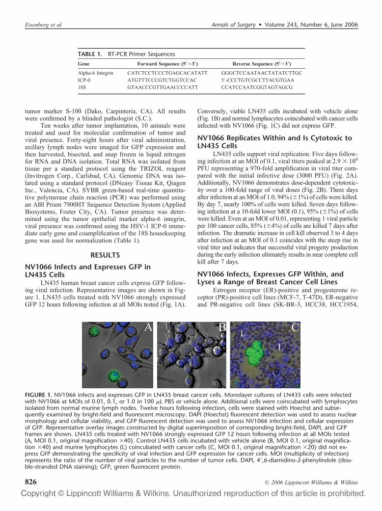
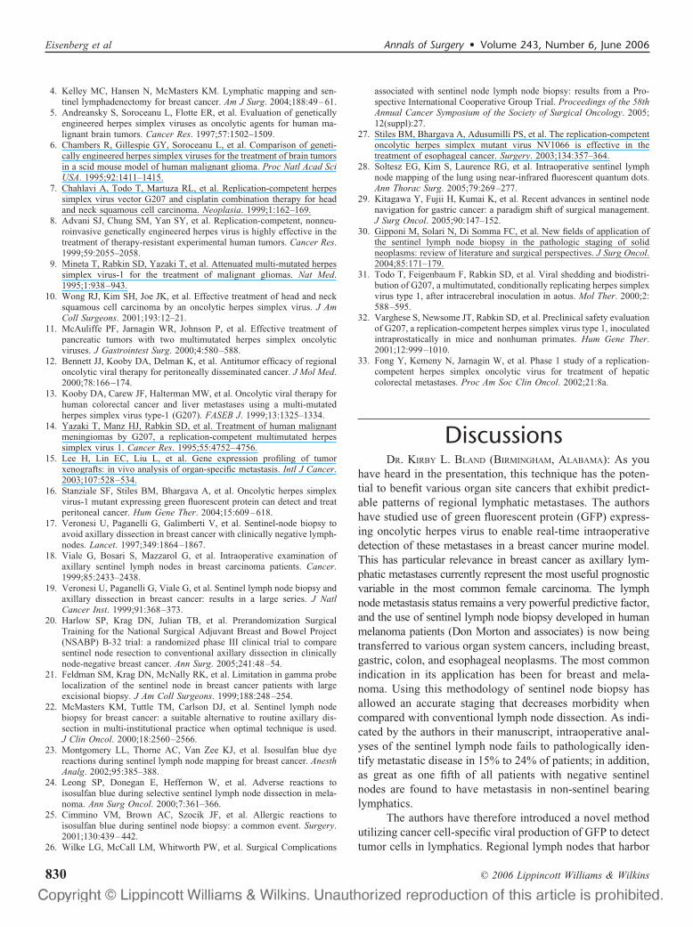
LN435 human breast cancer cells express GFP follow-ing viral infection. Representative images are shown in Fig-ure 1. LN435 cells treated with NV1066 strongly expressedGFP 12 hours following infection at all MOIs tested (Fig. 1A).
Conversely, viable LN435 cells incubated with vehicle alone(Fig. 1B) and normal lymphocytes coincubated with cancer cellsinfected with NV1066 (Fig. 1C) did not express GFP.
NV1066 Replicates Within and Is Cytotoxic toLN435 Cells
LN435 cells support viral replication. Five days follow-ing infection at an MOI of 0.1, viral titers peaked at 2.9 � 106
PFU representing a 970-fold amplification in viral titer com-pared with the initial infective dose (3000 PFU) (Fig. 2A).Additionally, NV1066 demonstrates dose-dependent cytotoxic-ity over a 100-fold range of viral doses (Fig. 2B). Three daysafter infection at an MOI of 1.0, 94% (�1%) of cells were killed.By day 7, nearly 100% of cells were killed. Seven days follow-ing infection at a 10-fold lower MOI (0.1), 95% (�1%) of cellswere killed. Even at an MOI of 0.01, representing 1 viral particleper 100 cancer cells, 85% (�4%) of cells are killed 7 days afterinfection. The dramatic increase in cell kill observed 3 to 4 daysafter infection at an MOI of 0.1 coincides with the steep rise inviral titer and indicates that successful viral progeny productionduring the early infection ultimately results in near complete cellkill after 7 days.
NV1066 Infects, Expresses GFP Within, andLyses a Range of Breast Cancer Cell Lines
Estrogen receptor (ER)-positive and progesterone re-ceptor (PR)-positive cell lines (MCF-7, T-47D), ER-negativeand PR-negative cell lines (SK-BR-3, HCC38, HCC1954,
TABLE 1. RT-PCR Primer Sequences
Gene Forward Sequence (5��3�) Reverse Sequence (5��3�)
Alpha-6 Integrin CATCTCCTCCCTGAGCACATATT GGGCTCCAATAACTATATCTTGC
ICP-0 ATGTTTCCCGTCTGGTCCAC 5�-CCCTGTCGCCTTACGTGAA
18S GTAACCCGTTGAACCCCATT CCATCCAATCGGTAGTAGCG
FIGURE 1. NV1066 infects and expresses GFP in LN435 breast cancer cells. Monolayer cultures of LN435 cells were infectedwith NV1066 at MOIs of 0.01, 0.1, or 1.0 in 100 �L PBS or vehicle alone. Additional cells were coincubated with lymphocytesisolated from normal murine lymph nodes. Twelve hours following infection, cells were stained with Hoechst and subse-quently examined by bright-field and fluorescent microscopy. DAPI (Hoechst) fluorescent detection was used to assess nuclearmorphology and cellular viability, and GFP fluorescent detection was used to assess NV1066 infection and cellular expressionof GFP. Representative overlay images constructed by digital superimposition of corresponding bright-field, DAPI, and GFPframes are shown. LN435 cells treated with NV1066 strongly expressed GFP 12 hours following infection at all MOIs tested(A, MOI 0.1, original magnification �40). Control LN435 cells incubated with vehicle alone (B, MOI 0.1, original magnifica-tion �40) and murine lymphocytes (L) coincubated with cancer cells (C, MOI 0.1, original magnification �20) did not ex-press GFP demonstrating the specificity of viral infection and GFP expression for cancer cells. MOI (multiplicity of infection)represents the ratio of the number of viral particles to the number of tumor cells. DAPI, 4�,6-diamidino-2-phenylindole (dou-ble-stranded DNA staining); GFP, green fluorescent protein.
Eisenberg et al Annals of Surgery • Volume 243, Number 6, June 2006
© 2006 Lippincott Williams & Wilkins826

MDA-MB-435S), low HER-2/neu expressing cell lines (MCF-7,HCC38, MDA-MB-435S), an intermediate HER-2/neu ex-pressing cell line (T-47D), and high HER-2/neu expressingcell lines (SK-BR-3, HCC1954) all were infected, expressedGFP, and lysed by NV1066 at all MOIs tested. Cell kill 7days after infection at an MOI of 1.0 for these cell linesranged from 87% to 99%.
NV1066-Guided GFP-Mediated FluorescentDetection of Lymph Node Metastases
We have established a reliable murine model of breastcancer axillary lymph node metastases that yields nodal
metastases in 40% of animals. Lymphatic mapping afterinjection of 1% isosulfan blue dye into the third mammary fatpad demonstrated intense uptake of blue dye in ipsilateralaxillary nodes (data not shown). Contralateral axillary andipsilateral cervical, inguinal, retroperitoneal, and mediastinallymph nodes did not stain blue.
We demonstrated that GFP expression in NV1066-infected axillary lymph node metastases can be detected within vivo fluorescent imaging. Representative images are shownin Figure 3. Mice with nodal metastases frequently demon-strated enlarged ipsilateral axillary lymph nodes (Fig. 3A).Involved lymph nodes exhibited intense focal expression ofGFP when imaged via fluorescent stereomicroscopy (Fig. 3C,D). Lymph nodes not harboring tumor and control lymphnodes did not express GFP.
Histologic and Molecular Analysis of LymphNodes
Histologic examination confirmed the presence of tu-mor and GFP in positive lymph nodes (Fig. 4). Furthermore,areas of GFP expression localized to tumor foci in lymphnodes. Absence of tumor and GFP in lymph negative lymphnodes (as determined by fluorescent imaging) was similarlyconfirmed by microscopic analysis.
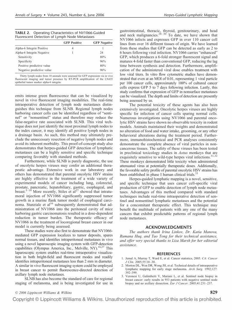
RT-PCR detection of the tumor-specific epithelialmarker alpha-6 integrin in lymph nodes demonstrated favor-able operating characteristics of NV1066-mediated GFP-guided detection of breast cancer lymph node metastasis(Table 2). Thirty lymph nodes (average, 3 per animal) wereanalyzed. Five of 30 lymph nodes (17%) analyzed weredetermined to harbor tumor by RT-PCR amplification ofalpha-6 integrin. Four of 5 (80%) of these positive lymphnodes expressed GFP when examined with fluorescent imag-ing. One of 25 (4%) lymph nodes not determined to harbortumor cells by RT-PCR was reported to express GFP viafluorescent imaging. As such, the operating characteristics are asfollows: sensitivity � 80%, specificity � 96%, positive predic-tive value � 80%, and negative predictive value � 96%.
PCR amplification of the viral gene ICP-0 detectedviral DNA in 3 of 5 (60%) lymph nodes that were GFPpositive. Viral DNA was not detected in any of the 25 lymphnodes that were GFP negative. The apparent absence of virusin 2 positive lymph nodes is likely due to sampling error, asthese lymph nodes contained focal disease yet were bisectedfor analysis.
DISCUSSIONLymph node status is a powerful prognostic factor for
many cancers. For women with breast cancer, axillary lymphnode status is the most important factor determining treat-ment, recurrence, and overall survival. Until recently, com-plete lymphadenectomy was routinely performed to stagepatients with breast cancer. This practice, however, subjectedmany women with early stage disease to a morbid procedurewith no benefit. It further hindered the identification of micro-metastases due to the impracticality of performing extensivehistologic analyses on a large number of resected nodes.
FIGURE 2. NV1066 replicates within and is cytotoxic toLN435 breast cancer cells. A, Monolayer cultures of LN435cells were treated with NV1066 at an MOI of 0.1 (3000PFU). Viral progeny were quantified daily after infection for 7days by standard plaque assay. Mean PFU for triplicate sam-ples are plotted (�standard error of the mean). B, Mono-layer cultures of LN435 cells were infected with NV1066 atMOIs of 0.01 (triangle), 0.1 (square), and 1.0 (circle). Cyto-toxicity assays were used to measure cell kill daily after infec-tion for 7 days. Mean cellular survival for triplicate samples isplotted as a percentage of control uninfected cells (�stan-dard error of the mean). MOI (multiplicity of infection) rep-resents the ratio of the number of viral particles to the num-ber of tumor cells. PFU, plaque-forming units.
Annals of Surgery • Volume 243, Number 6, June 2006 Herpes-Guided Lymphatic Mapping
© 2006 Lippincott Williams & Wilkins 827

The advent of SLNB has circumvented these shortcom-ings allowing a more selective approach to the identificationof regional lymph node metastases.2 SLNB relies on vital dyeenhancement and nodal uptake of radioactive colloid to identifythe individual lymph nodes draining the primary tumor. Thismethod is generally successful for the identification of sentinelnodes, the status of which is considered representative of theentire nodal basin. SLNB has resulted in accurate axillarystaging with decreased morbidity when compared with completelymph node dissection and has become an important method forstaging women with early breast cancer.4
The limitations of SLNB, however, have recently be-come apparent. Intraoperative analyses of sentinel nodes failto identify metastatic disease in 15% to 24% of patients.17,18
Lymphatic metastases missed intraoperatively mandate a sec-ond operation for completion axillary lymph node dissection.In addition, 7% to 19% of patients with negative sentinelnodes are found to have metastases in nonsentinel nodes.19–22
SLNB can be cumbersome and labor-intensive requiringmultiple preoperative interventions. Injection of vital bluedye can stain the breast for many months and is associatedwith a 1% risk of anaphylaxis.23–26 Administration of theradioactive colloid tracer requires well-coordinated interde-partmental planning to avoid operating room delays. Mostimportantly, recently presented data from a multi-institutionaltrial following over 5500 women indicates that morbidityfollowing SLNB is greater than expected with rates oflymphedema, axillary paresthesias, and reduced upper ex-tremity range of motion approaching 7%, 9%, and 4%,respectively.26
We introduce a novel method of lymphatic mappingthat uses cancer cell-specific viral production of GFP todetect tumor cells in lymph nodes. We have shown thatNV1066 travels to regional lymph nodes after administrationinto the primary tumor where it infects cancer cells. Infectionresults in expression of GFP, the protein product of which
FIGURE 3. NV1066-guided GFP-medi-ated fluorescent detection of lymphnode metastases. LN435 humanbreast cancer xenograft biopsies weremicrosurgically implanted into thethird right mammary fat pad in femaleathymic mice. After 10 weeks, 5 � 106
PFU of NV1066 was injected into theprimary tumor on treatment days 0and 5. Forty-eight hours later, animalswere killed and axillae were explored.Representative images are shown. A,Right axilla demonstrating two en-larged axillary lymph nodes (originalmagnification �5). B, Bright-field im-age of single enlarged axillary lymphnode ex vivo (original magnification�10). C, GFP fluorescent image oflymph node ex vivo demonstratingfoci of high GFP intensity (originalmagnification �10). D, Computer-generated overlay of bright-field (B)and GFP (C) digital images (originalmagnification �10).
FIGURE 4. Histologic analysis of tumorand GFP in lymph nodes. Serial sectionsof a negative lymph node (no GFP ex-pression by in vivo fluorescent imaging)is confirmed to have no tumor nor GFPexpression when examined with hema-toxylin and eosin (H&E), fluorescentmicroscopy, and immunohistochemistry(original magnification �20) (top row).Histologic analysis of a positive lymphnode (strong GFP expression by in vivofluorescent imaging) is confirmed toharbor tumor and express GFP (originalmagnification �20) (bottom row). GFP,green fluorescent protein.
Eisenberg et al Annals of Surgery • Volume 243, Number 6, June 2006
© 2006 Lippincott Williams & Wilkins828
emits intense green fluorescence that can be visualized bynovel in vivo fluorescent imaging modalities. The real-timeintraoperative detection of lymph node metastases distin-guishes this technique from SLNB. Regional lymph nodesharboring cancer cells can be identified regardless of “senti-nel” or “nonsentinel” status and therefore may reduce thefalse-negative rate associated with SLNB. This viral tech-nique does not just identify the most proximal lymph nodes tothe index cancer, it may identify all positive lymph nodes ina drainage basin. As such, this method may ultimately pre-clude the unnecessary resection of negative lymph nodes andavoid its inherent morbidity. This proof-of-concept study alsodemonstrates that herpes-guided GFP detection of lymphaticmetastases can be a highly sensitive and specific techniquecomparing favorably with standard methods.
Furthermore, while SLNB is purely diagnostic, the useof oncolytic herpes viruses may confer an additional thera-peutic advantage. Extensive work in our laboratory andothers has demonstrated that parental oncolytic HSV strainsare highly effective in the treatment of a wide variety ofexperimental models of cancer including lung, colorectal,prostate, pancreatic, hepatobiliary, gastric, esophageal, andbreast.5–14 More recently, Stiles et al27 showed that intratu-moral injection of NV1066 significantly suppressed tumorgrowth in a murine flank tumor model of esophageal carci-noma. Stanziale et al16 subsequently demonstrated that ad-ministration of NV1066 into the peritoneal cavity of miceharboring gastric carcinomatosis resulted in a dose-dependentreduction in tumor burden. The therapeutic efficacy ofNV1066 in the treatment of locoregional breast cancer in ourmodel is currently being assessed.
These studies were also first to demonstrate that NV1066-mediated GFP expression localizes to tumor deposits, sparesnormal tissues, and identifies intraperitoneal metastases in vivousing a novel laparoscopic imaging system with GFP-detectioncapabilities (Olympus America, Inc., Melville, NY).16,27 Thislaparoscopic system enables real-time intraoperative visualiza-tion in both bright-field and fluorescent modes and readilyidentifies intraperitoneal metastases less than 2 mm in diameter.A similar in vivo fluorescent imaging system could be employedin breast cancer to permit fluorescence-directed detection ofaxillary lymph node metastases.
SLNB has also become the standard of care for regionalstaging of melanoma, and is being investigated for use in
gastrointestinal, thoracic, thyroid, genitourinary, and headand neck malignancies.28–30 To date, we have shown thatNV1066 infects and expresses GFP in over 110 cancer celllines from over 16 different tissues of origin. We have learnedfrom these studies that GFP can be detected as early as 2 to6 hours following viral infection. NV1066 carries “enhanced”GFP, which produces a 6-fold stronger fluorescent signal andmatures 4-fold faster than conventional GFP, reducing the lagtime between synthesis and detection. Furthermore, amplifi-cation of the administered viral dose enables treatment withlow viral titers. In vitro flow cytometric studies have demon-strated that even at an MOI of 0.01, representing 1 viral particleper 100 cancer cells, approximately 100% of cultured cancercells express GFP 5 to 7 days following infection. Lastly, thisstudy confirms that expression of GFP in nonsurface metastasescan be visualized. The depth and limits of detection are presentlybeing assessed by us.
The potential toxicity of these agents has also beenextensively investigated. Oncolytic herpes viruses are highlyspecific for infection of cancer cells sparing normal cells.Numerous investigations using NV1066 and parental onco-lytic HSV strains have shown no observable toxicity in rodentmodels. Animals maintained their weights and demonstratedno alteration of food and water intake, grooming, or any otherbehavioral alterations during the treatment period. Further-more, immunohistochemical and PCR analyses consistentlydemonstrate the complete absence of viral particles in non-cancerous tissues. The safety of these viruses has been testedin preclinical toxicology studies in Aotus monkeys that areexquisitely sensitive to wild-type herpes viral infections.31,32
These monkeys demonstrated little toxicity when administeredattenuated virus at potentially therapeutic doses. Additionally,the favorable safety profile of parental oncolytic HSV strains hasbeen established in phase I human clinical trials.33
Herpes-guided lymphatic mapping is a novel, sensitive,and specific technique that uses cancer cell-specific viralproduction of GFP to enable detection of lymph node metas-tases. Advantages of this method compared with standardtechniques include real-time intraoperative detection of sen-tinel and nonsentinel lymphatic metastases and the potentialfor a concomitant therapeutic effect. This technique maybenefit the multitude of patients with any one of the manycancers that exhibit predictable patterns of regional lymphnode metastases.
ACKNOWLEDGMENTSThe authors thank Irina Linkov, Dr. Katia Manova,
Rumana Huq, and Tao Tong for their technical assistance,and offer very special thanks to Liza Marsh for her editorialassistance.
REFERENCES1. Jemal A, Murray T, Ward E, et al. Cancer statistics, 2005. CA: Cancer
J Clin. 2005;55:10–30.2. Morton DL, Wen DR, Wong JH, et al. Technical details of intraoperative
lymphatic mapping for early stage melanoma. Arch Surg. 1992;127:392–399.
3. Veronesi U, Galimberti V, Mariani L, et al. Sentinel node biopsy inbreast cancer: early results in 953 patients with negative sentinel nodebiopsy and no axillary dissection. Eur J Cancer. 2005;41:231–237.
TABLE 2. Operating Characteristics of NV1066-GuidedFluorescent Detection of Lymph Node Metastases
GFP Positive GFP Negative
Alpha-6 Integrin Positive 4 1
Alpha-6 Integrin Negative 1 24
Sensitivity 80%
Specificity 96%
Positive predictive value 80%
Negative predictive value 96%
Thirty lymph nodes from 10 animals were assessed for GFP expression via in vivofluorescent imaging and tumor presence by RT-PCR amplification of the LN435epithelial tumor marker alpha-6 integrin.
Annals of Surgery • Volume 243, Number 6, June 2006 Herpes-Guided Lymphatic Mapping
© 2006 Lippincott Williams & Wilkins 829
4. Kelley MC, Hansen N, McMasters KM. Lymphatic mapping and sen-tinel lymphadenectomy for breast cancer. Am J Surg. 2004;188:49–61.
5. Andreansky S, Soroceanu L, Flotte ER, et al. Evaluation of geneticallyengineered herpes simplex viruses as oncolytic agents for human ma-lignant brain tumors. Cancer Res. 1997;57:1502–1509.
6. Chambers R, Gillespie GY, Soroceanu L, et al. Comparison of geneti-cally engineered herpes simplex viruses for the treatment of brain tumorsin a scid mouse model of human malignant glioma. Proc Natl Acad SciUSA. 1995;92:1411–1415.
7. Chahlavi A, Todo T, Martuza RL, et al. Replication-competent herpessimplex virus vector G207 and cisplatin combination therapy for headand neck squamous cell carcinoma. Neoplasia. 1999;1:162–169.
8. Advani SJ, Chung SM, Yan SY, et al. Replication-competent, nonneu-roinvasive genetically engineered herpes virus is highly effective in thetreatment of therapy-resistant experimental human tumors. Cancer Res.1999;59:2055–2058.
9. Mineta T, Rabkin SD, Yazaki T, et al. Attenuated multi-mutated herpessimplex virus-1 for the treatment of malignant gliomas. Nat Med.1995;1:938–943.
10. Wong RJ, Kim SH, Joe JK, et al. Effective treatment of head and necksquamous cell carcinoma by an oncolytic herpes simplex virus. J AmColl Surgeons. 2001;193:12–21.
11. McAuliffe PF, Jarnagin WR, Johnson P, et al. Effective treatment ofpancreatic tumors with two multimutated herpes simplex oncolyticviruses. J Gastrointest Surg. 2000;4:580–588.
12. Bennett JJ, Kooby DA, Delman K, et al. Antitumor efficacy of regionaloncolytic viral therapy for peritoneally disseminated cancer. J Mol Med.2000;78:166–174.
13. Kooby DA, Carew JF, Halterman MW, et al. Oncolytic viral therapy forhuman colorectal cancer and liver metastases using a multi-mutatedherpes simplex virus type-1 (G207). FASEB J. 1999;13:1325–1334.
14. Yazaki T, Manz HJ, Rabkin SD, et al. Treatment of human malignantmeningiomas by G207, a replication-competent multimutated herpessimplex virus 1. Cancer Res. 1995;55:4752–4756.
15. Lee H, Lin EC, Liu L, et al. Gene expression profiling of tumorxenografts: in vivo analysis of organ-specific metastasis. Intl J Cancer.2003;107:528–534.
16. Stanziale SF, Stiles BM, Bhargava A, et al. Oncolytic herpes simplexvirus-1 mutant expressing green fluorescent protein can detect and treatperitoneal cancer. Hum Gene Ther. 2004;15:609–618.
17. Veronesi U, Paganelli G, Galimberti V, et al. Sentinel-node biopsy toavoid axillary dissection in breast cancer with clinically negative lymph-nodes. Lancet. 1997;349:1864–1867.
18. Viale G, Bosari S, Mazzarol G, et al. Intraoperative examination ofaxillary sentinel lymph nodes in breast carcinoma patients. Cancer.1999;85:2433–2438.
19. Veronesi U, Paganelli G, Viale G, et al. Sentinel lymph node biopsy andaxillary dissection in breast cancer: results in a large series. J NatlCancer Inst. 1999;91:368–373.
20. Harlow SP, Krag DN, Julian TB, et al. Prerandomization SurgicalTraining for the National Surgical Adjuvant Breast and Bowel Project(NSABP) B-32 trial: a randomized phase III clinical trial to comparesentinel node resection to conventional axillary dissection in clinicallynode-negative breast cancer. Ann Surg. 2005;241:48–54.
21. Feldman SM, Krag DN, McNally RK, et al. Limitation in gamma probelocalization of the sentinel node in breast cancer patients with largeexcisional biopsy. J Am Coll Surgeons. 1999;188:248–254.
22. McMasters KM, Tuttle TM, Carlson DJ, et al. Sentinel lymph nodebiopsy for breast cancer: a suitable alternative to routine axillary dis-section in multi-institutional practice when optimal technique is used.J Clin Oncol. 2000;18:2560–2566.
23. Montgomery LL, Thorne AC, Van Zee KJ, et al. Isosulfan blue dyereactions during sentinel lymph node mapping for breast cancer. AnesthAnalg. 2002;95:385–388.
24. Leong SP, Donegan E, Heffernon W, et al. Adverse reactions toisosulfan blue during selective sentinel lymph node dissection in mela-noma. Ann Surg Oncol. 2000;7:361–366.
25. Cimmino VM, Brown AC, Szocik JF, et al. Allergic reactions toisosulfan blue during sentinel node biopsy: a common event. Surgery.2001;130:439–442.
26. Wilke LG, McCall LM, Whitworth PW, et al. Surgical Complications
associated with sentinel node lymph node biopsy: results from a Pro-spective International Cooperative Group Trial. Proceedings of the 58thAnnual Cancer Symposium of the Society of Surgical Oncology. 2005;12(suppl):27.
27. Stiles BM, Bhargava A, Adusumilli PS, et al. The replication-competentoncolytic herpes simplex mutant virus NV1066 is effective in thetreatment of esophageal cancer. Surgery. 2003;134:357–364.
28. Soltesz EG, Kim S, Laurence RG, et al. Intraoperative sentinel lymphnode mapping of the lung using near-infrared fluorescent quantum dots.Ann Thorac Surg. 2005;79:269–277.
29. Kitagawa Y, Fujii H, Kumai K, et al. Recent advances in sentinel nodenavigation for gastric cancer: a paradigm shift of surgical management.J Surg Oncol. 2005;90:147–152.
30. Gipponi M, Solari N, Di Somma FC, et al. New fields of application ofthe sentinel lymph node biopsy in the pathologic staging of solidneoplasms: review of literature and surgical perspectives. J Surg Oncol.2004;85:171–179.
31. Todo T, Feigenbaum F, Rabkin SD, et al. Viral shedding and biodistri-bution of G207, a multimutated, conditionally replicating herpes simplexvirus type 1, after intracerebral inoculation in aotus. Mol Ther. 2000;2:588–595.
32. Varghese S, Newsome JT, Rabkin SD, et al. Preclinical safety evaluationof G207, a replication-competent herpes simplex virus type 1, inoculatedintraprostatically in mice and nonhuman primates. Hum Gene Ther.2001;12:999–1010.
33. Fong Y, Kemeny N, Jarnagin W, et al. Phase 1 study of a replication-competent herpes simplex oncolytic virus for treatment of hepaticcolorectal metastases. Proc Am Soc Clin Oncol. 2002;21:8a.
DiscussionsDR. KIRBY L. BLAND (BIRMINGHAM, ALABAMA): As you
have heard in the presentation, this technique has the poten-tial to benefit various organ site cancers that exhibit predict-able patterns of regional lymphatic metastases. The authorshave studied use of green fluorescent protein (GFP) express-ing oncolytic herpes virus to enable real-time intraoperativedetection of these metastases in a breast cancer murine model.This has particular relevance in breast cancer as axillary lym-phatic metastases currently represent the most useful prognosticvariable in the most common female carcinoma. The lymphnode metastasis status remains a very powerful predictive factor,and the use of sentinel lymph node biopsy developed in humanmelanoma patients (Don Morton and associates) is now beingtransferred to various organ system cancers, including breast,gastric, colon, and esophageal neoplasms. The most commonindication in its application has been for breast and mela-noma. Using this methodology of sentinel node biopsy hasallowed an accurate staging that decreases morbidity whencompared with conventional lymph node dissection. As indi-cated by the authors in their manuscript, intraoperative anal-yses of the sentinel lymph node fails to pathologically iden-tify metastatic disease in 15% to 24% of patients; in addition,as great as one fifth of all patients with negative sentinelnodes are found to have metastasis in non-sentinel bearinglymphatics.
The authors have therefore introduced a novel methodutilizing cancer cell-specific viral production of GFP to detecttumor cells in lymphatics. Regional lymph nodes that harbor
Eisenberg et al Annals of Surgery • Volume 243, Number 6, June 2006
© 2006 Lippincott Williams & Wilkins830

these cancer cells can be identified, regardless of their senti-nel or non-sentinel status, and therefore may reduce thefalse-negative rate associated with the sentinel node biopsy.As you have heard in the presentation, herpes-guided GFPdetection of lymphatic metastases can be a highly sensitiveand specific technique that compares favorably with standardmeasures.
The authors have suggested and confirmed in their labo-ratory that the novel oncolytic herpes viral strain (NV1066)specifically infects cancer cells and expresses GFP allowingintraoperative detection of these metastases. Indeed, histo-logic and molecular confirmation demonstrated favorableoperating characteristics of this method with a sensitivity of80% and a specificity of 96%. This converts to a positivepredictive value of 80% and a negative predictive value of96%. My question therefore is: what do you anticipate andhave you now had experience with human?
What is the evidence and your experience in humantumor cell lines that there will be expression of GFP oncolyticherpes virus in vivo to allow intraoperative detection of breastcancer lymphatic metastasis in humans?
It would appear that in the seven human cell lines thathave been infected in vitro with NV1066 and assessed forGFP expression, your data suggest viral transit to lymphaticsand infection of the metastases. With this high sensitivity andspecificity in the animal model, have you obtained an INDnumber for injection of GFP from your Institutional ReviewBoard to study such technique?
DR. KAREN J. HENDERSHOTT (NEW YORK, NEW YORK):The question of moving this from an animal model study intoa human setting is obviously very important. This study I justpresented is a proof of principal study that is meant to justestablish the concept that we could localize lymph nodesusing this technology.
The NV1066 virus is a second-generation herpes virus,and most of our experience using human trials comes fromthe parent viruses. We had attained an IND for studying thevirus, NV1020, and have completed a phase 1 trial at ourinstitution using this virus to treat liver cancer. We are lookingat moving on to a phase 2 trial as well. So we are working in thatdirection.
Everything that we look at in terms of in vitro data andalso with our work with animal models suggests that theNV1066 virus behaves similarly to the parental virus. So Ican at least hypothesize that it should move well into clinicaltrials. In terms of in vivo detection in humans, it certainly hasthe potential to go forward and do that.
In our laboratory, we actually have a modified Olympuslaparoscope that we have used in animal models, and SteveStanziale in our lab was able to detect intraperitoneal metas-tases as small as 2 mm using this laparoscope in a mousemodel. So I think that is one of the areas where we have the
greatest opportunity, using the laparoscopic system to expandthis technology into humans.
DR. MICHAEL J. EDWARDS (LITTLE ROCK, ARKANSAS):You showed that these viral vectors will track through thelymphatics in an effective way to arrive at the tumor cell.Could you comment about the ability of these agents to trackto other common sites of metastases (ie, the brain, the liver,the bone)? Are they as likely to be effective at that level asthey are in the lymph nodes?
I was a little surprised that you were able to grosslydetect the fluorescence in the axilla. What is the size of thelymph node in this model? How does that compare to the sizeof the lymph node in a human? The reason I bring that up isone of the great things about sentinel lymph node staging, asthe current state of the art, is the ability to detect tiny deposits ofdisease that are still clinically relevant at the 2 mm or so size.
Should a 2-mm metastasis, for example, fluorescethrough a lymph node? Your sensitivity was 80%; what doyou surmise was the reason for not detecting the one nodalmetastasis? Is it related to the size of the metastasis?
Finally, in the early days of sentinel lymph node staging,we had advocates of the blue dye and advocates of technetiumsulfur colloid. What we learned at the end of the day, and Dr.McMasters of this organization has so eloquently presented, thatwhen we combined the two it worked much, much better, andthe combination became the state of the art.
You have suggested that this new technique couldpotentially replace the current state of the art and pointed outmany “Achilles tendons” of the current state-of-the-art sen-tinel lymph node staging. I remind you, however, that theLouisville database has thousands of lymph nodes that havebeen analyzed defining sentinel lymph node staging. We aretalking about five lymph nodes today. How do you see thisunfolding for the future? What is the next step?
DR. KAREN J. HENDERSHOTT (NEW YORK, NEW YORK): Interms of the question where you asked about utility for othersites of metastases, this is really intriguing because of anunexpected finding in our study. As we serially passaged thetumors, the animals went on to develop not only axillarylymph node metastases but also metastatic disease in thelungs and even occasionally intra-abdominal disease.
What we found was that after we administered the virusinto the tumor, namely, at the site in the mammary fat pad,the green fluorescence was expressed not only within thoselymph node metastases but also within the lung metastases.We also detected mediastinal nodes that were also positivethat fluoresced green and also intra-abdominal deposits, whichsuggests that there is hematogenous as well as lymphaticdistribution of the virus. And this is consistent with what wehave seen in other animal models where we have looked atperitoneal metastases and thoracic metastases. So I think it does
Annals of Surgery • Volume 243, Number 6, June 2006 Herpes-Guided Lymphatic Mapping
© 2006 Lippincott Williams & Wilkins 831
have potential to be used to detect distant disease, and that is oneof the areas that we are continuing to explore.
In terms of the lymph nodes in mice, it can be variable.The average lymph node size is between 2 and 5 mm in theseanimals. Their axillary basin usually contains two to threelymph nodes. We confirmed through a blue dye study that thedrainage pattern is similar to what we have seen in humansprior to initiating our current study.
In terms of your question about depth of penetration ofthe fluorescence, I don’t have exact data to answer thatquestion. But anecdotally, I can tell you that where we had asmall tumor deposit in an animal behind the pectoralis mus-cle, we were actually able to see that green fluorescence allthe way through the pectoralis muscle. Now, in a mouse thatis probably 2 mm. But that still suggests that we are gettingfairly good penetration of the fluorescence.
I don’t have a good answer for how we missed that onelymph node. As I mentioned, we used RT-PCR as our goldstandard, which is holding us perhaps to a higher standardthan most folks would. So we might have simply picked upvery small amounts of disease that might not even have beenclinically relevant.
But one of the limitations of the study is the small numberof positive nodes. We have a sensitivity of 80% from missingone lymph node. I suspect those numbers would look better ifwe were able to increase the number of positive lymph nodes.And that was simply a challenge of the animal model.
DR. DAVID J. COLE (CHARLESTON, SOUTH CAROLINA): Acouple of questions: The intriguing part of this is the potentialclinical application. And I think the mark that you wouldwant to have is 100% sensitivity to the final pathology, notthe 59% that you presented. So although maybe the 80% ofyour test is better, it is not good enough to make clinicaldecisions on. I would be concerned that an ability to detect 2mm of cancer cells in a lymph node will still leave you shortof the mark. So I would be curious what your thoughts are interms of value sensitivity when compared to final pathology.
Also, you really didn’t mention a significant amount ofwork already done. There are molecular markers already well
defined with a high level of sensitivity for the detection ofmicrometastatic disease in lymph nodes. So I would thinkyou would ultimately need to compare to other moleculartechnologies that are currently out there in terms of how thisnew technology would come up to that mark.
DR. KAREN J. HENDERSHOTT (NEW YORK, NEW YORK):First of all, I agree with your concerns. And I think there area couple of important points to make of how this technologydiffers from sentinel lymph node biopsy.
First of all, what we are talking about is intraoperativedetection of lymph node metastases and to see if we can preventthe quarter of women who have falsely negative nodes intraop-eratively from having to go back and have a second surgery.
Secondly, our technique is a little bit different. Thevirus doesn’t just show you where the lymph tracks to, whichis basically what you are looking at with a sentinel lymphnode biopsy. What our virus allows to us do is to evaluate forcancer in all the lymph nodes within the basin regardless oftheir sentinel status or not, which I think is an advantage here.It is an advantage in breast cancer, but I think it is an even moresignificant advantage when you start applying this to othercancers, like gastric cancer, head and neck cancer, melanoma,and colon cancer, which is one of the next steps that we aregoing to be evaluating.
In addition, there is nothing that precludes the use ofthe radioactive dye at the same time as using the virus. Icertainly would see this as an adjuvant at the initial stages ofmoving us into clinical management. I think that those areimportant factors to consider.
Finally, one advantage of the viral detection method isthat it doesn’t preclude the use of molecular markers. In fact,the fluorescence can be used to highlight cancer cells so amore in depth analysis can be applied to these pre-selectedcells. Just like SLNB allowed us to perform focused analysesof a small number of lymph nodes, virally-directed greenfluorescence can facilitate in an in depth study of cancerouscells which is an important advantage with the developmentof molecular-targeted therapies.
Eisenberg et al Annals of Surgery • Volume 243, Number 6, June 2006
© 2006 Lippincott Williams & Wilkins832
Copyright © 2022 FDOKUMEN