Advancing Science. Improving Education - Universidade do ...
Upload
khangminh22Category
view
1download
0
�����������������
Citation: Sethakorn, N.; Heninger, E.;
Sánchez-de-Diego, C.; Ding, A.B.;
Yada, R.C.; Kerr, S.C.; Kosoff, D.;
Beebe, D.J.; Lang, J.M. Advancing
Treatment of Bone Metastases
through Novel Translational
Approaches Targeting the Bone
Microenvironment. Cancers 2022, 14,
757. https://doi.org/10.3390/
cancers14030757
Academic Editors: Ignacio Ochoa
and José María Ayuso-Dominguez
Received: 24 December 2021
Accepted: 29 January 2022
Published: 1 February 2022
Publisher’s Note: MDPI stays neutral
with regard to jurisdictional claims in
published maps and institutional affil-
iations.
Copyright: © 2022 by the authors.
Licensee MDPI, Basel, Switzerland.
This article is an open access article
distributed under the terms and
conditions of the Creative Commons
Attribution (CC BY) license (https://
creativecommons.org/licenses/by/
4.0/).
cancers
Review
Advancing Treatment of Bone Metastases throughNovel Translational Approaches Targeting theBone MicroenvironmentNan Sethakorn 1,2,3 , Erika Heninger 1 , Cristina Sánchez-de-Diego 1,4, Adeline B. Ding 1, Ravi Chandra Yada 4,Sheena C. Kerr 1,4, David Kosoff 1,2,3, David J. Beebe 1,4,5 and Joshua M. Lang 1,2,3,6,*
1 University of Wisconsin Carbone Cancer Center, University of Wisconsin-Madison, Madison, WI 53705, USA;[email protected] (N.S.); [email protected] (E.H.); [email protected] (C.S.-d.-D.);[email protected] (A.B.D.); [email protected] (S.C.K.); [email protected] (D.K.);[email protected] (D.J.B.)
2 Division of Hematology/Oncology, University of Wisconsin-Madison, 1111 Highland Ave.,Madison, WI 53705, USA
3 Department of Medicine, University of Wisconsin-Madison, Madison, WI 53705, USA4 Department of Pathology and Laboratory Medicine, University of Wisconsin-Madison,
Madison, WI 53705, USA; [email protected] Department of Biomedical Engineering, University of Wisconsin-Madison, Madison, WI 53705, USA6 Wisconsin Institutes for Medical Research, 1111 Highland Ave., Madison, WI 53705, USA* Correspondence: [email protected]; Tel.: +1-(608)-265-8131; Fax: +1-(608)-265-0614
Simple Summary: Solid tumors such as prostate, breast, and lung cancers frequently spread to bone,causing severe pain, disability, and cancer-related deaths. The multiple types of non-cancerous cellsin the bone interact with tumor cells to reduce the response to cancer therapies and promote furthercancer growth. Studies of cellular interactions in this environment are needed in order to discover newtherapies to treat and inhibit bone metastases. This review summarizes the current state of approachesused to study bone metastases, important pathways that could potentially be therapeutically targeted,and the status of clinical investigations of new drugs to treat bone metastases.
Abstract: Bone metastases represent a lethal condition that frequently occurs in solid tumors suchas prostate, breast, lung, and renal cell carcinomas, and increase the risk of skeletal-related events(SREs) including pain, pathologic fractures, and spinal cord compression. This unique metastaticniche consists of a multicellular complex that cancer cells co-opt to engender bone remodeling,immune suppression, and stromal-mediated therapeutic resistance. This review comprehensivelydiscusses clinical challenges of bone metastases, novel preclinical models of the bone and bonemarrow microenviroment, and crucial signaling pathways active in bone homeostasis and metastaticniche. These studies establish the context to summarize the current state of investigational agentstargeting BM, and approaches to improve BM-targeting therapies. Finally, we discuss opportunitiesto advance research in bone and bone marrow microenvironments by increasing complexity ofhumanized preclinical models and fostering interdisciplinary collaborations to translational researchin this challenging metastatic niche.
Keywords: bone metastases; tumor microenvironment; 3-D tumor-on-chip models; clinical trialstargeting bone metastases
1. Clinical Burden of Bone Metastases
Within solid tumors, prostate and breast carcinomas have greater tropism for bone,involved in up to 80–90% of patients with metastatic disease [1]. Breast (BC) and prostatecancers (PC) represent the most common solid tumors in female and male individuals,respectively, in the US, followed by lung cancers (LC) [2], and these tumor types commonly
Cancers 2022, 14, 757. https://doi.org/10.3390/cancers14030757 https://www.mdpi.com/journal/cancers
Cancers 2022, 14, 757 2 of 31
metastasize to bone [1]. As such, bone metastases (BM) affect a significant proportionof patients with cancer and contribute to lethality. BM arise most commonly in highlyvascularized bone containing red bone marrow in adults and cancellous bone (e.g., bonesforming the axial skeleton [3]. Bone lesions can generate skeletal instability, increasingrisks of pathologic fractures and spinal cord compression, causing pain and paralysisif not treated expeditiously. There are tumor-specific differences in BM subtypes, asPC favors osteoblastic and sclerotic lesions promoting abnormal bone deposition andsclerosis, whereas many other tumors skew towards osteolytic lesions with increasedosteoclastic activity [1]. Table 1 summarizes the frequency and quality of BM, and theincidence of skeletal-related events (SREs) in different solid tumors with advanced ormetastatic disease. The most devastating SREs include pathologic fractures and spinalcord compression. The frequency of these is noted specifically in Table 1. Other SREsinclude pain, need for palliative procedures such as radiotherapy, ablation, or surgery, andmalignant hypercalcemia.
The heterogeneity reported in BM percentages in Table 1 is due to several reasonsincluding different sample sizes and populations, research method (analysis of SEERdatabases vs. single-institution analysis), improvements in imaging modalities includingPET/CT and bone scans, clinically detected BM compared to subclinical disease found inautopsy studies, and overall advances in cancer therapies and survival (thereby allowingmore time to develop atypical metastases). The frequency of BMs can vary drasticallydepending on specific tumor subtypes, and whether studies examined patients with denovo BM or those that emerged throughout multiple therapies. Within a solid tumortype, molecular tumor alterations may further determine the type of bone lesions. Forexample, non-small cell lung cancer (NSCLC) with ALK (anaplastic lymphoma kinase)rearrangements showed a higher frequency of sclerotic bone lesions compared to NSCLCwith EGFR-activating mutations, or NSCLC lacking either of these targetable alterations [4].A retrospective analysis of patients with bone only metastases in BC showed that thedominant subtype was hormone-receptor-positive and HER2-negative [5].
Hematologic malignancies including leukemia, lymphoma, and multiple myeloma(MM) frequently involve the skeletal system. Leukemias can cause bone pain, fractures, andosseous radiographic abnormalities [6–10]. However, clinical manifestations are frequentlyrelated to profound cytopenias and coagulation disorders secondary to malignancy, aswell as treatment-induced ablation of the normal hematopoietic stem cell niche [11,12].Hematologic malignancies consist of heterogeneous subtypes based on the cell of originand pediatric vs. adult manifestations, but have unique cancer-stromal interactions withinthe specialized hematopoietic bone marrow niche (comprehensively reviewed by Mendez-Ferrer, et al.) [13]. Skeletal complications in lymphoma relate to long-term osteoporosisand insufficiency fractures secondary to premature bone loss from lymphoma therapiessuch as glucocorticoids and chemotherapy [14], although osteolytic lesions have beenreported [9]. Myeloma bone disease shares similar clinical complications seen in solidtumor BM (described in Table 1) and has been reviewed elsewhere [15].
In most cases, the presence of bone involvement confers a poor prognosis [1,5,16–24]. Itis possible that this reflects consequences of higher disease burdens; however, specific boneinvolvement could promote biological pathways that increase tumor aggressiveness. Theassociated morbidity can frequently preclude administration of additional life-prolongingtherapies. Patients with multiple BM had worse prognoses that those with limited BM in aretrospective analysis of patients with bone-only metastatic BC [25]. In some instances, suchas hormone receptor-positive BC with limited sites of bone-only metastases and no visceralorgan metastases, this represents a patient population with improved prognosis [26].

Cancers 2022, 14, 757 3 of 31
Table 1. Characteristics of bone metastases in diverse solid tumors. BM = bone metastases,SRE = skeletal related event, NSCLC = non-small cell lung cancer, SCLC = small cell lung can-cer, HCC hepatocellular carcinoma, NR not reported. * indicates frequency including all stages of thatmalignancy if frequency in advanced/metastatic disease was not specifically reported. ** indicatesinclusion of data reported from autopsy series. SRE frequency was reported as the number of patientswho experienced an SRE as a percentage of patients with clinically detected bone metastases.
Frequency of BMin Advanced
Disease
PredominantType Frequency of SRE
Prostate [17,18,23,27,28] 80–90% Blastic 22% (pathologic fracture)31–48%
Breast [29–32] 65–80% Lytic 39% (pathologic fracture)40–47%
NSCLC [20,33–35] 30–60% Lytic38–63%
20% (pathologic fracture)10% cord compression
SCLC [36,37] 50–66% Lytic 8–34%
Renal cell [38–40] 20–68% Lytic70–85%
28% (spinal cord/nerve rootcompression)
Urothelial ** [41] 32–47% Lytic 7%
Melanoma [42–44] 17–52% Lytic 47–58%
Thyroid [45–49] 4 *–50% Lytic 32–78%
HCC [50–53] 4.5–38% Lytic 56%
Biliary tract [54–57] 2–35% Lytic41%
16% (pathologic fracture)8% (cord compression)
Gastric ** [58–60] 4–45% Lytic 31–75%
Esophageal ** [61–63] 15–24% Lytic 91%
Colorectal [64–66] 3–24% Lytic62–68%
8–10% (pathologic fracture)6–9% (cord compression)
Pancreatic [67–69] 2–12% Blastic32–57%
6% (pathologic fracture)3% (cord compression)
Squamous cell carcinomaof the head and neck
[70–73]1–16% Lytic
9–31%2–12% (pathologic fracture)6–7% (cord compression)
Endometrial ** [74–76] 1 *–25% Lytic NR
Ovarian ** [77–79] 1 *–15% NR NR
Soft Tissue Sarcomas[80,81] 9–11% Lytic
40%22–31% (pathologic fracture)
13% (cord compression)
Multiple Myeloma [82–86] 80–90% Lytic22–60%
14–34% (pathologic fracture)4.7–7.8% (cord compression)
In contrast, several studies reported poor therapeutic response in patients with BM.In patients with NSCLC treated with the checkpoint inhibitor nivolumab, those with BMhad worse survival and less chance of responding to treatment [24]. Similarly, ipilimumabtreatment was less effective in patients with metastatic castrate-resistant PC with BM

Cancers 2022, 14, 757 4 of 31
compared to those without [87,88]. Patients with EGFR-driven lung adenocarcinomaexperienced limited sites of progression while on therapy with an effective EGFR inhibitor.These sites were all in bone, and effectively treated with high-dose ablative radiotherapy.Patients continued systemic treatment with the same EGFR inhibitor with durable diseasecontrol [89]. BM was associated with worse survival in patients with melanoma receivingeither targeted or immunotherapy, independently of the number of sites and other clinicalparameters [90]. Bone stroma confers resistance to systemic therapies, either throughsecretion of paracrine factors or direct cell-cell contacts [91]. Altogether these data indicatethat the bone and bone marrow niche is a unique microenvironment that permits tumorgrowth and treatment resistance.
Bone and bone marrow represent distinct niches compared to other sites in patientswith metastatic solid tumors; however, there is limited clinical data on bone responses. Thisis due, in part, to challenges in obtaining and processing BM from patients and difficultyassessing radiographic responses in bone. A sclerotic appearance may indicate a favorableresponse to targeted therapy, immunotherapy, or chemotherapy in NSCLC [92–94]. Jiao,et al., compared transcriptional profiles of primary prostate versus bone marrow biopsiesin patients with metastatic castrate-resistant PC, pre and post treatment with ipilimumab.The T cell populations differed between sites, with a greater degree of immunosuppressivepolarization within bone marrow. This was recapitulated during inoculation of murine PCcells either in subcutaneous or intraosseous pockets of syngeneic mice [87]. Exploratoryanalysis of bone marrow aspirates in a single-arm phase I clinical trial of patients withmetastatic PC receiving a dual checkpoint inhibitor with tremelimumab and durvalumabdemonstrated upregulation of myeloid and neutrophil signatures after immunotherapy [95].TGF-beta was highly enriched in both patient and murine bone marrow supernatant withBM. Consistent with these findings, there is a higher frequency of regulatory T cells (Tregs)in the bone marrow aspirates of patients with bone metastatic PC, but not from age-matcheddonors without cancer [96].
A major challenge to assessing efficacy is the endpoint used in clinical trials. BM aretypically considered non-measurable, which limits the ability to assess tumor responsesby standard CT imaging. This important topic is discussed by Hussain et al. [1]. Briefly,endpoints include pain response, patient quality of life, bone density, time to diseaseprogression and/or SRE, and overall survival. In cancers with a well-defined tumor marker,such as PSA in PC, a reduction in PSA is a common surrogate endpoint. Advances in nuclearmedicine imaging modalities could also provide an additional approach to measure tumorresponse in bone. Improving technology in detection of circulating biomarkers includingcirculating tumor cells, cell-free tumor DNA, or tumor-derived exosomes are emerging asnovel correlative markers of disease response.
Currently there are limited treatments for specific management of BM. Radiopharma-ceuticals such as radium-223 deposit only in bone and are approved in PC with bone-onlymetastasis; however the effect on prolonging survival is modest [97]. Agents that preventbone resorption include zoledronic acid, a bisphosphonate that inhibits the productionof RANK-ligand (RANKL) and stimulates synthesis of osteoprotegerin (OPG), and deno-sumab, an antibody that inhibits the osteoclast-promoter RANKL [98–101]. These agentshave minimal effect on overall survival, but prevent and delay SREs. Focused externalbeam radiation therapy is effective in palliating pain and can prevent further progressionat the sites of irradiation. In some cases, surgical intervention is required to decompressthe spinal cord or stabilize pathologic fractures. Newer approaches involve percutaneousablation and vertebroplasty as a less invasive interventional method to reinforce boneat risk for pathologic fractures and palliate pain [102]. Therefore, advances in systemictreatment of BM are acutely needed.
2. Complex Multicellular Composition of Bone and Bone Marrow Metastatic Niches
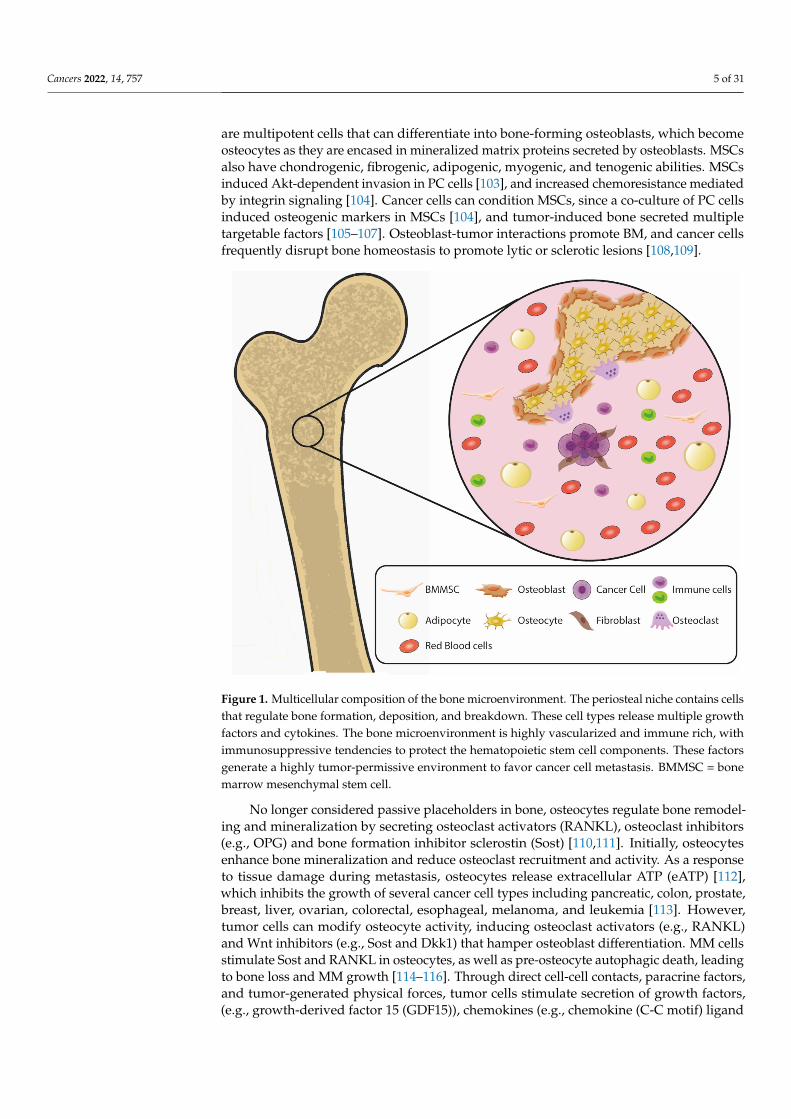
Bone is highly vascular and multicellular with extensive cell-cell and paracrine interac-tions, as depicted in Figures 1 and 2. Bone marrow-derived mesenchymal stem cells (MSCs)
Cancers 2022, 14, 757 5 of 31
are multipotent cells that can differentiate into bone-forming osteoblasts, which becomeosteocytes as they are encased in mineralized matrix proteins secreted by osteoblasts. MSCsalso have chondrogenic, fibrogenic, adipogenic, myogenic, and tenogenic abilities. MSCsinduced Akt-dependent invasion in PC cells [103], and increased chemoresistance mediatedby integrin signaling [104]. Cancer cells can condition MSCs, since a co-culture of PC cellsinduced osteogenic markers in MSCs [104], and tumor-induced bone secreted multipletargetable factors [105–107]. Osteoblast-tumor interactions promote BM, and cancer cellsfrequently disrupt bone homeostasis to promote lytic or sclerotic lesions [108,109].
Figure 1. Multicellular composition of the bone microenvironment. The periosteal niche contains cellsthat regulate bone formation, deposition, and breakdown. These cell types release multiple growthfactors and cytokines. The bone microenvironment is highly vascularized and immune rich, withimmunosuppressive tendencies to protect the hematopoietic stem cell components. These factorsgenerate a highly tumor-permissive environment to favor cancer cell metastasis. BMMSC = bonemarrow mesenchymal stem cell.
No longer considered passive placeholders in bone, osteocytes regulate bone remodel-ing and mineralization by secreting osteoclast activators (RANKL), osteoclast inhibitors(e.g., OPG) and bone formation inhibitor sclerostin (Sost) [110,111]. Initially, osteocytesenhance bone mineralization and reduce osteoclast recruitment and activity. As a responseto tissue damage during metastasis, osteocytes release extracellular ATP (eATP) [112],which inhibits the growth of several cancer cell types including pancreatic, colon, prostate,breast, liver, ovarian, colorectal, esophageal, melanoma, and leukemia [113]. However,tumor cells can modify osteocyte activity, inducing osteoclast activators (e.g., RANKL)and Wnt inhibitors (e.g., Sost and Dkk1) that hamper osteoblast differentiation. MM cellsstimulate Sost and RANKL in osteocytes, as well as pre-osteocyte autophagic death, leadingto bone loss and MM growth [114–116]. Through direct cell-cell contacts, paracrine factors,and tumor-generated physical forces, tumor cells stimulate secretion of growth factors,(e.g., growth-derived factor 15 (GDF15)), chemokines (e.g., chemokine (C-C motif) ligand

Cancers 2022, 14, 757 6 of 31
5 (CCL5)) and metalloproteases, that promote proliferation, migration, and invasion ofseveral cancer types including prostate, breast and lung [117,118].
Figure 2. Paracrine signaling pathways active in the bone niche. Under nonmalignant conditions,these promote normal bone homeostasis, balancing bone deposition/mineralization and breakdown.TGF-beta, BMP, and Wnt signaling pathways are highlighted as key mediators of paracrine signalingbetween bone stromal cells and tumor cells. Multiple cancer cell types co-opt these pathways topromote metastasis to bone, develop treatment resistance, and enhance cancer cell survival. Multipleapproaches to inhibiting these pathways include small molecule inhibitors, antibodies, and othertypes of antagonists.
Cancers 2022, 14, 757 7 of 31
The bone marrow is considered a secondary lymphoid organ that provides a niche forvast networks of innate and adaptive immune cells and regulates immune homeostasis.While bone marrow immune cells exert robust antitumor activity, cancer cells can subvertthe functional integrity of bone marrow immunity [119,120]. It has been hypothesized thatthis subversion could be promoted by the immune suppressive nature of this milieu andimmune-osseous interactions that regulate bone homeostasis. To nurture a hematopoieticstem-cell (HSC) niche and shield it from environmental stressors, bone marrow providesa hypo-immunogenic milieu enriched in regulatory T cells, myeloid-derived suppressorcells (MDSCs) and immune suppressive factors. This immune-privileged site maintains adistinct immune composition including an increased T cell to B cell ratio (5:1) and decreasedCD4:CD8 ratio (1:2) [121–123]. Most lymphocytes exhibit an antigen-experienced activatedmemory phenotype, while the frequency of naïve T cells is largely reduced compared tospleen or lymph nodes. Bone marrow preferentially retains Treg cells that can make upto ~25–30% of the CD4 subset, which can be further enriched in metastatic cancer [96].Regulatory T cells have profound effects at suppressing anti-tumor CD8 T cell and NK cellresponses and reduce IFN-gamma production. Additionally, Treg cells can promote the‘vicious cycle’ by suppressing osteoclasts and promoting osteoblastic PC dissemination.Th17 cells regulate tissue remodeling and bone reabsorption and may support both anantitumor activity or promote metastatic pathogenesis. Preferential expansion of Th17polarization was identified in bone marrow of patients with PC with BM undergoing anti-CTLA4 immune therapy (NCT02703623 and NCT02985957) in contrast with an enhancedTh1 signature in patients with soft-tissue metastasis [87].
Unconventional T cells, such as gamma-delta T cells and NKT cells in bone marrow,have significant roles in tumor immune surveillance and promising therapeutic potential intreatment of metastatic PC. Gamma-delta T cells produce IFN-gamma, promote antitumorT cell activity, directly suppress osteoclasts, and have been associated positive clinicaloutcomes in PC [124]. NKT cells exhibit potent antitumor activity and are greatly reducedin PC tissue [125] and in PC bone marrow [126]. NKT cells rapidly diminish at the peripheryafter antigen encounter and get replenished in bone marrow. Therefore, bone marrowalso plays a central role in NKT homeostasis during antitumor responses. Changes inhomeostasis and function within the complex T cell networks may play a robust, multi-faceted role in dissemination of tumor cells to the bone marrow. For example, disruptionof the CCL20-CCR6 axis prolonged survival and enhanced T cell reactivity in a syngeneicmouse model of metastatic PC [127]. Further research is needed to explore potentiallyactionable mechanisms to revert immune suppression and prevent invasion.
Macrophages are another cell population that are highly important in developmentof BM. Preclinical studies demonstrated that macrophage depletion decreased formationof skeletal metastases in vivo, indicating that macrophages support establishment of bonemetastatic foci as well a progression of these lesions [128,129]. While mechanisms by whichmacrophages promote BM continue to be under investigation, available data suggest thatthese pathways are multifaceted due to wide-ranging functions of macrophages withinbone [130]. Macrophages are an extremely diverse cell population with the ability toadapt their phenotype to the needs of each microenvironment [131]. Within the bonemicroenvironment there are multiple populations of macrophage-derived cells, each with aunique set of phenotypic characteristics and physiologic functions.
Osteoclasts are among the most well described population of macrophage-derivedcells within the bone marrow. Their primary function is bone resorption, which is a criti-cal component of the bone remodeling process that continually occurs to maintain bonestructure [132]. In addition to osteoclasts, osteal macrophages or “osteomacs” are anotherresident macrophage-derived population involved in bone remodeling. These cells con-stitute approximately 17% of the bone marrow cells and they differ from osteoclasts bythe expression of CD68 [133]. They are primarily located around osteoblasts and supportbone homeostasis by phagocytosing aging osteoblasts, and secrete cytokines stimulatingthe growth of newer osteoblasts [130]. Bone marrow derived macrophages (BMDMs) and

Cancers 2022, 14, 757 8 of 31
monocyte derived macrophages (MDMs) can also be recruited into the bone microenviron-ment. These cells can support a range of functions in the bone marrow, ranging from boneremodeling and cell growth to immune regulation and angiogenesis [134,135].
During development of BM, tumor-derived factors alter functions of various macrophagepopulations within the bone microenvironment to support tumor cell survival and prolifer-ation. This process begins prior to the arrival of tumor cells when cytokines secreted bythe primary tumor, such as VEGF, TNF-alpha, and TGF-beta, recruit immunosuppressivemyeloid cells to the bone [136]. Primary tumors also secrete exosomes, which contain anarray of factors (microRNA, proteins, among others) that increase macrophage-mediatedefferocytosis and osteoclastogenesis in anticipation of tumor cell arrival [137,138]. Oncepresent in the bone, tumor cells produce TGF-beta and parathyroid hormone-related pep-tide (PTHrP) to facilitate migration by inducing macrophage mediated remodeling of thesurrounding ECM [139]. Tumor cells within the bone are also able to recruit CD68+ cellsfrom the blood and bone marrow into the TME [140]. These tumor-associated macrophages(TAMs) support tumor growth and survival through the same TAM-mediated pathwaysthat are active in the primary tumor and other metastatic sites (i.e., secretion of growthfactors, angiogenesis, immune suppression) [135]. Additionally, efferocytosis of apoptoticcancer cells by macrophage populations leads to production of an array of factors, includingCXCL5, IL-6, IL-8, CCL4, and CCL5, which promote tumor growth and survival [141,142].
Adipocytes account for up to 60% of cells in bone marrow, but despite their abundancetheir role in BM has not been well characterized. Recent data suggest that adipocytesregulate tumor growth in BC and promote melanoma metastasis [143,144]. Bone marrowadipocytes (BMAs) secrete adipokines (e.g., CXCL12, CXCL10 and CX3CL1,) that increasevascular permeability and act as chemoattractants for tumor cells [143–145]. The release ofproinflammatory cytokines (e.g., IL-1beta, IL-6, TNF-alpha,) suppresses myeloid-derivedcells, and therefore inhibits both the innate and adaptive immune response [146]. Oncemetastasis is established in the bone marrow, BMAs release free fatty acids to provideenergy and support the growth of tumor cells. Treatment with ND-646, an inhibitor ofacetyl-CoA synthesis, reduced cancer cell lipid droplets and decreased tumor cell prolifera-tion in preclinical models of NSCLC [147]. BMAs perturb bone turnover, activate osteoclastdifferentiation through adipocyte-derived RANKL, CXCL1, and CXCL2, and suppress os-teoblast differentiation by inhibiting bone morphogenic protein (BMP) signaling [148–150].
3. Models of Bone Metastasis
A major challenge in tumor microenvironment modeling remains balancing tissuecomplexity and scalability, particularly relevant considering the complex cellular andmatrix components of bone and bone marrow [151]. Multicellular environments withthree-dimensional (3-D) properties require higher levels of technical expertise and oftenare associated with increased labor intensity to maintain in vivo mouse models or obtainand culture primary human tissue. Development of novel systemic therapies to preventor inhibit BM, however, requires a detailed understanding of stromal-tumor interactions.The bone microenvironment itself is highly complex and is comprised of multiple niches,including the osteogenic niche, hematopoietic stem cell niche, and perivascular niche. Thissection highlights commonly used in vivo and ex vivo models and discusses strategies toimprove modeling of the bone microenvironment.
3.1. In Vivo Models
Intracardiac injection of tumor cells reliably generates BM compared to other inocula-tion routes, although pretreatment of mice with melanoma-derived exosomes generatedBM even with subcutaneous implantation of melanoma cells [152]. Shiozawa, et al., usedan in vivo model in which osseous vossicles (vertebral bodies from 4–7 days old wildtype or mice expressing an inducible collagen transgene) were implanted subcutaneouslyinto SCID mice, facilitating assessment of metastatic tumor cells to bone and allowingbone specific ablation of collagen to target the endosteal niche [153]. Intra-iliac injection
Cancers 2022, 14, 757 9 of 31
also results in BM, and this technique has been combined with bone-in-culture ex vivoarrays using fragmented murine BM to interrogate multiple therapies [109]. Live animalimaging through mouse calvarium enabled visualization of human BC cell homing tospecific bone niches after intracardiac injection in immunocompromised mice [154]. In-traosseous injection can model local tumor-stromal interactions but does not address theearly stages of metastasis. A detailed description of murine BM protocols was summarizedby Park, et al. [155]. Mouse models have led to several important discoveries in signalingpathways mediating BM, are valuable in preclinical drug development but require a highdegree of technical complexity and higher costs, and should be interpreted with an un-derstanding of the differences between mouse and human hematopoietic and lymphoidlineages [156].
The human immune and stromal components within bone and bone marrow were notcaptured in previous in vivo models due to the use of immunocompromised or syngeneicmice. Humanized mice with reconstituted hematopoietic compartments have enabledsignificant advances in modeling preclinical immuno-oncology therapies. However otherregions of the bone remain of mouse origin [157]. Several groups have incorporated specifichuman microenvironments in immunocompromised mice. For instance, McGovern, et al.,incorporated human cancer-associated fibroblasts and lymphatic and blood endothelialcells in an orthotopic model of PC, combined with subcutaneous implantation of humanizedtissue-engineered bone constructs (hTEBC) [158]. Using this same model, a xenograft of aPC cell line responded to zoledronic acid but not denosumab (a human-specific RANKLantibody) [159]. This suggests that this model may lack human osteoclasts, the target ofRANKL inhibitor denosumab, which was as efficacious as, or better than, zoledronic acid inclinical trials [160,161]. Chen, et al., injected human MSCs and endothelial cells embeddedin Matrigel into immunocompromised mice, and subsequently introduced human cordblood cells intravenously. This method simulated extramedullary bone with human stromalcells engrafted with human hematopoietic cells [162]. Lee, et al., advanced a similarapproach with genetic engineering of human stromal cells to assess how distinct cytokinesmodulate HSC function and recruitment [163]. Additional models with implantation ofhumanized bone marrow niches in the study of bone marrow engraftment, hematopoiesis,and leukemia have been reviewed by Abarrategi, et al. [164].
3.2. Bioengineered Microfluidic Models
Microfluidic three-dimensional (3-D) tissue chip platforms have emerged as a promis-ing method to bridge the gap between two-dimensional (2-D) coculture and in vivo meth-ods and improve modeling of metastatic niches [165]. These platforms can accommodatethe culture of multiple cell types in a microenvironment, replicate the tumor architecture,generate physiologically relevant nutrient/chemokine gradients, and have tunable compo-nents that can control mechanical cues or stresses that can directly influence cell behaviorand function [166]. Furthermore, they enable low-input cell culture, which increases theanalytical endpoints that can be obtained from a limited resource such as primary humancells. High optical imaging capacity using confocal microscopy enables detailed 3-D vi-sualization of cancer cell extravasation and intravasation. It has been well described thatthe 3-D environment afforded by these platforms helps to better recapitulate cell polariza-tion, and crosstalk between metastatic cells and the niche [167]. The increased complexityof ECM organization in microfluidic models accentuates the migration and proliferationadvantages metastatic cells have in vivo compared to their 2-D counterparts [168,169].The interaction of tumor and endothelial cells is an often understated aspect of modelingthe metastatic niche. The addition of microvasculature into 3-D BM microfluidic modelsfurther captures the dynamics of cell recruitment and signaling in the microenvironment,which is particularly relevant as malignant angiogenesis is a hallmark of cancer cell growth,and led to development of anti-angiogenic agents as part of cancer therapies in multiplesolid tumor types [170]. Existing 2-D coculture systems, 3-D static and dynamic in vitroplatforms fail to account for this integral interaction and signaling. Microfluidic systems
Cancers 2022, 14, 757 10 of 31
are uniquely positioned to incorporate 3-D tumor spheroid cultures into an ECM matrixwith embedded stromal cells alongside microvasculature. The effect of secreted factors bythe growing cancer on the function of the surrounding bone niche and endothelial vesselscan be better explored in advanced microfluidic devices. Additionally, microfluidic modelsenable passive pumping or active pumping methods within lumens to investigate both theeffects of increased shear stress in the microenvironment, as well as the dynamics of cellextravasation in the context of a growing metastatic bone niche [171,172]. A comprehen-sive and well-crafted review by Laranga, et al., highlights many of the pros and cons ofdifferent BM methods, including the significant advantages microfluidic devices have ingenerating model systems for investigating a plethora of both biologically and clinicallyrelevant questions [173]. Importantly, the ease of use, customizability, and 3-D coculturecapabilities of microfluidics lends to its increased adoption compared to more expensive,and less reproducible scaffold based [174] or bioreactor systems [175] commonly employedfor studying metastatic microenvironments.
Bioengineered bone is actively being pursued in both cancer research and primarybone disease such as osteoporosis and tissue regeneration. One group used primary humanosteocytes cultured in 3-D with microbeads as a physical support, coupled with a passivepumping system to continuously replenish nutrients and excrete waste. They demon-strated bone mineralization of the system, and that coculture with PC cells altered Wntand fibroblast growth factor signaling that was not recapitulated in 2-D coculture [176].Bioreactor-cultured human bone fragments containing osteoblasts, osteocytes, osteoclasts,adipocytes, and hematopoietic progenitors were cocultured with BC cells, which upregu-lated osteoclastic cytokines consistent with lytic BC bone lesions [177]. Sieh, et al., usedmedical grade polycaprolactone-tricalcium phosphate scaffolds (used clinically as cranialimplants) embedded with human osteoblasts as a bone construct to show that this structureinduced expression of androgen-responsive genes in a PC cell line even in the absenceof an androgen mimic [178]. A bioengineered bone-mimetic environment demonstratedstroma-mediated chemoresistance and recapitulated localization of radium-223 [179]. An-other group developed a platform called human osteoblast-derived tissue-engineeredconstruct (hOTEC) that recapitulated encased osteocytes within mineralized tissue andwas cocultured with a PC PDX [180]. Other microscale chips incorporated osteoblasticcells derived from MSCs and showed that these cells could mineralize the surrounding3-D matrix, inducing BC cell invasion [181,182]. Coculture of MSCs and endothelial cellsformed a vascularized, mineralized structure enabling tracking of PC motility [183].
Structurally complex environments have been generated by incorporating luminalstructures lined by endothelial cells to mimic vasculature or epithelial cells to mimic ductalstructures [184,185]. Culturing cells in a physiologically relevant geometry, as found inthese models, can have a profound impact on cell function and production of secretedfactors [186]. Similar methods have been used to visualize fibrosarcoma and BC cell ex-travasation across a microfluidic endothelial barrier and extracellular matrix [187]. BCcells have also been seeded within an endothelial-lined lumen structure and observed tointravasate through this structure towards osteoblasts encapsulated in a 3-D matrix [181].These studies represent an important advancement in recapitulating endothelial-stroma-cancer cell crosstalk; however, there are some limitations. First, most of these studies focuson specific stromal cells—fibroblasts, osteoblasts, osteoclasts, or adipocytes. As discussed,the bone niche is comprised of multiple cell types, and thus its complexity is challeng-ing to capture ex vivo. Parikh, et al., described an ex vivo mixed, multicellular systemexpanded from bone marrow mononuclear cells that spontaneously generate osteoblasts,osteoclasts, adipocytes, and fibroblasts in the same culture [188]. Therefore, embeddingthese mixed cultures within a microfluidic bone tissue chip could bypass this limitation,and incorporating luminal structures can recreate the bone endosteal/perivascular niches.
Most of the models described above enrich for stromal cells such as osteoblasts andstromal progenitors. Additional methods are required to enrich for the hematopoieticand stem cell compartments. Torisawa, et al. [189] generated a PDMS device engineered

Cancers 2022, 14, 757 11 of 31
to create a physical bone mineralized structure derived from syngeneic implanted sub-cutaneously in mice. Over several weeks, hematopoietic cells infiltrated, and its cellularcontent resembled murine bone marrow histologically and immunophenotypically. Thisstructure could then be retrieved and cultured in vitro within a microfluidic device up toseven days, with appropriate responses to radiation and granulocyte colony-stimulatingfactor. Recently, Glaser, et al. [190] developed a multichamber 3-D organ-on-chip modelthat demonstrated coculture of an osteoblast cell line with primary human stromal cells,and human hematopoietic stem progenitor cells. This chip recapitulated bone niches thatincorporated vascular networks, maintained hematopoietic progenitor cell function, re-sponses to chemotherapy and granulocyte colony-stimulating factor, neutrophil egress, andBC cell line invasion into the bone niches. Increasing the cellular complexity to incorporatestromal, vascular, progenitor stem, immune, and tumor cells will enhance the ability toreplicate a human microenvironment. Platforms incorporating this complexity will enableex vivo analysis of factors mediating response or resistance to tumor immunotherapies inhumanized models.
4. Signaling Pathways Mediating Bone Metastasis and the Status of Clinical Trials ofBiologic Agents Targeting Bone Metastases
Bone metastasis is a complex, multistep process, and a recent review by Zhang, et al.,provides a global summary of the bone niche in solid tumors [191]. In this section wehighlight prominent signaling hubs involved in disseminated tumor cells to provide contextfor rational clinical targeting of these pathways and discuss the current status of clinicaltrials targeting these pathways. Importantly, many of the signaling pathways crosstalkwith each other, providing a potential avenue to target “bone metastatic signaling hubs”,which may be more effective than ablating individual components. It is hypothesized thattargeting the pathways that promote BM will reduce metastatic burden and prevent theenvironment-mediated treatment resistance from the tumor-permissive bone stroma.
As discussed above, antiresorption therapies have primarily been shown to preventor delay SREs in patients with bone metastatic cancers. A meta-analysis of patients withlocalized hormone receptor positive BC demonstrated that zoledronic acid given in theadjuvant setting after mastectomy modestly reduced the risk of BM [192]. Table 2 sum-marizes recent trials targeting solid tumor BM that are complete or in progress. Most ofthese trials were in early phases and thus outcomes focused primarily on safety; however,early data suggest only modest, limited antitumor activity. Given that the mechanismsin BM cover diverse pathways, it is likely that combination therapies targeting both thetumor and stroma will be more effective than single agents. Additionally, many of theseagents are tested in patients who have been heavily pretreated and already have multiplemetastases, raising the hypothesis that targeting tumor-stromal interactions at this laterstage may not confer meaningful clinical benefit. Therefore, trials that incorporate safetyand efficacy of bone targeting agents earlier in the disease course may be more effective,and understanding the effects of systemic therapies on bone deposition/resorption mustbe considered. For example, combining radium-223 with abiraterone (an inhibitor of andro-gen synthesis) increases the risk of fractures in PC and underscores the caution necessaryin this setting [193]. In contrast, a phase I trial evaluated the addition of radium-223 tostandard FDA-approved anti-angiogenic agents in renal cell carcinoma [194]. Combinationtherapy decreased bone turnover markers and showed early promising activity, leading toa larger randomized phase II clinical trial of radium-223 and cabozantinib in this cancer(NCT04071223). Lastly, these trials included a broad range of patients without selection ofparticular biomarkers that could predict response to targeted therapies. Thus, evaluationof multiple exploratory biomarkers in these early trials may guide selection of patients forfuture investigations.

Cancers 2022, 14, 757 12 of 31
Table 2. Status of select clinical trials targeting bone metastasis pathways. ORR overall responserate, DLT dose limiting toxicity, MTD maximum tolerated dose, CR complete response, PR partialresponse, SD stable disease, PFS progression free survival, OS overall survival, CBR clinical benefitrate (PR rate+ rate of SD at least 6 months), BC breast cancer, mCRPC metastatic castrate resistantprostate cancer, ZA zoledronic acid, SCLC small cell lung cancer, NSCLC non-small cell lung cancer,RCC renal cell carcinoma, MM multiple myeloma, MMP matrix metalloproteinase, ETA endothelin-A.
Drug Drug Class MolecularTarget Phase Trial Number Disease Type Results
Chemokineligand orreceptor
Balixafortide +eribulin
Targeted therapy +non-taxane mitotic
inhibitor
Balixafortide—selective CXCR4
antagonistI NCT01837095 HER2-negative
BCORR 30%, MTD not
reached [195]
Carlumab Monoclonal antibody CCL2 I/II NCT00992186mCRPC,
advanced solidtumors
9–34% of patients hadSD [3] 3 mo, 39%improved painscores [196,197]
Reparixin +paclitaxel
Targeted therapy +taxane mitotic
inhibitor
Reparixin—CXCR1/2 IB NCT02370238 HER2-negative
BC 30% ORR, no DLTs [198]
Propagermanium Targeted therapy
Glycosylphosphatidylinositol-
anchoredproteins (CCL2
pathways)
I UMIN000022494 Perioperative BC No DLTs [199]
LY2510924 +carboplatin and
etoposide
LY2510924—targetedtherapy, carboplatin—
platinating agent,etoposide—
topoisomeraseinhibitor
LY2510924—CXCR4
antagonistII NCT01439568 SCLC
No difference in PFS, OS,or ORR, no additional
toxicity [200,201]
BMS-986253 Monoclonal antibody CXCL8 (IL-8) I NCT02536469 Advanced solidtumors
No DLTs, 73% of patientshad SD with median
treatment duration 24 wks(range 4–54). No objective
tumor response [202]
TGF-beta
M7824First-in-classbifunctional
checkpoint inhibitor
PD-L1 andTGF-beta I NCT02517398 Advanced solid
tumorsMTD not reached, 1 CR, 2
PR, 3 SD [203]
Fresolimumab +focal
radiotherapyMonoclonal antibody TGF-beta II NCT01401062
mBC with atleast three
distinctmetastatic sites
10 mg/kg dose resulted inincreased median OS with
HR of 2.73 (95%CI1.02–7.3, p = 0.039)
compared to 1 mg/kgdose [204]
Galunisertib vs.placebo, in
combinationwith gemcitabine
Galunisertib—targeted therapy,
gemcitabine—antimetabolite
Galunisertib—TGB-beta kinase
I inhibitorIb/II NCT01373164
Advancedpancreatic cancer,first-line therapy
Median OS 8.9 and 7.1 mofor G v P, HR = 0.79
(95%CI 0.59–1.09) withposterior probabilityHR < 1 = 0.93 [205]
Matrixremodeling
BMS-275291 MMP inhibitor Broad range ofMMPs I
NCT00006229(CAN-NCIC-
BR18);NCT00039104;NCT00040755;NCT00036621
Advanced solidtumors; NSCLC,
PC, BC
27% of patients had SD[206,207]
Marimastat vs.placebo MMP inhibitor Broad range of
MMPs III NCT00003010 mBC No effect on PFS [208]
Marimastat +carboplatin and
paclitaxelSee above See above I NCT00003011 NSCLC; SCLC
57% PR, 19% SD, tolerablecombination therapy in
NSCLC [209]. No effect onsurvival in SCLC [210]

Cancers 2022, 14, 757 13 of 31
Table 2. Cont.
Drug Drug Class MolecularTarget Phase Trial number Disease Type Results
Other
KX2-391 Dual functiontargeted therapy
Src and tubulinpolymerization
inhibitorII NCT01074138;
NCT00658970
Bone mCRPC,chemotherapy-
naïve; advancedmalignancies
10% PSA response,median PFS 18.6 wks [211]
Dasatinib Targeted therapy Multikinaseinhibitor II NCT00385580;
NCT00918385
mCRPC,chemotherapy-
naïve
43% SD rate at 12 wks[212]
Significant changes in(18)F-fluoride
incorporation in responseto Tx [213]
Dasatinib + ZA See above See above I/II NCT00566618HER2-negativebone metastatic
BC23% PR, 36% CBR [214]
Atrasentan Targeted therapySelective ETA
receptorantagonist
II/III
NCT00134056;NCT00181558;NCT00036543;NCT00039429
CRPC, RCC No effect on inPFS/OS [215]
BHQ880 Monoclonal antibody DKK-1 Ib NCT00741377 Relapsed MM
Increased bone density,tolerable with concurrent
MM therapy includingZA [216]
4.1. TGF-Beta Family
Transforming growth factor beta (TGF-beta) and Smad signaling induces pleiotropiceffects on cancer progression and in bone homeostasis, which are highly dependent uponcellular contexts such as the stage of cell differentiation [217]. In early stages of bonedifferentiation, TGF-beta promotes osteoblast differentiation and proliferation, blocksosteoblast apoptosis, and enhances extracellular matrix production [217–220]. In laterstages, TGF-beta inhibits osteocyte differentiation and matrix mineralization [221,222].TGF-beta can also induce differentiation of osteoclast precursors, secretion of osteolyticcytokines, and osteoclastic activity [223]. Therefore, in the appropriate cellular context,TGF-beta blockade may confer dual antitumor and bone protective effects [224].
In advanced stages of cancer progression, TGF-beta induces oncogenic, invasive, andimmunosuppressive pathways that promote tumor growth. Kang, et al., leveraged in vivofluorescent and bioluminescence imaging to show increased Smad/TGF-beta pathway ac-tivity in bone metastatic BCs induced after intracardiac inoculation [225]. TGF-beta-inducedgenes include matrix degradation, cell adhesion, and growth factor pathways that con-tribute to bone destruction and cancer invasion [226,227]. Multiple studies demonstratedthat inhibition of TGF-beta through small molecule inhibitors, neutralizing antibodies,or overexpression of inhibitor pathways reduced BM in BC, PC, and melanoma, furtherhighlighting TGF-beta as an attractive target for novel therapies [228–233].
Luspatercept, a TGF-beta family blocking fusion protein, was recently FDA-approvedfor the treatment of anemia secondary to beta-thalassemia, and to reduce red blood celltransfusion requirements in low-risk myelodysplastic syndrome [234,235]. Toxicities in-cluded transient bone pain, joint pain, and hypertension. Clinical anticancer activity of thisagent remains unclear, though this study demonstrated drug tolerability. Other TGF-betaantagonists are under investigation in advanced solid tumors (Table 2). An immune-modulating agent, tasquinimod, decreased tumor growth, increased antitumor cytokines,and decreased TGF-beta in an intratibial model of bone metastatic PC. However, this agentalso increased osteoclastic cytokines, decreased osteogenic genes, and impaired osteoblastmineralization, and therefore must be investigated with caution to avoid unintended boneimpairment [236].
4.2. BMP Signaling
Bone Morphogenic Proteins (BMPs), members of the TGF-beta superfamily, are es-sential for differentiation of MSCs into chondroblasts and osteoblasts [237,238]. However,BMPs have a dual role in cancer, with the capacity to inhibit cancer cell growth and enhance

Cancers 2022, 14, 757 14 of 31
cell migration and invasion [239]. BMPs are aberrantly regulated in BM from BC andPC [240], and enhance BC bone invasion [241]. Different BMPs, or the same BMP ligandin different cancer cell types, can promote or inhibit several cancer hallmarks, includingcell proliferation, survival, stemness, and migration. In tumor microenvironments, BMPshave a tumor-promoting phenotype, and the use of BMP antagonists reduced PC and BCBM [242,243]. In mouse BC xenografts, knockdown of BMPR1a in BC cell lines suppressedRANKL production, inhibited cancer-induced osteoclastogenesis, and reduced osteolyticmetastases [244]. These results suggest that BMPs are potential targets to prevent BM,though potential therapies must be tailored to specific BMP molecules and metastaticcellular contexts.
4.3. Wnt Signaling
The Wnt/beta-catenin pathway is crucial in osteogenesis, is associated with invasionand poor prognosis in BC and PC [245–248], and communicates extensively with theTGF-beta pathway [249,250]. DKK-1, which represses osteoblast-induced Wnt molecules,inhibits osteoblastic differentiation and induces RANKL levels to enhance osteoclasticactivity [251]. DKK-1 promotes overall burden of BM, particularly in osteolytic lesionsin LC and PC, [252,253] and is highly expressed in BC and PC cell lines that classicallyproduce osteolytic metastases [254]. However, Wnt paracrine signaling between bonestromal cells and metastatic BC cells can also induce osteolytic processes and enhancebone turnover [250]. Furthermore, DKK1 overexpression suppressed lung metastases,promoted osteolytic lesions, and enhanced osteoclast numbers in a murine model of BC.Interestingly, treatment with a canonical Wnt inhibitor enhanced osteolysis and promotedosteoclastogenesis. Investigators saw reduction in both lung and osteolytic metastases whenmice were treated with a combination of JNK and TGF-beta inhibitors [255]. These studiesindicate that targeting the crosstalk that occurs between multiple cascades likely dependson the balance of signaling that occurs through either canonical or atypical pathways.Successful therapeutic targeting thus requires understanding biomarkers that reflect specificcellular contexts.
4.4. Chemokines
PC and BC cells hijack chemokine signaling to invade and metastasize to multiplesites [153,154]. Specifically, the CXCL12-CXCR4 axis normally used by immune cells tohome to the hematopoietic bone marrow niche, is appropriated by malignant cells to setup metastatic footholds in the perivascular bone space [153]. Multiple bone stromal celltypes secrete chemokines including CCL2, CCL5, and CXCL12 [256,257]. CXCR4 wasidentified in lung and bone metastasis signatures in BC [258,259] and therefore couldpotentially modulate invasiveness to other metastatic sites. The CCR6-CCL20 pathway wasidentified in vertebral BM from patients with PC, and its inhibition prolonged survival in amurine model [127]. The CCL7-CCR3 axis promoted migration of PC cell lines towardshuman bone marrow-derived adipocytes and, of note, CCR3 expression was enriched inPC metastases to bone as compared to lymph node or visceral metastases [260]. Clinicaluse of chemokine antagonists will need to account for effects on various immune subsetsto avoid inadvertent increase in tumor-promoting immune cells after cessation of therapy.This was demonstrated by Bonapace et al., where co-blockade of IL-6 mitigated unintendedeffects of CCL2 blockade on immune cells and effectively reduced BC metastases [261].
4.5. Other Bone Invasive Pathways
In addition to the pathways described above, multiple other cell adhesion molecules,cytokines, and signaling pathways contribute to BM in solid tumors, and extensive crosstalkoccurs between multiple signaling cascades. E-selectin expressed on bone vascular en-dothelium facilitates attachment of BC cells [154] and induces Wnt signaling [262]. The celladhesion molecule ALCAM promoted invasion to bone [226]. Osteopontin, a matricellularprotein secreted by osteoblasts, interacts with alpha-beta integrins on tumor cells to pro-

Cancers 2022, 14, 757 15 of 31
mote BM, and increased plasma osteopontin correlates with poor prognosis [227,263,264].Alpha(v)Beta integrin promotes BM in breast [265] and PCs [106]. Connective tissuegrowth factor (CTGF) interacts with both alpha(v)beta3 integrin and TGF-beta and is as-sociated with BM in prostate and BCs [227,266]. Src has also been implicated in latentBC BM [267,268]. Notch signaling promotes bone lesions in PC [269], BC, and MM [270],and may be targeted by a therapeutic antibody against the Notch ligand Jagged1 [271].MM cells increase osteocyte autophagic cell death while proteasome inhibitors such asbortezomib (used clinically in MM treatment) improved bone integrity in patients withMM [116]. IL-1beta expressed by PC cells induced cancer-associated-fibroblast relatedgenes in human MSCs, and ablation of IL-1R signaling by an IL-1R antagonist or in IL-1Rknockout mice significantly reduced BM in these models [272]. Matrix metalloproteinasesare also widely implicated in solid tumor invasion and promote BM and osteoclastogene-sis [259,273]. Targeting MMP pathways clinically, however, has thus far yielded minimalbenefit (Table 2). Newer approaches leverage the increased expression of proteases andpeptidases in tumors, and focus on protease-cleavable active chemotherapeutic agents inantibody-drug-conjugates [274] and on conjugating an MMP inhibitor to a bone-specificbisphosphonate [275].
Many of the above pathways signal through phosphoinositide 3-kinase and AKT(protein kinase B) (PI3K/AKT). PI3K is required for differentiation and survival of bothosteoblast and osteoclasts [276,277]. In vivo, continuous inhibition of PI3K leads to os-teopenia while its deletion impairs postnatal bone acquisition [278]. However, PI3K/AKTpromotes progression in multiple cancers, and AKT activation is higher in cells colo-nizing bone [279–283]. BC cells promoted AKT phosphorylation in osteoclasts, and thePI3K/mTOR-inhibitor PKI-402 inhibited osteoclastogenesis and osteolysis [279]. Activationof AKT in PC BM increased expression of RUNX2 and RUNX2-dependent genes (e.g.,MMP9) in tumor cells, facilitating invasion [284]. Furthermore, AKT upregulated RANKL,PTHrP, and BMP2, contributing to crosstalk between PC cells and osteoblasts and osteo-clasts [284]. Inhibition of PI3K/AKT in PC prevented BM, downregulated MMP9, andreduced osteolysis [285]. The PI3K inhibitor alpelisib has been approved for patients withmetastatic BC harboring PIK3CA-activating mutations [286] and, in one study, about 20%of patients had bone-only metastases and approximately 70% of patients had at least oneBM [287]. This suggests that alpelisib could have activity in BM in certain subtypes ofcancer, though this remains to be specifically evaluated.
Extracellular vesicles are important mediators of cell-cell communication by carryingmultiple active biomolecules including RNA, DNA, and proteins [288]. Growing evidencesupports diverse roles for tumor-derived exosomes in conditioning bone stromal cells toprepare a “premetastatic niche” [289]. Exosomes derived from PC cell lines with differentosteolytic abilities and from patients with PC, increased BM burden in a murine model andinduced bone stromal cells to secrete the chemokine CXCL12 [290]. Intravenous injectionof melanoma-derived exosomes demonstrated accumulation of exosomes in lung andBM and increased metastatic burden at those sites. Furthermore, transplantation of bonemarrow cells from mice treated with melanoma-derived exosomes into lethally irradiatedmice resulted in significantly greater tumor growth and vascular density of melanomatumors that were subsequently implanted after bone marrow reconstitution [152]. Severalother studies showed that exosomes derived from PC, BC, LC, and MM cells were able toinduce osteoblast proliferation and/or osteoclastogenesis and osteoclast proliferation ordifferentiation [291–295].
4.6. Barriers to Discovery of Novel Signaling Pathways in Human Bone Metastatic Cancers
The challenges of obtaining bone biopsies and routine decalcification in post bonebiopsy processing have limited discovery of crucial pathways in BM pathophysiology inhuman cancers. Out of over 500 publicly available datasets deposited in the Gene Ex-pression Omnibus, Sequence Read Archive, and The Cancer Genome Atlas, there werefew large-scale BM sequencing projects. Of those datasets and papers that focused on

Cancers 2022, 14, 757 16 of 31
solid tumor cancers, BM secondary to lung and PC dominated, and identified pathwaysassociated with epithelial-mesenchymal transition, immune response, and androgen re-ceptor splice variants [296–300]. These studies employed limited cohorts of between10–40 patients. The MSK-IMPACT study represents the largest source of genomic datain BM (326 patients with BM, 2 with bone marrow metastases); however, note that thisstudy had 10,000 patients overall [301]. One study in pediatric patients with neuroblas-toma examined bone marrow-derived disseminated tumor cells (DTCs) to identify therelapse-seeding clone [302]. Notably, the Human Cancer Metastasis Database (HCMDB)specifically indexes publicly accessible datasets related to metastases transcriptomic dataand includes 29 cancer types and 38 sites of metastasis [303]. These data underscore theneed for multicellular preclinical models of BM combined with improved bone biospecimencollection protocols to enhance understanding of the complex biological underpinnings ofthe osseous metastatic niche.
5. Future Directions
There are many opportunities to advance interdisciplinary research in understandingand targeting the bone metastatic niche, which span the disciplines of cancer biology, bonebiology, and hematology. Much of our understanding of the bone and bone marrow as atumor environment has emerged from research in malignant bone marrow disorders suchas multiple myeloma and leukemia, and understanding the pathways of bone depositionand resorption have already led to treatments that delay and/or prevent SREs. Systemictreatment of solid tumors is increasingly guided by specific genomic alterations within thetumor cells. However, we lack a detailed understanding of how cancer cells with thesespecific alterations interact with the surrounding stroma. Several preclinical models of bonemetastasis highlighted here utilize advantages of 3-D culture systems while continuingto address their limitations in recapitulating the in vivo environment seen in patients.Therefore, biomedical engineering advances will provide an opportunity to complementin vivo models to improve promising hits on preclinical drug screening. Complex anddurable ex vivo models provide a promising avenue to study cancer cell invasion andother biologic processes such as tumor dormancy within the bone and bone marrow, usinga combination of human primary cells and human cell lines. A combination of ex vivoand in vivo models will remain complementary strategies in the study of BM. Improvingpreclinical models could lead to discovery of new combination therapies to target BM andidentify predictive biomarkers to improve the selection of patients who would most benefitfrom targeted therapies.
Collaborations between hematology, surgical oncology, medical oncology, radiationoncology, and clinical researchers are crucial to understand clinical manifestations and thetreatment of BM, and to obtain patient-derived tissues that are crucial to developing ex vivohuman models of bone disease. The use of blanket tissue acquisition IRBs could facilitatecollecting human specimens that would normally be discarded (for instance, followinghip replacement surgery or orthopedic fixation of traumatic or osteoporotic fractures), andtypically have minimal additional risk to the patient or donor. Recently the Boston BMConsortium established an integrated protocol to obtain bone marrow aspirate and tumortissue from spinal metastases during spinal decompression surgeries for flow cytometryand single cell RNA-sequencing [127]. Collaborative labs could then establish a biobankof primary bone stromal cells from patients with solid tumors, hematologic malignancies,and donors without cancer. Integration of defined workflows that preserve bone biopsiesappropriate for genomic and transcriptomic analyses [95,296–300] will be crucial to advancediscovery of novel genomic alterations and gene signatures that reveal interactions betweentumors and the bone microenvironment. We participated in a multi-institution study toevaluate disseminated tumor cells (DTCs) in bone marrow from patients with localizedor metastatic PC, compared to healthy controls [304]. This study demonstrated that DTCswere rare in bone marrow, but illustrated the utility of a multi-institution protocol to obtainprimary human bone marrow tissue in patients with cancer. Sourcing primary human bone

Cancers 2022, 14, 757 17 of 31
stromal cells has been accomplished through few avenues. Bone marrow mononuclearcells are commercially available from companies specializing in stem cell culture, howeverobtaining from multiple donors to assess heterogeneity can be cost prohibitive. At ourinstitution, we have collaborations with the University of Wisconsin Bone Marrow Trans-plant Program to obtain bone marrow mononuclear cells from healthy donors, performedclinically for bone marrow donation [305]. We have successfully harvested cells remainingin used filters after bone marrow transplantation yielding sufficient cells for multiparamet-ric flow cytometry immunophenotyping [126], live cells to develop bone marrow stromalcultures, and cryobanking. This enables our interdisciplinary group to obtain primarybone stromal and immune cells for immunophenotyping and expansion of ex vivo stromalcultures. Other groups have obtained bone fragments after hip replacement surgeries tosuccessfully expand stromal cells ex vivo. Collaborations with surgical orthopedic oncologywould be invaluable in this setting to obtain tissue from fixation of pathologic fractures, asthis provides the potential to coculture autologous patient tumor and bone stroma, and toobtain metastatic tissue for multi-omic analysis (genomic, transcriptomic, proteomic).
6. Conclusions
Bone metastases are increasing in frequency, in part due to improved imaging modal-ities and to improved survival, as systemic therapies improve in multiple solid tumortypes. Given that bone metastases decrease quality of life and are associated with poorsurvival in several malignancies, new therapies are urgently needed. Remaining questionsinclude optimal timing to initiate antimetastatic therapy (adjuvant, early vs. late metastaticdisease), duration of therapy, safety and efficacy of combining antimetastatic therapy withconcurrent tumor-specific standard treatments (such as hormone therapy, targeted therapy,chemotherapy, and immunotherapy, radiotherapy) and appropriate clinical trial endpoints.
Improved preclinical models of the complex bone environment are urgently neededto develop effective therapies. Microfluidic organ-on-chip platforms that enable ex vivoculture and treatment of patient-derived cancer and stromal cells have strong potential tobridge the gap between 2-D coculture approaches and in vivo mouse models. Advancingmicroscale techniques has enabled investigators to better recapitulate complex multicellularstroma composition and mimic microvasculature and 3-D organoid biology. To enhance theclinical relevance of microscale preclinical models, primary human tissues will need to beobtained, which depends on increasing collaboration between multiple disciplines. Giventhe poor prognosis and significant impact of bone metastases on morbidity and mortality,understanding the bone metastatic niche is an area of research ripe for discovery with clearclinical relevance and potential to greatly improve clinical outcomes.
Author Contributions: Conceptualization of the article was done by N.S. and J.M.L. N.S. preparedthe original draft. N.S., E.H., C.S.-d.-D., A.B.D., S.C.K., R.C.Y. and D.K. contributed to writing—preparation of subsections and review and editing. C.S.-d.-D. prepared the figures. N.S. and E.H.prepared the tables. D.K., D.J.B. and J.M.L. acquired funding to support the project, and providedcritical review and commentary. All authors have read and agreed to the published version ofthe manuscript.
Funding: This work was supported by the National Institutes of Health UG3CA260692 to DJB andJML, and the Carbone Cancer Center Support Grant NIH P30CA014520. NS was supported by theUnited States Department of Veterans Affairs Advanced Fellowship in Women’s Health, and NS andRY are supported by the University of Wisconsin Department of Hematology T32 HL07899 Fellowshipfunded by the National Institute of Health. This work was also supported by the Assistant Secretary ofDefense for Health Affairs through the Prostate Cancer Research Program, Award No. W81XWH-18-1-0273 to DK and W81XWH-16-1-0514 to DJB. The U.S. Army Medical Research Acquisition Activity,820 Chandler Street, Fort Detrick, MD 21702-5014 is the awarding and administering acquisitionoffice. Opinions, interpretations, conclusions, and recommendations are those of the author andare not necessarily endorsed by the Department of Defense. This work was also supported by theProstate Cancer Foundation (PCF), Award No 19CHAL12 to JML.

Cancers 2022, 14, 757 18 of 31
Conflicts of Interest: David J. Beebe holds equity in Bellbrook Labs LLC, Tasso Inc., Salus DiscoveryLLC, Lynx Biosciences Inc., Stacks to the Future LLC, Turba LLC, Flambeau Diagnostics LLC, andOnexio Biosystems LLC.
References1. Hussain, A.; Lee, R.J.; Graff, J.N.; Halabi, S. The evolution and understanding of skeletal complication endpoints in clinical trials
of tumors with metastasis to the bone. Crit. Rev. Oncol. Hematol. 2019, 139, 108–116. [CrossRef] [PubMed]2. Siegel, R.L.; Miller, K.D.; Jemal, A. Cancer statistics, 2020. CA Cancer J. Clin. 2020, 70, 7–30. [CrossRef] [PubMed]3. Coleman, R.E. Clinical features of metastatic bone disease and risk of skeletal morbidity. Clin. Cancer Res. 2006, 12, 6243s–6249s.
[CrossRef]4. Mendoza, D.P.; Lin, J.J.; Rooney, M.M.; Chen, T.; Sequist, L.V.; Shaw, A.T.; Digumarthy, S.R. Imaging Features and Metastatic
Patterns of Advanced ALK-Rearranged Non-Small Cell Lung Cancer. AJR Am. J. Roentgenol. 2020, 214, 766–774. [CrossRef][PubMed]
5. Parkes, A.; Clifton, K.; Al-Awadhi, A.; Oke, O.; Warneke, C.L.; Litton, J.K.; Hortobagyi, G.N. Characterization of bone onlymetastasis patients with respect to tumor subtypes. NPJ Breast Cancer 2018, 4, 2. [CrossRef]
6. Angsubhakorn, N.; Suvannasankha, A. Acute lymphoblastic leukaemia with osteolytic bone lesions: Diagnostic dilemma. BMJCase Rep. 2018, 2018, bcr-2018. [CrossRef]
7. Kim, B.; Yoon, Y.A.; Choi, Y.J. Adult B-lymphoblastic leukemia initially presenting as multiple osteolytic lesions: Caution indiagnostic approaches. Blood Res. 2021, 56, 119–121. [CrossRef]
8. Lima, C.S.; Pinto Neto, J.V.; da Cunha, M.L.; Vassallo, J.; Cardinalli, I.A.; De Souza, C.A. Osteolytic lesions as a presenting sign ofacute myeloid leukemia. Haematologia 2000, 30, 325–331. [CrossRef]
9. Navarro, S.M.; Matcuk, G.R.; Patel, D.B.; Skalski, M.; White, E.A.; Tomasian, A.; Schein, A.J. Musculoskeletal Imaging Findings ofHematologic Malignancies. Radiographics 2017, 37, 881–900. [CrossRef]
10. Hogler, W.; Wehl, G.; van Staa, T.; Meister, B.; Klein-Franke, A.; Kropshofer, G. Incidence of skeletal complications duringtreatment of childhood acute lymphoblastic leukemia: Comparison of fracture risk with the General Practice Research Database.Pediatr. Blood Cancer 2007, 48, 21–27. [CrossRef]
11. Kwaan, H.C. Double hazard of thrombophilia and bleeding in leukemia. Hematol. Am. Soc. Hematol. Educ. Program 2007, 151–157.[CrossRef] [PubMed]
12. Morrison, V.A. Infections in patients with leukemia and lymphoma. Cancer Treat. Res. 2014, 161, 319–349. [CrossRef] [PubMed]13. Mendez-Ferrer, S.; Bonnet, D.; Steensma, D.P.; Hasserjian, R.P.; Ghobrial, I.M.; Gribben, J.G.; Andreeff, M.; Krause, D.S. Bone
marrow niches in haematological malignancies. Nat. Rev. Cancer 2020, 20, 285–298. [CrossRef]14. Mancuso, S.; Scaturro, D.; Santoro, M.; Di Gaetano, G.; Vitagliani, F.; Falco, V.; Siragusa, S.; Gonnelli, S.; Mauro, G.L. Bone damage
after chemotherapy for lymphoma: A real-world experience. BMC Musculoskelet Disord. 2021, 22, 1024. [CrossRef] [PubMed]15. Mateos, M.V.; Fink, L.; Koneswaran, N.; Intorcia, M.; Giannopoulou, C.; Niepel, D.; Cavo, M. Bone complications in patients with
multiple myeloma in five European countries: A retrospective patient chart review. BMC Cancer 2020, 20, 170. [CrossRef]16. Cho, Y.J.; Cho, Y.M.; Kim, S.H.; Shin, K.H.; Jung, S.T.; Kim, H.S. Clinical analysis of patients with skeletal metastasis of lung
cancer. BMC Cancer 2019, 19, 303. [CrossRef]17. DePuy, V.; Anstrom, K.J.; Castel, L.D.; Schulman, K.A.; Weinfurt, K.P.; Saad, F. Effects of skeletal morbidities on longitudinal
patient-reported outcomes and survival in patients with metastatic prostate cancer. Support Care Cancer 2007, 15, 869–876.[CrossRef]
18. Howard, L.E.; De Hoedt, A.M.; Aronson, W.J.; Kane, C.J.; Amling, C.L.; Cooperberg, M.R.; Terris, M.K.; Divers, C.H.; Valderrama,A.; Freedland, S.J. Do skeletal-related events predict overall survival in men with metastatic castration-resistant prostate cancer?Prostate Cancer Prostatic Dis. 2016, 19, 380–384. [CrossRef]
19. Kawai, A.T.; Martinez, D.; Saltus, C.W.; Vassilev, Z.P.; Soriano-Gabarro, M.; Kaye, J.A. Incidence of Skeletal-Related Events inPatients with Castration-Resistant Prostate Cancer: An Observational Retrospective Cohort Study in the US. Prostate Cancer 2019,2019, 5971615. [CrossRef]
20. Kong, P.; Yan, J.; Liu, D.; Ji, Y.; Wang, Y.; Zhuang, J.; Wang, J.; Hu, X.; Yue, X. Skeletal-related events and overall survival ofpatients with bone metastasis from nonsmall cell lung cancer—A retrospective analysis. Medicine 2017, 96, e9327. [CrossRef]
21. Shinagare, A.B.; Ramaiya, N.H.; Jagannathan, J.P.; Fennessy, F.M.; Taplin, M.E.; Van den Abbeele, A.D. Metastatic pattern ofbladder cancer: Correlation with the characteristics of the primary tumor. AJR Am. J. Roentgenol. 2011, 196, 117–122. [CrossRef][PubMed]
22. Taniguchi, Y.; Tamiya, A.; Nakahama, K.; Naoki, Y.; Kanazu, M.; Omachi, N.; Okishio, K.; Kasai, T.; Atagi, S. Impact of metastaticstatus on the prognosis of EGFR mutation-positive non-small cell lung cancer patients treated with first-generation EGFR-tyrosinekinase inhibitors. Oncol. Lett. 2017, 14, 7589–7596. [CrossRef] [PubMed]
23. Norgaard, M.; Jensen, A.O.; Jacobsen, J.B.; Cetin, K.; Fryzek, J.P.; Sorensen, H.T. Skeletal related events, bone metastasis andsurvival of prostate cancer: A population based cohort study in Denmark (1999 to 2007). J. Urol. 2010, 184, 162–167. [CrossRef][PubMed]

Cancers 2022, 14, 757 19 of 31
24. Landi, L.; D’Inca, F.; Gelibter, A.; Chiari, R.; Grossi, F.; Delmonte, A.; Passaro, A.; Signorelli, D.; Gelsomino, F.; Galetta, D.; et al.Bone metastases and immunotherapy in patients with advanced non-small-cell lung cancer. J. Immunother. Cancer 2019, 7, 316.[CrossRef] [PubMed]
25. Parkes, A.; Warneke, C.L.; Clifton, K.; Al-Awadhi, A.; Oke, O.; Pestana, R.C.; Alhalabi, O.; Litton, J.K.; Hortobagyi, G.N. PrognosticFactors in Patients with Metastatic Breast Cancer with Bone-Only Metastases. Oncologist 2018, 23, 1282–1288. [CrossRef] [PubMed]
26. Ahn, S.G.; Lee, H.M.; Cho, S.H.; Lee, S.A.; Hwang, S.H.; Jeong, J.; Lee, H.D. Prognostic factors for patients with bone-onlymetastasis in breast cancer. Yonsei Med. J. 2013, 54, 1168–1177. [CrossRef]
27. Aly, A.; Onukwugha, E.; Woods, C.; Mullins, C.D.; Kwok, Y.; Qian, Y.; Arellano, J.; Balakumaran, A.; Hussain, A. Measurementof skeletal related events in SEER-Medicare: A comparison of claims-based methods. BMC Med. Res. Methodol. 2015, 15, 65.[CrossRef]
28. Hussain, A.; Aly, A.; Daniel Mullins, C.; Qian, Y.; Arellano, J.; Onukwugha, E. Risk of skeletal related events among elderlyprostate cancer patients by site of metastasis at diagnosis. Cancer Med. 2016, 5, 3300–3309. [CrossRef]
29. Body, J.J.; Quinn, G.; Talbot, S.; Booth, E.; Demonty, G.; Taylor, A.; Amelio, J. Systematic review and meta-analysis on theproportion of patients with breast cancer who develop bone metastases. Crit. Rev. Oncol. Hematol. 2017, 115, 67–80. [CrossRef]
30. Jensen, A.O.; Jacobsen, J.B.; Norgaard, M.; Yong, M.; Fryzek, J.P.; Sorensen, H.T. Incidence of bone metastases and skeletal-relatedevents in breast cancer patients: A population-based cohort study in Denmark. BMC Cancer 2011, 11, 29. [CrossRef]
31. Yanae, M.; Fujimoto, S.; Tane, K.; Tanioka, M.; Fujiwara, K.; Tsubaki, M.; Yamazoe, Y.; Morishima, Y.; Chiba, Y.; Takao, S.; et al.Increased risk of SSEs in bone-only metastatic breast cancer patients treated with zoledronic acid. J. Bone Oncol. 2017, 8, 18–22.[CrossRef] [PubMed]
32. Yong, M.; Jensen, A.O.; Jacobsen, J.B.; Norgaard, M.; Fryzek, J.P.; Sorensen, H.T. Survival in breast cancer patients with bonemetastases and skeletal-related events: A population-based cohort study in Denmark (1999–2007). Breast Cancer Res. Treat. 2011,129, 495–503. [CrossRef] [PubMed]
33. Santini, D.; Barni, S.; Intagliata, S.; Falcone, A.; Ferrau, F.; Galetta, D.; Moscetti, L.; La Verde, N.; Ibrahim, T.; Petrelli, F.; et al.Natural History of Non-Small-Cell Lung Cancer with Bone Metastases. Sci. Rep. 2015, 5, 18670. [CrossRef] [PubMed]
34. Lagana, M.; Gurizzan, C.; Roca, E.; Cortinovis, D.; Signorelli, D.; Pagani, F.; Bettini, A.; Bonomi, L.; Rinaldi, S.; Berardi, R.; et al.High Prevalence and Early Occurrence of Skeletal Complications in EGFR Mutated NSCLC Patients with Bone Metastases. Front.Oncol. 2020, 10, 588862. [CrossRef] [PubMed]
35. Hirsh, V.; Major, P.P.; Lipton, A.; Cook, R.J.; Langer, C.J.; Smith, M.R.; Brown, J.E.; Coleman, R.E. Zoledronic acid and survivalin patients with metastatic bone disease from lung cancer and elevated markers of osteoclast activity. J. Thorac. Oncol. 2008, 3,228–236. [CrossRef] [PubMed]
36. Gong, L.; Xu, L.; Yuan, Z.; Wang, Z.; Zhao, L.; Wang, P. Clinical outcome for small cell lung cancer patients with bone metastasesat the time of diagnosis. J. Bone Oncol. 2019, 19, 100265. [CrossRef] [PubMed]
37. Megyesfalvi, Z.; Tallosy, B.; Pipek, O.; Fillinger, J.; Lang, C.; Klikovits, T.; Schwendenwein, A.; Hoda, M.A.; Renyi-Vamos, F.;Laszlo, V.; et al. The landscape of small cell lung cancer metastases: Organ specificity and timing. Thorac. Cancer 2021, 12, 914–923.[CrossRef]
38. Grunwald, V.; Eberhardt, B.; Bex, A.; Florcken, A.; Gauler, T.; Derlin, T.; Panzica, M.; Durr, H.R.; Grotz, K.A.; Giles, R.H.; et al. Aninterdisciplinary consensus on the management of bone metastases from renal cell carcinoma. Nat. Rev. Urol. 2018, 15, 511–521.[CrossRef]
39. Santoni, M.; Conti, A.; Procopio, G.; Porta, C.; Ibrahim, T.; Barni, S.; Guida, F.M.; Fontana, A.; Berruti, A.; Berardi, R.; et al. Bonemetastases in patients with metastatic renal cell carcinoma: Are they always associated with poor prognosis? J. Exp. Clin. CancerRes. 2015, 34, 10. [CrossRef]
40. Woodward, E.; Jagdev, S.; McParland, L.; Clark, K.; Gregory, W.; Newsham, A.; Rogerson, S.; Hayward, K.; Selby, P.; Brown, J.Skeletal complications and survival in renal cancer patients with bone metastases. Bone 2011, 48, 160–166. [CrossRef]
41. Wallmeroth, A.; Wagner, U.; Moch, H.; Gasser, T.C.; Sauter, G.; Mihatsch, M.J. Patterns of metastasis in muscle-invasive bladdercancer (pT2-4): An autopsy study on 367 patients. Urol. Int. 1999, 62, 69–75. [CrossRef] [PubMed]
42. Fon, G.T.; Wong, W.S.; Gold, R.H.; Kaiser, L.R. Skeletal metastases of melanoma: Radiographic, scintigraphic, and clinical review.AJR Am. J. Roentgenol. 1981, 137, 103–108. [CrossRef] [PubMed]
43. Mannavola, F.; Mandala, M.; Todisco, A.; Sileni, V.C.; Palla, M.; Minisini, A.M.; Pala, L.; Morgese, F.; Di Guardo, L.; Stucci, L.S.;et al. An Italian Retrospective Survey on Bone Metastasis in Melanoma: Impact of Immunotherapy and Radiotherapy on Survival.Front. Oncol. 2020, 10, 1652. [CrossRef]
44. Zekri, J.; Marples, M.; Taylor, D.; Kandukurti, K.; McParland, L.; Brown, J.E. Complications of bone metastases from malignantmelanoma. J. Bone Oncol. 2017, 8, 13–17. [CrossRef]
45. Andrade, F.; Probstner, D.; Decnop, M.; Bulzico, D.; Momesso, D.; Corbo, R.; Vaisman, M.; Vaisman, F. The Impact of ZoledronicAcid and Radioactive Iodine Therapy on Morbi-Mortality of Patients with Bone Metastases of Thyroid Cancer Derived fromFollicular Cells. Eur. Thyroid J. 2019, 8, 46–55. [CrossRef]
46. Farooki, A.; Leung, V.; Tala, H.; Tuttle, R.M. Skeletal-related events due to bone metastases from differentiated thyroid cancer. J.Clin. Endocrinol. Metab. 2012, 97, 2433–2439. [CrossRef] [PubMed]
47. Kato, S.; Demura, S.; Shinmura, K.; Yokogawa, N.; Shimizu, T.; Tsuchiya, H. Current Management of Bone Metastases fromDifferentiated Thyroid Cancer. Cancers 2021, 13, 4429. [CrossRef]

Cancers 2022, 14, 757 20 of 31
48. Nervo, A.; Ragni, A.; Retta, F.; Gallo, M.; Piovesan, A.; Liberini, V.; Gatti, M.; Ricardi, U.; Deandreis, D.; Arvat, E. Bone metastasesfrom differentiated thyroid carcinoma: Current knowledge and open issues. J. Endocrinol. Investig. 2021, 44, 403–419. [CrossRef][PubMed]
49. Choksi, P.; Papaleontiou, M.; Guo, C.; Worden, F.; Banerjee, M.; Haymart, M. Skeletal Complications and Mortality in ThyroidCancer: A Population-Based Study. J. Clin. Endocrinol. Metab. 2017, 102, 1254–1260. [CrossRef]
50. Fukutomi, M.; Yokota, M.; Chuman, H.; Harada, H.; Zaitsu, Y.; Funakoshi, A.; Wakasugi, H.; Iguchi, H. Increased incidence ofbone metastases in hepatocellular carcinoma. Eur. J. Gastroenterol. Hepatol. 2001, 13, 1083–1088. [CrossRef]
51. Longo, V.; Brunetti, O.; D’Oronzo, S.; Ostuni, C.; Gatti, P.; Silvestris, F. Bone metastases in hepatocellular carcinoma: An emergingissue. Cancer Metastasis Rev. 2014, 33, 333–342. [CrossRef] [PubMed]
52. Natsuizaka, M.; Omura, T.; Akaike, T.; Kuwata, Y.; Yamazaki, K.; Sato, T.; Karino, Y.; Toyota, J.; Suga, T.; Asaka, M. Clinicalfeatures of hepatocellular carcinoma with extrahepatic metastases. J. Gastroenterol. Hepatol. 2005, 20, 1781–1787. [CrossRef][PubMed]
53. Uchino, K.; Tateishi, R.; Shiina, S.; Kanda, M.; Masuzaki, R.; Kondo, Y.; Goto, T.; Omata, M.; Yoshida, H.; Koike, K. Hepatocellularcarcinoma with extrahepatic metastasis: Clinical features and prognostic factors. Cancer 2011, 117, 4475–4483. [CrossRef][PubMed]
54. Santini, D.; Brandi, G.; Aprile, G.; Russano, M.; Cereda, S.; Leone, F.; Lonardi, S.; Fornaro, L.; Scartozzi, M.; Silvestris, N.; et al.Bone metastases in biliary cancers: A multicenter retrospective survey. J. Bone Oncol. 2018, 12, 33–37. [CrossRef] [PubMed]
55. Thammaroj, P.; Chimcherd, A.; Chowchuen, P.; Panitchote, A.; Sumananont, C.; Wongsurawat, N. Imaging features of bonemetastases from cholangiocarcinoma. Eur. J. Radiol. 2020, 129, 109118. [CrossRef] [PubMed]
56. Wang, X.; Yu, G.Y.; Chen, M.; Wei, R.; Chen, J.; Wang, Z. Pattern of distant metastases in primary extrahepatic bile-duct cancer: ASEER-based study. Cancer Med. 2018, 7, 5006–5014. [CrossRef] [PubMed]
57. Yang, Y.; Tu, Z.; Ye, C.; Cai, H.; Yang, S.; Chen, X.; Tu, J. Site-specific metastases of gallbladder adenocarcinoma and theirprognostic value for survival: A SEER-based study. BMC Surg. 2021, 21, 59. [CrossRef]
58. Mikami, J.; Kimura, Y.; Makari, Y.; Fujita, J.; Kishimoto, T.; Sawada, G.; Nakahira, S.; Nakata, K.; Tsujie, M.; Ohzato, H. Clinicaloutcomes and prognostic factors for gastric cancer patients with bone metastasis. World J. Surg. Oncol. 2017, 15, 8. [CrossRef]
59. Petrillo, A.; Giunta, E.F.; Pappalardo, A.; Bosso, D.; Attademo, L.; Cardalesi, C.; Diana, A.; Fabbrocini, A.; Fabozzi, T.; Giordano, P.;et al. Bone Metastases from Gastric Cancer: What We Know and How to Deal with Them. J. Clin. Med. 2021, 10, 1777. [CrossRef]
60. Turkoz, F.P.; Solak, M.; Kilickap, S.; Ulas, A.; Esbah, O.; Oksuzoglu, B.; Yalcin, S. Bone metastasis from gastric cancer: Theincidence, clinicopathological features, and influence on survival. J. Gastric Cancer 2014, 14, 164–172. [CrossRef]
61. Ai, D.; Zhu, H.; Ren, W.; Chen, Y.; Liu, Q.; Deng, J.; Ye, J.; Fan, J.; Zhao, K. Patterns of distant organ metastases in esophagealcancer: A population-based study. J. Thorac. Dis. 2017, 9, 3023–3030. [CrossRef] [PubMed]
62. Imura, Y.; Yamamoto, S.; Wakamatsu, T.; Tanaka, T.; Tamiya, H.; Sugimura, K.; Miyata, H.; Ishihara, R.; Yano, M.; Naka, N.Clinical features and prognostic factors in patients with esophageal cancer with bone metastasis. Oncol. Lett. 2020, 19, 717–724.[CrossRef] [PubMed]
63. Wu, S.-G.; Zhang, W.-W.; Sun, J.-Y.; Li, F.-Y.; Lin, Q.; He, Z.-Y. Patterns of Distant Metastasis Between Histological Types inEsophageal Cancer. Front. Oncol. 2018, 8, 302. [CrossRef] [PubMed]
64. Christensen, T.D.; Jensen, S.G.; Larsen, F.O.; Nielsen, D.L. Systematic review: Incidence, risk factors, survival and treatment ofbone metastases from colorectal cancer. J. Bone Oncol. 2018, 13, 97–105. [CrossRef] [PubMed]
65. Park, H.S.; Chun, Y.J.; Kim, H.S.; Kim, J.H.; Lee, C.K.; Beom, S.H.; Shin, S.J.; Ahn, J.B. Clinical features and KRAS mutation incolorectal cancer with bone metastasis. Sci. Rep. 2020, 10, 21180. [CrossRef] [PubMed]
66. Santini, D.; Tampellini, M.; Vincenzi, B.; Ibrahim, T.; Ortega, C.; Virzi, V.; Silvestris, N.; Berardi, R.; Masini, C.; Calipari, N.; et al.Natural history of bone metastasis in colorectal cancer: Final results of a large Italian bone metastases study. Ann. Oncol. 2012, 23,2072–2077. [CrossRef] [PubMed]
67. Borad, M.J.; Saadati, H.; Lakshmipathy, A.; Campbell, E.; Hopper, P.; Jameson, G.; Von Hoff, D.D.; Saif, M.W. Skeletal metastasesin pancreatic cancer: A retrospective study and review of the literature. Yale J. Biol. Med. 2009, 82, 1–6. [PubMed]
68. Outani, H.; Akita, H.; Nakai, T.; Takada, R.; Imura, Y.; Tanaka, T.; Tamiya, H.; Oshima, K.; Takahashi, H.; Ohkawa, K.; et al.Clinical Features and Prognosis of Patients with the Bone Metastasis of Pancreatic Cancer: A Single-Institutional Cohort Study.Pancreas 2018, 47, e43–e46. [CrossRef]
69. Puri, A.; Chang, J.; Dragovich, T.; Lucente, P.; Kundranda, M.N. Skeletal metastases in advanced pancreatic ductal adenocarcinoma(PDAC): A retrospective analysis. J. Clin. Oncol. 2018, 36, 245. [CrossRef]
70. Basu, D.; Siegel, B.A.; McDonald, D.J.; Nussenbaum, B. Detection of occult bone metastases from head and neck squamous cellcarcinoma: Impact of positron emission tomography computed tomography with fluorodeoxyglucose F 18. Arch. Otolaryngol.Head Neck Surg. 2007, 133, 801–805. [CrossRef]
71. Grisanti, S.; Bianchi, S.; Locati, L.D.; Triggiani, L.; Vecchio, S.; Bonetta, A.; Bergamini, C.; Conte, P.; Airoldi, M.; Merlano, M.; et al.Bone metastases from head and neck malignancies: Prognostic factors and skeletal-related events. PLoS ONE 2019, 14, e0213934.[CrossRef] [PubMed]
72. Pietropaoli, M.P.; Damron, T.A.; Vermont, A.I. Bone metastases from squamous cell carcinoma of the head and neck. J. Surg.Oncol. 2000, 75, 136–141. [CrossRef]
Cancers 2022, 14, 757 21 of 31
73. Sakisuka, T.; Kashiwagi, N.; Doi, H.; Takahashi, H.; Arisawa, A.; Matsuo, C.; Masuda, Y.; Inohara, H.; Sato, K.; Outani, H.; et al.Prognostic factors for bone metastases from head and neck squamous cell carcinoma: A case series of 97 patients. Mol. Clin.Oncol. 2021, 15, 246. [CrossRef] [PubMed]
74. Hong, L.; Cristiano, L.; Peters, E.; Ioffe, Y. Detection of bone metastases in uterine cancer: How common are they and shouldPET/CT be the standard for diagnosis? Gynecol. Oncol. Rep. 2021, 35, 100698. [CrossRef] [PubMed]
75. Liu, Y.; Chi, S.; Zhou, X.; Zhao, R.; Xiao, C.; Wang, H. Prognostic value of distant metastatic sites in stage IV endometrial cancer:A SEER database study of 2948 women. Int. J. Gynaecol. Obstet 2020, 149, 16–23. [CrossRef] [PubMed]
76. Uccella, S.; Morris, J.M.; Bakkum-Gamez, J.N.; Keeney, G.L.; Podratz, K.C.; Mariani, A. Bone metastases in endometrial cancer:Report on 19 patients and review of the medical literature. Gynecol. Oncol. 2013, 130, 474–482. [CrossRef] [PubMed]
77. Deng, K.; Yang, C.; Tan, Q.; Song, W.; Lu, M.; Zhao, W.; Lou, G.; Li, Z.; Li, K.; Hou, Y. Sites of distant metastases and overallsurvival in ovarian cancer: A study of 1481 patients. Gynecol. Oncol. 2018, 150, 460–465. [CrossRef]
78. Thomakos, N.; Diakosavvas, M.; Machairiotis, N.; Fasoulakis, Z.; Zarogoulidis, P.; Rodolakis, A. Rare Distant Metastatic Diseaseof Ovarian and Peritoneal Carcinomatosis: A Review of the Literature. Cancers 2019, 11, 1044. [CrossRef]
79. Zhang, C.; Guo, X.; Peltzer, K.; Ma, W.; Qi, L.; Zhang, Y.; Han, X.; Baklaushev, V.P.; Yao, Y.; Wang, G.; et al. The prevalence,associated factors for bone metastases development and prognosis in newly diagnosed ovarian cancer: A large population basedreal-world study. J. Cancer 2019, 10, 3133–3139. [CrossRef]
80. Vincenzi, B.; Frezza, A.M.; Schiavon, G.; Santini, D.; Dileo, P.; Silletta, M.; Delisi, D.; Bertoldo, F.; Badalamenti, G.; Baldi, G.G.;et al. Bone metastases in soft tissue sarcoma: A survey of natural history, prognostic value and treatment options. Clin. SarcomaRes. 2013, 3, 6. [CrossRef]
81. Yoshikawa, H.; Myoui, A.; Ochi, T.; Araki, N.; Ueda, T.; Kudawara, I.; Nakanishi, K.; Tanaka, H.; Nakamura, H. Bone Metastasesfrom Soft Tissue Sarcomas. Semin. Musculoskelet. Radiol. 1999, 3, 183–190. [CrossRef] [PubMed]
82. Berenson, J.R.; Lichtenstein, A.; Porter, L.; Dimopoulos, M.A.; Bordoni, R.; George, S.; Lipton, A.; Keller, A.; Ballester, O.;Kovacs, M.J.; et al. Efficacy of pamidronate in reducing skeletal events in patients with advanced multiple myeloma. MyelomaAredia Study Group. N. Engl. J. Med. 1996, 334, 488–493. [CrossRef] [PubMed]
83. Kim, C.; Bhatta, S.; Cyprien, L.; Fonseca, R.; Hernandez, R.K. Incidence of skeletal-related events among multiple myelomapatients in the United States at oncology clinics: Observations from real-world data. J. Bone Oncol. 2019, 14, 100215. [CrossRef][PubMed]
84. Parrondo, R.D.; Sher, T. Prevention Of Skeletal Related Events In Multiple Myeloma: Focus On The RANK-L Pathway In TheTreatment Of Multiple Myeloma. Onco. Targets Ther. 2019, 12, 8467–8478. [CrossRef] [PubMed]
85. Zhou, J.; Sweiss, K.; Han, J.; Ko, N.Y.; Patel, P.R.; Chiu, B.C.; Calip, G.S. Evaluation of Frequency of Administration of IntravenousBisphosphonate and Recurrent Skeletal-Related Events in Patients With Multiple Myeloma. JAMA Netw. Open 2021, 4, e2118410.[CrossRef] [PubMed]
86. Chen, B.; Cai, L.; Zhou, F. Management of acute spinal cord compression in multiple myeloma. Crit. Rev. Oncol. Hematol. 2021,160, 103205. [CrossRef] [PubMed]
87. Jiao, S.; Subudhi, S.K.; Aparicio, A.; Ge, Z.; Guan, B.; Miura, Y.; Sharma, P. Differences in Tumor Microenvironment Dictate THelper Lineage Polarization and Response to Immune Checkpoint Therapy. Cell 2019, 179, 1177–1190.e1113. [CrossRef] [PubMed]
88. Beer, T.M.; Kwon, E.D.; Drake, C.G.; Fizazi, K.; Logothetis, C.; Gravis, G.; Ganju, V.; Polikoff, J.; Saad, F.; Humanski, P.; et al.Randomized, Double-Blind, Phase III Trial of Ipilimumab Versus Placebo in Asymptomatic or Minimally Symptomatic Patientswith Metastatic Chemotherapy-Naive Castration-Resistant Prostate Cancer. J. Clin. Oncol. 2017, 35, 40–47. [CrossRef] [PubMed]
89. Inomata, M.; Shukuya, T.; Takahashi, T.; Ono, A.; Nakamura, Y.; Tsuya, A.; Kenmotsu, H.; Naito, T.; Murakami, H.; Harada, H.;et al. Continuous administration of EGFR-TKIs following radiotherapy after disease progression in bone lesions for non-smallcell lung cancer. Anticancer Res. 2011, 31, 4519–4523.
90. Lee, Y.; Masub, N.; Wechter, T.; Zhong, J.; Moran, U.; Darvishian, F.; Polsky, D.; Berman, R.S.; Shapiro, R.L.; Weber, J.S.; et al. Bonemetastasis to predict treatment response rate and overall survival of patients with metastatic melanoma. J. Clin. Oncol. 2018, 36,e21585. [CrossRef]
91. Meads, M.B.; Hazlehurst, L.A.; Dalton, W.S. The bone marrow microenvironment as a tumor sanctuary and contributor to drugresistance. Clin. Cancer Res. 2008, 14, 2519–2526. [CrossRef] [PubMed]
92. Yamashita, Y.; Aoki, T.; Hanagiri, T.; Yoshii, C.; Mukae, H.; Uramoto, H.; Korogi, Y. Osteosclerotic lesions in patients treatedwith gefitinib for lung adenocarcinomas: A sign of favorable therapeutic response. Skeletal. Radiol. 2012, 41, 409–414. [CrossRef][PubMed]
93. Nakata, E.; Sugihara, S.; Sugawara, Y.; Kozuki, T.; Harada, D.; Nogami, N.; Nakahara, R.; Furumatsu, T.; Tetsunaga, T.; Kunisada,T.; et al. Early response of bone metastases can predict tumor response in patients with non-small-cell lung cancer with bonemetastases in the treatment with nivolumab. Oncol. Lett. 2020, 20, 2977–2986. [CrossRef] [PubMed]
94. Rong, D.; Mao, Y.; Yang, Q.; Xu, S.; Zhao, Q.; Zhang, R. Early osteosclerotic changes predict chemotherapy response in non-small-cell lung cancer patients with bone metastases. Eur. Radiol. 2018, 28, 4362–4369. [CrossRef]
95. Subudhi, S.K.; Siddiqui, B.A.; Aparicio, A.M.; Yadav, S.S.; Basu, S.; Chen, H.; Jindal, S.; Tidwell, R.S.S.; Varma, A.; Logothetis, C.J.;et al. Combined CTLA-4 and PD-L1 blockade in patients with chemotherapy-naive metastatic castration-resistant prostate canceris associated with increased myeloid and neutrophil immune subsets in the bone microenvironment. J. Immunother. Cancer 2021,9, e002919. [CrossRef]

Cancers 2022, 14, 757 22 of 31
96. Zhao, E.; Wang, L.; Dai, J.; Kryczek, I.; Wei, S.; Vatan, L.; Altuwaijri, S.; Sparwasser, T.; Wang, G.; Keller, E.T.; et al. Regulatory Tcells in the bone marrow microenvironment in patients with prostate cancer. Oncoimmunology 2012, 1, 152–161. [CrossRef]
97. Parker, C.; Nilsson, S.; Heinrich, D.; Helle, S.I.; O’Sullivan, J.M.; Fossa, S.D.; Chodacki, A.; Wiechno, P.; Logue, J.; Seke, M.; et al.Alpha emitter radium-223 and survival in metastatic prostate cancer. N. Engl. J. Med. 2013, 369, 213–223. [CrossRef]
98. Fizazi, K.; Carducci, M.; Smith, M.; Damiao, R.; Brown, J.; Karsh, L.; Milecki, P.; Shore, N.; Rader, M.; Wang, H.; et al. Denosumabversus zoledronic acid for treatment of bone metastases in men with castration-resistant prostate cancer: A randomised, double-blind study. Lancet 2011, 377, 813–822. [CrossRef]
99. Rosen, L.S.; Gordon, D.; Kaminski, M.; Howell, A.; Belch, A.; Mackey, J.; Apffelstaedt, J.; Hussein, M.A.; Coleman, R.E.;Reitsma, D.J.; et al. Long-term efficacy and safety of zoledronic acid compared with pamidronate disodium in the treatmentof skeletal complications in patients with advanced multiple myeloma or breast carcinoma: A randomized, double-blind,multicenter, comparative trial. Cancer 2003, 98, 1735–1744. [CrossRef]
100. Rosen, L.S.; Gordon, D.; Tchekmedyian, S.; Yanagihara, R.; Hirsh, V.; Krzakowski, M.; Pawlicki, M.; de Souza, P.; Zheng, M.;Urbanowitz, G.; et al. Zoledronic acid versus placebo in the treatment of skeletal metastases in patients with lung cancer andother solid tumors: A phase III, double-blind, randomized trial–the Zoledronic Acid Lung Cancer and Other Solid Tumors StudyGroup. J. Clin. Oncol. 2003, 21, 3150–3157. [CrossRef]
101. Saad, F.; Gleason, D.M.; Murray, R.; Tchekmedyian, S.; Venner, P.; Lacombe, L.; Chin, J.L.; Vinholes, J.J.; Goas, J.A.; Chen, B.; et al.A randomized, placebo-controlled trial of zoledronic acid in patients with hormone-refractory metastatic prostate carcinoma. J.Natl. Cancer Inst. 2002, 94, 1458–1468. [CrossRef] [PubMed]
102. Saravana-Bawan, S.; David, E.; Sahgal, A.; Chow, E. Palliation of bone metastases-exploring options beyond radiotherapy. Ann.Palliat. Med. 2019, 8, 168–177. [CrossRef] [PubMed]
103. Jung, Y.; Kim, J.K.; Shiozawa, Y.; Wang, J.; Mishra, A.; Joseph, J.; Berry, J.E.; McGee, S.; Lee, E.; Sun, H.; et al. Recruitment ofmesenchymal stem cells into prostate tumours promotes metastasis. Nat. Commun. 2013, 4, 1795. [CrossRef] [PubMed]
104. Lescarbeau, R.M.; Seib, F.P.; Prewitz, M.; Werner, C.; Kaplan, D.L. In vitro model of metastasis to bone marrow mediates prostatecancer castration resistant growth through paracrine and extracellular matrix factors. PLoS ONE 2012, 7, e40372. [CrossRef][PubMed]
105. Lee, Y.C.; Gajdosik, M.S.; Josic, D.; Clifton, J.G.; Logothetis, C.; Yu-Lee, L.Y.; Gallick, G.E.; Maity, S.N.; Lin, S.H. Secretome analysisof an osteogenic prostate tumor identifies complex signaling networks mediating cross-talk of cancer and stromal cells within thetumor microenvironment. Mol. Cell Proteom. 2015, 14, 471–483. [CrossRef]
106. Lee, Y.C.; Lin, S.C.; Yu, G.; Cheng, C.J.; Liu, B.; Liu, H.C.; Hawke, D.H.; Parikh, N.U.; Varkaris, A.; Corn, P.; et al. Identification ofBone-Derived Factors Conferring De Novo Therapeutic Resistance in Metastatic Prostate Cancer. Cancer Res. 2015, 75, 4949–4959.[CrossRef]
107. Ridge, S.M.; Sullivan, F.J.; Glynn, S.A. Mesenchymal stem cells: Key players in cancer progression. Mol. Cancer 2017, 16, 31.[CrossRef]
108. Wang, H.; Tian, L.; Liu, J.; Goldstein, A.; Bado, I.; Zhang, W.; Arenkiel, B.R.; Li, Z.; Yang, M.; Du, S.; et al. The OsteogenicNiche Is a Calcium Reservoir of Bone Micrometastases and Confers Unexpected Therapeutic Vulnerability. Cancer Cell 2018, 34,823–839.e827. [CrossRef]
109. Wang, H.; Yu, C.; Gao, X.; Welte, T.; Muscarella, A.M.; Tian, L.; Zhao, H.; Zhao, Z.; Du, S.; Tao, J.; et al. The osteogenic nichepromotes early-stage bone colonization of disseminated breast cancer cells. Cancer Cell 2015, 27, 193–210. [CrossRef]
110. Bonewald, L.F. The amazing osteocyte. J. Bone Miner. Res. 2011, 26, 229–238. [CrossRef]111. Toscani, D.; Bolzoni, M.; Ferretti, M.; Palumbo, C.; Giuliani, N. Role of Osteocytes in Myeloma Bone Disease: Anti-sclerostin
Antibody as New Therapeutic Strategy. Front. Immunol. 2018, 9, 2467. [CrossRef] [PubMed]112. Riquelme, M.A.; Cardenas, E.R.; Jiang, J.X. Osteocytes and Bone Metastasis. Front. Endocrinol. 2020, 11, 567844. [CrossRef]
[PubMed]113. Rapaport, E.; Fishman, R.F.; Gercel, C. Growth inhibition of human tumor cells in soft-agar cultures by treatment with low levels
of adenosine 5’-triphosphate. Cancer Res. 1983, 43, 4402–4406. [PubMed]114. Delgado-Calle, J.; Anderson, J.; Cregor, M.D.; Hiasa, M.; Chirgwin, J.M.; Carlesso, N.; Yoneda, T.; Mohammad, K.S.; Plotkin, L.I.;
Roodman, G.D.; et al. Bidirectional Notch Signaling and Osteocyte-Derived Factors in the Bone Marrow MicroenvironmentPromote Tumor Cell Proliferation and Bone Destruction in Multiple Myeloma. Cancer Res. 2016, 76, 1089–1100. [CrossRef][PubMed]
115. Giuliani, N.; Ferretti, M.; Bolzoni, M.; Storti, P.; Lazzaretti, M.; Dalla Palma, B.; Bonomini, S.; Martella, E.; Agnelli, L.; Neri, A.;et al. Increased osteocyte death in multiple myeloma patients: Role in myeloma-induced osteoclast formation. Leukemia 2012, 26,1391–1401. [CrossRef]
116. Toscani, D.; Palumbo, C.; Dalla Palma, B.; Ferretti, M.; Bolzoni, M.; Marchica, V.; Sena, P.; Martella, E.; Mancini, C.; Ferri, V.; et al.The Proteasome Inhibitor Bortezomib Maintains Osteocyte Viability in Multiple Myeloma Patients by Reducing Both Apoptosisand Autophagy: A New Function for Proteasome Inhibitors. J. Bone Miner. Res. 2016, 31, 815–827. [CrossRef]
117. Sottnik, J.L.; Dai, J.; Zhang, H.; Campbell, B.; Keller, E.T. Tumor-induced pressure in the bone microenvironment causes osteocytesto promote the growth of prostate cancer bone metastases. Cancer Res. 2015, 75, 2151–2158. [CrossRef]

Cancers 2022, 14, 757 23 of 31
118. Wang, W.; Yang, X.; Dai, J.; Lu, Y.; Zhang, J.; Keller, E.T. Prostate cancer promotes a vicious cycle of bone metastasis progressionthrough inducing osteocytes to secrete GDF15 that stimulates prostate cancer growth and invasion. Oncogene 2019, 38, 4540–4559.[CrossRef]
119. Feuerer, M.; Rocha, M.; Bai, L.; Umansky, V.; Solomayer, E.F.; Bastert, G.; Diel, I.J.; Schirrmacher, V. Enrichment of memory T cellsand other profound immunological changes in the bone marrow from untreated breast cancer patients. Int. J. Cancer 2001, 92,96–105. [CrossRef]
120. Schmitz-Winnenthal, F.H.; Volk, C.; Z’Graggen, K.; Galindo, L.; Nummer, D.; Ziouta, Y.; Bucur, M.; Weitz, J.; Schirrmacher, V.;Buchler, M.W.; et al. High frequencies of functional tumor-reactive T cells in bone marrow and blood of pancreatic cancer patients.Cancer Res. 2005, 65, 10079–10087. [CrossRef]
121. Riley, R.S.; Williams, D.; Ross, M.; Zhao, S.; Chesney, A.; Clark, B.D.; Ben-Ezra, J.M. Bone marrow aspirate and biopsy: Apathologist’s perspective. II. interpretation of the bone marrow aspirate and biopsy. J. Clin. Lab. Anal. 2009, 23, 259–307.[CrossRef] [PubMed]
122. van Lochem, E.G.; van der Velden, V.H.; Wind, H.K.; te Marvelde, J.G.; Westerdaal, N.A.; van Dongen, J.J. Immunophenotypicdifferentiation patterns of normal hematopoiesis in human bone marrow: Reference patterns for age-related changes anddisease-induced shifts. Cytom. B Clin. Cytom. 2004, 60, 1–13. [CrossRef] [PubMed]
123. Zhao, E.; Xu, H.; Wang, L.; Kryczek, I.; Wu, K.; Hu, Y.; Wang, G.; Zou, W. Bone marrow and the control of immunity. Cell Mol.Immunol. 2012, 9, 11–19. [CrossRef] [PubMed]
124. Dieli, F.; Gebbia, N.; Poccia, F.; Caccamo, N.; Montesano, C.; Fulfaro, F.; Arcara, C.; Valerio, M.R.; Meraviglia, S.; Di Sano, C.; et al.Induction of gammadelta T-lymphocyte effector functions by bisphosphonate zoledronic acid in cancer patients in vivo. Blood2003, 102, 2310–2311. [CrossRef] [PubMed]
125. Zhu, S.; Han, X.; Qiao, X.; Chen, S. The Immune Landscape and Prognostic Immune Key Genes Potentially Involved in ModulatingSynaptic Functions in Prostate Cancer. Front. Oncol. 2020, 10, 1330. [CrossRef] [PubMed]
126. Heninger, E.; Sethakorn, N.; Kosoff, D.; Hematti, P.; Kuczler, M.D.; Pienta, K.J.; Lang, J.M. Immune profiling of the bone marrowmicroenvironment in patients with high-risk localized prostate cancer. Oncotarget 2020, 11, 4253–4265. [CrossRef]
127. Kfoury, Y.; Baryawno, N.; Severe, N.; Mei, S.; Gustafsson, K.; Hirz, T.; Brouse, T.; Scadden, E.W.; Igolkina, A.A.; Kokkaliaris, K.;et al. Human prostate cancer bone metastases have an actionable immunosuppressive microenvironment. Cancer Cell 2021, 39,1464–1478.e1468. [CrossRef]
128. Hiraoka, K.; Zenmyo, M.; Watari, K.; Iguchi, H.; Fotovati, A.; Kimura, Y.N.; Hosoi, F.; Shoda, T.; Nagata, K.; Osada, H.; et al.Inhibition of bone and muscle metastases of lung cancer cells by a decrease in the number of monocytes/macrophages. CancerSci. 2008, 99, 1595–1602. [CrossRef]
129. Soki, F.N.; Cho, S.W.; Kim, Y.W.; Jones, J.D.; Park, S.I.; Koh, A.J.; Entezami, P.; Daignault-Newton, S.; Pienta, K.J.; Roca, H.; et al.Bone marrow macrophages support prostate cancer growth in bone. Oncotarget 2015, 6, 35782–35796. [CrossRef]
130. Lo, C.H.; Lynch, C.C. Multifaceted Roles for Macrophages in Prostate Cancer Skeletal Metastasis. Front. Endocrinol. 2018, 9, 247.[CrossRef]
131. Biswas, S.K.; Mantovani, A. Macrophage plasticity and interaction with lymphocyte subsets: Cancer as a paradigm. Nat. Immunol.2010, 11, 889–896. [CrossRef] [PubMed]
132. Feng, X.; Teitelbaum, S.L. Osteoclasts: New Insights. Bone Res. 2013, 1, 11–26. [CrossRef]133. Chang, M.K.; Raggatt, L.J.; Alexander, K.A.; Kuliwaba, J.S.; Fazzalari, N.L.; Schroder, K.; Maylin, E.R.; Ripoll, V.M.; Hume, D.A.;
Pettit, A.R. Osteal tissue macrophages are intercalated throughout human and mouse bone lining tissues and regulate osteoblastfunction in vitro and in vivo. J. Immunol. 2008, 181, 1232–1244. [CrossRef] [PubMed]
134. Yamaguchi, T.; Movila, A.; Kataoka, S.; Wisitrasameewong, W.; Ruiz Torruella, M.; Murakoshi, M.; Murakami, S.; Kawai, T.Proinflammatory M1 Macrophages Inhibit RANKL-Induced Osteoclastogenesis. Infect. Immun. 2016, 84, 2802–2812. [CrossRef][PubMed]
135. Mendoza-Reinoso, V.; McCauley, L.K.; Fournier, P.G.J. Contribution of Macrophages and T Cells in Skeletal Metastasis. Cancers2020, 12, 1014. [CrossRef] [PubMed]
136. Zoni, E.; van der Pluijm, G. The role of microRNAs in bone metastasis. J. Bone Oncol. 2016, 5, 104–108. [CrossRef]137. Ke, K.; Sul, O.J.; Rajasekaran, M.; Choi, H.S. MicroRNA-183 increases osteoclastogenesis by repressing heme oxygenase-1. Bone
2015, 81, 237–246. [CrossRef]138. Soki, F.N.; Koh, A.J.; Jones, J.D.; Kim, Y.W.; Dai, J.; Keller, E.T.; Pienta, K.J.; Atabai, K.; Roca, H.; McCauley, L.K. Polarization of
prostate cancer-associated macrophages is induced by milk fat globule-EGF factor 8 (MFG-E8)-mediated efferocytosis. J. Biol.Chem. 2014, 289, 24560–24572. [CrossRef]
139. Ozdemir, B.C.; Hensel, J.; Secondini, C.; Wetterwald, A.; Schwaninger, R.; Fleischmann, A.; Raffelsberger, W.; Poch, O.; Delorenzi,M.; Temanni, R.; et al. The molecular signature of the stroma response in prostate cancer-induced osteoblastic bone metastasishighlights expansion of hematopoietic and prostate epithelial stem cell niches. PLoS ONE 2014, 9, e114530. [CrossRef]
140. Wu, A.C.; He, Y.; Broomfield, A.; Paatan, N.J.; Harrington, B.S.; Tseng, H.W.; Beaven, E.A.; Kiernan, D.M.; Swindle, P.; Clubb, A.B.;et al. CD169(+) macrophages mediate pathological formation of woven bone in skeletal lesions of prostate cancer. J. Pathol. 2016,239, 218–230. [CrossRef]

Cancers 2022, 14, 757 24 of 31
141. Roca, H.; Jones, J.D.; Purica, M.C.; Weidner, S.; Koh, A.J.; Kuo, R.; Wilkinson, J.E.; Wang, Y.; Daignault-Newton, S.; Pienta, K.J.;et al. Apoptosis-induced CXCL5 accelerates inflammation and growth of prostate tumor metastases in bone. J. Clin. Investig.2018, 128, 248–266. [CrossRef]
142. Sulciner, M.L.; Serhan, C.N.; Gilligan, M.M.; Mudge, D.K.; Chang, J.; Gartung, A.; Lehner, K.A.; Bielenberg, D.R.; Schmidt, B.;Dalli, J.; et al. Resolvins suppress tumor growth and enhance cancer therapy. J. Exp. Med. 2018, 215, 115–140. [CrossRef] [PubMed]
143. Shin, E.; Koo, J.S. The Role of Adipokines and Bone Marrow Adipocytes in Breast Cancer Bone Metastasis. Int. J. Mol. Sci. 2020,21, 4967. [CrossRef] [PubMed]
144. Wang, J.; Chen, G.L.; Cao, S.; Zhao, M.C.; Liu, Y.Q.; Chen, X.X.; Qian, C. Adipogenic niches for melanoma cell colonization andgrowth in bone marrow. Lab. Investig. 2017, 97, 737–745. [CrossRef]
145. Luo, G.; He, Y.; Yu, X. Bone Marrow Adipocyte: An Intimate Partner With Tumor Cells in Bone Metastasis. Front. Endocrinol. 2018,9, 339. [CrossRef]
146. Clements, V.K.; Long, T.; Long, R.; Figley, C.; Smith, D.M.C.; Ostrand-Rosenberg, S. Frontline Science: High fat diet and leptinpromote tumor progression by inducing myeloid-derived suppressor cells. J. Leukoc. Biol. 2018, 103, 395–407. [CrossRef][PubMed]
147. Svensson, R.U.; Parker, S.J.; Eichner, L.J.; Kolar, M.J.; Wallace, M.; Brun, S.N.; Lombardo, P.S.; Van Nostrand, J.L.; Hutchins, A.;Vera, L.; et al. Inhibition of acetyl-CoA carboxylase suppresses fatty acid synthesis and tumor growth of non-small-cell lungcancer in preclinical models. Nat. Med. 2016, 22, 1108–1119. [CrossRef]
148. Goto, H.; Hozumi, A.; Osaki, M.; Fukushima, T.; Sakamoto, K.; Yonekura, A.; Tomita, M.; Furukawa, K.; Shindo, H.; Baba, H.Primary human bone marrow adipocytes support TNF-alpha-induced osteoclast differentiation and function through RANKLexpression. Cytokine 2011, 56, 662–668. [CrossRef]
149. Hardaway, A.L.; Herroon, M.K.; Rajagurubandara, E.; Podgorski, I. Marrow adipocyte-derived CXCL1 and CXCL2 contribute toosteolysis in metastatic prostate cancer. Clin. Exp. Metastasis 2015, 32, 353–368. [CrossRef]
150. Hozumi, A.; Osaki, M.; Goto, H.; Sakamoto, K.; Inokuchi, S.; Shindo, H. Bone marrow adipocytes support dexamethasone-inducedosteoclast differentiation. Biochem. Biophys. Res. Commun. 2009, 382, 780–784. [CrossRef]
151. Shiozawa, Y.; Eber, M.R.; Berry, J.E.; Taichman, R.S. Bone marrow as a metastatic niche for disseminated tumor cells from solidtumors. Bonekey Rep. 2015, 4, 689. [CrossRef] [PubMed]
152. Peinado, H.; Aleckovic, M.; Lavotshkin, S.; Matei, I.; Costa-Silva, B.; Moreno-Bueno, G.; Hergueta-Redondo, M.; Williams, C.;Garcia-Santos, G.; Ghajar, C.; et al. Melanoma exosomes educate bone marrow progenitor cells toward a pro-metastatic phenotypethrough MET. Nat. Med. 2012, 18, 883–891. [CrossRef] [PubMed]
153. Shiozawa, Y.; Pedersen, E.A.; Havens, A.M.; Jung, Y.; Mishra, A.; Joseph, J.; Kim, J.K.; Patel, L.R.; Ying, C.; Ziegler, A.M.; et al.Human prostate cancer metastases target the hematopoietic stem cell niche to establish footholds in mouse bone marrow. J. Clin.Investig. 2011, 121, 1298–1312. [CrossRef] [PubMed]
154. Price, T.T.; Burness, M.L.; Sivan, A.; Warner, M.J.; Cheng, R.; Lee, C.H.; Olivere, L.; Comatas, K.; Magnani, J.; Kim Lyerly, H.; et al.Dormant breast cancer micrometastases reside in specific bone marrow niches that regulate their transit to and from bone. Sci.Transl. Med. 2016, 8, 340ra373. [CrossRef] [PubMed]
155. Park, S.H.; Eber, M.R.; Shiozawa, Y. Models of Prostate Cancer Bone Metastasis. Methods Mol. Biol. 2019, 1914, 295–308. [CrossRef]156. Parekh, C.; Crooks, G.M. Critical differences in hematopoiesis and lymphoid development between humans and mice. J. Clin.
Immunol. 2013, 33, 711–715. [CrossRef]157. Brendel, C.; Rio, P.; Verhoeyen, E. Humanized mice are precious tools for evaluation of hematopoietic gene therapies and
preclinical modeling to move towards a clinical trial. Biochem. Pharmacol. 2020, 174, 113711. [CrossRef]158. McGovern, J.A.; Shafiee, A.; Wagner, F.; Lahr, C.A.; Landgraf, M.; Meinert, C.; Williams, E.D.; Russell, P.J.; Clements, J.A.;
Loessner, D.; et al. Humanization of the Prostate Microenvironment Reduces Homing of PC3 Prostate Cancer Cells to HumanTissue-Engineered Bone. Cancers 2018, 10, 438. [CrossRef]
159. Landgraf, M.; Lahr, C.A.; Sanchez-Herrero, A.; Meinert, C.; Shokoohmand, A.; Pollock, P.M.; Hutmacher, D.W.; Shafiee, A.;McGovern, J.A. Humanized bone facilitates prostate cancer metastasis and recapitulates therapeutic effects of zoledronic acidin vivo. Bone Res. 2019, 7, 31. [CrossRef]
160. Lipton, A.; Fizazi, K.; Stopeck, A.T.; Henry, D.H.; Brown, J.E.; Yardley, D.A.; Richardson, G.E.; Siena, S.; Maroto, P.; Clemens, M.;et al. Superiority of denosumab to zoledronic acid for prevention of skeletal-related events: A combined analysis of 3 pivotal,randomised, phase 3 trials. Eur. J. Cancer 2012, 48, 3082–3092. [CrossRef]
161. Scagliotti, G.V.; Hirsh, V.; Siena, S.; Henry, D.H.; Woll, P.J.; Manegold, C.; Solal-Celigny, P.; Rodriguez, G.; Krzakowski, M.; Mehta,N.D.; et al. Overall survival improvement in patients with lung cancer and bone metastases treated with denosumab versuszoledronic acid: Subgroup analysis from a randomized phase 3 study. J. Thorac. Oncol. 2012, 7, 1823–1829. [CrossRef] [PubMed]
162. Chen, Y.; Jacamo, R.; Shi, Y.X.; Wang, R.Y.; Battula, V.L.; Konoplev, S.; Strunk, D.; Hofmann, N.A.; Reinisch, A.; Konopleva, M.; et al.Human extramedullary bone marrow in mice: A novel in vivo model of genetically controlled hematopoietic microenvironment.Blood 2012, 119, 4971–4980. [CrossRef] [PubMed]
163. Lee, J.; Heckl, D.; Parekkadan, B. Multiple genetically engineered humanized microenvironments in a single mouse. Biomater. Res.2016, 20, 19. [CrossRef] [PubMed]
164. Abarrategi, A.; Mian, S.A.; Passaro, D.; Rouault-Pierre, K.; Grey, W.; Bonnet, D. Modeling the human bone marrow niche in mice:From host bone marrow engraftment to bioengineering approaches. J. Exp. Med. 2018, 215, 729–743. [CrossRef] [PubMed]

Cancers 2022, 14, 757 25 of 31
165. Gonzalez Diaz, E.C.; Sinha, S.; Avedian, R.S.; Yang, F. Tissue-engineered 3D models for elucidating primary and metastatic bonecancer progression. Acta Biomater. 2019, 99, 18–32. [CrossRef]
166. Holmes, D.; Gawad, S. The Application of Microfluidics in Biology. In Microengineering in Biotechnology; Hughes, M.P., Hoettges,K.F., Eds.; Humana Press: Totowa, NJ, USA, 2010; pp. 55–80.
167. Castiaux, A.D.; Spence, D.M.; Martin, R.S. Review of 3D Cell Culture with Analysis in Microfluidic Systems. Anal. Methods 2019,11, 4220–4232. [CrossRef] [PubMed]
168. Ayuso, J.M.; Vitek, R.; Swick, A.D.; Skala, M.C.; Wisinski, K.B.; Kimple, R.J.; Lambert, P.F.; Beebe, D.J. Effects of culture method onresponse to EGFR therapy in head and neck squamous cell carcinoma cells. Sci. Rep. 2019, 9, 12480. [CrossRef]
169. Tehranirokh, M.; Kouzani, A.Z.; Francis, P.S.; Kanwar, J.R. Microfluidic devices for cell cultivation and proliferation. Biomicroflu-idics 2013, 7, 51502. [CrossRef]
170. De Palma, M.; Biziato, D.; Petrova, T.V. Microenvironmental regulation of tumour angiogenesis. Nat. Rev. Cancer 2017, 17, 457–474.[CrossRef]
171. Sackmann, E.K.; Fulton, A.L.; Beebe, D.J. The present and future role of microfluidics in biomedical research. Nature 2014, 507,181–189. [CrossRef]
172. Buchanan, C.F.; Verbridge, S.S.; Vlachos, P.P.; Rylander, M.N. Flow shear stress regulates endothelial barrier function andexpression of angiogenic factors in a 3D microfluidic tumor vascular model. Cell Adhes. Migr. 2014, 8, 517–524. [CrossRef][PubMed]
173. Laranga, R.; Duchi, S.; Ibrahim, T.; Guerrieri, A.N.; Donati, D.M.; Lucarelli, E. Trends in Bone Metastasis Modeling. Cancers 2020,12, 2315. [CrossRef] [PubMed]
174. Marlow, R.; Honeth, G.; Lombardi, S.; Cariati, M.; Hessey, S.; Pipili, A.; Mariotti, V.; Buchupalli, B.; Foster, K.; Bonnet, D.; et al. Anovel model of dormancy for bone metastatic breast cancer cells. Cancer Res. 2013, 73, 6886–6899. [CrossRef] [PubMed]
175. Krishnan, V.; Shuman, L.A.; Sosnoski, D.M.; Dhurjati, R.; Vogler, E.A.; Mastro, A.M. Dynamic interaction between breast cancercells and osteoblastic tissue: Comparison of two- and three-dimensional cultures. J. Cell Physiol. 2011, 226, 2150–2158. [CrossRef][PubMed]
176. Choudhary, S.; Ramasundaram, P.; Dziopa, E.; Mannion, C.; Kissin, Y.; Tricoli, L.; Albanese, C.; Lee, W.; Zilberberg, J. Human exvivo 3D bone model recapitulates osteocyte response to metastatic prostate cancer. Sci. Rep. 2018, 8, 17975. [CrossRef] [PubMed]
177. Salamanna, F.; Borsari, V.; Brogini, S.; Giavaresi, G.; Parrilli, A.; Cepollaro, S.; Cadossi, M.; Martini, L.; Mazzotti, A.; Fini, M. Anin vitro 3D bone metastasis model by using a human bone tissue culture and human sex-related cancer cells. Oncotarget 2016, 7,76966–76983. [CrossRef]
178. Sieh, S.; Taubenberger, A.V.; Lehman, M.L.; Clements, J.A.; Nelson, C.C.; Hutmacher, D.W. Paracrine interactions between LNCaPprostate cancer cells and bioengineered bone in 3D in vitro culture reflect molecular changes during bone metastasis. Bone 2014,63, 121–131. [CrossRef]
179. Paindelli, C.; Navone, N.; Logothetis, C.J.; Friedl, P.; Dondossola, E. Engineered bone for probing organotypic growth and therapyresponse of prostate cancer tumoroids in vitro. Biomaterials 2019, 197, 296–304. [CrossRef]
180. Shokoohmand, A.; Ren, J.; Baldwin, J.; Atack, A.; Shafiee, A.; Theodoropoulos, C.; Wille, M.L.; Tran, P.A.; Bray, L.J.; Smith, D.;et al. Microenvironment engineering of osteoblastic bone metastases reveals osteomimicry of patient-derived prostate cancerxenografts. Biomaterials 2019, 220, 119402. [CrossRef]
181. Bersini, S.; Jeon, J.S.; Dubini, G.; Arrigoni, C.; Chung, S.; Charest, J.L.; Moretti, M.; Kamm, R.D. A microfluidic 3D in vitro modelfor specificity of breast cancer metastasis to bone. Biomaterials 2014, 35, 2454–2461. [CrossRef]
182. Hao, S.; Ha, L.; Cheng, G.; Wan, Y.; Xia, Y.; Sosnoski, D.M.; Mastro, A.M.; Zheng, S.Y. A Spontaneous 3D Bone-On-a-Chip forBone Metastasis Study of Breast Cancer Cells. Small 2018, 14, e1702787. [CrossRef] [PubMed]
183. Chong, M.S.; Lim, J.; Goh, J.; Sia, M.W.; Chan, J.K.; Teoh, S.H. Cocultures of mesenchymal stem cells and endothelial cells asorganotypic models of prostate cancer metastasis. Mol. Pharm. 2014, 11, 2126–2133. [CrossRef] [PubMed]
184. Kerr, S.C.; Morgan, M.M.; Gillette, A.A.; Livingston, M.K.; Lugo-Cintron, K.M.; Favreau, P.F.; Florek, L.; Johnson, B.P.; Lang, J.M.;Skala, M.C.; et al. A bioengineered organotypic prostate model for the study of tumor microenvironment-induced immune cellactivation. Integr. Biol. 2020, 12, 250–262. [CrossRef] [PubMed]
185. Ayuso, J.M.; Truttschel, R.; Gong, M.M.; Humayun, M.; Virumbrales-Munoz, M.; Vitek, R.; Felder, M.; Gillies, S.D.; Sondel, P.;Wisinski, K.B.; et al. Evaluating natural killer cell cytotoxicity against solid tumors using a microfluidic model. Oncoimmunology2019, 8, 1553477. [CrossRef] [PubMed]
186. Bischel, L.L.; Sung, K.E.; Jimenez-Torres, J.A.; Mader, B.; Keely, P.J.; Beebe, D.J. The importance of being a lumen. FASEB J. 2014,28, 4583–4590. [CrossRef]
187. Zervantonakis, I.K.; Hughes-Alford, S.K.; Charest, J.L.; Condeelis, J.S.; Gertler, F.B.; Kamm, R.D. Three-dimensional microfluidicmodel for tumor cell intravasation and endothelial barrier function. Proc. Natl. Acad. Sci. USA 2012, 109, 13515–13520. [CrossRef]
188. Parikh, M.R.; Belch, A.R.; Pilarski, L.M.; Kirshner, J. A three-dimensional tissue culture model to study primary human bonemarrow and its malignancies. J. Vis. Exp. 2014, 85, 50947. [CrossRef]
189. Torisawa, Y.S.; Spina, C.S.; Mammoto, T.; Mammoto, A.; Weaver, J.C.; Tat, T.; Collins, J.J.; Ingber, D.E. Bone marrow-on-a-chipreplicates hematopoietic niche physiology in vitro. Nat. Methods 2014, 11, 663–669. [CrossRef]
190. Glaser, D.E.; Curtis, M.B.; Sariano, P.A.; Rollins, Z.A.; Shergill, B.S.; Anand, A.; Deely, A.M.; Shirure, V.S.; Anderson, L.;Lowen, J.M.; et al. Organ-on-a-chip model of vascularized human bone marrow niches. Biomaterials 2021, 121245. [CrossRef]

Cancers 2022, 14, 757 26 of 31
191. Zhang, W.; Bado, I.; Wang, H.; Lo, H.C.; Zhang, X.H. Bone Metastasis: Find Your Niche and Fit in. Trends Cancer 2019, 5, 95–110.[CrossRef]
192. Early Breast Cancer Trialists’ Collaborative, G. Adjuvant bisphosphonate treatment in early breast cancer: Meta-analyses ofindividual patient data from randomised trials. Lancet 2015, 386, 1353–1361. [CrossRef]
193. Smith, M.; Parker, C.; Saad, F.; Miller, K.; Tombal, B.; Ng, Q.S.; Boegemann, M.; Matveev, V.; Piulats, J.M.; Zucca, L.E.; et al.Addition of radium-223 to abiraterone acetate and prednisone or prednisolone in patients with castration-resistant prostate cancerand bone metastases (ERA 223): A randomised, double-blind, placebo-controlled, phase 3 trial. Lancet Oncol. 2019, 20, 408–419.[CrossRef]
194. McKay, R.R.; Bosse, D.; Gray, K.P.; Michaelson, M.D.; Krajewski, K.; Jacene, H.A.; Walsh, M.; Bellmunt, J.; Pomerantz, M.;Harshman, L.C.; et al. Radium-223 Dichloride in Combination with Vascular Endothelial Growth Factor-Targeting Therapy inAdvanced Renal Cell Carcinoma with Bone Metastases. Clin. Cancer Res. 2018, 24, 4081–4088. [CrossRef] [PubMed]
195. Pernas, S.; Martin, M.; Kaufman, P.A.; Gil-Martin, M.; Gomez Pardo, P.; Lopez-Tarruella, S.; Manso, L.; Ciruelos, E.; Perez-Fidalgo, J.A.; Hernando, C.; et al. Balixafortide plus eribulin in HER2-negative metastatic breast cancer: A phase 1, single-arm,dose-escalation trial. Lancet Oncol. 2018, 19, 812–824. [CrossRef]
196. Sandhu, S.K.; Papadopoulos, K.; Fong, P.C.; Patnaik, A.; Messiou, C.; Olmos, D.; Wang, G.; Tromp, B.J.; Puchalski, T.A.; Balkwill, F.;et al. A first-in-human, first-in-class, phase I study of carlumab (CNTO 888), a human monoclonal antibody against CC-chemokineligand 2 in patients with solid tumors. Cancer Chemother. Pharmacol. 2013, 71, 1041–1050. [CrossRef]
197. Pienta, K.J.; Machiels, J.P.; Schrijvers, D.; Alekseev, B.; Shkolnik, M.; Crabb, S.J.; Li, S.; Seetharam, S.; Puchalski, T.A.; Takimoto,C.; et al. Phase 2 study of carlumab (CNTO 888), a human monoclonal antibody against CC-chemokine ligand 2 (CCL2), inmetastatic castration-resistant prostate cancer. Investig. New Drugs 2013, 31, 760–768. [CrossRef]
198. Goldstein, L.J.; Mansutti, M.; Levy, C.; Chang, J.C.; Henry, S.; Fernandez-Perez, I.; Prausova, J.; Staroslawska, E.; Viale, G.;Butler, B.; et al. A randomized, placebo-controlled phase 2 study of paclitaxel in combination with reparixin compared topaclitaxel alone as front-line therapy for metastatic triple-negative breast cancer (fRida). Breast Cancer Res. Treat. 2021, 190,265–275. [CrossRef]
199. Masuda, T.; Noda, M.; Kogawa, T.; Kitagawa, D.; Hayashi, N.; Jomori, T.; Nakanishi, Y.; Nakayama, K.I.; Ohno, S.; Mimori, K.Phase I dose-escalation trial to repurpose propagermanium, an oral CCL2 inhibitor, in patients with breast cancer. Cancer Sci.2020, 111, 924–931. [CrossRef]
200. Peng, S.B.; Zhang, X.; Paul, D.; Kays, L.M.; Gough, W.; Stewart, J.; Uhlik, M.T.; Chen, Q.; Hui, Y.H.; Zamek-Gliszczynski, M.J.;et al. Identification of LY2510924, a novel cyclic peptide CXCR4 antagonist that exhibits antitumor activities in solid tumor andbreast cancer metastatic models. Mol. Cancer Ther. 2015, 14, 480–490. [CrossRef]
201. Salgia, R.; Stille, J.R.; Weaver, R.W.; McCleod, M.; Hamid, O.; Polzer, J.; Roberson, S.; Flynt, A.; Spigel, D.R. A randomized phaseII study of LY2510924 and carboplatin/etoposide versus carboplatin/etoposide in extensive-disease small cell lung cancer. LungCancer 2017, 105, 7–13. [CrossRef]
202. Bilusic, M.; Heery, C.R.; Collins, J.M.; Donahue, R.N.; Palena, C.; Madan, R.A.; Karzai, F.; Marte, J.L.; Strauss, J.; Gatti-Mays, M.E.;et al. Phase I trial of HuMax-IL8 (BMS-986253), an anti-IL-8 monoclonal antibody, in patients with metastatic or unresectablesolid tumors. J. Immunother. Cancer 2019, 7, 240. [CrossRef] [PubMed]
203. Strauss, J.; Heery, C.R.; Schlom, J.; Madan, R.A.; Cao, L.; Kang, Z.; Lamping, E.; Marte, J.L.; Donahue, R.N.; Grenga, I.; et al. PhaseI Trial of M7824 (MSB0011359C), a Bifunctional Fusion Protein Targeting PD-L1 and TGFbeta, in Advanced Solid Tumors. Clin.Cancer Res. 2018, 24, 1287–1295. [CrossRef] [PubMed]
204. Formenti, S.C.; Lee, P.; Adams, S.; Goldberg, J.D.; Li, X.; Xie, M.W.; Ratikan, J.A.; Felix, C.; Hwang, L.; Faull, K.F.; et al. FocalIrradiation and Systemic TGFbeta Blockade in Metastatic Breast Cancer. Clin. Cancer Res. 2018, 24, 2493–2504. [CrossRef][PubMed]
205. Melisi, D.; Garcia-Carbonero, R.; Macarulla, T.; Pezet, D.; Deplanque, G.; Fuchs, M.; Trojan, J.; Oettle, H.; Kozloff, M.; Cleverly, A.;et al. Galunisertib plus gemcitabine vs. gemcitabine for first-line treatment of patients with unresectable pancreatic cancer. Br. J.Cancer 2018, 119, 1208–1214. [CrossRef] [PubMed]
206. Rizvi, N.A.; Humphrey, J.S.; Ness, E.A.; Johnson, M.D.; Gupta, E.; Williams, K.; Daly, D.J.; Sonnichsen, D.; Conway, D.; Marshall,J.; et al. A phase I study of oral BMS-275291, a novel nonhydroxamate sheddase-sparing matrix metalloproteinase inhibitor, inpatients with advanced or metastatic cancer. Clin. Cancer Res. 2004, 10, 1963–1970. [CrossRef]
207. Leighl, N.B.; Paz-Ares, L.; Douillard, J.Y.; Peschel, C.; Arnold, A.; Depierre, A.; Santoro, A.; Betticher, D.C.; Gatzemeier, U.; Jassem,J.; et al. Randomized phase III study of matrix metalloproteinase inhibitor BMS-275291 in combination with paclitaxel andcarboplatin in advanced non-small-cell lung cancer: National Cancer Institute of Canada-Clinical Trials Group Study BR.18. J.Clin. Oncol. 2005, 23, 2831–2839. [CrossRef]
208. Sparano, J.A.; Bernardo, P.; Stephenson, P.; Gradishar, W.J.; Ingle, J.N.; Zucker, S.; Davidson, N.E. Randomized phase III trialof marimastat versus placebo in patients with metastatic breast cancer who have responding or stable disease after first-linechemotherapy: Eastern Cooperative Oncology Group trial E2196. J. Clin. Oncol. 2004, 22, 4683–4690. [CrossRef]
209. Goffin, J.R.; Anderson, I.C.; Supko, J.G.; Eder, J.P., Jr.; Shapiro, G.I.; Lynch, T.J.; Shipp, M.; Johnson, B.E.; Skarin, A.T. Phase Itrial of the matrix metalloproteinase inhibitor marimastat combined with carboplatin and paclitaxel in patients with advancednon-small cell lung cancer. Clin. Cancer Res. 2005, 11, 3417–3424. [CrossRef]

Cancers 2022, 14, 757 27 of 31
210. Shepherd, F.A.; Giaccone, G.; Seymour, L.; Debruyne, C.; Bezjak, A.; Hirsh, V.; Smylie, M.; Rubin, S.; Martins, H.; Lamont, A.;et al. Prospective, randomized, double-blind, placebo-controlled trial of marimastat after response to first-line chemotherapy inpatients with small-cell lung cancer: A trial of the National Cancer Institute of Canada-Clinical Trials Group and the EuropeanOrganization for Research and Treatment of Cancer. J. Clin. Oncol. 2002, 20, 4434–4439. [CrossRef]
211. Antonarakis, E.S.; Heath, E.I.; Posadas, E.M.; Yu, E.Y.; Harrison, M.R.; Bruce, J.Y.; Cho, S.Y.; Wilding, G.E.; Fetterly, G.J.; Hangauer,D.G.; et al. A phase 2 study of KX2-391, an oral inhibitor of Src kinase and tubulin polymerization, in men with bone-metastaticcastration-resistant prostate cancer. Cancer Chemother. Pharmacol. 2013, 71, 883–892. [CrossRef]
212. Yu, E.Y.; Wilding, G.; Posadas, E.; Gross, M.; Culine, S.; Massard, C.; Morris, M.J.; Hudes, G.; Calabro, F.; Cheng, S.; et al. Phase IIstudy of dasatinib in patients with metastatic castration-resistant prostate cancer. Clin. Cancer Res. 2009, 15, 7421–7428. [CrossRef][PubMed]
213. Yu, E.Y.; Duan, F.; Muzi, M.; Deng, X.; Chin, B.B.; Alumkal, J.J.; Taplin, M.E.; Taub, J.M.; Herman, B.; Higano, C.S.; et al.Castration-resistant prostate cancer bone metastasis response measured by 18F-fluoride PET after treatment with dasatinib andcorrelation with progression-free survival: Results from American College of Radiology Imaging Network 6687. J. Nucl. Med.2015, 56, 354–360. [CrossRef] [PubMed]
214. Mitri, Z.; Nanda, R.; Blackwell, K.; Costelloe, C.M.; Hood, I.; Wei, C.; Brewster, A.M.; Ibrahim, N.K.; Koenig, K.B.; Hortobagyi,G.N.; et al. TBCRC-010: Phase I/II Study of Dasatinib in Combination with Zoledronic Acid for the Treatment of Breast CancerBone Metastasis. Clin. Cancer Res. 2016, 22, 5706–5712. [CrossRef]
215. Quinn, D.I.; Tangen, C.M.; Hussain, M.; Lara, P.N., Jr.; Goldkorn, A.; Moinpour, C.M.; Garzotto, M.G.; Mack, P.C.; Carducci, M.A.;Monk, J.P.; et al. Docetaxel and atrasentan versus docetaxel and placebo for men with advanced castration-resistant prostatecancer (SWOG S0421): A randomised phase 3 trial. Lancet Oncol. 2013, 14, 893–900. [CrossRef]
216. Iyer, S.P.; Beck, J.T.; Stewart, A.K.; Shah, J.; Kelly, K.R.; Isaacs, R.; Bilic, S.; Sen, S.; Munshi, N.C. A Phase IB multicentre dose-determination study of BHQ880 in combination with anti-myeloma therapy and zoledronic acid in patients with relapsed orrefractory multiple myeloma and prior skeletal-related events. Br. J. Haematol. 2014, 167, 366–375. [CrossRef] [PubMed]
217. Juarez, P.; Guise, T.A. TGF-beta in cancer and bone: Implications for treatment of bone metastases. Bone 2011, 48, 23–29. [CrossRef][PubMed]
218. Jilka, R.L.; Weinstein, R.S.; Bellido, T.; Parfitt, A.M.; Manolagas, S.C. Osteoblast programmed cell death (apoptosis): Modulationby growth factors and cytokines. J. Bone Miner. Res. 1998, 13, 793–802. [CrossRef]
219. Kassem, M.; Kveiborg, M.; Eriksen, E.F. Production and action of transforming growth factor-beta in human osteoblast cultures:Dependence on cell differentiation and modulation by calcitriol. Eur. J. Clin. Investig. 2000, 30, 429–437. [CrossRef]
220. Rodriguez-Carballo, E.; Gamez, B.; Ventura, F. p38 MAPK Signaling in Osteoblast Differentiation. Front. Cell Dev. Biol. 2016, 4, 40.[CrossRef]
221. Alliston, T.; Choy, L.; Ducy, P.; Karsenty, G.; Derynck, R. TGF-beta-induced repression of CBFA1 by Smad3 decreases cbfa1 andosteocalcin expression and inhibits osteoblast differentiation. EMBO J. 2001, 20, 2254–2272. [CrossRef]
222. Wu, M.; Chen, G.; Li, Y.P. TGF-beta and BMP signaling in osteoblast, skeletal development, and bone formation, homeostasis anddisease. Bone Res. 2016, 4, 16009. [CrossRef]
223. Karst, M.; Gorny, G.; Galvin, R.J.; Oursler, M.J. Roles of stromal cell RANKL, OPG, and M-CSF expression in biphasic TGF-betaregulation of osteoclast differentiation. J. Cell Physiol. 2004, 200, 99–106. [CrossRef] [PubMed]
224. Mohammad, K.S.; Chen, C.G.; Balooch, G.; Stebbins, E.; McKenna, C.R.; Davis, H.; Niewolna, M.; Peng, X.H.; Nguyen, D.H.;Ionova-Martin, S.S.; et al. Pharmacologic inhibition of the TGF-beta type I receptor kinase has anabolic and anti-catabolic effectson bone. PLoS ONE 2009, 4, e5275. [CrossRef] [PubMed]
225. Kang, Y.; He, W.; Tulley, S.; Gupta, G.P.; Serganova, I.; Chen, C.R.; Manova-Todorova, K.; Blasberg, R.; Gerald, W.L.; Massague,J. Breast cancer bone metastasis mediated by the Smad tumor suppressor pathway. Proc. Natl. Acad. Sci. USA 2005, 102,13909–13914. [CrossRef] [PubMed]
226. Hansen, A.G.; Arnold, S.A.; Jiang, M.; Palmer, T.D.; Ketova, T.; Merkel, A.; Pickup, M.; Samaras, S.; Shyr, Y.; Moses, H.L.; et al.ALCAM/CD166 is a TGF-beta-responsive marker and functional regulator of prostate cancer metastasis to bone. Cancer Res.2014, 74, 1404–1415. [CrossRef] [PubMed]
227. Kang, Y.; Siegel, P.M.; Shu, W.; Drobnjak, M.; Kakonen, S.M.; Cordon-Cardo, C.; Guise, T.A.; Massague, J. A multigenic programmediating breast cancer metastasis to bone. Cancer Cell 2003, 3, 537–549. [CrossRef]
228. Fournier, P.G.; Juarez, P.; Jiang, G.; Clines, G.A.; Niewolna, M.; Kim, H.S.; Walton, H.W.; Peng, X.H.; Liu, Y.; Mohammad, K.S.;et al. The TGF-beta Signaling Regulator PMEPA1 Suppresses Prostate Cancer Metastases to Bone. Cancer Cell 2015, 27, 809–821.[CrossRef]
229. Javelaud, D.; Mohammad, K.S.; McKenna, C.R.; Fournier, P.; Luciani, F.; Niewolna, M.; Andre, J.; Delmas, V.; Larue, L.; Guise,T.A.; et al. Stable overexpression of Smad7 in human melanoma cells impairs bone metastasis. Cancer Res. 2007, 67, 2317–2324.[CrossRef]
230. Juarez, P.; Fournier, P.G.J.; Mohammad, K.S.; McKenna, R.C.; Davis, H.W.; Peng, X.H.; Niewolna, M.; Mauviel, A.; Chirgwin,J.M.; Guise, T.A. Halofuginone inhibits TGF-beta/BMP signaling and in combination with zoledronic acid enhances inhibition ofbreast cancer bone metastasis. Oncotarget 2017, 8, 86447–86462. [CrossRef]

Cancers 2022, 14, 757 28 of 31
231. Mohammad, K.S.; Javelaud, D.; Fournier, P.G.; Niewolna, M.; McKenna, C.R.; Peng, X.H.; Duong, V.; Dunn, L.K.; Mauviel, A.;Guise, T.A. TGF-beta-RI kinase inhibitor SD-208 reduces the development and progression of melanoma bone metastases. CancerRes. 2011, 71, 175–184. [CrossRef]
232. Wan, X.; Li, Z.G.; Yingling, J.M.; Yang, J.; Starbuck, M.W.; Ravoori, M.K.; Kundra, V.; Vazquez, E.; Navone, N.M. Effect oftransforming growth factor beta (TGF-beta) receptor I kinase inhibitor on prostate cancer bone growth. Bone 2012, 50, 695–703.[CrossRef] [PubMed]
233. Yin, J.J.; Selander, K.; Chirgwin, J.M.; Dallas, M.; Grubbs, B.G.; Wieser, R.; Massague, J.; Mundy, G.R.; Guise, T.A. TGF-betasignaling blockade inhibits PTHrP secretion by breast cancer cells and bone metastases development. J. Clin. Investig. 1999, 103,197–206. [CrossRef] [PubMed]
234. Fenaux, P.; Platzbecker, U.; Mufti, G.J.; Garcia-Manero, G.; Buckstein, R.; Santini, V.; Diez-Campelo, M.; Finelli, C.; Cazzola,M.; Ilhan, O.; et al. Luspatercept in Patients with Lower-Risk Myelodysplastic Syndromes. N. Engl. J. Med. 2020, 382, 140–151.[CrossRef] [PubMed]
235. Cappellini, M.D.; Viprakasit, V.; Taher, A.T.; Georgiev, P.; Kuo, K.H.M.; Coates, T.; Voskaridou, E.; Liew, H.K.; Pazgal-Kobrowski,I.; Forni, G.L.; et al. A Phase 3 Trial of Luspatercept in Patients with Transfusion-Dependent beta-Thalassemia. N. Engl. J. Med.2020, 382, 1219–1231. [CrossRef] [PubMed]
236. Magnusson, L.U.; Hagberg Thulin, M.; Plas, P.; Olsson, A.; Damber, J.E.; Welen, K. Tasquinimod inhibits prostate cancer growthin bone through alterations in the bone microenvironment. Prostate 2016, 76, 383–393. [CrossRef]
237. Alarmo, E.L.; Kallioniemi, A. Bone morphogenetic proteins in breast cancer: Dual role in tumourigenesis? Endocr. Relat. Cancer2010, 17, R123–R139. [CrossRef]
238. Rahman, M.S.; Akhtar, N.; Jamil, H.M.; Banik, R.S.; Asaduzzaman, S.M. TGF-beta/BMP signaling and other molecular events:Regulation of osteoblastogenesis and bone formation. Bone Res. 2015, 3, 15005. [CrossRef]
239. Ketolainen, J.M.; Alarmo, E.L.; Tuominen, V.J.; Kallioniemi, A. Parallel inhibition of cell growth and induction of cell migrationand invasion in breast cancer cells by bone morphogenetic protein 4. Breast Cancer Res. Treat. 2010, 124, 377–386. [CrossRef]
240. Zabkiewicz, C.; Resaul, J.; Hargest, R.; Jiang, W.G.; Ye, L. Bone morphogenetic proteins, breast cancer, and bone metastases:Striking the right balance. Endocr. Relat. Cancer 2017, 24, R349–R366. [CrossRef]
241. Katsuno, Y.; Hanyu, A.; Kanda, H.; Ishikawa, Y.; Akiyama, F.; Iwase, T.; Ogata, E.; Ehata, S.; Miyazono, K.; Imamura, T. Bonemorphogenetic protein signaling enhances invasion and bone metastasis of breast cancer cells through Smad pathway. Oncogene2008, 27, 6322–6333. [CrossRef]
242. Lee, Y.C.; Cheng, C.J.; Bilen, M.A.; Lu, J.F.; Satcher, R.L.; Yu-Lee, L.Y.; Gallick, G.E.; Maity, S.N.; Lin, S.H. BMP4 promotes prostatetumor growth in bone through osteogenesis. Cancer Res. 2011, 71, 5194–5203. [CrossRef]
243. Owens, P.; Pickup, M.W.; Novitskiy, S.V.; Giltnane, J.M.; Gorska, A.E.; Hopkins, C.R.; Hong, C.C.; Moses, H.L. Inhibition of BMPsignaling suppresses metastasis in mammary cancer. Oncogene 2015, 34, 2437–2449. [CrossRef] [PubMed]
244. Liu, Y.; Zhang, R.X.; Yuan, W.; Chen, H.Q.; Tian, D.D.; Li, H.; Jiang, X.; Deng, Z.L.; Wang, Y. Knockdown of Bone MorphogeneticProteins Type 1a Receptor (BMPR1a) in Breast Cancer Cells Protects Bone from Breast Cancer-Induced Osteolysis by SuppressingRANKL Expression. Cell Physiol. Biochem. 2018, 45, 1759–1771. [CrossRef] [PubMed]
245. Krishnan, V.; Bryant, H.U.; Macdougald, O.A. Regulation of bone mass by Wnt signaling. J. Clin. Investig. 2006, 116, 1202–1209.[CrossRef] [PubMed]
246. Woolf, D.K.; Padhani, A.R.; Makris, A. Assessing response to treatment of bone metastases from breast cancer: What should bethe standard of care? Ann. Oncol. 2015, 26, 1048–1057. [CrossRef]
247. Hall, C.L.; Bafico, A.; Dai, J.; Aaronson, S.A.; Keller, E.T. Prostate cancer cells promote osteoblastic bone metastases through Wnts.Cancer Res. 2005, 65, 7554–7560. [CrossRef]
248. Xu, W.H.; Liu, Z.B.; Yang, C.; Qin, W.; Shao, Z.M. Expression of dickkopf-1 and beta-catenin related to the prognosis of breastcancer patients with triple negative phenotype. PLoS ONE 2012, 7, e37624. [CrossRef]
249. Letamendia, A.; Labbe, E.; Attisano, L. Transcriptional regulation by Smads: Crosstalk between the TGF-beta and Wnt pathways.J. Bone Jt. Surg. Am. 2001, 83-A (Suppl. S1), S31–S39. [CrossRef]
250. Johnson, R.W.; Merkel, A.R.; Page, J.M.; Ruppender, N.S.; Guelcher, S.A.; Sterling, J.A. Wnt signaling induces gene expression offactors associated with bone destruction in lung and breast cancer. Clin. Exp. Metastasis 2014, 31, 945–959. [CrossRef]
251. Bu, G.; Lu, W.; Liu, C.C.; Selander, K.; Yoneda, T.; Hall, C.; Keller, E.T.; Li, Y. Breast cancer-derived Dickkopf1 inhibits osteoblastdifferentiation and osteoprotegerin expression: Implication for breast cancer osteolytic bone metastases. Int. J. Cancer 2008, 123,1034–1042. [CrossRef]
252. Pang, H.; Ma, N.; Shen, W.; Zhao, Q.; Wang, J.; Duan, L.; Chen, W.; Zhang, N.; Zhao, Z.; Liu, L.; et al. Effects of DKK1overexpression on bone metastasis of SBC-3 cells. Oncol. Lett. 2018, 15, 6739–6744. [CrossRef] [PubMed]
253. Thudi, N.K.; Martin, C.K.; Murahari, S.; Shu, S.T.; Lanigan, L.G.; Werbeck, J.L.; Keller, E.T.; McCauley, L.K.; Pinzone, J.J.; Rosol,T.J. Dickkopf-1 (DKK-1) stimulated prostate cancer growth and metastasis and inhibited bone formation in osteoblastic bonemetastases. Prostate 2011, 71, 615–625. [CrossRef] [PubMed]
254. Clines, K.L.; Clines, G.A. DKK1 and Kremen Expression Predicts the Osteoblastic Response to Bone Metastasis. Transl. Oncol.2018, 11, 873–882. [CrossRef] [PubMed]
255. Zhuang, X.; Zhang, H.; Li, X.; Li, X.; Cong, M.; Peng, F.; Yu, J.; Zhang, X.; Yang, Q.; Hu, G. Differential effects on lung and bonemetastasis of breast cancer by Wnt signalling inhibitor DKK1. Nat. Cell Biol. 2017, 19, 1274–1285. [CrossRef]

Cancers 2022, 14, 757 29 of 31
256. Giri, J.; Das, R.; Nylen, E.; Chinnadurai, R.; Galipeau, J. CCL2 and CXCL12 Derived from Mesenchymal Stromal Cells Coopera-tively Polarize IL-10+ Tissue Macrophages to Mitigate Gut Injury. Cell Rep. 2020, 30, 1923–1934.e1924. [CrossRef] [PubMed]
257. Zimmerlin, L.; Park, T.S.; Zambidis, E.T.; Donnenberg, V.S.; Donnenberg, A.D. Mesenchymal stem cell secretome and regenerativetherapy after cancer. Biochimie 2013, 95, 2235–2245. [CrossRef]
258. Minn, A.J.; Gupta, G.P.; Siegel, P.M.; Bos, P.D.; Shu, W.; Giri, D.D.; Viale, A.; Olshen, A.B.; Gerald, W.L.; Massague, J. Genes thatmediate breast cancer metastasis to lung. Nature 2005, 436, 518–524. [CrossRef] [PubMed]
259. Minn, A.J.; Kang, Y.; Serganova, I.; Gupta, G.P.; Giri, D.D.; Doubrovin, M.; Ponomarev, V.; Gerald, W.L.; Blasberg, R.; Massague, J.Distinct organ-specific metastatic potential of individual breast cancer cells and primary tumors. J. Clin. Investig. 2005, 115, 44–55.[CrossRef]
260. Guerard, A.; Laurent, V.; Fromont, G.; Esteve, D.; Gilhodes, J.; Bonnelye, E.; Le Gonidec, S.; Valet, P.; Malavaud, B.; Reina, N.;et al. The Chemokine Receptor CCR3 Is Potentially Involved in the Homing of Prostate Cancer Cells to Bone: Implication ofBone-Marrow Adipocytes. Int. J. Mol. Sci. 2021, 22, 1994. [CrossRef]
261. Bonapace, L.; Coissieux, M.M.; Wyckoff, J.; Mertz, K.D.; Varga, Z.; Junt, T.; Bentires-Alj, M. Cessation of CCL2 inhibitionaccelerates breast cancer metastasis by promoting angiogenesis. Nature 2014, 515, 130–133. [CrossRef]
262. Esposito, M.; Mondal, N.; Greco, T.M.; Wei, Y.; Spadazzi, C.; Lin, S.C.; Zheng, H.; Cheung, C.; Magnani, J.L.; Lin, S.H.; et al.Bone vascular niche E-selectin induces mesenchymal-epithelial transition and Wnt activation in cancer cells to promote bonemetastasis. Nat. Cell Biol. 2019, 21, 627–639. [CrossRef] [PubMed]
263. Fedarko, N.S.; Jain, A.; Karadag, A.; Van Eman, M.R.; Fisher, L.W. Elevated serum bone sialoprotein and osteopontin in colon,breast, prostate, and lung cancer. Clin. Cancer Res. 2001, 7, 4060–4066.
264. Zhao, H.; Chen, Q.; Alam, A.; Cui, J.; Suen, K.C.; Soo, A.P.; Eguchi, S.; Gu, J.; Ma, D. The role of osteopontin in the progression ofsolid organ tumour. Cell Death Dis. 2018, 9, 356. [CrossRef]
265. Pecheur, I.; Peyruchaud, O.; Serre, C.M.; Guglielmi, J.; Voland, C.; Bourre, F.; Margue, C.; Cohen-Solal, M.; Buffet, A.; Kieffer, N.;et al. Integrin alpha(v)beta3 expression confers on tumor cells a greater propensity to metastasize to bone. FASEB J. 2002, 16,1266–1268. [CrossRef]
266. Kim, B.; Kim, H.; Jung, S.; Moon, A.; Noh, D.Y.; Lee, Z.H.; Kim, H.J.; Kim, H.H. A CTGF-RUNX2-RANKL Axis in Breast andProstate Cancer Cells Promotes Tumor Progression in Bone. J. Bone Miner. Res. 2020, 35, 155–166. [CrossRef]
267. Zhang, X.H.; Wang, Q.; Gerald, W.; Hudis, C.A.; Norton, L.; Smid, M.; Foekens, J.A.; Massague, J. Latent bone metastasis in breastcancer tied to Src-dependent survival signals. Cancer Cell 2009, 16, 67–78. [CrossRef]
268. Zhang, X.H.; Jin, X.; Malladi, S.; Zou, Y.; Wen, Y.H.; Brogi, E.; Smid, M.; Foekens, J.A.; Massague, J. Selection of bone metastasisseeds by mesenchymal signals in the primary tumor stroma. Cell 2013, 154, 1060–1073. [CrossRef]
269. Ganguly, S.S.; Hostetter, G.; Tang, L.; Frank, S.B.; Saboda, K.; Mehra, R.; Wang, L.; Li, X.; Keller, E.T.; Miranti, C.K. Notch3promotes prostate cancer-induced bone lesion development via MMP-3. Oncogene 2020, 39, 204–218. [CrossRef] [PubMed]
270. Sethi, N.; Dai, X.; Winter, C.G.; Kang, Y. Tumor-derived JAGGED1 promotes osteolytic bone metastasis of breast cancer byengaging notch signaling in bone cells. Cancer Cell 2011, 19, 192–205. [CrossRef] [PubMed]
271. Zheng, H.; Bae, Y.; Kasimir-Bauer, S.; Tang, R.; Chen, J.; Ren, G.; Yuan, M.; Esposito, M.; Li, W.; Wei, Y.; et al. Therapeutic AntibodyTargeting Tumor- and Osteoblastic Niche-Derived Jagged1 Sensitizes Bone Metastasis to Chemotherapy. Cancer Cell 2017, 32,731–747.e736. [CrossRef]
272. Shahriari, K.; Shen, F.; Worrede-Mahdi, A.; Liu, Q.; Gong, Y.; Garcia, F.U.; Fatatis, A. Cooperation among heterogeneous prostatecancer cells in the bone metastatic niche. Oncogene 2017, 36, 2846–2856. [CrossRef] [PubMed]
273. Shah, M.; Huang, D.; Blick, T.; Connor, A.; Reiter, L.A.; Hardink, J.R.; Lynch, C.C.; Waltham, M.; Thompson, E.W. An MMP13-selective inhibitor delays primary tumor growth and the onset of tumor-associated osteolytic lesions in experimental models ofbreast cancer. PLoS ONE 2012, 7, e29615. [CrossRef] [PubMed]
274. Cathcart, J.; Pulkoski-Gross, A.; Cao, J. Targeting Matrix Metalloproteinases in Cancer: Bringing New Life to Old Ideas. Genes Dis.2015, 2, 26–34. [CrossRef] [PubMed]
275. Tauro, M.; Shay, G.; Sansil, S.S.; Laghezza, A.; Tortorella, P.; Neuger, A.M.; Soliman, H.; Lynch, C.C. Bone-Seeking MatrixMetalloproteinase-2 Inhibitors Prevent Bone Metastatic Breast Cancer Growth. Mol. Cancer Ther. 2017, 16, 494–505. [CrossRef][PubMed]
276. Gamez, B.; Rodriguez-Carballo, E.; Graupera, M.; Rosa, J.L.; Ventura, F. Class I PI-3-Kinase Signaling Is Critical for Bone FormationThrough Regulation of SMAD1 Activity in Osteoblasts. J. Bone Miner. Res. 2016, 31, 1617–1630. [CrossRef]
277. Moon, J.B.; Kim, J.H.; Kim, K.; Youn, B.U.; Ko, A.; Lee, S.Y.; Kim, N. Akt induces osteoclast differentiation through regulating theGSK3beta/NFATc1 signaling cascade. J. Immunol. 2012, 188, 163–169. [CrossRef]
278. Valer, J.A.; Sanchez-de-Diego, C.; Gamez, B.; Mishina, Y.; Rosa, J.L.; Ventura, F. Inhibition of phosphatidylinositol 3-kinase alpha(PI3Kalpha) prevents heterotopic ossification. EMBO Mol. Med. 2019, 11, e10567. [CrossRef]
279. Yuan, G.; Lian, Z.; Liu, Q.; Lin, X.; Xie, D.; Song, F.; Wang, X.; Shao, S.; Zhou, B.; Li, C.; et al. Phosphatidyl inositol 3-kinase(PI3K)-mTOR inhibitor PKI-402 inhibits breast cancer induced osteolysis. Cancer Lett. 2019, 443, 135–144. [CrossRef]
280. Mancini, A.; Colapietro, A.; Pompili, S.; Del Fattore, A.; Delle Monache, S.; Biordi, L.A.; Angelucci, A.; Mattei, V.; Liang, C.;Gravina, G.L.; et al. Dual PI3 K/mTOR inhibition reduces prostate cancer bone engraftment altering tumor-induced boneremodeling. Tumour. Biol. 2018, 40, 1010428318771773. [CrossRef]
281. Hinz, N.; Jucker, M. AKT in Bone Metastasis of Solid Tumors: A Comprehensive Review. Cancers 2021, 13, 2287. [CrossRef]

Cancers 2022, 14, 757 30 of 31
282. Haber, T.; Jockel, E.; Roos, F.C.; Junker, K.; Prawitt, D.; Hampel, C.; Thuroff, J.W.; Brenner, W.; German Renal Cell Tumor, N. BoneMetastasis in Renal Cell Carcinoma is Preprogrammed in the Primary Tumor and Caused by AKT and Integrin alpha5 Signaling.J. Urol. 2015, 194, 539–546. [CrossRef]
283. Buschhaus, J.M.; Humphries, B.A.; Eckley, S.S.; Robison, T.H.; Cutter, A.C.; Rajendran, S.; Haley, H.R.; Bevoor, A.S.; Luker, K.E.;Luker, G.D. Targeting disseminated estrogen-receptor-positive breast cancer cells in bone marrow. Oncogene 2020, 39, 5649–5662.[CrossRef] [PubMed]
284. Xu, S.; Zhou, W.; Ge, J.; Zhang, Z. Prostaglandin E2 receptor EP4 is involved in the cell growth and invasion of prostate cancer viathe cAMPPKA/PI3KAkt signaling pathway. Mol. Med. Rep. 2018, 17, 4702–4712. [CrossRef] [PubMed]
285. Zhu, W.; Hu, X.; Xu, J.; Cheng, Y.; Shao, Y.; Peng, Y. Effect of PI3K/Akt Signaling Pathway on the Process of Prostate CancerMetastasis to Bone. Cell Biochem. Biophys. 2015, 72, 171–177. [CrossRef] [PubMed]
286. Andre, F.; Ciruelos, E.M.; Juric, D.; Loibl, S.; Campone, M.; Mayer, I.A.; Rubovszky, G.; Yamashita, T.; Kaufman, B.; Lu, Y.S.; et al.Alpelisib plus fulvestrant for PIK3CA-mutated, hormone receptor-positive, human epidermal growth factor receptor-2-negativeadvanced breast cancer: Final overall survival results from SOLAR-1. Ann. Oncol. 2021, 32, 208–217. [CrossRef] [PubMed]
287. Andre, F.; Ciruelos, E.; Rubovszky, G.; Campone, M.; Loibl, S.; Rugo, H.S.; Iwata, H.; Conte, P.; Mayer, I.A.; Kaufman, B.; et al.Alpelisib for PIK3CA-Mutated, Hormone Receptor-Positive Advanced Breast Cancer. N. Engl. J. Med. 2019, 380, 1929–1940.[CrossRef]
288. Wortzel, I.; Dror, S.; Kenific, C.M.; Lyden, D. Exosome-Mediated Metastasis: Communication from a Distance. Dev. Cell 2019, 49,347–360. [CrossRef] [PubMed]
289. Giannandrea, D.; Citro, V.; Lesma, E.; Bignotto, M.; Platonova, N.; Chiaramonte, R. Restoring Tissue Homeostasis at MetastaticSites: A Focus on Extracellular Vesicles in Bone Metastasis. Front. Oncol. 2021, 11, 644109. [CrossRef]
290. Dai, J.; Escara-Wilke, J.; Keller, J.M.; Jung, Y.; Taichman, R.S.; Pienta, K.J.; Keller, E.T. Primary prostate cancer educates bonestroma through exosomal pyruvate kinase M2 to promote bone metastasis. J. Exp. Med. 2019, 216, 2883–2899. [CrossRef]
291. Raimondo, S.; Saieva, L.; Vicario, E.; Pucci, M.; Toscani, D.; Manno, M.; Raccosta, S.; Giuliani, N.; Alessandro, R. Multiplemyeloma-derived exosomes are enriched of amphiregulin (AREG) and activate the epidermal growth factor pathway in the bonemicroenvironment leading to osteoclastogenesis. J. Hematol. Oncol. 2019, 12, 2. [CrossRef]
292. Taverna, S.; Pucci, M.; Giallombardo, M.; Di Bella, M.A.; Santarpia, M.; Reclusa, P.; Gil-Bazo, I.; Rolfo, C.; Alessandro, R.Amphiregulin contained in NSCLC-exosomes induces osteoclast differentiation through the activation of EGFR pathway. Sci.Rep. 2017, 7, 3170. [CrossRef] [PubMed]
293. Tiedemann, K.; Sadvakassova, G.; Mikolajewicz, N.; Juhas, M.; Sabirova, Z.; Tabaries, S.; Gettemans, J.; Siegel, P.M.; Komarova,S.V. Exosomal Release of L-Plastin by Breast Cancer Cells Facilitates Metastatic Bone Osteolysis. Transl. Oncol. 2019, 12, 462–474.[CrossRef] [PubMed]
294. Yuan, X.; Qian, N.; Ling, S.; Li, Y.; Sun, W.; Li, J.; Du, R.; Zhong, G.; Liu, C.; Yu, G.; et al. Breast cancer exosomes contribute topre-metastatic niche formation and promote bone metastasis of tumor cells. Theranostics 2021, 11, 1429–1445. [CrossRef] [PubMed]
295. Probert, C.; Dottorini, T.; Speakman, A.; Hunt, S.; Nafee, T.; Fazeli, A.; Wood, S.; Brown, J.E.; James, V. Communication of prostatecancer cells with bone cells via extracellular vesicle RNA; a potential mechanism of metastasis. Oncogene 2019, 38, 1751–1763.[CrossRef]
296. Cai, C.; Wang, H.; He, H.H.; Chen, S.; He, L.; Ma, F.; Mucci, L.; Wang, Q.; Fiore, C.; Sowalsky, A.G.; et al. ERG induces androgenreceptor-mediated regulation of SOX9 in prostate cancer. J. Clin. Investig. 2013, 123, 1109–1122. [CrossRef] [PubMed]
297. Haider, M.; Zhang, X.; Coleman, I.; Ericson, N.; True, L.D.; Lam, H.M.; Brown, L.G.; Ketchanji, M.; Nghiem, B.; Lakely, B.; et al.Epithelial mesenchymal-like transition occurs in a subset of cells in castration resistant prostate cancer bone metastases. Clin. Exp.Metastasis 2016, 33, 239–248. [CrossRef]
298. Hornberg, E.; Ylitalo, E.B.; Crnalic, S.; Antti, H.; Stattin, P.; Widmark, A.; Bergh, A.; Wikstrom, P. Expression of androgen receptorsplice variants in prostate cancer bone metastases is associated with castration-resistance and short survival. PLoS ONE 2011, 6,e19059. [CrossRef]
299. Ylitalo, E.B.; Thysell, E.; Jernberg, E.; Lundholm, M.; Crnalic, S.; Egevad, L.; Stattin, P.; Widmark, A.; Bergh, A.; Wikstrom, P.Subgroups of Castration-resistant Prostate Cancer Bone Metastases Defined Through an Inverse Relationship Between AndrogenReceptor Activity and Immune Response. Eur. Urol. 2017, 71, 776–787. [CrossRef]
300. Zheng, G.; Lin, M.T.; Lokhandwala, P.M.; Beierl, K.; Netto, G.J.; Gocke, C.D.; Eshleman, J.R.; McCarthy, E.; Illei, P.B. Clinicalmutational profiling of bone metastases of lung and colon carcinoma and malignant melanoma using next-generation sequencing.Cancer Cytopathol. 2016, 124, 744–753. [CrossRef]
301. Zehir, A.; Benayed, R.; Shah, R.H.; Syed, A.; Middha, S.; Kim, H.R.; Srinivasan, P.; Gao, J.; Chakravarty, D.; Devlin, S.M.; et al.Mutational landscape of metastatic cancer revealed from prospective clinical sequencing of 10,000 patients. Nat. Med. 2017, 23,703–713. [CrossRef]
302. Abbasi, M.R.; Rifatbegovic, F.; Brunner, C.; Mann, G.; Ziegler, A.; Potschger, U.; Crazzolara, R.; Ussowicz, M.; Benesch, M.;Ebetsberger-Dachs, G.; et al. Impact of Disseminated Neuroblastoma Cells on the Identification of the Relapse-Seeding Clone.Clin. Cancer Res. 2017, 23, 4224–4232. [CrossRef] [PubMed]
303. Zheng, G.; Ma, Y.; Zou, Y.; Yin, A.; Li, W.; Dong, D. HCMDB: The human cancer metastasis database. Nucleic Acids Res. 2018, 46,D950–D955. [CrossRef]

Cancers 2022, 14, 757 31 of 31
304. Chalfin, H.J.; Glavaris, S.A.; Malihi, P.D.; Sperger, J.M.; Gorin, M.A.; Lu, C.; Goodwin, C.R.; Chen, Y.; Caruso, E.A.; Dumpit, R.;et al. Prostate Cancer Disseminated Tumor Cells are Rarely Detected in the Bone Marrow of Patients with Localized DiseaseUndergoing Radical Prostatectomy across Multiple Rare Cell Detection Platforms. J. Urol. 2018, 199, 1494–1501. [CrossRef][PubMed]
305. Chinnadurai, R.; Rajakumar, A.; Schneider, A.J.; Bushman, W.A.; Hematti, P.; Galipeau, J. Potency Analysis of MesenchymalStromal Cells Using a Phospho-STAT Matrix Loop Analytical Approach. Stem Cells 2019, 37, 1119–1125. [CrossRef] [PubMed]
Copyright © 2022 FDOKUMEN