Paraaortic Sentinel Lymph Nodes - Journal of Nuclear Medicine
Upload
independentCategory
view
3download
0
Available online at www.sciencedirect.com
gy 36 (2009) 741–747www.elsevier.com/locate/nucmedbio
Nuclear Medicine and Biolo
Influence of colloid particle profile on sentinel lymph node uptakeEutimio Gustavo Fernández Núñeza,⁎, Bluma Linkowski Faintucha, Rodrigo Teodoroa,
Danielle Pereira Wieceka, Jose Roberto Martinellib, Natanael Gomes da Silvaa,Claudia E. Castanheiraa, Renato Santos de Oliveira Filhoc, Roberto Pasqualinid
aRadiopharmacy Center, Institute of Energetic and Nuclear Research, Sao Paulo, SP 05508-000, BrazilbCenter of Materials Science and Technology, Institute of Energetic and Nuclear Research, Sao Paulo, SP 05508-000, Brazil
cFaculty of Medicine, Federal University of Sao Paulo, SP 04020-041, BrazildCIS bio international, Research and Development, Gif sur Yvette, 91192, France
Received 28 February 2009; received in revised form 8 April 2009; accepted 27 April 2009
Abstract
Introduction: Particle size of colloids employed for sentinel lymph node (LN) detection is not well studied. This investigation aimed tocorrelate particle size and distribution of different products with LN uptake.Methods: All agents (colloidal tin, dextran, phytate and colloidal rhenium sulfide) were labeled with 99mTc according to manufacturer'sinstructions. Sizing of particles was carried out on electron micrographs using Image Tool for Windows (Version 2.0). Biodistribution studiesin main excretion organs as well as in popliteal LN were performed in male Wistar rats [30 and 90 min post injection (p.i.)]. The injected dosewas 0.1 ml (37 MBq) in the footpad of the left posterior limb. Dynamic images (0–15 min p.i.) as well as static ones (30 and 90 min) wereacquired in gamma camera.Results: Popliteal LN was clearly reached by all products. Nevertheless, particle size remarkably influenced node uptake. Colloidal rheniumsulfide, with the smallest diameter (5.1×10⁻³±3.9×10⁻³ μm), permitted the best result [2.72±0.64 percent injected dose (%ID) at 90 min].Phytate displayed small particles (b15 μm) with favorable uptake (1.02±0.14%ID). Dextran (21.4±12.8 μm) and colloidal tin (39.0±8.3 μm)were less effective (0.55±0.14 and 0.06±0.03%ID respectively). Particle distribution also tended to influence results. When asymmetric, itwas associated with biphasic uptake which increased over time; conversely, symmetric distribution (colloidal tin) was consistent with aconstant pattern.Conclusion: The results are suggesting that particle size and symmetry may interfere with LN radiopharmaceutical uptake.© 2009 Elsevier Inc. All rights reserved.
Keywords: Technetium colloids; sentinel lymph node; 99mTc-dextran; 99mTc-phytate; 99mTc-colloidal tin; 99mTc-colloidal rhenium sulfide
1. Introduction
The lymphatic net has many advantages over bloodcirculation as a transportation route for tumor cells.Capillaries are much wider and flow speeds markedlylower, thus maintaining optimal cell viability. Lymph is verysimilar to interstitial fluid and thus promotes cell integrity. Inaddition, the lymph node (LN) is an ideal incubator of cellswith long residence times and areas of stagnant fluid, alongwith access to other nodes and eventually to the bloodstream[1]. For these reasons, lymphatic mapping and sentinel LN
⁎ Corresponding author. Tel.: +55 11 31339557.E-mail address: [email protected] (E.G.F. Núñez).
0969-8051/$ – see front matter © 2009 Elsevier Inc. All rights reserved.doi:10.1016/j.nucmedbio.2009.04.009
(SLN) biopsy are extensively used to diagnose and definetherapeutic strategy for certain modalities of cancer [2].
The SLN is defined as the first LN to receive drainagefrom a primary tumor. Detection of metastasis in SLN marksthe potential spread of tumor cells to other LN, thus changingsurgical approach. In contrast, absence of tumor cells in thisfirst location predicts that the following nodes are probablytumor free as well [3,4]. The contribution of this medicaltechnique may be directly related to survival. Negativebiopsy in patients with penile cancer is associated with a 90%survival versus just 20–70 % when the opposite is true [5].
SLN biopsy has evolved into standard practice for surgicalmanagement of breast and prostate cancer as well as formelanoma [6–8], with gynecological and gastrointestinalmalignancies representing additional candidates [9,10].
742 E.G.F. Núñez et al. / Nuclear Medicine and Biology 36 (2009) 741–747
Common lymphatic mapping agents for clinical practiceare mostly 99mTc-labeled colloids and vital dyes, whichprovide radioactive signaling, color identification or both viathe combined method. When such double technique isemployed in tandem, the false-negative rate can besubstantially decreased [11].
Success rate of SLN detection depends largely onselection of the radiopharmaceutical. Particle size and surfacecharacteristics could influence rate of colloid drainage fromthe injection site to dermal lymphatic capillaries as well as ofphagocytosis by LN macrophages [12], even though auniversal drug based on particle size will probably never bedeveloped, as lymphatic irrigation varies among differentorgans [13].
There are reasons to suspect that in case of abundantflow larger particles are preferable because smaller oneswould be quickly washed away beyond the SLN.Conversely, tiny particles are suitable for regions withscant flow [14].
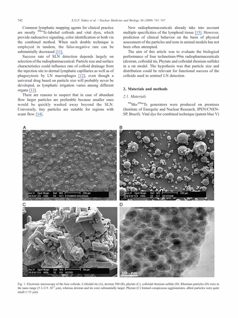
Fig. 1. Electronic microscopy of the four colloids. Colloidal tin (A), dextran 500 (Bthe nano range (5.1±3.9 .10-3 μm), whereas dextran and tin were substantially largsmall (b15 μm).
New radiopharmaceuticals already take into accountmultiple specificities of the lymphoid tissue [15]. However,prediction of clinical behavior on the basis of physicalassessment of the particles and tests in animal models has notbeen often attempted.
The aim of this article was to evaluate the biologicalperformance of four technetium-99m radiopharmaceuticals(dextran, colloidal tin, Phytate and colloidal rhenium sulfide)in a rat model. The hypothesis was that particle size anddistribution could be relevant for functional success of thecolloids used in sentinel LN detection.
2. Materials and methods
2.1. Materials
99Mo/99mTc generators were produced on premises(Institute of Energetic and Nuclear Research, IPEN/CNEN-SP, Brazil). Vital dye for combined technique (patent blue V)
), phytate (C), colloidal rhenium sulfide (D). Rhenium particles (D) were iner. Phytate (C) formed conspicuous agglomerates, albeit particles were quite

743E.G.F. Núñez et al. / Nuclear Medicine and Biology 36 (2009) 741–747
was purchased from Guerbet Prod. Radiológicos, Rio deJaneiro, RJ, Brazil.
Colloidal tin, dextran 500, and phytate kits were producedin house (IPEN/CNEN-São Paulo, Brazil) and colloidalrhenium sulfide kits were a donation from CIS BioInternational, France.
Twenty-four young male Wistar EPM-1 rats wereprovided by the Animal Facility of IPEN-CNEN, weightranging from 250 to 300 g. They were kept in cages withcontrolled temperature, humidity and noise, receivingindustrialized chow and water ad libitum. All animalstudies were performed at the Radiopharmacy Center,IPEN/CNEN, and the protocol was approved by the animalWelfare Ethical Committee.
2.2. Analysis of colloidal particles
Size and morphology of the nonradiolabeled particles wereexamined by transmission (Jeol JEM-2100, Tokyo, Japan) orscanning (Philips XL-30, Eindhoven, The Netherlands)electron microscopy following standard procedures. Sizingof samples was carried out from electron micrographs usingthe Image Tool for Windows (Version 2.0) software.Dimension was estimated by random Feret diameter.
2.3 Preparation of 99mTc-colloidal tin, 99mTc-dextran 500and 99mTc-Phytate
The three kits were prepared by similar procedures.Briefly, 2 ml of sodium [99mTc] pertechnetate (740 MBq)were added to the lyophilized product. After 20 min of
Fig. 2. Frequency histograms for the four colloids. Colloidal tin (A), dextran 500normal (Gaussian) distribution, all others being asymmetric.
incubation at room temperature (23–25°C), the radiophar-maceuticals were ready for use.
2.4 Preparation of 99mTc-colloidal rhenium sulfide
Two milliliters of sterile water for injections were addedto the vial B, containing lyophilized stannous salt. Then 0.5ml of this solution was transferred to vial A, containingrhenium sulfide colloid suspension. Immediately, 1.5 ml ofsodium pertechnetate (740 MBq) was added. The reactionwas carried out in boiling water bath for 30 min.
2.5. Radiochemical control
Radiochemical purity was assessed using paper chro-matography. For 99mTc-dextran 500 and 99mTc-colloidaltin, acetone was chosen for the mobile phase, andWhatman number 3 paper, as stationary phase. For99mTc-Phytate and 99mTc-colloidal rhenium sulfide metha-nol 85%/Whatman 3 and 2-butanone/Whatman 1 systemswere respectively utilized.
2.6. Popliteal node detection by gamma camera
The animals were anesthetized by means of 25 mg/kgtiletamine hydrochloride associated with 25 mg/kg zolaze-pam hydrochloride (Zoletil 50, Virbac, São Paulo, Brazil)administered intraperitoneally. Afterwards, 0.1 ml of eachradiopharmaceutical was injected in the footpad of the leftposterior limb.
Imaging was performed in a Mediso Imaging System,Budapest, Hungria, employing a low-energy, high-resolution
(B), phytate (C), colloidal rhenium sulfide (D). Only colloidal tin exhibited

744 E.G.F. Núñez et al. / Nuclear Medicine and Biology 36 (2009) 741–747
collimator using a 256×256×16 matrix size with 20% energywindow set at 140 keV. Dynamic studies were conducted fora period of 15 min. Static images were also acquired at 0.5and 1.5 h post injection (p.i.).
2.7. Surgical resection, biodistribution measurements andex vivo imaging
A second injection in the footpad followed with 0.05ml vital dye, five minutes before protocol sacrifice time(0.5 and 1.5 h).
The popliteal region was incised permitting access andremotion of the popliteal LN. Laparotomy with removal ofkidneys and liver was done at the same time. Theradioactivity of the specimens was determined by γ-countingusing as standard the injected dose of radiotracer. Results
Fig. 3. Dynamic study of 99mTc-colloidal tin over the initial 15 min. The dark dofirst minute.
were expressed as percentage of injected dose (%ID)per organ.
Ex vivo imaging analysis of the popliteal LN, kidneysand liver was performed using the syringe with injecteddose together allowing the calculation of the region ofinterest (ROI).
2.8. Statistical analysis
Particle size and biological data were analyzed byStatgraphics Plus 5.0 (Statistical Graphics, Fairfax, VA,USA). Standard skewness coefficient was used tomeasure the symmetry of colloid profiles. One-wayanalysis of variance (ANOVA) was performed to comparepopliteal LN, kidneys and liver uptakes of the products at0.5 and 1.5 h, followed by post hoc Tukey test. Student's
t, corresponding to the popliteal LN, can be noticed in all images since the

Fig. 4. Static images for all products after 90 min. 99mTc-colloidal tin(A), 99mTc-dextran 500 (B), 99mTc-Phytate (C) and 99mTc-colloidal rheniumsulfide (D). p.n., Popliteal node; i.n., inguinal node. Left kidney and bladder(A) or urinary bladder only (B, D) may be identified in the lower part of theanimal, not far from the marked LNs. The exception occurred withphytate (C), which elicited strong liver radioactivity, thus confirminghepatic excretion.
745E.G.F. Núñez et al. / Nuclear Medicine and Biology 36 (2009) 741–747
t test was carried out to identify tissue uptake variabilityover the time. The adopted significance level (α)was Pb.05.
3. Results
3.1. Colloidal particle morphology
Electron microscopy of nonradiolabeled products showedthat phytate particles were small (b15 μm), but they formedlarge agglomerates after solvent evaporation, interfering withsize calculation (Fig. 1).
In frequency histograms of particle size (Fig. 2), colloidaltin exhibited symmetric distribution (Standard skewnesscoefficient=−0.178), whereas distribution pattern for otherproducts (dextran, phytate and rhenium sulfide) wasasymmetrical — their standard skewness coefficient values
Table 1Biodistribution of 99mTc radiopharmaceutical in Wistar rats
Organ Lymph node
Time (h) 0.5 1.5
99mTc-colloidal tin 0.04±0.03 0.06±0.0399mTc-dextran 500 0.15±0.04 0.55±0.1499mTc-phytate 0.27±0.10 1.02±0.1499mTc-colloidal rhenium sulfide 1.13±0.32 2.72±0.64
Data are expressed as percentage of % injected dose per tissue±standard deviation
were 15.073, 9.194 and 11.541, respectively. When thevalues of this statistical parameter are outside the range −2 to2, the data may depart significantly from a normaldistribution (Statgraphics Plus 5.0).
With the exception of rhenium sulfide, with nanometricparticles (5.1±3.9 .10-3 μm), dextran displayed the smallestsize (21.4±12.8 μm), followed by colloidal tin (39.1±8.32μm) (P=1.248×10⁻⁷). Phytate had large nominal diameter(50.0±23.7 μm), as alluded to, but there was a bias due toagglomerates (Fig. 1).
3.2. Radiochemical controls
High purity levels (N95%) were achieved for all products,thus allowing biological evaluation.
3.3. Gamma camera diagnosis
In dynamic images acquired during the first 15 min, nodifference among the markers was documented. Popliteal LNwas visualized since the first minute p.i. (Fig. 3). However,99mTc-colloidal tin didn't migrate to the inguinal node, incontrast to all others (Fig. 4).
Static images confirmed renal excretion for 99mTc-colloidal tin, 99mTc-dextran 500 and 99mTc-colloidal rheniumsulfide. Only 99mTc-phytate was associated with hepaticmetabolization and biliary excretion (Fig. 4).
3.4. Organ biodistribution
Best LN uptake at both times corresponded by far to99mTc-colloidal rhenium sulfide, followed by 99mTc-Phytate,99mTc-dextran 500 and 99mTc-colloidal tin (P=.0002,ANOVA test for 30 min uptake). Differences between thelast two were minor and did not appear on Tukey test at30 min p.i. (Table 1).
99mTc-colloidal rhenium sulfide (P=.0186), 99mTc-phy-tate (P=.0017) and 99mTc-dextran 500 (P=.0080) increasedLN uptake between 0.5 and 1.5 h, by, respectively, 2.4-,3.71- and 3.72-fold. No change occurred with 99mTc-colloidal tin (P=.6300, nonsignificant) (Table 1).
Higher kidney values were reached by 99mTc-colloidalrhenium sulfide and 99mTc-colloidal tin, both after 0.5 h(P=.0116) and 1.5 h (P=.0001), when compared to the othermolecules (Table 1). However, for the same product, nodifference was detected in the two time points.
Kidneys Liver
0.5 1.5 0.5 1.5
0.69±0.17 1.02±0.29 0.32±0.10 0.38±0.100.08±0.06 0.13±0.05 0.04±0.04 0.06±0.050.18±0.04 0.16±0.02 13.09±1.94 22.04±1.410.94±0.49 0.91±0.05 0.74±0.26 2.36±0.93
(n=3).

Fig. 5. Ex vivo imaging analysis. ROI uptakes are expressed as a percentageof the injected dose. Dextran 500 (A), colloidal tin (B), phytate (C), colloidalrhenium sulfide (D). L, liver; K, kidneys; Do, dose.
746 E.G.F. Núñez et al. / Nuclear Medicine and Biology 36 (2009) 741–747
Liver uptake was low for all molecules excluding 99mTc-Phytate, which exhibited high percentage in this organ , bothat 0.5 and 1.5 h. Liver retention of the 99mTc-Phytateincreased 1.68-fold (P=.0029) between 0.5 and 1.5 h.
3.5. Ex vivo imaging
The ex vivo radioactivity quantification in LN, kidneysand liver by mean of the ROI for all radiocolloids were inaccordance with the observed results in biodistribution studyat 30 (Fig. 5) and 90 min p.i.
4. Discussion
Radiopharmaceuticals for SLN should promptly migratefrom the injection site, reach the target and remain therewithout substantial spill over [16]. Other LNs should remainclean, and contamination of blood vessels should not occur.In synthesis, they should concentrate inside the node, withminimal radioactivity in the other tissues [17].
Biological assessment of popliteal LN by using thecombination of radiopharmaceuticals and blue dye in mouseand rat model is well established [12,17,18]. Lymphaticdrainage from footpad of mice and rats appear to beexclusively into the polipteal LN, behind the knee. Thepopliteal LN is relatively large and is easy to find that it ispossible to locate and remove [19].
The four products considered in this study are radio-colloids. These radiopharmaceuticals are cleared by lym-phatic drainage to SLN, by passive diffusion, with a speedthat is inversely proportional to the particle size [20]. Once
inside the LN, they are retained by two main mechanism,phagocytosis and nonspecific mechanical trapping [21].Nevertheless, particle size should not be too small or nodalretention of the particle could be compromised [22].
Despite its pharmacologic importance, particle size isoverlooked in most protocols. Even products with the samebrand and supplier may undergo change of particle size frombatch to batch. It is worth mentioning that colloid andradiocolloid dimensions can be controllable by manipulatingthe conditions under which such colloids form [14].
Success of the SLN technique hinges not only onmolecular properties of the radiopharmaceutical but also onsurgical skills and injection routine.
With regard to the first variable, 99mTc sulfur colloid hasbeen used in the United States, and both 99mTc Nan colloidand 99mTc rhenium colloid have been used in Europe. InJapan, 99mTc tin colloid, 99mTc albumin and 99mTc phytateare available for sentinel node biopsy [23]. As here indicated,each agent is endowed with specific properties which have tobe tailored to different clinical circumstances.
In Japan and several other countries, large-particle tincolloid was popular at first for breast cancer patients, butsince nodal uptake was relatively poor because of the largeparticle size, small-particle tin colloid, phytic acid andrhenium colloid were progressively introduced.
Timing of the injection deserves attention as well. If alarge-particle preparation such as tin colloid is preferred, it isadministered 3–15 h before surgery because of slowmigration [24].
Delay to reach the popliteal LN was not an end point inthe present model, because small animals with nearbyinjection sites are not appropriate for such observation.Nevertheless, speedier access might have played a role inthe more remarkable LN uptake of small particles (Table 1and Fig. 2).
A hitherto unreported finding that will require furtherstudies, was the relationship between symmetry of particlesize profile and change in uptake over time. In the case ofcolloidal tin, constant uptake coincided with symmetricdistribution of colloid particle size. The contrary occurredwhen products displayed asymmetric distribution (dextran500, Tc-phytate and colloidal rhenium sulfide). Suchbiphasic uptake pattern can be ascribed to the hypothesisof small particles reaching the LN first, followed by a secondwave of larger ones. Additional studies should be performedtaking into consideration the radiocolloid particle profile toconfirm this result because some radiopharmaceuticals forSLN detection have shown differences in their particleprofiles before and after radiolabeling. However, nonradio-labeled and radiolabeled particles profile showed similarbehavior respect with symmetry [25].
Excretion mechanism is not a negligible detail, particu-larly for nodes and tumors located in the immediateneighborhood of excretory routes. Disappointing or mis-leading imaging results are a distinct possibility, whenprostate or endometrial cancers are analyzed by SLN

747E.G.F. Núñez et al. / Nuclear Medicine and Biology 36 (2009) 741–747
procedure with the help of agents with renal disposal [26,27].99mTc-phytate, mainly eliminated by hepatic metabolism,could be an option for such malignancies (Fig. 4C).
In synthesis, development of new radioactive agents forSLN should obviously focus biochemical specificities oflymphoid tissue, but without missing physiological reper-cussions of particle size and distribution.
5. Conclusion
The results are suggesting that particle size and profile ofthe colloids used as radiopharmaceuticals may interfere withsentinel LN uptake. Rat model is sensitive animal model tostudy these radiotracer properties. Such features should betaken in account when new radiopharmaceuticals, or novelapplications for existing drugs, are being examined.
Acknowledgments
The authors are indebted to the National Commission forNuclear Energy (CNEN, São Paulo, Brazil) and the Interna-tional Atomic Energy Agency (IAEA, Vienna, Austria) forscientific grants. The first author gratefully acknowledgesRelma Tavares de Oliveira, for the inspiration to write thispaper. The team of authors is also grateful to Nildemar A. M.Ferreira for his assistance in electron microscopy analysis.
References
[1] Tobler NE, Detmar M. Tumor and lymph node lymphangiogenesis —impact on cancer metastasis. J Leukoc Biol 2006;80:691–6.
[2] Tsopelas C, Bevington E, Kollias J, Shibli S, Farshid G, Coventry B,et al. 99mTc–Evans blue dye for mapping contiguous lymph nodesequences and discriminating the sentinel lymph node in an ovinemodel. Ann Surg Oncol 2006;13(5):692–700.
[3] Koyama T, Tsubota A, Nariai K, Yoshikawa T, Mitsunaga M, Sumi M,et al. Detection of sentinel nodes by a novel red-fluorescent dye, ATX-S10Na (II), in an orthotopic xenograft rat model of human gastriccarcinoma. Lasers Surg Med 2007;39:76–82.
[4] Chen SL, Iddings DM, Scheri RP, Bilchik AJ. Lymphatic mapping andsentinel node analysis: current concepts and applications. CA Cancer JClin 2006;56:292–309.
[5] Luciani A, Itti E, Rahmouni A, Meignan M, Clement O. Lymph nodeimaging: basic principles. Eur J Radiol 2006;58:338–44.
[6] Aarsvold JN, Alazraki NP. Update on detection of sentinel lymphnodes in patients with breast cancer. Semin Nucl Med 2005;35:116–28.
[7] Dancey AL, Mahonb BS, Rayatt SS. A review of diagnostic imaging inmelanoma. J Plast Reconstr Aesthet Surg 2008;61(11):1275–83.
[8] Meinhardt W. Sentinel node evaluation in prostate cancer. EAU-EBUUpdate Series 2007;5(6):223–31.
[9] Lambaudie E, Collinet P, Narducci F, Sonoda Y, Papageorgiou T,Carpentier P, et al. Laparoscopic identification of sentinel lymph nodesin early stage cervical cancer Prospective study using a combination ofpatent blue dye injection and technetium radiocolloid injection.Gynecol Oncol 2003;89:84–7.
[10] Yanagita S, Natsugoe S, Uenosono Y, Arigami T, Arima H, Kozono T,et al. Detection of micrometastases in sentinel node navigation surgeryfor gastric cancer. Surg Oncol 2008;17(3):203–10.
[11] Noguchi M. Sentinel lymph node biopsy and breast cancer. Br J Surg2002;89:21–34.
[12] Takagi K, Uehara T, Kaneko E, Nakayama M, Koizumi M, Endo K,et al. 99mTc-labeled mannosyl-neoglycoalbumin for sentinel lymphnode identification. Nucl Med Biol 2004;31:893–900.
[13] Jinno H, Ikeda T, Matsui A, Kitagawa Y, Kitajima M, Fujii H, et al.Sentinel lymph node biopsy in breast cancer using technetium-99mtin colloids of different sizes. Biomed Pharmacother 2002;56:213s–6s.
[14] Higashi H, Natsugoe S, Uenosono Y, Ehi K, Arigami T, Nakabeppu Y,et al. Particle size of tin and phytate colloid in sentinel nodeidentification. J Surg Res 2004;121:1–4.
[15] Wallace AM, Hoh CK, Darrah DD, Schulteis G, Vera DR. Sentinellymph node mapping of breast cancer via intradermal administration ofLymphoseek. Nucl Med Biol 2007;34:849–53.
[16] Keshtgar MRS, Ell PJ. Clinical role of sentinel-lymph-node biopsy inbreast cancer. Lancet Oncol 2002:3105–10.
[17] Paiva GR, Filho RSO, Ferreira LM, Wagner J, Nogueira SA, NovoNF, et al. Phytate technetium-99m versus dextran 500 technetium-99m in the sentinel lymph node biopsy. Acta Radiol 2006;47(1):65–70.
[18] Nathanson SD, Anaya P, Karvelis KC, Eck L, Havstad S. Sentinellymph node uptake of two different technetium-labeled radiocolloids.Ann Surg Oncol 1997;4(2):104–10.
[19] Løvik M, Alberg T, Nygaard UC, Samuelsen M, Groeng EC, GaarderPI. Popliteal lymph node (PLN) assay to study adjuvant effects onrespiratory allergy. Methods 2007;41(1):72–9.
[20] Linehan DC, Eberlein TJ. Mechanisms of radiocolloid localization insentinel node biopsy. Ann Surg Oncol 2000;7(2):77.
[21] Schöder H, Glass EC, Pecking AP, Harness JK, Wallace AM, Hirnle P,et al. Molecular targeting of the lymphovascular system for imagingand therapy. Cancer Metastasis Rev 2006;25:185–201.
[22] Fass L. Imaging and cancer: a review. Mol Oncol 2008;2:115–52.[23] Koizumi M, Nomura E, Yamada Y, Takiguchi T, Makita M, Iwase T,
et al. Radioguided sentinel node detection in breast cancer patients:comparison of 99mTc phytate and 99mTc rhenium colloid efficacy.Nucl Med Commun 2004;25(10):1031–7.
[24] Ikeda T, Jinno H, Fujii H, Kitajima M. Recent development of sentinellymph node biopsy for breast cancer in Japan. Asian J Surg 2004;27(4):275–8.
[25] Tsopelas C. Particle size analysis of 99mTc-labeled and unlabeledantimony trisulfide and rhenium sulfide colloids intended forlymphoscintigraphic application. J Nucl Med 2001;42:460–6.
[26] Takashima H, Egawa M, Imao T, Fukuda M, Yokoyama K, Namiki M.Validity of sentinel lymph node concept for patients with prostatecancer. J Urol 2004;171:2268–71.
[27] Niikura H, Okamura C, Utsunomiya H, Yoshinaga K, Akahira J, Ito K,et al. Sentinel lymph node detection in patients with endometrialcancer. Gynecol Oncol 2004;92(2):669–74.
Copyright © 2022 FDOKUMEN