Polo-like kinase and survivin are esophageal tumor-specific promoters
Upload
khangminh22Category
view
2download
0
�����������������
Citation: Kudo, S.; Chinda, D.;
Shimoyama, T.; Yasuda, K.; Akitaya,
K.; Arai, T.; Miyazawa, K.; Hayamizu,
S.; Yanagimachi, M.; Tsukamoto, T.;
et al. Influence of Esophageal
Endoscopic Submucosal Dissection
on the Changes of Energy
Metabolism during the Perioperative
Period. Cancers 2022, 14, 2015.
https://doi.org/10.3390/
cancers14082015
Academic Editor: David Wong
Received: 23 March 2022
Accepted: 14 April 2022
Published: 15 April 2022
Publisher’s Note: MDPI stays neutral
with regard to jurisdictional claims in
published maps and institutional affil-
iations.
Copyright: © 2022 by the authors.
Licensee MDPI, Basel, Switzerland.
This article is an open access article
distributed under the terms and
conditions of the Creative Commons
Attribution (CC BY) license (https://
creativecommons.org/licenses/by/
4.0/).
cancers
Article
Influence of Esophageal Endoscopic Submucosal Dissection onthe Changes of Energy Metabolism during thePerioperative PeriodSae Kudo 1, Daisuke Chinda 2,* , Tadashi Shimoyama 3, Kohei Yasuda 1, Kazuki Akitaya 1, Tetsu Arai 1,Kuniaki Miyazawa 1, Shiro Hayamizu 1, Miyuki Yanagimachi 4, Toshiaki Tsukamoto 5, Masatoshi Kaizuka 1,Yohei Sawada 1, Tetsuya Tatsuta 1, Keisuke Hasui 1, Hidezumi Kikuchi 6, Hiroto Hiraga 1, Hirotake Sakuraba 1,Tatsuya Mikami 7 and Shinsaku Fukuda 1
1 Department of Gastroenterology and Hematology, Hirosaki University Graduate School of Medicine,Hirosaki 036-8562, Japan; [email protected] (S.K.); [email protected] (K.Y.);[email protected] (K.A.); [email protected] (T.A.); [email protected] (K.M.);[email protected] (S.H.); [email protected] (M.K.); [email protected] (Y.S.);[email protected] (T.T.); [email protected] (K.H.); [email protected] (H.H.);[email protected] (H.S.); [email protected] (S.F.)
2 Division of Endoscopy, Hirosaki University Hospital, Hirosaki 036-8563, Japan3 Aomori General Health Examination Center, Aomori 030-0962, Japan; [email protected] Department of Endocrinology and Metabolism, Hirosaki University Graduate School of Medicine,
Hirosaki 036-8562, Japan; [email protected] Division of Rehabilitation, Hirosaki University Hospital, Hirosaki 036-8563, Japan; [email protected] Department of Community Medicine, Hirosaki University Graduate School of Medicine,
Hirosaki 036-8562, Japan; [email protected] Center of Healthy Aging Innovation, Hirosaki University Graduate School of Medicine,
Hirosaki 036-8562, Japan; [email protected]* Correspondence: [email protected]; Tel.: +81-172-33-5111
Simple Summary: This study aimed to measure resting energy expenditure (REE) and assess thephysical invasiveness of esophageal endoscopic submucosal dissection (ESD) during perioperativeperiods. In addition, the factors affecting changes in REE were investigated. Subjects were examinedusing an indirect calorimeter and the stress factor was calculated based on basal energy expenditureand body weight. REE/body weight on the day following ESD was significantly higher than thatof the same day. The stress factor on the day after ESD was 1.11. The increase in WBC, neutrophil,and CRP levels was associated with the change in REE ratio. Among the factors affecting changes inenergy metabolism, only the total resection area was associated with changes in REE. It is suggestedthat patients who undergo esophageal ESD require more attention in perioperative managementwhen the resection area of the lesions is larger.
Abstract: Esophageal endoscopic submucosal dissection (ESD) is considered to be more complexthan gastric ESD. This study aimed to assess the physical invasiveness of esophageal ESD duringperioperative periods by measuring resting energy expenditure (REE). The factors affecting REEthat could be used to identify patients requiring perioperative management were also investigated.Overall, 75 patients who had undergone esophageal ESD were prospectively enrolled. REE, bodyweight, and basal energy expenditure were measured on the day of and the day following ESD. Themean REE/body weight was 20.2 kcal/kg/day on the day of ESD and significantly increased to23.0 kcal/kg/day one day after ESD. The stress factor on the day after ESD was 1.11. White bloodcell, neutrophil, and C-reactive protein levels increased on the day after ESD and correlated with thechanges in REE. Among the factors including age, body mass index, total resection area, operationtime, and sarcopenia, only the total resection area was associated with changes in REE. In conclusion,energy metabolism increases during the perioperative period for esophageal ESD. The increase inthe stress factor for esophageal ESD was higher than that in gastric and colorectal ESD. Furthermore,patients with large resection areas require greater attention in perioperative management.
Cancers 2022, 14, 2015. https://doi.org/10.3390/cancers14082015 https://www.mdpi.com/journal/cancers

Cancers 2022, 14, 2015 2 of 11
Keywords: esophageal cancer; endoscopic submucosal dissection; energy metabolism; resting energyexpenditure; indirect calorimeter
1. Introduction
Esophageal cancer is the sixth leading cause of cancer-related deaths worldwide, andits treatment depends on the activities of daily living and patient disease stage [1]. In olderpatients, full esophagectomy is considered a highly invasive procedure because treatmentcan deteriorate patient conditions [2]. Chemoradiotherapy (CRT) has been recognizedas a reliable treatment regardless of disease stages in esophageal cancer. However, latetoxicities caused by CRT often become fatal [2,3]. Endoscopic submucosal dissection (ESD)for superficial esophageal cancer is an efficacious treatment in terms of functional preser-vation and safety, especially in older patients [4–7]. ESD has been the standard treatmentfor superficial esophageal cancer in Japan since 2012 as it allows en bloc resection andhas a low risk of local cancer recurrence [7,8]. Esophageal ESD is also recommended as afirst-line treatment for clinical T1a-Epithelium/lamina propria mucosae diagnosed as non-circumferential esophageal cancer or in long-axis whole-circumferential esophageal cancers< 50 mm [7]. In addition, clinical T1a-muscularis mucosae/T1b-submucosa 1 esophagealcancer is also considered an indication for ESD if it is noncircumferential [7].
During operations such as ESD, hypermetabolism can be induced by inflammation andprotein catabolism [9–11], reflecting the invasiveness of the surgery [10]. Differences in thedegree of energy metabolism are caused by the production of pro-inflammatory cytokinesand increased glucose oxidation during pathological stress [12]. In our previous study,changes in resting energy expenditure (REE) were measured using an indirect calorimeterduring the perioperative period for gastric and colorectal ESD. Based on these results, theincrease in REE on the day following ESD was low compared to that experienced afteropen surgery; therefore, ESD was recognized as a less invasive treatment [13,14].
However, esophageal ESD is considered a more complex procedure than gastricESD [15,16]. In addition, bacteremia and postoperative stricture are perioperative com-plications common in esophageal ESD. Several studies have reported that blood culturesobtained 10 min after gastric ESD and 5 min after colorectal ESD were positive in 4.3%and 2.5% of patients, respectively [17,18]. Furthermore, the rate of bacteremia was 12–22%after esophageal bougienage and 0–52% after variceal sclerotherapy [17,19]. In these pro-cedures, the cultured microorganisms were oral commensal bacteria [17,20]. Although afew studies have investigated bacteremia in esophageal ESD, bacteremia can occur afteresophageal ESD. Postoperative stricture occurred in 90% of patients with lesions > 3/4 ofthe circumference of the lumen [21]. For the above reasons, it is possible that the physicalinvasiveness of esophageal ESD during the perioperative period differs from that of gastricand colorectal ESD.
This study aimed to measure REE using an indirect calorimeter and assess physicalinvasiveness during the perioperative period for esophageal ESD. Additionally, we investi-gated the factors affecting changes in REE to identify cases that required additional carein perioperative management. We found that for esophageal ESD, energy metabolism in-creased during the perioperative period, and its degree of invasiveness was low comparedwith that of open surgery.
2. Materials and Methods2.1. Measuring REE Using an Indirect Calorimeter
Table 1 shows the patient characteristics. Between July 2013 and March 2019, we en-rolled 116 consecutive patients who were to undergo esophageal ESD at Hirosaki UniversityHospital. We excluded patients with a history of liver cirrhosis, respiratory diseases, andthyroid diseases; those undergoing artificial dialysis; and those with other malignancies.Finally, 75 patients (median age 66 years; 67 men) were included in the study (Figure 1).

Cancers 2022, 14, 2015 3 of 11
We evaluated our sample using a power of 80% and a two-sided alpha level of 0.05. Thestandard deviation was computed using the prediction value based on data from our previ-ous research on ESD for gastric cancer. As a result, the sample size of 75 had a statisticalpower of 0.9986 (REE/body weight) and a stress factor of 0.9995.
Table 1. Patient baseline characteristics.
Variables n/Median (Range)
Sex (Male:Female) 67:8Age (years old) 66 (45–90)BMI (kg/m2) 22.1 (16.2–30.4)PMI (cm2/m2) 6.4 (3.2–12.4)Main tumor location
Upper thoracic esophagus 6Middle thoracic esophagus 5Lower thoracic esophagus 48Gastroesophageal junction 16
Total resection area (cm2) 6.9 (0.5–106.0)Operation time (minutes) 75 (17–265)Histologic type
Squamous cell carcinoma 72Adenocarcinoma 2Angioma 1
ComplicationsBleeding 1 (1.3%)Perforation 0 (0%)Fever (>38 ◦C) 4 (5.3%)
Data are presented as number (percentage) or median (range). BMI: body mass index; PMI: psoas musclemass index.
Cancers 2022, 14, x 3 of 12
2. Materials and Methods
2.1. Measuring REE Using an Indirect Calorimeter
Table 1 shows the patient characteristics. Between July 2013 and March 2019, we en-
rolled 116 consecutive patients who were to undergo esophageal ESD at Hirosaki Univer-
sity Hospital. We excluded patients with a history of liver cirrhosis, respiratory diseases,
and thyroid diseases; those undergoing artificial dialysis; and those with other malignan-
cies. Finally, 75 patients (median age 66 years; 67 men) were included in the study (Figure
1). We evaluated our sample using a power of 80% and a two-sided alpha level of 0.05.
The standard deviation was computed using the prediction value based on data from our
previous research on ESD for gastric cancer. As a result, the sample size of 75 had a statis-
tical power of 0.9986 (REE/body weight) and a stress factor of 0.9995.
Figure 1. Flow chart of study patient selection.
Table 1. Patient baseline characteristics.
Variables n/median (range)
Sex (Male:Female) 67:8
Age (years old) 66 (45–90)
BMI (kg/m2) 22.1 (16.2–30.4)
PMI (cm2/m2) 6.4 (3.2–12.4)
Main tumor location
Upper thoracic esophagus 6
Middle thoracic esophagus 5
Lower thoracic esophagus 48
Gastroesophageal junction 16
Total resection area (cm2) 6.9 (0.5–106.0)
Operation time (minutes) 75 (17–265)
Histologic type
Squamous cell carcinoma 72
Adenocarcinoma 2
Angioma 1
Complications
Bleeding 1 (1.3%)
Perforation 0 (0%)
Fever (> 38 °C) 4 (5.3%)
Figure 1. Flow chart of study patient selection.
All ESD procedures (75 subjects) were carried out by five endoscopists certified by theJapan Gastroenterological Endoscopy Society (JGE) using a conventional single-channelvideo endoscope (GIF-Q260J, H260, or H290; Olympus, Tokyo, Japan) with a hood. The ESDprocedure was performed using a water jet short needle knife (Flush Knife BT-S; DK2620J,Fujinon, Tokyo, Japan), a water jet hook knife (KD-620LR, Olympus, Tokyo, Japan), anda high-frequency generator with an automatically controlled system (VIO3 or VIO300D;ERBE, Tübingen, Germany). All patients were administered intravenous pethidine hy-drochloride 25 mg/body and diazepam 5 mg/body or midazolam 2 mg/body prior to

Cancers 2022, 14, 2015 4 of 11
the start of ESD. These drugs were increased as appropriate depending on the degreeof sedation, and a total of 25–100 mg/body pethidine hydrochloride and 5–20 mg/bodydiazepam or midazolam in 5–10 mg/body were finally administered before and duringESD. In addition to these drugs, dexmedetomidine hydrochloride was used for sedationin three patients. Dexmedetomidine hydrochloride was started at 6 µg/h/kg for the first10 min and subsequently decreased to 0.4 µg/h/kg by continuous intravenous infusion.
REE was examined using an indirect calorimeter (METAVINE-N VMB-002N; VINE,Tokyo, Japan) on the day of and day after ESD [14,22]. Each patient fasted for more than12 h, and REE was measured after 30 min of bed rest early in the morning on the day ofand the day after ESD. The REE was determined three times and the average value wascalculated. If the variability exceeded the range of 100 kcal, a fourth measurement wastaken and REE was calculated using the average of the three values, excluding the onefarthest from the average of the two medians. In a previous study, gas infusion tests in threeindirect calorimeters showed great reproducibility and accuracy (within 3%) for energyexpenditure [23]. In this study, three REE measurements were within the range of 100 kcalfor 61 of 75 subjects (81.3%) before ESD and 62 of 75 subjects (82.6%) on postoperativeday 1 (POD1), indicating high reproducibility. Afterward, ESD was performed and REE inthe fasted state was similarly measured in the morning on POD1. Furthermore, the bodyweight of each patient was measured on the day of and the day after ESD. We used thesemeasurements to calculate the changes in REE/body weight ratio.
2.2. BEE and Stress Factors
Basal energy expenditure (BEE) was assessed using the Harris–Benedict equation [24]based on Long’s method [25]. The REE was calculated by multiplying the BEE with stressand activity factors. The stress factor is one of the markers of hypermetabolic status [26,27].Assuming REE/BEE on the day of ESD to be 1.00, the REE/BEE on POD1 can likely beconsidered a stress factor because the activity factors on the day of ESD and POD1 are thesame in the resting state.
2.3. Hematological Response in Perioperative Period
The blood samples were collected in the morning on the day of ESD and POD1 afterfasting for 12 h while resting. The number and differential counts of white blood cells(WBC) were measured using XE-5000 (Sysmex, Kobe, Japan). The serum levels of C-reactiveprotein (CRP) were measured using a JCA-BM6070 (EOL Ltd., Tokyo, Japan).
The change ratios of WBC, neutrophil, and CRP were investigated and correlated withchanges in REE using the Spearman rank correlation coefficient.
2.4. Factor Associated with Change Ratio of REE during Perioperative Period
Age, body mass index (BMI), total resection area, operation time, and sarcopenia wereevaluated as factors affecting changes in energy metabolism. The patients were dividedinto two groups. The cut-off values were as follows: age, 65 years (definition of olderage provided by the World Health Organization [28]) and BMI, 25 (kg/m2) (definition ofobesity [29]). The resection area was calculated by approximating an elliptical shape withthe long and short axes of the resected specimens. For the nine subjects with multiplelesions, the total resection area was computed from all resected specimens. The totalresection area and operation time were selected as the cut-offs for the median value (6.9 cm2
and 75 min). Sarcopenia is defined as a condition with low skeletal muscle mass andstrength [30]. Skeletal muscle mass was expressed as skeletal muscle index (SMI) [31,32].SMI evaluation using dual-energy X-ray absorptiometry or bioelectrical impedance analysisis the gold standard method; however, these methods are not easily available in commonfacilities [33]. As SMI correlates with psoas muscle index (PMI) [33,34], we used computedtomography in the preoperative period and estimated PMI by the cross-sectional area ofthe muscle at the third lumbar vertebra level normalized based on the patient’s height(cm2/m2) [35,36]. The cut-off value of PMI was defined as 6.0 cm2/m2 for men and
Cancers 2022, 14, 2015 5 of 11
3.4 cm2/m2 for women [35]. These factors were computed between the two groups todetermine the change ratio of REE from the preoperative to the postoperative state.
2.5. Statistical Analysis
The statistical analysis of the clinical data was performed using SPSS (version 24.0;SPSS Inc., Chicago, IL, USA) and R (R Foundation for Statistical Computing, version R-3.4.3). Data are expressed as medians and interquartile ranges. Statistical differenceswere analyzed using the paired t-test, the Wilcoxon signed-rank test, and the Mann–Whitney U test. We also used a non-parametric mixed regression model to examinethe relationship between each factor and changes in REE, and performed multivariateanalysis using generalized linear models. A p-value of less than 0.05 was consideredstatistically significant.
3. Results3.1. REE, REE/Body Weight, and REE/BEE
The changes in REE, REE/body weight, and REE/BEE are shown in Table 2. The REEon POD1 was elevated in 56 of 75 patients (74.7%) compared with that on the day of ESD.The median of REE was 1194.7 kcal/day on the day of ESD, but 1340.0 kcal/day on POD1,significantly higher by 12.2% (p < 0.001).
Table 2. Changes in REE, body weight, and BEE.
Measurements Day of ESD Day Following ESD
REE (kcal) 1194.7 (608.1–1583.7) 1340.0 * (847.6–2111.3)Body weight (kg) 59.4 (40.6–86.1) 58.1 * (38.9–86.5)BEE (kcal) 1235.0 (941.0–1677.0) 1247.1 (983.7–1562.8)
Data are presented as median (range). * p < 0.05, vs day of ESD. REE: resting energy expenditure; BEE: basalenergy expenditure; ESD: endoscopic submucosal dissection.
There was no significant difference in REE changes between the five operators (datanot shown).
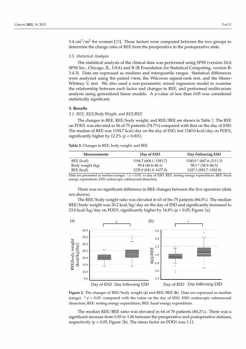
The REE/body weight ratio was elevated in 63 of the 75 patients (84.0%). The medianREE/body weight was 20.2 kcal/kg/day on the day of ESD and significantly increased to23.0 kcal/kg/day on POD1, significantly higher by 14.8% (p < 0.05; Figure 2a).
Cancers 2022, 14, x 6 of 12
Figure 2. The changes of REE/body weight (a) and REE/BEE (b). Data are expressed as median
(range). * p < 0.05: compared with the value on the day of ESD. ESD: endoscopic submucosal dissec-
tion; REE: resting energy expenditure; BEE: basal energy expenditure.
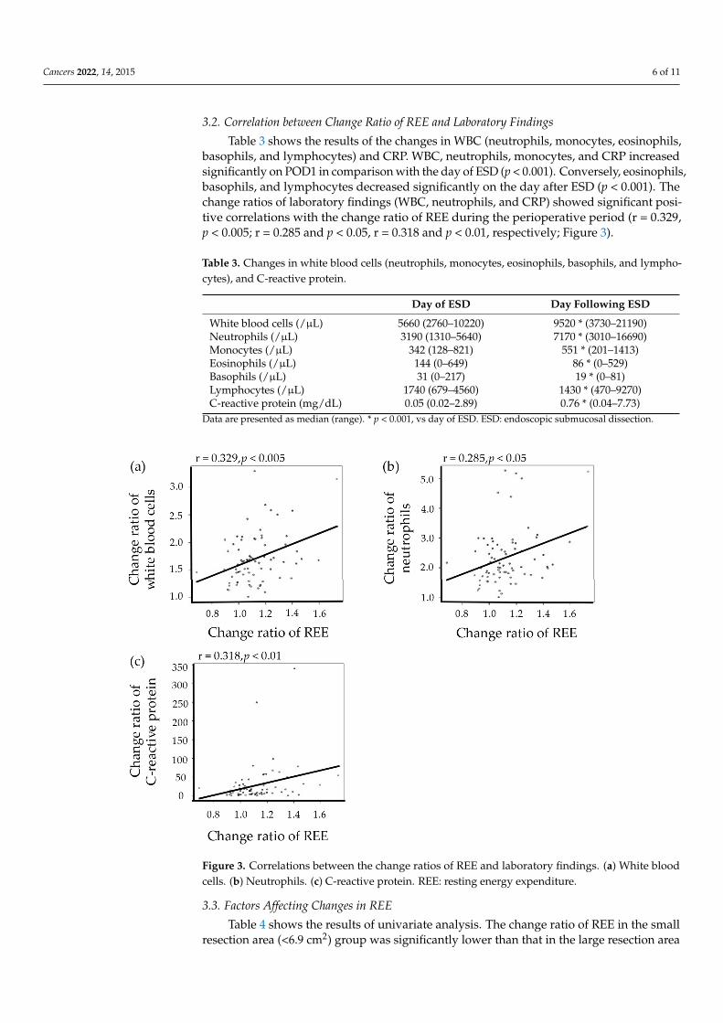
3.2. Correlation between Change Ratio of REE and Laboratory Findings
Table 3 shows the results of the changes in WBC (neutrophils, monocytes, eosino-
phils, basophils, and lymphocytes) and CRP. WBC, neutrophils, monocytes, and CRP in-
creased significantly on POD1 in comparison with the day of ESD (p < 0.001). Conversely,
eosinophils, basophils, and lymphocytes decreased significantly on the day after ESD (p <
0.001). The change ratios of laboratory findings (WBC, neutrophils, and CRP) showed sig-
nificant positive correlations with the change ratio of REE during the perioperative period
(r = 0.329, p < 0.005; r = 0.285 and p < 0.05, r = 0.318 and p < 0.01, respectively; Figure 3).
Table 3. Changes in white blood cells (neutrophils, monocytes, eosinophils, basophils, and lympho-
cytes), and C-reactive protein.
Day of ESD Day Following ESD
White blood cells (/μL) 5660 (2760–10220) 9520 * (3730–21190)
Neutrophils (/μL) 3190 (1310–5640) 7170 * (3010–16690)
Monocytes (/μL) 342 (128–821) 551 * (201–1413)
Eosinophils (/μL) 144 (0–649) 86 * (0–529)
Basophils (/μL) 31 (0–217) 19 * (0–81)
Lymphocytes (/μL) 1740 (679–4560) 1430 * (470–9270)
C-reactive protein (mg/dL) 0.05 (0.02–2.89) 0.76 * (0.04–7.73)
Data are presented as median (range). * p < 0.001, vs day of ESD. ESD: endoscopic submucosal dis-
section.
Figure 2. The changes of REE/body weight (a) and REE/BEE (b). Data are expressed as median(range). * p < 0.05: compared with the value on the day of ESD. ESD: endoscopic submucosaldissection; REE: resting energy expenditure; BEE: basal energy expenditure.
The median REE/BEE ratio was elevated in 64 of 76 patients (84.2%). There was asignificant increase from 0.95 to 1.06 between the preoperative and postoperative statuses,respectively (p < 0.05; Figure 2b). The stress factor on POD1 was 1.11.
Cancers 2022, 14, 2015 6 of 11
3.2. Correlation between Change Ratio of REE and Laboratory Findings
Table 3 shows the results of the changes in WBC (neutrophils, monocytes, eosinophils,basophils, and lymphocytes) and CRP. WBC, neutrophils, monocytes, and CRP increasedsignificantly on POD1 in comparison with the day of ESD (p < 0.001). Conversely, eosinophils,basophils, and lymphocytes decreased significantly on the day after ESD (p < 0.001). Thechange ratios of laboratory findings (WBC, neutrophils, and CRP) showed significant posi-tive correlations with the change ratio of REE during the perioperative period (r = 0.329,p < 0.005; r = 0.285 and p < 0.05, r = 0.318 and p < 0.01, respectively; Figure 3).
Table 3. Changes in white blood cells (neutrophils, monocytes, eosinophils, basophils, and lympho-cytes), and C-reactive protein.
Day of ESD Day Following ESD
White blood cells (/µL) 5660 (2760–10220) 9520 * (3730–21190)Neutrophils (/µL) 3190 (1310–5640) 7170 * (3010–16690)Monocytes (/µL) 342 (128–821) 551 * (201–1413)Eosinophils (/µL) 144 (0–649) 86 * (0–529)Basophils (/µL) 31 (0–217) 19 * (0–81)Lymphocytes (/µL) 1740 (679–4560) 1430 * (470–9270)C-reactive protein (mg/dL) 0.05 (0.02–2.89) 0.76 * (0.04–7.73)
Data are presented as median (range). * p < 0.001, vs day of ESD. ESD: endoscopic submucosal dissection.
Cancers 2022, 14, x 7 of 12
Figure 3. Correlations between the change ratios of REE and laboratory findings. (a) White blood
cells. (b) Neutrophils. (c) C-reactive protein. REE: resting energy expenditure.
3.3. Factors Affecting Changes in REE
Table 4 shows the results of univariate analysis. The change ratio of REE in the small
resection area (< 6.9 cm2) group was significantly lower than that in the large resection
area (≥ 6.9 cm2) group (p < 0.05). The size of the resection area was positively associated
with the change ratio of REE. In contrast, there were no significant differences in age (< 65
vs. ≥ 65), BMI (< 25 kg/m2 vs. ≥ 25 kg/m2), operation time (< 75 min vs. ≥ 75 min), or sarco-
penia (PMI < 6.0 cm2/m2 vs. ≥ 6.0 cm2/m2 for men and < 3.4 cm2/m2 vs. ≥ 3.4 cm2/m2 for
women, respectively). Table 5 shows the results of the multivariate analysis using gener-
alized linear models. There was no significant difference for each factor, but the value of
estimates was the highest in the total resection area.
Table 4. Univariate analysis for the factors associated with REE during the perioperative period of
ESD.
Variables n REE during the Perioperative Period Changes in the
Ratio of REE Day of ESD Day Following ESD
Age (years old)
<65 33 1250.0
(686.8–1535.0)
1391.0
(1009.1–2111.3) 1.08
≥65 42 1117.0
(608.1–1583.7)
1289.2
(847.6–1671.3) 1.10
BMI (kg/m2)
<25 60 1125.3
(608.1–1532.7)
1293.3
(847.6–2111.3) 1.09
Figure 3. Correlations between the change ratios of REE and laboratory findings. (a) White bloodcells. (b) Neutrophils. (c) C-reactive protein. REE: resting energy expenditure.
3.3. Factors Affecting Changes in REE
Table 4 shows the results of univariate analysis. The change ratio of REE in the smallresection area (<6.9 cm2) group was significantly lower than that in the large resection area

Cancers 2022, 14, 2015 7 of 11
(≥6.9 cm2) group (p < 0.05). The size of the resection area was positively associated with thechange ratio of REE. In contrast, there were no significant differences in age (<65 vs. ≥65),BMI (<25 kg/m2 vs. ≥25 kg/m2), operation time (<75 min vs. ≥75 min), or sarcopenia(PMI < 6.0 cm2/m2 vs. ≥6.0 cm2/m2 for men and <3.4 cm2/m2 vs. ≥3.4 cm2/m2 forwomen, respectively). Table 5 shows the results of the multivariate analysis using gener-alized linear models. There was no significant difference for each factor, but the value ofestimates was the highest in the total resection area.
Table 4. Univariate analysis for the factors associated with REE during the perioperative pe-riod of ESD.
Variables nREE during the Perioperative Period
Changes in theRatio of REEDay of ESD Day Following
ESD
Age (years old)
<65 33 1250.0(686.8–1535.0)
1391.0(1009.1–2111.3) 1.08
≥65 42 1117.0(608.1–1583.7)
1289.2(847.6–1671.3) 1.10
BMI (kg/m2)
<25 60 1125.3(608.1–1532.7)
1293.3(847.6–2111.3) 1.09
≥25 15 1346.0(1188.3–1583.7)
1498.0(859.7–1714.3) 1.08
Total resection area (cm2)
<6.9 37 1188.3(608.1–1560.7)
1291.3(847.6–1671.3) 1.07
≥6.9 38 1239.5(686.8–1583.7)
1397.7(962.0–2111.3) 1.13 *
Operation time (minutes)
<75 36 1159.5(826.0–1583.7)
1286.0(921.9–2111.3) 1.08
≥75 39 1241.7(608.1–1537.0)
1397.7(847.6–1714.3) 1.09
Sarcopenia
Non-sarcopenia 52 1231.3(686.8–1583.7)
1344.7(859.7–2111.3) 1.08
Sarcopenia 23 1111.7(608.1–1560.7)
1335.7(847.6–1714.3) 1.12
Data are presented as median (range). * p < 0.05, vs. total resection area < 6.9 cm2. REE: resting energy expenditure;ESD: endoscopic submucosal dissection; BMI: body mass index.
Table 5. Parameter estimates of main effects for changes in REE using generalized linear models.
Variables Estimates SE Odds Ratio p-Value
Age 0.6193 0.4985 1.8577 0.214BMI −0.1944 0.6002 0.8233 0.746Total resection area 0.7855 0.5003 2.1935 0.116Operation time 0.0576 0.5012 1.0593 0.908Sarcopenia 0.0690 0.5252 1.0714 0.895
REE: resting energy expenditure; SE: standard error; BMI: body mass index.
4. Discussion
Energy metabolism is accelerated by physical invasion. REE and REE/body weighton the day after esophageal ESD increased significantly by 12.2% and 14.8%, respectively,compared with those on the day of ESD. The stress factor on POD1 was 1.11, and REE/BEEon the day of ESD was 1.00. In addition, the WBC, neutrophil, and CRP levels wereelevated significantly on the day after ESD and were correlated with the change ratio ofREE. Therefore, it became clear that the physical invasiveness of esophageal ESD was dueto increased energy metabolism and inflammation.
In a previous study of surgical procedures, REE/body weight increased by 31% in35 Japanese male patients aged 40–76 years on the day after esophagectomy as com-

Cancers 2022, 14, 2015 8 of 11
pared with that in the preoperative period [37]. Furthermore, REE/body weight was23.3 ± 2.1 kcal/kg/day before esophagectomy, but it was 27.3 ± 3.5 kcal/kg/day on POD7.The REE/body weight on POD7 was significantly higher (by 12%) [12]. Our study showedthat the degree of increase in the REE/body weight of esophageal ESD on the next daywas approximately half that of esophagectomy, and at the same level of esophagectomy onPOD7. The stress factor of esophagectomy was defined as 1.8 on POD3 [38]. Thus, the stressfactor of esophageal ESD on the day after surgery was lower than that of esophagectomy onPOD3. The present study suggests that esophageal ESD is less invasive than open surgery.
Our previous study indicated that during the perioperative period of gastric andcolorectal ESD, REE/ body weight increased by 7.3% and 6.8%, respectively, on the dayafter ESD, and the stress factors were 1.07 and 1.06, respectively [13,14]. In this study, thedegree of increase in esophageal ESD was higher than that of those procedures, suggesting ahigher invasiveness of esophageal ESD in comparison with that of gastric or colorectal ESD.
In addition, the excessive production of inflammatory cytokines in the acute phaseresponse to surgery or infection is known to lead to the activation of leukocytes [39]. OnPOD1 of gastric and colorectal ESD, the WBC and CRP levels significantly increased [5,40].As shown in previous prospective studies, WBC and CRP levels were increased on POD1 ofcolorectal ESD or laparoscopy-assisted colectomy for colorectal cancer [41]. Data obtainedfrom esophageal ESD suggested that inflammation is also related to an increase in REE.
The present study revealed that the total resection area was associated with energymetabolism during the perioperative period of esophageal ESD. One possible reasonfor this is that oral commensal bacteria affect post-ESD ulcers. In previous studies ongastric ESD, bacteremia was caused by oral commensal bacteria [17], and the degreeof total resection area was associated with an increase in REE on POD1 [14]. In theevaluation of serum opsonic activity measured using the chemiluminescence method,the most significant increase was observed on the day after gastric ESD [5]. As withgastric ESD, early stimulation by oral commensal bacteria increases REE in esophagealESD. Therefore, the total resection area is presumed to be a factor affecting the physicalinvasiveness of post-esophageal ESD ulcers. Furthermore, post-ESD ulcers with lesionsover 6.9 cm2 were often approximately half of the circumference of the lumen and hadmarkedly increased REE. Patients with a resection area surpassing half of the lumen requirecareful postoperative management.
Previous studies have reported that age, BMI, resection area, operation time, andnutritional status are important factors for predicting prognosis in surgical operations formalignant tumors, including esophageal cancer [40,42,43]. Several studies on age haveshown that ESD can be performed safely in older patients [4,44]. Conversely, older patientswho underwent gastric ESD with perforations had a longer hospitalization than compara-bly younger patients [45]. In Japanese patients aged > 80 years, the occurrence of delayedbleeding after gastric ESD was reported to be high [46]. With regard to BMI, the risk ofpneumonia increased in the overweight group after gastric ESD [47]; additionally, duringperioperative colorectal ESD, a higher BMI (≥ 25 kg/m2) tended to increase the risk ofhypoxemia [48]. Previous studies reported that a long operation time was a risk factor forthe perforation of gastric ESD [49]. On the other hand, our previous study indicated thatoperation time was not found to affect energy metabolism during the perioperative periodof gastric and colorectal ESD [13,14]. In studies that measured sarcopenia, subjects who un-derwent gastric ESD were at high risk of complications such as pneumonia, hyponatremia,and sepsis [50]. In patients older than 80 years, a high PMI has been shown to be a goodprognostic factor associated with long-term survival after gastric ESD [51]. However, aprevious report showed that BMI and sarcopenia were not independent prognostic factorsfor postoperative outcomes after esophagectomy [30]. In the present study, age, BMI,operation time, and sarcopenia did not increase REE. Therefore, esophageal ESD can beperformed irrespective of age, BMI, operation time, and sarcopenia.
This study had several limitations. First, it was conducted in a single medical institute.However, we performed esophageal ESD according to standard procedures, sedation, and

Cancers 2022, 14, 2015 9 of 11
perioperative management according to the instructions of the JGE Society. Therefore,we obtained similar results to those of other multicenter studies. Second, this was asingle-arm study. No control group was established for the evaluation of ESD-associatedphysical invasiveness, as it would be difficult to set similar conditions for a group of healthyvolunteers. Third, measurements were only obtained from our subjects on the day of andthe day after esophageal ESD, although previously reported patients with esophagectomyhad REE measured on POD3 or POD7. Indeed, most patients who underwent esophagealESD ingested food again starting on POD2 and were discharged on POD6. However,energy metabolism was considered to return to the same level as that in the preoperativeperiod at the time of discharge.
5. Conclusions
Energy metabolism increased on POD1 esophageal ESD and the degree of invasivenesswas low compared with that of open surgery. However, esophageal ESD was more invasivethan gastric and colorectal ESD. Therefore, our results indicate that the perioperative man-agement of esophageal ESD should be carefully conducted. In addition, careful attention isrequired when the resection area of the lesions is large, especially when it comprises morethan half of the lumen.
Author Contributions: Conceptualization, S.K., D.C. and T.S.; methodology, D.C.; validation, D.C.;investigation, K.Y., K.A., T.A., K.M., S.H. and M.K.; data curation, D.C., K.Y., K.A., T.A., K.M., S.H.and M.K.; writing—original draft preparation, S.K. and D.C.; writing—review and editing, M.Y., T.T.(Toshiaki Tsukamoto), Y.S., K.H., T.T. (Tetsuya Tatsuta), H.K., H.H., H.S. and T.M.; supervision, S.F.;funding acquisition, D.C. All authors have read and agreed to the published version of the manuscript.
Funding: This research was funded by the Karoji Memorial Fund for Medical Research (grant number:2021A). And The APC was funded by the Karoji Memorial Fund for Medical Research.
Institutional Review Board Statement: The study was conducted in accordance with the Declarationof Helsinki, and approved by the Hirosaki University Ethics Committee (approval No. 2013-012,15 May 2013). Prior to admission or the day before the ESD procedure, the details of the investigationprocedure and the research objective were explained to the participants, and written informed consentwas obtained from all participants who were willing to collaborate.
Informed Consent Statement: Informed consent was obtained from all subjects involved in the study.
Data Availability Statement: The data presented in this study are available on request from thecorresponding author. The data are not publicly available due to privacy and ethical restrictions.
Conflicts of Interest: All authors declare no conflict of interest.
References1. Al-Kaabi, A.; Schoon, E.J.; Deprez, P.H.; Seewald, S.; Groth, S.; Giovannini, M.; Braden, B.; Berr, F.; Lemmers, A.; Hoare, J.; et al.
Salvage Endoscopic Resection after Definitive Chemoradiotherapy for Esophageal Cancer: A Western Experience. Gastrointest.Endosc. 2021, 93, 888–898.e1. [CrossRef] [PubMed]
2. Ikeda, A.; Hoshi, N.; Yoshizaki, T.; Fujishima, Y.; Ishida, T.; Morita, Y.; Ejima, Y.; Toyonaga, T.; Kakechi, Y.; Yokosaki, H.; et al.Endoscopic Submucosal Dissection (ESD) with Additional Therapy for Superficial Esophageal Cancer with Submucosal Invasion.Intern. Med. 2015, 54, 2803–2813. [CrossRef] [PubMed]
3. Nishimura, Y.; Koike, R.; Ogawa, K.; Sasamoto, R.; Murakami, Y.; Itoh, Y.; Negoro, Y.; Itasaka, S.; Sakayauchi, T.; Tamamoto, T.;et al. Clinical Practice and Outcome of Radiotherapy for Esophageal Cancer between 1999 and 2003: The Japanese RadiationOncology Study Group (JROSG) Survey. Int. J. Clin. Oncol. 2012, 17, 48–54. [CrossRef] [PubMed]
4. Chinda, D.; Sasaki, Y.; Tatsuta, T.; Tsushima, K.; Wada, T.; Shimoyama, T.; Fukuda, S. Perioperative Complications of EndoscopicSubmucosal Dissection for Early Gastric Cancer in Elderly Japanese Patients 75 Years of Age or Older. Intern. Med. 2015, 54, 267–272.[CrossRef] [PubMed]
5. Arai, T.; Chinda, D.; Shimoyama, T.; Sawada, K.; Akitaya, K.; Miyazawa, K.; Akimoto, N.; Sato, S.; Hayamizu, S.; Tatsuta, T.; et al.Influence of Gastric Endoscopic Submucosal Dissection on Serum Opsonic Activity Measured by Chemiluminescence. J. Clin.Biochem. Nutr. 2019, 64, 180–185. [CrossRef] [PubMed]
6. Oda, I.; Gotoda, T.; Hamanaka, H.; Eguchi, T.; Saito, Y.; Matsuda, T.; Bhandari, P.; Emura, F.; Saito, D.; Ono, H. Endoscopic Sub-mucosal Dissection for Early Gastric Cancer: Technical Feasibility, Operation Time and Complications from a Large ConsecutiveSeries. Dig. Endosc. 2005, 17, 54–58. [CrossRef]

Cancers 2022, 14, 2015 10 of 11
7. Ishihara, R.; Arima, M.; Iizuka, T.; Oyama, T.; Katada, C.; Kato, M.; Goda, K.; Goto, O.; Tanaka, K.; Yano, T.; et al. EndoscopicSubmucosal Dissection/Endoscopic Mucosal Resection Guidelines for Esophageal Cancer. Dig. Endosc. 2020, 32, 452–493.[CrossRef]
8. Nishizawa, T.; Suzuki, H. Long-Term Outcomes of Endoscopic Submucosal Dissection for Superficial Esophageal Squamous CellCarcinoma. Cancers 2020, 12, 2849. [CrossRef]
9. Fredrix, E.W.; Soeters, P.B.; von Meyenfeldt, M.F.; Saris, W.H. Resting Energy Expenditure in Cancer Patients before and afterGastrointestinal Surgery. J. Parenter. Enteral Nutr. 1991, 15, 604–607. [CrossRef]
10. Lobo, D.N.; Gianotti, L.; Adiamah, A.; Barazzoni, R.; Deutz, N.E.; Dhatariya, K.; Greenhaff, P.L.; Hiesmayr, M.; Hjort Jakobsen,D.; Klek, S.; et al. Perioperative Nutrition: Recommendations from the ESPEN Expert Group. Clin. Nutr. 2020, 39, 3211–3227.[CrossRef]
11. Powell-Tuck, J. Nutritional Interventions in Critical Illness. Proc. Nutr. 2007, 66, 16–24. [CrossRef] [PubMed]12. Okamoto, H.; Sasaki, M.; Johtatsu, T.; Kurihara, M.; Iwakawa, H.; Akabane, M.; Hoshino, N.; Yamamoto, H.; Murata, S.;
Yamaguchi, T.; et al. Resting Energy Expenditure and Nutritional Status in Patients Undergoing Transthoracic Esophagectomyfor Esophageal Cancer. J. Clin. Biochem. Nutr. 2011, 49, 169–173. [CrossRef] [PubMed]
13. Chinda, D.; Shimoyama, T.; Miyazawa, K.; Arai, T.; Hayamizu, S.; Yanagimachi, M.; Tsukamoto, T.; Akitaya, K.; Tatsuta, T.;Kawaguchi, S.; et al. Estimation of Perioperative Invasiveness of Colorectal Endoscopic Submucosal Dissection Evaluated byEnergy Metabolism. J. Clin. Biochem. Nutr. 2018, 63, 164–167. [CrossRef] [PubMed]
14. Chinda, D.; Shimoyama, T.; Hayamizu, S.; Miyazawa, K.; Arai, T.; Yanagimachi, M.; Tsukamoto, T.; Mikami, T.; Fukuda, S. EnergyMetabolism during the Perioperative Period of Gastric Endoscopic Submucosal Dissection. J. Clin. Biochem. Nutr. 2017, 61,153–157. [CrossRef]
15. Harlow, C.; Sivananthan, A.; Ayaru, L.; Patel, K.; Darzi, A.; Patel, N. Endoscopic Submucosal Dissection: An Update on Tools andAccessories. Ther. Adv. Gastrointest. Endosc. 2020, 13, 1–13. [CrossRef]
16. Aadam, A.A.; Abe, S. Endoscopic Submucosal Dissection for Superficial Esophageal Cancer. Dis. Esophagus 2018, 31, 1–9.[CrossRef]
17. Itaba, S.; Iboshi, Y.; Nakamura, K.; Ogino, H.; Sumida, Y.; Aso, A.; Yoshinaga, S.; Akiho, H.; Igarashi, H.; Kato, M.; et al.Low-Frequency of Bacteremia after Endoscopic Submucosal Dissection of the Stomach. Dig. Endosc. 2011, 23, 69–72. [CrossRef]
18. Min, B.H.; Chang, D.K.; Kim, D.U.; Kim, Y.H.; Rhee, P.L.; Kim, J.J.; Rhee, J.C. Low Frequency of Bacteremia after an EndoscopicResection for Large Colorectal Tumors in Spite of Extensive Submucosal Exposure. Gastrointest. Endosc. 2008, 68, 105–110.[CrossRef]
19. Botoman, V.A.; Surawicz, C.M. Bacteremia with Gastrointestinal Endoscopic Procedures. Gastrointest. Endosc. 1986, 32, 342–346.[CrossRef]
20. Hirota, W.K.; Petersen, K.; Baron, T.H.; Goldstein, J.L.; Jacobson, B.C.; Leighton, J.A.; Mallery, J.S.; Waring, J.P.; Fanelli, R.D.;Wheeler-Harbough, J.; et al. Guidelines for Antibiotic Prophylaxis for GI Endoscopy. Gastrointest. Endosc. 2003, 58, 475–482.[CrossRef]
21. Ono, S.; Fujishiro, M.; Niimi, K.; Goto, O.; Kodashima, S.; Yamamichi, N.; Omata, M. Predictors of Postoperative Strictureafter Esophageal Endoscopic Submucosal Dissection for Superficial Squamous Cell Neoplasms. Endoscopy 2009, 41, 661–665.[CrossRef] [PubMed]
22. Tamura, T.; Ichinoseki, N.; Yoshimura, T.; Torii, Y. Development and Evaluation of a Simple Calorimeter for the Measurement ofResting Metabolism. Clin. Exp. Pharmacol. Physiol. 2002, 29, 2–6. [CrossRef] [PubMed]
23. Wells, J.C.; Fuller, N.J. Precision and Accuracy in a Metabolic Monitor for Indirect Calorimetry. Eur. J. Clin. Nutr. 1998, 52, 536–540.[CrossRef] [PubMed]
24. Harris, J.A.; Benedict, F.G. A Biometric Study of Human Basal Metabolism. Proc. Natl. Acad. Sci. USA 1918, 4, 370. [CrossRef][PubMed]
25. Long, C.L.; Schaffel, N.; Geiger, J.W.; Schiller, W.R.; Blakemore, W.S. Metabolic Response to Injury and Illness: Estimation ofEnergy and Protein Needs from Indirect Calorimetry and Nitrogen Balance. J. Parenter. Enteral Nutr. 1979, 3, 452–456. [CrossRef]
26. Sasaki, M.; Okamoto, H.; Johtatsu, T.; Kurihara, M.; Iwakawa, H.; Tanaka, T.; Shiomi, H.; Naka, S.; Kurumi, Y.; Tani, T. RestingEnergy Expenditure in Patients Undergoing Pylorus Preserving Pancreatoduodenectomies for Bile Duct Cancer or PancreaticTumors. J. Clin. Biochem. Nutr. 2011, 48, 183–186. [CrossRef]
27. Barot, L.R.; Rombeau, J.L.; Feurer, I.D.; Mullen, J.L. Caloric Requirements in Patients with Inflammatory Bowel Disease. Ann.Surg. 1982, 195, 214–218. [CrossRef]
28. World Health Organization Ageing and Health. Available online: https://www.who.int/westernpacific/health-topics/ageing(accessed on 11 November 2021).
29. Matsuura, B.; Nunoi, H.; Miyake, T.; Hiasa, Y.; Onji, M. Obesity and Gastrointestinal Liver Disorders in Japan. J. Gastroenterol.Hepatol. 2013, 28, 48–53. [CrossRef]
30. Grün, J.; Elfinger, L.; Le, H.; Weiß, C.; Otto, M.; Reißfelder, C.; Blank, S. The Influence of Pretherapeutic and PreoperativeSarcopenia on Short-Term Outcome after Esophagectomy. Cancers 2020, 12, 3409. [CrossRef]
31. Chen, L.K.; Liu, L.K.; Woo, J.; Assantachai, P.; Auyeung, T.W.; Bahyah, K.S.; Chou, M.Y.; Chen, L.Y.; Hsu, P.S.; Krairit, O.; et al.Sarcopenia in Asia: Consensus Report of the Asian Working Group for Sarcopenia. J. Am. Med. Dir. Assoc. 2014, 15, 95–101.[CrossRef]
Cancers 2022, 14, 2015 11 of 11
32. Portal, D.; Hofstetter, L.; Eshed, I.; Dan-Lantsman, C.; Sella, T.; Urban, D.; Onn, A.; Bar, J.; Segal, G. L3 Skeletal Muscle Index(L3SMI) Is a Surrogate Marker of Sarcopenia and Frailty in Non-Small Cell Lung Cancer Patients. Cancer Manag. Res. 2019,11, 2579–2588. [CrossRef] [PubMed]
33. Ito, K.; Ookawara, S.; Imai, S.; Kakuda, H.; Bandai, Y.; Fueki, M.; Yasuda, M.; Kamimura, T.; Kiryu, S.; Wada, N.; et al. MuscleMass Evaluation Using Psoas Muscle Mass Index by Computed Tomography Imaging in Hemodialysis Patients. Clin. Nutr. 2021,44, 410–414. [CrossRef] [PubMed]
34. Hamaguchi, Y.; Kaido, T.; Okumura, S.; Kobayashi, A.; Hammad, A.; Tamai, Y.; Inagaki, N.; Uemoto, S. Proposal for NewDiagnostic Criteria for Low Skeletal Muscle Mass Based on Computed Tomography Imaging in Asian Adults. Nutrition 2016,32, 1200–1205. [CrossRef] [PubMed]
35. Nakazawa, N.; Sohda, M.; Ogata, K.; Sano, A.; Sakai, M.; Ogawa, H.; Kobayashi, K.; Iwanami, K.; Maemura, M.; Shirabe, K.;et al. Impact of Neutrophil–Lymphocyte Ratio, Glasgow Prognostic Score, and Postoperative Decrease in Psoas Muscle Index onRecurrence after Curative Gastrectomy. J. Med. Investig. 2021, 68, 119–124. [CrossRef] [PubMed]
36. Durand, F.; Buyse, S.; Francoz, C.; Laouénan, C.; Bruno, O.; Belghiti, J.; Moreau, R.; Vilgrain, V.; Valla, D. Prognostic Value ofMuscle Atrophy in Cirrhosis Using Psoas Muscle Thickness on Computed Tomography. J. Hepatol. 2014, 60, 1151–1157. [CrossRef]
37. Sato, N.; Oyamatsu, M.; Tsukada, K.; Suzuki, T.; Hatakeyama, K.; Muto, T. Serial Changes in Contribution of Substrates to EnergyExpenditure after Transthoracic Esophagectomy for Cancer. Nutrition 1997, 13, 100–103. [CrossRef]
38. Inoue, Y. Estimation of Energy Need-Should Stress Factor and Activity Factor Be Considered? JPEEN J. Parrenter. Eteral. Nutr.2010, 25, 573–579, (In Japanese with English Abstract). [CrossRef]
39. Kato, M.; Suzuki, H.; Murakami, M.; Akama, M.; Matsukawa, S.; Hashimoto, Y. Elevated Plasma Levels of Interleukin-6,Interleukin-8, and Granulocyte Colony-Stimulating Factor during and after Major Abdominal Surgery. J. Clin. Anesth. 1997,9, 293–298. [CrossRef]
40. Chinda, D.; Shimoyama, T.; Arai, T.; Sawada, K.; Akitaya, K.; Kudo, S.; Yasuda, K.; Miyazawa, K.; Akimoto, N.; Sato, S.;et al. Usefulness of Serum Opsonic Activity Measured by Chemiluminescence Method to Assess the Invasiveness of ColorectalEndoscopic Mucosal Dissection. Free Radiic. Res. 2020, 54, 810–817. [CrossRef]
41. Nakamura, F.; Saito, Y.; Sakamoto, T.; Otake, Y.; Nakajima, T.; Yamamoto, S.; Murakami, Y.; Ishikawa, H.; Matsuda, T.; Nakamura,F.; et al. Potential Perioperative Advantage of Colorectal Endoscopic Submucosal Dissection versus Laparoscopy-AssistedColectomy and Other Interventional Techniques. Surg. Endosc. 2015, 29, 596–606. [CrossRef]
42. Menezes, T.M.; Dias, M.O.; dos Reis, R.; Elias, J.; Lucchesi, F.R.; Araujo, R.L.C. Prognostic Value of Muscle Depletion Assessed byComputed Tomography for Surgical Outcomes of Cancer Patients Undergoing Total Esophagectomy and Gastrectomy. J. Surg.Oncol. 2020, 121, 814–822. [CrossRef] [PubMed]
43. Wright, C.D.; Kucharczuk, J.C.; O’Brien, S.M.; Grab, J.D.; Allen, M.S. Predictors of Major Morbidity and Mortality afterEsophagectomy for Esophageal Cancer: A Society of Thoracic Surgeons General Thoracic Surgery Database Risk AdjustmentModel. J. Thorac. Cardiovasc. Surg. 2009, 137, 587–596. [CrossRef] [PubMed]
44. Kim, G.H.; Choi, K.D.; Ko, Y.; Park, T.; Kim, K.W.; Park, S.Y.; Na, H.K.; Ahn, J.Y.; Lee, J.H.; Jung, K.W.; et al. Impact ofComorbidities, Sarcopenia, and Nutritional Status on the Long-Term Outcomes after Endoscopic Submucosal Dissection for EarlyGastric Cancer in Elderly Patients Aged ≥ 80 Years. Cancers 2021, 13, 3598. [CrossRef]
45. Tokioka, S.; Umegaki, E.; Murano, M.; Takeuchi, N.; Takeuchi, T.; Kawakami, K.; Yoda, Y.; Kojima, Y.; Higuchi, K. Utilityand Problems of Endoscopic Submucosal Dissection for Early Gastric Cancer in Elderly Patients. J. Gastroenterol. Hepatol.2012, 27, 63–69. [CrossRef] [PubMed]
46. Sugimoto, M.; Hatta, W.; Tsuji, Y.; Yoshio, T.; Yabuuchi, Y.; Hoteya, S.; Doyama, H.; Nagami, Y.; Hikichi, T.; Kobayashi, M.; et al.Risk Factors for Bleeding after Endoscopic Submucosal Dissection for Gastric Cancer in Elderly Patients Older than 80 Years inJapan. Clin. Transl. Gastroenterol. 2021, 12, e00404. [CrossRef] [PubMed]
47. Kim, N.Y.; Lee, H.S.; Lee, K.Y.; Jeon, S.; Choi, S.Y.; Joo, H.J.; Kim, J.E.; Kim, S.Y. Impact of BMI on Complications of GastricEndoscopic Submucosal Dissection. Dig. Dis. 2021, 39, 301–309. [CrossRef]
48. Tachikawa, J.; Chiba, H.; Okada, N.; Arimoto, J.; Ashikari, K.; Kuwabara, H.; Nakaoka, M.; Higurashi, T.; Goto, T.; Nakajima, A.Impact of Obesity in Colorectal Endoscopic Submucosal Dissection: Single-Center Retrospective Cohort Study. BMC Gastroenterol.2021, 21, 74. [CrossRef]
49. Mannen, K.; Tsunada, S.; Hara, M.; Yamaguch, K.; Sakata, Y.; Fujise, T.; Noda, T.; Shimoda, R.; Sakata, H.; Ogata, S.; et al. RiskFactors for Complications of Endoscopic Submucosal Dissection in Gastric Tumors: Analysis of 478 Lesions. J. Gastroenterol. 2010,45, 30–36. [CrossRef]
50. Hisada, H.; Tamura, N.; Tsuji, Y.; Nagao, S.; Fukagawa, K.; Miura, Y.; Mizutani, H.; Ohki, D.; Yakabi, S.; Minatsuki, C.; et al.The Impact of Sarcopenia on Adverse Events Associated with Gastric Endoscopic Submucosal Dissection. Surg. Endosc. 2022.[CrossRef]
51. Ito, N.; Funasaka, K.; Miyahara, R.; Furukawa, K.; Yamamura, T.; Ishikawa, T.; Ohno, E.; Nakamura, M.; Kawashima, H.; Hirooka,Y.; et al. Relationship between Psoas Muscle Index and Long-Term Survival in Older Patients Aged ≥ 80 Years after EndoscopicSubmucosal Dissection for Gastric Cancer. Int. J. Clin. Oncol. 2022, 27, 729–738. [CrossRef]
Copyright © 2022 FDOKUMEN