Quantitative tissue proteomics of esophageal squamous cell carcinoma for novel biomarker discovery
Upload
independentCategory
view
2download
0
2014;74:6364-6374. Published OnlineFirst August 27, 2014.Cancer Res Zhiqiang Wang, Thiago G. Da Silva, Ke Jin, et al. Esophageal AdenocarcinomaNotch Signaling Drives Stemness and Tumorigenicity of
Updated version
10.1158/0008-5472.CAN-14-2051doi:
Access the most recent version of this article at:
Material
Supplementary
http://cancerres.aacrjournals.org/content/suppl/2014/08/23/0008-5472.CAN-14-2051.DC1.html
Access the most recent supplemental material at:
Cited Articles
http://cancerres.aacrjournals.org/content/74/21/6364.full.html#ref-list-1
This article cites by 22 articles, 6 of which you can access for free at:
E-mail alerts related to this article or journal.Sign up to receive free email-alerts
Subscriptions
Reprints and
To order reprints of this article or to subscribe to the journal, contact the AACR Publications Department at
Permissions
To request permission to re-use all or part of this article, contact the AACR Publications Department at
on November 5, 2014. © 2014 American Association for Cancer Research. cancerres.aacrjournals.org Downloaded from
Published OnlineFirst August 27, 2014; DOI: 10.1158/0008-5472.CAN-14-2051
on November 5, 2014. © 2014 American Association for Cancer Research. cancerres.aacrjournals.org Downloaded from
Published OnlineFirst August 27, 2014; DOI: 10.1158/0008-5472.CAN-14-2051

Tumor and Stem Cell Biology
Notch Signaling Drives Stemness and Tumorigenicity ofEsophageal Adenocarcinoma
Zhiqiang Wang1, Thiago G. Da Silva1, Ke Jin1, Xiaoqing Han1, Prathibha Ranganathan1, Xiaoxia Zhu1,Avencia Sanchez-Mejias1, Feng Bai1, Bin Li1, Dennis Liang Fei1, Kelly Weaver1, Rodrigo Vasquez-Del Carpio1,Anna E. Moscowitz1, Vadim P. Koshenkov2, Lilly Sanchez2, Lynne Sparling2, Xin-Hai Pei1,2,3,Dido Franceschi2,3, Afonso Ribeiro2,3, David J. Robbins1,2,3, Alan S. Livingstone2,3, andAnthony J. Capobianco1,2,3
AbstractEsophageal adenocarcinoma ranks sixth in cancer mortality in the world and its incidence has risen
dramatically in the Western population over the last decades. Data presented herein strongly suggest thatNotch signaling is critical for esophageal adenocarcinoma and underlies resistance to chemotherapy. We presentevidence that Notch signaling drives a cancer stem cell phenotype by regulating genes that establish stemness.Using patient-derived xenograft models, we demonstrate that inhibition of Notch by gamma-secretase inhibitors(GSI) is efficacious in downsizing tumor growth. Moreover, we demonstrate that Notch activity in a patient'sultrasound-assisted endoscopic–derived biopsy might predict outcome to chemotherapy. Therefore, this studyprovides a proof of concept that inhibition of Notch activity will have efficacy in treating esophagealadenocarcinoma, offering a rationale to lay the foundation for a clinical trial to evaluate the efficacy of GSIin esophageal adenocarcinoma treatment. Cancer Res; 74(21); 6364–74. �2014 AACR.
IntroductionEsophageal cancer is the eighth most common cancer and
the sixth leading cause of cancer-related death in the world (1).The incidence of esophageal adenocarcinoma, a subtype ofesophageal cancer, has been on the rise in the United Statesand other Western countries over the past 30 years (2–4). Theprognosis for esophageal adenocarcinoma is poor, with a 5-year survival rate of 19% (5) and only 0.9% for advancedesophageal adenocarcinoma (6). Currently, a detailed molec-ular understanding of the underlying pathophysiology ofesophageal adenocarcinoma has not been realized. However,it is generally accepted that the etiologic condition that drivesthe onset of esophageal adenocarcinoma is gastrointestinalreflux disease followed by a metaplastic condition termedBarrett esophagus (7, 8). Although the link is compelling,only a small percentage of patients with Barrett esophagusprogress to frank adenocarcinoma (9). Similarly, the moleculardetails that drive progression from Barrett esophagus to
esophageal adenocarcinoma remain poorly understood.Therefore, it is imperative that a detailed knowledge of themolecular mechanisms driving esophageal adenocarcinoma isobtained to develop more effective treatment strategies andimprove clinical management of esophageal adenocarcinoma.
Given that Notch acts as an oncogene and is aberrantly re-activated inmany human neoplasms, we sought to determine arole for Notch in esophageal adenocarcinoma (10, 11). Herein,we present evidence that Notch signaling is greater in the lessdifferentiated tumors and drives a cancer stem cell phenotype.We demonstrate that Notch signaling is critical for theseesophageal adenocarcinoma cancer stem cells (CSC) andregulates genes that establish stemness. Using patient-derivedxenograft models, we demonstrate that inhibition of Notchsignaling by gamma-secretase inhibitors (GSI) is efficacious indownsizing tumor growth.Moreover, we provide evidence thatdemonstrates Notch activity in a patient's ultrasound-assistedendoscopic (EUS)–derived biopsy sample can predict outcometo chemotherapy. Therefore, it appears that Notch signaling isdriving resistance to chemotherapy by maintaining a robustpopulation of CSC and that inhibition of Notch depletes theCSC population and sensitizes cells to chemotherapeuticagents, which should lead to a better and more durableresponse to neoadjuvant chemotherapy (NAC).
Materials and MethodsHuman esophageal adenocarcinoma and normalesophageal mucosa samples
Human esophageal adenocarcinoma tumors, matchedadjacent nontumor tissues, and normal esophageal mucosawere obtained from tissue microarray (Biomax.US; ES8011)
1Molecular Oncology Program, DeWitt Daughtry Family Department ofSurgery, Miller School of Medicine, University of Miami, Miami, Florida.2Division of Surgical Oncology, DeWitt Daughtry Family Department ofSurgery, Miller School of Medicine, University of Miami, Miami, Florida.3Sylvester Comprehensive Cancer Center, Miller School of Medicine,University of Miami, Miami, Florida.
Note: Supplementary data for this article are available at Cancer ResearchOnline (http://cancerres.aacrjournals.org/).
Corresponding Author: Anthony J. Capobianco, University of MiamiSchool of Medicine, Sylvester Comprehensive Cancer Center, MillerSchool of Medicine, University of Miami, Miami, FL 33136. Phone: 305-243-6308; Fax: 305-243-8039; E-mail: [email protected]
doi: 10.1158/0008-5472.CAN-14-2051
�2014 American Association for Cancer Research.
CancerResearch
Cancer Res; 74(21) November 1, 20146364
on November 5, 2014. © 2014 American Association for Cancer Research. cancerres.aacrjournals.org Downloaded from
Published OnlineFirst August 27, 2014; DOI: 10.1158/0008-5472.CAN-14-2051

and the patients undergoing surgery at the Miller School ofMedicine, University of Miami. We obtained consent from allpatients and approval from the Institutional Research EthicsCommittee.
Cell cultureHuman esophageal adenocarcinoma cell lines OE33 and
OE19 were obtained from the European Collection of CellCulture. FLO1 and JH-EsoAd1 cells were a generous gift fromother laboratories (see Acknowledgments). Normal humanprimary esophageal epithelial cells EAC09N, EAC10N, andEAC11N were isolated from human esophageal mucosaobtained from normal adjacent tissue. Het-1A, a humanimmortalized esophageal epithelial cell line,was obtained fromATCC. All cell lines were characterized by short tandem repeatanalyses (STR) profiling (LGS Standards SLU) within 6 monthsafter receipt.
Immunohistochemistry and immunofluorescenceImmunohistochemical staining of Notch intracellular
domain (NICD; 1:200; ab-8925) and DLL4 (1:200; ab-7280)were carried out using a Dako autostainer. Anti-rabbit IgGlabeled with red-fluorescent Alexa Fluor594 dye (1:200; Invi-trogen A21207) was used as the secondary antibody forimmunofluorescence.
Quantitative RT-PCRTotal RNA was isolated and cDNA was synthesized accord-
ing to the manufacturer's protocol. Amplifications were per-formed in an ABI PRISM 7000 Sequence Detection System(Applied Biosystems). Thermal cycler conditions were 50�C for2minutes and 95�C for 10minutes, then 40 cycles of 15 secondsat 95�C (denaturation) followed by 1minute at 59�C (annealingand extension; ref. 12). GAPDH was used to normalize geneexpression. All samples were normalized to the relative levelsof GAPDH and results were expressed as fold increase inrelative levels of all.
Western blottingCells lysates were resolved by SDS-PAGE and transferred
onto Immobilon-P membranes (Millipore). Membranes wereblocked in milk and incubated with the antibodies followedby incubation with the anti-mouse or anti-rabbit secondaryantibody conjugated with horseradish peroxidase. For detec-tion, enhanced chemiluminescence reaction (AmershamBiosciences) was done according to the manufacturer'sspecification.
Lentiviruses and infectionLentiviruses expressing various shRNAs and overexpression
plasmids were produced as described previously (13). For viralinfection, subconfluent cells were overlaid with the virus-containing medium and fresh growth medium in the presenceof polybrene (Sigma).
Luciferase assayCells grown in 24-well plates were transiently transfected
with CSL/GFP reporter plasmid using Lipofectamine 2000
(11668-019; Invitrogen) and luciferase activity was measuredin cell lysates after 24 hours.
Colony formation assay and cell viability assaysCells were cultured at lowdensity under treatment, and then
colonies were stained with 0.01% crystal violet and counted.The cells were measured using the Cell Titer-Glo assay (G7572;Promega) for cell viability assays.
Tumor sphere formation assayTo obtain tumor spheres, cells were cultured in DMEM/F12
with 2% B-27 serum-free supplement (17504-044; Invitrogen),20 ng/mL epidermal growth factor (EGF; PHG0311L; Invitro-gen), and 20 ng/mL basic fibroblastic growth factor (FGF;PHG0266; Invitrogen) for 14 days to select for CSCs and earlyprogenitor cells. Resulting tumor spheres were examined andcounted under the microscope.
Flow cytometric analysis of aldehyde dehydrogenaseCells were stained using the ALDEFLUOR Kit (STEMCELL
Technologies) following the manufacturer's instructionsand were analyzed by flow cytometry, as described previ-ously (14).
Chromatin immunoprecipitation assayOE33 and FLO1 cells were cross-linked with 1% formalde-
hyde and cross-linking was quenched by adding glycine to afinal concentration of 0.125 mol/L. Cells were resuspended inSDS lysis buffer and sonicated to yield chromatin fragments ofapproximately 300 to 800 bp. Lysates were immunoprecipi-tated with a-Notch 927 (polyclonal), a-Notch (ab27526;Abcam), or a-Pragmin (Bethyl Laboratories) antibodies andwere reverse cross-linked at 65�C in 200 mmol/L NaCl for 4hours followed by incubation with RNase A and proteinase K.DNA was cleaned using the PCR Purification Kit (Qiagen) andSOX2 and GAPDH were amplified by qPCR. Primer sequencesare available upon request.
Animal experimentsSix-week-old SCID/hairless mice and CD-1 Nude mice were
purchased from Charles River Laboratories, and NOD-SCIDgamma (NSG) mice from The Jackson Laboratory. Animalexperiments were approved by the University of Miami Insti-tutional Animal Care and Use Committee. Esophageal adeno-carcinoma cells were injected subcutaneously. When thetumor size reached 200 mm3, the mice were split into twogroups uniformly. Patient-derived xenograft (PDX) cancermodels were established as described previously (15) in NSGmice. Tumor volume was measured by the formula: volume ¼(S� S� L)/2 (15). The xenografts were harvested and sampleswere subjected to histologic examination.
Genome-wide expression meta-analysisThe genome-wide expression data from 64 patients with
esophageal adenocarcinoma using Illumina human-6 v2.0expression microarrays (Illumina) were obtained from NCBIGene Expression Omnibus (GEO) database (GEO accessionnumber: GSE13898; ref. 16). The 64 patients with esophageal
Notch Signaling Drives Stemness in Esophageal Adenocarcinoma
www.aacrjournals.org Cancer Res; 74(21) November 1, 2014 6365
on November 5, 2014. © 2014 American Association for Cancer Research. cancerres.aacrjournals.org Downloaded from
Published OnlineFirst August 27, 2014; DOI: 10.1158/0008-5472.CAN-14-2051

adenocarcinoma were divided according to their expressionpattern using an unsupervised hierarchical clustering analysisas previously described (16). Expression analysis was per-formed to compare the gene expression profile on the 64esophageal adenocarcinoma samples using the Agilent Gene-Spring software v12.0 (Agilent Technologies). Significant dif-ferences in gene expression were determined by the Student ttest. The P values were further adjusted for multiple compar-isons using the Benjamini–Hochberg FDR multiple testingcorrection, and was set at 0.05.
Statistical analysisP value was calculated using the x2 in contingency table.
Data are presented as mean� SD and were analyzed by the 2-tailed Student t test. A P value of less than 0.05 was consideredsignificant. Enhanced expression of NICD in esophageal ade-nocarcinoma tumors versus normal mucosa was determined
by the Mann–Whitney U test. In all other cases, statisticalsignificance was determined by the Student t test. P < 0.05 wasconsidered statistically significant.
ResultsElevated Notch activity is associated with thedifferentiation state and clinical stage of esophagealadenocarcinoma, drives resistance to chemotherapy,and results in poor prognosis
To assess the status of the Notch pathway in esophagealadenocarcinoma, we screened primary esophageal adenocar-cinoma samples for presence of NICD and expression of Notchtarget genes. NICD was present in 72.5% (29 of 40) of primaryesophageal adenocarcinoma tumor tissues via immunohis-tochemistry (IHC; Fig. 1A). In contrast, only low levels of NICDcan be detected in approximately 20% of cells in the basal layer
Figure 1. Notch activity elevated in esophageal adenocarcinoma (EAC) and associated with the differentiation state and clinical stage. A, NICD stainingwas shown in human esophageal adenocarcinoma and normal esophageal mucosa tissues. B, level of NICDwas detected byWestern blotting in esophagealadenocarcinoma and adjacent normal mucosa. C, mRNA levels of Notch targets were determined by qPCR and were normalized to the relative expressionvalues of matched adjacent normal mucosa (set to 1). Error bars, � SEM. D, hematoxylin and eosin (H&E) and IHC staining of NICD were determined inthe esophageal adenocarcinoma with well-, moderately, and poorly differentiated tumors. E, percent positive of NICD staining in the esophagealadenocarcinoma with well-, moderate, and poorly differentiated tumors (���, P < 0.0001). F, percent positive of NICD staining in the esophagealadenocarcinoma with different clinical stages (���, P < 0.0001). See also Supplementary Figs. S1 and S2.
Wang et al.
Cancer Res; 74(21) November 1, 2014 Cancer Research6366
on November 5, 2014. © 2014 American Association for Cancer Research. cancerres.aacrjournals.org Downloaded from
Published OnlineFirst August 27, 2014; DOI: 10.1158/0008-5472.CAN-14-2051
of the normal esophageal mucosa (Fig. 1A) and in cells of thenormal gastric cardia (Supplementary Fig. S1C). Similarly,Western blot analysis of primary tumors and normal tissuedisplayed an increase in NICD expression (Fig. 1B). The mRNAlevels of Notch target genes (HES1, HEY1, HEY2, andHEYL) andNotch ligands (JAG1, JAG2, DLL1, DLL3, and DLL4) were alsoelevated in tumor samples compared with normal tissue (Fig.1C, Supplementary Fig. S1A). We observed a similar increase inNICD and a commensurate increase in Notch target genetranscription in esophageal adenocarcinoma cell lines as com-pared with normal cells (Supplementary Fig. S2). When wecompared the levels of NICD in esophageal adenocarcinomatumors relative to their stage and degree of differentiation,we find that high levels of NICD were observed in 94.4% (17 of18) of the poorly differentiated esophageal adenocarcinomatumor cases, whereas fewer and weaker positive nuclearstaining for NICD was observed in 54.5% (12 of 22) of the wellor moderately differentiated esophageal adenocarcinomacases (P < 0.0001; Fig. 1D and E). This indicates that Notchactivity correlates with the stage of esophageal adenocarcino-ma (Fig. 1F).Current treatment guidelines for esophageal adenocarci-
noma include NAC followed by surgical resection. Patientsthat had a complete pathologic response to NAC exhibited asignificantly greater 5-year survival compared with patientswith esophageal adenocarcinoma who did not have asignificant response. However, only 16% of patients expe-rience a complete pathologic response. The other 74% ofpatients with esophageal adenocarcinoma either had noresponse or partial response to NAC therapy (17–19). Wesought to determine if Notch played a role in the responseto NAC. Therefore, we analyzed a set of 28 surgicallyresected esophageal adenocarcinoma samples derived frompatients that failed NAC therapy for activation of the Notchpathway by IHC and qPCR. Although Notch activity waselevated in the untreated group, the tumors that showedonly a partial response, or were refractory to treatment, hadappreciably greater levels of Notch signaling (Fig. 2A–C).This indicates that tumors with elevated Notch activity mayhave been selectively enriched by chemotherapy or maybe resistant to chemotherapy. To gain insight to this issue,we analyzed the expression of NICD in chemo-naive esoph-ageal adenocarcinoma samples derived from EUS biopsies.Consistent with our hypothesis, a patient sample that hadundetectable levels of NICD had a complete response tochemotherapy, whereas two patients that had high levelsof activated Notch did not show significant response tochemotherapy (Fig. 2D). Therefore, activated Notch seemsto predict response to chemotherapy. Consistent withthese results, an analysis of gene expression data from theGEO indicates that activation of Notch signaling is associ-ated with poor prognosis in esophageal adenocarcinoma(Table 1).
Notch activity promotes the proliferation and/orsurvival of esophageal adenocarcinoma cell lines in vitroTo assess the role of Notch signaling in cell proliferation and
survival, we blocked the Notch pathway by treating cells with
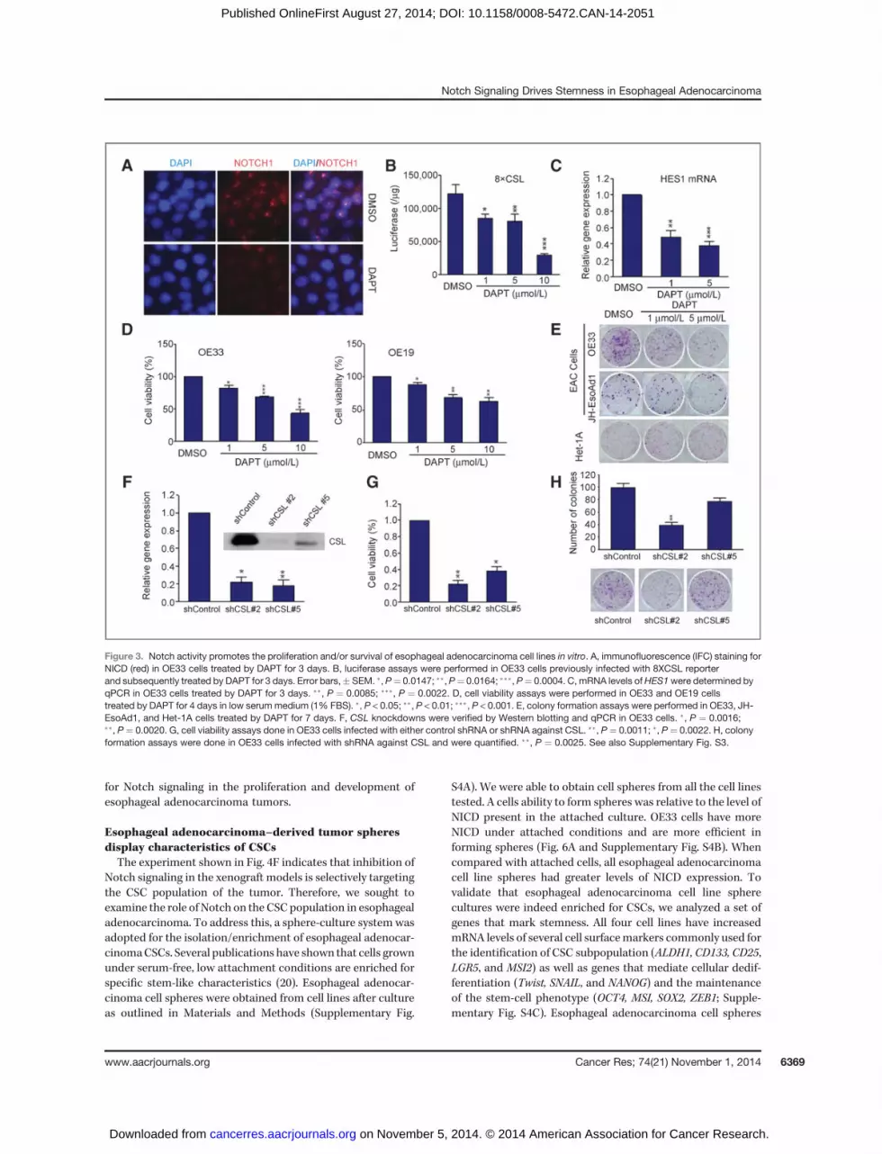
DAPT, a commonly used GSI. Efficacy of the treatment wasdemonstrated by a dramatic reduction of NICD as seen byimmunofluorescence in OE33 cells (Fig. 3A). There was also areduction in Notch-mediated transcription as seen by lucifer-ase reporter activity and a decrease in transcription of Notchtarget genes (Fig. 3B and C). Treatment of esophageal adeno-carcinoma cell lines with DAPT caused a decrease in cellviability (Fig. 3D). The number and size of colonies formedby OE33 and JH-EsoAd1 cells treated with DAPT were signif-icantly lower than those from mock treated cells, suggestingthat the reduction in proliferation is due to loss of Notchsignaling. Inhibition of the Notch pathway did not alter theproliferation of Het-1A cells (Fig. 3E, bottom). To furthervalidate this observation, and to rule out off-target effects ofGSI, we knocked down the expression of CSL via shRNA (80%–85% reduction; Fig. 3F). Therewas a significant reduction in theproliferation and colony formation of OE33 cells with CSLknockdown compared with control cells (Fig. 3G and H).Together, these results suggest that the Notch pathway isrequired for proliferation and survival of esophageal adeno-carcinoma cells in vitro. Conversely, an increase in Notchsignaling had the opposite effect.We establishedHet-1A/NICDand FLO1/NICD cell lines with stable expression of NICD.Ectopic activation of Notch pathway was confirmed by qPCRand immunoblotting (Supplementary Fig. S3). Het-1A/NICDand FLO1/NICD cells formed more colonies as compared withcontrol cells harboring an empty vector (Supplementary Fig.S3C and S3F), suggesting that increased activation of Notchsignaling promoted the transformation of Het-1A cells andproliferation of FLO1 cells. Similar results were observed whenexogenous NICD was expressed in OE33 cells (SupplementaryFig. S3G). Collectively, these data indicate that NICD is acritical regulator of esophageal adenocarcinoma cell prolifer-ation and transformation of normal esophageal epithelial cellsin vitro.
Suppression of Notch activity inhibits tumor growth inesophageal adenocarcinoma xenograft models
To test the effect of Notch signaling on the growth ofesophageal adenocarcinoma cell lines xenografts, OE19 andOE33 cells were injected into the flank of immunocompro-mised mice. Once tumors reached 200 mm3, we treated theanimals with DAPT (20 mg/kg) via daily intraperitonealinjections. Tumor growth was significantly stunted in theDAPT treatment group as compared with vehicle group forboth cell line xenografts (Fig. 4A and B). Compared withcontrol, DAPT treatment reduced expression of the Notchtarget gene HES1 (P ¼ 0.0119; Fig. 4C) and a decreased thelevel of NICD (Fig. 4D). DAPT treatment also caused adecrease in proliferation and an increase in apoptosis asmeasured via Ki67 staining and TUNEL assay, respectively(Fig. 4D). Furthermore, treatment completely inhibited thegrowth of OE19 cell xenografts when treatment was initiated1 day following cell transplantation (Fig. 4E). These dataindicate that Notch activity is indispensible for tumor estab-lishment and maintenance.
Because we observed that Notch activity was required forthe initiation of xenograft tumor formation, we wanted to
Notch Signaling Drives Stemness in Esophageal Adenocarcinoma
www.aacrjournals.org Cancer Res; 74(21) November 1, 2014 6367
on November 5, 2014. © 2014 American Association for Cancer Research. cancerres.aacrjournals.org Downloaded from
Published OnlineFirst August 27, 2014; DOI: 10.1158/0008-5472.CAN-14-2051

determine if the residual tumor cells in a xenograft tumortreated with DAPT could reestablish a tumor. To addressthis point, OE19 xenograft tumors were established andtreatment was initiated as previously described for 2 weeks.We observed that tumors treated with DAPT were signifi-cantly reduced in size compared with the control group (Fig.4F). Following treatment, tumors were harvested and cellsuspensions were prepared. Both mock and DAPT-treatedcell suspensions contained approximately 80% viable cells asdetermined by trypan blue exclusion. A total of 1� 106 viablecells from each treatment group were then transplanted intoboth flanks of five nude mice. Mock treated cells developedtumors, whereas DAPT-treated cells failed to develop anynoticeable tumors 10 weeks after reimplantation (Fig. 4F).The remaining cells, we postulate, are comprised of bulktumor cells that evidently lack the ability to reestablish atumor in mice. Therefore, inhibition of Notch signaling
selectively abolished the subpopulation of cells capable offorming new tumors (i.e., tumor-initiating cells or CSCs) inthe xenograft.
We sought to extend our findings using PDX. PDX modelsbetter represent the diversity of human cancer compared withcell line–based xenografts and are more representative of theoriginal tumor. Therefore, the therapeutic efficacy in PDXmodels is a better predictor of the clinical response of thepatient's tumor from which the model is derived. Three PDXmodels were established from patients with esophageal ade-nocarcinoma, and these tumors display the same histologicand IHC characteristics as the primary tumors (Fig. 5A).Similar to cell line–based xenografts, inhibition of Notchsignaling by DAPT in the PDX model led to significantlystunted growth and a reduction in proliferation and increasedapoptosis as compared with the vehicle group (Fig. 5B and C).Taken together, these data clearly demonstrate a critical role
Figure 2. Elevated Notch activity drives resistance to chemotherapy in esophageal adenocarcinoma (EAC). A, representative stainings of NICD are shownin the esophageal adenocarcinoma without treatment, with partial response, or resistant to chemotherapy. B, percent positive of NICD staining in thegroups (���,P <0.0001). C,mRNA levels ofHES1,NOTCH1, andNOTCH3were determined by qPCRandwere normalized to the expression values ofGAPDH(set to 1). D, hematoxylin and eosin (H&E) and IHC staining of NICD in human EUS biopsies.
Wang et al.
Cancer Res; 74(21) November 1, 2014 Cancer Research6368
on November 5, 2014. © 2014 American Association for Cancer Research. cancerres.aacrjournals.org Downloaded from
Published OnlineFirst August 27, 2014; DOI: 10.1158/0008-5472.CAN-14-2051
for Notch signaling in the proliferation and development ofesophageal adenocarcinoma tumors.
Esophageal adenocarcinoma–derived tumor spheresdisplay characteristics of CSCsThe experiment shown in Fig. 4F indicates that inhibition of
Notch signaling in the xenograft models is selectively targetingthe CSC population of the tumor. Therefore, we sought toexamine the role of Notch on the CSC population in esophagealadenocarcinoma. To address this, a sphere-culture systemwasadopted for the isolation/enrichment of esophageal adenocar-cinomaCSCs. Several publications have shown that cells grownunder serum-free, low attachment conditions are enriched forspecific stem-like characteristics (20). Esophageal adenocar-cinoma cell spheres were obtained from cell lines after cultureas outlined in Materials and Methods (Supplementary Fig.
S4A). We were able to obtain cell spheres from all the cell linestested. A cells ability to form spheres was relative to the level ofNICD present in the attached culture. OE33 cells have moreNICD under attached conditions and are more efficient informing spheres (Fig. 6A and Supplementary Fig. S4B). Whencompared with attached cells, all esophageal adenocarcinomacell line spheres had greater levels of NICD expression. Tovalidate that esophageal adenocarcinoma cell line spherecultures were indeed enriched for CSCs, we analyzed a set ofgenes that mark stemness. All four cell lines have increasedmRNA levels of several cell surfacemarkers commonly used forthe identification of CSC subpopulation (ALDH1, CD133, CD25,LGR5, and MSI2) as well as genes that mediate cellular dedif-ferentiation (Twist, SNAIL, and NANOG) and the maintenanceof the stem-cell phenotype (OCT4, MSI, SOX2, ZEB1; Supple-mentary Fig. S4C). Esophageal adenocarcinoma cell spheres
Figure 3. Notch activity promotes the proliferation and/or survival of esophageal adenocarcinoma cell lines in vitro. A, immunofluorescence (IFC) staining forNICD (red) in OE33 cells treated by DAPT for 3 days. B, luciferase assays were performed in OE33 cells previously infected with 8XCSL reporterand subsequently treated by DAPT for 3 days. Error bars,� SEM. �,P¼ 0.0147; ��,P¼ 0.0164; ���, P¼ 0.0004. C, mRNA levels ofHES1were determined byqPCR in OE33 cells treated by DAPT for 3 days. ��, P ¼ 0.0085; ���, P ¼ 0.0022. D, cell viability assays were performed in OE33 and OE19 cellstreated by DAPT for 4 days in low serum medium (1% FBS). �, P < 0.05; ��, P < 0.01; ���, P < 0.001. E, colony formation assays were performed in OE33, JH-EsoAd1, and Het-1A cells treated by DAPT for 7 days. F, CSL knockdowns were verified by Western blotting and qPCR in OE33 cells. �, P ¼ 0.0016;��, P ¼ 0.0020. G, cell viability assays done in OE33 cells infected with either control shRNA or shRNA against CSL. ��, P¼ 0.0011; �, P ¼ 0.0022. H, colonyformation assays were done in OE33 cells infected with shRNA against CSL and were quantified. ��, P ¼ 0.0025. See also Supplementary Fig. S3.
Notch Signaling Drives Stemness in Esophageal Adenocarcinoma
www.aacrjournals.org Cancer Res; 74(21) November 1, 2014 6369
on November 5, 2014. © 2014 American Association for Cancer Research. cancerres.aacrjournals.org Downloaded from
Published OnlineFirst August 27, 2014; DOI: 10.1158/0008-5472.CAN-14-2051

were alsomuchmore efficient in forming tumors. Injecting 5�105OE33 sphere cells resulted in tumors of equal or greater sizethan those obtained by injecting 5 � 106 parental cells,suggesting that the sphere cells are at least 10 times moreefficient in forming xenograft tumors (Supplementary Fig.S4D). Furthermore, as few as 5� 104 sphere-derived cells wereable to initiate tumor formation in 5 weeks.
CSCs marker genes are sensitive to inhibition of Notchsignaling
Once we established that esophageal adenocarcinoma cellspheres formed xenografts more efficiently, we tested whetherinhibition of Notch would inhibit the growth of spheres. Whencompared with attached cells, spheres had a higher transcrip-tion of Notch target genes (Fig. 6B). Treatment of OE33 sphereswithDAPT reduced the amount of NICD andNotch target genetranscription (Fig. 6B and C). GSI treatment also inhibits theability of cell spheres to form secondary spheres (Fig. 6D). Incontrast, FLO1/NICD cells containing a constitutive active
form of Notch had a greater sphere-forming potential ascompared with FLO1/Control cells (Fig. 6F).
To further confirm the role of Notch in the formation ofesophageal adenocarcinoma spheres, we treated sphere cul-tures with GSI and measured the expression level of severalstem-cell markers. The transcription of all stem-cell markergenes assayed was higher in cell spheres versus attached cells.Inhibition of the Notch pathway caused a significant decreasein transcription (P < 0.001) of several stem-cell marker genes,including ALDH, CD24, LGR5, SOX2, and TWIST1 (Fig. 6E). Thisindicates that this set of CSCmarker genes is directly regulatedby Notch. The transcription of another subset of genes testedwas increased in sphere conditions but not significantly alteredby GSI treatment (SNAIL, MSI2, NANOG, OCT4, and ZEB1),indicating that the increase in transcription is due to the loss ofstemness but independent of Notch signaling in esophagealadenocarcinoma. GSI treatment of cell spheres increased thetranscription of KLF4, which is in agreement with publisheddata on the regulation of this gene by Notch. To validate this
Figure 4. Suppression of Notch activity inhibits tumor growth in xenograftmodels. A andB, DAPT (20mg/kg) daily treatment inhibitedOE19 andOE33 cell line–derived tumor growth in the mice xenograft models. �, P < 0.05; ��, P < 0.01. C, mRNA levels of HES1 were determined by qPCR in OE19-derivedtumors without DAPT treatment and were normalized to the expression values of GAPDH (set to 1). P ¼ 0.0119. D, representative images of OE19-derivedxenografts treated by DAPT (bottom) and vehicle (top) with hematoxylin and eosin (H&E) and staining of NICD, Ki67, and TUNEL (green). E, DAPTtreatment following cell transplantation inhibited the growth of OE19 cell xenografts. ���, P < 0.001. F, tumor reestablishment from the residual tumor cells inthe OE19 xenografts treated with DAPT (right) or vehicle (left) in nude mice. n ¼ 5; ���, P < 0.001.
Wang et al.
Cancer Res; 74(21) November 1, 2014 Cancer Research6370
on November 5, 2014. © 2014 American Association for Cancer Research. cancerres.aacrjournals.org Downloaded from
Published OnlineFirst August 27, 2014; DOI: 10.1158/0008-5472.CAN-14-2051

observation, we used FLO1/NICD cells and tested for theexpression of two stem-cell marker genes, SOX2 and OCT4.Exogenous expression of NICD increased the transcription ofSOX2 7 fold compared with control cells but did not alter thetranscription of OCT4, indicating that SOX2 is specificallyregulated by Notch (Fig. 6G). This was further confirmed bychromatin immunoprecipitation (ChIP), which demonstratesthat NOTCH1 binds to the SOX2 promoter in both FLO1 andOE33 cells (Fig. 6H).
Esophageal adenocarcinoma CSC population is moresensitive to inhibition of Notch than bulk tumor cellsBecause esophageal adenocarcinoma cell spheres show
traits attributed to CSCs, we sought to determine if theirmaintenance was dependent on Notch signaling. We testedadherent cells separated according to their expression ofaldehyde dehydrogenase (ALDH), a common CSC marker(21), for their ability to grow in sphere media. Analysis of four
esophageal adenocarcinoma cell lines demonstrated that theALDH-positive cells displayed a 2 to 5-times greater ability toform spheres when compared with ALDH-negative cells (Fig.7A). In addition, treatment of attached cells withGSI decreasedthe ALDHþ fraction by 40% to 60% (Fig. 7B). Therefore, theesophageal adenocarcinoma CSC population can be identifiedaccording toALDHpositivity and that this population of cells ismore sensitive to inhibition of the Notch signaling than ALDH-negative cells.
Inhibition of Notch signaling sensitizes esophagealadenocarcinoma for treatment with 5-fluorouracil
The effect of blocking Notch on esophageal adenocarcinomaCSCs has been characterized by various CSC assays. Notchsignaling seems to promote the growthmaintenance of tumor-forming cells in esophageal adenocarcinoma. There is alsoclear evidence that higher level of Notch signaling in chemo-na€�ve patients is a marker for poor prognosis following
Figure 5. Suppression of Notch activity inhibits tumor growth in three esophageal adenocarcinoma (EAC) PDX models. A, same histologic andIHC characteristics were revealed in the primary tumors (top) and three PDX models (bottom) established from patients with esophagealadenocarcinoma. B, DAPT (20 mg/kg) daily treatment inhibited tumor growth of the three PDX models in NSG mice. n ¼ 6; error bars, � SEM;�, P < 0.05; ��, P < 0.01. C, representative images of NICD, Ki67, and TUNEL (green) staining in EAC28-derived PDX tumors treated with DAPT (top)and vehicle (bottom).
Notch Signaling Drives Stemness in Esophageal Adenocarcinoma
www.aacrjournals.org Cancer Res; 74(21) November 1, 2014 6371
on November 5, 2014. © 2014 American Association for Cancer Research. cancerres.aacrjournals.org Downloaded from
Published OnlineFirst August 27, 2014; DOI: 10.1158/0008-5472.CAN-14-2051

chemotherapy. To explore the role of Notch signaling onesophageal adenocarcinoma chemoresistance, we performeda survival assay using 5-fluorouracil (5-FU), a common agentused in esophageal adenocarcinoma chemotherapy. We choseto use OE33 and FLO1 cells for this experiment because theyhave very high and low levels of NICD, respectively (Fig. 6A).OE33 cells are insensitive to 5-FU treatment. Combinationtreatment of OE33 cells with GSI and 5-FU was more effectivein killing OE33 (Fig. 7C). In contrast, FLO1 cells are moderatelysensitive to 5-FU. When NICD is ectopically introduced toFLO1 cells, these cells (FLO1/NICD) now exhibit a resistance to5-FU (Fig. 7D). Taken together, these results suggest that Notchsignaling confers chemoresistance in esophageal adenocarci-noma cells. Therefore, it is establishing proof-of-concept thatcombinatorial therapy with GSI and other chemotherapeutic
agents will be a useful strategy in esophageal adenocarcinomatreatment.
DiscussionThe incidence of esophageal adenocarcinoma has shown a
dramatic increase in theWestern population (3, 22). The 5-yearsurvival rate of esophageal adenocarcinoma is far below allother tumor types (5). Factors that have contributed to thispoor 5-year survival are late diagnosis, resistance to chemo-therapy, and metastatic disease (5, 6). Current treatmentregimens for esophageal adenocarcinoma include NAC fol-lowed by surgical resection. Various chemotherapy regimenshave been used and typically include 5-FU, oxiplatin, anddocetaxel (18). In addition, radiotherapy can be given con-comitantly with neoadjuvant chemotherapy. Despite these
Figure 6. CSC marker genes are sensitive to Notch signaling inhibition. A, expressions of NICD in esophageal adenocarcinoma adherent cells and spheres.B, mRNA levels of Notch targets were determined. The data were normalized to the values of control spheres. C, expressions of NICD were detected byWestern blotting in OE33 spheres treated by DAPT for 3 days. D, secondary sphere formation assays were done in OE33 cells after isolating from thefirst generation of spheres. The data were plotted by bar graph (top) and representative fields are shown (bottom). ���, P < 0.001. E, mRNA levels of stem celltranscription factors were determined in OE33 spheres treated by DAPT for 3 days. F, tumor sphere formation assays were done in FLO1/Control andFLO1/NICDcells. �,P=0.0249.G,SOX2 andOCT4mRNAswere detected by q-PCR. ���,P=0.0005. H,Notch1 on theSOX2promoter are shownusingChIP.See also Supplementary Fig. S4.
Wang et al.
Cancer Res; 74(21) November 1, 2014 Cancer Research6372
on November 5, 2014. © 2014 American Association for Cancer Research. cancerres.aacrjournals.org Downloaded from
Published OnlineFirst August 27, 2014; DOI: 10.1158/0008-5472.CAN-14-2051

treatment regimens, the clinical response is relatively poor andthis leads to dismal 5-year outcome. Local response rates tothese therapies vary but for the vastmajority, treatment resultsin only a modest downsizing or no response at all. Less than20% of patients display a complete pathologic response. How-ever, it is these patients who obtain the greatest improvementin 5-year survival (18). Therefore, for improved clinical man-agement of esophageal adenocarcinoma, a treatment mustimprove the local response to neoadjuvant therapy. Herein, wedemonstrate that Notch signaling is critical for esophagealadenocarcinoma and underlies resistance to chemotherapy.We present evidence that Notch signaling is greater in the lessdifferentiated tumors and drives a CSC phenotype. We dem-onstrate that Notch signaling is critical for these esophagealadenocarcinoma CSCs and that Notch regulates genes thatestablish stemness. Using PDXmodels, we clearly demonstratethat inhibition of Notch signaling by GSIs is efficacious indownsizing tumor growth.Moreover, we provide evidence thatdemonstrates Notch activity in a patient's EUS-derived biopsysample might predict outcome to chemotherapy. Taken
together, our data strongly suggest that Notch signaling drivesa significant proportion of esophageal adenocarcinomas.Notch signaling does so by establishing and maintaining aCSC-like population of cells, which also underlies resistance tochemotherapy. Therefore, inhibition of Notch depletes the CSCpopulation and sensitizes cells to chemotherapeutic agents,which should lead to a better and more durable response toNAC. This study, therefore, provides a strong foundation forexamining the efficacy of inhibitors of Notch signaling, such asGSI compounds, for the treatment of esophageal adenocarci-noma. Given that Notch seems to drive CSCs and resistance tochemotherapy, one might expect that the outcome of suchclinical trials would be more profound on 5-year survival ratethan in downsizing of the primary tumor itself as a single agent.Therefore, it would be a useful strategy to use GSIs to sensitizetumors to NAC and, therefore, improve both the local responseand outcome of treatment. The use of a targeted therapy incombination with a typical chemotherapeutic and surgicaltreatment strategy will ultimately provide a more durable cureto this disease.
Figure 7. Notch inhibition selectively abolishes the CSC population, as well as sensitizes esophageal adenocarcinoma for 5-FU. A, tumor sphere assays weredone in ALDH-sorted esophageal adenocarcinoma cell lines. B, flow cytometry analysis using ALDH in DAPT-pretreated esophageal adenocarcinomas.C, cell viability assays were performed in OE33 cells cotreated with 5-FU and DAPT for 4 days. �, P < 0.05; ��, P < 0.01; ���, P < 0.001. D, cell viabilityassay in FLO1/Control and FLO1/NICD cells treated with 5-FU for 4 days. ��, P ¼ 0.0023; ���, P ¼ 0.0006.
Notch Signaling Drives Stemness in Esophageal Adenocarcinoma
www.aacrjournals.org Cancer Res; 74(21) November 1, 2014 6373
on November 5, 2014. © 2014 American Association for Cancer Research. cancerres.aacrjournals.org Downloaded from
Published OnlineFirst August 27, 2014; DOI: 10.1158/0008-5472.CAN-14-2051

Disclosure of Potential Conflicts of InterestNo potential conflicts of interest were disclosed.
Authors' ContributionsConception and design: Z. Wang, T.G. Da Silva, A.S. Livingstone,A.J. CapobiancoDevelopment of methodology: Z. Wang, T.G. Da Silva, A. Sanchez-Mejias, F.Bai, D.L. Fei, R.V.-D. Carpio, A.J. CapobiancoAcquisition of data (provided animals, acquired and managed patients,provided facilities, etc.): Z.Wang, T.G. Da Silva, K. Jin, P. Ranganathan, X. Zhu,K. Weaver, V.P. Koshenkov, L. Sanchez, X.-H. Pei, D. Franceschi, A. Ribeiro,A.S. Livingstone, A.J. CapobiancoAnalysis and interpretation of data (e.g., statistical analysis, biostatistics,computational analysis): Z. Wang, T.G. Da Silva, K. Jin, X. Han, A. Sanchez-Mejias, B. Li, A.E. Moscowitz, D.J. Robbins, A.J. CapobiancoWriting, review, and/or revision of the manuscript: Z. Wang, T.G. Da Silva,P. Ranganathan, A. Sanchez-Mejias, K. Weaver, V.P. Koshenkov, D. Franceschi,A.S. Livingstone, A.J. CapobiancoAdministrative, technical, or material support (i.e., reporting or orga-nizing data, constructing databases): Z. Wang, X. Han, A.E. Moscowitz,V.P. Koshenkov, L. Sanchez, L. Sparling, A.J. CapobiancoStudy supervision: Z. Wang, L. Sparling, A.S. Livingstone, A.J. Capobianco
AcknowledgmentsThe authors thank themembers of theCapobianco laboratory for support and
technical assistance. They also thank Drs. David G. Beer (Department of Surgery,Section of General Thoracic Surgery, University ofMichiganMedical School, AnnArbor, MI), Steven J. Hughes (Department of Surgery, University of Pittsburgh,Pittsburgh, PA), and James R. Eshleman and Anirban Maitra (Department ofPathology, Johns Hopkins University School of Medicine, Baltimore, MD) for theesophageal adenocarcinoma cell lines.
Grant SupportThis work was funded, in part, by grants from the National Cancer Institute
(NCI R01CA083736-12A1, NCI R01CA125044-02, and NCI R01CA169805-01), andby funding from the Dewitt Daughtry Family Department of Surgery, and theSylvester Comprehensive Cancer Center.
The costs of publication of this article were defrayed in part by the payment ofpage charges. This article must therefore be hereby marked advertisement inaccordance with 18 U.S.C. Section 1734 solely to indicate this fact.
Received July 11, 2014; accepted August 1, 2014; published OnlineFirst August27, 2014.
References1. ParkinDM,BrayF, Ferlay J, Pisani P.Global cancer statistics, 2002.CA
Cancer J Clin 2005;55:74–108.2. BrownLM,DevesaSS,ChowWH. Incidence of adenocarcinomaof the
esophagus among White Americans by sex, stage, and age. J NatlCancer Inst 2008;100:1184–7.
3. Lassen A, Hallas J, de Muckadell OB. Esophagitis: incidence and riskof esophageal adenocarcinoma–apopulation-basedcohort study. AmJ Gastroenterol 2006;101:1193–9.
4. LepageC,Rachet B, Jooste V, Faivre J,ColemanMP.Continuing rapidincrease in esophageal adenocarcinoma in England and Wales. Am JGastroenterol 2008;103:2694–9.
5. Siegel R, Naishadham D, Jemal A. Cancer statistics, 2012. CA CancerJ Clin 2012;62:10–29.
6. Paulson TG, Reid BJ. Focus on Barrett's esophagus and esophagealadenocarcinoma. Cancer Cell 2004;6:11–6.
7. Horwhat JD, Baroni D, Maydonovitch C, Osgard E, Ormseth E, Rueda-Pedraza E, et al. Normalization of intestinal metaplasia in the esoph-agus and esophagogastric junction: incidence and clinical data. AmJ Gastroenterol 2007;102:497–506.
8. Pera M. Trends in incidence and prevalence of specialized intestinalmetaplasia, barrett's esophagus, and adenocarcinoma of the gastro-esophageal junction. World J Surg 2003;27:999–1008.
9. Shaheen NJ, Crosby MA, Bozymski EM, Sandler RS. Is there publi-cation bias in the reporting of cancer risk in Barrett's esophagus?Gastroenterology 2000;119:333–8.
10. Artavanis-Tsakonas S, Matsuno K, Fortini ME. Notch signaling.Science 1995;268:225–32.
11. Ranganathan P, Weaver KL, Capobianco AJ. Notch signalling in solidtumours: a little bit of everything but not all the time. Nat Rev2011;11:338–51.
12. Fei DL, Sanchez-Mejias A, Wang Z, Flaveny C, Long J, Singh S, et al.Hedgehog signaling regulates bladder cancer growth and tumorige-nicity. Cancer Res 2012;72:4449–58.
13. Singh S, Wang Z, Fei DL, Black KE, Goetz JA, Tokhunts R,et al. Hedgehog-producing cancer cells respond to and
require autocrine hedgehog activity. Cancer Res 2011;71:4454–63.
14. Liu P, BrownS, Goktug T, Channathodiyil P, Kannappan V, Hugnot JP,et al. Cytotoxic effect of disulfiram/copper on human glioblastoma celllines and ALDH-positive cancer-stem-like cells. Br J Cancer 2012;107:1488–97.
15. Fichtner I, Rolff J, Soong R, Hoffmann J, Hammer S, Sommer A, et al.Establishment of patient-derived non-small cell lung cancer xeno-grafts as models for the identification of predictive biomarkers. ClinCancer Res 2008;14:6456–68.
16. KimSM,Park Y-Y, Park ES,Cho JY, Izzo JG, ZhangD, et al. Prognosticbiomarkers for esophageal adenocarcinoma identified by analysis oftumor transcriptome. PloS One 2010;5:e15074.
17. Ardalan B, Spector SA, Livingstone AS, Franceschi D, Mezentsev D,Lima M, et al. Neoadjuvant, surgery and adjuvant chemotherapywithout radiation for esophageal cancer. Jpn J Clin Oncol 2007;37:590–6.
18. SolomonN,MezentsevD,Reis I, LimaM,Rios J, Avisar E, et al. A phaseII study of neoadjuvant and adjuvant chemotherapywith 5-fluorodeox-yuridine, leucovorin, oxaliplatin and docetaxel in the treatment ofpreviously untreated advanced esophageal adenocarcinoma. Jpn JClin Oncol 2011;41:469–76.
19. Allan BJ, Pedroso F, Gennis ER, Livingstone AS, Montero A, Lally B,et al. Influenceof treatmentmodality in outcomes for different stagesofresectable esophageal adenocarcinomas. Ann Surg Oncol 2013;20:1660–7.
20. Kanwar SS, Yu Y, Nautiyal J, Patel BB, Majumdar AP. The Wnt/beta-catenin pathway regulates growth andmaintenance of colonospheres.Mol Cancer 2010;9:212.
21. Muzio G, Maggiora M, Paiuzzi E, Oraldi M, Canuto RA. Aldehydedehydrogenases and cell proliferation. Free Radic Biol Med 2012;52:735–46.
22. Blot WJ, Devesa SS, Kneller RW, Fraumeni JF Jr. Rising incidence ofadenocarcinoma of the esophagus and gastric cardia. JAMA 1991;265:1287–9.
Cancer Res; 74(21) November 1, 2014 Cancer Research6374
Wang et al.
on November 5, 2014. © 2014 American Association for Cancer Research. cancerres.aacrjournals.org Downloaded from
Published OnlineFirst August 27, 2014; DOI: 10.1158/0008-5472.CAN-14-2051
Copyright © 2022 FDOKUMEN