regulating executive compensation in the - Drexel University
Upload
khangminh22Category
view
4download
0
Citation: Singla, B.; Aithabathula, R.V.;
Kiran, S.; Kapil, S.; Kumar, S.;
Singh, U.P. Reactive Oxygen Species
in Regulating Lymphangiogenesis
and Lymphatic Function. Cells 2022,
11, 1750. https://doi.org/10.3390/
cells11111750
Academic Editors: Edward E.
Schmidt, Hozumi Motohashi and
Anna Kipp
Received: 15 April 2022
Accepted: 24 May 2022
Published: 26 May 2022
Publisher’s Note: MDPI stays neutral
with regard to jurisdictional claims in
published maps and institutional affil-
iations.
Copyright: © 2022 by the authors.
Licensee MDPI, Basel, Switzerland.
This article is an open access article
distributed under the terms and
conditions of the Creative Commons
Attribution (CC BY) license (https://
creativecommons.org/licenses/by/
4.0/).
cells
Review
Reactive Oxygen Species in Regulating Lymphangiogenesis andLymphatic FunctionBhupesh Singla 1,*, Ravi Varma Aithabathula 1, Sonia Kiran 1 , Shweta Kapil 2, Santosh Kumar 1
and Udai P. Singh 1
1 Department of Pharmaceutical Sciences, The University of Tennessee Health Science Center,Memphis, TN 38017, USA; [email protected] (R.V.A.); [email protected] (S.K.);[email protected] (S.K.); [email protected] (U.P.S.)
2 Division of Gastroenterology, Hepatology and Nutrition, Cincinnati Children′s Hospital Medical Center,Cincinnati, OH 45229, USA; [email protected]
* Correspondence: [email protected]; Tel.: +1-901-448-4135
Abstract: The lymphatic system is pivotal for immunosurveillance and the maintenance of tissuehomeostasis. Lymphangiogenesis, the formation of new lymphatic vessels from pre-existing vessels,has both physiological and pathological roles. Recent advances in the molecular mechanisms regulat-ing lymphangiogenesis have opened a new area of research on reparative lymphangiogenesis forthe treatment of various pathological disorders comprising neurological disorders, cardiac repair,autoimmune disease, obesity, atherosclerosis, etc. Reactive oxygen species (ROS) produced by thevarious cell types serve as signaling molecules in several cellular mechanisms and regulate variousaspects of growth-factor-mediated responses, including lymphangiogenesis. The ROS, includingsuperoxide anion, hydrogen peroxide, and nitric oxide, play both beneficial and detrimental rolesdepending upon their levels and cellular microenvironment. Low ROS levels are essential for lym-phangiogenesis. On the contrary, oxidative stress due to enhanced ROS generation and/or reducedlevels of antioxidants suppresses lymphangiogenesis via promoting lymphatic endothelial cell apop-tosis and death. In this review article, we provide an overview of types and sources of ROS, discussthe role of ROS in governing lymphangiogenesis and lymphatic function, and summarize the role oflymphatics in various diseases.
Keywords: lymphatic vessels; lymphangiogenesis; reactive oxygen species; superoxide anion;hydrogen peroxide; nitric oxide
1. Introduction
The lymphatic system, constituted by a network of lymphatic vessels (LVs), lymphnodes (LNs), and lymphoid organs, runs parallel to the blood vascular system. Both lym-phatic and blood circulatory systems work in synchrony to maintain tissue homeostasis.The blood vascular system supplies nutrients, oxygen, and hormones to various body or-gans. In contrast, the lymphatic system plays an important role in transporting extravasatedinterstitial fluid, immune cells, inflammatory cytokines, antigens, and lipoproteins fromthe peripheral tissue to the draining LNs and back to the systemic venous circulation. Thus,the lymphatic system is pivotal for the maintenance of interstitial fluid homeostasis, hostdefense, adaptive immunity, and regulation of inflammatory responses [1–3]. The LVsare present throughout the human body with the exception of bone marrow and tissues,such as the epidermis, where blood vessels are also absent. These vessels are categorizedin a hierarchical network of vessels, including capillaries, pre-collecting, and collectinglymphatics, based on their specific functions and morphological features [4,5]. Lymphflows through the lymphatic network in a unidirectional manner and at low pressure. Thelymph flow rate is influenced by various extrinsic and intrinsic forces. Dysfunctional lym-phatic vessels are responsible for several pathological conditions, including inherited and
Cells 2022, 11, 1750. https://doi.org/10.3390/cells11111750 https://www.mdpi.com/journal/cells
Cells 2022, 11, 1750 2 of 20
acquired lymphedema, malabsorption syndromes, autoimmune disease, atherosclerosis,neurological disorders, and immune deficiency [6–8].
The LVs are lined by a single layer of lymphatic endothelial cells (LECs), which pos-sess specific markers and regulatory molecules, including Prospero homeobox 1 (Prox1),podoplanin, vascular endothelial growth factor receptor 3 (VEGFR3), and lymphatic vesselendothelial hyaluronan receptor 1 (LYVE-1) [9–13]. Like angiogenesis, lymphangiogen-esis is the formation of new lymphatic vessels from pre-existing lymphatics. It resultsfrom a complex series of cellular events, including proliferation, sprouting, migration, andformation of vessel-like structures by LEC. Unlike developmental lymphangiogenesis, lym-phangiogenesis in adults is dysregulated and less coordinated and occurs in pathologicalconditions such as inflammation, wound healing, and tumor growth [14–16]. These patholo-gies are often characterized by an accumulation of inflammatory cells and tissue edema,which necessitate lymphangiogenesis and LV remodeling for the removal of immune cells,cytokines, and tissue fluid [15,16]. Due to the various functions of LVs, lymphangiogenesisis regulated by multiple signaling pathways, as reviewed earlier [17,18].
Reactive oxygen species (ROS) such as superoxide anion, hydrogen peroxide, andnitric oxide are known to play both physiological and pathophysiological roles. Low levelsof ROS are required to mediate LEC proliferation and migration, contributing to lymphan-giogenesis. Increased generation of ROS by LEC leads to oxidative stress and inhibits LECproliferation, migration, and tube formation via inducing apoptosis and cell death [19–21].Based on these and the literature review, in this review article, we provide an overview oftypes and sources of ROS, discuss the role of ROS in governing lymphangiogenesis andlymphatic function in various pathological conditions, and summarize the role of LVs invarious diseases.
2. Reactive Oxygen Species and Free Radicals
Reactive molecules and free radicals derived from molecular oxygen are called ROS.Free radicals are molecular species capable of independent existence that contain one ormore unpaired electrons and include nitric oxide (NO•), superoxide anion (O2
•−), hydroxylradical (•OH), and lipid peroxyl radical (LOO•). Examples of non-radical derivatives ofmolecular oxygen are peroxynitrite (ONOO−), hydrogen peroxide (H2O2), hypochlorousacid (HOCl), and ozone (O3). ROS are synthesized as necessary intermediates in a broadrange of biochemical processes and function as second messengers in physiological signal-ing mechanisms, contributing to the maintenance of tissue homeostasis [22]. In contrast,under pathophysiological conditions, overproduction of ROS and/or diminished antiox-idant systems (also known as oxidative stress) may induce oxidative damage to DNA,protein, and lipid molecules. Oxidative-stress-induced alterations from physiological topathophysiological signal transduction pathways and subsequent cellular damage play acritical role in the initiation, development, and progression of several diseases, includingcardiovascular, inflammatory, neurologic, cancer, diabetes, and aging [19,23–25]. In thissection, we briefly describe the chemistry and biochemistry of ROS and free radicals.
2.1. Nitric Oxide (NO•)
NO• plays a role in several biological processes and has vasodilatory, anti-inflammatory,and anti-thrombotic activities. It is produced by endothelial nitric oxide synthase (eNOS),neuronal NOS (nNOS), and inducible NOS (iNOS) [22]. These isoforms catalyze the forma-tion of L-citrulline from L-arginine, and NO• is produced as a byproduct of the reaction.NOS-mediated NO• production is dependent on the availability of oxygen, so in case oflimited oxygen supply, the nitrate–nitrite–NO pathway acts as a backup system to main-tain sufficient NO• production [26]. Nitrate is first reduced by gastrointestinal and oralcommensal bacteria to nitrite, which is further reduced to NO• by various pathways, asmentioned in Table 1. The formation of the iron–nitrosyl complex by the interaction of NO•
to Fe2+ heme protein activates soluble guanylyl cyclase (sGC), which synthesizes secondmessenger cyclic guanosine monophosphate (cGMP). This sGC-cGMP signaling plays an
Cells 2022, 11, 1750 3 of 20
important role in vasodilation, nerve signaling, mitochondrial biogenesis, angiogenesis,and lymphangiogenesis [27,28]. NO• is also responsible for decreased production of O2
•−
from complex I and III of the electron transport chain, mitochondrial cytochrome c release,and apoptosis [29,30]. Furthermore, NO• is responsible for limiting calcium availability invascular smooth muscle cells due to ATP-dependent potassium channel opening, whichprevents myosin light chain 2 (MLC2) phosphorylation and inhibits vasoconstriction [31].The equilibrium between NO• and O2
•− production is regulated by the availability oftetrahydrobiopterin (BH4, a cofactor required for NOS activity). The outcomes of reducedavailability of BH4 relative to its oxidized form, dihydrobiopterin (BH2), involve increasedO2•− release and decreased NO• production [32]. This situation is called eNOS uncou-
pling, which can be culpable for more superoxide production by the activation of oxidaseenzymes, particularly NADPH oxidases (NOXs) and xanthine oxidase (XO); and progres-sive reduction in NO• bioavailability [33]. Moreover, the interaction between NO• andO2•− forms ONOO−, a strong oxidant that can oxidize BH4 and leads to enhanced eNOS
uncoupling.
2.2. Superoxide Anion (O2•−)
O2•− is the precursor of most ROS and is rapidly dismuted to H2O2 spontaneously
or via the reaction catalyzed by superoxide dismutase (SOD). The rate of spontaneousdismutation of O2
•− is very low compared to enzymatic dismutation (8 × 10⁴ M−1 s−1
versus 2 × 10⁹ M−1 s−1) [34]. Enzymes involved in the biological production of O2•−
include NOX isoforms, XO, uncoupled eNOS, and lipoxygenase. Phagocytes involvingmyeloid cells-neutrophils, macrophages, and monocytes, kill invading pathogens viaproducing a large amount of O2
•−, also called a respiratory burst. It is also formed due tothe leakage of one electron from the mitochondrial electron transport chain to molecularoxygen [35]. It has been reported that approximately 1–3% of electrons leak to producesuperoxide anions during the mitochondrial transport chain [36].
2.3. Hydrogen Peroxide (H2O2)
H2O2, a highly stable and cell-permeable molecule, is generated because of O2•−
dismution catalyzed by SOD. It acts as a signaling molecule in cellular signal transductionpathways [37]. In addition, XO and Nox4 are direct sources of H2O2 [37,38]. It playsa role in various physiological activities involving cell differentiation, proliferation, andmigration [22,39]. Due to its cell-permeable nature, it can act in a paracrine manner betweenvarious cell types to regulate cellular signaling.
2.4. Peroxynitrite (ONOO−)
It is formed because of the interaction of NO• with O2•− and can lead to increased
NOS uncoupling in the endothelium. It is highly toxic and acts as a substrate for theformation of super active nitroso peroxo carboxylate (ONOOCO2
−) or peroxynitrousacid (ONOOH) [40]. It is capable of altering the oxidative state of lipids, DNA, andtyrosine and methionine residues in proteins [41]. Peroxynitrite stimulates the nitration oftyrosine residues present in proteins, for instance, SOD, by reaction mediated by transitionmetals. [42]. Further, peroxynitrite in phagocytic cells acts as a cytotoxic effector moleculeagainst invading pathogens, including bacteria and parasites [43–45].

Cells 2022, 11, 1750 4 of 20
Table 1. Types and generation of reactive oxygen species.
Name of Molecule Half-Life of Molecule Generation of Molecule
Nitric oxide (NO•) 10−5 to 10−3 s
Nitric oxide synthaseL-arginine + O2 + NADPH→ L-citrulline + NO• +NADP+
Reduction of nitriteDeoxyhaemoglobin/myoglobinNO2
− + Fe2+ + H+ → NO• + Fe3+ + OH−
Xanthine oxidoreductaseNO2
− + Mo4+ + H+ → NO• + Mo5+ + OH−
ProtonsNO2
− + H+ → HNO22 HNO2 → 2 N2O3 + H2ON2O3 → NO• + •NO2
AscorbateNO2
− + H+ → HNO22 HNO2 + Asc→ 2 NO• + dehydroAsc + 2 H2O
Polyphenols (Ph-OH)NO2
− + H+ → HNO2Ph-OH + HNO2 → Ph-•O + NO• + H2O
Superoxide (O2•−) 10−11 to 10−9 s
NADPH oxidaseNADPH + 2O2 → NADP+ + 2O2
•− + 2H−
Xanthine oxidaseHypoxanthine + H2O + 2O2 → Xanthine + 2O2
•− +2H−
Xanthine + H2O + 2O2 → Uric acid + 2O2•− + 2H−
Uncoupled endothelial nitric oxide synthaseNADPH + 2O2 → NADP+ + 2O2
•− + 2H−
Mitochondrial electron transport chain complexes I and IIIO2 → O2
•−
LipooxygenaseArachidonic acid + O2 → HPETE+ O2
•−
Hydroxyl radical (•OH) 10−9 s
Fenton reactionFe2+ + H2O2 → Fe3+ + •OH + OH−
Haber-Weiss reaction•O2
− + H2O2 → •OH + OH− + O2
HOONO→ •OH + NO2•
Lipid peroxyl radical (LOO•) 7 s
L-H + •X→ L•+ XHLOO• + L-H→ LOOH + L•
L• + O2 → LOO•
L-H: polyunsaturated fatty acid•X: oxidizing character (i.e., •OH or O2
•−)L•: lipid radical
Peroxynitrite (ONOO−) 10−2 s O2•− + NO• → ONOO−
Hydrogen peroxide (H2O2) 10−8 (in presence of catalase) or 10−3 s 2O2•− + 2H+ → H2O2 + O2
Hypochlorous acid (HOCl) <1 min MyeloperoxidaseH2O2 + Cl− → HOCl + OH−
Ozone (O3) 1 min x1O2 + yH2O
Cells 2022, 11, x FOR PEER REVIEW 4 of 20
NADPH + 2O2 → NADP+ + 2O2•− + 2H− Mitochondrial electron transport chain complexes I and III O2 → O2•− Lipooxygenase Arachidonic acid + O2 → HPETE+ O2•−
Hydroxyl radical (•OH) 10−9 s
Fenton reaction Fe2+ + H2O2 → Fe3+ + •OH + OH−
Haber-Weiss reaction •O2− + H2O2 → •OH + OH− + O2
HOONO → •OH + NO2•
Lipid peroxyl radical (LOO•)
7 s
L-H + •X → L•+ XH LOO• + L-H → LOOH + L• L• + O2 → LOO•
L-H: polyunsaturated fatty acid •X: oxidizing character (i.e., •OH or O2•−) L•: lipid radical
Peroxynitrite (ONOO−) 10−2 s O2•− + NO• → ONOO−
Hydrogen peroxide (H2O2) 10−8 (in presence of catalase) or 10−3 s
2O2•− + 2H+ → H2O2 + O2
Hypochlorous acid (HOCl) <1 min Myeloperoxidase H2O2 + Cl− → HOCl + OH−
Ozone (O3) 1 min x1O2 + yH2O ↔ [H2O3(y−1)H2O] → H2O2 + (x−1)3O2 + O3
2.2. Superoxide Anion (O2•−) O2•− is the precursor of most ROS and is rapidly dismuted to H2O2 spontaneously or
via the reaction catalyzed by superoxide dismutase (SOD). The rate of spontaneous dis-mutation of O2•− is very low compared to enzymatic dismutation (8 × 10⁴ M−1 s−1 versus 2 × 10⁹ M−1 s−1) [34]. Enzymes involved in the biological production of O2•− include NOX isoforms, XO, uncoupled eNOS, and lipoxygenase. Phagocytes involving myeloid cells-neutrophils, macrophages, and monocytes, kill invading pathogens via producing a large amount of O2•−, also called a respiratory burst. It is also formed due to the leakage of one electron from the mitochondrial electron transport chain to molecular oxygen [35]. It has been reported that approximately 1–3% of electrons leak to produce superoxide anions during the mitochondrial transport chain [36].
2.3. Hydrogen Peroxide (H2O2) H2O2, a highly stable and cell-permeable molecule, is generated because of O2•− dis-
mution catalyzed by SOD. It acts as a signaling molecule in cellular signal transduction pathways [37]. In addition, XO and Nox4 are direct sources of H2O2 [37,38]. It plays a role in various physiological activities involving cell differentiation, proliferation, and migra-tion [22,39]. Due to its cell-permeable nature, it can act in a paracrine manner between various cell types to regulate cellular signaling.
[H2O3(y−1)H2O]→ H2O2 +(x−1)3O2 + O3
2.5. Hydroxyl Radical (•OH)
The dismution of H2O2 (Fenton reaction) and peroxynitrous acid (ONOOH), whichis formed from the oxidation of ONOO−, results in the generation of •OH. Haber–Weiss

Cells 2022, 11, 1750 5 of 20
reaction comprising spontaneous interaction of O2•− and H2O2 is an alternate source of
hydroxyl radical production [46]. Generally, •OH does not play any role in cell signaling,but it is an important contributor to oxidative stress [47]. Its cellular levels can be alteredby antioxidant enzymes and iron ligands.
2.6. Lipid Peroxyl Radical (LOO•)
Oxidation of unsaturated fatty acids present in cell membranes and lipoproteins leadsto the formation of a lipid alkyl radical (L•), which rapidly reacts with molecular oxygento form lipid peroxyl radical (LOO•). LOO• radical reacts with various proteins andcarbohydrates by lowering the activation energy even much more than enzyme-catalyzedreactions, leading to the production of corresponding carbonyl compounds [48]. Theseradicals are highly destructive to cells, and once produced, they may lead to unrelentinglipid breakdown [49].
2.7. Hypochlorous Acid (HOCl)
It is generated in inflammatory cells such as activated neutrophils, monocytes, andmacrophages, which have myeloperoxidase (MPO) enzyme required to produce HOCl.It is synthesized by the interaction of H2O2 with chloride ions. This HOCl has a strongantimicrobial activity and can oxidize lipoproteins, lipids, and proteins [50,51]. HOCl canalso be responsible for the formation of monochloramines (NH2Cl), ONOO−, •OH, singletoxygen (O2), and O3 via interacting with NH3 and other ROS [50].
2.8. Ozone (O3)
Ozone has a powerful oxidizing property and can increase the process of leukocytosisand phagocytosis. It is produced by electric discharge and irradiation of oxygen (air) withshort-wavelength ultraviolet radiations. It may be synthesized in vivo by an antibody-mediated H2O oxidation pathway [52]. It can oxidize various biomolecules including lipids,proteins, and nucleic acids [53,54].
3. Role of ROS in Regulating Lymphangiogenesis and Lymphatic Function
Excessive production of ROS, for instance, H2O2, contributes to cell death. ROS-stimulatedcell death occurs due to oxidative damage to cellular macromolecules involving proteins,lipids, and nucleic acids, and/or induction of cell-death-signaling pathways [55,56]. Al-ternatively, high ROS levels activate cell survival molecular-signaling cascades such asmitogen-activated protein kinase and PI3K/Akt [57,58]. The PI3K/Akt signaling preventsoxidative-stress-induced cell death and promotes cell survival. Furthermore, ROS havebeen shown to trigger cell surface growth factor receptor-mediated cell survival signaling invarious cell types [59–62]. Thus, induction of growth factor receptor-mediated signaling inresponse to ROS protects against oxidative-stress-induced cell damage. Nitric oxide derivedfrom eNOS in vascular endothelial cells plays an important role in stimulating angiogenesisand maintaining vascular contractility [63,64]. Like vascular endothelial cells, LECs haveeNOS, and NO-derived from eNOS has been observed to be essential for lymphangio-genesis [65]. Despite numerous indications of the regulation of blood vessel formation(angiogenesis) by oxidative stress, the effects of ROS in modulating lymphangiogenesis areunderstudied to date.
3.1. Nitric Oxide
Stimulation of LECs with VEGF-C, a lymphangiogenic factor, has been shown toactivate eNOS, which leads to the generation of NO and a subsequent increase in LECproliferation and lymphangiogenesis [66] (Figure 1). Under physiological conditions, vari-ous immune cells, interstitial fluid, cytokines, and antigens present in initial lymphaticsare carried by collecting LVs to draining LNs. The lymph flow in collecting LVs dependson the contractility of these collecting LVs. NO-derived from LEC eNOS is importantfor the maintenance of lymphatic contractility [67]. In addition, NO released by LECs

Cells 2022, 11, 1750 6 of 20
regulates lymphatic permeability [68,69]. Additionally, inhibition of NO production with aNOS inhibitor, L-NMMA, blocks LV regeneration. Expression/activity of NOS in tumortissues positively correlates with lymphatic metastasis in various types of tumors [70]. Ge-netic deletion of eNOS and pharmacological inhibition of its activity reduces peritumorallymphatic hyperplasia in VEGF-C-overexpressing fibrosarcoma and attenuates traffick-ing of tumor cells to draining LNs, suggesting the role of NO in regulating lymphaticdrainage [66]. Inhibition of NO-mediated signaling using an sGC inhibitor abrogates ultra-violet B-irradiation-induced LV enlargement, edema formation, and skin inflammation inmouse models [28]. A recent study by Singla et al. reported matricellular protein R-spondin2 (RSPO2) as an anti-lymphangiogenic protein [21]. The authors observed that RSPO2suppresses VEGF-C-stimulated Akt and eNOS phosphorylation, leading to attenuationof NO production by LEC and subsequent impairment in lymphangiogenesis. Moreover,pharmacological NO supplementation using an NO donor prevented the inhibitory ef-fects of RSPO2 on lymphangiogenesis [21]. All this information suggests that NO-sGCsignaling is pivotal in regulating lymphangiogenesis and maintenance of LVs. IncreasediNOS levels disrupt endogenous NO gradients normally regulated by eNOS and leadto supra-physiological levels of NO, which in turn results in the induction of nitrosativestress [71]. Liao et al. demonstrated that under inflammatory conditions, NO derived fromiNOS-overexpressing CD11b+ Gr-1+ myeloid-derived suppresser cells present around thesubcutaneous LVs suppresses lymphatic contractions [72] (Figure 1). A study by Rehalet al. reported leaky and dilated LVs surrounded by iNOS+ CD11b+ inflammatory cells inobese mice [73]. These inflammatory cells caused an increased generation of peroxynitritein obese mice. Additionally, LECs isolated from obese mice exhibited reduced VEGFR3and podoplanin expression. These findings suggest that enhanced iNOS-derived NOgeneration in obesity contributes to lymphatic injury and impaired lymphatic pumpingvia nitrosative stress. Other reports revealed inhibition of lymphatic contractile functionwith high NO levels and impaired NO signaling in diabetic condition [74–76]. Additionally,reduced NO production due to decreased eNOS levels in the lymphatic thoracic ductisolated from rats with metabolic syndrome has been shown to be responsible for reducedvessel contractility [77]. Morris et al. showed KLF2-PPAR-γ-signaling-dependent elevationof NOX-derived ROS production and reduction of bioavailable NO in response to shearstress as observed with chronically increased lymph flow [78]. Further, it has been shownthat shear stress reduces eNOS expression and activity in LECs [79].

Cells 2022, 11, 1750 7 of 20Cells 2022, 11, x FOR PEER REVIEW 7 of 20
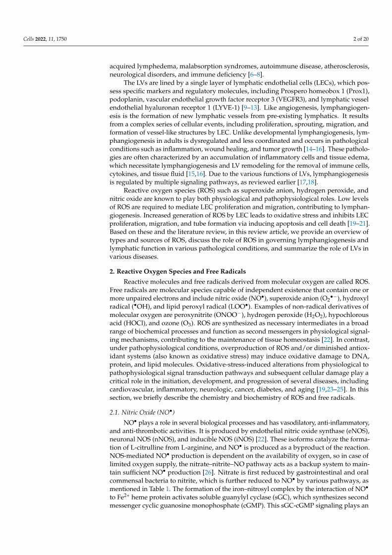
Figure 1. Role of ROS in regulating lymphangiogenesis and lymphatic function. Activation of VEGFR3 present in LECs by its ligand VEGF-C induces optimal Nox4-derived H2O2 production, which in turn enhances VEGFR3 autophosphorylation and stimulates downstream pro-lymphangi-ogenic signaling (upper panel). Oxidized LDL and RSPO2 inhibit lymphangiogenesis via suppres-sion of Akt/eNOS pathway. Under diabetic condition, excessive H2O2 generation elevates epsin ex-pression and promotes VEGFR3 degradation, leading to attenuated lymphangiogenesis and re-duced lymphatic transport. In inflammatory condition, supra-physiological NO production by CD11b+ myeloid immune cells surrounding LVs contributes to nitrosative stress leading to the sup-pression of lymphatic contractions and inducing LV leakiness (lower panel).
3.2. Superoxide Anion and H2O2 Increased generation of O2•− scavenges NO and reduces NO bioavailability. Interac-
tion of NO with O2•− forms peroxynitrite (ONOO−), which can further enhance protein modification and DNA damage. Singla et al. observed Nox4 as the major NOX isoform in LECs, which mainly generates H2O2 [21]. Exposure of LECs to H2O2 induces VEGFR3 ac-tivation and downstream Akt phosphorylation [80]. VEGFR3 activation following H2O2
Figure 1. Role of ROS in regulating lymphangiogenesis and lymphatic function. Activationof VEGFR3 present in LECs by its ligand VEGF-C induces optimal Nox4-derived H2O2 pro-duction, which in turn enhances VEGFR3 autophosphorylation and stimulates downstream pro-lymphangiogenic signaling (upper panel). Oxidized LDL and RSPO2 inhibit lymphangiogenesis viasuppression of Akt/eNOS pathway. Under diabetic condition, excessive H2O2 generation elevatesepsin expression and promotes VEGFR3 degradation, leading to attenuated lymphangiogenesisand reduced lymphatic transport. In inflammatory condition, supra-physiological NO productionby CD11b+ myeloid immune cells surrounding LVs contributes to nitrosative stress leading to thesuppression of lymphatic contractions and inducing LV leakiness (lower panel).
3.2. Superoxide Anion and H2O2
Increased generation of O2•− scavenges NO and reduces NO bioavailability. Interac-
tion of NO with O2•− forms peroxynitrite (ONOO−), which can further enhance protein
modification and DNA damage. Singla et al. observed Nox4 as the major NOX isoformin LECs, which mainly generates H2O2 [21]. Exposure of LECs to H2O2 induces VEGFR3
Cells 2022, 11, 1750 8 of 20
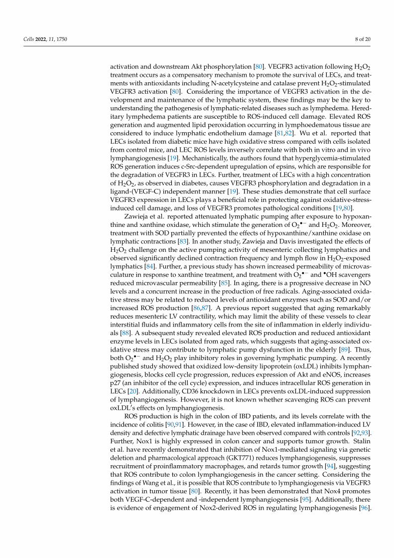
activation and downstream Akt phosphorylation [80]. VEGFR3 activation following H2O2treatment occurs as a compensatory mechanism to promote the survival of LECs, and treat-ments with antioxidants including N-acetylcysteine and catalase prevent H2O2-stimulatedVEGFR3 activation [80]. Considering the importance of VEGFR3 activation in the de-velopment and maintenance of the lymphatic system, these findings may be the key tounderstanding the pathogenesis of lymphatic-related diseases such as lymphedema. Hered-itary lymphedema patients are susceptible to ROS-induced cell damage. Elevated ROSgeneration and augmented lipid peroxidation occurring in lymphoedematous tissue areconsidered to induce lymphatic endothelium damage [81,82]. Wu et al. reported thatLECs isolated from diabetic mice have high oxidative stress compared with cells isolatedfrom control mice, and LEC ROS levels inversely correlate with both in vitro and in vivolymphangiogenesis [19]. Mechanistically, the authors found that hyperglycemia-stimulatedROS generation induces c-Src-dependent upregulation of epsins, which are responsible forthe degradation of VEGFR3 in LECs. Further, treatment of LECs with a high concentrationof H2O2, as observed in diabetes, causes VEGFR3 phosphorylation and degradation in aligand-(VEGF-C) independent manner [19]. These studies demonstrate that cell surfaceVEGFR3 expression in LECs plays a beneficial role in protecting against oxidative-stress-induced cell damage, and loss of VEGFR3 promotes pathological conditions [19,80].
Zawieja et al. reported attenuated lymphatic pumping after exposure to hypoxan-thine and xanthine oxidase, which stimulate the generation of O2
•− and H2O2. Moreover,treatment with SOD partially prevented the effects of hypoxanthine/xanthine oxidase onlymphatic contractions [83]. In another study, Zawieja and Davis investigated the effects ofH2O2 challenge on the active pumping activity of mesenteric collecting lymphatics andobserved significantly declined contraction frequency and lymph flow in H2O2-exposedlymphatics [84]. Further, a previous study has shown increased permeability of microvas-culature in response to xanthine treatment, and treatment with O2
•− and •OH scavengersreduced microvascular permeability [85]. In aging, there is a progressive decrease in NOlevels and a concurrent increase in the production of free radicals. Aging-associated oxida-tive stress may be related to reduced levels of antioxidant enzymes such as SOD and/orincreased ROS production [86,87]. A previous report suggested that aging remarkablyreduces mesenteric LV contractility, which may limit the ability of these vessels to clearinterstitial fluids and inflammatory cells from the site of inflammation in elderly individu-als [88]. A subsequent study revealed elevated ROS production and reduced antioxidantenzyme levels in LECs isolated from aged rats, which suggests that aging-associated ox-idative stress may contribute to lymphatic pump dysfunction in the elderly [89]. Thus,both O2
•− and H2O2 play inhibitory roles in governing lymphatic pumping. A recentlypublished study showed that oxidized low-density lipoprotein (oxLDL) inhibits lymphan-giogenesis, blocks cell cycle progression, reduces expression of Akt and eNOS, increasesp27 (an inhibitor of the cell cycle) expression, and induces intracellular ROS generation inLECs [20]. Additionally, CD36 knockdown in LECs prevents oxLDL-induced suppressionof lymphangiogenesis. However, it is not known whether scavenging ROS can preventoxLDL′s effects on lymphangiogenesis.
ROS production is high in the colon of IBD patients, and its levels correlate with theincidence of colitis [90,91]. However, in the case of IBD, elevated inflammation-induced LVdensity and defective lymphatic drainage have been observed compared with controls [92,93].Further, Nox1 is highly expressed in colon cancer and supports tumor growth. Stalinet al. have recently demonstrated that inhibition of Nox1-mediated signaling via geneticdeletion and pharmacological approach (GKT771) reduces lymphangiogenesis, suppressesrecruitment of proinflammatory macrophages, and retards tumor growth [94], suggestingthat ROS contribute to colon lymphangiogenesis in the cancer setting. Considering thefindings of Wang et al., it is possible that ROS contribute to lymphangiogenesis via VEGFR3activation in tumor tissue [80]. Recently, it has been demonstrated that Nox4 promotesboth VEGF-C-dependent and -independent lymphangiogenesis [95]. Additionally, thereis evidence of engagement of Nox2-derived ROS in regulating lymphangiogenesis [96].

Cells 2022, 11, 1750 9 of 20
LPA-stimulated ROS production, which is mediated by phospholipase C and protein kinaseC, regulates LPA 1/3-dependent VEGF-C expression in prostate cancer cells [97]. LPA stim-ulates lymphangiogenesis in the tumor-xenograft mouse models via controlling calreticulinexpression. Boehme et al. have recently demonstrated that LECs exposed to prolongedpathologically elevated lymph flow exhibit hyperproliferative growth, which is mediatedby elevated hypoxia-inducible factor (HIF)-1α expression and increased mitochondrialNOX-derived ROS production [98]. In addition, the authors report attenuated proliferationof LECs exposed to mitochondrial antioxidants, indicating the importance of mitochondrialROS in driving LEC proliferation under mechanical stress. Under hypoxic conditions, tu-mor cells upregulate HIF expression and activity, which induce the transcription of variousgrowth factors′ genes responsible for increasing angiogenesis and lymphangiogenesis [99].All the above findings suggest that ROS regulates lymphatic permeability, contractility anddrainage, and lymphangiogenesis. Further studies are required to investigate the role ofROS in specific diseases and cell types.
4. Role of Lymphatic Vessels in Various Pathologies4.1. Tumor Metastasis
Lymphatics serve as a route for cancer progression and metastasis. Secretion ofwell-known lymphangiogenic factor vascular endothelial growth factor (VEGF)-C andother growth factors by tumor cells lead to increased LV density in tumor tissues [100].Tumor-associated lymphatics aid in the drainage of interstitial fluid containing various celltypes and tumor-cell-originated macromolecules to sentinel LNs. Metastatic cells that de-tach from the primary tumor readily enter into highly permeable surrounding LVs and reachdistant organs (Figure 2). Previous studies have indicated positive correlations between thelevels of VEGF-C, VEGF-D, and VEGFR3 with an increased incidence of LN and distantorgan metastasis [101–103]. In addition to metastasis, angiogenesis and lymphangiogenesissupport tumor growth by other mechanisms. For example, the blood vascular system sup-plies nutrients and oxygen to the tumor hypoxic environment, while inflamed lymphatics in-duce an immunosuppressive microenvironment via reducing dendritic cell (DC)-mediatedcytotoxic lymphocyte function by producing T cell inhibitory programmed-death ligand1 (PD-L1), transforming growth factor-β (TGF-β), iNOS), and indoleamine 2,3-dioxygenase(IDO), thereby stimulating tumor progression [104]. Moreover, tumor tissue LV densityhas been shown to correlate with poor prognosis in cancer patients [105–107]. A previ-ous study employing photodynamic therapy with benzoporphyrin-derivative verteporfinto destroy tumor-associated LVs showed prevention of mouse melanoma cell metasta-sis and subsequent tumor relapse. In addition, the combination of photodynamic andanti-lymphangiogenic therapies further reduced the invasion of tumor cells into LVs [108].Conversely, drainage of tumor antigen-carrying migratory DCs to draining LNs is requiredfor an antitumor immune response [109,110]. In addition, Fankhauser et al., in their studyusing the mouse melanoma model, reported enhanced T cell infiltration and potentiation ofimmunotherapy with experimental induction of tumor lymphangiogenesis [111]. Similarbeneficial effects of augmented intra-tumoral LV density have been shown in colorectalcancer [112]. Therefore, despite the role of LVs in promoting tumor metastasis, LVs arerequired for the generation of optimum antitumor immune response to potentiate im-mune therapies. Future studies are required to further explore the role of LVs in specificcancer types.
Cells 2022, 11, 1750 10 of 20Cells 2022, 11, x FOR PEER REVIEW 10 of 20
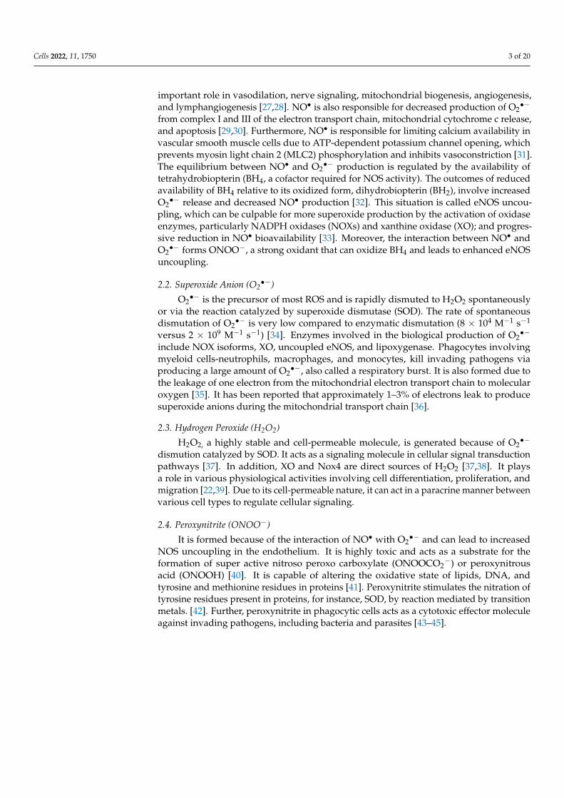
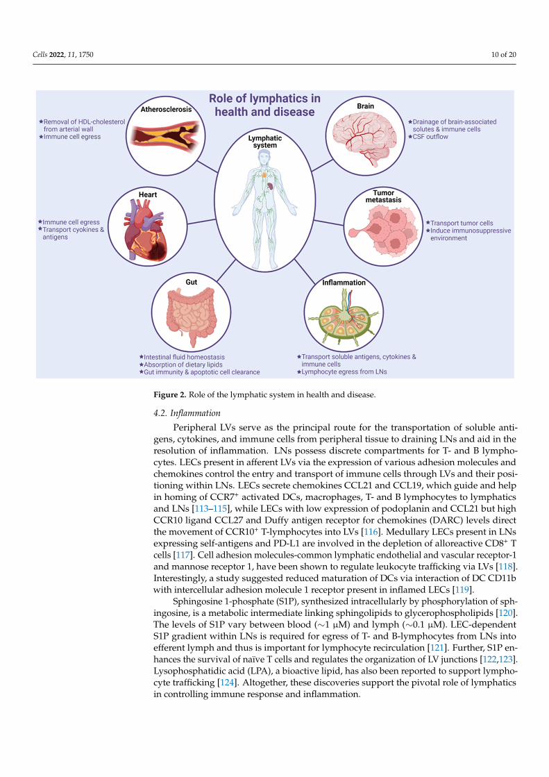
Figure 2. Role of the lymphatic system in health and disease.
4.2. Inflammation Peripheral LVs serve as the principal route for the transportation of soluble antigens,
cytokines, and immune cells from peripheral tissue to draining LNs and aid in the resolu-tion of inflammation. LNs possess discrete compartments for T- and B lymphocytes. LECs present in afferent LVs via the expression of various adhesion molecules and chemokines control the entry and transport of immune cells through LVs and their positioning within LNs. LECs secrete chemokines CCL21 and CCL19, which guide and help in homing of CCR7+ activated DCs, macrophages, T- and B lymphocytes to lymphatics and LNs [113–115], while LECs with low expression of podoplanin and CCL21 but high CCR10 ligand CCL27 and Duffy antigen receptor for chemokines (DARC) levels direct the movement of CCR10+ T-lymphocytes into LVs [116]. Medullary LECs present in LNs expressing self-antigens and PD-L1 are involved in the depletion of alloreactive CD8+ T cells [117]. Cell adhesion molecules-common lymphatic endothelial and vascular receptor-1 and mannose receptor 1, have been shown to regulate leukocyte trafficking via LVs [118]. Interestingly, a study suggested reduced maturation of DCs via interaction of DC CD11b with intercel-lular adhesion molecule 1 receptor present in inflamed LECs [119].
Sphingosine 1-phosphate (S1P), synthesized intracellularly by phosphorylation of sphingosine, is a metabolic intermediate linking sphingolipids to glycerophospholipids [120]. The levels of S1P vary between blood (∼1 μM) and lymph (∼0.1 μM). LEC-depend-ent S1P gradient within LNs is required for egress of T- and B-lymphocytes from LNs into efferent lymph and thus is important for lymphocyte recirculation [121]. Further, S1P en-hances the survival of naïve T cells and regulates the organization of LV junctions [122,123]. Lysophosphatidic acid (LPA), a bioactive lipid, has also been reported to sup-port lymphocyte trafficking [124]. Altogether, these discoveries support the pivotal role of lymphatics in controlling immune response and inflammation.
Figure 2. Role of the lymphatic system in health and disease.
4.2. Inflammation
Peripheral LVs serve as the principal route for the transportation of soluble anti-gens, cytokines, and immune cells from peripheral tissue to draining LNs and aid in theresolution of inflammation. LNs possess discrete compartments for T- and B lympho-cytes. LECs present in afferent LVs via the expression of various adhesion molecules andchemokines control the entry and transport of immune cells through LVs and their posi-tioning within LNs. LECs secrete chemokines CCL21 and CCL19, which guide and helpin homing of CCR7+ activated DCs, macrophages, T- and B lymphocytes to lymphaticsand LNs [113–115], while LECs with low expression of podoplanin and CCL21 but highCCR10 ligand CCL27 and Duffy antigen receptor for chemokines (DARC) levels directthe movement of CCR10+ T-lymphocytes into LVs [116]. Medullary LECs present in LNsexpressing self-antigens and PD-L1 are involved in the depletion of alloreactive CD8+ Tcells [117]. Cell adhesion molecules-common lymphatic endothelial and vascular receptor-1and mannose receptor 1, have been shown to regulate leukocyte trafficking via LVs [118].Interestingly, a study suggested reduced maturation of DCs via interaction of DC CD11bwith intercellular adhesion molecule 1 receptor present in inflamed LECs [119].
Sphingosine 1-phosphate (S1P), synthesized intracellularly by phosphorylation of sph-ingosine, is a metabolic intermediate linking sphingolipids to glycerophospholipids [120].The levels of S1P vary between blood (∼1 µM) and lymph (∼0.1 µM). LEC-dependentS1P gradient within LNs is required for egress of T- and B-lymphocytes from LNs intoefferent lymph and thus is important for lymphocyte recirculation [121]. Further, S1P en-hances the survival of naïve T cells and regulates the organization of LV junctions [122,123].Lysophosphatidic acid (LPA), a bioactive lipid, has also been reported to support lympho-cyte trafficking [124]. Altogether, these discoveries support the pivotal role of lymphaticsin controlling immune response and inflammation.

Cells 2022, 11, 1750 11 of 20
4.3. Gut Homeostasis and Inflammatory Bowel Disease
Intestinal LVs include lacteals present in intestinal villi and mesenteric collecting LVsand constitute the largest lymphatic bed of the human body. These vessels aid in thedrainage of lymph from gastrointestinal and lumbar regions into cisterna chyli and pro-mote intestinal homeostasis [125]. Intestinal LVs also play crucial roles in the absorption ofdietary lipids and the maintenance of gut immunity [126]. In adults, most of the lymphaticsare quiescent under physiological conditions; however, lacteal LECs proliferate slowly andcontinuously, suggesting an ongoing lymphangiogenesis in adult lacteals [127]. A contin-uous lacteal regeneration may be a compensatory mechanism for consistent exposure tohigh lipoprotein levels, other biologically active dietary and microbial products, osmolaritygradients, and high mechanical strain originating from gut peristalsis and villus contrac-tions [128–130]. Despite knowing the important role of intestinal lymphatics, the molecularfactors regulating maintenance, renewal, and above functions of intestinal lymphatics arenot fully understood.
Intestinal LVs are pivotal for gut immunosurveillance involved in inducing mucosalimmunity and tolerance. These vessels regulate homing of CCR7+ DCs to mesenteric LNsin response to LEC-secreted CCL21, thereby establishing oral tolerance [131]. The intestinalepithelium has a high renewal rate and renews every 4–5 days in rodents and 5–7 days inhumans. Intestinal LVs help in the transportation of apoptotic intestinal epithelial cells tomesenteric LNs and induce regulatory T (Treg) cells [132]. There are distinct mesentericLNs to which different regions of the intestine drain and orchestrate adaptive immuneresponses specific to different intestinal segments [133].
Inflammatory bowel disease (IBD), including Crohn′s disease (CD) and ulcerativecolitis (UC), is characterized by chronic intestinal inflammation. Its primary causes includegenetic factors, dysregulation in immune response, diet, and other environmental cues [134].Increased permeability of blood vessels in CD leads to accumulation of interstitial fluidthat results in enhanced lymph flow to prevent edema. In human IBD patients, studieshave reported augmented lymphangiogenesis, LV obstruction, dilation, and submucosaledema [135–137]. The presence of obstructive lymphoid aggregates consisting of T cells, Bcells, and macrophages formed within lymphatic vasculature and expansion of mesentericfat on the intestinal wall (also called creeping fat) are the major characteristics observed inpatients with CD and result from impaired lymphatic drainage [138–140].
4.4. Lymphatics in Neurological Disorders
Under normal physiological conditions, the brain parenchyma is devoid of LVs, andclearance of cellular debris and waste products of the brain are mediated by the glym-phatic system [141]. This system was proposed to aid in the exchange of small moleculesbetween cerebrospinal fluid (CSF) and interstitial fluid [142,143]. Recent studies havecharacterized the architecture and importance of LVs present in the brain and spinalcord meninges [144–147]. In contrast to most peripheral lymphatic vasculature, whichdevelops during mouse embryonic stage, meningeal LVs mature postnatally [148]. Inadults, the development and maintenance of meningeal LVs depend on VEGF-C/VEGFR3signaling [148]. The development, cytoarchitecture and morphology of meningeal LVshave been reviewed recently [149]. Meningeal LVs are identified as the major route of CSFoutflow into deep and superficial cervical LNs in both humans and rodents [145,150–153].These lymphatics are also required for the drainage of brain-associated interstitial fluid,solutes, and various immune cells (lymphocytes and CD11c+ CCR7+ DC) egress from CSFto the peripheral lymphatic system [145,151,153,154]. Citing the role of meningeal LVsin the above-mentioned processes, these lymphatics are involved in the pathogenesis ofvarious age-related neurodegenerative diseases, such as Alzheimer′s disease. Ablation ofmeningeal LVs in mouse models of Alzheimer′s disease has been shown to increase brainβ-amyloid levels and promotes its deposition in the meninges, demonstrating the role ofthese vessels in the clearance of β-amyloids [145,155]. Further, meningeal LVs are essentialfor the removal of extracellular tau aggregates from the central nervous system (CNS),

Cells 2022, 11, 1750 12 of 20
which is another neuropathological hallmark of Alzheimer′s disease [156]. In addition,impaired meningeal lymphatic drainage leads to the accumulation of brain α-synuclein ag-gregates in Parkinson′s disease models [157,158] and induces neuroinflammation [159,160].
Contrary to the beneficial roles of meningeal LVs in Alzheimer′s and Parkinson′s dis-ease and attenuating neuroinflammation, these lymphatics contribute to the pathogenesisof autoimmune demyelinating disease of the CNS, multiple sclerosis. Further, by servingas a route for the drainage of neurological immune cells and antigens to cervical LNs,meningeal LVs may support CNS infection [153]. All these studies demonstrate the keyrole of lymphatics in neurological pathologies.
4.5. Cardiovascular Disease
Atherosclerosis is a vascular inflammatory disease characterized by the accumulationof lipids in large and medium-sized arteries. Hoggan et al. first reported the presence ofLVs in the arterial wall >100 years ago [161]. Subsequent studies described the existence oflymphatics in the blood vessels of dogs, pigs, and humans [162–166]. Despite the presenceof LVs in the arterial wall, the functional role of arterial lymphatics in atherosclerosisdevelopment has not been investigated until recently. Martel et al. demonstrated that thesearterial LVs play a critical role in the removal of high-density lipoprotein cholesterol fromatherosclerotic arteries and skin via macrophage reverse cholesterol transport (mRCT) [167].Lymphatic insufficiency has been shown to elevate plasma cholesterol levels [168]. Inaddition, hypercholesterolemia downregulates the expression of lymphangiogenic fac-tors, including VEGF-C, Ang2, and FoxC2 in peripheral tissues, and impairs lymphaticdrainage [169]. ApoA-I and VEGF-C152S treatments have been shown to improve lym-phatic transport and attenuate atherosclerotic lesion formation [170,171]. In addition, arecent study by Singla et al. demonstrated the inhibition of atherosclerosis and reducedaccumulation of fluorescently-labeled cholesterol in the left carotid artery with improvedarterial drainage to periarterial LNs [21]. The role of lymphatics in cholesterol transport hasbeen reviewed in the past [172]. Dissections of the left carotid artery draining deep cervicalLNs and surrounding LVs in ApoE-/- mice have been observed to augment atherosclerosisand promote the accumulation of CD3+ T cells in the intimal and adventitial arterial lay-ers [173]. In addition, inhibition of lymphangiogenic VEGFR3-mediated signaling led to Tcell enrichment in atherosclerotic lesions, suggesting the role of arterial lymphatics in theegression of arterial T cells. Taken together, these studies suggest that lymphatics play abeneficial role in arterial cholesterol efflux and inflammatory cell egress, which suppressesatherosclerosis development [174].
Blockade of coronary arteries due to atherosclerosis is responsible for the reducedsupply of blood to the cardiac muscles, consequently leading to cardiac tissue damage, celldeath, and heart failure. This process is accompanied by increased permeability of my-ocardial microvasculature and results in interstitial fluid accumulation (myocardial edema)and elevated lymph flow [175,176]. Previous studies have suggested that impairment ofcardiac lymphatic drainage stimulates cardiac edema, and stimulated cardiac lymphan-giogenesis associates with improved cardiac function via supporting the resolution ofinflammation and reducing edema [177–180]. This information advocates that stimulatedcardiac lymphangiogenesis may be a novel and potential therapeutic approach for treatingcardiac disease.
4.6. Lymphedema
Tissue edema caused by an accumulation of protein-rich fluid due to impaired lym-phatic drainage is referred to as lymphedema. It mostly occurs in limbs but can also affectthe chest wall, abdomen, neck, and genital regions. Primary lymphedema is a geneticdisease, e.g., Nonne–Milroy disease and Meige disease [181]. Secondary lymphedema isusually caused by infection (Wuchereria bancrofti) and surgical and radiotherapy proce-dures [182–184]. These diseases result from insufficient lymph drainage.

Cells 2022, 11, 1750 13 of 20
5. Conclusions
LVs have significantly different structures and functions from blood vessels due totheir roles in the maintenance of tissue fluid homeostasis, uptake of dietary lipids andmacromolecules, and immunosurveillance. Significant advances have been made in the lastdecade regarding the molecular mechanisms regulating LV formation. Low levels of ROSare required for lymphangiogenesis, while excess generation of ROS contributes to inhibi-tion of lymphangiogenesis and impaired lymphatic drainage. However, the effects of ROSderived from LECs and other neighboring cell types (s) in regulating lymphangiogenesisare understudied. Existing data support a pivotal role of NOX-derived ROS in regulatingLV formation, and future mechanistic studies are necessary to identify novel molecularstimulators of NOXs, investigate the mechanisms of NOX activation and downstreamtargets of ROS, and examine the effects of antioxidants in modulating lymphangiogenesis.Understanding these mechanisms may provide new potential therapeutic targets for thetreatment of lymphatic-related pathological disorders.
Author Contributions: B.S. wrote, edited, and proofread the manuscript. R.V.A. assisted in themanuscript writing. S.K. (Shweta Kapil) and S.K. (Sonia Kiran). helped with the illustrations. Figureswere created using BioRender. S.K. (Shweta Kapil), S.K. (Santosh Kumar), and U.P.S. reviewed andedited the article. All authors have read and agreed to submit the manuscript to the journal Cells forpublication. All authors have read and agreed to the published version of the manuscript.
Funding: This work was supported by the National Institutes of Health grants (K99HL146954 andR00HL146954) awarded to B.S.
Institutional Review Board Statement: Not applicable.
Informed Consent Statement: Not applicable.
Data Availability Statement: Not applicable.
Conflicts of Interest: The authors declare no conflict of interest.
References1. Girard, J.P.; Moussion, C.; Forster, R. HEVs, lymphatics and homeostatic immune cell trafficking in lymph nodes. Nat. Rev.
Immunol. 2012, 12, 762–773. [CrossRef] [PubMed]2. Miller, N.E.; Michel, C.C.; Nanjee, M.N.; Olszewski, W.L.; Miller, I.P.; Hazell, M.; Olivecrona, G.; Sutton, P.; Humphreys, S.M.;
Frayn, K.N. Secretion of adipokines by human adipose tissue in vivo: Partitioning between capillary and lymphatic transport.Am. J. Physiol. Endocrinol. Metab. 2011, 301, E659–E667. [CrossRef]
3. Randolph, G.J.; Miller, N.E. Lymphatic transport of high-density lipoproteins and chylomicrons. J. Clin. Investig. 2014, 124, 929–935.[CrossRef] [PubMed]
4. Petrova, T.V.; Koh, G.Y. Biological functions of lymphatic vessels. Science 2020, 369, eaax4063. [CrossRef]5. Ulvmar, M.H.; Makinen, T. Heterogeneity in the lymphatic vascular system and its origin. Cardiovasc. Res. 2016, 111, 310–321.
[CrossRef]6. Aspelund, A.; Robciuc, M.R.; Karaman, S.; Makinen, T.; Alitalo, K. Lymphatic System in Cardiovascular Medicine. Circ. Res. 2016,
118, 515–530. [CrossRef] [PubMed]7. Tammela, T.; Alitalo, K. Lymphangiogenesis: Molecular mechanisms and future promise. Cell 2010, 140, 460–476. [CrossRef]
[PubMed]8. Alitalo, K.; Tammela, T.; Petrova, T.V. Lymphangiogenesis in development and human disease. Nature 2005, 438, 946–953.
[CrossRef]9. Banerji, S.; Ni, J.; Wang, S.X.; Clasper, S.; Su, J.; Tammi, R.; Jones, M.; Jackson, D.G. LYVE-1, a new homologue of the CD44
glycoprotein, is a lymph-specific receptor for hyaluronan. J. Cell Biol. 1999, 144, 789–801. [CrossRef]10. Breiteneder-Geleff, S.; Soleiman, A.; Kowalski, H.; Horvat, R.; Amann, G.; Kriehuber, E.; Diem, K.; Weninger, W.; Tschachler, E.;
Alitalo, K.; et al. Angiosarcomas express mixed endothelial phenotypes of blood and lymphatic capillaries: Podoplanin asa specific marker for lymphatic endothelium. Am. J. Pathol. 1999, 154, 385–394. [CrossRef]
11. Kriehuber, E.; Breiteneder-Geleff, S.; Groeger, M.; Soleiman, A.; Schoppmann, S.F.; Stingl, G.; Kerjaschki, D.; Maurer, D. Isolationand characterization of dermal lymphatic and blood endothelial cells reveal stable and functionally specialized cell lineages.J. Exp. Med. 2001, 194, 797–808. [CrossRef] [PubMed]
12. Wigle, J.T.; Harvey, N.; Detmar, M.; Lagutina, I.; Grosveld, G.; Gunn, M.D.; Jackson, D.G.; Oliver, G. An essential role for Prox1 inthe induction of the lymphatic endothelial cell phenotype. EMBO J. 2002, 21, 1505–1513. [CrossRef] [PubMed]

Cells 2022, 11, 1750 14 of 20
13. Kaipainen, A.; Korhonen, J.; Mustonen, T.; van Hinsbergh, V.W.; Fang, G.H.; Dumont, D.; Breitman, M.; Alitalo, K. Expression ofthe fms-like tyrosine kinase 4 gene becomes restricted to lymphatic endothelium during development. Proc. Natl. Acad. Sci. USA1995, 92, 3566–3570. [CrossRef] [PubMed]
14. Zheng, W.; Tammela, T.; Yamamoto, M.; Anisimov, A.; Holopainen, T.; Kaijalainen, S.; Karpanen, T.; Lehti, K.; Yla-Herttuala, S.;Alitalo, K. Notch restricts lymphatic vessel sprouting induced by vascular endothelial growth factor. Blood 2011, 118, 1154–1162.[CrossRef] [PubMed]
15. Betterman, K.L.; Harvey, N.L. The lymphatic vasculature: Development and role in shaping immunity. Immunol. Rev. 2016,271, 276–292. [CrossRef]
16. Christiansen, A.; Detmar, M. Lymphangiogenesis and cancer. Genes Cancer 2011, 2, 1146–1158. [CrossRef]17. Bui, K.; Hong, Y.K. Ras Pathways on Prox1 and Lymphangiogenesis: Insights for Therapeutics. Front. Cardiovasc. Med. 2020,
7, 597374. [CrossRef]18. Csanyi, G.; Singla, B. Arterial Lymphatics in Atherosclerosis: Old Questions, New Insights, and Remaining Challenges. J. Clin.
Med. 2019, 8, 495. [CrossRef]19. Wu, H.; Rahman, H.N.A.; Dong, Y.; Liu, X.; Lee, Y.; Wen, A.; To, K.H.; Xiao, L.; Birsner, A.E.; Bazinet, L.; et al. Epsin deficiency
promotes lymphangiogenesis through regulation of VEGFR3 degradation in diabetes. J. Clin. Investig. 2018, 128, 4025–4043.[CrossRef]
20. Singla, B.; Lin, H.P.; Ahn, W.; White, J.; Csanyi, G. Oxidatively Modified LDL Suppresses Lymphangiogenesis via CD36 Signaling.Antioxidants 2021, 10, 331. [CrossRef]
21. Singla, B.; Lin, H.P.; Chen, A.; Ahn, W.; Ghoshal, P.; Cherian-Shaw, M.; White, J.; Stansfield, B.K.; Csanyi, G. Role of R-spondin 2in arterial lymphangiogenesis and atherosclerosis. Cardiovasc. Res. 2021, 117, 1489–1509. [CrossRef] [PubMed]
22. Droge, W. Free radicals in the physiological control of cell function. Physiol. Rev. 2002, 82, 47–95. [CrossRef]23. Charles, R.L.; Eaton, P. Redox signalling in cardiovascular disease. Proteom. Clin. Appl. 2008, 2, 823–836. [CrossRef]24. Yang, S.; Lian, G. ROS and diseases: Role in metabolism and energy supply. Mol. Cell Biochem. 2020, 467, 1–12. [CrossRef]
[PubMed]25. Papaharalambus, C.A.; Griendling, K.K. Basic mechanisms of oxidative stress and reactive oxygen species in cardiovascular
injury. Trends Cardiovasc. Med. 2007, 17, 48–54. [CrossRef] [PubMed]26. Lundberg, J.O.; Weitzberg, E.; Gladwin, M.T. The nitrate-nitrite-nitric oxide pathway in physiology and therapeutics. Nat. Rev.
Drug Discov. 2008, 7, 156–167. [CrossRef]27. Hobbs, A.J. Soluble guanylate cyclase: The forgotten sibling. Trends Pharmacol. Sci. 1997, 18, 484–491. [CrossRef]28. Kajiya, K.; Huggenberger, R.; Drinnenberg, I.; Ma, B.; Detmar, M. Nitric oxide mediates lymphatic vessel activation via soluble
guanylate cyclase alpha1beta1-impact on inflammation. FASEB J. 2008, 22, 530–537. [CrossRef]29. Lundberg, J.O.; Gladwin, M.T.; Weitzberg, E. Strategies to increase nitric oxide signalling in cardiovascular disease. Nat. Rev.
Drug Discov. 2015, 14, 623–641. [CrossRef]30. Chung, H.T.; Pae, H.O.; Choi, B.M.; Billiar, T.R.; Kim, Y.M. Nitric oxide as a bioregulator of apoptosis. Biochem. Biophys Res.
Commun. 2001, 282, 1075–1079. [CrossRef]31. Rogers, N.M.; Sharifi-Sanjani, M.; Csanyi, G.; Pagano, P.J.; Isenberg, J.S. Thrombospondin-1 and CD47 regulation of cardiac,
pulmonary and vascular responses in health and disease. Matrix Biol. 2014, 37, 92–101. [CrossRef] [PubMed]32. Pacher, P.; Beckman, J.S.; Liaudet, L. Nitric oxide and peroxynitrite in health and disease. Physiol. Rev. 2007, 87, 315–424.
[CrossRef] [PubMed]33. Forstermann, U.; Munzel, T. Endothelial nitric oxide synthase in vascular disease: From marvel to menace. Circulation 2006,
113, 1708–1714. [CrossRef]34. Fridovich, I. Superoxide radical: An endogenous toxicant. Annu. Rev. Pharmacol. Toxicol. 1983, 23, 239–257. [CrossRef]35. Turrens, J.F. Mitochondrial formation of reactive oxygen species. J. Physiol. 2003, 552, 335–344. [CrossRef] [PubMed]36. Valko, M.; Leibfritz, D.; Moncol, J.; Cronin, M.T.; Mazur, M.; Telser, J. Free radicals and antioxidants in normal physiological
functions and human disease. Int. J. Biochem. Cell Biol. 2007, 39, 44–84. [CrossRef] [PubMed]37. Meijles, D.N.; Pagano, P.J. Nox and Inflammation in the Vascular Adventitia. Hypertension 2016, 67, 14–19. [CrossRef]38. Kelley, E.E.; Khoo, N.K.; Hundley, N.J.; Malik, U.Z.; Freeman, B.A.; Tarpey, M.M. Hydrogen peroxide is the major oxidant product
of xanthine oxidase. Free Radic. Biol. Med. 2010, 48, 493–498. [CrossRef]39. Ushio-Fukai, M.; Alexander, R.W. Reactive oxygen species as mediators of angiogenesis signaling: Role of NAD(P)H oxidase.
Mol. Cell Biochem. 2004, 264, 85–97. [CrossRef]40. Beckman, J.S.; Koppenol, W.H. Nitric oxide, superoxide, and peroxynitrite: The good, the bad, and ugly. Am. J. Physiol. 1996, 271,
C1424–C1437. [CrossRef]41. Douki, T.; Cadet, J. Peroxynitrite mediated oxidation of purine bases of nucleosides and isolated DNA. Free Radic. Res. 1996,
24, 369–380. [CrossRef] [PubMed]42. Ischiropoulos, H.; Zhu, L.; Chen, J.; Tsai, M.; Martin, J.C.; Smith, C.D.; Beckman, J.S. Peroxynitrite-mediated tyrosine nitration
catalyzed by superoxide dismutase. Arch. Biochem. Biophys. 1992, 298, 431–437. [CrossRef]43. Zhu, L.; Gunn, C.; Beckman, J.S. Bactericidal activity of peroxynitrite. Arch. Biochem. Biophys. 1992, 298, 452–457. [CrossRef]44. Denicola, A.; Rubbo, H.; Rodriguez, D.; Radi, R. Peroxynitrite-mediated cytotoxicity to Trypanosoma cruzi. Arch. Biochem.
Biophys. 1993, 304, 279–286. [CrossRef]

Cells 2022, 11, 1750 15 of 20
45. Radi, R. Oxygen radicals, nitric oxide, and peroxynitrite: Redox pathways in molecular medicine. Proc. Natl. Acad. Sci. USA 2018,115, 5839–5848. [CrossRef]
46. Kehrer, J.P. The Haber-Weiss reaction and mechanisms of toxicity. Toxicology 2000, 149, 43–50. [CrossRef]47. Thomas, C.; Mackey, M.M.; Diaz, A.A.; Cox, D.P. Hydroxyl radical is produced via the Fenton reaction in submitochondrial
particles under oxidative stress: Implications for diseases associated with iron accumulation. Redox. Rep. 2009, 14, 102–108.[CrossRef] [PubMed]
48. Spiteller, G.; Afzal, M. The action of peroxyl radicals, powerful deleterious reagents, explains why neither cholesterol norsaturated fatty acids cause atherogenesis and age-related diseases. Chemistry 2014, 20, 14928–14945. [CrossRef] [PubMed]
49. Cheeseman, K.H. Tissue injury by free radicals. Toxicol. Ind. Health 1993, 9, 39–51. [CrossRef]50. Hampton, M.B.; Kettle, A.J.; Winterbourn, C.C. Inside the neutrophil phagosome: Oxidants, myeloperoxidase, and bacterial killing.
Blood 1998, 92, 3007–3017. [CrossRef]51. Hawkins, C.L.; Davies, M.J. Role of myeloperoxidase and oxidant formation in the extracellular environment in inflammation-induced
tissue damage. Free Radic. Biol. Med. 2021, 172, 633–651. [CrossRef] [PubMed]52. Lerner, R.A.; Eschenmoser, A. Ozone in biology. Proc. Natl. Acad. Sci. USA 2003, 100, 3013–3015. [CrossRef] [PubMed]53. Freeman, B.A.; Mudd, J.B. Reaction of ozone with sulfhydryls of human erythrocytes. Arch. Biochem. Biophys. 1981, 208, 212–220.
[CrossRef]54. Mustafa, M.G. Biochemical basis of ozone toxicity. Free Radic. Biol. Med. 1990, 9, 245–265. [CrossRef]55. Martindale, J.L.; Holbrook, N.J. Cellular response to oxidative stress: Signaling for suicide and survival. J. Cell Physiol. 2002,
192, 1–15. [CrossRef] [PubMed]56. Irani, K. Oxidant signaling in vascular cell growth, death, and survival: A review of the roles of reactive oxygen species in smooth
muscle and endothelial cell mitogenic and apoptotic signaling. Circ. Res. 2000, 87, 179–183. [CrossRef] [PubMed]57. Wang, X.; Martindale, J.L.; Liu, Y.; Holbrook, N.J. The cellular response to oxidative stress: Influences of mitogen-activated protein
kinase signalling pathways on cell survival. Biochem. J. 1998, 333 Pt 2, 291–300. [CrossRef] [PubMed]58. Qin, S.; Chock, P.B. Implication of phosphatidylinositol 3-kinase membrane recruitment in hydrogen peroxide-induced activation
of PI3K and Akt. Biochemistry 2003, 42, 2995–3003. [CrossRef]59. Huang, R.P.; Peng, A.; Golard, A.; Hossain, M.Z.; Huang, R.; Liu, Y.G.; Boynton, A.L. Hydrogen peroxide promotes transformation
of rat liver non-neoplastic epithelial cells through activation of epidermal growth factor receptor. Mol. Carcinog. 2001, 30, 209–217.[CrossRef]
60. Nair, V.D.; Olanow, C.W.; Sealfon, S.C. Activation of phosphoinositide 3-kinase by D2 receptor prevents apoptosis in dopaminergiccell lines. Biochem. J. 2003, 373, 25–32. [CrossRef]
61. Saito, S.; Frank, G.D.; Mifune, M.; Ohba, M.; Utsunomiya, H.; Motley, E.D.; Inagami, T.; Eguchi, S. Ligand-independent trans-activation of the platelet-derived growth factor receptor by reactive oxygen species requires protein kinase C-delta and c-Src.J. Biol. Chem. 2002, 277, 44695–44700. [CrossRef] [PubMed]
62. Koshio, O.; Akanuma, Y.; Kasuga, M. Hydrogen peroxide stimulates tyrosine phosphorylation of the insulin receptor and itstyrosine kinase activity in intact cells. Biochem. J. 1988, 250, 95–101. [CrossRef] [PubMed]
63. Wang, X.; Loutzenhiser, R.D.; Cupples, W.A. Frequency modulation of renal myogenic autoregulation by perfusion pressure. Am.J. Physiol. Regul. Integr. Comp. Physiol. 2007, 293, R1199–R1204. [CrossRef] [PubMed]
64. Forstermann, U.; Sessa, W.C. Nitric oxide synthases: Regulation and function. Eur. Heart J. 2012, 33, 829–837. [CrossRef]65. Marchetti, C.; Casasco, A.; Di Nucci, A.; Reguzzoni, M.; Rosso, S.; Piovella, F.; Calligaro, A.; Polak, J.M. Endothelin and nitric
oxide synthase in lymphatic endothelial cells: Immunolocalization in vivo and in vitro. Anat. Rec. 1997, 248, 490–497. [CrossRef]66. Lahdenranta, J.; Hagendoorn, J.; Padera, T.P.; Hoshida, T.; Nelson, G.; Kashiwagi, S.; Jain, R.K.; Fukumura, D. Endothelial nitric
oxide synthase mediates lymphangiogenesis and lymphatic metastasis. Cancer Res. 2009, 69, 2801–2808. [CrossRef]67. Schmid-Schonbein, G.W. Nitric oxide (NO) side of lymphatic flow and immune surveillance. Proc. Natl. Acad. Sci. USA 2012,
109, 3–4. [CrossRef]68. Ohhashi, T.; Mizuno, R.; Ikomi, F.; Kawai, Y. Current topics of physiology and pharmacology in the lymphatic system. Pharmacol.
Ther. 2005, 105, 165–188. [CrossRef]69. Hagendoorn, J.; Padera, T.P.; Kashiwagi, S.; Isaka, N.; Noda, F.; Lin, M.I.; Huang, P.L.; Sessa, W.C.; Fukumura, D.; Jain, R.K.
Endothelial nitric oxide synthase regulates microlymphatic flow via collecting lymphatics. Circ. Res. 2004, 95, 204–209. [CrossRef]70. Fukumura, D.; Kashiwagi, S.; Jain, R.K. The role of nitric oxide in tumour progression. Nat. Rev. Cancer 2006, 6, 521–534.
[CrossRef]71. Soskic, S.S.; Dobutovic, B.D.; Sudar, E.M.; Obradovic, M.M.; Nikolic, D.M.; Djordjevic, J.D.; Radak, D.J.; Mikhailidis, D.P.;
Isenovic, E.R. Regulation of Inducible Nitric Oxide Synthase (iNOS) and its Potential Role in Insulin Resistance, Diabetes andHeart Failure. Open Cardiovasc. Med. J. 2011, 5, 153–163. [CrossRef] [PubMed]
72. Liao, S.; Cheng, G.; Conner, D.A.; Huang, Y.; Kucherlapati, R.S.; Munn, L.L.; Ruddle, N.H.; Jain, R.K.; Fukumura, D.; Padera, T.P.Impaired lymphatic contraction associated with immunosuppression. Proc. Natl. Acad. Sci. USA 2011, 108, 18784–18789.[CrossRef]
73. Rehal, S.; Kataru, R.P.; Hespe, G.E.; Baik, J.E.; Park, H.J.; Ly, C.; Shin, J.; Mehrara, B.J. Regulation of lymphatic function and injuryby nitrosative stress in obese mice. Mol. Metab. 2020, 42, 101081. [CrossRef] [PubMed]

Cells 2022, 11, 1750 16 of 20
74. Gasheva, O.Y.; Zawieja, D.C.; Gashev, A.A. Contraction-initiated NO-dependent lymphatic relaxation: A self-regulatory mecha-nism in rat thoracic duct. J. Physiol. 2006, 575, 821–832. [CrossRef] [PubMed]
75. Scallan, J.P.; Davis, M.J. Genetic removal of basal nitric oxide enhances contractile activity in isolated murine collecting lym-phatic vessels. J. Physiol. 2013, 591, 2139–2156. [CrossRef]
76. Scallan, J.P.; Hill, M.A.; Davis, M.J. Lymphatic vascular integrity is disrupted in type 2 diabetes due to impaired nitric oxide signalling.Cardiovasc. Res. 2015, 107, 89–97. [CrossRef]
77. Zawieja, S.D.; Gasheva, O.; Zawieja, D.C.; Muthuchamy, M. Blunted flow-mediated responses and diminished nitric oxidesynthase expression in lymphatic thoracic ducts of a rat model of metabolic syndrome. Am. J. Physiol. Heart. Circ. Physiol. 2016,310, H385–H393. [CrossRef]
78. Morris, C.J.; Kameny, R.J.; Boehme, J.; Gong, W.; He, Y.; Zhu, T.; Maltepe, E.; Raff, G.W.; Fineman, J.R.; Datar, S.A. KLF2-mediateddisruption of PPAR-gamma signaling in lymphatic endothelial cells exposed to chronically increased pulmonary lymph flow. Am.J. Physiol. Heart Circ. Physiol. 2018, 315, H173–H181. [CrossRef]
79. Datar, S.A.; Gong, W.; He, Y.; Johengen, M.; Kameny, R.J.; Raff, G.W.; Maltepe, E.; Oishi, P.E.; Fineman, J.R. Disrupted NOSsignaling in lymphatic endothelial cells exposed to chronically increased pulmonary lymph flow. Am. J. Physiol. Heart Circ.Physiol. 2016, 311, H137–H145. [CrossRef]
80. Wang, J.F.; Zhang, X.; Groopman, J.E. Activation of vascular endothelial growth factor receptor-3 and its downstream signalingpromote cell survival under oxidative stress. J. Biol. Chem. 2004, 279, 27088–27097. [CrossRef]
81. Siems, W.G.; Brenke, R.; Beier, A.; Grune, T. Oxidative stress in chronic lymphoedema. QJM 2002, 95, 803–809. [CrossRef][PubMed]
82. Ohkuma, M. Lipoperoxide in the dermis of patients with lymph stasis. Lymphology 1993, 26, 38–41. [PubMed]83. Zawieja, D.C.; Greiner, S.T.; Davis, K.L.; Hinds, W.M.; Granger, H.J. Reactive oxygen metabolites inhibit spontaneous lym-
phatic contractions. Am. J. Physiol. 1991, 260, H1935–H1943. [CrossRef] [PubMed]84. Zawieja, D.C.; Davis, K.L. Inhibition of the active lymph pump in rat mesenteric lymphatics by hydrogen peroxide. Lymphology
1993, 26, 135–142. [PubMed]85. Del Maestro, R.F.; Bjork, J.; Arfors, K.E. Increase in microvascular permeability induced by enzymatically generated free radicals.
II. Role of superoxide anion radical, hydrogen peroxide, and hydroxyl radical. Microvasc. Res. 1981, 22, 255–270. [CrossRef]86. Brown, K.A.; Chu, Y.; Lund, D.D.; Heistad, D.D.; Faraci, F.M. Gene transfer of extracellular superoxide dismutase protects against
vascular dysfunction with aging. Am. J. Physiol. Heart Circ. Physiol. 2006, 290, H2600–H2605. [CrossRef]87. Brown, K.A.; Didion, S.P.; Andresen, J.J.; Faraci, F.M. Effect of aging, MnSOD deficiency, and genetic background on endothelial
function: Evidence for MnSOD haploinsufficiency. Arterioscler. Thromb. Vasc. Biol. 2007, 27, 1941–1946. [CrossRef]88. Nagai, T.; Bridenbaugh, E.A.; Gashev, A.A. Aging-associated alterations in contractility of rat mesenteric lymphatic vessels.
Microcirculation 2011, 18, 463–473. [CrossRef]89. Thangaswamy, S.; Bridenbaugh, E.A.; Gashev, A.A. Evidence of increased oxidative stress in aged mesenteric lymphatic vessels.
Lymphat. Res. Biol. 2012, 10, 53–62. [CrossRef]90. Sedghi, S.; Keshavarzian, A.; Klamut, M.; Eiznhamer, D.; Zarling, E.J. Elevated breath ethane levels in active ulcerative colitis:
Evidence for excessive lipid peroxidation. Am. J. Gastroenterol. 1994, 89, 2217–2221.91. Nishikawa, M.; Oshitani, N.; Matsumoto, T.; Nishigami, T.; Arakawa, T.; Inoue, M. Accumulation of mitochondrial DNA mutation
with colorectal carcinogenesis in ulcerative colitis. Br. J. Cancer 2005, 93, 331–337. [CrossRef]92. Lee, A.S.; Hur, H.J.; Sung, M.J. The Effect of Artemisinin on Inflammation-Associated Lymphangiogenesis in Experimental
Acute Colitis. Int. J. Mol. Sci. 2020, 21, 8068. [CrossRef] [PubMed]93. D’Alessio, S.; Tacconi, C.; Fiocchi, C.; Danese, S. Advances in therapeutic interventions targeting the vascular and lymphatic
endothelium in inflammatory bowel disease. Curr. Opin. Gastroenterol. 2013, 29, 608–613. [CrossRef] [PubMed]94. Stalin, J.; Garrido-Urbani, S.; Heitz, F.; Szyndralewiez, C.; Jemelin, S.; Coquoz, O.; Ruegg, C.; Imhof, B.A. Inhibition of host NOX1
blocks tumor growth and enhances checkpoint inhibitor-based immunotherapy. Life Sci. Alliance 2019, 2, e201800265. [CrossRef][PubMed]
95. Wang, X.; Liu, Z.; Sun, J.; Song, X.; Bian, M.; Wang, F.; Yan, F.; Yu, Z. Inhibition of NADPH oxidase 4 attenuates lymphangiogenesisand tumor metastasis in breast cancer. FASEB J. 2021, 35, e21531. [CrossRef]
96. Coso, S. Molecular mechanisms regulating lymphangiogenesis. Ph.D. Thesis, Monash University, Melbourne, Australia, 2017.[CrossRef]
97. Lin, C.C.; Lin, C.E.; Lin, Y.C.; Ju, T.K.; Huang, Y.L.; Lee, M.S.; Chen, J.H.; Lee, H. Lysophosphatidic acid induces reactive oxygenspecies generation by activating protein kinase C in PC-3 human prostate cancer cells. Biochem. Biophys Res. Commun. 2013,440, 564–569. [CrossRef] [PubMed]
98. Boehme, J.T.; Morris, C.J.; Chiacchia, S.R.; Gong, W.; Wu, K.Y.; Kameny, R.J.; Raff, G.W.; Fineman, J.R.; Maltepe, E.; Datar, S.A.HIF-1alpha promotes cellular growth in lymphatic endothelial cells exposed to chronically elevated pulmonary lymph flow. Sci.Rep. 2021, 11, 1468. [CrossRef]
99. Semenza, G.L. Cancer-stromal cell interactions mediated by hypoxia-inducible factors promote angiogenesis, lymphangiogenesis,and metastasis. Oncogene 2013, 32, 4057–4063. [CrossRef]
100. Karaman, S.; Detmar, M. Mechanisms of lymphatic metastasis. J. Clin. Investig. 2014, 124, 922–928. [CrossRef]

Cells 2022, 11, 1750 17 of 20
101. Stacker, S.A.; Caesar, C.; Baldwin, M.E.; Thornton, G.E.; Williams, R.A.; Prevo, R.; Jackson, D.G.; Nishikawa, S.; Kubo, H.;Achen, M.G. VEGF-D promotes the metastatic spread of tumor cells via the lymphatics. Nat. Med. 2001, 7, 186–191. [CrossRef]
102. Hoshida, T.; Isaka, N.; Hagendoorn, J.; di Tomaso, E.; Chen, Y.L.; Pytowski, B.; Fukumura, D.; Padera, T.P.; Jain, R.K. Imagingsteps of lymphatic metastasis reveals that vascular endothelial growth factor-C increases metastasis by increasing delivery ofcancer cells to lymph nodes: Therapeutic implications. Cancer Res. 2006, 66, 8065–8075. [CrossRef] [PubMed]
103. Karnezis, T.; Shayan, R.; Caesar, C.; Roufail, S.; Harris, N.C.; Ardipradja, K.; Zhang, Y.F.; Williams, S.P.; Farnsworth, R.H.;Chai, M.G.; et al. VEGF-D promotes tumor metastasis by regulating prostaglandins produced by the collecting lymphatic endothelium.Cancer Cell 2012, 21, 181–195. [CrossRef] [PubMed]
104. Garnier, L.; Gkountidi, A.O.; Hugues, S. Tumor-Associated Lymphatic Vessel Features and Immunomodulatory Functions. Front.Immunol. 2019, 10, 720. [CrossRef]
105. Wilczak, W.; Wittmer, C.; Clauditz, T.; Minner, S.; Steurer, S.; Buscheck, F.; Krech, T.; Lennartz, M.; Harms, L.; Leleu, D.; et al.Marked Prognostic Impact of Minimal Lymphatic Tumor Spread in Prostate Cancer. Eur. Urol. 2018, 74, 376–386. [CrossRef][PubMed]
106. Stacker, S.A.; Williams, S.P.; Karnezis, T.; Shayan, R.; Fox, S.B.; Achen, M.G. Lymphangiogenesis and lymphatic vessel remodellingin cancer. Nat. Rev. Cancer 2014, 14, 159–172. [CrossRef]
107. Dadras, S.S.; Paul, T.; Bertoncini, J.; Brown, L.F.; Muzikansky, A.; Jackson, D.G.; Ellwanger, U.; Garbe, C.; Mihm, M.C.; Detmar, M.Tumor lymphangiogenesis: A novel prognostic indicator for cutaneous melanoma metastasis and survival. Am. J. Pathol. 2003,162, 1951–1960. [CrossRef]
108. Tammela, T.; Saaristo, A.; Holopainen, T.; Yla-Herttuala, S.; Andersson, L.C.; Virolainen, S.; Immonen, I.; Alitalo, K. Photodynamicablation of lymphatic vessels and intralymphatic cancer cells prevents metastasis. Sci. Transl. Med. 2011, 3, 69ra11. [CrossRef]
109. Fransen, M.F.; Schoonderwoerd, M.; Knopf, P.; Camps, M.G.; Hawinkels, L.J.; Kneilling, M.; van Hall, T.; Ossendorp, F. Tumor-draining lymph nodes are pivotal in PD-1/PD-L1 checkpoint therapy. JCI Insight. 2018, 3, e124507. [CrossRef]
110. Salmon, H.; Idoyaga, J.; Rahman, A.; Leboeuf, M.; Remark, R.; Jordan, S.; Casanova-Acebes, M.; Khudoynazarova, M.; Agudo, J.;Tung, N.; et al. Expansion and Activation of CD103(+) Dendritic Cell Progenitors at the Tumor Site Enhances Tumor Responses toTherapeutic PD-L1 and BRAF Inhibition. Immunity 2016, 44, 924–938. [CrossRef]
111. Fankhauser, M.; Broggi, M.A.S.; Potin, L.; Bordry, N.; Jeanbart, L.; Lund, A.W.; Da Costa, E.; Hauert, S.; Rincon-Restrepo, M.;Tremblay, C.; et al. Tumor lymphangiogenesis promotes T cell infiltration and potentiates immunotherapy in melanoma. Sci.Transl. Med. 2017, 9, eaal4712. [CrossRef]
112. Mlecnik, B.; Bindea, G.; Angell, H.K.; Maby, P.; Angelova, M.; Tougeron, D.; Church, S.E.; Lafontaine, L.; Fischer, M.;Fredriksen, T.; et al. Integrative Analyses of Colorectal Cancer Show Immunoscore Is a Stronger Predictor of Patient SurvivalThan Microsatellite Instability. Immunity 2016, 44, 698–711. [CrossRef]
113. Card, C.M.; Yu, S.S.; Swartz, M.A. Emerging roles of lymphatic endothelium in regulating adaptive immunity. J. Clin. Investig.2014, 124, 943–952. [CrossRef] [PubMed]
114. Forster, R.; Davalos-Misslitz, A.C.; Rot, A. CCR7 and its ligands: Balancing immunity and tolerance. Nat. Rev. Immunol. 2008,8, 362–371. [CrossRef] [PubMed]
115. Xuan, W.; Qu, Q.; Zheng, B.; Xiong, S.; Fan, G.H. The chemotaxis of M1 and M2 macrophages is regulated by different chemokines.J. Leukoc. Biol. 2015, 97, 61–69. [CrossRef] [PubMed]
116. Wick, N.; Haluza, D.; Gurnhofer, E.; Raab, I.; Kasimir, M.T.; Prinz, M.; Steiner, C.W.; Reinisch, C.; Howorka, A.; Giovanoli, P.; et al.Lymphatic precollectors contain a novel, specialized subpopulation of podoplanin low, CCL27-expressing lymphatic endothe-lial cells. Am. J. Pathol. 2008, 173, 1202–1209. [CrossRef]
117. Cohen, J.N.; Guidi, C.J.; Tewalt, E.F.; Qiao, H.; Rouhani, S.J.; Ruddell, A.; Farr, A.G.; Tung, K.S.; Engelhard, V.H. Lymphnode-resident lymphatic endothelial cells mediate peripheral tolerance via Aire-independent direct antigen presentation. J. Exp.Med. 2010, 207, 681–688. [CrossRef]
118. Salmi, M.; Jalkanen, S. Cell-surface enzymes in control of leukocyte trafficking. Nat. Rev. Immunol. 2005, 5, 760–771. [CrossRef]119. Podgrabinska, S.; Kamalu, O.; Mayer, L.; Shimaoka, M.; Snoeck, H.; Randolph, G.J.; Skobe, M. Inflamed lymphatic endothelium
suppresses dendritic cell maturation and function via Mac-1/ICAM-1-dependent mechanism. J. Immunol. 2009, 183, 1767–1779.[CrossRef]
120. Kihara, A. Sphingosine 1-phosphate is a key metabolite linking sphingolipids to glycerophospholipids. Biochim. Biophys. Acta2014, 1841, 766–772. [CrossRef]
121. Yanagida, K.; Hla, T. Vascular and Immunobiology of the Circulatory Sphingosine 1-Phosphate Gradient. Annu. Rev. Physiol.2017, 79, 67–91. [CrossRef]
122. Pham, T.H.; Baluk, P.; Xu, Y.; Grigorova, I.; Bankovich, A.J.; Pappu, R.; Coughlin, S.R.; McDonald, D.M.; Schwab, S.R.; Cyster, J.G.Lymphatic endothelial cell sphingosine kinase activity is required for lymphocyte egress and lymphatic patterning. J. Exp. Med.2010, 207, 17–27. [CrossRef] [PubMed]
123. Mendoza, A.; Fang, V.; Chen, C.; Serasinghe, M.; Verma, A.; Muller, J.; Chaluvadi, V.S.; Dustin, M.L.; Hla, T.; Elemento, O.; et al.Lymphatic endothelial S1P promotes mitochondrial function and survival in naive T cells. Nature 2017, 546, 158–161. [CrossRef][PubMed]

Cells 2022, 11, 1750 18 of 20
124. Hisano, Y.; Kono, M.; Cartier, A.; Engelbrecht, E.; Kano, K.; Kawakami, K.; Xiong, Y.; Piao, W.; Galvani, S.; Yanagida, K.; et al.Lysolipid receptor cross-talk regulates lymphatic endothelial junctions in lymph nodes. J. Exp. Med. 2019, 216, 1582–1598.[CrossRef] [PubMed]
125. Jeltsch, M.; Tammela, T.; Alitalo, K.; Wilting, J. Genesis and pathogenesis of lymphatic vessels. Cell Tissue Res. 2003, 314, 69–84.[CrossRef]
126. Bernier-Latmani, J.; Petrova, T.V. Intestinal lymphatic vasculature: Structure, mechanisms and functions. Nat. Rev. Gastroenterol.Hepatol. 2017, 14, 510–526. [CrossRef]
127. Bernier-Latmani, J.; Cisarovsky, C.; Demir, C.S.; Bruand, M.; Jaquet, M.; Davanture, S.; Ragusa, S.; Siegert, S.; Dormond, O.;Benedito, R.; et al. DLL4 promotes continuous adult intestinal lacteal regeneration and dietary fat transport. J. Clin. Investig. 2015,125, 4572–4586. [CrossRef] [PubMed]
128. Abreu, M.T. Toll-like receptor signalling in the intestinal epithelium: How bacterial recognition shapes intestinal function. Nat.Rev. Immunol. 2010, 10, 131–144. [CrossRef] [PubMed]
129. Hallback, D.A.; Jodal, M.; Mannischeff, M.; Lundgren, O. Tissue osmolality in intestinal villi of four mammals in vivo and in vitro.Acta Physiol. Scand. 1991, 143, 271–277. [CrossRef]
130. Womack, W.A.; Barrowman, J.A.; Graham, W.H.; Benoit, J.N.; Kvietys, P.R.; Granger, D.N. Quantitative assessment of vil-lous motility. Am. J. Physiol. 1987, 252, G250–G256. [CrossRef]
131. Pabst, O.; Mowat, A.M. Oral tolerance to food protein. Mucosal. Immunol. 2012, 5, 232–239. [CrossRef]132. Cummings, R.J.; Barbet, G.; Bongers, G.; Hartmann, B.M.; Gettler, K.; Muniz, L.; Furtado, G.C.; Cho, J.; Lira, S.A.; Blander, J.M.
Different tissue phagocytes sample apoptotic cells to direct distinct homeostasis programs. Nature 2016, 539, 565–569. [CrossRef]133. Esterhazy, D.; Canesso, M.C.C.; Mesin, L.; Muller, P.A.; de Castro, T.B.R.; Lockhart, A.; ElJalby, M.; Faria, A.M.C.; Mucida, D.
Compartmentalized gut lymph node drainage dictates adaptive immune responses. Nature 2019, 569, 126–130. [CrossRef][PubMed]
134. D’Alessio, S.; Tacconi, C.; Danese, S. Targeting lymphatics in inflammatory bowel disease. Oncotarget 2015, 6, 34047–34048.[CrossRef] [PubMed]
135. Heatley, R.V.; Bolton, P.M.; Hughes, L.E.; Owen, E.W. Mesenteric lymphatic obstruction in Crohn’s disease. Digestion 1980,20, 307–313. [CrossRef] [PubMed]
136. Pedica, F.; Ligorio, C.; Tonelli, P.; Bartolini, S.; Baccarini, P. Lymphangiogenesis in Crohn’s disease: An immunohistochemicalstudy using monoclonal antibody D2-40. Virchows Arch. 2008, 452, 57–63. [CrossRef]
137. Geleff, S.; Schoppmann, S.F.; Oberhuber, G. Increase in podoplanin-expressing intestinal lymphatic vessels in inflammatory boweldisease. Virchows Arch. 2003, 442, 231–237. [CrossRef]
138. Thaunat, O.; Kerjaschki, D.; Nicoletti, A. Is defective lymphatic drainage a trigger for lymphoid neogenesis? Trends Immunol.2006, 27, 441–445. [CrossRef]
139. von der Weid, P.Y.; Rehal, S.; Ferraz, J.G. Role of the lymphatic system in the pathogenesis of Crohn’s disease. Curr. Opin.Gastroenterol. 2011, 27, 335–341. [CrossRef]
140. Randolph, G.J.; Bala, S.; Rahier, J.F.; Johnson, M.W.; Wang, P.L.; Nalbantoglu, I.; Dubuquoy, L.; Chau, A.; Pariente, B.;Kartheuser, A.; et al. Lymphoid Aggregates Remodel Lymphatic Collecting Vessels that Serve Mesenteric Lymph Nodes inCrohn Disease. Am. J. Pathol. 2016, 186, 3066–3073. [CrossRef]
141. Plog, B.A.; Nedergaard, M. The Glymphatic System in Central Nervous System Health and Disease: Past, Present, and Future.Annu. Rev. Pathol. 2018, 13, 379–394. [CrossRef]
142. Iliff, J.J.; Wang, M.; Liao, Y.; Plogg, B.A.; Peng, W.; Gundersen, G.A.; Benveniste, H.; Vates, G.E.; Deane, R.; Goldman, S.A.; et al.A paravascular pathway facilitates CSF flow through the brain parenchyma and the clearance of interstitial solutes, includingamyloid beta. Sci. Transl. Med. 2012, 4, 147ra111. [CrossRef]
143. Rasmussen, M.K.; Mestre, H.; Nedergaard, M. The glymphatic pathway in neurological disorders. Lancet Neurol. 2018, 17, 1016–1024.[CrossRef]
144. Aspelund, A.; Antila, S.; Proulx, S.T.; Karlsen, T.V.; Karaman, S.; Detmar, M.; Wiig, H.; Alitalo, K. A dural lymphatic vascularsystem that drains brain interstitial fluid and macromolecules. J. Exp. Med. 2015, 212, 991–999. [CrossRef] [PubMed]
145. Da Mesquita, S.; Louveau, A.; Vaccari, A.; Smirnov, I.; Cornelison, R.C.; Kingsmore, K.M.; Contarino, C.; Onengut-Gumuscu, S.;Farber, E.; Raper, D.; et al. Functional aspects of meningeal lymphatics in ageing and Alzheimer’s disease. Nature 2018,560, 185–191. [CrossRef] [PubMed]
146. Absinta, M.; Ha, S.K.; Nair, G.; Sati, P.; Luciano, N.J.; Palisoc, M.; Louveau, A.; Zaghloul, K.A.; Pittaluga, S.; Kipnis, J.; et al.Human and nonhuman primate meninges harbor lymphatic vessels that can be visualized noninvasively by MRI. Elife 2017,6, e29738. [CrossRef] [PubMed]
147. Visanji, N.P.; Lang, A.E.; Munoz, D.G. Lymphatic vasculature in human dural superior sagittal sinus: Implications for neurode-generative proteinopathies. Neurosci. Lett. 2018, 665, 18–21. [CrossRef]
148. Antila, S.; Karaman, S.; Nurmi, H.; Airavaara, M.; Voutilainen, M.H.; Mathivet, T.; Chilov, D.; Li, Z.; Koppinen, T.; Park, J.H.; et al.Development and plasticity of meningeal lymphatic vessels. J. Exp. Med. 2017, 214, 3645–3667. [CrossRef]
149. das Neves, S.P.; Delivanoglou, N.; Da Mesquita, S. CNS-Draining Meningeal Lymphatic Vasculature: Roles, Conundrums andFuture Challenges. Front. Pharmacol. 2021, 12, 655052. [CrossRef]

Cells 2022, 11, 1750 19 of 20
150. Ma, Q.; Ineichen, B.V.; Detmar, M.; Proulx, S.T. Outflow of cerebrospinal fluid is predominantly through lymphatic vessels and isreduced in aged mice. Nat. Commun. 2017, 8, 1434. [CrossRef]
151. Ahn, J.H.; Cho, H.; Kim, J.H.; Kim, S.H.; Ham, J.S.; Park, I.; Suh, S.H.; Hong, S.P.; Song, J.H.; Hong, Y.K.; et al. Meningeallymphatic vessels at the skull base drain cerebrospinal fluid. Nature 2019, 572, 62–66. [CrossRef]
152. Melin, E.; Eide, P.K.; Ringstad, G. In vivo assessment of cerebrospinal fluid efflux to nasal mucosa in humans. Sci. Rep. 2020,10, 14974. [CrossRef] [PubMed]
153. Louveau, A.; Herz, J.; Alme, M.N.; Salvador, A.F.; Dong, M.Q.; Viar, K.E.; Herod, S.G.; Knopp, J.; Setliff, J.C.; Lupi, A.L.; et al. CNSlymphatic drainage and neuroinflammation are regulated by meningeal lymphatic vasculature. Nat. Neurosci. 2018, 21, 1380–1391.[CrossRef] [PubMed]
154. Song, E.; Mao, T.; Dong, H.; Boisserand, L.S.B.; Antila, S.; Bosenberg, M.; Alitalo, K.; Thomas, J.L.; Iwasaki, A. VEGF-C-drivenlymphatic drainage enables immunosurveillance of brain tumours. Nature 2020, 577, 689–694. [CrossRef]
155. Da Mesquita, S.; Papadopoulos, Z.; Dykstra, T.; Brase, L.; Farias, F.G.; Wall, M.; Jiang, H.; Kodira, C.D.; de Lima, K.A.; Herz, J.; et al.Meningeal lymphatics affect microglia responses and anti-Abeta immunotherapy. Nature 2021, 593, 255–260. [CrossRef] [PubMed]
156. Patel, T.K.; Habimana-Griffin, L.; Gao, X.; Xu, B.; Achilefu, S.; Alitalo, K.; McKee, C.A.; Sheehan, P.W.; Musiek, E.S.; Xiong, C.; et al.Dural lymphatics regulate clearance of extracellular tau from the CNS. Mol. Neurodegener. 2019, 14, 11. [CrossRef]
157. Zou, W.; Pu, T.; Feng, W.; Lu, M.; Zheng, Y.; Du, R.; Xiao, M.; Hu, G. Blocking meningeal lymphatic drainage aggravatesParkinson’s disease-like pathology in mice overexpressing mutated alpha-synuclein. Transl. Neurodegener. 2019, 8, 7. [CrossRef]
158. Ding, X.B.; Wang, X.X.; Xia, D.H.; Liu, H.; Tian, H.Y.; Fu, Y.; Chen, Y.K.; Qin, C.; Wang, J.Q.; Xiang, Z.; et al. Impaired meningeallymphatic drainage in patients with idiopathic Parkinson’s disease. Nat. Med. 2021, 27, 411–418. [CrossRef]
159. Bolte, A.C.; Dutta, A.B.; Hurt, M.E.; Smirnov, I.; Kovacs, M.A.; McKee, C.A.; Ennerfelt, H.E.; Shapiro, D.; Nguyen, B.H.;Frost, E.L.; et al. Meningeal lymphatic dysfunction exacerbates traumatic brain injury pathogenesis. Nat. Commun. 2020, 11, 4524.[CrossRef]
160. Hsu, S.J.; Zhang, C.; Jeong, J.; Lee, S.I.; McConnell, M.; Utsumi, T.; Iwakiri, Y. Enhanced Meningeal Lymphatic DrainageAmeliorates Neuroinflammation and Hepatic Encephalopathy in Cirrhotic Rats. Gastroenterology 2021, 160, 1315–1329.e1313.[CrossRef]
161. Hoggan, G.; Hoggan, F.E. The Lymphatics of the Walls of the Larger Blood-Vessels and Lymphatics. J. Anat. Physiol. 1882, 17, 1–23.162. Drozdz, K.; Janczak, D.; Dziegiel, P.; Podhorska, M.; Patrzalek, D.; Ziolkowski, P.; Andrzejak, R.; Szuba, A. Adventitial lymphatics
of internal carotid artery in healthy and atherosclerotic vessels. Folia Histochem. Cytobiol. 2008, 46, 433–436. [CrossRef] [PubMed]163. Johnson, R.A. Lymphatics of blood vessels. Lymphology 1969, 2, 44–56. [PubMed]164. Miller, A.J.; DeBoer, A.; Palmer, A. The role of the lymphatic system in coronary atherosclerosis. Med. Hypotheses 1992, 37, 31–36.
[CrossRef]165. Nakano, T.; Nakashima, Y.; Yonemitsu, Y.; Sumiyoshi, S.; Chen, Y.X.; Akishima, Y.; Ishii, T.; Iida, M.; Sueishi, K. Angiogenesis and
lymphangiogenesis and expression of lymphangiogenic factors in the atherosclerotic intima of human coronary arteries. Hum.Pathol. 2005, 36, 330–340. [CrossRef] [PubMed]
166. Sacchi, G.; Weber, E.; Comparini, L. Histological framework of lymphatic vasa vasorum of major arteries: An experimental study.Lymphology 1990, 23, 135–139. [PubMed]
167. Martel, C.; Li, W.; Fulp, B.; Platt, A.M.; Gautier, E.L.; Westerterp, M.; Bittman, R.; Tall, A.R.; Chen, S.H.; Thomas, M.J.; et al.Lymphatic vasculature mediates macrophage reverse cholesterol transport in mice. J. Clin. Investig. 2013, 123, 1571–1579.[CrossRef]
168. Vuorio, T.; Nurmi, H.; Moulton, K.; Kurkipuro, J.; Robciuc, M.R.; Ohman, M.; Heinonen, S.E.; Samaranayake, H.; Heikura, T.;Alitalo, K.; et al. Lymphatic vessel insufficiency in hypercholesterolemic mice alters lipoprotein levels and promotes atherogenesis.Arterioscler. Thromb. Vasc. Biol. 2014, 34, 1162–1170. [CrossRef]
169. Lim, H.Y.; Thiam, C.H.; Yeo, K.P.; Bisoendial, R.; Hii, C.S.; McGrath, K.C.; Tan, K.W.; Heather, A.; Alexander, J.S.; Angeli, V.Lymphatic vessels are essential for the removal of cholesterol from peripheral tissues by SR-BI-mediated transport of HDL. CellMetab. 2013, 17, 671–684. [CrossRef]
170. Milasan, A.; Jean, G.; Dallaire, F.; Tardif, J.C.; Merhi, Y.; Sorci-Thomas, M.; Martel, C. Apolipoprotein A-I Modulates AtherosclerosisThrough Lymphatic Vessel-Dependent Mechanisms in Mice. J. Am. Heart Assoc. 2017, 6, e006892. [CrossRef]
171. Milasan, A.; Smaani, A.; Martel, C. Early rescue of lymphatic function limits atherosclerosis progression in Ldlr-/- mice.Atherosclerosis 2019, 283, 106–119. [CrossRef]
172. Huang, L.H.; Elvington, A.; Randolph, G.J. The role of the lymphatic system in cholesterol transport. Front. Pharmacol. 2015,6, 182. [CrossRef] [PubMed]
173. Rademakers, T.; van der Vorst, E.P.; Daissormont, I.T.; Otten, J.J.; Theodorou, K.; Theelen, T.L.; Gijbels, M.; Anisimov, A.; Nurmi, H.;Lindeman, J.H.; et al. Adventitial lymphatic capillary expansion impacts on plaque T cell accumulation in atherosclerosis. Sci.Rep. 2017, 7, 45263. [CrossRef]
174. Alitalo, K. The lymphatic vasculature in disease. Nat. Med. 2011, 17, 1371–1380. [CrossRef]175. Dongaonkar, R.M.; Stewart, R.H.; Geissler, H.J.; Laine, G.A. Myocardial microvascular permeability, interstitial oedema, and
compromised cardiac function. Cardiovasc. Res. 2010, 87, 331–339. [CrossRef] [PubMed]176. Feola, M.; Lefer, A.M. Alterations in cardiac lymph dynamics in acute myocardial ischemia in dogs. J. Surg. Res. 1977, 23, 299–305.
[CrossRef]

Cells 2022, 11, 1750 20 of 20
177. Henri, O.; Pouehe, C.; Houssari, M.; Galas, L.; Nicol, L.; Edwards-Levy, F.; Henry, J.P.; Dumesnil, A.; Boukhalfa, I.; Banquet, S.; et al.Selective Stimulation of Cardiac Lymphangiogenesis Reduces Myocardial Edema and Fibrosis Leading to Improved CardiacFunction Following Myocardial Infarction. Circulation 2016, 133, 1484–1497, discussion 1497. [CrossRef]
178. Klotz, L.; Norman, S.; Vieira, J.M.; Masters, M.; Rohling, M.; Dube, K.N.; Bollini, S.; Matsuzaki, F.; Carr, C.A.; Riley, P.R. Cardiaclymphatics are heterogeneous in origin and respond to injury. Nature 2015, 522, 62–67. [CrossRef]
179. Vieira, J.M.; Norman, S.; Villa Del Campo, C.; Cahill, T.J.; Barnette, D.N.; Gunadasa-Rohling, M.; Johnson, L.A.; Greaves, D.R.;Carr, C.A.; Jackson, D.G.; et al. The cardiac lymphatic system stimulates resolution of inflammation following myocardial infarction.J. Clin. Investig. 2018, 128, 3402–3412. [CrossRef]
180. Liu, X.; De la Cruz, E.; Gu, X.; Balint, L.; Oxendine-Burns, M.; Terrones, T.; Ma, W.; Kuo, H.H.; Lantz, C.; Bansal, T.; et al.Lymphoangiocrine signals promote cardiac growth and repair. Nature 2020, 588, 705–711. [CrossRef]
181. Oliver, G.; Kipnis, J.; Randolph, G.J.; Harvey, N.L. The Lymphatic Vasculature in the 21(st) Century: Novel Functional Roles inHomeostasis and Disease. Cell 2020, 182, 270–296. [CrossRef]
182. Rockson, S.G. Lymphedema. Am. J. Med. 2001, 110, 288–295. [CrossRef]183. Lourens, G.B.; Ferrell, D.K. Lymphatic Filariasis. Nurs. Clin. N. Am. 2019, 54, 181–192. [CrossRef] [PubMed]184. Witte, M.H.; Bernas, M.J.; Martin, C.P.; Witte, C.L. Lymphangiogenesis and lymphangiodysplasia: From molecular to clinical lymphology.
Microsc. Res. Tech. 2001, 55, 122–145. [CrossRef] [PubMed]
Copyright © 2022 FDOKUMEN