Lymphangiogenesis in post-natal tissue remodeling: Lymphatic endothelial cell connection with its...
13
Review Lymphangiogenesis in post-natal tissue remodeling: Lymphatic endothelial cell connection with its environment Jenny Paupert, Nor Eddine Sounni, Agnès Noël ⇑ Laboratory of Tumor and Development Biology, Groupe Interdisciplinaire de Génoprotéomique Appliquée-Cancer (GIGA-Cancer), University of Liège, B-4000 Liège, Belgium article info Article history: Available online 29 April 2011 Keywords: Lymphangiogenesis Extracellular matrix Interstitial tissue Cell–cell junctions Integrins Metalloproteases abstract The main physiological function of the lymphatic vasculature is to maintain tissue fluid homeostasis. Lymphangiogenesis or de novo lymphatic formation is closely associated with tissue inflammation in adults (i.e. wound healing, allograft rejection, tumor metastasis). Until recently, research on lymphangiogenesis focused mainly on growth factor/growth factor-receptor pathways governing this process. One of the lymphatic vessel features is the incomplete or absence of basement membrane. This close association of endothelial cells with the underlying interstitial matrix suggests that cell–matrix interactions play an important role in lymphangiogenesis and lymphatic functions. However, the explora- tion of interaction between extracellular matrix (ECM) components and lymphatic endo- thelial cells is in its infancy. Herein, we describe ECM–cell and cell–cell interactions on lymphatic system function and their modification occurring in pathologies including can- cer metastasis. Ó 2011 Elsevier Ltd. All rights reserved. Contents 1. Introduction ............................................................................................ 147 2. Cell–cell interactions ..................................................................................... 148 2.1. Classical organization of intercellular junctions .......................................................... 148 2.2. Intercellular junctions in LEC ......................................................................... 149 3. Cell–matrix interactions................................................................................... 150 3.1. The extracellular matrix ............................................................................. 150 3.2. LEC anchorage to the ECM ........................................................................... 151 3.3. Role of integrins in LEC functions ..................................................................... 151 3.4. Impact of physical constraints on LEC .................................................................. 152 3.5. Matrix remodeling during lymphangiogenesis ........................................................... 152 4. LEC interactions with cancer cells and leukocytes .............................................................. 153 5. Concluding remarks ...................................................................................... 154 Acknowledgments ....................................................................................... 154 References ............................................................................................. 154 0098-2997/$ - see front matter Ó 2011 Elsevier Ltd. All rights reserved. doi:10.1016/j.mam.2011.04.002 ⇑ Corresponding author. Address: Laboratory of Tumor and Developmental Biology, GIGA-Cancer, University of Liège, Tour de Pathologie, CHU (B23) Sart Tilman, Avenue de l’Hôpital 3, B-4000 Liège, Belgium. Tel.: +32 4 366 25 69; fax: +32 4 366 29 36. E-mail address: [email protected] (A. Noël). Molecular Aspects of Medicine 32 (2011) 146–158 Contents lists available at ScienceDirect Molecular Aspects of Medicine journal homepage: www.elsevier.com/locate/mam
-
Upload
independent -
Category
Documents
-
view
1 -
download
0
Transcript of Lymphangiogenesis in post-natal tissue remodeling: Lymphatic endothelial cell connection with its...

Molecular Aspects of Medicine 32 (2011) 146–158
Contents lists available at ScienceDirect
Molecular Aspects of Medicine
journal homepage: www.elsevier .com/locate /mam
Review
Lymphangiogenesis in post-natal tissue remodeling: Lymphaticendothelial cell connection with its environment
Jenny Paupert, Nor Eddine Sounni, Agnès Noël ⇑Laboratory of Tumor and Development Biology, Groupe Interdisciplinaire de Génoprotéomique Appliquée-Cancer (GIGA-Cancer), University of Liège, B-4000Liège, Belgium
a r t i c l e i n f o
Article history:Available online 29 April 2011
Keywords:LymphangiogenesisExtracellular matrixInterstitial tissueCell–cell junctionsIntegrinsMetalloproteases
0098-2997/$ - see front matter � 2011 Elsevier Ltddoi:10.1016/j.mam.2011.04.002
⇑ Corresponding author. Address: Laboratory of TuTilman, Avenue de l’Hôpital 3, B-4000 Liège, Belgiu
E-mail address: [email protected] (A. Noël).
a b s t r a c t
The main physiological function of the lymphatic vasculature is to maintain tissue fluidhomeostasis. Lymphangiogenesis or de novo lymphatic formation is closely associated withtissue inflammation in adults (i.e. wound healing, allograft rejection, tumor metastasis).Until recently, research on lymphangiogenesis focused mainly on growth factor/growthfactor-receptor pathways governing this process. One of the lymphatic vessel features isthe incomplete or absence of basement membrane. This close association of endothelialcells with the underlying interstitial matrix suggests that cell–matrix interactions playan important role in lymphangiogenesis and lymphatic functions. However, the explora-tion of interaction between extracellular matrix (ECM) components and lymphatic endo-thelial cells is in its infancy. Herein, we describe ECM–cell and cell–cell interactions onlymphatic system function and their modification occurring in pathologies including can-cer metastasis.
� 2011 Elsevier Ltd. All rights reserved.
Contents
1. Introduction . . . . . . . . . . . . . . . . . . . . . . . . . . . . . . . . . . . . . . . . . . . . . . . . . . . . . . . . . . . . . . . . . . . . . . . . . . . . . . . . . . . . . . . . . . . . 1472. Cell–cell interactions . . . . . . . . . . . . . . . . . . . . . . . . . . . . . . . . . . . . . . . . . . . . . . . . . . . . . . . . . . . . . . . . . . . . . . . . . . . . . . . . . . . . . 148
2.1. Classical organization of intercellular junctions . . . . . . . . . . . . . . . . . . . . . . . . . . . . . . . . . . . . . . . . . . . . . . . . . . . . . . . . . . 1482.2. Intercellular junctions in LEC . . . . . . . . . . . . . . . . . . . . . . . . . . . . . . . . . . . . . . . . . . . . . . . . . . . . . . . . . . . . . . . . . . . . . . . . . 149
3. Cell–matrix interactions. . . . . . . . . . . . . . . . . . . . . . . . . . . . . . . . . . . . . . . . . . . . . . . . . . . . . . . . . . . . . . . . . . . . . . . . . . . . . . . . . . . 150
3.1. The extracellular matrix . . . . . . . . . . . . . . . . . . . . . . . . . . . . . . . . . . . . . . . . . . . . . . . . . . . . . . . . . . . . . . . . . . . . . . . . . . . . . 1503.2. LEC anchorage to the ECM . . . . . . . . . . . . . . . . . . . . . . . . . . . . . . . . . . . . . . . . . . . . . . . . . . . . . . . . . . . . . . . . . . . . . . . . . . . 1513.3. Role of integrins in LEC functions . . . . . . . . . . . . . . . . . . . . . . . . . . . . . . . . . . . . . . . . . . . . . . . . . . . . . . . . . . . . . . . . . . . . . 1513.4. Impact of physical constraints on LEC . . . . . . . . . . . . . . . . . . . . . . . . . . . . . . . . . . . . . . . . . . . . . . . . . . . . . . . . . . . . . . . . . . 1523.5. Matrix remodeling during lymphangiogenesis . . . . . . . . . . . . . . . . . . . . . . . . . . . . . . . . . . . . . . . . . . . . . . . . . . . . . . . . . . . 1524. LEC interactions with cancer cells and leukocytes . . . . . . . . . . . . . . . . . . . . . . . . . . . . . . . . . . . . . . . . . . . . . . . . . . . . . . . . . . . . . . 1535. Concluding remarks . . . . . . . . . . . . . . . . . . . . . . . . . . . . . . . . . . . . . . . . . . . . . . . . . . . . . . . . . . . . . . . . . . . . . . . . . . . . . . . . . . . . . . 154
Acknowledgments . . . . . . . . . . . . . . . . . . . . . . . . . . . . . . . . . . . . . . . . . . . . . . . . . . . . . . . . . . . . . . . . . . . . . . . . . . . . . . . . . . . . . . . 154References . . . . . . . . . . . . . . . . . . . . . . . . . . . . . . . . . . . . . . . . . . . . . . . . . . . . . . . . . . . . . . . . . . . . . . . . . . . . . . . . . . . . . . . . . . . . . 154
. All rights reserved.
mor and Developmental Biology, GIGA-Cancer, University of Liège, Tour de Pathologie, CHU (B23) Sartm. Tel.: +32 4 366 25 69; fax: +32 4 366 29 36.

J. Paupert et al. / Molecular Aspects of Medicine 32 (2011) 146–158 147
1. Introduction
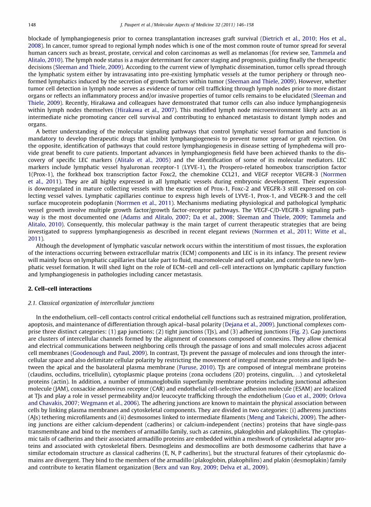
The adult lymphatic system is composed of peripheral capillaries, collecting vessels, lymph nodes, larger trunks and thethoracic duct. Lymphatic vessels are present in the skin and in most internal organs except central nervous system, bonemarrow, retina and avascular tissues such as cartilage, hair, nails, cornea and epidermis (Tammela and Alitalo, 2010). Lym-phatic capillaries (also referred to in the literature as initial lymphatics or absorbing lymphatics), the initial absorptive partof the lymphatic vasculature, are blind-ended vessels formed by a single layer of lymphatic endothelial cells (LEC) devoid ofpericyte coverage and continuous basement membrane (BM) (Fig. 1). In contrast, collecting lymphatic vessels are coated byperivascular smooth muscle cells to allow fluid propulsion and contain valves to prevent backflow (Lund and Swartz, 2010).The main physiological function of the lymphatic vasculature is to maintain tissue fluid homeostasis through the uptake offluid and macromolecules that leak out of blood capillaries into interstitial tissues spaces, and return them back to the bloodcirculation via the inferior vena cava in the form of lymph. In addition, the lymphatic system plays an important part in im-mune defenses against infection through the transport of antigen presenting cells to the lymph nodes and of lymphocytesexiting in lymph nodes. It also contributes to the intestinal fatty acid and fat absorption and transport (for review see,von der Weid and Rainey, 2010; Wang and Oliver, 2010). The absence of lymphatic system is incompatible with life, andlymphatic dysfunctions lead to chronic lymphedema and impaired immune responses (Karpanen and Alitalo, 2008).
Lymphangiogenesis, the formation of new lymphatic vessels from pre-existing one, is primarily an embryonic event(Karpanen and Alitalo, 2008; Makinen et al., 2007). In adults, this process is closely associated with tissue inflammation andoccurs in wound healing, chronic inflammation, autoimmunity, allograft rejection and tumor metastasis (Achen et al., 2005;Huggenberger et al., 2010; Mouta and Heroult, 2003; Regenfuss et al., 2008). During inflammatory conditions, lymphaticsfacilitate tissue edema resolution and immune response. Postnatal inflammatory lymphangiogenesis is extensively studiedin the cornea which is avascular under physiological conditions, but can undergo blood and lymphatic neovascularizationunder certain inflammatory conditions such as corneal graft or infections (Ellenberg et al., 2010; Regenfuss et al., 2008). In-deed, graft rejection is attributed in part to lymphangiogenesis. Of great interest is the recent demonstration that selective
Interstitial Flow
BV
HAProteoglycan
Collagen
LV
L
M
Fluid
proteinsLipids
Interstitial Flow
Collec
tingly
mphati
c
FN
Lymphaticcapillary
BM
Smoothmuscle cell
Pericyte
LECBEC
BM
Valve
Fibroblast
Anchoringfilaments
BJ
DC
Fig. 1. Schematic overview of the structure and function of the lymphatic vasculature. Fluid containing proteins, lipids and other solutes is leaking out fromblood vessels (BV), percolates through the interstitial tissue and returns to the circulation by the venous capillary bed and lymph vessels (LV). Endothelialcells of lymphatic capillaries have an oak shaped with overlapping scalloped edges (flaps). These flaps are only sealed on the sides by discontinuous button-like junction (BJ) allowing fluid entry through these flaps without disturbing cell–cell cohesion. Lymph then moves to collecting vessels specialized in fluidtransport by perivascular smooth muscle cells coverage that allows fluid propulsion. The collecting lymphatics contain valves to prevent backflow. Immunecells (lymphocytes (L), macrophages (M), dendritic cells (DC)) likely enter lymphatic capillaries through the intermingled flaps. In contrast to BV, interstitialmatrix constitutes the principal microenvironment of initial lymphatic since they are devoid of a continuous basement membrane (BM). Anchoringfilaments connect lymphatic capillaries to extracellular matrix and modulate vessel diameter by pulling adjacent endothelial cells apart. Lymphaticendothelial cells (LEC), blood endothelial cells (BEC), fibronectin (FN), hyaluronan (HA).
148 J. Paupert et al. / Molecular Aspects of Medicine 32 (2011) 146–158
blockade of lymphangiogenesis prior to cornea transplantation increases graft survival (Dietrich et al., 2010; Hos et al.,2008). In cancer, tumor spread to regional lymph nodes which is one of the most common route of tumor spread for severalhuman cancers such as breast, prostate, cervical and colon carcinomas as well as melanomas (for review see, Tammela andAlitalo, 2010). The lymph node status is a major determinant for cancer staging and prognosis, guiding finally the therapeuticdecisions (Sleeman and Thiele, 2009). According to the current view of lymphatic dissemination, tumor cells spread throughthe lymphatic system either by intravasating into pre-existing lymphatic vessels at the tumor periphery or through neo-formed lymphatics induced by the secretion of growth factors within tumor (Sleeman and Thiele, 2009). However, whethertumor cell detection in lymph node serves as evidence of tumor cell trafficking through lymph nodes prior to more distantorgans or reflects an inflammatory process and/or invasive properties of tumor cells remains to be elucidated (Sleeman andThiele, 2009). Recently, Hirakawa and colleagues have demonstrated that tumor cells can also induce lymphangiogenesiswithin lymph nodes themselves (Hirakawa et al., 2007). This modified lymph node microenvironment likely acts as anintermediate niche promoting cancer cell survival and contributing to enhanced metastasis to distant lymph nodes andorgans.
A better understanding of the molecular signaling pathways that control lymphatic vessel formation and function ismandatory to develop therapeutic drugs that inhibit lymphangiogenesis to prevent tumor spread or graft rejection. Onthe opposite, identification of pathways that could restore lymphangiogenesis in disease setting of lymphedema will pro-vide great benefit to cure patients. Important advances in lymphangiogenesis field have been achieved thanks to the dis-covery of specific LEC markers (Alitalo et al., 2005) and the identification of some of its molecular mediators. LECmarkers include lymphatic vessel hyaluronan receptor-1 (LYVE-1), the Prospero-related homeobox transcription factor1(Prox-1), the forkhead box transcription factor Foxc2, the chemokine CCL21, and VEGF receptor VEGFR-3 (Norrmenet al., 2011). They are all highly expressed in all lymphatic vessels during embryonic development. Their expressionis downregulated in mature collecting vessels with the exception of Prox-1, Foxc-2 and VEGFR-3 still expressed on col-lecting vessel valves. Lymphatic capillaries continue to express high levels of LYVE-1, Prox-1, and VEGFR-3 and the cellsurface mucoprotein podoplanin (Norrmen et al., 2011). Mechanisms mediating physiological and pathological lymphaticvessel growth involve multiple growth factor/growth factor-receptor pathways. The VEGF-C/D-VEGFR-3 signaling path-way is the most documented one (Adams and Alitalo, 2007; Da et al., 2008; Sleeman and Thiele, 2009; Tammela andAlitalo, 2010). Consequently, this molecular pathway is the main target of current therapeutic strategies that are beinginvestigated to suppress lymphangiogenesis as described in recent elegant reviews (Norrmen et al., 2011; Witte et al.,2011).
Although the development of lymphatic vascular network occurs within the interstitium of most tissues, the explorationof the interactions occurring between extracellular matrix (ECM) components and LEC is in its infancy. The present reviewwill mainly focus on lymphatic capillaries that take part to fluid, macromolecule and cell uptake, and contribute to new lym-phatic vessel formation. It will shed light on the role of ECM–cell and cell–cell interactions on lymphatic capillary functionand lymphangiogenesis in pathologies including cancer metastasis.
2. Cell–cell interactions
2.1. Classical organization of intercellular junctions
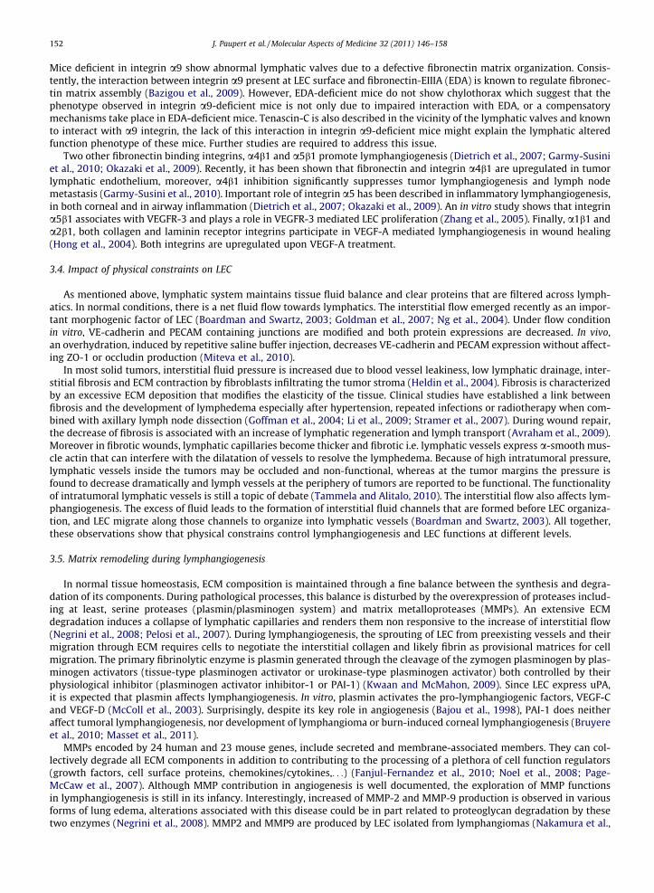
In the endothelium, cell–cell contacts control critical endothelial cell functions such as restrained migration, proliferation,apoptosis, and maintenance of differentiation through apical–basal polarity (Dejana et al., 2009). Junctional complexes com-prise three distinct categories: (1) gap junctions; (2) tight junctions (TJs), and (3) adhering junctions (Fig. 2). Gap junctionsare clusters of intercellular channels formed by the alignment of connexons composed of connexins. They allow chemicaland electrical communications between neighboring cells through the passage of ions and small molecules across adjacentcell membranes (Goodenough and Paul, 2009). In contrast, TJs prevent the passage of molecules and ions through the inter-cellular space and also delimitate cellular polarity by restricting the movement of integral membrane proteins and lipids be-tween the apical and the basolateral plasma membrane (Furuse, 2010). TJs are composed of integral membrane proteins(claudins, occludins, tricellulin), cytoplasmic plaque proteins (zona occludens (ZO) proteins, cingulin,. . .) and cytoskeletalproteins (actin). In addition, a number of immunoglobulin superfamily membrane proteins including junctional adhesionmolecule (JAM), coxsackie adenovirus receptor (CAR) and endothelial cell-selective adhesion molecule (ESAM) are localizedat TJs and play a role in vessel permeability and/or leucocyte trafficking through the endothelium (Guo et al., 2009; Orlovaand Chavakis, 2007; Wegmann et al., 2006). The adhering junctions are known to maintain the physical association betweencells by linking plasma membranes and cytoskeletal components. They are divided in two categories: (i) adherens junctions(AJs) tethering microfilaments and (ii) desmosomes linked to intermediate filaments (Meng and Takeichi, 2009). The adher-ing junctions are either calcium-dependent (cadherins) or calcium-independent (nectins) proteins that have single-passtransmembrane and bind to the members of armadillo family, such as catenins, plakoglobin and plakophilins. The cytoplas-mic tails of cadherins and their associated armadillo proteins are embedded within a meshwork of cytoskeletal adaptor pro-teins and associated with cytoskeletal fibers. Desmogleins and desmocollins are both desmosome cadherins that have asimilar ectodomain structure as classical cadherins (E, N, P cadherins), but the structural features of their cytoplasmic do-mains are divergent. They bind to the members of the armadillo (plakoglobin, plakophilins) and plakin (desmoplakin) familyand contribute to keratin filament organization (Berx and van Roy, 2009; Delva et al., 2009).

APICAL
BASAL
MATRIX
MOLECULAR COMPOSITION
GAP JUNCTIONS
TRANSMEMBRANEMOLECULES
CYTOPLASMIC PARTNERS
CYTOSKELETAL COMPONENT
connexin Not applicable Not applicable
TIGHTJUNCTIONS
claudinoccludintricellulinJAMESAMCAR
ZOCingulinMUPP1PATJ
Actin
ADHERENSJUNCTION
Nectins Afadin Actin
Classical cadherinsVE-cadherin,cadherin E, N, P
EPLINNezhaArmadillo family:Catenins
Actin, microtubule
DESMOSOMECadherin family: desmoglein,desmocollin
Armadillo family:plakoglobin,plakophilinPlakin family: desmoplakin
Keratin intermediatefilament
Junction Type MOLECULAR COMPOSITION
GAP JUNCTIONS
TRANSMEMBRANEMOLECULES
CYTOPLASMIC PARTNERS
CYTOSKELETAL COMPONENT
connexin Not applicable Not applicable
TIGHTJUNCTIONS
claudinoccludintricellulinJAMESAMCAR
ZOCingulinMUPP1PATJ
Actin
ADHERENSJUNCTION
Nectins Afadin Actin
Classical cadherinsVE-cadherin,cadherin E, N, P
EPLINNezhaArmadillo family:Catenins
Actin, microtubule
DESMOSOMECadherin family: desmoglein,desmocollin
Armadillo family:plakoglobin,plakophilinPlakin family: desmoplakin
Keratin intermediatefilament
CELL1 CELL 2Integrin
ICAM VCAMPECAM-1
VESSEL LUMEN
Fig. 2. The main molecular players of cell junctional complexes and cell–matrix interactions. Molecular players found in LEC are written in red. Junctionaladhesion molecule (JAM), coxsackie adenovirus receptor (CAR), endothelial cell-selective adhesion molecule (ESAM), zona occludens (ZO), cell adhesionmolecules (CAM), multi-PDZ domain protein (MUPP1), PALS1-associated tight junction protein (PATJ), epithelial protein lost in neoplasm (EPLIN).
Table 1Mouse knock-out models and human mutations of cell–cell and cell–matrix molecules associated with a lymphatic phenotype.
Lymphatic phenotype References
GJC2 (encoding connexin 47) missense mutations inhuman
Lymphedema Ferrell et al. (2010)
Col18a1�/� mice Enhanced corneal lymphangiogenesis after keractectomy wound Kojima et al. (2008)Emilin 1�/� mice Reduction of anchoring filaments Danussi et al.
(2008)Hyperplastic and enlarged lymphatic vesselsDevelop larger lymphangiomaLymph leakage
Integrin a9�/� mice Severe bilateral chylotorax Huang et al. (2000)Abnormal lymphatic valves Bazigou et al.
(2009)Edema and inflammatory cell accumulation in the chest wall
Missense mutation in human integrin a9 gene(c.1210G > A, p.G404S)
Severe bilateral chylothorax Ma et al. (2008)
Integrin a1b1�/� mice Decreased of lymphangiogenesis after cornea transplantation Chen et al. (2007)Tie2Cre + a4loxp/loxp mice Lymphangiogenesis reduced in a in vivo matrigel plug assay Garmy-Susini et al.
(2010)Integrin a4Y991A knock-in mutation (mice) Lymphangiogenesis reduced in an in vivo matrigel plug assay and in
the lymphatic ring assayGarmy-Susini et al.(2010)
Reduction of metastasis to lymph nodeReduction of tumor-induced lymphangiogenesis
J. Paupert et al. / Molecular Aspects of Medicine 32 (2011) 146–158 149
2.2. Intercellular junctions in LEC
Gap junctions were described only in collecting vessels where these junctions play a role in the propagation and coordi-nation of contractions in lymphatics in the mesentery of the rat small intestine (Zawieja et al., 1993). Recently, Ferrell et al.show that GJC2 (encoding connexin 47) missense mutations cause human lymphedema (Ferrell et al., 2010) (Table 1).

150 J. Paupert et al. / Molecular Aspects of Medicine 32 (2011) 146–158
TJs and AJs are not as frequently seen in lymphatics as in blood vessels (Leak and Burke, 1968; Pepper and Skobe, 2003).Adherens and tight junction proteins in LEC have been detected by immunohistochemistry and gene array profiling, and in-clude VE-cadherin, occludin, claudin-5, ZO-1, ESAM and JAM-A, JAM-B, -C as well as CAR (Aurrand-Lions et al., 2001; Baluket al., 2007; Hirakawa et al., 2003; Ueki et al., 2008; Vigl et al., 2009). CAR is expressed specifically by human LEC, but notmurine LEC (Raschperger et al., 2006; Vigl et al., 2009). The knock-down of CAR in human LEC by specific siRNAs leads to areduction of LEC–LEC adhesion, migration and tube formation into collagen matrix. Interestingly, CAR re-expression in LECrestores these LEC properties in vitro. The expression of LEC adhesion and junctional proteins seems to be modulated duringcancer progression (Clasper et al., 2008). Gene array analyzes of LEC isolated from tumors in comparison to LEC isolated fromnormal tissue show downregulation of JAM-B, N-cadherin, cadherin-11, whereas JAM-A, ESAM and CAR are upregulated intumor-associated LEC. The upregulation of ESAM in tumor-associated LEC is confirmed by immunostaining (Clasper et al.,2008).
An adhering junction (‘‘complexus adhaerentes’’) specific to LEC has been evidenced in various human, bovine and rodenttissues (Schmelz et al., 1994). These junctions are usually larger than desmosomes and characterized by a dense plaque con-taining plakoglobin and desmoplakin, but not desmoglein and desmocollin. These «complexus adhaerentes» are also positivefor a-catenin, VE-cadherin, ZO-1 and claudin 5 (Hammerling et al., 2006). Comparative gene array analyzes of cultured pri-mary blood endothelial cells (BEC) and primary LEC revealed that desmoplakin is upregulated in LEC (4.6-fold) (Hirakawaet al., 2003). Immunohistochemical study demonstrates that desmoplakin is detected in small lymphatic vessels, but notin the thoracic duct or blood vessels (Schmelz et al., 1994). This observation has been also confirmed on human tongue tissue(Ebata et al., 2001).
The elegant ultrastructural study of Baluk et al. has revisited the initial view of lymphatics displaying poorly developed orno intercellular junctions (Leak and Burke, 1968; Baluk et al., 2007). This study reports that junctions between LEC in lym-phatic capillaries are unique at likely sites of fluid entry. Endothelial cells of capillaries display a typical oak leaf shape thatdiffers from the classical spindle-shaped endothelial cells of collecting lymphatics and blood vessels (Fig. 1). These oak leafshaped cells extend overlapping scalloped edges (flaps) resembling valve-like structures. These flaps lack junctions at the tipare sealed on the sides by discontinuous button-like junctions containing AJ and TJ proteins (Baluk et al., 2007) (Fig. 1). Thus,the authors hypothesized that these junctions would permit fluid entry through the opening of these free flaps and withoutdisrupting the overall junctional organization. The more distal collecting vessels have continuous zipper-like junctionsresembling those of blood vessel endothelium. The main differences between buttons and zippers rely on their organizationrather than their composition. It is worth noting that the presence of button-like structures does not translate the dynamicfeature of the immature region of lymphatic sprouts, since zippers like junctions, but not buttons are seen in growing tips oflymphatics neoformed in a sustained inflammation model of lymphangiogenesis (Baluk et al., 2007). In addition, discrepan-cies can be found between data generated in vitro and in vivo regarding junction composition and function since LEC mono-layer does not show button junction but rather zipper (Kriehuber et al., 2001).
3. Cell–matrix interactions
3.1. The extracellular matrix
One of lymphatic capillaries features is the incomplete or absence of BM (Leak and Burke, 1968). This characteristic mightbe related to low levels of BM component production by LEC (Hirakawa et al., 2003; Podgrabinska et al., 2002) or to the lackof pericytes which are an abundant source of BM constituents (Petrova et al., 2004; Stratman et al., 2009). In line with thislatter hypothesis is the observation of lymphatic capillaries in Foxc2�/�mice that are invested by pericytes and surroundedby a deposition of BM components such as collagen IV. Thus, in contrast to BEC, the interstitial matrix and fluid constitute theprincipal microenvironment of LEC, both in physiological and pathological conditions. Major extracellular matrix (ECM)components include collagens, proteoglycans, fibronectin, laminins, elastin and vitronectin (Miner and Yurchenco, 2004).For the description of ECM components, readers are referred to recent reviews addressing their structure and their role incell function regulation (Durbeej, 2010; Gordon and Hahn, 2010; Kielty, 2006; Schaefer and Schaefer, 2010; Singh et al.,2010; Wiig et al., 2010). Here, we focus on the description of interstitial matrix components known to affect the functionof lymphatic capillaries and lymphangiogenesis.
The primary scaffold of interstitial matrix surrounding lymphatic capillaries is made of fibrillar collagens that entrap pro-teoglycans and glycoproteins. Interstitial collagens and fibrin both serve as provisional matrices during wound healing(Helm et al., 2007). The fact that LEC are in close contact with the interstitial matrix can also explain the higher LEC survivaland tubulogenesis observed on type I collagen without exogenously added growth factors, a condition in which BEC poorlysurvive (Podgrabinska et al., 2002). We recently set up a 3D model of lymphatic ring cultures (Bruyere et al., 2008) in whichLEC sprout and organize into tube-like structures in a type I collagen gel, but not in a basement reconstituted matrix (matri-gel) (Agnès Noël personal data). In the presence of growth factors and fluid flow, BEC organize better in a matrix composed ofequal amount of collagen and fibrin, whereas LEC organize better in a matrix composed of fibrin only (Helm et al., 2007).However, in other studies, collagen matrix promotes LEC tube formation without flow and growth factor added (Leak andJones, 1994; Podgrabinska et al., 2002). Differences in the experimental design and conditions used to perform the tubulo-genesis assay (such as cell suspension in the collagen gel or seeding between two collagen layers and the type of collagen

J. Paupert et al. / Molecular Aspects of Medicine 32 (2011) 146–158 151
used) can explain discrepancies between results. In vivo, in secondary lymphedema induced by skin excision in the mousetail, type I collagen accelerates lymphatic regeneration and wound repair further supporting its role in the control of lym-phatic cell function (Clavin et al., 2008). As previously described for angiogenesis (Hamano et al., 2003), some collagen frag-ments also exert an inhibitory effect on lymphangiogenesis. For instances, endostatin and neostatin 7, fragments of collagentype XVIII inhibit lymphangiogenesis (Brideau et al., 2007; Kojima et al., 2008). Accordingly, collagen XVIII-deficient miceexhibit increased corneal lymphangiogenesis after keratectomy wound (Kojima et al., 2008) (Table 1).
Hyaluronan (HA) is a major ECM constituent which regulates cell proliferation and migration, as well as controls matrixstability and water retention (Jackson, 2009). It is a polymer of disaccharides, themselves composed of D-glucuronic acid andD-N-acetylglucosamine, ranging in size from 104 to 107 Da. HA has been assigned various physiological functions due to itsviscoelastic properties, its hygroscopic capacities. Polymers are either anchored firmly in the plasma membrane or bound viareceptors (Nusgens, 2010). One third of HA is replaced daily in tissues in which a part of it is removed by the lymphatic ves-sels and/or degraded by lymphatic cells in the lymph nodes. In mammary tumors, HA increases intratumoral lymphangio-genesis and lymphatic vessels are mostly found in association with HA-rich stroma (Koyama et al., 2008). Accordingly,similar results are reported in idiopathic pulmonary fibrosis where HA is found around lymphatic vessels and short frag-ments of HA induce lymphangiogenesis (El-Chemaly et al., 2009). Lymphatic hyaluronan receptor, Lyve-1, is one of the lym-phatic marker commonly used. However, Lyve 1�/� mice display normal phenotype, have a functional lymphatic networkand a normal hyaluronan metabolism which is likely due to compensatory mechanisms by other HA-receptors (Gale et al.,2007). However, Lyve-1 null mice show larger lymphatic vessels with distended lumen in liver and intestine, but not in othertissues tested (lung, ovary, uterus, vagina, cervix, salivary gland, skin, kidney, brain and foot pad) (Huang et al., 2006).
Finally, lymphatic vessels are surrounded by patches of fibronectin (Oh et al., 1997) that is known to enhance VEGF-C-mediated LEC proliferation in vitro (Zhang et al., 2005). Interestingly, the spliced variant fibronectin EDA (fibronectin-EIIIA,also called EDA and containing EDA domain) detected in the tumor stroma but not in normal tissue, stimulates LEC tubulo-genesis (Ou et al., 2010). Netrin-4, a laminin-related secreted protein, is also a pro-lymphangiogenic factor. Netrin-4 impli-cated in neuron guidance during development is also expressed by LEC and induces LEC proliferation, migration and survival.Its overexpression in tumors increases the number of lymphatics, reduces lymphatic vessel permeability and enhancesmetastasis (Larrieu-Lahargue et al., 2010).
3.2. LEC anchorage to the ECM
An additional feature of lymphatic capillaries is their close connection to surrounding tissue by a fibrillar elastic apparatuswhich is absent in blood vessels (Gerli et al., 1990). All fibers of this apparatus are composed of microfibrils that ensure thelink between lymphatic vessels and the surrounding tissue. This fibrillar elastic apparatus is composed of three successivetypes of interconnected fibers including (from the capillary wall to the tissue): (1) oxytalan (usually called anchoring fila-ment) composed of microfibril bundles associated to the abluminal surface of lymphatic capillaries, (2) elaunin consistingof microfibrils embedded in a small amount of elastin and (3) elastic fibers contain elastin surrounded by microfibrils (Gerliet al., 1990). These filaments are extremely important for draining the excess of extracellular fluid occurring during tissueinjury, inflammation or tumor growth. Increased interstitial pressure stretches the connective tissue fibers and anchoringfilaments, pull adjacent endothelial cells apart and increase the diameter of lymphatic vessels that are usually collapsed un-der normal circumstances. This increase of lymphatic lumen is essential for fluid and particles passage into the lymphatics(Ji, 2006). The importance of anchoring filament in lymphatic function has been recently demonstrated through the gener-ation of Emilin1�/�mice (Danussi et al., 2008). EMILIN1 (elastin microfibril interface-located protein 1) is a connective tis-sue glycoprotein associated with elastic fibers in the extracellular matrix of blood vessels as well as in connective tissue ofother organs (Bressan et al., 1993; Colombatti et al., 2000). EMILIN1 is expressed at the abluminal surface of LEC as well as inthe fibers radiate from LEC. The number of anchoring filaments is significantly reduced in Emilin1�/� mice and these miceshow leaky lymphatic vessels (Table 1).
3.3. Role of integrins in LEC functions
Most of the effects of ECM components on lymphangiogenesis are mediated via integrins. Integrins are a large family ofheterodimeric transmembrane glycoproteins that mediate cell–ECM or cell–cell interactions. Integrins contain 18 large (a)and 8 small (b) subunits generating 24 heterodimers (Avraamides et al., 2008). Integrins serve as transmembrane linkers be-tween their extracellular ligands and the cytoskeleton and associated intracytoplasmic partners. Through these multipleinteractions, they control cell adhesion, migration and differentiation in physiological and pathological conditions. Effectof fibronectin and EDA fibronectin domain mentioned above are mediated by a5b1 and a9b1, respectively (Ou et al.,2010; Zhang et al., 2005). The a9 integrin mediates LEC chemotaxis and migration of LEC toward VEGF-C (Mishima et al.,2007). It has been shown that a9 integrin binds directly VEGF-C and D (Vlahakis et al., 2005). Integrin a9b1 is currentlyviewed as a major integrin associated with lymphangiogenesis since this integrin is required for the development of the lym-phatic system (Huang et al., 2000). The known ligands of a9b1 are tenascin, fibronectin, thrombospondin, VCAM-1, collagenand laminin. Until recently, none of these ligands were known to affect lymphangiogenesis in vivo. Mice lacking the integrina9b1 died shortly and develop chylotorax, edema and inflammatory cell accumulation around the thoracic duct. Chylotoraxhas also been described in human fetuses carrying a mutation in the a9 integrin subunit gene (Ma et al., 2008) (Table 1).
152 J. Paupert et al. / Molecular Aspects of Medicine 32 (2011) 146–158
Mice deficient in integrin a9 show abnormal lymphatic valves due to a defective fibronectin matrix organization. Consis-tently, the interaction between integrin a9 present at LEC surface and fibronectin-EIIIA (EDA) is known to regulate fibronec-tin matrix assembly (Bazigou et al., 2009). However, EDA-deficient mice do not show chylothorax which suggest that thephenotype observed in integrin a9-deficient mice is not only due to impaired interaction with EDA, or a compensatorymechanisms take place in EDA-deficient mice. Tenascin-C is also described in the vicinity of the lymphatic valves and knownto interact with a9 integrin, the lack of this interaction in integrin a9-deficient mice might explain the lymphatic alteredfunction phenotype of these mice. Further studies are required to address this issue.
Two other fibronectin binding integrins, a4b1 and a5b1 promote lymphangiogenesis (Dietrich et al., 2007; Garmy-Susiniet al., 2010; Okazaki et al., 2009). Recently, it has been shown that fibronectin and integrin a4b1 are upregulated in tumorlymphatic endothelium, moreover, a4b1 inhibition significantly suppresses tumor lymphangiogenesis and lymph nodemetastasis (Garmy-Susini et al., 2010). Important role of integrin a5 has been described in inflammatory lymphangiogenesis,in both corneal and in airway inflammation (Dietrich et al., 2007; Okazaki et al., 2009). An in vitro study shows that integrina5b1 associates with VEGFR-3 and plays a role in VEGFR-3 mediated LEC proliferation (Zhang et al., 2005). Finally, a1b1 anda2b1, both collagen and laminin receptor integrins participate in VEGF-A mediated lymphangiogenesis in wound healing(Hong et al., 2004). Both integrins are upregulated upon VEGF-A treatment.
3.4. Impact of physical constraints on LEC
As mentioned above, lymphatic system maintains tissue fluid balance and clear proteins that are filtered across lymph-atics. In normal conditions, there is a net fluid flow towards lymphatics. The interstitial flow emerged recently as an impor-tant morphogenic factor of LEC (Boardman and Swartz, 2003; Goldman et al., 2007; Ng et al., 2004). Under flow conditionin vitro, VE-cadherin and PECAM containing junctions are modified and both protein expressions are decreased. In vivo,an overhydration, induced by repetitive saline buffer injection, decreases VE-cadherin and PECAM expression without affect-ing ZO-1 or occludin production (Miteva et al., 2010).
In most solid tumors, interstitial fluid pressure is increased due to blood vessel leakiness, low lymphatic drainage, inter-stitial fibrosis and ECM contraction by fibroblasts infiltrating the tumor stroma (Heldin et al., 2004). Fibrosis is characterizedby an excessive ECM deposition that modifies the elasticity of the tissue. Clinical studies have established a link betweenfibrosis and the development of lymphedema especially after hypertension, repeated infections or radiotherapy when com-bined with axillary lymph node dissection (Goffman et al., 2004; Li et al., 2009; Stramer et al., 2007). During wound repair,the decrease of fibrosis is associated with an increase of lymphatic regeneration and lymph transport (Avraham et al., 2009).Moreover in fibrotic wounds, lymphatic capillaries become thicker and fibrotic i.e. lymphatic vessels express a-smooth mus-cle actin that can interfere with the dilatation of vessels to resolve the lymphedema. Because of high intratumoral pressure,lymphatic vessels inside the tumors may be occluded and non-functional, whereas at the tumor margins the pressure isfound to decrease dramatically and lymph vessels at the periphery of tumors are reported to be functional. The functionalityof intratumoral lymphatic vessels is still a topic of debate (Tammela and Alitalo, 2010). The interstitial flow also affects lym-phangiogenesis. The excess of fluid leads to the formation of interstitial fluid channels that are formed before LEC organiza-tion, and LEC migrate along those channels to organize into lymphatic vessels (Boardman and Swartz, 2003). All together,these observations show that physical constrains control lymphangiogenesis and LEC functions at different levels.
3.5. Matrix remodeling during lymphangiogenesis
In normal tissue homeostasis, ECM composition is maintained through a fine balance between the synthesis and degra-dation of its components. During pathological processes, this balance is disturbed by the overexpression of proteases includ-ing at least, serine proteases (plasmin/plasminogen system) and matrix metalloproteases (MMPs). An extensive ECMdegradation induces a collapse of lymphatic capillaries and renders them non responsive to the increase of interstitial flow(Negrini et al., 2008; Pelosi et al., 2007). During lymphangiogenesis, the sprouting of LEC from preexisting vessels and theirmigration through ECM requires cells to negotiate the interstitial collagen and likely fibrin as provisional matrices for cellmigration. The primary fibrinolytic enzyme is plasmin generated through the cleavage of the zymogen plasminogen by plas-minogen activators (tissue-type plasminogen activator or urokinase-type plasminogen activator) both controlled by theirphysiological inhibitor (plasminogen activator inhibitor-1 or PAI-1) (Kwaan and McMahon, 2009). Since LEC express uPA,it is expected that plasmin affects lymphangiogenesis. In vitro, plasmin activates the pro-lymphangiogenic factors, VEGF-Cand VEGF-D (McColl et al., 2003). Surprisingly, despite its key role in angiogenesis (Bajou et al., 1998), PAI-1 does neitheraffect tumoral lymphangiogenesis, nor development of lymphangioma or burn-induced corneal lymphangiogenesis (Bruyereet al., 2010; Masset et al., 2011).
MMPs encoded by 24 human and 23 mouse genes, include secreted and membrane-associated members. They can col-lectively degrade all ECM components in addition to contributing to the processing of a plethora of cell function regulators(growth factors, cell surface proteins, chemokines/cytokines,. . .) (Fanjul-Fernandez et al., 2010; Noel et al., 2008; Page-McCaw et al., 2007). Although MMP contribution in angiogenesis is well documented, the exploration of MMP functionsin lymphangiogenesis is still in its infancy. Interestingly, increased of MMP-2 and MMP-9 production is observed in variousforms of lung edema, alterations associated with this disease could be in part related to proteoglycan degradation by thesetwo enzymes (Negrini et al., 2008). MMP2 and MMP9 are produced by LEC isolated from lymphangiomas (Nakamura et al.,

J. Paupert et al. / Molecular Aspects of Medicine 32 (2011) 146–158 153
2004) and by lymphatic capillaries sprouting from thoracic duct explants (Bruyere et al., 2008). A broad-spectrum MMPinhibitor impairs LEC tubulogenesis in vitro and inhibits lymph node metastasis in vivo (Nakamura et al., 2004). Recently,Matsuo et al. hypothetize that the inhibitory effect of curcumin on tube formation in vitro is partially mediated by MMP-2 blockade (Matsuo et al., 2007). A more direct clue of MMP2 contribution in lymphangiogenesis is provided by the impairedlymphangiogenesis observed in the lymphatic ring assay performed with lymphatic duct explant from MMP-2 deficient mice(Bruyere et al., 2008). In this model, MMP-9-deficiency does not affect lymphangiogenesis, this emphasizes the specific con-tribution of MMP2 in this process. As seen in angiogenesis, the cleavage of ECM components by MMPs is expected to gen-erate fragments endowed with lymphangio-inhibitory functions. For instance, as mentioned above, neostatin-7, a type XVIIIcollagen fragment generated by MMP-7 inhibits corneal lymphangiogenesis probably by its association to VEGFR-3 (Kojimaet al., 2008).
4. LEC interactions with cancer cells and leukocytes
The transmigration of cells (cancer cells or leukocytes) through an endothelium is a multi-step process that involvescell attraction by cell adhesion molecules and cell migration between endothelial cells. LEC in capillaries produce a panelof chemokines which attract leukocytes such as CCL-21, CXCL-12, MCP-1 and Rantes (Mouta and Heroult, 2003). For in-stance, CCL-21 production by LEC has been described to attract both cancer cells and leukocytes. It is suggested that tumorcells use the same mechanisms as immune cells to enter the lymphatic capillaries (Shields et al., 2007). Moreover, highlyinvasive cancer cells are known to produce high levels of CCR-7 ligand, CCR-7, CCl-21 and CCL-19 receptors compared topoorly metastatic cancer cell lines. In a 3D in vitro cell migration model, tumor cells migrate in a CCR-7 dependent mannertowards LEC and fluid flow enhances tumor cell migration (Shields et al., 2007). A complex molecular crosstalk betweentumor cells and LEC appears to be established during cancer invasion (Issa et al., 2009). Tumor-derived VEGFC increasesCCL-21 secretion by LEC which in turn drives CCR-7-dependent chemoinvasion of tumor cells toward lymphatics (Issaet al., 2009). In addition to this paracrine effect, tumor cell attraction appears to be amplified by a so-called autologouschemotaxis (Shields et al., 2007). According to this concept, CCR7-positive tumor cells which autocrinely produce CCR-7ligand are surrounded by high concentration of this ligand and thus migrate and are guided toward the direction ofthe interstitial fluid flow.
LEC express various cell adhesion molecules (CAM) such as PECAM-1, VCAM-1, ICAM-1, ICAM-2 and E-selectin (Baluket al., 2007; Johnson et al., 2006; Raschperger et al., 2006; Sawa et al., 1999). Most of them are expressed at low levels onnormal lymphatic vessels and are upregulated upon inflammatory stimuli or under increased interstitial flow (Johnsonet al., 2006; Miteva et al., 2010; Sawa et al., 2008). Dendritic cells (DC) transmigration requires the adhesion via ICAM-1and VCAM-1 that is a prerequisite step for transmigration across lymphatic vessels (Johnson et al., 2006). The interactionof DC with lymphatic vessels via ICAM-1 also modulates DC differentiation and functions (Podgrabinska et al., 2009). PE-CAM-1 involvement in leukocyte migration through lymphatics seems to be dependent on the mice genetic background.In C57BL/6 background, PECAM-1 is not involved in leucocyte migration through lymphatic endothelium (Baluk et al.,2007). Furthermore, PECAM-deficient FVB/n mice have reduced leukocyte emigration in acute models of inflammation, whilePECAM-deficient C57BL/6 mice display normal responses (Schenkel et al., 2006, 2004). The PECAM-1-independent leukocytetransmigration is unique to C57BL/6 mice since a reduced leukocyte emigration is also observed in different strains of miceor rat using anti-PECAM Abs (Schenkel et al., 2004). A quantitative trait locus mapping between PECAM-deficient FVB/n andC57BL/6 mice has identified a single locus, at 35.8 Mb on murine chromosome 2, associated with PECAM-independent leu-kocyte transmigration in model of peritonitis. However, the specific gene involved in this process is still unknown (Seidmanet al., 2009). In cancer, ICAM-1 produced by LEC has been described to also mediate breast adenocarcinoma cell adhesion tohuman LEC (Kawai et al., 2008). Moreover, conditioned media from highly metastatic MDA-MB-231 cells, but not frompoorly metastatic MCF-7 cells induce an upregulation of ICAM-1 by LEC, which in turn stimulates cancer cell adhesion toLEC. This study emphasizes the importance of ICAM-1 in the establishment of a cross talk between tumor cells and LEC. Addi-tional membrane proteins produced by LEC are contributing to endothelial transmigration. Common lymphatic and vascularendothelial receptor CLEVER-1/Stabilin-1 and mannose receptor (MR), both expressed by LEC are involved in lymphocytetrafficking (Salmi et al., 2004). MR allows binding of L-selectin positive lymphocytes to LEC (Irjala et al., 2001). In vivo, CLE-VER-1 blockade or the MR absence on LEC impairs B and T cell trafficking from the periphery into the lymph node (Karikoskiet al., 2009; Marttila-Ichihara et al., 2008). Interestingly, both CLEVER-1 and MR mediate the adhesion of lymphomas andhead and neck squamous cell carcinoma (HNSCC) to lymphatic endothelium in vitro (Irjala et al., 2003). Immunohistochem-ical study of MR performed on 17 tumor tissues biopsies of HNSCC and 72 breast carcinoma, show that intratumoral lymphvessel MR expression is associated with the presence of axillary lymph node metastases at the time of diagnosis (Irjala et al.,2003). Experimental studies using MR deficient mice support the importance of the lymphatic MR in metastatic dissemina-tion. Indeed, B16 melanoma cells fail to metastasize to local lymph nodes of MR deficient (Marttila-Ichihara et al., 2008). Anadditional LEC expressed membrane protein involved in cell adhesion is Thy1, a thymus cell antigen. It is expressed at highlevels on mouse LEC but not on BEC (Jurisic et al., 2010). As assessed by immunofluorescent staining, lymphatic vessels ofnormal tissues express low Thy1 levels, while those of human prostate cancer tissue express high Thy-1 levels. Thy-1 block-ing antibody decreases tumor cell adhesion on mouse LEC monolayer and leukocyte adhesion on human LEC monolayerstimulated with phorbol myristate acetate. Cell adhesion to Thy-1 seems to be integrin-dependent. Indeed, Thy-1 protein

154 J. Paupert et al. / Molecular Aspects of Medicine 32 (2011) 146–158
sequence contains an integrin binding site promoting the interaction with aMb2 on leucocytes and aVb3 on melanoma cells(Saalbach et al., 2005; Wetzel et al., 2004).
Current knowledge of the mechanisms underlying leukocyte and cancer cell entry into lymphatics is extremely sparseand whether cell trafficking occurs in a passive or an active way is still being debated. Under basal condition, immune cellsintravasate into lymphatics in a passive manner (Lund and Swartz, 2010). However, more and more studies show that underinflammation and/or in the tumor microenvironment, cell junction proteins, cell adhesion molecules and other membraneproteins are actively involved in lymphatic endothelial transmigration. Button-rich junctions are currently viewed as gatekeepers for leucocyte entry. Few or no MHC-II positive cells (mainly DC and macrophages) has been detected to be associatedwith LEC in vivo on resting lymphatic vessels (Baluk et al., 2007). After bacterial lypopolysaccharide exposure, most leuko-cytes are associated with the button-rich junctions. However, whether leukocytes enter through openings between button-rich regions of lymphatic capillaries is not yet solved. Dendritic cell association at the button-rich junctions has also beendescribed in response to an increase of interstitial flow (Miteva et al., 2010). Another argument on the role of LEC junctionsin cellular transmigration is the increased DC transmigration through lymphatic endothelium observed in JAM-A deficientmice (Cera et al., 2004).
5. Concluding remarks
Recent progresses in the field of lymphangiogenesis offer new therapeutic options and led to the development ofanti-lymphangiogenesis drugs to inhibit tumor lymphangiogenesis, metastatic spread or graft rejection. Given that theVEGF-C/VEGF-D/VEGFR-3 axis is the best validated signaling pathway for promoting lymphangiogenesis, different strat-egies have been used to block these growth factors and their receptors or co-receptor (such as neuropilin-2) (Norrmenet al., 2011). Neutralizing antibodies (anti-VEGFR-3, VEGF-C/D or neuropilin-2), as well as soluble VEGFR-3 fusion pro-teins (VEGF-C/D trap) and siRNA targeting VEGF-C mRNA have proven efficacy to counteract tumor-induced lymphangio-genesis (Chen et al., 2005; He et al., 2008, 2002; Lin et al., 2005; Rinderknecht et al., 2010; Roberts et al., 2006; Tvorogovet al., 2010). A novel soluble form of VEGFR-2 issued from an alternative splicing emerged recently as a novel putativeinhibitor of lymphangiogenesis (Albuquerque et al., 2009). Interestingly, blocking peptides against VEGFR-3 specificallyinhibit corneal lymphangiogenesis in animal models without affecting hemangiogenesis (Bock et al., 2008). Another sig-naling system with specificity to endothelial cells consists of the tyrosine kinase Tie receptor (Tie 1 and Tie2) and angio-poietin ligands (Ang1 and Ang2). It offers an additional option to block lymphangiogensis (Tammela et al., 2005). Small-molecule inhibitors of tyrosine kinase activity often lack specificity and block different pathways (e.g. VEGFRs, FGFRs andPDGFRs). Inhibitors of both VEGFR-2 and VEGFR-3 like Sorafenib or Sunitinib (SU-11248) lead to the blockade of angi-ogenesis and lymphangiogenesis associated to tumor and injured cornea (Bauerschlag et al., 2010; Bono et al., 2010;Gridelli et al., 2007; Perez-Santonja et al., 2010; Roskoski, 2007; Young et al., 2010). It is anticipated that blocking dif-ferent pathways involved in pathological lymphangiogenesis will be required for optimal effect and to avoid the devel-opment of resistance against treatment. A complementary strategy could interfere with chemokines such as CCL21 orCXCL12 (SDF-1) involved in tumor cell dissemination (Kim et al., 2010; Lanati et al., 2010). Thanks to the evidence ofthe specific interactions of lymphatic endothelial cells with their interstitial environment, therapeutic strategies couldalso include the blockage of cell matrix interaction by interfering with integrins (Dietrich et al., 2007; Garmy-Susiniet al., 2010) or the inhibition matrix remodeling enzymes (Bruyere et al., 2008). It is worth mentioning that most ofthe anti-lymphangiogenic approaches that have emerged until now, have been derived from animal studies. These re-sults must be taken with care since the intensity of lymphangiogenic response and its inhibition varied with the mousegenetic background (Regenfuss et al., 2010). Moreover, emerging concept such as the fact that tumor cells actively enterinto lymphatic vasculature by using cell adhesion molecule and receptors need to be consolidated in order to design newdrugs. Further characterization of potential candidates to be targeted is required. In this context, a better understandingof LEC interactions with its surrounding cellular and molecular environment may enhance our mechanistic knowledgeand ultimately lead to the design of new therapeutic drug to regulate lymphangiogenesis.
Acknowledgments
This work was supported by grants from the Fondation Contre le Cancer, FP7-HEALTH-2007-A Proposal No. 201279‘‘MICROENVIMET’’ and from the NEOANGIO Program No. 616476 of the Direction Générale Opérationnelle de l’Economie,de l’Emploi et de la Recherche from the S.P.W. (Service Public de Wallonie, Belgium).
References
Achen, M.G., McColl, B.K., Stacker, S.A., 2005. Focus on lymphangiogenesis in tumor metastasis. Cancer Cell 7 (2), 121–127.Adams, R.H., Alitalo, K., 2007. Molecular regulation of angiogenesis and lymphangiogenesis. Nat. Rev. Mol. Cell Biol. 8 (6), 464–478.Albuquerque, R.J., Hayashi, T., Cho, W.G., Kleinman, M.E., Dridi, S., Takeda, A., Baffi, J.Z., Yamada, K., Kaneko, H., Green, M.G., Chappell, J., Wilting, J., Weich,
H.A., Yamagami, S., Amano, S., Mizuki, N., Alexander, J.S., Peterson, M.L., Brekken, R.A., Hirashima, M., Capoor, S., Usui, T., Ambati, B.K., Ambati, J., 2009.Alternatively spliced vascular endothelial growth factor receptor-2 is an essential endogenous inhibitor of lymphatic vessel growth. Nat. Med. 15 (9),1023–1030.

J. Paupert et al. / Molecular Aspects of Medicine 32 (2011) 146–158 155
Alitalo, K., Tammela, T., Petrova, T.V., 2005. Lymphangiogenesis in development and human disease. Nature 438 (7070), 946–953.Aurrand-Lions, M., Duncan, L., Ballestrem, C., Imhof, B.A., 2001. JAM-2, a novel immunoglobulin superfamily molecule, expressed by endothelial and
lymphatic cells. J. Biol. Chem. 276 (4), 2733–2741.Avraamides, C.J., Garmy-Susini, B., Varner, J.A., 2008. Integrins in angiogenesis and lymphangiogenesis. Nat. Rev. Cancer 8 (8), 604–617.Avraham, T., Clavin, N.W., Daluvoy, S.V., Fernandez, J., Soares, M.A., Cordeiro, A.P., Mehrara, B.J., 2009. Fibrosis is a key inhibitor of lymphatic regeneration.
Plast. Reconstr. Surg. 124 (2), 438–450.Bajou, K., Noel, A., Gerard, R.D., Masson, V., Brunner, N., Holst-Hansen, C., Skobe, M., Fusenig, N.E., Carmeliet, P., Collen, D., Foidart, J.M., 1998. Absence of host
plasminogen activator inhibitor 1 prevents cancer invasion and vascularization. Nat. Med. 4 (8), 923–928.Baluk, P., Fuxe, J., Hashizume, H., Romano, T., Lashnits, E., Butz, S., Vestweber, D., Corada, M., Molendini, C., Dejana, E., McDonald, D.M., 2007. Functionally
specialized junctions between endothelial cells of lymphatic vessels. J. Exp. Med. 204 (10), 2349–2362.Bauerschlag, D.O., Schem, C., Tiwari, S., Egberts, J.H., Weigel, M.T., Kalthoff, H., Jonat, W., Maass, N., Meinhold-Heerlein, I., 2010. Sunitinib (SU11248) inhibits
growth of human ovarian cancer in xenografted mice. Anticancer Res. 30 (9), 3355–3360.Bazigou, E., Xie, S., Chen, C., Weston, A., Miura, N., Sorokin, L., Adams, R., Muro, A.F., Sheppard, D., Makinen, T., 2009. Integrin-alpha9 is required for
fibronectin matrix assembly during lymphatic valve morphogenesis. Dev. Cell 17 (2), 175–186.Berx, G., van Roy, F., 2009. Involvement of members of the cadherin superfamily in cancer. Cold Spring Harbor Perspect. Biol. 1 (6), a003129.Boardman, K.C., Swartz, M.A., 2003. Interstitial flow as a guide for lymphangiogenesis. Circ. Res. 92 (7), 801–808.Bock, F., Onderka, J., Dietrich, T., Bachmann, B., Pytowski, B., Cursiefen, C., 2008. Blockade of VEGFR3-signalling specifically inhibits lymphangiogenesis in
inflammatory corneal neovascularisation. Graefes Arch. Clin. Exp. Ophthalmol. 246 (1), 115–119.Bono, A.V., Pannellini, T., Liberatore, M., Montironi, R., Cunico, S.C., Cheng, L., Sasso, F., Musiani, P., Iezzi, M., 2010. Sorafenib’s inhibition of prostate cancer
growth in transgenic adenocarcinoma mouse prostate mice and its differential effects on endothelial and pericyte growth during tumor angiogenesis.Anal. Quant. Cytol. Histol. 32 (3), 136–145.
Bressan, G.M., Daga-Gordini, D., Colombatti, A., Castellani, I., Marigo, V., Volpin, D., 1993. Emilin, a component of elastic fibers preferentially located at theelastin-microfibrils interface. J. Cell Biol. 121 (1), 201–212.
Brideau, G., Makinen, M.J., Elamaa, H., Tu, H., Nilsson, G., Alitalo, K., Pihlajaniemi, T., Heljasvaara, R., 2007. Endostatin overexpression inhibitslymphangiogenesis and lymph node metastasis in mice. Cancer Res. 67 (24), 11528–11535.
Bruyere, F., Melen-Lamalle, L., Blacher, S., Detry, B., Masset, A., Lecomte, J., Lambert, V., Maillard, C., Hoyer-Hansen, G., Lund, L.R., Foidart, J.M., Noel, A., 2010.Does plasminogen activator inhibitor-1 drive lymphangiogenesis? PLoS ONE 5 (3), e9653.
Bruyere, F., Melen-Lamalle, L., Blacher, S., Roland, G., Thiry, M., Moons, L., Frankenne, F., Carmeliet, P., Alitalo, K., Libert, C., Sleeman, J.P., Foidart, J.M., Noel, A.,2008. Modeling lymphangiogenesis in a three-dimensional culture system. Nat. Methods 5 (5), 431–437.
Cera, M.R., Del Prete, A., Vecchi, A., Corada, M., Martin-Padura, I., Motoike, T., Tonetti, P., Bazzoni, G., Vermi, W., Gentili, F., Bernasconi, S., Sato, T.N.,Mantovani, A., Dejana, E., 2004. Increased DC trafficking to lymph nodes and contact hypersensitivity in junctional adhesion molecule-A-deficient mice.J. Clin. Invest. 114 (5), 729–738.
Chen, Z., Varney, M.L., Backora, M.W., Cowan, K., Solheim, J.C., Talmadge, J.E., Singh, R.K., 2005. Down-regulation of vascular endothelial cell growth factor-Cexpression using small interfering RNA vectors in mammary tumors inhibits tumor lymphangiogenesis and spontaneous metastasis and enhancessurvival. Cancer Res. 65 (19), 9004–9011.
Chen, L., Huq, S., Gardner, H., de Fougerolles, A.R., Barabino, S., Dana, M.R., 2007. Very late antigen 1 blockade markedly promotes survival of cornealallografts. Arch. Ophthalmol. 125 (6), 783–788.
Clasper, S., Royston, D., Baban, D., Cao, Y., Ewers, S., Butz, S., Vestweber, D., Jackson, D.G., 2008. A novel gene expression profile in lymphatics associated withtumor growth and nodal metastasis. Cancer Res. 68 (18), 7293–7303.
Clavin, N.W., Avraham, T., Fernandez, J., Daluvoy, S.V., Soares, M.A., Chaudhry, A., Mehrara, B.J., 2008. TGF-beta1 is a negative regulator of lymphaticregeneration during wound repair. Am. J. Physiol. Heart Circ. Physiol. 295 (5), H2113–H2127.
Colombatti, A., Doliana, R., Bot, S., Canton, A., Mongiat, M., Mungiguerra, G., Paron-Cilli, S., Spessotto, P., 2000. The EMILIN protein family. Matrix Biol. 19 (4),289–301.
Da, M.X., Wu, Z., Tian, H.W., 2008. Tumor lymphangiogenesis and lymphangiogenic growth factors. Arch. Med. Res. 39 (4), 365–372.Danussi, C., Spessotto, P., Petrucco, A., Wassermann, B., Sabatelli, P., Montesi, M., Doliana, R., Bressan, G.M., Colombatti, A., 2008. Emilin1 deficiency causes
structural and functional defects of lymphatic vasculature. Mol. Cell. Biol. 28 (12), 4026–4039.Dejana, E., Orsenigo, F., Molendini, C., Baluk, P., McDonald, D.M., 2009. Organization and signaling of endothelial cell-to-cell junctions in various regions of
the blood and lymphatic vascular trees. Cell Tissue Res. 335 (1), 17–25.Delva, E., Tucker, D.K., Kowalczyk, A.P., 2009. The desmosome. Cold Spring Harbor Perspect. Biol. 1 (2), a002543.Dietrich, T., Bock, F., Yuen, D., Hos, D., Bachmann, B.O., Zahn, G., Wiegand, S., Chen, L., Cursiefen, C., 2010. Cutting edge: lymphatic vessels, not blood vessels,
primarily mediate immune rejections after transplantation. J. Immunol. 184 (2), 535–539.Dietrich, T., Onderka, J., Bock, F., Kruse, F.E., Vossmeyer, D., Stragies, R., Zahn, G., Cursiefen, C., 2007. Inhibition of inflammatory lymphangiogenesis by
integrin alpha5 blockade. Am. J. Pathol. 171 (1), 361–372.Durbeej, M., 2010. Laminins. Cell Tissue Res. 339 (1), 259–268.Ebata, N., Nodasaka, Y., Sawa, Y., Yamaoka, Y., Makino, S., Totsuka, Y., Yoshida, S., 2001. Desmoplakin as a specific marker of lymphatic vessels. Microvasc.
Res. 61 (1), 40–48.El-Chemaly, S., Malide, D., Zudaire, E., Ikeda, Y., Weinberg, B.A., Pacheco-Rodriguez, G., Rosas, I.O., Aparicio, M., Ren, P., MacDonald, S.D., Wu, H.P., Nathan,
S.D., Cuttitta, F., McCoy, J.P., Gochuico, B.R., Moss, J., 2009. Abnormal lymphangiogenesis in idiopathic pulmonary fibrosis with insights into cellular andmolecular mechanisms. Proc. Natl. Acad. Sci. U. S. A 106 (10), 3958–3963.
Ellenberg, D., Azar, D.T., Hallak, J.A., Tobaigy, F., Han, K.Y., Jain, S., Zhou, Z., Chang, J.H., 2010. Novel aspects of corneal angiogenic and lymphangiogenicprivilege. Prog. Retin. Eye Res. 29 (3), 208–248.
Fanjul-Fernandez, M., Folgueras, A.R., Cabrera, S., Lopez-Otin, C., 2010. Matrix metalloproteinases: evolution, gene regulation and functional analysis inmouse models. Biochim. Biophys. Acta 1803 (1), 3–19.
Ferrell, R.E., Baty, C.J., Kimak, M.A., Karlsson, J.M., Lawrence, E.C., Franke-Snyder, M., Meriney, S.D., Feingold, E., Finegold, D.N., 2010. GJC2 missensemutations cause human lymphedema. Am. J. Hum. Genet. 86 (6), 943–948.
Furuse, M., 2010. Molecular basis of the core structure of tight junctions. Cold Spring Harbor Perspect. Biol. 2 (1), a002907.Gale, N.W., Prevo, R., Espinosa, J., Ferguson, D.J., Dominguez, M.G., Yancopoulos, G.D., Thurston, G., Jackson, D.G., 2007. Normal lymphatic development and
function in mice deficient for the lymphatic hyaluronan receptor LYVE-1. Mol. Cell. Biol. 27 (2), 595–604.Garmy-Susini, B., Avraamides, C.J., Schmid, M.C., Foubert, P., Ellies, L.G., Barnes, L., Feral, C., Papayannopoulou, T., Lowy, A., Blair, S.L., Cheresh, D., Ginsberg,
M., Varner, J.A., 2010. Integrin alpha4beta1 signaling is required for lymphangiogenesis and tumor metastasis. Cancer Res. 70 (8), 3042–3051.Gerli, R., Ibba, L., Fruschelli, C., 1990. A fibrillar elastic apparatus around human lymph capillaries. Anat. Embryol. (Berlin) 181 (3), 281–286.Goffman, T.E., Laronga, C., Wilson, L., Elkins, D., 2004. Lymphedema of the arm and breast in irradiated breast cancer patients: risks in an era of dramatically
changing axillary surgery. Breast J. 10 (5), 405–411.Goldman, J., Conley, K.A., Raehl, A., Bondy, D.M., Pytowski, B., Swartz, M.A., Rutkowski, J.M., Jaroch, D.B., Ongstad, E.L., 2007. Regulation of lymphatic
capillary regeneration by interstitial flow in skin. Am. J. Physiol. Heart Circ. Physiol. 292 (5), H2176–H2183.Goodenough, D.A., Paul, D.L., 2009. Gap junctions. Cold Spring Harbor Perspect. Biol. 1 (1), a002576.Gordon, M.K., Hahn, R.A., 2010. Collagens. Cell Tissue Res. 339 (1), 247–257.Gridelli, C., Maione, P., Del Gaizo, F., Colantuoni, G., Guerriero, C., Ferrara, C., Nicolella, D., Comunale, D., De Vita, A., Rossi, A., 2007. Sorafenib and sunitinib in
the treatment of advanced non-small cell lung cancer. Oncologist 12 (2), 191–200.
156 J. Paupert et al. / Molecular Aspects of Medicine 32 (2011) 146–158
Guo, Y.L., Bai, R., Chen, C.X., Liu, D.Q., Liu, Y., Zhang, C.Y., Zen, K., 2009. Role of junctional adhesion molecule-like protein in mediating monocytetransendothelial migration. Arterioscler. Thromb. Vasc. Biol. 29 (1), 75–83.
Hamano, Y., Zeisberg, M., Sugimoto, H., Lively, J.C., Maeshima, Y., Yang, C., Hynes, R.O., Werb, Z., Sudhakar, A., Kalluri, R., 2003. Physiological levels oftumstatin, a fragment of collagen IV alpha3 chain, are generated by MMP-9 proteolysis and suppress angiogenesis via alphaV beta3 integrin. Cancer Cell3 (6), 589–601.
Hammerling, B., Grund, C., Boda-Heggemann, J., Moll, R., Franke, W.W., 2006. The complexus adhaerens of mammalian lymphatic endothelia revisited: ajunction even more complex than hitherto thought. Cell Tissue Res. 324 (1), 55–67.
He, X.W., Liu, T., Chen, Y.X., Cheng, D.J., Li, X.R., Xiao, Y., Feng, Y.L., 2008. Calcium carbonate nanoparticle delivering vascular endothelial growth factor-CsiRNA effectively inhibits lymphangiogenesis and growth of gastric cancer in vivo. Cancer Gene Ther. 15 (3), 193–202.
He, Y., Kozaki, K., Karpanen, T., Koshikawa, K., Yla-Herttuala, S., Takahashi, T., Alitalo, K., 2002. Suppression of tumor lymphangiogenesis and lymph nodemetastasis by blocking vascular endothelial growth factor receptor 3 signaling. J. Natl Cancer Inst. 94 (11), 819–825.
Heldin, C.H., Rubin, K., Pietras, K., Ostman, A., 2004. High interstitial fluid pressure – an obstacle in cancer therapy. Nat. Rev. Cancer 4 (10), 806–813.Helm, C.L., Zisch, A., Swartz, M.A., 2007. Engineered blood and lymphatic capillaries in 3-D VEGF-fibrin-collagen matrices with interstitial flow. Biotechnol.
Bioeng. 96 (1), 167–176.Hirakawa, S., Brown, L.F., Kodama, S., Paavonen, K., Alitalo, K., Detmar, M., 2007. VEGF-C-induced lymphangiogenesis in sentinel lymph nodes promotes
tumor metastasis to distant sites. Blood 109 (3), 1010–1017.Hirakawa, S., Hong, Y.K., Harvey, N., Schacht, V., Matsuda, K., Libermann, T., Detmar, M., 2003. Identification of vascular lineage-specific genes by
transcriptional profiling of isolated blood vascular and lymphatic endothelial cells. Am. J. Pathol. 162 (2), 575–586.Hong, Y.K., Lange-Asschenfeldt, B., Velasco, P., Hirakawa, S., Kunstfeld, R., Brown, L.F., Bohlen, P., Senger, D.R., Detmar, M., 2004. VEGF-A promotes tissue
repair-associated lymphatic vessel formation via VEGFR-2 and the alpha1beta1 and alpha2beta1 integrins. FASEB J. 18 (10), 1111–1113.Hos, D., Bock, F., Dietrich, T., Onderka, J., Kruse, F.E., Thierauch, K.H., Cursiefen, C., 2008. Inflammatory corneal (lymph)angiogenesis is blocked by VEGFR-
tyrosine kinase inhibitor ZK 261991, resulting in improved graft survival after corneal transplantation. Invest. Ophthalmol. Vis. Sci. 49 (5), 1836–1842.Huang, S.S., Liu, I.H., Smith, T., Shah, M.R., Johnson, F.E., Huang, J.S., 2006. CRSBP-1/LYVE-l-null mice exhibit identifiable morphological and functional
alterations of lymphatic capillary vessels. FEBS Lett. 580 (26), 6259–6268.Huang, X.Z., Wu, J.F., Ferrando, R., Lee, J.H., Wang, Y.L., Farese Jr., R.V., Sheppard, D., 2000. Fatal bilateral chylothorax in mice lacking the integrin
alpha9beta1. Mol. Cell. Biol. 20 (14), 5208–5215.Huggenberger, R., Ullmann, S., Proulx, S.T., Pytowski, B., Alitalo, K., Detmar, M., 2010. Stimulation of lymphangiogenesis via VEGFR-3 inhibits chronic skin
inflammation. J. Exp. Med. 207 (10), 2255–2269.Irjala, H., Alanen, K., Grenman, R., Heikkila, P., Joensuu, H., Jalkanen, S., 2003. Mannose receptor (MR) and common lymphatic endothelial and vascular
endothelial receptor (CLEVER)-1 direct the binding of cancer cells to the lymph vessel endothelium. Cancer Res. 63 (15), 4671–4676.Irjala, H., Johansson, E.L., Grenman, R., Alanen, K., Salmi, M., Jalkanen, S., 2001. Mannose receptor is a novel ligand for L-selectin and mediates lymphocyte
binding to lymphatic endothelium. J. Exp. Med. 194 (8), 1033–1042.Issa, A., Le, T.X., Shoushtari, A.N., Shields, J.D., Swartz, M.A., 2009. Vascular endothelial growth factor-C and C–C chemokine receptor 7 in tumor cell-
lymphatic cross-talk promote invasive phenotype. Cancer Res. 69 (1), 349–357.Jackson, D.G., 2009. Immunological functions of hyaluronan and its receptors in the lymphatics. Immunol. Rev. 230 (1), 216–231.Ji, R.C., 2006. Lymphatic endothelial cells, lymphangiogenesis, and extracellular matrix. Lymphat. Res. Biol. 4 (2), 83–100.Johnson, L.A., Clasper, S., Holt, A.P., Lalor, P.F., Baban, D., Jackson, D.G., 2006. An inflammation-induced mechanism for leukocyte transmigration across
lymphatic vessel endothelium. J. Exp. Med. 203 (12), 2763–2777.Jurisic, G., Iolyeva, M., Proulx, S.T., Halin, C., Detmar, M., 2010. Thymus cell antigen 1 (Thy1, CD90) is expressed by lymphatic vessels and mediates cell
adhesion to lymphatic endothelium. Exp. Cell Res. 316 (17), 2982–2992.Karikoski, M., Irjala, H., Maksimow, M., Miiluniemi, M., Granfors, K., Hernesniemi, S., Elima, K., Moldenhauer, G., Schledzewski, K., Kzhyshkowska, J., Goerdt,
S., Salmi, M., Jalkanen, S., 2009. Clever-1/Stabilin-1 regulates lymphocyte migration within lymphatics and leukocyte entrance to sites of inflammation.Eur. J. Immunol. 39 (12), 3477–3487.
Karpanen, T., Alitalo, K., 2008. Molecular biology and pathology of lymphangiogenesis. Annu. Rev. Pathol. 3, 367–397.Kawai, Y., Kaidoh, M., Ohhashi, T., 2008. MDA-MB-231 produces ATP-mediated ICAM-1-dependent facilitation of the attachment of carcinoma cells to
human lymphatic endothelial cells. Am. J. Physiol. Cell Physiol. 295 (5), C1123–C1132.Kielty, C.M., 2006. Elastic fibres in health and disease. Expert Rev. Mol. Med. 8 (19), 1–23.Kim, M., Koh, Y.J., Kim, K.E., Koh, B.I., Nam, D.H., Alitalo, K., Kim, I., Koh, G.Y., 2010. CXCR4 signaling regulates metastasis of chemoresistant melanoma cells
by a lymphatic metastatic niche. Cancer Res. 70 (24), 10411–10421.Kojima, T., Azar, D.T., Chang, J.H., 2008. Neostatin-7 regulates bFGF-induced corneal lymphangiogenesis. FEBS Lett. 582 (17), 2515–2520.Koyama, H., Kobayashi, N., Harada, M., Takeoka, M., Kawai, Y., Sano, K., Fujimori, M., Amano, J., Ohhashi, T., Kannagi, R., Kimata, K., Taniguchi, S., Itano, N.,
2008. Significance of tumor-associated stroma in promotion of intratumoral lymphangiogenesis: pivotal role of a hyaluronan-rich tumormicroenvironment. Am. J. Pathol. 172 (1), 179–193.
Kriehuber, E., Breiteneder-Geleff, S., Groeger, M., Soleiman, A., Schoppmann, S.F., Stingl, G., Kerjaschki, D., Maurer, D., 2001. Isolation and characterization ofdermal lymphatic and blood endothelial cells reveal stable and functionally specialized cell lineages. J. Exp. Med. 194 (6), 797–808.
Kwaan, H.C., McMahon, B., 2009. The role of plasminogen-plasmin system in cancer. Cancer Treat. Res. 148, 43–66.Lanati, S., Dunn, D.B., Roussigne, M., Emmett, M.S., Carriere, V., Jullien, D., Budge, J., Fryer, J., Erard, M., Cailler, F., Girard, J.P., Bates, D.O., 2010. Chemotrap-1:
an engineered soluble receptor that blocks chemokine-induced migration of metastatic cancer cells in vivo. Cancer Res. 70 (20), 8138–8148.Larrieu-Lahargue, F., Welm, A.L., Thomas, K.R., Li, D.Y., 2010. Netrin-4 induces lymphangiogenesis in vivo. Blood 115 (26), 5418–5426.Leak, L.V., Burke, J.F., 1968. Ultrastructural studies on the lymphatic anchoring filaments. J. Cell Biol. 36 (1), 129–149.Leak, L.V., Jones, M., 1994. Lymphangiogenesis in vitro: formation of lymphatic capillary-like channels from confluent monolayers of lymphatic endothelial
cells. In Vitro Cell. Dev. Biol. Anim. 30A (8), 512–518.Li, X., Shimada, T., Zhang, Y., Zhou, X., Zhao, L., 2009. Ultrastructure changes of cardiac lymphatics during cardiac fibrosis in hypertensive rats. Anat. Rec.
(Hoboken) 292 (10), 1612–1618.Lin, J., Lalani, A.S., Harding, T.C., Gonzalez, M., Wu, W.W., Luan, B., Tu, G.H., Koprivnikar, K., VanRoey, M.J., He, Y., Alitalo, K., Jooss, K., 2005. Inhibition of
lymphogenous metastasis using adeno-associated virus-mediated gene transfer of a soluble VEGFR-3 decoy receptor. Cancer Res. 65 (15), 6901–6909.Lund, A.W., Swartz, M.A., 2010. Role of lymphatic vessels in tumor immunity: passive conduits or active participants? J. Mammary Gland Biol. Neoplasia 15
(3), 341–352.Ma, G.C., Liu, C.S., Chang, S.P., Yeh, K.T., Ke, Y.Y., Chen, T.H., Wang, B.B., Kuo, S.J., Shih, J.C., Chen, M., 2008. A recurrent ITGA9 missense mutation in human
fetuses with severe chylothorax: possible correlation with poor response to fetal therapy. Prenat. Diagn. 28 (11), 1057–1063.Makinen, T., Norrmen, C., Petrova, T.V., 2007. Molecular mechanisms of lymphatic vascular development. Cell. Mol. Life Sci. 64 (15), 1915–1929.Marttila-Ichihara, F., Turja, R., Miiluniemi, M., Karikoski, M., Maksimow, M., Niemela, J., Martinez-Pomares, L., Salmi, M., Jalkanen, S., 2008. Macrophage
mannose receptor on lymphatics controls cell trafficking. Blood 112 (1), 64–72.Masset, A., Maillard, C., Sounni, N.E., Jacobs, N., Bruyere, F., Delvenne, P., Tacke, M., Reinheckel, T., Foidart, J.M., Coussens, L.M., Noel, A., 2011. Unimpeded
skin carcinogenesis in K14-HPV16 transgenic mice deficient for plasminogen activator inhibitor. Int. J. Cancer 128 (2), 283–293.Matsuo, M., Sakurai, H., Koizumi, K., Saiki, I., 2007. Curcumin inhibits the formation of capillary-like tubes by rat lymphatic endothelial cells. Cancer Lett.
251 (2), 288–295.McColl, B.K., Baldwin, M.E., Roufail, S., Freeman, C., Moritz, R.L., Simpson, R.J., Alitalo, K., Stacker, S.A., Achen, M.G., 2003. Plasmin activates the
lymphangiogenic growth factors VEGF-C and VEGF-D. J. Exp. Med. 198 (6), 863–868.

J. Paupert et al. / Molecular Aspects of Medicine 32 (2011) 146–158 157
Meng, W., Takeichi, M., 2009. Adherens junction: molecular architecture and regulation. Cold Spring Harbor Perspect. Biol. 1 (6), a002899.Miner, J.H., Yurchenco, P.D., 2004. Laminin functions in tissue morphogenesis. Annu. Rev. Cell Dev. Biol. 20, 255–284.Mishima, K., Watabe, T., Saito, A., Yoshimatsu, Y., Imaizumi, N., Masui, S., Hirashima, M., Morisada, T., Oike, Y., Araie, M., Niwa, H., Kubo, H., Suda, T.,
Miyazono, K., 2007. Prox1 induces lymphatic endothelial differentiation via integrin alpha9 and other signaling cascades. Mol. Biol. Cell 18 (4), 1421–1429.
Miteva, D.O., Rutkowski, J.M., Dixon, J.B., Kilarski, W., Shields, J.D., Swartz, M.A., 2010. Transmural flow modulates cell and fluid transport functions oflymphatic endothelium. Circ. Res. 106 (5), 920–931.
Mouta, C., Heroult, M., 2003. Inflammatory triggers of lymphangiogenesis. Lymphat. Res. Biol. 1 (3), 201–218.Nakamura, E.S., Koizumi, K., Kobayashi, M., Saiki, I., 2004. Inhibition of lymphangiogenesis-related properties of murine lymphatic endothelial cells and
lymph node metastasis of lung cancer by the matrix metalloproteinase inhibitor MMI270. Cancer Sci. 95 (1), 25–31.Negrini, D., Passi, A., Moriondo, A., 2008. The role of proteoglycans in pulmonary edema development. Intensive Care Med. 34 (4), 610–618.Ng, C.P., Helm, C.L., Swartz, M.A., 2004. Interstitial flow differentially stimulates blood and lymphatic endothelial cell morphogenesis in vitro. Microvasc.
Res. 68 (3), 258–264.Noel, A., Jost, M., Maquoi, E., 2008. Matrix metalloproteinases at cancer tumor-host interface. Semin. Cell Dev. Biol. 19 (1), 52–60.Norrmen, C., Tammela, T., Petrova, T.V., Alitalo, K., 2011. Biological basis of therapeutic lymphangiogenesis. Circulation 123 (12), 1335–1351.Nusgens, B.V., 2010. Hyaluronic acid and extracellular matrix: a primitive molecule? Ann. Dermatol. Venereol. 137 (Suppl. 1), 3–8.Oh, S.J., Jeltsch, M.M., Birkenhager, R., McCarthy, J.E., Weich, H.A., Christ, B., Alitalo, K., Wilting, J., 1997. VEGF and VEGF-C: specific induction of angiogenesis
and lymphangiogenesis in the differentiated avian chorioallantoic membrane. Dev. Biol. 188 (1), 96–109.Okazaki, T., Ni, A., Ayeni, O.A., Baluk, P., Yao, L.C., Vossmeyer, D., Zischinsky, G., Zahn, G., Knolle, J., Christner, C., McDonald, D.M., 2009. Alpha5beta1 Integrin
blockade inhibits lymphangiogenesis in airway inflammation. Am. J. Pathol. 174 (6), 2378–2387.Orlova, V.V., Chavakis, T., 2007. Regulation of vascular endothelial permeability by junctional adhesion molecules (JAM). Thromb. Haemost. 98 (2), 327–332.Ou, J.J., Wu, F., Liang, H.J., 2010. Colorectal tumor derived fibronectin alternatively spliced EDA domain exerts lymphangiogenic effect on human lymphatic
endothelial cells. Cancer Biol. Ther. 9 (3), 186–191.Page-McCaw, A., Ewald, A.J., Werb, Z., 2007. Matrix metalloproteinases and the regulation of tissue remodelling. Nat. Rev. Mol. Cell Biol. 8 (3), 221–233.Pelosi, P., Rocco, P.R., Negrini, D., Passi, A., 2007. The extracellular matrix of the lung and its role in edema formation. An. Acad. Bras. Cienc. 79 (2), 285–297.Pepper, M.S., Skobe, M., 2003. Lymphatic endothelium: morphological, molecular and functional properties. J. Cell Biol. 163 (2), 209–213.Perez-Santonja, J.J., Campos-Mollo, E., Lledo-Riquelme, M., Javaloy, J., Alio, J.L. 2010. Inhibition of corneal neovascularization by topical bevacizumab (Anti-
VEGF) and Sunitinib (Anti-VEGF and Anti-PDGF) in an animal model. Am. J. Ophthalmol. 150 (4), 519–528 e511.Petrova, T.V., Karpanen, T., Norrmen, C., Mellor, R., Tamakoshi, T., Finegold, D., Ferrell, R., Kerjaschki, D., Mortimer, P., Yla-Herttuala, S., Miura, N., Alitalo, K.,
2004. Defective valves and abnormal mural cell recruitment underlie lymphatic vascular failure in lymphedema distichiasis. Nat. Med. 10 (9), 974–981.Podgrabinska, S., Braun, P., Velasco, P., Kloos, B., Pepper, M.S., Skobe, M., 2002. Molecular characterization of lymphatic endothelial cells. Proc. Natl. Acad. Sci.
U. S. A 99 (25), 16069–16074.Podgrabinska, S., Kamalu, O., Mayer, L., Shimaoka, M., Snoeck, H., Randolph, G.J., Skobe, M., 2009. Inflamed lymphatic endothelium suppresses dendritic cell
maturation and function via Mac-1/ICAM-1-dependent mechanism. J. Immunol. 183 (3), 1767–1779.Raschperger, E., Thyberg, J., Pettersson, S., Philipson, L., Fuxe, J., Pettersson, R.F., 2006. The coxsackie- and adenovirus receptor (CAR) is an in vivo marker for
epithelial tight junctions, with a potential role in regulating permeability and tissue homeostasis. Exp. Cell Res. 312 (9), 1566–1580.Regenfuss, B., Bock, F., Parthasarathy, A., Cursiefen, C., 2008. Corneal (lymph)angiogenesis-from bedside to bench and back: a tribute to Judah Folkman.
Lymphat. Res. Biol. 6 (3–4), 191–201.Regenfuss, B., Onderka, J., Bock, F., Hos, D., Maruyama, K., Cursiefen, C., 2010. Genetic heterogeneity of lymphangiogenesis in different mouse strains. Am. J.
Pathol. 177 (1), 501–510.Rinderknecht, M., Villa, A., Ballmer-Hofer, K., Neri, D., Detmar, M., 2010. Phage-derived fully human monoclonal antibody fragments to human vascular
endothelial growth factor-C block its interaction with VEGF receptor-2 and 3. PLoS ONE 5 (8), e11941.Roberts, N., Kloos, B., Cassella, M., Podgrabinska, S., Persaud, K., Wu, Y., Pytowski, B., Skobe, M., 2006. Inhibition of VEGFR-3 activation with the antagonistic
antibody more potently suppresses lymph node and distant metastases than inactivation of VEGFR-2. Cancer Res. 66 (5), 2650–2657.Roskoski Jr., R., 2007. Sunitinib: a VEGF and PDGF receptor protein kinase and angiogenesis inhibitor. Biochem. Biophys. Res. Commun. 356 (2), 323–328.Saalbach, A., Wetzel, A., Haustein, U.F., Sticherling, M., Simon, J.C., Anderegg, U., 2005. Interaction of human Thy-1 (CD 90) with the integrin alphavbeta3
(CD51/CD61): an important mechanism mediating melanoma cell adhesion to activated endothelium. Oncogene 24 (29), 4710–4720.Salmi, M., Koskinen, K., Henttinen, T., Elima, K., Jalkanen, S., 2004. CLEVER-1 mediates lymphocyte transmigration through vascular and lymphatic
endothelium. Blood 104 (13), 3849–3857.Sawa, Y., Shibata, K., Braithwaite, M.W., Suzuki, M., Yoshida, S., 1999. Expression of immunoglobulin superfamily members on the lymphatic endothelium of
inflamed human small intestine. Microvasc. Res. 57 (2), 100–106.Sawa, Y., Ueki, T., Hata, M., Iwasawa, K., Tsuruga, E., Kojima, H., Ishikawa, H., Yoshida, S., 2008. LPS-induced IL-6, IL-8, VCAM-1, and ICAM-1 expression in
human lymphatic endothelium. J. Histochem. Cytochem. 56 (2), 97–109.Schaefer, L., Schaefer, R.M., 2010. Proteoglycans: from structural compounds to signaling molecules. Cell Tissue Res. 339 (1), 237–246.Schenkel, A.R., Chew, T.W., Chlipala, E., Harbord, M.W., Muller, W.A., 2006. Different susceptibilities of PECAM-deficient mouse strains to spontaneous
idiopathic pneumonitis. Exp. Mol. Pathol. 81 (1), 23–30.Schenkel, A.R., Chew, T.W., Muller, W.A., 2004. Platelet endothelial cell adhesion molecule deficiency or blockade significantly reduces leukocyte emigration
in a majority of mouse strains. J. Immunol. 173 (10), 6403–6408.Schmelz, M., Moll, R., Kuhn, C., Franke, W.W., 1994. Complexus adhaerentes, a new group of desmoplakin-containing junctions in endothelial cells: II.
Different types of lymphatic vessels. Differentiation 57 (2), 97–117.Seidman, M.A., Chew, T.W., Schenkel, A.R., Muller, W.A., 2009. PECAM-independent thioglycollate peritonitis is associated with a locus on murine
chromosome 2. PLoS ONE 4 (1), e4316.Shields, J.D., Fleury, M.E., Yong, C., Tomei, A.A., Randolph, G.J., Swartz, M.A., 2007. Autologous chemotaxis as a mechanism of tumor cell homing to
lymphatics via interstitial flow and autocrine CCR7 signaling. Cancer Cell 11 (6), 526–538.Singh, P., Carraher, C., Schwarzbauer, J.E., 2010. Assembly of fibronectin extracellular matrix. Annu. Rev. Cell Dev. Biol. 26, 397–419.Sleeman, J.P., Thiele, W., 2009. Tumor metastasis and the lymphatic vasculature. Int. J. Cancer 125 (12), 2747–2756.Stramer, B.M., Mori, R., Martin, P., 2007. The inflammation-fibrosis link? A Jekyll and Hyde role for blood cells during wound repair. J. Invest. Dermatol. 127
(5), 1009–1017.Stratman, A.N., Malotte, K.M., Mahan, R.D., Davis, M.J., Davis, G.E., 2009. Pericyte recruitment during vasculogenic tube assembly stimulates endothelial
basement membrane matrix formation. Blood 114 (24), 5091–5101.Tammela, T., Alitalo, K., 2010. Lymphangiogenesis: molecular mechanisms and future promise. Cell 140 (4), 460–476.Tammela, T., Petrova, T.V., Alitalo, K., 2005. Molecular lymphangiogenesis: new players. Trends Cell Biol. 15 (8), 434–441.Tvorogov, D., Anisimov, A., Zheng, W., Leppanen, V.M., Tammela, T., Laurinavicius, S., Holnthoner, W., Helotera, H., Holopainen, T., Jeltsch, M., Kalkkinen, N.,
Lankinen, H., Ojala, P.M., Alitalo, K., 2010. Effective suppression of vascular network formation by combination of antibodies blocking VEGFR ligandbinding and receptor dimerization. Cancer Cell 18 (6), 630–640.
Ueki, T., Iwasawa, K., Ishikawa, H., Sawa, Y., 2008. Expression of junctional adhesion molecules on the human lymphatic endothelium. Microvasc. Res. 75 (2),269–278.
Vigl, B., Zgraggen, C., Rehman, N., Banziger-Tobler, N.E., Detmar, M., Halin, C., 2009. Coxsackie and adenovirus receptor (CAR) is expressed in lymphaticvessels in human skin and affects lymphatic endothelial cell function in vitro. Exp. Cell Res. 315 (2), 336–347.

158 J. Paupert et al. / Molecular Aspects of Medicine 32 (2011) 146–158
Vlahakis, N.E., Young, B.A., Atakilit, A., Sheppard, D., 2005. The lymphangiogenic vascular endothelial growth factors VEGF-C and -D are ligands for theintegrin alpha9beta1. J. Biol. Chem. 280 (6), 4544–4552.
von der Weid, P.Y., Rainey, K.J., 2010. Review article: lymphatic system and associated adipose tissue in the development of inflammatory bowel disease.Aliment. Pharmacol. Ther. 32 (6), 697–711.
Wang, Y., Oliver, G., 2010. Current views on the function of the lymphatic vasculature in health and disease. Genes Dev. 24 (19), 2115–2126.Wegmann, F., Petri, B., Khandoga, A.G., Moser, C., Khandoga, A., Volkery, S., Li, H., Nasdala, I., Brandau, O., Fassler, R., Butz, S., Krombach, F., Vestweber, D.,
2006. ESAM supports neutrophil extravasation, activation of Rho, and VEGF-induced vascular permeability. J. Exp. Med. 203 (7), 1671–1677.Wetzel, A., Chavakis, T., Preissner, K.T., Sticherling, M., Haustein, U.F., Anderegg, U., Saalbach, A., 2004. Human Thy-1 (CD90) on activated endothelial cells is
a counterreceptor for the leukocyte integrin Mac-1 (CD11b/CD18). J. Immunol. 172 (6), 3850–3859.Wiig, H., Keskin, D., Kalluri, R., 2010. Interaction between the extracellular matrix and lymphatics: consequences for lymphangiogenesis and lymphatic
function. Matrix Biol. 29 (8), 645–656.Witte, M.H., Dellinger, M.T., McDonald, D.M., Nathanson, S.D., Boccardo, F.M., Campisi, C.C., Sleeman, J.P., Gershenwald, J.E., 2011. Lymphangiogenesis and
hemangiogenesis: potential targets for therapy. J. Surg. Oncol. 103 (6), 489–500.Young, E., Miele, L., Tucker, K.B., Huang, M., Wells, J., Gu, J.W., 2010. SU11248, a selective tyrosine kinases inhibitor suppresses breast tumor angiogenesis
and growth via targeting both tumor vasculature and breast cancer cells. Cancer Biol. Ther. 10 (7), 703–711.Zawieja, D.C., Davis, K.L., Schuster, R., Hinds, W.M., Granger, H.J. 1993. Distribution, propagation, and coordination of contractile activity in lymphatics. Am.
J. Physiol. 264 (4 Pt 2), H1283–1291.Zhang, X., Groopman, J.E., Wang, J.F., 2005. Extracellular matrix regulates endothelial functions through interaction of VEGFR-3 and integrin alpha5beta1. J.
Cell. Physiol. 202 (1), 205–214.
Dr. Paupert earned her Ph.D in Cellular Biology in 2006 at the University of Toulouse (France). She performed a post-doctoral research in the laboratory ofProfessor Barcellos-Hoff at the Lawrence Berkeley National Laboratory (Berkeley, CA) and then at the University of New York (New York, NY). Since 2009, Dr.Paupert works at the GIGA institute (GIGA-Cancer, University of Liege, Belgium) in the laboratory of Professors Agnès Noel and Jean-Michel Foidart. Herresearch focuses on the influence of tumor microenvironment in tumor progression. She has gained expertise in 3D cell cultures and is now studying theimpact of matrix metalloproteases on lymphangiogenesis.
Dr. Sounni earned his Ph.D. in Biomedical Sciences in 2004 at the University of Liège (Belgium). He conducted a first post-doctoral (2005–2007) training inthe Laboratory of Professor Lisa Coussens at the University of California San Francisco (San Francisco, CA). In 2008, he joined the Laboratory of Professor AlexStrongin at Burnham Institute for Medical Research, La Jolla, CA. Currently, Dr. Sounni works at the GIGA institute (GIGA-Cancer) at the University of Liège,Belgium. Dr. Sounni’s research activity focuses on the role of MT-MMPs in tissue remodeling and intracellular cell signaling associated with cancermetastasis.
Prof. Agnes Noel (Ph.D) is Professor of Molecular and Cellular Biology at the University of Liège (Belgium) and head of the Laboratory of Biology of Tumorand Development (LBTD) (GIGA-Cancer) with Prof. J.M. Foidart. She has gained expertise in proteases (MMPs and serine proteases), cell matrix biology,angiogenesis, lymphangiogenesis, tumor-fibroblast interactions and metastatic dissemination. Her research team focuses mainly on the role played byMMPs in tumor-associated (lymph) angiogenesis and metastatic dissemination.