Associative structure of fear memory after basolateral amygdala lesions in rats
11
Associative Structure of Fear Memory After Basolateral Amygdala Lesions in Rats Christine A. Rabinak and Stephen Maren University of Michigan The authors have recently demonstrated that rats with basolateral amygdala (BLA) lesions acquire Pavlovian fear conditioning after overtraining. However, it is not known whether the associative basis of Pavlovian fear memory acquired by rats with BLA lesions is similar to that of intact rats. Associations are typically formed between the conditional (CS) and unconditional (US) stimuli (stimulus–stimulus; S-S), although it is possible for stimuli to enter into association with the responses they produce (stimulus–response; S-R). Indeed, the central nucleus of the amygdala, which is essential for fear conditioning in rats with BLA lesions, may mediate S-R associations in some Pavlovian tasks. The authors therefore used a postconditioning US inflation procedure (i.e., exposure to intense footshock USs) to assess the contribution of S-S associations to fear conditioning after overtraining in rats with BLA lesions. In Experiment 1, intact rats that were overtrained and later inflated displayed elevated freezing levels when tested, indicating that S-S associations contribute to overtrained fear memories. Interestingly, neither neurotoxic BLA lesions nor temporary inactivation of the BLA during overtraining prevented the inflation effect (Experiment 2 and 3, respectively). These results reveal that S-S associa- tions support Pavlovian fear memories after overtraining in both intact rats and rats with BLA lesions, and imply that the central nucleus of the amygdala encodes CS-US associations during fear conditioning. Keywords: Pavlovian conditioning, basolateral amygdala, associative structure, US inflation Pavlovian fear conditioning is a behavioral model used to in- vestigate the neurobiology underlying the development and main- tenance of fear learning and memory (Bouton, Mineka, & Barlow, 2001; Grillon, Southwick, & Charney, 1996; Kim & Jung, 2006; LeDoux, 1998, 2000; Maren, 2001a, 2005). In this paradigm an innocuous conditioned stimulus (CS), such as a tone, is paired with an aversive unconditioned stimulus (US), such as a footshock. After one or more pairings the rat learns that the CS predicts the US. As a consequence, CS presentations alone elicit a conditioned fear response (CR), which includes increases in heart rate, arterial blood pressure, hypoalgesia, potentiated acoustic startle, stress hormone release, and freezing (somatomotor immobility). Many years of work have identified the critical brain structures involved in the formation, consolidation, and retrieval of fear memories (Davis & Whalen, 2001; Fendt & Fanselow, 1999; LeDoux, 2000; Maren, 2001a; Maren & Quirk, 2004). Among them, the amygdala is a candidate region in which fear memories are encoded and stored. Within the amygdala there are two sub- regions that contribute to fear learning and the expression of learned fear responses. The basolateral complex of the amygdala (BLA, consisting of the lateral, basolateral, and basomedial nuclei) is where CS and US information converge and become associated (yielding the fear memory), and the central nucleus of the amyg- dala (CEA) translates this information into behavioral fear re- sponses (Davis & Whalen, 2001; Fanselow & Gale, 2003; Fendt & Fanselow, 1999; LeDoux, 1998, 2000; Maren, 2001a; Schafe, Nader, Blair, & LeDoux, 2001). In support of this view, many studies have shown that either neurotoxic lesions or pharmacolog- ical inactivation of the BLA or CEA prevent the acquisition and/or expression of fear memories (Campeau & Davis, 1995; Cousens & Otto, 1998; Fanselow & Gale, 2003; Gale et al., 2004; Goosens & Maren, 2001, 2003; Helmstetter, 1992; Helmstetter & Bellgowan, 1994; Killcross, Robbins, & Everitt, 1997; Koo, Han, & Kim, 2004; Maren, 1998; Maren, 1999, 2001a, 2001b; Maren, Aha- ronov, & Fanselow, 1996a, Maren, Aharonov, Stote, & Fanselow, 1996b; Muller, Corodimas, Fridel, & LeDoux, 1997; Nader, Majidis- had, Amorapanth, & LeDoux, 2001; Walker & Davis, 1997; Wilen- sky, Schafe, Kristensen, & LeDoux, 2006, Wilensky, Schafe, & LeDoux, 1999; Wilensky Schafe, & LeDoux, 2000; Zimmerman, Rabinak, McLachlan, & Maren, 2007). However, rats with pretraining BLA lesions can acquire fear CRs if given sufficient training (Maren, 1999; Zimmerman et al., 2007). This suggests that another brain area is involved in forming fear associations in the absence of the BLA (Gale et al., 2004; Maren, 1998, 1999; Zimmerman, et al., 2007). Recent studies implicate the CEA in the acquisition and consolidation of fear memories (Goosens & Maren, 2003; Pare, Quirk, & Ledoux, 2004; Samson & Pare, 2005; Wilensky et al., 2006; Zimmerman et al., 2007). For example, Wilensky and colleagues (2006) have shown that temporary inactivation of the CEA impairs the acquisition of fear responses. In addition, we have recently reported that Christine A. Rabinak, Department of Psychology, University of Mich- igan; Stephen Maren, Department of Psychology and Neuroscience Pro- gram, University of Michigan. Supported by a grant from the National Institute of Health (R01MH073655) to S. M. We thank Elizabeth Dixon and Stephanie Jimenez for their assistance in performing surgeries and the behavioral testing. Correspondence concerning this article should be addressed to Stephen Maren, Department of Psychology, University of Michigan, 530 Church St., Ann Arbor, MI 48109-1043. E-mail: [email protected] Behavioral Neuroscience Copyright 2008 by the American Psychological Association 2008, Vol. 122, No. 6, 1284 –1294 0735-7044/08/$12.00 DOI: 10.1037/a0012903 1284
Transcript of Associative structure of fear memory after basolateral amygdala lesions in rats

Associative Structure of Fear Memory After Basolateral AmygdalaLesions in Rats
Christine A. Rabinak and Stephen MarenUniversity of Michigan
The authors have recently demonstrated that rats with basolateral amygdala (BLA) lesions acquirePavlovian fear conditioning after overtraining. However, it is not known whether the associative basis ofPavlovian fear memory acquired by rats with BLA lesions is similar to that of intact rats. Associationsare typically formed between the conditional (CS) and unconditional (US) stimuli (stimulus–stimulus;S-S), although it is possible for stimuli to enter into association with the responses they produce(stimulus–response; S-R). Indeed, the central nucleus of the amygdala, which is essential for fearconditioning in rats with BLA lesions, may mediate S-R associations in some Pavlovian tasks. Theauthors therefore used a postconditioning US inflation procedure (i.e., exposure to intense footshockUSs) to assess the contribution of S-S associations to fear conditioning after overtraining in rats withBLA lesions. In Experiment 1, intact rats that were overtrained and later inflated displayed elevatedfreezing levels when tested, indicating that S-S associations contribute to overtrained fear memories.Interestingly, neither neurotoxic BLA lesions nor temporary inactivation of the BLA during overtrainingprevented the inflation effect (Experiment 2 and 3, respectively). These results reveal that S-S associa-tions support Pavlovian fear memories after overtraining in both intact rats and rats with BLA lesions,and imply that the central nucleus of the amygdala encodes CS-US associations during fear conditioning.
Keywords: Pavlovian conditioning, basolateral amygdala, associative structure, US inflation
Pavlovian fear conditioning is a behavioral model used to in-vestigate the neurobiology underlying the development and main-tenance of fear learning and memory (Bouton, Mineka, & Barlow,2001; Grillon, Southwick, & Charney, 1996; Kim & Jung, 2006;LeDoux, 1998, 2000; Maren, 2001a, 2005). In this paradigm aninnocuous conditioned stimulus (CS), such as a tone, is paired withan aversive unconditioned stimulus (US), such as a footshock.After one or more pairings the rat learns that the CS predicts theUS. As a consequence, CS presentations alone elicit a conditionedfear response (CR), which includes increases in heart rate, arterialblood pressure, hypoalgesia, potentiated acoustic startle, stresshormone release, and freezing (somatomotor immobility).
Many years of work have identified the critical brain structuresinvolved in the formation, consolidation, and retrieval of fearmemories (Davis & Whalen, 2001; Fendt & Fanselow, 1999;LeDoux, 2000; Maren, 2001a; Maren & Quirk, 2004). Amongthem, the amygdala is a candidate region in which fear memoriesare encoded and stored. Within the amygdala there are two sub-regions that contribute to fear learning and the expression oflearned fear responses. The basolateral complex of the amygdala
(BLA, consisting of the lateral, basolateral, and basomedial nuclei)is where CS and US information converge and become associated(yielding the fear memory), and the central nucleus of the amyg-dala (CEA) translates this information into behavioral fear re-sponses (Davis & Whalen, 2001; Fanselow & Gale, 2003; Fendt &Fanselow, 1999; LeDoux, 1998, 2000; Maren, 2001a; Schafe,Nader, Blair, & LeDoux, 2001). In support of this view, manystudies have shown that either neurotoxic lesions or pharmacolog-ical inactivation of the BLA or CEA prevent the acquisition and/orexpression of fear memories (Campeau & Davis, 1995; Cousens &Otto, 1998; Fanselow & Gale, 2003; Gale et al., 2004; Goosens &Maren, 2001, 2003; Helmstetter, 1992; Helmstetter & Bellgowan,1994; Killcross, Robbins, & Everitt, 1997; Koo, Han, & Kim,2004; Maren, 1998; Maren, 1999, 2001a, 2001b; Maren, Aha-ronov, & Fanselow, 1996a, Maren, Aharonov, Stote, & Fanselow,1996b; Muller, Corodimas, Fridel, & LeDoux, 1997; Nader, Majidis-had, Amorapanth, & LeDoux, 2001; Walker & Davis, 1997; Wilen-sky, Schafe, Kristensen, & LeDoux, 2006, Wilensky, Schafe, &LeDoux, 1999; Wilensky Schafe, & LeDoux, 2000; Zimmerman,Rabinak, McLachlan, & Maren, 2007).
However, rats with pretraining BLA lesions can acquire fearCRs if given sufficient training (Maren, 1999; Zimmerman et al.,2007). This suggests that another brain area is involved in formingfear associations in the absence of the BLA (Gale et al., 2004;Maren, 1998, 1999; Zimmerman, et al., 2007). Recent studiesimplicate the CEA in the acquisition and consolidation of fearmemories (Goosens & Maren, 2003; Pare, Quirk, & Ledoux, 2004;Samson & Pare, 2005; Wilensky et al., 2006; Zimmerman et al.,2007). For example, Wilensky and colleagues (2006) have shownthat temporary inactivation of the CEA impairs the acquisition offear responses. In addition, we have recently reported that
Christine A. Rabinak, Department of Psychology, University of Mich-igan; Stephen Maren, Department of Psychology and Neuroscience Pro-gram, University of Michigan.
Supported by a grant from the National Institute of Health(R01MH073655) to S. M. We thank Elizabeth Dixon and StephanieJimenez for their assistance in performing surgeries and the behavioraltesting.
Correspondence concerning this article should be addressed to StephenMaren, Department of Psychology, University of Michigan, 530 ChurchSt., Ann Arbor, MI 48109-1043. E-mail: [email protected]
Behavioral Neuroscience Copyright 2008 by the American Psychological Association2008, Vol. 122, No. 6, 1284–1294 0735-7044/08/$12.00 DOI: 10.1037/a0012903
1284
neurotoxic CEA lesions completely eliminate both the acquisitionand expression of conditioned freezing even in rats that have beenovertrained (Zimmerman et al., 2007). Temporary inactivation ofthe CEA also prevents both the acquisition and expression ofovertrained fear memories (Zimmerman et al., 2007). This sug-gests that the CEA may mediate fear memory in the absence ofthe BLA.
Interesting recent work in appetitive conditioning paradigmsalso suggests a role for the CEA in Pavlovian learning. In theseparadigms, it has been proposed that the type of association me-diated by the CEA might be quite different than that mediated bythe BLA (Blair, Sotres-Bayon, Moita, & Ledoux, 2005; Cardinal,Parkinson, Hall, & Everitt, 2002; Everitt, Cardinal, Parkinson, &Robbins 2003; Holland & Gallagher, 2004; Holland & Rescorla,1975; Parkinson, Robbins, & Everitt, 2000; Pickens & Holland,2004; Pickens et al., 2003; Rescorla, 1973, 1974). Everitt andcolleagues suggest that while the BLA may represent associationsbetween the CS and US (stimulus-stimulus associations; S-S) andcode US value, CEA neurons may drive Pavlovian CRs throughdirect associations with the behavioral response (stimulus–response associations; S-R) (Everitt et al., 2003; Killcross et al.,1997). In addition, these associations are a function of the amountof training. In instrumental conditioning, for example, S-S associ-ations mediate responding early in training, while S-R associationsmediate performance in extensively trained animals (Dickinson,Balleine, Watt, Gonzalez, & Boakes, 1995; Holland, 2004; Hol-land & Gallagher, 2004). It is unknown whether the associativestructure of Pavlovian fear memories changes as a function oftraining. However, BLA lesions disrupt acquisition of conditionedfear responses with limited training, but not overtraining (Maren,1998, 1999; Zimmerman et al., 2007). This suggests that theassociative basis of fear conditioning may also change as a func-tion of training. Alternatively, it is possible that the associativebasis of conditioned fear in rats with BLA lesions is different fromthat in intact rats. More specifically, S-S associations (mediated bythe BLA) may underlie fear memory in intact rats, whereas S-Rassociations (mediated by the CEA) may underlie memory in ratswith BLA lesions. The following experiments addressed thesepossibilities by using a US inflation procedure (Rescorla, 1974) toprobe the associative structure of fear memory in intact rats andrats with BLA lesions after overtraining.
General Method
Subjects
The subjects were 192 adult male Long–Evans rats (60–90 daysold; 200–224 g; Blue Spruce) obtained from a commercial sup-plier (Harlan Sprague–Dawley, Indianapolis, IN). Upon arrival allrats were individually housed in conventional Plexiglas hangingcages and kept on a 14-hr light/10-hr dark cycle (lights on at 7:00am) with free access to food and tap water. To acclimate the ratsto the experimenter they were handled daily (10–15 s per rat) for5 days following their arrival. All experimental procedures wereconducted in accordance with the approved guidelines as statedby the University of Michigan Committee on Use and Care ofAnimals.
Behavioral Apparatus
All sessions were conducted in eight identical rodent condition-ing chambers (30 � 24 � 21 cm; MED Associates, St. Albans,VT). The chambers were positioned inside sound-attenuating cab-inets located in an isolated room. Each chamber was constructed ofaluminum (two side walls) and Plexiglas (rear wall, ceiling, andhinged front door); the floor consisted of 19 stainless steel rods,(4-mm diameter) spaced apart 1.5 cm (center to center). The gridfloor was connected to a shock source and solid-state grid scram-bler (MED Associates), which delivered the footshock US.Mounted on one wall of the chamber was a speaker to provide adistinct auditory CS and on the opposite wall was a 15-W houselight; a fan provided background noise (65dB).
Three distinct contexts were created by manipulating multiplevisual, olfactory, and tactile cues: 1) Context A: 1% acetic acidodor in the chamber, houselights and room lights on, fans on in thecabinets, cabinet doors open, and grid floors; 2) Context B: 1%ammonium hydroxide odor in the chamber, red lights on in theroom, houselights off, fans off in the cabinets, cabinet doorsclosed, and Plexiglas floors; and 3) Context C: 70% ethanol odorin the chamber, house lights on, room lights off, fans off in thecabinets, cabinet doors open, and grid floors.
Each chamber rested on a load-cell platform, which was used torecord chamber displacement in response to each rat’s motoractivity. The output from each load-cell was amplified to a levelpreviously established to detect freezing responses. For eachchamber, the load-cell amplifier output was digitized at 5 Hz (300observations per min per rat) and acquired online using ThresholdActivity software (MED Associates). Locomotor activity wasquantified by the raw load cell values (range � 0–100) andfreezing behavior was quantified by calculating the number of loadcell values below the freezing threshold (threshold � 10). How-ever, to prevent the inclusion of momentary bouts of inactivity asfreezing, (i.e., �1 s) freezing was only scored after five or morecontiguous observations below the freezing threshold (for detailssee Maren, 1998; Maren, 1999, 2001a). Freezing observationsduring each session were transformed into a percentage of totalobservations. In Experiments 2 & 3, sensitivity to the footshockUS was measured by comparing the average locomotor activityover the 2-s period prior to the first footshock presentation and theaverage locomotor activity during the first presentation of thefootshock (2 s).
Data Analysis
Freezing data were converted to a percentage of total observa-tions, which is a probability estimate that is amenable to analysiswith parametric statistics. These values were analyzed using anal-ysis of variance (ANOVA) and post hoc comparisons using Fish-ers LSD tests were performed after a significant overall F ratio wasobtained. All data are represented as means � SEM.
Experiment 1
In order to characterize fear memory in rats with BLA lesions,an overtraining procedure must be used. However, overtrainingitself may alter the associative basis of fear memory. Indeed, theassociative structure of instrumental learning is a function of
1285AMYGDALA LESIONS AND US INFLATION
training insofar as S-S associations control performance early inlearning and S-R associations dominates performance of well-trained responses (Dickinson et al., 1995; Holland, 2004). There-fore, the purpose of Experiment 1 was to determine whetherovertraining itself alters the associative basis of Pavlovian fearconditioning, which normally depends on S-S associations(Rescorla, 1974).
Method
Subjects and design. Thirty-two rats were randomly assignedto one of three training groups. One group received 75 pairedtone-shock trials (P75), while another group received 75 unsig-naled shocks (U75); the third group did not receive training (NS).After overtraining, rats in each group received a US inflationsession (INF), in which several high-intensity shocks were deliv-ered in a novel context. The U75-INF and NS-INF groups servedas controls to assess the contribution of sensitization to condi-tioned responding to the CS. In addition, another group of rats thatreceived 75 conditioning trials, but no inflation (P75-NoINF),served as a control for the magnitude of inflation in the P75-INFgroup. Conditioned freezing was measured during all phases oftraining to index fear to the conditioning context and the auditoryCS. This yielded the following groups: P75-INF (n � 8), P75-NoINF (n � 8), U75-INF (n � 8), and NS-INF (n � 8).
Conditioning, inflation, and test procedure. Fear conditioningwas conducted using an overtraining procedure. Rats were trans-ported from their home cages in squads of eight and placed in theconditioning chambers (Context A). Chamber position and exper-imental group were counterbalanced for each squad. Rats in thepaired overtraining group (P75) received 75 paired presentationsof a tone (10 s, 2 kHz, 85 dB) that coterminated with a footshock(1.0 mA, 2 s) beginning 3 min after being placed in the chambers.There was a 60-s intertrial interval (ITI) and the animals remainedin the boxes 60 s after the last footshock presentation. Rats inthe unsignaled overtraining group (U75) received a similar proce-dure except that they were given 75 unsignaled presentations of thesame footshock. Rats in the no-training group were placed in theconditioning chambers for the same amount of time as the traininggroups, but did not receive tone or shock presentations. Twenty-four hours after conditioning all rats were placed in another, novelenvironment (Context C) for US inflation. The inflation sessionconsisted of exposure to five high-intensity footshocks (3.0 mA,2 s) 3 min after placement in the chambers. There was a 60-s ITI,and the animals remained in the boxes 60 s after the last footshock.Rats in the P75-NoINF group were placed in the chamber for thesame duration as the rats in the inflation groups but did not receivefootshocks. Forty-eight hours after conditioning, all rats wereplaced back into Context A for 10 min to assess contextual fear.Twenty-four hours after the context test, fear to the tone was testedby placing the rats into a third novel context (Context B) andpresenting 30 tone alone presentations (10 s, 2 kHz, 85 dB, and60-s ITI) 3 min after placement into the chambers. Freezingbehavior was measured throughout all experimental sessions.
Results and Discussion
Behavior. An ANOVA of the average postshock freezing dur-ing the conditioning session revealed a significant main effect of
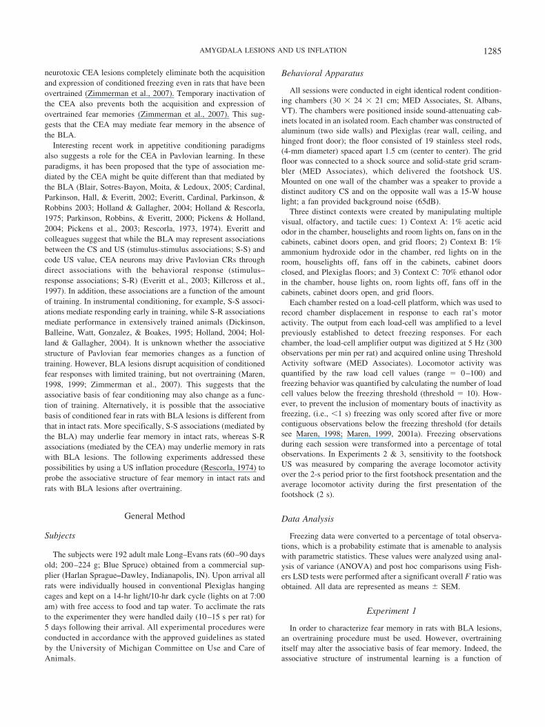
group, F(2,28) � 32.2; p � .0001, (Figure 1). Not surprisingly, bothgroups of rats that received 75 footshocks (P75 and U75) acquiredhigh levels of freezing, while the no-training group remained low.Rats in the no-training group exhibited some immobility late in thesession that was not related to fear, but rather to quiescence late inthe session. This significant difference in freezing between thegroups that had received training versus the no-training group wasconfirmed by further post hoc comparisons ( p � .0001).
Data from the inflation session are shown in Figure 2. AnANOVA calculated for the average freezing during the postshockITI periods of the inflation session (Minutes 4–8) revealed asignificant main effect of group, F(3,27) � 9.0; p � .0003. Post hoccomparisons revealed that all rats receiving inflated shocks (P75-INF, NS-INF, and U75-INF) exhibited more freezing than rats notreceiving shock (P75-NoINF), p � .0001 for comparison to theP75-INF group; p � .0002 for comparison to the U75-INF group;and p � .01 for comparison to the NS-INF group. There was nosignificant difference between any of the inflation groups.
Conditioned freezing during the context test is displayed inFigure 3a. During the context test, rats in the P75-INF and U75-INF groups exhibited significantly more freezing than rats in theNS-INF and P75-NoINF groups. This observation was confirmedin an ANOVA that revealed a significant main effect of group,F(3,27) � 24.7; p � .0001. Post hoc comparisons revealed that thegroups that had received overtraining and inflation (P75-INF andU75-INF) displayed elevated levels of freezing that were signifi-cantly different from the other groups (NS-INF and P75-NoINF;Figure 3a). Although both the NS-INF and P75-NoINF groupsfroze significantly less than the groups that had received overtrain-ing and inflation, the NS-INF group displayed higher levels offreezing than the P75-NoINF group ( p � .01). Elevated levels offreezing behavior in the NS-INF group is likely due to the gener-alization of fear from the inflation context to the test context.Overall, US inflation increased contextual fear in animals that hadpreviously received 75 footshocks in that context, whether theywere signaled or not. This was not simply the result of sensitizationby intense footshocks, insofar as the NS-INF rats exhibited rela-tively low levels of freezing.
An ANOVA performed for the tone freezing data (Figure 3b)revealed that there was a significant main effect of group, F(3,27) �4.8; p � .01, and post hoc tests revealed significantly higher levelsof freezing in the P75-INF group as compared with all othergroups throughout the entire session ( p � .02 for comparison to
Figure 1. Postshock freezing during the overtraining session(Experiment 1). Mean percentage of freezing (�SEM) during the 75-trialsession in Context A are displayed for each of the three groups.
1286 RABINAK AND MAREN

the P75-NoINF group; p � .005 for comparison to the U75-INFgroup; and p � .002 for comparison to the NS-INF group). Therewas no significant difference between any of the other groups(P75-NoINF, U75-INF, and NS-INF). In contrast to the contexttest, only rats that received inflation after 75 tone-shock trialsexhibited elevated freezing during the tone test (Figure 3b). Theseresults indicate that elevated freezing in the P75-INF group wasdue to US revaluation rather than nonassociative shock sensitiza-tion, because neither 75 conditioning shocks nor inflation shocksalone were sufficient to elevate freezing to the tone CS. Duringboth the context and tone tests, the noninflated group displayedlow levels of freezing, which may be due to generalized extinctionfrom exposure to similar chambers during the inflation session.Nonetheless, freezing in the inflation groups following overtrain-ing was augmented by the inflation procedure, and this is due to anassociative increase in fear. Overall these data indicate thatovertrained fear memories are sensitive to US inflation, sug-gesting that S-S associations mediate fear memory even afterextended training.
Experiment 2
Experiment 1 reveals that S-S associations contribute to theexpression of fear in overtrained rats. It has been argued inprevious work that BLA damage impairs both the encoding of S-Sassociations and interferes with US revaluation (Blundell, Hall, &Killcross, 2001; Hatfield, Han, Conley, Gallagher, & Holland,1996; Holland & Gallagher, 2004; Killcross et al., 1997; Pickenset al., 2003). Because rats with BLA damage acquire conditionedfear after overtraining (Maren, 1999; Zimmerman et al., 2007), itis therefore possible that S-R associations mediate this memory. Ifso, the fear memory in rats with BLA lesions may be insensitive toUS revaluation procedures. Experiment 2 used the inflation pro-cedure to examine this possibility in rats in which neurotoxic BLAlesions were made prior to overtraining.
Methods
Subjects and design. The subjects were 64 rats, housed andhandled as described in Experiment 1. Prior to overtraining theywere divided into two equal groups: one group that receivedbilateral neurotoxic lesions in the basolateral complex of the
amygdala (BLA) and a second group that underwent sham surgery(SHAM). Following overtraining each surgery group was furtherdivided into two groups: one that received US inflation (INF) afterovertraining or a group that did not undergo US inflation (NoINF).
Surgery. One week prior to training, each rat was anesthetizedwith an intraperitoneal (ip) injection of a Nembutal (sodium pen-tobarbital; 65 mg/kg body weight) and atropine methyl nitrate (0.4mg/kg body weight) cocktail. Ocular lubricant was used to moistenthe eyes. The scalp was shaved, cleaned with antiseptic (Betadine)and the rat was mounted in a stereotaxic apparatus (David KopfInstruments, Tujunga, CA). After the scalp was incised and re-tracted, the skull was positioned so that bregma and lambda werein the same horizontal plane. Small burr holes were drilled bilat-erally in the skull to allow placement of 28-gauge injectors in theBLA (3.3 mm posterior to bregma and 5.0 mm lateral to themidline). Injectors were attached to polyethylene tubing and con-nected to 10-�l syringes mounted on an infusion pump (HarvardApparatus, South Natick, MA). N-methyl-D-aspartate (NMDA;20 mg/mL) dissolved in 100 mM phosphate-buffered saline [PBS],pH 7.4; Sigma, St. Louis, MO) was infused (0.1 �l/min) at twosites for each BLA lesion: 8.0 mm ventral to the brain surface (0.2�l) and 7.5 mm ventral to the brain surface (0.1 �l) ventral to thebrain surface. Five minutes were allowed for diffusion of the druginto the target structure before the injectors were removed. SHAMrats received a similar surgery except that the injectors were notlowered into the brain. After surgery the incision was closed with
Figure 2. Postshock freezing during the inflation session (Experiment 1).Mean percentage of freezing (�SEM) during the five 60-s ITI periodsbetween the inflation shocks. Data are shown for NS-INF (gray bar),U75-INF (hatched bar), P75-INF (white bar), and P75-NoINF (black bar).
Figure 3. Conditioned freezing during the context and tone tests (Exper-iment 1). A: Mean percentage of freezing (�SEM) across the 10-mincontext test. B: Mean percentage of freezing (�SEM) during the 10-trialtone test. Data are an average of freezing during the ITI periods. Data areshown for NS-INF (gray bar), U75-INF (hatched bar), P75-INF (white bar)and P75-NoINF (black bar).
1287AMYGDALA LESIONS AND US INFLATION
stainless steel wound clips and the rats were kept on a heating paduntil they recovered from anesthesia before returning to their homecages. The rats were allowed one week of recovery prior toovertraining.
Conditioning, inflation, and test procedure. One week aftersurgery, all groups received overtraining, US inflation, and reten-tion testing as described in Experiment 1.
Histology. After behavioral testing, rats were euthanized withan overdose of sodium pentobarbital (ip, 100 mg/kg) and weretranscardially perfused with physiological saline followed by 10%formalin. Brains were removed and postfixed in 10% formalinfollowed by 10% formalin/30% sucrose solution until sectioning.Coronal brain sections (45 �m) were cut on a cryostat and wet-mounted with 70% ethanol on glass microscope slides. Once dry,the sections were stained with 0.25% thionin to visualize neuronalcell bodies and identify lesion sites.
Results and Discussion
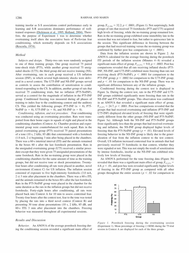
Histology. Eight rats were excluded from the analyses becausetheir lesions were either larger than intended, misplaced, or uni-lateral. This yielded the following groups designated by lesion typeand inflation condition: BLA-INF (n � 12), BLA-NoINF (n �11), SHAM-INF (n � 16), and SHAM-NoINF (n � 16). Success-ful lesions were generally confined to the targeted structure, al-though some rats in the BLA group had damage to the rostralendopiriform nucleus and caudate putamen (see Figure 4). NMDAinfusions into the BLA spared the central nucleus of the amygdala.
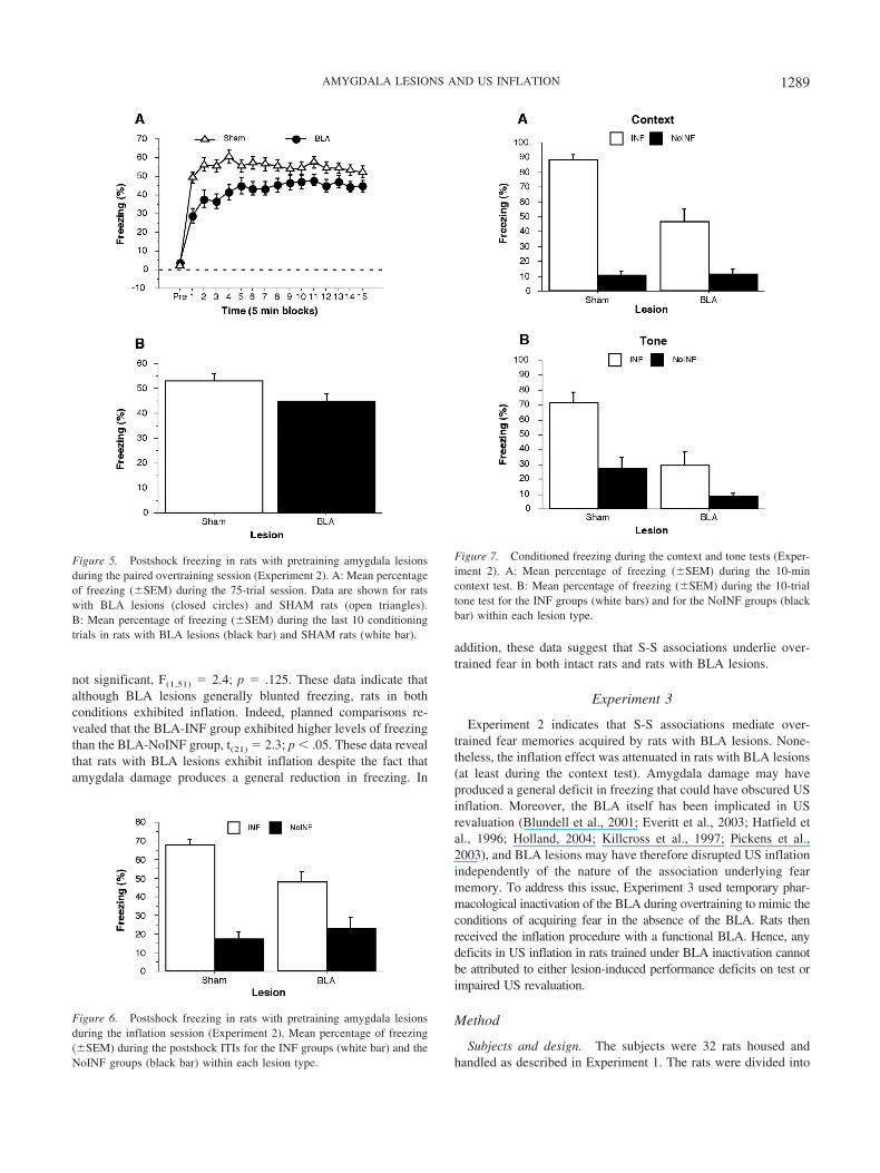
Behavior. A two-way ANOVA of the average postshockfreezing during the overtraining session revealed a significantmain effect of lesion, F(1, 53) � 7.7; p � .01, a main effect of time(5 minute blocks), F(15, 795) � 92.5; p � .0001, and an interactionbetween lesion and time, F(15, 795) � 5.5; p � .0001, (Figure 5a).These results indicate that freezing levels differed among thegroups across the session. Rats with BLA lesions displayed freez-ing levels that were slightly below those in the SHAM group.However, a post hoc comparison of average freezing during thelast 10 trials of the overtraining session revealed that rats withBLA lesions acquired a level of freezing comparable to that ofSHAM rats ( p � .05; Figure 5b). In addition, BLA lesions did notaffect sensitivity to the footshock US, F(1, 53) � 0.69; p � .41,which suggests that BLA lesions impaired conditional freezingcompared to controls during the beginning of the overtrainingsession (data not shown).
Postshock freezing during the inflation session is shown inFigure 6. A two-way ANOVA calculated for the average freezingduring Minutes 4–8 of the session revealed a significant maineffect of inflation condition, F(1, 51) � 72.3; p � .0001, and aninteraction between lesion and inflation condition, F(1, 51) � 8.4;p � .01. It is apparent that rats in the SHAM-INF group displayedmore freezing behavior relative to their noninflated controls thanthose in the BLA-INF group ( p � .005 for comparison withBLA-INF and p � .001 for all other comparisons). Rats in theBLA-INF group displayed lower levels of freezing than rats in theSHAM-INF group. It was important that BLA lesions did not altersensitivity to the intense footshocks used during the inflationsession, F(1, 26) � 2.56; p � .12 (data not shown).
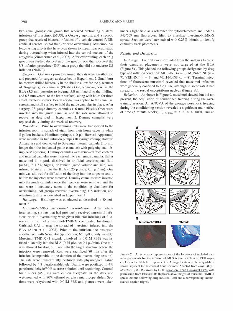
Conditioned freezing during the context is displayed inFigure 7a. Rats in both the SHAM-INF and BLA-INF groups
exhibited more freezing than noninflated controls, and this effectappeared more robust in the SHAM rats. These impressions wereconfirmed by a two-way ANOVA that revealed a significant maineffect of lesion, F(1,51) � 17.6; p � .0001, a main effect of inflationcondition, F(1,51) � 136.6; p � .0001, and an interaction betweenlesion and inflation condition, F(1,51) � 18.5; p � .0001. Althoughrats with BLA lesions exhibited less inflation than SHAM controls,rats in the BLA-INF group did display significantly higher levelsof freezing than those in the BLA-NoINF group ( p � .0001).
A two-way ANOVA of the tone test data (Figure 7b) revealed asignificant main effect of lesion, F(1,51) � 16.7; p � .0005, and amain effect of inflation condition, F(1,51) � 19.4; p � .0001;however the interaction between lesion and inflation condition was
Figure 4. A: Schematic representation of the extent of pretrainingNMDA lesions in the BLA for Experiment 2. Adapted from Brain Maps:Structure of the Rat Brain by L. W. Swanson, 1992. Copyright 1992, withpermission from Elsevier. B: Representative thionin-stained section fromrats that received lesions of the BLA.
1288 RABINAK AND MAREN
not significant, F(1,51) � 2.4; p � .125. These data indicate thatalthough BLA lesions generally blunted freezing, rats in bothconditions exhibited inflation. Indeed, planned comparisons re-vealed that the BLA-INF group exhibited higher levels of freezingthan the BLA-NoINF group, t(21) � 2.3; p � .05. These data revealthat rats with BLA lesions exhibit inflation despite the fact thatamygdala damage produces a general reduction in freezing. In
addition, these data suggest that S-S associations underlie over-trained fear in both intact rats and rats with BLA lesions.
Experiment 3
Experiment 2 indicates that S-S associations mediate over-trained fear memories acquired by rats with BLA lesions. None-theless, the inflation effect was attenuated in rats with BLA lesions(at least during the context test). Amygdala damage may haveproduced a general deficit in freezing that could have obscured USinflation. Moreover, the BLA itself has been implicated in USrevaluation (Blundell et al., 2001; Everitt et al., 2003; Hatfield etal., 1996; Holland, 2004; Killcross et al., 1997; Pickens et al.,2003), and BLA lesions may have therefore disrupted US inflationindependently of the nature of the association underlying fearmemory. To address this issue, Experiment 3 used temporary phar-macological inactivation of the BLA during overtraining to mimic theconditions of acquiring fear in the absence of the BLA. Rats thenreceived the inflation procedure with a functional BLA. Hence, anydeficits in US inflation in rats trained under BLA inactivation cannotbe attributed to either lesion-induced performance deficits on test orimpaired US revaluation.
Method
Subjects and design. The subjects were 32 rats housed andhandled as described in Experiment 1. The rats were divided into
Figure 5. Postshock freezing in rats with pretraining amygdala lesionsduring the paired overtraining session (Experiment 2). A: Mean percentageof freezing (�SEM) during the 75-trial session. Data are shown for ratswith BLA lesions (closed circles) and SHAM rats (open triangles).B: Mean percentage of freezing (�SEM) during the last 10 conditioningtrials in rats with BLA lesions (black bar) and SHAM rats (white bar).
Figure 6. Postshock freezing in rats with pretraining amygdala lesionsduring the inflation session (Experiment 2). Mean percentage of freezing(�SEM) during the postshock ITIs for the INF groups (white bar) and theNoINF groups (black bar) within each lesion type.
Figure 7. Conditioned freezing during the context and tone tests (Exper-iment 2). A: Mean percentage of freezing (�SEM) during the 10-mincontext test. B: Mean percentage of freezing (�SEM) during the 10-trialtone test for the INF groups (white bars) and for the NoINF groups (blackbar) within each lesion type.
1289AMYGDALA LESIONS AND US INFLATION
two equal groups: one group that received pretraining bilateralinfusions of muscimol (MUS), a GABAA agonist, and a secondgroup that received bilateral infusions of a vehicle control (VEH;artificial cerebral spinal fluid) prior to overtraining. Muscimol haslong-lasting effects that have been shown to impair fear acquisitionduring overtraining when infused into the central nucleus of theamygdala (Zimmerman et al., 2007). After overtraining, each druggroup was further divided into two groups: one that received theUS inflation procedure (INF) and a group that did not undergo USinflation (NoINF).
Surgery. One week prior to training, the rats were anesthetizedand prepared for surgery as described in Experiment 2. Small burrholes were drilled bilaterally in the skull to allow for the placementof 26-gauge guide cannulas (Plastics One, Roanoke, VA) in theBLA (3.3 mm posterior to bregma, 5.0 mm lateral to the midline,and 6.5 mm ventral to the brain surface), along with holes for threesmall jeweler’s screws. Dental acrylic was applied to the cannulas,screws, and skull surface to hold the guide cannulas in place. Aftersurgery, 33-gauge dummy cannulas (16 mm; Plastics One) wereinserted into the guide cannulas and the rats were allowed torecover as described in Experiment 2. Dummy cannulas werereplaced daily during the week of recovery.
Procedure. Prior to overtraining, rats were transported to theinfusion room in squads of eight from their home cages in white5-gallon buckets. Hamilton syringes (10 �l; Harvard Apparatus)were mounted in two infusion pumps (10 syringes/pump; HarvardApparatus) and connected to 33-gauge internal cannula (1.0 mmlonger than the implanted guide cannulas) with polyethylene tub-ing (A-M Systems). Dummy cannulas were removed from each ratand internal cannulas were inserted into each guide cannula. Eithermuscimol (1 mg/mL dissolved in artificial cerebrospinal fluid[aCSF], pH 7.4; Sigma) or vehicle (same volume and rate) wasinfused bilaterally into the BLA (0.25 �l/side; 0.1 �l/min). Onemin was allowed for diffusion of the drug into the target structurebefore the injectors were removed. Dummy cannulas were insertedinto the guide cannulas once the injectors were removed and therats were immediately taken to the conditioning chambers forovertraining. All groups received overtraining, US inflation, andretention testing as described in Experiment 1.
Histology. Histology was conducted as described in Experi-ment 2.
Muscimol-TMR-X intracranial microinfusion. After behav-ioral testing, six rats that had previously received muscimol infu-sions prior to overtraining were given bilateral infusions of fluo-rescent muscimol (muscimol-TMR-X conjugate; Invitrogen,Carlsbad, CA) to map the spread of muscimol infused into theBLA (Allen et al., 2008). Prior to the infusion, the rats wereanesthetized with Nembutal (ip injection; 65 mg/kg body weight).Muscimol-TMR-X (1 mg/mL dissolved in 0.01M PBS) was in-fused bilaterally into the BLA (0.25 �l/side; 0.1 �l/min). One minwas allowed for drug diffusion into the target structure before theinjectors were removed. Rats were sacrificed 80 min after theinfusion (comparable to the duration of the overtraining session).The rats were transcardially perfused with physiological salinefollowed by 4% paraformaldehyde. Brains were postfixed in 4%paraformaldehyde/30% sucrose solution until sectioning. Coronalbrain slices (45 �m) were cut on a cryostat in the dark andwet-mounted with 70% ethanol on glass microscope slides. Sec-tions were rehydrated with 0.01M PBS and pictures were taken
under a light field as a reference for cytoarchitecture and under a543/569 nm fluorescent filter to visualize muscimol-TMR-Xspread. Sections were later stained with 0.25% thionin to identifycannulas track placements.
Results and Discussion
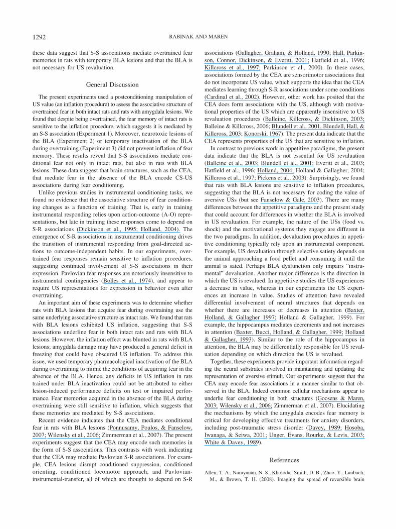
Histology. Four rats were excluded from the analyses becausetheir cannulas placements were not targeted at the BLA(Figure 8a). This yielded the following groups designated by drugtype and inflation condition: MUS-INF (n � 6), MUS-NoINF (n �7), VEH-INF (n � 7), and VEH-NoINF (n � 8). Terminal injec-tions of fluorescent muscimol revealed that muscimol infusionswere generally confined to the BLA, although in some rats it hadspread to the rostral endopiriform nucleus (Figure 8b).
Behavior. As shown in Figure 9, muscimol slowed, but did notprevent, the acquisition of conditioned freezing during the over-training session. An ANOVA of the average postshock freezingduring the conditioning session revealed a significant main effectof time (5 minute blocks), F(15, 390) � 31.6; p � .0001, and an
Figure 8. A: Schematic representation of the locations of included can-nula placements for the infusion of MUS (closed circles) or VEH (opencircles) in the BLA for Experiment 3. A magnification of the amgydala isshown adjacent to the coronal brain sections. Adapted from Brain Maps:Structure of the Rat Brain by L. W. Swanson, 1992. Copyright 1992, withpermission from Elsevier. B: Representative images of muscimol-TMR-Xspread 80 min following drug infusion (left) and a corresponding thionin-stained section (right).
1290 RABINAK AND MAREN

interaction between drug and time, F(15, 390) � 5.9; p � .0001 (seeFigure 9). The effect of muscimol on freezing during overtrainingwas similar to that of BLA damage (see Figure 5). Muscimol in theBLA did not affect sensitivity to the footshock US, F(1, 26) � 0.09;p � .76 (data not shown).
Postshock freezing during the inflation session is shown inFigure 10. It is apparent that rats receiving inflation shocks dis-played increased freezing relative to their no-inflation controls( p � .0001 for comparison between INF and NoINF and p � .576for comparison between MUS and VEH). Prior drug experiencedid not have a significant effect on freezing levels during inflation.These observations were confirmed by a two-way ANOVA onaverage freezing over Minutes 4–8 that revealed a significantmain effect of inflation condition, F(1,24) � 48.9; p � .0001.
Conditioned freezing during the context test is displayed inFigure 11a. Rats in the VEH-INF and MUS-INF groups exhibitedsignificantly more freezing than rats in the noninflated controls.This observation was confirmed by a two-way ANOVA thatrevealed a significant main effect of inflation condition, F(1,24) �81.7; p � .0001, however there was no significant interactionbetween the inflation condition and drug, F(1,24) � 0.07; p � .8.These data indicate that although muscimol blunted freezing dur-ing overtraining, rats in both conditions exhibited similar degrees
of inflation when inflation and testing occurred with a functionalamygdala.
A two-way ANOVA of the tone test data (Figure 11b) revealeda significant main effect of drug type, F(1,24) � 6.4; p � .02, anda main effect of inflation condition, F(1,24) � 14.8; p � .0001, butthe interaction between the inflation condition and drug type wasnot significant, F(1,24) � 0.5; p � .475. Both inflation groups(MUS-INF and VEH-INF) displayed significantly higher levels offreezing as compared with the noninflated groups (MUS-NoINFand VEH-NoINF; p � .05 for all comparisons). Although bothnoninflated groups displayed lower levels of freezing than theinflated groups, the VEH-NoINF group displayed significantlyhigher levels of freezing than the MUS-NoINF group ( p � .05).Previous reports suggest that remote memories acquired by ratswith BLA inactivation are weaker than BLA-dependent memories,which could explain the difference in freezing between the non-inflated groups (Poulos, Sterlace, & Fanselow, 2006). Overall
Figure 9. Postshock freezing in rats with pretraining muscimol infusionsduring the paired overtraining session (Experiment 3). A: Mean percentageof freezing (�SEM) during the 75-trial session. Data are shown for ratsthat received MUS (closed circles) and rats that received VEH (opentriangles).
Figure 10. Postshock freezing during the inflation session (Experiment 3).Mean percentage of freezing (�SEM) during the postshock ITIs for the INFgroups (white bar) and the NoINF groups (black bar) within each drug group.
Figure 11. Conditioned freezing during the context and tone tests (Ex-periment 3). A: Mean percentage of freezing (�SEM) during the 10-mincontext test. B: Mean percentage of freezing (�SEM) during the 5-trialtone test for the INF groups (white bars) and for the NoINF groups (blackbar) within each drug group.
1291AMYGDALA LESIONS AND US INFLATION
these data suggest that S-S associations mediate overtrained fearmemories in rats with temporary BLA lesions and that the BLA isnot necessary for US revaluation.
General Discussion
The present experiments used a postconditioning manipulation ofUS value (an inflation procedure) to assess the associative structure ofovertrained fear in both intact rats and rats with amygdala lesions. Wefound that despite being overtrained, the fear memory of intact rats issensitive to the inflation procedure, which suggests it is mediated byan S-S association (Experiment 1). Moreover, neurotoxic lesions ofthe BLA (Experiment 2) or temporary inactivation of the BLAduring overtraining (Experiment 3) did not prevent inflation of fearmemory. These results reveal that S-S associations mediate con-ditional fear not only in intact rats, but also in rats with BLAlesions. These data suggest that brain structures, such as the CEA,that mediate fear in the absence of the BLA encode CS-USassociations during fear conditioning.
Unlike previous studies in instrumental conditioning tasks, wefound no evidence that the associative structure of fear condition-ing changes as a function of training. That is, early in traininginstrumental responding relies upon action-outcome (A-O) repre-sentations, but late in training these responses come to depend onS-R associations (Dickinson et al., 1995; Holland, 2004). Theemergence of S-R associations in instrumental conditioning drivesthe transition of instrumental responding from goal-directed ac-tions to outcome-independent habits. In our experiments, over-trained fear responses remain sensitive to inflation procedures,suggesting continued involvement of S-S associations in theirexpression. Pavlovian fear responses are notoriously insensitive toinstrumental contingencies (Bolles et al., 1974), and appear torequire US representations for expression in behavior even afterovertraining.
An important aim of these experiments was to determine whetherrats with BLA lesions that acquire fear during overtraining use thesame underlying associative structure as intact rats. We found that ratswith BLA lesions exhibited US inflation, suggesting that S-Sassociations underline fear in both intact rats and rats with BLAlesions. However, the inflation effect was blunted in rats with BLAlesions; amygdala damage may have produced a general deficit infreezing that could have obscured US inflation. To address thisissue, we used temporary pharmacological inactivation of the BLAduring overtraining to mimic the conditions of acquiring fear in theabsence of the BLA. Hence, any deficits in US inflation in ratstrained under BLA inactivation could not be attributed to eitherlesion-induced performance deficits on test or impaired perfor-mance. Fear memories acquired in the absence of the BLA duringovertraining were still sensitive to inflation, which suggests thatthese memories are mediated by S-S associations.
Recent evidence indicates that the CEA mediates conditionalfear in rats with BLA lesions (Ponnusamy, Poulos, & Fanselow,2007; Wilensky et al., 2006; Zimmerman et al., 2007). The presentexperiments suggest that the CEA may encode such memories inthe form of S-S associations. This contrasts with work indicatingthat the CEA may mediate Pavlovian S-R associations. For exam-ple, CEA lesions disrupt conditioned suppression, conditionedorienting, conditioned locomotor approach, and Pavlovian-instrumental-transfer, all of which are thought to depend on S-R
associations (Gallagher, Graham, & Holland, 1990; Hall, Parkin-son, Connor, Dickinson, & Everitt, 2001; Hatfield et al., 1996;Killcross et al., 1997; Parkinson et al., 2000). In these cases,associations formed by the CEA are sensorimotor associations thatdo not incorporate US value, which supports the idea that the CEAmediates learning through S-R associations under some conditions(Cardinal et al., 2002). However, other work has posited that theCEA does form associations with the US, although with motiva-tional properties of the US which are apparently insensitive to USrevaluation procedures (Balleine, Killcross, & Dickinson, 2003;Balleine & Killcross, 2006; Blundell et al., 2001, Blundell, Hall, &Killcross, 2003; Konorski, 1967). The present data indicate that theCEA represents properties of the US that are sensitive to inflation.
In contrast to previous work in appetitive paradigms, the presentdata indicate that the BLA is not essential for US revaluation(Balleine et al., 2003; Blundell et al., 2001; Everitt et al., 2003;Hatfield et al., 1996; Holland, 2004; Holland & Gallagher, 2004;Killcross et al., 1997; Pickens et al., 2003). Surprisingly, we foundthat rats with BLA lesions are sensitive to inflation procedures,suggesting that the BLA is not necessary for coding the value ofaversive USs (but see Fanselow & Gale, 2003). There are manydifferences between the appetitive paradigms and the present studythat could account for differences in whether the BLA is involvedin US revaluation. For example, the nature of the USs (food vs.shock) and the motivational systems they engage are different inthe two paradigms. In addition, devaluation procedures in appeti-tive conditioning typically rely upon an instrumental component.For example, US devaluation through selective satiety depends onthe animal approaching a food pellet and consuming it until theanimal is sated. Perhaps BLA dysfunction only impairs “instru-mental” devaluation. Another major difference is the direction inwhich the US is revalued. In appetitive studies the US experiencesa decrease in value, whereas in our experiments the US experi-ences an increase in value. Studies of attention have revealeddifferential involvement of neural structures that depends onwhether there are increases or decreases in attention (Baxter,Holland, & Gallagher 1997; Holland & Gallagher, 1999). Forexample, the hippocampus mediates decrements and not increasesin attention (Baxter, Bucci, Holland, & Gallagher, 1999; Holland& Gallagher, 1993). Similar to the role of the hippocampus inattention, the BLA may be differentially responsible for US reval-uation depending on which direction the US is revalued.
Together, these experiments provide important information regard-ing the neural substrates involved in maintaining and updating therepresentation of aversive stimuli. Our experiments suggest that theCEA may encode fear associations in a manner similar to that ob-served in the BLA. Indeed common cellular mechanisms appear tounderlie fear conditioning in both structures (Goosens & Maren,2003; Wilensky et al., 2006; Zimmerman et al., 2007). Elucidatingthe mechanisms by which the amygdala encodes fear memory iscritical for developing effective treatments for anxiety disorders,including post-traumatic stress disorder (Davey, 1989; Hosoba,Iwanaga, & Seiwa, 2001; Unger, Evans, Rourke, & Levis, 2003;White & Davey, 1989).
References
Allen, T. A., Narayanan, N. S., Kholodar-Smith, D. B., Zhao, Y., Laubach,M., & Brown, T. H. (2008). Imaging the spread of reversible brain
1292 RABINAK AND MAREN
inactivations using fluorescent muscimol. Journal of NeuroscienceMethods, 171, 30–38.
Balleine, B. W., Killcross, A. S., & Dickinson, A. (2003). The effect oflesions of the basolateral amygdala on instrumental conditioning. Jour-nal of Neuroscience, 23, 666–675.
Balleine, B. W., & Killcross, S. (2006). Parallel incentive processing: Anintegrated view of amygdala function. Trends in Neurosciences, 29,272–279.
Baxter, M. G., Bucci, D. J., Holland, P. C., & Gallagher, M. (1999).Impairments in conditioned stimulus processing and conditioned re-sponding after combined selective removal of hippocampal and neocor-tical cholinergic input. Behavioral Neuroscience, 113, 486–495.
Baxter, M. G., Holland, P. C., & Gallagher, M. (1997). Disruption ofdecrements in conditioned stimulus processing by selective removalof hippocampal cholinergic input. Journal of Neuroscience, 17,5230 –5236.
Blair, H. T., Sotres-Bayon, F., Moita, M. A. P., & Ledoux, J. E. (2005).The lateral amygdala processes the value of conditioned and uncondi-tioned aversive stimuli. Neuroscience, 133, 561–569.
Blundell, P., Hall, G., & Killcross, S. (2001). Lesions of the basolateralamygdala disrupt selective aspects of reinforcer representation in rats.Journal of Neuroscience, 21, 9018–9026.
Blundell, P., Hall, G., & Killcross, S. (2003). Preserved sensitivity tooutcome value after lesions of the basolateral amygdala. Journal ofNeuroscience, 23, 7702–7709.
Bolles, R. C., Riley, A. L., Cantor, M. B., & Duncan, P. M. (1974). Therat’s failure to anticipate regularly scheduled daily shock. BehavioralBiology, 11, 365–372.
Bouton, M. E., Mineka, S., & Barlow, D. H. (2001). A modern learningtheory perspective on the etiology of panic disorder. PsychologicalReview, 108, 4–32.
Campeau, S., & Davis, M. (1995). Involvement of the central nucleus andbasolateral complex of the amygdala in fear conditioning measured withfear-potentiated startle in rats trained concurrently with auditory andvisual conditioned stimuli. Journal of Neuroscience, 15, 2301–2311.
Cardinal, R. N., Parkinson, J. A., Hall, J., & Everitt, B. J. (2002). Emotionand motivation: The role of the amygdala, ventral striatum, and prefron-tal cortex. Neuroscience and Biobehavioral Reviews, 26, 321–352.
Cousens, G., & Otto, T. (1998). Both pre- and posttraining excitotoxiclesions of the basolateral amygdala abolish the expression of olfac-tory and contextual fear conditioning. Behavioral Neuroscience, 112,1092–1103.
Davey, G. C. L. (1989). Ucs revaluation and conditioning models ofacquired fears. Behaviour Research and Therapy, 27, 521–528.
Davis, M., & Whalen, P. J. (2001). The amygdala: Vigilance and emotion.Molecular Psychiatry, 6, 13–34.
Dickinson, A., Balleine, B., Watt, A., Gonzalez, F., & Boakes, R. A.(1995). Motivational control after extended instrumental training. Ani-mal Learning & Behavior, 23, 197–206.
Everitt, B. J., Cardinal, R. N., Parkinson, J. A., & Robbins, T. W. (2003).Appetitive behavior: Impact of amygdala-dependent mechanisms ofemotional learning. Annals of the New York Academy of Sciences, 985,233–250.
Fanselow, M. S., & Gale, G. D. (2003). The amygdala, fear, and memory.Annals of the New York Academy of Sciences, 985, 125–134.
Fendt, M., & Fanselow, M. S. (1999). The neuroanatomical and neuro-chemical basis of conditioned fear. Neuroscience and BiobehavioralReviews, 23, 743–760.
Gale, G. D., Anagnostaras, S. G., Godsil, B. P., Mitchell, S., Nozawa, T.,Sage, J. R., et al. (2004). Role of the basolateral amygdala in the storageof fear memories across the adult lifetime of rats. Journal of Neuro-science, 24, 3810–3815.
Gallagher, M., Graham, P. W., & Holland, P. C. (1990). The amygdalacentral nucleus and appetitive pavlovian conditioning: Lesions im-
pair one class of conditioned behavior. Journal of Neuroscience, 10,1906 –1911.
Goosens, K. A., & Maren, S. (2001). Contextual and auditory fear condi-tioning are mediated by the lateral, basal, and central amygdaloid nucleiin rats. Learning & Memory, 8, 148–155.
Goosens, K. A., & Maren, S. (2003). Pretraining nmda receptor blockadein the basolateral complex, but not the central nucleus, of the amygdalaprevents savings of conditional fear. Behavioral Neuroscience, 117,738–750.
Grillon, C., Southwick, S. M., & Charney, D. S. (1996). The psychobio-logical basis of posttraumatic stress disorder. Molecular Psychiatry, 1,278–297.
Hall, J., Parkinson, J. A., Connor, T. M., Dickinson, A., & Everitt, B. J.(2001). Involvement of the central nucleus of the amygdala and nucleusaccumbens core in mediating pavlovian influences on instrumental be-haviour. European Journal of Neuroscience, 13, 1984–1992.
Hatfield, T., Han, J. S., Conley, M., Gallagher, M., & Holland, P. (1996).Neurotoxic lesions of basolateral, but not central, amygdala interferewith pavlovian second-order conditioning and reinforcer devaluationeffects. Journal of Neuroscience, 16, 5256–5265.
Helmstetter, F. J. (1992). The amygdala is essential for the expression ofconditional hypoalgesia. Behavioral Neuroscience, 106, 518–528.
Helmstetter, F. J., & Bellgowan, P. S. (1994). Effects of muscimol appliedto the basolateral amygdala on acquisition and expression of contextualfear conditioning in rats. Behavioral Neuroscience, 108, 1005–1009.
Holland, P. C. (2004). Relations between pavlovian-instrumental transferand reinforcer devaluation. Journal of Experimental Psychology: AnimalBehavior Processes, 30, 104–117.
Holland, P. C., & Gallagher, M. (1993). Amygdala central nucleus lesionsdisrupt increments, but not decrements, in conditioned stimulus process-ing. Behavioral Neuroscience, 107, 246–253.
Holland, P. C., & Gallagher, M. (1999). Amygdala circuitry in attentionaland representational processes. Trends in Cognitive Sciences, 3, 65–73.
Holland, P. C., & Gallagher, M. (2004). Amygdala-frontal interactions andreward expectancy. Current Opinion in Neurobiology, 14, 148–155.
Holland, P. C., & Rescorla, R. A. (1975). The effect of two ways ofdevaluing the unconditioned stimulus after first- and second-order ap-petitive conditioning. Journal of Experimental Psychology: Animal Be-havior Processes, 1, 355–363.
Hosoba, T., Iwanaga, M., & Seiwa, H. (2001). The effect of ucs inflationand deflation procedures on ‘fear’ conditioning. Behaviour Researchand Therapy, 39, 465–475.
Killcross, S., Robbins, T. W., & Everitt, B. J. (1997). Different types offear-conditioned behaviour mediated by separate nuclei within amyg-dala. Nature (London), 388, 377–380.
Kim, J. J., & Jung, M. W. (2006). Neural circuits and mechanisms involvedin Pavlovian fear conditioning: A critical review. Neuroscience & Biobe-havioral Reviews, 30, 188–202.
Konorski, J. (1967). Integrative activity of the brain. Chicago: Universityof Chicago Press.
Koo, J. W., Han, J. S., & Kim, J. J. (2004). Selective neurotoxic lesions ofbasolateral and central nuclei of the amygdala produce differentialeffects on fear conditioning. Journal of Neuroscience, 24, 7654–7662.
LeDoux, J. (1998). Fear and the brain: Where have we been, and where arewe going? Biological Psychiatry, 44, 1229–1238.
LeDoux, J. E. (2000). Emotion circuits in the brain. Annual Review ofNeuroscience, 23, 155–184.
Maren, S. (1998). Overtraining does not mitigate contextual fear condi-tioning deficits produced by neurotoxic lesions of the basolateral amyg-dala. Journal of Neuroscience, 18, 3088–3097.
Maren, S. (1999). Neurotoxic basolateral amygdala lesions impair learningand memory but not the performance of conditional fear in rats. Journalof Neuroscience, 19, 8696–8703.
1293AMYGDALA LESIONS AND US INFLATION
Maren, S. (2001a). Neurobiology of pavlovian fear conditioning. AnnualReview of Neuroscience, 24, 897–931.
Maren, S. (2001b). Is there savings for pavlovian fear conditioning afterneurotoxic basolateral amygdala lesions in rats? Neurobiology of Learn-ing and Memory, 76, 268–283.
Maren, S. (2005). Building and burying fear memories in the brain.Neuroscientist, 11, 89–99.
Maren, S., Aharonov, G., & Fanselow, M. S. (1996a). Retrograde abolitionof conditional fear after excitotoxic lesions in the basolateral amygdalaof rats: Absence of a temporal gradient. Behavioral Neuroscience, 110,718–726.
Maren, S., Aharonov, G., Stote, D. L., & Fanselow, M. S. (1996b).N-methyl-D-aspartate receptors in the basolateral amygdala are requiredfor both acquisition and expression of conditional fear in rats. Behav-ioral Neuroscience, 110, 1365–1374.
Maren, S., & Quirk, G. J. (2004). Neuronal signalling of fear memory.Nature Reviews Neuroscience, 5, 844–852.
Muller, J., Corodimas, K. P., Fridel, Z., & LeDoux, J. E. (1997). Functionalinactivation of the lateral and basal nuclei of the amygdala by muscimolinfusion prevents fear conditioning to an explicit conditioned stimulusand to contextual stimuli. Behavioral Neuroscience, 111, 683–691.
Nader, K., Majidishad, P., Amorapanth, P., & LeDoux, J. E. (2001).Damage to the lateral and central, but not other, amygdaloid nucleiprevents the acquisition of auditory fear conditioning. Learning & Mem-ory, 8, 156–163.
Pare, D., Quirk, G. J., & Ledoux, J. E. (2004). New vistas on amygdalanetworks in conditioned fear. Journal of Neurophysiology, 92, 1–9.
Parkinson, J. A., Robbins, T. W., & Everitt, B. J. (2000). Dissociable rolesof the central and basolateral amygdala in appetitive emotional learning.European Journal of Neuroscience, 12, 405–413.
Pickens, C. L., & Holland, P. C. (2004). Conditioning and cognition.Neuroscience & Biobehavioral Reviews, 28, 651–661.
Pickens, C. L., Saddoris, M. P., Setlow, B., Gallagher, M., Holland, P. C.,& Schoenbaum, G. (2003). Different roles for orbitofrontal cortex andbasolateral amygdala in a reinforcer devaluation task. Journal of Neu-roscience, 23, 11078–11084.
Ponnusamy, R., Poulos, A. M., & Fanselow, M. S. (2007). Amygdala-dependent and amygdala-independent pathways for contextual fear con-ditioning. Neuroscience, 147, 919–927.
Poulos, A. M., Sterlace, M., & Fanselow, M. S. (2006, October). Deficits inremote but not recent long-term memory in the absence of the fta: Aretrieval or storage failure. Paper presented at the Society for Neuroscience,Atlanta, GA.
Rescorla, R. A. (1973). Effect of us habituation following conditioning.Journal of Comparative & Physiological Psychology, 82, 137–143.
Rescorla, R. A. (1974). Effect of inflation of the unconditioned stimulusvalue following conditioning. Journal of Comparative & PhysiologicalPsychology, 86, 101–106.
Samson, R. D., & Pare, D. (2005). Activity-dependent synaptic plasticity inthe central nucleus of the amygdala. Journal of Neuroscience, 25,1847–1855.
Schafe, G. E., Nader, K., Blair, H. T., & LeDoux, J. E. (2001). Memoryconsolidation of Pavlovian fear conditioning: A cellular and molecularperspective. Trends in Neurosciences, 24, 540–546.
Swanson, L. W. (1992). Brain maps: Structure of the rat brain. New York:Elsevier.
Unger, W., Evans, I. M., Rourke, P., & Levis, D. J. (2003). The s-sconstruct of expectancy versus the s-r construct of fear: Which motivatesthe acquisition of avoidance behavior? Journal of General Psychology,130, 131–147.
Walker, D. L., & Davis, M. (1997). Double dissociation between theinvolvement of the bed nucleus of the stria terminalis and the centralnucleus of the amygdala in startle increases produced by conditionedversus unconditioned fear. Journal of Neuroscience, 17, 9375–9383.
White, K., & Davey, G. C. L. (1989). Sensory preconditioning and ucsinflation in human ‘fear’ conditioning. Behaviour Research and Ther-apy, 27, 161–166.
Wilensky, A. E., Schafe, G. E., Kristensen, M. P., & LeDoux, J. E. (2006).Rethinking the fear circuit: The central nucleus of the amygdala isrequired for the acquisition, consolidation, and expression of Pavlovianfear conditioning. Journal of Neuroscience, 26, 12387–12396.
Wilensky, A. E., Schafe, G. E., & LeDoux, J. E. (1999). Functionalinactivation of the amygdala before but not after auditory fear condi-tioning prevents memory formation. Journal of Neuroscience, 19, RC48.
Wilensky, A. E., Schafe, G. E., & LeDoux, J. E. (2000). Functionalinactivation of amygdala nuclei during acquisition of pavlovian fearconditioning. Society for Neuroscience Abstracts, 30, 465–469.
Zimmerman, J. M., Rabinak, C. A., McLachlan, I. G., & Maren, S. (2007).The central nucleus of the amygdala is essential for acquiring andexpressing conditional fear after overtraining. Learning & Memory, 14,634–644.
Received April 24, 2008Revision received May 20, 2008
Accepted May 21, 2008 �
1294 RABINAK AND MAREN