Prenatal diagnosis of genetic disease
12
Prenat Diosis of Genec Disease New techniques are making it possible to detect hereditary diseases early in pregnancy. To what extent is the control of such births justed on biological and social grounds? M ore than 1,600 human diseases caused by defects in the content or the expression of the genetic information in DNA have been identi- fied. Some of these diseases are very rare; others, such as cystic fibrosis and sickle-cell anemia, are relatively com- mon and are responsible for much illness and death. It has been estimated that more than 25 percent of the hospitaliza- tions of children are for illnesses with a major genec component. Thanks to new techniques of biochemistry and cell biology, we are learning a great deal about the biochemical mechanisms that lead from a genetic defect to clinical dis- ease. Particularly important has been the discovery that cells from patients can be grown and. studied in tissue culture in artificial nutrient media, and that these cells often continue to express the abnormal function of a mutant gene. As a result genetic manipulative techniques are being developed through which man may acquire the ability to control as- pects of his own evolution, to eliminate disease and even to improve his genetic makeup. Another application of these techniques is prenatal genetic diagnosis. The ability to establish the diagnosis of genetic disease prior to birth came through studies of the fluid that bathes the developing fetus within the amniotic cavity: a sac surrounding the fetus that is lined by two layers of cells (the chori- on and the amnion) and is filled with fluid derived mainly from fetal urine and fetal respiratory secretions. Sus- pended in the fluid are viable cells shed from the fetal skin and respiratory tract, and perhaps from the fetally derived lining of the cavity itself. Amniocentesis, the removal of fluid from the amniotic cavity by needle puncture, became use- ful and important in the early 1960's for the detection of unborn infants who ran the risk of Rh incompatibility. When an 34 by Theodore Friedmann Rh-negative mother carries an Rh-posi- tive fetus, the mother may become sen- sitized to the "foreign" red blood cells from the fetus, and the antibodies that develop may in later pregnancies cross the placenta, cleave to the fetus's red blood cells and cause the destruction of red cells, severe anemia, brain damage and even death [see "The Prevention of 'Rhesus Babies: " by C. A. Clarke; SCI- ENTIFIC AMERICAN, November, 1968]. Obstetricians discovered that they could estimate the degree of blood destruction by measuring the concentration of he- moglobin breakdown products in the amniotic fluid. D uring amniocentesis for Rh disease it became clear that much useful in- formation could be obtained by examin- ing cells in the amniotic fluid. In 1949 Murray Barr of Canada had discovered that nerve cells from female cats were distinguishable from those of the male by the presence in the female cells of a darkly staining piece of chromoso- mal material on the nuclear membrane. These "Barr bodies" were then found to characterize female cells in many other mammals, including human females. Mary Lyon of England subsequently found that early in mammalian embryo- genesis one of the two X chromosomes in female cells (male cells have only one X chromosome and a Y chromosome) randomly becomes condensed and inac- tive. In the unusual instances where cells have more than two X chromo- somes all except one are condensed. In male cells condensation does not take place. The altered physical properties of these condensed chromosomes give rise to their staining characteristics. Since amniotic-fluid cells are mostly fetal in origin, investigators turned to them as an unprecedentedly valuable material for detecting the kinds of ge- netic disease that are caused by muta- tions on the X chromosome and that therefore affect only males. The best- known of the "X-linked" diseases is the bleeding disorder hemophilia. In this disease a deficiency of one of the pro- tein factors required for clotting results in prolonged and intractable bleeding, particularly at sites of traumatic injury. An ovum from a female carrier of hemophilia will have either a normal X chromosome or a defective one. After fertilization half of all the resulting males will receive as their only X chro- mosome the one carrying the mutant gene. Since there is no normal copy of the gene from the father such males will express the defect fully. (The father must pass his Y chromosome in order for the fetus to be male.) The other males will be normal, since they have by chance received the normal X chromo- some from the mother. Therefore detec- tion of a female fetus in utero by the presence of Barr bodies in a pregnancy in which there is a risk of hemophilia rules out the possibility of disease, al- though 50 percent of the females will be carriers. A male fetus, however, may be either affected or normal. Predictions can also be made on the genetic constitution of the fetus in the case of diseases associated with an ab- normal number of chromosomes or ar- rangement of chromosomes. In 1959 the French geneticist Jerome Lejeune dis- covered that patients with mongolism (now usually called Down's syndrome) have no mutant or defective gene but rather carry an extra chromosome that in itself is probably normal. We now know that most cases of Down's syndrome are caused by a defect in the separation of chromosomes, called nondisjunction, in the developing egg resulting in the for- mation of ova with two No. 21 chromo- somes instead of the usual one. © 1971 SCIENTIFIC AMERICAN, INC
Transcript of Prenatal diagnosis of genetic disease

Prenatal Diagnosis of Genetic Disease New techniques are making it possible to detect hereditary
diseases early in pregnancy. To what extent is the control
of such births justified on biological and social grounds?
More than 1,600 human diseases caused by defects in the content or the expression of the genetic
information in DNA have been identified. Some of these diseases are very rare; others, such as cystic fibrosis and sickle-cell anemia, are relatively common and are responsible for much illness and death. It has been estimated that more than 25 percent of the hospitalizations of children are for illnesses with a major genetic component. Thanks to new techniques of biochemistry and cell biology, we are learning a great deal about the biochemical mechanisms that lead from a genetic defect to clinical disease. Particularly important has been the discovery that cells from patients can be grown and. studied in tissue culture in artificial nutrient media, and that these cells often continue to express the abnormal function of a mutant gene. As a result genetic manipulative techniques are being developed through which man may acquire the ability to control aspects of his own evolution, to eliminate disease and even to improve his genetic makeup. Another application of these techniques is prenatal genetic diagnosis.
The ability to establish the diagnosis of genetic disease prior to birth came through studies of the fluid that bathes the developing fetus within the amniotic cavity: a sac surrounding the fetus that is lined by two layers of cells (the chorion and the amnion) and is filled with fluid derived mainly from fetal urine and fetal respiratory secretions. Suspended in the fluid are viable cells shed from the fetal skin and respiratory tract, and perhaps from the fetally derived lining of the cavity itself. Amniocentesis, the removal of fluid from the amniotic cavity by needle puncture, became useful and important in the early 1960's for the detection of unborn infants who ran the risk of Rh incompatibility. When an
34
by Theodore Friedmann
Rh-negative mother carries an Rh-positive fetus, the mother may become sensitized to the "foreign" red blood cells from the fetus, and the antibodies that develop may in later pregnancies cross the placenta, cleave to the fetus's red blood cells and cause the destruction of red cells, severe anemia, brain damage and even death [see "The Prevention of 'Rhesus Babies: " by C. A. Clarke; SCIENTIFIC AMERICAN, November, 1968]. Obstetricians discovered that they could estimate the degree of blood destruction by measuring the concentration of hemoglobin breakdown products in the amniotic fluid.
During amniocentesis for Rh disease it became clear that much useful in
formation could be obtained by examining cells in the amniotic fluid. In 1949 Murray Barr of Canada had discovered that nerve cells from female cats were distinguishable from those of the male by the presence in the female cells of a darkly staining piece of chromosomal material on the nuclear membrane. These "Barr bodies" were then found to characterize female cells in many other mammals, including human females. Mary Lyon of England subsequently found that early in mammalian embryogenesis one of the two X chromosomes in female cells (male cells have only one X chromosome and a Y chromosome) randomly becomes condensed and inactive. In the unusual instances where cells have more than two X chromosomes all except one are condensed. In male cells condensation does not take place. The altered physical properties of these condensed chromosomes give rise to their staining characteristics.
Since amniotic-fluid cells are mostly fetal in origin, investigators turned to them as an unprecedentedly valuable material for detecting the kinds of ge-
netic disease that are caused by mutations on the X chromosome and that therefore affect only males. The bestknown of the "X-linked" diseases is the bleeding disorder hemophilia. In this disease a deficiency of one of the protein factors required for clotting results in prolonged and intractable bleeding, particularly at sites of traumatic injury.
An ovum from a female carrier of hemophilia will have either a normal X chromosome or a defective one. After fertilization half of all the resulting males will receive as their only X chromosome the one carrying the mutant gene. Since there is no normal copy of the gene from the father such males will express the defect fully. (The father must pass his Y chromosome in order for the fetus to be male.) The other males will be normal, since they have by chance received the normal X chromosome from the mother. Therefore detection of a female fetus in utero by the presence of Barr bodies in a pregnancy in which there is a risk of hemophilia rules out the possibility of disease, although 50 percent of the females will be carriers. A male fetus, however, may be either affected or normal.
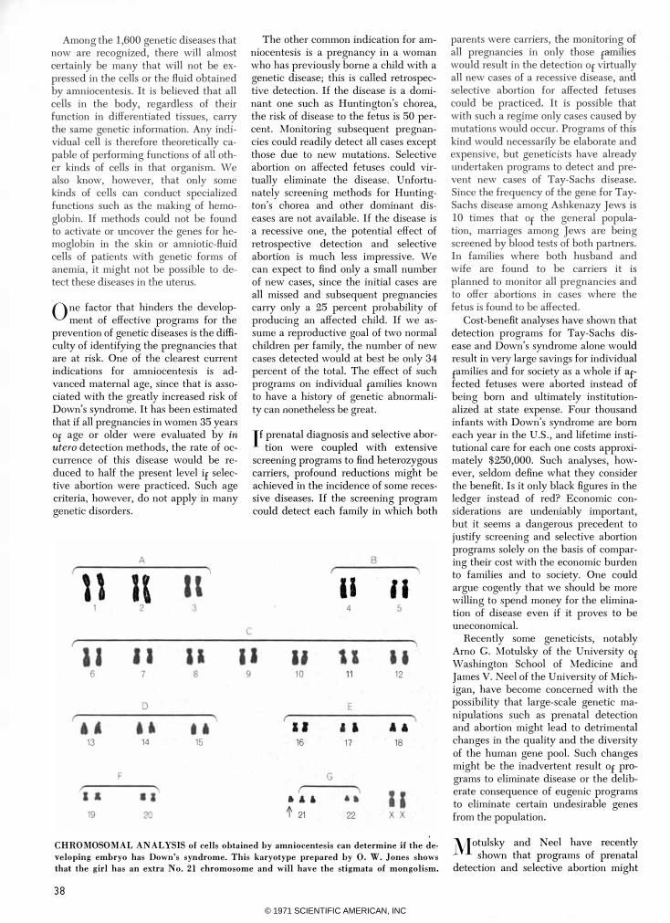
Predictions can also be made on the genetic constitution of the fetus in the case of diseases associated with an abnormal number of chromosomes or arrangement of chromosomes. In 1959 the French geneticist Jerome Lejeune discovered that patients with mongolism (now usually called Down's syndrome) have no mutant or defective gene but rather carry an extra chromosome that in itself is probably normal. We now know that most cases of Down's syndrome are caused by a defect in the separation of chromosomes, called nondisjunction, in the developing egg resulting in the formation of ova with two No. 21 chromosomes instead of the usual one.
© 1971 SCIENTIFIC AMERICAN, INC

When such an egg is fertilized by a n<?rmal sperm, the result is an embryo carrying three normal No. 21 chromosomes. This "trisomy" of chromosome No. 21 causes Down's syndrome. An important feature of this kind of Down's syndrome is its increased incidence with advanced maternal age. A pregnant woman 40 years old is more than 10 times as likely to have an affected infant as one 25 years old. The disease is the single most common chromosomal aberration found in live-born infants.
In a rarer type of Down's syndrome the extra chromosome No. 21 is translocated onto another chromosome. A healthy carrier mother has only 45 chromosomes instead of the usual 46, but the total amount of genetic information is normal. During the formation of the ovum, however, segregation of chromosomes can give rise to an ovum carrying the translocated No. 21 plus the free No. 21. Fertilization of such an ovum leads to an embryo with an apparently normal chromosome number, but with an extra chromosome No. 21. The abnormalities in this kind of Down's syndrome are similar to those in the nondisjunction type.
Parallel with developments in cell bi-
ology have been advances in our knowledge of the flow of genetic information from DNA through RNA to protein. The precise biochemical defects involved in several hundred human disorders have been discovered, and this is due largely to the realization that tissue-culture cells from patients often continue to show the biochemical defect characteristic of a given disease. Such culture methods have most often used skin cells, but recently geneticists have learned that amniotic-fluid cells can also be cultured and used diagnostically.
O ne disease that illustrates the usefulness of these methods is the Lesch
Nyhan syndrome, a severe neurological disease of males characterized by mental retardation, involuntary writhing motions called choreoathetosis and compulsive self-mutilation of the lips and fingertips by biting. In the healthy carrier mother one of the X chromosomes carries a defective gene for the enzyme hypoxanthine-guanine phosphoribosyl transferase (HGPRT), which is required for the conv.ersion of the bases hypoxanthine and guanine into nucleotides for incorporation into new RNA and DNA by the "reutilization" pathway. Most
body cells can function normally without this enzyme, since there is another path-way for nucleotide synthesis. The function of cells in the basal ganglia of the brain, however, is severely impaired without the HGPRT pathway.
To determine whether or not a male fetus carries a defective HGPRT gene, amniotic-fluid cells in tissue culture can be labeled with radioactive hypoxanthine and then exposed to X-ray film. Normal cells, having incorporated the radioactive precursor, will appear densely labeled in this autoradiographic method, whereas Lesch-Nyhan cells will remain free of radioactivity. Alternatively cells can be homogenized and the enzyme assayed directly.
Many of the diseases that are associated with enzyme deficiencies show autosomal recessive inheritance: the defective gene lies on one of the 22 pairs of non-sex chromosomes (autosomes), and two mutant copies of the gene, one from each parent, are passed on to the offspring. The offspring are thus homozygous for the recessive gene and exhibit the disease; the parents are heterozygous carriers. In many instances where a single mutant copy of the gene is present about half of the normal amount of
FLUID: COMPOSITION
I } CELLS. SEX DETERMINATION
BIOCHEMICAL AND
ENZYMATIC STUDIES 1
} CELL CULTURE
BIOCHEMICAL STUDIES
CHROMOSOMAL ANALYSIS
SAMPLE OF FLUID surrounding the fetus is taken by inserting a
sterile needle into the amniotic cavity and withdrawing a small
amount of fluid. The process is called amniocentesis. The fluid, de·
rived mostly from fetal urine and secretions, contains fetal cells
(not drawn to scale). Care must be taken not to puncture the pia·
centa or the fetus. The sample is centrifuged to separate cells and
fluid. A variety of tests can be made. Optimum time for the amni·
otic tap for genetic diagnosis is about the 16th week of gestation.
35
© 1971 SCIENTIFIC AMERICAN, INC

LOCATION AND SIZE of the placenta can be determined by ultrasonic scanning. Bursts of high·frequency sound waves are emitted from a probe moved along the surface of the
abdomen. Echoes returned from tissue interfaces within the body are displayed on a stor·
age oscilloscope. The technique is simple, safe and rapid. These sonograms were made at
the 18th week of gestation to locate the placenta before amniocentesis. The placenta ap·
pears as a crescent.shaped dotted area. The dark area directly below it is part of the amni· otic cavity. Scattered echoes within the cavity are from the fetus. The dark shape on the
right is the urinary bladder. The scan was made longitudinally from above the navel (left) to the pubic area (right). The arrow is at the site of the transverse scan shown below.
OVAL SHAPE to the right of center is formed by echoes from the fetal head. Above it and
to the left is a section of the placenta. The dark area below the placenta is part of the
amniotic cavity. The patient's left and right sides respectively are on the left and right
of the photograph. The arrow is at the site of the scan in the top illustration. Both scans are necessary in order to locate the placenta precisely. The sonograms were prepared by
Mitsunao Kobayashi of the Downstate Medical Center of the State University of New York.
36
the gene product is made, which is often enough to protect against the di.sease. This is not the case, however, when the mutant gene is dominant.
One particularly devastating autosomal recessive disease is Tay-Sachs disease, which causes blindness, severe mental retardation and death, usually before three or four years of age. It is most common in Jews of northern European origin (Ashkenazy Jews). In 1969 John O'Brien and his colleagues at the School of Medicine of the University of California at San Diego discovered that in Tay-Sachs disease a mutation leads to a
defective form of the enzyme hex osaminidase A, which normally splits the terminal sugar from a lipid-polysaccharide complex called GM2 ganglioside. There results a massive accumulation of the undegraded ganglioside in nerve cells throughout the body. The enzyme deficiency is detectable in fetal amniotic cells, and O'Brien's group has followed 20 pregnancies in women known to be carriers. Seven affected fetuses were detected, and in all seven cases the women chose to terminate the pregnancy through abortion.
Some diseases in the fetus manifest themselves not by defects in amnioticfluid cells but rather in abnormal concentrations of metabolites in the fluid itself. For example, fetuses with Hurler's disease (in which cells accumulate mucopolysaccharides, resulting in retardation and skeletal deformities) excrete large amounts of heparitin sulfate with their urine into the amniotic fluid.
Some 40 genetic diseases can now be diagnosed prenatally by tests on the
amniotic fluid and its cells. Several referral centers, including the one at the University of California at San Diego, have facilities for performing all or almost all of these tests. It is likely that other methods of prenatal diagnosis will eventually become available, such as studying changes in the enzyme patterns in the mother's blood or even detecting fetal cells in the mother's circulation. Meanwhile the increased availability (and probable automation) of assay procedures, including chromosome analysis, will lead to widespread prenatal diagnosis through amniocentesis in the next few years. This fact compels us to reflect on several important questions.
The most obvious question concerns the safety of the procedure itself. For prenatal diagnostic purposes the optimum time to perform amniocentesis seems to be around the 16th week of gestation. By that time enough amniotic fluid has accumulated for a sample to be
© 1971 SCIENTIFIC AMERICAN, INC

FEMALE MALE
X CHROMOSO M E ( NORMAl) X CHROMOSO M E ( ABNORMAL) X CHROMOSOME Y CH ROMOSOME
PARE N T X X 1 M EIOSIS
x (NORMAL) x (ABNORMAL)
GAMETE
EMBRYO x x x x x
x X 1
x x x NORMAL FEMALE CARRI ER FEMALE AFFECTED MALE
( PROT E I N -DEFICIENT)
NORMAL MALE
HEMOPHILIA is transmitted by mothers who have a defective gene for a clotting protein in one of their X chromosomes. Female offspring do not develop the disease becanse they receive at least
one X chromosome that is not defective, but half of them like their mother will be carriers. A male offspring has a 50 percent chance of getting the defective gene and being afflicted with the disease.
easily obtained by needle puncture. The fetus is small and not likely to be traumatized, and sufficient time is still available to grow amniotic cells, perform the diagnostic procedures and proceed with a therapeutic abortion if that is desired. In the hands of the relatively small number of experienced individuals now performing amniocentesis the procedure does not seem to present much danger of traumatic injury to the mother or the fetus. The fetal parts can be manipulated away from the site of the needle puncture, and the placenta can be located by ultrasonic methods. One may expect traumatic complications to increase temporarily as less experienced physicians with insufficient specialized training begin to perform amniocentesis. Since any diagnostic procedure with a small but irreducible rate of complications is justifiably used only if the conditions being searched for lead to disease or damage much more frequently than the diagnostic procedure itself, it is important that these questions of safety be answered.
A more vexing problem concerns the possibility of developmental damage to the fetus through interference with the fetal environment, perhaps as a conse-
quence of removal of fluid and its metabolites or through changes in pressure within the uterus. To detect this kind of damage would require a long-term developmental and intellectual evaluation of infants who had experienced amniocentesis. The National Institute of Child Health and Human Development is now undertaking such studies through a newly organized Amniocentesis Registry, which will collect and evaluate long-term effects of the procedure on fetus and mother, Preliminary claims for the safety of amniocentesis have already been made, but since experience with the procedure is still limited, we must be prepared for the possibility that some untoward effects on the fetus will become evident as the number of amniotic taps increases.
If we assume that the procedure will prove to be relatively safe, we can ask how and for what purpose we want to use such methods of prenatal genetic diagnOSiS. Some have suggested that prenatal genetic screening through amniocentesis may become a routine part of prenatal care-similar to tests on the mother for syphilis, diabetes and high blood pressure-as an effort to detect most fetal diseases and malformations
before birth. This possibility seems remote. The problem of creating facilities to evaluate the three million births per year in the U.S. are of course immense, and many local and regional centers would have to be established. More important is the probability that there is almost certain to be a risk inherent in amniocentesis greater than the probability of detecting an abnormal fetus in an unsel�cted population. This objection will obviously need to be modified if less hazardous methods of obtaining fetal genetic information are developed.
The most obvious purpose of such procedures is to reduce or eliminate the occurrence of genetic diseases that impose a devastating emotional burden on parents and often cause suffering and death in affected children. At present the only genetic diseases that might be detected in utero are those in which tissue-culture cells in the laboratory express the genetic abnormality of the disease, or those in which biochemical abnormalities are present in urine and other excretions and might therefore appear in the amniotic fluid. So far most developmental abnormalities, such as congenital heart disease, cannot be detected before birth.
37
© 1971 SCIENTIFIC AMERICAN, INC
Among the 1,600 genetic diseases that now are recognized, there will almost certainly be many that will not be expressed in the cells or the fluid obtained by amniocentesis. It is believed that all cells in the body, regardless of their function in differentiated tissues, carry the same genetic information. Any individual cell is therefore theoretically capable of performing functions of all other kinds of cells in that organism. We also know, however, that only some kinds of cells can conduct specialized functions such as the making of hemoglobin. If methods could not be found to activate or uncover the genes for hemoglobin in the skin or amniotic-fluid cells of patients with genetic forms of anemia, it might not be possible to detect these diseases in the uterus.
One factor that hinders the develop-ment of effective programs for the
prevention of genetic diseases is the difficulty of identifying the pregnancies that are at risk. One of the clearest current indications for amniocentesis is advanced maternal age, since that is associated with the greatly increased risk of Down's syndrome. It has been estimated that if all pregnancies in women 35 years of age or older were evaluated by in utero detection methods, the rate of occurrence of this disease would be reduced to half the present level if selective abortion were practiced. Such age criteria, however, do not apply in many genetic disorders.
A
1) It 1 2
II II 6 7
o
3
II 8
('--,-,--.--, --I --A � 13 14 15
F
The other common indication for amniocentesis is a pregnancy in a woman who has previously borne a child with a genetic disease; this is called retrospective detection. If the disease is a dominant one such as Huntington's chorea, the risk of disease to the fetus is 50 percent. Monitoring subsequent pregnancies could readily detect all cases except those due to new mutations. Selective abortion on affected fetuses could virtually eliminate the disease. Unfortunately screening methods for Huntington's chorea and other dominant diseases are not available. If the disease is a recessive one, the potential effect of retrospective detection and selective abortion is much less impressive. We can expect to find only a small number of new cases, since the initial cases are all missed and subsequent pregnancies carry only a 25 percent probability of producing an affected child. If we assume a reproductive goal of two normal children per family, the number of new cases detected would at best be only 34 percent of the total. The effect of such programs on individual families known to have a history of genetic abnormality can nonetheless be great.
If prenatal diagnosis and selective abor-tion were coupled with extensive
screening programs to find heterozygous carriers, profound reductions might be achieved in the incidence of some recessive diseases. If the screening program could detect each family in which both
B (---------.
II II 4 5
c
II 9
� II I' II 10 11 12
E �----------\
II , . AI 16 17 18
( .,,' t 21
G \
. " 22
II X X
CHROMOSOMAL ANALYSIS of cells obtained by amniocentesis can determine if the de·
veloping embryo has Down's syndrome. This karyotype prepared by .0. W. Jones shows
that the girl has an extra No. 21 chromosome and will have the stigmata of mongolism.
38
parents were carriers, the monitoring of all pregnancies in only those f:tmilies would result in the detection of virtually all new cases of a recessive disease; and selective abortion for affected fetuses could be practiced. It is possible that with such a regime only cases caused by mutations would occur. Programs of this kind would necessarily be elaborate and expensive, but geneticists have already undertaken programs to detect and prevent new cases of Tay-Sachs disease. Since the frequency of the gene for TaySachs disease among Ashkenazy Jews is 10 times that of the general population, marriages among Jews are being screened by blood tests of both partners. In families where both husband and wife are found to be carriers it is planned to monitor all pregnancies and to offer abortions in cases where the fetus is found to be affected.
Cost-benefit analyses have shown that detection programs for Tay-Sachs disease and Down's syndrome alone would result in very large savings for individual families and for society as a whole if affected fetuses were aborted instead of being born and ultimately institutionalized at state expense. Four thousand infants with Down's syndrome are born each year in the U.S., and lifetime institutional care for each one costs approximately $250,000. Such analyses, however, seldom define what they consider the benefit: Is it only black figures in the ledger instead of red? Economic considerations are undeniably important, but it seems a dangerous precedent to justify screening and selective abortion programs solely on the basis of comparing their cost with the economic burden to families and to SOCiety. One could argue cogently that we should be more willing to spend money for the elimination of disease even if it proves to be uneconomical.
Recently some geneticists, notably Arno C. Motulsky of the University of Washington School of Medicine and James V. Neel of the University of Michigan, have become concerned with the possibility that large-scale genetic manipulations such as prenatal detection and abortion might lead to detrimental changes in the quality and the diversity of the human gene pool. Such changes might be the inadvertent result of programs to eliminate disease or the deliberate consequence of eugenic programs to eliminate certain undesirable genes from the population.
M otulsky and Neel have recently shown that programs of prenatal
detection and selective abortion might
© 1971 SCIENTIFIC AMERICAN, INC

PARENT
GAMETE
EMBRYO
FEMALE
XX 21 1 21
MEIOSIS
xxx x TRISOMIC 21
(DOWN'S SYNDROME)
MONOSOMIC 21 (NONVIABLE)
DOWN'S SYNDROME, or mongolism, results when chromosomes fail to separate (nondisjunction) during meiosis, giving rise to
FEMALE
MALE
XX 21 1 21
xxx x TRISOMIC 21 •
(DOWN'S SYNDROME)
MONOSOMIC 21 (NONVIABLE)
some ova with two No. 21 chromosomes. When the ovum with the extra chromosome is fertilized, the embryo develops abnormally.
MALE
x I TRANSLOCATED 21
X x XX PARENTS
GAMETE
EMBRYOXXX X NORMAL
GROUP D 21 1 MEIOSIS
MONOSOMIC 21 (NONVIABLE)
TRANSLOCATION of a No. 21 chromosome to another chromosome can also result in Down's syndrome if the ovum with the
xx 21 21
xx TRISOMIC 21
(DOWN'S SYNDROME)
1 GROUP D
XXx CARRIER
extra chromosome is fertilized. Fewer than 5 percent of the cases of Down's syndrome are produced by the translocation mechanism.
39
© 1971 SCIENTIFIC AMERICAN, INC

INOSINIC ACID
t�RT I �ME
OH HYPOXANTHINE I
N�C
'C-N� I II C-H
H-C� /c_NI N I
H
HG�I EN� I
GUANYLIC ACID
OH GUANINE I
.6' C ...... N N9" ...... C- � I II C-H
HZN-C� /c_NI N I
H
NUCLEOTIDE SYNTHESIS by cells of the body can occur via two pathways: a de novo
route and a reutilization route. In the latter the enzyme hypoxanthine.guanine phosphori. bosyl transferase (HGPRT) catalyzes the attachment of ribose phosphate onto hypoxanthine and guanine. Most body cells can produce enough of the nucleotide, guanylic acid, by the de novo route, so that their function is not impaired. The basal ganglia of the brain, how· ever, require the HGPRT·mediated pathway, and when the enzyme is missing, their function is severely impaired. This result is the Lesch·Nyhan syndrome, a fatal neurological disease.
CH3 I
(CHzhz CERAMIDE I
CH II CH I
HC-OH 0 I II
CHPH HT -NH-C-R �O - CHZ GALACTOSE OH H
H o H OH GLUCOS E � O
CH,OH )/H
HO�O 0 0 OH H H H "
H H � O H-C-CH3 H N H H HO
N·ACETYL I COOH H
H N·ACETYL NEURAMINIC C=O GALACTOSAMINE I HC OH ACID CH3 I
HCOH I
CH20H
TAY-SACHS DISEASE, which leads to blindness, severe retardation and early death in infants, is caused by the accumulation of the ganglioside GM2 in nerve cells. The ganglioside is a complex molecule composed of a lipid fraction, cerami de, linked to a polysaccharide containing glucose, galactose, N-acetyl galactosamine and N·acetyl neuraminic acid (sialic acid). Lack of the enzyme hexosaminidase A, which participates in cleaving the terminal sugar N.acetyl galactosamine from the GM2 ganglioside at the site indicated by the ar· row, results in accumulation of the GM2 ganglioside in nerve cells throughout the body.
40
bring about unexpected increases in the frequency of genes for some diseases. As an illustration let us assume that in the future each family will have two normal children and will compensate for fetuses lost through abortion. Under these conditions the prevention of natural gene loss through death or nonreproducibility of the affected homozygotes could lead to an increase over many generations of 50 percent in the gene frequency. For cystiC fibrosis this means that over 50 generations the frequency of carriers in a population would increase from its present 5 percent to a new equilibrium level of 7.5 percent. Motulsky considers that, in view of the long time required, the change is minimal and probably of no real concern, particularly when it is compared with the effect on gene frequencies of a program of premarital counseling and the prevention of mating between heterozygous carriers. If any selective advantage exists for the heterozygote and persists in the future, as seems to be the case with sickle-cell anemia and cystic fibrosis, the frequency of carriers might rise over many generations to a startling level of 50 percent. Every second person would be a carrier.
Another startling effect could occur in X-linked diseases such as hemophilia, where no distinction can yet be made between affected and normal males in utero. If abortion of all male fetuses in carrier mothers were to become widespread, the result could be a 50 percent increase in the gene frequency with each generation.
The directed elimination of genes by selective abortion after prenatal detection is certainly feasible and, as Motulsky shows, is probably not deleterious in most instances. The gene responsible for a dominant disease such as Huntington's chorea could be virtually eliminated with a modest detection and abortion program, although that is not yet possible since biochemical tests for this disease and other dominant diseases are not available. It is unlikely that the loss of such a gene would in any way be injurious to the species. The elimination of genes for recessive diseases, however, would require not only much more elaborate screening programs for large segments of the population but also the abortion of all unaffected heterozygous fetuses. It is difficult to see how this could ever be justified.
GenetiCists realize that genetic diver-sity is important for ensuring the
adaptability, and therefore the survival, of a species in the face of continually changing evolutionary selective pres-
© 1971 SCIENTIFIC AMERICAN, INC

: . .
.-
. : . " " -
41P' . t
. 4t . .. . " .. .. • . # • �
..
. � .... . " " � " . "
AUTORADJOGRAPHIC METHOD is used to determine if fetal
cells contain the enzyme HGPRT. Lack of the enzyme results in the
fatal Lesch.Nyhan disease. The fetal cells are grown in a tissue cuI·
ture and then exposed to radioactive hypoxanthine. The cells are
washed with acid and methanol to remove any free hypoxanthine.
The washed cells are placed in contact with a film that is sensitive
to radioactivity. Normal cells (left) are able to incorporate hypo.
xanthine and become radioactive. They tnrn out black on the de·
veloped film. Mutant cells deficient in the IIGPRT enzyme cannot
take up the hypoxanthine and darken the film very little (right).
sures. Presumably the human species is no exception. This is not to say that our present gene pool is an optimum one, and it is certain that the evolution of our gene pool is continuing even now. Although one might argue that some changes can be made in our gene pool that are evolutionarily neutral or even advantageous, we must question our ability to use this capacity to even partially direct our genetic future with sufficient wisdom and foresight.
Until now prenatal diagnosis combined with selective abortion has been applied mostly in cases where there has been little or no doubt about the inevitability of disease with a demonstrated genetic abnormality. vVe are now, however, beginning to understand more about the role of normal genetic variation in man, and about the existence of aberrations that are not so closely associated with disease. Several recent clinical situations have emphasized the uncertainties in the relationship between the existence of a genetic abnormality and the development of clinical disease.
A
B
N
A chromosomal abnormality, the XYY syndrome, has recently been described in which some men have an extra Y chromosome. Clinical studies have suggested that such men are statistically more likely to develop aggressive antisocial behavior than normal XY males. Some of the affected men are criminals but most are not, and most important of all we have no firm evidence that it is the extra chromosome that is directly responsible for the suspected tendency toward criminal behavior.
In another instance a normal parent carrying a translocation of material from a No. 3 chromosome to another chromosome has given birth to a child with apparently the identical chromosomal abnormality but with severe mental retardation. The detection in fetal cells of this kind of translocation would therefore not have helped in determining if the fetus were destined to be severely retarded or of normal intelligence. These findings illustrate the fact that in some cases our knowledge of the mechanism by which a defective gene leads to the
II
development of human clinical disease is inadequate. vVe may also recall that some genes may confer a selective advantage on heterozygous carriers. Heterozygotes for sickle-cell anemia are less susceptible to infection with malaria. It is probable that other genes that are detrimental under some conditions are of value to the species. Knowledge of such challenges to our diagnostic and prognostic accuracy may make it increasingly difficult to provide information on which prospective parents can make informed and intelligent decisions.
Some fear that in a society where abor-tion is becoming acceptable, increas
ingly arbitrary standards may be applied in the making of decisions on the fitness or desirability of a given fetus. In present circumstances it is not difficult to imagine the emergence of pressures to set standards for desirability in genetically determined human characteristics. At the moment it is not at all clear how these standards might be arrived at, and whose standards they might be. Neel
III N
STARCH·GEL ELECTROPHORESIS is used to separate two simi.
lar enzymes, hexosaminidase A and hexosaminidase B. This exam·
pIe, prepared by John O'Brien of the University of California at
San Diego, shows that normal individuals (N) have both the A and
the B isozymes. Patients with Tay.Sachs disease (1) lack the A en·
zyme, and patients with SandholI's disease (II) lack both forms of
the enzyme. A deficiency of the enzyme hexosaminidase A (shown
in Ill) results in a disease known as juvenile GM2 gangliosidosis.
41
© 1971 SCIENTIFIC AMERICAN, INC

DISORDER
DEFEC TIVE ENZYME OR
METABOLIC DERANGEMEN T
ASSOCIATED WITH MENTAL RETARDATION
CHROMOSOMAL ABNORMALI TIES EXCESS OR DEFICIENCY OF T O TAL (DOWN'S SYNDROME, GENETIC INF ORMATION
T URNER'S SYNDROME, XYY, E T C.)
ARGINOSUCCINIC ACIDURIA ARGINOSUCCINASE
CIT RULLI NEMIA ARGINOSUCCINATE SYNTHETASE
F UCOSIDOSIS ALPHA·FUCOSIDASE
GALACT OSEMIA GALACTOSE ·l·PHOSPHAT E URI DYL TRANSFERASE
GAUCHER'S DISEASE INFANTILE TYPE ABSENT CEREBROSIDASE ADULT T YPE DEFICIENT CEREBROSIDASE
GENERALIZED GANGLIOSIDOSIS ABSENT BETA GALAC TOSIDASE
JUVENILE GM 1 GANGLIOSIDOSIS DEFICIENT BE TA·GALAC T OSIDASE
JUVENILE GM2 GANGLIOSIDOSIS DEFICIENCY OF HEXOSAMINIDASE A
GLYCOGEN S TORAGE DISEASE T YPE 2 ALPHA 1 A GLUCOSIDASE
HUNT ER'S DISEASE INCREASED AMNIOTIC FLUID HEPARI TIN SULFATE
HURLER'S DISEASE INCREASED AMNIOTIC FLUID HEPARI TIN SULFATE
I·CELL DISEASE MULTIPLE LYSOMAL HYDROLASES
ISOVALERIC ACIDEMIA ISOVALERYL CoA DEHYDROGENASE
LESCH·NYHAN SYNDROME HYPOXAN THINE·GUANINE· PHOSPHORIBOSE TRANSFERASE
MAPLE SYRUP URINE DISEASE ALPHA ·KE T O ISOCAPROATE DECARBOXYLASE
METACHROMATIC LEUCODYS TROPHY LATE INFANTILE TYPE ABSENT ARYLSULFATASE A JUV ENILE AND ADULT TYPES DEFICIENT ARYLSULFATASE A
METHYLMALONIC ACIDEMIA ME THYLMALONYL CoA CARBONYL
MUTASE
NIEMANN·PICK DISEASE SPHINGOMYELINASE
REFSUM'S DISEASE PHY TANIC ACID ALPHA·OXIDASE
SANDHOFF'S DISEASE HEXOSAMINIDASE A AND B
SANFILIPPO DISEASE INCREASED AMNIOTIC FLUID HEPARITIN SULFATE
TAY·SACHS DISEASE HEXOSAMINIDASE A
WOLMAN'S DISEASE ACID LIPASE
POSSIBLY ASSOCIATED WITH MENTAL RETARDATION
CYSTATHIONINURIA CYSTATHIONASE
HOMOCYS TINURIA CYSTATHIONINE SYNTHASE
NOT ASSOCIATED WITH MENTAL RE T ARDATION
ADRENOGENITAL SYNDROME
CYST INOSIS
FABRY'S DISEASE
HYP ERVALINEMIA
OROTIC ACIDURIA
INCREASED AMNIOTIC FLUID CORTI CO S TEROIDS
INCREASED CELLULAR CYSTINE
ALPHA ·GALAC T OSIDASE
VALINE TRANSAMINASE
OROT IDYLIC PYROPHOSPHORYLASE AND ORO T IDYLIC DECARBOXYLASE
GENETIC DISEASES that at present can be detected prenatally by studies of fetal cells
and amniotic fluid are listed. Most of the diseases result in severe mental retardation,
42
has pointed out that condemning many of today's infants to famine and inadequate dcvelopment does not display greater respect for the quality of human life than is found in primitive societies that practice infanticide with undesired or defective newborn infants. Others have questioned the moral justiRcation for making these life-death decisions. Through recent upheavals in our social, legal and religious institutions we are now faced with the dilemma of assigning to the fetus certain rights, In several instances damages have been awarded on behalf of fetuses in criminal and tort suits, At the same time abortion laws have been liberalized to the paint of suggesting that early fetuses do not have the right to life if the mother wants to abort a pregnancy. Prenatal genetiC diagnosis seemed at Rrst no different from most other new diagnostic methods. Now we see that we are faced with problems of assigning values to individuals with given genetic characteristics and designing programs directed against them. Until now most of these characteristics have been associated with severe clinical disease. Now we are not always certain what the existence of genetic anomalies will mean to the health of an individual. These uncertainties could result in an accenluation of the conflict in our society between personal choice and governmental control, which could possibly come in the form of selected programs of compulsory screening and mandatory abortion for some conditions that are deemed socially intolerable, The obviously dangerous extensions of such a practice would impinge so drastically on our individual liberties as to make them unacceptable and morally unjustiRable.
I ntriguing developments in our legal concepts of the unborn child seem
imminent, since there are certain to be legal tests of the liability of parents and others for ofIsprin g born with genetically determined handicaps that are predictable. Such "wrongful life" suits have already been brought on behalf of illegitimate infants and some infants born with severe developmental defects due to prenatal infection with syphilis and German measles. Similar suits involving genetic diseases will soon test the concept that we-as parents, physicians and human beings-have obligations to the unborn to protect them from the likelihood of genetically determined defects, One may hope that advances in other social institutions will at the same time help us to resolve our individual and societal attitudes toward life, born and unborn,
© 1971 SCIENTIFIC AMERICAN, INC
We want to be useful . . . and even interesting
Time com pressor That camera up there can be set to expose silently one frame every 1 Y4 seconds, or one frame every 90 seconds, or anything i n between . A t normal projection speed the 7200 frames i n the super 8 cartridge will run 6 2/3 minutes, into which is compressed a time span from 2 112 hours to 7Yz days. For glaciology that's probably not enough compression, but for many other scientific endeavors and for engineering studies it 's just fine.
Now as i t happens that camera up there is neither designed nor priced as a scientific instrument. I t i s intended to keep an eye on thieves and robbers. Considered in need of watching are a vast acreage of floor space devoted to banking, retail ing, warehousing - and many gates. I f there is no reason to process the film, so m uch the better. But there is demand for a great many more such cameras than if the market consisted of a scientist or engineer here and there who would l ike to try some time-lapse movies on some problem or phenomenon but hesitates to promise his business manager that time-lapse photography wi l l usher in a new era of progress for the enterprise.
Sad as is the need for so many such cameras, let us hope that the il l wind will blow science and engineering a bit of good .
A t less than $240, the KODAK ANALYST Super 8 Camera may nut be priced like a scien tific instrument, but a request to Instrumen tation Products, East III an Kodak C o m p a n y , R oc h es ter, N . Y . 1 4650 call bring a visitor bearing the camera, COIIII sel Oil its possible application tu your problem, cartridges of KODAK MFX Fillll (less than $4.50 each), and instructions for g e t t i n g it processed as q u ic k ly as though there had been a bank robbery . T h a t MFX fi l m was specifically created for time-lapse photography at an effective exposure index of 1 60. For color (balanced for 3400 K) you use the fast new KODAK EKTACHROME 1 60 Mode Film ( Type A ).
Prices subject to change willlOlI l llotice.
Is holography ready to leave the la boratory? Kodak's role in holography - which means registering l ight waves instead of images-is shaping up i n the production of photographic m aterials to make i t work better for the companies staki ng their l ives on it.
The w e l l - k n o w n KODAK " S p e c t roscopic" P lates and Film, Type 649-F, nou rished holography through i n fancy and early childhood. Nothing else available on a practical basis provided as much resolution. The price paid i n loss o f speed was worthwhile. Here's what we mean :
Lines/mm
5000
2000
1000
500
200
. \ \ Type 649 \ \
\ \ o \ \ Virtu a l ly a l l \ 0 \ Koda k aeri a l , 0 i n strumentation , \00 0\ a n d other \ 0 record ing f i lms
'\ 0 � fa l l with i n these \0 : \ l i m its. \ j 100 � 0 \
50 '---=='-3 _...l...---1._-'----...l.\�o_=_:.....J.':\�
2: T 0 2 Relative log sensit ivity
The price need no longer be paid. Advances in emulsion-making can now keep the dots from leaking out to the left at the top of the normal range on the chart.
We are now happy to inform holographers that we can furthermore provide plates and film of enhanced diffraction efficiency along with the more favorable speed-grain relationship. We can also reduce the several micrometers of sag in the emulsion as i t dries a fter processing.
That's what our technology can do. Now we are about to learn how much economic support exists for the technology. The entertainment field? Recording, storage, processing, and retrieval of information? You and we know that it costs less to manufacture by the mile than by the foot .
Conversation and correspondence on this matter are welcomed by D epartment 9 1 6 , Eastman Kodak Company, Rochester, N.Y. 1 4650.
Dust , the study of "7000 3 " s e e m s to be a i r b o r n e d u s t
from stainless stee l .
"02 1 70" a r i s e s p r i m a r i l y fro m t h e abrading o f concrete floors and bearing meta l .
o 2 7 0 re presents r e l a t i v e distr ibu-
I � t i o n a mong the fol l ow i n g
c l e ments :
< 5 '10 Ni and Cr 65 ·75'10 Ca, Mg, and S i 5 · 1 5 '10 P b , Sn , and Zn 1 5 -25 % Cli
�---- < 5 'io Fe
If one day 70003 suddenly shows up in the air of a production area and persists thereafter, i t bears looking into. Iron i n the air may harm the product. Or 70003 i n the air may be important as an early warning of trouble in a machine. We have learned how to interpret some of these 5 -digit profile n umbers that appear on the computer printout from a system whose input i s an a i r sample . Other organizations may need more digits than 5 or fewer in their adaptations of our syste m .
At a n y rate, whoever wishes t o adopt or adapt i t for the logging of dust can look up A pplied Spectroscopy 25 : 270-5 ( 1 9 7 1 ) . No Kodak products are used in the system , but the system is used in making Kodak products. If our methods of elaborate vigilance against inorganic contamination can help i n ways apart from the reliabi l i ty of the Kodak sensitized goods produced, so much the better.
The spectrograph used is a commercially available direct-reading instrument. The dirt is collected o n filter paper i n ' a fully described filter holder connected to a vacuum pump or inserted i n a high-velocity air duct. The paper is burned in a hollow electrode with the gap maintained at 2mm for 30 sec by a cardioid cam. It's the method of reporting results that may be the most interesting.
43
© 1971 SCIENTIFIC AMERICAN, INC

Some things are changing for the better-• • ·
M a n y peo p l e k n o w us as an i n stru m e n t m a n u factu re r : we m a ke m o re t h a n 2,000 p ro d u cts fo r m e as u re m e n t , test and a n a l y s i s . O t h e rs know u s as a c o m p u t e r c o m pany : m o re t h a n 1 0 ,000 own o u r p ro g ra m m a b l e c a l c u l ato rs a n d co m puters . W e p refe r to th i n k t h at o u r b u s i ness i s t o s e rve measu re m e n t , a n a l ys i s a n d c o m p u ta t i o n n e e d s . . . i n s c i e n c e , i n d u st ry, m e d i c i n e a n d e d u cat i o n . T h i s i s the rat i o n a l e be h i n d eve ry new i n st r u m e n t , com p u t e r or system t h a t we te l l you a b o u t i n these a d s . Th is m o n t h :
For picky people with particular problems: A deslgn-your-own calculator .
A u s e r i n v i rt u a l l y any d i sc i p l i n e now c a n c u s
t o m i z e a powe rfu l new p ro g ra m m a b l e ca l c u l at o r
to h i s spec i f i c c o m p utat i o n a l need s .
A n ag ro n o m i st , f o r exa m p l e , m ay w a n t t o
exam i n e t h e c h a racte r i s t i c s of a l a r g e p l ant p o p u
l at i o n a n d dete r m i ne t h e m e a n , sta n d a rd d e v i at i o n
a n d sta n d ard e r r o r o f t h e i r d i st r i b u t i o n . W i t h t h e
M o d e l 1 0 , h e s i m p l y e n t e r s t h e r a w d a t a a n d h i ts a
W h a tever your job, here's a calculator tha t speaks your la nguage. You can cus tomize its keyboa rd, memory size, display, programs and peripherals to suit your number-crunching tasks.
s i n g l e key fo r the co m p l ete stat i s t i c a l a n a l y s i s . A
c h romatog rap h e r c a n obta i n p e r cent c o n ce ntrat i o n
a n d re lat ive rete n t i o n t i m e o f e a c h c o m p o n e n t o n h i s
c h r o m ato g ra m . . . a t a s i n g l e keyst r o k e . A p hysi c i st
c o m p l etes a s e q u e n c e of acce l e rat i o n , ve l o c i ty,
force and w o r k . . . and a c l i n i ca l path o l og i st c o m
p utes a fu l l b l o o d gas ana lys i s . . . at a s i n g l e key
st roke. Et cete ra .
Th is i s poss i b l e because t h e new Model 1 0
ca l c u l ator h as i nterc h a n g e a b l e f u n ct i o n b l o c ks w h i c h
c a n def i n e i t s keyboard to m eet vary i ng n e e d s . O n e
sta n d a r d p l u g - i n b l o c k e m p h as izes powerf u l stat i st i
cal c o m p utat i o n s , a n o t h e r g i ves h i g h e r m a t h e m a t i c s
capa b i l i ty , a n d t h e t h i rd i s c o m p l et e l y u se r- d ef i n a b l e . T h i s b l o c k p rov i des s i n g l e keyst roke sol u t i o n s t o
m u l t i p l e-step ca l c u l at i o n s c o m m o n l y e n c o u ntered b y
t h e u s e r . O n c e p ro g r a m m e d e a c h k e y p e rfo r m s i t s
c u sto m i ze d f u n c t i o n w h e never h e str i kes i t .
For m o re o n ta i l o r i n g t h e $2,975 M o d e l 1 0 t o
you r part i c ul a r p rofess i o n (fu l l a l p h a n u m e r i c p r i nt i n g
capa b i l i ty , expa n d a b l e m e m o ry, a w i d e l i ne o f p e
r i p h e ra l s , etc . ) w r i te f o r o u r b roc h u re .
© 1971 SCIENTIFIC AMERICAN, INC

This man is o b vio usly n o t in bed. Yet the EGG teleme try system he is wearing enables n u rses at a cen tral monito ring sta tion to keep close watch on h is heart a c tio n .
Freedom with protection for the post-coronary patient
O n c e t h e c o r o n a ry pat i e n t i s re l eased f rom t h e
i nt e n s i ve c a re u n i t , h i s recove ry can often be a i d e d
by free d o m to m ove a b o u t a n d m i l d e x e r c i s e . . .
p ro v i d e d h i s E C G can be c o nt i n u o u s l y m o n i to red .
W i t h t h e new H P ECG Tel e m et ry System , t h e p ost-co r o n a ry p at i e n t can be a m b u l ato ry. Whe reve r
h e g oes, h i s h e a rt act i o n i s t ransm i tted to a rec e i v e r at t h e n u rs i n g stat i o n w h e r e i t c a n be c o n t i n u o u s l y
o bse rve d . T h e t rans m i tter i s s m a l l e n o u g h to be carr ied c o m fo rt a b l y in a bat h robe p o c ket, has a
stro n g e n o u g h s i g n a l to reach t h e n u rS i n g stat i o n
f r o m 2 0 0 feet e v e n t h ro u g h several maso n ry wal l s ,
a n d i s r u g g e d e n o u g h to o p e rate re l i a b l y even i f d ro p p e d .
At t h e n u rs i n g stat i o n ,
t h e p at i e n t ' s E C G s i g n a l i s
m o n i t o r e d by a rece i v e r t h at o p e rates a u to m at i c a l ly , never
req u i res tu n i n g a n d accepts
only val i d s i g n a l s , m i n i m i z i n g
art i facts f r o m p a t i e n t m ot i o n .
A n a u t o m at i c warn i n g l i g h t
a l e rts t h e n u rse of s u c h i n o p e rat ive c o n d i t i o n s as :
p at i e n t o u t of r a n g e , d i s l o d g ed e l ectrode, l ow batte ry powe r . It is co m p l et e l y c o m pat i b l e w i t h H P pat i e n t
m o n i to r i n g syste m s . Because i t does n ' t req u i re new w i r i n g , the E C G Te l e metry System is eas i l y i nt ro
d u ce d i nto ex ist i ng fac i l i t i es . P r i c e is $ 1 , 800 fo r each
p at i e n t u n i t . W r i te fo r o u r new i l l u st rated b roc h u re .
Want to wield more powerl Use a computer that has connections
A d d i n g t h e power of a c o m p u t e r to t h e l a b o ratory
can c h a n g e the ro l e of a tec h n o l o g i st for the bette r .
T h a t ' s espec i a l l y t r u e w i t h H P ' s n e w 2 1 00, a s m a l l
c o m p ut e r th at g rows w i t h y o u r needs.
Once i t ro l l s t h ro u g h yo u r d o o r , t h e 2 1 00 resi sts
rep l ac e m e n t by expa n d i n g as yo u r needs i n c rease.
Start i n g with i ts s m a l l est c o n f i g u ra t i o n for o n l y
$6,900, y o u g et a powerfu l c o m p u te r t h at u n d e rsta n d s yo u r i nstru m e nts . Fou rtee n i n p u t / o u t p u t d e v i c e s
a n d p e r i p h e r a l s , a n d doze n s of i n stru m e nts, c a n be
p l u g g e d d i rect ly i nto t h e 2 1 00 . HP has a b ig advan
tage h e re becau se we p ro b a b l y m a d e many of you r i n st r u m e nts i n t h e f i rst p l ac e .
• • • • • • • • • • • • • • • • • • • • • • • • • • • • • • • • • • • • • •
• The 2 1 00 is more than just a pretty face. This easily expandable min i can toil as a general p urpose digital computer, be dedic a ted to specific applica tions, or work in modular a u tomated systems.
W h e n you need m o re capab i l i ty , you m e r e l y
p l u g i n a d d i t i o n a l m e m o ry a n d h o o k o n p e r i p he ra l s .
S m a l l e r a n d n e a r l y twi c e as fast as p rece d i n g m o d e l s , t h e 2 1 00 c a n e x p a n d f r o m 4 K to 32K of c o re m e m o ry,
al l w i t h i n i ts 1 2- i n c h h i g h m a i n f r a m e . The same 2 1 00
y o u sta rt with c a n be u p g raded to a t i me-s h a r i n g ,
batc h p rocess i n g o r a u t o m ated meas u re m e nt , test
or a n a l ys i s syste m s .
T h e 2 1 00 a l so g i ves you a w i d e r c h o i ce o f
o p e rat i n g software packages t h a n a n y oth e r s m a l l
c o m p u t e r . O n e ot h e r p o i n t : ou r 2 1 00, as i ts p red ecessors, b r i n g s HP " i nst r u m e ntat i o n rel i a b i l i ty" to
c o m p uters . M o re i nf o r m at i o n i s you rs for t h e ask i n g .
F o r m o re co m p l ete i nfo r m at i o n , w r i te Hewl ett- P a c k a r d ,
1 505 Page M i l l Road , Pal o A l t o , Cal i fo r n i a 94304 . I n E u ro p e : 1 2 1 7 M ey r i n-Ge neva, S w i tze r l a n d . 001 13
HE WLETT� PACKARD
© 1971 SCIENTIFIC AMERICAN, INC