Basilar bifurcation: a comparison of prenatal and postnatal cases
6
Published online 17 December, 2008 © http://www.neuroanatomy.org Original Article Neuroanatomy (2008) 7: 66–71 Introduction In prenatal human cerebral arteries, the free-surfaced endothelial cells at the segments of medial defects might participate in defining the pattern of vascular organization [1]. The most bifurcations of the cerebral arteries in postnatal status are usually structurally stable, except of a small number which develop a weakness that causes the wall to expand outwardly in the region near the flow divider of the branching artery [2]. The branching regions of these arteries are disproportionately involved in vascular pathology compared with the remainder of the human body arteries [3]. The dimensions of aneurysms located at the basilar bifurcation seemed to be on average twice as large as the equivalent dimensions in the remainder of the population [4]. Furthermore, aneurysms arising from the basilar bifurcation remain the most difficult of all aneurysms for surgical treatment because of their location deep within or near the interpeduncular fossa and because of the perforating rami in its vicinity that irrigate the brainstem and thalamus [5]. Though basilar bifurcation angle normally ranges between 30 and 180 degrees in adult [6], the aim of this study was to investigate the vascular geometry of basilar bifurcation during the prenatal period and its correlation with the same in adults, especially in cases having aneurysms at this localization. Materials and Methods The material was 100 cerebral arterial circles (CACs) from 56 male and 44 female fetuses whose age ranged from 17 to 24 weeks of gestation. All fetuses were obtained medicolegally * from the Clinic of Gynecology and Obstetrics in Niš. Documentation for these fetuses did not include restricted information on possible reasons for abortion. Macroscopic investigation did not reveal any congenital malformations. The ages of the individual fetuses were determined according to crown- rump length (CRL) [7]. Fetal arteries had been injected with hydrosoluble solutions of Micropaque or Latex directly through the left ventricle of the heart or left or right common carotid artery [8]. The lengths and outer diameters of precommunicating parts of the posterior cerebral arteries (PCA-P1; P1), as well as D1, D2 and D3 distances in each CACs were measured with an ocular micrometer mounted on an operative microscope with a lens magnification of 10x. The ocular micrometer was calibrated using an objective micrometer (1:100). Parallel with the microdissection, 100 CAC’s models were created based on the D1-D3 distances [9]. The construction of the models, which dimensions were 10x magnified because * All clinics and departments, as part of the Faculty of Medicine in Nis, Serbia have integrated professional cooperation and there was internal ethical control over fetal material having being used in the period 1983-1990. The Council for Postgraduate Study of our Faculty of Medicine gave permission to investigate the fetal material. Ljiljana VASOVIC [1] Ivan JOVANOVIC [1] Sladjana UGRENOVIC [1] Dragan STOJANOV [2] Zoran RADOVANOVIC [2] Department of Anatomy [1], and Clinic of Radiology [2], Faculty of Medicine, University of Nis, SERBIA. Prof. Ljiljana Vasovic, MD-PhD Faculty of Medicine, Dept. of Anatomy Blvd. “Dr Zoran Dindic” 81, 18000 Nis, SERBIA. +381 18 570029 +381 18 238770 [email protected] Received 1 April 2008; accepted 26 August 2008 ABSTRACT Study investigates and compares the reliability, of basilar bifurcation geometry obtained by microdissection, conventional geometrical models of the fetal cerebral arterial circles (CACs) and, the results of current angiographic and experimental studies on adult humans present in the accessible literature. Hundred CACs were selected from 200 fetal brains. All fetuses were free from any macroscopically visible malformations. The lengths and outer diameters of precommunicating parts of the posterior cerebral arteries (P1s), as well as D1, D2 and D3 distances in each of the CACs were measured with an ocular micrometer. The construction of CAC models and measurement of basilar bifurcation angle were performed on the paper with conventional instruments. Basilar bifurcation angle ranged from 35 to 175 degrees. Almost two thirds of the cases had the angle larger than 90 degrees. The P1 length did not influence on average basilar bifurcation angle. Perforating branches originated from basilar bifurcation in 7.3% of the cases; the oculomotor nerve had direct contact with the basilar bifurcation in 2.9% of 68 fetuses. It may be freely concluded that there is ‘copying’ of fetal basilar bifurcation vascular geometry into the adult pattern and, vice versa. Basilar bifurcation angle represents predominant form of bifurcation, and this led us to assume that it represents as a marker of optimal vascular geometry, and the presence of aneurysms might lead to the basilar bifurcation angle deformation more frequently in cases having an acute basilar bifurcation angle. © Neuroanatomy. 2008; 7: 66–71. Key words [fetal brain base] [basilar bifurcation] [cerebral arterial circle] [P1 segment] [vascular geometry] eISSN 1303-1775 • pISSN 1303-1783 Basilar bifurcation: a comparison of prenatal and postnatal cases
-
Upload
independent -
Category
Documents
-
view
0 -
download
0
Transcript of Basilar bifurcation: a comparison of prenatal and postnatal cases

Published online 17 December, 2008 © http://www.neuroanatomy.org
Original Article
Neuroanatomy (2008) 7: 66–71
IntroductionIn prenatal human cerebral arteries, the free-surfaced endothelial cells at the segments of medial defects might participate in defining the pattern of vascular organization [1]. The most bifurcations of the cerebral arteries in postnatal status are usually structurally stable, except of a small number which develop a weakness that causes the wall to expand outwardly in the region near the flow divider of the branching artery [2]. The branching regions of these arteries are disproportionately involved in vascular pathology compared with the remainder of the human body arteries [3].The dimensions of aneurysms located at the basilar bifurcation seemed to be on average twice as large as the equivalent dimensions in the remainder of the population [4]. Furthermore, aneurysms arising from the basilar bifurcation remain the most difficult of all aneurysms for surgical treatment because of their location deep within or near the interpeduncular fossa and because of the perforating rami in its vicinity that irrigate the brainstem and thalamus [5].Though basilar bifurcation angle normally ranges between 30 and 180 degrees in adult [6], the aim of this study was to investigate the vascular geometry of basilar bifurcation during the prenatal period and its correlation with the same in adults, especially in cases having aneurysms at this localization.
Materials and MethodsThe material was 100 cerebral arterial circles (CACs) from 56 male and 44 female fetuses whose age ranged from 17 to 24 weeks of gestation. All fetuses were obtained medicolegally* from the Clinic of Gynecology and Obstetrics in Niš. Documentation for these fetuses did not include restricted information on possible reasons for abortion. Macroscopic investigation did not reveal any congenital malformations. The ages of the individual fetuses were determined according to crown-rump length (CRL) [7]. Fetal arteries had been injected with hydrosoluble solutions of Micropaque or Latex directly through the left ventricle of the heart or left or right common carotid artery [8]. The lengths and outer diameters of precommunicating parts of the posterior cerebral arteries (PCA-P1; P1), as well as D1, D2 and D3 distances in each CACs were measured with an ocular micrometer mounted on an operative microscope with a lens magnification of 10x. The ocular micrometer was calibrated using an objective micrometer (1:100). Parallel with the microdissection, 100 CAC’s models were created based on the D1-D3 distances [9]. The construction of the models, which dimensions were 10x magnified because
* All clinics and departments, as part of the Faculty of Medicine in Nis, Serbia have integrated professional cooperation and there was internal ethical control over fetal material having being used in the period 1983-1990. The Council for Postgraduate Study of our Faculty of Medicine gave permission to investigate the fetal material.
Ljiljana VASOVIC [1] Ivan JOVANOVIC [1]
Sladjana UGRENOVIC [1]
Dragan STOJANOV [2]
Zoran RADOVANOVIC [2]
Department of Anatomy [1], and Clinic of Radiology [2], Faculty of Medicine, University of Nis, SERBIA.
Prof. Ljiljana Vasovic, MD-PhD Faculty of Medicine, Dept. of Anatomy Blvd. “Dr Zoran Dindic” 81, 18000 Nis, SERBIA. +381 18 570029 +381 18 238770 [email protected]
Received 1 April 2008; accepted 26 August 2008
ABSTRACT
Study investigates and compares the reliability, of basilar bifurcation geometry obtained by microdissection, conventional geometrical models of the fetal cerebral arterial circles (CACs) and, the results of current angiographic and experimental studies on adult humans present in the accessible literature. Hundred CACs were selected from 200 fetal brains. All fetuses were free from any macroscopically visible malformations. The lengths and outer diameters of precommunicating parts of the posterior cerebral arteries (P1s), as well as D1, D2 and D3 distances in each of the CACs were measured with an ocular micrometer. The construction of CAC models and measurement of basilar bifurcation angle were performed on the paper with conventional instruments. Basilar bifurcation angle ranged from 35 to 175 degrees. Almost two thirds of the cases had the angle larger than 90 degrees. The P1 length did not influence on average basilar bifurcation angle. Perforating branches originated from basilar bifurcation in 7.3% of the cases; the oculomotor nerve had direct contact with the basilar bifurcation in 2.9% of 68 fetuses. It may be freely concluded that there is ‘copying’ of fetal basilar bifurcation vascular geometry into the adult pattern and, vice versa. Basilar bifurcation angle represents predominant form of bifurcation, and this led us to assume that it represents as a marker of optimal vascular geometry, and the presence of aneurysms might lead to the basilar bifurcation angle deformation more frequently in cases having an acute basilar bifurcation angle. © Neuroanatomy. 2008; 7: 66–71.
Key words [fetal brain base] [basilar bifurcation] [cerebral arterial circle] [P1 segment] [vascular geometry]
eISSN 1303-1775 • pISSN 1303-1783
Basilar bifurcation: a comparison of prenatal and postnatal cases
67Basilar bifurcation geometry
of clarity, was performed on the paper with classical ruler, dividers and protractor (Figure 1).Statistical analysis was performed with NCSS-PASS software (http://www.ncss.com/). It included cluster analysis (k-means method). Morphometric parameters (PCA-P1 or P1 outer diameter, P1 length, distances D1 to D3, and value of BA bifurcation angle) of the CAC were used as classification parameters. The significance of difference between obtained CAC groups’ morphometric parameters was established with One Way ANOVA (F-test). In cases where data had not normal distribution,
Figure 1. Distances D1–D3 in a geometrical model of the CAC. Color version of figure is available online. (ACoA: Anterior communicating artery; A1 or ACA-A1: Anterior cerebral artery–precommunicating part; ICA-C4: Internal carotid artery-cerebral part, communicating and choroidal subparts; PCoA: Posterior communicating artery; P1 or PCA-P1: Posterior cerebral artery–precommunicating part; P2: Posterior cerebral artery–postcommunicating part; BA: Basilar artery; D1: Distance between middle of ACoA and tip of BA; D2: Distance between origins of ACAs from ICA-C4; D3: Distance between PCoA-PCA junctions)
the differences between the medians of obtained groups were tested with Kruskal Wallis One Way ANOVA. Additionally, t-test was used for testing the differences between the mean values of male and female fetuses, as well as, left and right side morphometric parameters. However, in cases where statistical data hadn’t normal distribution, Man Whitney U test was used for testing the differences between medians.Neurovascular relationships of perforating branches and oculomotor nerve with basilar bifurcation were studied in 68 fetuses.ResultsDescriptive analysisBasilar bifurcation angleBasilar bifurcation angle ranged from 35 to 175 degrees (107 degrees, average). So, we can conclude that, according to the geometric rules, this group of fetuses included the cases with acute, right, and obtuse angles (Figure 2).
• Twenty-six cases (16 male and 10 female) had acute basilar bifurcation angle (<90 degrees). The relationship between the diameter of P1s on either side was as follows: left P1 = right P1 in 17 (11 males and 6 females, 65.3%); left P1 > right P1 in 8 (4 males and 4 females, 30.7%); and, left P1 < right P1 in one male (3.8%).
• Two female fetuses had right angled basilar bifurcation.• Seventy-two cases (40 males and 32 females) had obtuse angled basilar bifurcation (>90 degrees). The relationship between the diameter of P1s on either side was as follows: left P1 = right P1 in 28 (14 males and 14 females, 38.8%); left P1 > right P1 in 21 (13 males and 8 females, 29.1%); and, left P1< right P1 in 23 (13 males and 10 females, 31.9%).Basilar bifurcation angle in specific cases.• In fifteen cases that P1 caliber and length were bilaterally equal, had basilar bifurcation angle ranged from 35 to 156 degrees (average value was 107.3 degrees). • Basilar bifurcation angle ranged from 60 to 175 degrees (average value was 112 degrees) in 30 cases with P1 caliber and length was bilaterally asymmetric.
• The length of P1 ranged from 0.6-6.6 mm. Three cases, with P1 length ≤ 2.2 mm bilaterally, had basilar bifurcation angle ranged from 113 to 122 degrees (118.3
Figure 2. Different angles of fetal basilar artery (BA) bifurcation: acute (A), right (B) and obtuse (C) angle.
ICA ACoA
BA
D1D2
D3
ACA
PCA
PCoA
A B C

68 Vasovic et al.
degrees, average). The right P1 was ≤ 2.2 mm in 13 cases and, their basilar bifurcation angle ranged from 80 to 175 degrees (117.2 degrees, average). Finally, in 10 cases the length of P1 was ≤ 2.2 mm on the left side and in these cases basilar bifurcation angle ranged from 85 to 164 degrees (118.3 degrees, average).
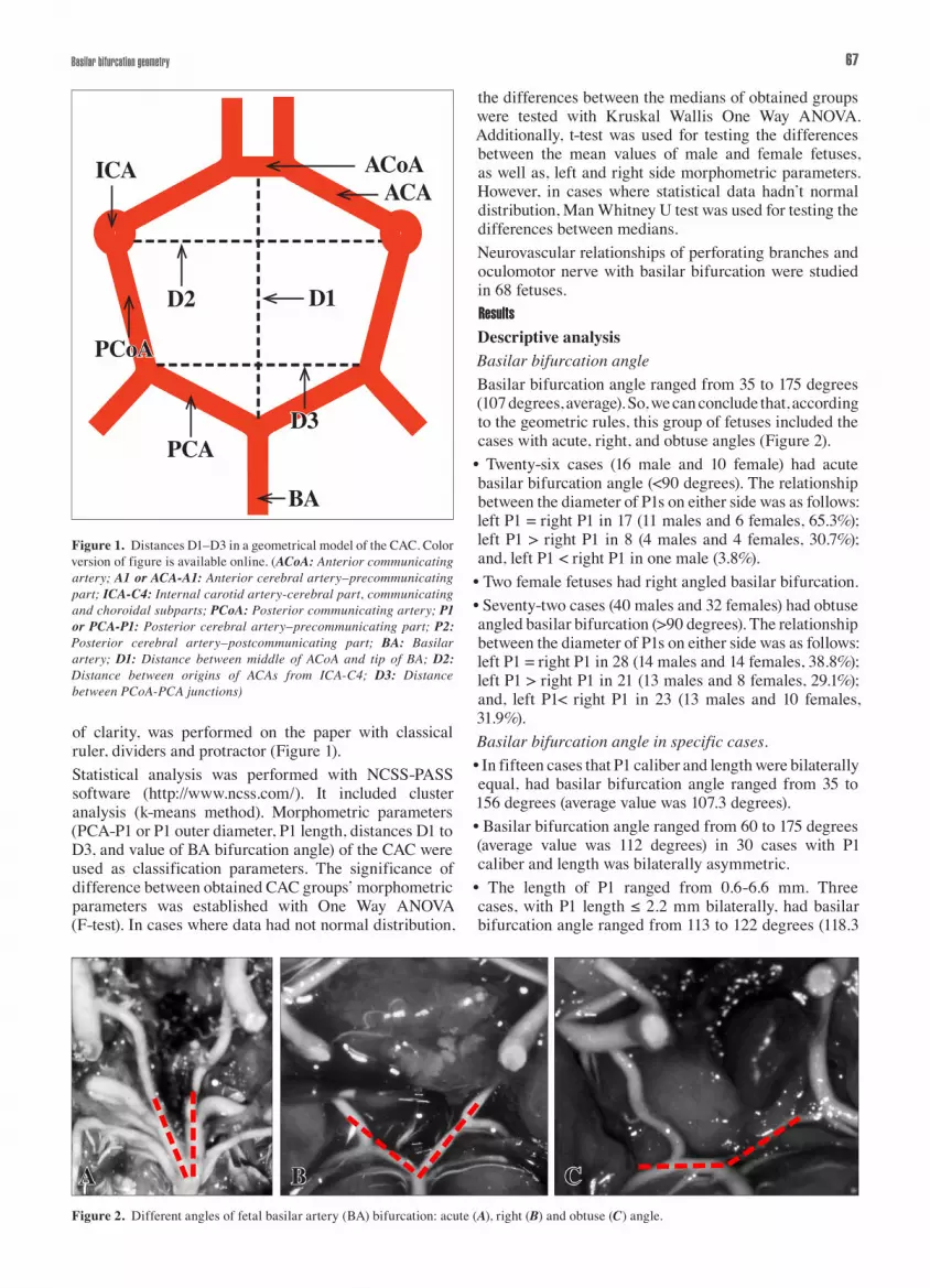
Figure 3. Five fetal cases (A–E) of perforators origin (arrow) from basilar bifurcation angle. Note: Arteries of CACs (a, e) were injected by Micropaque that was colored with black ink.
A
C
E
D
B

69Basilar bifurcation geometry
There was not significant difference in bifurcation angles between these groups.Neurovascular relationships• Basilar apex perforating branches were observed in 5 or 7.3% of the cases (Figure 3).• Direct relationship of oculomotor nerve with basilar bifurcation and P1 was observed in 18 or 26.7% of 68 evaluated cases. Two of them (2.9%), one on the right (Figure 4) and the other on the left side (Figure 3A), had oculomotor nerve that was directly related to the basilar bifurcation. In the remaining 16 cases (11 on the left, 2 on the right and 3 on both sides), left and/or right oculomotor nerve was related either with the middle part of P1 or PCoA junction (Figures 2B, 2C; 3A, 3D, 3E).
Statistical analysisCluster analysis of morphometric parameters showed a significantly different CAC geometry between three groups (Table 1).The first group included 75 cases (42 males and 33 females), which average CRL was 165.73±22.85 mm. Eighteen cases (11 males and 7 females) which average CRL was 170.56±23.38 mm were in the second group. Finally, 7 cases (3 males and 4 females) which average CRL was 208.57±30.78 mm, were in the third group. Fischer’s least significant difference (FSD) post hoc test showed that the third group had significantly higher CRL in relation to the first and the second group. Further, we focused on the posterior part of CAC and its influence on the whole CAC geometry. Precommunicating part of PCA had significantly (p<0.05) the highest values of average diameter in the third group, compared to the remaining two groups. The average measurements in the second group were higher than in the first group, but the difference was not significant (p>0.05). Left P1 had slightly larger average outer diameter than the right in all groups. This difference was not significant (p>0.05), but was the highest in the third group. Average length of P1 was highest in the second group, lower in the third and the lowest in the first group. In the second group this parameter was almost equal bilaterally, while in the remaining two groups it was insignificantly (p>0.05) higher on the left than on the right side. Average D1 distance was highest in the third, lower in the first and lowest in the second group. Third group had the highest average D2 distance vales, while it was lowest in the first group. Average D3 distance, conversely to the D1 distance, was highest in the second, lower in the third and the lowest in the first group. However, mutual relationships of these distances were different in the second group compared to the first and the third group. In this group average D1 was lower than D2 and higher than D3, while in the first and the third group average D1 was highest, average D2 was lower and, average D3 was lowest. Average bifurcation angle was significantly
Figure 4. Relationship of the left and right oculomotor nerves (CN III) with surrounding arteries. Right CN III emerged at the level of basilar tip and crossed superior surface of ipsilateral P1 segment. Left CN III crossed inferior surface of a branch of the left P1 segment. (PCoA: posterior communicating artery; PCA: posterior cerebral artery - precommunicating part; BA: basilar artery; RON: right occulomotor nerve; LON: left occulomotor nerve; LSCA: left superior cerebellar artery)
PCoA
BA
LON
PCARON
LSCA
Table 1. Crown-rump length (CRL) and CAC morphometric parameters values in obtained groups of fetuses.
Cluster I (n=75) Cluster II (n=18) Cluster III (n=7)
Mean SD Median Mean SD Median Mean SD Median DF1 DF2 F-Ratio p
CRL 165.73 22.85 160.00 170.56 23.38 165.00 208.57 30.78 220.00 2 97 10.65 0.000066
L-PCA-P1-OD 0.37 0.10 0.40 0.39 0.12 0.40 0.76 0.17 0.70 2 97 39.04 0.000000
L-PCA-P1-L 2.78 0.70 2.60 3.81 1.21 3.45 3.60 0.95 3.30 2 97 13.08 0.000009
R-PCA-P1-OD 0.36 0.10 0.40 0.38 0.12 0.40 0.63 0.29 0.60 2 97 14.31 0.000004
R-PCA-P1-L 2.63 0.81 2.60 3.82 1.44 3.30 3.36 0.93 3.30 2 97 12.22 0.000018
D1 12.70 2.00 12.60 12.34 1.40 12.60 15.84 2.38 15.00 2 97 9.24 0.000212
D2 11.29 1.67 11.40 13.46 1.91 13.60 13.54 1.29 14.00 2 97 15.63 0.000001
D3 4.36 0.95 4.50 6.41 0.91 6.00 4.87 1.07 4.60 2 97 33.87 0.000000
Alpha (degrees) 108.08 25.20 100.00 133.89 33.86 144.50 83.00 22.05 83.00 2 97 10.87 0.000055
(CRL: crown-rump length in mm; L-PCA-P1-OD: left PCA-P1 outer diameter in mm; L-PCA-P1-L: left PCA-P1 length in mm; R-PCA-P1-OD: right PCA-P1 outer diameter in mm; R-PCA-P1-L: right PCA-P1 length in mm)

70 Vasovic et al.
(p<0.05) the highest in the second group (133.89º±33.86), lower in the first (108.08º±25.20) and the lowest in the third group (83.00º±22.05).We did not established significant differences between male and female cases.DiscussionDescriptive analysisVertebral arteries (VAs), the branches of the left and right subclavian arteries, pass through the cervical vertebrae’s foramina transversaria and foramen magnum. Frequently, at the level of bulbopontine sulcus, VAs form basilar artery. The angle between the VAs at the vertebrobasilar junction varies between 10 and 160 degrees [10]. Basilar artery gives off branches and usually two terminal branches, posterior cerebral arteries. The average basilar angle in adults is 109 degrees and ranges between 30 and 180 degrees [6]. During our study, this angle ranged from 35 to 175 degrees and we can assume that angles would maintain during the postnatal period as well. The fact that fibrous structures composed of “chordae” stabilize the position of cerebral arteries, which are bathed by the cerebrospinal fluid, support our hypothesis [11].Ravensbergen et al. [12] noted that convergent arterial junctions and bifurcations each show a specific distribution of regions with complex flow patterns and low wall shear stress. Foutrakis et al. [13] suggest that the shear stress and pressure developed along the outer wall of a curved artery and at the apex of an arterial bifurcation create a hemodynamic state that can promote aneurysm formation. Wolfe et al. [14] cited literature data that pressure generated at the apex of the arterial bifurcation ranges from two to three times the peak luminal pressure in the proximal parent artery, leading to aneurysmal development. Arterial bifurcation geometry was described after three-dimensional DS angiography [15], and after the investigation with glass models [16,17]. However, we combined microdissection and classical geometrical method. Distances D1-D3, defined by Khamlichi et al. [9] helped to the construction of CAC models.The possibility of perforating arteries arising from the dome of the sac of a basilar tip aneurysm was usually denied [18]. We observed the perforating branches, which originated from basilar apex itself, in 7.3% of the cases. Gailloud et al. [18] also showed a large basilar tip aneurysm incorporating the origin of both P1s. We observed bilaterally short P1s (≤ 2 mm) only in three cases.Equal P1 diameters were observed in 17 of 26 analyzed fetuses which basilar bifurcation angle was lower than 90 degrees. Chow et al. [19] described that, depending on the particular anatomy of a basilar bifurcation, one of the PCAs may originate at a more acute angle in relation to the basilar trunk. Basilar bifurcation, with an angle of 90 degrees, was observed only in two fetuses. Roach et al. [17] described that in a symmetrical 90 degrees of bifurcation, when
one branch had twice larger diameter than the other, the flow in the small branch is more stable.Basilar bifurcations with an angle larger than 90 degrees were observed in almost two thirds of our cases. Postnatally, the angles formed between the basilar trunk and the largest right P1 and the smallest left P1 were 63 and 65 degrees, respectively [15]. Same authors also observed significantly larger branching angles in bifurcations with aneurysms compared with those without aneurysms [15].It is obvious that in majority of our cases with obtuse basilar bifurcation angle (approximately 61.2%), there was left/right asymmetry in the outer diameter of P1; which is higher than that in cases with an acute basilar bifurcation angle (approximately 34.7%).Valencia and Solis [20] cited literature data that with uneven branch flow, the flow dynamics inside a terminal aneurysm and the shear stresses acting on the intra-aneurismal wall increase proportionally with the bifurcation angle. We observed at figure of Valleé et al. [21] that a basilar apex aneurysm originated from bifurcation angle of about 90 degrees, and P1s had equal diameters. We also noted that in the case of Takaishi et al. [22] the basilar bifurcation angle was about 180 degrees and in cases of Chow et al. [19] and Valleé et al. [21] this angle was about 140 degrees. In all of these cases, P1 diameters were relatively equal. In our fetal material, higher incidence of equal P1s was observed in cases with acute angled (65.3%) than in cases with obtuse angled basilar bifurcations. Statistical analysisThe first group fetuses which mean CRL was 165.73 mm and, the third group cases, which mean CRL was 208.57 mm or 25% of all cases (Table 1), had similar CAC geometry with its major axis parallel to cerebral sagittal axis. This was accompanied with lower D2 and D3 distances – which were parallel to the cerebral transverse axis – compared to the D1 distance. The cases of the second group which mean CRL was 170.56 mm or 75% of all cases (Table 1), had larger D2 distance than the D1 and D3 distances. So the cases in this group had major axis parallel to the transverse cerebral axis. Khamlichi et al. [9] did not find significant differences between D1 to D3 values. They found D1/D3 ratio as 2.7:1, while D2/D3 ratio as 2:1. We calculated fetal D1/D3 ratio as 2.9:1, while D2/D3 as 2.6:1.Basilar bifurcation angle in the second group was significantly larger than that in the latter two groups. Since third fetal group was consisted only 7 cases, we consider that the reason for the lowest values of basilar bifurcation angle in this group might be coincidence.Aneurysms of the CACs’ posterior part are the most frequently located at the bifurcation of the BA [5,20]. According to Brisman et al. [23] the incidence of aneurysms on basilar bifurcation was 7%, and it was very similar to the incidence of internal carotid artery bifurcation aneurysms (7.5%).Hypothetically, the presence of aneurysms might led to basilar bifurcation angle deformations more frequently

71Basilar bifurcation geometry
in cases having an angle lesser than 90 degrees than in cases larger than 90 degrees.ConclusionThere was ‘copying’ of fetal basilar bifurcation vascular geometry into the adult pattern and, vice versa.
Our finding that almost two thirds of the fetuses have basilar bifurcation angle larger than 90 degrees, coincides with the literature data that the presence of cerebral aneurysms, which were anyway rare at this localization, was less frequently in cases with such basilar bifurcation angles.
References
[1] Fujimoto K. `Medial defects’ in the prenatal human cerebral arteries: an electron microscopic study. Stroke. 1996; 27: 706–708.
[2] Rowe AJ, Finlay HM, Canham PB. Collagen biomechanics in cerebral arteries and bifurcations assessed by polarizing microscopy. J. Vasc. Res. 2003; 40: 406–415.
[3] Canham PB, Finlay HM. Morphometry of medial gaps of human brain artery branches. Stroke. 2004; 35: 1153–1157.
[4] Parlea L, Fahrig R, Holdsworth DW, Lownie SP. An analysis of the geometry of saccular intracranial aneurysms. AJNR Am. J. Neuroradiol. 1999; 20: 1079–1089.
[5] Dumont AS, Oskouian RJ Jr, Chow MM, Kassell NF. Surgical management of unruptured basilar artery bifurcation aneurysms. Technical note. Neurosurg. Focus. 2002; 13: e3.
[6] Caruso G, Vincentelli F, Guidecelli G, Grisoli F, Xu T, Gouaze A. Perforating branches of the basilar bifurcation. J. Neurosurg. 1990; 73: 259–265.
[7] Patten MB. Human Embryology. 2nd Ed., Philadelphia, Blakiston, 1948; 184.
[8] Vasovic L. Morphological characteristics of the cerebral arterial circle with different origin of the vertebral arteries. (In Serbian, unpubl. data). PhD thesis, Faculty of Medicine, Niš, 1990.
[9] el Khamlichi A, Azouzi M, Bellakhdar F, Ouhcein A, Lahlaidi A. Configuration anatomique du polygone de Willis de l’adulte, étudié par les techniques d’injection. À propos de 100 cerveaux. Neurochirurgie. 1985; 31: 287–293.
[10] Guiffrè R, Sherkat S. Maldevelopmental pathology of the vertebral artery in infancy and childhood. Childs. Nerv. Syst. 2000; 16: 627–632.
[11] Arutiunov AI, Baron MA, Majorova NA. The role of mechanical factors in the pathogenesis of short-term and prolonged spasm of the cerebral arteries. J. Neurosurg. 1974; 40: 459–472.
[12] Ravensbergen J, Ravensbergen JW, Krijger JK, Hillen B, Hoogstraten HW. Localizing role of hemodynamics in atherosclerosis in several human vertebrobasilar junction geometries. Arterioscler. Thromb. Vasc. Biol. 1998; 18: 708–716.
[13] Foutrakis GN, Yonas H, Sclabassi RJ. Saccular aneurysm formation in curved and bifurcating arteries. AJNR Am. J. Neuroradiol. 1999; 20: 1309–1317.
[14] Wolfe SQ, Baskaya MK, Heros RC, Tummala RP. Cerebral aneurysms: learning from the past and looking toward the future. Clin. Neurosurg. 2006; 53: 157–178.
[15] Ingebrigtsen T, Morgan MK, Faulder K, Ingebrigtsen L, Sparr T, Schirmer H. Bifurcation geometry and the presence of cerebral artery aneurysms. J. Neurosurg. 2004; 101: 108–113.
[16] Stehbens WE. Turbulence of blood flow. Q. J. Exp. Physiol. Cogn. Med. Sci. 1959; 44: 110–117.
[17] Roach MR, Scott S, Ferguson GG. The hemodynamic importance of the geometry of bifurcations in the circle of Willis (glass model studies). Stroke. 1972; 3: 255–267.
[18] Gailloud P, Fasel JH, Muster M, de Tribolet N, Rufenacht DA. A case in favor of aneurysmographic studies: a perforating artery originating from the dome of a basilar tip aneurysm. AJNR Am. J. Neuroradiol. 1997; 18: 1691–1694.
[19] Chow MM, Woo HH, Masaryk TJ, Rasmussen PA. A novel endovascular treatment of a wide-necked basilar apex aneurysm by using a Y-configuration, double-stent technique. AJNR Am. J. Neuroradiol. 2004; 25: 509–512.
[20] Valencia A, Solis F. Blood flow dynamics and arterial wall interaction in a saccular aneurysm model of the basilar artery. Computers & Structures. 2006; 84: 1326–1337.
[21] Vallee JN, Aymard A, Vicaut E, Reis M, Merland JJ. Endovascular treatment of basilar tip aneurysms with Guglielmi detachable coils: predictors of immediate and long-term results with multivariate analysis – 6-year experience. Radiology. 2003; 226: 867–879.
[22] Takaishi Y, Yamashita H, Tamaki N. Cadaveric and clinical study of endoscope-assisted microneurosurgery for cerebral aneurysms using angle-type rigid endoscope. Kobe J. Med. Sci. 2002; 48: 1-11.
[23] Brisman JL, Song JK, Newell DW. Cerebral aneurysms. N. Eng. J. Med. 2006; 355: 928–939.