Prenatal test for Down Syndrome Screening
49
1 Prenatal test for Down Syndrome Screening Dr Farid Hadi Regional Medical Affairs Roche Diagnostics Asia-Pacific, Singapore
-
Upload
khangminh22 -
Category
Documents
-
view
4 -
download
0
Transcript of Prenatal test for Down Syndrome Screening

1
Prenatal test for Down Syndrome Screening
Dr Farid Hadi Regional Medical Affairs
Roche Diagnostics Asia-Pacific, Singapore

2
Conflict of Interest and Disclaimers
The opinions and content presented are the professional views of Farid Hadi, MD and do not necessarily reflect the opinion of his employer. Harmony is a non-invasive prenatal test (NIPT) based on cell-free DNA analysis and is considered a prenatal screening test, not a diagnostic test. Harmony does not screen for potential chromosomal or genetic conditions other than those expressly identified in this document. Before making any treatment decisions, all women should discuss their results with their healthcare provider, who can recommend confirmatory, diagnostic testing where appropriate. HARMONY and HARMONY and Design are trademarks of Ariosa Diagnostics, Inc. in the US. HARMONY is a trademark of Roche in other countries. All other trademarks are the property of their respective owners. The Harmony Prenatal Test was developed, and its performance characteristics determined by Ariosa Diagnostics, a CLIA and CAP accredited clinical laboratory in San Jose, CA USA. This testing service has not been cleared or approved by the US FDA.

3

4
Importance of prenatal screening in women of any age
55% of the estimated DOWN SYNDROME
BIRTHS occur in women under 35 years old.
Maternal Age
30,000
25,000
20,000
15,000
10,000
5,000
Estim
ated num
ber of D
own syndrome
cases in 1995
10 15 20 25 30 35 40 45 50
The California Prenatal Screening Program. March 2009. Provider Handbook 2009. Retrieved from www.cdph.ca.gov/programs/pns
Majority of babies born with Down syndrome are in women under 35
years old
10 15 20 25 30 35 40 45 50
30,000
25,000
20,000
15,000
10,000
5,000 Actual live births in 1995
87% of ALL LIVE
BIRTHS are to women under
35 years old.
Effective screening strategy is required for all pregnant
women

Professional Society Guidelines Who to screen for Down Syndrome?
• Candidates for prenatal screening:
– All women should be offered aneuploidy screening before 20 weeks gestation
– All women should have the option of invasive testing, regardless of age
• Candidates for prenatal diagnosis:
– Previous pregnancy complicated by foetal trisomy
– At least one major or two minor fetal structural anomalies in the current pregnancy
– Chromosomal translocation, inversion or aneuploidy in the pregnant women or her partner
American College of Obstetricians and Gynecologists. Practice Bulletin. Clinical Management Guidelines for Obstetrician-Gynecologist. Number 163, May 2016.
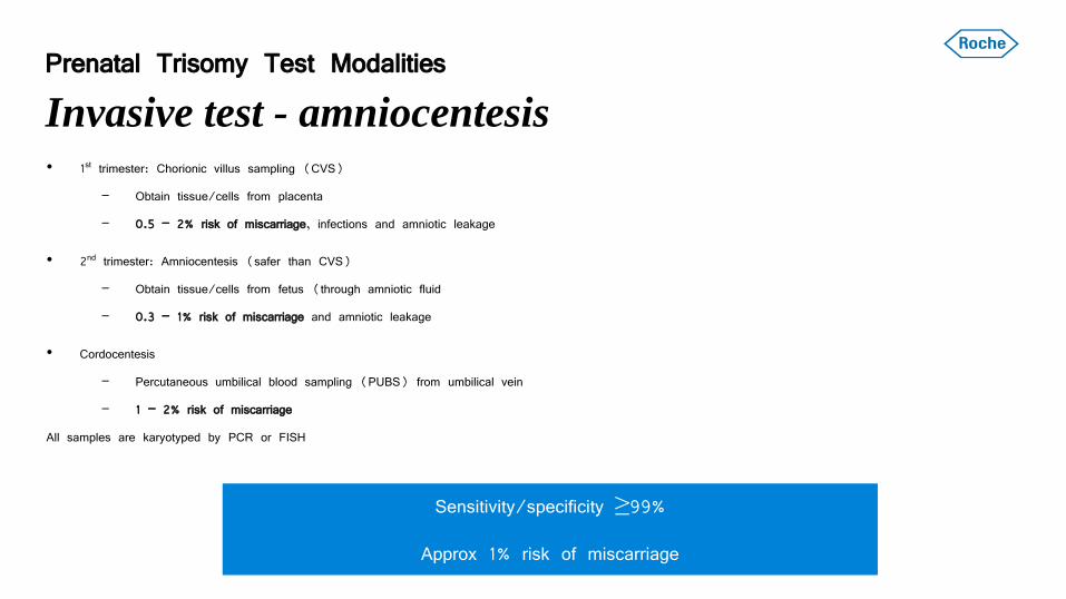
Prenatal Trisomy Test Modalities
Invasive test - amniocentesis • 1st trimester: Chorionic villus sampling (CVS)
– Obtain tissue/cells from placenta
– 0.5 – 2% risk of miscarriage, infections and amniotic leakage
• 2nd trimester: Amniocentesis (safer than CVS)
– Obtain tissue/cells from fetus (through amniotic fluid
– 0.3 – 1% risk of miscarriage and amniotic leakage
• Cordocentesis
– Percutaneous umbilical blood sampling (PUBS) from umbilical vein
– 1 – 2% risk of miscarriage
All samples are karyotyped by PCR or FISH
Sensitivity/specificity ≥99%
Approx 1% risk of miscarriage

Prenatal Trisomy Test Modalities
Single Test DR= 69%
DR= 82-87% (cFTS)
DR= 81%
DR= 64-70%
DR= 99%
1 2 3 4 5 6 7 8 9 10 11 12 13 14 15 16 17 18 19 20 21 22 23 24
FIRST TRIMESTER SECOND TRIMESTER
Gestational Week
Down
Syndrome
Tests
Screening
Diagnostic CVS
Amniocentesis
Cordocentesis
First trimester screening
(FTS)
b-hCG & PaPP-A
cfDNA
NIPT
Ultrasound
Nuchal
translucency &
Nasal bone
Quadruple test
b-hCG, AFP, uE3, DIA
Triple test
b-hCG, AFP, uE3
American College of Obstetricians and Gynecologists. Practice Bulletin. Clinical Management Guidelines for Obstetrician-Gynecologist. Number 163, May 2016.

Prenatal Trisomy Test Modalities
Combined Integrated Tests
SERUM INTEGRATED DR= 88%
INTEGRATED DR= 96%
1 2 3 4 5 6 7 8 9 10 11 12 13 14 15 16 17 18 19 20 21 22 23 24Gestational Week
FIRST TRIMESTER SECOND TRIMESTER
Down
Syndrome
Tests
Screening
Diagnostic CVS
Amniocentesis
Cordocentesis
First trimester
screening (FTS)
b-hCG & PaPP-A
Quadruple test
b-hCG, AFP, uE3, DIA
Triple test
b-hCG, AFP, uE3
RESULT
POSITIVENEGATIVE
Ultrasound
Nuchal
translucency &
Nasal bone
American College of Obstetricians and Gynecologists. Practice Bulletin. Clinical Management Guidelines for Obstetrician-Gynecologist. Number 163, May 2016.

Prenatal Trisomy Test Modalities
Combined Stepwise Tests
SEQUENTIAL STEPWISE DR= 95%
1 2 3 4 5 6 7 8 9 10 11 12 13 14 15 16 17 18 19 20 21 22 23 24Gestational Week
FIRST TRIMESTER SECOND TRIMESTER
Down
Syndrome
Tests
Screening
DiagnosticCVS
Amniocentesis
Cordocentesis
First trimester
screening (FTS)
b-hCG & PaPP-A
cfDNA
NIPT
Ultrasound
Nuchal translucency &
Nasal bone
Quadruple test
b-hCG, AFP, uE3, DIA
Triple test
b-hCG, AFP, uE3
RESULTPOSITIVE
NEGATIVE
American College of Obstetricians and Gynecologists. Practice Bulletin. Clinical Management Guidelines for Obstetrician-Gynecologist. Number 163, May 2016.
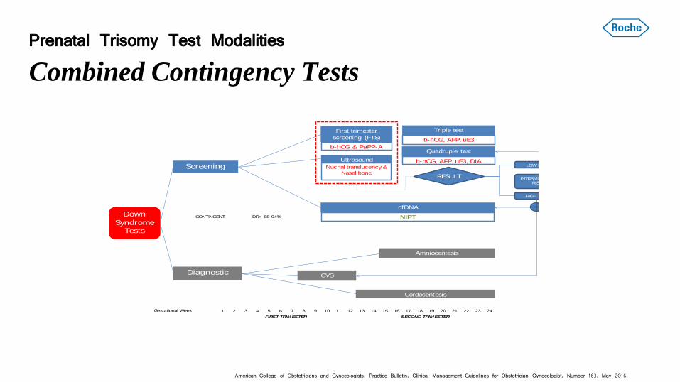
Prenatal Trisomy Test Modalities
Combined Contingency Tests
CONTINGENT DR= 88-94%
1 2 3 4 5 6 7 8 9 10 11 12 13 14 15 16 17 18 19 20 21 22 23 24Gestational Week
FIRST TRIMESTER SECOND TRIMESTER
Down
Syndrome
Tests
Screening
DiagnosticCVS
Amniocentesis
Cordocentesis
First trimester
screening (FTS)
b-hCG & PaPP-A
cfDNA
NIPT
Ultrasound
Nuchal translucency &
Nasal bone
Quadruple test
b-hCG, AFP, uE3, DIA
Triple test
b-hCG, AFP, uE3
RESULT
HIGH RISK
LOW RISK
INTERMEDIATE
RISK
American College of Obstetricians and Gynecologists. Practice Bulletin. Clinical Management Guidelines for Obstetrician-Gynecologist. Number 163, May 2016.

Biomarkers in First Trimesters
60
65
79
83
50
55
60
65
70
75
80
85
90
95
100
Nuchal Translucency (NT)* free b-hCG PAPP-A Combined test
Detection Rates (%)
Detection Rate (%) at 5% SPR
*) 10–13 completed weeks of gestation (without use of maternal age) SPR= Screen Positive Rates
Wald NJ, et alFirst and second trimester antenatal screening for Down’s syndrome: the results of the Serum, Urine and Ultrasound Screening Study (SURUSS). Health Technology Assessment 2003; Vol. 7: No. 11

Advancement in Prenatal Trisomy Screening
2012
1988 1980s
1960s
NIPT SNP Microarray Detection rate 92-99%
Gestational age 9w+
Chromosomes screened: T21, T18, T13, SCA, Microdeletions
Triple Screen Detection rate 60-74%
Gestational age 15w+
Chromosomes screened: T21, T18
Maternal Age Detection rate 27%
Gestational age N/A
Chromosomes screened: All
MSAFP Detection rate 36%
Gestational age 15wk+
Chromosomes screened: T21
1996 Quadruple Screen Detection rate 70-81%
Gestational age 15wk+
Chromosomes screened: T21, T18
1997
FTS NT/Serum Detection rate 80-95%
Gestational age 10-11wk
Chromosomes screened: T21, T18, T13
2011
NIPT Quantitative MPSS Detection rate 66-99%
Gestational age 10w+
Chromosomes screened: T21, T18, T13, SCA

Contingency Model cFTS & NIPT
Women who are high risk are offered a choice of proceeding to NIPT or directly to invasive testing
Hui L and Hyett J. Australian and New Zealand Journal of Obstetrics and Gynaecology 2013; 53: 416–424

Contingency Model cFTS & NIPT
Contingency model reduced the rates of invasive test
Hui L and Hyett J. Australian and New Zealand Journal of Obstetrics and Gynaecology 2013; 53: 416–424

Professional Society Guidelines Summary of NIPT Information Organization Policy Year
Recommends “informing all pregnant women that NIPS is the most sensitive screening option for traditionally screened aneuploidies”
2016
ACOG
“any patient may choose cell-free DNA analysis as a screening strategy for common aneuploidies regardless of her risk status”
2015
“Different scenarios are possible, including NIPT as an alternative first tier option” 2015
ISPD
“ The following protocol options are currently considered appropriate: 1. cfDNA screening as a primary test offered to all pregnant women.”
2015

Professional Society Guidelines
Summary of Down Syndrome Screening Biomarkers
• The Fetal Medicine Foundation promotes screening for Down syndrome at 11-13+6 weeks by Nuchal Translucency (NT) or a combination of nuchal translucency and maternal serum biomarkers.
• The combination of nuchal translucency and maternal serum free ßhCG and PAPP-A improves the detection rate to 90%. There is evidence that the detection rate of 90% can be achieved with a reduction in the false positive rate from 5% to 2.5% by examining the nasal bone.
• Use of both biochemical markers and nuchal translucency measurement is more effective than nuchal translucency measurement alone at detecting Down´s syndrome
• If first trimester screening is positive: offer genetic counseling and either chorionic villus sampling or second trimester amniocentesis
• Specific training, standardization for optimal NT measurement is important
• Even first trimester testing is done, it is still important to do second trimester screening for neural tube defects.
American College of Obstetricians and Gynecologists (ACOG)

17
Detection rate for Trisomy 21 (Down syndrome)
New possibilities in screening: Non-invasive prenatal testing
30%
35%
81%
85%
95%
>99%
0% 20% 40% 60% 80% 100% 120%
Maternal age
AFP only
Quad Marker Screen
First Trimester Screen
Full Integrated Screen
NIPT 4
1
1
1
2
3
False positive rate: 3-5%
False positive rate: <0.1%4
1. Ball et al. Obstet Gynecol. 2007 Jul;110(1):10-7. 2. Wald et al. BMJ. 1988 Oct 8;297(6653):883-7. 3. Thompson & Thompson Genetics in Medicine, Sixth Edition. Nussbaum, McInnes, Huntington. Saunders, 2001. 4. Norton M, et al, N Engl J Med. 2015 Apr 23;372(17):1589-97.

18
Limitations of conventional screening
1 in 20 women will receive a “positive” result1:
Vast majority will be “false positives”2
Referral to specialist, multiple office visits
Prolonged uncertainty, worry3
Risk of miscarriage with diagnostic testing options4
1. ACOG Practice Bulletin No. 77. Obstet Gynecol 2007;109:217-27. 2. Benn et al. Prenat Diagn. 2015 May 13. doi: 10.1002/pd.4608. [Epub ahead of print] 3. Acta Obstet Gynecol Scand. 2015 Jan;94(1):15-27. 4. Caughey et al. Obstet Gynecol 2006; 108(3 Pt 1): 612–6.

19
Limitations of conventional screening
19 in 20 women will receive a “negative” result:
But some of these women still have risk for trisomy
(due to 80-95% detection rate)
ACOG Practice Bulletin No. 77. Obstet Gynecol 2007;109:217-27.

Have we given the best for our baby?
Picture was taken from http://www.ashacarlos.com/blog/2012/04/10/pui-family-portrait-session/ accessed on 18-Oct-16

How it works Standard Blood Draw
Prenatal cell-free DNA assessment

How it works Standard Blood Draw
Prenatal cell-free DNA assessment

23
Non-invasive prenatal testing using cell-free DNA
• Cell-free DNA (cfDNA) are short DNA fragments
• During pregnancy, cfDNA from both the woman and fetus are present in maternal blood1
• Amount of fetal cfDNA present is a small fraction of the total cfDNA2
• Rapid clearance of fetal cfDNA after delivery (<24 hours)3
1. Lo et al. Lancet 1997;350:485-87. 2. Lo et al. Am J Hum Genet. 1998 Apr;62(4):768-75. 3. Lo et al. Am J Hum Genet 1999; 64:218–224.

24

25

26
NEXT Study
Norton M, et al, N Engl J Med. 2015 Apr 23;372(17):1589-97.
Largest blinded prospective NIPT study to date 35 clinical sites in 6 countries (US, EU)

27
NEXT Study1 – Objective & Background
Compare the performance of Harmony to traditional screening for trisomy 21 in a clinical setting
• Collect outcome data on all subjects
Powered for BOTH sensitivity and specificity
• Previous studies of NIPT in a general population were not large enough to evaluate sensitivity2,3
First trimester screening (FTS) and Harmony performed simultaneously for direct comparison
• Previous studies performed NIPT after the first trimester, when fetal fraction is higher2,4
1. Norton M, et al, N Engl J Med. 2015 Apr 23;372(17):1589-97. 2. Bianchi et al. N Engl J Med. 2014 Feb 27;370(9):799-808. 3. Nicolaides et al. Am J Obstet Gynecol. 2012 Nov;207(5):374.e1-6. 4. Wang et al. Prenat Diagn. 2013 Jul;33(7):662-6.

28
NEXT Study - Overview
18,955 pregnancies
First trimester screening*
Test + Test - Test + Test -
38 trisomy 21
DR = 100% FPR=0.06%
DR = 79% FPR=5.4%
Mean maternal age: 30.7 years Mean gestational age: 12.5 weeks Mean maternal weight: 65.8 kg
Norton M, et al, N Engl J Med. 2015 Apr 23;372(17):1589-97. *hCG and PAPP-A, nuchal translucency measurement DR = detection rate; FPR = false positive rate
Outcome obtained for 15,841 subjects by genetic testing or newborn exam
38 8 30 0
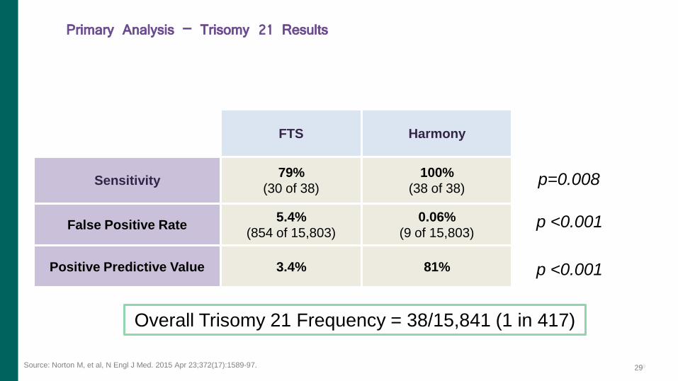
29
p=0.008
p <0.001
p <0.001
Overall Trisomy 21 Frequency = 38/15,841 (1 in 417)
FTS Harmony
Sensitivity 79%
(30 of 38)
100%
(38 of 38)
False Positive Rate 5.4%
(854 of 15,803)
0.06%
(9 of 15,803)
Positive Predictive Value 3.4% 81%
Primary Analysis – Trisomy 21 Results
29 Source: Norton M, et al, N Engl J Med. 2015 Apr 23;372(17):1589-97.

Harmony is statistically superior to first-trimester screening for the detection of trisomy 21 in a general pregnancy population.
Significantly Higher Detection Rate:
Harmony: 100%
FTS: 79%
90-fold Lower False-Positive Rate:
Harmony: 1 in 1,756
FTS: 1 in 19
20-fold Higher Positive Predictive Value:
Harmony: 81%
FTS: 3.4%
NEJM Harmony Study - Conclusions
Source: Norton M, et al, N Engl J Med. 2015 Apr 23;372(17):1589-97.
15

31
Advantages of Directed Analysis (DANSRTM)
Chr 21, 18, 13, X, Y cfDNA
Other Chr cfDNA
Unmapped cfDNA
cfDNA in blood
Massively Parallel Shotgun Sequencing (MPSS) Directed analysis (DANSR)

32
Original state of the
genome
Random approach Harmony approach
The Harmony approach – Advantages of DANSRTM
• Harmony provides the deepest analysis of chromosomes of interest
• DANSR targets chromosomes of interest • Chromosomes 21, 18, and 13 represent <10% of the genome1
1. Lander et al. Nature 409, 860-921 (15 February 2001) doi:10.1038/35057062

33
DANSR result is analyzed with Fetal fraction Optimized Risk of Trisomy Evaluation (FORTE) algorithm
Trisomy Non-trisomy False positive
Ashoor G et al., Am J Obstet Gynecol. 2012 Apr;206(4):322.e1-5.
34
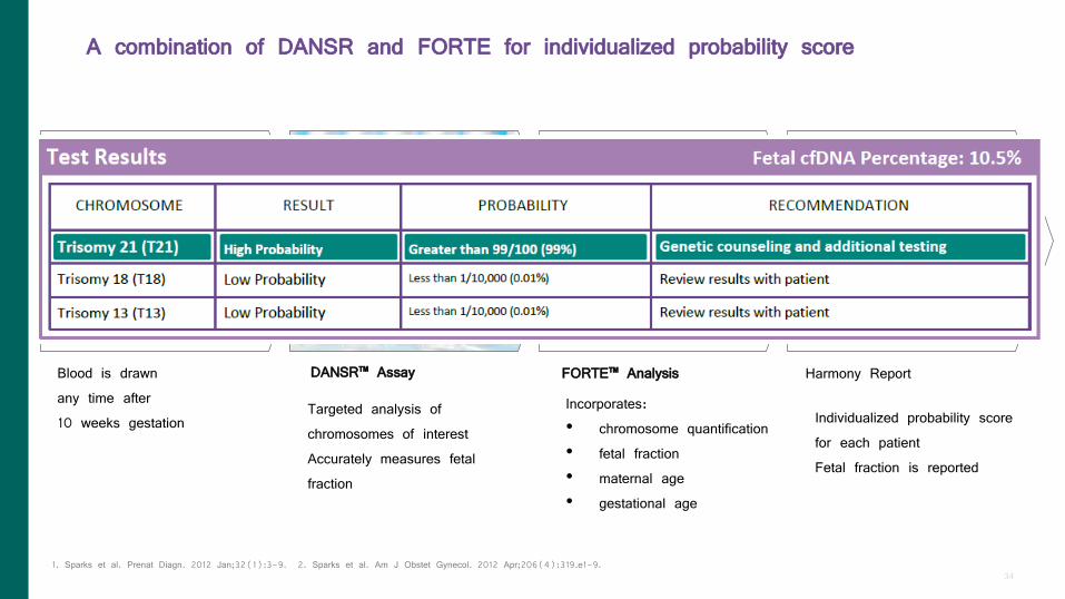
A combination of DANSR and FORTE for individualized probability score
SNP
MPSS
DANSR™ Assay FORTE™ Analysis Harmony Report
)()|(
)()|(
DPDxP
TPTxP
j
j
Targeted analysis of chromosomes of interest Accurately measures fetal fraction
Incorporates: • chromosome quantification • fetal fraction • maternal age • gestational age
Individualized probability score for each patient Fetal fraction is reported
Blood is drawn any time after 10 weeks gestation
1. Sparks et al. Prenat Diagn. 2012 Jan;32(1):3-9. 2. Sparks et al. Am J Obstet Gynecol. 2012 Apr;206(4):319.e1-9.
35
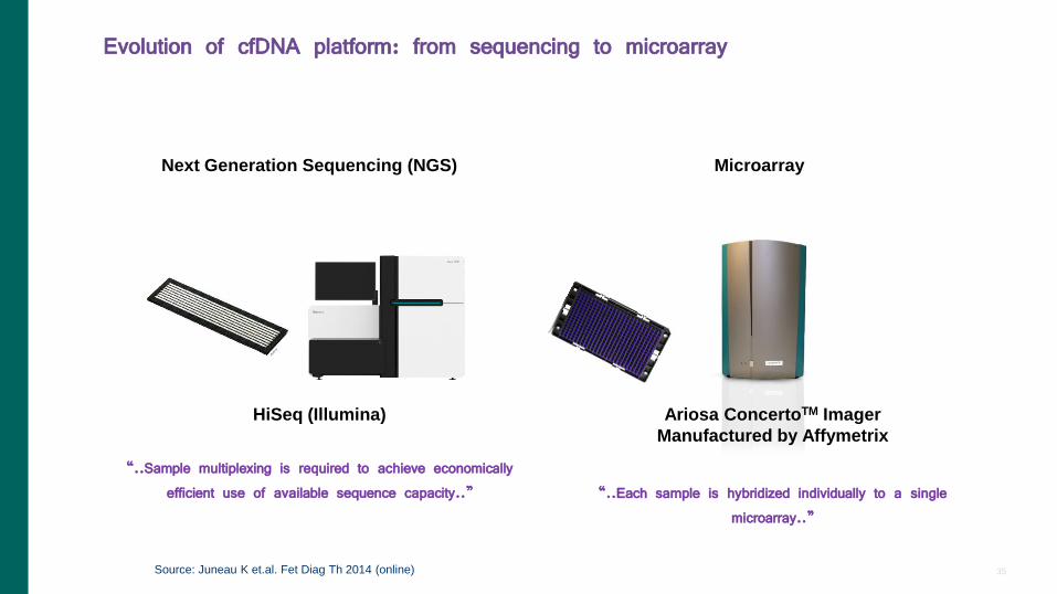
Evolution of cfDNA platform: from sequencing to microarray
HiSeq (Illumina)
Next Generation Sequencing (NGS)
Ariosa ConcertoTM Imager
Manufactured by Affymetrix
Microarray
Source: Juneau K et.al. Fet Diag Th 2014 (online)
“..Sample multiplexing is required to achieve economically efficient use of available sequence capacity..” “..Each sample is hybridized individually to a single
microarray..”

36
Increase efficiency in NIPT
Number of samples per hour
64
12
Juneau K et al. Fetal Diagn Ther. 2014;36(4):282-6.
“.. Both microarray and sequencing technologies continue to improve. Some sequencing systems have accelerated sequencing modes that could decrease the time differential observed between microarrays and sequencing. However, in these modes, as the speed of sequencing increases,
the capacity decreases and the cost per sample rises.”

37
Less variability in assays and fetal fraction observed with microarray
Juneau K et al. Fetal Diagn Ther. 2014;36(4):282-6.
38
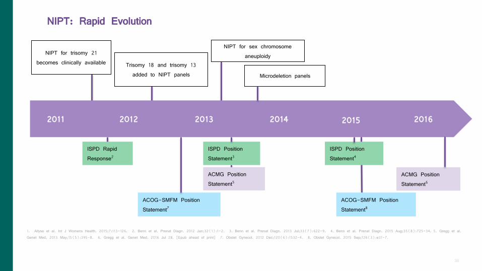
NIPT: Rapid Evolution
2011 2012 2013 2014 2015 2016
NIPT for trisomy 21 becomes clinically available
NIPT for sex chromosome aneuploidy
ISPD Rapid Response2
1. Allyse et al. Int J Womens Health. 2015;7:113-126. 2. Benn et al. Prenat Diagn. 2012 Jan;32(1):1-2. 3. Benn et al. Prenat Diagn. 2013 Jul;33(7):622-9. 4. Benn et al. Prenat Diagn. 2015 Aug;35(8):725-34. 5. Gregg et al. Genet Med. 2013 May;15(5):395-8. 6. Gregg et al. Genet Med. 2016 Jul 28. [Epub ahead of print] 7. Obstet Gynecol. 2012 Dec;120(6):1532-4. 8. Obstet Gynecol. 2015 Sep;126(3):e31-7.
Trisomy 18 and trisomy 13 added to NIPT panels
ISPD Position Statement3
ISPD Position Statement4
ACMG Position Statement5
ACMG Position Statement6
ACOG-SMFM Position Statement7
ACOG-SMFM Position Statement8
Microdeletion panels

39
“With suitable genetic counseling, MPS can be helpful for women who may have been determined to be high risk by one of the previously recommend screening strategies.”
-International Society for
Prenatal Diagnosis (ISPD), 20111
Evolving clinical application of NIPT
1.Benn et al. Prenat Diagn. 2012 Jan;32(1):1-2. 2. Benn et al. Prenat Diagn. 2015 Aug;35(8):725-34.
“cfDNA screening as a primary test offered to all pregnant women [is considered appropriate].” -International Society for Prenatal
Diagnosis (ISPD), 20152

40
Current professional guidelines: Low risk pregnancies
• International Society for Prenatal Diagnosis, 20151:
Appropriate to offer NIPT as a primary screening test to all pregnant women
• European and American Societies of Human Genetics, 20152:
NIPT as a first-tier screening test is an option
• American Congress of Obstetricians and Gynecologists/Society for Maternal Fetal Medicine, 20153:
NIPT should be offered to all women (but conventional methods are the appropriate choice for most women)
• American College of Medical Genetics and Genomics, 20164:
All pregnant women should be informed that NIPT is the most sensitive screening option for trisomy 21, trisomy 18, and trisomy 13
1. Benn et al. Prenat Diagn. 2015 Aug;35(8):725-34. 2. Dondorp et al. Eur J Hum Genet. 2015 Nov;23(11):1592. 3. ACOG Committee Position 640. Obstet Gynecol. 2015 Sep;126(3):e31-7. 4. Gregg et al. Genet Med. 2016 Jul 28. [Epub ahead of print]

41
DANSR and FORTE validation with microarray and NGS
41 Stokowski R et al. Prenat Diagn. 2015 Dec;35(12):1243-6.

42
DANSR and FORTE validation with microarray and NGS
42 Stokowski R et al. Prenat Diagn. 2015 Dec;35(12):1243-6.
43
• Twin pregnancies1,2
– Single result is reported for both fetuses
– Fetal Sex assessment available for twin pregnancies
• A male result indicates one or two male fetuses
*Monosomy X and Sex Chromosome Aneuploidy Panel has not been validated in twin pregnancies
*Harmony has not been validated in higher order multiples
• NIPT validation for use in IVF pregnancies3,4, including:
– Singleton or twin
– Self or non-self egg donor
– Surrogate pregnancies
Additional offerings
1. Bevilacqua et al. Ultrasound Obstet Gynecol. 2015 Jan;45(1):61-6. 2. Gil et al. Fetal Diagn Ther. 2014;35:204-11. 3. Stokowski et al. Prenat Diagn. 2015;35:1-4. 4. Norton ME et al. N Engl J Med. 2015 Apr 1.

44
Bevilacqua et al.1 (prospective)
Gil et al.2 (retrospective)
Gil et al.2 (prospective)
Trisomy 21 11 of 12 9 of 10 2 of 2
Trisomy 18 5 of 5 - 1 of 1
Trisomy 13 - 1 of 1 -
Euploid 323 of 323 181 of 181 60 of 60
Identified as “High Risk”: • 22 of 24 cases of trisomy 21 • 6 of 6 cases of trisomy 18 • 1 of 1 case of trisomy 13 • No “false positives” in over 500 euploid cases
1. Bevilacqua et al. Ultrasound Obstet Gynecol. 2015 Jan;45(1):61-6. 2. Gil et al. Fetal Diagn Ther. 2014;35:204-11.
Performance in twin pregnancies

45
Prevalence of common SCAs1: • 47,XXY (Klinefelter syndrome) 1/500-1/1,000 males • 47,XXX (Triple X syndrome) 1/1,000 females • 47,XYY (Jacobs syndrome) 1/1,000 males • 45,X (Turner syndrome) 1/2,500 females
Overall incidence of SCAs: ~1/500 live births (Overall incidence of Down syndrome: ~ 1/800 live births2)
1. Thompson & Thompson Genetics in Medicine, Sixth Edition. Robert L. Nussbaum, Roderick McInnes, Willard Huntington. Saunders, 2001. 2. U.S. National Library of Medicine. Genetics Home Reference. Down Syndrome. http://ghr.nlm.nih.gov/condition/downsyndrome. Accessed Jan 25, 2016.
Incidence of sex chromosome aneuploidy (SCA)
46
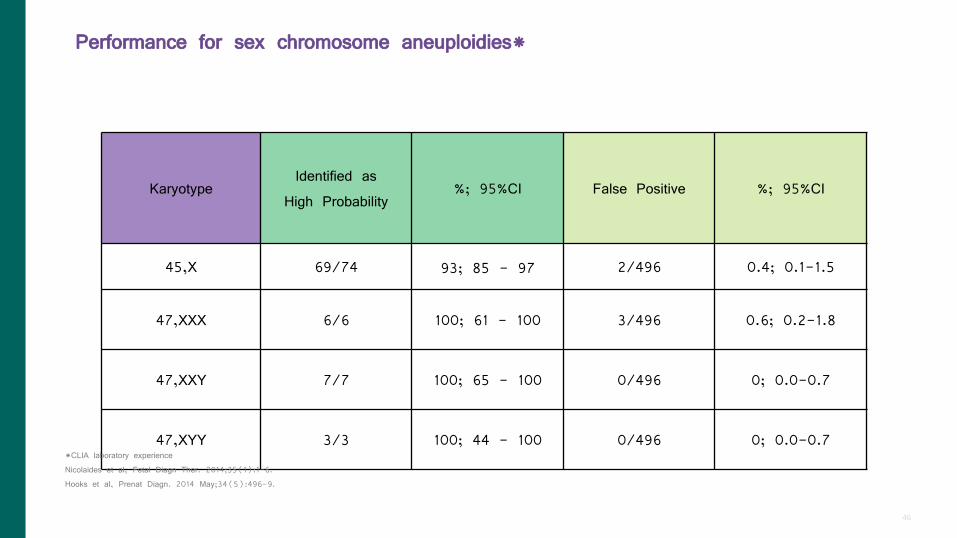
Karyotype Identified as
High Probability %; 95%CI False Positive %; 95%CI
45,X 69/74 93; 85 - 97 2/496 0.4; 0.1-1.5
47,XXX 6/6 100; 61 - 100 3/496 0.6; 0.2-1.8
47,XXY 7/7 100; 65 - 100 0/496 0; 0.0-0.7
47,XYY 3/3 100; 44 - 100 0/496 0; 0.0-0.7 *CLIA laboratory experience Nicolaides et al, Fetal Diagn Ther. 2014;35(1):1-6. Hooks et al, Prenat Diagn. 2014 May;34(5):496-9.
Performance for sex chromosome aneuploidies*

47
Current professional guidelines: Microdeletions
• International Society for Prenatal Diagnosis, 20151:
Patients should be counseled regarding limitations. Testing should be limited to clinically significant disorders.
• European and American Societies of Human Genetics, 20152:
Currently not recommended
• American Congress of Obstetricians and Gynecologists/Society for Maternal Fetal Medicine, 20153:
Routine screening for microdeletions should not be performed
• American College of Medical Genetics and Genomics, 20164:
Patients should be informed of availability of testing, including limitations
1. Benn et al. Prenat Diagn. 2015 Aug;35(8):725-34. 2. Dondorp et al. Eur J Hum Genet. 2015 Nov;23(11):1592. 3. ACOG Committee Position 640. Obstet Gynecol. 2015 Sep;126(3):e31-7. 4. Gregg et al. Genet Med. 2016 Jul 28. [Epub ahead of print]

48
• All pregnant women should be screened for Down syndrome
• NIPT is targeted approach for specific chromosomes, i.e. T21, T13, T18
• Fetal fraction >4% is important for accurate result
• Microarray technology was developed to improve NGS platform with comparable performance and greater reproducibility
• DANSR and FORTE are validated to assess twin and IVF pregnancy
• Expanded menu will be made available in clinically relevant abnormalities, i.e. DiGeorge Syndrome
Conclusions

Thank You.
49