Prenatal diagnosis of lissencephaly: A case report
6
VOJNOSANITETSKI PREGLED Page 1 Correspondence to: Nataša Cerovac, Bulevar Arsenija Čarnojevića 124/2, 11 070 Novi Beograd, Serbia. Phone.: +381 11 3115 919, E-mail: [email protected] CASE REPORT UDC: 618.33-07::[616-053.31:616.8-053.2 DOI: 10.2298/VSP140806010C Prenatal diagnosis of lissencephaly: A case report Prenatalna dijagnoza lizencefalije Nataša Cerovac* † , Milan Terzić ‡† , Milan Borković*, Nevena Divac §† , Radan Stojanović §† , Milica Prostran §† *Clinic for Neurology and Psychiatry for Children and Youth, Belgrade, Serbia; † Faculty of Medicine, University of Belgrade, Belgrade, Serbia; ‡ Clinic for Gynecology and Obstetrics, Clinical Center of Serbia, Belgrade, Serbia; § Institute of Pharmacology, Clinical Pharmacology and Toxicology, Faculty of Medicine, University of Belgrade, Belgrade, Serbia Abstract Introduction. Lissencephaly (“smooth brain”) forms a major group of brain malformations due to abnormal neu- ronal migration. It can cause severe intellectual and motor disability and epilepsy in children. The prenatal diagnosis of this malformation is rare. Case report. We presented a case of the prenatal diagnosis of lissencephaly. A 30-year old pregnant woman was reffered to the hospital at the week 35 of gestation for magnetic resonance imaging (MRI) after an ultrasound examination demonstrated fetal cerebral ventriculomegaly. Fetal MRI of the brain showed “smooth”, agyrya cortex. The female infant was born at term with birth weight of 2,500 g and Apgar score 8, showing global developmental delay. Postnatal ultrasound and MRI confirmed classical lissencephaly. She is now 8 years old and has spastic quadriparesis, mental retardation and epilepsy. Conclusion. Confirmation of the ultrasound diagnosis with MRI is desirable for the prenatal diagnosis of lissencephaly. Key words: lissencephaly; fetal monitoring; ultrasonography; magnetic resonance imaging; mental retardation. Apstrakt Uvod. Lizencefalija („gladak mozak”) predstavlja važnu grupu malformacija mozga koja nastaje zbog poremećaja neuronske migracije. Može uzrokovati teško zaostajanje u intelektualnom i motornom razvoju i epilepsiju kod dece. Prenatalna dijagnostika ovog poremećaja je retka. Prikaz bolesnika. Prikazali smo jedan slučaj prenatalno dijag- nostikovane lizencefalije. Trudnica, stara 30 godina, upućena je u bolnicu u 35. nedelji gestacije radi magnetne rezonancije (MR) posle ultrazvučnog pregleda koji je ukazao na fetalnu moždanu ventrikulomegaliju. Fetalni MR pregled mozga pokazao je glatku koru, sa izostankom razvoja vijuga. Dete ženskog pola rođeno je u terminu, telesne mase 2 500 g i Apgar skora 8, a pokazalo je usporen rani razvoj. Postna- talni ultrazvučni i MR pregled mozga potvrdili su dijagnozu lizencefalije. Devojčica sada ima 8 godina i kliničku sliku spastične kvadripareze, mentalne retardacije i epilepsije. Zaključak. Potvrda ultrazvučne dijagnoze putem MR pre- gleda značajna je za prenatalnu dijagnostiku lizencefalije. Ključne reči: lizencefalija; fetus, praćenje; ultrazvuk; magnetna rezonanca, snimanje; mentalna zaostalost. Introduction Malformations of cortical development are significant causes of delay in psychomotor development and epilepsy in children 1–3 . Lyssencephaly (“smooth brain”) forms a major group of brain malformations due to widespread abnormal migration 1, 2, 4 . Other two major categories of malformations are cobblestone complex malformations (also known as type 2 lissencephaly) and all types of heterotopia. With magnetic resonance imaging (MRI), these disorders can be identified in life 5 . Classical lissencephaly (OMIM # 607432), formely de- signed as type 1, is a severe neurological malformation char- acterized by a lack of sulcation of the cortical plate, that pro- duces a smooth brain surface, cortical thickening with four primitive layers and ventriculomegaly 3, 6, 7 . The brain has no gyri (agyria) or very low gyri (pachygyria) or there is a re- lated disorder known as subcortical band heterotopia (SBH) 3, 7, 8 . Due to contribution of computed tomography (CT) and MRI, this spectrum of gyral abnormalities was graded in the following way: grade 1, complete agyria; grade 2, diffuse agyria with few sulci in anterior regions; grade 3, anterior pachygyria (few, broad gyri) and posterior agyria;
Transcript of Prenatal diagnosis of lissencephaly: A case report

VOJNOSANITETSKI PREGLED Page 1
Correspondence to: Nataša Cerovac, Bulevar Arsenija Čarnojevića 124/2, 11 070 Novi Beograd, Serbia. Phone.: +381 11 3115 919, E-mail: [email protected]
C A S E R E P O R T UDC: 618.33-07::[616-053.31:616.8-053.2
DOI: 10.2298/VSP140806010C
Prenatal diagnosis of lissencephaly: A case report
Prenatalna dijagnoza lizencefalije
Nataša Cerovac*†, Milan Terzić
‡†, Milan Borković*, Nevena Divac
§†, Radan
Stojanovi槆
, Milica Prostran§†
*Clinic for Neurology and Psychiatry for Children and Youth, Belgrade, Serbia; †Faculty of Medicine, University of Belgrade, Belgrade, Serbia; ‡Clinic for Gynecology
and Obstetrics, Clinical Center of Serbia, Belgrade, Serbia; §Institute of Pharmacology,
Clinical Pharmacology and Toxicology, Faculty of Medicine, University of Belgrade,
Belgrade, Serbia
Abstract
Introduction. Lissencephaly (“smooth brain”) forms a major group of brain malformations due to abnormal neu-ronal migration. It can cause severe intellectual and motor disability and epilepsy in children. The prenatal diagnosis of this malformation is rare. Case report. We presented a case of the prenatal diagnosis of lissencephaly. A 30-year old pregnant woman was reffered to the hospital at the week 35 of gestation for magnetic resonance imaging (MRI) after an ultrasound examination demonstrated fetal cerebral ventriculomegaly. Fetal MRI of the brain showed “smooth”, agyrya cortex. The female infant was born at term with birth weight of 2,500 g and Apgar score 8, showing global developmental delay. Postnatal ultrasound and MRI confirmed classical lissencephaly. She is now 8 years old and has spastic quadriparesis, mental retardation and epilepsy. Conclusion. Confirmation of the ultrasound diagnosis with MRI is desirable for the prenatal diagnosis of lissencephaly. Key words: lissencephaly; fetal monitoring; ultrasonography; magnetic resonance imaging; mental retardation.
Apstrakt Uvod. Lizencefalija („gladak mozak”) predstavlja važnu grupu malformacija mozga koja nastaje zbog poremećaja neuronske migracije. Može uzrokovati teško zaostajanje u intelektualnom i motornom razvoju i epilepsiju kod dece. Prenatalna dijagnostika ovog poremećaja je retka. Prikaz bolesnika. Prikazali smo jedan slučaj prenatalno dijag-nostikovane lizencefalije. Trudnica, stara 30 godina, upućena je u bolnicu u 35. nedelji gestacije radi magnetne rezonancije (MR) posle ultrazvučnog pregleda koji je ukazao na fetalnu moždanu ventrikulomegaliju. Fetalni MR pregled mozga pokazao je glatku koru, sa izostankom razvoja vijuga. Dete ženskog pola rođeno je u terminu, telesne mase 2 500 g i Apgar skora 8, a pokazalo je usporen rani razvoj. Postna-talni ultrazvučni i MR pregled mozga potvrdili su dijagnozu lizencefalije. Devojčica sada ima 8 godina i kliničku sliku spastične kvadripareze, mentalne retardacije i epilepsije. Zaključak. Potvrda ultrazvučne dijagnoze putem MR pre-gleda značajna je za prenatalnu dijagnostiku lizencefalije.
Ključne reči: lizencefalija; fetus, praćenje; ultrazvuk; magnetna rezonanca, snimanje; mentalna zaostalost.
Introduction
Malformations of cortical development are significant
causes of delay in psychomotor development and epilepsy in
children 1–3
. Lyssencephaly (“smooth brain”) forms a major
group of brain malformations due to widespread abnormal
migration 1, 2, 4
. Other two major categories of malformations
are cobblestone complex malformations (also known as type
2 lissencephaly) and all types of heterotopia. With magnetic
resonance imaging (MRI), these disorders can be identified
in life 5.
Classical lissencephaly (OMIM # 607432), formely de-
signed as type 1, is a severe neurological malformation char-
acterized by a lack of sulcation of the cortical plate, that pro-
duces a smooth brain surface, cortical thickening with four
primitive layers and ventriculomegaly 3, 6, 7
. The brain has no
gyri (agyria) or very low gyri (pachygyria) or there is a re-
lated disorder known as subcortical band heterotopia
(SBH) 3, 7, 8
. Due to contribution of computed tomography
(CT) and MRI, this spectrum of gyral abnormalities was
graded in the following way: grade 1, complete agyria; grade
2, diffuse agyria with few sulci in anterior regions; grade 3,
anterior pachygyria (few, broad gyri) and posterior agyria;
Page 2 VOJNOSANITETSKI PREGLED
Cerovac N, et al. Vojnosanit Pregl 2015; OnLine-First March (00): 10–10.
grade 4, pachygyria more prominent in the posterior brain
regions than in anterior; grade 5, pachygyria posteriorly with
SBH and grade 6, SBH only 9. Lissencephaly due to muta-
tions of LIS1 at 17 p.13.3 is highly specific for more severe
changes in posterior brain regions ( p > a gradient), while lis-
sencephaly due to mutations of XLIS at X q 22.3-q23 often
have more severe gyral abnormalities in the anterior brain
regions (a > p gradient) 10
. TUBA1A usually show posterior-
predominant lissencephaly similar to LIS1 11
.
Studies have identified two major genes responsible for
classical lissencephaly: LIS1 (named PAFAH1B1) gene at 17
p13.3 and the XLIS (DCX) gene at Xq 22.3-q23 12–14
. Both
proteins are important for normal neuronal migrational proc-
esses. Approximately 76% of patients with classical lissen-
cephaly show mutations in these two genes 15
. Recently, mu-
tations of TUBA1A gene at the 12q12-q14 is detected in
several cases with lissencephaly. TUBA1A belongs to the
alpha-tubulin protein family which is needed for correct cell
movements. Mutation of TUBA1A are responsible for 1–4%
of cases 16, 17
. Other types of lissencephaly caused by muta-
tion of: RELN, VLDLR and ARX have been decribed 10, 18
.
These types of lissencephaly are less common and known as
“variant lissencephaly”. It is important that the morphology
of lissencephaly caused by mutations of those three genes
differs from that caused by LIS1, DCX and TUBA1A muta-
tions. Lissencephaly with cerebellar hypoplasia (LCH) re-
sults from mutations of two genes: the reelin (RELN) gene
and very low-density lipoprotein receptor gene (VLDLR). X-
linked lissencephaly with abnormal genitalia and agenesis of
the corpus callosum (XLAG) has been associated with ARX
gene.
Children with classical lissencephaly usually have hy-
potonia at birth, but spasticity develop later in infancy. Clini-
cal manifestations include seizures, spastic quadriplegia and
profound mental retardation. The onset of seizures is usually
between 6–12 months. Infantile spasms followed by hypsar-
rhytmia are seen in the majority of children and they respond
at first to corticotropin or other antiepileptic drugs. Unfortu-
nately, almost all children will go on to have frequent sei-
zures and severe psychomotor retardation. LIS1 gene which
cause classical lissencephaly is connected with two clinical
disorders. The first one is the isolated lissencephaly sequence
(ILS) which is characterized by lacks of typical facial ap-
pearance and the second is Miller-Dieker syndrome (MDS)
(OMIM #247000), where typical facial features and other
congenital defects exist 19, 20
.
The prenatal diagnosis of this malformation is rare.
MRI imaging should be useful in screening for malformation
of cortical development such as lissencephaly. The aim of
this case report was to characterise the delivery and postnatal
neurodevelopmental outcome of the fetus reffered for MRI
following suspicion on ultrasound of ventriculomegaly.
Case report
We presented a case of the prenatal diagnosis of the lis-
sencephaly. A 30-year-old pregnant woman was reffered to
the hospital at the week 35 of gestation for MRI after an ul-
trasound examination demonstrated fetal ventriculomegaly,
defined as ventricular size (measured at the atrium of the lat-
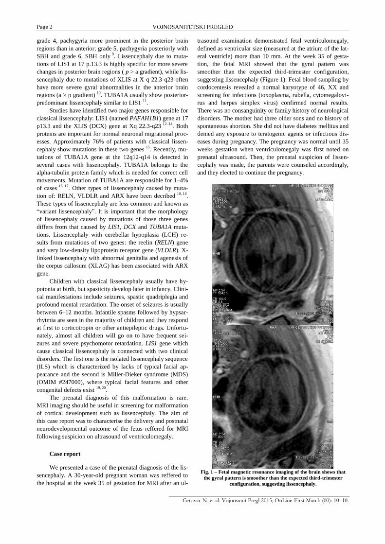
eral ventricle) more than 10 mm. At the week 35 of gesta-
tion, the fetal MRI showed that the gyral pattern was
smoother than the expected third-trimester configuration,
suggesting lissencephaly (Figure 1). Fetal blood sampling by
cordocentesis revealed a normal karyotype of 46, XX and
screening for infections (toxoplasma, rubella, cytomegalovi-
rus and herpes simplex virus) confirmed normal results.
There was no consanguinity or family history of neurological
disorders. The mother had three older sons and no history of
spontaneous abortion. She did not have diabetes mellitus and
denied any exposure to teratogenic agents or infectious dis-
eases during pregnancy. The pregnancy was normal until 35
weeks gestation when ventriculomegaly was first noted on
prenatal ultrasound. Then, the prenatal suspicion of lissen-
cephaly was made, the parents were counseled accordingly,
and they elected to continue the pregnancy.
Fig. 1 – Fetal magnetic resonance imaging of the brain shows that
the gyral pattern is smoother than the expected third-trimester
configuration, suggesting lissencephaly.

VOJNOSANITETSKI PREGLED Page 3
Cerovac N, et al. Vojnosanit Pregl 2015; OnLine-First March (00): 10–10.
Fig. 2 – Axial magnetic resonance images at the age of two months shows colpocephaly, the absence of gyration and an
open insula - features typical for lissencephaly.
The female infant was born at the week 38 of gestation
with the body weight of 2,500 g and 5-minutes Apgar score was
8. Abnormal fetal movement had not been noted during fetal ul-
trasonography. There was no complications during the vaginal
vertex delivery which followed. Upon examination, mild gener-
alised hypotonia and poor growth were noted, but apart from
that, physical condition was unremarkable. Postnatal ultrasound
of the brain showed characteristic findings for lissencephaly.
There was the absence of gyration underneath the supperior sag-
ittal sinus and a pseudo-liver pattern of echoreflections in the pa-
renchyma between pia matter and ventricle caused by subcorti-
cal heterotopic neurons. The interhemispheric fissure was not
flanked by branching sulci and the lateral fissure did not show a
horizontal Y, but had been reduced to a slit, the point of which
courses caudally downwards. This was a consequence of the ab-
sense of opercularization with a widely patient sylvian fossa that
points caudally. Discrepant dilatation of the occipital horns, col-
pocephaly, was present and agenesis of the corpus callosum,
too. MRI of the brain showed that the surface of the brain was
flat due to the lack of sulcation, and that the sylvian fissures
were shallow and vertically oriented; therefore, the brain had a
figure-of-eight shape in axial section. The cortex was markedly
thickened, and a hyperintense band corresponding to the sparse
cell zone was clearly visible with asymmetric and mildly dilated
lateral ventricles. There was also marked callosal hypoplasia and
the diagnosis of lissencephaly was made. Colpocephaly, the
completely “smooth” (agyrya) cortex and the open insula, were
seen on axial MR planes (Figures 2 and 3). Cytogenetic analysis
of blood lymphocytes revealed a 46, XX karyotype. Mutation
analysis of the LIS1 gene was not performed because the parents
refused.
First seizures were noted at the age of two months, and
the baby was admitted to our hospital. Infantile spasms were
continuosly observed and electroencephalography (EEG)
showed hypsarrhythmia; thus, the diagnosis of West syn-
drome was made (Figure 4). Neurological examination was
unremarkable except for hypotonia. Facial appearance, cra-
nial nerves, and deep tendon reflexes were normal. Spasms
disappeared after adrenocorticotropic hormone (ACTH) and
vigabatrin therapy. Focal seizures appeared at the age of two
years and were intractable, not responding to various antiepi-
leptic drugs. Hypotonia was replaced by hypertonia and op-
isthotonic posturing. Deep tendon reflexes were exaggerated,
and the Babinski sign and ankle clonus were elicited bilater-
ally. The growth was poor associated with microcephaly. She
was not aware of her surroundings. Visual tracking was not
adequate for the age, in the presence of intermittent ocular
deviation with nystagmus. Later, focal seizures predominated
and interictal EEG showed generalized spike-and wave dis-
charges (Figure 5). They were resistant to different medica-
tions (lamotrigine, valproate, topiramate, levetiracetam). The
weakness progressed to paralysis and intelectual retardation
was severe. Fundoduplication was performed at the age of
four years due to persistent symptoms of gastroesophageal
disease.
The girl is now aged 8 years, and her general condition is
relatively stable. She remained with severe psychomotor delay,
developing head control at the age of 4 years and not rolling un-
til the age 5 years. She did not show any progression in psy-
chomotor development and displayed spastic quadriplegia, men-
tal retardation, intractable seizures and microcephaly.

Page 4 VOJNOSANITETSKI PREGLED
Cerovac N, et al. Vojnosanit Pregl 2015; OnLine-First March (00): 10–10.
Fig. 3 – Coronal magnetic resonance planes show that the cortical surface is flat in classical lissencephaly.
Fig. 4 – Electroencephalography shows hypsarrhytmia. Fig. 5 – Electroencephalography shows generalized spike-
and-wave discharges.
Discussion
Gene mutations, extrinsic factors, maternal metabolic
disturbances and specific syndromes are associated with mal-
formations of cortical development. Apart from genetic fac-
tors which are responsible for lissencephaly, different envi-
ronmental factors can cause lissencephalic-like syndromes,
such as teratogens (trauma, hypoxia, toxins, drugs, radia-
tion), infections (fetal cytomegalovirus infection) and mater-
nal diabetes mellitus and phenylketonuria 21, 22
. Genetic test-
ing when there is a chromosome abnormality or gene muta-
tions in the affected family, or ultrasound and MRI findings
by detecting different structural defects, are diagnostic tools
for prenatal diagnosis of lissencephaly 23–25
. The diagnosis is
not easy when it is an isolated case as the one reported.
We reported the ultrasound and MRI prenatal diagnosis
and postnatal confirmation of classical lissencephaly associ-
ated with severe intellectual and motor disability and intrac-
table epilepsy. Clinical course of this child was significant
for continued seizures and global developmental delay. Sei-
zures were not responding to various antiepileptic drugs,
confirming results of other studies about no effective treat-
ment 26
. Many patients require better care because of feeding
problems and infectious complications, and in that cases,
children do reach early adulthood 27
. It is similar with our re-
ported case.

VOJNOSANITETSKI PREGLED Page 5
Cerovac N, et al. Vojnosanit Pregl 2015; OnLine-First March (00): 10–10.
Gyryfication is perhaps the most important change that
occurs in the fetal brain during gestation 28
. In very preterm
babies born around the week 22–23 of gestation the brain
surface is smooth with very few sulci and gyri. Gyrification
is progressing rapidly between 25 and 30 weeks 29
. Only after
the 30th gestational week will gyration be developed suffi-
ciently to allow the diagnosis of lissencephaly 30
. Discrepant
dilatation of the occipital horns, colpocephaly and the absence
of opercularization of insula are nearly always present ultra-
sound findings which should raise the suspicion on lissen-
cephaly. When the disorder occurs in connection with other
malformations such as cardiac defects, genital abnormalities
and characteristic facies, the Miller-Dieker syndrome may be
present. In many cases there is a deletion of the short arm of
chromosome 17 and genetic testing is important to diagnose
it 31
.
The important characteristic of classical lissencephaly is
the similar pathological and radiological pattern even when
different genetic causes are responsible for disease 16
. The new
data show that location of the mutation can not predict the se-
verity of the clinical presentation in the LIS1–related lissen-
cephaly directly 32, 33
. On the other hand, the severity of the
mutation on the LIS1 protein confirm good relationship with
radiological phenotype and lissencephaly grading 34, 35
. The re-
sults suggest that genetic causes of lissencephaly could ac-
count for the type of neuroimiging changes. Here, we reported
a non-moleculary confirmed case, but we showed the impor-
tance of fetal ultrasound and MRI for understanding of normal
brain development and providing practical help to families of
affected patients in the form of prognosis and counselling.
Depending on the severity, malformation of the cortex
can cause a range of outcomes including death in infancy,
psychomotor retardation and seizures 36
. Prognosis is usually
poor and related to the degree of smoothness, but early diag-
nosis could allow better care for the patient. The phenotype
could be characterised by severe neurological abnormalities,
like in the presented case 36–38
.
Fetal MRI can depict smooth brain surface, but only in
the third trimester of pregnancy 27, 28, 39, 40
. The presented case
confirms that fetal MRI may identify additional important
finding apart from ventriculomegaly, which could alter patient
counselling 41
. Although prenatal diagnosis of ventriculo-
megaly is now easy and much more frequent finding in routine
ultrasound examination, ventriculomegaly could be associated
with different neurological outcomes. In the presented case,
MRI was helpful to carefully identify lissencephaly in utero.
We conclude that the ventriculomegaly detected with ultra-
sound is important clinical indication for fetal MRI. As the ge-
netics of congenital malformations becomes more complex,
MRI in combination with ultrasound can provide important in-
formation on specific brain phenotypes.
Conclusion
Confirmation of the ultrasound diagnosis with MRI is
desirable for the prenatal diagnosis of lissencephaly. A
combination of these two technique in utero is an important
diagnostic tool in the combination with genetic testing for
lissencephaly.
R E F E R E N C E S
1. Barkovich AJ, Kuzniecky RI, Jackson GD, Guerrini R, Dobyns WB. A developmental and genetic classification for malformations of cortical development. Neurology 2005; 65(12): 1873−87.
2. Barkovich JA, Guerrini R, Kuzniecky RI, Jackson GD, Dobyns WB. A developmental and genetic classification for malformations of cortical development: update 2012. Brain 2012; 135(Pt 5): 1348−69.
3. Norman MC, McGilliuray BC, Kalousek DK, Hill A, Poskitt KJ. Congenital malformations of the brain: pathologic, em-briologic, clinical, radiologic and genetic aspects. Oxford: Ox-ford University Press; 1995.
4. Kato M. Lissencephaly and the molecular basis of neuronal mi-gration. Hum Mol Genet 2003; 12(90001): 89−96.
5. Barkowich AJ. Congenital malformations of the brain and skull. In: Barkovich AJ, editor. Pediatric neuroimiging. New York: Lippincott Wiliams Wilkins; 2000. p. 291−439.
6. Barkovich JA, Koch TK, Carrol CL. The spectrum of lissen-cephaly: Report of ten patients analyzed by magnetic reso-nance imaging. Ann Neurol 1991; 30(2): 139−46.
7. Dobyns Wb, Truwit CL. Lissencephaly and Other Malforma-tions of Cortical Development: 1995 Update. Neuropediatrics 1995; 26(3): 132−47.
8. Barkovich AJ, Guerrini R, Battaglia G, Kalifa G, N'Guyen T, Par-meggiani A, et al. Band heterotopia: correlation of outcome with magnetic resonance imaging parameters. Ann Neurol 1994; 36(4): 609−17.
9. Kuzniecky RI, Barkovich AJ. Malformations of cortical develop-ment and epilepsy. Brain Dev 2001; 23(1): 2−11.
10. Forman MS, Squier W, Dobyns WB, Golden JA. Genotypically de-fined lissencephalies show distinct pathologies. J Neuropathol Exp Neurol 2005; 64(10): 847−57.
11. Bahi-Buisson N, Poirier K, Boddaert N, Saillour Y, Castelnau L, Philip N, et al. Refinement of cortical dysgeneses spectrum as-sociated with TUBA1A mutations. J Med Genet 2008; 45(10): 647−53.
12. Gleeson JG, Allen KM, Fox JW, Lamperti ED, Berkovic S, Scheffer I, et al. Doublecortin, a brain-specific gene mutated in human X-linked lissencephaly and double cortex syndrome, encodes a putative signaling protein. Cell 1998; 92(1): 63−72.
13. des Portes V, Francis F, Pinard JM, Desguerre I, Moutard ML, Snoeck I, et al. Doublecortin is the major gene causing X-linked subcortical laminar heterotopia (SCLH). Hum Mol Genet 1998; 7(7): 1063−70.
14. Reiner O, Carrozzo R, Shen Y, Wehnert M, Faustinella F, Dobyns WB, et al. Isolation of a Miller-Dieker lissencephaly gene con-taining G protein beta-subunit-like repeats. Nature 1993; 364(6439): 717−21.
15. Pilz DT, Matsumoto N, Minnerath S, Mills P, Gleeson JG, Allen KM, et al. LIS1 and XLIS (DCX) mutations cause most classi-cal lissencephaly, but different patterns of malformation. Hum Mol Genet 1998; 7(13): 2029−37.
16. Kumar RA, Pilz DT, Babatz TD, Cushion TD, Harvey K, Topf M, et al. TUBA1A mutations cause wide spectrum lissencephaly (smooth brain) and suggest that multiple neuronal migration pathways converge on alpha tubulins. Hum Mol Genet 2010; 19(14): 2817−27.

Page 6 VOJNOSANITETSKI PREGLED
Cerovac N, et al. Vojnosanit Pregl 2015; OnLine-First March (00): 10–10.
17. Morris-Rosendahl DJ, Najm J, Lachmeijer AM, Sztriha L, Martins M, Kuechler A, et al. Refining the phenotype of alpha-1a Tubu-lin (TUBA1A) mutation in patients with classical lissencephaly. Clin Genet 2008; 74(5): 425−33.
18. Jissendi-Tchofo P, Kara S, Barkovich JA. Midbrain-hindbrain in-volvement in lissencephalies. Neurology 2009; 72(5): 410−8.
19. Cardoso C, Leventer RJ, Ward HL, Toyo-Oka K, Chung J, Gross A, et al. Refinement of a 400-kb Critical Region Allows Geno-typic Differentiation between Isolated Lissencephaly, Miller-Dieker Syndrome, and Other Phenotypes Secondary to Dele-tions of 17p13.3. Am J Hum Genet 2003; 72(4): 918−30.
20. Dobyns WB, Stratton RF, Parke JT, Greenberg F, Nussbaum RL, Ledbetter DH. Miller-Dieker syndrome: lissencephaly and monosomy 17p. J Pediatr 1983; 102(4): 552−8.
21. Gressens P, Kosofsky BE, Evrard P. Cocaine-induced disturbances of corticogenesis in the developing murine brain. Neurosci Lett 1992; 140(1): 13−6.
22. Friocourt G, Marcorelles P, Saugier-Veber P, Quille M, Marret S, Laquerrière A. Role of cytoskeletal abnormalities in the neuro-pathology and pathophysiology of type I lissencephaly. Acta Neuropathol 2011; 121(2): 149−70.
23. Chen CP, Chang TY, Guo WY, Wu PC, Wang LK, Chern SR, et al. Chromosome 17p13.3 deletion syndrome: aCGH charac-terization, prenatal findings and diagnosis, and literature re-view. Gene 2013; 532(1): 152−9.
24. Cushion T, Paciorkowski A, Pilz D, Mullins JL, Seltzer L, Marion R, et al. De Novo Mutations in the Beta-Tubulin Gene TUBB2A Cause Simplified Gyral Patterning and Infantile-Onset Epilepsy. Am J Hum Genet 2014; 94(4): 634−41.
25. Greco P, Resta M, Vimercati A, Dicuonzo F, Loverro G, Vicino M, et al. Antenatal diagnosis of isolated lissencephaly by ultra-sound and magnetic resonance imaging. Ultrasound Obstet Gynecol 1998; 12(4): 276−9.
26. Menascu S, Weinstock A, Farooq O, Hoffman H, Cortez MA. EEG and neuroimaging correlations in children with lissencephaly. Seizure 2013; 22(3): 189−93.
27. de Wit MC, de Rijk-van Andel J, Halley DJ, Poddighe PJ, Arts WF, de Coo IF, et al. Long-term follow-up of type 1 lissencephaly: survival is related to neuroimaging abnormalities. Dev Med Child Neurol 2011; 53(5): 417−21.
28. Wright R, Kyriakopoulou V, Ledig C, Rutherford MA, Hajnal JV, Rueckert D, et al. Automatic quantification of normal cortical folding patterns from fetal brain MRI. Neuroimage 2014; 91: 21−32.
29. Comstock CH, Chervenak FA. Transabdominal sonography of the fetal forebrain. In: Kurjak A, editor. Progress in Obstetric and Gynecological Sonography Series, Ultrasound of the Fetal Brain. Carnforth, UK: Parthenon Publishing; 1995. p. 43−82.
30. Ghai S, Fong KW, Toi A, Chitayat D, Pantazi S, Blaser S. Prenatal US and MR imaging findings of lissencephaly: review of fetal cerebral sulcal development. Radiographics 2006; 26(2): 389−405.
31. Chen C, Chien S. Prenatal Sonographic Features of Miller-Dieker Syndrome. J Med Ultrasound 2010; 18(4): 147−52.
32. Saillour Y, Carion N, Quelin C, Leger P, Boddaert N, Elie C, et al. LIS1-related isolated lissencephaly: spectrum of mutations and relationships with malformation severity. Arch Neurol 2009; 66(8): 1007−15.
33. Uyanik G, Morris-Rosendahl DJ, Stiegler J, Klapecki J, Gross C, Ber-man Y, et al. Location and type of mutation in the LIS1 gene do not predict phenotypic severity. Neurology 2007; 69(5): 442−7.
34. Cardoso C, Leventer RJ, Matsumoto N, Kuc JA, Ramocki MB, Mew-born SK, et al. The location and type of mutation predict mal-formation severity in isolated lissencephaly caused by abnor-malities within the LIS1 gene. Hum Mol Genet 2000; 9(20): 3019−28.
35. Cardoso C, Leventer RJ, Dowling JJ, Ward HL, Chung J, Petras KS, et al. Clinical and molecular basis of classical lissencephaly: Mutations in the LIS1 gene (PAFAH1B1). Hum Mutat 2002; 19(1): 4−15.
36. Guerrini R, Parrini E. Neuronal migration disorders. Neurobiol Dis 2010; 38(2): 154−66.
37. Lockrow JP, Holden KR, Dwivedi A, Matheus MG, Lyons MJ. LIS1 duplication: expanding the phenotype. J Child Neurol 2012; 27(6): 791−5.
38. Verrotti A, Spalice A, Ursitti F, Papetti L, Mariani R, Castronovo A, et al. New trends in neuronal migration disorders. Eur J Paediatr Neurol 2010; 14(1): 1−12.
39. D'Addario V, Resta M, Greco P, Caruso G, Donatelli M. Magnetic resonance imaging of the fetal brain. In: Timor-Tritsch I, Mon-teagudo A, Cohen HL, et al, editors. Ultrasonography of the Prenatal and Neonatal Brain. Stamford, Connecticut: Appleton and Lange; 1996. p. 355−76.
40. Revel MP, Pons JC, Lelaidier C, Fournet P, Vial M, Musset D, et al. Magnetic resonance imaging of the fetus: a study of 20 cases performed without curarization. Prenat Diagn 1993; 13(9): 775−99.
41. Saltzman DH, Krauss CM, Goldman J, Benacerraf B. Prenatal diag-nosis of lissencephaly. Prenat Diagn 1991; 11(3): 139−43.
Received on August 6, 2014. .Accepted on December 8, 2014.