Prenatal ultrasonographic diagnosis of thoracopagus conjoined twins
8
Quiroz et al, Thoracopagus conjoined twins 297 j. Permat. Med. 17(1989)297 Prenatal ultrasonographic diagnosis of thoracopagus conjoined Victor H. Quiroz 1 , Waldo H. Sepulveda 1 ' 4 , Maria Mercado 2 , Ruben Bermudez 3 , Rodrigo Fernandez 1 , and Jorge Varela 1 Departments of Obstetrics and Gynecology, 2 Pediatrics, and 3 Cardiology, "Guillermo Grant Benavente" Hospital, and 4 Department of Histology and Embryology, University of Conception, Conception, Chile 1 Introduction The term "conjoined twins" is applied to those infants who are united at some point of their body as a result of incomplete fission of the embryonic disc before the third week of preg- nancy. They are classified according to the ana- tomical site of union in: 1) thoracopagus — joined at the chest, 2) xiphopagus — joined at the lower sternum and/or upper abdomen, 3) pygopagus — joined at the buttocks, 4) ischio- pagus —joined at the ischium, and 5) craneo- pagus —joined at the head [4,20]. Thoracopagus twins, the most common form of conjoining, account for approximately 75% of the cases [4, 5, 20]. From the developmental point of view, they originate from a single ovum and belong to the monochorionic-monoamniotic type of mono- zygotic twins [4,20]. The earliest case on record seems to be in Eng- land in A. D. 1.100 [1, 22], and the most famous ones are the "Siamese twins" Chang and Eng Bunker, who were born in 1811 in Siam (now Thailand), were exhibited worldwide and died at the age of 63 [10]. In the past, conjoined twins were only occasionally suspected prenatally by means of roentgenographic studies [3], whereas at present, the diagnosis is usually made by ul- trasound [7, 8, 12, 17]. In a 1-year period, the authors have encountered two cases of thora- copagus conjoined twins, both dignosed pre- natally by ultrasound. Since the estimated inci- dence of such cases is 1/50.000 to 1/60.000 deliv- eries [4, 9, 20] or 1/650 to 1/900 twin deliveries [20], documentation of new cases and discussion Curriculum vitae VICTOR H. QUIROZ, bom in 1947, received his me- dical diplome from the University of Chile in 1971. Since then he is working at the Depart- ment of Obstetrics and Gynecology, "Guillermo Grant Benavente" Hospi- tal, University of Con- ception, obtaining his specialist's qualification in 1974. His area of interest is ultrasound diagnosis in Obstetrics and Gynecology, realizing studies in this field in Valencia, Spain, in 1983. of their management is, therefore, of great value. At "Guillermo Grant Benavente" Hospital the only one previous case occurred in November, 1966 [11], and from our country only two cases of thoracopagus twins have been reported [16]. 2 Case reports Case 1: A 35-year-old woman, gravida 3, para 2, was referred at 28 weeks' gestation for ultra- sound examination because of suspected multiple pregnancy. Ultrasound demonstrated male twins in breech position, lying parallel and at the same level, facing each other and with an abnormal curvature of the cervico-thoracic spine in one of 1989 by Walter de Gruyter & Co. Berlin · New York
-
Upload
independent -
Category
Documents
-
view
0 -
download
0
Transcript of Prenatal ultrasonographic diagnosis of thoracopagus conjoined twins

Quiroz et al, Thoracopagus conjoined twins 297
j. Permat. Med.17(1989)297
Prenatal ultrasonographic diagnosis of thoracopagus conjoined
Victor H. Quiroz1, Waldo H. Sepulveda1'4, Maria Mercado2, Ruben Bermudez3,Rodrigo Fernandez1, and Jorge Varela1
Departments of Obstetrics and Gynecology, 2Pediatrics, and 3Cardiology,"Guillermo Grant Benavente" Hospital, and 4Department of Histology andEmbryology, University of Conception, Conception, Chile
1 Introduction
The term "conjoined twins" is applied to thoseinfants who are united at some point of theirbody as a result of incomplete fission of theembryonic disc before the third week of preg-nancy. They are classified according to the ana-tomical site of union in: 1) thoracopagus —joined at the chest, 2) xiphopagus — joined atthe lower sternum and/or upper abdomen, 3)pygopagus — joined at the buttocks, 4) ischio-pagus — joined at the ischium, and 5) craneo-pagus — joined at the head [4,20]. Thoracopagustwins, the most common form of conjoining,account for approximately 75% of the cases [4,5, 20]. From the developmental point of view,they originate from a single ovum and belong tothe monochorionic-monoamniotic type of mono-zygotic twins [4,20].The earliest case on record seems to be in Eng-land in A. D. 1.100 [1, 22], and the most famousones are the "Siamese twins" Chang and EngBunker, who were born in 1811 in Siam (nowThailand), were exhibited worldwide and died atthe age of 63 [10]. In the past, conjoined twinswere only occasionally suspected prenatally bymeans of roentgenographic studies [3], whereasat present, the diagnosis is usually made by ul-trasound [7, 8, 12, 17]. In a 1-year period, theauthors have encountered two cases of thora-copagus conjoined twins, both dignosed pre-natally by ultrasound. Since the estimated inci-dence of such cases is 1/50.000 to 1/60.000 deliv-eries [4, 9, 20] or 1/650 to 1/900 twin deliveries[20], documentation of new cases and discussion
Curriculum vitae
VICTOR H. QUIROZ, bomin 1947, received his me-dical diplome from theUniversity of Chile in1971. Since then he isworking at the Depart-ment of Obstetrics andGynecology, "GuillermoGrant Benavente" Hospi-tal, University of Con-ception, obtaining hisspecialist's qualificationin 1974. His area of interest is ultrasound diagnosis inObstetrics and Gynecology, realizing studies in this fieldin Valencia, Spain, in 1983.
of their management is, therefore, of great value.At "Guillermo Grant Benavente" Hospital theonly one previous case occurred in November,1966 [11], and from our country only two casesof thoracopagus twins have been reported [16].
2 Case reports
Case 1: A 35-year-old woman, gravida 3, para2, was referred at 28 weeks' gestation for ultra-sound examination because of suspected multiplepregnancy. Ultrasound demonstrated male twinsin breech position, lying parallel and at the samelevel, facing each other and with an abnormalcurvature of the cervico-thoracic spine in one of
1989 by Walter de Gruyter & Co. Berlin · New York

298 Quiroz et al, Thoracopagus conjoined twins
the twins. Their ventral part was fusioned fromthe umbilicus to the chest and only a fusionedheart and a common liver could be demonstrated(figure 1). There was a moderate hydramnios,no separating membrane between the twins anda four-vessel umbilical cord. The pregnancy wasallowed to continue until term with no significantprenatal complications and under frequent ultra-sound examinations. At 38 weeks' gestation, anelective cesarean section was performed, with thedelivery of male thoracopagus twins with a com-bined weight of 5.720-g and Apgar score of 8and 9 at 1 and 5 minutes, respectively (figure 2).Initial electrocardiogram and angiographic stud-ies of the infants revealed a single cardiac activityand two distinct hearts fusioned at the level ofthe ventricles. The infants died 52 hours afterbirth because of cardio-respiratory insufficiencyand, at autopsy, the ultrasound findings wereconfirmed as well as a triventricular fusioned
heart with a common pericardium, transpositionof the great vessels, an atrophic pulmonary ar-tery, and independent gastrointestinal tracts.
Case 2: A 30-year-old woman, gesta 2, para 1,was referred at 22 weeks' gestation because ofdiscrepancy between gestational age and uterinesize. Her past medical history was unremarkableand no other complications during the currentpregnancy were noted. Ultrasound examinationrevealed conjoined twins in which similar find-ings to the above were found (figures 3, 4), butin this case the twins were female and the am-niotic fluid volume was normal. The subsequentprenatal course was uneventful, and an electivecesarean section was performed at 37 weeks'gestation, with the delivery of female thoraco-pagus twins with a combined weight of 4.300-gand Apgar score of 8 and 9 at 1 and 5 minutes,respectively (figure 5). Simultaneous electrocar-
Figure 1. Transverse sonogram through abdomen of thoracopagus conjoined twins demonstrating a commonliver. Note continuous skin outline with no separating space between the twins (arrows), sp = fetal spines.
J. Perinat. Med. 17 (1989)

Quiroz et al, Thoracopagus conjoined twins 299
Figure 2. Male thoracopagus twins soon after birth. Note the backward flexion of the cervical spine of the twinon left.
diogram taken immediately after birth confirmeda fusioned heart. The infants died on the 7th dayof life because of progressive impairment of theirrespiratory function. Further studies and au-topsy were refused by the parents.
3 DiscussionThe antepartum diagnosis of conjoined twins isobviously essential for a proper obstetrical andperinatal management. Unfortunately, the diag-nosis is often missed unless this possibility isentertained. Since twin pregnancies are increas-ingly diagnosed prenatally by ultrasound, severalauthors [7, 12, 17, 20] have stressed the role ofcareful examination of all identified twins inorder to rule out conjoining whenever multiplepregnancy is diagnosed. This is specially true inthose cases in which no separating membrane isdemonstrated. The prenatal diagnosis of con-joined twins by ultrasound was first reported byWILSON et al. [21], and since then this methodhas proved its crucial role not only in the diag-nosis but also in establishing the degree of con-joining, allowing to make the fetal prognosis aswell as determine the possibility of postnatalsurgical separation [2, 6-8, 12, 14, 17, 19-22].
The ultrasound criteria for the antepartum di-agnosis of conjoined twins have been pointedout by KOONTZ et al. [12]: 1) lack of separatingmembrane between the twins, 2) inability to sep-arate the fetal bodies, 3) detection of fetal anom-alies, 4) more than three vessels in a single um-bilical cord, and 5) ultrasonographic identifica-tion of any of the classic radiological signs ofthoracopagus conjoined twins (both fetal headsat the same level and body plane, an unusualextension and/or proximity of the spines, and nochange in relative positions after movements ormanipulation of the fetuses or the passage oftime [3]).The ultrasound diagnosis of conjoined twins hasbeen usually made in the second half of preg-nancy, the main reason for referral being theclinical suspicion of multiple pregnancy or a uter-ine size larger than expected for dates [2, 6—8,12, 14, 17, 19-22]. Occasionally, the diagnosishas been made incidentally even in the firsttrimester, during routine assessment of gesta-tional age [13, 18]. Once the diagnosis is made,is important to establish the extent of conjoining,since survival depends on the site of union andthe resultant fusion of vital organs. In all docu-mentated cases of thoracopagus twins the liver
J. Perinat. Med. 17 (1989)

300 Quiroz et al, Thoracopagus conjoined twins
Figure3. Transverse sonogram showing separate fetal heads (H) with hyperextension. Twins are facing eachother.
Figure 4. Transverse sonogram through fetal thorax demonstrating fusioned hearts (white arrows). Black arrowshows continuous skin outline.
is found to be common, with a common peri- to the cardiac structures since fetal prognosiscardial sac in 90%, a fusioned heart in 75% and depends mainly on severity of fusion and anom-a common gastrointestinal tract in 50% [5, 7, alies of the hearts [5, 15]. However, a complete15]. Therefore, special attention must be payed individual assessment can only be achieved in
J. Perinat. Med. 17 (1989)

Quiroz et al, Thoracopagus conjoined twins 301
Figure 5. Female thoracopagus twins.
the neonatal period by a multiprofessional team,in order to determine the final prognosis and thepossibility of surgery. In relation to the route ofdelivery, the best choice is an elective cesareansection avoiding both fetal and maternal trauma[7, 8, 17, 20]. However, this is not valid if preg-nancy termination is chosen or the fetuses aresmall enough that can pass through the vaginalcanal without damaging the mother [8, 17, 20].As our cases demonstrate, the ultrasound diag-nosis of conjoined twins is not difficult. We wereable to diagnose this anomaly in the first ultra-sound examination and the vital organs that theinfants shared were clearly established. Both ofour cases were diagnosed in the second trimester,at 28 and 22 weeks' gestation respectively, andstrongly suggestive of thoracopagus twins werethe inability to separate the fetal bodies at the
level of their ventral portion, the discovery offusioned hearts with syncronic beats and a face-to-face fetal position with a backward flexion ofthe cervical spines. Unfortunately, there waspractically no possibility of neonatal surgery be-cause the hearts were fusioned in both cases,which was the main cause of the final outcomeof the infants.In summary, the careful ultrasound examinationof all identified sets of twins is the cornerstoneof the prenatal diagnosis of conjoined twins,which is essential for planning a rational andappropriate obstetrical and perinatal manage-ment.Parent counseling in the perinatal period is alsodesirable, to prepare them for the final outcomeof the infants and to conform them with the factthat there is no known case of recurrence [17].
Abstract
Conjoined twins are a rare obstetric event occurring1/50.000 to 1/60.000 deliveries as a result of incompletefission of the embryonic disc before the third week ofpregnancy. They belong to the monochorionic-monoamniotic type of monozygotic twins and areclassified according to the area of union, the mostcommon site being the chest and upper abdomen (thor-acopagus).
Reported are two cases of thoracopagus twins in whichthe diagnosis was made prenatally by ultrasound inthe second trimester of pregnancy. The most significantultrasound findings included the demonstration of asingle cardiac activity, the inability to separate the fetalbodies at their ventral portion, and a face-to-face fetalposition. The pregnancies were allowed to continueuntil term with no significant prenatal complications,
J. Perinat. Med. 17 (1989)

302 Quiroz et al, Thoracopagus conjoined twins
and an elective cesarean section was performed toavoid a traumatic delivery. In both cases the infantsdied during the first week of life because of cardio-respiratory insufficiency.The ultrasound criteria for the antenatal diagnosis ofconjoined twins are reviewed, concluding that the care-ful ultrasound examination of all identified sets of
twins, specially in those cases in which no separatingmembrane is demonstrated, is the cornerstone in mak-ing the prenatal diagnosis. In addition, ultrasoundplays a crucial role not only in the diagnosis, but alsoin establishing the degree of conjoining, which is es-sential for planning an appropriate obstetrical andperinatal management.
Keywords: Conjoined twins, fetal diseases, prenatal diagnosis, ultrasound diagnosis.
Zusammenfassung
Pränatale sonographische Diagnose eines ThorakopagusDurch Mißbildung miteinander verwachsene Zwillingekommen in der Geburtshilfe mit einer Inzidenz von1/50000 bis 1/60000 Entbindungen sehr selten vor.Zugrunde liegt eine unvollständige Spaltung der Keim-scheibe vor der dritten Schwangerschaftswoche. DieAnlage der stets eineiigen Zwillinge ist monoamni-otisch-monochoriatisch. Ihre Klassifikation erfolgt jenach Sitz der Verwachsung, wobei die Verbindungzwischen Brust und oberem Abdomen am häufigstenvorkommt (Thorakopagus).Wir berichten über zwei Fälle solcher mißgebildetenZwillinge, bei denen die Diagnose sonographisch imzweiten Schwangerschaftstrimenon gestellt wurde. Da-bei waren die wichtigsten Ultraschallbefunde die Dar-stellung nur einer Herzaktion, die nicht mögliche Tren-nung der Körperumrisse in ihrem ventralen Anteilsowie Gegenüberstellung der Gesichter der Feten. Die
Schwangerschaften wurden bis zum Termin ohne grö-ßere Komplikationen fortgeführt. Um traumatisie-rende Entbindungen zu vermeiden, wurde dann elektivsektioniert. In beiden Fällen starben die Kinder in derersten Lebenswoche an einer cardio-pulmonalen In-suffizienz.Es wurde die sonographischen Kriterien für die ante-natale Diagnose von miteinander verwachsenen Zwil-lingen genannt. Alle Zwillingspaare müssen per Ultra-schall sorgfaltig untersucht werden, besonders aberdiejenigen, wo sich keine trennende Membran darstel-len läßt. Dies ist die Grundlage der pränatalen Dia-gnose. Darüberhinaus ist der Ultraschall nicht nur fürdie Diagnose entscheidend, sondern auch um das Aus-maß der Verwachsung festzulegen, damit ein adäquatesgeburtshilfliches und perinatales Management geplantwerden kann.
Schlüsselwörter: Fetrale Erkrankungen, pränatale Diagnose, Ultraschalldiagnostik, verwachsene Zwillinge.
Resume
Diagnostic echographique prenatal de jumeaux conjointsthoracopagesLes siamois representent une eventualite exceptionnelleen obstetrique survenant une fois pour 50000 a 60000naissances; us resultent d'une separation incompletedu disque embryonnaire avant la troisieme semäine degrossesse. Us font partie des jumeaux monozygotiquesmonochoriaux monoamniotiques et on les classe selonla zone d'union; le site le plus habituel est le torse etla partie superieure de Pabdomen (thoracopage).Nous rapportons les deux cas de jumeaux thoraco-pages pour lesquels le diagnostic avait ete porte enprenatal par echographie au cours du second trimestrede la grossesse. Les aspects echographiques les plussignificatifs comprennent la mise en evidence d'uneactivite cardiaque unique, Pimpossibilite de separer lescorps foetaux dans leur portion ventrale, et la positionface contre face. On a autorise la poursuite des gros-
sesses jusqu'au terme sans complication prenatale sig-nificative, et une cesarienne a ete effeczuee afin d'evi-ter un accouchement traumatique. Dans les deux ob-servations les enfants sont decodes au cours de lapremiere semäine de vie en raison d'une insuffisancecardio-respiratoire.Les criteres echographiques du diagnostic prenatal dejumeaux siamois sont passes en revue, et enconclue que le fondement du diagnostic prenatal est1'examen echographique soigneux de toute paire dejumeaux diagnostiquees, et tout particulierement cellespour lesquelles il n'existe pas de membrane de sepa-ration.En outre, 1'echographie joue un role crucial non seu-lement pour le diagnostic, mais encore pour etablir dedegre de fusion, ce qui est essentiel pour plannifier uneprise en charge appropriee tant obstetricale que peri-natale.
Mots-cles: Diagnostic echographique, diagnostic prenatal, jumeaux siamois, maladie foetale.
J. Perinat. Med. 17 (1989)
Quiroz et al, Thoracopagus conjoined twins 303
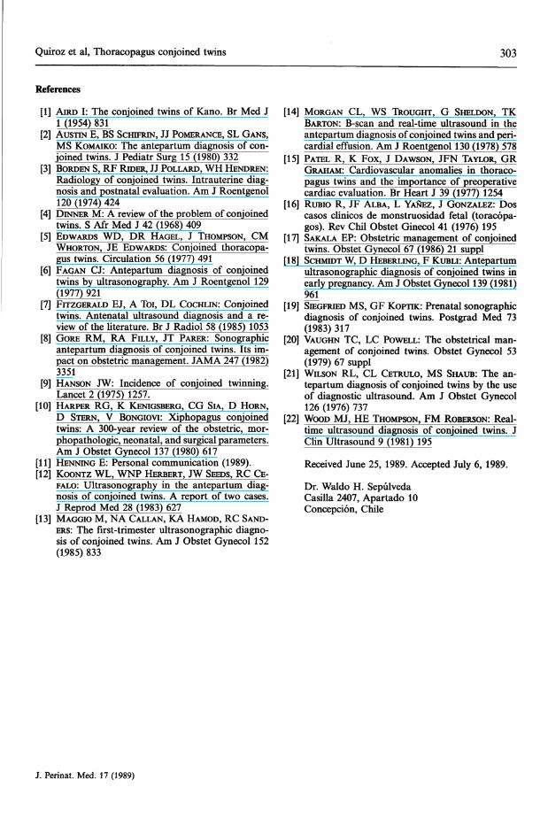
References
[1] AIRD I: The conjoined twins of Kano. Br Med J1 (1954) 831
[2] AUSTIN E, BS SCHIFRIN, JJ POMERANCE, SL GANS,MS KOMAIKO: The antepartum diagnosis of con-joined twins. J Pediatr Surg 15 (1980) 332
[3] BORDEN S, RF RIDER, JJ POLLARD, WH HENDREN:Radiology of conjoined twins. Intrauterine diag-nosis and postnatal evaluation. Am J Roentgenol120 (1974) 424
[4] DINNER M: A review of the problem of conjoinedtwins. S Afr Med J 42 (1968) 409
[5] EDWARDS WD, DR HAGEL, J THOMPSON, CMWHORTON, JE EDWARDS: Conjoined thoracopa-gus twins. Circulation 56 (1977) 491
[6] FAGAN CJ: Antepartum diagnosis of conjoinedtwins by ultrasonography. Am J Roentgenol 129(1977) 921
[7] FITZGERALD EJ, A Toi, DL COCHLIN: Conjoinedtwins. Antenatal ultrasound diagnosis and a re-view of the literature. Br J Radiol 58 (1985) 1053
[8] GORE RM, RA FILLY, JT PARER: Sonographicantepartum diagnosis of conjoined twins. Its im-pact on obstetric management. JAMA 247 (1982)3351
[9] HANSON JW: Incidence of conjoined twinning.Lancet 2 (1975) 1257.
[10] HARPER RG, K K NIGSBERG, CG SIA, D HORN,D STERN, V BONGIOVI: Xiphopagus conjoinedtwins: A 300-year review of the obstetric, mor-phopathologic, neonatal, and surgical parameters.Am J Obstet Gynecol 137 (1980) 617
[11] HENNING E: Personal communication (1989).[12] KOONTZ WL, WNP HERBERT, JW SEEDS, RC CE-
FALO: Ultrasonography in the antepartum diag-nosis of conjoined twins. A report of two cases.J Reprod Med 28 (1983) 627
[13] MAGGIO M, NA CALLAN, KA HAMOD, RC SAND-ERS: The first-trimester ultrasonographic diagno-sis of conjoined twins. Am J Obstet Gynecol 152(1985) 833
[14] MORGAN CL, WS THOUGHT, G SHELDON, TKBARTON: B-scan and real-time ultrasound in theantepartum diagnosis of conjoined twins and peri-cardial effusion. Am J Roentgenol 130 (1978) 578
[15] PATEL R, K Fox, J DAWSON, JFN TAYLOR, GRGRAHAM: Cardiovascular anomalies in thoraco-pagus twins and the importance of preoperativecardiac evaluation. Br Heart J 39 (1977) 1254
[16] RUBIO R, JF ALBA, L YANEZ, J GONZALEZ: Doscasos clinicos de monstruosidad fetal (toracopa-gos). Rev Chil Obstet Ginecol 41 (1976) 195
[17] SAKALA EP: Obstetric management of conjoinedtwins. Obstet Gynecol 67 (1986) 21 suppl
[18] SCHMIDT W, D HEBERLING, F KUBLI: Antepartumultrasonographic diagnosis of conjoined twins inearly pregnancy. Am J Obstet Gynecol 139 (1981)961
[19] SIEGFRIED MS, GF ΚΟΡΊΤΚ: Prenatal sonographicdiagnosis of conjoined twins. Postgrad Med 73(1983) 317
[20] VAUGHN TC, LC POWELL: The obstetrical man-agement of conjoined twins. Obstet Gynecol 53(1979) 67 suppl
[21] WILSON RL, CL CETRULO, MS SHAUB: The an-tepartum diagnosis of conjoined twins by the useof diagnostic ultrasound. Am J Obstet Gynecol126 (1976) 737
[22] WOOD MJ, HE THOMPSON, FM ROBERSON: Real-time ultrasound diagnosis of conjoined twins. JClin Ultrasound 9 (1981) 195
Received June 25, 1989. Accepted July 6, 1989.
Dr. Waldo H. SepulvedaCasilla 2407, Apartado 10Concepcion, Chile
J. Perinat. Med. 17 (1989)
Trace Element Analytical Chemistryin Medicine and Biology · Volume 5Proceedings of the Fifth International WorkshopNeuherberg, Federal Republic of Germany, April 1988Editors Peter Brätter - Peter Schramel1988.17 cm 24 cm. XVII, 666 pages. Numerous illustrations. Hardcover.DM 330,-; approx. US$188.00 ISBN 311 0113406
The subject matter of this volume is oriented towards the state of the art of traceelement analytical techniques. Invited experts presented papers reporting on neweraspects of analytical methods, recent developments in preanalytical treatment ofbiological samples and the combination of analytical methods for speciation analy-sis. Aluminium and plantinum were elements of special interest. Attention was alsopaid to the trace element levels in body fluids and tissues as well as to the role oftrace elements in metabolic processes and in human nutrition.
Contents (Main Chapters)Newer Aspects of Analytical Methods - Preanalytical Steps: Sampling and SampleTreatment · Speciation Analysis - Trace Element Levels (and Reference Data) inBody Fluids and Tissues · Special Elements: Aluminium and Platinum · Nutrition andFood Stuffs · Trace Element Analysis in Diagnosis and Pathological States · TraceElement Metabolism - Subject Index · Author Index
Also available
Trace Element Analytical Chemistry in Medicine and BiologyVolume 1:1980, XV, 851 pages. DM 180,-; approx. US$ 100.00Volume 2:1983, XX, 1189 pages. DM 280,-; approx. US$ 160.00Volume 3:1984, XVI, 763 pages. DM 240,-; approx. US$ 140.00Volume 4:1987, XVI, 761 pages. DM 295,-; approx. US$ 170.00
Prices are subject to change without notice
WD E
Gde Cruyter · Berlin · New York