The future of genetic diagnosis – from research to clinical ...
Upload
khangminh22Category
view
0download
0

LIBRARY OF CONGRESS,
UNITED STATES OF AMERICA.


CLINICAL DIAGNOSIS
BY
1/
ALBERT ABRAMS, M. D. o^™.Professor of Pathology, Cooper Medical College, San Francisco, Cal. ; Pathologist
to The City and County Hospital, San Francisco ; Author of " A Synopsis
ofMorbid Renal Secretions" etc. ; President of The San Francisco
Medico Chirurgical Society (1893-1894) ; President of The
Alumni Association of Cooper Medical College
(1888-1889)
THIRD EDITION, REVISED AND ENLARGED
ILLUSTRATED
pMAR 6 1894
Zlcr v^svC
fiytt 7jNew York
E. B. TREAT, 5 COOPER UNION1894
PRICE, $2.75

Copyright,
By ALBERT ABRAMS, M. D.,
1890, 1892, 1894.

TO HIS ESTEEMED FRIEND
JOSEPH 0. HIRSCHFELDER, M. D.,
PROFESSOR OF CLINICAL MEDICINE, COOPER MEDICAL COLLEGE,
THIS HUMBLE VOLUME IS DEDICATED, TO EXPRESS FOR
ACTS OF KINDNESS, THE OBLIGATIONS
AND GRATITUDE OF
—THE AUTHOR.

PREFACE TO THE THIRD EDITION.
rjlHE favorable reception accorded to the previous editions, has
induced the author to undertake the revision and enlargement
of this work by the addition of many synoptic tables, a chapter on
insanity, and a summary of recent methods of diagnosis. The
original character of the work has been preserved as an index to
or as an abstract of more pretentious works on the subject. " I have
gathered a posie of other men's flowers, and nothing but the thread
that binds them is mine own."
ALBERT ABRAMS, M.D.
Cooper Medical College, \
San Francisco, Jan. 12, 1894. »


CONTENTS
CHAPTER I.
PAQX.
Examination of Medical Cases— diathesis— position of the
body — expression of countenance— examination of the
skin— perspiration— oedema of the skin— emphysemaof the skin—temperature of the skin 1- 9
CHAPTER II.
Temperature— fever— temperature and symptoms of the
acute infectious diseases 10- 20
CHAPTER III.
Examination of Respiratory System— examination of the
nose and larynx— diseases of the larynx..... 21- 27
CHAPTER IV.
Examination of the Thorax— inspection— palpation—men-suration— percussion—auscultation — succussion—asso-
ciation of the physical signs of the lungs— physical
diagnosis of respiratory diseases — phonograph in
medicine 28- 59
CHAPTER V.
Cough— Sputum— the sputum in diseases of the respiratory
apparatus 60- 68(lx)
X CONTENTS.
CHAPTER VI.FAOB.
Examination op the Heart— inspection and palpation —table of thrills— percussion— auscultation—endocardial
murmurs— diagnosis of cardiac diseases G9- 84
CHAPTER VII.
Examination op the Arteries and Veins— thoracic aneu-
rism 85-
CHAPTER VIII.
The Pulse— sphygmography 90- 94
CHAPTER IX.
The Blood— microscopic examination of the blood— diseases
of the blood 95- 102
CHAPTER X.
The Digestive System— examination of the oesophagus—examination of the stomach— chemical analysis of the
stomach— diseases of the stomach — vomiting— exam-ination of the intestines— examination of the peritoneum— differential diagnosis of ascites and cysts of the ovary— examination of the faeces— examination of the liver
—
examination of the pancreas, omentum and retroperito-
neal glands— examination of the spleen 103- 132
CHAPTER XL
Examination of the Genito-Urinary Organs— examination
of the kidneys— differential diagnosis of diffused diseases
of the kidneys— diagnosis of diseases of the bladder
—
urinary calculi— pathological concrements— the urine
—
secretions of the male generative organs— a synopsis of
morbid urine 133 - 159

CONTENTS. xi
CHAPTER XII.PAGE.
The Nervous System— anatomy and physiology of the brain
and spinal cord — paralysis of the cranial nerves —paralysis of the spinal nerves— testing the sensibility—motor symptoms of irritation — reflexes — examination
of the nerves and muscles by electricity— instantaneous
diagnosis of nervous diseases — medical ophthalmology—160- 183
CHAPTEE XIII.
Parasites— animal parasites— vegetable parasites— bacteria
oo. .184- 194
APPENDIX.
The Nervous System— examination of the mind— diagnosis
of insanity— relation of diseases and functions to insanity
— topography of the brain— spinal localization— diagnosis
of nervous diseases, synoptic table of paralyses— diagnosis
of vaso-motor and trophic diseases, and diseases of muscles
s 197-222
Diagnosis of Diseases of the Skin— drug exanthemata—..223 - 228
Bacteriological Diagnosis— bacteriological analects 229 - 234
Diseases of the Intestines and Peritoneum — analectic re-
view of gastric digestion and gastric neuroses— relation of
diseases of the stomach to other diseases 235 - 243
The Employment of Drugs in Diagnosis 243- 247
Recent Methods of Diagnosis. . . 247- 254


ILLUSTRATIONS
no. FA«X1. Temperature chart in variola • • • . . 15
2. Temperature chart in typhus abdominalis . . . 16
3. Temperature chart in febris intermittens . . 18
4. Temperature chart in croupous pneumonia . • , 18
6. Paralysis of the vocal cords 24
6. Topography of the chest (anterior view) . . . 29
7. Topography of the chest (posterior view) • • 30
8. Microscopy of the sputum .... . 63
9. Normal pulse traciug 92
10. Pathological sphygmographic tracings . • . , 93
11. Microscopical examination of the blood ... 97
12. Microscopical examination of the vomit • • . 116
13. Microscopy of the stool 122
14. Crystals of the urine . 145
15. Casts of the urine 157
16. Side view of the human brain 160
17. Vertico-transverse section of the cerebrum . . 162
18. Transverse section of the spinal cord . . . . 163
19.~-23. Motor points ••••;• 17(1—178
24. Taenia solium . 185
25. Taenia saginata 185
26. Eggs of the oxyuris and ascaris lumbricoides • , 185
27. Muscle trichina 185
28. Bacteria •••••••••<> 194


CHAPTER L
EXAMINATION OF MEDICAL CASES.
In investigating a medical case the following or a
similar plan should be pursued, the object being to makea thorough examination, which is facilitated by a definite
procedure.
Name. Age. Occupation. Date of Examination.Patient's history {anamnesis). 1. Family history. 2. Diseases
of childhood. 3. Previous diseases. 4. Mode of Life. 5. PresentSickness (date, mode of invasion, etc).
Objective Examination (Status praesens).
General symptoms.1. Constitution (bone, muscular and fat development).
2 Position iin bed—mode of lying;
L. Position-j Q?t o{ bed—movements.
3. Expression of countenance.4. Skin (color, perspiration, ozdema, etc)*
5. Temperature.6. Respiration (frequency, type, etc).
7. Pulse (frequency, rythm, etc).
8. Tongue.
Then follows the examination of special regions, beginningwith the one presumably involved.
Respiratory System.—1. Nose. 2. Larynx. 3. Rythm of respi-ration. 4. Form of thorax. 5. Type of respiration. 6. Inspectionof thorax. 7. Palpation. 8. Percussion. 9. Auscultation.10. Cough and expectoration.
Circulatory System.—1. Examination of the heart and largearteries (beginning with inspection and following with the otherphysical signs). 2. Examination of the veins. 3. Examinationof the blood.
Digestive System.—1. Lips. 2. Tongue. 3. Mouth. 4. (Esoph-agus. 5. Stomach. 6. Intestines. 7. Peritoneum. 8. Liver.9. Spleen. 10. Pancreas. 11. Examination of the stomachcontents and faeces.
Genito-Urinary System.— 1. Examination of the kidneys, uret-and bladder. 2. Examination of the urine (to be made in all
(l)
2 MANUAL OF CLINICAL DIAGNOSIS.
cases) ;quantity, specific gravity, reaction, color, odor, sediment;
quantity and character by microscopical examination ; albumenand sugar.
Nervous System.—1. Examination of the head. 2. Vertebralcolumn. 3. Sensibility. 4. Motility. 5. Examination by meansof electricity. 6. Reflexes. 7. Organs of special sense.
Diagnosis. Treatment. Event.
Diathesis. In the examination of patients, the
physician often generalizes the symptoms in certain
cases, showing that the patient has some peculiar andspecific constitutional morbid tendency. The following
diatheses are usually referred to :
Phthisical Diathesis.—Clubbing of the finger ends, undue curva-
ture of the nails, red line on the margin of gums, momentaryelevation of the skin in percussing the thorax (myoidema), flush-
ing of the face (hectic flush) paralytic thorax and emaciation.
Apoplectic Diathesis.—Red and congested face, short neck,rigidity of the arteries and obesity.
Gouty Diathesis.—Obesity, varicose veins, oppression in breath-ing, deformities of the joints with local deposits of uric acid andatheroma of the arteries.
Position of the Body (decubitus).—Patients withacute unilateral affections of the chest (pneumonia,pleuritis, pneumothorax) usually lie on the affected side to
avoid pain during respiration and allow unaffected, to
compensate the disturbed functions of affected lung.
Patients with pneumonia occasionally lie on the unaf-
fected side in order to avoid pain. The prone position
may be adopted in certain cases of gastric ulcer. Thehead is thrown back in laryngeal and tracheal disease.
In diseases of the heart and lungs when dyspnoeais intense, the sitting posture is adopted (orthopncea).
Restlessness (jactitation) occurs during the invasion of
acute disease, in delirium and acute mania. In menin-gitis the head is drawn backward. In affections of thecerebellum the entire body is often drawn to one side. Incircumscribed affections of the cerebrum the head may bedrawn to one side. If patient is about, the gait is animportant sign in diagnosis, especially in affections ofthe nervous system.Gait in Hemiplegia (paralysis of one lateral half of body).—The
arm hangs by affected side and the shoulder droops. At each

EXAMIXATION OF MEDICAL OASES. 3
step the paralyzed half is lifted to swing weak leg forward. Thereis a swinging semi-circular movement of paralyzed foot (sickle-
walk). The shoe is worn off at the outer part of sole and toe.
Gait in Paraplegia (paralysis of lower half of body).—Patientshuffles along without raising either foot from the ground (hoppinggait).
Gait of Lateral Sclerosis (combination of paresis, muscularrigidit)T and tremor).—Feet are turned inward and appear glued to
the ground. They cross in walking and the knees are liable tobecome locked together. Weight is thrown first on one cane andthen on the other, to lift body so as to move the feet.
Gait of Paralysis Agitans—Tottering and trembling with atendency to trot. Head bent forward and held stiffly whenwalking.
Gait of Pseudo-hypertrophic paralysis.—Patients are children.Gait like the waddling of a duck. In the erect posture theback is excessively curved (disappears when the patient sits), sothat a vertical line dropped from the shoulders falls behind thesacrum. When the patients are placed on the floor great diffi-
culty is experienced in rising, which may however be accom-plished by a characteristic movement known as climbing upthe thighs.
Gait of Tabes Dorsalis.—Legs are flung about in an uncertainway, although the steps are characterized by deliberation. Thefeet are brought down with the heel projecting {flopping gait).
Eyes are kept on the ground wThile walking. Gait due to muscu-lar incoordination.
Gait of Cerebellar Disease.—Likewise due to muscular incoor-dination. It is staggering like an intoxicated person.
Gait of Cerebro-Spinal Sclerosis.—Unsteady and irregular. Nodeliberate walking in a straight line, but patient snoots suddenlyforward or to one side (propulsion).
Gait of Hysterical Hemiplegia.—Leg drawn along sweeping theground as if lifeless. It is not swung around describing the arcof a circle as in ordinary hemiplegia.Hemiplegia almost always on the left side, and is developed
and passes away suddenly.
Expression of Countenance.—In Fades Hippo-cratica, the nose is sharp, eyes hollow, temples collapsed; the
ears cold, skin about forehead rough, distended and parched ;
the color of the whole face being green, black, livid or lead
colored. Described in the words of Hippocrates, this is thephysiognomy of approaching death. The same facial
expression occurs in acute collapse. In typhoid fever theexpression is dull (Fades slupida). In pneumonia theface has a dusky flush, in circulatory disturbances a bluish

4 MANUAL OF CLINICAL DIAGNOSIS.
hue. In cerebral congestion the face is full and red ; inacute peritonitis the countenance is pinched. Puffiness ofthe eyelids may be expressive of BrighVs Disease.
Examination of the Skin.—1. Changes in color.
2. Perspiration. 3. (Edema. 4. Emphysema. 5.
Changes in temperature. Changes in color : Pale, red,
cyanotic, icteric, bronze skin, gray skin. Pale color.
Physiological in those not exposed to the air. Tempo-rary in fear and syncope. Occurs rapidly in profuse
haemorrhage and in collapse. Often symptomatic of aweakened heart. In diseases of the blood and haemato-poietic organs : Chlorosis, pernicious anaemia, leucaemia andpseudo-leucaemia. Also present in malarial cachexia,
phthisis, carcinoma and chronic intoxication from mer-cury and lead. In a number of these conditions not
only the pale hue but the color of the skin is character-
istic, e.g. In severe anaemia : waxy lustre and yellowish;
Large white kidney : light wtrte skin ; Tuberculosis andlead intoxication : grayish white ; Chlorosis : greenish
;
Diseases of the heart : dirty yellow ; Cachexia of Carcin-
oma : grayish yellow. Red color may be caused by
:
1. Active dilatation of cutaneous blood vessels. 2.
Increase in the quantity of blood. 3. Increase in the
blood coloring matter. Universal skin redness occurs in
fever, scarlatina and atropine poisoning.
Local redness occurs most frequently in the face.
Blushing on one side of the face occurs in the paralytic
form of migraine. Hectic flush in tuberculosis. Anaemiaattended with irritability of the heart may render face
intensely red.
Cyanosis. Lividity or duskiness of the skin especially
marked in the face, lips and finger nails. It occurs
when the blood contains too little oxygen and is sur-
charged with carbonic acid.
Increase of carbonic acid in the blood occurs in : 1. Disturbancesinterfering with the interchange of gases in the lungs; 2. Slowingof the blood current in the capillaries. To the first belong
:
a. All conditions leading to a narrowing of the air passages.Examples: Obstruction of larynx, compression of trachea anddiffuse bronchitis.

EXAMINATION OF MEDICAL CASES. 6
b. Diseases of the lungs and surroundings. Examples; Emphy-sema, pleuritic exudation, and abdominal affections interferingwith the movements of the diaphragm.
c. Paralyses, spasms and diseases of the respiratory muscles.Examples: Bulbar paralysis, tetanus and progressive muscularatrophy. Slowing of the blood current occurs in general venousstasis, when from any cause the right ventricle of the heart is
weakened, or when the large veins are compressed, just beforetheir entrance into the Tight auiicle (tumors of mediastinum).Cyanosis is pathognomonic of miliary tuberculosis. Its occurrencein pneumonia is an ominous sign. Local cyanosis in the face maybe due to extreme cold or occurring in other situations to venousstasis, the result <»f thrombosis or compression of the veins. Theterm morbus cxruleus is applied to extreme lividity, occurring incongenital malformations of the heart.
In peritonitis, pleuritis and inflammatory affections of therespiratory muscles, the insufficient respirations on account ofpain lead to dyspnoea and cyanosis. The character of the formerand latter may be correctly diagnosed if they disappear after theadministration of narcotics.
Icterus.—A yellow discoloration of the skin occurring
over the entire body. The discoloration is first manifestin the conjunctiva sclerx and other mucous membranes,and in the skin, where the epidermis is thin.
An object glass, applied with slight pressure to the lips, will
render the latter anaemic, and through the glass the yellow colorwill become more evident. Color varies according to the intensityof the icterus. It is light yellow in mild, and green, or brownish-green in severe forms of the affection {icterus viridis and melasicte"
rus.) Icterus cannot be detected by gas or lamp light. In exam-ining the conjunctiva the presence of subconjunctival fat mustnot be mistaken for icterus. Yellow discoloration of the skin maydevelop after the use of santonin or picric acid and its salts. Theyellow color of the skin in the, beginning of icterus is produced bythe biliary pigments in the blood plasma. Later the cells of therete malpighii imbibe the pigment, which fact accounts for thecontinuance of the icteric coloration after the immediate cause is
removed. The symptoms peculiar to icterus are : Itching of theskin, minute cutaneous haemorrhages, slowing of the pulse (dueto toxic paresis of the cardiac ganglia), yellow vision (xanthopsia)and nervous symptoms (delirium, coma, convulsions, etc.), due tocholxmia.
Hepatogenic Icterus (icterus of resorption). Usuallyresults from bile stasis, i. e., any interference with theentrance of bile into the intestines. Causes: Gastro-duo-
denal catarrh, involving ductus choledochus, compressionof the latter by tumors, or the presence in the same of

6 MANUAL OF CLINICAL DIAGNOSIS.
ascarides, gall stones, etc.; compression of the hepatic
duct, closure of a number of small biliary ducts by intra-
hepatic gall stones, and, finally, enlargement of the ven-
ules of the liver.
The bile is secreted under very low pressure, and even trivial
obstructions suffice to prevent its passage into the intestines.
After ligating the ductus choledochus in animals, sixty to seventy
hours eiapse before the conjunctivae become colored. The dia-
phragm in contracting subjects the liver to pressure, and is anactive factor in forcing the bile from the smaller to the larger bili-
ary ducts. Interference with the movements of the diaphragm[pleuritis diaphragmatica dextra) is likely to cause icterus of resorp-
tion. The immediate cause of icterus is the accumulation in theolood of biliary pigments.
Hematogenic Icterus.—Caused by the destruction of
A«fed blood corpuscles. Causes: Acute infectious diseases,
particularly pyaemia, yellow fever and pneumonia ; after
the use of chloroform, ether, chloral, chlorate of potash,
etc.
In hsematogenic icterus the bile freely enters the intestines, so
that the stools are not discolored, whereas in hepatogenic icterus,
the motions being usually free from bile, present a paler appear-
ance than natural (clayey stool). In this form of icterus constipa-
tion is present, and the fseces are highly offensive. The bile acts
normally, both as a purgative and an antiseptic. Examination of
the urine is of great importance in differentiating both forms of
icterus. (See urine.)
WeiVs Disease, also called infectious and epidemic icterus, is anaffection characterized by a remittent type of fever, ending bylysis, headache, vertigo and prostration. The pulse is frequent,
nausea and vomiting occur in half the cases, and the spleen andliver are enlarged. Icterus is always present. Albuminuria is
present in about half the cases. Morbus Weilii may occur spor-
adically or epidemically.
Icterus Neonatorum occurs at birth and is an example of howsudden diminution of pressure in the portal vem will producehepatogenic icterus. Normal pressure in the branches of theportal vein is greater than in the neighboring bile ducts ; hence adiminution of pressure in the former conduces to bile stasis.
Bronzed Shin (Addison's disease).—Addison, in 1855,
called attention to a bronze discoloration of the skin,
associated with disease of the supra-renal capsules. This
affection is characterized by a gray, brown, or even black-
ish discoloration of the skin, beginning in parts exposed
to the air (face and hands), and then involving parts nor-

EXAMINATION OF MEDICAL CASES. 7
mally pigmented, or diffusing itself over the entire body.The nails and conjunctivae remain unaffected. Circum-scribed pigmented spots may develop on the mucous mem-brane of the lips. The cause of the Bkin discoloration is
a deposit of granular pigment in the cl-IIs of the rete mal-pighii. The constitutional symptoms associated with this
disease are: Great feebleness of the muscles and heart,
pains in the back and vomiting. It is commonest in
young male adults, and is often complicated with phthisis
or disease of the vertebrae.
Grayish Discoloration of the Skin (Argyria).—The long
continued use of nitrate of silver conduces to a deposit of
black granules (metallic silver) in the skin. The skin,
especially of parts exposed to the light (face and hands),
shows the most pronounced discoloration. A similar
deposit in the viscera causes the latter to become darkcolored. Argyria has been observed to follow even the
long continued local application of lunar caustic.
Perspiration.— An increase of the sudoriparoussecretion is called hyperidrosis, a diminution hyphidrosis,
and an absence of sweat, anidrosis.
Hyperidrosis may be local (hyperidrosis localis), confined to oneside of the body ( liemidrosis) or diffused (hyperidrosis universalis).
Universal hyperidrosis is observed in febrile diseases, tetanus,fever, pain, dyspnoea, collapse, and after the use of diaphoreticsand opium. Acute articular rheumatism is characterized by profusediaphoresis. In phthisis {night sweats) increased perspiration is
frequent. In febrile diseases a fall of temperature is attendedwith sweating. Local sweating occurs in various neuroses andanatomical lesions of the nervous system. Anidrosis is encoun-tered in high continuous fever, and in affections attended by alarge loss of water (diabetes, cholera and contracted kidney).
Qualitative Change of the Sweat.—Colored sweat (chromidrosis)is observed in icteru3 (yellow sweat). Blue, green and black sweatare said to have been observed. Bloody sweat (hcematidrosis) is
really caused by extravasations of blood from the cutaneous blood-vessels. In retarded urinary excretion urea may be excreted bythe sweat (uridrosis), in the form of glistening white scales, whichgive the reactions of urea.
(Edema of the Skin.—From the capillaries andvenules of the skin and subcutaneous tissue a continuoustransudation of fluid takes place, which, after subserving

8 MANUAL OF CLINICAL DIAGNOSIS.
the purposes of nutrition is taken up by the lymphaticsand again conveyed to the blood. An abnormal accumu-lation of this fluid in the substance of the tissues is called
oedema. When the fluid collects in the greater cavities of
the body we have hydrops or dropsy. It is usual to
describe oedema of the integumentary structures as anas-
arca. If the effusion of liquid is general throughout the
body, we speak of general dropsy, if limited to the peri-
toneal cavity, it is called ascites.
In pronounced oedema other tissues, especially the muscles,contain fluid. (Edematous parts are increased in size, the skinis pale {pressure of the fluid on the blood-vessels), smooth, shiny,tense and possessed of a certain transparency. Pressure with thefinder on cedematons parts leaves a depression (pathognomonic)called pitting, which soon disappears. When oedema first
appears in the feet (malleoli ), some interference with blood pres-sure may be assumed, if on the contrary it is first manifested inthe eyelids, some constitutional cause (nephritis) is probable.(Edema is caused by one of the following conditions : 1. Venous-stasis (mechanical hydrops). 2. Altered or watery condition of
the blood (hydremia). 3. Inflammation. When oedema is asso-ciated with cyanosis and dyspnoea it i-^ usually symptomatic of anon-compensated cardiac lesion. (Edema in venous stasis
results from the distended veins, filled to repletion, being unableto take up the fluid normally transuded.
(Edema with Albuminuria.—A combination of these
symptoms is called after Richard Blight, BrighVs Dis-
ease. (Edema in albuminuria is explained as follows :
The endothelial cells of the small vessels do not nor-
mally allow of the passage of any large quantity of
plasma. If the nutrition of the endothelium is in anyway impoverished as occurs in nephrit s owing to the
retention in the blood of excretory products, then theblood-vessels become permeable and oedema results.
(Edema with Cachexia.— (Edema without dyspnoea,cyanosis or albuminuria, is usually associated with avitiated state of the body as occurs in anaemia, phthisis,
carcinoma, etc. This oedema is also explained by nutri-
tive disturbances of the blood-vessels.
(Edema with Inflammation.—Usually local and often
characteristic of deep-seated accumulations of pus. (Edemaof one side of the chest is frequently present when thefluid in the pleural cavity is purulent. This oedema is

EXAMINATION OF MEDICAL CASES. 9
also called collateral oedema and results from increased
pressure in the capillaries surrounding the area of
inflammation.
Emphysema of the Skin.—This signifies the pres-
ence of air in the sub-cutaneous connective tissue. It is
characterized by abnormal distension of the skin in
certain regions and crepitation is felt and heard onpalpation. Pitting is obtained on pressure over the
emphysematous parts, but unlike oedema it disappears
rapidly. Percussion yields a tympanitic sound.
Two forms of cutaneous emphysema are differentiated, spon-taneous and aspira'ed. The spontaneous form (rare) is presentwhen gas develop* from tub cutaneous extravasations of blood orabscesses. As pi* a ted emphysema occurs whenever air or gasenters the sub-cutaneous tissue either from without or within.As examples of the former: wounds of the neck or chest. Asexamples of origin from without: abnormal communications withthe subcutaneous tissue from any part of the respiratory oralimentary tract.
Temperature of the Skin.—With the hand applied
to the skin of a patient, the body temperature can beapproximately determined. Palpation of the skin is of
value in localizing pathological processes, etc.—Seethermo-palpation.

CHAPTER IL
TEMPERATURE.
Method of Examination—The temperature of the "body may hetaken in the axillary space, rectum, vagina or mouth. The ther-
mometer (self-registering) must be very sensitive, compared witha standard one and verified. It should be divided so as to exhibitclearly, fifths of a degree. The vagina or rectum is preferred asrepresenting more nearly the body temperature. When for
reasons of delicacy the axillary space is selected, the patientshould lie diagonally on the right or left side, his arm firmlycompressing the thermometer, whichremains in position for atleast ten minutes. When the rectum is selected five minutes willsuffice. In the rectum, temperature is about 1° F. higher than inthe axilla. Before introducing the thermometer into the rectumthe bulb is oiled. On the continent of Europe temperature is
measured with the scale of Celsius, also called centigrade (freezingpoint 0°, boiling, 100°) whereas in England and in the United Statesthe Fahrenheit scale is employed (freezing 32°, boiling, 212°).
The scale of centigrade is reduced to that of Fahrenheit bymultiplying by 9 and dividing by 5 ; that of Reaumur (Kussia anil
Sweden) to that of Fahrenheit by multiplying by 9 and divid-ing by 4; and when above zero in either case add 32. Fahren-heit is reduced to either of the preceding by reversing theprocess. C 100° x9= 900-f 5= 180 + 32=212°F. ; It. 80° x 9= 720
+
4= 180°+ 32= 212°F. The following formula is also employed
:
N°C.=f n° R.=f n°+32*F.
o. R. F.
36* 28.5* 96.8*37* 29.
6°98 6*
38* 30.4° 100.4*39* 31.2° 102.2*40° 32.0* 104.0*41° 32.8° 105.8*
Normal Temperature.—Tn the axilla this variesbetween 86.2° C. (97.1° F.) and 37.5° C. (99.5° F.). Thetemperature is highest (daily maximum) in the eveningbetween five and eight, and lowest {daily minimum) in
(10)
PEVER. 11
morning between two and six o'clock. The difference
between minimum and maximum is about 1° C. (in rare
instances 2° C). Slight temporary elevations of tempera-
ture occur after a full meal (fever of digestion), physical
exertion and hot baths. A continuous elevation of tem-perature occurs in fever.
Subnormal Temperature.—This is observed in
febrile conditions at the crisis and the normal tempera-ture is again attained after one, two or three days.
Observed in collapse it is accompanied by diminished
cardiac activity, increased pulse frequency, paleness of
the skin and general weakness. It is further observedafter haemorrhages and in the course of chronic, cardiac
and pulmonary affections. Permanent subnormal tempera-
ture (rare) may be encountered in wasting diseases andinsane patients.
FEVER.
Fever is not only characterized by a continuous eleva-
tion of temperature, but by a symptom complex, the
result of increased tissue metamorphosis and functional
disturbances of all the organs. Fever may be experi-
mentally produced by the introduction of septic or aseptic
matter into the circulation. In infectious diseases the
febrile temperature is believed to be caused by the direct
action of micro-organisms upon the nerve centres, or bythe action of a poison which they develop within the body.It is now believed by many that elevation of temperatureattending acute infections is salutary. The growth of the
tubercle bacillus ceases at a temperature above 41° C.
(105.8° F.), and the spirilla of relapsing fever disappearat the close of each paroxysm when the temperaturereaches 42° C. (107.6° F.). The lesions peculiar to pro-
longed fever are distributed among the viscera and consist
of granular fatty degenerations of the cellular elements.
Symptoms of Fever.—Usually the pulse rises syn-chronously with the temperature, and averages an increase
of ten beats for every degree above 98° F.

12 MANUAL OF CLINICAL DIAGNOSIS.
When the pulse frequency is more than 160 per minute in fever
the prognosis is bad. The respirations in fever are increased, there
is loefl of appetite, increased thirst, digestive disturbances, and
diminished secretion of a highly colored urine, with an increase of
the solid constituents. The pulse is very slow in meningitis and
rapid in scarlatina; in uncomplicated typhoid, pulse frequency is
not usually more than 110. Herpes of the lips and nose absent in
typhoid and present in pneumonia and meningitis.
CLASSIFICATION OF TEMPERATURE. {Wunderlieh).
36° C. (96.8° F.) Temperature of collapse.
37 5° - 33° C. (99.5° - 100 4° F.) Sub-febrile temperature.
3S°-38.5° C. (100.4°- 101.3° F.) Slight fever.
39° C. (102.2° F.) morning; 39.5° 0. (103.1° F.) evening.
Moderate fever.
39.5° 0. (103 1° F.) morning; 40.5° C. (104.9° F.) evening.
Considerable fever.
Over 39.5° 0. (10 1.1° F.) morning; Over 40.5° C. (104.9° F.)evening. High fever.
Over 41.5° C. (106.7° F.) Hyperpyrexia.
As the temperature rises above 40° C. (104° F.) the gravity of the
disease rapidly increases. In certain affections of the nervous
system (tetanus and hydrophobia) the temperature may reach 43°-
45° C. (110°-113° F.), and in a few cases this temperature has beenexceeded.
Daily Variation of Temperature in Fever.—Analogous to the daily variations in health there is usu-
ally in fever an increase of temperature in the evening
(exacerbation) and a fall in the morning (remission).
When this is reversed we have the typus inversus (usually
in phthisis).
Types of Fever.—Febris continua; when a daily dif-
ference of not more than 1° C. (1.8° F.) exists (tempera-
ture usuallv high). Febris remittens: a daily difference
of not more'than 1.5° C. (2.7° F.).
Febris Intermittens: The fever usually lasts only a fewhours fever paroxysm) whereas during the rest of the
day no fever is present (apyrexia).
Febris Recurrens: A continuous fever, lasting from 5 to
7 days, followed by apyrexia from 5 to 8 days; then tem-perature again rises and lasts from 5 to 7 days, ending bycrisis.

TEMPERATURE OF INFECTIOUS DISEASES. 13
Febrile Stages.— Stadium incrementi: the stage of
rising temperature. Fastigium: the stage of highest tem-
perature. Stadium decrementi: the stage when the tem-perature falls.
When the temperature falls quickly we speak of the fever as ter-
minating by crisis. When the fall is slow, occupying several days,the termination is by lysis. A high rise of temperature precedingcrisis is called perturbatio crilica. Crisis is usually accompaniedwith profuse perspiration and diminished pulse frequency. Febris
hectica is a form of the remittent type in which the exacerbationsare very high, whereas the remissions go below normal. Hecticfever is observed in purulent and septic conditions.
Stages of Exanthematous Diseases: 1. Incuba-
tion : the period from the exposure to the infection, to the
outbreak of the disease. 2. Prodromal stage: time fromthe beginning of the fever to the appearance of eruption.
3. Eruption. 4. Defervescence.
Character of the Fever.—Febris stupida : when the
patient is apathetic and very quiet. Febris versatilis:
slight delirium, twitching of the tendons of the wrist
(subsultus tendinum) and picking at the bed-clothes (floe-
citatio). These signs are very unfavorable. Fever is
further classified as dynamic or sthenic (full pulse, flushed
skin, active delirium) and adynamic or asthenic (pulse
feeble, pale skin, low delirium and great prostration.)
TEMPERATURE AND SYMPTOMS OF THEACUTE INFECTIOUS DISEASES.
MEASLES—MORBILLI.Incubation, 10 days. Prodromal stage, 3 days, attended
by running at the nose and eyes, sneezing and coughing.
Slight fall of temperature on the 2d or 3d day. Eruption(beginning on the face) on the 3d or 4th day, whentemperature reaches its highest point. Continuous fever
from the 4th to the 7th day. Crisis of the 7th day.
Desquamation, 14 days, with very annoying itching.
Eruption appears first on the face, then spreading to the
trunk, and from the trunk to the limbs. It consists of
elevated red patches, which tend to assume a circular
outline; between these patches the skin is of natural color.

U MANUAL OF CLINICAL DIAGNOSIS.
Complications.—Pneumon'a, bronchitis and pleuritis.
Sequelae.—Chronic cough, otorrhcea, enlarged lymphaticglands, etc.
SCARLET FEVER—SCARLATINA.Incubation, 2 to 4 days. Prodromal stage, 1 to 2 days,
commencing with a chill and rapid rise of temperature.Eruption on 2d day, with increased temperature. Fromthe 4th day on, temperature falls by lysis. Desquamationfrom 4 to 14 days. Eruption presents a bright uniformredness, similar to that of a boiled lobster. It appearsfirst on the thorax, abdomen, neck or back. In malignantforms of the disease eruption comes out late, and is either
indistinct or cWk and livid.
Diagnosis.—Tongue red and papillae prominent (straw-
berry tongue), angina, and very rapid pulse.
Complications.—Nephritis with dropsy (usually betweenthe tenth and twentieth day of disease), cerebral symp-toms (in children), diphtheria, oedema of glottis, etc.
Sequelae.—Boils, swelling of lymphatic glands, diarrhoea,
otitis, etc.
Mortality in epidemics, 50 to 60 per cent.; otherwise
about 15 per cent.
SMALL-POX—VARIOLA.Incubation, 10 to 13 days. Prodromal stage, 2 to
5 days, commencing with a chill and high fever.
Eruption on 2d or 3d day, with diminution of fever
lasting to the 9th day. From the 9th to the 11th daytemperature again rises (fever of suppuration), and is
remittent in type. Fever ends by lysis. Eruption in
most cases appears first on the neck and face, as red
papules, which feel like shot embedded under the skin.
After a day or two the papules becomes vesicular, thenpurulent. The pustules may run into one another in
grave cases (confluent small-pox) or remain isolated (dis-
crete small-pox). A depression in the centre of the pustule

TEMPERATURE OF INFECTIOUS DISEASES. 15
is present (umbilicatioii). Eruption may also affeat
mouth. In severe forms of the disease haemorrhages are
seen under the skin, as well as inside the pustules
(hemorrhagic small pox). During invasion severe lumbar
Fig. 1. Temperature Chart in Variola
<to,oiipislliiilll40,0
89,0
B|SBBimmmm88,0 !JLili!™ii!wmmmi87,0 HBiiiiiC6J)
105.8
104.0
102.2
100.4
98.6
96.8
2I*
o
pain is characteristic,
is about 4 per cent,
per cent.
The mortality in discrete variola
in confluent small-pox about 50
VARIOLOID.This is modified small-pox, occurring in a person' par-
tially protected by vaccination. Usually mild.Incubation and prodromal stages are the same as in
variola, although lighter. Fever of suppuration is absent,
Desquamation begins on the Cth or 10th day. Eruptionmay resemble that of variola, although it often consistsof only a few abortive papules, without vesication or pus-tulation.
CHICKEN-POX—VARICELLA.Prodromes usually absent. Fever begins with a chill
and lasts until drying of the exanthema (2 to 4 days).

16 MANUAL OF CLINICAL DIAGNOSIS.
Eruption is vesicular, preceded by red spots. This affeo-
tion is not prevented by vaccination.
TYPHUS FEVER.Incubation, 3 to 21 days. Prodromes absent. Begins
with a chill and rapid rise of temperature, which is con-
tinuous from 13 to 17 days, with slight remissions at the
end of the 1st week; and ends by crisis, with perturbatio
critica. Eruption appears from 4th to 7th day and looks
like that of measles. The spots (mulberry rash) are of a
dark tint and very numerous on the trunk and extremities
(rare upon the face). The mortality is about 25 per cent.
TYPHOID FEVER—TYPHUSABDOMINALIS.
Incubation, 7 to 21 days. Prodromal stage lasts about
a week, and is accompanied by a feeling of malaise. In
the 1st week temperature rises slowly, reaching its highest
point in from 4 to 7 days. Then febris continua until the
3d week in the mild and the 5th week in the severe forms.
Then, while the evening temperature is still high, the
morning temperature begins to fall and the fever ter-
minates by lysis, which, in mild cases, is about the 4th
week.Temperature chart In Typhus abdommaTIs,
Fig. 2.
Diagnosis.—Tumefaction of spleen, epistaxis, diarrhoea?
ileo-csecal tenderness and gurgling, distension of abdomen(tympanites') and nervous disturbances (headache, delir-

TEMPERATURE OF INFECTIOUS DISEASES. 17
ium, somnolence). Eruption (absent in 12 per cent, of
cases) appears about the 7th day or later,' and con-
sists of small red spots, similar to flea bites. The spots
are usually confined to the abdomen and chest, and dis-
appear on pressure. Later in the disease an eruption of
minute transparent vesicles (sudamina) may appear.
The mortality is about 18 per cent, in hospital and 10 to
12 per cent, in private practice.
RELAPSING FEVER—FEBRISRECURRENS.
Incubation, 5 to 7 days. Prodromal stage usually
absent. Begins with a chill and sudden rise of tempera-
ture. Continuous fever 5 to 7 days, and then termination
by crisis. Following crisis no fever (apyrexia) from 5 to
8 days ; after this the temperature again rises as at first,
but is of shorter duration. After a period of 7 days there
may be a third attack, lasting however from 2 to 3 daysonly. Diagnosis : Enlargement of the spleen, and the
presence in the blood of spirilla (see Blood). The mortality
in private practice is about 20 per cent.
MALARIA—FEBRIS INTERMITTENS.Incubation, 7 to 21 days. Prodromal stage not
marked. There is a chill followed by rapid rise of
temperature lasting but a few hours, and terminatingby crisis with profuse perspiration. The period betweenthe termination of one attack and the beginning of
another is called intermission or apyrexia. When the
fever recurs every day it is called quotidian ; everysecond day tertian and every third day quartan inter-
mittent fever. Two attacks of fever occurring on the
same day is spoken of as febris intermittens duplicata.
When the second attack of fever occurs at an earlier
M. C. D. 2

18 MANUAL OF CLINICAL DIAGNOSIS.
hour of the day than the first attack, we speak of febris
intermittens anteponens, when at a later hour, post-ponens.
Fig. 3. Temperature Chart InFebris Intermittens.
Fig. 4. Temperature Chart inCroupous Pneumonia.
Diagnosis.—Periodicity of the febrile attacks, enlarge-
ment of the spleen, specific action of quinine and the Plas-
modium malarias in the blood. When an intermittent
fever does not yield to quinine, endocarditis, latent tuber-
culosis or pus somewhere in the organism may be sus-
pected.
PNEUMONIA CROUPOSA.Begins with a severe chill and sudden elevation of
temperature. The fever is continuous and ends by crisis
on the 3d, 5th, 7th or 9th day. When the fever persists,
empyema or the termination of the pneumonia in abscess ofthe lung, gangrene or tuberculosis may be suspected.
Diagnosis.—Dullness, bronchial respiration, crepitantrales, rusty sputum and pneumococci. Mortality 8 to 20per cent. Pneumonias in drunkards are grave.
ERYSIPELAS.Incubation, 1 to 8 days. Begins with a chill and high
temperature. Inflammation of the skin on the 1st or

TEMPERATURE OF INFECTIOUS DISEASES. 19
2d day. Continuous fever during the time erysipelas
spreads. Temperature falls when spreading ceases.
Complications.—(Edema of the glottis, bronchitis,
pneumonia, endocarditis and cerebral erysipelas (extends
to brain from facial vein).
Acute Articular Rheumatism.—The tempera-ture remains steady after the symptoms develop, withevening exacerbations and morning remissions when the
joint affection is yielding, but rises when new joints
become involved.
Diagnosis.—Tumefaction, redness and tenderness of
the joints. Affection yields to salicylic acid, salol or
antipyrin.
Complications.—Pericarditis (usually between the
4th and 14th day), endocarditis (in about 20 per cent,
of the cases), bronchitis, pleuritis, etc.
Diphtheria.—Temperature is atypical and for theprognosis of little value.
Diagnosis.—A white or gravish exudation on the ton-
sils, uvula and soft palate. Mucous membrane of nose,
larynx or bronchi may be involved in severe cases.
Exudation which is not easily removed leaves a bleeding
raw surface which is soon covered with a new exudation,Submaxillary and cerv c:il glands enlarged and tender.
Weakness and prostration are prominent symptoms.
Sequelae.—Profound anaemia, nephritis and postdiphtheritic paralyses (about 2 to 3 weeks after recovery).
Prognosis.—Always grave. In a mild epidemic aver-
age mortality 5 per cent., in a severe epidemic 33 per
cent.
Acute Miliary Tuberculosis.—Temperature is
atypical, although the typus inversus is frequent.
Diagnosis.—Cyanosis, dyspnoea, crepitant rales withoutdullness on percussion and choroid tubercles seen withophthalmoscope.
Cerebro-spinal Meningitis.— Temperature maybe continuous or remittent, and is of long duration.

20 MANUAL OF CLINICAL DIAGNOSIS.
Diagnosis.—Somnolence, stiffness of the neck muscles,
vomiting, retracted abdomen, slow pulse, pupils usually-
contracted, eruption (absent in one-half the cases) con-
sists of petechial spots of extravasated blood.
Prognosis.—The mortality varying with the epidemic
is from 20 to 75 per cent. The disease is less fatal in
children than adults.
Syphilis.—Incubation (3 to 4 weeks). Primary stage;
chancre and swelling of neighboring lymph glands.
Secondary stage (9 to 11 weeks after exposure to infec-
tion) ; usually begins with a chill and fever (fever oferuption) of a remittent type. Tertiary stage ; develop-
ment of gummata.
Diagnosis of the Exanthema. Pain and itching nottroublesome, copper color, polymorphism (papules mac-ules, pustules, scales, etc. co-exist), tendency to circular
form of the patches and eruption shows a predilection for
certain parts ; around the forehead (corona veneris) palmof the hand, sole of the foot, etc.

CHAPTER III.
EXAMINATION OF THE RESPIRATORYSYSTEM.
EXAMINATION OF THE NOSE AND LARYNX.
THE NOSE.
Rhinoscopy. This is the art of inspecting the nasalcavities and naso-pmiryngeal space, it is divided into
anterior and posterior rhinoscopy. In anterior rhinoscopy
the view obtained even under favorable conditions is
limited, and usually comprises the anterior portions of
the lower and middle turbinated bones, with the cartilagin-
ous portion of the septum.
Posterior Rhinoscopy. The rhinoscopic image is madeup of the following : vomer or nasal septum, floor
of nose, superior meatus, middle meatus, superior turbi-
nated bone, middle turbinated bone, inferior turbinatedbone, pharyngeal orifice of Eustachian tube, upper por-
tion of Rosenmuller's groove, glandular tissue at vault ofpharynx and posterior surface of velum.The nasal cavities are in direct communication with other cavi-
ties situated in the bones of the skull ; these are the antra ofHigh-more, situated in the body of the superior maxillary bone and com-municating with the nasal cavities by an opening in the middlemeatus \ frontal sinuses situated between the two tables of the frontalbone with an opening in middle meatus, and finally the sphenoidalcells or sinuses, situated in the body of the sphenoid bone with smallopenings in the superior meatus. Examination of the nasal cavitiesis absolutely necessary in diagnosis as many neuroses owe theirorigin to nasal anomalies, this is notably the casein asthma wherethe paroxysms are found to be associated with nasal polypi andwith catarrh of the naso-pharyngeal mucous membrane. "Runningfrom the nose" is a symptom during the invasion of measles.Fetor from nose {pzxna) may be distinguished from fetor due to
(21)

22 MANUAL OF CLINICAL DIAGNOSIS.
lung gangrene, carious teeth, etc., by testing the breath while themouth and nostrils are closed alternately. Difficulty in breathingthrough the nose in infancy {snuffles) may be due to syphilis.
In bleeding from the nose (epistaxis), it must be rememberedthat the blood may be swallowed or accumulate in the throat andthus simulate hsematemesis or haemoptysis.
THE LARYNX.
Anatomy and Physiology. The larynx is situated between theupper border of the 3d, and lower border of the 6th, cervical
vertebra. Daring respiration, phonation and deglutition, it rises
and falls. In stenosis of the larynx the rise and fall are exaggerated.Widening of the vocal chink (abduction of vocal cords) is effected
by the posterior crico-arytenoid muscle which also turns theprocessus vocalis of the arytenoid cartilage outward. Closure ofthe vocal cords (adduction of vocal cords) is effected by thelateral crico-aryfenoid and inter- arytenoid (transverse and oblique)muscles. Tension of the vocal cords is maintained by the crico-
thyroid and thyro- arytenoid muscles, the actual muscles of the vocalcords. The nerve supply is from the vagus with motor branchesfrom the accessorius. The superior laryngeal nerve innerv it^s thecrico-thyroid muscle and muscles of epiglottis (motor fibres). It
also supplies the mucous membrane of the larynx (sensory fibres).
The inferior laryngeal nerve (recurrent laryngeal) supplies all theother muscles of the larynx not supplied by the superior laryngealnerve. This nerve curves backward around the subclavian arteryon the right side and around the arch of the aorta on the left sideand passes upward in the groove between the trachea andoesophagus, entering the larynx behind the articulation of theinferior cornu of the thyroid cartilage with the cricoid.
Laryngoscopy.—An examination with the laryn-
goscope reveals in a normal case the following structures :
1. Epiglottis. 2. Glosso-epiglottic ligaments which con-
nect tongue with epiglottis. 3. Ary-epiglottic folds withthe cartilages of Wrisberg. 4. Arytenoid cartilages,
cartilage of Santorini, sinus Morgagni. 5. True and false
vocal cords, the latter are parallel to and above the former.
The true vocal cords are divided into two parts, theanterior part (ligamentous) extends to the apex of theprocessus vocalis, the posterior part, from the apex to
the base of same. The anterior part of the glottis
is called Glottis phonatoria, the posterior part, Glottis
respiratoria. The laryngeal image, being a reflected one, it
is reversed.

EXAMINATION OF LARYNX. 23
Auto-Laryngoscopy.—The use of this method by the studentwill enable him to attain proficiency in laryngoscopy quicker
than by any other method of practice. Let the student seat him-self beside a table upon which, at his left, is placed a lamp a little
behind his head, and the center of the flame on a level with his
eyes. In front of him is fixed an ordinary laryngeal reflector
held in some kind of stem, and side by side with it a small toilet
mirror. The light from the lamp is reflected on the fauces, theprotruded tongue is grasped between the folds of a towel and the
laryngeal mirror introduced in the usual manner and the imageis seen in the toilet mirror.The laryngeal mucous membrane is pale in anaemia ; red in acute
and grayish-red in chronic laryngitis. Swelling of the laryngeal
tissues occurs in catarrh, oedema and deep-seated inflammation.Ulcers may be caused by catarrh (rare), tuberculosis, syphilis,
carcinoma and lupus. Exudations, cicatrices, tumors and paral-
ysis of the vocal cord may also be detected by the laryngoscope.Voice.—1. Open and closed nasal voice. The former occurs
when closing of the posterior nares is impossible, as in paralysis
or ulcerative destruction of the soft palate. The latter occurs in
obstruction of the nose, e. g. polypi and catarrh. 2. Hoarse voice,
in various affections of the larynx. 3. Want of voice (aphonia) in
functional and organic affections of the larynx. Intermittent
aphonia is usually hysterical, it begins and disappears suddenly,and the cough may be clear. 4. Bass voice, in destruction of thevocal cords. 5. Diphlhonia in polypi of the vocal cords.
PARALYSIS OF THE VOCAL CORDS
During inspiration the true vocal cords separate, comingtogether again during expiration. In the act of singing the vocalcords come in almost immediate contact ; and in laughing andcoughing they intermittingly strike against each other.
Paralysis of the Recurrent Laryngeal. Leads to paraly-
sis of the muscles supplied by this nerve. If double-
sided (rare), the vocal cords are immovable in the half-
way position in phonation and respiration. When the
nerve is paralyzed on one side the healthy vocal cord in
respiration moves outward, while in phonation it
approaches the affected cord by crossing of the arytenoidcartilages.
Symptoms. Aphonia without dyspnoea.
Paralysis of Individual Branches.—Paralysis of{he Posterior Crico-arytenoid Muscle. The vocal cord cannot be moved outward in respiration. In paralysis of

24 MANUAL OF CLINICAL DIAGNOSIS.
both cords there is only a narrow space for air to enter
the larynx.
Symptom. Pronounced inspiratory dyspnoea.
Paralysis of the Inter-arytenoid. In phonation there
remains an open triangle iii the posterior part of the
glottis.
Paralysis of the Thyro- arytenoid. The vocal cord duringphonation is not sufficiently tense, and it is bowed out-
ward with its free edge concave.
Fig. a.
Fig. 5. a. Paralysis of the crico-arytaenoideus posticus;posi-
tion of inspiration, b. Paralysis of the inter-arytsenoideus;phonation. c. Paralysis of the thyro arytaenoideus; phonation.d. Paralysis recurrent laryngeal on both sides; respiration andphonation. e. Paralysis of the thyro-arytienoidei and inter-
arytaenoidei muscles.
Paralysis of the Adductors (lateral crico-arytenoid andinter-arytenoid). The glottis remains open during phon-ation as a large triangle. Paralysis of the lateral crico-
arytenoid alone, gives the glottis a lozenge shape.
Paralysis of the Superior Laryngeal Nerve. The voice is
deeper because the crico-thyroid muscles which renderthe vocal cords tense are paralyzed. When the finger is
placed between the thyroid and cricoid cartilages theyare no longer approximated, as is the case normallywhen an attempt is made to produce high tones. Thereis also anxsthesia of the laryngeal mucous membraneextending down to the vocal cords.
Paralysis of the muscles of the larynx may be myopathic orneuropathic, according to whether the paralysis is due to affectionsof the muscles or nerves. According to " the functions of themuscles of the vocal cords, we have paralysis of respiration,
DISEASES OF LARYNX. 25
phonation and ccmhined paralysis. The posterior cricoarytenoidmuscles are concerned in the rirst form, their ohject being to sep-arate the vocal cords during inspiration, thus allowing air to
enter the lungs. All the other laryngeal mu&chs are concernedin phonation and their involvement leads to paralysis of phona-tion, whereas a combination of both conduces to combined paralysis.
The following are some of the causes of paralysis: 1. Affec-
tions of the central nervous system (lesions of the medulla, ponsand cerebrum). 2. Compression of the vagus or its branches(tumors, aneurism, pericarditis, pleuritis, enlarged bronchialglands, etc). 3. Neuroses (hysteria and epilepsy). 4. Reflexparalysis. 5. Toxic (lead, opium, belladonna, etc). 6. Infec-
tious diseases (diphtheria, typhoid fever, variola, etc). 7. Dis-
eases of the larynx.
Palpation, Percussion and Auscultation of Larynx.—If the fingers
are placed lightly on the larynx while speaking a peculiar vibra-
tion (laryngeal fremitus) is communicated to them, which is
equally strong on both sides. In laryngeal paralysis, laryngealfremitus is diminished or absent. Internal palpation of lamyx is
of great value in determining the presence of foreign bodies andoedema of the glottis. Percussion of the larynx gives a tympaniticsound, auscultation gives loud tubal respiration, called
'
laryngealrespiration*
DISEASES OF THE LARYNX.
Acute Laryngitis .
—
Laryngoscopical examination.Hyperemia (diffused or circumscribed) swelling ofmucous membrane, and at times superficial erosions.
Diagnosis. Cough, expectoration, hoarseness, aphonia,tickling in the throat and slight pain in swallowing.
Chronic Laryngitis.—Laryngoscopical examination.Mucous membrane of a bluish-red color, and thickeningof affected parts. Vocal cords occupied by nodulareminences. (Chorditis tuberosa). Erosions.
Diagnosis. Voice readily fatigued and from hoarse-ness it may pass over to aphonia, slight cough andexpectoration and morbid sensations in the larynx(pressure, dryness, tickling, etc).
Laryngitis Diphtheritica.— (Laryngeal croup).Laryngoscopical Examination (opportunities are rare).
Fibrinous exudation.
Diagnosis. Stenosis of the larynx (in—and expiratorydyspnoea and excessive activity of the respiratory mus-

26 MANUAL OF CLINICAL DIAGNOSIS.
cles), barking, brassy cough, cyanosis and stupor (carbonic
acid intoxication). Fever usually moderate, unless
inflammation extends to trachea and bronchi.
A distinction must be made between primary and diphtheritic
croup which resemble each other.
Primary Croup is . 1. A local disease. 2. Begins in larynx. 3.
Pharynx slightly affected. 4. Neither contagious nor infectious.
5. Not epidemic.
Diphtheritic Croup is, 1. A constitutional disease. 2. Begins in
the fauces. 3. Pharynx extensively affected. 4. Contagious andinfectious. 5. Epidemic.
False Croup (catarrhal laryngitis) may be confounded with true
croup (croupous laryngitis). The furmer begins suddenly in
perfect health, whereas in true croup, cough, hoarseness, fever andangina precede the attack. Dyspnoea when present in false croupis of short duration, and we find no diphtheritic throat deposits.
(Edema of the Glottis.—-Laryngeal examination.
Swelling of the mucous and sub-mucous tissues,
especially marked in the epiglottis and ary-epiglottic
folds.
Diagnosis. Dyspnoea is very great and very sudden.
First it is inspiratory, but soon becomes inspiratory andexpiratory. Swelling is distinctly felt by the examiningfinger.
Laryngeal Tuberculosis.—Occurs as a complica-
tion in phthisis pulmonalis in about 30 per cent, of the
cases.
Laryngoscopical Examination. The first manifestation
is pyriform thickening of the mucous membrane covering
arytenoid cartilages. Later, tubercles are seen in the
mucous membrane as small yellowish-white spots (second
stage). The third stage is the stage of fully developedulceration. Phthisical ulcers are broad, shallow, irregular,
gray in color, and essentially of slow progress. Theexudation from the ulcers contains the tubercle bacillus.
Diagnosis. Fever, pulmonary tuberculosis, hoarse-
ness, aphonia, pain in deglutition and demonstration of
the bacilli.
Laryngeal Syphilis.—Laryngoscopical examination.In the early stages the mucous membrane is of a rose-red
color (syphilitic laryngitis). Later stages, circumscribed
DISEASES OF LARYNX. 27
infiltrations or diffuse gummy deposits which rapidly
degenerate (characteristic) leaving ulcers, which are
deeply excavated with sharp cut edges, yellow purulentdischarge, and rapidly destructive.
Syphilitic ulcers are frequently found on the epiglottis.
Diagnosis. History of syphilis and other symptoms of
this affection. Successful results from antisyphilitic
treatment. Syphilitic affections of the larynx are usuallypainless, and may result in dangerous adhesions, cicatri-
zations and stenosis.

CHAPTER IV.
EXAMINATION OF THE THORAX.
Topography of the Chest.—For localizing disease
and defining the situation of organs, the chest is region-
ally divided as follows:
Anterior regions.—1. Supra-clavicular, triangular
shaped space above the clavicle containing the apex of
lung which rises from \ to 1| inches above the clavicle.
2. Infra-clavicular, extends from clavicle to 3d rib.
3. Supra-sternal (jugular fossa), hollow space above
notch of sternum and bounded on either side by the
sterno-cleido-mastoid muscle. 4. Sternal, occupied by
the sternum.
Lateral Regions.—1. Axillary, bounded by the
anterior and posterior axillary lines.
Posterior Regions.—1. Supra-scapular, situated
above scapula and contains the apex of lung whichrises to 7th cervical vertebra. 2. Infra-scapular, belowscapula. 3. Inter-scapular, between the scapulae anddivided into right and left by vertebral column. Containon both sides ; lung, bronchi and bronchial glands
;
opposite 3d dorsal vertebra (2d rib in front), bifurcation of
trachea occurs. Left inter-scapular region also contains
oesophagus, and from 4th dorsal vertebra downward, the
descending aorta.
When exactness is required in localization, measurements maybe taken from definite anatomical landmarks.
The breadth of thorax is determined by the followingperpendicular lines:
1. Median, drawn through the middle of sternum.2. Sternal (right and left), drawn along borders of
sternum.(28)
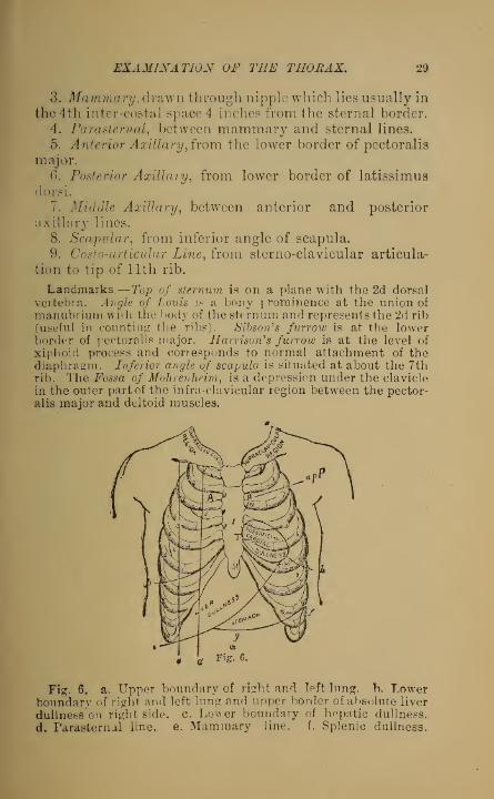
EXAMINATION OF THE THORAX. 29
3. Mammary, drawn through nipple which lies usually in
the 4th inter-costal space 4 inches from the sternal border.
4. Parasternal, between mammary and sternal lines.
5. Anterior Axillary, from the lower border of pectoralis
major.
6. Posterior Axillaiy, from lower border of latissimus
dorsi.
7. Middle Axillary, between anterior and posterior
axillary lines.
8. Scapular, from inferior angle of scapula.
9. Costo-arlicular Line, from stcrno-clavicular articula-
tion to tip of 11th rib.
Landmarks.
—
Top of sternum is on a plane with the 2d dorsalvertebra. Angle of Louis is a bony } rominence at the union of
manubrium wiih the body of the sternum and represents the 2d rib
(useful in counting the ribs). Sibson's furrow is at the lowerborder of pectoralis major. Harrison's furrow is at the level of
xiphoid process and corresponds to normal attachment of thediaphragm. Inferior angle of scapula is situated at about the 7thrib. The Fossa of Mohrenhrim, is a depression under the clavicle
in the outer part of the infra-clavicular region between the pector-
alis major and deltoid muscles.
Fig. 6. a. Upper boundary of rfcht and left lung. b. Lowerboundary of right and left lung and upper border of absolute liver
dullnesson right side. c. Lower boundary of hepatic dullness.
d. Parasternal line. e. Mammary line. f. Splenic dullness.

30 MANUAL OF CLINICAL DIAGNOSIS.
g. Lower boundary of distended stomach. M. Auscultation of
mitral valve, m. Anatomical position of mitral valve.A. Auscultation of aortic valves. T. Auscultation of tricuspidvalve, t. Anatomical position of tricuspid valve, jfi a p P.
Anatomical position of aortic and pulmonary valves andauscultation of Litter. 1. Right superior interlobar fissure,
showing boundary line between the upper and middle lobes of
right lung. 2. Kight inferior interlobar fissure. 3. Left inter-
lobar fissure ending in the mammary line at the 7th rib.
Fig. 7.
Fig. 7. a. Upper boundary of right and left lung. b. Lowerboundary of right and left lung. c. Lower boundary of liver,
d. Scapular line. 1. Right and left interlobar fissures; theformer dividing into superior (2) and inferior (3) interlobarfissures.
Methods of Physical Diagnosis.—1. Inspection.
2. Palpation. 8. Mensuration. 4. Percussion. 5. Aus-cultation. 6. Succussion.
Phonometry, plegaphony and thermo-palpation are auxiliarymethods of diagnosis. Phonometry is a method of examinationconsisting in the applica'ion of a tuning-fork to determine thephysical condition of the thoracic organs. When the handle of atuning-fork after being struck is applied successively to the thigh,stomach and thorax, the tone wi h reference to duration andintensify is different in all three situations. Over the stomachthe greatest and over the thigh the least resonance is obtained
;
while over the lung the resonance is weak. In relaxation of thelung and over lung cavities the resonance is loud, whereas over
EXAMINATION OF THE THORAX. 31
consolidations it is weak. Plegaphony is introduced as a sub-stitute for bronchophony when the Utter can not be practicedfrom any cause which prevents the vibration of the vocal cordsas in diseases of the larynx. In this method of examination, thelarynx is percussed, the observer auscultating the ch.st wall at
the same time. The percussion tone in the larynx is conductedby the air in the bronchi to the chest wall. Over inliltrated lung,the sound loses but little of its original character. Over anexudation in the pleura, the sound is weakened or absent. Overcavities the sound is loud and tympanitic. Thermo-palpation is anew method of examination described by Benczur and Jonas. Theprinciple governing it is that the skin, when palpated over air
containing organs, is warmer than over airless viscera. Thus,palpation with the palmar surface of the ringer shows a dimin-ished temperature over the regions occupied by the liver, heartand spleen, when compared with adjacent parts. It is also
recommended in defining pathological processes ; the upper borderof pleural exudations, and the borders of aneurisms and abdo-minal tumors are said to be accurately determined.
INSPECTION.
This signifies observation of the chest. We determineby inspection : 1. Form of Thorax. 2. Thoracic move-ments. 3. Frequency of respiration. The normal chest
is nearly symmetrical. Not over 20 per cent, of peoplehave a perfectly symmetrical chest. The anterior wallsof a normal chest arch forward to the nipples and thenslope downward to the lower ribs. Even in health, thesupra-clavicular regions are slightly concave.
The most common deviations of a perfect chest are slightcurvatures of the spinal column. This column has a normalcurvature at the cervical, dorsal, lumbar and sacral regions. Acurvature of the column convexly backward is called Kyphosis; acurvature forward, Lordosis; a lateral curvature, Scoliosis; anda curvature laterally and backward, Kyphoscoliosis. Scoliosisfrequently results from contraction of one lung or chronic changesin the pleura.
Pathological Changes in Shape.—1. Generalbilateral bulging (barrel-shaped chest) occurs in Emphy-sema.
2. Unilateral dilatationis observed in pleural exudations,pneumothorax and croupous pneumonia.

32 MANUAL OF CLINICAL DIAGNOSIS.
3. Circumscript bulging in emphysema, encapsulated
exudations, tumors and enlargement of viscera.
4. Paralytic Thorax is characteristic of pulmonarytuberculosis. Chest is flat, narrow and long; intercostal
spaces wide, bony structures prominent, and the scapu-
lar angles project like wings.
5. Unilateral retraction occurs after pleural exudations
and contraction of lung.
Retraction after pleural exudations is explained as follows:
The lung compressed by fluid and bound by adhesions does notexpand to its previous dimensions after absorption of the fluid,
thus allowing the atmospheric pressure to produce retraction of
the chest on the affected side.
6. Circumscribed retraction. After retraction of a
cavity in lung. Observed in supra-clavicular region, it
may indicate shrinkage of the lung apex. The supra-
clavicular regions are normally depressed, and it is only
when the depression is exaggerated, or more marked onone side, that apical disease is suspected.
7. Chicken Breast (pectus carinatum). The sides of the
chest are flattened and the sternum has a keel shape.
Observed in children when unusual force is exerted uponlungs.
8. Rickety Chest. A shallow, longitudinal groove is
found on either side of the chest, at the junction of the
ribs and costal cartilages.
9. Funnel Breast. Depression at lower part of sternum.Congenital.
10. Cobbler's Breast. Like former, although acquired,
and is due to the pressure of instruments against the
breast.
Transverse Constriction of the Chest. A well marked depressionof the lower portion of chest wall in front, from the xiphoidcartilage to the axillary line is due to some impediment to thefree entrance of air into the lower portion of the lungs. It is
usually present in bronchitis during infancy.
Thoracic Movements.—In normal respiration,
inspiration is an active and expiration a passive act.
The lungs undergo no active movements during respira-
tion but passively follow the movements of the thoraxand diaphragm.

EXAMINATION OF THE THORAX. 33
Important Muscles of Inspiration. Intercostal musclesand diaphragm. In females, the scaleni muscles are
normally active as muscles of inspiration.
Accessory Muscles of Inspiration. Sterno-mastoid
,
scaleni, pectorales, serrati and trapezius. These musclesare only employed when the blood is overloaded withcarbonic acid gas, causing difficulty of breathing
(dyspnoea).
Muscles of Expiration. Abdominal muscles, quad-ratus lumborum and serratus posticus inf.
The act of expiration is caused normally by the elasticity of thechest unaided by muscles. Inspiration is accomplished by theelevation of ribs and sternum, and the rotation outward of theformer by the external and internal intercostal mns les. Thisconstitutes the costal type of respiration. Inspiration is further-more ai»ied by contraction of the diaphragm {abdominal type ofrespiration). The type of respiration in males is a combination of
costal and abdominal {Costo abdominal respiration). In femalesand children it is costal. The thoracic movements are usuallyequal on both sides. Sibson^ demonstrated that the rightexpands more than the left side of thorax which Eichhorstattributes to the better developed muscles on the right side,
increased width of right bronchus and larger size of right lung.The connection between sex and type of respiration has beenvariously discussed. It is maintained by some that the dia-phragm is the essential muscle of respiration, but that its move-ments are impeded in women by the wearing of corsets, thisnecessitating the vicarious action of the intercostal muscles.Hutchinson maintains that the costal type is present in girls wThohave never worn corsets, and explains the costal respiration infemales by supposing that pregnancy would interfere with themovements of the diaphragm. Perhaps the greater elasticity of
the thorax in women and children is the cause of the costal type.
Pathological Changes in Thoracic Move-ments.—Costal Respiration in Males occurs in anyinterference with the movements of the diaphragm (pain,
mechanical obstruction and paralysis). AbdominalRespiration in Females occurs in paralysis of the inspira-
tory muscles and in thoracic pain.
The action of the diaphragm can be noted by a prominence ofthe epigastrium occurring during inspiration.
Asymmetry of Respiration is observed in painful tho-
racic affections, and in diseases of the thoracic viscera.
inspiratory Retraction of Chest indicates that air does notM. C. D. 3

34 MANUAL OF CLINICAL DIAGNOSIS.
enter lung alveoli, which allows the external atmosphericpressure to preponderate.
In the lower lateral portions of thorax, physiological retractions
are often present, but occurring as they do only during the begin-
ning of inspiration, they are diagnosed from pathological retrac-
tions which persist during the entire act.
Diffuse Unilateral Retraction, in obstruction of the mainbronchus. Expiratory Bulging of Chest, in emphysemaand phthisis. It can only occur when positive intra-
thoracic pressure exists.
Frequency of Respiration.—In the adult male
there are 16 to 24 respirations per minute. The normalrelation between frequency of respiration and pulse, is as
l:3-£ to 4. It takes four times as long for the blood to go
through the systemic as through the pulmonic circulation.
The respirations are more frequent in females, [nfluence ofage on the number of respirations: at birth, 44; 15th to 2( th
year, 20; 20th to 25th, 18; 25th to 30th; 16; 30th to 50th, 18 timesper minute. Influence of position on the number of respirations :
lying, 13; sitting, 19; standing, 22 time3 per minute. Duringsleep, the number of respirations is diminished.
The respirations are diminished in frequency in cerebral
affections, infectious diseases, opium poisoning, and in
stenosis of the air passages.
When the seat of obstruction is#
in the larynx, at every inspi-
ration the latter descends, to rise again during expiration;whereas, in obstruction below the larynx, the latter is compara-tively immovable.
The respirations are increased in frequency in painful
affections, e. g. pleuritis, rheumatism of the thoracic
muscles, peritonitis, accumulation of carbonic acid in the
blood, and from nervous causes, e. g. hysteria.
An increase in the frequency of respiration also occurs in fever.
and is due to the direct action of the heated blood on the centerof respiration in the medulla. In dyspnceic persons, the diffi-
culty in counting the respirations may be overcome by placingthe finger on the scaleni muscles (situated in the neck, betweenthe trapezius and sterno-mastoid muscles), which at every inspi-
ration are raised by their contraction. By counting the eleva-tions of the finger, the number of respirations is determined.
The respirations are irregular in coma and Cheyne-Stokes* respiration.

EXAMINATION OF THE THORAX. 85
Cheyne-Stokes' respiration is a form of dyspnoea in whichperiods of complete cessation from breathing (apncca) are variedwith periods of slowly rising respiratory movements, which, after
becoming gradually deeper, become slower and shallower, until
they cease altogether. Observed in diseases of the brain and heart,
coma and opium poisoning, although it may occur in healthduring sleep.
Dyspnoea (difficulty of breathing), occurs when bloodis surcharged with carbonic acid gas. The forms ofdyspnoea are inspiratory, expiratory, or a combinationof both. Inspiratory dyspnoea occurs in paralysis of thecrico-arytenoidei postici, which in health open glottis
;
and in narrowing of the air passages. Expiratorydyspnoea is observed in bronchial asthma and emphy-sema, and when movable tumors so situated below theglottis only occlude latter during expiration.
Respiration is regulated by the nervous system and by stimu-lation of the respiratory center in the medulla. As long as theoxygen is maintained at a certain standard in the blood, thiscenter is not stimulated, but whenever the oxygen is reduced,stimulation of the center, characterized by an increase in thenumber of respirations, results.
PALPATION.
This is an adjunct to inspection, and is the act of
laying on the hand. The objects determined are;pain,
movements of thorax, palpation of vocal, bronchial, andfriction fremitus and fluctuation.
Pain on Pressure is observed in visceral affections involving thepleura, and in painful affections of chest- wall. In the diagnosisof respiratory diseases, the important fact must be remembered,that the development of pain nearly always indicates inflamma-tory involvement of the pleura. In intercostal neuralgia, threepainful points (points douloureux) are usually detected (vertebral,lateral, and sternal points). In rheumatism of the thoracic muscles,the pain is diffused, and the affected muscles are painful whencompressed by the fingers. In practicing palpation of the thoracicmovements, place the hands on symmetrical parts of the thorax.In pneumonia, pleuritis, and unilateral tuberculosis, the thoraxover affected parts is retarded in action. The action of the
diaphragm is determined by placing both fingers in the epigastricregion on either side of the median line. An absence of diaphrag-matic contraction on one side is present in diaphragmaticpleuritis, local peritonitis^ and paralysis of phrenic nerve.

36 MANUAL OF CLINICAL DIAGNOSIS.
Vocal Fremitus is the vibration of the thoracic
walls during speaking, perceptible to the hand. It is
due to the vibration of the vocal cords in speakingcommunicated by the column of air in trachea andbronchi to the air vesicles, and so to the chest-wall. It
is more evident on the right side of thorax, because the
right bronchus is more capacious and given off at a
less acute angle than the left. Vocal fremitus is dimin-ished as the distance from larynx: increases.
The louder and deeper the voice, the more evident the vocal
fremitus. For this reason it is more evident in males thanfemales, while in children it is with difficulty felt. In determi-ning vocal fremitus, the palmar surface of hand is applied to
chest. If greater accuracy is required in limiting the fremitus,
use linear palpation, which consists in placing one end of
a rod of wood (a lead pencil will suffice) on the chest, and sup-porting the other end with the fingers. In this way the vibra-
tions of chest-wall are well conducted from a limited area. Inlimiting the situation of solid viscera (liver, spleen, heart,) fromthe lung, linear palpation is well adapted, the fremitus beingabsent over the airless organs. Vocal fremitus is increased in
consolidation of the lung (better conduction), over cavities withden&e walls communicating with a bronchus, and in emaciatedpersons. Vocal fremitus is diminished, or absent, in pleural exuda-tions, pneumothorax, obstruction to bronchus, and in excessivedevelopment of chest coverings (fat and muscles). The studentwill do well to repeat the following experiments : Remove fromthe cadaver, the lungs and trachea. Into latter tie a rubber tube,
into the end of which introduce a funnel. When the latter is
spoken into, the hand can readily feel the fremitus over the 1 urn.:.
Next, inflate a stomach, and placing it over the lung, feel for thefremitus, it is absent (analogous condition in pneumothorax).Now fill a rubber balloon, or similar object, with water, andplacing it over the lung, feel for fremitus, it is also absent (analo-
gous condition in pleural exudations).
Bronchial Fremitus is sometimes present in bron-
chitis. In this affection the air in the tubes is throwninto extra vibration, by the vibration of the bronchial
walls and of the fluid contained within them.
Friction Fremitus {pleural fremitus) is the grating
of one roughened surface of the pleura over the other.
Each lung is covered by a serous membrane (visceral layer ofpleura), which is reflected upon the inner surface of the thorax(parietal layer). Both layers are brought in constant contact bythe movements of respiration, but during health they are smooth,

EXAMINATION OF THE THORAX. 37
and no friction results. In inflammation the fibrinous exudationconduces to friction fremitus. Fluctuation is present in excessivepleural effusion. Palpation may determine the extra or intra-
pleural origin of pus accumulation.i
If the abscess is intra-pleural,gradual pressure will cause its disappearance ; not so, if theabscess is of extra-pleural origin.
MENSURATION.
The circumference of the chest at the height of the
nipple in healthy men is about 32| inches after deepexpiration and 36 inches after deep inspiration. Theaverage difference after full inspiration and forced expi-
ration is from 2% to 3^ inches. The Sterno-vertebral
diameter measures in healthy men, 6^ inches above and7 i inches below. The broad diameter in men at the
height of nipple is 10 inches. The right side of chest
exceeds the left by about f of an inch in circumference in
right-handed persons. The relative length of inspiration
and expiration maybe represented as follows : Inspiration,
5 ; expiration, 4 ; rest, 1.
In emphysema expiration is three times as long as inspiration.In taking the above measurements a graduated tape is all that is
necessary. Select the spinous process of a vertebra behind andthe median line in front as fixed points of measurement. Thestemo- vertebral and transverse diameters are determined bymeans of callipers.
Stethometry is a method of graphically recording themovements of the chest by means of the stethometer.
Pneumatometry is a method of measuring the force ofinspiration and expiration by means of a mercurialmanometer (Pneumatometer). It shows that the powerexerted in expiration is greater than in inspiration. Theinspiratory power is diminished in phthisis, stenosis ofthe air passages and pneumonia. The expiratory poweris diminished in emphysema, asthma and bronchitis.
Spirometry is a method of measuring the amount of air
received into the lungs by means of the spirometer.
The total capacity of the lungs in men is about 200 to 2,~0 cubicinches, in women 163 cubic inches. This capacity inci eases withthe growth of the body. Tidal air is that which goes in and out inordinary respiration, and is about 33 cubic inches. Reserve air is

38 MANUAL OF CLINICAL DIAGNOSIS.
the air which after quiet expiration can be expelled by forced
expiration, and is about 100 cubic inches. Complemental an is
the air which after qnie: inspiration can be introduced by forced
inspiration, and is about 100 cubic inches. Residual air is the
air which remains in the lungs after the deepest expiration, and
is equal to about 100 to 125 cubic inches.
PERCUSSION.
This method of diagnosis consists in striking the chest
walls to elicit sound. It was first used by Auenbrugger
(born 1722, died 1809), a Viennese physician, in 1761.
Percussion may be immediate or mediate. Immediate
Percussion is striking chest wall directly, and is only
practiced over clavicle or sternum. Mediate Percussion
is striking the chest wall indirectly by interposing
media. The medium to receive blow is called pleximeter,
and may be of ivory, glass, wood, leather, metal or the
finger. For percussing a hammer (plessor) or finger is
used. Methods: 1. Finger—Finger Percussion. 2. Fin-
ger—Pleximeter Percussion. 3. Hammer—Pleximeter Per-
cussion.
It is better to rely upon the fingers in percussion. Instru-
mental is easier than finger percussion, although less reliable.
The index or second finger of the left hand is applied closely and
evenly to the chest, and then tapped with the second finger of
ri^ht hand. A single, double, or repeated percussion blows maythen be made. In finger—finger percussion we can appreciate
the resistance of tissues percussed [palpable percussion) and
adapt the finger to irregularities of the chest.
Observe the following: 1. Press finger firmlv against part to
be percussed. 2. Movement of percussing hand must spring
only from wrist, while forearm is motionless. 3. Blow must be
sharp and quick, direct and perpendicular. 4. The finger must
be instantly removed after percussion, so as not to interfere with
chest vibration. The results obtained by percussion are as muchdue to the method of execution as to the condition of the tissues.
The force of the percussion blowris alwTays secondary to theknm k
of obtaining full vibration in resonant tissues. The student to
gain flexibility of the wrist joint should practice movements of
this joint with the arm adducted toward the thorax and the fore-
arm at right angles and motionless. Hammer—Pleximeter percus-
sion is used for class demonstration, to elicit sounds from thickly
padded parts and for uniformity of stroke.

PERCUSSION. 39
Symmetrical Percussion is used for comparison between
the two sides of the chest. Percuss at symmetrical points,
using the same percussion blow. This is useful in
determining slight variations in the percussion sonnd.
Palpable Percussion determines the resistance of parts
percussed as perceived by the finger.
Auscultatory Percussion consists in auscultating the
chest with the stethoscope while percussion is practiced.
Linear Percussion consists in limiting the percussion
blow, e. #., striking on the edge of a coin.
Respiratory Percussion consists in percussing after deepinspiration or prolonged expiration.
In Emphysema the vesiculotympanitic note is unchanged bypercussion during the act of breathing. In cases of fluid in thepleural cavity, a deep inspiration will clearly define the borderline between the resonance above and the flat note below. Tnbronchitis,the clearness of normal resonance may become impaired
;
all doubt is at once dissipated by directing the patient to make aforced inspiration, when the normal lung resonance is restored.
Congestion of the lungs in cardiac disease often yields a slight
dullness on percussion leading to the suspicion of consolidation;
this will be removed after repeated forced inspirations.
Topographical Percussion consists in defining the situa-
tion of viscera by dermography.
Demography is a method of delineating on the chest with ananiline pencil or, better still, with a pencil made for this purpose(dermograph) , the limitation of organs obtained by percussion. If
it is desirable to observe the resurption of exudations, a perma-nent line may be made with a stick of nitrate of silver. Whenpermanent records are desired for the results obtained by per-cussion, schemata of the chest may be used on which may bemarked with colored inks, the location of viscera, etc.
Light Percussion is practiced when air-containing tissue is to becarefully delineated from airless structures as in topographicalpercussion. Strong Percussion is employed when airless tissue is
in immediate contact with thorax and overlies air-containing tissueand resonance of latter is desired ; or to obtain dullness from air-
less tissue covered by tissue containing air. The percussion blowis propagated from \}£ to 2>£ inches on the surface and to a depthof about 2J4 inches. Caution. Every percussion blow is trau-matic in action and when often repeated aggravates or may eveninduce inflammatory affections. Over aneurisms (danger of dis-
lodging thrombus) and painful points, percussion must be light.
Patients suffering from hxmoptysis should not be percussed.

10 MANUAL OF CLINICAL DIAGNOSIS.
The sound produced by percussion of the chest posses-
ses pitch and intensity. Pitch depends on the numberof vibrations in a given unit of time. Over anything
solid (heart, liver) the pitch is high; over normal lung,
the pitch is low. The greater the quantity of air, the
lower the pitch. Pitch is highest over fluids because
they are most dense. Intensity is the loudness of per-
cussion note and depends chiefly on the force exerted in
percussion. Percussion sounds may be 1. Clear, 2. Dull,
3. Tympanitic. Modifications of tympanitic; 1. Amphoric
or metallic, 2. Cracked-pot or cracked-metal sound. Thepercussion sound is caused by vibration of chest wall andthe air within the alveoli of lung.
PERCUSSION OF THE THORAX.
Over the lung like any other air-containing organ percussion
yields a clear sound and is called normal vesicular or pulmonaryresonance. Over the liver and heait like other airless viscera, theBound is dull. The left infra- clavicular region is usually takenas the standard of pulmonary resonance. Always percuss sym-metrical parts of the thorax, using the same percussion blow. Thenormal percussion note (pulmonary resonance) is compared byFlint to the sound elicited by percussing a loaf of bread coveredwiih a napkin. The posterior thoracic regions do not yield a per-
cussion note of the same intensity as the anterior regions onaccount of the density of the dorsal muscles. In percussing theinterscapular space, the patient's head is bowed and the hand oneither side is made to grasp the convexity of the shoulder. Thisposition as suggested by Corson will increase the interscapular
region more than twofold and will render the dense musclestense and thin, thus reducing to a minimum obstacles to percus-
sion and auscultation. The percussion note over the apices pos-teriorly is less intense in right-handed individuals on the right
than on the left side (the opposite holds good in left handed indi-
viduals) owing to the better developed muscles in the former situ-
ation. Similarly the percussion note is less intense on the right
side on the anterior surface of the chest.
Dullness over the lung is obtained : 1. When lungadjacent to thorax is deprived of air by infiltration or
atelectasis. The airless tissue must be flt least l-§ inches
in extent, in order to be recognized. Infiltration of the
lung .occurs in pneumonia, phthisis, hemorrhagicinfarction, tumors and abscess. Atelectasis results from

PERCUSSION OF THE THORAX. 41
compression of the lung (pleuritic and pericardial exuda-
tions) or by an obstruction in the bronchus. 2. Whenfluid (pleuritic effusion) or solid substance (tumor or
thickened pleura) is between chest and lung. In adults
400 ccm. of fluid at least, must be present in order to
be recognized.
Pleuritic Exudations first accumulate in pleural cavity providedno adhesions exist, in the posterior or inferior parts,thence extend-ing, if the fluid increases, forward and upward. If the exudationwas formed during the time patient was in bed, then the upperborder of dullness will describe a line higher behind than in front.
If exudation was formed during the time patient was walkingabout then the line is almost horizontal. In exudations under-going resorption, the upper border often has a curved course, thehighest part of which is in the axillary region (Curve of Ellis).
Inflammatory pleural exudations change little or not at all whenthe patient changes position, because exudation is encapsuled byadhesions. This observation has recently been confirmed by theinvestigations of Strauch. Hydrothorax (generally bilateral) givesa dullness changeable on position. When air and fluid are pre-sent in the pleural cavity (pyo-and sero-pneumothorax), a dullnessis present in the lower anterior portion of thorax when patient is
erect, which is supplanted by a tympanitic sound in the recumb-ent posture. It will be observed that the finger appreciates anincreased sense of resistance on percussion when fluid is presentin the pleural cavity. The presence of large quantities of air orfluid in the pleural cavity depresses the diaphragm, enlarges theaffected half of thorax and dislocates the heart toward the healthyside. The depression of the diaphragm in exudation of the rightpleural sac causes displacement downward of the liver {see percus-sion of liver). In exudations on the leftside, the half-moon shapedspace of Traube disappears. This space is found in normal individ-uals between the left lobe of liver, lower border of left luug andanterior end of spleen. It extends upwards to the 5th or 6th costalcartilage, its upper border describing a convex line directedupward. It contains the fundus of the stomach and yields a tym-panitic percussion note. Beginning resorption of a pleuriticexudation on the left side is first manifested by the reappearanceof the tympanitic percussion sound of this space. Emphysemadiminishes and contraction of the left lung increases the extent ofthis space.
; A Tympanitic sound indicates the presence of air withina cavity with elastic walls. The sound over the stomachis typically tympanitic.
Tympanitic sound over the lung is obtained : 1. Inconsolidation of lung which allows transmission of percus-sion blow to the air in bronchus and trachea as in con-

42 MANUAL OF CLINICAL DIAGNOSIS.
solvation of the upper lobe (William's tracheal tone).
2. In pathological air conducting cavities, (a) In bron-chiectatic or tuberculous cavities, empty of fluid, nearthe surface and at least as large as a walnut, (b) In
pneumothorax, when the air is under low tension. Usually,however the sound is loud but not tympanitic.
A metallic sound can almost always be obtained in pneumothorax,if the following procedure is adopted: fix a plexinieter over theaffected side and while one observer is percussing the plexirneterwith a lead pencil, another auscultates the thorax and hears a line
metallic sound.
Pyo pneumothorax Subphrenicus is a collection of pus and air
between th •*. liver and diaphragm on the right side in consequenceof perforation of the stomach or intestines. Such abscesses morerarely occur on the left side. They are diagnosed from pneumo-thorax as follows: history of preceding peritonitis, absence of coughand expectoration, slight dislocation of the heart only, intercostal
spaces not bulged, vesicular respiration heard as far down asthe abscess. Pleuritic friction can be heard throughout the regionof subphrenic dullness. High punctures in the 5th intercostal
space show a collection of pus or serum while a low puncture,according to Scheurlen, in the 8th intercostal space, yields a puswhich is always ichorous.
3. In relaxation of the lung tissue, as in the neigh-
borhood of exudations and infiltrations.
4. In incomplete infiltrations when lung tissue con-
tains air and fluid, as in oedema, catarrhal and croupouspneumonia (1st and 3d stages).
The Amphoric Sound is a concentrated tympaniticsound of raised pitch, and denotes a large cavity, withfirm, elastic walls. It resembles the sound obtained bypercussing the cheek when the mouth is closed and fully
but not tensely inflated. It also resembles the soundproduced on percussing an empty bottle. It is heardover cavities of a diameter not less than 2| inches. Alsoheard in pneumothorax when the tension of air is of a
certain degree.
Cracked-Pot Sound (bruit de pot felS.) This may beimitated by striking the back of the hands loosely folded
across each other, against the knee ; the sound resulting
is like the clinking of coin. The conditions necessary
for its production seem to be the sudden escape of air
from an inclosed space under pressure. A strong percus-

PERCUSSION OF THE LUNGS. 43
sion blow is necessary. It may be heard in healthypersons, especially children, if the chest is percussedduring speaking or crying. Pathological occurrence: 1.
Over cavities communicating with a bronchus. 2. Overpleural effusions. 3. In pneumonia around consolidated
parts. 4. In pneumothorax with opening in the thoracic
wall. The cracked-pot sound is clearer when patient
opens mouth and protrudes tongue during the act of per-
cussion, or when it is practiced during expiration.
Wintrich's Change of Note is that in which the percussion noteis higher when the mouth is open, and lower when it is closed.
Usually heard in cavities in free communication with a bronchus.This phenomenon can be imitated by percussing larnyx with themonth open and then closed.
Wintricfi's Interrupted Charge of Tone is the preceding phenom-enon ob.-erved on lying down, and absent on sitting up, or vice
versa. It indicates that the bronchus leading to the cavity is
obstructed in certain positions by the flaid contents.Respiratory Change of Tone is a higher tone in deeper inspira-
tion, and is heard over cavities.
Gerhardt's Change of Tone, when present, is an almost certainsign of a cavity, and is due to the presence of fluid in a cavity,changing the percussion sound according to the position of thepatient.
Biermer's Change of Note is the percussion note in seropneu-mothorax. It is deeper in the erect than recumbent posture,because in the former position the fluid displaces the diaphragmdownward, thus making the sounding cavity larger.
PERCUSSION OF THE LUNGS.
The apices of the lungs extend above the clavicles in the supra-clavicular regions from 1-1% inches; behind they rise in thesupra- scapular regions to a level with the spinous process of the7th cervical vertebra. Resonance of the apices is separated fromdullness of surrounding structures by light percussion, but caremust be exercised in percussing away from the trachea.
Situation of the Lower Border of Right Lung (Figs. 6 and 7).
In right sternal line; upper border of cartilage of 6th rib." " parasternal
" lower " " " " "11 " mammary u upper " " " 7th "<« " axillary (i lower " 7th rib.
" " scapular " at 9th rib.
At vertebral column at 11th dorsal vertebra.

44 MANUAL OF CLINICAL DIAGNOSIS.
Situation of the Lower Border of Left Lung:
In left mammary line ; upper border of cartilage of 7th rib." " axillary " lower " " " " M" " scapular " at 9th rib.
Along vertebral column at 11th dorsal vertebra.
In practicing topographical percussion of the lungs, or otherorgans, the percussion blow must be light. In children, the lowerborder of lung is higher by one intercostal space, whereas in theold, it is one intercostal space lower. " The lower border of rightlung is separated from the liver dullness; the lower border of
left lung from the dullness of the 6pleen and left kidney, and thetympanitic sound of the stomach.
TOPOGRAPHY OF THE LOBES OF THE LUNGS.
The following table will approximately define the situation of
the pulmonary lobes on the different surfaces of the thorax.(Figs. 6 and 7.)
Right Lung.
Upper lobe..
Middle M..
Lower " .,
Anterior Surface.
Extends to 3d or4th rib
From 4th rib down-ward
Lateral Surface.
Extends to 4th rib.
4th to 6th rib.
6th to 8th rib.
Posterior Surface.
To spine of scapula.
From spine of scap-ula downward.
Left Lung.
Upper lobe
Lower " «...
In mammary lineto 6th rib
Extends to 4th rib.
From 4th rib down-ward.
To spine of scapula.
From spine of scap-ula downward.
Dislocation of Lung Border.—During deep inspi-
ration the lower border of the lung descends and ascends
during expiration {active mobility'). During deep inspira-
tion when the patient is lying on the side, the lung
may descend as much as 3-| inches. The dislocation
of the lower lung border is greatest in the axillary

AUSCULTATION OF THE LUNGS. 45
line. The dislocation upward during forced expiration
is slightly less than the dislocation during inspiration.
During quiet respiration the difference in the pos.tion of
the lower lung border is not more than y2 inch. Theaverage dislocation of the lower lung border in the
various lines during forced inspiration is as follows:
parasternal line, 1 inch; mammary line, \\ inch; axillary
line, 1^ inch; scapular line, 1 inch.
Passive mobility. This is observed when the posi-
tion of patient is changed. In the recumbent, thelower lung border descends about -§ inch lower than in
the erect posture. When lying on the side, passivemobility is greatest. Active and passive mobility aredecreased in emphysema and pleuritis and absent whenlower lung border is adherent.
The lower border of the lung is permanently lower in emphysemaand temporarily during asthmatic attacks. It is higher thannormal on both sides when diaphragm is pushed upward (air, fluidand tumors in abdominal cavity). It is higher on one side in con-traction of the lung or pleura.
The visceral layer of pleura directly covers the lung, whereasthe parietal layer forms a cavity in which the lung is allowed con-siderable movement. This cavity forms spaces in many placeswhich are larger than the volume of the luug, especially at itslower border. These spaces (complemented or reserve spaces) allowof an augmentation of the lung, and it is there where fluid in thepleural cavity first accumulates.
AUSCULTATION.
Definition.—Application of the ear to the chest for
the detection of sounds. Methods of auscultation:—1.
Mediate or indirect. 2. Immediate or direct.
Mediate auscultation is practiced with an instrumentcalled a stethoscope and is possessed of the followingadvantages : 1. Localization of sound. 2. Obviateschest exposure (important in the examination of women)3. Avoids contact with the bodies of unhealthy persons.4. Intensification of feeble sounds. 5. Exclusion ofextraneous sounds. 6. Auscultation of regions inacces-sible to ear, e. g., supra-clavicular regions.

46 MANUAL OF CLINICAL DIAGNOSIS.
Disadvantages of the Stethoscope : 1. By intensification
sounds are modified. 2. The training of the ear is liable to beneglected. 3. The constant use of the stethoscope may impair thehearing for feeble sounds.
Stethoscopes are of various forms and are made of glass, wood,gutta-percha, metal and ivory. Binaural stethoscopes are used in
preference to others, and the objection that they modify andexaggerate sounds is of minor consideration when compared to
their many other advantages.
Students should accustom themselves to the use of one stetho-
scope only. Stethoscopes although similarly constructed developcertain adventitious sounds, which become gradually excluded bythe trained ear. It is a common occurrence for students who bor-
row instruments to auscultate badly. In the selection of a steth-
oscope, note its proper adaptation to the ears. A soft rubber cupattached to the pectoral end of the instrument is of value in aus-
cultating emaciated patients, the object being to secure perfect
adaptation of the instrument with the thorax. Immediate or direct
auscultation of the chest, the latter covered wTith a towel, is often
a necessity in rapid examinations; but when it is necessary to
analyze circumscribed sound the stethoscope is always preferable.
Gabritchewsky has recently constructed an instrument, the
pneumatoscope, consisting of two funnels of different sizes, the
smaller fitting into the larger. The former is provided with a
vibrating membrane, while the latter is so constructed as to adaptitself to the patient's mouth. The apparatus is connected by tworubber tubes with the ears of the observer. The patient supports
the apparatus in front of the mouth and is instructed to breathe
quietly through the nose. By this means, sounds heard within
the lungs and produced by percussion are said to be conveyed to
the ear.
Auscultation in Health.—Bronchial breathing is
heard over larynx, trachea and bronchi. Both inspira-
tory and expiratory sounds resemble the utterance of the
aspirate H. The expiratory is louder and longer than
the inspiratory sound. It is also likened to the sound
produced by blowing down a tube of the same bore as the
main bronchus. The best situation for hearing it is at
the 7th cervical vertebra. It is also heard over the upper
part of the sternum and the inter-scapular region at the
point of bifurcation of the trachea.
The results achieved in auscultation are dependent in a measureon the way the patient breathes. The patient should be instructed
how to breathe, before auscultation is attempted, and caut :onedto make no noise wTith the mouth. If this is impossible, let thepatient breathe through the nose, with closed mouth.

AUSCULTATION OF THE LUNQS. 47
Origin of Bronchial Breathing. In the larynx, and ia
due to the formation of eddies after the air has passed the
narrow glottis.
Vesicular Murmur.—This is heard over the chest
where the lungs are adjacent to the thorax, excepting at
points where the respiration is bronchial. It is a soft,
breezy sound, heard usually only during inspiration, andis loudest at the end of inspiration. During expiration
it is an uncertain murmur, and very often absent. Bysaying /, or v, softly, vesicular breathing may be imi-
tated. Puerile respiration is heard in children, and is
the vesicular breathing exaggerated.
Origin of Vesicular Murmur. In the larynx. It is
bronchial breathing conducted to the ear and modified bynormal lung.
The following simple experiment will demonstrate that thevesicular murmur is bronchial breathing modified by normallung: If airless tissue, e. g., a piece of liver, is placed over thelarynx, and the stethoscope is placed over the liver, bronchialbreathing is heard. If the same experiment is tried with a lungwhich has been inflated, a vesicular murmur is heard.
Intensity of Vesicular Murmur. This is dependent onforce of respiration, elasticity of thorax, and density oflung tissue. It is heard more distinctly on the right
than the left side, although Stokes maintains that it is
louder on the left side. The vesicular murmur is mostdistinct where the lung is thinly covered. It is loud in
the infra-clavicular, and feeble in the supra-scapularregion. In i of all normal chests, only inspiration is
heard in ordinary respiration.
Bronchial Breathing differs from Vesicular Murmur:1. By its higher pitch. 2. By its occurrence in inspira-
tion and expiration. 3. By its blowing character duringexpiration. 4. By the distinct pause between inspiration
and expiration.
The difference between bronchial breathing and vesicular mur-mur is with difficulty appreciated by the beginner. In cases of
doubt it is best to auscultate in those situations where bronchialbreatking is normally heard, and by comparison the character ofthe sound is easily determined.

48 MANUAL OF CLINICAL DIAGNOSIS.
Auscultation in Disease.—1. Changes in vesicular
murmur. 2. Bronchial replaces the vesicular murmur.3. Undetermined respiratory murmur. 4. Dry rales.
5. Moist rales. 6. Crepitant rales. 7. Pleural friction
Bound.
Changes in Vesicular Murmur: 1. Diminished. 2.
Increased vesicular murmur. 3. Prolonged expiration.
4. Jerking respiration. 5. Systolic vesicular respiration.
Diminished vesicular murmur occurs in obstruction of th8bronchi, emphysema, exudations in pleural cavity and whfnbreathing is painful. Increased vesicular murmur is observed innarroning of the bronchi, e. g>, in bronchial catarrh. Prolongedexpiration is heard when the exit of air from the lung is prevented,either from loss of elasticity of the lung (emphysema) or obstruc-tion of the bronchi (bronchitis). In health, the expiratory soundwhen heard is not one-third as long as the inspiratory sound.Jerking respiration is heard when inspiration instead of being con-tinuous is interrupted
;present in bronchitis. Systolic vesicular
respiration is heard about the cardiac region, when the heart is
increased in action, the contraction and dilatation of the sameinfluencing the entrance of air into the lung.
Bronchial breathing replaces the vesicular murmur whenthe murmur arising in the larynx is transmittedunchanged through consolidated lung to the chest wall,
e. gn in pneumonia, infarction, tumors, compression of
lung above pleural exudation, etc. If bronchus leadingto consolidated area is obstructed (mucus, fibrin, etc )
no bronchial breathing is heard, but the same appearswhen the offending object is dislodged by coughing.Bronchial breathing is also heard in open pneumothoraxand over lung cavities as amphoric respiration.
Forms of bronchial breathing. Amphoric and metamor-phosing respiration.
Amphoric respiration is imitated by blowing into an empty jar.It is bronchial breathing possessed of a metallic character. It isheard over large lung cavities with dense walls and in pneumo-thorax.
Metamorphosing respiration is characterized by an inspirationwhich begins with a vesicular murmur and then passes over intobronchial breathing. It is a certain sign of a cavity and isexplained by the air reaching the cavity through a narrow bron-chus, which, in the second part of inspiration is dilated by theair current.
AUSCULTATION OF THE LUNGS. 49
Undetermined Respiration represents a form of breath-
ing bearing the character of neither the vesicular norbronchial.
Dry Rales are heard in catarrh of the bronchi whichyield a viscid secretion with swollen mucous membrane.Air passing through the narrow tubes produces the sound.They are divided into Sibilant and Sonorous Rales. Theformer are whistling or hissing sounds occurring in the
smaller bronchial tubes. Sonorous rales are snoring or
cooing sounds produced in the larger air tubes.
Moist Rales are produced by the breaking of bubbles of
air in fluid (blood, mucus or pus). They are coarse or
fine according to their origin in the large or small bron-chial tubes. They are modified or disappear on coughingowing to dislodgment of mucus.
They are heard during inspiration and expiration whenrales are present in abundance, otherwise only duringinspiration. They are common in bronchitis after secre-
tion has become established. Also present in cavities
and when blood and pus are present in the bronchi.
Metallic or Ringing Rales are heard added to respiratorycough or voice sound in a large cavity containing air.
Bell sound is obtained by percussing the chest with twocoins, one used as a pleximeter, the other as a plessorwhen a ringing sound is heard. Present in pneumothorax.
Metallic Tinkling Rales are associated with amphoricrespiration and are produced by drops of purulent matterfalling from a large cavity containing air and fluid. Alsoheard in pneumothorax.
Crepitant Rales occur in the smallest bronchioles andair cells and are due to the inspired air forcing open the air
cells agglutinated by exuded lymph. They resemble thenoise produced by throwing salt on the fire or rubbingthe hairs together near the ear, or, better still, by moist-ening thumb and index finger and separating themrapidly in front of the ear.
They are, as a rule, only heard during inspiration. They areproduced in the first {crepitaiio indux) and beginning of tfie 3d
50 MANUAL OF CLINICAL DIAGNOSIS.
stage (crepitatio redux) of croupous pneumonia, in catarrhal pneu-
monia and oedema of the lung.
Atelectatic Crepitation is heard over collapsed portions
of lung and disappears after deep inspiration.
If we remember that the tidal or breathing volume of the air
amounts to only 33 cubic inches, and the complemental air, whichis the air taken into the lungs by forced breathing, which, in
addition to the tidal volume, amounts to about 100 cubic inches,
and knowing that the average respiratory capacity of an adult is
about 225 cubic inches, the conclusion is evident, that even in a
state of health the lungs are imperfectly aerated, and in a con-
dition of physiological atelectasis. My invariable custom before
conducting an examination of the lungs is to have the patient
make repeated forced inspirations. In this way I avoid manyerrors in auscultation and percussion.
Sub-crepitant Rales are heard during inspiration andexpiration, and are thus distinguished from crepitant
rales. They are produced by air currents breaking
through mucus in small bronchial tubes. Heard in capil-
lary bronchitis.
Pleural Friction Sound. The respiratory rubbing of
the layers of the pleura occurs in health without noise.
The presence of a fibrinous exudation on their surfaces
(pleuritis sicca), causes superficial noises {pleuritic fric-
tion) heard in inspiration and expiration. On palpation,
pleuritic friction can often be felt. If fluid is present in
pleural cavity no friction is heard or felt because the
pleural layers are separated. The absence of friction is
also noted when adhesion of pleural surfaces is present.
Friction is heard in all forms of pleuritis, especially in
the beginning and after disappearance of the serous
exudation.
Differential Diagnosis between Rales and Friction Sounds: 1.
Rales are continuous, friction sounds a series of interrupted jerks.
2. Cough changes character of rales while friction sound is notinfluenced. 3. Rales are diffused, friction sounds confined to
a smaller space. 4. Pressure w7ith stethoscope in pleuritis is
painful, and friction sounds are increased, rales are uninflu-
enced. 5. Friction sounds are superficial and increased by forcedinspiration. Dr. Bruen directs attention to the value of makingthe chest wall immovable. When the chest is fixed, especially at
the lower two-thirds, by the hands of an assistant, and the stetho-
scope or ear is applied over the doubtful sounds, they will befound to have disappeared if of pleural origin, but to be fitill pres-

AUSCULTATION OF THE VOICE. 51
ent, if rales. Further, cause the patient to incline to the opposite6ide to that diseased, and place the hand of the diseased side onthe head ; this puts the pleura in a state of tension, and will oftenobliterate a friction rale.
AUSCULTATION OF THE VOICE.
Vocal Resonance.—During phonation, auscultationof the normal chest, except in situations where bronchialbreathing is heard, distinguishes the voice as a confusedhum, and words can not be recognized owing to the poorconducting quality of the normal lung.
Bronchophony.—When voice as heard over larynx,trachea or bronchi, is conducted to chest, so that spokenwords become distinct. It has the same significance asbronchial respiration. As a pathological condition it
always denotes consolidation of pulmonary tissue for thelung in this state is a better conductor of sound thanwhen in a normal condition.
Pectoriloquy.—This is exaggerated bronchophony.It is heard over large and superficial lung cavities, andhas an amphoric character.
.ZEgophony.—A variety of bronchophony. A hightrembling voice and sound likened to the bleating of agoat. It is observed when the voice reaches the chestwall intermittingly. Present in incomplete compressionof the bronchi, and at the upper border of a medium-sizedpleuritic exudation. In speaking with the nose closedthis sound may be imitated.
Metallic Sound of the Voice is heard overlarge cavities and in pneumothorax.
Whispered Voice.—The vibrations producer1 bythis voice in the normal condition of the lung tissue aretoo feeble to be appreciated. When solidification of thelung or a cavity exists the whispered voice is transmittedwith unusual distinctness (whispering bronchophony).
Bacelli observed that in serous exudations of the pleura, thewhispered voice was conducted to the ear ; whereas, when the

52 MANUAL OF CLINICAL DIAGNOSIS.
exudation was purulent, it was not. This sign, called the Pheno-menon of Bacelli, was supposed to be of importance in differenti-
ating serous from purulent exudations; but, not being alwayspresent in serous, and being occasionally heard in purulent exu-dations, it loses much of its practical importance.
SUCCUSSION.
This is a splashing sound, produced when air and fluid arepresent in the pleural cavity at the game time (sero- orpyopneumo-thorax). The sound is somewhat similar to that obtained byshaking a cask containing air and fluid. The direction of Hippo-crates, in order to obtain this sound, was to shake the patient bygrasping the shoulders, and with the ear applied to the chest it
could be heard.
The succussion sound is sometimes so loud that it can be heardin every part of a room. The less fluid and the greater quantityof air, the louder the splashing. Being most pronounced inpneumo-hydrothnrax, it may be heard to a lesser degree over verylarge cavities with fluid contents.
Examination of the Thymus Gland. This ductless gland is atemporary organ, attaining its full size about the age of two years.
It subsequently atrophies, until at puberty it has almost disap-
peared. It consists of two lateral lobes, and extends from the4th costal cartilage to the lower border of the thyroid gland. Inthe mediastinum it rest* upon the pericardium. In children,percussion will indicate the presence of this gland by a finger
breadth dullness in the left sternal line, extending from the 2d tothe 4th costal cartilage. The thymus gland may be hypertro-phied. or the seat of haemorrhage, abscess, tuberculosis, tumors,etc. Dullness at the upper part of sternum may also be due to
aneurism of the arch of aorta, mediastinal tumor, or a subster-nal struma.
Puncture of the Pleura is practiced with an ordinary hypoder-mic syringe, with antiseptic precautions. Its object is to deter-
mine the presence and character of fluid in the pleural cavity. It
is a harmless procedure, and is further indicated in diagnosingpleuritis from other affections (pneumonia, tumors, thickening ofpleura, etc.) The character of the exudation in pleuritis may beserous, sero-fibrinous, sero-purulent, purulent, or haemorrhagic.
Microscopical Examination of Fluid may reveal the presence of
streptococci, actinomyces, and bacilli of tuberculosis, although theabsence of the latter does not exclude tuberculous pleuritis. Car-cinomatous cells may be present in carcinomatous pleuritis.

ASSOCIATION OF PHYSICAL SIGNS. 63
ASSOCIATION OF THE PHYSICAL SIGNSOF THE LUNGS.—(Altered from Da Costa.)
PercussionAuscultation
ofRespiration.
AuscultationOF
Voice.
VocalFremitus.
Physical Condi-tion.
Clear Vesicular mur-mur or its
modifications
Normal vocalresonance
Unimpaired Normal (or nearly)lung tissue
Dull Bronchial res-piration
Bronchophony Increased Consolidation ofthe lung
DuU Absent respira-tion.
Absent voice Diminishedor absent
Effusion into pleu-ral cavity; ob-struction of thebronchus asso-ciated with con-solidation of thelung.
Tympanitic Cavernous, orfeeble, ac-cording tocause.
Cavernous ordiminished
Increased,diminishedor absent
Cavities of thelung ; relaxationof the lung tissue;pneumothorax.
Amphoric ormetallic
Amphoric ormetallic.
Amphoric ormetallic
Increased ordiminished
Large cavity withelastic walls;pneumothorax.
Cracked me-tal sound
Cavernous res-piration.
Cavernousvoice.
Uncertain,increased ordiminished
Generally a cavityof the lung com-municating witha bronchial tube

ot—
1
H<1
hJDO
<Dry
rales
when
fluid
is
vis-
cid.
Sonorous
in
large,
and
sibilant
rales
in
small
tubes.
Prolonged
expiration.
Mucous
rdles
when
secretion
is
free.
Vesicular
respiration
dim-
inished
or
absent.
Whis-
tling
or
hissing
1
mds.
Bronchial
respiration
with
metallic
sounds
when
cavities
are
super-
ficial,
otherwise
vesicu-
lar
respiration.
Moist
rales
when
fluid
is
pres-
ent
in
cavities.
Diminished
vesicular
res-
piration
and
sibilant
rales
heard
loudest
dur-
ing
expiration.
They
become
moist
at
term-
ination
of
attack
oHHmp«Ph i
aEhO
Normal
in
beginning
but
soon
followed
by
a
tym-
panitic
sound
{sign
of
lung
relaxation.)
When
dilatation
is
superfi-
cial
and
large,
tympan-
itic
or
tympanitic
metal-
lic
sound.
Other
signs
of
cavities.
Signs
ab-
sent
when
cavities
are
filled
with
secretion
but
reappear
when
contents
are
expectorated.
Hyper-resonance.
Immo-
bility
of
lung
borders.
Absolute
cardiac
dull-
ness
diminished
owing
to
acute
inflation
of
lungs.
ao
Circumference
of
thorax
on
affected
side
diminished.
.£ © ©rO pn— ®cd n,_; © ©
£ p do-~ ©
Circumference
of
thorax
in-
creased
dur-
ing
parox-
ysm.
OHPh
<Ph
S3 O
"a©
0J«*H
© gr4
2>aCQ
-ap-a
•a
P 'w
o3 o^
>Large
bronchiectases
when
superficial
and
surrounded
by
con-
solidated
lung
cause
increased
vocal
fre-
mitus.
BO
a©u
1p8w
O»—
i
HOPhm
.2 oho c3•<s> M"« ©
e a 5o p.£Thoracic
movements
on
aft'ected
side
diminished.
Inspir-
atory
dyspnoea
and
inspiratory
retrac-
tion
of
chest.
Flattening
of
affected
side
due
to
retraction
of
connective
.tissue
around
dilated
bron-
chi.
Inspiratory
re-
traction
of
chest.
Expiratory
dyspnoea.
Chest
semi-distend-
ed
with
little
move-
ment.
HCO<TO
A
t3
s©O«Ph
TO
Wo
H00oK©o
n
CQ
io
3©o«n
3
o W
(54)

o
«!
H-:
PoCO
p
s® ©a 2
p tt 2o > P
3 §
©.3.2M ft^
.3 33 ©g§52-q^pq
» © d
Ho- r
Ph M
525
O
PowPh
9-.
•= © <D .3
2 *?£© w ^ a.
*s" iwags -2
©T33+2 53
.j£3pC3>-£
oh St3
M!&1!>>3j33 Pi
H
•2 d ft^<b a P
J*op03 p-p_ .
ft 3 © J- 3
.2x3 oq'3.2
pq
•5§opo 2
^1 P P 03 aj g *,2'o M-prj^pC^m C P.3 ©£ M
g>.S;5o
<ri
**a§-°*3^oS.3 .
•^a, p *n » •'
gf * o^p^-pQ.P ^3-PP^©P-2 03 c£ V P,
o
•4H P,O © BJ
- p *>p o *-
•a St.
P <u p
P ...a© © p
i
r3 > «
£ O 3, op T3
^g^O*flp;
P ^3^ P+3© ep-p^'C<°rr* p -p
•3 05 ^ C p,*3 ^ P5 © Fj p. p aj
^X?»" © 03 33 +J
tot: © o«•=. © g
p?"-3O S r. *-
l|g!
io*.s s P '
^ P- or-2-^ p
p 33 ^^
' >>3 3=c 03 4-1
H c£> OJ
p .2 p p 2. -s
Q
5?;
o
I
o®32^.5©QPtS
a* *3 © «
*3 O aQii
** p^j
>s p
0Q S^ Oc3 ^ PG) aj ott P ^«5ft
S3 o3
^ P
P o ^ 3 3
g - •- ^£ • g fl sgT3.5.S:2P Bt'O t>
fl » C f j
a 3£©^ ?
o -
§ °'cj © O
o © •
>
* I.-3 rp P© 03 © ©p > a cl© £ o3 P© O J ©«P^ ^ 3^CD©P
p © o*=*
cc © 03 w
|g||a -P .3 fj
32 © so PiS
O J?03-w
ra P P 55 «- p .
C S^^ SoTO ° 03 O CO
l^ft^.3^© £ © © a? ^ 3r© .3 /v» *- © 'P£p3 Cq © m o in
<
j, © ©2 ^> •
t3 ^ 03 OQ
§§.2 S
1^9
55OI—
i
HOwPhCQ
P t^jJ^
"ISA5 pd
U a©p .
©^^03 cc
>> -3of-+a ©!3p o3 o; c3 =:"
r; 33M o -p
'iJ 3 e3 P ©£133.2 —
2cS|_|5g:^ 5p^J"8fla x"-3 P ^30 3©ra£ .23 © rt © m&
°"o ^ 2 ° o©-P
P3
©"3 s- ? © SP so -tj *-fc0©
ftp
. a>03 *-«
8 ^
03 O-P>-.+J ©q
HCQ
3
SS5
Pm Ph3
o(55)

SO
s
gq
H
H02
«oH
«MGO
s
oCQHCQ
O
oMCQ
wPh
55
OH
poCO
p
• # > n .p -" r
55O
oPh
wPh
Ot—
i
<Pht-H*
Ph
Ot—
i
OPh
WCO
HCO
1) o
.2 5 a P be M p
g 3 m'O >>£ o^ft,2 ^ g o S £ «
a>7;t£ p.ftO>^
» & i Sf *
K P X w
c — £ © w•
<u © p w+* © a2 *wh „
£ © © P<K o©£ p,c>.S3p*
M8*g£— ? .p © p S3'C £ > fc ad 7
© »- ftj^ g Q'
* © -p^ p-
P^d bo "S
*w p»p p'Pip£.£ ©,p ©d
x a ^P Poo c-£*3tC Cm© oj r
P 73
© M S
•n ©°p t> ©2.8 s
« 2^-
lis"
©42 pPOPP-M OQ
s-S+J o p
c3 >£*H 55 ->© © !>
© H85'.
0> © 03 SO
p2p«
P o ® "£
© ftw
2 m 22 cj—.
5 p bs
Ph
04
o
p°p.2j
«43 .
00 C SW p,©
5°«P bf p
0) S3
-Si<
.ti © ft
^H © ©o c *p
»©«©s
•P P
« O P-
C0 P CQ pp*^ © p
g p p»GJ cc oo P
p ^?
CJ.H ^ O ®
t-PHa^
£-si !
23 P ° -T
SS33S© "t^^S p
© *5 r^ © »
P 03
8 « &
l©^oPh
14
PM
^ftp*C! ® ® hgpSgo
W p^QQ
sfl PP © . "^
l©bSPh
"rf A H
PP,Q ©
A cj * »
S
^ P
63
p p - ^ g
S-« p '-t:P p © o <u
«2«3*oo.P. ft •
PZ3 c ° M^j=! Pp o
CO
>- P^- (H
O 83 p c3
•o g co O
"^'--tHcp cS'O
flL © •
2«Pd"
> SQtH P SB
o
© k © ©OO c3 P
5»©pS+i 6C*P •
^ P gP p•o wft§.2CO tip* (jqCS
^ . P ft
-c3|bcgp-SP
§p£p«3^l> © O
So%*P o
03
Pi ©. ^
d©5P3
«"-g o P
pl^s^
r-4-U ©•"c3 03 > «*— o o
© © o ©
.S<i © 03 P5
^ 2g P P
fc H P4
• *-9
«^:
^^c
o"-S5Sh
(66)

PHONOGRAPH IN MED1CIN& 11
THE PHONOGRAPH IN MEDICINE.*
A few years have elapsed since Edison devised an instrumentconsisting essentially of a mounted diaphragm, eo fixed as tooperate a stylus which recorded the inflections of the voice on tin
foil. With this crude apparatus, which sacrificed distinctness of
articulation in order to secure a loud tone, not only was soundrecorded, but reproduced. Attention has of late again beendirected to an improved phonograph, embodying the same princi-
ples as the original instiument, but so modified as to be of prac-tical value. It is now but a few months since the untiring Edison,dissatisfied with his previous achievements, added furtherimprovements to the phonograph, making it now automaticallyadjustable and combining the recorder and reproducer in one.The perfected phonograph requires no dexterity in manipulation,and I will briefly describe the method of taking a phonogramwith it.
To record sound, a wrax cylinder is fitted on to a metal one.The diaphragm, which is automatically adjustable, is then madeto rest on the wax and the instrument is ready for use ; on speak-ing into a tube, which is in communication with the diaphragm,the sound pulsations are incised by means of a small puint in thenoiselessly revolving cylinders. Upon examining the wax cylin-
der, a number of fine lines can be seen, which are an absoluteequivalent for the sounds made by the voice. All that is neces-sary to reproduce the sounds is to move a lever, when anotherself-adjustable point is presented, which passes over the indenta-tions in the wax made by the recording needle, and absolutereproduction of the voice, even to the lightest shades and varia-tions, results. This may be repeated an indefinite number of
times. The wax cylinders cost twenty-five cents apiece, makingphonograms for permanent record very expensive. This objectionwill soon be obviated, as mailable cylinders, costing but a fewcents and available for single records, are to be introduced.
To run the machine, either an electric motor or foot power maybe used. I use the latter, as the instrument is under better con-trol : An electric motor, however, possesses the advantage of
rendering the revolutions of the wax cylinder almost noiselessan indi^pensible factor in the reproduction of fine sounds. Theaccurate reproduction of the human voice, is well illustrated bythe following occurrence : A lady on hearing the wTords of a gen-tleman, spoken into my instrument but a few days before, at onceexclaimed: " I know that voice! It is the voice of Mr. H ."
I asked her if she knew the gentleman and she replied in the neg-
Reprint from Occidental Medical Times, April, 1890.

58 MANUAL OF CLINICAL DIAGNOSIS.
ative, but she had heard him recite once at an entertainment
many months before. Here is an instance of how unerring is the
memory for sounds.
I have employed the phonograph in taking the histories of cases,
thus saving time and obtaining copious notes. The cylinders can
be transcribed at the convenience of the amanuensis. This is but
one of the many practical uses to which it may be put. In physi-
cal diagnosis the application of the phonograph suggests itself,
and it is claimed, that in the laboratory of Edison, the normal
heart sounds were first successfully recorded. Its practical value
in medicine was tested by the Pans Academy of Sciences and the
Vienna Medical Society, with results largely in favor of the instru-
ment. Dr. Bleyer, in an article on " The Phonograph in Physical
Diagnosis," says that by means of a microphonic stethoscope, he
was&enabled to record all the thoracic sounds in their varied
pitch and tone. The sounds thus obtained were projected bymeans of a tin cylinder acting as a resonator and directly instruc-
ted a large class of students. I must confess, that similar experi-
ments faithfully executed with all due regard to details, wereunattended by these results. I found in my investigations, which
were varied in every possible manner, that the recording of heart
sounds was practically impossible. The fault rests with the pho-
nograph. There is no question about the sensitiveness of the
diaphragm for recording even the feeblest sounds, but the dim-
culty lies in reproduction. Even loud sounds emanating from the
chest are with difficulty detected by an ear accustomed to the
phonograph. When the reproducing needle is adjusted to the
revolving wax cylinder a hissing sound is heard, the result of fric-
tion between the needle and the wax. It is this sound whichinterferes with proper reproduction, and I have found that lubri-
cating the cylinder renders reproduction decidedly more distinct.
If this objection were obviated, the phonograph would prove anideal instrument in physical diagnosis.
Many sounds heretofore inappreciable can be brought out by the
phonograph. The deepest tone that we are able to appreciate con-
tains 16 vibrations a second ; the phonograph will record 10 vibra-
tions or less, and can then raise the pitch until we hear a repro-
duction from them. Similarly, vibrations above the highest rate
audible to the ear can be recorded and then reproduced by lower-' ing the pitch. I have been able to record nearly all the percussion
sounds of sufficient intensity, and with a degree of distinctness
which is remarkable. The percussion sounds obtained in this wayare exaggerated, and the method resembles auscultatory percus-
sion. The fine lines which can be seen on the wax cylinder after
talking into the phonograph convey a fair idea of the number andvariety of air waves necessary to the production of words and sen-
tences.
In clinical medicine the uses of the phonograph are varied.
The different anomalies of speech, whether due to paresis, paraly-
sis, tremor, spasm, or even ataxia of the muscles of articulation!

PHONOGRAPH IN MEDICINE. 59
can be faithfully recorded. Coughs of various kinds can also
receive accurate registration
.
Determining the sense of hearing is of great importance to theaurist, and the present apparatus for this purpose is unsatisfactory.
Koosa, in his text-book, refers to the characteristic answer of aboy who, upon being tested by the ticking of a watch, suddenlyexclaimed: "What do I care about hearing a watch ? I want to
hear what people say V Dr. Lichtwitz, in a recent article on "ThePhonograph in Otology and Laryngology, " considers the follow-
ing as essential to an apparatus for ascertaining the perception of
sound; (1) It should produce all the tones and murmurs appreci-
ated by a normal ear, and especially speech in all its inflections.
(2) It should be a constant sound producer. (3) It should be anuniversal apparatus, so that otologists in different countries canexpress themselves definitely regarding the acuteness of hearing,as ophthalmologists do in testing vision. (4) Its use should beunaccompanied by inconvenience. (5) It should allow of the per-ception of sound by bone and air conduction. The phonograph,according to this author, absolutely subserves the first two indica-tions, and is the best apparatus for determining the sense of hear-ing.
Of all the wonderful accomplishments possessed by the phono-graph, its availability for the blind, by placing them in communi-cation with the world of literature, far exceeds any similar devicefor the relief of this unfortunate class. By the majority of thosewhose eyesight is impaired or destroyed it will be hailed as a verita-
ble God-send. Each wax cylinder will receive from 800 to 1,000words, and the whole of "Nicholas Niekleby" can be put on fourcylinders eight inches long with a diameter of five. By a mechan-ical process these cylinders can be multiple-copied, which will
render them comparatively inexpensive. For educational pur-poses, as in the study of foreign languages, which to physiciansis often a necessity, the phonograph is beyond comparison, for nosystem of phonetic spelling or verbal description will convey thesame pronunciation of a foreign language so well as a machinethat reproduces the human voice.
The phonograph is not without a therapeutic use. The psychicinfluence of sound cannot be denied, and we need only recall thewell-known effects of enlivening music in cheering troops on themarch or while under the depressing influence of siege, and theprofound influence of certain music in exciting the highest emo-tional centres. When music, and good music at that, can be had"on tap," and supplemented by stories, songs and recitations bywell-known authors, vocalists and elocutionists, the convalescentmay command a variety of entertainment.

* CHAPTER V.
COUGH—SPUTUM.
Cough is a symptom of disease of the respiratory apparatus. It
is a reflex act, the result of laryngeal, tracheal, or bronchial irrita-
tion. The chief object of this act is the expulsion of pathological
products which, if allowed to accumulate, would result primarily
in dyspnoea, to be followed by asphyxia. For this reason, the use
of narcotics which render anaesthetic the respiratory mucous mem-brane, are dangerous when the secretions are abundant. The pro-
bable direct cause of cough is irritation of the fibres of Xhepneumo-gastric nerve, or its branches. A cough centre is supposed to exist
in the floor of the 4th ventricle. Cough may result from irritation
of the pharynx, esophagus, stomach, liver, spleen, and female geni-
talia. In many people, cough may be produced by touching thenasal mucous membrane with a sound, and many intractable
spasmodic coughs have been cured by the removal of nasal polypi.
In about 17 per cent, of persons, cough will follow irritation
of the external auditory meatus. In hysteria a persistent coughmay be present. We also speak of a nervous cough. Irritation of
the alveoli of the lung will not^ result in cough, and secretions
must first reach the communicating bronchus, before coughing is
produced. Kinds of cough : 1. Dry cough is present in pleuritis,
pneumonia (1st stage), acute bronchitis, and broncho-pneumonia.2. Moist cough, when the air passages are filled with an abundantsecretion. 3. Brassy (barking) cough is heard in stenosis of thelarynx (croup). This cough is usually associated with stridulous
respiration, when pressure is exerted on the trachea (Aneurisms,
tumors). 4. Whooping-cough in pertussis. It is along drawn, crow-ing sound, coincident with inspiration, and follows a violent series
of coughs. It is due to the passage of air through the spasmodi-cally closed glottis.
In Laryngismus Stridulus, a crowing inspiration resembling thatof pertussis, although not usually associated with cough, is heard,and is also due to spasm of the glottis. A pharyngeal cough is
frequently attended with vomiting. An empyema perforating theair passages, is followed by sudden and copious expectoration.
In phthisis, cough is often dependent on the ingestion of food, andis caused by epiglottis, the seat of tuberculous infiltration, imper-fectly closing the opening of larynx.
(60)

SPUTUM- 61
Signs Obtained by Coughing (Tussive signs).—Auscultatory signs
obtained by respiration and the voice, may likewise be obtainedby acts of coughing. Coughing will remove accumulations of
mucus within bronchial tubes, and thus restore a diminished orsuppressed murmur over some part of chest. In coughing, moreair is expelled than by an ordinary expiration, and in the follow-
ing inspiration the vesicles capable of greater expansion, give rise
to a proportionately loud inspiiatory 6ound, thus intensifyingnormal and abnormal inspiratory sounds. Cavernous gurglingmay be obtained very distinctly with cough. Cough changes thecharacter of rales, leaving friction sound uninfluenced.
SPUTUM.Origin of the Sputum.—The constituents are furnished by the
secretions of the pulmonary alveoli, bronchial tubes, larynx, phar-ynx, mouth and the nares with their adjacent cavities. In healththe secretion from the air-passages does not exceed in quantitythat required to moisten the mucous surfaces. In disease thesecretion may be in excess and contain many new constituents.Children and aged individuals usually swallow the sputum.Chemical analysis of the sputum is rarely of diagnostic value.In the examination of the sputum we note: 1. Consistency.
2. Color. 3. Odor. 4. Quantity. 5. Reaction.
Consistency is dependent on the quantity of mucus.In pneumonia the sputum is very viscid while purulentSDutum has little consistency.sputum has little consistency
Color.— Whitish or colorless in mucous, yellowish in
muco-purulent, and greenish in purulent sputum.
When the blood-coloring matter in the sputum is altered the colorimparted may be red, brown or yellowish green. Yellow-ochrecolor (presence of hcematoidin) is present in abscess of the lung.A green color (biie pigments) is met with in pneumonia compli-cated by icterus. A green color may also be caused by micro-organisms. The sputum may be colored blue (laborers in dyeworks), black (coal laborers) and ochre-colored and red (iron-work-ers.)
Odor.—Very fetid in dilated bronchi, putrid bron-chitis and gangrene of the lung.
Quantity.—Daily estimation is of value in controlling
the progress of a case. The quantity is large in purulent

62 MANUAL OF CLINICAL DIAGNOSIS.
bronchitis, dilated bronchi, tuberculous cavities, oedema,
abscess, etc.
Reaction.—Usually alkaline.
According to the principal constituents, the sputum may bedivided as follows : 1. Mucous. It resembles the natural secre-
tion. It is tough, transparent and colorless, and is present in
beginning bronchitis. 2. Purulent. It is of a greenish-yellowcolor, and is found in rupture of abscesses from the lung or neigh-boring organs, empyema, chronic bronchitis (bronchorrhoea), etc.
3. Serous. Pathognomonic of lung-cedema. 4. Bloody (haemop-
tysis). Present in the first stage of tuberculosis (initial haemoptysis)
or in any part of its course : in cardiac disease when venous stasis
develops (particularly in mitral lesions) : aneurisms (bleeding pro-
fuse); haernorrhagic infarctions, etc.
We also distinguish the following mixed forms : 1. Mucopuru-lent in bronchitis (mucus and pus intimately mixed) and phthisis.
In the latter affection, the mucus and pus are not mixed and thepus is in the form of balls covered with mucus which sink inwater {sputum globosum).
2. Sanguineo-mucous found in carcinoma of the lung (raspberry-
jelly sputum), pneumonia (1st. stage), and hemorrhagic infarction.
3. Sanguineo-serous (prune-juice sputum) is found in oedema of
the lung and pneumonia. Blood-colored saliva may be expecto-rated by hysterical individuals who often prick and suck their
gums: the blood is more watery than usual and is generally notaerated. Shreds of the parenchyma of the lung may be found in
abscess and gangrene of the lung.
With the microscope the following constituents maybe found in the sputum :
1. White Blood Corpuscles. Always present, andespecially abundant in purulent sputum.
2. Red Blood Corpuscles. Present in bloody sputum.Corpuscles are preserved in form, although pale.
3. Epithelium. Pavement (Fig. 8, a) from the buccalcavity and true vocal cords. Cylindrical, from the nasalcavity, upper part of pharynx, the larynx and bronchi.
Alveolar (fig. 8, b) from the alveoli of lungs and finest
bronchi. They are large, round or oval cells, and manycontain fat, carbon and myeline. They are of no special
diagnostic importance, but their presence in large quan-tities might suggest tuberculosis.

MICROSCOPY OF SPUTUM, 6S
4. Crystals. Charcot- Leyden Crystals (Fig. 8, c).
Colorless pointed octahedra, insoluble in cold water,
ether, alcohol or chloroform, but easily soluble in alka-
lies, mineral acids, warm water and acetic acid.
Fig. 8. a. Epithelium from mouth, h. Epithelium fromlung alveoli containing granules of black pigment, c. Oharcot-Leyden crystals, d. Elastic fibres, e. Spiral of Curschmann.f. Echinococcus (scolex and hooklets). g. Actinomyces, h.
Needles of margaric acid.
They may at times be seen with unaided eye in yel-
lowish masses found in the sputum. They are met with
in bronchial asthma (diagnostic) during and after the
paroxysm. Also observed (rarely) in acute bronchitis
and tuberculosis.
Hcematoidin Crystals appear as amorphous yellow-brownishgrains, or in rhombic plates and needle-like crystals. Theycome from extravasated blood which has been retained for sometime in the air passages.
Needles of Margaric Acid (Fig. 8, h). Soluble in ether or chloro-form. Found in gangrene and putrid bronchitis inclosed inminute plugs which emit an intensely foul odor.

64 MANUAL OF CLINICAL DIAGNOSIS.
Cholesterin Crystals. Rhombic plates (Test: with diluted sul-
phuric acid and tincture of iodine, a green then a red color). Foundin old purulent sputum. Crystals of leucin and tyrosin are oftenseen in conjunction with cholesterin.
Crystals of Oxalate of Lime (in diabetes) and triple phosphate(when sputum has undergone decomposition with formation of
ammonia) have also been observed.
5. Spirals of Curschmann (Fig. 8, e). May be recog-
nized with unaided eye as fine threads in small sago-like
clumps of mucus. They are spiial-shaped bodies witha bright central fibre. Curschmann supposed that theywere diagnostic of bronchial asthma, but they are also
found in capillary bronchitis, croupous pneumonia andpulmonary tuberculosis. They probably consist of mucin.
6. Corpora Amylacea. Look like amyloid grains, and arewithout clinical importance.
7. Entozoa.—Echinococcus hydatids (Fig. 8, f ). May developin the lung or come from the liver; Ascaris lumbricoid.es
derived by migration from intestinal tract ; Eggs of Bilha/ziahaematobia.
8. Bronchial Fibrinous Casts.—They are moulds of the finer
bronchi, and occur in fibrin us inflammations (croupous pneu-monia, fibrinous bronchitis).
9. Elastic Fibres.— (Fig. 8, d.) Observed in all de-
structive affections of the lung (tuberculosis, gangrene,
abscess), and rarely from the mucous membrane of the
respiratory tract, vocal cords or epiglottis. In certain
cases of lung gangrene the elastic fibres are absent,
owing to the formation of a ferment which dis-olves
them. They are recognized by their transparency,
sharpness of outline and noose-shaped arrangement.They stain with a watery solution of magenta.
Elastic fibres are also derived from the foo I, hence the neces-sity of washing the mouth before the sputa are collected. Inorder to find the elastic fibres, select a part of the sputum (case-
ous plugs preferred), and mix it on a slide with a drop of a tenper cent, solution of caustic potash. When the elastic fibres arenot abundant, mix all the sputa collected in 24 hours with anequal bulk of caustic potash, and boil until the mass is fluid. (If
boiled too long, elastic fibres lose their characteristic appearance);now pour the liquefied sputum into a conical-shaped glass andexamine sediment for the fibres (Fenwick's method). Elasticfibres should only be diagnosed when they show an alveolararrangement, or when at least several adhere together. Thenumber and mass of elastic fibres convey some idea of the extentand rapidity of the destructive process.

MICROSCOPY OF SPUTUM. 65
10. Micro-Organisms, Leptothrix buccalis, sarcinve, tubercle-bacil-
lus, pneumonia microbes, micro-cocci and bacilli, actinomyces, andaspergillu*. Leptothrix threads, when found in the mouth, are
without diagnostic significance. They are present in the bron-
chial plugs in putrid bronchitis. They are stained blue by a
solution of iodine in iodide of potash. When this reaction is not
practiced, they may be confounded with elastic fibres or fatty acid
crystals.
Sarcince pulmonis rarely occur in the sputum, and are withoutclinical importance.
Tubercle Bacillus (Koch).—Found in the sputa in
pulmonary and laryngeal tuberculosis. Likewise in tuber-
culosis of the nasal or pharyngeal mucous membrane(rare). The presence in the sputum of the bacilli mayprecede all other physical signs ofpulmonary tuberculosis;
and the diagnosis of the latter affection is only then cer-
tain when the specific organisms have been demonstrated.
(See page 56.) A failure to find the bacillus, after oneexamination, does not exclude tuberculosis; on the con-
trary, many examinations are often necessary. The bacilli
are few in number in the incipient stages of tuberculosis,
and their presence in the sputum may be intermittent in
all stages of the disease, when the bronchus communicat-ing with the seat of bacillary destruction has beenoccluded, or when a temporary arrest of the destructive
process has occurred with retention of the bacilli in the
tissues. Errors in " technique " must also be taken into
consideration, and when in doubt about the reliability of
staining solutions, etc., a specimen known to contain the
bacilli must be stained at the same time. An Abbe sub-
stage condenser is a decided aid to the discovery of the
bacilli in cover-glass preparations ; but when this is
not obtainable, flood the specimen with as much light
as possible. The microscope should magnify at least 450diameters.
Methods of Staining for the Tubercle Bacillus. Method ofEhrlich.—The following solutions are necessary: Solution (1).
Aniline water (a filtered saturated solution of aniline oil in water).Solution (2). Concentrated alcoholic solution of fuchsin. Solu-tion (3). Diluted nitric or hydrochloric acid (1:3 water). Solu-tion (4). Concentrated watery solution of methyl blue.
Selection of sputum and preparation of cover glass.—Pour thesputum in a vessel with a black back-ground (a black vulcanitedish or porcelain plate painted black will do). With a needlepreviously sterilized by heating in the flame of a lamp, search for
M. C. D. 5

66 MANUAL OF CLINICAL DIAGNOSIS.
minute opaque white points about the size of a pin's head or yel-lowish masses; if both are absent select from the purulent por-tion. Now place a very small quantity of the selected materialbetween two cover glasses, which are rubbed together until a thinfilm of the matter is deposited on each. The separated coveredglasses, protected from dust, are now allowed to dry. When drypass cover glasses three times quickly through a spirit flame to fix
the preparation and coagulate the albumen, when they are readyfor staining.
Staining.—To solution (1) in a watch glass add enough of solu-tion (2) until the fluid becomes turbid (about six drops are neces-sary). Now float cover glasses in this solution and allow them toremain protected from the dust for 24 hours. If time is an objectstaining may be hastened by first heating the solution, as preparedabove, in a test tube and then pouring it into a wTatch glass towhich fa then added the glasses, which remain in the solution for
about ten minutes. The cover glasses are next dipped for a fewseconds into solution (3) and then washed in water. In this solu-tion the glasses must not remain too long, or the bacilli will bedecolorized. To prevent this possibility a concentrated solution ofoxalic acid has been recommended, but I do not find this satis-
factory. After removal from solution (3), all micro-organisms aredecolorized except the tubercle-bacillus (and lepra-bacillus). Tocolor the remaining portion of the specimen (the tubercle-bacillionly being colored), a drop or more of solution (4) is added to thecover glass and allowed to remain in contact for about one minute.Now wTashin water thoroughly and dry the cover glass, if it is to beexamined in cedar oil or Canada balsam. The bacilli of tubercu-losis, if present, wrill be seen as intensely stained red rods on ablue back-ground. A watery solution of malachite-gieen may besubstituted for solution (4), the green contrast stain making' thered bacilli very evident.
For rapid staining with ready nrepared solutions, the follow-
ing method of Gabbet will give excellent results
:
Solution (1) Fuchsin, 1.0
Absolute alcohol, 10.0Carbolic acid, 5.0
Distilled water, 100.0
Solution (2) Methyl blue. 2.0Sulphuric acid, 25.0
Distilled water, 100.0
The cover-glass preparation is placed in solution (1) for 10
minutes, then washed in water and immersed for 3 minutes in
solution (2) ; washed again in water, and examined. The bacilli
are red, back-ground, blue.
When the bacilli are suspected to be few in number, the methodof Biedert may be employed : Mix one tablespoonful of sputumwith two of water, and add 15 drops of caustic soda; cook until
entire mass is liquified, when it is put into a conical glass andallowed to remain for two days, but no longer.
^The sediment will
contain elastic fibres and tubercle-bacilli. Stain sediment for thelatter in the usual way.

MICROSCOPY OF SPUTUM. 67
Pneumo-Coccus.—The diagnostic value of this microbeis not definitely established. It is invariably present
in the sputum of croupous pneumonia, although bodies
practically indistinguishable from it by the aid of
the microscope are found in broncho-pneumonia, andeven in healthy persons. The most common variety
of the pneumo-cocci consists of small, round bodies,
arranged in pairs (diplococci) , surrounded by a gelatinous
capsule ; they may occur singly or in strings.
The pneumo-cocci are best stained by Friedlander's method.The cover-glass preparation is put for a few minutes in a one percent, solution of acetic acid ; the excess of acid is blown off bymeans of a pipette, and the glass then allowed to dry. Now intro-
duce the glass into a solution of methyl aniline violet (aniline
water, 100 parts, alcoholic solution methyl-violet, 11 parts, absolutealcohol, 10 parts), for a few seconds; wrash in water; dry andmount. By this method the capsule is clearly demonstrated.
Micrococci and bacilli are found in every sputum, but areincreased in fetid bronchitis, bronchiectasis, and gangrene of thelung.
Actinomyces (ray fungus).—Found in actinomycosis of
the lung. In obscure cases simulating empyema or
localized abscess in the thorax, the pus is characteristic.
It contains little yellow masses as large as a millet-seed,
or smaller; which show under the microscope, after beinggently crushed, a large number of fine, radially arrangedfilaments, which end in thick knobs.
Staining of the ray fundus is not usually necessary, but whenemployed, picro-carmine is excellent, the actinomyces are stainedyellow, the remaining parts of the specimen, red.
Aspergillus (glaucus and niger)—Found in the sputum of phthi-sis and other destructive processes of the lung. They have beenidentified with a condition of the lung known as Pneumonomycosisaspergillina. They are branched filaments without double contourand contain pigmented spores.
THE SPUTUM IN DISEASES OP THERESPIRATORY APPARATUS.
Croupous Pneumonia.—In the early stages the sputum is colorlessand small in quantity. Later, at times even a few hours after theinitial chill, it becomes rusty and unusually viscid. In the secondstasre of the disease,the rusty color (due to intimate mixture of bloodand sputum) is most pronounced. As the pneumonia approachesresolution, the sputum is of a citron-yellow color. If oedema of thelung complicates the affection, the sputum is increased in quan-

68 MANUAL OF CLINICAL DIAGNOSIS.
tity, and is fluid and foamy. In unfavorable cases, sputum lookslike prune juice. Green color of sputum, when icterus complicatesthe pneumonia or when resolution is protracted, or when abscessof the lung follows. Fine fibrinous bronchial casts (detected byshaking sputum in water) belong to the stage of hepatization,appearing on the third and disappearing on the seventh day of
disease. Pneumo- cocci.
Abscess of the Lung.—Expectoration abundant and resemblespus. Odor like butter-milk; becomes fetid when gangrene is
present. Sputum in standing separates into an upper watery andlower layer consisting of pus corpuscles. Minute pieces of lungtissue may be present. Microscopical examination: Lung tissue
and elastic fibres with alveolar arrangement.
Gangrene of the Lung.—Abundant expectoration of a fetid odorand dirty green color. Sputum separates on standing into threelayers. Upper layer frothy and turbid ; middle, watery and lowerlayer viscid and made up of a sediment of pus corpuscles,bronchialplugs containing leptothrix pulmonalis, cercomonas, fat drops,etc. Elastic fibres usually absent owing to the action of a fermentwhich destroys them.Hemorrhagic Infarction.—When recent, compact blood masses
intimately mixed with mucus and of a light red color are expec-torated. After several days sputum becomes brownish.
Tuberculosis.—In acute miliary tuberculosis no tubercle-bacilli
are found. Character of sputum in pulmonary tuberculosis variesaccording to the anatomical condition existing and stage of disease.
The presence of the tubercle-bacillus is alone diagnostic. The num-ber of bacilli present is usually no index of the severity of the dis-
ease. They are more abundantin pyrexial than apyrexial periods.When haemoptysis occurs, they are apparently diminished owingto dilution of sputum with blood. Elastic fibres also present, butshow only lung destruction, the character of which is determinedby demonstrating the tubercle-bacilli.
Pneumonoconiosis.—A condition of the lung traceable to the con-stant inhalation of dust or irritating particles. In anthracosis,
sputum contains particles of carbon, and the color is gray or black.
In siderosis pulmonum due to inhalation of iron dust, the^ sputumis of a brownish black color. Leucocytes and alveolar epitheliumas in former condition are filled with pigment.
Bronchitis.—Character varies according to the form and stage of
the affection. In the early stages it is mucous {sputum crudum),while later it is muco-purulent (sputum coctum). When this affection
is of long standing, it may be wholly purulent. In fibrinous bron-
chitis regularly formed casts of the tubes are found. In putridbronchitis odor of sputum is like that of gangrene, but it containgno elastic fibres or particles of exfoliated lung.
Bronchiectasis.—Sputum abundant, especially in the morning.Owing to stagnation it is often fetid.
Bronchial Asthma.—Sputum is muco-purulent and somewhatfrothy. Asthma crystals. Spirals of Curschmann.
CHAPTER VI.
EXAMINATION OP THE HEART.
Methods: 1. Inspection. 2. Palpation. 3. Percussion.
4. Auscultation.
INSPECTION AND PALPATION.
Inspection of the cardiac region in a healthy person shows nodifference in comparison with a corresponding region on the right
Bide. A protrusion of the cardiac region is called voussure, whichis present in enlargement of the heart and when air or fluid is
present in the pericardial sac. The degree of protrusion is
dependent on the size of heart, the quantity of air or fluid andthe resistance of the thorax.
The heart's action is discernible in healthy persons either as adiffuse vibration of the cardiac region or as a circumscribed eleva-tion in the lower portion of the same region. The first is the
impulse of the heart, the second, the apex beat. The apex beat maybe brought into prominence for clinical purposes by directing thepatient to breathe rapidly or to make physical exertion of somekind.
In investigating the apex beat we observe the follow-
ing: 1. Location. 2. Breadth. 3. Force. 4. Time. 5.
Rythm. The location of apex beat is in the 5th left inter-
costal space between the left mammary and parasternalline. The normal location of apex beat excludes as arule; hypertrophy, dilatation, pericardial effusion anddislocation of the heart.
Physiological change in the location of apex beat: 1. Inchildren up to the 10th year, it may be found in 4thintercostal space and more outward. 2. In old age, it maybe found in 6th intercostal space. 3. During deep inspi-
ration it may descend one intercostal space. 4. Position:

70 MANUAL OF CLINICAL DIAGNOSIS.
Lying on the left side may dislocate apex beat to the left
axillary line. Lying on the right side will not dislocate it
more than 1 inch. 5. After physical or mental exertion
it is diffused. 6. It is higher in short than long chests.
Causes of apex beat: 1. Change in the form of the heart duringsystole ; its anterior-posterior diameter is increased (Ludwig) andapex is dislocated forward, upward, and to the right. 2. Changein the position of heart; it descends and turns on its long axis
(Skoda).
Pathological change in location : 1. Dislocation of the
heart. 2. Enlargement of the heart.
Dislocation of heart occurs in deformities of the thorax, emphy-sema, air and fluid in pleural sac and contraction of the lung. It
is also dislocated upward, when the diaphragm is pressed upon byabdominal tumors, ascites, etc.
Enlargement of heart occurs in hypertrophy and dilatation
of the left ventricle which dislocate apex beat outward or out-
ward and downward. Like conditions of the right ventricle
may dislocate apex slightly toward left side. The student
will readily appreciate how a dilated right ventricle may dis-
locate the apex beat and prevent it from reaching the chest sur-
face by the following experiment : Put one finger on the
apex beat and another on the pulse of some artery ; now suspendrespiration, and, as the sense of suffocation approaches, the apexgrowTs indistinct and may even disappear while the pulse is notmanifestly affected, showing that the left ventricle is still contract-
ing efficiently. When fluid and air are present in the pleural sac
the dislocation of the apex is greater in accumulations on the right
than on the left side. In dislocation from the causes last men-tioned the heart is pushed il
in loto" to one side.
The normal breadth of apex beat may be covered by the
ungual phalanx of index finger and measures about 1
inch.
Increase in breadth occurs when heart is approximated
to chest wall ; e. g. contraction of lung, deformities of
thorax and cardiac dilatation.
The normal force of apex beat is determined by palpa-
tion in health. Increase in force is observed in increased
cardiac activity with or without (fever, exercise, etc.,)
organic change.
When heart is dislocated toward chest-wall, there is an apparentincrease in force. Permanent increase in force and breadth is the
most important sign of hypertrophy of the left ventricle.
Diminished force of apex beat may exist in health, owing

INSPECTION AND PALPATION OF HEART. 71
to narrow intercostal spaces, increase in the coverings of
the chest-wall, and from other causes not always under-stood.
Diminished force in disease is observed in emphysema, fluid in
pleura or pericardium (which act by dislocating heart from chest-wall), and degeneration of heart-muscle.
Disappearance of apex beat is an important sign of beginningexudation in pericarditis. In this affection, the weakness of theapex beat is in marked contrast with the comparatively strong rad-
ial pulse, and the latter, in exudative pericarditis, is a gpod iudexof cardiac activity. Fluid in pericardium will usually dislocate apexdownwards, for the following reasons: 1. The weight of heart is
greater than the fluid, the latter occupying the upper, the heart,the lower part of pericardial sac. 2. Diaphragm and heart arepushed downward owing to the wTeight of the fluid.
The time of apex beat is usually synchronous with thepulse in the carotid and radial arteries.
The synchronism of apex beat and carotid pulse may be notedby placing one finger over the carotid artery, the other over theapex beat at the same time. Apex beat corresponds with contrac-tion of the heart, i. e. systole. When the heart is irregular inaction, and murmurs are heard, it is often difficult to say whetherthe murmurs are systolic or diastolic; by remembering that thecarotid pulse is synchronous with the systole of the heart, thetime of the murmur may be recognized. The Carotid pulse is notin reality coincident with the apex beat, inasmuch as the bloodrequires a certain time (0.093 of a second) to reach the artery.
Duplication of the apex beat is present when two apexbeats can be felt corresponding with one carotid pulsa-
tion. Present in mitral lesions, and it is supposed thathemisystole exists, i. e. both ventricles do not contract at
the same time.
The cardiograph is an instrument for registering the apex beat.
Abnormal Pulsations.—When present at the baseof the heart in the second intercostal space to the right
or left border of the sternum they originate respectively
from the aorta or pulmonary artery. They are more often
felt than seen. Systolic pulsations may indicate aneurismof these vessels.
Patella has recently directed attention to visible pulsationsoccurring over the pulmonary artery in anaemia.
Pulsations about the heart region occur in empyema of
the left side (empyema pulsans) and in aortic aneurisms.
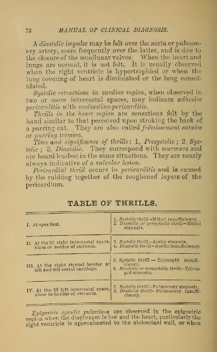
72 MANUAL OF CLINICAL DIAGNOSIS.
A diastolic impulse may be felt over the aorta or pulmon-ary artery, more frequently over the latter, and is due to
the closure of the semilunar valves. When the heart andlungs are normal, it is not felt. It is usually observed
when the right ventricle is hypertrophied or when the
lung covering of heart is diminished or the lung consol-
idated.
Systolic retractions in cardiac region, when observed in
two or more intercostal spaces, may indicate adhesive
pericarditis with mediastino-pericarditis.
Thrills in the heart region are sensations felt by the
hand similar to that perceived upon stroking the back of
a purring cat. They are also called Jremissement cataire
or purring tremors.
Time and significance of thrills : 1, Presystolic ; 2, Sys-
tolic ; 3, Diastolic. They correspond with murmurs andare heard loudest in the same situations. They are nearly
always indicative of a valvular lesion.
Pericardial thrill occurs in pericarditis and is caused
by the rubbing together of the roughened layers of the
pericardium.
TABLE OF THRILLS.
I. At apex beat.
II At the 2d right intercostal space,
close to border of sternum.
III. At the right sternal border at
5th aad 6th costal cartilage.
IV. At the 2d left intercostal space,
close to border of sternum.
1. Systolic thrill—Mitral insufficiency.2. Diastolic or presystolic thrill—Mitral
stenosis.
3. Systolic thrill—Aortic stenosis.4. Diastolic thrill—Aortic insufficiency.
5. Systolic thrill — Tricuspid insuffi-ciency.
6. Diastolic or presystolic thrill—Tricus-pid stenosis.
7. Systolic thrill—Pulmonary stenosis.8. Diastolic thrill- Pulmonary insuffi-
ciency.
Epigastric systolic pulsations are observed in the^ epigastric
region when the diaphragm is low and the heart, particularly the
right ventricle is approximated to the abdominal wall, or when

PERCUSSION OF THE HEART. 73
hypertrophy of the right ventricle is present. Also present in abdom-inal aneurism. Pulsations in this region may also be transmittedby the liver, enlarged stomach or tumors overlying aorta. Whenthe abdominal walls are thin, aortic pulsations may be observedas a normal condition. The knee-elbow position will, as a rule,
remove a transmitted pulsation, but if aneurism is present it will
continue to pulsate distinctly.
PERCUSSION OF THE HEART.
Over the anterior surface of the heart uncovered by lung, flat-
ness is obtained on percussion. This is in accordance with thegeneral rule that all airless tissues, whatever their histological
structure may be, give a pronounced dull sound on percussion.The anterior surface of the heart at its upper part and right half,
being covered by lung tissue, yields, when the latter is not toodense, a dull sound on percussion. It will be seen from this, thattwo different forms of cardiac dullness are obtained ; and further-
more, that where the lung tissue covering the heart is too dense,simple percussion will yield no results.
Forms of cardiac dullness : 1. Superficial or absolute.
2. Deep or relative. 3. Resistance of the heart.
Superficial or absolute cardiac dullness (Fig. 6), is
obtained by weak percussion only. It represents that
portion of the heart uncovered by lung tissue, and is theeasiest to obtain.
A small portion of uncovered heart behind the sternum givesresonance on percussion, because the percussion blow is conveyedby the sternum to the adjacent lung structure.
The superficial cardiac dullness begins above, at the
lower border of the 4th rib; the right boundary is at the
left border of the sternum, the left boundary is formed bya line drawn from the 4th costal cartilage, curving con-
vexly around, and ending at the apex beat. The lowerboundary can not as a rule be determined by percussion,
owing to the almost immediate contact of the left lobe of
the liver with the heart. We may theoretically construct
the lower border by drawing a line from the apex beat to
the sternal insertion of the 6th rib on the left side.
The absolute cardiac duHness in children is relatively greater(large heart) than in adults. It may begin above in the 3d inter-
costal space, with its left border at or near the mammary line andthe apex beat at the 4th intercostal space. In aged personsthe area of cardiac dullness is diminished (emphysema.)

74 MANUAL OF CLINICAL DIAGNOSIS.
Area of cardiac dullness in respiration. Quiet respiration doesnot appreciably affect dullness. Forced inspiration diminishes,and expiration increases the area of dullness. Deep inspirationin certain persons may cause an entire disappearance of cardiacdullness.
Deep or relative cardiac dullness does not actually repro-
duce the entire size of the heart. Strong percussion is
necessary. Like the absolute, it forms a triangular-shaped
figure of dullness. The upper border is at the sternal
insertion of the 3d left costal cartilage; the right boundaryis at the right border of the sternum; the left boundary is
formed by a line drawn from the upper border to the
apex beat, exceeding the left boundary of absolute dull-
ness by about one inch.
Resistance of the heart. This method, introduced byEbstein, and determined by palpable percussion, is sup-
posed to reproduce the actual size of the heart, a viewwhich is justly questioned by competent observers.
Regarding the diagnostic value of the different forms of cardiacdullness, no unanimity of opinion exists. While percussion for
the absolute dullness is generally employed owing to its compari-tive simplicity, it is influenced by the position of the lung borderswhich, when consolidated or retracted, would give an apparentincrease and when emphysematous an apparent decrease in thearea of dullness. The determination of the relative cardiac dull-
ness is largely influenced by the prejudiced wish of the observer.
It does not reproduce the entire size of the heart, because thelung tissue covering the latter at certain parts is too dense, thuspreventing the blow reaching the dullness beneath. It is
decidedly inferior in accuracy to the former method, which, whilepresenting many errors, their careful elimination is possible byother physical signs.
The area of cardiac dullness is increased in hypertrophy
and dilatation of the heart. The dullness is increased fromabove, downwards and outwards when the left ventricle
alone is involved, while in hypertrophy and dilatation of
the right ventricle the heart's dullness is broader andincreased toward the right side. When both ventricles
are implicated the dullness is increased transversely andlongitudinally.
Fluid in the pericardial sac (Hydropericardium). Thecharacteristic feature of this dullness is, that the outline
of precordial flatness is a blunt cone. An important sign
is the change of the area of flatness in different posi-

PERCUSSION OF THE HEART. 75
tions. The dullness is first increased upwards on a level
with the first intercostal space and laterally at the base.
The dullness is usually extended to the left of the
apex beat.The cardiac dullness may be apparently increased even when
the heart is normal as in retraction of the lung. In such a case, thelung borders about the heart undergo no change of position dur-ing respiration. An apparent increase of cardiac dullness mayalso occur in pleuritic effusions on the left side, or when the lungadjacent to the heart is infiltrated, or when tumors are present.Percussion in such cases is of no value in defining the cardiacdullness and recourse must be had to other signs.
The area of cardiac dullness is diminished in atrophy of
the heart, emphysema of the lung and mediastinum, andpneumo-pericardium.In emphysema the increased volume of lung causes it to envelop
the area of cardiac dullness and replaces it by a vesiculotympan-itic resonance. In pneumo pericardium, there is hyper-resonanceover the prsecordia, sometimes of a distinctly metallic character,when the patient is on his back ; when he sits up this disappears.Changes in location of the heart. In transposition of the viscera
(situs viscerum trans versus) the heart dullness and apex beat arefound in the corresponding place on the opposite side. The heartmav be dislocated to the right or left by pleuritic fluid, adhesions,abdominal accumulations, tumors, etc.
WEIGHT OF THE HEART.Average male 10—12 oz. (Gray).
" female 8—10 oz. «
SIZE.Length 5 inches.Width Z% "
Thickness 2>£ "
THICKNESS OF HEART WALLS.Thickness of right auricle 1 line(l-12th of an inch.)
"left " 1% lines." " right ventricle 2}| to 3 lines.11 " left
" 4 to 5 lines.
CAPACITY OF VENTRICLES.Capacity of right ventricle 2 fl. oz.
11 " left " 2fl.oz.
SIZE OF THE VALVULAR OPENINGS.Aortic orifice 1 In.
Mitral " 1.8 in.
Pulmonary orifice 1.2 in.
Tricuspid " 2 in.

76 MANUAL OF CLINICAL DIAGNOSIS.
AUSCULTATION OF THE HEART.
Indirect Auscultation with the small attachment of the stetho-
scope is alone indicated. Auscultate patient in a state of phys-ical and psychical rest, and in various positions. Auscultateduring quiet respiration, then during inspiration and finally in
expiration. It is often necessary to stimulate cardiac activity
before auscultation is attempted, this may be done by directing
patient to make various movements.
Acoustic Phenomena in Auscultation of the Heart. 1.
Heart sounds. 2. Endocardial murmurs. 3. Exocardialmurmurs.
Heart Sounds.—Two sounds are heard over the
heart, the first corresponding with ventricular contrac-
tion, the Systolic tone, the second, the Diastolic tone.
A very small interval of time is appreciable between the twosounds, with a distinct pause after the second sound. The first
sound corresponding with the apex beat and carotid pulse is
loudest at the apex, whereas the second sound is loudest at thebase of the heart, and occupies the first portion of the period of
diastole.
Difference Between the 1st and %d Sounds. The first
sound is longer, duller, and less clear than the secondsound which is short, sharp and of high pitch.
The heart tones and murmurs are best heard at their points of
origin. Auscultation can not always be practiced over theanatomical situations of the orifices of the heart owing to certainanatomical reasons. The mitral valve can not be auscultated at its
anatomical situation owing to a covering of dense lung tissue, andthe aorta is partially covered at its origin by the pulmonaryartery. The following table gives the anatomical situations of
the valves and points of auscultation. (See Fig. 6.)
NAME OP VALVE. ANATOMICAL SITUATION. POINT OF AUSCULTATION.
Mitral valve.
Upper border of the 3d left cos-tal cartilage close to thesternum.
Apex beat.
Tricuspid valve.Between the 3d left inter-
costal space and the 5thright costal cartilage.
Median line on a levelwith the 5th costalcartilage.
Pulmonary valves.
In the 2d left intercostal spaceclose to the left border ofsternum.
2d left intercostal spaceclose to the left bor-der of sternum.
Aortic valves.Between the median line and
3d left costal cartilage.2d right intercostal
space close to theborder of sternum.

AUSCULTATION OF THE HEART. 77
Number and Origin of Heart Tones. Six sounds are
heard over the heart. From each venous opening (mitral
and tricuspid valves), a systolic tone, and from the arte-
rial openings (aorta and pulmonary), a systolic and dia-
stolic tone. Two sounds are heard at each opening.
The second sound at the mitral and tricuspid is trans-
mitted from the aorta and pulmonary artery. Over the
ventricles the accent is on the first sound (trochee), over
the aorta and pulmonary artery it is on the second sound(iambus). The second aortic tone is normally stronger
than the second pulmonary tone. The following schemawill show on what tone the accentuation falls:
Mitral valve, I I I I
Tricuspid valve, \ —-V —
V
—
V
Pulmonary valves,)| |
Aortic valves, f V— V— V
—
Duration of the Two Sounds and Two Rests. The entire period of
the heart action being represented by 10.
Duration of the first sound, 444 " « rest, I
14 " 4< second sound, 2" " " 4 * rest 3
—10Causes of the heart tones :
Apex beat (mitral orifice).
First tone : closure of the mitral valve and contraction of theventricle.
Second tone : transmitted 2d aortic tone.
Lower end of sternum on a level with 5th costal cartilage (tri-
cuspid orifice).
First Tone: closure of tricuspid valve and contraction of the
ventricle.
Second tone: transmitted 2d pulmonary tone.
Second right (aorta) and left {pulmonary artery) intercostal
space
:
First tone : sudden tension of the aorta and pulmonary artery,
and transmitted ventricle tone.
Second tone: closure of the aortic and pulmonary valves.
Observe the Following in Auscultation of the Heart Tones:
1. Rythm. 2. Strength. 3. Reduplication of the heart
sounds.Rythm. The rythm of the heart tones may be changed,
and still be consistent with a normal heart, these changes
being due to psychical causes, use of tobacco, sexual

7S MANUAL OF CLINICAL DIAGNOSIS.
excesses, or they may be congenital. Variations of rythmmay, however, be due to degeneration of the myocardium,or associated with valvular disease.
Strength of the Heart Tones. Normally the intensity of
the tones is influenced by the approximation of the heart
to the chest-wall, and the density of the latter's coverings.
In children and emaciated persons, the tones are relatively loud.
When the heart is pushed toward chest-wall, the tones are also
loud. They are louder in the erect than in the recumbentposition. During inspiration, the intensity of the tones is dimin-ished, owing to lung covering heart, which is a poor conductor of
sound ; the contrary holds good when the lung is consolidated. Thegreater the activity of the heart's action, the louder the tones.
Strengthening of the First Heart Tone is observed whenthe work of the heart is increased, as in hypertrophy; also
in mitral stenosis.
The cause of the strengthened first sound in mitral stenosis maybe explained as follows: In consequence of stenosis, the left ventri-
cle receives only slowly a small amount of blood ; if, now, a normalsystole of the ventricle occurs, a decided difference in the tensionof the valve exists in systole and diastole, resulting in intensifi-
cation of the systolic tone.
Strengthening of the 2d Aortic and Pulmonary Tones,
if lasting, is a most important sign of hypertrophy of the
left and right ventricles respectively.
Weakened First Sound occurs in degeneration of the
heart musculature, emphysema and aortic insufficiency.
The Systolic tone at the apex is often weakened in aortic insuffi-
ciency and is owing to the great tension of the mitral valve at theend of diastole owing to the regurgitation of blood from the aorta,
so that at the systole of the ventricle, the tension of this valve notbeing greatly increased the first tone is consequently weak.
Weakened Second Sound over the Aorta and PulmonaryArtery occurs in stenosis of these openings, and is dueto the diminished blood pressure and vibration of thediseased valves. Also present in stenosis or insufficiency
of the mitral orifice, and is due to the diminished tension
of the semilunar valves caused by the small quantity of
blood thrown into the aorta.
Reduplication of the Heart Tones is observed in
unequal tension of the blood in both ventricles, whichcauses the valves on both sides to close at different
times.

AUSCULTATION OF THE HEART. 79
Beduplication of the tones may be physiological and dependentupon respiration. It may also be associated with pronounceddisease of the circulatory apparatus.
Metallic Sounding Heart Tones are heard at times when lungcavities are in proximity to the heart ; also in pneumo-thorax,pneumopericardium, dilatation of the stomach, etc.
ENDOCARDIAL MURMURS.These murmurs have their origin within the heart, and are
usually divided into organic and inorganic murmurs. Organicmurmurs are usually produced by constriction of an orifice of theheart (stenosis) or an incomplete closure of the valves {insufficiency).
The former is called an obstructive, the latter a regurgitantmurmur.Causes of Cardiac Murmurs.—When fluid passes through a tube
which is suddenly narrowed, eddies are formed, which lead toaudible murmurs. The more rapid the current the greater arethe eddies and the louder the murmurs. As the cardiac orifices
normally represent no decided narrowing, the blood produces nomurmurs. If an obstruction (stenosis) to the outward flow of
blood is present, then eddies are formed and murmurs heard.Murmurs due to obstructions are heard during systole. Murmursarise in insufficiency of the valves in the following manner:After the blood has passed through an orifice, the valves close, butbeing insufficient, a portion regurgitates through the insufficient
valves, causing a murmur.The murmur of insufficiency is heard at the arterial opening
during diastole, and during systole at the venous openings.Roughness of the arterial coats or of the orifices of the heart alonewill not produce murmurs.
Cardiac murmurs are differentiated according to time,
character, and position.
Time: Murmurs may occur during systole or diastole.
A murmur peculiar to mitral stenosis and called a pre-
systolic murmur is heard during the termination of dias-
tole, or, occurring at the beginning of diastole, it is louder
during the end.
Character: This has been variously described as blow-
ing, rasping, sawing, etc. Murmurs of a musical char-
acter are sometimes heard and owe their origin as a rule
to the vibration of a membranous substance, suspendedin the blood stream.
Position: This refers to the point where murmurs are
heard loudest. As a rule murmurs are most intense at

80 MANUAL OF CLINICAL DIAGNOSIS.
their point of origin. Murmurs are propagated in the
direction of the blood current by which they are developed.
Axioms:—1. The character or intensity of a murmur is no indexto the gravity of the lesion producing it. The loudest murmurmay be produced by the smallest lesion and vice versa.
2. The intensity of a murmur is largely dependent on the activ-
ity of the heart. Faint murmurs may often be converted intoloud murmurs after increasing cardiac activity by active exercise.Weakness of the heart may abolish a murmur previously distinct,
the murmur reappearing after cardio-tonic medication.
3. A murmur is generally louder when the patient is lying down,then when he is standing. Always auscultate murmurs with thepatient in various positions.
4. Murmurs are less loud in inspiration than expiration.
5. Strong pressure on the chest may cause the disappearance of
murmurs, the pressure inhibiting cardiac action.
6. When the heart is rapid or irregular in action, it is difficult
to determine the time of a murmur. Remember that systolic
murmurs are synchronous with the carotid pulse. Also regulatethe action of the heart with digitalis.
7. Systolic are usually louder though less prolonged then dias-
tolic murmurs.8. When murmurs are faint, the patient should suspend respi-
ration during auscultation.
Coexistence of Several Murmurs.—This is frequent when insuf-
ficiency of the valves is combined with stenosis of the same orifice.
In such a case murmurs would be heard during systole and dias-
tole. When different orifices are involved at the same time it is
difficult and even impossible to localize the lesions by ausculta-tion alone.
Endocardial Inorganic Murmurs (anaemic or hsemic
murmurs). The cause of these murmurs is yet sub judice.
They have been attributed to disturbances in innerva-
tion, to relative insufficiency of the valves, i. e. the valves,
without having undergone any anatomical change, are nolonger able to close the enlarged orifices of the heart,
and finally to diminution in the quantity of blood. Inthe latter condition the smaller arteries, veins and capil-
laries must accommodate themselves to the diminishedquantity of blood, and contract; but the aorta and pulmo-nary artery cannot contract to the same degree; their
diameter, in consequence, being proportionately larger
than that of the orifices through which the blood enters
them, murmurs are produced.

CARDIAC MURMURS. 81
TABULAR VIEW OF CARDIAC VALVULARMURMURS.
TIMEOF
MURMUR.
USUAL SEATOF
MURMUR.
OCCASIONAL SEATOF
MURMUR.
CAUSEOF
MURMUR.
Aortic orifice—Systolicmurmur.
2d right intercos-tal space closeto the border ofsternum.
Over the carotid arte-
ries. The most wide-ly diffused of all car-diac murmurs.
Obstruction to out-ward flow of bloodthrough aortic ori-
fice.
Mitral orifice—Systolicmurmur.
Apex beat.
2d left intercostalspace close to theborder of sternum;may also be tra'smit-ted by chest-wall tothe axillary borderor beyond it.
Regurgitation of bloodthrough mitral ori-
fice into left auricle.
Pulmonaryorifice—
Systolic mur-mur.
2d left intercostalspace close to theborder of the ster-num.
Obstruction to out-ward flow of bloodthrough pulmonaryorifice.
Tricuspid ori-
fice—Systo-lic murmur.
Median line ofsternum on a lev-el with 5th costalcartilage.
Regurgitation of bloodthrough tricuspidorifice into rightauricle.
Aortic orifice—Diastolicmurmur.
2d right intercos-tal space close tothe border of thesternum.
In the middle of ster-
num. It may also beheard over the aortaand carotids.
Regurgitation of bloodthrough aortic ori-fice into left ventri-cle.
Mitral orifice,—Diastolicmurmur.
Apex beat.Murmur rarely dif-fused. Usually lim-ited to apex beat.
Obstruction to the flowof blood from leftauricle to the leftventricle.
Pulmonnryorifice —Dias-tolic murmur.
2d left intercostalspace close to theborderof the ster-num.
Down right side ofheart.
Regurgitation of bloodthrough pulmonaryorifice into rightventricle.
Tricuspid ori-
fice—Diastolicmurmur.
Median line ofsternum on a lev-el with 5th costalcartilage.
May be transmitted tothe base of the heartor to the right axilla.
Obstruction to the flowof blood from rightauricle into rightventricle.
Characteristics of anaemic murmurs :
—
1. Usually soft and blowing in character, and not prolonged.2. Occur during systole only, and usually systolic tone is heard.3. Generally loudest at base of heart.
M. CD. e

82 MANUAL OF CLINICAL DIAGNOSIS.
4. Accompanied with anaemic symptoms, and murmurs in theveins of the neck.
5. Unaccompanied, as a rule, by changes in size of the heart.6. They frequently change their character.7. Under appropriate treatment of the general condition, th^y
disappear.
Ancemic murmurs are usually loudest over the pulmonary artery,
a point where organic systolic murmurs are frequently heard.The chief means of differentiation between the two, lies in thefact that with organic murmurs we find dilatation and hypertrophy
of the hearty which are usually absent in anaemic murmurs.
Exocardial or Pericardial murmurs. These murmursare friction sounds produced by the rubbing of one sur-
face of the pericardium upon the other when roughenedby a fibrinous exudation.
Characteristics of pericardial murmurs :
—
1. Unlike endocardial murmurs, which are limited to a certain
phase of the heart's action, they may be systolic, diastolic, orboth, or even presystolic.
2. They are increased in intensity upon pressure with thestethoscope, which maneuver facilitates the friction between thepericardial layers.
3. During deep inspiration the lung approximates the layers
of the pericardium, thus increasing pericardial murmurs duringthis phase of respiration. Endocardial murmurs by the sameact are diminished in intensity, because the interposed lungoffers a poor medium of conduction to the chest-wall.
4. Pericardial murmurs are circumscribed, and, unlike endocar-dial murmurs, are not transmitted beyond the area of cardiac
dullness.5. Change of position exerts a greater influence on the character
of pericardial than endocardial murmurs. Pericardial murmursare rendered especially distinct when the patient is in the sitting
posture, with the body inclined to the left 6ide.
6. Pericardial murmurs give the impression of being of superfi-
cial origin.
7. Pericardial murmurs frequently change their character, thusbeing unlike endocardial murmurs, the character of which is
almost constant.8. Pericardial murmurs are rough, grating to and fro, or rub-
bing and scratching sounds.
Extra-Pericardial Murmurs.—These sounds are with difficulty
distinguished from pericardial sounds proper. They occur whenthe pleura or peritoneum adjacent to the heart is roughened.They differ from pericardial murmurs by being dependent moreon the movements of respiration than of the heart, and theymay at times be made to disappear by suspending respiration.
Palpitation of the Heart.—This common complaint ii often

DIAGNOSIS OF CARDIAC DISEASES. 83
mistaken by the patient lor some organic heart affection. Amongthe causes are weakness of the heart muscle (fatty degeneration)
;
excessive use of stimulants (tea, coffee, tobacco), and psychicaldisturbances. It is also frequent in so-called nervous patients.
It is manifested in the main by paroxysms of increased frequencyof the heart action, unattended by any increase in the heartdullness. When associated with systolic murmurs the latter aieusually of anaemic origin. Palpitation as a functional disturbanceis not associated with dyspnoea and venous stasis or other symp-toms peculiar to organic affections of the heart.
Puncture of Pericardium.—This may be practiced as an aid to
diagnosis or in the removal of large effusions. The point of
election, according to Roberts, is in the 5th intercostal spicefrom 2 to 1%. inches to the left of the median line of the sternum.Aside from injuring the heart or puncturing the pleura, theselection of this point prevents injury to the internal mammaryartery which runs from X to j£ an inch from the left border of thesternum.
DIAGNOSIS OP CARDIAC DISEASES.
Hypertrophy of Left Ventricle.—Symptoms: Increased tension ofthe radial pulse,pulsation of temporal and carotid arteries, vertigo,headache, florid face, ringing in the ears, epistaxis and iosomnia!Physical signs. Inspection : Bulging of praecordial region, apexvery forcible and extended downwards and to the left. Per-cussion; increased dullness downwards and to the left. Auscul-tation ; loud heart tones with accentuation of 2d aortic tone.
Hypertrophy of Right Ventricle.—Symptoms: Stasis in thepulmonic circulation leading to dyspnoea, haemoptysis and bron-chitis. Physical signs. Inspection; Apex beat diffused andmarked in the epigastrium. Percussion; increased cardiac dull-ness to the right. Auscultation ; accentuation of 2d pulmonarytone.
Dilatation of the Heart.—Symptoms: Peculiar to a feeblecirculation, turgid veins, feeble pulse, dyspnoea, oedema andeffusions in the internal cavities and symptoms indicative of con-gestion of the viscera. Physical signs. Inspection: Apex beatdiffused and very feeble. Percussion; Cardiac dullness increasedto the right or left according to side of heart involved. Ausculta-tion ; Cardiac tones are very feeble and indistinct.
Pericarditis, 1st or dry stage.—Apex beat tumultuous and irregu-lar; friction fremitus felt and friction sound heard.2d or stage of effusion.—Apex beat feeble and fluttering; cardiac
dullness of a triangular form. Heart sounds, feeble or absent overthe area of dullness, but are heard above the line of effusion dis-tinctly and loud.

84 MANUAL OF CLINICAL DIAGNOSIS.
Sd or stage of absorption.—Disappearance of dullness, and reap-pearance of heart tones and friction sound. If effusion is notabsorbed, the physical signs of 2d stage continue.
Aortic Regurgitation.
—
Symptoms; pulsation of the arteries,
pulse is forcible, but recedes from the finger (waterhammer, or the
Corrigan pulse) ; capillary pulse. Sphygmographic tracing charac-teristic. (See pulse).
Physical signs.—Bulging precordial region, apex beat extendedto the left and downwards, increased cardiac dullness to the left.
Diastolic murmur over the aorta. Systolic murmur at times at
apex, and accentuation of 2d pulmonary tone.Aortic Stenosis.—Vertigo frequent. Apex beat extended to the
left and downwards. Cardiac dullness increased to the left. Loudsystolic murmur over aorta ; also heard over other ostia, althoughfeeble; 2d aortic tone absent.
Mitral Regurgitation.—Pulse irregular. Apex beat dislocateddownwards and outwards and diffused. Apex beat is feeble, anda systolic thrill is felt. Increased dullness peculiar to hypertrophyand dilatation of right ventricle. Systolic murmur at apex andover pulmonary artery, traceable to axillary region and angle of
scapula.Mitral Stenosis.—Apex beat felt in epigastrium and to the right
of sternum. Presystolic thrill at apex. Percussion same as mitralregurgitation. Prolonged presystolic murmur at apex, and usu-ally localized.
Tricuspid Regurgitation.—Increased cardiac dullness to theright, systolic murmur over tricuspid valve, weakened 2d pulmo-nary tone, venous pulsation. (See examination of the veins.)
Tricuspid Stenosis.—This is the rarest of all heart diseases.Venous stasis occurs, and the symptoms are the same as tricus-
pid regurgitation.Diseases of the pulmonary valves are usually congenital, and
characterized by pronounced cyanosis, dilatation of the right side
of heart, and murmurs, according to character of lesion, bestheard over pulmonary artery.

CHAPTER VII.
EXAMINATION OF THE ARTERIES ANDVEINS.
The Arteries.
—
Methods : Inspection, palpation, percussion, auscul-tation.
Inspection. Pulsation of the larger arteries is notusually discerned in healthy persons. Pulsations of thearteries of the neck are seen when the heart is very muchincreased in action. In hypertrophy of the left ventricle
f
these pulsations are permanent and pronounced.
Capillary pulse. In healthy individuals, the capillary blood-vessels at the roots of the nails often show systolic redness anddiastolic paleness. This so-called " capillary pulse " becomesexaggerated in diseased conditions, notably in aortic regurgitation.The explanation of this phenomenon is as follows : In conse-quence of the hypertrophy of the left ventricle of the heart, anincreased quantity of blood received by regurgitation from theaoi ta and left ventricle, is thrown into the relatively empty aorticsystem ; then follows a rapid emptying of this system toward thecapillaries and the heart.
Ophthalmological examination often shows pulsation of the retinalblood-vessels. At times, pulsations of the liver and spleen can befelt. F. Mueller has lately called attention to pulsations of thesoft palate, which he claims are similar in origin and significanceto the capillary pulse. Capillary pulsation may be demonstratedby making lines with some blunt pointed instrument on the skinof the forehead, and the lines are 3een to become alternately paleand red.
Palpation. In many of the arteries, notably the radial
and temporal, the wails are thickened and rigid, and car-
tilaginous-like plates are felt. This condition is peculiar
to arterio sclerosis, and is due to calcification of the middlearterial coat.
Auscultation. This is accomplished by means of astethoscope, avoiding pressure of the artery.
(85)

86 MANUAL OF CLINICAL DIAGNOSIS.
Normal auscultation. Over the carotid and subclavian arteries,
two tones are heard. The first is the transmitted 1st aortic tone,
the second, the transmitted 2d aortic tone,.
Over the abdominal aorta and femoral arteries, one tone (due to
tension of the artery) is at times heard, although as a rule no soundsare normally heard. Murmurs heard over arteries are usuallypathological. The student will do well to familiarize himself withcertain errors consequent upon unskilled auscultation of the arte-
ries. For study, select the brachial artery at the elbow joint ; nowmake moderate pressure with the stethoscope on the artery, whichwill develop a murmur (pressure murmur). If the pressure is
increased to a certain point, a short, sharp tone is produced(pressure tone). Auscultation of the carotid artery is practiced atalmost any part of its accessible course and of the subclavian artery
above the clavicle, or below in the fossa of Mohrenheim.
Arterial Murmurs. In aortic stenosis a murmur repla-
ces the first tone over the carotids. The second tone
over the carotids and subclavian is replaced by a mur-mur in aortic regurgitation. Murmurs originating at the
mitral and tricuspid valves can likewise be transmitted
to the arteries. Murmurs from the aorta are best trans-
mitted by the right, and murmurs from the pulmonaryartery by the left carotid.
Locally Produced Arterial Murmurs occur wheneveran artery in its course is dilated or narrowed. The mostimportant of these murmurs is the aneurysmal murmur
}
which may be systolic, diastolic, or both.
Differences in Symmetrical Arteries. In health sym-metrical arteries show the same pulse qualities, unlessanatomical anomalies exist. Any mechanical obstruc-
tion, whether due to emboli, aneurisms or tumors com-pressing an artery will make a comparative change in
the qualities of the pulse.
Examples : If an aneurism involving the arch of the aorta is
situated between the innominate and left subclavian artery, thenthe pulse of the right radial and carotid arteries can be f^lt
sooner than in the same arteries on the left side. Again,if the aneurism lies between the left carotid and left subclavianarteries, the pulse can be felt earlier in both carotids and inthe right radial artery than in the left radial and femoral arteries.
Paradoxical Pulse. This occurs when the radial pulsedisappears either during inspiration or expiration. It is
caused by adhesions between the subclavian artery andthe pulmonary pleura.

THORACIC ANEURISM. 87
When the shoulders are pressed backwards and downwardswith the hands on the gluteal regions, and deep inspiration is
made, the radial pulse on both sides can be made to disappear.This is explained by compression of the subclavian arteriesby the first rib. When the first rib hau become rigid in conse-quence of an ossifying perichondritis, a condition peculiar tophthisis, tben notwithstanding deep inspiration, the radial pulse is
still present.
Percussion is of limited application, although it is useful in
defining the situation of an aneurism.
Subclavian murmurs are heard over the subclavianarteries either during inspiration (more often) or expira-
tion.
These murmurs are frequently present in phthisical persons, andthe explanation of their occurrence is similar to that of the para-doxical pulse, viz. adhesions between the subclavian artery andpulmonary pleura. Fuller found the subclavian murmur present12 times in 100 healthy persons.Brain murmurs are heard in children from the 3d month to the
6th year by auscultation of the head. Over the regions of thefontanelles the murmurs are loudest. They are physiologicaland are transmitted from the carotids.
Pathological arterial tones are heard over arteries where tonesare normally absent. In aortic regurgitation, in consequence of
sudden tension of the arteries, tones are heard correspondingwith the pulse in peripheral arteries.
Thyroid arterial murmurs are present in all enlargements of
this gland.
THORACIC ANEURISM.*Symptoms. Pain is nearly always present. Pressure on vari-
ous nerves, on the trachea, oesophagus, etc., will produce char-acteristic symptoms.
Physical Signs.
—
Inspection. Until the aneurism has attaineda certain size nothing is observed. Later, bulging is apparentupon the surface of the chest involving the upper part of,
or immediately to the right, of the stornum. If the transverse
portion of the aorta is involved then pulsation is^ observed in thesupra sternal fossa. The pulsation of an aneurism correspondswith the systole of the heart and is equally distended in all parts.
* Schnell has recently directed attention to an apparatus called tbeAneurysmatoscopc^ for the diagnosis of aneurisms of the descending aorta. It
consists of an ordinary soft rubber stomach tube closed at its lower end, andcontaining at its upper end a glass tube. The whole is then filled with acolored fluid up to the latter. When the tube is introduced into the oesopha-gus, the pulsations of the aneurism are transmitted to the fluid in the tube,and can thus be recognized.

88 MANUAL OF CLINICAL DIAGNOSIS.
Palpation. Careful palpation will discover an abnormal pulsa-tion usually systolic in time, even before the aneurism reachesthe surface of the chest. A systolic or diastolic thrill may accom-pany the pulsation.
Percussion yields a dullness even in the early stages of theaneurism, which may be limited to the manubrium or to the rightof the sternum. The dullness may be continuous with or distinctfrom the cardiac dullness.
Auscultation. One or both heart sounds maybe heard; or asystolic or diastolic murmur or both. At times no sound or mur-mur is heard.
THE VEINS.Inspection and palpation. 1. Increased distension of the
veins. 2. Abnormal movements of the cervical veins. 3.
Thrombosis.
Increased Distension. This is present when the returnof the venous blood to the heart is rendered difficult in
consequence of general and local causes.
General causes : Diseases of the heart and lungs.Whenever the power of the right auricle and particularly of the
right ventricle is reduced, these cavities are incompletely emptiedand the return of venous blood to the heart by means of the supe-rior and inferior venae cavae is prevented. The return of venousblood to the heart is facilitated by the aspiration of the thorax andthis is dependent on the elasticity of the lungs. Any affectiondiminishing lung elasticity conduces to venous stasis and abnor-mal distension of the veins.
Local causes : Thrombosis of or compression of theveins. Abnormal movements of the cervical veins may berespiratory or pulsatory.
During inspiration the return of venous blood to the heart is
facilitated, whereas during expiration a physiological hindranceexists. When the veins are filled with a normal quantity of blood,no respiratory movement is observed. It is only when abnormaldistension is present, that this movement is discerned. The veinsmay swell only in expiration, in mediastino-pericardiiis owing to adiminished caliber of the veins in inspiration.
Pulsatory movements are dependent on cardiac activity
and show the same rythm as the latter. The pulsatorymovement may be communicated or develop in the veinitself.
The first is communicated to the internal jugular vein by thepulsation of the carotid artery especially when the latter pulsatesviolently.

THE VEINS. 89
If the pulsation of the vein is communicated, pressure with thefinger on the vein in the middle of the neck, will cause the pulsa-
tions to cease in the veins below the finger. If the carotid arteryis effectually compressed the pulsation in the vein will also disap-pear showing that it is communicated from the artery.
True venous pulsation is observed in the jugular veins
independent of any impulse communicated by the carotid
artery, and is caused by regurgitation of blood duringthe systole of the heart into the superior vena cava andjugular veins. This pulsation is pathognomonic of tri-
cuspid insufficiency.
Unlike the communicated pulsation, pressure with the finger
on the vein, only increases the pulsation "below the finger. Thetrue venous pulse is most pronounced in the right internal jugularvein.
Venom Pulse of the Liver. This is a pulsation of the liver pres-
ent in tricuspid insufficiency and is caused by regurgitation of
blood into the inferior vena cava.
In aortic regurgitation pulsation of the liver has been observedand is caused by an abnormal distension of the hepatic arteries.
Auscultation.—Venous tones occur when bloodregurgitates with a certain force from the heart into the
veins, thus bringing the valves of the veins into suddentension and causing a tone. Present in tricuspid insuf-
ficiency.
Venous murmurs when present are heard most fre-
quently over the internal jugular vein of the right side at
its junction with the subclavian to form the innominate.
These murmurs are usually continuous (continuous hum or
bruit de diable), and are well marked in ansemic andchlorotic persons.
Pressure on the vein with the stethoscope must be avoided.The venous murmur is not confined to either the systole or dias-
tole of the heart, but persists through both. It is loudest duringinspiration and is increased in intensity when the head is turnedto the other side.

CHAPTER VIII.
THE PULSE.
The pulse is the expansion of an artery produced bya wave of blood set in motion by the passage of blood
into the aorta at the systole of the ventricle. Palpationdetermines: 1. The frequency. 2. Therythm. 3. Thequality of the pulse.
Frequency of the Pulse. In the healthy adult there is
an average of 70 beats in a minute, in children 100-140,
and in old men, 70-90, or more.
The frequency of pulse is greater in the female than in the male.The daily variation in the frequency of the pulse corresponds witha like variation in the body temperature. It is most frequentbetween noon and evening, and is least frequent in the early hoursof the morning. The influence of position is such that it is mostfrequent standing, and least frequent in the recumbent posture.External temperature when high increases, when low diminishesthe frequency. Increased muscular exertion, acceleration of res-
piration, and psychical activity also increase the frequency of thepulse.
Pathological Pulse Frequency. A slower movement of
the pulse (pulsus rarus), and acceleration of the pulse(pulsus frequens) are distinguished.
Pulsus Rarus (bradycardia). Observed in conditions
leading to an irritation of the vagus, or paralysis of the' intra-cardiac ganglia and of the sympathetic. Also,
in increased cranial pressure (meningitis), icterus (fromthe action of the gall acids on the cardiac ganglia), ste-
nosis of the aortic and mitral orifices, degeneration of themyocardium (fatty heart) ; in colic, and after adminis-tration of digitalis.
Pulsus Frequens (tachycardia). Observed in paralysisof the vagus, irritation of the sympathetic and affections
of the cardiac ganglia ; in excessively increased cerebral
(90)

THE PULSE. 91
pressure (last stage of basilar meningitis), acute diseases
of the heart (peri and endocarditis), and in valvular
heart diseases when compensation is disturbed. Exces-sive rapidity (over 160) is a sign of heart-weakness(collapse.)
Paroxysmal tachycardia is also a symptom of functional dis-
eases of the heart. In hysteria and Graves' disease the pulse is
also increased in frequency.
Eythm of the Pulse. Irregularity of the pulse (pulsus
irregularis) when observed in children and adults, nearlyalways indicates disease of the heart or brain. Inadvanced age irregularity of the pulse is of no importance.
Forms of Pulsus Irregularis: Pulsus alternans, a low pulse-wavefollowing a high one; pulsus bigeminus, sl long pause after twobeats; pulsus trigeminus, a long pause after three beats; pulsusparadoxus, the pulse is smaller with each inspiration, or disap-pears (observed in stenosis of the air-passages, mediastinitis, andpericardial adhesions). Retardation, or unequal size of the pulsebetween symmetrical arteries, or between arteries of the upperand lower half of the body, is observed in stenosis of the arteries
and aneurism.
Quality of the Pulse. In determining the quality, weconsider: 1. Expansion; 2. Force and tension; 3.
Size of pulse.
The arteries distend rapidly (pulsus celer) in hypertrophy of theleft ventricle and in increased heart action. Pulsus celer is char-acteristic of aortic insufficiency, contracted kidney, and exophthalmicgoitre. The arteries distend slowly (pulsus tardus) in old age(senile pulse), aortic and mitral stenosis, and in aneurisms. Inconsidering the force and tension of the pulse, we speak of a hard(pulsus durus) and a soft (pulsus mollis) pulse. The hardness of thepulse is dependent on the tension of the arterial wall ; the greater
the tension the harder the pulse. Such a pulse is difficult of
compression. A hard pulse is observed in hypertrophy of the left
ventricle, and in spasm of the arterial wall, as occurs in lead colic.
A soft pulse is easily compressed, and is found in cardiac degene-ration, fever, and anaemia. In atheroma of the arteries, wherethe walls are infiltrated with calcareous salts, the pulse is appa-rently hard. The artery in such a condition can be rolled underthe finger.
The size of the pulse is dependent on the force of the heart, theamount of blood in the artery, and the tension of its wall. Alarge pulse (pulsus magnus) is observed in aortic insufficiency, anda small pulse (pulsus parvus) in aortic stenosis. Sometimes thepulse is so slight as to be thready (pulsus filiformis.)
The Pulse in Diseases of the Heart.
—
1. Mitral Insufficiency ; no decided departure from the normal.

92 MANUAL OF CLINICAL DIAGNOSIS.
2. Mitral Stenosis; pulse small and irregular, frequencyincreased.
3. Aortic Insufficiency ; pulsus celer, frequency normal orincreased, usually regular.
4. Aortic Stenosis ; pulse small, retarded, normal or diminishedfrequency, and regular, as a rule.
5. Myocarditis ; pul^e small and soft, irregular, frequency nor-mal, diminished or increased.A comparatively strong pulse, with feeble apex beat and heart-
tones, is of great value in the diagnosis of exudative pericarditis.
SPHYGMOGRAPHY.This is a method of graphically recording the charac-
ters of the pulse Dy means of the sphygmograph.Normal Pulse Tracing.—(Fig. 9) We notice first an ascending
line which is straight, and then a descending line which showscertain elevations. The ascending and descending lines meetabove at what is called the summit of the pulse curve. Where theascending line begins and the descending line ends is called thebase of the curve. The ascending corresponds with the filling of
the artery ; the descending line with its collapse. The more rapidthe flow and the more quickly the artery distends the more vertical
the line.
Pis. 9.
Fig. 9. Normal pulse, al. Line of ascent, dl. Line of
descent, cs. Summit of curve, b. Base of curve, r. Elevationof recoil, e' e". Elastic elevations.The descending line is broken in its descent by elevations
which are distinguished as the elevations of elasticity and theelevation of recoil. The elevation of recoil is situated about half
way down the descending line and is produced by the column of
blood after impinging on the already closed aortic valves, produc-ing a new wave.The elevations of elasticity are smaller than the elevation of
recoil (also called dicrotic wave) and are produced by vibration of
the tense elastic arterial wall. In a normal pulse curve twoelastic elevations are found, one above the other below thedicrotic wave.

SPHYMOORAPHY. 93
Pathological Sphygmographic Tracings.—Anacrotic pulse. This is present when elevations are
found on the ascending line, andindicate some abnormality in thearterial distribution of the blood.
It is found in any disease of theheart or arteries which allows of onlyspasmodic entrance of blood into thelatter.
The greater the decrease in arterial
tension the more pronounced is theelevation of recoil. When the tensionof the artery is increased (lead colic,
acute and chronic nephritis) the elastic
elevations of the artery are pro-nounced, and the elevation of recoil
is but feebly represented. The eleva-
tions of elasticity may disappear whenthe arterial tension is diminished.
Dicrotic pulse. This is charac-
teristic of fever, which decreases
arterial tension, thus bringing
into prominence the recoil eleva-
tion, and causing the disappear-
ance of the elastic elevations.
Many forms of dicrotic pulse aredistinguished: 1. Subdicrotic (whenthe elevation of recoil appears beforethe descending line has reached thebase of the curve). 2. Dicrotic (after
it has reached the base of the curve).
3. Hyperdicrotic (when the recoil ele-
vation belongs to the ascending partof the next wave). 4. Monocrotic(when no recoil elevation can berecognized.)
Pulsus tardus (senile pulse). Aslow ascending line, with roundand broad summit and no ele-
vations on the descending line.
Observed in atheroma of the
arteries.
Pulse Curve in Valvular Lesionsof the Heart.
—
Aortic Stenosis. Theascending line is more sloping owingto the retarded entrance of blood into
the arteries, and the recoil elevation
Dicrotic Pulse.
Hyperdicrotic
Pulse.
Subdicrotic Pulse.
Monocrotic Pulse.
Pulsus Magnus,
et celer.
Irregular Pulse in
dilatation of
the heart
Slow Pulse,
(action of digitalis)
Pulsus Tardus.
Fig. 10.

94 MANUAL OF CLINICAL DIAGNOSIS.
is absent or imperfectly marked because the arteries are incom-pletely filled. Aortic regurgitation : the ascending line is high andvertical, the summit pointed and the dicrotic wave absent. Mitrallesions: in mitral obstruction, the pulse curve corresponds withdiminished arterial tension, whereas in insufficiency the tracingsare varied in relation to the amount of hypertrophy present.
Pulse Curve in the Veins.—This is the reverse of that in thearteries, the ascending line rising slowly, whereas the descendingline falls quickly.In tricuspid insufficiency the venous pulse begins in the diastole
of the heart and reaches its maximum in the systole, whereas thevenous pulse occurring with a normal tricuspid valve falls imme-diately before the beginning of the systole. The venous pulse intricuspid insufficiency results from the blood wave, which is
thrown back during the systole of the heart through the insuf-
ficient orifice into the auricle and thence into the venous system.The clinical value of the sphygmograph cannot be denied. It
not only confirms the evidence obtained by digital examination of
the pulse, but makes evident certain qualities not appreciated bythe fingers. It furnishes a record for future reference, and is
almost indispensable for class instruction. Various modificationsof Marey's sphygmograph have been introduced, one of the latest
and best, being that of Ludwig.

CHAPTER IX.
THE BLOOD.
The whole quantity of blood in an adult is about ^ of
the body weight (10 lbs), whereas in new-born infants it is T̂ of
the body weight. Specific gravity varies between 1045 and 1075.
Reaction is alkaline.
Color of the Blood.—The blood removed from a
healthy person is lighter when it contains much oxygen(arterial) and dark when oxygen is deficient (venous).
The blood of dyspnoeic persons is very dark. Carbonic oxidepoisoning, cherry-red color; anaemia and chlorosis (hydremia),watery ; leucsemia, pale.
Haemoglobin. Amount contained in 100 ccm. of bloodis for man, 13 to 15 grams, in woman slightly less. Onheating, haemoglobin is resolved into albumen and hsema-tin.
Teichmann's test for blood. Dried blood heated on an object-glass
with one or two drops of glacial acetic acid to the boiling point towhich is added a grain of common salt and then slowly evaporatedwill show the formation of brownish-yellow rhombic crystals{muriate of hcemcetin) the so-called hozmin.
Quantitative estimation of haemoglobin can be approximatelyand very satisfactorily determined by means of FleischVs Hcemom-eter, which consists essentially of comparing the color of the blooddissolved in water with a wedge of colored glass marked in percent-ages by an empirically determined scale. The hamioglobinome-ter of Gowers, consists of two slender glass tubes, one of whichcontains a preparation of carmine and glycerine jelly, colored to
represent a dilution of 1 part of healthy blood in 100 parts of water,the other tube being: graduated into 100 divisions, each of which is
equivalent to the volume of blood taken, so that 100 divisions=100times the volume of blood.The absolute estimation of haemoglobin can only be determined
by quantitative spectral-analysis. Hcemoglobin is diminished inchlorosis, secondary anaemia and advanced cases of leucaemia.
(W)

96 MANUAL OF CLINICAL DIAGNOSIS.
Spectroscopical examination. Examination of the bloodby means of the spectroscope is of diagnostic importancein determining the following : hxmoglobinxmia, carbonic
oxide and chlorate of potash poisoning.
Hcemoglobincemia (when the haemoglobin escapes from the corpus-cles and is suspended in the blood plasma). Blood removed bymeans of the cupping-glass is allowed to stand in a covered vesselfor 24 hours. In this time the serum is separated from the clot
and is yellow in normal blood and ruby-red in hsemoglobinsemia.The serum obtained after this method shows in the spectroscopetwo bands in the yellow and green between the d and e lines of
Frauenhofer. If a few drops of ammon. sulphide are added to thesolution, the oxyhemoglobin becomes reduced hcemoglobin andonly 1 band is seen between D. and E. (This is likewise the spec-troscopical reaction of normal blood.)
Carbonic oxide poisoning. Two absorption bands between D and Ebut closer together than in normal blood. The addition of ammon.sulphide does not cause their disappearance (because carbonic oxidehemoglobin cannot be reduced.)
Chlorate of potash intoxication. The chocolate-colored bloodshows, besides the oxy-haemoglobin bands, an absorption band inthe red of the spectrum. The addition of ammon. sulphide causesthe disappearance of the 3 bands and the appearance of 1 bandpeculiar to reduced haemoglobin.
MICROSCOPIC EXAMINATION OF THE BLOOD.
Method. After providing a clean object and cover glass, cleansethe finger thoroughly and, with a needle or a lancet, make a woundsufficiently lar^e to admit of the exit of a drop of blood withoutpressure
;place the object glass on the blood drop and adapt the
cover glass to it without pressure. We find normally in the blood,
red and white corpuscles and blood plaques (h&matoblasts). Indisease the corpuscles maybe altered in number and size.
Oligocythemia (diminution in the number of red bloodcorpuscles). Observed in all forms of anxmia. Inchlorosis, no diminution in the number of the red corpus-
cles is observed. Normally, there is in man 5 millions,
in woman A\ millions of red corpuscles to a cubic milli-
metre.
Of all apparatus to determine the number of red blood corpusclesthat of Thoma-Zeiss is the simplest and best. It consists of a glass
pipette about 10 cm. long, which contains in its upper third areservoir inclosing a glass bead ; to the upper end is attached arubber tube, which, being placed between the lips, causes the fluid
to ascend to any desired height by aspiration. The pipette is

MICROSCOPIC EXAMINATION OF BLOOD. 97
graduated 0.1, 0.5, 1, and so on to 101. In order to count the cor-
puscles, suck up with the pipette the escaping blood made by adeep puncture in the finger tip, until it reaches the mark 1. Thepoint of the instrument is now wiped and the diluting fluid (3 %solution of chloride of sodium) is sucked up to mark 101 ; after thewhole is well shaken, a drop is placed on the counting chamber andcovered by a cover glass. The counting chamber is fixed to theobject glass and is TV mm. deep and its floor is divided intomicroscopical quadrates; ^Vo" cubic millimetre being the capac-ity of a quadrate. After counting the number of red blood cor-
puscles contained in 16 quadrates we can easily estimate thenumber contained in a cubic millimetre. If the blood has beensucked up to the mark 101, the blood dilution is 1 : 100. Thenumber of counted blood corpuscles is first multiplied by 4000
(rffVff being the cubic capacity of a quadrate) then with 100 (thedilution of the blood) ; the product divided by the number of
quadrates counted, equals the number of corpuscles in a cubic milli-
metre of blood. The white corpuscles may be similarly counted
;
the addition of methyl-violet to the diluting fluid causes them, bytaking up the coloring matter, to become more prominent.
Yfucleate<S _
red blood corpuscles* TofVilocytc**
T)wa rfblood corpuscles,'- <kcJi
A normal ^^^\^)r^ S) r/^red blood - ^0/>K>forpusclt*
Ctanffeiood corpuscles*
iMicrocytcs*
Fig. 11.
The Red Blood Corpuscle.—The size of the red
blood corpuscles may be determined by comparison with
the blood corpuscles from a healthy individual.
Macrocytes (Giant blood corpuscles). Found in anaemia
and progressive pernicious ansemia.
Microcytes. Exceedingly small, bi-concave red blood
corpuscles. Found in the blood of anaemic or hydremicpersons.

98 MANUAL OF CLINICAL DIAGNOSIS.
Poikilocytes. Red blood corpuscles of irregular form(pear, club or biscuit-shaped and other forms). Foundin severe anaemia.
Nucleated Red Blood Corpuscles. Only recognized in
stained preparations, and found in severe anaemia.The White Blood Corpuscle.—The number of
these corpuscles varies from 5,000 to 10,000 to the cubic
millimetre and are temporarily increased after a heartymeal. The proportion between the white and red in health
is 1 white to 335 or 600 red blood corpuscles ; and morethan 1 to 400 must be considered pathological. Notmore than 5 white corpuscles normally appear in the field
of vision when high objectives are employed. When morethan 10 are seen the white corpuscles are increased in
number.Leucocytosis. A moderate increase in the number
of leucocytes (1 to 100 red).
Leucaemia. An excessive increase in the number of
leucocytes (1:50 or even 1:2).
Ehrlich divides the white blood corpuscles into : (1) Lymphocytes,they originate in the lymphatic glands and are about the size of ared corpuscle with a large round nucleus and little protoplasm.
(2) Monocular leucocytes, larger than the red corpuscles with large
oval nucleus and large protoplasmic body. They are the earlier
stages of the development of the third. (3) Polynuclear leucocytes,
they contain a divided nucleus and are deeply stained with anilinecolors. (4) Eosinophilous cells, leucocytes containing in theirprotoplasm fatty granules which are colored an intense red onstaining the dried blood with a 1 % watery solution of eosin. Theeosinophilous cells originate in the marrow of bones. In myelo-genetic leuccemia they are very much increased being present innormal blood only in very small numbers. The lymphocytes areincreased in leucaemia of lymphatic origin.
In all cases of leucocytosis the monocular and polynuclear cells
are increased in number, but the eosinophilous cells are not.
In leucaemia the eosinophilous cells are increased in number ; so
in doubtful cases of leucaemia the diagnosis can often be madeby finding the eosinophilous cells.
Blood-plaques (hcematoblasts).—These are colorless flat rounddiscs about one-half the diameter of a red blood corpuscle. They aresupposed to take an active part in coagulation. They may bedemonstrated as follows : A drop of a solution of methyl-violet (1 to
5,000) is placed upon a punctured wound of the finger and mixedwith the blood which is examined with high objectives.Method of Staining Blood Specimens.—After puncturing the
finger, place a drop of blood between two cover glasses and spread

MICROSCOPIC EXAMINATION OF BLOOD. 99
it out in a thin film ; next separate cover glasses and expose to theair until thoroughly dry. Then fix the haemoglobin by heating thecover glasses for a few hours at a temperature of 212° F. ; this
may be accomplished by putting the glasses on a metal plate
to one corner of which the heat may be applied. It is now readyfur coloring and the following solution is usually employed.
Hematoxylin, 2 grams.Alcohol,Glycerine,Distilled water, aa 100 grams.Glacial acetic acid, 10 grams.Alum in excess.
This solution is exposed to the light for 3 weeks and a fev.
grains of eosin added. The cover glasses remain in the solutionfor 30 minutes and are then washed in water. The red bloodcorpuscles are stained red, the nuclei of the white and red corpuscles intensely black, the eosinophilous granules, red ; whereas theprotoplasm of the white blood corpuscles is only feebly stained.
The stained preparation is examined with the oil immersion.
Melansemia. Dark pigment scales either floating free
in the blood or enclosed by amceboid movements of the
white blood corpuscles. These scales may be deposited
in the viscera. They are probably liberated by the disin-
tegration of the red corpuscles and are found immediatelyafter severe attacks of malaria.
Micro - organisms in the Blood.—1. Bacillus
anthracis. May be present in the blood in splenic fever(malignant pustule, carbuncle) and is the sole cause of
the affection.
The bacilli anthracis can usually be demonstratedwithout staining as immobile thick rods as long or twice
as long as the diameter of a red blood corpuscle.
Spirochsete Obermeieri (spirillum). The spirilla are
found in relapsing fever (febris recurrens) only duringthe febrile attacks in very large numbers and are the
cause of the disease. They are long fine spiral fibres
about 6 to 7 times the diameter of a red blood corpuscle
in length, and are endowed with rapid movement. Theirpresence according to Gunther may be demonstrated as
follows : The blood specimen prepared in the usual wayon the cover glass is exposed for 10 seconds to a 10% solu-
tion of acetic acid in order to decolorize the red blood cor-
puscles, now remove the cover glass from the former

100 MANUAL OF CLINICAL DIAGNOSIS.
solution and blow the acid off with a pipette. Stain
with a saturated solution of gentian violet in aniline
water.
Bacillus Tuberculosis. May be found in the blood in
cases of acute miliary tuberculosis. For detecting them,see Sputum.
Bacillus Typhosus. Often found in the blood in typhoidfever. It is a short (£ the diameter of a red blood corpus-
cle) thick rod with rounded ends.
Bacillus Mallei (glanders). Has been frequently foundin this disease.
Plasmodium Malarice. This organism is found in the blood in
malaria inclosed in the red corpuscles as an amoeboid mass fre-
quently containing black pigment. The organism is best stainedwith methyl-blue. Whereas the etiology and biology of this organ-ism is not definitely known, it has tnus far only been discoveredin the blood in malaria and its detection is of diagnostic importancein doubtful cases.
Animal Parasites (Haematozoa) in the Blood.
—
Dutoma Hcema-tobium. Occurs in the tropics and lives in the abdominal veins.
It causes diarrhoea, hematuria and chyliiria.
Filaria sanguinis hominis.
Chemical Changes in the Blood.—The alkalinity of the blood is
diminished in severe anaemia, high fever, and diabetes. Ureamay be increased in the blood in insufficiency of the kidneys.Uric acid is increased in gout. Normally uric acid is not demon-strable in the blood ; but during and preceding the attack of gout it
is greatly increased (0. 25—1.75%.) Garrod's method of detect-
ing uric acid in the blood is as follows : To the fluid contents of ablister placed in a watch glass add 6-12 drops of diluted acetic
acid (30%), then introduce into fluid a cotton fiber and let it
remain for 24 hours, when, if uric acid is present, the crystals aredeposited on the fiber and may be recognized by the microscope.The blood after removal from the body is variously influenced
in the rapidity of coagulation by different diseases. In healthcoagulation begins in about 9 minutes and is accelerated in chronicdisturbances of nutrition. Fine fat granules are present in theblood (Lipamia) in chyluria, diabetes and alcoholism. Indiabetes mellitus the sugar (present in minimum quantities innormal blood) is increased.
Cholcemia. Biliary acids and pigment in the blood ; the formerdissolve the red corpuscles.
Uramiia. Accumulation of urinary constituents in the blood.The theory that urea is the essential toxic agent is no longeraccepted.

DISEASES OF THE BLOOD. 101
DISEASES OF THE BLOOD.
Secondary Anaemia.—Observed in tuberculosis,
severe dyspepsia, malaria, syphilis, carcinoma, ankylos-tomiasis (see anchylostomum duodenale), lead poisoning,
nephritis, etc. The number of red blood corpuscles andthe amount of haemoglobin is diminished. The numberof leucocytes is increased (leucocytosis). Secondary, maypass into pernicious anaemia.
Progressive Pernicious Anaemia.—The numberof red corpuscles is enormously reduced (a cubic milli-
metre may contain only 400,000) to a degree rarely
attained even in the severest form of ordinary anaemia.
Poikilocytes, macrocytes and microcytes are seen. Theimportant diagnostic condition is the relative increase in
the blood, of haemoglobin. Prognosis is usually bad.
Chlorosis.—Observed usually in young girls. Theessential condition is a great reduction in the amount of
haemoglobin, without reduction in the number of red, or
an increase of white corpuscles. Poikilocytes may bepresent. Prognosis is usually good.
Leucocytosis.—This is a condition characterized bya temporary increase in the number cf white bloodcorpuscles. Such an increase is physiological from 1 to
2 hours after the principal meal, when the relation to the
red corpuscles maybe 1:150 or even 1:100 (usual relation
varies from 1:335 to 600). Leucocytosis is observed in
many acute (pneumonia, erysipelas) and chronic diseases
(carcinoma). In this affection the eosinophilous cells
are not increased in number.
Leucaemia.—The white corpuscles are increased in
number, the proportion of white to red exceeding 1:50
(the proportion may even reach 1:2).. In the earlier
stages of the disease it can only be diagnosed by the very
rapid increase of leucocytes. Eosinophilous cells only
occur in leucaemia. The number of red corpuscles andthe amount of haemoglobin is diminished. Crystals, like
the asthma crystals (see Sputum) are sometimes found

102 MANUAL OF CLINICAL DIAGNOSIS.
in the blood. The following forms of leucaemia are
differentiated : 1. Lymphatic leucsemia ; lymphocytes
increased and lymphatic glands enlarged. 2. Myelogenic
leucsemia; eosinophilous cells, mononuclear cells, andnucleated *red corpuscles are present and the bones(especially sternum and vertebrae) are painful on pressure.
Splenic (lienal) leucsemia: Blood appearance like the
former and the spleen is enlarged. These forms are not
always distinct and may be combined. The prognosis
is usually bad.
Pseudo-leucaemia (Hodglirts disease).—An affec-
tion characterized by cachexia, enlargement of the spleen
and lymphatic glands. The leucocytes are not increased,
although there is a slight decrease in the number of red
corpuscles and in the amount of haemoglobin.

CHAPTER X.
THE DIGESTIVE SYSTEM.
Odor from the mouth (fcetor ex ore) is observed in caries
of the teeth, dyspepsia, etc. In lead, phosphorus, alcohol
and chloroform intoxication the odor is often diagnostic.
In diabetes, especially before or during diabetic coma the
odor is likened to that of fresh apples.
The Lips.—The color acquaints us with the condition
of the blood. Dryness and formation of crusts are
observed in fever. In children rhagades at the angles of
the mouth are characteristic of hereditary syphilis.
The Teeth.—In diabetes mellitus caries of the teeth
may be pronounced. Bad teeth often lead to affections
of the stomach. Notched teeth, keratitis, fissure at the lips,
and mucous tubercles at the anus or mouth are characteris-
tic symptoms of congenital syphilis. The teeth are loose in
scorbutus and mercurial intoxication.
FORMULA OF THE MILK TEETH.—(Finlayson.)
M 2 0, I, d M2| go in all
TIME OF APPEARANCE.
Central incisors 7th monthLateral incisors 9th u
First molars 15th "Canines 18th "Second molars 24th "
FORMULA OF THE PERMANENT TEETH.
Ms B 2 O t I, C t B 2 M 3[ 32 in all
M.B.UxI^CiBaM.i(108)

104 MANUAL OF CLINICAL DIAGNOSIS.
TIME OF APPEARANCE.
Anterior molars 7th yearCentral incisors 8th "
Lateral incisors 9th M
Anterior bicuspids 10th "
Posterior bicuspids 11th M
Canines 12th "
Second molars 12th to 14th u
Third molars 18th to 25th "
The Gums.—Spongy with submucous haemorrhagesin scurvy; swelling of the gums with salivation in mercu-rial poisoning. Blue or blackish line on the gums just
above the teeth occurs in lead-poisoning. A red line onthe gums is considered characteristic of a phthisical
constitution.
The Tongue.—Coated in catarrhal affections of the
stomach although in ulcer of the stomach and hyper-acidity this is not usual. Soft white patches (Muguet,Thrush) due to the vegetation of the oidium albicans maybe present; most often seen in children but likewise in
adults suffering from exhausting diseases. The vegeta-
ble parasite may be seen with the microscope after remov-ing the spots and adding liquor potassse.
The tongue is enlarged in the different forms of stomatitis,
inflammation (Glossitis), etc. Circumscribed swellings maybe dueto syphilis or carcinoma. Wounds and cicatrices on the tongueoccur in epilepsy. In hystero-epilepsy wounding of the tonguerarely occurs.
Tremor in alcoholism and typhoid fever.
Strawberry tongue in scarlatina.
Saliva is an alkaline fluid (about 2j£ pounds secreted in 24hours), with a specific gravity of 1,002 to 1,006. It sometimescontains sulphocyanide of potassium (SCNK) which is recognize!by adding a few drops of hydrochloric acid and a diluted solutionof chloride of iron ; a blood-red color appears, which is taken upon shaking with ether. Saliva may be further recognized byadding to it some diluted starch-paste and maintaining it at thebody temperature for a few minutes when the conversion of starchinto sugar can be shown by the usual test for the latter.
Saliva is increased {Salivation, Ptyalism) in all irritations of themouth, after the use of mercury and in bulbar paralysis. Dimin-ished in fever, diabetes, diarrhoea, etc. .Reaction is acid instomatitis and diabetes.
The Fauces.—Large tonsils with deep follicles indicate previ-
ous inflammations;prominent whtte cicatrices, syphilis. Chronic

EXAMINATION OF (ESOPHAGUS. 105
ulcers on tonsils may be tuberculous or, more often, syphilitic in
character. Retropharyngeal abscesses may form between the phar-ynx and the bodies of the vertebrae.
AncESthesia of the throat is present in hysteria and alcoholism.
Hyperesthesia also present in drunkards.
EXAMINATION OP THE (ESOPHAGUS.Anatomy of the (Esophagus.—It commences at the lower border
of the cricoid cartilage (opposite the 5th cervical vertebra), corre-
sponding to the base of the xiphoid appendix, it terminates atthe cardiac orifice of the stomach. It presents two or three slight
curvatures in its course. At its commencement it is in front of
the vertebral column, but inclines to the left of the column asfar as the root of the neck, gradually passes in front of the columnagain, and finally, again deviates to the left as it passes forwardto the oesophageal opening of the diaphragm. The averagelength of the oesophagus in adults is about 25 cm. (10 inches).Measurements of the oesophagus are usually reckoned from theupper incisor teeth. The distance from the upper incisors to
the beginning of the oesophagus is about 15 cm. (6 inches). Thedistance from the incisor teeth to a point in the oesophagus oppo-site the bifurcation of the trachea is about 22 cm. (9 inches). Thedistance to the cardiac orifice of the stomach from the incisor teethis in infants about 17 cm. (7 inches), and in adults about 40 cm.(16 inches). The narrowest parts of the oesophagus are at its
commencement and at the point where it passes through thediaphragm.The important neighboring structures of the oesophagus are :
trachea, bronchial glands, pleura, pericardium, aorta (from thebifurcation of the trachea downwards), and the inferior laryngealnerve (from the bifurcation of the trachea upwards.)
Methods of Examination : Inspection, Palpation,
Percussion, Auscultation.
Inspection is usually limited to the neck portion of
the oesophagus. Tumors and diverticula may be noted.
For internal inspection of the oesophagus, a methodknown as cesophagoscopy has been introduced but withresults negativing its universal practical application.
Palpation. — The finger introduced into the mouthcannot reach the oesophagus. Mediate palpation withsounds is the most important method of examination.
Introduction of Sound.—The sound must first be lubricated withglycerine (not oil). The patient sits on a chair with the headelevated ; introduce the index: and middle ringers of the left
hand into the mouth of the patient down to the root of the tongue,

106 MANUAL OF CLINICAL DIAGNOSIS.
and with the sound grasped in the right hand, direct it to the endsof the fingers in the mouth, and with the ends of the fingers directthe tip of the sound downwards, and by elevating the right handpush it slowly in the same direction. Physiological resistance tothe introduction of the sound is met with at the following points
.
1. Wall of the pharynx. 2. At the commencement of oesopha-gus. Resistance is also encountered owing to the contraction of
the oesophageal musculature (disappears after waiting a shorttime).
Dangers in the Introduction.—1. Perforation of the trachea(seldom). 2. Wounding or perforating the oesophagus. Abscessesand aneurism adjacent to the oesophagus may be penetrated byinjudicious manipulation of the sound. Never employ strong
pressure with the sound when a resistance is encountered, andalways use smaller sizes if the larger will not pass.
In examinations with the sound pain, the presence ofdiverticula, narrowing and dilatation are noted.
Pain. May be produced by inflammation, ulceration or
involvement of structures adjacent to the oesophagus.
Diverticula. When they are present the sound may at
one sitting pass through the oesophagus and at anothertime it may meet with an obstruction.
Narrowing. The seat and degree of narrowing is deter-
mined. The seat of the narrowing (stricture) can readily
be determined by measurements from the upper incisor
teeth. The degree of narrowing is determined by the
size of sounds necessary to pass the stricture. The nature
of the narrowing may occasionally be determined byparticles of tissue remaining attached to the sound after
removal.Dilatation is present above strictures and in paralysis
of the musculature of oesophagus.
Percussion is only of very limited application. Diverticula in
the cervical portion of the oesophagus when filled give a dull
sound. If they are situated deeper, a dullness maybe obtainedalong side the vertebral column. V. Ziemssen made dilatations
of the oesophagus above the stricture evident by artificial
distension with bicarbonate of soda and tartaric acid (which in
combination yield carbonic acid gas) and obtaining in consequencea tympanitic or tympanitically dull sound over the diverticulum.
Auscultation is of little practical value.
A short murmur immediately after swallowing is heard over theentire course of the oesophagus during health. In stricture, this
murmur may be delayed, weakened or absent. A longer murmurheard in the epigastric region about^ 7 seconds after swallowing(murmur of Kronecker and Meltzer) is of less importance.

EXAMINATION OF THE STOMAC&. 107
EXAMINATION OF THE STOMACH.Topography of the Abdomen.—If two circular lines are drawn
round the body the one parallel with the cartilages of the 9th ribs,
and the other with the highest points of the crests of the ilia, theabdominal cavity will be divided into three zones, upper, middleand lower. These zones are further divided by two parallel linesdrawn from the cartilage of the 8th rib on either side down to thecenter of Poupart's ligament. Thus the three zones are subdividedinto a middle and two lateral parts.
MIDDLE REGION. LATERAL REGIONS.
Upper Zone. EpigastricRight and left hypochon-driac
Middle Zone. Umbilical. Right and left lumbar.
Lower Zone. Hypogastric Right and left inguinal
Anatomy of the Stomach. — Nearly the entire stomach lies tothe left of the median line with the exception of the pyloruswhich is to the right of this line. The fundus lies under the left
leaflet of the diaphragm extending as high up as the 4th inter-
costal space. The pylorus lies between the right sternal andparasternal lines, at the height of the tip of the ensiformcartilage. The cardiac orifice lies behind the sternal attachmentof the seventh rib. The lesser curvature of the stomach and thepylorus are covered by the left lobe of the liver. The greater cur-
vature in health rarely attains the umbilicus, being as a rule
about 1 inch above the latter. That portion of the stomach whichis uncovered by organs and directed to the surface of the chestand abdomen lies in the half moon shaped space of Traube.
INSPECTION AND PALPATION.
Normal condition. Inspection of the region of thestomach rarely shows any departure from the normal.After distension of the stomach by food a slight prom-inence in the epigastric region is noted.
Pathological Conditions : Pain, dilatation of the
stomach, thickening of the stomach, increased peristaltic
action of the stomach, and circumscribed tumors.
Pain. On pressure it may be absent in all diseases of
the stomach. It may be circumscribed or diffuse. In
108 MANUAL OF CLINICAL DIAGNOSIS.
acute and chronic catarrh of the stomach, pain if present is
diffuse. In ulcer of the stomach, the pain is circumscribed.
In order to render the stomach wall palpable it is often
necessary to distend the stomach with carbonic acid gas.
This is done, according to Frerkhs, by administering a
teaspoonful of bicarbonate of soda and of tartaric acid as
separate doses, in water.
As this method of inflation often leads to dangerous results, it
can be replaced with success by a method of recent introduction.This consists of introducing a stomach sound, through whichair is forced in any desired quantity into the stomach.
If after the administration of bicarbonate of soda and tartaric
acid no inflation of the stomach, but of the intestines occurs, thenincontinence of the pylorus most likely exists. Incontinentia pylorimay be functional or occasioned by destruction of the musculartissue of the pylorus by carcinoma or ulceration.
Dilatation of the Stomach. When this is present inspec-
tion often shows a prominence in the upper part of the
abdomen extending down to or below the umbilicus.
Palpation may define the size of the stomach by its
peculiar resistance. Of greater importance is the produc-tion of a splashing noise on palpation. Leube at one timedetermined the size of the stomach, by means of a sound,the point of which after introduction into the stomach wasfelt through the abdominal walls. He no longer practices
this method, relying on percussion as a more certain meansof diagnosis.
Thickening of the Stomach. This is determined byincreased resistance on palpation
;present in hypertrophy
of the musculature of stomach, a condition often accom-panying dilatation. Circumscribed thickening shouldalways awaken the suspicion of carcinoma ; but con-
traction of the abdominal muscles must be excluded.
Increased peristaltic action is determined by inspection
and palpation. The movements of the stomach are
usually directed from the cardiac orifice to the pylorus,
at other times the peristaltic movements are irregular.
They may occur spontaneously or must be induced bypercussion or faradization of the abdomen. Increasedperistaltic action is most frequently observed in stenosis
of the pylorus, which leads to dilatation, and hypertrophy

EXAMINATION OF THE STOMACH. 109
of the musculature of the stomach. Kussmaul hasshown that increased action may be purely a neurosis.
Circumscribed tumors are more often felt than seen.
They usually indicate carcinoma of the stomach, andrarely, a cicatrized ulcer. Hypertrophy of the muscularcoat of the stomach may lead to a circumscribed intum-escence, but unlike carcinoma, which is hard with anuneven surface, it is smooth and less resistant.
Carcinomata of the stomach are seldom influenced by respira-tion unless adhesions exist between them and the liver, the respi-
ratory movements of the latter being communicated to thetumor. Foreign bodies in the stomach and scybala in the colonmay be confounded with tumors, but the differential diagnosis is
usually easy.
PERCUSSION OF THE STOMACH.
The only portion of the stomach which can be percussedlies in the left hypochondriac and epigastric regions andcomprises a part of the anterior wall. In order to define
the stomach by percussion, the dullness of the liver is
first determined to the right, the resonance of the lungto the left and above, and the tympanitic sound of the
colon below. Between these organs the stomach gives
a deep tympanitic sound which is with difficulty distin-
guished from the high tympanitic percussion note of the
intestines. The size of the stomach is usually determinedby percussing the lower border of the greater curvature,
remembering that when it reaches below the umbilicalline, the stomach is considered dilated. Percussion is
best practised after inflation of the stomach, but as
carbonic acid gas which is used for this purpose is dan-gerous we adopt the following method:Introduce the stomach tube, remove the stomach contents and
percuss abdomen while patient is standing ; as a rale the boundaryof the stomach cannot be made out; now introduce through thetube into the stomach about two pints of water and percuss again
;
a dullness is obtained, which disappears on removal of the fluid orwhen the patient lies down. In health, this dullness which repre-sents the lower border of the stomach never reaches the umbilicus.
110 MANUAL OF CLINICAL DIAGNOSIS.
AUSCULTATION OF THE STOMACH.
Murmur of Deglutition. This is a murmur, normallyheard in auscultating the epigastric region, occurring
from 6-7 seconds after swallowing fluid. In insufficiency
of the cardiac orifice (present in syphilis and phthisis)
the murmur is heard immediately after swallowing. It
is absent in obstruction at or above the cardiac orifice.
Splashing Noise. This is heard when fluid and gas are
present in the stomach. It may denote a normal con-
dition, but is frequently present in dilatation of the
organ.
A sound likened to that of boiling water may be heard infermentation of the contents of the stomach.
Oastroscope. This is an instrument used for the purpose of
inspecting the interior of the stomach but it is not always adaptedfor practical use.
THE CHEMICAL ANALYSIS OF THE STOMACH AS AMEANS OF DIAGNOSING GASTRIC
DISTURBANCES.*
Gentlemen : In the brief period of time at our disposal we canonly, succinctly, review the late methods of diagnosis in diseasesof the stomach. The artificial removal of the stomach contentsis the only certain means of determining the character of thesecretion and the degree of digestion. The removal of the gastric
contents is best accomplished by the aid of the soft rubber tube,which I show you, working on the syphon principle. The intro-
duction of the tube is simple. You ask the patient to makeefforts at deglutition, and then gently and rapidly pass it into thestomach, which, when reached, is indicated by a black band onthe tube. The real difficulty with which you will most frequentlycontend is the nausea and vomiting induced by the introductionof the tube. This sensitiveness of the fauces was formally combat-ed by administering bromide of potassium for a few days before theintroduction of the tube. This develops a moderate anaesthesia of
the throat and diminishes the reflexes. A better method of prepar-ation is that of painting the palate and the fauces with a solutionof cocaine and lubricating the tube with a mixture of the samesolution and olive oil. A difficulty in breathing is often com-plained of by the patient, which you can alleviate by asking himto take as deep breaths as possible.
Clinical lecture by Albert Arrams. Reported by Ernest Johannsen.Reprinted from Occidental Medical Times, April, 1889.

ANALYSIS OF THE STOMACH. Ill
You will now ask the patient to cough violently or, if this doesnot suffice, to forcibly compress his abdominal muscles, when asufficient amount of 'fluid for chemical analysis will usually beejected from the tube. If then no fluid returns, connect yourtube with a funnel, introduce some water into the latter, thenhold the funnel ov«r the patient's head until the water enters thestomach, but before all the fluid has left the funnel, lower it
quickly and you will then get the contents of the stomach, pluswater. Particles of food may stop up the orifice of the syphon.When this is suspected ask the patient to cough, or pour morefluid into the funnel, which will renew the suction action of thesyphon.After the ingestion of a meal, there is first a digestive period
called the amylolytic, which lasts, on an average, three-quarters
of an hour. After this time free hydrochloric acid is present, theamylolytic digestion ceases, and a peptonization of the albumenbegins. The maximum degree of acidity (0.15 to 0.2 per cent, of
hydrochloric acid) is attained about five hours after eating. Dur-ing the entire period of digestion, the contents of the stomach, byEeristaltic action, slowly pass into the duodenum. About six
ours after eating, the stomach should be practically empty, andits contents 6hould possess a neutral reaction. A diminishedamount of gastric secretion interferes with the digestion of albu-
men, an excess (hyperacidity) interferes with amylolytic digestion,
as the latter can only occur in the absence of free hydrochloricacid.
For the purpose of determining the duration of digestion I havegiven this patient a meal composed of soup, bread and beefsteak.
Lavage of the stomach practiced seven hours after consuming sucha meal would show, in normal digestion, that organ to be prac-
tically empty or containing a few fragments of food: under theseconditions we would conclude that digestion is normal. We couldfurther assume that there is probably no diminution of secretion,
and absolutely, that the stomach has no difficulty in emptyingitself. {Should the stomach seven hours after the ingestion of ameal contain a considerable amount of food, then digestion is
delayed or the viscus has some difficulty in emptying itself. Thisdifficulty may occur in dilatation, in stenosis of the pylorus orwhen the motor activity of the stomach is impaired. After theduration of digestion has been determined, it will be well to studythe contents during digestion, and the best time for this purposeis about one and one-half hours before the stomach is empty, atime at wrhich. you wrill remember, the degree of acidity is at its
maximum. We filter the fluid which has been removed andproceed to examine as follows
:
The reaction is first determined with litmus paper, and we findit acid. An acid reaction may depend upon the presence of hydro-chloric acid , or organic acids. I will first make the various tests for
hydrochloric acid, of which there are many. The first, to which I
will call your attention, is, that with methyl-violeU Two test tubesare filled with an aqueous solution of methyl-violet, the solution

112 MANUAL OF CLINICAL DIAGNOSIS.
being reddish-violet. To the one is added some of the gastric
fluid, the other being used for comparison; should hydrochloricacid be present, the solution becomes distinctly blue. Organicacids will also render the solution blue, when in sufficient concen-tration, which, however, doe3 not occur in the stomach. This is
one of the oldest tests, and is very uncertain, as no reaction will
occur in the presence of peptones or phosphates, and on the otherhand, the presence of sodium chloride may yield the same reac-
tion as that of hydrochloric acid. The second test is that with asolution of tropeolin, a few drops of which, when mixed with thegastric solution on a porcelain plate and heated, will yield aviolet-red color. Another test is that with a solution of congo redin water, which turns blue in the presence of the acid. Filteringpaper saturated with the congo solution serves as a ready test ior
the acid. A useless test is that with a solution of fuchsine, whichbecomes decolorized in the presence of the acid. The most certain
and simple test for hydrochloric acid is that of Gunzberg. Albu-minoids and organic acids will not interfere with the reaction, andthe smallest quantity of the acid may be determined. Thephloroglucin-vanillin solution, which is the fluid used by Gunzberg,is composed of 2 parts of phloroglucin and 1 part of vanillin to 30parts of alcohol ; a few drops of this when mixed with a similarquantity of the gastric solution and heated on a porcelain plate,
will yield a red precipitate in the presence of the hydrochloricacid. From the intensity of the red color, we can determineapproximately the quantity of acid present, the intensity of theredness being in direct proportion to the amount of acid.
We next determine the presence of the organic acids, and forall practical purposes it will suffice, if we test for lactic, butyricand acetic acids. A solution composed of 3 drops of liquor ferri
perchloridi, with the same quantity of carbolic acid, in 20 cubiccentimeters of water, shows the presence of lactic acid by becom-ing yellow ; but if free hydrochloric acid is present the solutionbecomes colorless. Butyric acid can be recognized by its odor,that of rancid butter, and will also turn the latter solution yellow.Acetic acid is also determined by its odor, and when boiled in atest tube with the chloride of iron carbolic acid solution, will turnthe latter a brownish co^or. The quantitive determination of
hydrochloric acid* is of importance, and consists essentially ofadding a -rV normal sodium hydrate solution to the gastric
secretion, 1 c. cm. of the former neutralizing 0.00365 of hydro-chloric acid. Whenever the amount of hydrochloric acid is morethan 0.3 per cent., it must be considered pathological. Thequalitative determination of peptone t is without diagnostic
* Let us suppose that 5.6 ccm. of soda solution was necessary to neutralize10 ccm. of the filtered gastric fluid, then the following calculation will givethe percentage of hydrochloric acid: 1 ccm. of the soda solution will neu-tralize .00365 of hydrochloric acid ; now if 5.6 ccm. of soda solution wasnecessary to neutralize 10 ccm. of gastric fluid, then 56 ccm. would be neces-sary to neutralize 100 ccm. 56 X .00365 = .204% of hydrochloric acid.
+"Reaction for peptone. A little of the gastric fluid is rendered strongly alka-line, and a dilute solution of cupric sulphate is added drop by drop; a redcolor.

ANALYSIS OF THE STOMACH. 113
importance, and the same may be said of ptpdne,* which is
never absent when free hydrochloric acid is present.What conclusions may we arrive at after making these tests ?
We lind hydrochloric acid absent in atrophy and amyloid degen-eration of the gastric mucous membrane, which affections are rare,
and also in fevers. We find it absent in the majority of cases of
dilatation of the stomach due to carcinoma of the pyloric orifice,
and in a few instances of so-called nervous dyspepsia. Theabsence of hydrochloric acid when dilatation of the stomachexists, is almost an absolute sign of carcinoma, although thenormal acid reaction, and even hyperacidity, have been found in
a few isolated cases, attended by dilatation of the stomach. Lacticacid is normally found in the stomach, but not longer than onehour after eating. It originates from grape sugar, by the action of
microorganisms, the sugar as you know being a product of thestarch from amylolytic digestion. Now, when lactic acid is foundin the stomach at a longer interval than one hour after eating, it
indicates abnormal fermentation, and in the majority of cases is
present when hydrochloric acid is deficient or absent. Hyperaci-dity of the gastric secretion often occurs in ulcer of the stomach,and in certain forms of nervous dyspepsia.The determination of the motor activity of the stomach, by
means of salol, calculated by the time consumed in the appear-ance in the urine of salicylic acid, or the recent method of
Klemperer, by measuring the quantity of its contents dischargedinto the intestine in a given time, are superfluous. For thepractical physician, the motor activity is best determined bywashing out the stomach seven hours after eating, when, if nothingis found, it indicates the sufficiency of the muscular tissue.
Let me, in conclusion, refer to a method of determining theabsorptive power of the stomach. You direct the patient toswallow, on an empty stomach, a gelatine capsule, containing 0.1
gm. of iodide of potassium. You then have him expectorateon a piece of starched paper. The addition of a few drops of
fuming nitric acid, if iodine is present, will give a red color, andif the iodine be in excess, a blue color. In normal cases it hasbeen found that the absorption is accomplished in from eight tofifteen minutes. In chronic catarrh, absorption was found to bedelayed for thirty minutes, and in carcinoma for three to fourhours.
* The digesting capacity of the gastric fltiid is tested as follows: To two test tu&ctcontaining: the fluid, a bit of washed fibrin is added, and to one of the tubesa fpw drops of 1% hydrochloric acid, and both tubes are placed in a waterbath at body temperature, if after 6-12 hours the fibrin in neither tube is dis-solved, pepsine is deficient; if the fibrin in tube containing the acid is alonedigested, then the gastric fluid contains pepsine, but no hydrochloric acid.Normally the fibrin in both tubes should disappear in 1-2 hours.
M. C. D. 8
114 MANUAL OF CLINICAL DIAGNOSIS.
DIAGNOSIS OP DISEASES OP THESTOMACH.
Acute Gastric Catarrh (symptoms of dyspepsia).—Nausea, no appetite (anorexia) coated tongue, bilious
vomiting, slight epigastric pain, little fever, thirst, vertigo
in the morning, headache, etc.
Examination of vomit shows deficient hydrochloric acid;
lactic acid and mucus in excess.
Chronic Gastritis.— Early morning vomiting of
glairy mucus and exaggeration of the symptom complexof the previous disease. Atrophy of the mucous membraneof the stomach and dilatation of the organ are sequelae of
the affection. In atrophy, when the contents of the stomachare removed, hydrochloric acid, or mucus, is not present.
Gastric Ulcer. — Frequent in women (anaemia).
Symptoms of dyspepsia. Fixed pain (varying in inten-
sity according to position of patient). Vomiting after
taking of food. Haematemesis in about £ the cases. Anexcess of hydrochloric acid is almost constant.
Gastric Cancer.—Occurs in advanced life. Symp-toms of dyspepsia ; emaciation and cachexia
;pain is
severe and independent of the introduction of food;
vomiting occurs immediately after eating if the cancer is
at the cardiac orifice ; if at the pylorus, it occurs several
hours after eating. Haematemesis in about \ the cases.
Tumor in about 80% of the cases. Hydrochloric acid
usually absent.
Duration of disease; 1 to 5 years.
Dilated Stomach.—Symptoms of dyspepsia. Habit-
ual vomiting of enormous masses containing a number of
parasites (sarcina ventriculi, bacteria, etc.)
Lower border of the stomach reaches below the umbili-
cus. Constipation, skin dry, emaciation and diminishedsecretion of urine.
Cause of dilatation must be determined ; may be due to
stricture of the pylorus (ulcer cicatrix or carcinoma) or
atony of the stomach.

VOMITING. 115
Nervous Dyspepsia.—Pronounced dyspeptic symp-toms, notwithstanding physical examination of the stom-ach and its contents show nothing abnormal. Nervoussymptoms in other parts of the body.
VOMITING.Vomiting consists in contractions of the abdominal muscles
and diaphragm while the pylorus is closed and the cardiacorifice is open.The vomit center in the medulla is irritated directly {central
vomiting) or indirectly (reflex vomiting) by means of the sensoryfibres of the vagus. Children vomit more easily than adults.
Forms of Vomiting : 1. Reflex from the stomach,occurs in nearly all diseases of this organ, from the action
of poison, emetics, etc. 2. Reflex from the abdominalviscera; female sexual organs, peritonitis and affections
of the kidneys.
Under this head may be mentioned throat irritation
and vomiting after paroxysms of coughing.
3. Central Vomiting : Diseases of the brain andmeninges, uraemia and at the beginning of infectious
diseases (pneumonia, scarlatina, variola, etc.).
Vomiting is accompanied with nausea, pallor, increased pulsefrequency and sweating. Spontaneous Vomiting not preceded bynausea is characteristic of some brain affection.
In uraemia, the vomit has an, ammoniacal odor. Periodicalvomiting occurs as the gastric crises in affections of the spinal cordnotably tabes. Vomiting before breakfast (vomitas matutinus) is
an early symptom of potation, the vomit consisting of salvia
unconsciously swallowed during the night. Severe and pei'sisfer/t
vomiting unaccompanied by diseases of the stomach or abdominalviscera may be caused by infectious or brain diseases, ursemia orhysteria.
Examination of Vomited Matter.—1. Quantity.2. Macroscopical appearance. 3. Microscopical appear-ance. 4. Odor. 5. Reaction.
1. Quantity. Large quantities in dilatation of the stom-
ach. When vomiting is repeated the quantity expelledis less with each succeeding act.
2. Macroscopical appearance. Watery, Mucus, Bloody,Purulent, Biliary and Fcecal.

116 MANUAL OF CLINICAL DIAGNOSIS.
Watery vomit is represented by the vomitus matutinus {morningsickness of drunkards), consisting of swallowed saliva. The latter
is recognized by the presence of ferro-cyanide of potash (blood redcolor on the addition of chloride of iron).
Watery vomit may occur in nervous dyspepsia. If the vomitcontains hydrochloric acid, it is the gastric juice and may indicatehyper-secretion of this fluid. In Asiatic cholera the vomit is
alkaline and resembles rice water.Mucus in the vomit is observed in gastric catarrh.
Bloody vomit (haematemesis). Examination of the nose,throat and lungs must first be made to exclude haemorrhagefrom these points, Bloody vomit occurs in ulcer and carcinomaof the stomach, cirrhosis of the liver, lesions of the stomach,haemorrhagic diathesis, etc., etc. In ulcer of the stomach thequantity of blood is large, in carcinoma it has a coffee groundappearance, because the haemorrhage is slow and is a long timein contact with the acid of the stomach. In haemorrhage fromthe stomach, blood may pass by the bowels {tarry stools). Vicari-
ous haemorrhage occurs in delayed menstruation.
Fig. 12.
Fig. 12. Microscopical examination of the vomit, a. Muscularfibres b. Leucocytes, c c c' Pavement epithelium, c", Cylin-
drical epithelium, d. Amyloid bodies altered by digestion, e. Fatglobules, f . Barcinae ventriculi. g. Yeast fungi, h. Comma-bacilli.i. Various micro-organisms, j. Vegetable cells, k. Fat needles.
Differential Diagnosis of Haemoptysis and Hsematemesis.
—
Haemoptysis. Blood is red, frothy, alkaline and salty to the taste,

EXAMINATION OF INTESTINES. 117
variable in amount, brought up by coughing, and there are usuallyphysical signs of thoracic disease.
Hcematemesis. Blood is dark, clotted and acid, large in amount,brought up by vomiting, and there are signs of abdominal disease.
Purulent Vomit is rare and is observed in phlegmonous gastritis,
and from an abscess of neighboring structures perforating thestomach.
Biliary Vomit is frequent and of no special diagnostic impor-tance. Fluid gives reactions for bile.
Fcecal Vomit usually indicates some mechanical obstruction of
the intestines, and is a grave sign.
3. Microscopical Examination.—The elements usuallyrecognized may be seen in Fig. 12.
4. Odor. Fatty acids give an acrid odor. In poisoning
certain substances impart a characteristic odor. In urae-
mia, ammoniacal odor (conversion of urea into carbonateof ammonia.)
5. Reaction. Usually acid (hydrochloric or organicacids.)
The reaction is alkaline in urcemia and when large quantities of
blood are vomited. Vomit of esophageal origin (stricture) is
alkaline.
EXAMINATION OF THE INTESTINES.
Inspection.—Peristaltic movements are only excep-tionally seen in health, when the abdominal walls are
thin and flaccid.
The movements are pathologically increased in stenosis
of the intestines. According to the localization of increased
peristaltic action the seat of the obstruction is determined.
In intestinal obstruction there is obstinate constipation, pain andtenderness of abdomen, followed by violent peristaltic action andstercoraceous vomiting with collapse.
The obstruction may be caused by; fcecal accumulation; historyof constipation and obstruction comes on slowly
; peritoneal adhe-sions; history of preceding peritonitis; hernia, twisting of intestinal
loops (volvulus) ; obstruction is snd len and occurs in perfect health.
Site of obstruction. Lower part of colon ; disused prominence of
the abdomen, faecal vomiting, no increase of indican in the urineand no decided decrease of urinary secretion ; duodenum or jejunum;abdomen is distended in the epigastric region while in other partsit is retracted ; collapse, anuria and indicanuria (see urine) ; ileum

118 MANUAL OF CLINICAL DIAGNOSIS.
and ccecum; distension is limited to the middle of the abdomen
;
faecal vomiting, collapse and anuria.
Diffused prominence of the abdomen may be caused by fat, infla-
tion of the intestines with gas {meteorismus intestinalis) , air in theperitoneal cavity (meteorismus peritonei) the result of stomach orintestinal perforation and fluid in the peritoneal cavity (ascites).
Retraction of the abdomen occurs in emaciation and when an activecontraction of the intestines is present, as in basilar meningitisand lead colic.
Palpation.—Pain on pressure may be diffused (in peri-
tonitis and enteritis) or localized.
Localized pain in the right inguinal region may be causedby intestinal tuberculosis, typhoid fever, typhlitis or
affections of the vermiform appendix. The situation of the
latter may be determined when the patient is lying downby drawing a line from the ant. sup. spine of the iliumto the umbilicus; about 2 inches from the spine of iliumin the course of this line is the vermiform appendix.
Pain in the left inguinal region may be caused byinflammation of the descending colon (dysentery).
Intestinal tumors may be confounded with fazcal impaction.Facal tumors are usually soft and may be moulded by the fingers;
they disappear after brisk purgation. If the tumor involves theduodenum the symptoms are similar to cancer of the pylorus (gas-tral^ia, vomiting, dyspepsia, dilatation of stomach and retractionof abdomen). Contractions of the abdominal muscles must notbe confounded with intestinal tumors. When the contractions of
the abdominal muscles are irregular and associated with tympan-itic distension we have the so-called phantom tumors. Thesefictitious tumors disappear when an anaesthetic is used.
Palpation of the Rectum is often necessary in the diagnosis ofintestinal disease. The method of examination consists not onlyin the introduction of the finger, but, under the influence of ananaesthetic, of the whole hand. The hand, if small, may in manycases be introduced as far as the sigmoid flexure, and in this waythe whole of the lower part of the abdomen may be explored. Inorder to test the calibre pi the large intestine, flexible sounds,supplemented by the injection of water, constitute importantdiagnostic measures.
Percussion.—Over intestines containing gas, percus-
sion yields a tympanitic sound, the height of whichis influenced by the lumen of the intestines and thetension of its walls. If the intestines contain faeces
in sufficient quantity, the sound while retaining thetympanitic quality, becomes duller. To determine the
EXAMINATION OF PERITONEUM. 119
relative position of the descending colon to other organs,it may be inflated with air by means of a sound connectedwith a Davidson syringe.
Auscultation of the intestines maybe of importance in detectingthe gurgling in the ileo-csecal region and frictionfremitus in inflam-mation of the serous coat of the intestines.
EXAMINATION OF THE PERITONEUM.Free fluid in the peritoneal cavity (Ascites') is distinctly
a transudation, and -consequently non-inflammatory inorigin. In inflammatory exudations of the peritoneum,the fluid is usually encysted and immovable.
Physical signs of Ascites. 1. Distension of the Abdomen, in therecumbent position it is most manifest laterally. The fluid beingmovable it alters its position according to the position of thepatient, thus producing, in different postures, distention of definiteregions. 2. The skin, in consequence of increased tension, is
smooth and glistening, and the cutaneous veins are dilated inhigh grades of ascites (collateral routes for the intra-abdominalveins which are compressed by the fluid), 3. Palpation practicedin the following manner will detect fluctuation: put one hand onthe abdomen and with the fingers of the other hand practicepercussion, when, if fluid is not under too great pressure, a distinctwave is felt by the hand. This symptom may be counterfeited byan excess of subcutaneous or omental fat. If the fluid is encystedfluctuation is indistinct. 4. Mensuration shows an increase in theabdominal circumference. This sign is only necessary when wedesire to determine during treatment the increase or diminutionin the amount of fluid present. 5. Percussion shows a horizontalboundary line of dullness which changes position according tothe position of the patient. When the patient is in the recum-bent position a tympanitic sound is obtained on the anteriorsurface of the abdomen {the intestines floating on the surface of the
fluid,) whereas, laterally and behind, a dull sound is obtained. Inpercussing in various positions remember that a certain time is
necessary for the fluid to change its level. If the fluid is verylarge in amount or when the mesentery is too short, the intes-
tines do not reach the surface of the fluid and there is dullnesseverywhere on percussion. When the fluid is small in amount,it first accumulates in the pelvis, in this case, put the patient onhis side, so that the pelvis becomes higher and dullness will
appear in the lateral region of the abdomen on the side in wrhichthe patient lies. To detect the presence of a small quantity of
fluid, i. e., less than "2 pints, the patient is placed in the knee-elbow position, when dullness will supersede the tympanitic soundin the umbilical region.

120 MANUAL OF CLINICAL DIAGNOSIS.
DIFFERENTIAL DIAGNOSIS OF ASCITESAND CYSTS OF THE OVARY.
ASCITES. OVARIAN CYSTS.
Form of the abdomen.Anterior surface of ab-domen flat, sides dis-tended.
Usually distension of theanterior surface & moremarked on one side.
Fluctuation. May be felt about the dull-ness.
Confined strictly to theseat of dullness.
Umbilicus. Disappears or is promi-nent.
Pushed upwards.
Percussion in the recum-bent position.
Tympanitic sound on theanterior surface of abdo-men ; laterally and belowdullness. Dullness alsochanges on position.
The opposite. Dullnessdoes not change accord-ing to patient's position.
Examination per vagi-nam.
Position of uterus usuallynot influenced, if so, is
slightly lower than nor-mal. It is easily mova-ble.
Uterus dislocated upwardsor laterally. Mobilityslight.
Microscopical and chem-ical examination ofthe aspirated fluid.
Pavement epithelium.Specific gravity of fluid
L 1010-1015. Fluid clear &thin. Paralbumen (Tare);
Test: dilute fluid withwater and introduce intosame carbonic acid; aprecipitate. Albumensmall in quantity.
Columnar epithelium.Specific gravity 1018-1024. Fluid cloudy andthick. Paralbumen fre-quently present.
* Transudations and exudations. Serous exudations (inflammatory), have agreater specific gravity than transudations. When the specific gravity of afluid exceeds 1018, it is of inflammatory origin. It is only a transudation whenits specific gravity in ascites is less than 1012. The specific gravity is depen-dent upon the amount of albumen contained in these fluids and from thespecific gravity, amount of albumen may be approximately determined afterthe formula of Reuss;
E = % (s—-1000) — 2.8, in which E denotes amount per cent, of albumensought and S the specific gravity; thus fluid of a specific gravity of 1018 con-tains 3.95 % of albumen. These rules hold good for serous exudations only.In taking the specific gravity the fluid must be the same as surrounding tem-perature; for every increase of 3° C. C5.4 °F.), the specific gravity is diminishedby 1 degree.

EXAMINATION OF THE FJECES. 121
Causes of Ascites: Ascites without cedema in other parts ofthe body is usually caused by venous stasis in the portal system, achronic affection of the peritoneum or tuberculous or carcinoma-tous peritonitis. In ascites due to disturbances in the portalcirculation no albuminuria exists; as a rule, ascites co-existingwith oedema of the extremities and cyanosis is usually caused bydiseases of the heart or lungs. A chemical and microscopicalexamination of the urine will furnish evidence, if a nephritis is
the cause of the ascites.
Physical signs of free gas in the peritoneal cavity: dis-
tension of the abdomen, dislocation of the thoracic viscera,
tympanitic sound all over the abdomen, absence ofhepatic and splenic dullness.
Physical signs of gas and fluid in the peritoneal cavity:
tympanitic sound over gas; and dullness over fluid.
Change of position alters percussion sound, the gasalways being above the fluid. If the patient is shaken,a splashing sound is heard.
Subphrenic peritonitis {subphrenic abscess), is an encystedperitoneal exudation (pus) under the diaphragm, caused usuallyby ulcer of the stomach or affections of the intestines. If theabscess is punctured the fluid flows more rapidly during inspira-tion, whereas, if the fluid is confined to the pleural cavity, theflow during inspiration is less rapid because the pressure in thelatter cavity sinks during this act.
EXAMINATION OF THE FAECES.Faeces usually consist of the residue from the process of diges-
tion mixed with the secretions of the intestines.
Microscopical Examination.—In the microscopic examinationof the stool, an inspected part is taken up with the forceps anddeposited on a slide to which is added a drop or more of water.A thin watery solution of eosin is useful when staining is desirable.
In -almost every stool we find the following (See Fig. 13)
:
1. Vegetable Food. Cellulose is very indigestible. It
forms the frame of the vegetable form-elements and is
recognized by its characteristic shape. Young vegetables
are easily digested.
2. Fat. Present even in normal stools. Pathologicallyit is increased when fat resorption in the intestines is
prevented and is present in icterus and diseases of thepancreas. The fat is in the form of globules or needles
3. Muscular Fibres. Present even in health. In creased

122 MANUAL OF CLINICAL DIAGNOSIS.
in digestive disturbances. Recognized by transvere stri-
ation of the fibres.
4. Granular Detritus. Present in every stool and indi-
cates thorough digestion.
5. Blood Corpuscles. Undergo rapid destruction in the
intestines. They are only recognized when the bloodoriginates from the lower part of the large intestine andis rapidly discharged. Altered blood is recognized byTeichmann's test {see blood) conducted with the dried
faeces.
Fig. 13.
Fig. 13. Microscopy of the stool. I. Vegetable food. 2. Mus-cular fibres. 3. Fat. 4. Crystal-needles of fat. 5. Granulardetritus. 6. Blood corpuscles. 7. Mucus. 8. Triple phosphates.
7. Mucus. Pathologically very much increased andappears microscopically as white globules imbedded in agelatinous substance.

EXAMINATION OF THE FMCES. 123
8. Epithelium. In health only sparingly present.
Appears as the cylindrical variety in acute enteritis withcatarrh and in Asiatic cholera.
9. Crystals. The triple phosphates are normally pres-
ent; neutral calcium phosphate, cholesterin, leucin andtyrosin crystals may be found.
10. Vegetable Parasites. Bacterium termo most frequent.
Sarcinse ventriculi when they are present in the stomach.Yeast cells.
11. Pathogenic Organisms. The bacilli of cholera,
typhoid fever and tuberculosis.
Cholera bacillus (comma bacillus). This is pathognomo-nic of Asiatic cholera. It resembles a comma and is aboutone-half to one-third the size of the tubercle bacillus, butplumper and thicker. It cannot with absolute certainty
be recognized by microscopical examination so that thediagnosis must be based on the culture of the organism.
Method of Beijwid.—The following is applicable for the physician.A two per cent, sterilized solution of peptone is renderedalkaline by the addition of a 0.5 per cent, solution of sodiumchloride and bicarbonate of sodium. This solution is then inocu-lated with the suspicious faecal matter and allowed to remain ina thermostat for 24 hours. With the mixture thus obtained anaddition of hydrochloric, sulphuric or oxalic acid, will yield abeautiful violet-red color if cholera bacilli are present. A similarreaction may be obtained with other bacilli, but it is less pro-nounced and requires alonger interval of time for development.
Method of Schottelius. The suspected stool is mixed with anequal quantity of alkaline meat broth and placed in an open glassfor 12 hours at a temperature of 30° -40° C. The bacilli rapidlydevelop on the surface, and are then examined under the micro-scope after staining.
Bacilli of typhoid fever. They are constantly presentin this disease in the affected portion of the intestine,
mesenteric glands, liver, spleen, kidneys and blood, as wellas in the stool. The bacilli are straight rounded at thpirends and thick (thickness equals about i their length).The cover glasses are stained with methyl-blue in theusual way (see methods of demonstrating bacteria). Theirpresence microscopically in the faeces cannot with cer-
tainty be determined owing to the large number of othermicro-organisms present.

124 MANUAL OF CLINICAL DIAGNOSIS.
Bacilli of tuberculosis. Present in the stool in intestinal
tuberculosis. Their presence in the stool of phthisicalpersons may be due to swallowed sputum. For their
demonstration (see sputum).
MACROSCOPICAL EXAMINATION OF THEEfflCES.
NORMAL CONDITIONS. ABNORMAL CONDITIONS.
The reaction is alkaline. Acid reaction in infants, and in acidfermentation of the intestines.
The normal color varies from a lightto a dark brown and is colored bythe bile. In an exclusive meat dietthe color is brownish black. In a dietof starchy food, yellow brown; milkdiet, yellow white.
Absence of gall in the intestines (ic-
terus) stool gray, greasy and clayey.Iron and bismuth make stool black;mercurial preparations, greenishbrown; santonin and rhubarb, yel-low brown; logwood preparations,reddish brown. Blood from theintestines high up, tarry ; from thelower parts, red.
The quantity in 24 hours in an healthyadult is from 100 - 200 grams, con-sisting of 75% water and 25% solids.
It is dependent on the quantity andqualty of the food ingested. Theconsistency is firm or thick fluid.
The quantity is increased in diar-rhoea, and after chronic constipa-tion, and administration of purga-tives. Consistency, thin fluid and•watery in diarrhoea.
No departure from normal odor is
diagnostic
The odor is very intense when bile is
prevented from reaching the intes-tines (Icterus). The odor is alsointense in carcinomatous and syph-ilitic intestinal ulcerations. Whenurine is present, ammoniacal odor.
The stools in typhoid fever have the appearance of cooked peasoup; the smell is offensive and characteristic. In dysentery thestools contain bloody mucus ; in cholera, they resemble rice water.In pancreatic disease, the stools are colorless and contain much fat
either free or as an oily scum forming tallow-like masses on cool-
ing. In increased peristaltic action of the intestines, the stool
may consist of undigested food (lientery).
Mucous and membranous casts may be present in membranouscolitis. Portions of bowel may be found in intussusception and

EXAMINATION OF THE LIVER. 125
exfoliated mucous membrane in dysentery and ulcerative colitis.The chemical examination of the faeces is without special clinicalvalue.
EXAMINATION OF THE LIVER.Methods. Inspection, Palpation, Percussion, Ausculta-
tion.
Anatomy of the Liver.—It is covered by the peritoneum andsituated in the right hypochondriac region extending across theepigastrium into the left hypochondrium. It is the largest glandin the body, weighing from 3 to 4 pounds. It measures,in its transverse diameter, from 10 to 12 inches ; from 6to 7 inches in its antero-posterior diameter ; and is about3 inches thick at its thickest part. About X of the liveris situated to the right and % to the left of the medianline. The upper part of the liver on the right side is
covered by lung and rises under the diaphragm to the 4thintercostal space. The lower border is situated in the scapularand middle axillary lines at the eleventh rib; in the mammary lineat the curvature of the ribs and in the median line between thexiphoid process and the umbilicus ; it then takes a curved direc-tion, attaining a point near the apex beat, between the left mam-mary and parasternal lines.
The gall bladder lies between the right mammary line and theouter border of the rectus abdominis muscle, directly under thecurvature of the ribs.
Inspection.—The region of the liver in health showsno prominence unless in children who possess a relatively
large liver, the latter being in a condition of physiologi-cal fatty infiltration. When the liver is enlarged and theribs elastic (in children and young women) the hepaticregion becomes prominent.
The intercostal spaces are preserved in hepatic enlargement andthis fact serves as a means of diagnosis from pleural exudationsof the right side, which cause a disappearance of these spaces. Thelower border of the liver is rarely r-een in enlargement of this organ,but the abdomen is often marked by a furrow which represents inmany cases the lower border of the enlarged vise us. The liver
descends during inspiration and so does the spleen but not to thesame extent as the former. This respiratory dislocation serves asa clue to diagnosis in differentiating enlargement of the liver orspleen from tumors of the kidneys, stomach, pancreas, omentumand intestines, which undergo no dislocation during respirationunless adhesions exist between them and the liver and spleen,in which case the movements of these organs are communicated.

126 MANUAL OF CLINICAL DIAGNOSIS.
The lower liver border may be discernible irrespective of enlargementof this organ when it is dislocated by pleural exudations, tumors,deformities of the thorax and emphysema, The liver is alsodislocated (in women who have borne many children) when thesuspensory ligament is relaxed leading to the condition known aswandering liver. An enlarged gall bladder may likewise causea prominence of the hepatic region. Pulsations in hepatic regionmay be transmitted from the underlying aorta in which case theyare usually confined to the left lobe of the liver and the motionis an up and down one. True pulsation of the liver is an importantsymptom of tricuspid insufficiency.
Palpation.—This is the most important and certain
method of examination. To accomplish palpation prop-erly observe the following
:
Place patient on his back and relax abdominal muscles bybending the legs and introducing a cushion under the shoulders.Palpate with warm hands and direct patient to open the mouthwide and breathe naturally. The palpation of the lower borderis facilitated by asking the patient to take a deep breath. Fluidin the peritoneal cavity interferes with palpation, and beforethis can be accomplished the fluid must be aspirated or thepatient put in the knee-elbow position.
In the healthy adult the surface and lower border of the liver
cannot be felt. In children, however, the contrary is the case.
In healthy women the lower liver border may occasionally befelt. This constitutes the liver of tight lacing (schniirleber) andusually involves the right lobe, which may be separated from therest of the liver by a horizontal furrow.
In palpation of the diseased liver the following are
determined : Pain on pressure, size, form, consistency
and surface of the liver. These conditions will receive
consideration in the appended table of liver diseases.
The gall bladder may be felt in cases of extremeemaciation. When distended with bile (hydrops vesicae
fellse) and gall stones it may be palpated.
The latter condition gives the same impression on palpation asdoes a bag filled with stones. When distension of the gall blad-der is produced by the accumulation of bile, it may be diminishedin size by compression or faradization of the region occupied bythe gall bladder, the effect secured being the emptying of its
contents into the duodenum.
Percussion.—Two forms of liver dullness are differ-
entiated; the superficial or absolute and the deep or relative.
The superficial or absolute liver dullness represents the
liver entirely uncovered by lung tissue. The percussion

EXAMINATION OF THE LIVER. 127
sound over this area is flat and light percussion mustinvariably be employed. The upper border of this dull-
ness corresponds with the lower border of the lung and is
as follows :
In the right sternal line, at the lower border of the 5th costal
cartilage.
In the right parasternal line, at the upper border of the 6thcostal cartilage.
In the right mammary line, at the lower border of the 6th rib.
In the right axillary line, at the upper border of the 7th rib.
In the right scapular line, at the 9th rib.
Alongside of the vertebral column, at the 11th rib.
Lower border of the absolute dullness
:
In the median line, between the xiphoid process and umbilicus.In the right mammary line, at the curvature of the ribs.
In the axillary line, between the 10th and 11th ribs. Along side
the vertebral column it cannot be determined owring to the situa-
tion of the kidney.
Deep or Relative Liver Dullness—This represents not
only the liver uncovered but also partially covered bylung tissue. It does not reproduce the actual size,
because it is impossible for the percussion blow to reach
the upper part of the liver which is covered by lung tissue
of too great thickness.
Strong percussion is necessary in obtaining this form of dullness,
the upper border of which runs parallel with the superficial dull-
ness, although about 1)4 inches higher ; whereas the lower boldercorresponds with the lowrer border of absolute dullness. Thelatter form of dullness is usually relied on.
Area of Liver Dullness in Respiration. During deep inspiration
the liver dullness is diminished in area owing to the descent of
the lung border, about 1*4 inches. When the patient lies on theleft side the dullness of the liver disappears, because the right
lung almost completely fills the complemental space.
In disease the liver dullness may be absent, increased,
diminished or dislocated.
IAver dullness is absent in wandering liver, gas in the peritonealcavity and in transposition of the viscera.
Increased liver dullness does not always correspond with anincrease in the size of the organ, inasmuch as the dullness is
influenced by the position of the lower lung border. When thelowTer lung border is in its normal situation and the liver borderis lowrer than normal, sufficient evidence is furnished of an enlarge-ment of the liver. In such a case the left lobe of the liver {it
extends normally from 7 to 8 cm. to the left of the median line)
may extend to the anterior border of the spleen.


EXAMINATION OF THE SPLEEN. 129
Pleural exudations, tumors of the lung and pleura and consoli-
dation of the luntf on the right side render percussion of the upperborder of the liver impossible, as one dullness cannot be separatedfrom another. An apparent increase of liver dullness can also
be produced by solid matter in the colon and stomach.
Diminished liver dullness does not always correspond with adecrease in the size of the organ. An apparent diminution in thearea of dullness may occur; when the transverse colon is pushedbetween the liver surface and the chest wall, in emphysema of
the lung and in distension of the stomach and intestines.
Dislocation of the liver dullness occurs when the liver is dislo-
cated upwards in inflation of the intestines, ascites, tumors of
the abdominal organs and contraction of the right lung. Adislocation downwards occurs in pulmonary emphysema, pleural
exudations, mediastinal tumors, etc.
Auscultation of the liver is usually devoid of positive results.
Friction murmurs in perihepatitis and arterial murmurs either
transmitted from the heart or occurring spontaneously in theUver, may at times be heard.
EXAMINATION OF PANCREAS, OMENTUMAND RETROPERITONEAL GLANDS.
Pancreas. Tumors of this organ may occasionally be palpatedfn the epigastrium, directly under the lower border of the liver.
Omentum. This is only palpable immediately about theumbilicus in rare instances, when thickened by inflammationor is the seat of new growths.
Retroperitoneal Glands. They may^ be secondarily involved in
carcinoma. They are deeply seated in the abdomen on a level
with the umbilicus.
EXAMINATION OF THE SPLEEN.Methods. Inspection, Palpation, Percussion, Auscul-
tation.
Anatomy of the Spleen.—The spleen is a long, nearly ovalshaped organ, lying in the left hypochondrium, between the 9thand 11th ribs. Its anterior end does rot normally reach, or atany rate, go beyond a line drawn from the tip of the 11th rib to
the left sterno-clavicular articulation (costo- articular line). Its
posterior border usually terminates at a distance of 1 inch fromthe 10th dorsal vertebra. It has 3 surfaces. The outer convexsurface is directed towards the under surface of the diaphragm,the inner concave surface is directed towards the fundus of theetomach, while a small part of its under surface covers the upper
M. 0, ». 9

130 MANUAL OF CLINICAL DIAGNOSIS.
part of the left kidney. The weight of the spleen in the male is
about 7 ounces; female, 6 ounces. It is about 5 inches long, 3j^inches broad, and li£ inches thick. In old age the organ deci easesin weight.
Inspection.—The splenic region shows no departure from thenormal unless the spleen is considerably enlarged, in which case,
a prominence of the left hypochondrium and the adjacentabdominal region may be noted. When the spleen enlarges it
grows downwards from the left hypochondrium into the umbilicaland hypogastric regions, and may, in excessive enlargement,occupy a large portion of the peritoneal cavity.
An enlarged liver grows downwards from the right side; anenlarged uterus grow7
s upwards from the hypogastric region, whilean enlarged ovary grows upwards from either inguinal region.
Splenic tumors are nearly always recognized by their dislocationon change of position and during respiration.
Palpation.—This is the most important of all themethods of examination, and we are less likely to err in
palpation than percussion. A normal spleen is rarely
felt.
. In palpation put patient in the right diagonal position; patient
lies on the right shoulder with the left arm raised towTard thehead and midway betwreen the dorsal decubitus and side position.This position is the most favorable for palpation of the spleen. Theringers of the right hand are now pressed dovvnwards andupwards into the space between the 10th and the free end ofthe 11th ribs, and the patient is told at the same time to take adeep breath, when, if the organ is enlarged, it is felt as a roundedbody, receding from the fingers during expiration. If the organis soft only an increased resistance is felt. The spleen may bepalpated when it is dislocated and not necessarily enlarged.
The following conditions are observed in palpation:form, size, consistency, pain, mobility and the conditionof the surface of the spleen.
Form of the Spleen. Tumors of the spleen usuallyreproduce on a larger scale the original form of thisorgan. The anterior margin of the normal organ presentsfrom one to four notches which are pronounced in enlarge-ments of this organ. Palpation of these notches is ofgreat importance in differential diagnosis.
Size of the Spleen. This varies according to the causesconcerned in the enlargement. The largest spleen is
usually of leucsemic origin.
Consistency of the Spleen. As a rule, the larger the

EXAMINATION OF THE SPLEEN. 131
spleen the harder the consistency. Acute are usually
of softer consistency than chronic splenic enlargements.
Pain. Usually absent in splenic enlargements unless
the peritoneum is secondarily involved.
Mobility of the Spleen. This is effected by pressurewith the hand, change of position and during respiration.
Condition of the Splenic Surface. It may be nodularand irregular; in thickening of the capsule (rare),
carcinoma, sarcoma, echinococci, gummata, cysts,
abscesses and varicose dilatation of the splenic veins.
Mobility of the Spleen during inspiration may be absent whenthe enlarged organ presses on the diaphragm arid interferes withits contraction. In women the spleen may leave its normalsituation (wandering spleen). It is recognized by its form, by thenotches on its anterior margin, by excessive mobility, absence of
dullness in the splenic region, and by its relationto the colon.Wandering and enlarged spleens lie in front of the colon arelationship which can be recognized by inflation of the latter.
Palpation of the spleen may give rise to a cough (spleen cough)and is due to mechanical irritation of the peripheral branches of
the pneumo-gastric nerve.
Percussion.—A large part of the spleen is covered bylung which interferes with percussion of the entire organ.
Only that portion can be percussed which is not coveredby lung.
The Area of Percussion Dullness is bounded above by theleft lower lung border, which in the right diagonal position is atthe 9th rib in the middle axillary line ; the lower border runsparallel with the 11th rib, and just before reaching the left scap-ular line it joins the lateral border of the left kidney. Thebreadth of the splenic dullness in the middle axillary line is from2 to 2}£ inches. The normal splenic dullness does not reachforward beyond the costo-articular line. The percussion blowmust be very light. The splenic dullness is separated from theresonance oi the lung above, and on the sides, from a tympaniticsound. The figure of dullness will alter according to the positi. nof the patient, hence the necessity of always percussing in
one position, the right diagonal being preferred. On inspiration,
owing to the descent of the lower border of the left lung, thesplenic dullness is diminished.
In health the figure of splenic dullness is influenced by so manyconditions that percussion ceases to be an exact method of
examination. The position of the left lower lung border will
either increase or diminish the dullness, and a fatty omentumor accumulations in the stomach and colon, exert a decided

132 MANUAL OF CLINICAL DIAGNOSIS.
influence on the percussion area of the spleen. If an apparentenlargement of the spleen is found on percussion and the organ is not
palpable , always question the correctness of the percussion. Theexcellent advice of Piorry should be remembered, viz. : That thecertainty of splenic percussion increases after free purgation.
The splenic-dullness may be increased, diminished or
absent.
Increased splenic dullness occurs in certain acute infec-
tious diseases (typhoid, malaria, pyaemia, etc.), obstruction
of the portal circulation, amyloid degeneration, leucaemia
and new growths in the organ.
Enlargement is assumed to exist when the percussion al breadthis increased ; when the dullness goes beyond the costo-articular
line (in only TV of the cases does the normal eplenic dullnessgo beyond this line), and when the sense of resistance onpercussion is increased. An apparent enlargement is observedin pleural exudations of the left side, and in infiltration of thelower portion of the left lung.
Diminished splenic dullness occurs in pulmonary emphys-ema owing to encroachment of the dilated lung on the
area of splenic dullness, and when the intestines are
distended with gas.
The splenic dullness is absent when the spleen is absent
(rare), in wandering spleen, and when air is present in
the peritoneal cavity ; the air occupies the region betweenthe surface of the spleen and the thoracic wall thus substi-
tuting a tympanitic for a dull sound.
Auscultation of the spleen. When the capsule of the
spleen is roughened friction sounds may be heard.Arterial murmurs have been heard in intermittent fever.

CHAPTER XI.
EXAMINATION OP THE GENITO-TTRIN-ARY ORGANS.
EXAMINATION OF THE KIDNEYS.
Anatomy. The kidneys lie on both sides of the vertebral col-
umn from the level of the 12th dorsal to the 3d lumbar vertebra.The right is about % of an inch lower than the left kidney. Thekidney is about 4 inches long, 2 inches wide, 1 inch thick, andweighs in the male about 5J^, and in the female 4J^ ounces.Tiie right kidney encroaches above on the liver, the left kid-ney on the spleen. The outer border of the kidneys is about4 inches external to the spinous processes of the vertebne, andcorresponds with the outer border of the sacro-spinalis muscle.The anterior surfaces of the kidneys are covered by the parietallayer of the peritoneum. The ascending colon lies in front of
the right kidney, the descending colon in front of the left.
Physiology. In the kidneys the water is filtered off through theglomeruli with certain inorganic salts, and is dependent entirelyupon blood pre sure. The secretion proper takes place in theepithelium lining the tubules, and is practically independent ofblood pressure.
Examination of the Normal Kidneys. The usual phys-ical signs are without special value, even under themost favorable conditions (flaccid and thin abdom-inal walls, atrophy of the sacro-spinalis and quadratuslumborum muscles).
Inspection. When the kidney is excessively enlarged,
a distension of the region occupied by it may be noted.
Palpation. This is the most important local methodof examination.
In palpating the kidney the patient is placed on his back withthe legs flexed on the abdomen and bimanual palpation is madewith one hand on the lumbar region, the other on the abdomen.Pain on pressure is occasionally experienced in acute nephritis,
(133)
134 MANUAL OF CLINICAL DIAGNOSIS.
renal tumors, inflammatory hydronephrosis and perinephritis.Large tumors of the kidney (sarcoma, carcinoma) may be felt asirregular nodules; whereas in hydronephrosis the kidney surfaceis smooth, tense and fluctuation may be present. Echinococci of
the kidney may be felt as elastic cysts, eliciting a peculiar vibra-
tion, similar to the sensation perceived on striking a mass of
JeUy {fremitus of hydatids). Tumors of the kidneys exhibit nomovement during inspiration or by pressure, unless a wanderingkidney exists.
Wandering or floating kidney is comparatively frequent in
women who have borne children. The right kidney is the oneusually involved (greater length of the renal vessels and thelooser attachment of the right kidney).
An important point to remember in differential diagnosis is,
that the colon always lies in front of the kidney, and inflation of
the colon from the rectum with gas or air is often necessary toestablish this relation.
Percussion.—Only the outer convex and lowerborder of the kidney can be percussed from the tympan-itic sound of the neighboring intestines. Even this is
only possible when the latter are empty.
Examination of the Ureters and the Bladder—Ureters.—Theyare frequently palpated with the hand introduced into therectum. In women they may be felt by vaginal examination.The endoscope has been used to render manifest the opening of theureters into the bladder, and to determine their length (normal, 16
to 18 inches) by the introduction of sounds. The ureters arethickened and painful on pressure in cysto-pyelitis and tuberculosis
of the urinary apparatus. They may be dilated in pyelitis calcu-
losa.
Bladder.—This may be palpated through the abdominal wallwhen it is distended with urine. When abnormally distended it
may almost reach the ensiform cartilage. Pressure on the distendedbladder occasions a desire to urinate. Percussion is serviceable
in marking out the distended viscus. The internal examinationof the bladder maybe made by digital examination per rectum.
Auscultation practiced with the sound moved about in the bladderis valuable when calculi are present. A filled bladder has beenconfounded with tumors of the pelvic viscera; the use of tiie
catheter will solve the difficulty. The improved apparatus of
Leiter and Nitze, with which the interior of the bladder is illum-inated by means of the electric light is a valuable aid to diagnosisin the hands of experts.

w
O(0
s03
w03
03
Pft
ft
ft
o03M03O
©
I
03*
pHM
HH
pj
sft
I
s uration,
espe-
lly
caries,
thisis
pulm.
?s
(particular-
amyloid
con-
cted
kidney),
laria
carcin-
a,
ulcers
of
foot,
etc.,
dom.)
08
a
oP
go
to
o
d
1dMO
'3d" 4->
d
pdo-u ©*d*> *^
d o3 .
1d-03 <-< £3 03 * d ® 73 d *j
O o3 <x>
"3
0)en
0) ~H(/J co 3^D 03 <*-i
CO <5 Ph » <1 <
po3
a5
03
09
OJ .
o d2 w
Slowly
deve
chronic
nepprimar
(contracted
,
d p*-?« .2
eB«.S .a
P.
da03
d,ap. a3
".d^ . o3 c o3 +2>>
O.P
od co
a> 3 to
•5
o > J J 05 CO
5» -?*§ «>»
d03 53
8 ©.d—
!
^ d
SS8 o
Acute
nephritis
(scarlatina,
et
slowly
acting
cold
(moist
hab
tions),
malaria,
valvular
card
lesions
and
phthisis.
O o3
g
d -u
gal
<1
>
r-t
ft.P
S ^
o
09
il
|§55
03
"3
dCO
p
ft
'S
o3Uo^4d S
3?
^>5
o3 ..
^ d
GO 03
73 p *-,
A3
oo
M
oMa>
rdbo
o3
TH C" d dCO
do3dd
<
4l
1atoning
cold,
acute
poisoning
(cantharides)
acute
infection
(scarlatina,
pneumonia,
ty-
phus,diphtheria,
sepsis,
etc.).
*3
aCO
dM
03
od+- d«D O"3 « ba
d03
•add
ado3
ddd-a
o Ph 3 <J «J
i
1 eases
of
the
art
and
lungs
;
rombosis
of
e
renal
vein
or
f.
vena
cava.
dsnt
(a
few
red
rpuscJes
may
present).
I&^ d
a? bo
| rA2^ ^*
c3."
i"3
BCO
o3 SPco O <1> d 03 **
o >'Z€ Q P 5 ^ S
^03Fh60
© £ a"
o O Vh3o d
oj
fJo
•d"
oadM o-^ o ,o
H d o P«
w G? o CQ 3 5•3t*y4fl*^?/o uotjip woo
(135)

wH
O03
W03
«03
PW .
t> 1
fl9
o03M03
oft
<HM
HHft
«
wn
03
Hft
PM
5*
^5
bsent
at
times,
very
few
hyalint
and
fatty
casts;
leucocytes. *3
go
bsent,
unless
an
amyloid
con-
tracted
kidney
is
present.
6GP.
>ou
IIbsent,
unless
an
am
v1oid
con-
tracted
kidney
exists.etinitis
absent,
symptoms
of
the
causal
affection,
(See
Etiology).
rom
the
causal
affec
tion
,
ex-
haustion
or
ema-
ciation.
< fc «< & < H fe
pps c
~PS-P
>G o3 jh
©'-£ © o3
P £ P
- ©rjP ©*I3
•d p p2© +J-«-> CO
09
«o a° a
— P4
00 +*
^»© P o7-i <a ©edrt © pi«n
S5 P«i* OB
•Jd©P
i
00
soa2
•r
© Sccoa
ISP o3 °o
K « a
© a «< D > •3 «
5o
" bo
f00
S3
soeg «J
d
"6
11c o
*|8
•s
?
.M
S3CJ
O00•**
s
13
S5-S©*» 00
©CO©MPi
"3
p
5g
©03^
p©
2
CQ o3
© 2.p ©^ a
o80 rt
gol-©>»2© © dp.2*©«*-!* © o££p8*32
o Q & S fe 22 9© oPX3
D
g ©»a * dM ©P^P© rj
©d
00©p.
"el
do
© ©
i ^ © oOm C C3
a s.%
!6
S O o3 08 p%w ©»m ©»jgn-t © OS
©o££.f .
*d^ .
© O 03
p £ p
g.23
•Oft
g-S
2^
08 ©I
53
P©o
•O 08
p °
§•:p©2©
5§Sp
ffitflfl
Q O Ph CU D
•5 *"3®PP'd-M
P©
E
o3 oa
w
• 09 00
03*432 .
©P«5PP *^»
Sis
all
P^Stisag.
s
0Q
d i>~ os" ©
ca p ©° IT'S <
8 © r-< 03
© o oS'd
d © 03 ©o'd-S-P
© o o'3©^-2 dM & Pw
cS
0003
§00
t3 **
s* ©
°0 © KA o3
©ppfsP .. fe O© P Ir do
2^2 §
^°S 5
gSdS°2£ H -*! PL. fe D
I
11
1
03 c.5 PI g& 8i 'o1csS
<
"IS 5s "^03 - © O
d-^rs 6•H P * 2© ^^9 of °*
P « o bO
P S © o^ao m ^^q d
© Pi
©2•° 2
SI© bD00 O§3
© «T* ^^ o P ©
o3-^—'P*-+*
p©00
5 ©©t> "S 09
*3 09 S
© S m© 00 P»
CO .
cc ©d ©
03 •**
-< <J O Q <J o u13
a>
©03
3B0
ao© .
.sfl3*
«_ oS
I"©
I2•a
6 P 1
09
1P.
a00
o
p03©
O©00
1 .*t*H22 *Y7/<> UOfflpWQ
(136)
DISEASES OF THE BLADDER. 137
DIAGNOSIS OP DISEASES OP THEBLADDER.
Cystitis.—In the acute form of this affection the
bladder region is painful ; the urine is diminished,
cloudy and acid in reaction. The sediment is abundant,and contains leucocytes, red blood corpuscles and bladderepithelium. In chronic cystitis, the urine is cloudy,
dirty brown in color, alkaline (ammoniacal fermentation)
and contains a sediment of pus.
Cystitis Calculosa (bladder calculi).—Usual signs
of cystitis. Hematuria is a frequent complication.
Crystalline deposits are present in the urine, or they are
expelled as gravel. Pain is often intense, and radiates
towards the urethra and glans penis. The pain is usuallyincreased by active movements of the body. Strangury.
The diagnosis is only positive when a calculus is detected
by means of the sound.
Cancer of the Bladder.—Hematuria, and pain in
the bladder region uninfluenced by movement. Cancerousparticles may be expelled with the urine. Enlargementof the inguinal glands and cachexia. The diagnosis is
positive when a tumor can be palpated from the rectumor vagina, or by means of a catheter.
Nervous Disturbances.—In paralysis of the longi-
tudinally arranged muscular fibres of the bladder(Detrusor vesica) there is retention of the urine (Ischuria
paralytica) ; the retention continues until the pressure of
the accumulated fluid overcomes the resistance of the
circularly arranged muscular fibres of the bladder(Sphincter vesicae) and the urine is expelled drop by drop(Incontinentia urinse or Enuresis paralytica). In spasmof the bladder (cystospasmus) involving the Sphinctermuscle the urine is passed in small quantities (Dysuriaspastica) or when the spasm is pronounced, no urine is
expelled (Ischuria spastica). Spasm of the Detrusor is
characterized by a sudden and frequent desire to urinate.
138 MANUAL OF CLINICAL DIAGNOSIS.
URINARY CALCULI.Calculi are solid masses formed by deposition of inorganic or
organic constituents of the urine. They may occur of any sizefrom mere granules (gravel) to masses as large as the fist (calculi).
Calculi are usually composed of a nucleus consisting most fre-
quently of oxalate of lime or organic matters (mucus, blood,parasites or foreign bodies).
Calculi in Acid Urine.— Uric acid. Hard and heavy, of a red-dish yellow color. Moisten some of the powder with a drop ofnitric acid and slowly evaporate in a dish. If uric acid is present,an orange-red mark is left which turns purple on being moistenedwith ammonia (murexide test). Oxalate of Lime {mulberry calculi)
occur next in frequency to uric acid, forming about £ of all calculi.
Very common in children, and may be passed as hemp seed-likebodies. They are hard, irregular and dark brown or purple incolor. The powdered calculus is not dissolved by acetic acidbut dissolves without effervescence in mineral acids.
Cystine Calculi (very rare);yellowish-white color with a granular,
glistening crystalline surface. Fragments dissolve in ammoniaand separate on evaporation as regular hexagonal crystals ofcystine. Xanthine (very rare)
;yellow brown color, and glistens
like wax on friction.
Calculi found in Alkaline Urine.—-l/&red phosphates (fusiblecalculi) ; consist of the mixed calcium, ammonium and mag-nesium phosphates. These substances are usually constituentsof other calculi which have remained in the bladder until theurine has become alkaline and ammoniacal. Under thesecircumstances they form a very friable crust over the calculi.
They are soluble in acids and fuse under the blow-pipe into aglassy slag.
Phosphate of Lime (rare) ; white and chalky, break with anearthy fracture and appear regularly stratified.
Carbonate of Lime (rare) ; small rounded bodies and generallyvery hard. Dissolved with effervescence in hydrochloric acid.
The Uro-stealith calculus is probably composed of some fattymaterial. Blood calculi only occur in renal hematuria.
Analysis of Urinary Calculi.—The calculus is powdered or aportion of each layer, or any layer scraped off is powdered andburnt on a piece of platinum foil over a Bunsen flame or spiritlamp. The specimen may : 1. Carbonize and disappear. 2. Maypartly carbonize and leave a residue. 3. May undergo little or noblackening.
1. If the specimen burns and leaves no residue the calculusconsists of uric acid urate of ammonia, cystine, xanthine or fibrin.
A fresh portion is digested with a strong solution of ammonia

PATHOLOGICAL CONCREMENTS. 139
and filtered; the filtrate is evaporated depositing crystals ofcystine (recognized microscopically). The insoluble portion is
dissolved in nitric acid and the solution evaporated; a yellowresidue which turns red on adding caustic potash in the cold, andviolet when heated, indicates xanthine ; a pink residue, giving apurple color with ammonia, indicates uric acid,
2. The specimen carbonizes and burns, leaving a residue. Thecalculus contains organic and inorganic constituents. To deter-mine these, boil in water another portion of the powdered calculusfilter while hot; a deposit on cooling indicates urates or uric acid.
The insoluble portion in boiling water should be treated withacetic acid and filtered. Filtrate may be tested for earthy phos-phates, whilst the residue dissolved in hydrochloric acid, and thesolution super-saturated with ammonia, will give a crystalline
deposit if oxalate of lime is present.
PATHOLOGICAL CONCREMENTS.Facal calculi (enteroliths), consist of organic substances and
inorganic salts (phosphate of calcium, ammonio-phosphate of
magnesium, etc). They should be dissolved in muriatic acid andexamined in the usual way.
Salivary calculi generally consist of carbonate of lime.
Rhinolilhs are calcareous concretions formed around some im-pacted foreign body in the nasal cavity. They may be due to theinspissation of nasal secreticns.
Gall Stones.—They consist of cholesterine or bile pigment orrarely of carbonate or phosphate of lime. Biliary concretions areusually deposited around a iricleus of inspissated mucus. Choles-terine calculi are smooth, light-colored faceted when multiple andquite hard. Their crushed surface shows radiating lines offracture. Cholesterine is detected by dissolving a portion of
the powdered calculus in hot alcohol and filtering; after coolingit crystallizes in slender plates. If cholesterine is dissolved inchloroform and sulphuric acid added, a cherry-red color is formed.Gall-stones consisting of bile pigment are almost black in color,small and irregular in shape, very friable, and occur in largenumbers. Test (or bilirubin with Gmelin's reaction.
THE URINE.Examination of the urine is essential in learning the condition
of the kidneys and bladder; and determining qualitatively andquantitatively those products [urea, etc.) which originate in thedisintegration of albumen. The force of the heart's action andcertain affections of other organs may also be determined. The

140 MANUAL OF CLINICAL DIAGNOSIS.
products of the decomposition of fat and carbo-hydrates passout of the body as carbonic acid and water by the lungs ; whereasthe products of albumen pass out in the urine.
Normal Urine.—It is a transparent amber-yellowcolored fluid ; consistency of water; saline taste; aromaticodor; acid reaction; specific gravity, 1015 to 1025, at atemperature of 60° F. The quantity excreted in 24hours in health, by men, is about 1500—2000 ccm. (40
—
60 ounces); and by women 1000—1500 ccm. (30—40ounces).
Reaction.—Normal urine is acid, due to the acid
phosphates of sodium (Na H 2 P0 4 ). The reaction is
determined by blue and red litmus paper (tumeric paperis of little value). Blue litmus paper will be turned to a
red color, if urine is acid; if alkaline, it will turn red
litmus paper blue. Amphoteric reaction is present whenred litmus paper is turned blue and blue litmus paperred. To distinguish between fixed alkali and that due to
the presence of ammonia (decomposed urine), the litmus
paper after being dipped in the liquid is allowed to dry;
the blue color disappears in ammoniacal urine, butremains if the alkalinity is due to a fixed alkali.
The degree of acidity of the urine is determined by acidimetry,
i. c., titration with a decinormal solution of potassium hydrate,expressing result in the corresponding amount of oxalic acid.
Alkaline Reaction from fixed Alkalies.—This occurs after avegetable diet, the use of caustic alkalies, alkaline carbonates oralkaline salts of the vegetable acids, by mixture with blood orpus, etc. During digestion urine may become alkaline.
Alkaline Reaction from a Volatile Alkali.—This results fromthe presence of bacteria decomposing urea into ammonium car-
bonate;
CO (N H2) 2 + 2 H2 O - (NH,) 2 C03
urea water ammonium carbonate
This alkaline fermentation may occur within or without thebody.
The alkalinity from fixed alkalies causes precipitation of theearthy phosphates while ammoniacal alkalescence causes theformation of triple-phosphate.
An acid reaction in the presence of pus is strong evidence of its
renal origin. Chronic cystitis is usually associated with an alkalineurine.

THE URINE. 141
Color.—This may vary in health owing to concentra-
tion or dilution, or in disease owing to the presence of
pathological pigments.
The urine of diabetes, hysteria, anaemia, convulsions
and contracted kidney is usually pale ; in fever it is
high-colored owing to a diminished quantity of water.
The normal urinary coloring bodies are indican andurobilin. Indican (uroxanthine) is increased (indicanuria)
in obstruction of the intestines (absorption of indol of the
faeces), peritonitis, chronic constipation, and occasionally
in health.
Test for indican: Mix with the urine an equal part of hydro-chloric acid and 1 or 2 drops of a concentrated solution of calciumchloride diluted one half with water. The chloride of calciumsolution is added drop by drop until a blue color appears. A fewccm. of chloroform are then added and the whole shaken, whichbrings out the indigo.
Urobilin colors the urine red or brownish-red. Presentin health in small quantities. It is increased in febrile
disease and during the resorption of large blood extrava-sations.
Tests for urobilin : Spectroscope. An absorption line betweenthe green and the blue of the spectrum.
Chemical : Add to the urine 2—5 drops of a 10% solution ofchloride of zinc and enough ammonia afterwards to re-dissolvethe precipitated oxide of zinc ; a green fluorescence is observed if
the test-tube is held against a dark background.
Blood-coloring matter. The urine is either bright redwith a greenish iridescence owing to the presence ofoxy-haemoglobin or a dark brownish-red, owing to thepresence of meta-hsemoglobin.
Blood-coloring matter occurs in: 1. Hxmaturia (color-
ing matter present in combination with blood corpuscles).
2. Hemoglobinuria (coloring matter is in solution withoutblood corpuscles in sediment). Hsemoglobinuria results
from hsemoglobinsemia which is caused by poisoning(chlorate of potash, mineral acids, etc.), infectious
diseases, burns, etc.
Bile coloring matter. The urine is of a beer-brown color
and has a yellow foam on shaking.
Medicinal agents. The urine is brown or black after

142 MANUAL OF CLINICAL DIAGNOSIS.
the ingestion of carbolic acid and gallic acid; yellow after
santonin and chrysophanic acid.
A dark brown or blackish color of the urine occurs in
melanotic tumors, owing to the elimination of uromelanin,
a black coloring body similar to the choroidal pigment.
Quantity.—1. Increased (polyuria) in hydrsemia.2. Contracted kidney (caused by the accompanyinghypertrophy of the left ventricle increasing bloodpressure). 3. During the resorption of exudations andtransudations. 4. Diabetes insipidus and mellitus. 5.
After great thirst (polydipsia).
Transitory polyuria is observed in nervous individuals after
mental excitement, and after the use of coffee, beer, wine anddiuretics.
The quantity is decreased : 1. From loss of water(perspiration, diarrhoea, formation of exudations andtransudations). 2. Fever. 3. Diminished blood pressure
(valvular heart diseases). 4. Acute and chronic paren-chymatous nephritis. 5. Obstructions to the excretion
of urine (strictures of the urethra and ureters).
Transparency.—Normal urine is clear. An opacity
occurring in urine of an acid reaction may be due to
uric acid salts (urine clears on heating) or to certain
organic constituents recognizable with the microscope.
An opacity in alkaline urine is usually due to the phos-phates or more rarely to calcium oxalate or organic
constituents. The urine is rendered opaque in chyluria
owing to the presence of emulsified fat (galacturia) or to
large fat drops on the surface of the vr'me (lipuria).
Specific Gravity.—This is ascertained with the
urinometer (ardometer) or for greater accuracy by a
specific gravity bottle (picnomeler). In taking the specific
gravity, the urine of 24 hours should be collected and asample taken from it.
The specific gravity may vary from 1002 (contracted
kidney and diabetes insipidus) to over 1060 (diabetes
mellitus and fever). As a rule, a low specific gravity
indicates a diminished and a high specific gravity, anincreased excretion of urea.

CONSTITUENTS OF NORMAL URINE.. 143
Estimation of Total Solids.—This can be approximately deter-
mined, if the last two figures of the specific gravity be multipliedby the co-efficient 2.33 (Hxser) or by 2 {Trapp) or 2.2 (Losbisch);
the result, is the amount in grams in 1000 ccm. of urine. If
1200 ccm. be the amount of urine and the specific gravity 1022,
then
22x2.33=51.26 grams in 1000 ccm.• -.o™ 51.26x1200 C1 K1
or in 1200 ccm. = —
—
= 61.51 grams.
Odor.—It is ammoniacal in decomposition of the
urine. In diabetes the urine has a fruity odor. Spirits of
turpentine yield an odor like violets; copaiba, cubebs,
and oil of sandalwood, an aromatic odor. Urine contain-
ing cystine smells at first like sweetbriar, but afterwards
becomes very offensive. The odor of the urine after
taking garlic and asparagus is well known. The urine
has a putrid odor when it contains decomposing blood or
pus.
CONSTITUENTS OF NORMAL URINE.
Urea [CO(NH 2 ) 2 ].—The daily amount excreted byhealthy persons is between 20-40 grams (300-600 grains).
Urea is increased in an albuminous diet, and in anincreased loss of the albumen of the body (diabetes mel-litus, phosphorus poisoning, fever, etc.) It is decreasedin inanition, in a deficient nitrogenous diet, in acute yel-
low atrophy of the liver and in diseases of the kidney.
Urea is a soluble organic salt and represents the chief ulti-
mate product of nitrogenous waste ; it constitutes about 3 percent, by weight of the urine. It crystallizes in silky, four-sidedprisms, with oblique ends or in delicate white needles.
Chemical Test (biuret reaction).—Heat a specimen of urea untilit ceases to give off ammoniacal vapors. If potassium hydrate anda drop of cupric sulphate solution are added to the residue, thecolor is changed to a reddish violet.
Microscopical Test.—This is important in detecting urea insputum, vomit, transudations, etc., in the diagnosis of uraemicconditions. Evaporate fluid to the consistency of syrup, extractwith alcohol and filter. The tiltrate is then evaporated, the residuedissolved in a little water and concentrated nitric acid added to it.
After a little while, hexagonal crystals of nitrate of urea appear.
The quantitative determination of urea may be accomplishedby the balance, precipitation by means of certain standard liquids

U4 MANUAL OF CLINICAL DIAGNOSIS.
and volumetric analysis. The last is the only ready and rapidmethod suitable for the physician. It is based upon the fact thaturea decomposes into nitrogen and carbonic acid in the presenceof certain bodies and by ascertaining the quantity of gas produced,the urea is estimated.
The ureometer of Doremus is a simple and useful instrument. It
consists of a bulb and graduated tube, and a small curvedpipette, to hold 1 ccm. of urine. The tube is filled with hypobromitesolution of the usual strength to the mark indicated on the Jongarm of the apparatus and then water is added to fill the rest of thearm and lower part of the bulb. The pipette filled with urine is
then introduced as far into the bend as it will go and the nipplecompressed to expel all the urine. The tube is so graduated that
each of the small divisions = .001 gram of urea. The percentagecan be obtained by multiplying the weight of the urea used asindicated by the graduation on the glass by 100. The simpleapparatus of Squibb is sufficiently accurate for clinical purposes.
Uraemia represents a group of- nervous symptoms causedby retention in the blood of the urinary excretory products(particularly urea). The mild symptoms of uraemia, are: sleepi-
ness, headache, nausea, vomiting, dyspnoea (ursemic asthma) andCheyne-Stoke's respiration; severe symptoms: coma, delirium, con-vulsions and amaurosis.
Uric Acid (C 5 H4 N 4 3 ).—The daily amount passed
is between 0.2 gram (3 grains) and 1.0 gram (15 grains).
Nitrogenous food increases and carbohydrates diminish
uric acid in the urine. It is also diminished during, butincreased after an attack of gout. It is increased in affec-
tions of the respiratory (pneumonia, bronchitis, etc.) andcirculatory systems. It is very much increased in leu-
caemia.
Uric acid is slightly soluble in water (1 to 18,000),
and insoluble in alcohol and ether. The crystals of uric
acid separated from the urine appear to the naked eye as
small reddish-brown particles (brick-dust sediment).
Microscopically (Fig. 14), they present a variety of shapes,
the most frequent are the whetstone or lozenge formrounded off at their obtuse angles. Other forms resem-
ble barrels, rosettes, combs, etc.
Test for uric acid (murexide test).—See Urinary calculi,
Quantitative estimation. Acidulate 200 ccm. of urine with
10 ccm. of hydrochloric acid and set aside for 48 hours.
The separated crystals are collected by filtering the fluid,
then dried and weighed.
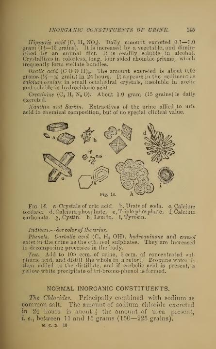
INORGANIC CONSTITUENTS OF URINE. 145
Hippuric acid (C9 H9 N03). Daily amount excreted 0.1—1.0
gram (1]—15 grains). It is increased by a vegetable, and dimin-ished by an animal diet. It is readily soluble in alcohol.
Crystallizes in colorless, long, four-sided rhombic prisms, whichfrequently form stellate bundles.
Oxalic acid (COO H) 2 . The amount excreted is about 0.02
grams [}/i—X grain) in 24 hours. It appears in the sediment as
calcium oxalate in small octahedral crystals, insoluble in acetic
and soluble in hydrochloric acid.
Creatinine (C 4 H 7 Ns O). About 1.0 gram (15 grains) is daily
excreted.
Xanthin and Sarkin, Extractives of the urine allied to uric
acid in chemical composition, but of no special clinical value.
Figf. 14
Fig. 14. a, Crystals of uric acid, b, Urate of soda, c, Calciumoxalate, d, Calcium phosphate, e, Triple phosphate, f, Calciumcarbonate, g, Cystin. h, Leucin. i, Tyrosin.
Indican.—See color of the urine.
Phenols. Carbolic acid (C 6 H5 OH), hydroquinone and cressol
exist in the urine as the ethereal sulphates. They are increasedin decomposing processes in the body.
Test. Add to 100 ccm. of urine, 5 ccm. of concentrated sul-
phuric acid, and distill the whole in a retort. Bromine water is
then added to the distillate, and if carbolic acid is present, ayellow-white precipitate of tri-bromo-phenol is formed.
NORMAL INORGANIC CONSTITUENTS.
The Chlorides. Principally combined with sodium as
common salt. The amount of sodium chloride excretedin 24 hours is about \ the amount of urea present,
i. e., between 11 and 15 grams (150—225 grains),M. C. D. 10

146 MANUAL OF CLINICAL DIAGNOSIS.
The chlorides are decreased in all acute febrile diseases (espec-
ially pneumonia) and reappear with convalescence. This is
particularly the case in diseases accompanied by exudations andtransudations, which retain the surplus chlorides until their
formation is complete.
Test. Add to the urine in a test tube a few drops of nitric acid,
and then a nitrate of silver solution until no more precipitate forms.Precipitate is dense and curdy, if the chlorides are normal ; milkyif diminished ; and faint if almost or entirely absent. The relative
amount may be estimated by comparison with a normal specimenof urine.
The Phosphates. Phosphoric acid occurs in the urine,
in part as sodic and potassic phosphate (alkaline phos-
phates) and calcic and magnesic phosphates (earthy phos-
phates). It is derived from the food and from theretrograde metamorphosis of tissues containing phosphorus.The earthy phosphates are probably increased in diseases
of the brain, osteomalacia, rachitis, etc. They are
decreased in chronic spinal affections, dropsy, etc.
Test for the Earthy Phosphates. Fill a test tube % full withurine, and add a few drops of caustic po'ash or ammonia and heatuntil the phosphates precipitate in flakes. The test tube is thenput on a stand for 15 minutes, and the quantity of the precipitateis determined. In an ordinary-sized test tube a deposit 1 cm. highrepresents a normal amount.
In health the phosphates are held in solution by an acid urine,and it is only when the urine has become neutral or alkaline thatthey are deposited.
Triple Phosphate. (Fig. 14 e.) When urea undergoes decompo-sition into ammonium carbonate, this with the. magnesiumphosphate in the urine, forms ammonio-magnesium phosphate(NH 4 Mg POJ. Triple phosphate is recognized (Fig. 14 e.) byits large transparent crystals, occurring mostly in triangularprisms or feathery crystals. The crystals may be confounded withcommon salt and calcium oxalate. Common salt is only found inurine which has been concentrated by evaporation. Acetic acidwill dissolve triple-phosphate, but not calcium oxalate. Thecrystals of triple-phosphate are usually present in cystitis asso-ciated with ammoniacal urine.
Neutral Phosphate of Calcium (Fig. 14 d.) forms wedge-shapedcrystals, which unite to form rosettes.
Magnesium Phosphate forms long quadrilateral plates, withrounded ends.
Sulphuric acid (H2 S0 4).—Daily amount excreted is about 2grams (}4 drachm). It appears partly as the sulphates of thealkalies, and a small portion as organic sulpho-acids. The sul-
phates are increased by an animal diet and exertion.

ABNORMAL CONSTITUENTS OF URINE. 147
Test for the sulphates.—Acidulate the urine with a few drops of
nitric acid and add a solution of barium chloride. A precipitate
of barium sulphate is formed insoluble in water or acids.
Carbonic acid (C0 2).—Present in small quantities in human
urine. Increased after vegetable food, certain drugs and in
decomposed urine. Large quantities of the^ carbonates causeeffervescence of the urine on the addition of acids.
Calcium carbonate (Fig. 14, f,) exceptionally, forms dumb-bellcrystals. It is recognized by its effervescence and solubility uponthe addition of mineral acids, which may be observed under themicroscope.
ABNORMAL CONSTITUENTS OF THEURINE.
Albumen.—Albumen occurring in the urine (albu-
minuria), is usually pathological. The albumens foundin the urine are ordinarily serumalbumen and serumglobu-
lin. The quantity of albumen may vary from meretraces to 1-2% ('rarely more.) The blood-serum contains
about 5% of albumen. Albuminuria may be caused byaffections of the kidney (renal albuminuria), e. g. acute
and chronic nephritis, amyloid kidney; by hydremicconditions of the blood (anaemia, leucaemia) ; in fever andacute poisoning; after epileptic attacks and apoplexy(transitory albuminuria).
Physiological albuminuria may occur in the femaleafter suppression of the milk, after a diet rich in
albuminous food (particularly a diet of eggs), increased
renal blood pressure (after a cold bath), excessive mentalor muscular exertion and in total absence of sodiumchloride from the food.
Albumen may be present in inflammatory affections of
the urinary tract below the kidney. Under these circum-stances it forms part of such fluids as blood, pus andsecretions from the generative organs; the quantity of
albumen is usually small, and the formed elements (bloodcorpuscles, pus cells, etc.), usually indicate the source of
the albumen .
Renal albuminuria is usually associated with a large
quantity of albumen and the presence of casts. In

148 MANUAL OF CLINICAL DIAGNOSIS.
women albuminuria on account of contamination of the
urine by vaginal discharge is comparatively frequent anderrors may be averted by the use of a catheter.
Qualitative tests.—1. Heller's test. Add to the urine in a test tubesome nitric acid allowing it to flow down the sides of the inclinedtest tube, so that the twTo fluids form separate layers; at their line
of contact if albumen is present, there is formed a sharply definedring-shaped cloudiness. In very concentrated urine, a precipitate
may be caused by urates, but in this case, the ring is not so dis-
tinct and is more toward the surface of the urine. If warmed, thecloudiness produced by urates will disappear, but not so if due to
albumen.
2. Heat test.—Heat the urine to the boiling point in a test tubeand add one or two drops of acetic acid or 10-20 drops of nitric acid.
If albumen be present a w7hite opacity will appear. The additionof acid prevents the precipitation of the phosphates of the alkalineearths and favors the coagulation of albumen, which if present insmall quantities, would not separate in an alkaline fluid.
If after the heat test the urine is allowed to stand for 24 hours,the quantity of albumen may be approximately determined fromthe degree of precipitation. A slight cloudiness but no precipitateoccurs, w7hen there is less than 0.ul% of albumen present; 0.l'5%
when the curved part of the test tube is barely filled with albu-men; 0.1% when the coagulum in the test tube reaches TV theway up; 0.25%, % the way up; 0.5%, K the way up; 1%, % thew:ay up and 2%-3% when the wThole fluid is completely coagulated.
3. Acetic Acid and Ferrocyanide of Potassium. Add to theurine 3-5 drops each of acetic acid and a 10% solution of fer-
rocyanide of potassium; the minutest traces of albumen aredemonstrated by the formation of a wThite cloud. This test indi-
cates the presence of albumen, globulin or albumose, but notpeptone.
4. Picric Acid. The addition to the urire of a saturatedsolution of picric acid will produce a greenish cloudiness.
5. Biuret Test. The urine is rendered alkaline with caustic
potash and 1—3 drops of a diluted solution of sulphate of copperare added. In the presence of albumen, the blue solution acquires
a violet tinge.
6. Salt and Hydrochloric Acid. A saturated solution of commonsalt containing 2% of pure hydrochloric acid added to cold urinewill produce a cloudiness.
7. Metaphosphoric Acid. One or two drops of this acid addedto the urine will produce a cloudiness, even when mere traces of
albumen are present.
8. Potassio - Mercuric Iodide (Tanrefs reagent). Solution:
Potassii iodid., 3,32 grams. Hydrarg. bichlorid., 1.35 grams.Aquae destillat., q.s, ad. 100 cub. cent. First acidulate the urine
with acetic acid, then add the double iodide solution drop by drop.

TESTS FOR ALBUMEN. 149
This will give a precipitate when there is only 1 grain of albumento a quart of water.
Quantitative Tests.
—
(Esbach's Albuminometer) . This consistsof a test tube marked at its upper end with U and R and at thebottom with 1, 2, 3, 4, 5, 6, 7. The urine is added up to U andthe reagent (picric acid 10 grams, citric acid 20 grams, water 10('O
cub. cent.) to R. The tube is well shaken, and after standing 24
hours the l^vel of the precipitate is read off, the number on thescale representing grams of albumen per liter. To obtain thepercentage the respective figure is divided by 10. When thealbumen is abundant the urine is first diluted with one or twovolumes of water, and then multiplying the resulting figures by 2
or 3, as the case may be.
Tanrefs method. To 10 cub. cent, of filtered urine add 2 cub.cent, of acetic acid diluting with a little water; next add Tanret'8reagent (see above) drop by drop, counting the number of dropsused ; when the precipitate thus formed grows less, a drop of theurine is taken out and brought in contact with a few drops of a 1%solution of corrosive sublimate on a porcelain plate; if on mixingthe two, a red precipitate occurs, the reaction is complete, andfor each drop of the reagent used, less 3 drops allowed for excess,
0.5 grams of dry albumen per liter are present.
Method by Weighing, Pour 100 cub. cent, of filtered urine into abeaker and add a drop or two of acetic acid ; heat on water bathfor half an hour or more ; collect precipitate, wash with alcoholand ether, then dry and weigh. The weight, less that of thefilter, represents the percentage of albumen.
Peptones. Present in the urine {peptonuria) during the absorp-tion of pus and the formation of exudations (pneumonia, abscesses,
etc.).
Peptones are detected by the Biuret test after albumen or hemi-albumose has been removed or proved absent.
Hemialbumose ( propeptone) . Rarely present in the urine,but found in osteomalacia and intestinal tuberculosis.
Test for Hemialbumose. Acidify the urine with a few drops of
acetic acid and add J its volume of a concentrated salt solution
;
boil and filter off the precipitate. Albumen and globulin remainon the filter. The filtrate is allowed to cool, and if a turbidityarises by the further addition of the salt solution, which disap-pears by heating and reappears on cooling, then hemialbumose is
inferred to be present.
Fibrin is present in the urine in cases of hematuria,chyluria, tuberculosis of the gemto-urinary tract, etc.
It is recognized by spontaneous coagulation in the urine.
Mucin.—Occurs in traces in normal urine. Increasedin diseases of the urinary passages. It is precipitated
by alcohol and dilute mineral acids but not by heat.
150 MANUAL OF CLINICAL DIAGNOSIS.
Test.—If acetic acid is added to cold urine a cloudiness
or precipitate appears which is not dissolved by an excess
of the acid if mucus is present.
Blood.—Blood in the urine, differs in appearanceaccording to the part of the urinary tract from whence it
is derived. If from the renal parenchyma, the blood is
well mixed with the urine giving it a smoky appearance;
it appears in small quantities and renal casts are pres-
ent. From the ureters, long semicircular clots and strings
are present. Prom the urethra and bladder, the quantity
of blood is large and blood clots are present.
Tests for Blood. Spectroscope.—Two dark absorption bandsin D and E of the spectrum, i. e., in the yellow and green, theformer being narrower, the latter broader (oxyhemoglobin).
Heller's Test.—If the urine be heated with caustic potash, theearthy phosphates in precipitating carry down the blood-coloringmatter and appear reddish brown instead of white.
Almen's Test.—Add to the urine a few drops of fresh tinc'ure ofguaiac; after shaking, add a few drops of resinous turpentine oil.
If haemoglobin is present, the color will change to a distinct blue.
The following mixture may be employed in place of turpentineoil : Glacial acetic acid, 30 drops ; Distilled water, 15 drops ; Oil of
turpentine, absolute alcohol, chloroform,—of each 3 ounces(Hiihnerfeld's mixture).
Microscopic Examination.—Teichmann's test (See Blood) may beconducted with the urinary sediment.The most trustworthy evidence of blood in the urine even when
the foregoing tests prove negative, is the recognition in the sedi-
ment of red-blood corpuscles under the microscope. When thecorpuscles are pile as occurs in dilute urine, the addition of eosinor iodine in solution will deepen their color.
Bile.—Biliary matters in the urine may be caused byhepatogenic or haematogenic icterus (See pages 5 and 6).
In the former, resulting from obstruction of the bile ducts,
the biliary coloring matter as well as the biliary acids are
present in the urine. In haematogenic icterus, the biliary
coloring matter is present but not the biliary acids.
Tests for Biliary Coloring Matter.—Grnelin's test.—To the urine,
yellow nitric acid is added in a manner to cause the two to formdifferent layers; there will be a play of colors from green, blue,
violet, red to yellow. The colors appear in the order mentioned.A blue ring alone may be caused by indigo, a reddish-brown oneby hydrobilirubin and other substances.
MarechaVs Test.—If a solution of iodine in iodide of potash beadded to the urine, so that the two fluids may touch but not

TESTS FOR BILE AND PUS. 151
mix, a green color will immediately develop below the layer ofiodine.
If urine containing biliary coloring matter is shaken withchloroform, the latter becomes yellowish. The chloroformic solu-tion of the bile pigment, may then be tested after Umehn'smethod.In all these tests, darkly-colored urine should be diluted with
water before applying the test. Albumen does not interfere withthe tests.
Tests for Biliary Acids.—Pettenkofer'stest.—Unless applied in thefollowing manner the results are usually negative: Evaporateabout 6 ounces of the urine to dryness in the water bath. Extractthe residue with absolute alcohol, filter, and add ether in excess(20 times the bulk of alcohol used). By this means the biliaryacids are precipitated. The precipitate is removed by filtering,
redissolved in distilled water and then decolorized by filteringthrough animal charcoal. The filtrate is now ready for the test;a single drop of a solution of syrup of cane-sugar diluted withwater, is added to the filtrate in a test tube; sulphuric acid is thenadded drop by drop, while the test tube is immersed in coldwater ; a fine cherry -red or purple-violet color appears.Method of Strassburg. Some cane sugar is dissolved in the
urine, and a piece of filtering: paper is then dipped into this andallowed to dry. If the filtering paper is now touched with con-centrated sulphuric acid, a violet-red spot appears.
Pus.—Pus in the urine (pyuria) may arise from aninflammatory affection in some part of the urinary tract,
or the communication therewith of abcesses. Pus arisingin the urethra may be squeezed out by pressure, andusually escapes with the first portion of the urine. Pusfrom the bladder is accompanied with hypogastric pain,frequent urination, alkaline urine; and the quantity ofpus is large. Pus from the pelvis of the kidney is small inquantity, and the reaction of the urine is usually acid.
Pus from the kidney is small in quantity, and theurine is acid, as a rule ; renal casts are found in thesediment, and the amount of albumen is always greaterthan can be accounted for by the pus present. There arealso symptoms of renal disease. The localization of sup-purative changes is aided by the character of the epithe-lium found in the sediment. In pyuria arising fromaffections above the bladder, there is a notable absence ofmucus.
Tests.—Donne's Test. Pour off the supernatant urine, and tothe sediment add liquor potassae; the pus is changed to a gela-tinous, ropy mass.

152 MANUAL OF CLINICAL DIAGNOSIS.
Microscope. Pus cells are j£ larger than red-blood corpuscles,
and granular. Granules disappear, and a group of nucleoli
appear, on the addition of acetic acid.
Grape Sugar.— (dextrose) C 6 H 12 6 , occurs in
traces, in rormal urine (about 0.5 gram in 24 hours).
The temporary presence of small quantities of sugar in
the urine (under \%) is without diagnostic importance,
and occurs in health after a diet rich in carbohydrates
(transitory glycosuria). It is persistently present in
larger quantities in diabetes mellitus. The urine in this
affection is increased in quantity, is clear, pale, of high
specific gravity, and contains an increased quantity of
urea. Glycosuria (temporary presence of sugar in urine)
occurs : after poisoning (curare, nitrite of amyl, chloral,
etc.)? in acute infectious diseases, affections of the medulla
oblongata, after epileptic attacks, etc.
Qualitative Tests. In the following tests if albumen is present it
is first separated by boiling and filtration._
1. Moore's Test. Heat the urine a few minutes with J its volume
of liquor potassce, when, if sugar be present, a yellowish -brown
color appears, which is darker the larger the quantity of sugar
present. On adding dilute sulphuric acid, the smell of burnt
sugar (caramel) is perceptible. This test is now little used,
because, for its success, 0.3% of sugar must be present.
2. Trommer's Test. Add to the urine x/2 or 3^ volume of liquor
potassw and a few drops of a 10% solution of cupric sulphate. Heat
test tube in its upper half to boiling, and if sugar is present, there
will be a reddish-yellow precipitate of cuprous oxide.
3. Fehling's Test.—Solution ; crystalline sulphate of copper, 520
grains, neutral tartrate of potash, b% ounces, officinal caustic
soda solution, 3 ounces, distilled water to make 30 ounces.
This solution is diluted with 3 to 4 volumes of water, heated to
the boiling point (should remain clear, otherwise to be discarded)
and a little urine added. If sugar is present, a yellowish red
precipitate of cuprous oxide will form.
Fehling's Method modified by Seegen.—Urine is filtered through
a tvdck layer of animal charcoal (of blood) which absorbs the whole
of the sugar ; the charcoal is washed out with distilled water whichdissolves^out the sugar, which may be tested with Fehling's solu-
tion; traces of sugar may be found even in the 4th washing.
4. Bismuth Test (Bottger).—Add an equal quantity of liquor
potassce to urine and a small quantity of bismuth sub nitrate ; boil
for a minute or two, and if sugar is present, the bismuth salt is
reduced and a brown or black color is formed. With this test,
albumen or sulphides in the urine, produce similar effects as
sugar. Nylander's modification of this test consists in the use of the
following solution: Bismuth subnitrate, 2 grams, rochelle salt, 4

TESTS FOli SUGAlt. 153
grams, 8% solution of sodium hydrate, 100 grams. With this solu-tion, urine containing sugar turns brown or black after boiling.
5. Mulder's Test.— Xiter the urine is rendered alkaline with car-bonato of sodium, a solution of indigo carmine (sulphate of indigo)is added until the urine turns blue. On heating, indigo blue is
changed to indigo wdiite and on exposure to the air turns blueagain.
t>. Phenylhydrazin Test.—This test is based on the power of
phmylhydrazine to unite with grape sugar and form characteristiccrystals. To a measuring beaker half full of water add 2 drachmsof hydrochloric phenylhydrazine and three of sodium acetate; thecompound having been heated, the same quantity of urine is addedand placed iu a vessel of boiling water for 15 minutes; it is thenquickly put in cold water. After standing for some minutes, ayellow crystalline sediment of phenylglucosazone slowly falls. If
only a little grape sugar is present, then the sediment under themicroscope shows yellow rod like crystals terminating at each endin round balls or bunches.
7. Molisch's Method —Menthol, thymol or alpha-naphthol are usedin their alcoholic solutions (1 to 7 alcohol) by mixing a few dropswith the urine. To the mixture sulphuric acid is added so that thesolutions do not mix ; if sugar is present a red color (if thymol ormenthol was used) is produced at the line of contact, or if alpha-naphthol was employed, a violet color with greenish borders.
8. Johnson's Method.—If a few drops of picric acid are added to
the urine, which is then mixed with an alkaline hydrate, a deepred color appears if sugar is present. A light red color appears innormal urine.
The most reliable tests at present known to us, are Nylander'smodification of the Bismuth test and the Phenylhydrazin test.
In the former test, one part of the solution is taken to ten parts of
the urine, which is boiled one minute, but never exceeding twominutes, when a dark solution will be obtained on cooling fromthe oxidation of the bismuth. This very sensitive test detects
the presence of 0.08% of sugar.
Quantitative Tests for Sugar.—1. Fermentation. This test
depends on the decomposition in the urine of sugar by the torula
cerecisice (veast plant) with corresponding reduction of thespecific gravity of the fluid. After taking the specific gravitypour 4 ounces of urine into a bottle to which is added a piece of
yrast about the size of a pea ; the bottle is then loosely corked andset aside in a warm place for 24 hours, wrhen fermentation will becompleted. Allow the bottle to cool, and then take the specific
gravity again. The difference gives the number of grains of sugarto the ounce.
Example
:
Specific gravity before fermentation = 1040Specific gravity after fermentation = 1015
Difference = 25

154 MANUAL OF CLINICAL DIAGNOSIS.
25 grains of sugar are present to the ounce. The percentage ia
obtained by multiplying this number by 0.23. This method is themost certain test for su^ar. It is well, in making the test to havetwo other bottles, one containing a solution of grape sugar and alittle yeast (to show that the, yeast is active) , the other containingnc rmal urine and yeast (to show that the yeast is free from sugar).
Fehling's method. Mix in a flask or beaker 10 c. c. of Fehling'ssolution (see Fehling's test) diluted with 40 c. c. of distilled water.Heat to boiling, and add from a burette containing a mixture of
one part of urine and nine of water, a little at a time, stirring themixture until the blue color of the te-t solution has entirely dis-
appeared. As 10 c. c. of Fehling's solution are reduced by 0.05
grams of sugar, that amount of diluted urine which has reduced10c. c. of Fehling's solution contained 0.05 grams of sugar. If 16
c. c. of diluted urine (1 in 10) were used, 1.6 c. c. of urine con-tained 0.05 grams of sugar. The percentage is obtained by thefollowing proportion : 1.6 :0.05= 100 :x ; x=3. 1 per cent.
By means of the polariscope, the quantity of glucose is readilyascertained, but the apparatus requires great care in adjustment,and errors are liable owing to the presence of other bodies allied
to sugar.
Inosite (C 6 H12 6 ). Muscle sugar is present in theurine of renal disease and diabetes insipidus. It does notferment, precipitate copper salts or affect polarized light.
Acetone (CH 3 COCH 3 ). May be found in normalurine. Increased in febrile diseases, in certain forms of
diabetes, affections of the brain, etc. It is looked for in
diabetic coma, in which condition it imparts a vinousodor to the breath and urine.
Tests. Perch loride of iron with acetone gives a Burgundy-redcolor (unreliable).
Ziehen's Test. Twenty grains of potassium iodide are dissolvedin one drachm of liquor potasses and boiled. The urine is pouredon the surface of this solution producing at first a precipitationof the phosphates, but if acetone is present the deposit becomesyellow, owing to the development of iodoform, which is recognizedby its characteristic odor.
LegaVs Test. Add to the urine a few drops of freshly preparednitro-ferrocyanide of sodium and then caustic soda until it is
strongly alkaline. When the beginning purple tint turns yellow,1-3 drops of acetic acid are added, and if the acetones are presenta crimson-purple color is formed at the point of contact of theacetic acid and the mixture.
EhrlicWs Diazoreaction.—This reaction present in
typhoid fever and in relapses of this affection is of somevalue in doubtful cases. Its disappearance is a good sign.

ORGANIC SEDIMENTS. 155
It is also present in severe cases of phthisis, pneumoniaand measles.
Test. Solution 1. Sulphanile acid, 5 grams, muriatic acid,
50 grams, distilled water, 1000 grams. Solution 2. Nitrite^ of
FoUum 0.5 grams, water, 100 grams. Take 50 ccm. of solution
1 and 1 ccm. of solution 2 and mix in a test tube with an equalquantify of urine to which is added % volume of ammonia, andthe whole well shaken. The reaction is positive when a red color
is formed, especially noticeable in the foam.The "Diazo" reaction constitutes one of the earliest and most
constant signs of typhoid fever. Febrile abdominal catarrh nevershows this reaction. Disappearance of the reaction points to
approaching apyrexia. Relapses are almost invariably accom-panied by its reappearance.
Sulphuretted Hydrogen (H 2 S). Present in decom-posed urine.
Test. Filtering paper saturated with acetate of lead
solution when held over the bottle turns black (formation
of lead sulphide).
.
Cystin. Occasionally found in urine. Under the
microscope it appears as colorless, shining, six-sided
plates or prisms (Fig. 14).
Leucin and Tyrosin. Found in acute yellow atrophy of
the liver and in phosphorus poisoning.
Under the microscope leucin appears as yellow-colored
spheres, at times concentrically striated with protrudingpoints (Fig. 14).
Tyrosin appears in white needles arranged in bundles.
To obtain these crystals for microscopical examinationthe urine is evaporated to a syrupy consistency.
ORGANIC SEDIMENTS.White Blood Corpuscles (leucocytes). Normally pres-
ent in small numbers. Increased in inflammation andsuppuration of the genito-urinary tract (nephritis, pyelitis,
cystitis, urethritis and leucorrhcea).
Red Blood Corpuscles. Present in hematuria. Theyare usually pale, and may appear as casts.
Renal epithelium. Round or cuboid cells with a vesic-
ular nucleus and often full of fat drops (Fig. 15, i).

156 MANUAL OF CLINICAL DIAGNOSIS.
Arranged in cylindrical form they make epithelial casts.
Renal epithelium nearly always indicates an affection of
the kidney.
Epithelial cells from the renal pelvis, ureters and bladder cannotbe differentiated from each other. The cells of the superficial
layers have a polygonal form, while those of the deeper layer areround, with processes, and contain a vesicular nucleus. Anincrease of these cells in the urine may indicate cystitis, pyelitis
or inflammation of the ureters. The vagina and prepuce havepavement, and the male urethra cylindrical epithelium.
Casts. These are moulds of the renal tubules producedby the escape of a coagulable material which coagulates
and entangles other substances, and are characteristic of
renal disease. They may be divided according to com-position and appearance into three groups (v. Jaksch).
1. Cellular. 2. Those composed of products of degenera-
tion. 3. Hyaline.
1. The cells found in casts are red-blood corpuscles,
leucocytes, renal epithelium and sometimes colonies of
bacteria.
2. The products of degeneration are granules derived
from epithelial dSbris, amyloid material and fat.
3. Hyaline cylinders are composed of the ground sub-
stance only.
Blood Casts (Fig. 15, b) are red-blood corpuscles held togetherby coagulation. They are a certain sign of renal hematuria.
Leucocyte casts may occur in purulent affections involving therenal tubules.
Epithelial casts (Fig. 15, a) are composed essentially of renalcells embedded in a hyaline albuminoid substance.
Casts of Bacteria are present in septic embolic nephritis orpyelo-nephritis. With a low power they resemble granular casts.
Granular casts (Fig. 15, d) are composed of granules, whichconsist of broken-down blood or epithelial cells.
Fatty casts are usually hyaline casts dotted with oil globules.
Any cast covered with oil drops is known as a '
'fatty cast."
These casts may indicate fatty degeneration in the kidney.
Amyloid casts are transparent, homogeneous and very brittle,
and stain deeply with a solution of iodine.
Hyaline casts (Fig. 15. c) are colorless, very long and narrow.They are easily overlooked, and it is advisable in searching for
them as well as other casts to add a drop of Lugol's solution or

CASTS W TUB URINE. 15*>
aniline red to make them more distinct. These casts have beekoccasionally found in normal urine.
Mucous casts of the uriniferous tubules are usually very long,
and when present do not necessarily indicate disease.
Casts of the seminal tubes are very rare, and are usually of
greater calibre than renal casts.
Casts of Ammonium Urate (Fig. 15, f) are commonly foundduring the first days of life in children. Treated with potassiumhydrate, ammonia escapes, and the casts disappear.
Fig. 15.
Fig. 15. a. Epithelial cast. b. Blood cast. c. Hyaline cast,
d. Granular cast. e. Waxy cast. f. Cast of ammonium urate.
g. Pavement epithelium from the bladder (upper layer), h.
Same (lower layer), i. Epithelial cells from the uriniferous
tubules.
Spermatozoa may be found after coitus, nocturnal emissions,spermatorrhoea, etc. Found in the urine of women they may beof medico-legal importance. Fragments of morbid growths may bemet with in the urine.
Micro-organisms are present in many infectious diseases(diphtheria, recurrent fever, etc.).

158 MANUAL OF CLINICAL DIAGNOSIS.
Tubercle bacilli are present in tuberculosis of the genito-
urinary tract, and gonococci in gonorrhoea.
In examining for the tubercle-bacilli, the method is the Fame as
that detailed elsewhere {see Sputum). A number of cover-glasspreparations from the sediment of the urine must be made.
Animal parasites are occasionally found in the sediin-nt. Theshreds or hooklets of echinococci occur in the urine when this
parasite affects the kidney.
The Listoma haematobium inhabits the small veins of the kidneyand portal system and the ova are expelled with the urine accom-panied by biood.
In chyluria, the Filaria sanguinis has been found at times inthe urine, but it is more constantly present in ihe blood.Non pathogenic organisms develop in uri^e exposed to the air.
In urine containing sugar, the yeast plant {Torula cerevisi&j oftenoccurs.
SECRETIONS OF THE MALE GENERA-TIVE ORGANS.
Semen. Semen is a complex fluid composed of secretions
of the testicles, of the seminal vesicles, of the accessoryglands of the urethra, especially the prostate, Cowper'sglands, and the glands of the mucous membraneof the urethra. With the microscope, spermatozoa,spermatic cells, epithelium from the prostate and urethraand seminal granules can be distinguished. In fresh
semen, the motion of the spermatozoa is very active.
The amount of semen discharged at a single evacuationvaries from 5-10 grams.
Pathological Changes. Absence of semen (Aspermia) may becongenital or acquired. Acquired permanent aspermia may followocclusion of the ejaculatory ducts or destruction of the glandulartissue of the prostate gland. Temporary aspermia may occur witha comparatively normal state of the sexual apparatus.
Polyspermia. An increase in the amount of semen dischargedat a single ejaculation is relatively seldom observed.
Oligospermia signifies a decrease in the amount of semendischarged. Observed in advanced age, and after inflammationof the testicles and diseases of the prostate.
Oligozoospermia. Signifies a diminution in the number of sper-
matozoa, while azoospermia means their total absence. Oli^ozoo-spermia is most frequently caused by gonorrhceal inflammationsof the epididymes and spermatic cords.

© & *. ,
•2 ,2 £ s."S
*. © < e.
g Or-i Pf 2 s ° i£nS2s o gE'
sS oo ^> S:F ~ .»_> ~•£ e « « ^tJ c c .g
-
o 5'3 s£ g^ «3
^tsS ^ 5k 9 p
s do a
—
.. .^, ,
•*» s p a
80fl >° oq © t
o3j2£g-B&
OQ © fl'^o*J CO.£ *»
P P eo<© t» ^ Qi
P<© O P p n ^h
«- o o g^ o+^ g., o ^^ © be3^<N=?g
rVd
g sis*! olfS £s g^Sl'Sa g^ g^ a^ %^s%£ a^gg s g
•O © PSR ^? ©p
w © bc^d,
'Kg?!
^£g.l
ftt£i*l ^1^2"gOzJS&Sg© O h © o s
8,3 ^ 2'£ a
^©sS*•§.2o^°a
be-"- IS is © m^© g PT^'OB 8 g *£ «g © era's © a© ec ©»p © _ O
^h »-, is p co h . 5H flj «H M)d
** 03 © O'e^^>^j o3*^
> P-ti •
§S S ©^^-2go^i-i v 2 ^7^
*P w ^io ©'Or^
P co as be o © .2
-5 © ©^3 © ©
«_^-» co-"*^ tCfl
O P « >>% g ©
©<1 u o3 O © ••
"Sa-g g pcqo*^ © if **-•
t^ Ow p P o«4_-I p SCrt mq •
ii 5 c cfl *-• © w PPop<«bcnfl S
••^t^'O ^o P.2*r!t-3«a O^ || -'53 g
IgSp. 1 ©§o
P'ppSgpP-*
5 -^ p .5 5 * 0805 P,o3 SM ^«w_.M © C53
p• 3 », «>
.2 © §^ © P 2 2« p n o^ ^ S*p
>,© o CSO >>,•° a S & - os © 2•O'S w§5 P^ P
« ^ flH CO 2
a^ S 2 gP to £; p Qp © g <a^o
O c*£ v. ^3
^ ®-3© o© «P T3 *!2 .C^ «fl PC«M CO fe P© O 03 ^ O^ w © <u ©* P ft t3 fe.
P3.2o®
^3iS^ s .
•S^h © e ©^ 03 ^J « p<
p g P O 03
2 o "2 a fe
Is
12 SS-
§fl§2
p^so-?^
S^pS'p^)P_ O'P'Pfd
p s- *-• 53^h © b£'C'.2.M
©cog 03 ©
a^P © P p
°*iSH^ p ©
S S-.2 2 g *^ a^"p p
&» S^ i;^
© © o fcM «a
S © S P ^»rt©pP^SpP03 wp'oaSfe'd©^OpSS©^ ©^oS 03 ^©
ScoP ^^ 8!
t3 ^,-^> o'd ©
5 5 p x « b p


w&.:.:':.•
..,
•"?z*r:
:ip:
:p: V.
'" IST Jlfa?
1 s"b
—SSr}-"!'-
:§p
pp


SECRETIONS OF MALE GENERATIVE ORGANS. 159
Secretion of the Prostate. This is a milky fluid show-ing under the microscope cylindrical epithelial cells
white blood corpuscles, amyloid bodies and glistening
granules. If a few drops of a 1% solution of phosphateof ammonia is added to the fluid under the microscope,
spermatic crystals are formed.
Prostatorrhcea signifies involuntary loss of the prostatic
secretion. It occurs in all irritable conditions of the
prostate, whether produced by gonorrhoea, masturbation'or senile hyperplastic alterations of the gland.
Secretion from the Glands of Cowper is an odorless,
clear viscid fluid, showing under the microscope epithelial
and round cells. This secretion normally appears duringerection of the penis as a clear, transparent drop at the
meatus.

CHAPTER XII.
THE NERVOUS SYSTEM.
Anatomy and Physiology of the Brain and Spinal Cord.—Thecerebral cortex i. e. the gray matter upon the surface of brain is
the seat of mental activity. It has ridges (gyri or convolutions)and depressions {sulci or fissures). The psycho-motor region of the
^^4obuhis paracenfralis
Fig. 16.
cortex (motor area) comprises the anterior and posterior convolu-
tions and the lobus paraceniralis of either hemisphere {this area is
shaded in Fig. 16).
( 160)
PSYCHO-MOTOR REGION, PYRAMIDAL TRACT. 161
When certain limited areas of the brain cortex are stimulated byelectricity in animals, according to the area stimulated, move-ments of definite nmsole-gronpa can be evoked, which is not thecase when these areas are extirpated.
The excitable areas of the cortex are as follows :
1. The region of the arm lies in the middle third of the anteriorconvolution (G. centralis ant.)
2. The region of the leg lies in the upper third of the central
convolutions and the lobus paracentralis.
3. The region of the head lies in the lower third of the central
convolutions and that portion of the frontal convolution borderingon the fissure of Sylvius.
4. Region of the trunk, convex surface of the frontal lobe border-ing on the pne<entral fissure.
5. Center of vision, occipital lobes.
6. ('enter of hearing, temporal lobes.
7. Center of speech, inferior frontal convolution {left side) andadjacent area, around the lowrer part of the fissure of Sylvius.
The cortex of the parietal lobes is associated with the sensorytract.
Pyramidal Tract (Fig. 17). From the cortical centres themotor fibres converge toward the large ganglia at the base of thebrain, where they present a white strand of fibres (internal cap-
sule).
That portion of the capsule in front of the head of the caudatenucleus and behind the lenticular nucleus, constitutes the anterior
limb of the internal capsule. As the capsule passes along theinternal margin of the lenticular nucleus it makes a bend (genu)and runs between the front of the optic thalamus and behind thelenticular nucleus, forming the posterior limb of the internal
capsule. From the internal capsule, the motor fibres pass into
the pons and from the pons into the medulla forming thepyramidal bodies.
At the lowTer part of the medulla, about 10% of the fibres pa ss downthe same side of the spinal cord in the anterior pyramidal tracts,
whereas the other fibres (crossed fibres) pass to the opposite side of
the cord into the lateral pyramidal columns. From these columnsthe fibres pass into the ganglionic cells of the anterior horns, andfrom these cells the anterior roots of the spinal cord are formedwhich send motor fibres to the muscles. The centers of thecerebral cortex control the nutrition of the pyramidal tracts up to
the point where they enter the ganglia of the anterior horns of
the cord, whereas the ganglia of the anterior horns control thenutrition of the peripheral motor nerves and muscles.
Lesions of the motor tract result not only in motor paralysis but also
in descending degeneration of the pyramidal tracts, inasmuch astheir trophic center is in the cerebrum. The trophic center of theperipheral motor nerves is situated in the ganglionic cells of theanterior horns, hence lesions here result in degeneration of thenerves, and paralysis and atrophy of the affected muscles.

162 MANUAL OF CLINICAL DIAGNOSIS.
The Sensory or Centripetal tract takes its origin in the sensorynerves of the periphery which reach the cord through the posterior
or sensory roots which on their entrance immediately cross overto the opposite side.
Fig. 17.
Fig. 17. Diagrammatic Transverse Vertical Section of the
Cerebrum—(After Schwalbe) .
co, cortex
coi, island of Reil
cl, claustrum
na, nucleus amygdalae
nc, caudate nucleus
th, optic thalamuscm, middle commisurecs, subthalamic bodym, substantia nigra
nl, lenticular nucleus
ci, internal capsule
ce, external capsule
stt, stria terminalis (taenia semi-
circularis)
cf, anterior pillar of the fornix
/, fornix
cc, corpus callosum
villi third ventricle
vl, lateral ventricle
to, optic tract

DIAGNOSIS OF BRAIN LESIONS, 163
The blood supply of the brain is derived from the internal
carotid and vertebral arteries. Each carotid divides into theanterior cerebral and middle cerebral arteries. The middlecerebral artery enters the fissure of Sylvius and sends branches to
the lenticular nucleus and adjacent parts of the brain, includingthe caudate body ot the corpus striatum, optic thalamus andinternal capsule. The anterior and middle cerebral arteries
do not freely anastomose, and are called terminal arteries.
Anterior pyramidal tract.Anterior
Spinal Cord (Fig. 18). The Anterior and lateral pyramidaltracts of each side contain only motor fibres. Those in the former[Turk's columns) are connected with the corresponding cerebralhemisphere; the latter (crossed pyramidal tracts) with the oppo-site cerebral hemisphere. The posterior columns (tracts of Gulland Burdach) convey sensory nerve fibres.
Diagnosis of Brain Lesions.—Lesion of the internal
capsule: there is hemiplegia or hemianesthesia (loss of
sensation in one lateral half of body) or both combined;hemiparalysis of the lower half of the face. Tremor,hemichorea and athetosis.
Lesions of the cms cerebri: if the lesion is confined to
the tegmentum cruris (sensory portion), there is hemian-esthesia of the opposite side of the body and paralysis of
the 3d and 5th cranial nerves on the same side. If themotor portion (crusta cruris) is alone implicated, there is
paralysis of the 3d nerve with hemiplegia on the opposedside.
Corpora Quadrigemina : incoordination of movement,absence of pupillary reflex, nystagmus and strabismus.Blindness may exist independent of a choked disc, optic
atrophy or neuritis. Pons varolii; lesions may be aboveor below the line of Gubler. This is an imaginary line,
that connects the apparent origin of the trigeminal roots
and marks the level of the decussation of the facial nerve

164 MANUAL OF CLINICAL DIAGNOSIS.
fibres that pass ccphalad. Lesion above the line of Gubler:
facial paralysis and hemiplegia upon the same side of the
body and opposed to the seat of lesion. Below the line of
Gubler: facial paralysis on the same side as the lesion
and hemiplegia on the opposed side. Cerebellum: inco-
ordination of movement, intense vertigo and titubating
gait. Only when the vermiform process is implicated
are the symptoms of cerebellar lesions pronounced. Me-dulla oblongata: disturbances of respiration, circulation,
phonation, deglutition and articulation (when cranial
nerve roots are implicated). Diabetes and albuminuriamay be present. Lesions at the base of the brain (anterior
fossa): implication of the olfactorius. Middle fossa:
implication of the opticus, oculomotorius, trochlearis andabducens. Posterior fossa: implication of the trochlearis,
abducens, facialis, acusticus, gl^sso-pharyngeus, vagus,
accessorius and hypoglossus. Basal lesions may also
involve the pyramidal tracts.
Lesion of the thalamus opticus : post-hemiplegic chorea,
athetosis, and tremor of the paralyzed limbs. Hemian-opsia.
Diagnosis of Spinal Lesions.—Lesions of the cord
before pyramidal decussation cause paralyses of the sameside. Usually lesions of the spinal cord affect both sides
alike leading to paraplegia, a frequent form of spinal
paralysis. In cases of transverse section of the cord, the
columns of Goll and cerebellar lateral tracts degenerate up-
ward from the point of injury and the pyramidal columnsdownward. Lesions of the spinal cord are systematic andnon-systematic or focal. The former refer to affections
limited to recognized subdivisions of the spinal cord
without spreading to adjacent parts; by the latter is
meant affections not limited but which spread as the
lesion progresses. In. Tabes dorsalis the lesion is in the
posterior columns; Lateral sclerosis: lateral columns,Amyotrophic lateral sclerosis: anterior cornua and lateral
columns ; Acute anterior poliomyelitis (infantile spinal
paralysis): anterior cornua; Progressive muscular atrophy
:
anterior cornua. In bulbar paralysis there is degenerative
atrophy of the nuclei of nerves of the medulla oblongata.
PARALYSIS. 165
Paralysis.—When the movement of an extremity is
entirely absent, the term paralysis is used; when slight
movement is possible, paresis.
Paralysis may be cerebral, spinal or peripheral.
Hemiplegia signifies paralysis in one lateral half of the body.Paraplegia signifies paralysis of both legs or arms. Hemi-paraple-gia: parjlyeis of motion in the lower one-half of one lateral halfof the body. Monoplegia: paraVsis of a distinct group of muscles.Central paralysis: lesions of the motor tracts proximal to theanterior horns of the cord. Peripheral paralysis: lesions of themotor nerves peripheral to the anterior horns of the cord. Flaccidparalysis: when the paralyzed limb readily yields to passive move-ment. Spastic paralysis: when the paralyzed limb offers a resist-
ance to passive movement. Spastic paralyses are usuallyassociated with
#
exaggerated reflexes. Functional paralysis (noanatomical lesion) : are usually unattended by trophic orelectrical disturbances.
Cortical Paralysis.—This is due to some lesion (abscess,
blood clot, tumor, embolism, meningeal thickening,
depressed bone, etc.,) of the cerebral cortex. Cortical
lesions may be irritative or destructive.
Irritative lesions are usually connected with paroxysmal crampsin £ome special group of muscles (cortical or Jacksonian epilepsy).
Paralysis if present is usually transient and returns after subse-quent paroxysms.In destructive lesions there is paralysis of special groups of mus-
cles {monoplegia). Consciousness is not lost as in lesions of centralportions of the brain. Pain within the head in these lesions is
usually localized or percussion over the seat of the lesion will elicit
it. No impairment in sensibility of the paralyzed parts exists,unless sensory paralysis is present as a complication. Affectedmuscles show normal electro-contractility. Paralysis (as in othercerebral lesions) is on the side opposite to the lesion.
Hemiplegia (seat of lesion). If intra-cranial, the paral-ysis is on opposite side of body; if spinal, on the sameside.
Symptoms. Seat of Lesion.Hemiplegia with motor aphasia. 3d left frontal convolution.
" " paralysis of Posterior limb of internal cap-lower facial sule.
branches.M " hemianses- Posterior third of internal cap-
thesia. sule.11 " crossed paral- Crus cerebri on same side as
ysis of 3d cran- paralyzed nerve,ial nerve.
•* " crossed facial Pons,paralysis.

166 MANUAL OF CLINICAL DIAGNOSIS.
Etiology. 1. Embolism. 2. Apoplexy. 3. Syphilis. 4. ToxicIn embolism, the paralysis is usually sudden without loss of con-sciousness and usually associated with motor aphasia,. There is
usually a heart lesion. Patient usually young.
Apoplexy. Origin sudden with loss of consciousness and usuallyassociated with lower facial paralysis. Arteries atheromatous andpatient is usually old. In the stage of reaction, there is a rise of
temperature (from inflammatory changes around the clot.)
Syphilis. Origin slow. Lesions multiple. History of syphilis.
Good results from anti- syphilitic treatment. Occurs in the young.
Toxic. In uraemia and the later stages of phthisis and carci-
noma (hemiplegia is transitory).
Paraplegia (seat of lesion). When the paralysis is
incomplete the term paraparesis is used.
Lesion may affect lumbar (frequent), dorsal or cervical
region (paralysis of all four extremities).
Etiology. Myelitis, trauma, carcinoma, tuberculosis,
caries and syphilis.
PARALYSIS OF THE CRANIAL NERVES1. Olfactorius. Loss of smell (anosmia). Test with asafcetida,
etc.
2. Opticus. Diminished sharpness of vision and sense of color
;
narrowing of the field of vision (examine with ophthalmoscope).
Loss of vision in one lateral half of each retina is called hemi-anopsia. The most common form is homonymous hemianopsia,when nasal half of one eye and temporal half of the other eye is
blind. This may result from some lesion of the optic tracts or thecortex of occipital lobe. The binasal variety indicates a lesion of
the chiasm. Total blindness (amblyopia or amaurosis) is due to alesion of the opticus peripheral to the chiasm.
Hemianopsia may be detected by requesting the patient to close
one eye and to fix the open eye on some near object. Now takesome object easily seen and move it to the right and left and rUoabove and below the object upon which the patient is gazing,
asking the patient in each case if the two objects are seendistinctly and simultaneously. The retina is blind upon the side
opposite to that upon which the moving object is lost to sight.
3. Oculomotorius. Falling of the upper eyelid (ptosis), externalstrabismus, dilatation and absence of the pupillary reaction,
double vision (diplopia) and disturbances of accommodation.
4. Trochlears. Eye cannot be raised or rotated outwards.

PARALYSIS OF CRANIAL AND SPINAL NERVES. 167
5. Trigeminus. Anaesthesia of the sensory distribution of this
nerve; paralysis of the masticatory muscles (motor fibres) andimpairment of taste (lingualis to ant. % of tongue).
6. Abducens. Eyeball cannot be turned outward.
7. Facialis. Immobility of the muscles of expression of oneside of the face; eye cannot be closed, the face is drawn to theopposite side, dribbling of saliva, etc. In lesions proximal to theganglion geniculi, the soft palate on the same side is paralyzed ; if
lesion is between the ganglion geniculi and the passage of th ••
chorda tympaai, there are disturbances of taste in the anterior 'i
of tongue. In central paralysis of the facialis, only the lowerhalf of the face is usually paralyzed, the electro muscular con-tractility is unchanged and hemiplegia is frequent. Theprognosis of peripheral facial paralysis is influenced by the electric
reaction. If the electric reaction is normal, recovery can beexpected in 2 or 3 wTeeks ; if diminished but not lost, recovery in
6 wTeeks ; loss of the faradic and galvanic excitability of the nerveand loss of the faradic excitability of the muscles [reaction ofdegeneration) ,
gives an unfavorable prognosis.
8. Acusticus. Disturbances of hearing (examine with theotoscope).
9. Glossopharyngeus. Loss of taste in the posterior % of
tongue.
10. Vagus. Quickening of the pulse and slowing of therespiration.
11. Accessorizes. Paralysis of the sterno-cleido-mastoid andtrapezius.
12. Hypoglossus (motor nerve of tongue). Tongue turns towardthe paralyzed side when protruded.
PARALYSIS OF THE SPINAL NERVES.Plexus Brachialis. Paralysis of the deltoid, biceps, brachialis
anticus, supinator longus and infraspinatus {paralysis of Erb)muscles.
Nervus Medianus. Pronation and flexion of the hand, flexion
and opposition of the thumb and flexion of the finger in the lactwo phalanges is impossible.
Nervus Ulnaris. Power is diminished to flex and draw thehand to the ulnar side as well as to flpx the last 3 fingers. Little
finger is immovable. On account of atrophy of the interossei.
the firet phalanges are extended and the second and third phalangesare flexed (claw-hand).
Nervus Radialis (musculo-spiral). Hand hangs in flexion andcannot he extended. Fingers are flexed. Thumb cannot beextended or abducted. Outstretched arm cannot be supinated,

163 MANUAL OF CLINICAL DIAGNOSIS.
but on flexion of the arm the forearm can be supinated by thebiceps. Lead poisoning is a frequent cause of paralysis of this
nerve, but the supinator longus is not usually involved.
Nervus Peroneus. Flexion of the foot is impossible. Tips of
toes and the outer edge of foot first touch the ground in walking(abduction of these parts abolished).
Nervus libialis. Extension of the foot is lost (patient cannotrise on his toes). Abduction of the foot and plantar flexion of
the toes (claw-like appearance).
Ataxia. Inability to coordinate with intact muscularpower certain muscular movements. If patient is directed
to thread a needle, pick up a pin or make other com-plicated movements, the motions are clumsy. Ataxiaoccurs in cerebral and spinal affections (tabes), affections
of cerebellum and in diseases of the peripheral nerves(neuritis) following diphtheria, alcoholism, etc.
Aphasia. Ataxic (motor) aphasia , the power of coor-
dinating the movements for articulate speech is partially
or totally lost.
Amnesic (sensory) aphasia ; the power of recollecting
words as aggregate acoustic phenomena is impaired.
Word dumbness; the inability with intact hearing to
understand words. This is also called word deafness.
Paraphasia ; the ability to connect word images andcorresponding connection is lost.
Agraphia ; this is the inability to convey thoughts inwriting. It is present in amnesic aphasia.
The lesion in aphasia is most frequently found in theleft hemisphere in the inferior frontal convolution(Broca's convolution).
TESTING THE SENSIBILITY.
Touch Sense. This signifies (eyes closed) the ability to
immediately perceive the contact and describe thesensation, when the skin is touched with the finger or anyother object.
Sense of Locality. Normal individuals locate almostaccurately the part of the body touched. By the use of
compasses (sesthesiometer) , the smallest distance maybe

TESTING THE SENSIBILITY. 169
found in which both points may be recognized. Thedistance varies in health, and the following measures canbe used as the healthy standard for comparison: 1.
Point of tongue, -fa inch. 2. Tip of finger, -fa inch. 3.
Mucous surface of lips, -fa inch. 4. Tip of nose, | inch.
5. Chin, i inch. 6. End of big toe, cheek and eyelids, -J
inch. 7. Bridge of nose, i inch. 8. Heel, i inch. 9.
Back of hand, | inch. 10. Neck, | inch. 11. Fore-arm,
If inch. 12. Sternum, If inch. 13. Middle of thigh,
2| inches. 14. Back, 2f inches.
Sense of Pressure. This may be tested by placing
different weights on the extremity. When the sense of
pressure is greatly disturbed, it can be determined bypressure with the finger.
Sense of Temperature. May be tested with test tubes
filled with water of different temperatures applied to the
skin. It may be rapidly tested by breathing and blowingon the skin.
Electro-cutaneous sensibility. Tested with a wire-brushelectrode and determining the minimum strength of the
current felt.
Joint and Muscular sense. This is the ability to deter-
mine the position of the extremities and their passive
movements with closed eyes.
Sensibility to Pain. Tested by sticking with a needle,
electric current, etc.
Abnormal Conditions of Sensation.—Anaesthesia
signifies entire loss of sensibility and depends on inter-
ruption of sensory conduction. Hypxsthesia, diminishedsensibility. Hyperesthesia, increase of sensibility. It is
present in tetanus, hydrophobia, strychnia poisoning, hys-
teria, etc. Hemianesthesia, loss of sensation in one lateral
half of body. Present in lesions of the posterior third of
the internal capsule and in hysteria. Polyxsthesia, is the
perception of more than one impression although onlyone is made.
Delayed sensation, is an evidence of imperfect conduction
of sensations to the brain and is a frequent symptom of
tabes. Analgesia, is the absence of pain when the skin ig

170 MANUAL OF CLINICAL DIAGNOSIS.
pricked with a needle. Occurs with unimpaired tactile
sense in hysteria and tabes. Perverse sense of temperature
is present when anything cold is perceived as warm.Symptom of Romberg; the patient totters or falls when the
eyes are closed. It is caused by anaesthesia of the soles of
the feet and in disturbance of the muscular sense in the
extremities. It is a frequent symptom of tabes. Pares-thesia refers to sensations of tingling, creeping, numbnessor formication.
Girdle pain {cincture-feeling), is a sensation of constric-
tion around the body (usually in the region of the dorsal
vertebrae), and is present in diseased conditions of the
posterior columns of the cord.
Spontaneous Pain.
—
Headache (cephalagia) maybecaused by: 1. Meningitis (pain violent and persistent). 2.
Syphilis (nocturnal exacerbations). 3. Neurasthenia andhysteria. In the latter affections, the pain is limited to a
small spot along the sagittal suture (clavus hystericus).
4. Migraine; paroxysms of pains occurring usually on oneside of the head at irregular periods and attended withvaso-motor and gastric disturbances. 5. Intoxication
from lead, alcohol, mercury and in uraemia. 6. Acuteinfectious diseases. 7. Anaemia. 8. Hyperaemia.
Nitrite of amyl inhalations diminish anaemic and increase hyper-aemic headaches. Infants manifest severe headache by boring of
the head into the pillow.
Neuralgia.—The chief symptom of this affection is
pain ; it is paroxysmal, usually unilateral, follows thecourse and distribution of a nerve, inflammatory symp-toms are absent and tender points (puncta dolorosa) canbe felt along the course of nerve in the intervals of painsbut more pronounced during the paroxysms. The pain-ful points are found where the nerves pass through bonycanals or penetrate the fasciae of muscles.
MOTOR SYMPTOMS OF IRRITATION.Spasms are involuntary muscular contractions of a
single muscle or a group of muscles. They may be clonic
(alternate contraction and relaxation of a *nuscle) or tonic
MOTOR SYMPTOMS OF IRRITATION*. \1\
(rapid recurrence of contractions, so that affected muscleappears in a fixed condition). Convulsions arc clonicspasms extending over the entire body.
Clonic-tonic spasms occur in epilepsy, puerperal con-vulsions, uraemia, irritation of the cortical centers(tumors abscess, etc.), at the onset of acute febrile dis-
ease, and in children from reflex causes (dentition, intes-
tinal worms, indigestion, etc.)
Tonic spasms occur in: tetany and tetanus. In tetany,
"Trousseau's sign' 7is characteristic, i. e., energetic con-
tractions can be excited by pressure of the arm in theregions of the median nerve and brachial artery. Mechan-ical and electrical excitability of the peripheral nervesand muscles also exist. Temperature is normal. Intetanus, there is an elevation of temperature. The disease
is as a rule fatal.
Varieties of Local Spasm.—Tonic spasm of the internal rectus is
recognized by strabismus. Nystagmus is a bilateral affection fre-
quently occurring in multiple sclerosis and is particularly markedwhen the patient looks at remote objects. It is due to clonicspasm of the muscles of the eyeballs. Trismus is a tonic spasm of
the masticatory muscles (lock-jaw) and occurs in tetanus.
Facial spasm (convulsive tic) affects the muscles of the face.
Partial spasm of the eyelids causes constant winking (nictitating
spasm) , if the entire orbicularis palpebrarum is affected the eyesmay be firmly closed (blepharospasm). Spasm of the Oesophagusfrequently occurs in hysteria {globus hystericus).
Laryngismus stridulus, is caused by spasm of the glottis and is
frequent in children. Hiccough, is spasm of the diaphragm (sin-
gultus). Writer 1
s cramp and similar professional neuroses consistof spasmodic contraction of definite muscle-groups when certainmovements are made.
Thomsen's disease is characterized hy the occurrence ofspasmodic rigidity of voluntary muscles, when they arecalled into action after intervals of rest (intentional
spasms).
Contracture is the permanent contraction of a muscle,which fixes the limb either in a flexed or extended posi-
tion. Contracture of the muscles occurs in organicdiseases of the spinal cord (especially if the lateral
columns are implicated) and is associated with an increaseof the spinal reflexes.

172 MANUAL OF CLINICAL DIAGNOSIS.
Tremor consists of slight contractions of bundles of
muscular fibres. It is physiological after physical andpsychical exertion. Tremor may be permanent and still
physiological in old people (tremor senilis). Tremor alco-
holicus occurs particularly in the extremities and tongue.
In morbus Basedowii (exophthalmic goitre) a fine tremoralso occurs. The tremor of paralysis agitans is controlled
by active exertion and ceases during sleep. The thumbcloses on the fingers as in " rolling pills."
Intentional tremor is observed in voluntary movementof the muscles and is an important symptom of multiple
sclerosis. It is also present in tremor mercurialis. Trem-bling of the eyes (nystagmus) is present in multiple
sclerosis, hysteria and in affections of the eye.
Fibrillary contractions of muscular bundles are seen in
atrophic paralyses, particularly in progressive muscularatrophy.
Choreic movements are quick, involuntary and incoordi-
nate movements, which prevent and interfere withvoluntary motion. They cease during sleep and are
pathognomonic of chorea. Choreic movements on oneside of the body (hemichorea) can precede or follow hemi-plegia (lesion in the posterior part of internal capsule).
Athetosis. The characteristic movements of this affec-
tion are most pronounced in the hand and fingers. Thefingers are never at rest and they are constantly flexed,
extended or intertwined. Athetosis may be idiopathic
but more frequently it is symptomatic of cerebral infantile
paralysis. Hemiathetosis has the same significance as
hemichorea.
Catalepsy is characterized by a peculiar rigidity of the
muscles (flexibilitas cerea) which enables the physician to
put the limbs in any posture where they will remain fixed
until the position is again changed. Catalepsy is mostfrequent in hysteria. Also present in meningitis and in
certain psychoses (melancholia attonita).

REFLEXES. 173
EEFLEXES.Reflexes are of two kinds, superficial (skin) and deep
(tendon). The former are excited by irritating the skin,
the latter by exciting the tendons or fasciae of muscles.
£kin Reflexes.—1. Plantar reflex. This is excited by ticklingthe sole of the foot. It causes a dorsal flexion of the foot and whenthe irritation is strong, the leg is drawn up.
2. Cremaster reflex. Irritating the inner side of the thigh will
cause retraction of the testicle.
3. Reflex of the abdominal walls. Irritating the skin of the abdo-men causes contraction of the abdominal muscles. Irritating theskin of the scapular (scapular reflex) and gluteal (gluteal reflex)
regions causes contraction of the corresponding muscles.
Skin reflexes are absent or diminished when the reflex circuit
(centripetal nerve, anterior horn of spinal cord and motor nerve)is broken. This occurs in affections of the peripheral nerves andspinal cord.
Tendon Reflexes.—1. Patellar reflex (knee-jerk) is obtained bypercussing the patellar tendon while the leg is crossed and com-pletely relaxed; this is followed by contraction of the quadricepsand the leg is extended. 2. Tendo Achillis reflex: percussingthis tendon causes contraction of the calf muscles. 3. Foot clonus;if the leg is slightly bent upon the thigh and the foot quicklyflexed rythmical contractions of the calf muscles will ensue.Tendon reflexes in the upper extremities rarely occur in health.
In health the knee-jerk is almost constant (about 2% of all
adults fail to show the knee-jerk), the tendo Achillis reflex is fre-
quent, while a persistent foot clonus never occurs. Tendonreflexes are absent when the reflex circuit is broken. The Reflexcircuit is formed by the nerves (sensory) going to the spinal cordfrom the muscle or tendon, the motor nerves going to the musclesand by that portion of the spinal cord connecting both. The ten-don reflexes are absent in peripheral paralyses, tabes dorsalis andpoliomyelitis. Increased reflexes occur wThen the inhibitory centers
are diseased or their fibres interrupted in their course from thebrain through the pyramidal tracts of the spinal cord.
Paradox contraction (Westphal) occurs in multiple sclerosis andparalysis agitans. It consists of a contraction of the tibialis anticuswith prominence of its tendon when the foot is firmly and quicklyflexed, the foot remaining in this position for a few minutes after
it is let go.
Reflex Functions.—1. Pupillary reflex. The pupil is supplied bythe oculomotorius for the sphincter and the sympathetic for thedilator pupillce. Irritation of the former nerve causes contraction(myosis), of the latter, dilatation of the pupil (mydriasis). The cen-

174 MANUAL OF CLINICAL DIAGNOSIS.
ter for the pnpil reflex (centrum cilio-spin ale) is situated in thelower cervical region. In tabes, the pnpils contract to accommo-dation but not to light {Argyle Robertson symptom.) The pupilsare also contracted (myosis spinalis) and irregular in tabes andparetic dementia.
2. Disturbances in the passing of the urine and faeces as well asthe sexual reflex occur in lesions involving the lumbar region of
the spinal cord.
Mucous membrane reflexes. 1. Conjunctival: closing the eyeswhen eyeball is touched. 2. Pharyngeal: nausea or vomitingwhen the pharynx is touched. 3. Coughing when the larynx orthe air-passages are irritated. 4. Sneezing when the nasal mucous*membrane is irritated.
EXAMINATION" OF THE NERVES ANDMUSCLES BY ELECTRICITY.
The faradic and galvanic currents are employed in
medical diagnosis. Static electricity (Franklinism) is
sometimes used as a therapeutical agent.
The faradic current is produced by the magnetizing anddemagnetizing of a bar of soft iron by means of a galvanic
current. The strength of the faradic current is regulated
by sliding one coil over the other as indicated by a grad-
uated scale. The strength of the galvanic current is regu-
lated by the number of elements used or by means of arheostat Electricity is applied to the body by means of
electrodes.
Electro-motive Force. This is the work which a definite
quantity of electricity can do and is influenced by theresistance offered to the current. The strength of electric
currents is determined by Ohm's law : The strength or
intensity (7) of the current is always proportionate to the
electro-motive force {E) divided by the resistance. This is
mathematically represented by the following formula
:
I=|. An ampere is the intensity of the current (I) gener-
ated by the electro-motive force (E) of 1 volt in an electric
current of resistance (R) of 1 ohm. One volt equals T9^
the electro-motive force of a Daniell element; one ohmequals a column of mercury 106 cm. long, and 1 squaremillimetre in section (1.06 Siemen's unit). The resistance

ELBCTRO-DIAGNOSIS. 175
of the human body is diminished by saturation of andpressure upon the electrodes. The relative resistance of
the tissues is represented by the following figures (100
taken as the maximum); eye, 4; muscle, 6; nerve, 10;
fat, 75; bone and skin, 100. The epidermis when dry, is
practically a non-conductor of electric currents.
Measurement.—The unit of current strength adoptedfor medical purposes is the milliampere which is the onemillionth part of an ampere. Galvanometers nowemployed are divided into milliamperes and from the
deflections of a needle, the number of milliamperes can benoted.
The Poles of the Galvanic current.—The galvanic
current is generated by the contact of dissimilar metalsexposed to chemical action. The simplest form of a gal-
vanic cell, consists of a plate of zinc and one of carbonimmersed in a vessel containing dilute sulphuric acid.
The wire connected with the zinc is the negative pole or
Kathode (Ka) while the wire connected with the carbon
is the positive pole or Anode (An).
One pole is distinguished from the other as follows : With a weakcurrent place both electrodes on either cheek ; a peculiar taste is
experienced at the anode electrode. If the wires are dipped in
water, bubbles of hydrogen accumulate about the kathode. If thewires are placed on moistened blue litmus paper the latter will
become red at the anode. If the wires are immersed in a solutionof iodide of potash containing starch, a blue color, due to free
iodine and starch, appears at the anode. German authors employthe symbols S, for closing (Schliessung), O, for opening (Oeffnung),and Z, for contraction (Zuckung).
Electro - Diagnosis. — The indifferent electrode
(usually large) is placed on the sternum, whereas the
different or active electrode (usually small) is placed onthe nerve or muscle to be examined. A muscle may bemade to contract directly by placing the electrode on the
muscle or indirectly by placing it on the nerve (motor point).
Stimulation of the motor points indirectly excite the mus-cles to contraction, each muscle possessing a motor point
which has been empirically established (Figs. 19-23).

bo
•o -o ^ aS• .n * a.-
S Sc
(176)

Nervus crura! is. Nervus tibialis:
7i
9
*2
oc~ n
6•S5 *•
igS S
13
3
X
3 ^«n 3
"J
.5
)
3 •
Pgc
"3
3
,3•3 s
oo
X % S 2 S *5 fH <. s S s *£
Nervus obturatonus. Nervus peroneus.
Nervus tibialis.
ULL
177)

178 MANUAL OF CLINICAL DIAGNOSIS.
With the faradic current muscular contractions occurby direct or indirect irritation. The weakest current
necessary to produce muscular contraction is noted bythe position of the coils as indicated by a scale in
millimetres.
Region of centralconvolutions.
Region of thirdfrontal convolu-tion and islandof Reil.
M. temporalis.
{U. branch.
M.branch.
Trunk.
L. branch.
N, auricular post.
Mtsplenius capitis.
M sternocleido-mastoid.
N. accessor Wii-lisii.
M- cucullaris;
N. dors, scapulae.
N. axillaris.
N. thoracic, long.(M. serratus aati-
cas,
)
N. phrenicus.
Plexus brachialis.
Supraclavicular point of £rb (Mm. deltoldeus, biceps,supinator longus et brevis, infraspinatus et
Fig. 23.
H. frontalis.
M. corrug. super*cil.
M. orbicular, pal*
Debr>
M» levator labii
super* alaequenasi,
.Mm^zygomaticii
M. orbicular, oris.
M, masseter,
M» levator menti.
M. quadrat* mem.ti.
M vtriangul.ment?,
N.: hypoglossus,
Platysmamyoides,
M«scles of hyoldbone»
j^ .
N, thoracicus_art'
\terior (M.pecto.-
> ral major,)
brachial, intern. [antTcusj,
subscapularis).
With the galvanic current, the Kathode is placed on themuscle or nerve to be examined. The strength of currentis gradually increased until a contraction occurs at theclosing of the current (Kathodal closing contraction, KCC.)The intensity of the current necessary to produce contrac-tion is indicated by the galvanometer or by the numberof elements used.
KCC occurs normally in superficial motor nerves with a currentintensity of 1-3 milliamperes (MA).
By means of the polarity changer or commutator, withoutchanging the position of the electrodes the kathode may be con-verted into the anode and vice versa.

BORMULA OF NORMAL POLAR ACTION. 1*9
Formula of Normal Polar Action.
1. Weak current: Kathodal closing contraction (KCC).
2. Medium current : Strong Kathodal closing contraction (KCC),moderate anodal closing contraction (ACC) and moderate anodalopening contraction (A 0(7).
3. Strong current: Tetanic Kathodal closing contraction (TeKCC), strong anodal closing contraction (ACC), strong anodalopening contraction (AOC) and Kathodal opening contraction
(KOC).
This formula only holds good when the muscles are
indirectly stimulated with the galvanic current. In direct
stimulation of the muscles there are only closing contrac-
tions, and ACC may be equal to KCC.Quantitative changes of electrical irritability refer to the
energy and amplitude of contractions. The electrical
irritability is increased in tetan}' ; diminished, in paralysis
resulting in simple atrophy as in apoplexy and in bulbarand spinal paralyses when the trophic ganglia are intact.
Increased Electric Excitability occurs when a weak current will
produce energetic contractions and it is diminished when a strongcurrent is necessary to excite muscular contraction. In unilateralparalysis the healthy side may be used as a basis of comparison.
Qualitative changes refer to abnormal polar reaction. If
a motor nerve is cut off from its trophic centre or if thetrophic centre is diseased, paralysis occurs and the nervedegenerates (degenerative atrophy), the degenerationextending to the muscle supplied by it. There is also
diminished electric irritability of the nerve for the far-
adic and galvanic currents and after 8 to 14 days theirritability is entirely lost. Direct faradic irritability of
the muscle is also entirely lost. After 2 weeks direct
galvanic irritability of the muscle is increased and contrac-
tions occur even with mild currents ; the contractions are
not short and quick as in health, but long and slow.
ACC with the same current, occurs as soon or sooner thanKCC; KOC may be obtained with weaker currents thanthe AOC. The foregoing reaction is the reaction of degen-
eration (RD). In severe cases, showing RD, the galvanicirritability is finally lost after 4 to 8 weeks.
In curable cases showing RD, the voluntary power of the affectedmuscle is restored sooner than^ the electric irritability. Thereaction of degeneration is present in all peripheral lesions of the

180 MANUAL OF CLINICAL DIAGNOSIS.
motor nerves whether of traumatic, rheumatic, toxic or diphtheriticorigin ; also in diseases of the gray matter of the anterior horns of
spinal cord and gray nuclei of medulla. The RD is absent in all
cerebral and spinal paralyses, the cause of which is central fromthe trophic centre.
The presence or absence of the RD is of value in prognosis. If
present, it indicates either that the atrophy of the affected musclesis incurable or that at least 2-3 months will be necessary for theirrestoration. If absent, it indicates the absence of gross anatomicallesions and cure may result in a few weeks.
Partial Reaction of Degeneration.—In this condition the
nerve retains its faradic and galvanic irritability and the
muscle its faradic irritability, but the direct galvanic
irritability of the muscle is increased, the normal formulaof polar action is altered and the contractions are slow
and long. This reaction shows anatomical changes in the
muscle but not in the nerve.
INSTANTANEOUS DIAGNOSIS IN NER-VOUS DISEASES (Erb).
Tabes dorsalis is, as a rule, easy to diagnose; although, even atthe present time, it is mistaken for myelitis, spastic paralysis,
neuritis, etc. The diagnosis of this affection can at once be made,when the patient enters the room with an ataxic gait, when hecomplains of lancinatingpains, double vision, weakness, tired feeling,
and paresthesia of the legs, vesical and genital weakness. Anexamination of the eyes will show contraction of the pupils, and afailure of the latter to respond to light. The patient will totter
with closed eyes, and the knee-jerk is abolished. Parkinson's dis-
ease, or paralysis agitans, is also capable of immediate diagnosis.
The marked inclination of the body forwards, the attitude of the
fingers, as if employed in holding trie pen, the immobility of facialexpression, the peculiar gait, as if the patient were about to tumbleforward, all give, when combined with the peculiar tremor, a charac-teristic picture. The tremor is not always essential in making thediagnosis, for there are cases in which this symptom is permanentlyabsent. Tetany.—A young man or woman is troubled with par-oxysms of tonic muscular spasms in the hands or legs. If, now,with the end of the finder, or a lead pencil, a vertical stroke is
made running from the temporal region to the lower jaw, and arapid contraction of the facial group of muscles occurs, the diag-
nosis of tetany can with certainty be made. The diagnosis is
confirmed when, after the nerve trunks of the arm are struck,
violent contractions of the muscles ensue ; or when, by application
of the galvanic current to the nerve trunk, kathodal closuretetanus and anodal closure tetanus are rapidly developed with

DIAGNOSIS IN NERVOUS DISEASES. 181
even weak currents, the muscles showing increased irritabilityThomsen's disease.—A young person complains with reference toa weakness and stiffness of intended movements. You ask thepatient to grasp your hand, and he will be unable, when told, toimmediately loosen his grasp. Now strike the exposed deltoidmuscle, or the biceps, and an energetic contraction, lasting manyseconds, occurs. This is Thomsen's disease, or myotonia congenita.If the symptomatic picture is to be completed, u^e the faradic andgalvanic currents. The nerves show normal irritability; themuscles, on the contrary, are irritable and qualitatively changed(myotonic reaction). Besides the tendency of the muscles to tonicspasm during attempts at voluntary movement, and the myotonicreaction, absolutely nothing else abnormal is found, other thanthe disproportion existing between the well-developed muscles andthe weakness of the patient. Basedow's disease (exophthalmicgoitre) : The well-known symptoms, exophthalmos, pulsating struma,and cardiac palpitations, are so characteristic that the affection is
at once recognized. Cases exist, however, where the exophthalmosis absent, or the thyroid gland not enlarged. In such cases thecardiac palpitations and acceleration of the pulse (120 to 160 beatsper minute) without manifest cardiac change, auscultation of thepulsating struma, the feeling of weakness and tremor, the tendencyto sweating, sleeplessness, and diminished electrical conductionresistance of the skin, are symptoms of value in undeveloped formsof Basedow's disease.
Dystrophia Muscularis Progressiva.—The patient is a child withweakness of the legs, clumsiness in walking and difficulty in rising
from the recumbent or sitting position; you ask to have the clothes
removed and proceed to examine the patient more carefully. Awaddling movement is noticeable, together with lordosis in thelumbar region. The thighs are characteristically attenuated whencontrasted with the muscles of the calf, which are increased insize. When the child is raised from the arms, the shouldersascend to the ears and the head sinks between the shoulders, sothat difficulty is experienced in lifting it in this manner, so readilyaccomplished in healthy children. The failure is attributed tothe inability to fix the shoulders below. The individual forms of
this disease can at once be recognized. The pseudohypertrophic
form, by the increase in size of the muscles, which is not propor-tionate to their motor power; the juvenile, by the more severeinvolvement of the upper half of the body, the pronounced atrophyand relatively late development of the affection ; the infantile, bythe early invasion of the face. Infantile paralysis {poliomyelitis
anterior acuta). In these cases the child presents an atrophicparalysis of one or more extremities, whereas individual muscles,or muscle groups, are exempt. The sphircters are intact, and theaffection has occurred suddenly. Sensation is not involved, andthe tendon reflexes in the affected muscle regions are absent. Theexamination is completed when the reaction of degeneration is
determined. Multiple sclerosis: Intentional tremor of the hand,shaking of the entire body when an attempt is made to rise or

182 MANUAL OF CLINICAL DIAGNOSIS.
walk, scanning speech, nystagmus, spastic paresis of the legs, andhistory of headache, vertigo, vesical weakness, etc., constitute thecharacteristic symptoms of this disease. ^ Progressive bulbarparalysis : Difficulty in articulation, nasal voice, thin lips and withdifficulty moved, the inability to protrude an atrophic tongue in
which fibrillary contractions are observed, difficulty in swallowing,
etc. Amyotrophic lateral sclerosis: This affection shows a com-bination of atrophic paresis with increased tendon reflexes in theupper, and spastic paresis in the lower extremities.
—
Deutsche Med-icin. Wochenschrift, October 17, 1889.
MEDICAL OPHTHALMOLOGY.Ophthalmic Examination of the eye is of great importance in
medical diagnosis. In the following affections the ophthalmoscopeestablishes or corroborates a diagnosis.
Aortic Insufficiency.—An alternate flushing and pallor of theoptic disc analogous to the capillary pulse, and pulsation of the
retinal vessels is observed.
Congenital Heart Lesions.—The retinal vessels are distended.
Leucsmia —Diffuse retinitis with haemorrhages and yellow color
of the eye ground.
Pernicious Anaemia.—(Edema of the retina with haemorrhages.
Haemorrhage.—Loss of blood may cause impaired vision fromtransient anaemia of the retina or cerebral centres. Ophthalmo-scopic examination not diagnostic.
Bright's Disease.—(Edema of the disc and surrounding retina
with irregular white splotches, and striated haemorrhages. About23% of patients with renal diseases have disorders of vision at someperiod of the disease. The eye lesion may be the first symptomof the renal affection.
Tuberculosis.—Usually in children tubercles are deposited in
the choroid, intra-ocular end of the optic nerve, retina or iris.
Their favorite seat is the macular region and its vicinity. Thedeposit of tubercles may precede other symptoms. According to
Cohnheim in all cases of acute miliary tuberculosis, tubercles are to
be found in the choroid; although this view is vigorously disputed
by other observers.
Diabetes Mellitus.
—
Diabetic neuroretinitis and atrophy. Theretinal changes resemble those due to albuminuria. Cataract is
also found and grape sugar may be detected in such lenses bychemical examination.
Tabes Dorsalis.—Atrophy of the optic nerve is an early andfrequent symptom, and may precede by many years, the develop-
ment of spinal symptoms. In the early stages, the discs are of adull, reddish-gray tint, wThich gradually becomes paler, and at last
white. Later £he nerves assume a greenish tint, the surface of

MEDICAL OPHTHALMOLOGY. 183
the disc becomes excavated, and the retinal blood-vessels shrink.Atrophy of the optic nerve is also observed in disseminated sclerosis
and paralytic dementia.
In tabes the field of vision is narrowed and the sense of color is
disturbed ; green being lost first, then red, and finally, yellow andblue.
Increased intracranial pressure —{Turnon, meningitis, hydro-
cephalus). The optic nerve is swollen and projects above the level
of the surrounding retina ; the margin of the disc is either obscuredor entirely lost ;the arteries are small, the veins large and tortuous
(choked disc). Ihese changes usually occur on both sides alike,
and of equal intensity. Like changes in the fundus occur in about
J of all tumors of the brain.
Exophthalmic Goitre.—Dilatation and tortuosity of the retinal
veins. Functions of the retina unimpaired. Spontaneous pulsa-
tion of the retinal vessels has been observed. Protrusion of theeye ball is often combined with a want of agreement between themovement of the lid and the raising or lowering of the eye{symptom of Graefe),
Syphilis.
—
(Syphilitic retinitis). Retina is covered by a grayfilm, and in the macular region numerous irregularly placedpunctate sj-ots are present.
Lead intoxication may be followed by optic neuritis with consec-utive optic nerve atrophy. In quinine poisoning, there is pallor of
the optic disc, and narrowing of the field ot vision. Alcohol andtobacco intoxication also lead to atrophy of the optic nerve.
CHAPTER XTIL
PARASITES.
ANIMAL PARASITES.
The diagnosis of animal parasites is only possible in many casesafter careful examination of fresh fsecal matter with the micro-scope. The amozba and infusoria lose their activity soon afterevacuation of the stool, and for this reason Eichhorst recommendsthe removal of the mucus and faeces from the rectum by means ofa glass tube, and direct transference of the same to the objectglass. The microscope will also aid in the diagnosis of eggs ofthe intestinal parasites.
Animal parasites belong either to the Protozoa or to the worms.The protozoa are divided into rhizpoda and infusoria, and theworms into flat (platodes) and round worms (nematodes). Thefollowing parasites are of practical importance: I. Protozoa; a.
Rhizpoda : amoeba coli. b. Infusoria : 1. Cercomonas intestinalis.
2. Trichomonas intestinalis. 3. Balantidium coli.
IL Worms.—a. Flat worms (platodes)*
1. Taenia solium.2. Taenia saginata or taenia mediocanellata,3. Bothriocephalus latus.
4. Taenia echinococcus.
b. Bound worms (nematodes*)
1. Ascaris lumbricoides.2. Oxyuris vermicularis.3. Trichocephalus dispar.
4. Anchylostomum duodenal©.6. Trichina spiralis.
6. Filaria sanguinis.
Only finding in the stools the parasites, segments of the tape-worms or eggs by the microscope can be considered as diagnostic.
Protozoa. Amoeba coli, A round granular structure with anucleus and several vacuoles.
Cercomonas intestinalis. This parasite is pear-shaped with ciliated
extremities and is yV~A °* an *ncn l°ng-
Trichomonas intestinalis. Almond-shaped with ciliated extremi*ties and is ^~A of an inch long.
(184)

FLAT WORMS. 185
BalantkUum cch rear-shaped, ciliated and with an invertedmouth. It is about *
4 of an inch long.
Tape or Flat Worms (platodes).—They are acquiredby the ingestion of raw or insufficiently cooked meat con-taining the eggs. Taenia solium is acquired by eatingill-cooked pork.
to
Taenia saginata is derived from beef and the Bothrio-cephalus latus, from fish. The three species can be diag-nosed by voiding of the segments (proglottides). Thebothriocephakis is the most easily removed by medication,whereas the T. saginata requires the most active treatment
Fig. 25.
?-g>
It' J8enra solium
- a. head, b. egg, c. segment,tig. 25. Taenia saginata. a. head, b. egg, c. segment.
i i!?"- .\i
a' £g of the oxyuris vermicularis. b. egg of ascarislumbncoides. Fig. 27. Muscle trichina.
The tapeworms are composed of a head (scolex) and segments(Prof/hmdes) The eggs come from the matured segments (herma-phrodite), and, when taken into the stomach of an animal the

186 MANUAL OF CLINICAL DIAGNOSIS.
capsules are dissolve! and the embryo is dislodged. It thenreaches the tissues and develops into a cysticercus. This cysticer-
cus when contained in the food ingested by man develops- into anew tapeworm. The method of examining a segment is to pressit slightly between two object glasses and examine with trans-mitted light.
Taenia solium. This (tapeworm) is 1-3 meters (yards)
long. The head is about the size of a pin's head, contain-
ing 4 suckers, and is prolongated into a proboscis (rostel-
htm) surrounded by a double row of hooks (20-30).
The matured proglottides have the sexual opening at the
side somewhat behind the middle, They have a uterus
writh 7-10 thick lateral branches, which subdivide. Theeggs are round or oval with a striped shell containing anembryo with 6 hooks. This cystic condition of the taenia
(cysticercus cellulosse) may be found under the skin, in the
brain, eye, and muscles, causing severe disturbances.
Taenia saginata. (7-8 meters long). Larger than the
former. The head has 4 suckers without rostellum or
hooks. Sexual openings are on the sides of the proglottides.
The uterus has 20-30 lateral branches, which are finer
than those of the T. solium. The eggs are like the latter
but larger. The cysticercus does not develop in the tissues
of man.Bothriocephalic latus (4-5 meters long.) Lancet-shaped
head with two lateral grooves. Matured proglottides are
broader than long and genital apertures are found in themedian line.
The uterus is arranged in the form of a rosette aroundthe sexual openings.
Taenia echinococcus. Found in the dog. It has a head(scolex) with hooklets (30-40), suckers and 3 segments, of
which the last one alone is matured. Only the cystic formor hydatid is found in man, which he acquires by eggs
introduced into the alimentary canal. When the embryomigrates from the intestine to some other organ (liver,
spleen, kidneys, lungs, etc.), it is transformed into a cyst
incapable of active motion.
The hydatid cyst consists of an external elastic cuticle, and aninternal lining, the parenchymatous layer. An echinococcus cyst
may be simple (unilocular) . filled with daughter cysts, or it maycontain a large number of minute cavities filled with a gelatinous
substance and with concentrically arranged walls {multilocular)
.

BOUND WORMS. 187
The nature of an echinococcus cyst can only be determined bypuncture and examination of the withdrawn fluid. The fluid of a
cyst is usually clear, neutral or alkaline, with a specific gravity of
1*003-1013. It contains little or no albumen, but large quantities of
sodium chloride, frequently grape sugar and succinic acid. Thelatter is detected as follows: Evaporate the fluid and add hydro-chloric acid; shake with ether and allow the ether to evaporate.
If succinic acid is present it remains as a crystalline masd. Thelatter when dissolved in water will give with chloride of iron, arust-colored gelatinous precipitate of succinate of iron. Heated in
a test-tube irritating fumes of the succinic acid are given off, caus-
ing cough. The microscopic demonstration of the echinococcusis based on the presence of scolices or their hooklets, or the cystic
membrane. The scolices are easily recognized by the presence of
suckers and hooklets. The hooklets, are small and transparentand a magnifying power of 500 diameters is necessary for their
detection.
In the case of acephalo-cysts, i. e., non-propagating cysts, exam-ination of the membranes is necessary in diagnosis. The mem-branes have a characteristic striped appearance.
Round Worms.—Ascaris Lumbricoides (round worm).Its habitat is in the small intestine, but it may pass to
other parts of the body. It resembles the ground-worm in
shape. The male is smaller (4-6 inches) than the female.
When the worm does not pass off with the faeces, thenexamination must be made for the eggs, which are expelled
in large quantities. They have a thick, concentrically
striped shell, upon which lies a thick, tuberculated albu-
minous cover. These worms are generally innocuous.
Oxyuris vermicularis (thread worm). Its habitat is
chiefly the rectum. When passed in a living condition, it
exhibits a lively motion. These worms cause intense
itching in the region of the anus. The female is longer
(-£ inch) than the male (i~i inch). The female has afinely pointed tail-end, whereas the male fmore rare)
has a blunt tail-end curved in the shape of a hook.The diagnosis is made from the constant passage of theworms with the stool, or by searching for the eggs whichare oval and possess a thin shell.
Tricho-cephalus dispar {whip-worm). It inhabits the caecum,although rarely in large numbers. It is 1K~2 inches long, and hasa thread-like head extremity, wThile the tail-end is considerablythicker. The egus (necessary for diagnosis) are about the size ofthose of the oxyuris and shaped li ke a lemon.

188 MANUAL OF CLINICAL DIAGNOSIS.
Anchylostomum duodenale (hook-worm). It inhabits theemail intestine, where it hooks itself fast and sucks blood,
causing pronounced anaemia. This worm is found in tun-nel workmen, brickmakers and miners. In persistent
anaemia of these individuals the worm should be soughtfor after the administration of an anthelmintic.
\ The anchylostomum is somewhat larger then the oxyuris and its
mouth is armed with three pairs of hooked teeth. The eggs areabout the size of those of the oxyuris and are usually found" in thestages of vitelline segmentation. The eggs arj developed a fewdays after the passage of the larvae from the intestines. If the eggsare not recognized, the faeces are allowed to stand in a warm placefor 2 or 3 days and then reexamined with the microscope.
Trichina spiralis. Found in the human body as muscleand intestinal trichinx. It enters the intestine by meansof trichinosed pork.
When the pork containing the live encysted trichinae reaches thestomach the capsules are dissolved and the trichinae set free.
They mature in the intestine in about 2*4. days, and bring forthafter 5-7 days young trichinae which migrate through the intestinalwalls and reach the striated muscles where they become encapsuled.A single worm may bring forth 100\)-1300 young. The male trichinais xV inch long, the female \ inch long. 1 richinosis is attendedwTith various symptoms. When trichinous flesh is first ingested,there are symptoms of gastric and intestinal catarrh.
Muscular invasion is attended with fever, oedema, partial paral-yses, muscular abscesses and pain. The symptoms reach theirheight in the 4th or 5th wreek. The violence and gravity of thesymptoms depend generally on the number of trichinae which haveentered the muscles. The trichinae are most abundant in the dia-phragm, intercostal, cervical and laryngeal muscles. They areless often found in the muscles of the extremities where they areusually crowded together at the attachment of the muscle to its
tendon.
The removal of a piece of musclefor microscopical examinationis often necessary for diagnosis during life. An accurate and sim-ple method for determining the presence of trichinae in pork is thefollowing: The suspected piece of meat is put into a mixture ofpepsin and dilute hydrochloric acid and allowed to remain in aconical-shaped glass at the body temperature. In the latter afterdigestion the free trichinae are deposited and may be removed bymeans of a pipette for microscopical examination.
Filaria sanguinis hominis. Indigenous only within thetropics (Egypt. Brazil, India, etc.). In its sexually matureform it is a filiform worm (8-10 ctm. long). It inhabits thelymphatics especially those of the scrotum and lower

ANIMAL PARASITES. 189
limbs. It obstructs the lymph-vessels and causes inflam-
mations which terminate in elephantiasis of the tissues
with oedema and lymphangiectasis.
The embryos pass from the lymphatics into the bloo'l and causehematuria and chyluria. The embryos may be excreted with theurine. An examination of the blood with the microscope showsthe presence of embryos which appear as little worms r^ inchlong and as broad as the diameter of a red blood corpuscle. Theyare found in the blood at limes only during the night hours, hencean examination at this time may be necessary. They may remainfor months or years in the body without creating manifest disturb-
ances.
EXTERNAL PARASITES.
The external parasites may be divided into animal andvegetable.
ANIMAL PARASITES.
Sarcoptes hominis (acarus scabiei).—The itch mite bores
its way obliquely through the horny layer of the skin
until it reaches the rete mucosum in the neighborhood of
the papilla. In this way it forms furrows (cuniculi) at
the ends of which the insect is found. In its course it
leaves behind excreta (yellow, brown or black grains andlumps). The female lays its eggs in the furrow and as
these are hatched (8-14 days), the young mites may beseen in all stages of development.
The itch mite selects the soft skin between the fingers, flexor
surfaces of the joints and parts pressed upon by tight portions of
clothing. In males, the penis and lower part of the abdomen areusually invaded. The parasite is secured by passing a needlealong the furrow toward the papule ; on tearing op m the furrow,the acarus usually adheres to the point of the needle. The bestspecimen is obtained by snipping by means of curved scissors, apiece of the skin containing the furrow.
Acarus folliculorum.—Occurs in about 10 per cent, of all healthyadults in the sebaceous and hair follicles of the face.
Pediculus capitis (head louse).—Inhabits the hairy scalp. It
fastens its eggs to the hairs by means of a chitinous covering andthey may be seen as grayish oval bodies. The young louseemerges in about 8 days.
190 MANUAL OF CLINICAL DIAGNOSIS.
Pediculus pubis (crab lmse). — Smaller than the former andinvades the hairy parts of the genitals.
Pediculus vestimentorum (body louse).—Inhabits the underclothingand passes to the surface of the body in order to feed.
Pulex irritans (flea).—This draws blood from the skin like thepediculi. A small punctiform haemorrhage surrounded by a red-dened areola is found at the point attacked.
Dracunculus rnedinensis (Guinea worm) —Found in those residingin the tropics. Occupies the subcutaneous tissue about the ankle.The usual method of securing the parasite is by fixing an exposedportion to a short rod and twisting it a little every day, until theentire worm is withdrawn without breaking.
Chigoe (pulex penetrans).—The sand-flea is principally confinedto the West Indies and attacks the bare feet of the natives.
VEGETABLE PARASITES.
Mould and yeast fungi, like bacteria, draw their nutri-
ment from organic carbon compounds. The mould fungi
are seen in decaying organic substances, while the yeast
fungi (blastomycetes) set up alcoholic fermentation. Morph-ology.*—As they occur in man, the mould fungi appear in
the form of jointed or unjointed filaments (hyphse) and of
ovoid or spherical cells. The filaments form compactmasses (mycelia). The ovoid or spherical cells are the
spores (conidia).
Achorion Schoenleinii (favus fungus).—Present in favus, a skindisease appearing on the hairy parts of the scalp and characterized
by dry, flat, yellow crusts, circular and depressed in the centre,
through which the hairs proiect. If a small piece of a crust is
placed on a glass with a little caustic potash and examined with aJ^-inch objective, spores and a densely felted mycelium may beseen.
Trichophyton Tonsurans —This is the fungus of herpes tonsurars
and parasitic sycosis. The fungus filaments are present in theepidermis, the spores in the hair. Both fungi give characteristic
cultures, which by inoculation reproduce their respective diseases.
Microsporon Furfur (Pi'yriasis versicolor).—The yellowish epi-
dermic scales produced in this affection may be confounded withpigmented spots. The fungus is demonstrated by adding to a fewof the scraped off scales on an object glass, a few drops of caustic
potash. Under the micros ope, a number of branched filaments(mycelium) and shining spores {conidia) are seen. The same pro-
cedure is applicable in examining for other fungi in the hairs andscales of the skin.

BACTERIA, 191
Oidium Albicans.—This fungus forms the white patches known a-s
thrush (or aphthce) present in the mouth, pharynx, oesophagus andstomach of weakly children and debilitated persons. It consists of
branching filaments with shining sporesat the points of bifurcation.Aspergillus Glaucus and Niger.—Found in the external auditory
meatus and no^e. They may also vegetate in the lungs (pneumo-oomycosis aspergillina) where they are usually secondary to destruc-tive processes (gangrene, tuberculous cavities, etc.). They formdouble-contoured, branched filaments containing pigmentedspores.
BACTERIA—(Schizomycetes).
Morphology and Physiology.—These parasites
are minute vegetable organisms of the lowest and simplest
form and are widely distributed in the air, water, surface
soil and about substances, animal and vegetable, undergo-ing decay. In their growth, the bacteria develop certain
products ('ptomaines) which, if poisonous, are called toxines.
It appears at present, as if the deleterious effects of bac-
teria are largely due to ptomaines. Brieger has shown that
the ptomaines cadaverin, neurin and mytilotoxin will
produce in rabbits convulsions, paralysis, gastro-intestinal
disturbances, etc. The ptomaine of tetanus (tetanin) will
produce tetanus by inoculation. Coloring matters are
often developed by bacteria. Bacteria are always present
in the cavities of men and animals and are only active in
ftie presence of moisture and may remain inert for a long
time, either as spores or developed organisms to becomeagain active under favorable conditions. At a temperaturebelow 23°F., they are incapable of proliferation. Theyare most active at about the body temperature. In fluids
all bacteria are killed when the boiling point of water is
reached (212°F.) When dry, they are more resistant to
heat than when moist. The spores are more resistant
than the bacteria. Of all the agents which reduce the
activity or destroy the bacteria, corrosive sublimate, even in
extremely dilute solutions, is the most powerful. Anaqueous solution of 1: 20,000 kills the spores of bacilli in
ten minutes; and a solution of 1: 300,000 stops the germ-ination of bacterial spores. Disinfecting agents should beused in aqueous solution. In alcohol or oil, their action

192 MANUAL OF CLINICAL DIAGNOSIS.
is feeble. Bacteria may be pathogenic or non-pathogenic.
The former multiply in the living organism and are the
cause of infectious diseases. The non-pathogenic bacteria
only vegetate on dead organic material (saprophytes) andare the cause of putrefaction and fermentation.
Phagocytes.—This term has been applied to certain
cells of the body which are capable of taking up into
their protoplasm and destroying or digesting bacteria
which get into the tissues. The resistance of the organismto certain infectious diseases may be explained by the
theory of phagocytosis.
Classification of Bacteria.—The following classifi-
cation is based on the shape of the various known species
and will no doubt be modified according to our increased
knowledge of these organisms.1. Spheroidal bacteria (micrococci).—They may occur in
beaded chains (streptococci), in pairs (diplococci), in masses(zooglea). or in grape-like groups (staphylococci). Path-ogenic micrococci have been found in the following diseases:
diphtheria, scarlatina, ulcerative endocarditis, erysipelas,
cerebro-spinal meningitis, pneumonia, osteo-myelitis,
periositis, pyaemia, puerperal fever, gonorrhoea, and in
connection with suppurative inflammation.2. Rod-shaped bacteria (bacilli).—This form ofbacterium
may form long, slender, filiform bacilli (leptothrix). Path-ogenic bacilli have been found in: malignant pustule,
tuberculosis, typhoid fever, leprosy, Asiatic cholera andglanders.
3. Spiral-shaped bacteria.—The most important species
of this group is the spirillum of relapsing fever.
METHOD OF DEMONSTRATING THE BACTERIA.For clinical purposes the color ;n^of micro-organisms in a dried,
cover-glass preparation is generally employed, but in a few instancesthis procedure will not suffice, necessitating artificial cultivationof bacteria and inoculation into healthy animals.
METHOD OF MAKING DRIED COVER GLASSPREPARATIONS.
Spread with a sterilized needle (sterilized by heating in a flameand allowing to cool) the fluid to be examined on a clean cover-

BACTERIA 193
glass. With another cover-glass rub both together so that a thinfilm of the fluid is deposited on each. The cover-glasses are nextdried by gentle heating over a flame which fixes and renders insol-uble any albuminous matter mixed with the bacteria. The nextstep is staining of the preparation.
Staining.*—Any one of the following basic aniline colors, eitherin concentrated alcoholic or watery solution after filtration, may beemployed; Fuchsia {muriate of rosaniline), methyl blue, methyl-violet, gentian-violet, Bismarck brown (vesuvin) or malachite. Tothe cover-glass preparation a few drops of any of the above solutionsmay be added and the staining is usually complete in two or threeminutes. The cover-glass is next washed in water and placed ona slide when it is ready for the microscope. If the specimen is tobe a permanent one, then the cover-glass is allowed to dry afterwashing in water when it is mounted in balsam. Balsam softenedwith oil of cedar or xylol is usually used, because chloroform willdecolorize the bacteria.
Nearly all the micro-organisms may be shown with any of thebasic aniline colors except the tubercle bacillus {see page 65).
To stain the gonococci {Neisser's method) press a drop of gonor-rheal pus between two cover-glasses, after which they are drawnapart, allowed to dry and then stained with methyl blue. Accord-ing to Steinschneider, the application of the Gram method of stain-
ing is necessary to the satisfactory study of the gonococcusinasmuch as the only positive characteristic of this coccus is, thatit is not stained by this method, while nearlv all other diplococci,
found in the urethra are colored thereby. The bacilli of syphilis
are conveniently stained according to the method of Giacomi.The cover-glass preparations are stained for a few minutes in aheated solution of fuchsin in aniline wTater, then washed in watercontaining a few drops of a solution of chloride of iron ; washed,and then decolorized in a concentrated solution of chloride of iron.
The syphilis bacilli are colored red while all other bacteria aredecolorized.
Gram's Method.—For isolated staining of the bacteria, this
method is to be recommended.Solution 1. A saturated solution of gentian violet in aniline water.Solution 2. Solution of iodine in iodide of potash (iodine, 15
grains, iodide of potash, 30 grains, distilled water, 9>£ ounces).Solution 3. Absolute alcohol.
Solution 4. Saturated watery solution of Bismarck brown.The cover-glass preparation is placed in solution 1, for three
minutes ; then in solution 2, for about two minutes ; then in solu-
tion 3, until the preparation is decolorized. The micro-organismsare now stained a bluish black ; but in order to make the colored
All the materials, instruments, microscopes, etc., needed for bacteriologicalstudy, can be obtained at the opticians, Henry Kahn <fe Co., 642 Market street,
and Hirsch, Kahn & Co., 333 Kearny street, San Francisco.

194 MANUAL OF CLINICAL DIAGNOSIS.
microbes more evident, solution 4, is used for back-ground stain-
ing. The preparation may be examined in water or dried andexamined in balsam.
a b c <j e
& K s> <#Fig. 28.
a. Streptococcus erysipelatis. b. Bacillus tuberculosis, c. Bacil-
lus leprae, d. Gonococcus. e. Pneumococcus. f. Bacillus typho-sus, g. Bacillus anthracis. h. Spirillum of recurrent fever, i.
Comma-bacillus of Asiatic cholera.
In examining stained bacteria, good homogeneous immersionlenses (not less than rV) and an achromatic condenser of approvedpattern, are indispensable. The condenser is best used withoutthe diaphragm.
APPENDIX.


THE NERVOUS SYSTEM.
EXAMINATION OF THE MIND.
Disturbances of Consciousness.—Stupor, sleepi-
ness from which the patient is easily awakened. Sopor,
sleepiness from which the patient is aroused with difficulty.
Coma, complete loss of consciousness from which the pa-
tient cannot be aroused.
Coma may result from traumatism, organic disease of the
brain, epilepsy, diabetes, sunstroke, iirwmia, infectious dis-
eases, hysteria, carcinoma, and from drugs {alcohol andopium). The following points are of value in differenti-
ating coma. Evidences of paralysis and conjugate devia-
tion of the eyes speak for apoplexy ; albumen and casts in
the urine, and convulsions and vomiting at the beginningof coma, speak for urmmia ; in opium poisoning, the con-
tracted pupils and slow respiration, are characteristic.
Delirium is a wandering of the mind, manifested bya rapid flight of ill-assorted thoughts, which are unintelli-
gible. It may result from i anition, alcoholism, urmmia,infectious diseases, and hysteria.
Acute Delirium (Bell's Mania) is a disease running a
rapidly fatal course. Its cause is unknown, as there are nodefinite post-mortem lesions. In delirium associated withinsanity, the patient is usually in good health in all save
his intellect.
Vertigo (dizziness) is a temporary loss of conscious-
ness. It is symptomatic of a large number of conditions.
It may only indicate a sense of unstable equilibrium, as
occurs in diplopia. Vertigo may result from : Congestion
or ancemia of the brain, cerebral neurasthenia, organic dis-
ease of the brain (tumors, especially of the vermiform pro-cess) and cord {multiple sclerosis and locomotar ataxia), dis-

198 MANUAL OF CLINICAL DIAGNOSIS.
eases of the stomach {vertigo a stomacho Icesa), diseases of
the ear (vertigo ab aurce Icesa), and from the action of cer-
tain toxic agents (lead, alcohol, belladonna).
Meniere's Disease (labyrinthine vertigo) is an affec-
tion depending upon disease of the internal ear, and charac-terized by paroxysmal vertigo, associated with deafness andringing in the ears (tinnitus aurium).
Kinging or subjective noises in the ear may be classified
as central and peripheral. To the former belong thosenoises produced exclusively by pathological changes of the
structures of the labyrinth ; and peripheral, to those due to
causes external to the labyrinthal structures. Noises orig-
inating in the labyrinth may be caused by exudations, in-
creased pressure, anaemic and hypersemic conditions, andfrom the action of drugs. These noises always accompanya disturbance in hearing. Catarrh of the middle ear fre-
quently gives' rise to entotic or subjective noises. Some-times the noises come from the vessels, particularly thecarotid, which passes in close proximity to the ear. Othernoises emanate from the jugular fossa, and others are pro-duced by muscular contraction, particularly of the mas-seter.
Trance is a condition in which the patient shows noapparent signs of life. It may occur after attacks of hys-tero-epilepsy.
Ecstasy is a condition in which the mind is completelyabsorbed with some delusion.
Somnambulism is a condition in which the patientexecutes automatically various feats—such as writing, talk-
ing, walking, etc. It occurs in hysteria, and under theinfluence of hypnotic suggestion.
Catalepsy. Vide p. 172.
DIAGNOSIS OF INSANITY.
Hallucination is the perception of objects which haveno real existence. The hallucination may be of taste, smell,
vision, hearing and tact. If a person sees men where there
are no men, he has an hallucination of vision.

199 DIAGNOSIS OF INSANITY.
Illusion is the misinterpretation of the character of anobject, which has no real existence. If a person sees a
chair and calls it a man, he has an illusion.
Delusion has no direct connection with sensation in
any form. It results from disturbances in reasoning. Aperson who asserts he is the Almighty labors under a delu-
sion. An expansive delusion or a delusion of grandeur i,
one which exalts its possessor. The grandiose ideas mayembrace the possession of fabulous wealth, the undertakingof gigantic enterprises, etc.
Imperative Conceptions are ideas which the patientknows are absurd, but which nevertheless occur to himand dominate his thoughts, and often direct his actions.
When he is unable to recognize the absurdity of his concep-tion, it becomes a delusion.
Morbid Impulse is an irresistible desire to commit anact, which the patient knows to be wrong.
Moral Insanity is a congenital or acquired condition,
of which depravity and absence of moral understandingconstitute the chief manifestations.
Periodical Insanity (Folie circulaire) is character-
ized by alternating attacks of melancholia and mania, or
mania and melancholia, returning at stated intervals.
The chief forms of insanity are mania, melancholia, mo-nomania, dementia, idiocy and imbecility.
Mania is increased mental and physical activity. Allsensations are exalted. The patients cry, laugh, sing anddance. They are in constant motion. Usually there is noelevation of temperature. Mania transitoria is a sthenic
variety of mania, which lasts but a few hours or a day.
Melancholia is characterized by mental depression.
According to the degree of depression there are three formsof melancholia : 1. simple; 2. agitated; 3. attonita. In all
forms illusions and hallucinations are common. In melan-cholia attonita the patients are stupid, and. remain in oneposition however uncomfortable. Nutritive and vascular
disturbances attend this kind of melancholia.
Monomania signifies partial insanity on one subject,
the mental powers in other respects being normal. The

200 MANUAL OF CLINICAL DIAGNOSIS.
delusions of monomania may include an endless variety of
ideas. A monomaniac may, from the force of delusions or
impellant ideas, commit theft (kleptomania) or arson (pyro-
mania).
Imperative Conceptions occur in persons of a neu-ropathic constitution, in whom they may develop wheneverthe general nutrition of the body is lowered. The patients
may fear contamination (mysophobia), and avoid touchingarticles previously used. Some even forego eating, believ-
ing the food to be unclean. Cases of perverted sexual in-
stinct may be included under this caption.
Dementia may be divided into the following forms :
1. Senile dementia which results from senile disturb-
ances of nutrition occurring in the brain. It is manifestedby defective memory, apoplectic attacks, vertigo, insom-nia, and mental depression. Ordinary insanity occurringin old people, must not be confounded with senile de-
mentia.
2. Terminal dementia is weakness of the mind resulting
from uncured cases of mental disease.
3. Paralytic dementis (general paralysis of the insane) is
a chronic disease of the brain and its membranes, markedby psychical and motor disturbances, leading to dementiaand paralysis. In the prodromal stage the following symp-toms are usually present : irritability, loss of memory, ego-
ism, apoplectiform attacks, tremor of the tongue and lips,
slowness and hesitancy of speech, and inequality of thepupils.
Symptoms of second stage : mental excitement, insomnia,delusions of grandeur, intensification of prodromal symp-toms, spastic or tabetic gait, and monoplegic or hemiplegiaparalysis. Syphilis is the chief etiological factor aided byexcesses of all kinds. The average duration of the disease
is three or four years, and is invariably fatal, althoughlucid intervals or remissions may occur lasting several
months.
Idio3y is a result of arrested brain development and is
often associated with deformities and malformations of the
body. The following forms of idiocy are recognized :
1. Genetous idiocy which results from intrauterine dis-

RELATION OF DISEASES TO INSANITY. 201
turbances. Two-thirds of the children die of consumption.In 517 cases examined by Howe, 21 were blind, L2 weredeaf. 23 had some deformity of the mouth and nose, 54 haddeformed hands or feet, and in 96 cases paralyses werepresent. They usually have the vaulted palate, protrudingjaw and teeth, and retain an infantile appearance.
2. Microcephalic idiots have a narrow head taperingtoward the top.
3. Eclamptic idiocy is produced by the convulsive seizures
of infancy and childhood.
4. Epileptic idiocy is produced by epilepsy in infancy orchildhood.
5. Hydrocephalic idiocy is produced by hydrocephalus.6. Paralytic idiocy follows a celebral lesion.
7. Traumatic idiocy is produced by traumatism.8. Idiocy by deprivation occurs when a child at birth or
early life is deprived of two or more of its senses.
9. Cretinism.—Cretinoid idiots are confined to definite
localities, usually in closed narrow mountain passes.
Cretins have a stupid, apathetic appearance, short, broadfeatures, large mouth, hands and feet, thick lips andtongue (macroglossia), harsh voice, shuffling gait and goitre
(characteristic). Cretinism differs from idiocy in its sus-
ceptibility to treatment.
Imbecility is a less profound arrest of the mental pro-
cesses as compared with idiocy. Imbeciles are susceptible
of more or less education.
RELATION OF VARIOUS DISEASES ANDFUNCTIONS TO INSANITY.
Hypochondria.—Tn individuals predisposed to mentaland nervous diseases, disturbances of the physical functionsmay lead to a condition of the mind in which the patientconstantly fixes his attention on the morbid sensations. If
he suffers from dyspepsia, he imagines he has cancer of thestomach. Such a condition is known as hypochondria.Hypochondria becomes melancholia when the imaginarytroubles of the patient are actual delusions out of which hecannot be reasoned. Hypochondria is a much abused sub-

202 MANUAL OF CLINICAL DIAGNOSIS.
terfuge employed by the physician when he is incapable of
detecting a cause for the disease. All hypochondriacs havea tangible reason for their complaints.
Epilepsy.—Insanity may follow the convulsions (post-
epileptic insanity), or it may precede them. After an epi-
leptic attack, the patient may be in a state of irritability
and excitement (post-epileptic delirium); he may havemaniacal attacks or hallucinations.
Hysteria.—In hysterical individuals, there is always a
maniacal element in which the self-control, moral elementor the feelings are involved. The insane manifestations
may be mania, melancholia, or dementia. The diagnosis
is based on the history of the case and the presence of
hysterical stigmata (disturbances of sensation and motion,globus, etc.). Hystero-Epilepsy may simulate true epilepsy.
The following are the chief points of differentiation
:
Epilepsy: no apparent cause for attack, sudden onset,
biting of tongue, of short duration, micturition and defeca-
tion may occur during the attack, convulsion is manifestedby rigidity followed by jerking. Eise of temperature com-mon.
Hystero-Epilepsy : caused by emotion, gradual onset, theconvulsion consists of rigidity alone leading to arching of
the back, the tongue is rarely bitten, the convulsions are of
longer duration, micturition and defecation rarely occur,
firm pressure over the ovaries may inhibit or excite anattack. There is only exceptionally a rise of temperature.
Chorea.—In an extreme form of this affection, which is
more common in adult women and may develop duringpregnancy (usually during the first five months), there maybe maniacal excitement (chorea insaniens). Psychical dis-
turbances are common in chorea and are manifested byirritability of temper, emotional outbreaks and mentalenfeeblement.
Huntingdon9s Chorea (chronic chorea) is an hereditary
affection occurring in adult life, and is commonly followedby some mental disorder which terminates in dementia.
Physiological Crises (puberty, maternity, meno-pause, senility).
RELATION OF DISEASES TO INSANITY. 203
Puberty.—At this period of life in both sexes, a moral
perversity may occur, manifested by a sudden disposition
to steal, lie, swear, etc. It may be of long duration andfinally result in incurable insanity. In girls, when men-struation is first established, excitement with delusions
may occur.
Maternity.—About fifteen per cent, of cases of insanity
among women occur during the performance of the func-
tions of pregnancy, parturition, and lactation.
Puerperal mania usually occurs two or three weeks after
delivery. The attack is preceded by excitement, insomnia,
constriction of the head and disturbance of vision. Theinsanity, when fully developed, may amount to delirious
mania.Menopause.—Nervous and mental symptoms of a mild
character are common to all women during this period.
Mental disorder may occur during any stage of the meno-pause, and is characterized by the various shades of melan-cholia.
Senility.—In men between the ages of 55 and 65, there
is a period corresponding to the menopause of women. Inthis period retrograde changes involving the arteries andcortex of the brain occur, which conduce to mental dis-
turbances.
Alcoholism.—The long-continued use of alcohol maylead to all grades of mental impairment from enfeeblementof the mind to dementia {alcoholic dementia).
Dipsomania constitutes a manifestation of chronic alco-
holism in which the patient is impelled periodically to
indulge in a morbid craving for alcohol.
Delirium Tremens (mania a poke) follows the long-con-tinued use of alcohol. It may occur during the time the
patient is indulging himself in alcohol or after its suddenwithdrawal. Sometimes an accident or acute infectious
disease(pneumonia) may determine the onset of the deli-
rium. Symptoms : The patient talks constantly and hashallucinations of sight and hearing. He sees rats, mice,and hears unusual sounds, there is muscular tremor, rise
of temperature and insomnia. Abatement of restlessness
and tendency to sleep usually terminate the attack. Ee-covery is usual.

204 MANUAL OF CLINICAL DIAGNOSIS.
The effects of alcohol as a tissue poison is manifested bydegenerative changes of the parenchyma of the viscera,
thickening and fibroid changes of the arteries, and fatty
changes due to diminished oxidation of the tissues, since
alcohol is consumed in lieu of fat. The habitual use of
alcohol leads specially to the following affections : hemor-rhagic pachymeningitis, alcoholic neuritis, paralytic de-
mentia, catarrh of the stomach, cirrhosis of the liver, arte-
riosclerosis and granular kidney.
Toxic Insanities result from the action of certain
toxic agents, such as morphine, lead, and mercury.
Organic Dementia has its anatomical substratum in
cerebral organic affections, such as embolism, thrombosis,softening and apoplexy. The mental disturbances result
from defective nutrition of the cortical centers. The men-tal alienation is usually characterized by loss of memory,although mental exaltation or depression may be present.
The patients are usually stupid and silly.
Syphilis may relead to insanity as the result of the
luetic poison on the brain (cerebral syphilis). Cerebralsyphilis may first show itself by sudden and violent deli-
rium. Some cases show a prolonged torpor. Syphilis notonly predisposes to, but directly produces dementia para-lytica. Syjjhilophobia is a condition in which the patient
imagines he has acquired syphilis, or believes that everysimple skin lesion may be a manifestation of it.
Etiology of Insanity.—The causes may be predis-
posing and exciting. To the former belong the excessive useof alcohol, tobacco, coffee and anxiety. Insanity is equally
frequent in both sexes. Before puberty insanity is rare;
if occurring before this period it may be congenital or
acquired from certain habits, as masturbation, or from dis-
eases, such as epilepsy, chorea, infectious diseases, etc.
The greater percentage of insanities occurs, in women,between 20 and 35 ; and in men, between 30 and 50 years
of age. Heredity is an important factor in the etiology of
insanity.
Exciting causes.—Fright, worry, organic affections of thebrain and the acute infectious diseases.
Prognosis of Insanity.—In simple cases of mania,the prognosis is favorable. If recovery does not occur, it is

RELATION OF DISEASES TO INSANITY. 205
followed by mental weakness. A large number of cases of
Melancholia recover, but the prognosis is not so favorableas in mania. Kecovery is slow and marked by periods of
exacerbation. If recovery does not take place, it termi-
nates into either chronic melancholia or dementia. Senile
dementia lasts from about one to three years. The prog-nosis of hypochondria is always doubtful ; it begins andprogresses slowly and may have remissions. Dementiaparalytica is unfavorable
;patients rarely living over two,
three or four years.
Topography of the Brain.—To locate the fissures
and convolutions of importance on the surface of the brain,
the following method of Eeid is recommended for its sim-
plicity. Keid's base line runs backward from the infra-
orbital ridge through the middle of the external auditory
meatus, and prolonged to the middle line of the head pos-
teriorly. The fissure of Sylvius runs from a point an inchand a quarter behind the external angular process of the
frontal bone to a point three-fourths of an inch below the
most prominent point of the parietal eminence. Measuringfrom above backward, the first three-fourths of an inchwill represent the main fissure ; the rest indicates the hori-
zontal limb. The ascending limb starts at a point three-
fourths of an inch back of the anterior extremity ; that is,
two inches behind and slightly above the external angularprocess, and runs vertically upward and forward about one-
fourth of an inch.
To find the fissure of Rolando, draw the base line andthe lines for the great longitudinal fissure (an imaginaryline corresponding to the course of this fissure) and the
fissure of Sylvius. Then draw two lines perpendicular to
the base line ; one from the depression in front of the
external meatus and the other from the posterior border of
the mastoid process at its root. We now have on the sur-
face of the head a four-sided figure, bounded above andbelow by the lines for the longitudinal fissure and thehorizontal limb of the fissure of Sylvius, respectively, andin front and behind by the two perpendicular lines just
described. Now draw a diagonal line from the posterior
superior angle to the anterior inferior angle. This corre-
sponds to the fissure of Rolando, which, as a rule, does notquite join the fissure of Sylvius.

206 MANUAL OF CLINICAL DIAGNOSIS.
LOCALIZATION OF THE FUNCTIONS OF THESEGMENTS OF THE SPINAL CORD.—(Starr.)
SEGMENT. MUSCLES. KEFLEX. SENSATION.
2d and 3dcervical
Sterno-mastoid, tra-
pezius, scaleni andneck, diaphragm.
Hypochondrium (?) ; sud-den inspiration producedby sudden pressure be-neath the lower borderof ribs.
Back of head to
vertex ; neck.
4thcervical
Diaphragm, deltoid,
biceps, coraco-bra-chialis, supinatorlongus, rhomboid,supra- and infra-
spinatus.
Deltoid, biceps,coraeo - brachialis,
brachialis anticus,
supinator longus,supinator b re vis,rhomboid, teres mi-nor, pectoralis, ser-
ratus magnus.
Biceps, brachialisanticus, pectoralis( clavicular part
),
serratus magnus,triceps, extensorsof wrist and fingers,
pronators.
Pupil, 4th to 7th cervical
;
dilatation of the pupilproduced by irritation ofthe neck.
Xeck,upper shoul-der, outer arm.
5thcervical
Scapular, 5th cervical to
1st dorsal ; irritation ofthe skin over scapula pro-duces contraction of thescapula muscles, supina-tor longus ; tapping its
tendon in wrist producesflexion of forearm.
Back of shoulderand arm
; outerside of arm andforearm,front andback.
6thcervical
Triceps, 5th to 6th cervi-
cal ; tapping elbow ten-
don produces extensionof forearm
;posterior
wrist, 6th to 8th cervical
;
tapping tendon causesextension of hand.
Outer side of fore-
arm, front andback ; outer halfof hand.
7thcervical
Triceps (longhead),extensors of wristand fingers, prona-tors of wrist, flexorsof wrist, subscapu-lar, pectoralis (cos-tal part), latissimusdorsi, teres major.
Anterior wrist, 7th to 8thcervical ; tapping ante-rior tendons causes flex-
ion of wrist;palmar, 7th
cervical to first dorsal
;
striking palm causes clo-
sure of fingers.
Inner side of backof arm and fore-
arm ; radial halfof hand.
8thcervical
Flexors of wrist andfingers, intrinsic
muscles of hand.
: Forearm and hand,inner half.
1st
dorsalExtensors of thumb,intrinsic hand mus-cles, thenar and hy-pothenar eminen-ces.
Forearm, innerhalf; ulnar distri-
bution to hand.

SPINAL LOCALIZATION. 20?
MENT. MUSCLES. REFLEX. SENSATION.
2d to 12th
dorsal
Muscles ofback andabdomen, erectores
spins.
Epigastric, 4th to 7th dor-sal ; tickling mammaryregion causes retraction
of the epigastrium;
ab-dominal, 7th to 11th dor-sal ; striking side of ab-domen causes retractionof belly.
Skin of chest andabdomen in bandsrunning aroundand downwardscorresponding tospinal nerves ; up-per gluteal re-
gion.
SPINAL LOCALIZATION.
1st
lumbarIlio-psoas, sartorius,
muscles of abdo-men.
Cremasteric, 1st to 3d lum-bar ; striking inner thighcauses retraction of scro-
tum.
Skin over groinand front of scro-tum.
2dlumbar
Ilio-psoas, sartorius,
flexors of knee (Re-mak) quadriceps,femoris.
Patella tendon ; strikingtendon causes extensionof leg.
Outer side ofthigh.
3dlumbar
Quadriceps femo-ris, inner rotators
of thigh, abductorsof thigh.
— Front and innerside of thigh.
4thlumbar
Abductors of thigh,adductors of thigh,flexors of knee (Fer-
rier), tibialis anti-
cus.
Gluteal, 4th and 5th lum-bar ; striking buttockcauses dimpling [in foldof buttock.
Inner side of thighand leg to ankle
;
inner side of foot.
5thlumbar
Outward rotators ofthigh, flexors ofknee (Ferrier), flex-
ors of ankle, exten-sors of toes.
Back of thigh,back of leg, andouter part of foot.
1st and 2dsacral
Flexors of ankle,long flexors of toes,
peronei, intrinsic
muscles of foot.
Plantar; tickling sole offoot causes flexion of
toes and retraction of leg.
Back of thigh, leg,
and foot, outerside.
3d to 5thsacral
Perineal muscles. Foot reflex, Achilles ten-don ; over extension offoot causes rapid flexion,
ankle clonus, bladder andrectal centres.
Skin over scrotum,anus, perineum,genitals.

208 MANUAL OF CLINICAL DIAGNOSIS.
DIAGNOSIS OF DISEASES OF THE NERVOUSSYSTEM.
DISEASES OF THE NERVES.
Morbid Anatomy. General Symptoms. Characteristic Symptoms.
1. NEURITIS.Inflammation localized
in a single nerve. Thesheath may be involvedor the axis cylinders {pa-renchymatous neuritis).
Slight constitutionaldisturbances, pain alongthe nerve, muscular pow-er is impaired and trophicdisturbances are present.
2. MULTIPLE NEURITIS, (Polyneuritis.)
Inflammation involv-ing a large number ofnerves.
The onset is like thatof an acute infectiousdisease. Pain, numbness,loss of power and the re-
flexes, tenderness of thenerve trunks and degen-erative atrophy of themuscles.
Pain is constant. Thereis first hyperesthesia andlater anaesthesia of theaffected parts. The af-
fected nerve is tenderand thickened.
Like the foregoing, be-sides the constitutionalsymptoms. Wrist drop,
when the upper, andjboldrop, when the lower ex-tremities are involved.Steppage gait; the foot is
raised high, toe raised,
and the heel broughtdown first.
DISEASES OF THE SPINAL CORD AND MENINGES.
1. PACHYMENINGITIS CERVICAEIS.Inflammation of the
Dura mater caused bycaries or tuberculosis ofthe vertebrae, syphilis,
etc.
2. LEPTOMENINGITIS.Inflammation of the
soft spinal meninges,which may be acute orchronic.
The first stage is mark-ed by pains, the secondstage by paralysis. Thedisease usually lasts overyears. Recovery rare.
Moderate fever, painsin the back, rigidity ofspinal muscles and para-lysis.
3. HE3IATORRHACHIS. (Meningeal Apoplexy
The symptoms resem-ble spinal meningitis,only that the onset is
very sudden. There is
complete freedom fromdisturbances of con-sciousness.
Pains and stiffness inthe neck, paresthesias inthe arms followed by pa-ralysis.
Stiffness of the musclesof the back and tender-ness of the vertebras.
The hemorrhage maybe extra-meningeal (be-tween the dura and spi-
nal canal) or intra-menin-geal (within the dura).
If hemorrhage is inlumbar cord, the legs,
bladder and rectum suf-
fer. In dorsal cord, irri-
tation of intercostal
nerves. In cervical cord,
motor and sensory dis-
turbances in upper ex-tremities.

DISEASES OF THE SPINAL CORD.
DISEASES OF THE SPINAL CORD—Continued.
Morbid Anatomy. Qeru red Symptoms. ( 'haracteristic Symptoms.
4. CAISSON DISK ASK.Liberation in the spinal
cord of bubbles of nitro-
gen which have been ab-
sorbed by the blood un-der high pressure, result-
ing in myelitis and hem-orrhages.
5. ACUTE 3IYEEITIS.
When a transverse sec-
tion of the cord is in-
volved it is called trans-
verse myelitis; when thegray matter is involved,central myelitis: when alarge vertical section ofthe~ cord is involved, dif-
fuse myelitis.
(Diver's 1'aralysis).
Found in divers andworkers in eaisp tns. Par-
aplegia which followson returning from thecompressed atmosphereto the surface.
Acute central myelitis :
sensory and trophic dis-
turbances, paralysis ofbladder and rectum, rap-id wasting, electrical
changes and fever. Trans-verse myelitis ; fever, gir-
dle pain at level of dis-
ease, flabby paralysis of
the muscles, bed sores,
and anaesthesia.
6. POLIO-3IYEEITIS ANTERIOR ACUTA.The anterior horns of
the gray matter are atro-
phied, the motor cells
are degenerated. Theanterior'nerve roots andaffected muscles are atro-
phied. The cervical orlumbar enlargements are
usually affected.
7. LANDRY'S PARAEYOccurs in males be-
tween the twentieth andthirtieth years. The dis-
ease is like a peripheralneuritis, although no def-
inite demonstrable le-
sions have been found.
<s
Sudden onset of thesymptoms.
Usually occurs duringthe first three years oflife. Onset is abrupt.No sensory disturbances.The paralysis is confinedto certain groups of mus-cles in the upper andlower extremities.
Transverse myelitis ofthe cervical region ; paral-ysis of motion and sensa-tion in upper extremi-ties, vomiting, slowpulse, contraction of thepupils. Myelitis of the
dorsal region, ; motor andsensory paralysis in thelower extremities.
Acute disease, occur-ring in young childrenwith fever, loss of powerin certain muscles andrapid atrophy. The re-
flexes are absent.
SIS. (Acute Ascending.)
An advancing paraly-sis beginning in the legsand extending rapidly to
the trunk and arms, end-ing usually in implica-tion of the respiratorymuscles.
Rapidly advancing mo-tor paralysis without af-
fecting the sphincters,without wasting or elec-
trical changes in themuscles, absence of tro-
phic lesions and fever.
SPASTIC PARAPLEGIAIn primary spastic para-
plegia there is primarysclerosis of the lateral
tracts. Secondary spastic
paraph-gin is secondarysclerosis of the lateral
tracts after any trans-verse lesion of the cord.
(Lateral Sclerosis.
The patient complainsof a tired feeling andstiffness in the legs. Thereflexes are so increasedthat the slightest touchupon the patella tendonresults in an active kneejerk.
Characteristic gait (p.
3). Loss of power withspastic contraction of themuscles of the lower ex-tremities, exaggeratedknee jerk, presence ofankle clonus. No mus-cular wasting or disturb-ance of sensation.

210 MANUAL OF CLINICAL DIAGNOSIS.
DISEASES OF THE SPINAL CORD—Continued.
Morbid Anatomy. (ri a, ml Symptoms. (1
haracteristic Symptom*.
9. HEREDITARY ATAXIA. (Friedreich's Ataxia.)
Extensive sclerosis ofthe posterior and lateral
columns of the cord.
A family disease oc-curring in early life andcharacterized by almostthe same clinical featureas tabes dorsalis.
10. LOCOMOTOR ATAXIA. (Tabes Dorsalis.)
Pre-ataxic stage. Light-The peripheral nervesand posterior roots ofthe cord are degenerated.The posterior columns ofthe cord are sclerotic,
especially in the dorsaland lumbar regions. Themembranes over the pos-terior columns are in-
flamed.
ning pains in the legs,
ptosis, Argyll-Robertsonpupil, loss of the kneejerk.
Ataxic stage. Symptomof Romberg (p. 170),
ataxic gait (p. 3), light-
ning pains continue, sen-sory disturbances andgastric crises.
Paralytic stage. Loss ofmotor power.
11. SYRINGO-MYELIA.There is an overgrowth
of embryonal neurogliain which degenerationoccurs, leading to a cav-ernous condition of thecord. It usually involvesthe posterior portion ofthe cord, and extendsinto one posterior cor-
nu.
13. C03IPRESSION MYELITIS.
A disease of slow de-velopment, persisting along time, appearingabout the time of puber-ty, and characterized byneuralgic pains, wastingof the muscles, and loss
of painful and thermicsensations.
The developed symp-toms may be due to com-pression of the verte-
bral column (deformity),nerve roots (pains), orcord (paralysis).
Interference of thefunctions of the spinalcord by slow compres-sion, and produced byany of the followingcauses : caries of thespine, tumors, trauma,and aneurism.
13. BROWN SEQUARD'S PARALYSIS.Spinal hemiplegia.
Loss of sensation on the
side opposite to the le-
sion. The tactile sensa-
tion is usually unim-paired. The reflexes are
increased on the side of
the lesion.
Destruction of one-halfthe cord by fracture,
caries, tumors, stab-
wounds, etc.
A disease of childhoodmarked by incoordina-tion, nystagmus, scolio-
sis, scanning speech andtalipes equinus.
Lightning pains, ab-
sence of knee jerk, Ar-gyll-Robertson pupil,
marked incoordinationwith no loss of muscularpower, diplopia, optic
atrophy, impotency andRomberg's symptom.
Paralysis of an amyo-trophic type, with reten-
tion of tactile and loss
of thermic and painfulsensations.
Tenderness of the ver-
tebral column, painfulareas of the skin whichare anaesthetic (ancedJiesia
dolorosa) and deformityof the spine. The symp-toms develop slowly.
Motor paralysis on the
side of the lesion and a
sensory paralysis on the
opposite side. There is
often hyperesthesia onthe side of the motorparalysis.

DISEASES OF THE BRAIN AND MENINGES. 211
DISEASES OF THE SPINAL CORD—Continued.
Morbid Anatomy, Gft neral Symptoms. Characteristic Symptoms.
14. AMYOTROPHIC LATERAL SCLEROSIS.
Simultaneous implica-tion of the ganglioniccolls of the gray anteriorhorns and the pyramidalor lateral tracts of thecord.
The disease is first no-ticed in the arm, and is
attended by paresis andwasting of the musclesof the hand and lingers.
Other muscles becomeatrophied. At a later pe-riod of the disease thepatient has bulbar symp-toms.
The co-existence of
muscular atrophy, in-
crease of the tendon re-
flexes, bulbar symptom*and absence of sensoryand bladder disturb-
ances.
15. POLIOMYELITIS ANTERIOR CHRONICA. (Progressive MuscularAtrophy.)
Atrophy of the mus-cles, atrophy of the an-terior roots, and atrophyof the ganglion cells ofthe anterior horns. Theessential lesion is of theganglion cells in the an-terior horns.
It is a disease of adultsoccurring after the thir-
tieth year, and mani-fested anatomically bydegeneration of the gan-glion cells, and clinically
by loss of power andatrophy of the corre-
sponding muscles. Themusclesof the hand arefirst affected.
Muscular atrophy, in
the adult beginning in
the thumb muscles andslowly involving the in-
terossei and lumbricales.
16. GLOSSO-LABIO-LARYNGEAL(Duchenne's Disease.)
PARALYSIS. (Bulbar Paralysis.)
Degenerative atrophyof the nuclei of nervesthat take their origin onthe floor of the fourthventricle. The degener-ation is rarely primarybut secondary to somedisease of the motorpath.
Impairment of speech,paralysis and atrophy oftongue, lips, and phar-ynx, inability to swallow,dribbling of saliva, sup-pression of the voice, dif-
ficult respiration and dis-
turbed cardiac rhythm.
Disturbances confinedto the motor functionspresiding over the mus-cles of the tongue, lips,
pharynx and larynx.There are no sensory dis-
turbances.
DISEASES OF THE BRAIN AND MENINGES.
1. HEMORRHAGIC PACHYMENINGITIS,
Found in insanity andchronic alcoholism/ Sub-dural hemorrhage in con-nection with atrophy ofthe convolutions.
Headache is a promi-nent symptom, althoughthe affection may existwithout symptoms.
Headache, abuse ofalcohol and disturbancesof intellection.

212 MANUAL OF CLINICAL DIAGNOSIS.
DISEASES OF THE BRAIN—Continued.
Morbid Anatomy. General Symptoms. Characteristic Symptom*.
2. SIMPLE LEPTOMENINGITIS.The convexity of the Moderate fever, retrac-
pia is usually affected, tion of the head, convul-It presents all the evi- sions, headache and co-dences of inflammation, ma. The prognosis is
It maybe caused by trau- usually unfavorable,matism, rheumatism, andother infectious diseases,and extension of inflam-mation from ear.
3. TUBERCULOUS MENINGITIS.
The meninges at the Disease of childhoodbase of the "brain are usually. Begins insidi-
most involved (basilar ously with nervousmeningitis). The inflam- symptoms followed bymation of the membrane headache, shrill screamis a tuberculous one. (hydrocephalic cry), rigidi-
ty of neck muscles, fe-
ver, delirium, convul-sions and coma.
4. CEREBRAL AN.E3IIA.
Deficient supply ofblood to the brain maybe associated with gen-eral anaemia, aortic steno-sis and degeneration ofthe cerebral blood- ves-sels.
The blood-vessels areatheromatous and occu-pied by miliary aneu-risms. The most fre-
quent seat of the hemor-rhage is in the internalcapsule. The hemor-rhage may be cerebral,
meningeal, or intraventric-
idar.
Patient, usually with-out warning, becomessuddenly unconscious,with flushed face andstertorous breathing, ro-
tation of the eyes to oneside (conjugate" de?'iatio?i)
and paralysis. Sensationis unaffected unless pos-terior limb of the inter-
nal capsule is involved.
Headache, stiffness
and retraction of theneck muscles and vomit-ing.
Pallor of the face, ver-
tigo, dimness of vision,
dilatation of the pupil,
syncope, failure of mem-ory, etc. The symptomsare ameliorated after in-
halation of nitrite ofaniyl.
5. CEREBRAL HYPEREMIA.Congestion of the brain There are no definite
may be active, as occurs S3Tmptoms, in fact the
in fevers, or passive from clinical picture may re-
obstruction in the cere- semble that of cerebralbral sinuses and veins. anemia.
6. CEREBRAL HE3IORRHAGE. (Apoplexy.)
Acute beginning withepileptiform convul-sions, trismus, strabis-
mus, rigidity of the mus-cles of the neck, vomit-ing, neuritis and tuber-cles in the choroid.
The ophthalmoscopeshows retinal anaemia.Head symptoms asso-ciated with anemia andimproving after removalof the latter.
The ophthalmoscopeshows retinal hyperemia,There is usually head-ache and congestion ofthe face.
Met with in aged indi-
viduals in whom the ar-
teries are atheromatous.Sudden loss of conscious-ness accompanied withparalysis.

DISEASES OF THE BRAIJST. 213
DISEASES OF THE BRAIN—Continued.
JTorbid Anatomy, ral Symptom*. Characteristic Symptoms.
7. CEREBRAL EMBOLISM AND THROMBOSIS. (Cerebral Softening.)
Emboli arc most fre- In embolism the onset Diagnosis of embolism :
quent in a branch of the
left middle cerebral ar-
tery. The majority ofemboli are derived fromvalves of the heart in
endocarditis. Thrombiare most common in themiddle cerebral and ba-silar arteries.
is sudden without pro-dromes. Embolism ofthe left middle cerebralartery causes hemiplegiaWith aphasia. In throm-bosis the symptoms(headache, vertigo, in-
somnia, etc.) are devel-oped slowly.
presence of a cardiac le-
sion, occurrence in theyoung, absence of pro-dromes and no uncon-sciousness during theattack.
8. THROMBOSIS OF THE CEREBRAL VEINS AND SINUSES.Primary thrombosis is
rare. Secondary thrombo-sis follows extension ofinflammation from neigh-boring parts, such as dis-
eases of the ear, fractureand erysipelas.
The onset of the dis-
ease is usually suddenand marked by symp-toms of septicemia.There is fever, rigors,
pain in the occipital re-
gion and the neck.
9. CEREBRAL PARALYSIS IN CHILDREN.Atrophy and sclerosis
of the cerebral convolu-tions and porencephalies
(a cystic condition of thecortex). Difficult labor,
injury with the forcepsand infectious diseasesare etiological factors.
10. TUMORS OF THE BRAIN.
The paralysis may behemiplegic, diplegic, orparaplegic. It occurs at
birth or during the first
few years of life. Thechildren may be imbe-ciles or idiots.
The most common va-rieties are tubercle, gum-ma, glioma, cysts, sar-
coma and carcinoma.
Headache with local-
ized tenderness, vomit-ing, optic neuritis, verti-
go, failure of memoryand symptoms resultingfrom pressure (paralysis,
convulsions, etc.).
11. ABSCESS OF THE BRAIN.In four-fifths of all
cases, the abscess is soli-
tary. The temporo-sphenoidal lobe and thecerebellum are the mostfrequent seats.
A history of trauma-tism followed by fever,headache, delirium,vomiting and rigors.
There may be otorrheaor some suppurating dis-
ease.
The diagnosis is diffi-
cult, and can only bemade if, in conjunctionwith the general symp-toms, signs are added pe-culiar to sinus thrombo-sis as, for instance, oede-ma of the eyelids andcongestion of the retinapoint to involvement ofthe cavernous sinus.
In more than half thecases, the paralysis is
preceded by partial orgeneral convulsions andloss of consciousness.Fever is usually present.
Headache, optic neu-ritis and vomiting. Thecharacter and location ofthe tumor must be de-termined along with theexistence of the growth.
History of traumatismor existence of somesuppurating disease fol-
lowed by all the symp-toms of a brain tumorplus the fever.

214 MANUAL OF CLINICAL DIAGNOSIS.
DISEASES OF THE BRAIN-Continued.
Morbid Anatomy. General Symptoms. Characteristic Symptoms
12. INSULAR SCLEROSIS.A disease occurring in
middle-aged persons, ofchronic course, and char-acterized by volitional
Sclerosed patches dif-
fusely distributed in thebrain and cord, and con-sisting of an increasedgrowth of connective tis-
sue which destroys themedulla of the nerves,the axis cylinders per-sisting.
13. DEMENTIA PARALYTICACerebral membranes
are thickened and adhe-rent to the brain, thecortex is atrophied andthere is an increase ofconnective tissue and de-generation of nerve fi-
bres and ganglionic cells.
tremor, scanning speech,nystagmus and vertigo.
Tremor on attemptingmovements, nystagmus,scanning speech andspastic weakness of thelegs.
(General Paresis.)
Delusions of grandeur,trembling of the tongue,slow speech, unequal pu-pils, shuffling gait andArgyll-Robertson pupil.
A chronic, progressivemeningo-encephalitis as-
sociated with psychicaland motor disturbancesleading to paralysis anddementia.
14. HYDROCEPHALUS.Accumulation of fluid
within the ventricles ofthe brain, associatedwith a large head, su-tures and fontanelles.The condition may becongenital or acquired.
Congenital hydroceph-alus occurs in the first
few years of life asso-ciated with impaired in-
telligence, increased re-
flexes, convulsions andvarious motor phenom-ena.
Globular enlargementof the head, open suturesand fontanelles and im-paired intelligence. Thecongenital cases usuallydie within the first fouror five years.
FUNCTIONAL NERVOUS DISEASES.
1. ACUTE DELIRIUM.There are no constant
pathological lesions.
Acute infection is a pos-sible factor in the etiol-
ogy.
2. PARALYSIS AGITANS. (Parkinson's Disease.)
(Bell's Mania.)
A disease running arapidly fatal course, as-
sociated with fever, de-lirium and insomnia.
Abrupt onset, deliri-
um, fever and fatal ter-
mination within threeweeks.
No constant lesions,
although the anatomicalsubstratum of its occur-rence is due to the usuallesions of cerebro-spinalsenility.
A chronic affection,
more frequent in menthan women, and rarelyoccurring before the for-
tieth year. It is char-acterized by muscularweakness, tremors andrigidity.
Gait (p. 3), tremorwhen the muscles are at
repose, weakness andrigidity of the muscles,expressionless face andshrill and piping voice.

FUNCTIONAL NERVOUS DISEASES, 215
FUNCTIONAL NERVOUS DISEASES-Continued.
Morbid Anatomy, General Symptoms. Characteristic Symptoms.
3. ACUTE CHOREA.
No constant lesions.
Emboli in the cerebralvessels. Endocarditis is
believed by some to bethe cause.
A disease of children,manifested by irregularinvoluntary contractionsof the muscles. It is
more frequent in fe-
males.
4. INFANTILE CONVULSIONS. (Eclampsia.)
May occur from debil-
ity, rickets, peripheralirritation, as dentition,
fever and cerebral con-gestion.
5. EPILEPSY.
No definite lesion. Thefollowing causes may becited : heredity, chronicalcoholism in the par-
ents, syphilis and trau-
ma.
The convulsive seizureresembles that of epi-
lepsy, but differs fromthe latter, that when thecause is removed theconvulsions do not re-
cur.
Attacks of uncon-sciousness with or with-out convulsions. Theformer is known &s grandmat, the latter as petit
mal. The attacks arepreceded by a sensationknown as aura.
Occurrence in youngpatients of female sex";
contractions of musclesdisappear during sleep
;
contractions occur inde-pendent of the will.
Convulsions associatedwith a loaded stomachor as a prelude to an in-
fectious disease, in whichinstance it is accompa-nied by fever and vomit-ing.
Abrupt loss of con-sciousness followed bytonic and clonic spasmsending in coma.
6. HEMICRANIA. (Migraine.)
The nature of the dis-
ease is not known. It is
related to epilepsy, andconsists of a nerve stormor periodic dischargefrom sensory centers.
The early symptoms are
supposed to be due to
vaso-constrictor and thelater symptoms to vaso-dilator" influences.
A paroxysmal affectionmanifested by unilateralheadache and associatedwith visual and gastricdisturbances. Womenand neurotic membersof a family are predis-posed. Nausea and vom-iting are frequent symp-toms.
Periodical unilateralheadache, the patient re-
maining well during theintervals of the attacks.Spastic hemierama is as-
sociated with pallor, andparalytic hemicrania withredness of the face.
7. TETANY.
No lesion. Tetany maybe : 1, epidemic ; 2, mayfollow lactation or chronic
diawhea ; 3, after remov-al of the thyroid gland ;
4, it may be associatedwith dilatation of thestomach.
It is manifested by to-
nic spasms of the ex-tremities. The irrita-
bility of the nerves is
increased, and pressureon a nerve trunk or ar-
tery will cause a spasm.^
See pages 171 and 180.

216 MANUAL OF CLINICAL DIAGNOSIS.
FUNCTIONAL NERVOUS DISEASES-Continued.
Morbid Anatomy. Genu ral Symptoms. Characteristic Symptoms.
8. HYSTERIA.There are no anatom-
ical changes. It is a
state in which ideas con-trol the body and pro-
duce morbid changes in
its functions. It is anincreased excitability ofthe whole nervous sys-
tem.
It is a functional dis-
ease with motor (paraly-
sis, spasms, convul-sions), sensory (anaesthe-
sia and hyperesthesia,etc.), and psychicalsymptoms.
9. TRAU3IATIC NEUROSIS. (Railway Spine.)
Symptoms of neurasthe-
nia: headache, tired feel-
ing, irritability, numb-ness in the extremities,disturbed digestion.
The condition follow-ing a shock may presentthe symptoms of neuras-thenia or hysteria, orboth, or may be the re-
sult of organic diseaseof the brain and cord.
10. NEURASTHENIA.No demonstrable ana-
tomical lesions. It is aweakness or exhaustionof the nervous system.
Symptoms of hystena:sensory disturbances,tremor and limitation ofthe held of vision.
According to the clini-
cal picture :
Cerebral nenrastheniei :
depression of spirits,
vertigo, insomnia, irrita-
bility and hystericalmanifestations.
Spinal neurasthenia:pain in the back, weak-ness on exertion, andlocal tenderness of thespine.
11. JACKSONIAN EPILEPSY.
Irritative lesions in themotor zone of the cere-
bral cortex, such as tu-
mor, softening, menin-gitis, abscess and hem-orrhage.
12. ASTASIA ABASIA.A neurosis related to
hysteria.
Spasms occurring in
definite muscle groups,as the face, arm or legwith retention of con-sciousness ; as a preludeto the spasms there maybe numbness in the part.
The patient is unableto stand erect or walkproperly, notwithstand-ing muscular strength,coordination and sensa-tion are intact.
The symptoms devel-op and disappear ab-ruptly, are unaccompa-nied by general ill-
health, are intensified or
inhibited by emotionalexcitement.
Organic changes areindicated by optic atro-
phy, bladder symptoms,tremor and exaggeratedreflexes. The history ofa shock preceding theS}
Tmptoms.
Absence of symptomspointing to organic mis-chief. "The diagnosis ofneurasthenia should on-ly be made after an ex-clusion by systematicexamination of organicdisease. Neurastheniais hysteria occurring inman.
Paroxysmal spasmsconfined to definite mus-cle groups with perfectconsciousness.
When the patientstands, the legs giveway as if made of cot-
ton.

FUNCTIONAL NERVOUS DISEASES. 817
FUNCTIONAL NERVOUS DISEASES -Continued.
Morbid Anatomy, Gem ral Symptoms. < 'haracteristic Symptoms.
13, TI< (ONVILsif. (Tourette's Disrase.)
A neurosis related to
hysteria and occurringusually in young chil-
dren with a neuroticfamily history.
14. ATHETOSIS.
It is usually symptom-atic of a cerebral or spi-
nal affection, or it mayoccur idiopathically.
Involuntary muscularmovements of the face.
arm or other groups of
muscles, accompaniedby explosive utterancesin which had languageis often used (coprolalia).
Rhythmical move-ments of the fingers andtoes which cannot becontrolled by the patient,
and persist without in-
terruption during wak-ing and sleeping.
Involuntary muscularmovements, explosiveutterances and mentaldisturbances.
Athetosis is usually asymptomatic affection.r
lhe athetoid movementscontinue during sleepand are usually uninflu-enced by treatment.
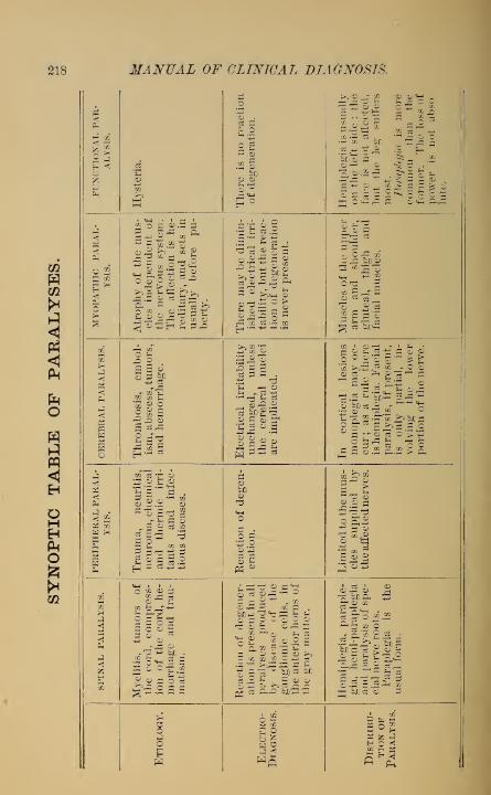
218 MANUAL OF CLINICAL DIAGNOSIS.
1^ CO
IsOKPft
o3
o .
o o
2 S
.2 §
£^Hemiplegia
is
usually
on
the
left
side
;the
face
is
not
affected,
but
the
leg
suffers
most.
Paraplegia
is
more
common
than
the
former.
The
loss
of
power
is
not
abso
lute.
h-3
8Atrophy
of
the
mus-
cles
independent
of
the
nervous
system.
The
affection
is
he-
reditary,
and
sets
in
usually
before
pu-
berty. There
may
be
dimin-
ished
electrical
irri-
tability,
but
the
reac-
tion
of
degeneration
is
never
present.
Muscles
of
the
upper
arm
and
shoulder,
gluteal,
thigh
and
facial
muscles.
CO
CO
nPh
hI
gHO
Thrombosis,
embol-
ism,
abscess,
tumors,
and
hemorrhage.
Electrical
irritability
unchanged,
unless
the
cerebral
nuclei
are
implicated.
In
cortical
lesions
monoplegia
may
oc-
cur;
as
a
rule
there
is
hemiplegia.
Facial
paralysis,
if
present,
is
only
partial,
in-
volving
the
lower
portion
of
the
nerve.
h3
tf to
1sPn
Trauma,
neuritis,
neuroma,
chemical
and
thermic
irri-
tants
and
infec-
tious
diseases.
CD
O
Limited
to
the
mus-
cles
supplied
by
the
affected
nerves.
CO
OQ
Ph
i—
i
PUCO
Myelitis,
tumors
of
the
cord,
compress-
ion
of
the
cord,
he-
morrhage
and
trau-
matism.Reaction
of
degener-
ation
is
present
in
all
paralyses
produced
by
disease
of
the
ganglionic
cells,
in
the
anterior
horns
of
the
gray
matter.
Hemiplegia,
paraple-
gia,
hemi-paraplegia
and
paralysis
of
spe-
cial
nerve
roots.
Paraplegia
is
the
usual
form.
iooH
62
«5
Distribu-
tion
of
Paralysis.

SYNOPTIC TABLE OF PARALYSES. 21!)
<Ah
HJ CO
%io•A
Ph
03
o
P< .
2 S
»! Ancesthesia
is
common
and
usually
confined
to
one
half
the
body.
It
is
as
a
rule
limited
to
the
middle
line
and
involves
the
deep
parts.
The
reflexes
may
be
increased
or
normal.
Hysterical
symptoms.
Hysterical
paraplegia
is
not
attended
by
implication
of
the
bladder.
i
O CO
Sio>-<
3Atrophy
in
conjunc-
tion
with
hypertro-
phy
of
the
muscles.
Fibrillary
contrac-
tions
are
absent.
There
is
usually
no
disturbance
of
sensa-
tion.The
tendon
reflexes
are
present,
but
in
cases
where
the
mus-
cle
atrophy
is
pro-
nounced
they
may
be
reduced.
The
paralysis
is
al-
ways
proportionate
to
the
atrophy.
CO*
0Q
<
<
4
B
wo No
degenerative
mus-
cular
atrophy,
not-
withstanding
long
continuance
of
the
paralysis.
Hemianaesthesia
may
be
present,
especially
if
the
lesion
is
in
the
internal
capsule.
There
may
be
no
sensory
disturbances.
The
deep
reflexes
are
increased
on
the
par-
alyzed
side,
and
an-
kle
clonus
may
be
present.
The
sphinc-
ters
are
not
affected.
Psychical
disturb-
ances,
headache,
dis-
turbances
of
vision
and
hearing,
ophthal-
moscopic
changes,
vertigo
and
aphasia.
>-3 CO*
a
CO
Ph£
<-4*In
a
mixed
nerve,
ancesthesia
limited
to
the
distribution
of
the
nerve.
Absent,
if
the
reflex
circuit
is
destroyed
by
the
nerve
lesion.
Absence
of
cerebral
and
spinal
symp-
toms.
CO*
CO
«
id
gsCO
Atrophy
of
the
mus-
cles,
when
the
cells
of
the
anterior
horn
are
affected,
or
when
the
anterior
nerve
roots
are
separated
from
their
trophic
center.
Disturbances
of
sen-
sation
are
common,
especially
if
the
pos-
terior
nerve
roots
are
implicated.
In
implication
of
the
lateral
column,
the
reflexes
are
increased,
otherwise
they
are
usually
diminished
or
absent.
Girdle
sensations,
im-
pairment
of
the
blad-
der
or
rectum.
Condition
OF
THE
Muscles.Disturb-
ances
of
Sensation.
CO*
HNHa Concur-
rent
Symp-
toms.

220 MANUAL OF CLINICAL DIAGNOSIS.
VASO-MOTOR AND TROPHIC DISEASES.
Morbid Anatomy. General Symptoms. Characterid ic Symptoms.
1. RAYNAUD'S DISEASE. (Symmetrical Gangrene.)
A vascular affection
characterized by variousgrades of intensity, viz. :
l. Local syncope ; 2. Lo-cal asphyxia ; 3. Localgangrene. The first is
produced by contractionof the blood-vessels, andthe second by dilatation
of the capillaries andsmall veins.
Syncope. The fingersor toes look like dead.Asphyxia. The toes or
fingers are livid, swollenand painful. The dis-
ease is usually confinedto the extremities.
The affection is usuallysymmetrical, and hemo-globinuria is not uncom-mon during an attack.
3, ANGIO-NEUROTIC OEDEMA.
A neurosis with anhereditary tendency, andcaused by vaso-motor di-
latation of the blood-ves-sels with transudation ofserum.
(Edematous swellingappearing in variousparts of the bod}7
,par-
ticularly in the face andhands.
(Edema is circum-scribed and appears sud-denly attended by gas-tro-intestinal distress.
3. ACROMEGALIA. (Marie's Disease.)
Hypertrophy of thebones and pituitary
body, and persistence ofthe thymus gland.
A dystrophy charac-terized by increasedgrowth of the bones ofthe face and extremitiesand overlying tissues.
4. FACIAL HEMIATROPHY.Interstitial neuritis of
the trigeminus nerve.The nature of the dis-
ease is doubtful.
Progressive wasting ofthe bones and soft tis-
sues of one side of theface. It usually devel-ops in childhood.
Egg-shaped face withthe large end downward,dullness over the manu-brium sterni due to per-sistence or enlargementof the thymus gland.
The affected side is
sunken, and the skinshows a whitish-browndiscoloration.
EXOPHTHALMIC GOITRE.
Nothing definite. En-largement and thicken-ing of the cervical sym-pathetic ganglia havebeen observed.
A disease of earlyadult life, frequent in
females and character-ized b}7 palpitation of theheart, protrusion of theeye-balls, and enlarge-ment of the thyroidgland.
See page 181. One ofthree important symp-toms may be absent.Tremor may be presentand shows a great regu-larity of rhythm.

DISEASES OF THE MUSCLES. 221
VASO-MOTOR AND TROPHIC DISEASES-Continued.
Morbid Anatomy. General Symptoms. (Characteristic Symptoms.
(». MVX(E1)E>IA.
Mucoid degenerationof tlu i subcutaneous tis-
sues, and atrophy of thethyroid gland.
A constitutional dis-
ease, more frequent inwomen than in men, andmarked by swelling in
the face and upper ex-tremities, mental failure
and dementia.
The swelling does notpit on pressure. Feat-ures arc coarse, and overthe cheeks there is a red-dish patch. Tempera-ture is subnormal, andthere is atrophy of thethyroid gland.
7. SUNSTROKE. (Thermic Fever.)
Early rigor mortis,congestion of the brainand lungs, and paren-chymatous changes in
the liver and kidneys.
After exposure to hightemperatures, exhaus-tion, vertigo, nausea andheadache followed bycoma, hyperpyretic tem-perature with full andrapid pulse.
The mode of onset andhigh temperature ena-bles the coma to be dif-
ferentiated from apo-plexy, alcoholism, etc.
DISEASES OF THE MUSCLES.
1. IDIOPATHIC MUSCULAR ATROPHY.
Muscular wasting withor without initial hyper-trophy, and occurringindependently of thenervous system.
An hereditary diseaseof progressive character,
presenting two chief
types : 1, primary hyper-trophy ; and 2, primaryatrophy.
The primary myopa-thies involve the musclesof the calves, trunk, faceor shoulder girdle. Theyoccur usually before pu-berty. (See pseudo-hy-pertrophic paralysis).
2. MYOTONIA CONGENITA. (Tliomsen's Disease.)
The nature of the af- An hereditary diseasefection is unknown. The characterized by tonic
muscular fibres of the cramp or spasm of the
voluntary muscles are muscles on attemptedincreased in width. movement. The affec-
tion first appears in
childhood.
Muscular spasms onattempting voluntarymovements. The sensa-tion and reflexes are nor-mal. Myotonic reaction
(on galvanic stimulationof the muscles, the con-tractures are slow andprolonged, lasting aslong as thirty seconds).

222 MANUAL OF CLINICAL DIAGNOSIS.
DISEASES OF THE MUSCLES—Continued.
Morbid Anatomy. General Symptoms. Characteristic Symptoms.
3. PARAMYOCLONUS MULTIPLEX.
The nature of the dis-
ease is unknown, al-
though it may be regard-ed as an emotional neu-rosis of hysterical origin.
Clonic muscularspasms in the musclesof the face and extrem-ities, usually symmetri-cal and appearing in par-oxysms. The strengthof the muscles and elec-trical excitability re-
main unaltered.
Clonic contractions ofthe muscles unaccompa-nied by sensory or motordisturbances.
4. GRAPHOSPASM. (Writers' Cramp.)
There are no anatomicalchanges. It results froma deranged action of thenerve centers presidingover the muscles in-
volved in writing.
Spastk form. Clonicor tonic spasm when thepen is grasped.
Tremulous form. Tre-mor when the musclesare used.
Paralyticform. Weak-ness and fatigue of themuscles.
Sensory and vaso-mo-tor disturbances asso-ciated either with spasm,paresis, paralysis or tre-
mor of the muscles em-ployed in writing.
PSEUDO-HYPERTROPHIC PARALYSIS.tropliy.)
(Pseudo-Muscular Hyper-
There is no lesion ofthe nervous system. It
is a primary affection of
the muscles character-
ized by an excess of fat
and connective tissue
between the muscular fi-
bres, and degenerativeatrophy of the latter.
A disease of childhoodmanifested by enlarge-ment of the muscles ofthe calf, thigh, buttockor back, and muscularweakness without senso-ry disturbances. Theprognosis is unfavora-ble.
Gait (page 3). Enlarge-ment of the muscles withweakness of the latter,
and absence of the re-
action of degeneration.

VESICULAR INFLAMMATIONS OF TUE SKIN. 223
DIAGNOSIS OF DISEASES OF THE SKIN.
ERYTHEMATOUS INFLAMMATIONS OF THE SKIN.
Site of Predilection.General Character of the
Eruption.Characteristic Signs.
1. ERYTHEMA NODOSUM.The extremities partic-
ularly the lower.Bright red nodes vary-
ing in size from a pea toan egg.
3. ERYTHEMA MULTIFORME.
The extremities. The cutaneous lesionschange frequently, andmay consist of macules,papules, vesicles, or bul-lae.
3. URTICARIA. (Hives.)
The wheals occurmostly on the face andtrunk, although otherparts of the body are notexempt.
An inflammatory affec-
tion attended by theeruption of pale red,evanescent wheals.
Eruption accompaniedby febrile disturbances.Nodes change color like
a bruise.
The variety of the le-
sions.
Suddenness and shortduration of eruption as-
sociated with intenseitching.
VESICULAR INFLAMMATIONS OF THE SKIN.
1. HERPES ZOSTER. (Shingles.)
Follows the distribu-
tion of the nerves.Groups of small vesi-
cles on inflammatorybases accompanied withneuralgia.
2. MILIARIA. (Prickly Heat.)
On covered parts(breast, abdomen, ex-tremities, etc.).
3. ECZEMA. (Tetter.)
The skin of any part ofthe body, with predilec-tion for the face, genita-lia, hands and feet.
An inflammatory dis-
ease of the sweat glands,attended by a discreteeruption of minute vesi-
cles.
A non-contagious af-
fection with multiformlesions (erythema, papu-les, vesicles, pustules,scales and crusts).
Grouping of vesiclesand their relation to thenerves.
Lesions are discreteand attended with burn-ing, itching and perspir-ation.
Multiform skin lesions,
itching and discharge.

224 MANUAL OF CLINICAL DIAGNOSIS.
BULLOUS INFLAMMATION OF THE SKIN.
Site of Predilection.General Character of the
Eruption.Characteristic Signs.
1. PE3IPHIGUS.May attack the skin or
mucous membranes inany part of the body.
Characterized by theeruption of successivecrops of bullae or blebs.Bulla? are elevations ofthe skin containing se-
rous fluid, and varyingin size from a pea to anegg or an apple.
Acute pemphigus is aninfectious disease, oc-
curring in the new born.P. foUaceus is a fatal
disease, characterized bybursting of the bullae
and the formation ofthick crusts.
PUSTULAR INFLAMMATIONS OF THE SKIN.
1. IMPETIGO.
On the face and ex-
tremities.
Eruption of discretepustules varying in size
from a pea to a cherry,and commonly observedin children.
2. IMPETIGO CONTAGIOSA.On the face and ex-
tremities.
An acute contagiousdisease, occurring in de-bilitated children. Thepustules are flat or um-bilicated.
3. ACNE.
The face and back.
4. ACNE ROSACEA.On the face, especially
the nose, cheeks, chin,
etc.
Inflammatory diseaseof the sebaceous glands,characterized by pap-ules and pustules, andassociated with come-dones.
Acne lesions associ-
ated with dilatation ofthe blood-vessels and tis-
sue hypertrophy.
5. ACNE MENTAGRA. (Simple Sycosis.)
Beard, eye-brows, and,
rarely, the hair follicles
of the head.
A chronic inflamma-tion of the hair follicles,
leading to tubercles, pus-tules, crusts and diffusedinfiltration of the skin.
Discrete pustular erup-tion which dries in a fewdays, leaving thin yel-
lowish crusts,which dropoff and leave a normalsurface.
The yellowish crustswhen they drop off leavebehind an excoriatedsurface.
Chronic character ofthe affection and impli-cation of the sebaceousglands.
Acne lesions, dilata-
tion of the vessels andovergrowth of tissue.
Inflammation is super-ficial, the hairs are notinvolved, and the trico-
plujton is absent.

PAPULAR INFLAMMATIONS OF TUE SKIN. 22r>
PUSTULAR INFLAMMATIONS OF THE SKIN-Continued.
Site qf Predilection.i liarader of the
Vharactt ristic Signs.
6. TINEA SYCOSIS. (Barber's Itch.)
Bearded region. Like the foregoing.The hairs become lustre-
less, brittle and loose.If involved hairs are al-
lowed to remain for fif-
teen minutes in a solu-tion of caustic potash(1 : 3), the spores andthreads of the tricophy-
ton tonsurans can be seen.
Inflammation is deep,the hairs an- involved,and the trichophyton eanbe demonstrated.
SCALY INFLAMMATIONS OF THE SKIN.
1. PSORIASIS.
Extensor surfaces ofthe knees and elbows,hair of the head and ears.
The lesions may ap-
pear on any part of thebody.
A chronic inflamma-tion leading to the de-velopment of pearlyscales which, when de-tached, leave behind ared or bleeding surface.Itching is slight or ab-sent.
2. PITYRIASIS RUBRA.Usually diffused over
the body.A rare disease of
chronic duration, attend-ed by tension and con-traction of the skin, fe-
ver and the developmentof red spots which scale.
Predilection for theextensor surfaces, drypearly scales, absenceof itching, protractedcourse and tendency torelapse.
Fever attends the de-velopment of the erup-tion. The disease leadsto marasmus.
PAPULAR INFLAMMATIONS OF THE SKIN.
1. PRURIGO.
The back and extensorsurfaces of the extremi-ties.
A chronic disease at-
tended by an eruption ofsmall, discrete, pale redpapules and itching. De-velops in early "child-
hood, and persiststhrough life.
The development inearly childhood, charac-ter of the eruption andintense itching.

226 MANUAL OF CLINICAL DIAGNOSIS.
PAPULAR INFLAMMATIONS OF THE SKIN-Continued.
Site of Predilection.General Character* of tfie
Eruption.Characteristic Signs.
2. LICHEN EUBER.Breast, abdomen, geni-
talia and flexor surfacesof the extremities.
A chronic disease oc-curring in middle-agedmales, characterized bythe development of smallred papules, itching andfailure of health.
3. LICHEN SCROFULOSIS.Back, breast, lower
part of abdomen, andlater the flexor surfacesof the extremities.
A chronic disease, oc-curring in children of astrumous diathesis, at-
tended by sma 11 pale redpapules.
Itching, failure of thegeneral health, and its
occurrence in poorlynourished middle-agedindividuals.
Absence of itching,
scrofulosis, pale skin,
and occurrence in chil-
dren.
HYPERTROPHY OF THE SKIN.
1. ICHTHYOSIS. (Fish Skin Disease.)
Extensor surface ofthe extremities. May in-
volve the skin over theentire body, exceptingthe flexor surfaces of thejoints, genitalia, palmsand soles.
2. SCLERODERMA.Upper half of the body,
although it may be dif-
fused.
A chronic affectionmanifested by dryness,thickening of the epi-
dermis and scaliness. It
is first detected in earlychildhood, and is an in-
curable disease.
. ELEPHANTIASIS.Legs and genitals.
A chronic affectionleading to induration ofthe skin in patches, orinvolving the entirebody. Death occursusually from marasmus.
Hypertrophy of theskin and subcutaneoustissues, leading to enor-mous enlargement.
4. CHELOID. (Keloid.)
Chest and back. Hypertrophy of theskin, beginning as a palered nodule, which sendsout claw-like processes,making it resemble acrab. It returns afterremoval.
Absence of inflamma-tory symptoms, chron-icity and detection inearly childhood.
Induration of the skin,
which renders the partsimmobile.
Thickening of the skinand subcutaneous tis-
sues, associated withlymphangitis.
Occurs frequently in
the colored race, historyof heredity. It is dis-
tinguished from a scarby the fact that the lat-
ter does not extend be-yond the injury.

PABASITES OF THE SKIN. 227
ATROPHY OF THE PIGMENT OF THE SKIN.
Site of Predilection.General Character of the
Kniption. Characteristic Signs.
1. VITILIGO. (Leucoderma.)
On parts of the bodyexposed to injuries andpressure.
An acquired affectioncharacterized by milk-white patches surround-ed by areas of increasedpigmentation.
Aside from the absenceof pigment, the skin is
normal.
NEUROSIS OF THE SKIN.
1. PRURITUS,
Any part of the body. A functional affectionindependent of any ana-tomical changes in theskin.
Itching and the ab-sence of any definite skinlesions, unless they arethe result of scratching.
PARASITES OF THE SKIN.
1. SCABIES. (Itch.)
Between the fingers,
wrists, axilla?, genitals,
and beneath the mammae.The face and scalp are
never implicated.
A contagious diseasecaused by the AcarusScabiei, and manifestedby the development ofpapules, vesicles, pus-tules and burrows.
The itching is intense,history and sites of pre-dilection. For demon-stration of the acarus,
see p. 189.
2. TINEA TRICOPHYTINA. (Ringworm.)
Tinea Tonsurans: char-acterized by scaly, ele-
vated patches, throughwhich dry, brittle hairs
project.Tinea Circinaia: scaly
patches which clear upin the center.
Ringworm occurringon the scalp is known as
Tinea Tonsurans; on thebody, Tinea Circinata: onthe heard, Tinea Sycosis.
Itching and demon-stration of the tricophy-
ton, see p. 190.
3. TINEA VERSICOLOR. (Pityriasis Versicolor.)
Chest and back. It
may occur on the face,
though rarely.
Salmon colored, yel-
lowish or dark-brownspots, generally scaly,
caused by the microspo-
ronfurfur.
Scaly pigmented spotsand demonstration of themicrosporon, see p. 190.

228 MANUAL OF CLINICAL DIAGNOSIS.
PARASITES OF THE SKIN-Continued.
Site of Predilection.General Character of the
Eruption.
4. PHTHEIRIASIS. (Pediculosis.)
PedkuLus capitis ; lice
confined to the scalp.
Pedicvlus corporis; bodyor clothes lice.
lUlicuhis pubis ; crablouse confined to pnbes,axillae, etc.
Eczematous lesionsand scratch markscaused by the intenseitching
;petechia? caused
by the bite of the insect.
5. TINEA FAVOSA. (Favus.)
The scalp. It rarely
affects the nail substance(onych oi i \ ycosisfavosa)
.
A contagious scalp af-
fection manifested byyellowish, cup-shapedcrusts, through whichdry, brittle, lustreless
hairs project. The crustshave a peculiar mustyodor.
Characteristic Sign %.
Demonstration of theova or nits and the lice.
See page 189.
Yellow, cup-shapedcrusts, odor and demon-stration of the fungus,Achorion Schoenleinii, seepage 190.
DRUG EXANTHEMATA.
Quinine, morphine, opium, atropine, digitalis, chloral
hydrate, antipyrine, plienacetine, sulphonal and strychnine,
often produce an eruption of an erythematous character.
After the use of the salicylates a bullous eruption has been
observed.
Arsenic may produce an itching papular or vesicular
eruption.
Copaiba and Turpentine, produce an urticaria-like erup-
tion.
Iodides and bromides produce an acne eruption.
BACTERIOLOGICAL DIAGNOSIS.
BACTERIOLOGICAL DIAGNOSIS.
PATHOGENIC MICROBES.
nul Arrangement. Growth. Reaction.
1. ACTINOMYCOSIS. (Kay Fungus.)
Cultures obtained af-
ter five to six days onagar-agar and gelatinewithout liquefaction.
Found in man and cat-
tle as a round, flattened.
hollow body, with a cen-tral cavity made up ofclub-shaped elements in
th e form of a ro s e tt e . Inthe pus or scrapings, lit-
tle yellow grains "aboutthe size of a pin's headare seen containing theray fungus.
2. ANTHRAX BACILLUS. (Bacillus Anthracis.)
Cover glass prepara-tions are best stained byGram's method. (Seepage 193.)
Found in the bloodand organs of animalsafflicted with splenic fe-
ver. The bacilli are rodswith broad cup-shapedends. In cultures longthreads are formed withlarge oval spores.
3. CHOLERA BACIIXUS. (Comma Bacillus.)
Grows rapidly between1*2° and 45° C, and re-
quires plenty of oxygen.Colonies develop in twodays on gelatine plates.
Found in the intestinalcanal and faeces of chol-era patients. It is acurved bacillus abouthalf the size of a tuber-cle bacillus. Discoveredby Koch in 1884.
Develops at ordinarytemperatures on all nu-trient media that havean alkaline or neutral re-
action. In a solution ofpeptone at 37° C, theydevelop rapidly on thesurface in six to twelvehours.
They take the aniline
dyes. To bring out thecup-shaped ends, methy-lene blue is best.
It is best colored withaqueous solution of fuch-sin, and may be decolor-ized by Gram's method.For color reaction, see
page 123.
4. DIPHTHERIA BACILLUS. (Bacillus Diphtheria-.)
Found in diphtheriticmembranes. Straight orcrooked rods, the samelength as the tuberclebacilli, but twice as thick.They stain most intenseat the ends.
They grow on pepto-nized meat-sugar-serum
After Loffler's meth-od : few minutes in a
infusion at 37° C. They concentrated alcoholicgrow readily on all me- solution of methylenedia between 20° C. and blue, to which has been40° C. added a KHO solution
(1 to 10,000), then in
one-half per cent acetic
acid, then absolute alco-
hol, mount in balsam.

230 MANUAL OF CLINICAL DIAGNOSIS.
PATHOGENIC MICROBES-Continued.
Form and Arrangement. Growth. Reaction.
5. GONORRHEA. (Gonococcus.)
Found in the secre-tions of gonorrhoic in-
flammation as cocci, al-
ways in pairs (diplococcus i,
biscuit-shaped, with flat
surfaces lying togetherresembling the coffee-bean.
6. LEPROSY. (Lepra Bacillus.)
Growth doubtful. Onlyupon human blood se-
rum have they given auysemblance of growth.
Found in all leprousproducts. Small rods,shorter than tuberclebacilli, with occasionallypointed ends.
7. BACILLUS MALLEIFound in the nodules
of the disease. Smallrods about the size ofthe tubercle bacillus.The ends are rounded.They appear singly as arule".
Grows on blood serumto which peptone andglycerine have beenadded. Growth slow andrequires about eight daysat a temperature of37° C.
. (Glanders.)
Grow abundantly onglycerine-agar or bloodserum at 3f° C.
Best stained with fuch-sin or methyl-violet. It
is not discolored byGram's method.
Same reaction as tu-
bercle bacillus (see page65), but stain more quick-ly. They do not stain in
LCffier's alkaline methylblue solution, thus dif-
fering from the tuberclebacillus.
Gram's method is notapplicable. Loffler's me-thylene blue is the beststain.
8. 31ALARIA. (Plasm
Found in the bloodcorpuscles of malarialpatients. They are pro-toplasmic corpuscles ofvarious forms (round,oval, crescentic and fla-
gella). They may ormay not contain blackpigment. They showrapid amoeboid move-ments.
9. PNEUMONIA. (Bacillus Pneumonia?.) (Diplococcus Pneumoniae.)
odium Z>Ialariae.)
They have not beencultivated. Inoculationswith the blood of a
malarial patient will pro-duce a typical attack ofmalaria fin another per-
son. They disappearfrom the blood after theuse of quinine.
The organisms are bestobserved when the bloodis fresh and unstained.A powerful lens is neces-sary. Dried specimensof blood may be stained
with methylene blue.
Both forms have beenfound in pneumonia.The bacilli are oval-shaped rods nearly aswide as they are long.In tissues each bacillushas a faint capsule. Thediplococci are rod-shaped,longer than broad, usu-ally^ in pairs, and sur-rounded by a capsule.
The bacilli grow rapid-
idly on all media. Thediplococci grow slowlyon alkaline media.
Stained by Friedland-er's method (page 67),
although the ordinary
aniline stains will suffice.

BACTERIOLOGICAL DIAGNOSIS. 231
PATHOGENIC MICROBES—Continued.
Form and Arrangement. Growth. liraction.
10. RELAPSING FEVER. (Spirillum or Spirochete Obermaierl.)
Found in the blood ofrelapsing or recurrentfever. "The organismsare long spiral "threadswith a distinct flagellum,and tlu\y move by meansof wavy undulations.They are only found inthe blood during thefever.
They have not been Ordinary aniline stains,
cultivated. After fixing in the flame,cleanse with a four percentacetic aeid solutionand stain with the ordi-nary aniline solutions.
11. SUPPURATOIN. (Streptococcus Pyogenes.)genes Anreus.)
Found in pus. Theyproduce suppuration.The streptococci are coc-ci found singly and inchain-like groups. Thestaphylococci are micro-cocci in clusters likebunched grapes.
The streptococci growin bouillon and the sta-
phylococci on gelatine.
(Staphylococcus Pyo-
Ordinary stainsGram's method.
and
12. STREPTOCOCCUS ERYSIPELATIS.
Found in the lymphchannels of the skin in
erysipelas as very smallcocci in pairs or chains.
Grows on gelatine, agar-agar and blood serum at
a temperature from 30° C.to 37° C.
Retains colorGram's method.
after
13. SYPHILIS. (Bacillus Syphilis.)
A bacillus found in
the tissues and secre-
tions of syphilis. In size
and shape it resemblesthe tubercle bacillus. It
is frequently curved, andhas clubbed ends.
Hasvated.
not been culti- Stain in fuchsin solu-tion for twenty-fourhours, rinse in water,then in a chloride of ironsolution, then alcohol,and clear in clove oil.
14. TETANUS. (Tetanus Bacillus.)
Found in the earth andin the secretions fromthe wounds of tetanuspatients. They are fine
bacilli, and form threadsin irregular masses.
Develops slowly andonly when oxygen is ex-cluded. Grows in nearlyall the media.
Colors with all the or-
dinary aniline dyes, andGram's method.

232 MANUAL OF CLINICAL DIAGNOSIS.
PATHOGENIC MICROBES -Continued.
Form and Arrangement. Growth. Jo action.
15. TUBERCULOSIS. (Bacillus Tuberculosis.)
Found in the productsof human and animal tu-
berculosis as thin rods,
one-fourth to one-half
the diameter of a redblood corpuscle in
Length, beaded and withrounded ends.
Grows in gryeerineagar-agar, on potatoesand blood serum. Theygrow slowly, and areonly visible after ten to
fifteen days.
16. TYPHOID FEVER. (Bacillus Typhosus.)
Found in the blood,urine, faeces and tissues
of typhoid patients asbacilli, three times as
long as they are broad,with rounded ends.
They grow slowly onall nutrient media.
See page 65.
They take the aniline
dyes less deeply thanother bacilli. Not col-
ored by Gram's method.
BACTERIOLOGICAL ANALECTS.
1. The diagnostic value of the iyplioid bacilli whenfound in the stools is questionable, as it is almost impossible
to differentiate them from the bacterium coli commune which
is also present. Puncture of the spleen for the purpose of
obtaining cultures of the typhoid bacilli is a procedure not
always free from danger.
2. Typhoid bacilli retain their vitality for weeks in
water. They thrive in the soil and are not killed by
freezing.
3. The spirilla of relapsing fever are met with in the
blood only during the paroxysms of fever, and disappear in
the intervals. Their number is not proportionate to the
severity of the attack. The spirilla are absent in the secre-
tions.

BACTERIOLOGICAL ANALECTS. 233
4. The streptococci of erysipelas are found in the periph-
eral parts of the erysipelatoid inflammation, and almost
exclusively in the lymphatic vessels.
5. The incubatory period of inoculated erysipelas is from
fifteen to sixty hours.
6. The bacilli of cholera Asiatica occurs only in the stools,
and never in the blood, of cholera patients.
7. The bacilli of cholera nostras, which resemble the
bacilli of Asiatic cholera, are distinguished from the latter
by being plumper and larger.
8. The bacillus diphtheria? lies in the surface of diph-
theritic pseudo-membranes, and is never found in the blood
or viscera. Cover glass specimens may be made from the
membrane.
9. The plasmodium malarice may be found in the blood in
the following forms : 1. An unpigmented hyaline body within
the red blood corpuscles, which shows active movements.
2. A pigmented amoeboid body inside the red corpuscle. 3. Asegmenting body inside the red corpuscle. 4. Crescentic
bodies within the red corpuscles. 5. Flagellate organisms.
10. The anaemia of malaria is caused by a destruction of the
red blood corpuscles by the plasmodia.
11. In empyema an examination of the aspirated fluid is of
great importance. If the pneumococcus is present, the case
will progress favorably ; if the pus microbes, staphyococci and
streptococci are present, the empyema is of septic origin and
the prognosis is exceedingly bad. If the fluid contains no
organisms, it is, as a rule, of tuberculous origin.
12. The diplococcus pneumonicc of Fraenkel is the most
constant organism in croupous pneumonia, although found

234 MANUAL OF CLINICAL DIAGNOSIS.
in the buccal secretion in twenty per cent, of healthy indi-
viduals.
13. In the secondary affections ofpneumonia, such as pleu-
risy, meningitis, etc., the diplococcus pneumonia is almost
constantly present.
14. It is difficult to distinguish the bacillus of syphilis from
the smegma bacillus, which is found beneath the prepuce.
15. The bacillus leprce has been cultivated successfully, but
inoculation experiments on animals have been unsuccessful.
16. The anthrax bacilli die rapidly under unfavorable con-
ditions, while the spores possess a high power of resistance.
17. Anthrax in the human being occurs by inoculation
through the skin in which case the disease is usually local
{malignant pustule) ; through the lungs as in the wool sorter's
disease {pulmonic anthrax), and finally through the intestinal
canal by the reception of the virus mixed with food or water.
18. Malignant oedema is caused by a bacillus. It occurs in
connection with compound fractures, deep wounds, and after
hypodermatic injections. It is manifested by emphysema of the
skin, putrefaction and oedematous softening of the muscles
adjacent to the injury, and finally death.
19. The tubercle bacillus was discovered by Koch on the
24th of March, 1882. It is an anaerobic (living without air)
bacterium. The transmission of tuberculosis is chiefly through
the lungs.
20. If in doubt regarding the diagnosis of glanders, inocu-
lation can be conducted on mice or guinea pigs. Ulcers form
at the point of inoculation, followed by nodules, which
caseate.
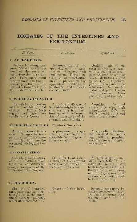
Diseases of intestines and peritoneum. 235
DISEASES OF THE INTESTINES ANDPERITONEUM.
Etiology. Pathology. Symptoms.
1. APPENDICITIS.
Occurs in young per-sons. More than fifty percent, of the cases oc-
cur before the twentiethyear. Foecal masses andforeign bodies in the ap-pendix play the most im-portant etiological role.
Traumatism is also a fac-
tor.
Inflammation of theappendix may be catar-
rhal or ulcerative withperforation. Faecal con-cretions or enterolithsmay be present in theappendix. Adhesions,peritonitis and abscessare sequences.
2. CHOLERA INFANTUM.
Prevails in hot weatherand in artificially fedchildren. Faulty feed-ing and bad hygiene arepredisposing factors.
An infantile disease ofbacteritic origin (no spe-cific microbe has beenfound), with inflamma-tion of the mucosa of thestomach and intestines.
3. CHOLERA MORBUS. (Cholera Nostras.)
An acute sporadic dis-
ease. Changes in tem-perature, summer seasonand unripe fruit are theessential etiological fac-
tors.
4. CONSTIPATION.
Sedentary habits,atonyof the intestines fromlocal or constitutionalcauses, weakness of theabdominal muscles, etc.
5. DIARRHOEA.
Changes of tempera-ture, ptomaines and irri-
tating food in the intes-
tines, bacteria, poisons,reflex disturbances, etc.
A ptomaine or a spe-cific bacillus may be re-
sponsible for the gastro-enteric catarrh.
The chief local causeis atony of the sigmoidflexure which forces thefasces into the rectum.
Catarrh of the intes-
tines.
Sudden pain in theright iliac fossa, attendedwith fever, and local ten-derness with or withoutfever. McBurney's point(page 118). If generalperitonitis occurs, it is
recognized by suddenabdominal pain, tympa-nites, increased feverand rapidity of the pulse.
Vomiting, frequentwatery discharge, hightemperature (105° to108° F.), rapid pulse andcollapse symptoms.
A sporadic affection,
characterized by vomit-ing, bilious purging,moderate fever and greatprostration.
No special symptoms.Many symptoms of anindefinite nature havebeen noted as a result ofthe resorption of noxiousmatter (copramiia) andchlorosis is attributedto faecal poisoning.
Frequent passages. Inmemfoanous ente?'itis^thereare false membranes ormucous easts in thestools.

236 MANUAL OF CLINICAL DIAGNOSIS.
DISEASES OF INTESTINES AND PERITONEUM—Continued.
Etiology. Pathology. Symptoms.
6. DILATATION OF THE COLON.A congenital condi- Dilatation of the sig-
tion, or it may result moid flexure occurs,from over-distension when it is congenitallyfrom faecal accumula- long. An acute dilata-
tions, tion may follow a twistin the mesocolon.
7. DYSENTERY. (Bloody Flux.)
Bad hygiene, cachecticconditions, exposure to
cold, irritating food, etc.
In the tropics, as well as
in cold climates, the dis-
ease is produced by theamoeba coll. See page 184.
8. ENTERO-COLITIS.Bad food and hygiene,
and climatic conditionsare predisposing ele-
ments.
9. ENTEROPTOSIS.Dilatation of the stom-
ach and mobility of thekidney are frequently as-
sociated with this condi-tion.
Prolapse of the ab-dominal viscera, espe-cially the transverse co-
lon. When all the vis-
cera fall down the condi-tion is called splanchno-ptosis.
10. INTESTINAL COLIC, (Enteralgia.)
As a symptom it occurs A functional affection
in plumbism, intestinal caused by spasmodicobstruction and inflammation. It may be pro-
duced by flatulence, fae-
cal accumulation or irri-
tating food.
11. INTESTINAL ULCERS
contraction of the intes-
tines.
Ulcers may be specific
from syphilis, tubercu-losis or typhoid fever.
Ulceration may also arise
from the various formsof intestinal inflamma-tion and pressure of fas-
ces {stercoral ulcers).
The duodenal ulcer is
of peptic origin, like theround ulcer of stomach.The site of tuberculousand typhoid ulcers is theileum, while the ulcersof dysentery are locatedin the larse "intestine.
Tympanitic distensionof the abdomen. If thewalls of the colon arehypertrophied, the colonmay be felt. If disten-sion of the colon is pro-nounced, it may press onthe diaphragm and inter-
fere with the heart.
The inflammation of thecolon may be catarrhal,
diphtheritic or amoebic. Inconnection with the lat-
ter form, abscess of theliver is a frequent se-
quela.
Inflammation of themucous membrane ofthe ileum and colon.The solitary and agmi-nated glands are involvedand often ulcerated.
passages of fre-
blood-streakedThe
quentstools are accompaniedby tenesmus. In theamoebic or tropical form,the diagnosis is made byan examination of thestools.
Diarrhoea with bloodand mucus in the stools,
moderate fever, painalong the colon, andwasting of the body.
Constipation from nar-rowing of the colon, andmotor, sensory and vas-cular disturbances fromfalling down of the stom-ach and intestines.
Severe spasmodic painlocalized around the um-bilicus, relieved by pres-sure and the dischargeof wind. Unattended byfever as a rule.
The diagnosis is oftendifficult, and the casemay furnish no symp-toms. The followingsymptoms are of note :
diarrhoea, pain, and bloodand pus in the stools.

DISEASES OF INTESTINES AND PERITONEUM. 237
DISEASES OF INTESTINES AND PERITONEUM—Continued.
Etiology. Ttothology. Symptoms.
12. INTESTINAL OBSTRUCTION. (Ileus.)
Acute obstruction may becaused by : 1. Congenitalocclusion. 2. Strangula-tion. 3. Twists (volvuhis).
4. Intussusception.Chronic obstruction : 1.
Faecal accumulations.2. Strictures. 3, Foreignbodies (gall-stones). "4.
Tumors.
13. PERITONITIS.
Primary peritonitis
may arise from exposureto cold or traumatism.It may be secondary fromextension of inflamma-tion from neighboringorgans. Chronic perito-
nitis may be caused bytuberculosis, s}7philis,
cancer, etc.
Strangulation is themost frequent cause ofacute obstructions, andthe site is usually in thesmall intestines. Twistsare associated with a
very long mesentery.The most usual site ofintussusception is at theileo-caecal valve, as it
descends into the colon.
According to the char-acter of the exudation,the inflammation may be
:
1, fibrinous ; 2, sero-
fibrinous ; 3, purulent
;
4, putrid ; 5, hemor-rhagic.
Symptoms of acute ob-
struction : constipation,pain in the abdomenand vomiting. The de-gree of abdominal dis-
tension depends on thesite of obstruction.
('Iironic obstruction :
same as foregoing, al-
though the developmentof symptoms is slow.
Moderate fever, ab-dominal tenderness, ab-dominal distension,vom-iting, pinched featuresand constipation.
14. PSOROSPERMOSIS.
Common in the inver-tebrates and rare in thehigher mammals. Theparasites are called p.so-
rosperms, and they pro-duce in man a diseasesimilar to that occurringin rabbits.
In internal psorosper-miasis, nodules resem-bling tubercles and con-taining psorosperms arefound upon the perito-
neum, omentum, liver
and other viscera.
15. TYPHLITIS STERCORAEIS.
Caused by the lodg-ment of the faeces andoccurs especially inyoung pei sons who havebeen constipated or suf-fered some error in diet.
An inflammation ofsthe
caecum. Many cases oftyphlitis are probablydue to disease of the ap-pendix.
Fever of an intermit-tent type, diarrhoea, ten-derness of the affectedorgans, _ and usuallydeath. In cutaneous pm-rospermiasis, there arepapillomatus growthscontaining psorosperms.
Pain in the right iliac
fossa. A sausage~-shapeddoughy tumor "is presentin this fossa. The feveris moderate. Prognosisis favorable.

S38 MANUAL OP CLINICAL DIAGNOSIS.
ANALECTIC REVIEW OF GASTRIC DIGES-TION AND GASTRIC NEUROSES.
THE PROCESS OF DIGESTION.
1. The first period of digestion in the stomach is theamylolytic, and consists in the conversion of starch intodextrine (achroo- and erythro-dextrine). The digestion is
effected in the stomach by the swallowed saliva.
2. Grape sugar, which is a final product of starch diges-
tion, is converted into lactic acid by the action of micro-organisms that excite fermentation.
3. The amylolytic period of digestion lasts, on an aver-
age, three-quarters of an hour.
4. Muriatic acid and pepsin are secreted immediatelyafter the ingestion of food.
5. When muriatic acid is first secreted it is in combina-tion, and the free acid is not present until three-quarters
of an hour after the ingestion of food.
6. Free muriatic acid inhibits amylolytic digestion, as
the digestion of starch cannot proceed in an acid medium.7. Lactic acid disappears from the stomach one hour
after the ingestion of food, or at a time when free muriaticacid is present.
8. Simultaneous with the free hydrochloric acid, themilk curdling ferment {rennet ferment), which coagulates
the casein of milk, and pepsin, are secreted.
9. Pepsin and rennet ferment are not secreted as such,
but are formed by their ferments, propepsin and rennet
zymogen into pepsin and rennet ferment, respectively, un-der the influence of muriatic acid.
10. The second or muriatic period of digestion consists
in a peptonizing action of the gastric secretion upon thealbuminous bodies.
11. The free muriatic acid is antiseptic, destroying all
microorganisms concerned in fermentation and putrefac-
tion.
12. During the period of digestion, some of the digested
food is absorbed, while the remaining portion slowly passes
into the duodenum.
EXAMINATION OF GASTRIC DIGESTION. 230
13. About six hours after a mixed meal, the stomachshould be practically empty.
14. In the intervals of digestion, the stomach contains
only a small amount of clear fluid of neutral reaction.
EXAMINATION OF GASTRIC DIGESTION.
1. To determine the duration and other facts concern-
ing digestion, the patient takes the experimental meal of
Leube.2. Leube 9
s experimental meal consists of a plate of water-
gruel, an ordinary piece of beef-steak, and white bread.
3. Seven hours after the meal of Leube, if digestion is
normal, the stomach should only contain a few remnantsof food.
4. After removal of the gastric contents, examinationsmust be made for the following: free muriatic acid, lactic
acid, butyric and acetic acids, determination of the total
acidity, determination of the digestive potver of the gastric
juice, its coagulating effect upon milk, the transformation
of starch, the presence of peptone and propeptoiie, the ab-
sorptive and motive power of the stomach.
5. The most certain test for hydrochloric acid is the
pliloroghicin-vanillin test. It consists of phloroglucin twoparts, vanillin one part, to thirty parts of absolute alcohol.
One or two drops of this solution heated on a porcelain
plate with the gastric contents (filtered), will, if free hydro-
chloric acid is present, give a red color, the intensity
depending on the percentage of the acid. It is distinct
to 0.05 per thousand.6. The phloroglucin-vanillin test, or Gihnzberg's reaction,
is only interfered with if sulphuretted hydrogen is present.
7. Lactic acid is detected by the solution of Uffelman (to
about three ounces of a two per cent, solution of carbolic
acid, add one or two drops of chloride of iron solution).
This steel-blue mixture becomes yellow or yellowish-green
if lactic acid is present. It becomes like water if only HCLis present. The test may be counterfeited in the presence
of phosphates, mineral acids, alcohol and grape sugar.
These errors may be removed if the gastric secretion is
shaken with ether. After evaporating the ether, dissolve
the ether residue in water and apply test.

240 MANUAL OF CLINICAL DIAGNOSIS.
8. Butyric and acetic acids are recognized by their odor.They convert Vffelmarfs solution into a yellowish-red color.
9. The total acidity of the gastric contents is determinedby neutralizing it with a one-tenth normal solution of
soda. 1 c.c. of the soda solution neutralizes 0.00365 of
hydrochloric acid.
10. The digestive power of the gastric juice is determined!)Y the digestion of a piece of the white of a hard-boiledegg or a bit of washed fibrin. See page 113.
11. The coagulating effect of the gastric juice due to therennet ferment is demonstrated as follows : add three to
five drops of the filtered gastric juice to 10 c.c. of milk,when curdling will occur, in about fifteen minutes, at 37°
to 40° C.
12. The transformation of starch is tested by adding a
drop of LugoVs solution (iodine one part, iodide of potashtwo parts, aq. dest. two hundred parts) to the filtered gas-
tric juice. If starch is present the solution becomes blue,
if erythrodextrin, purple. A mixture of starch and dex-
trin with the first few drops of the solution becomes color-
less, but upon adding more of the solution, it becomes redand then blue.
13. The test for peptone is the biuret reaction. Seepage 112.
14. Testing the absorptive and motive power of the stom-ach is described on page 113.
ANOMALIES OF GASTRIC DIGESTION.
1. Anacidity or inacidity denotes a condition in whichfree hydrochloric acid is absent. It occurs in destruction,
atrophy and degeneration of the mucous membrane of the
stomach, in gastric cancer with dilatation, in severe anae-
mia, in febrile conditions, and in certain cases of nervousdyspepsia.
2. Anacidity interferes with the digestion of albumenand the absence of hydrochloric acid favors putrefaction
and fermentation.
3. Hyperacidity or superacidity is a condition in whichthe free hydrochloric acid is present earlier than nsual and is
in excess. It occurs in certain nervous dyspepsias {pyrosis
hydrochlorica), and in acute and chronic gastric catarrh.t
241 GASTRIC NEUROSES.
4. Hyperacidity interferes with the digestion of starch.
Albuminous bodies are usually rapidly peptonized.
5. Chronic hypersecretion signifies a continuous secretion
of gastric juice even though the stomach is empty. Thefluid present resembles gastric juice with a high degree of
acidity.
GASTRIC NEUROSES.
1. When a patient has a ravenous appetite which hefinds difficult to satiate, the condition is called bulimia or
polyphagia. It may occur alone or as a symptom of manynervous affections, neurasthenia, hysteria, psychoses, etc.
It is likewise a prominent symptom in diabetes.
2. Pica is a condition of perverted apj)etite often symp-tomatic of hysteria, in which the patient craves for ink,
chalk and other substances.
3. Anorexia is a lack of appetite or repugnance for food.
It accompanies many dyspeptic and mental disturbances.
4. Gastralgia or Gastrodynia is a neuralgic affection of
the stomach dependent on irritation of the sensory nerves.
It may be symptomatic of tabes (gastric crises), hysteria,
neurasthenia, primary anaemia and various mental diseases.
5. An increased secretion of hydrochloric acid {hyper-
acidity) may exist as a primary neurosis of the stomach.6. Nervous belching occurs in hysteria and neurasthenia.
7. Pneumatosis is a condition in which the stomach is
inflated with air often conducing to typical asthmaticattacks (asthma dyspepticiim).
8. Nervous vomiting is frequent in hysteria. It is notattended by severe nausea or retching.
9. Peristaltic unrest of the stomach is a condition usually
dependent on gastrectasis, in which the peristalsis of the
stomach is so marked, that the movements may be ob-
served through the relaxed abdominal parietes. The move-ments may be attended by loud rumbling sounds.
10. Nervous anacidity is often encountered as a pureneurosis in hysteria and neurasthenia.
11. Incontinence of the pylorus and cardia are condi-
tions likened to paralysis. Paresis of the latter allows of
the regurgitation of air and food, while paresis of the
former allows the undigested food to pass at once into the
intestines.

242 MANUAL OF CLINICAL DIAGNOSIS.
12. Incontinentia pylori may be recognized by distend-ing the stomach with air or gas (tartaric acid and bicar-
bonate of soda). It will be noted that the stomach becomesincompletely distended, and that the air or gas passes at
once into the intestines.
13. Rumination or merycismus is a neurosis in whichfood, after it is ingested, returns to the mouth to be chewedand swallowed a second time.
14. Atony of the stomach may be partial or complete. It
is suppressed peristalsis of the stomach.
RELATION OF DISEASES OF THE STOMACH TOOTHER DISEASES.
A?icemia.—Absorption and peristalsis of the stomach are
disturbed, leading to a complex variety of digestive dis-
turbances.
Cardiac Diseases.—As a result of venous congestion,
catarrh of the stomach occurs.
Diabetes.—Dyspeptic disturbances frequently serve, as anindex to this disease. Hydrochloric acid, wrhen temporarily
absent, maybe merely an expression of a neurosis; if absentpermanently, the cause is atrophy of the mucous mem-brane, the result of interstitial inflammation.
Gout.—There may be a specific gouty disorder of thestomach, the result of the uric acid diathesis, or the stom-ach may participate in the symptoms of this affection.
Hepatic Diseases.—Many affections of the liver may bedisguised as a chronic gastric catarrh. There is an inti-
mate relationship existing between the functions of bothorgans.
Malaria.—Aside from dyspeptic disturbances as mani-festations of this disease, there are typical neuroses withcharacteristic periodicity, which yield to quinine.
Nervous Diseases.—In tabes dorsalis, paroxysms of gas-
tralgia and gastric crises occur not only in the late, but in
the early stages of the disease. Gastric crises are severe
paroxysms of pain, confined to the epigastrium, accompa-nied by painful and uninterrupted vomiting, nausea andvertigo. A characteristic feature of the crises is this, that
almost immediately after the pains cease, the patients de-
mand food, which they digest perfectly.

THE EMPLOYMENT OF DR UQS IN DIA GNOSIS. 243
Renal Diseases.—Disturbances of digestion and vomitingoccur as a result of the excretion of excrementitious prod-
ucts by the stomach, symptoms in reality of ursemia.
Mobility of the kidney may cause gastrectasis by compres-sion of the duodenum.
Tuberculosis.—Many cases of phthisis begin with dys-
peptic disturbances ; in fact, Hutchinson maintains that
dyspeptic symptoms precede the onset of tubercular mani-festations in thirty-three per cent, of the cases. Fenwickfound evidences, of gastric catarrh in seventy-three per
cent, of his phthisical cases.
THE EMPLOYMENT OF DRUGS INDIAGNOSIS.
Cocaine.—As a local anaesthetic, this drug subserves a
useful purpose in diagnosis. Eecurring or persistent head-aches, as well as a large group of neuroses, are often de-
pendent on some abnormity in refraction, or to an improperbalance between the sets of muscles which regulate theocular movements. A solution of cocaine, sprayed on theocular conjunctiva, paralyzes the accommodation, and will,
in many instances, for the time being, ameliorate or cause
to disappear the reflex symptoms dependent on the ocular
defect. For examining the back ground of the eye, dila-
tation of the pupil had better be secured by cocaine thanby atropine, inasmuch as the latter paralyzes the accom-modation for too long a period. Any irregularity in thedilatation of the pupil may direct attention to an old iritis.
Among the reflex nasal neuroses, the following may becited: headache, vertigo, insomnia, neuralgic affections in
typical situations (scapula, sternum, renal region), and dis-
turbances in the visceral functions (asthma, palpitations,
vomiting, etc.). If the nasal mucous membrane is anaes-
thetized by cocaine, many of these neuroses, will disappear,
thus furnishing an invaluable aid in diagnosis. Variousreflex troubles may originate from urethral irritation, espe-
cially from stricture. These reflex troubles may merelyamount to pains in the perineum, abdomen, and in other
remote parts, or they may constitute such affections as
neurasthenia, epileptiform convulsions, etc. The urethral

244 MANUAL OF CLINICAL DIAGNOSIS.
instillation of a solution of cocaine may prove an aid in
diagnosis. Similarly, reflex rectal, uterine, vaginal andlaryngeal troubles may be determined.
Nitrite of Amyl.—The physiological action of this
remedy is to produce vascular dilatation. It is adminis-
tered by inhalation. The pains of angina pectoris are fre-
quently relieved by the inhalation of this substance, and it
may serve as a means of differentiation from pseudo-a?igina.
Headaches of anaemic origin are relieved, whereas, if the
headache is of hyperaemic origin, it is intensified. Spastic
migraine is relieved, while paralytic migraine is intensified.
Neuralgias of anaemic origin are likewise benefited. I
have used inhalations of amyl nitrite in differentiating the
rales of asthma from those of the accompanying bronchitis ;
the former disappear, while the latter persist. In manypulmonary neuroses of spasmodic character, nitrite of
amyl inhalations are of signal advantage in aiding the
results of auscultation.
Iodide of Potash.—This remedy, like mercury, is
almost diagnostic when benefit is secured, from its adminis-
tration in neuralgias, ulcerations, paralyses, etc., of syphi-
litic origin. It must be given alone, and in large doses,
until its physiological action is secured. It is also used in
the diagnosis of suspected pulmonary tuberculosis when in
apical lung affections, dullness or modified respiration is
present without rales. The latter may be artificially pro-
duced by its administration. When the cough is dry with-
out sputa, the latter may be produced by giving iodide of
potash. To intensify the auscultatory phenomena of anold plenritis, iodide of potash, by increasing the secretions,
may be of some value. It is also used for determining the
absorptive power of the stomach (see page 113). The value
of potassium iodide in the removal by resorption of hyda-tid cysts is determined by its administration in a givencase. If aspiration of the sac shows the presence of iodine,
then the potash treatment will be of value, otherwise, the
results will prove negative.
Iron.—The ferruginous preparations are of diagnostic
value in determining the ancemic origin of neuralgias, head-aches, cardiac murmurs, etc. They should disappear or
become ameliorated after their persistent use. In chlorosis,

EMPL TMEWT OF DRUGS IX DIA GNOSIS. 2 f
5
iron is a specific. Iron intensifies the symptoms of anyaffection due to plethora. It is always better to look uponanaemia, not as a substantive, but as a symptomatic affec-
tion. I have shown elswhere that pulmonary atelectasis
(page 50) is a frequent factor in the etiology of anaemia,
and furthermore, that by persistent, daily, methodical lunggymnastics, many intractable forms of anaemia may becured.
Digitalis.—This drug, by slowing the action of the
heart, is of value in irregular action of that organ, to de-
termine the time of a murmur, should this be present. Atthe same time, by increasing the force of the heart's action,
it will render cardiac murmurs more distinct, inasmuch as
the intensity of a murmur is dependent on the activity of
the heart. Fluid in the pericardium may be an exudationor transudation. Digitalis, by increasing diuresis, will
often cause the resorption of the latter, leaving the formerunaffected. Kesorption is noted by a diminution in the
area of precordial dullness. In tachycardia, resulting fromvagus paralysis, the heart does not respond to digitalis.
Quinine.—In the typical forms of malaria, the diagno-
sis is always easy ; but there are types of the disease in
which the febrile manifestations are irregular. In the lat-
ter cases, the therapeutic test of quinine is unquestioned,for the symptoms of malaria, however unusual, alwaysyield to quinine. Any intermittent fever which does notrespond to quinine is not malarial. In applying this
therapeutic test, we must be lavish with our drug, alwayscarrying it to its physiological effects, and maintainingthem for several days.
Colchicum.—This is a specific palliative in acute gout.
There are cases of irregular gout dependent on a so-called
gouty diathesis, with a train of manifestations implicating
nearly every organ of the body. Among the symptoms,headache and neuralgia are frequent. It is in the irregu-
lar cases of gout, that the colchicum test should be applied,
coupled with the other routine methods of treatment.
Ergot.—The action of this drug is to produce arterial
anaemia, and its use is suggested in the differential diag-
nosis of anaemic and congestive affections. The latter wTill
be ameliorated.

246 MANUAL OF CLINICAL DIAGNOSIS.
Salicylic Acid.—This preparation is almost a specific
in rheumatic affections, especially in the acute cases. Inatypical manifestations of presumable rheumatic origin,
the salicylates should always be given as a therapeutic test.
Salol.—This agent is used for determining the motoractivity of the stomach. It is a compound of phenol andsalicylic acid, and it is only split up in an alkaline medium.After taking fifteen grains of salol in a capsule, salicyluric
acid will be detected in the urine in from forty to seventy-
five minutes ; which shows that the salol has passed out of
the stomach. The acid may be easily detected by placing
a drop of urine on filter paper ; if to the latter a drop of a
ten per cent, ferric chloride solution is added, a violet
color appears. In atony of the stomach and gastric dilata-
tion, the salicyluric acid may not be detected in the urinefor many hours. There are many cases of diarrhoea due to
bacteritic intestinal infection. A diarrhoea of this kindresponds favorably to salol or any other intestinal anti-
septic.
Opium.—This drug will relieve dyspnoea produced bymany painful thoracic affections.
Anaesthetics.—They are employed in the detection of
simulation of various kinds. It- may only be necessary to
carry the action of the anaesthetic up to the stage of excite-
ment.Hysteria is an affection which may simulate almost any
disease, and the detection of the malady is often possible
only after narcosis. Hysterical contractures and joint
affections are examples.A phantom tumor is of hysterical origin. It is produced
by relaxation of the recti muscles and spasmodic contrac-
tion of the diaphragm in conjunction with gaseous infla-
tion of the intestines, and an arching forward of the ver-
tebral column. This tumor always disappears under full
anaesthesia, A thorough palpation of the abdomen is only
possible, in many cases, after complete relaxation of the
abdominal parietes under an anaesthetic.
Blisters.—They are of value in diagnosis when it is
necessary to secure the blood serum for examination, as in
the application of Garrod's test (page 100). The serum of

BECENT METHODS OF DIAGNOSIS. 247
blisters produced over suspected leprous patches may con-
tain the bacilli of leprosy.
Anthelmintics.—There are many nervous phenom-ena, such as epilepsy, convulsions and chorea, which maybe caused by intestinal parasites. In all cases of doubtregarding the etiology of a neurosis, intestinal parasites
should be excluded by the administration of an anthel-
mintic.
Parasiticides.—There are many skin affections of a
parasitic origin, the nature of which can always be satis-
factorily determined by the application of parasiticides.
Jaborandi.—This powerful diaphoretic is usually em-ployed in the form of its alkaloid, pilocarpine. It is notinfrequent, especially in children, that a pleuritic effusion
may yield the same physical signs as a pneumonia, in whichcase a differentiation may be necessary without having re-
course to the exploratory needle. If the upper line of dull-
ness is marked and then an hypodermic injection of pilo-
carpine is given, the line of dullness on percussion wTill be
lower after than before the injection. Pulmonary oedemamay be differentiated from the first stage of pneumoniaand capillary bronchitis in a similar manner, the rales of
the former disease often disappear for a limited time, at
least, after the injection.
RECENT METHODS OF DIAGNOSIS.
Intra-Thoracic Auscultation.—Dr. B. W. Eichard-
son employs for this purpose an oesophageal tube with a large
aperture at the end. An aperture in the tube is necessary,
otherwise there will be poor conduction of sound. Thefree end of the tube is connected with a binaural stetho-
scope. This method, according to the writer, is foundapplicable in diagnosing pulsating tumors (aneurisms) as
they pulsate along the oesophagus and stomach. It is also
of service in diagnosing diseases of the heart. Eecourse is
not had to this method unless the conventional methodsyield negative results,
A New Method of Auscultatory Percussion.—This method, advocated by Dr. A. H. Smith, may be con-

248 MANUAL OF CLINICAL DIAGNOSIS.
ducted with the usual binaural stethoscope, to which is at-
tached the small extremity intended for examining theheart, but this, instead of being applied to the chest, is heldbetween the patient's teeth, and the lips are closed around it.
During the percussion the nostrils are compressed with the
fingers. By this method there is a confined column of air,
reaching uninterruptedly from the patient's lung to theexaminer's ear. I have employed this method of examina-tion and can recommend it, when it is desirable to intensify
the sounds obtained by ordinary percussion. The pneu-matoscope described on page 46, embodies the principle of
the method just described, but it is more complicated andyields poorer results.
Determining the Lower Liver Border by Aus-cultation.—This method, described by C. Verstraelen, is
based on the fact that the intensity of the heart tones is
well preserved over the entire hepatic region; at the lowerborder of the liver, however, the tones are suddenly lost.
In this way, the lower liver border is determined. The ab-
dominal walls must be relaxed, otherwise they will conductthe heart tones. I consider this an imperfect method of ex-
amination, especially if the heart tones are weak. The de-
marcation of the lower liver border by percussion is very diffi-
cult, if not often impossible, in the epigastric region, owingto the relative thickness of the abdominal parietes and thethinness of the liver border. My method of determiningthe lower liver border by linear percussion is as follows :
the patient is placed in a position favoring the approxima-tion of the liver to the abdominal walls. This position is
the knee-elbow one, or if this is impossible, by the erect
posture, the body being inclined forward. The edge of alarge coin is then deeply imbedded in the abdominal walls,
and the free edge of the coin is percussed lightly until a
dullness is obtained. If this dullness represents the liver
border, then, in accordance with the fact that the liver
undergoes respiratory dislocation, the line of dullness will
be lower during inspiration and higher during the act of
expiration. The essential principle embodied in my methodis this, to verify the correctness of my percussion by dis-
location of the organ during respiration. This methodwill be found applicable in the percussion of the anterior
border of the spleen, remembering, however, the addi-

RECEXT METHODS OF DIAGNOSIS. &48
tional fact, that change of posture will also influence the
position of this viscus.
A New Diagnostic Sign of Stenosis of the
(Esophagus.—According to Dr. Vanni, a sub-tympanitic
sonorous sound is discovered by percussion on the left side
of the vertebral column, the area affected occupying a rect-
angular space with a longitudinal axis. This resonance is
changed into dullness when the patient has drunk a large
quantity of fluid. If the oesophageal sound is then intro-
duced down to the point of the stricture, it brings out a
muddy and fetid fluid. Besides these diagnostic signs, the
following phenomenon is noted: on assuming a reclining
position, the pulsations of the femoral artery dimmish in
volume and strength when the patient drinks a quantity ot
fluid exceeding thirteen ounces ; and if this liquid is slowly
withdrawn, by means of the stomach pump, the beats of the
femoral artery also slowly resume their normal amplitude.
The Movements of the Diaphragm.—The move-
ments of this muscle have, heretofore, been determined by
percussion. According to Sutton, they may be noted in
nearly every individual. They are recognized by undulatory
movements, beginning on either side at the sixth inter-
costal space, descending a number of intercostal spaces
during deep inspiration, to rise again during expiration.
The play of the diaphragm can also be observed in the
back For the proper observance of these movements on
the anterior surface of the chest, the patient should be in
the recumbent position.
New Methods of Diagnosis in Aneurism.—If
the extremities are tied, or if the femoral and axillary arte-
ries are compressed, the pressure symptoms of an aortic
aneurism become intensified. This is also true regarding
the signs obtained by auscultation. Tracheal tugging is a
valuable sign, first described by Porter. To test this, the
patient is placed in the erect position and directed to c.ose
the mouth and elevate the chin to the fullest extent then
the cricoid cartilage is grasped between the thumb and
fino-er and gently pressed upwards, when, if dilatation or
amurism exists, the pulsation of the aorta will be distinctly
felt, transmitted through the trachea to the hand. Dr.
Wm. Evart has recently studied the occurrence ot this

250 MANUAL OF CLINICAL DIAGNOSIS.
symptom in healthy individuals. He found that, in health,
the symptom is only slightly, if at all present. Drummonddirects attention to the occurrence of a systolic murmurwhich may be heard in the trachea, or at the patient's
mouth, when opened. It is either the sound conveyedfrom the sac, or is produced by the air as it is driven outof the trachea during the pulsations of the aneurism.
Osier describes another important symptom in large
thoracic aneurisms, and chat is, obliteration of the pulse
in the abdominal aorta and its branches. In these cases,
the cac probably acts as a reservoir, annihilating the ven-
tricular systole and converting the intermittent into a
continuous stream.
The Clinical Value of Vertebral Tenderness,—In neurasthenia, tenderness over the vertebra? is rarely ab-
sent. In acute transverse myelitis, there is frequently a zoneof hyperesthesia at the level of the lesion, which may be dis-
covered by passing a test-tube containing hot water alongthe spine, when the sensation of warmth changes to one of
actual pain. In round ulcer of the stomach, Boas claims that
there is a painful dorsal point of great diagnostic value. It
is found at the level of, and to the left of, the tenth to the
twelfth dorsal vertebrae. The painful area lies usually
directly against the vertebra. In no other disease of the
stomach is such a painful area to be found with equal con-
stancy. Another as yet undescribed painful area described
by the same author, is found in cholelithiasis, in the back,
at the region of the twelfth dorsal vertebra, and somewhatto the right side ; this is found at the time of the attack,
and even weeks and months thereafter. In caries of the
vertebrae, the spinal processes of the affected vertebrae are
painful on pressure, and pain is experienced when the ver-
tebral column is moved. The muscles in the implicated
district are nearly always rigid. In intercostal neuralgia*
percussion of the vertebrae corresponding to the affected
nerves will always elicit pain,
Ehrlich's Method of Examination of theBlood. — In staining blood specimens, the haemoglobinmust first be fixed on the cover glasses, but, instead of re-
sorting to the complicated, although thorough method, as
described on page 99, the cover glasses may be dropped into

RECENT METHODS OF DL 1 GNOSIS. 251
a solution of absolute alcohol and ether of equal parts. Ina half-hour the specimen is ready to stain. The most con-venient stain for every day use is the following solution :
Saturated aqueous solution of orange—
G
125Saturated solution (in 20 per cent alcohol) of acid fuchsin. \2~>
Absolute alcohol 75Saturated aqueous solution of methyl green 125
The methyl green must be added drop by drop whilestirring or shaking the solution. The solution must stand,
some weeks, and the fluid used should be taken witha pipette from the middle of the solution. The coverglasses are stained in this solution for from twro to five
minutes, washed in water, dried, and mounted in oil andbalsam. With this stain, the red corpuscles are stained anorange or buff color, the nuclei of the colorless corpusclesgreen, the eosinophilic granules a deep red ; the nuclei of
the nucleated red corpuscles, when present, deep green,almost black. The diagnostic value of the eosinopMleleucocytes in leukaemia has lately been questioned. Theyhave been found in gonorrheal ptiG, purulent ophthalmia,human pus, in nasal polypi, and finally in the sputum of
patients suffering from asthma, phthisis and bronchitis.
The Heematokrit.—This instrument is so constructedas to separate by centrifugal force the blood corpuscles fromthe containing serum, the former being of greater specific
gravity.
One turn of the large wheel of this instrument causes
the brass frame at the top of an upright piece of steel to
revolve one hundred and four times. The frame receives
two glass tubes, which are to contain the blood, each meas-uring thirty-five millimetres in length, and held securely
in position by a spring. The glass tubes have a capacity
of about twenty-seven and a half cubic millimetres, are
three and three-fourths millimetres thick, thirty-five milli-
metres long, and have a calibre throughout measuring onemillimetre in diameter. A scale on the outside divides it
into fifty equal parts, in the same manner as the scale onan ordinary thermometer.To obtain the volume of corpuscles : the blood is mixed
with an equal quantity of a fluid preventing coagulation,
and is then rotated ; the red corpuscles form a column at
252 MANUAL OF CLINICAL DIAGNOSIS.
the periphery of the tube, and measure, let us say, twelveand a half. As the blood is diluted one-half, this result is
multiplied by two, equal to twenty-five, and to convert this
into percentage it is again multiplied by two, as the scale
is divided into but fifty parts, which gives fifty per cent.
After considerable experimentation, Dr. J. Daland hasconcluded that a two and a half per cent, solution of po-tassium bichromate is by far the most useful liquid to
dilute the blood for counting red blood-corpuscles. Themethod of applying the haematokrit is extremely simple,
and requires very little additional apparatus.
The haematokrit gives more accurate results than theapparatus of Thoma-Zeiss (page 96), and requires less skill
and time in its employment.
The Centrifugal Machine.—This apparatus is de-
signed for the purpose of separating the corpuscular elementsof fluids by centrifugal force. The motion is produced bytwo cog-wheels, that put in action a large beveled friction-
wheel of rubber, causing a vertical spindle to revolve rap-
idly ; to the latter a horizontal arm is connected, equally
divided from a common centre, and containing two glass
tubes, into which the fluid for separation is placed. Bymeans of this apparatus casts can be separated from theurine in a few minutes. By means of this apparatus, I
have frequently discovered elements in the urine, which, bythe ordinary method of sedimentation by gravity, wouldnever have been discovered. The galvanic current hasalso been used for obtaining a sediment. The current ob-
tained from two zinc-carbon elements is passed, by meansof platinum electrodes, through urine or other fluids con-
tained in a bottle-shaped flask, during five to ten minutes.A layer of froth due to disengaged gas appears at the sur-
face, and beneath this a turbid layer. If the current is
allowed to act longer, the sediment sinks to the bottom.
Ptomaine Poisoning.—Ptomaines are poisonous com-pounds developed from bacterial decomposition of animalmatters. They are frequently ingested with the following
tainted foods
:
1. Shell-fish and fish,—This follows the eating of either
raw or cooked mussels, stale oysters, salted sturgeon, etc.

RFj EXT METEODB OF I < S2& 253
The symptoms are those of acute poisoning, with symptomsof collapse. The poison of the mussel is mytilotoxin whichexists in the liver of the mussel.
2. Milk.—The substance which causes the poisonoussymptoms is tyrotoxicon.
3. Meat.—Sausage poisoning (botulism) is relatively fre-
quent. The symptoms are those of gastro-intestinal irri-
tation, accompanied by headache., prostration, thirst, ab-
dominal pain, excessive sweating and collapse.
Illumination of the Stomach.— Renvers recently
demonstrated to the Berlin Medical Society an apparatuswhich consisted of a small Edison's lamp fixed to the endof a bougie and covered by a small glass case filled withwater.
The electric current is obtained from a battery of twentycells. If the stomach is full of food, illumination is impos-sible, but if it is quite full of water when the lamp is
passed into the stomach an illuminated area is seen, whichcorresponds exactly to the limits of the organ. The appa-ratus can only be used in the erect posture, when thegreater curvature is usually seen a little below the level of
the umbilicus.
. Abnormal dilatation of the stomach can be readily de-
tected, and a case of carcinoma of the organ has beendiagnosed by the use of this apparatus, the diagnosis beingconfirmed by post mortem examination. The tumor ap-
peared as a dark spot in the light field.
Transillumination as a Means of Diagnosis.—Antral empyema has been diagnosed by illuminating the
bones of the face by an incandescent lamp introduced into
the patient's mouth. Darkness on one side of an illumi-
nated face might be taken as a probable symptom of empy-ema. The thickness of the bone varies so greatly in different
individuals that it cannot be considered a reliable test.
The diagnosis of pus in the antrum is often made by the
simple method of Frankel. The nose having been thor-
oughly cleansed, and it having been ascertained by inspec-
tion that the nostril corresponding to the suspected antrumis quite free from secretion, the patient is instructed to lie
down across a chair, holding the head down with the

254 MANUAL OF CLINICAL DIAGNOSIS.
affected side uppermost. After remaining in this positionfor half a minute, he is told to sit up, when an examinationis made. Pus in the middle meatus will be noted if thedischarge comes from the antrum. If a small incandescentlamp be inserted in the auditory meatus, in a dark room, ahealthy mastoid is immediately illuminated with a ruddyglow. If the mastoid cells be occupied by a purulent col-
lection, the glow will be absent and the cells dark. Com-parison with the healthy side should always be made.
The Virile Reflex.—Dr. Hughes has recently directed
attention to this reflex, which is obtained as follows : thepenis is taken and pulled with some little force upwards in
the direction of the umbilicus ; the foreskin is then caughtnear the fraenum, and the corpus cavernosum grasped withthe second and third fingers ; if the corpus cavernosumis now struck with the other hand, a jerk is elicited.
When this reflex is not obtained, the patient is said to beaddicted to masturbation, or there is some loss of sexual
power.

INDEX.
Abdomen, prominence of, 118.
" retraction of, 118.11 topography of, 107.
Abscess, of Brain, 213.
" of lung, sputum of, 68.
" retropharyngeal, 105.
" subphrenic, 42, 121.
Absolute cardiac dullness, 73.
Acarus folliculorum, 189.
" scabiei, 189.
Acephalocysts, 187.
Acetone, tests for, 154.
Achorion Schoenleinii, 190.
Acid, carbonic, 147.
" hippuric, 145.
" hydrochloric, 111.
" lactic, 112.
" oxalic, 145.
" sulphuric, 146.
" uric, 144.
Acne, 224.
" mentagra, 224.
" rosacea, 2:24.
Actinomyces, in sputum, 67.
Actinomycosis, 229.
Acute ascending paralysis, 209.
" delirium, 197, 214.
" miliary tuberculosis, 19, 68.
" myelitis, 209.
Addison's disease, 6.
iEgophony, 51.
^sthesiometer, 168.
Agraphia, 168.
Air, complemental, 38.
" reserve, 37.
" residual, 38.
11 tidal, 37.
Albumen, in urine, 147.
" amount of, by sp. gr., 120.
" qualitative tests for, 148.
" quantitative tests for, 149.
Albuminuria, 147.
" with oedema, 8.
Albuminometer, 149.
Alcoholism, 203.
Alcoholic dementia, 203.
Alkali, fixed in urine, 140.
" volatile in urine, 140.
Almen's test, 150.
Amblyopia, 166.
Amoeba coli, 184.
Ammonio-magnesium phosphate, 146.
Ampere, 174.
Amphoric respiration, 48.
" sound, 42.
Amphoteric reaction, 140.
Amyl Nitrite in diagnosis, 244.
Amylolytic digestion, 111.
Amyotrophic lateral sclerosis, 164,
182, 211.
Anaemia, 96, 101,
" cerebral, 212.
" haemoglobin in, 95.
M murmurs of, 81.
" progressive pernicious, 101.
" secondary, 101.
Anaesthesia, 169.
Anaesthetics in diagnosis, 246.
Analectic review of gastric digestion,
238, et seq^" " of neuroses, 238,
et seq.
Analgesia, 169.
Anasarca, 8.
Anchylostomum duodenale, 188.

256 INDEX.
Aneurism, murmur of, 86.
" new methods of diagnosis,
249.
" signs of, 87, 88.
" thoracic, 87.
Aneurysmatoscope, 87.
Angio-neurotic oedema, 220.
Angle of Louis, 29.
Anidrosis, 7.
Anode, 175.
Anomalies of gastric digestion, 240.
Anorexia, 241.
Anosmia, 166.
Anthelmintics, 247.
Anthrax, 234.
bacillus of, 229, 234.
Aortic valves, auscultation of, 76." " disease of, 81." " pulse curve of, 93, 94.
Apex beat, 69.
" l< causes of, 70.
" "in disease, 70.
" " in health, 70.
Aphasia, 168.
Aphonia, 23.
Apnoea, 35.
Apoplexy, 197, 212.
" diathesis of, 2.
" causes of hemiplegia, 166.
" meningeal, 208.
Appendicitis, 235.
Apyrexia, 12, 17.
Araometer, 142.
Argyll-Robertson symptom, 174.
Argyria, 7.
Arterial murmurs, 86.
Arteries, atheroma of, 93.
" examination of, 85.
" murmurs of, 86.
" percussion of, 87.
" tones of, 87.
Arterio sclerosis, 85.
Ascaris lumbricoides, 187.
Ascites, causes of, 121.
" diagnosis of, 120.
" signs of, 119.
Asiatic cholera, 123.
Aspergillus, 67, 191.
Aspermia, 158.
Aspirating Emphysema, 8.
Astasia abasia, 216.
Asthma, causes of, 21.
" crystals of, 63.
" diagnosis of, 54.
" sputum of, 68.
" in uraemia, 144.
Ataxia, 168.
" Friedrieh's, 210.
" hereditary, 210.
" locomotor, 210.
Atelectasis, 50.
Atelectatic crepitation, 50.
Atheroma, 93.
Athetosis, 172, 217.
Atony of stomach, 242.
Atrophy, degenerative, 179.u idiopathic muscular, 221.
" progressive muscular, 211.
Auscultation, of arteries, 85.
" of the chest, 45.
" in disease, 48.
" in health, 46.
" of heart, 76.
" of intestines, 119." intra-thoracic. 247." lower border of liver
by, 348." of larynx, 25.
" methods of, 45, 247.11 of stomach, 107." of veins, 89." of voice, 51.
Auscultatory percussion, 39.
Auto-laryngoscopy, 23.
Axioms of murmurs, 80.
Bacelli, 51.
Bacilli, 192.
Bacillus, anthrax, 229, 234." " in blood, 99.
" cholera, 229, 233." " nostras. 233.
" coli commune, 232.

INDEX. 257
Bacillus, comma, 383.
diphtherias, 229, 233.
Leprte, 230.ik mallei, 230.
in blood, 100.
11 pneumoniae, 230.
" smegma, 234.11 syphilis, 193, 231, 234.
" tuberculosis, 23:2, 234.
" " iu blood, 100.11 " in sputum, 65.
" in stool, 124.
" typhosus, 232." " in blood, 100.
Bacteria, 191.
classification of, 192." demonstration of, 192." non-pathogenic, 192.11 pathogenic, 192." staining of, 193.
Bacteriological diagnosis, 229.
Bacterium coli commune, 232.
Balantidium coli, 185.
Barbers' itch, 225.
Barrel-shaped chest, 32.
Basedow's disease, diagnosis of, 183.
Bass voice, 23.
Beijwid's method, 123.
Belching, nervous, 241.
Bell's mania, 197, 214.
Bell sound, 49.
Biedert's method, 66.
Biermer, 43.
Bile, secretion of, 6.
" stasis of, 5.
l.* in urine, 150.
Biliary acids, test for, 151.
Biliary coloring matter, 151.
Biuret reaction, 143, 148.
Bladder, diseases of, 137.11 examination of, 134.
Blastomycetes, 190.
Blepharospasm, 171.
Blisters, 246.
Blood, animal parasites in, 100.
" bacillus mallei in, 100.
" bacillus tuberculosis in, 100.
Blood, bacillus typhosus in, LOO.
" chemical changes iii, 100.
" coagulation of, LOO.
" color of, 95.
" diseases of, 101.u examination of, (Ehrlich), 250.u microorganisms in, 99.
" microscopical examination of,
96.
" plaques of, 98.
" quantity of, 95.
" reaction of, 95.
" spectroscopical examinationof, 96.
" specific gravity of, 96.
" staining specimens, 98, 99.
" tests for, 95.
" tests for, in urine, 150.
" in urine, 141.
" corpuscles, counting of, 96." " in the faeces, 122." " number of, 96." " size of, 97.
" " in sputum, 62." " white, counting of,
97.
Bloody flux, 236.
Bottger's test, 152.
Bradycardia, 90.
Brain, abscess of, 213.
" blood supply of, 163.
" centers of, 161.
" convolutions of, 161.
" diseases of, 211, et seq.
" lesions of, 163.
" murmurs of, 87.
" topography of, 205.
" tumors of, 213.
Brassy cough, 60.
Breast, chicken, 32.
" cobblers, 32.
" funnel, 32.
" rickety, 32.
Brick-dust sediment, 144.
Bright' s disease, 4, 8, 135, 182.
Broca's convolution, 168.
Bronchial breathing, 46.

258 INDEX.
Bronchial breathing, forms of, 48.
" casts, 64.
" fremitus, 36.
Bronchiectasis, diagnosis of, 54.
" sputum of, 68.
Bronchitis, diagnosis of, 54." sputum of, 68.
Bronchophony, 51.
" whispering, 51.
Bronchostenosis, 54.
Bronzed skin, 6.
Brown Sequard's paralysis, 210.
Bruit de diable, 89.
" " potfele, 42.
Bulbar paralysis, 211." " diagnosis of, 182." " lesion of, 164.
Bulimia, 241.
Bullous inflammation of skin, 224,
Burdach, columns of, 163.
CCachexia, with oedema, 8.
Caisson disease, 209.
Calcium carbonate, in urine, 147.
Calculi, analysis of, 138.
" biliary, 139.
" of bladder, 137.
" faecal, 139.
" salivary, 139.
" urinary, 138.
Capillary pulse, 85.
Carbonic acid, in blood, 4.
" " in urine, 147.
" oxide poisoning, 95.
Cardiac diseases, diagnosis of, 83.
" dullness, forms of, 73." " area of, 74,
Cardiograph, 71.
Casts, bronchial, 64.
" in faeces, 124.
" of kidney, 156.
" of seminal tubes, 157.
Catarrhal pneumonia, 55.
Catalepsy, 172, 198.
Celsius, 10.
Centers of brain, 161.
Centers of cord, 164.
Centigrade, 10.
Centrifugal machine, 252.
Cephalalgia, 170.
Cercomonas intestinalis, 184.
Cerebellar disease, gait in, 3.
Cerebellum, lesion of, 164.
Cerebral anaemia, 212.
" embolism, 213.
" haemorrhage, 212,
" hyperemia, 212.
" paralysis, 213, 218, 219.
" syphilis, 204.
" thrombosis, 213.
Cerebro-spinal sclerosis, gait in, 3.
" " meningitis, 19.
Charcot-Leyden crystals, 63.
Cheloid, 226.
Chest, auscultation of, 45.
" examination of, 28.
" inspection of, 31.
" landmarks of, 29.
" lines of, 28.
" mensuration of, 37.
" movements of, 32.
" palpation of, 35.
" percussion of, 38, 40.
" regions of, 28
Cheyne-Stokes' respiration, 35.
Chicken breast, 32.
Chicken-pox, 15.
Chigoe, 190.
Chlorosis, 101.u haemoglobin in, 95.
Chlorate of potash poisoning, 96.
Chlorides, test for, 146." in urine, 145.
Choked disc, 183.
Cholaemia, 5, 100.
Cholera, bacillus, 229.11 " culture of, 123.
" infantum, 235.
" morbus, 235.
" nostras, 235." " bacillus of, 233.
" stools in, 124.
Cholesterine crystals, in sputum, 64.
INDEX. 259
Cholesterine, test for, 139.
Chorea, 172, 20:2.
u acute, 214.
" chronic, 202.
" Huntingdon's, 202.
" insaniens, 202.
Chromidrosis, 7.
Chyluria, 158, 189.
Clavus hystericus, 170.
Claw hand, 167.
Clayey stool, 6.
Clonic spasms, 170.
Cobbler's breast, 32.
Cocaine in diagnosis, 243.
Colchicum in diagnosis, 245.
Coli commune bacillus, 232.
Colic, intestinal, 236.
Collapse, pulse in, 91.
11 temperature in, 11.
Colon, dilatation of, 236.
Coma, 197.
" diabetic, 154.
Comma bacillus, 229.
Complemental spaces, 45.
Conceptions, imperative, 190, 200.
Condenser, achromatic, 194.
Confluent small-pox, 14.
Conidia, 190.
Consciousness, disturbances of, 197.
Constipation, 235.
Contracture, 171.
Convulsions, 171." infantile, 214.
Convulsive tic, 171.
Corona veneris, 20.
Corpora amylacea, 64.
" quadrigemina, lesions of, 163.
Corrosive sublimate, 191.
Costo articular line, 29, 131.
Cough, 60.
" kinds of, 60.
" signs obtained by, 61.
Coughing, signs obtained by, 61.
Countenance, expression of, 3.
Cracked-pot sound, 42.
Cramp, writers', 222.
Cranial nerves, paralysis of, 166.
Creatinine, 145.
Cremaster reflex, 173.
Crepitant rales, 49.
Cretinism, 201.
Crisis, 13.
Crises, physiological, 202.
Croup, 26.
" diphtheritic, 26.
" false, 26.
" primary, 26.
Croupous pneumonia, 55, 67.
Cms cerebri, lesion of, 163.
Crystals of Charcot, 63.
" " haematoidin, 63.
" " haemin, 95.
" " semen, 159.
Curschmann, spirals of, 64.
Curve of Ellis, 41.
Cutaneous emphysema, 9.
Cyanosis, 4.
Cysticercus, 186.
Cystin, 155.
Cystitis, 137, 138.
I>
Decubitus, 2.
Defervescence, 13.
Degeneration, reaction of, 179, 180.
Delirium, 197.
" acute, 214.
" tremens, 203.
Delusion, 199.
Dementia, 200." alcoholic, 203." organic, 204." paralytic, 200, 214." " symptoms of , 200." senile, 200.u terminal, 200.
Dermography, 39.
Detrusor vesicas, 137.
Dextrose, 152.
Diabetes, eye in, 182." coma of, 154.
" urine in, 141, 142.
Diagnosis, by transillumination, 253." drugs used in, 243, et seq.

260 INDEX.
Diagnosis, in aneurism (new method),249.
" of insanity, 198.
" nervous diseases, 208, et seq.
" recent methods of, 247.
Diaphragmatic pleuritis, 35.
Diarrhoea, 236.
Diathesis, 2.
Diazo reaction, 154, 155.
Dicrotic pulse, 93.
" wave, 93.
Digestion, fever of, 11." process of, 238, 239.
Digestive system, 103.
Digitalis in diagnosis, 245.
Dilatation of colon, 236.
Diphtheria, 19.
" bacillus of, 229.
Diphthonia, 23.
Diplococcus pneumoniae, 230, 233.
Diplopia, 166.
Dipsomania, 203.
Discrete small-pox, 14.
Disease, Duchenne's, 211.
" fish skin, 226.
" functional nervous, 214.
" Marie's, 220.
" Meniere's, 198.
" Parkinson's, 214.
" Raynaud's, 220.
" Thomsen's, 221.
Tourette's, 217.
Diseases of brain, 211, et seq.
" " intestines, 235, et seq.
" " muscles, 221, et seq.
" " peritoneum, 235, et seq.
" " skin, 223, et seq.
ft " spinal cord, 208, et seq.
" vaso-motor, and trophic, 220,
et seq.
Distoma haematobium, 100, 158.
Divers' paralysis, 209.
Donne's test, 151.
Dracunculus medinensis, 190.
Dropsy, 8.
Drug exanthemata, 228.
Drugs in diagnosis, 243, et seq.
Duchenne's disease, 211.
Dullness, liver, 127.
Dysentery, 236.
Dyspepsia, nervous, 115.
Dyspnoea, 35.
blood in, 95.
Dystrophia muscularis progress., 181.
Dysuria, 137.
EEarthy phosphates, 146.
Echinococcus cyst, 186.
Eclampsia, 214.
Ecstasy, 198.
Eczema, 223.
Ehrlich, blood examination of, 250,
251." diazo reaction of, 154, 155.
" examination of sputum, 65.
Elastic fibres, 64.
Electricity, diagnosis by, 175." forms of, 174.
Electro-motive force, 174.
Elephantiasis, 226.
Ellis, curve of, 41.
Emphysema of skin, 9.
" spontaneous, 9.
Emp3Tema, organisms found in, 233." pulsans, 71.
Endocardial murmurs, 79.
Enteralgia, 236.
Entero-colitis, 236.
Enteroliths, 139.
Enteroptosis, 236.
Entozoa, in sputum, 64.
Eosinophilous cells, 98.
Epigastric pulsations, 72.
Epilepsy, 214.
" Jacksonian, 216.
Epistaxis, 22.
Epithelium, in faeces, 123." in kidney, 155." from lung alveoli, 63." " mouth, 63." in sputum, 62.
Erb, paralysis of, 167.
Ergot in diagnosis, 245.

INDEX. 201
Eruption, 130.
Erysipelas, IS.
Erythema multiforme, 2:23.
11 nodosum, 223.
Erythematous inflammation of skin,
223.
Esbach's albuminometer, 149.
Etiology of insanity, 204.
Examination of gastric digestion, 239,^
240." of the mind, 197.
Exanthemata, drug, 228,
Exanthematous diseases, stages of, 13.
Exophthalmic goitre, 220." " diagnosis of, 131." " eye in, 183." " tremor of, 172.
Extra-pericardial murmurs, 81.
Exudation, 120." pleuritic, 41.
Facial hemiatrophy, 220.
Facies, Hippocratica, 3.
Faecal tumors, 118.
Faeces, 121.
" microscopical examination of,
121.
" macroscopical examination of,
121.
Fahrenheit, 10.
Fastigium, 13.
Faeces, 104.
Favus, 190, 228.
Febris, continua, 12.
" hectica, 13.
" intermittens, 12, 17.
" recurrens, 12, 99.
" remittens, 12.
" stupida, 13.
" versatilis, 13.
Fehling's test, 152.
Fenwick's method, 64.
Fermentation test, 153.
Fever, character of, 13.
" of digestion, 11.
" dynamic or sthenic, 13.
Fever, lesions of, 11.
" relapsing, 231.
" stages of, 13.
" symptoms of, 11.
" thermic, 221.
Fibres, elastic, 64.
Fibrin in urine, 149.
Filaria sanguinis hominis, 158, 184, 188,
Fish skin disease, 220.
Fleischl's hasmometer, 90.
Flexibilitas cere a, 172.
Floating kidney, 134.
Flopping gait, 3.
Floccitatis, 13.
Flux, bloody, 236.
Folie circulaire, 199.
Foot clonus, 173.
Forms of idiocy, 200.
Fossa of Mohrenheim, 29.
Franklinism, 174.
Fremitus, bronchial, 36." friction, 36." pleural, 36." vocal, 36.
Friction sounds, pleural, 50.
Friedlander's method, 67.
Friedrich's ataxia, 210.
Fuchsin, 193.
Functional nervous diseases, 214, el seq.
paralysis, 218, 219.
Funnel breast, 32.
Furrow of Harrison, 29.
" " Gibson, 29.
GGabbet's method, 66.
Gait, in nervous diseases, 2.
Galacturia, 142.
Gall bladder, examination of, 126,
Gall stones, 139.
Galvanic current, 175.
Gangrene, symmetrical, 220.
Garrod's method, 100.
Gastralgia, 241.
Gastrectasis, 241.
Gastric cancer, 114.
" catarrh, 114.

262 INDEX.
Gastric digestion, 238, et seq.
" " analectic review of,
238, et seq.
" " anomalies of, £40,
241." " examination of, 239,
240.
" juice, digesting capacity of, 13.
" neuroses, 238, et seq.
Gastro-duodenal catarrh, 5.
Gastrodynia, 241.
Gastroscope, 110.
General paralysis of the insane, 200.
" paresis, 214.
Genito-urinary organs, 133.
Gerhardt, 143.
Giacomi, method of, 193.
Girdle pain, 170.
Glanders, 230." diagnosis of, 234.
Globus hystericus, 171.
Glosso-labio-laryngeal paralysis, 211.
Glottis, 22.
" oedema of, 26.
Gluteal reflex, 1?'3.
Glycosuria, 152.
Gmelin's test for bile, 150.
Goitre, exophthalmic, 220.
Goll, columns of,
Gonococcus, 193, 230.
Gonorrhoea, 230.
Gouty diathesis, 2.
Gower's apparatus, 95.
Graefe's symptom, 183.
Gram's method, 193.
Grape sugar in urine, 152.
Graphospasm, 222.
Gravel, 138.
Grave's disease, pulse of, 91.
Gubler, line of, 163.
Guinea worm, 190.
Gums, 114.
Giinzberg's test, 112.
HHaematemesis, 22, 114, 116.
" diagnosis of> 116.
[Haematin, 95.
Haematoblasts, 98.
| Hematogenic icterus, 6.
I Haematoidiu crystals, 63.
IHaematokrit, 251.
Haematorrhaehis, 208,
Haematozoa, 100.
Hematuria, 141, 189.
Haemic murmurs, 80.
Haemin, 95.
Haemoglobin, 75.
" reduced, 96.
Haemoglobinaemia, 96.
Haemoglobinometer, 95.
Haemoglobinuria, 141.
Haemometer, 95.
Haemoptysis, 22, 62, 116." diagnosis of, 116.
Haemorrhagic infarction, (5S.
Haemorrhagic-pachy-meningitis, 211.
Half-moon-shaped space, 41.
Hallucination, 198.
Headache, 170.
Heart, auscultation of, 76.
" changes in location of, 75.
" diseases of, 83.
". enlargement of, 70.
" examination of, 69.u hypertrophy of, 83.
" murmurs of, 81.
" palpitation of, 82.
" percussion of, 73.u pulsation of, 71.
" resistance of, 74.
" rhythm of, 77.
" size of, 75.
" sounds of, 76o
" thrills of, 72.
" tones of, 77, 78, 79.
" weight of, 75.
Heller's test, 148, 150.
Hemialbumose, 149.
Hemianaesthesia, 169.
Hemianopsia, 166.
Hemiathetosis, 172.
Hemiatrophy, facial, 220«
Hemichorea, 172.
INDEX. 263
Hemicrania, 214.
Hemidrosis, 7.
Hemiplegia, cause of, 165.
gail in, 2.
Lesion of, 165.
Hepatogenic icterus, 5.
Hereditary ataxia, 210.
Herpes of lips, 12.
Herpes Zoster, 223.
Hiccough, 171.
Hippocrates, 3.
Hippuric acid, 145.
Hives, 223.
Hoarse voice, 23.
Hodgkin's disease, 102.
Hook worm, 18S.
Hopping gait, 3, 52.
HulmerfVkrs mixture, 150.
Huntingdon's chorea, 202.
Hydatid cyst, 186." " fremitus of, 134.
Hydremia, 8.
Hydrocephalus, 214.
Hydrochloric acid, tests for, 111, 112.
Hydropericardium, 74.
Hydrops, 8.
" vesicae felite, 126.
Hydrothorax, 41.
Hypaesthesia, 169.
Hyperesthesia, 169.
Hyperidrosis, 7.
Hyperpyrexia, 12.
Hypertrophy of left ventricle, 83.
" of right ventricle, 83.
" pseudo muscular, 222.
Hypochondria, 201.
Hysteria, 202, 215.
" diagnosis b}^ anaesthetics,
246.
" hemiplegia of, 3.
Hysterical stigmata, 202.
Hystero-epilepsy, 202.
Ichthyosis, 226.
Icterus, 4.
lei ens, haematogenic, 6.
u hep itogenic, 5.
11 neonatorum, '*>.
" symptoms of, 5.
" \ : ridis, 5.
Idiocy, forms of, 200.
Idiopathic muscular atrophy, 221.
Ileus, 237.
Illumination of stomach, 258.
illusion, 1*99.
Imbecility, 201.
Imperative conceptions, 199, 200,
Impetigo, 224." contagiosa, 224.
Impulse, morbid, 199.
Incontinence of cardia, 241.
" pylorus, 108, 241,242.
Incubation, 13.
Indican, 141.
Indicanuria, 141.
Infantile convulsions, 214.
" paralysis, 181.
Infarction, hemorrhagic, 68.
Inflammation with oedema, 8.
Inflammations of skin, 223, et seq.
Infusoria, 184.
Inosite, 154.
Insane, general paralysis of, 200.
Insanities, toxic, 204.
Insanity, diagnosis of, 198.
" etiology of, 204.
" moral, 199.
" periodical, 199.
" prognosis of, 204.
Inspiration, muscles of, 33.
Insular sclerosis, 214.
Intercostal neuralgia, 35.
Intermittent aphonia, 23.
Internal capsule, lesion of, 163.
Intestinal colic, 236.M obstruction, 237." ulcers, 236.
Intestines, auscultation of, 119.
" diseases of, 235, et seq.
" inspection of, 117.
" palpation of, 118.
" percussion of, 118.

264 INDEX.
Intestines, tumors of, 118.
Intra-thoracic auscultation, 247.
Iodide of potash in diagnosis, 244.
Iron in diagnosis, 244.
Ischuria, 137.
Itch, 227.kt barbers', 225.
Jaborandi, 247.
Jacksonian epilepsy, 216.
Jactitation, 2.
Johnson's test, 153.
KKathode, 175.
Kidnej^s, anatomy of, 133.14 diseases of, 135, 136.
" percussion of, 134.
" physiology of, 133.
" tumors of, 134.
Kleptomania, 200.
Koch's lymph, 56.
Kyphosis, 21.
Labyrinthine vertigo, 198.
Lactic acid, tests for, 113.
Landry's paralysis, 209.
Laiyngismus stridulus, 171.
Laryngitis, 25.
Laryngoscopy, 22.
Larynx, anatomy of, 22.
" diseases of, 25.
" palpation of, 25.
" percussion of, 25.
" syphilis of, 26.
" tuberculosis of, 26.
Lateral sclerosis, 209." " amyotrophic, 211." " gait in, 3.
" " lesion of, 164.
Lead poisoning, 168, 183.
Legal' s test, 154.
Lepra bacillus, 230.
Leprosy, 230.
Leptomeningitis, 208, 212.
Leptothrix buccalis, 65.
Leucaemia, 95, 98.
" eye in, 182.
Leucin, 155.
Leucocytes, 98.
Leucocytosis, 98, 101.
Leucoderma, 227.
Lichen ruber, 226.
" scrofulosis, 226.
Lieben's test, 154.
Lientery, 124.
Line of Gubler, 163.
Lines of thorax, 28, 29.
Lipaemia, 100.
Lips, 103.
Lipuria, 142.
Liver, anatomy of, 125.
" auscultation of, 129.
" borders of, 127.
" diseases of, 128.
" inspection of, 125.
" lower border by auscultation,
248.
" palpation of, 126.
" percussion of, 126.
" venous pulse of, 189.
Localization, spinal, 206, 207.
Lockjaw, 171.
Locomotor ataxia, 210.
Lordosis, 31.
Louis, angle of, 29.
Lungs, auscultation of, 46, et seq.
" borders of, 43, 44.
" capacity of, 37.
" dislocation of, 44.
" dullness of, 40.
" percussion of, 43.
" physical signs of, 53.
" sputum in disease of, 67, etseq.
" table of diseases, 54, et seq.
Lymph, Koch's, 56.
Lysis, 13.
MMachine, centrifugal, 252.
Macrocytes, 97,
Macroglossia, 201.

INDEX.
Magnesium phosphate, 146.
Malaria, 17, 280.
Malignant oedema, 235.
pustule; 284.
Mania, L99.
" a p6tu, 203.
" Bell's, 197,214." puerperal, 203.
MarechaTs test, 150.
Margaric acid, 63.
Marie's disease, 220.
Measles, 13.
Mechanical hydrops, 8.
Medical cases, examination of, 1.
" ophthalmology, 182.
Medulla oblongata, lesion of, 164.
Melanaemia, 99.
Melancholia, 199, 201.
Melasicterus, 5.
Meniere's disease, 198.
Meningeal apoplexy, 208.
Meningitis, 19." tuberculous, 212.
Menopause, 203.
Mensuration, 37.
Merycismus, 242.
Metamorphosing respiration, 48
Meteorismus, 118.
Method of Beijwid, 123.
" of Schottelius, 123.
Methods of diagnosis, recent, 247.
Micrococci, 192.
Microsporon furfur, 190.
Micro organisms, figure of, 194." " in sputum, 65.
11 " staining of, 192.
Microcytes, 97.
Migraine, 214.
Miliaria, 223.
Milliampere, 175.
Mind, examination of, 197.
Mitral lesions, pulse curve of, 94.
Mitral regurgitation, 84.
pulse of, 92.
" stenosis, 84.
Mitral valves, diseases of, 84.
Molisch's test, 153.
Monomania, L99.
Monoplegia, L65.
Moore's test, L52.
Moral Insanity, 199.
Morbid inpulse, 199.
Morbilli, L3.
Morbus caeruleus, 5;
Weilii, 6.
Motor points, 175." " illustrations of, 170,
177, 178.
Mouth, odor from, 103.
Mucin, tests for, 150.
Mucus in fasces, 122.
Muguet, 104.
Mulder's test, 153.
Multiple neuritis, 208.
" sclerosis, 181.
Murexide test, 138, 144.
Murmurs, anaemic, 81." aneurysmal, 86." arterial, 86." axioms of, 80." brain, 87.
" causes of, 79." coexistence of, 80.
'* deglutition, 110." differentiation of, 79.
" endocardial, 78.
" extrapericardial, 81." hsemic, 80.
" inorganic, 79, 80.
" of Kronicker and Meltzer,
106." organic, 79.
" pericardial, 82.
" subclavian, 87." tabular view of, 81.u thyroid, 87.
" venous, 89.
Muscles, diseases of, 221.
" of expiration, 33.
" of inspiration, 13.
Muscular atrophy, 221.
u " lesion of, 164.
Mycelia, 190.
Myelitis, acute, 209.

266 INDEX.
Myelitis, compression, 210.
Myopathic paralysis, 218, 219.
Myosis, 173.
Myotonia congenita, 221.
Misophobia, 200.
Mytilotoxin, 253.
Myxedema, 221.
Neisser's method, 193.
Nerves, cranial, 166.
" spinal, 167.
Nervous anaeidity, 241, 242.
" belching, 241.
" diseases, diagnosis of, 208,
et seq.
" " instantaneous diag-
nosis of, 180.
*' " functional, 214, etseq.
u dyspepsia, 115.
" system, 160." u anatomy of, 160.u " physiology of, 160.
Neuralgia, 170.
Neurasthenia, 215.
Neuritis, 208.
" multiple, 208.
Neuroses, gastric, 241, 242." traumatic, 216.
New method of auscultation, 247. 24S,
Night sweats, 7.
Nitrite of amyl diagnosis, 244.
Nylander's test for sugar, 153.
Nystagmus, 171, 172.
oObstruction, intestinal, 237.
(Edema, with albuminuria, 8.
tl angio neurotic, 220.
" with cachexia, 8.
" collateral, 8.
" of glottis, 26.
" with inflammation, 8,
u of the lungs, 5.
" malignant. 234.
(Esophagoscopy, 105.
(Esophagus, anatomy of, 105.
(Esophagus, auscultation of, 106." dilatation of, 106." diverticulum of, 106." examination of, 105." narrowing of, 106." new method of diagnosis
of stenosis of, 249.
Ohm's law, 174.
Oidium albicans, 104, 191.
Oligocythemia, 96.
Oligospermia, 158.
OligozoCspermia, 158.
Omentum, examination of, 129.
Ophthalmology, 182.
Opium poisoning, 197.
" in diagnosis, 246.
Optic thalamus, lesion of, 164.
Organic dementia, 204.
Organisms in empyema, 233.
Orthopnoea. 2.
Ovary, cysts of, 120.
Oxalate of lime, 64, 145.
Oxalic acid in urine, 145.
Oxyuris vermicularis, 187.
Ozena, 21.
Pachymeningitis cervicalis, 20S." hemorrhagica, 211.
Pain, sensibility to, 169.
" spontaneous, 170.
Palpitation of heart. 82.
Pancreas, examination of, 129.
Pancreatic disease, stools in, 124.
Papular inflammation of skin, 225, 262,
Paradox contraction, 173.
Paradoxical pulse, SQ.
Paresthesia, 170.
Paralysis, 165." agitans,214." " diagnosis of, 180." " gait in, 3.
" " tremor of, 172." Brown Sequard's, 210." bulbar. 211.
" cerebral, 213.
" cortical, 165.

INDEX. 267
Paralysis, of cranial nerves, 166,
" diver's paralysis, 209.
functional, 165, 218, 219.
Landry's, 209.
" myopathic, 218, 219.u of the insane, 200." peripheral, 218, 219.11 pseudo hypertrophic, 222.
spinal, 167, 218, r21P.
" synoptic table of, 218, 219.
" of vocal cords, 23.
Paralytic dementia, 200." symptoms of , 200,
" thorax, 32.
Paramyoclonus multiplex, 223.
Paraplegia, gait in, 3.
lesion of, 166.
Parasites, animal, 184, 189.ll vegetable, 190.
Parasiticides, 247.
Paresis, general, 214.
Parkinson's disease, 180, 214.
Patellar reflex, 173.
Pathogenic microbes, 229, et seq.
Pectoriloquy, 51.
Pediculosis, 228.
Pediculus, capitis, 189." pubis, 190." vestimentorum, 190.
Pemphigus, 224.
Peptones, reaction for, 112." in urine, 149.
Peptonuria, 149.
Percussion, immediate, 38.u of larynx, 25." linear, 39." mediate, 38.
" methods of, 38." palpable, 39.
" respiratory, 39." sounds of, 40." topography of, 39.
Pericardial murmurs, 81, 82." thrills, 72.
Pericarditis, 83.
Pericardium, puncture of, 83.
Periodical insanity, 199.
Peripheral paralysis, 218.
Peristaltic unreel of Btomach, 241.
Peritoneum, diseases of, 235, et seq.
11 examination of, l L9.
Peritonitis. 237,14 subphrenic, 121.
Perspiration. 7.
Pertubatio critica, 13.
Phagocytes, 192.
Phantom tumor, 118,246.
Phenols, tests for, 145.
Phenomenon of lmeelli, 52.
Phenyl hydrazin test, 153.
Phonograph, 57.
Phonom etry, 30.
Phosphates, tests for, 146.
Phtheiriasis, 228.
Phthisical diathesis, 2.
ulcer, 36.
Phthisis pulmonalis, diagnosis of, 56." " sputum of, 68.
Physical signs of lungs, 53.
Physiological crises, 202.
Pica, 241.
Picnometer, 142.
Pitting, 8, 9.
Pityriasis rubra, 225.
" versicolor, 190, 227.
Plasmodium malaria-, 18, 100, 230, 233.
Plegaphony, 31.
Pleura, friction sound of, 50.
" puncture of, 52.
Pleuritis, diagnosis of, 56.
" diaphragmatic, 6.
Pneumatom etry, 37.
Pneumatoscope, 46.
Pneumatosis, 241.
Pneumococcus, 67, 230, 233.
Pneumonia, 230." diagnosis of, 55." expression in, 30." sputum of, 67.
Pneumonia crouposa, 18.
Pneumonoconiosis, 68.
Pneumothorax, diagnosis of, 56.
Poikilocytes, 98.
Points, douloureux, 35.

208 INDEX.
Poisoning by ptomaines, 252, 253.
Polariscope, 154.
Poles of galvanic current, 175.
Poliomyelitis, 209." anterior chronica, 211." lesion of, 164.
PohTa3sthesia, 169.
Polydipsia, 142.
Polyneuritis, 208.
Polyspermia, 158.
Polyuria, 142.
Pons varolii, lesion of, 163.
Position, of body, 2.
" right diagonal, 130.
Potassium iodide in diagnosis, 244.
Pressure murmur, 85.
" tone, 86.
Prickly heat, 223.
Process of digestion, 238, 239.
Prodromal stage, 13.
Proglottides, 185.
Prognosis of insanity, 204.
Progressive muscular atrophy, 211.
Propeptone, 149.
Propulsion, 3.
Prostatorrhoea, 159.
Protozoa, 184.
Prurigo, 225.
Pruritis, 227.
Pseudo-hypertrophic paralysis, 222." " " gait in, 3.
" muscular hypertrophy, 222.
Pseudo-leucsemia, 102.
Psoriasis, 225.
Psorospermiasis, 237.
Ptomaine poisoning, 252, 253.
Ptomaines, 191.
Ptosis, 166.
Ptyalism, 104.
Puerperal mania, 203.
Pulmonary valves, diseases of, 84.
Pulmonic anthrax, 234.
Pulex irritans, 190.
" penetrans, 190.
Pulse, capillary, 85.
" curve of, in valvular lesions, 93.
" u of, in veins, 94.
Pulse, dicrotic, 93.
" frequency of, 90.
" in diseases of heart, 91.
" hyperdicrotic, 93.
" monocrotic, 93.
" paradoxical, 86.
" pathological tracings of, 93.
" quality of, 91.
" rhythm of, 91.
" senile, 91.
" subdicrotic, 93.
" tracing of, 92.
" unequal size of, 91.
Pulsus, alternans, 91.
" bigeminus, 91.
" celer, 91.
" durus, 91.
" frequens, 90.
" irregularis, 91.
" magnus, 91.
" mollis, 91.
" rams, 90.
" tardus, 93.
" trigeminus, 91.
Puncta dolorosa, 35, 170.
Pupillary reflex, 123.
Pus, in urine, 151.
" tests for, 151, 152,
Pustular inflammation of skin, 224,
225.
Pyopneumothorax, 41." subphrenicus, 42.
Pyramidal tract, 161.
Pyromania, 200.
Pyuria, 151.
QQuinine in diagnosis, 245.
RRailway spine, 216.
Rales, crepitant, 49.
" dry, 49.
" metallic, 49.
" moist, 49.
" sibilant, 49.
" sonorous, 49.

INDKX. 269
Rales, subcrepitant, 50.
Kay fungus, 67, 2:29.
Raynaud's disease, 220.
Reaction of degeneration, 170, 180.
Recent methods of diagnosis, 247.
Rectum, examination of, 118.
Reduplication of heart tones, 78.
Reflexes, 173.
" of mucous membrane, 174.
Keflex circuit, 173.
Reflex, virile, 254.
Regions of thorax, 28.
Regurgitant murmur, 79.
Relapsing fever, 17, 231." " spirilla? of, 231, 232.
Relation of stomach diseases to other
diseases, 242.
Reserved spaces, 45.
Respiration, frequency of, 34.4 '
irregularity of, 34." relation to pulse, 34." types of, 33.
Respiratory diseases, 54, et seq.
Retroperitoneal glands, 129.
Reuss, formula of, 120.
Rhagades, 123.
Rheostat, 4, 174
Rheumatism, diagnosis of, 19.
Rhinoliths, 139.
Rhinoscopy, 21.
Rickety chest, 39.
Ring-worm, 227.
Romberg, symptom of, 170.
Rosenmiiller's groove, 21.
Rumination, 242.
SSalicylic acid, in diagnosis, 246.
Saliva, 104.
Salivary calculi, 139.
Salivation, 104.
Salol in diagnosis, 246.
Saprophytes, 192.
Sarcinse, 65, 114, 116.
Sarcoptes hominis, 189.
Sarkin, 145.
Scabies, 227.
Scarlatina (Scarlet fever,, 11.
Schizomycetes, I'd.
Schottelius, method of, L28.
Scleroderma, 226.
Sclerosis, insular, 214.
lateral, 209.
Scoliosis, 31.
Sell nil richer, 126.
Semen, 158.
Senile dementia, 200.
Senility, 203.
Sensation, delay of, 169.
Sense, of locality, 168.
" of pain, 169.
'' of pressure, 169.
" of temperature, 169.
" of touch, 168.
Sensibility, electro-cutaneous, 169." testing of, 168.
Shingles, 223.
Singultus, 121.
Skin, atrophy of pigment, 227.
" bullous inflammation of, 224.
" changes in color, 4." " in temperature, 4.
" diagnosis of diseases of, 223.M emphysema of, 9.
" erythematous inflammation of,
223.
" grayish discoloration of, 7.
" hypertrophy of, 226.
" neurosis of, 227.
" cedema of, 7.
" papular inflammation of, 225,
226.
" parasites of, 227.
" pustular inflammation of, 224.
225.
" scaly inflammation of, 225.
" temperature of, 9.
" vesicular inflammation of, 223.
Small -pox, 14.
Smegma bacillus, 234.
Snuffles, 22.
Somnambulism, 198.
Sopor, 197.
Sound, metallic, 41.

.:::) INDEX.
Sound, oesophageal, 105, 106.'
' tympanitic, 41, 42.
Sounds of heart, 76.
Spasms, clonic, 170.
" tonic, 170.
Spastic paraplegia, 209.
Spectroscopical examination, 96.
Spermatozoa, 157, 158.
Sphygmography, 92, et seq.
Spinal cord, anatomy of, 163.
" " diseases of, 208, et seq.
" lesions, 164.
" localization, 206, 207.
" nerves, paralysis of, 167.
" paralysis, 218.
Spirals of Curschmann, 64.
Spirillum of relapsing fever, 11, 99,
231, 232.
Spirochaete Obermaierii, 99, 231.
Spirometry, 37.
Spleen, anatomy of, 129.
" auscultation of, 132.'
' examination of, 129, et seq.
" inspection of, 130.
" palpation of, 130.
" percussion of, 131.
Sputum, coctum, 68.
" color, 61.
" consistency of, .61.
" crudum, 68.
" micro-organisms of, 65.
" microscopy of, 62.
" odor of, 61.
" origin of, 61.
" quantity of, 61.
" reaction of, 62.
" in respiratory diseases, 67.
" varieties of, 62.
Stadium decrementi, 13.
" incrementi, 13.
Staining, of bacteria, 193.
" Gram's method, 193.
Staphylococci, 192.
Staphylococcus pyogenes aureus, 231.
Stenosis of oesophagus, 249.
Stethometry, 37.
Stethoscope, 46.
Sthenic fever, 13.
Stigmata, hysterical, 202.
Stomach, absorptive power of, 113.
" anatomy of, 107." auscultation of, 110.
" chemical analysis of, 110, et
seq.
" dilatation of, 108.
" diseases of, 114.
" diseases of, in relation to
other diseases, 242.
" inspection and palpation of,
107.
" motor activity of, 108.
" organic acids of, 112.11 percussion of, 109.
" peristaltic action of, 108." " unrest of, 224.
" thickening of, 108.'
' topography of, 107.
" tumors of, 108.
Stool, clayey, 6.
Strassburg, method of, 151.
Strawberry tongue, 104.
Streptococci, 192.
Streptococcus erysipelatis, 231, 233." pyogenes, 231.
Stridulous respiration, 60.
Stupor, 197.
Subnormal temperature, 10.
Subphrenic abscess, 42, 121.
Subsultus tendinum, 13.
Succinic acid, 187.
Succussion, 52.
Sugar, tests for, 152, et seq.
" in urine, 152.
Sulphates, tests for, 147.
Sulphocyanide of potassium, 104.
Sulphuretted hydrogen, 155.
Sulphuric acid in urine, 146.
Sunstroke, 221.
Suppuration, 231.
Sweat, changes of, 7.
Sycosis, 190.
" simple, 224.
" tinea, 225.
Symmetrical gangrene, 220.

INDEX. % ] I
Syphilis, 204, 231.
bacillus of, 103, 231, 284.
" cause of hemiplegia, 166.u cerebral, 204.44 eye in, 183.44 of larynx, 20.14 teeth in, 103.
Sypliilophobia, 204.
Syringo-myelia, 210.
TTabes dorsalis, 210.
44 u ataxia of, 168.44 " diagnosis of, 180.44 " eye in, 182.44 " gait in, 3.
44 " lesion of, 104.
Tachycardia, 90, 91.
Taenia, echinococcus, 186.44 latus, 186.44 medio canellata, 186.41 saginata, 186.44 solium, 186.
Tanret's method, 149.
Tape-worm, 185.
Teeth, 103.41 time of appearance of, 104.
Teichmann's test, 95.
Temperature in cerebro-spinal menin-
gitis, 19.
44 classification of, 12.
4 ' in croupous pneumonia
,
18.
" " diphtheria, 19.
44 " erysipelas, 18.
14 " malaria, 17.
44 " measles, 13.
44 method of examination,
10.
44 in miliary tuberculosis,
19.
44 4' relapsing fever, 17.
44 " rheumatism, 19.44 scales of, 10.
44 in scarlatina, 14.
44 ' 4 small pox, 14.
44 subnormal, 19.
Temperature, in syphilis, 20." k
' typhoid fever, L6.44 l
* typhus fever, 16.
44 ikvaricella, L5.
u " varioloid, r>.
Tenderness, of vertebra 1
, 200.
Terminal dementia, 200.
Tetanin, 191.
Tetanus, 231.
bacillus of, 281.
Tetany, 119, 214.44 diagnosis of, 180.
Tetter, 223.
Thermic fever, 221.
Thermo-palpation, 31.
Thoma-Zeiss apparatus, 96.
Thomsen's disease, 171, 221." " diagnosis of, 181.
Thorax, lines of, 28.14 paralytic, 32.44 regions of, 28.
Thrills, tables of, 72.
Throat, anaesthesia, 105.
Thrombosis of cerebral veins, 213.
Thrush, 104.
Thymus gland, 52.
Tic convulsif, 214.
Tinea favus, 228.44 sycosis, 225.44 tricophytina, 227.
" versicolor, 227.
Tones of heart, 76.
Tongue, 104.
Tonic spasms, 170.
Tonsils, 104.
Topography of brain, 205.
Torula cerevisiae, 158.
Tourette's disease, 217.
Trance, 198.
Transillumination for diagnosis, 253.
Transudation, 120.
Traube, space of, 41.
Traumatic neuroses, 210.
Tremor, 172.
Trichina spiralis, 188.
Trichinosis, 188.
Tricocephalus dispar, 187,
272 INDEX.
Trichomonas intestinalis, 184.
Tricophyton tonsurans, 190.
Tricuspid regurgitation, 84.
" stenosis, 84.
" valves, auscultation of, 76.
Triple phosphates, 146.
Trismus, 171.
Trommer's test, 152.
Trophic diseases, 220.
Trousseau's sign, 171. ]
Tubercle bacilli, staining of, 65." " in urine, 158.
Tuberculosis, 232." acute miliary, 19." bacillus of, 65, 232,
234." eye in, 182." of larynx, 2, 6.
" sputum in, 68.
Tuberculous meningitis, 212.
Tumors, of brain, 213.
faecal, 118.
" of intestines, 118.
" of kidney, 134.
" phantom, 118.
" spleen, 130.
" stomach, 100, 114.
Tiirck, columns of, 163.
Types of fever, 12.
Typhlitis stercoralis, 237.
Typhoid fever, 3, 16, 123, 232." " bacillus of, 232." " diazo reaction of, 154." u stools in, 124.
Typhoid bacilli, 232.
Typhus abdominalis, bacilli of, 123.u fever, 16.
Typus inversus, 12, 19,
Tyrosin, 153.
Tyrotoxicon, 253.
uUlcer, intestinal, 236.
Urea, tests for, 143, 144.
Uraemia, 100, 144, 197.
" vomit in, 117
Ureometer, 144.
Ureters, examination of, 134.
Uric acid, in blood, 100.
" " test for, 144.
Uridrosis, 7.
Urine, abnormal constituents of, 147.
" albumen in, 147.
" bile in, 150.
" blood in, 150.
" calculi of, 138.
" casts of, 156.
" color of, 141.11 drugs in, 141, 158.11 examination of, 139, et seq.
11 fibrine in, 149.
" micro-organisms of, 157, 158.
" mucin in, 149.
" normal, 140.
" normal constituents of, 143,
145.
" odor of, 143.
" organic sediments of, 155.
" pus in, 151.
" quantity of, 142.
" reaction of, 140.
" specific gravity of, 142.
" sugar in, 152.
" synopsis of, 158.
" transparency of, 142.
Urobilin, 141.11 tests for, 141.
Uromelanin, 142.
Urostealith calculi, 138.
Uroxanthin, 141.
Urticaria, 223.
VValves of heart, 76.
" " " table of, 76.
Varicella, 15.
Variola, 14.
Varioloid, 15.
Vaso-motor and trophic diseases, 220,
et seq.
Veins, auscultation of, 89.
" distension of, 88.
" examination of, 85.
" pulsations of, 88.

INDEX.
Veins, pulse curve of, 04.
Venous pulse, 89.
Vermiform appendix, 11?.
Vertebra] tenderness, clinical value
of, 260.
Vertigo, 197.
Vesicular inflammation of skin, 223.
murmur, 47.
Virile reflex, 254.
Vocal cords, paralysis of, 23, 24.
k- fremitus. 3(5.
resonance, 51.
Voice, 23.
" auscultation of, 51.
" metallic sound of, 51.
" whispered, 51.
Volt, 174.
Vomit, examination of, 115, et seq.
" odor of, 117.11 reaction of, 117.
Vomiting, 115.4 ' forms of, 115.
Vomitus matutinus, 115.
Voussure, 09.
wWandering kidney, 134.
Wandering spleen, L31
liv.r, 12&
Weil's disease, 6,
Whip-worm, L87.
Whispered voice, 51.
Whooping cough, 60.
Wintrich, 4o.
Wool-sorter's disease, - ; ji.
Worms, flat, 184.
hook, 188.
" round, 184.
thread, 187.
" whip, 187.
Writer's cramp, 171, 22:2.
Xanthin, 145.
" calculi of, 138.
test for, 139.
Yeast plant, 158.
Zeiss, 196.

Favorite Prescriptions
OF
DISTINGUISHED PRACTITIONERSWITH
"NOTES OH TREATMENT.Compiled from the Published Writings or Unpublished Records of Drs.
Fordyce Barker, Roberts Bartholovv, Samuel D Gross, Austin Flint,
Alonzo Clark, Alfred L. Loomis, F. J. Bumsteal. T. G. Thomas,H. C. Wood, Win, Goodell, A. Jacobi, J. M. Fothergill, N.
S. Davis, J. Marion-Sims, Wm. II. Byford, L. A. Duh-ring, E. O. Janeway, J. M. Da Costa, J. Solis Cohen,
Meredith Clymer, J. Lewis Smith, W. H. Thom-son, C. E. Brown-Sequard, M. A. Pallen,
Geo. H. Fox, W. A. Hammond,E. C. Spitzka, etc., etc,
BY
B. W. PALMER, A.M., M.D.
New, Enlarged and Revised Edition, with Blank Pages inter*
leaved in its Several Departjnents for Registering
Formulce worth Preserving.
" We believe that the physician will find that this guide
will very materially lighten his labor in searching lor the
most reliable agents in treatment, indeed will be an aid to him
which he cannot well dispense with after once possessing it."
—Medical Gazette.
:i The modest volume in hand contains a large number
of good prescriptions. It is gotten up in dainty style, and
the publisher has done his work well."
—
St. Louis Clin. Rec.
!n on 3 large Octavo vo'ume. 256 pages, Handsomely bound, $? /5,
E. B. TREAT, Publisher, 5 Cooper Union, New York.

HANDBOOK OFMTIEllT.ARRANGED AS AN ALPHABETICAL INDEX OF DISEASES, TO FA
CILITATE REFERENCE, AND CONTAINING NEARLYONE THOUSAND FORMULAE.
By William Aitken, M.D., (Edinburgh,) F. R. S.,
Professor of Pathology in the Army Medical School; Examine? in Medicine for the Mili-
tary Medical Services of the Queen; Fellow of the Sanitary Institute of GreatBritian; Corresponding Member of the Royal Imperial Society
of Physicians of Vienna; and of the Society of JVIedi- •
cine and Natural History of Dresden, etc.
Edited with Notes and Additions
By A. D. ROCKWELL, A.M., M.D.,Late Electro Therapeutist to the New York State Woman's Hospital.
There is, perhaps, no more striking characteristic of the medical prac-
titioner of to-day, and none better illustrating the pervading spirit of the
age, than the universally observed tendency among medical men to shun (in
medical literature) the unrealities of theoretical discussion, and to appropri-ate with avidity only facts which they can instantly transform into workingforce.
A book which is at once concise and comprehensive, arranged so that
the practitioner, given a disease to treat, may have before him in a nutshell
the latest treatment recommended by the best authorities ; and a bookwhich is above all else a book 0/ treatment, we here offer to the profession.
It is composed of the chapters on Treatment compiled from the seventh(latest) edition of Dr. Aitken's classical work on the Science and Practice
of Medicine, which chapters have been revised and rearranged, by Dr.Aitken, so as to make them more availahle for reference. The work notonly embrace the experience of its distinguished author, but also that of
many widely known authorities.
The N. Y. Medical Recoi'a says :" This book is a compilation from the
last edition of Dr. Aitken's well-known work on ' The Science and Practice
of Medicine,' and comprises the chapters on treatment as written by that
author. The diseases are arranged in alphabetical order, with numerouscross references, whereby the reader is enabled to turn at once to any de-
sired subject without being obliged to refer to an index. Under each
heading is found a short definition of the disease, and then follows imme-diately the portion on treatment. Dr, Aitken's work is too well known to
require any discussion here.
The New England Medical Monthly says :" From beginning to end
it proves itself to be one ol those rare books which the general practitioner
should have and always wants on his desk. At a glance he finds in a nut-
shell many things which to work out from from his library would consumetoo much valuable time.
The Medical Age says :" The characteristics of the book before us a©
its conciseness and its comprehensiveness. It aims to be thoroughly prac-
tical, and to this end devotes the smallest possible space to definitions of
the diseases which it discusses, and the largest possible space to treatment"
Z', formula will commend it t) a large class of practitioners.
In one large octavo vol., 444 pages, handsomely bound. Price, $2.75.
E. B. TREAT, Publisher, 5 Cooper Union, New York.

Surgical Handicraft.-A MANUAL-
OF
Surgical Manipulations, Minor Surgery, and
other matters connected with the work of
House Surgeons and Surgical Dressing.
By WALTER PYE, F.R.C.S.,
Surgeon to St. Mary's Hospital and the Victoria Hospital for Sick Children ; late
Examiner in Surgery at the University of Glasgow.
UPWARDS OF 300 ILLUSTRATIONS ON WOOD.
FIRST AMERICAN EDITION,From the Third London Edition,
Revised and Edited by T. H. R. CROWLE, F.R.C.S.,
Surgical Register to St. Mary's Hospital, and Surgical Tutor and Joint Lectureron Practical Surgery in the Medical School.
" The subjects cover the whole range of surgery. The book is an exceedingly
good guide for those for whom it seems to be intended, and it furnishes not onlySiem but the general medical practitioner with an excellent book of reference for
any emergency which may arise. The matter is well chosen, the style is clear andpleasant, the illustrations are many of them new, and the methods of treatment
described are fully up to date. It is different in its scope from any book on minorsurgery with which we are acquainted, and it compares favorably with them all."
-—The New York MedicalJournal.
Edinburgh Medical Journal says:—" Such a rapid sale of the first edition is
in itself a strong testimony to the favour with which the work has been received bythe profession. The book can be recommended as a really useful one, containing
much practical information, described in a clear and interesting way. Recentauthorities have been consulted, so as to bring the matter well up to date. Theillustrations are excellent, and have all the vividness of good life portraits. Thepresence of special chapters on subjects not usually treated of in works of this kindis a distinct advantage."
The Therapeutic Gazette says:—"In the main it is thoroughly reliable,
covers the principal points intended by its author, is clearly expressed, is excellently
illustrated, and is a useful addition to the surgeon's library."
British Medical Journal says:—"We had occasion to notice favourably
the first edition of this manual. The work is a very practical and useful one, andthe author is to be congratulated upon its success. The book is certainly worthyof its author's reputation, and can be safely recommended."
New England. Medical Monthly says:—*'As a book of minor surgery it
seems to be complete in every respect, covering the whole range of varied subjects.
Its sale when it was first issued abroad was phenomenally rapid and it received thegeneral commendation of the profession. The illustrations are grand, the paper is
perfect and it is typographically splendidly gotten up."
In One Octavo Volume, 600 pages, and upwards of Three HundredIllustrations. Cloth, $3.50 net; Sheep, $4 net,
E. B. TREAT. Publisher. 5 Cooper Union. N. Y.


Copyright © 2022 FDOKUMEN