Atlas of clinical surgery; with special reference to diagnosis and ...
682
Miiiiiiiiliiili^^ II, ;ttltiUiUH»UUtiHintitl
-
Upload
khangminh22 -
Category
Documents
-
view
1 -
download
0
Transcript of Atlas of clinical surgery; with special reference to diagnosis and ...

Miiiiiiiiliiili^^II,
;ttltiUiUH»UUtiHintitl


>irii

ATLASOF
CLINICAL SURGERYWITH SPECIAL REFERENCE TO
DIAGNOSIS AND TREATMENTFOR
PRACTITIONERS AND STUDENTSBY
Dr. Ph. BockenheimerProfessor of Surgery in the University of Berlin.
ENGLISH ADAPTATION
BY
C. F. Marshall, M.D., F.R.C.S.Late Assistant Surgeon to the Hospital for Diseases of the Skin, London.
2SHitt) 150 Colored JfigurcsFrom Models by F. Kolbow in the Pathoplastic Institute of Berlin.
>*'*K>^;
NEW YORKREBMAN COMPANY
1123 BROADWAY

Copyright, 190S, bv
REBMAN COMPANYNew York
Entered at Stationers' Hall, liondon, England
All rights reserved
Printed in A nierica
Biomrdical
Library
WO517
Preface
Those who are acquainted with the history of
medicine know that, even in ancient times, it wassought to represent pictures of diseases by the aid
of plastic art. No wonder then that, at the present
day, when medicine has made such great progress
in all domains, we take advantage of all measures
which may facilitate the study of morbid conditions.
The rich material of von Bergviann's clinic, which
has been placed at my disposal, renders it possible
to give plastic representations of all surgical dis-
eases which are suitable for reproduction in this way.
The models were executed with the greatest skill byF. Kolbow in the pathoplastic institute at Berlin,
and have proved of much value in the teaching of
clinical surgery.
The models have been reproduced by the four-
color process, which gives a more natural appear-
ance than can be obtained in reproduction by water
colors.
In this work clinical pictures have been repre-
sented with a view to assist the practitioner in diag-
nosis, and to give the student a survey of the moreimportant surgical diseases. For this purpose, the
malignant and benign tumors, a number of pyogenic,
tuberculous and syphilitic conditions which are
common in surgical practice have been figured and
described, along with numerous other cases which
belong to the domain of surgery.
In the text, which represents the teaching of von
Bergmann's school, all cases described have beeniii
67.S7W

under the author's observation. Diagnosis, differ-
ential diagnosis, prognosis and treatment are dealt
with from the modern standpoint.
The author begs to acknowledge his indebtedness
to his master, the late Professor von Bergmann, and
thinks this can be expressed in no better way than
by an endeavor to give a true exposition of his
teaching, which will always remain a landmark in
the science of surgery.
Ph. Bockenheimer.Berlin.
IV

Translator's Preface
With the exception of Lister, few surgeons have
had more influence on the progress of surgical science
than the late Professor von Bergviann. We are,
therefore, much indebted to Professor Bockenheimer
for placing before us the teaching of von Benjmann's
school in a concise and practical form. The repro-
ductions of Kolbow's models have been executed
with remarkable skill, and give a most faithful and
life-like representation of the various diseases.
In this English adaptation I have followed the
original text pretty closely. I have added a few
paragraphs in brackets where they appeared to be
useful.
C. F. Marshall.27 New Cavendish Street, London, W.


Complete
Index of PlatesPlate
Abscess, gununatoiis XCVAbscess, subcutaneous LXVIIAcne rosacea—Rhinophyma LIVAcromegaly—llacromelia—Macroglossia CXXActinomycosis, incipient XCIIActinomycosis, progressive XCIIIAmputations, amniotic C'XIX
Aneurism, arterial LXIVAngiosarcoma of skin XIXAnthrax—Malignant pustule XCAnthrax, necrosis XCIArthritis, gonorrheal phlegmonous LXXXIX.\rthritis, gouty CXIIIArthritis, tuberculous fibrous—osseous anchy-
losis CArthritis, tuberculous fibrous—white tumor. ... CI
Arthritis, tuberculous fungous—fibrous anchy-
losis XCIX.\rthritis, tuberculous purulent CAtheromatous cyst—carcinoma of skin XIIBronchocele XXXIIIBiu-ns CVIII
Bum, X-ray CXBursitis, prepatellar XXXICarbuncle .
LXXCarcinoma of breast—cancer en cuirasse XCarcinoma of breast—carcinomatous lymphan-
gitis XICarcinoma of breast—carcinomatous lymphoma VCarcinoma of breast—disseminations IXCarcinoma of brea.st—Paget's disease VIII
Carcinoma of breast, ulcerated VI
Carcinoma of face I
Carcinoma of forehead I
Carcinoma of leg after burn XIVCarcinoma of lip II
Carcinoma of lip—lupus Ill
Carcinoma of nipple VII
Carcinoma of nose II
Carcinoma of penis—leukoplakia XIII
Carcinoma of skin in cicatri.x XVCarcinoma of skin after wart XV
vii
"igure

Plate Figure
Carcinoma of tongue, incipient IV 8
Carcinoma of tongue, ulcerated—leukoplakia . . IV 9
Carcinoma and papilloma of tongue IV 7
Chancre of tongue, syphilitic XCIV 118
Chondromyxosarcoma—malignant exostosis. . . . XXVI 34
Cla\iis—purulent arthritis LXXIX 100
Contracture, aponeurotic (Dupuytren) XLVI 60
Contracture, ischaemic XLVIII 63
Contracture, tendinous (after whitlow) XLVII 61
Dactylitis tuberculotis—spina ventosa CIV 131
Dermoid XXXVI 48
Dermoid—phimosis XXXV 47
Dermoid, recurrent XXXV 46
Detachment of skin LVII 73
Dislocation with fracture of leg—Pseudarthrosis. LI 66
Duct, persistent omphalomesenteric CXVIII 147
Elephantiasis nervorum—Fibromata moUusca. . LIV 69
Elephantiasis of penis, Ij-mphangiectatic LV 71
Encephalocele, occipital—Rhachischisis CXIV 142
Enchondroma of hand XXXVII 50
Endothelioma of parotid—Mixed timaour XXX 40
Endothelioma of skin XXIX 39
Erysipelas, erj'thematous LXXI 90
Erysipelas, hemorrhagic bullous LXXII 91
Erj-sipeloid LXXIII 92
Fibro-adenoma of mamma, cystic XXVIII 37
Fil)rolipoma, pendulous subcutaneous XXXIX 52
Fibroma of tendon sheath XXXVII 49
Fistula, median of neck XLIV 57
Fistula, from foreign body XLIII 56
Frost-bite CIX 137
Furunculosis LXIX 88
Furimculus—Lj-mphangitis LXIX 87Ganghon, carpal XXXI 41
Gangrene, carbolic CVII 135
Gangrene, diabetic—.Arteriosclerosis CXII 140
Gangrene, dry—Mummification CV 133
Gangrene, moist—Decubital ulcer CVI 134
Gangrene moist, of foot CIV 132
Gumma of the lip and nose XCV 120
Gumma of the tongue XCIV 119
Hallux valgus— hammer-toe— Arthrogenous
contracture XLIX 64
Hemangioma LVIII 75
Hemangioma, cavernous, of tongue XXVII 36
Hemangioma, cavernous subcutaneous LXII 80
Hemangioma, cutaneous and subcutaneous tel-
angiectases LXIII 81
Hemarthrosis — compression of ulnar nerve
—
neurogenous contracture XLVII 62
Hematoma, diffuse—Hemophilia LIX 77
Hemorrhoids—Fibromata ani XXXVIII 51
Hernia, congenital umbilical CXVIII 148
viii
Page
6
6
6
299, 311
34,62
232
115
122
118
317. 343
92
92
92
148
132
381,404
139
142
381,383
99
77
74
204
208
211
69
104
96
110
109
360
196
196
80
354
370
345
350
317. 344
299.313
299, 312
126
152
66
166
168
120
156
102
406.381

Plate
Horn, cutaneous—Sebaceous adenoma XXIXHygroma, multilocular XXXIIInfection, generalized LXXXVIKeloid, after laparotomy XLVKeloid, after vaccination XLVLipoma, diffuse subcutaneous XLLipoma, symmetrical subcutaneous XLILj-mphadenitis, circumscribed suppurative. . . . XCIILymphadenitis, diffuse (Bubo) LXXXVIIILymphangioma, congenital multiple CXVIILymphoma, tuberculous, of neck XCVIIILjTnphosarcoma of neck XVIIMastitis, purulent puerpural LXVIIIMelanocarcinoma of skin, after wart XVIMelanosarcoma of skin—Sarcomatous lymphoma XXMyelocele—Pes varus CXVMyelocystocele—Mj^olipoma CXVINcevus, neuromatous—X'eurofibroma of skin. . . . LIII
Nebvus, pigmented hairy LII
Nsevus, vascular LIXNseviis, warty—Carcinoma of skin XIICEdema. malignant—Gangrenous emphysema-
tous phlegmon LXXXVIIOsteomyelitis, chronic, of humerus LXXXIVOsteomyelitis of lower maxilla LXXXIIOsteomyelitis, acute LXXXIIIOsteomyelitis of tibia—X'ecrosis LXXXVOstitis, giimmatous XCVIOthematoma LVIII
Papilloma of skin, inflanmiatory XXXIVPapilloma of tongue IVParonychia LXXVIIIPerforating ulcer of foot—Raynaud's gangrene. CXIPeriostitis, purulent alveolar—Parulis LXXXIPetechise and hemorrhage, by compression LXIPhlegmon of neck—Wooden phlegmon LXXXPhlegmon, progressive putrefactive LXXIXRickets—Greenstick fracture LSarcoma, epipharj'ngeal — malignant nasal
polypus XVIIISarcoma of fascia, ulcerated XXVSarcoma, fungoid, of orbit XIXSarcoma, giant celled—Epulis XXVIISarcoma of humerus, peripheral XXIVSarcoma of mamma, cystic XXIISarcoma of mamma, ulcerated XXISarcoma of skin, multiple XXIIISkin-grafting XLIISuggillations and Suffusions—Subcutaneous and
Hematoma LXTeratoma, monogerminal CXVIIThrombophlebitis, acute purulent LXVITongue, geographical (Marginate glossitis) XCIV
ix
Figure

Plate
Tuberculosis of hand CIII
Tuberculosis of testicle CII
Ulcer, gummatous XCVIIUlcer, varicose—Elephantiasis—Pachydermia. . LVIUnguis incarnatus (Ingrowing toe-nail) LXXVIIIVarix, cirsoid—Pes valgus LXVWhitlow, iiit«rdigital LXXVIIWhitlow, osteal and articular LXXVIWhitlow, subcutaneous—Lymphangitis LXXVWhitlow, subepidermal LXXIVWhitlow, tendinous LXXVII
Figure


Bockenlieiiiier, Atlas. Tab. 1.
Eo
U
IE
Ec
u
Rehiiian roni|)anv. New-Vork

Cutaneous Carcinoma
CARCINOMA PLANUM FACIEI (o/ the Face)
Plate I, Fig. 1.
CARCINOMA FRONTIS {of the Forehead)
Plate I, Fig. 2.
CARCINOMA NASI {of the Nose)Plate n, Fig. 4.
Cutaneous cancers of the face are of great im-
portance because they constitute almost a tenth part
of all cases of cancer {Gurlt, Heivxann). The nose,
eyelids, cheeks, temples and forehead come in the
first line, while the chin and ears are least affected.
In youth, these tumors very seldom occur, and then
originate in various ways from the basis of a Xero-
derma pigmentosum (Kaposi). From the fortieth
to the seventieth year the disease is common and
develops from pre-existing warts, cutaneous horns,
adenomata, dermoid or atheromatous cysts {H.
Wolff), as well as from diseases which cause chronic
irritation of the skin (erysipelas, eczema, tuberculous
and syphilitic ulceration).
In old country people the flat cutaneous carcinoma
(Figs. 1 and 2) occurs very frequently, and can be
traced to early wrinkling of the skin, uncleanliness
and senile seborrhea, causing an accumulation of
dirty scales on the skin. By scratching this epider-
mic accumulation, superficial easily bleeding sores
are formed, which, however, heal quickly so long as
they are not cancerous.
The Carcinoma planum faciei {von Schnh's
"ulcus rodens") presents itself at first as a hard,
flat, reddish nodule, which, when scratched or broken,
1
forms a flat ulcer with little tendency to heal. Of
slow growth, and only attaining a conspicuous
size after some years, it generally remains a long
time unnoticed by the patient, especially as it causes
no inconvenience. When it presents itself as a
growing superficial ulceration, this generally has a
circular form with hard, raised edges of overlapping
thinned epidermis; while the floor of the ulcer is,
for the most part, soft at first, and the whole growth
is movable over the deeper structures.
In the region of the chin especially there is a re-
semblance to the syphilitic chancre or gumma, but
the base of the cancerous ulcer is distinguished by
manifold irregularities and fissures. Easily bleed-
ing granulations alternate with more yellowish, fatty
looking parts (Fig. 1). It is characteristic of these
cutaneous carcinomata that plugs the size of a pin's
head can be pressed from the yellow surface of the
ulcer; microscopic examination shows that these
consist of broken-down, fatty, cancer cells. Theulcer is often covered by a scab so that the diagnosis
is only possible after its removal. As the tumor
extends there appear radiating contractions of the
surrounding skin and consequent deformity (of the
eyelids, for example). The original circular shape
is then often wanting, and the outline becomes irreg-
ular (Fig. 2). At first superficial, the tumor mayafter some years extend to the deeper parts and
cause extensive destruction; for instance, of the
bones of the face (Fig. 4). This deep extension is
especially seen in parts where the subcutaneous
fatty tissue is not developed (the temples, bridge of
the nose and zygomatic arch, Figs. 2 and 4). Thedeep growth is evident at the commencement in the
slight mobility of the tumor over the subjacent
structures.
On account of the spontaneous cicatrization,
which may take place at different parts of the ulcer
or over its whole surface, although it is not perma-
2
nent, these growths were formerly wrongly placed in
the group of benign tumors (canci'oid). Their mi-
croscopic structure is in most cases that of squamous-
celled, epithelial cancer, which by extension into the
deep glandular regions may later on cause metas-
tatic growths in the organs (Virchow).
Differential Diagnosis. Carcinoma is distin-
guished from papilloma or adenoma by its hard
edges and the characters mentioned above.
Treatment. Transient epidermization can gen-
erally be (luickly obtained in small flat cutaneous
carcinomas by aseptic and antiseptic dressings. Apermanent healing is, however, not to be obtained
in carcinoma by this means, nor by caustic pastes
(Vienna paste, etc.), nor by treatment with X-rays
or radium. Such healing is only deceptive, for the
cancer extends deeply and gives rise to metastases;
hence the only rational treatment of cancerous
ulcers is early excision about one centimeter beyond
the edge of the ulcer in the healthy tissue, and of
sufficient depth. Infiltration anaesthesia should not
be employed, for it obscures the limits of the tumor.
Diseased glands, which can be recognized as small
hard lumps, should always be removed.
In excision no regard must be paid to adjacent
parts (e.g. eyelids). The defect can be remedied by
plastic surgery, especially by DieJJenbacfis methods.
Recurrence seldom takes place in carcinoma planum
after early excision.
Fig. 1. Shows a flat cutaneous cancer in a typical
situation on the face: still clear of the subjacent tis-
sues. Cured by excision, and repair of the defect
by a pedunculated flap from the left part of the
forehead. The defect in the forehead was repaired
by Thiersch's grafts.
Fig. 2. Advanced carcinoma of the skin with
3
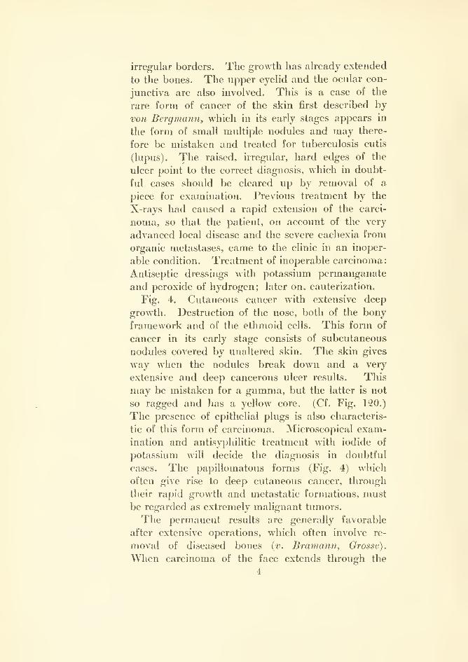
irregular borders. The growth has already extended
to the bones. The upper eyelid and the ocular con-
junctiva are also involved. This is a case of the
rare form of cancer of the skin first described by
von Bcrgmann, which in its early stages appears in
the form of small multiple nodules and may there-
fore be mistaken and treated for tuberculosis cutis
(lupus). The raised, irregular, hard edges of the
ulcer point to the correct diagnosis, which in doubt-
ful cases should be cleared up by removal of a
piece for examination. Previous treatment by the
X-rays had caused a rapid extension of the carci-
noma, so that the patient, on account of the very
advanced local disease and the severe cachexia from
organic metastases, came to the clinic in an inoper-
able condition. Treatment of inoperable carcinoma:
Antiseptic dressings with potassium permanganate
and peroxide of hydrogen; later on, cauterization.
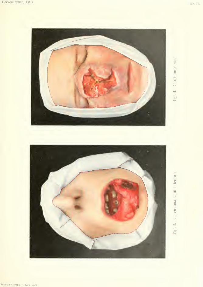
Fig. 4. Cutaneous cancer with extensive deep
growth. Destruction of the nose, both of the bony
framework and of the ethmoid cells. This form of
cancer in its early stage consists of subcutaneous
nodules covered by unaltered skin. The skin gives
way when the nodules break down and a very
extensive and deep cancerous ulcer results. This
may be mistaken for a gumma, but the latter is not
so ragged and has a yellow core. (Cf. Fig. 120.)
The presence of epithelial plugs is also characteris-
tic of this form of carcinoma. Microscopical exam-
ination and antisyphilitic treatment with iodide of
potassium will decide the diagnosis in doubtful
cases. The papillomatous forms (Fig. 4) which
often give rise to deep cutaneous cancer, through
their rapid growth and metastatic formations, must
be regarded as extremely malignant tumors.
The permanent results are generally favorable
after extensive operations, which often involve re-
moval of diseased bones (v. Bramann, Grosse).
When carcinoma of the face extends through the

dura mater, operation is not indicated, and the case
must be treated according to the rules for inoperable
cancer. In all extensive carcinomas of the face the
patients may die from septic pneumonia when the
destructive process reaches the buccal cavity.
A special form of cancer arising in the deep parts
of the corium as cancerous nodules constitutes whatKrompecher described as basal-celled cancers. Ac-
cording to Coenen these are not to be classed with
endotheliomas, as formerly, for they arise from the
basal cells of the sweat and sebaceous glaud epithe-
lium, or from the epithelium of the hair follic'«s. In
distinction to the other cutaneous cancers they do not
become cornified, and were, therefore, classed hy Borst
among the endotheliomas.
Multiple carcinomas of the face have been noted
by several observers {v. Bergmann, Coenen, Schim-
melbitsch). Von Bergmann, in a case of carcinoma
of the forehead, which after some years was followed
by another in the floor of the mouth, was of opinion
that these were separate, independent carcinomas,
because metastases in the tongue and floor of the
mouth are very rare, and there was a long time
between the development of the two carcinomas.
Carcinoma of the Mucous
J Membranes
CARCINOMA LABII INFERIORIS (o/ lower Lip)
Plate II, Fig. 3.
TUBERCULOSIS CUTIS (of the Skin)
Plate III, Fig. 5.
PAPILLOMA LINGUAE {of Tongue)
Plate rV", Fig. 6.
CARCINOMA ET PAPILLOMA LINGUAE {of Tongue)Plate IV. Fig. 7.
CARCINOMA LINGUAE INCIPIENS{Incipient Carcinoma of Tongue)
Plate IV. Fig. 8.
CARCINOMA LINGUAE EXULCERATUM{Ulcerating Carcinoma of Tongue)
LEUKOPLAKIA {Leukoplakia)
Plate IV, Fig. 9.
Cancers of the lips resemble cancers of the skin
in their form and structure, for they are squamous-
celled epitheliomas, and tend to cornification. Theyarise in the form of cauliflower-shaped, polypoid
tumors on the mucous membrane of the lips, cheeks,
and glans penis, or as deep ragged ulcers (lips and
tongue), and appear in these principal forms in all
mucous membranes covered with squamous epithe-
lium. Carcinoma of the upper lip is very rare, but
V. Bergmann has obsei'ved a case where a carci-
noma of the upper lip developed within a few weeks
after a cancer of the lower lip, in a symmetrical
position. Carcinomas of the lower lip form 45.6
per cent, of all cancers of the face, nearly all occur-
ring in the male sex. The action of tobacco mustplay a special role in the origin of cancel: of the lip,
6
Bockenheimer, Atlas.'lali.
U
CO
Rebman Company, New-York.

for the patients, of both sexes, are mostly great
smokers.
Cancer of the lower lip often begins at the junc-
tion of the skin with the red part of the lip, generally
between the center of the lip and the angle of the
mouth, as a small, hard nodule at first covered bymucous membrane. The mucous membrane soon
becomes broken and the nodule grows, infiltrating
the surrounding tissues rapidly, while the mucousmembrane breaks down more and more and forms
an ulcer. Antecedent diseases of the mucous mem-brane, such as tuberculosis and psoriasis (leuko-
plakia) appear to predispose to carcinoma. Thewhole of the lower lip may be gradually destroyed
(Fig. 3). Scabs and crusts form at several places onthe ulcer, and when separated give rise to bleeding.
While in its early stages the cancerous ulcer is recog-
nized by its hard, raised edges and crateriform
floor, the advanced cancer of the lip shows papillo-
matous proliferations springing from the floor of the
ulcer (Fig. 3). The more the carcinoma extends,
the more it implicates the underlying bones and the
mucous membrane of the cheeks and floor of the
mouth, so that the bones and the buccal mucousmembrane may be completely destroyed. The exu-
dation of growing cancer of the lip gives rise to muchcachexia, gastritis and enteritis, and the secretion
may reach the lungs and cause death from septic
pneumonia. In such inoperable forms the sub-
maxillary and submental regions are usually filled
with hard, fixed glands.
Differential Diagnosis. Although these ad-
vanced forms, which are often neglected, especially
in country people, are unmistakable, there may be
difficulty in the diagnosis of the early stage of the
cancerous ulcer. The irregular, ragged surface of
the carcinoma is in marked contrast to the smooth
surface of primary syphilis, and the comedo-like
7

epithelial plugs which are characteristic of all
squamous-celled epitheliomas can be extruded from
it by pressure. The glands are affected very early
in carcinoma, first in the submental region, and are
usually, small, very hard and isolated, in contrast to
the multiple glands in primary syphilis, which are
not so hard and mostly situated at the bifurcation of
the carotid.
Isolated tuberculosis, or an ulcer extending from
tuberculosis of the buccal mucous membrane or
tongue, is very rare on the lip. It has in-egular
edges which are not so raised and hard as those of
cancer. The surface of the ulcer, which results
from the breaking down of small tubercles, is of a
reddish-gray color and bleeds very easily. It is
usually covered with a single large scab. No plugs
can be expressed from it. Glandular enlargement
is soft and isolated.
Ulcerated cavernoma (cavernous angioma) of the
lip may have a cancerous appearance, but it usually
occurs in children and is generally associated with
other anomalies of the blood-vessels.
The induration of fissures of the lips resulting
from chronic eczema heals quickly under rational
treatment, and is thus distinguished from carcinoma-
tous induration.
It is important to note that cancer of the lip occurs
not only in old people but also soon after the thirtieth
year.
Treatment. All depends on early diagnosis, for
the cuneiform excision of small tumors gives the best
chance of a radical cure. In doubtful cases excision
is to be preferred to antisyphilitic or antitubercu-
lous treatment, so as to lose no time. In extensive
growths, from one and one half to two centimeters of
healthy tissue should be removed round the tumors,
and the neighboring parts suspected of disease, such
as bones and buccal mucous membrane, should also
8

Bockenheimer, Atlas. Tab. Ill
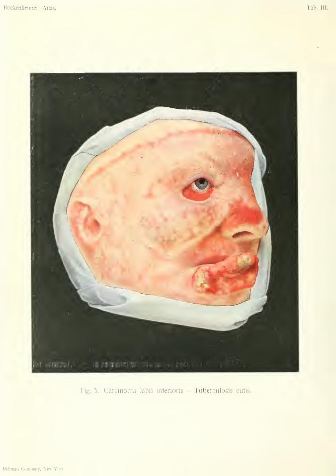
Fie. 5. Carcinoma labii inferioris — Tuberculosis cutis.
Rebman Company, New-York.
be removed. The defect can be repaired by plastic
operations, the best of which are Dieffcnbach'n or
Jacsche's.
Palpable glands should always be removed by
separate incisions in the submental and submaxillary
regions. The submaxillary gland which is often
affected is best removed at the same time. By radical
operation a permanent cure is possible even in
extensive carcinomas.
Fig. 3 shows a carcinoma involving the whole
lower lip. Deep ulcerations alternate with papillo-
matous outgrowths. In some parts there are scabs
on the surface of the ulcers, in others isolated yellow
epithelial plugs. The growth is hardly movable
over the lower jaw, and is on the point of extending
to the buccal mucous membrane. After free exci-
sion of the tumor, removal of the enlarged glands in
the submental and submaxillary regions, the exten-
sive defect was repaired by double cheiloplasty
{DiejfcnhacJi's operation) and a cure was obtained.
Fig. 5 represents a large cancerous ulcer, originat-
ing from tuberculosis of the skin, involving half the
lower lip. The hard, raised edges of the ulcer
divested of mucous membrane are characteristic.
The floor of the ulcer is irregular and ragged and
beset with yellowish epithelial plugs. Cancerous
ulcers arising on the basis of tuberculosis of the
skin have a great tendency to bleed. In contrast to
the forms of hypertrophic lupus, which gives rise to
soft, fungoid, slow-growing tumors, the hardness
and rapid growth of the lupus-carcinoma is charac-
teristic. Excision of the carcinoma, removal of the
glands, and repair of the defect by DiefJenbacWs
cheiloplasty led to a cure.
Fig. 5 also shows a characteristic picture of differ-
ent forms of cutaneous tuberculosis; lupus of the
face. The disease appears most frequently in this
situation and usually begins on the nose (tuberculosis
9
.of the nasal mucosa), and extends over the face in
the form of a butterfly. The sharp, irregular outline
on the forehead, neck, and behind the ears is charac-
teristic. The disease begins with small reddish-
brown nodules situated in the cutis giving rise to
exfoliation of the epidermis (lupus exfoliativa)
;
these become confluent and form flat, reddish-gray,
easily bleeding ulcers (lupus exulcerans, which after
healing leave radiating cicatrices, often after consid-
erable destruction of tissue. (Fig. 5, ear.) After a
time papillomatous proliferations may arise of soft
and spongy consistence, especially about the ear
(lupus hypertrophicus). These three forms are usu-
ally present in the same patient (v. Bergmann).
Treatment. In circumscribed forms excision of
the skin with the diseased subcutaneous tissue is
indicated, with repair of the defect by skin flaps.
The diffuse forms are treated in v. Bergmanii's clinic
by the sharp spoon {Volkmann). The diseased parts
are scraped and the bleeding surface treated with
Pacqueli7i's cautery or with hot air. Many sittings
are often necessary in order to arrest the disease,
and the patients often succumb from tuberculous
disease of the internal organs, or relapsing facial
erysipelas.
Cancer of the buccal cavity occurs on the tongue,
the floor of the mouth and the cheek. Cancer of the
tongue (Figs. 7, 8 and 9) occurs almost exclusively
in man (after the fortieth year), owing to the action
of tobacco and alcohol. Antecedent lingual or buc-
cal leucoplakia predisposes to buccal carcinoma;
V. Bergmann finds it present in fifty per cent, of his
cases of cancer of the tongue. Leucoplakia forms
hard, white, opaline patches raised above the surface
of the mucous membrane of the tongue, consisting of
horny epithelium (hyperkeratosis). The surface, at
first smooth, after a time becomes fissured, especially
after excessive smoking, and the patches of leuco-
10
Bockenheinier, Atlas.lab. IV.
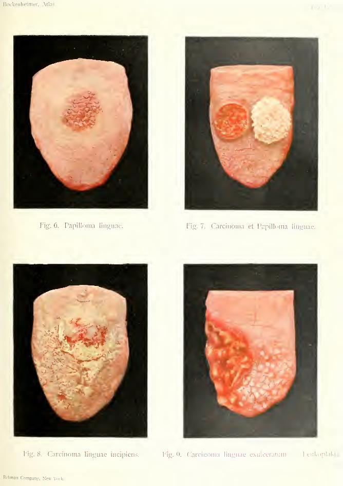
Fig. 6. Papilloma linouae. Fig. 7. Carcinoma et Papilloma linguae.
Fig. 8. Carcinoma linguae incipiens. Fig. 9. Carcinoma linguae cxiilccratum. I.euixoplakia.
Rcbman Company, Ncw-\ork.

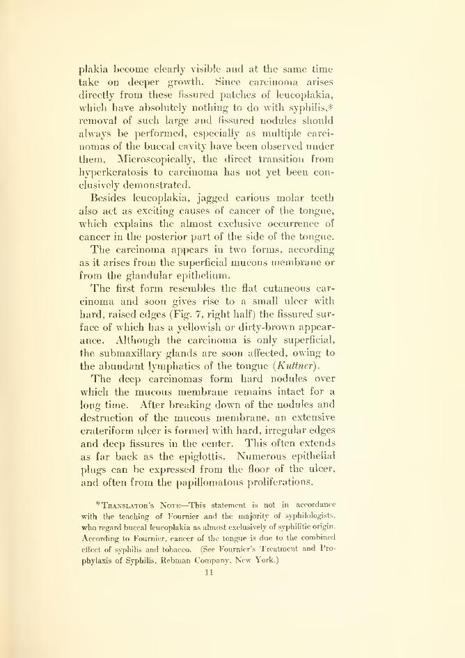
plakia become clearly visible and at the same time
take on deeper growth. Since carcinoma arises
directly from these fissured patches of leucoplakia,
which have absolutely nothing to do with syphilis,*
removal of such large and fissured nodules should
always be performed, especially as multiple carci-
nomas of the buccal cavity have been observed under
them. Microscopically, the direct transition from
hyperkeratosis to carcinoma has not yet been con-
clusively demonstrated.
Besides leucoplakia, jagged carious molar teeth
also act as exciting causes of cancer of the tongue,
which explains the almost exclusive occurrence of
cancer in the posterior part of the side of the tongue.
The carcinoma appears in two forms, according
as it arises from the superficial mucous membi'ane or
from the glandular epithelium.
The first form resembles the flat cutaneous car-
cinoma and soon gives rise to a small ulcer with
hard, raised edges (Fig. 7, right half) the fissured sur-
face of which has a yellowish or dirty-brown appear-
ance. Although the carcinoma is only superficial,
the submaxillary glands are soon affected, owing to
the abundant lymphatics of the tongue (Kuttner).
The deep carcinomas form hard nodules over
which the mucous membrane remains intact for a
long time. After breaking down of the nodules and
destruction of the mucous membrane, an extensive
crateriform ulcer is formed with hard, irregular edges
and deep fissures in the center. This often extends
as far back as the epiglottis. Numerous epithelial
plugs can be expressed from the floor of the ulcer,
and often from the papillomatous proliferations.
* Translator's Note—^This statement is not in accordance
with the teaching of Foumier and the majority of syphilologists,
who regard buccal leucoplakia as almost exclusively of syphilitic origin.
According to Foumier, cancer of the tongue is due to the combined
effect of syphilis and tobacco. (See Foumier's Treatment and Pro-
phylaxis of Syphihs, Rebman Company, New York.)
11

The patients suffer great pain from the irritation
of free nerve-endings in the floor of the ichorous
ulcer, and, in untreated eases, succumb usually
within a year from glandular metastases extending
along the carotid to the supra-clavicular region
(Fig. 9). Early diagnosis is, therefore, of the great-
est possible importance.
Differential Diagnosis. The superficial carci-
noma (Fig. 7) is recognized by the characteristic
features of flat cutaneous carcinoma and differs
from s^^hilitic chancre by its sharp, hard edges, the
irregular floor of the ulcer with epithelial plugs, and
the small, hard glands. As long as the flat carci-
noma of the tongue is covered with mucous mem-brane it may in its earliest stages be confounded with
papilloma (Fig. 6), especially in the rare cases where
it lies more in the center of the dorsal surface of the
tongue. Papillomas, however, generally appear as
multiple, soft elevations the size of a pin's head, so
that the surface of the tongue may appear furnished
with small points, or may assume a lobulated form;
or there may be fungiform sessile tumors, like stal-
actites, which often form high projections and have
a warty appearance (Fig. 7). That a flat carcinoma
and a papilloma of this kind may occur independ-
ently without microscopic transition into each other
is shown by v. Bergmanrts case ("Handbook of
Practical Surgery', III edition: Text-book of Sur-
gery, II edition"). Small papillomata cause the
patient hardly any inconvenience and can be re-
moved with the sharp spoon or Pacquelhi s cautery.
Larger papillomata should be excised (Fig. 7, left
half).
The diagnosis is difficult when, as in Fig. 8, a
hard, carcinomatous nodule develops under a patch
of leucoplakia. The irregular, deep, hard infiltra-
tion and the rapid growth point to a commencing newgrowth, which should always be removed before it
12
breaks through, especially when there is leucoplakia
over the nodule.
Abscesses of the tongue, which result from
injury by foreign bodies (steel pens, etc.), andform hard nodules in the substance of the tongue,
are characterized by the early painfulness on
pressure. Actinomycosis causes a more diffuse,
wooden infiltration of the whole tongue and very
soon interferes with its motion. (Abscess is treated
by incision and actinomycosis by incision and
scraping).
The small carcinomatous ulcer of the edge of the
tongue is liable to be confounded with ulcerations
caused by the irritation of broken teeth (dental
ulcers), especially when it is situated opposite a
sharp tooth; however, the cancerous ulcer con-
tinues to grow after removal of the offending tooth.
Larger ulcerations which result from the breaking
down of deep carcinoma may be confounded with
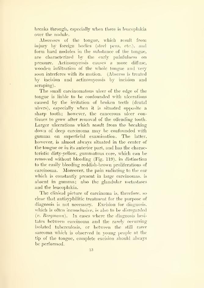
gumma on superficial examination. The latter,
however, is almost always situated in the center of
the tongue or in its anterior part, and has the charac-
teristic dirty-yellow, gummatous core, which can be
removed without bleeding (Fig. 119), in distinction
to the easily bleeding reddish-brown proliferations of
carcinoma. Moreover, the pain radiating to the ear
which is constantly present in large carcinomas, is
absent in gumma; also the glandular metastases
and the leucoplakia.
The clinical picture of carcinoma is, therefore, so
clear that antisyphilitic treatment for the purpose of
diagnosis is not necessary. Excision for diagnosis,
which is often inconclusive, is also to be disregarded
(v. Bergmann). In cases where the diagnosis hesi-
tates between carcinoma and the rarely occurring
isolated tuberculosis, or between the still rarer
sarcoma which is observed in young people at the
tip of the tongue, complete excision should always
be performed.
13
Treatment. Small carcinomas can be excised
and the wound closed, after compression of the tongue
by a ligature. Excision by Pacquelin's cautery and
subsequent plugging may also be done.
For large carcinoma a radical operation by section
of the lower jaw is necessary (according to Sedilloi
and Kocher in the middle line; according to v,
Bergmann and Langenbeck, laterally) with subse-
quent ligation of the lingual artery (cf. Bocken-
heimer & Frohse's "Atlas of Typical Operations").*
By this means not only can the tumor of the tongue
be excised through healthy tissues as far as the
epiglottis, but also the masses of glands which ex-
tend from the submaxillary region to the ear can
be removed. Even after extirpation of extensive
portions of the tongue the patients, after a few
months, can make themselves well understood.
Permanent cures, are however, unfortunately rare,
even after radical operations, in progressive cases of
cancer of the tongue, especially when the lower
jaw is involved and the glands have become fixed,
so that some surgeons content themselves with the
local treatment of carcinoma by caustics and cauter-
ization.
The treatment of cancer of the buccal cavity,
which often arises on the basis of leucoplakia, in the
same form and with the same symptoms, is carried
out on the same principles.
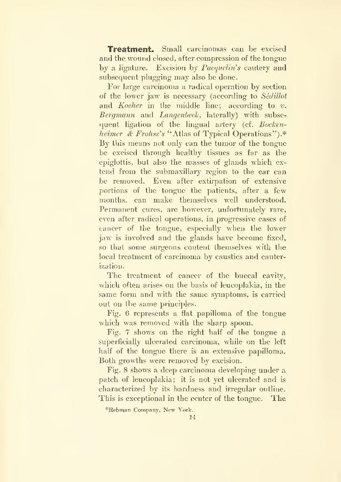
Fig. 6 represents a flat papilloma of the tongue
which was removed with the sharp spoon.
Fig. 7 shows on the right half of the tongue a
superficially ulcerated carcinoma, while on the left
half of the tongue there is an extensive papilloma.
Both growths were removed by excision.
Fig. 8 shows a deep carcinoma developing under a
patch of leucoplakia; it is not yet ulcerated and is
characterized by its hardness and irregular outline.
This is exceptional in the center of the tongue. The
^Rebman Company, New York.
14

growth was removed by excision and subsequentsuture.
Fig. 9 represents the most common form of cancerof the tongue; a carcinomatous ulcer of the sideof the tongue with extensive destruction, leucopla-kia and ghuidular metastases. After section ofthe lower jaw the growth was widely removed,the stump of the tongue sutured and the glandsremoved from the neck.
15
Glandular Carcinoma
CARCINOMA MAMMAE (of Breast)
LYMPHOMATA CARCINOMATOSA {Carcinomatous)
Plate V. Fig. 10.
CARCINOMA MAMMAE EXULCERATUM{Ulcerating Carcinovia of Breast)
Plate VI, Fig. 11.
CARCINOMA MAMMILLAE {of Nipple)
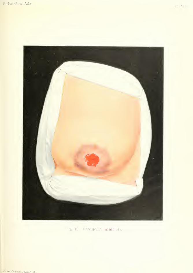
Plate VII, Fig. 12.
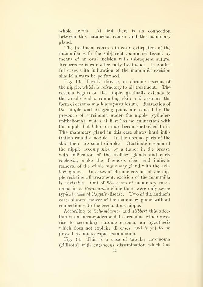
CARCINOMA MAMMAE (of Breast)—PAGETS DIS-EASE—ECZEMA CHRONICUM MAMMILLAE
(Chronic Eczema of Nipple)
Plate VIII, Fig. 13.
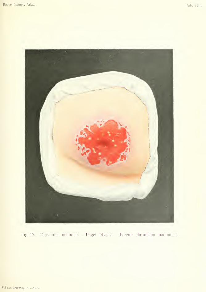
CARCINOMA MAMMAE—DISSEMINATIONES(Disseminated Carcinoma of Breast)
Plate IX, Fig. 14.
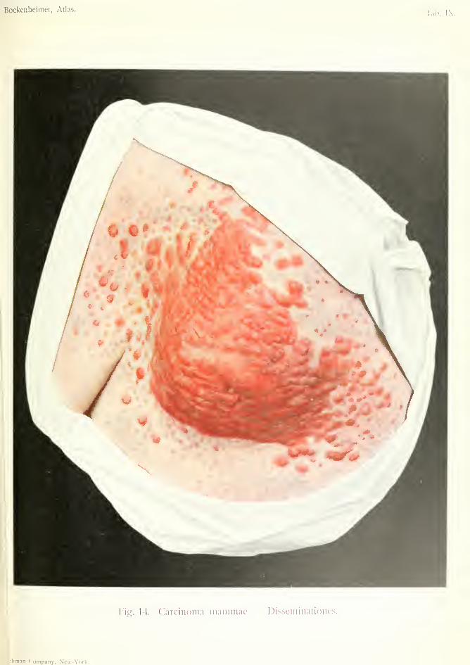
CARCINOMA MAMMAE UTRIUSQUE(o/ both Breasts)
—"CANCER EN CUIRASSE"Plate X, Fig. 15.
CARCINOMA MAMMAE (of i?rfo*0—LYMPHANGITISCARCINOMATOSA (Carcinomatous Lymphangitis)
Plate XI, Fig. 16.
Of the carcinomas of glandular organs those of
the female mammary gland are among the most
common (they take the third place). They show a
typical unrestricted epithelial proliferation in their
origin and development. Observations made on
cancer of the breast, therefore, have manifold bear-
ings on carcinoma of other organs. A division into
soft, many-celled, rapidly growing tumors of which
the medullary cancers represent the most malignant,
and slow-growing scirrhous forms with few cells, is
of clinical importance.
The exciting causes include inflammatory irrita-
tion, puerperal interstitial mastitis, eczema of the
nipple, antecedent benign tumors (fibro-adenoma,
16
Bockcnheimer, Atlas. Tab. V.
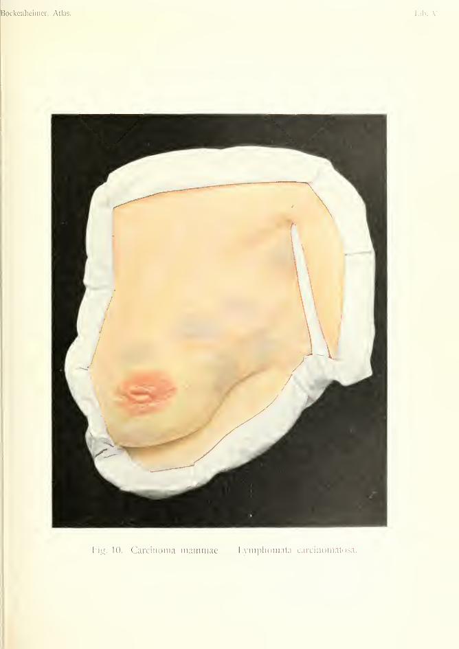
Fi^. 10. Carcinoma inainiiiac — L}-mph()mata carciiiomatosa.

Bockenheimer, Atlas. Tab. VI.
o
t/.
Ktbman Company, New-York.

cysts) injuries, mechanical irritation, frequent par-
turition with prolonged suckling ot" infants. Cancer
of the breast is attributed by the public to injuries
(blows), but these are often too recent to be accepted
as an etiological factor, considering the slow growth
of the carcinoma.
It is a remarkable fact that of sterile women only
10 per cent, have cancer of the breast. In 10 per
cent, of the cases there is said to be a hereditary
tendency.
Women are most often affected at the menopause
(fortieth to fifty-fifth years), and come to the sui'geon
with nodules in the breast which have been hitherto
painless and are only accidentally observed. These
nodules very soon form a malignant growth of
hard consistency and irregular surface. The most
important sign of a malignant new growth is the
absence of any demarcation or encapsulation. Thetumor cannot, like all benign tumors, be separated
from the mammary tissue and moved freely, but is
fixed immovably in the glandular tissue, with ill-
defined boundaries, and is anchored in the meshes
of the mammary tissue by numerous offshoots.
The nodules, which at first appear harmless, thus
soon show their malignity. ^Accompanied by lan-
cinating pains in the thorax, upper arm and shoulder,
the tumor sends its destructive offshoots in all direc-
tions into the neighboring tissues, without limit or
restraint, and reaching the surface adheres to the
skin and causes retraction and fixation of the nip-
ple. Finally, it gives rise to a hard inflammatory
infiltration of the whole of the overlying skin. At the
same time the tumor extends deeply and soon infil-
trates the lymphatics beneath the pectoralis major
muscle and also the regional lymphatic vessels and
glands of the axilla (Fig. 10), which are usually
affected about a year after the formation of the
nodules in the breast, and take the form of hard,
solid, painless nodules, which are often difficult to
17
feel in corpulent women. Extensive glandular af-
fection gives rise to radiating pain and oedema of the
arm (supra-clavicular glands). Although the cancer
usually arises as a single nodule, there are cases in
which several nodules develop simultaneously (Fig.
10) and extend through the whole breast to the
axilla (Fig. 10). The prognosis is unfavorable in
these cases, and in disease of both breasts (Fig. 15).
The disease is very frequently situated in the up-
per and outer quadrant of the breast, especially on
the left side. The tumors situated in the outer half
of the mamma towards the axilla, wrongly called
paramammary carcinomas, are really glandular can-
cers, for they originate in the offshoots of the mammawhich extend towards the clavicle, sternum, axilla
and twelfth rib in the form of long, thin cords.
Cancer of the breast, like all cancers rich in cells
(acinous, tubular), grows rapidly, especially during
pregnancy, and causes destruction of the skin. Acancerous ulcer results, characterized like cutaneous
carcinoma by its hard, raised, fixed borders, crateri-
form base and sanious discharge. A hard infiltra-
tion develops round the tumor which is usually fixed
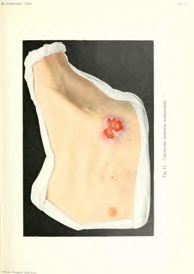
to the thorax. Small nodular thickenings of the ad-
jacent unbroken skin sometimes constitute the first
sign of commencing general cutaneous dissemination
(Fig. 11). In this way the whole mamma may be
transformed into a large ulcer (Fig. 15).
In other cases a tumor is gradually developed
involving the whole breast without breaking through
externally. The skin, however, may be infiltrated
and the redress may be mistaken for inflammatory
infiltration (Figs. 14 and 16). These leathery infil-
trating forms of breast cancer finally envelop the
whole mammary region like a cuirass (Fig. 15).
In the infiltrated skin these often appear small,
pin-point disseminations of the carcinoma (Fig. 15,
right side), which by confluence give rise to a nodu-
lar infiltration of the whole thorax (Fig. 14).
18
Bockenheimer, Atlas.
Tab. VII.
PifT. 12. Carcinoma mammillae.
Kcbnan Company, New-York.

In cancers which are poor in cells (scirrhous) the
mammary gland is often diminished in size byshrinking, and the skin becomes puckered over the
tumor by cicatricial contraction (Fig. 10).
Differential Diagnosis. Ulcerated cancers of
the breast and those with hard, raised infiltration
are difficult to mistake, but small tumors in the sub-
stance of the breast have to be diagnosed from inter-
stitial mastitis, benign tumors (fibro-adenomas, cysts
and mixed tumors) and abscesses, in which there is
frequently deceptive induration. The age of the pa-
tient, the continuous growth of the nodules, the
appearance of hard, lymphatic glands in the axilla,
and the frequent occurrence of emaciation andcachexia even in small cancers assist in the diagnosis,
which in doubtful cases can be established by exci-
sion of a piece for examination. Sarcoma occurs at
an earlier age in the form of soft tumors extending
to the skin, and presents a fairly typical clinical pic-
ture which should not be confounded with carcinoma
(Figs. 29 and 30). The glands are generally unaf-
fected in sarcoma.
Treatment. Radical excision of the whole breast
and its processes as early as possible, with removal
of the pectoralis major and in some cases also
the pectoralis minor, and complete removal of the
axillary glands is necessary for a permanent cure.
In V. Bergmann's clinic there were 29.79 per cent,
permanent cures out of 1,000 cases, i.e. free from
recurrence three years after the operation. Recur-
rence is much less common in the axillary glands
than locally. If of small extent they can be treated
by excision, if larger by the X-rays (Fig. 15).
All cases with extensive dissemination in the skin
(Fig. 14), diffuse infiltrating cancer, "cancer en
cuirasse" (Figs. 15 and 16) are unsuitable for opera-
19
tion. In cases where the supra-clavicular glands are
extensively affected, permanent cures are hardly ever
obtained, even after radical operations including sec-
tion of the clavicle and ligation of the axillary vein; so
that it is best to abandon the operation. Also tumors
which are adherent to the ribs, and fixed glandular
tumors extending to the axilla are unsuitable for
operation, for the recurrence generally takes place
before the patient has recovered from the operation.
Operation is also contra-indicated in cases of severe
cachexia, in the atrophic slow-growing forms met
with in old people, in cases with metastatic growths
in the lung, liver and bones (often leading to sponta-
neous fracture of the neck of the femur.)
In the region of the head metastatic carcinomas are
sometimes inoperable. Owing to their circumscribed
encapsuled formation with soft contents they may be
confounded with atheromatous cysts. According to
Schimvielbusch they arise in this form through em-bolism of cancer cells, and thus form encapsuled
freely movable nodules.
[The first brain tumor operated upon was an en-
capsulated metastatic carcinoma resulting from a
mammary cancer.]
In cases of inoperable carcinoma the X-rays maylead to epidermization, especially in the ulcerated
forms, after previous removal of the ulcerated parts.
In discharging cancers powdered charcoal or chlo-
ride of zinc may be used locally, and high doses of
morphia internally.
Cases hitherto reported as cured by X-rays are
fallacious. No doubt a carcinomatous nodule maydisintegrate and disappear under the action of the
X-rays, but there is always a further growth in other
parts—glands and internal organs. As regards cas-
tration for advanced mammary carcinoma in women,further experience is required.
Doyen's serum treatment of cancer has so far given
no results.
20
Fig. 10 shows an acinous carcinoma forming sev-
eral nodules in the breast, already infiltrating the
skin. The axillary glands form hard, fixed, indolent
nodular swellings, and nodules can be easily traced
in the form of a rosary from the mammary gland to
the axilla. The nipple is retracted and fixed, andthe whole breast is diminished in size. Operation
was performed in the usual way. The patient wasalready emaciated.
Fig. 11. A single cancerous nodule in a malebreast. The skin has broken down and shows a
cancerous ulcer with hard, raised, jagged edges,
which has destroyed the nipple. The floor of the
ulcer is irregular and the whole tumor is fixed to the
pectoral muscle. At the edge of the ulcer the skin
is radially contracted and shows isolated cancerous
nodules. The axillary glands are hard, visible andhardly movable. In spite of the small size of the
tumor there was already cachexia. After removal of
the mamma with the pectoralis major and the axil-
lary glands the wound, which could not be com-
pletely closed by suture, was repaired by Thiersch's
grafts.
Cancer of the male breast (about 1 per cent, of all
mammary carcinomas according to Schuchardt) gen-
erally arises as a small, hard nodule (scirrhous) in the
neighborhood of the nipple and giv'es rise to a typical
cancerous ulcer. The tumor occurs between the
fortieth and seventieth years. Heredity appears to
be frequent. Occasionally cancer of the breast is
seen in husband and wife.
Fig. 12 shows a very rare case of carcinoma arising
from the nipple (squamous-celled epithelioma). This
is more common in men than in women. It com-
mences as a hard infiltration of the nipple, in the
same way as commencing carcinoma of the navel.
The nipple is much retracted and the whole areola
is transformed into a rigid wall. A cancerous ulcer
soon develops which destroys the nipple and the
21
whole areola. At first there is no connection
between this cutaneous cancer and the mammarygland.
The treatment consists in early extirpation of the
mammilla with the subjacent mammary tissue, bymeans of an oval incision with subsequent suture.
Recurrence is rare after early treatment. In doubt-
ful cases with induration of the mammilla excision
should always be performed.
Fig. 13. Paget's disease, or chronic eczema of
the nipple, which is refractory to all treatment. Theeczema begins on the nipple, gradually extends to
the areola and surrounding skin and assumes the
form of eczema madidans pustulosum. Retraction of
the nipple and dragging pains are caused by the
presence of carcinoma under the nipple (cylinder-
epithelioma), which at first has no connection with
the nipple but later on may become attached to it.
The mammary gland in this case shows hard infil-
tration round a nodule. In the normal parts of the
skin there are small dimples. Obstinate eczema of
the nipple accompanied by a tumor in the breast,
with infiltration of the axillary glands and early
cachexia, make the diagnosis clear and indicate
removal of the whole mammary gland with the axil-
lary glands. In cases of chronic eczema of the nip-
ple resisting all treatment, excision of the mammilla
is advisable. Out of 884 cases of mammary carci-
noma in V. Bergvianns clinic there were only seven
typical cases of Paget's disease. Two of the author's
cases showed cancer of the mammary gland without
connection with the eczematous nipple.
According to Scliamhacher and Ribbert this affec-
tion is an inti"a-epidermoidal carcinoma which gives
rise to secondary chronic eczema, an hypothesis
which does not explain all cases, and is yet to be
proved by microscopic examination.
Fig. 14. This is a case of tubular carcinoma
(Billroth) with cutaneous dissemination which has
22
Bockenlieimer, Atlas. Tab. Vill.
Fig. 13. Carcinoma mammae — Paget Disease — Eczema clironicum mammillae.
Rcbman Company, New-York.

Bockenheimer, Atlas.
Fitr. 14. Carcinoma iiiaiiimae - Disseniinatioiies.
iian Company, New-York.

Bockenheiiner, Atlas.Tab. \.
U
lan Pnmmrn- V»w-V'<-.irb

w
Bockenheimcr, Atlas.
Fig. 16. Carcinoma mammae. - Lymphangitis carcinomatosa.

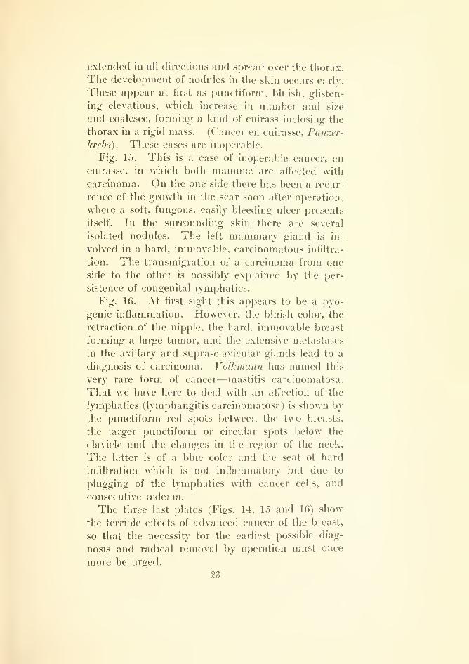
extended in all directions and spread over the thorax.
The development of nodules in the skin occurs early.
These appear at first as punctiform, bluish, glisten-
ing elevations, which increase in number and size
and coalesce, forming a kind of cuirass inclosing the
thorax in a rigid mass. (Cancer en cuirasse, Panzer-
krebs). These cases are inoperable.
Fig. 15. This is a case of inoperable cancer, en
cuirasse, in which both mammae are affected with
carcinoma. On the one side there has been a recur-
rence of the growth in the scar soon after operation,
where a soft, fungous, easily bleeding ulcer presents
itself. In the surrounding skin there are several
isolated nodules. The left mammary gland is in-
volved in a hard, immovable, carcinomatous infiltra-
tion. The transmigration of a carcinoma from one
side to the other is possibly explained by the per-
sistence of congenital lymphatics.
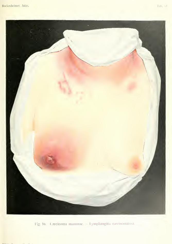
Fig. 16. At first sight this appears to be a pyo-
genic inflammation. However, the bluish color, the
retraction of the nipple, the hard, immovable breast
forming a large tumor, and the extensive metastases
in the axillary and supra-clavicular glands lead to a
diagnosis of carcinoma. J^olkmcuin has named this
very rare form of cancer—mastitis carcinomatosa.
That we have here to deal with an affection of the
lymphatics (lymphangitis carcinomatosa) is shown by
the punctiform red spots between the two breasts,
the larger punctiform or circular spots below the
clavicle and the changes in the region of the neck.
The latter is of a blue color and the seat of hard
infiltration which is not inflammatory but due to
plugging of the lymphatics with cancer cells, and
consecutive oedema.
The three last plates (Figs. 14, 15 and 16) show
the terrible effects of advanced cancer of the breast,
so that the necessity for the earliest possible diag-
nosis and radical removal by operation must once
more be urged.
23
Naevus CarcinomaPlate XII, Fig. 17.
ATHEROMA—CARCmOMA (Sebaceous)
Plate XIII, Fig. 18.
Fig. 17. Carcinoma of the scalp is very rare and
usually arises on the basis of old scars, ulcers, warts,
atheroma (sebaceous cysts) and moles. Pigmentary
nsevi, which are congenital or appear soon after
birth, when they appear as warty formations, belong
to the class of benign tumors. Occurring over the
whole body, they were included by v. Recklinghausen
among diseases of nerves. While the growth of the
naevus ceases with the growth of the body, changes
occur in later years which may take the form of
papilloma, sarcoma, carcinoma or malignant melan-
oma. In the case represented in Fig. 17, a rapidly
growing tumor arose from a congenital naevus in
the thirty-seventh year; the cutaneous covering soon
disappeared and the tumor was separated by deep
fissures into cauliflower growths. The ulcerated sur-
face is covered with sanious secretion, so that macro-
scopic examination often does not decide whether it
is a case of ulcerated carcinoma or sarcoma. Thatit is a malignant growth is shown by the rapid
growth of the tumor, which in a short time extends
over and destroys the whole nsevus; the early adhe-
sion to the bones; the regional glandular metastases
in the form of hard, slightly movable nodules behind
the ear, and the cachexia of the patient. On account
of the glandular metastases which soon extend along
the large vessels from the neck to the supra-clavicular
24

uI
p
<
5
3Oo
>
2
in


region, the case is presumed to be a carcinoma of the
scalp on the basis of a na?vus (pigmentary carci-
noma), but there remains the possibiHty that micro-
scopic examination may show it to be a pigmentary
sarcoma.
Treatment. This consists in extirpation of the
tumor and the rest of the naevus, repair by a plastic
operation, and removal of the diseased glands. In
large nsevi of the head and face a portion of the
nsevus can, in some situations, be removed by an
elliptical incision and subsequent suture {DieJJen-
hach). Owing to the elasticity of the skin of the
head large nsevi can often be removed without re-
pair by plastic operation. As soon as changes of
any kind appear in a naevus, especially in advanced
age, it is important to remove it as soon as possible.
It is best to remove all pigmentary nsevi because
fatal malignant melanomatous growths so often
develop even from the smallest pigmentary spots.
Fig. 18. Along with multiple sebaceous cysts
scattered over the whole scalp, is a carcinoma origi-
nating from one of the cysts. The sebaceous cysts,
commencing as small yellowish nodules in the skin,
slowly grow into large tumors with a broad base andsmooth surface. The cysts are fixed to the skin but
easily movable over the subjacent bone, and have a
doughy consistence often resembling fluctuation. If
this mobility of the cyst over the subjacent tissues
ceases and the originally soft tumor becomes a hard
nodule with an irregular rough surface, malignant
degeneration is to be suspected; apart fi'om the
occurrence of calcification in its walls, in which,
moreover, the spherical smooth surface is generally
preserved. This suspicion becomes a certainty whenthe skin gives way and there appears a rapidly
growing nodular tumor characterized by multiple
lobulation and secreting a fetid discharge. These
carcinomas resemble in many ways the formation
25
of a discharging sarcoma (Fig. 33), and often cause
severe pain owing to inflammation round the tumor.
Cachexia occurs early, and the patients are usually
of advanced age.
The diagnosis of carcinoma depends on the hard
multiple glandular enlargement, which affects the
whole nape of the neck. This usually occurs later
and is not so hard in sarcoma.
Treatment. This consists in extirpation of the
carcinoma, and involves removal of part of the exter-
nal table of the skull on account of the tumor being
fixed to it. The extensive space left by removal of
the tumor can be sutured after making two long
lateral incisions over both ears and undermining of
the scalp. The spaces left by the lateral incisions
can be repaired by Thiersch's grafts. The glands in
the nape of the neck must also be removed.
On account of the early appearance of glandular
metastases the excision of especially indurated seba-
ceous cysts is indicated. Moreover, as there is
always a possibility of malignant degeneration, it
is advisable to remove every sebaceous cyst by
dissecting it out, so as to avoid recurrence.
26

Bockenheimer, Atlas.
Fio;. IQ. Carcinoma penis — Leukoplai<ia.
Kcbman Company, New-York

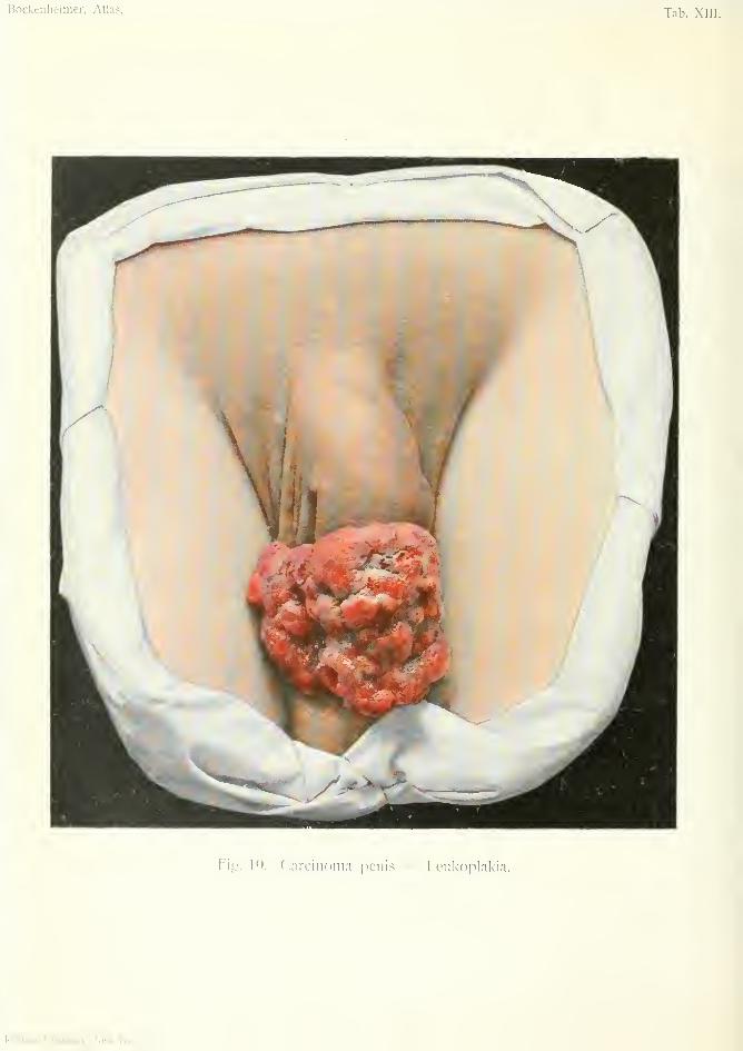
Carcinoma PenisPlate XIII, Fig. 19.
Carcinoma of the penis begins on the glans or in
the coronary sulcus as a squamous-celled epitheli-
oma, generally between the fiftieth and seventieth
year. Predisposing causes are congenital phimosis
with preputial concretions, leucoplakia prjepu-
tialis (white glistening patches similar to leucoplakia
of the tongue and cheek), warts, long-standing
tuberculous and syphilitic ulcerations. Old fistulae,
which occur especially in eunuchs after removal of
the scrotum, testicles and pendulous part of the
penis, near the symphysis or perineum, also predis-
pose to carcinoma.
The usual form is that represented in the figure, a
warty carcinoma which destroys the prepuce andsoon forms a cauliflower growth. Between the indi-
vidual hard nodules destitute of skin appear crateri-
form excavations which are characteristic. Epithe-
lial plugs can be expressed from the growth, and in
other parts the surface is cornified. Thus, contin-
uous growth alternates with permanent disintegra-
tion. The rapidly developing nodules often cause
exhausting hemorrhage, while the breaking down of
the carcinoma gives rise to a fetid sanious discharge.
The borders of the growth are hard, raised and promi-
nent. The whole penis may be transformed into a
large growth which may extend to the scrotum, testi-
cles and pelvis. The growth may destroy the urethra
and cause much pain on micturition.
A more rare form of carcinoma arises as a small
ulcer, generally on the corona glandis. It is hidden
by the resulting phimosis, but its characteristic hard
borders can be felt distinctly and there is a sanious
secretion. The inguinal glands are affected early
and point to the diagnosis of carcinoma. Thegrowth at first causes the patient little inconvenience,
but quickly leads to severe cachexia, so that the
patients often present themselves with extensive met-
astases of the inguinal and retro-peritoneal glands,
and are in an inoperable condition. A saying of
Kauffmann's, "In old men with phimosis and offen-
sive discharge the possibility of cancer is always to
be borne in mind," merits special consideration.
Differential Diagnosis. Both forms of carci-
noma are so characteristic that they can hardly be
confounded with other affections. The papillo-
matous form at first sight suggest condylomata
acuminata when these have coalesced into soft
tumors, but in these the borders are as soft as the rest
of the growth. A phagedenic ulcer may cause de-
struction of the glans penis, but the necrosis resulting
from the rapid destruction differs from the prolif-
eration of the carcinoma, and the phagedenic ulcer
soon heals after cauterization. Syphilitic chancre
also has hard borders like the cancerous ulcer, but
its surface is smooth in distinction to the ragged sur-
face which is always present even in small cancerous
ulcers. Search may also be made for the Spiro-
chseta pallida of syphilis.
Sarcoma affecting the corpus cavernosum are soft
rapidly growing tumors, and for a long time have no
glandular metastases.
Treatment. Amputation of the penis and re-
moval of the glands from both inguinal regions.
The prognosis is favorable if the glands are not
affected before operation. In cases where the car-
28

ciuoma has already affected the whole penis, testi-
cles and prostate, a radical operation may be
attempted by temporary section of the pubis onboth sides {Bramann, Lexer, Manz), unless extensive
glandular or organic metastases contra-indicate anyintervention. Recurrence is frequent at the seat of
amputation. In inoperable cases the cancerous
ulcer can be destroyed with Pacquelin's cautery andafterwards treated by X-rays.
29
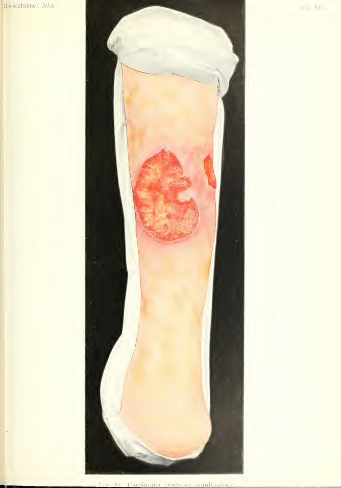
CARCINOMA CUTIS EX COMBUSTIONE (of Skin after Bum)Plate XIV, Fig. 20.
CARCINOMA CUTIS EX VERRUCA (o/ SUn after Wart)
Plate XV. Fig. 21.
CARCmOMA CUTIS EX CICATRICE {of Skin after Cicatrix)
Plate XV, Fig. 22.
Cutaneous carcinomas of the extremities are com-
paratively rare and always follow preceding changes
or morbid conditions in the skin. Most frequently
they arise on the basis of old scars of various origins,
especially from hypertrophic keloidal scars left after
extensive burns. Hawkins, in 1835, described car-
cinomas arising from scars left after severe flogging,
mostly in sailors. Dietrich described a carcinoma
originating in the scar from osteomyelitis, which was
for a long time regarded as primary carcinoma of
bone. The scar generally becomes fissured, form-
ing a small wound which afterwards becomes a car-
cinomatous ulcer (Fig. 21) with all its characteristic
features, hard borders, papillomatous proliferations,
ragged surface and epithelial plugs. A cauliflower
tumor grows which soon becomes fixed to the fascia
(Figs. 20 and 22).
Warts, old-standing ulcers of the leg and lupoid
changes in the skin also lead to carcinoma of the
extremities. Eczema of the skin occurring in chim-
ney-sweeps and workers in paraffin has often led to
multiple carcinoma of the extremities.
Fig. 20 shows a papillary carcinoma of the skin
of the leg arising from the scar of a burn. Thesmooth, partly white and partly brownish, shiny
scars of the burn are seen over the whole leg. Thecarcinoma has extended above and below and has
30
Tab. .\l\-.
['I.r or r^nrrinrMnn rrwrW i'\ rninhlisi

Bockeiiheimer, Atlas.Tab. .W.
U(M
;/,
Rebman Company, New-York.

extended round the whole circumference of the leg.
The soft, cauliflower proliferations have given rise to
severe hemorrhages. From the depth of the growth
there is a sanious discharge. The borders of the
tumor are very hard and raised, and are immovable
over the fascia. The inguinal glands were already
involved.
Treatment. Amputation through the thigh with
removal of the inguinal glands. In cases of chronic
ulcer of the leg with commencing carcinoma in the
form of hard, prominent tumors in the soft granula-
tions, it is best to remove the whole ulcer as early as
possible.
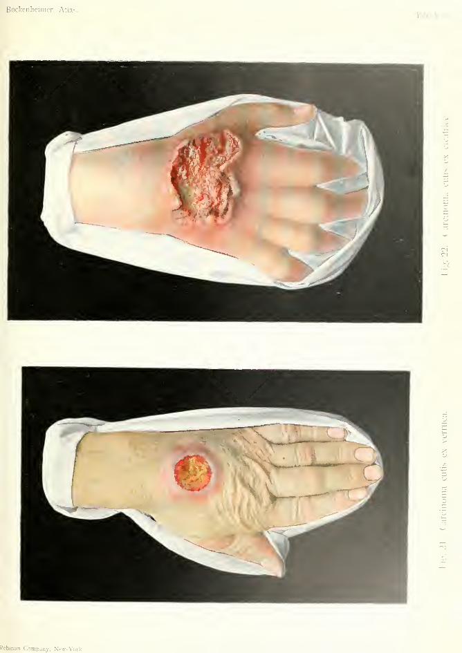
Fig. 21 shows a carcinoma in a common situation,
the back of the hand, arising from a wart and form-
ing a characteristic cai'cinomatous ulcer. As the
growth was still movable over the fascia, and there
were no glandular enlargements, it was excised and
the gap repaired by a pedunculated flap from the fore-
arm. The rapid growth of these small tumors with
hard borders makes early diagnosis and removal
necessary, so as to avoid recurrence.
Fig. 22 shows a very extensive carcinoma arising
from the scar of an injury two years before. In this
ease the irregular, wall-like, hard, irregular borders
are very marked. The floor of the ulcer is in some
places cornified and is covered with crusts and
sanious secretion. The carcinoma has already ex-
tended through the fascia to the bones, interfering
with the function of the hand. The glands of the
elbow and axilla are hard and nodular. The rapid
growth of the tumor has led to severe cachexia.t>
Treatment. Amputation through the arm and
removal of glands.
31
MelanocarcinomaPlate XYI, Fig. 23.
The malignant melanomas (melanosarcoma, mel-
anoendothelioma and the rarely occurring melano-
carcinoma) occur most often in the skin, also in the
adjacent mucous membrane, and in the choroid and
iris. In the skin they arise from benign melanomas,
especially from flat pigmentary naevi, and from
warts which become continually irritated. Warts on
the sole of the foot and on the fingers often give rise
to these growths. A sessile or pedunculated tumor
develops, which is characterized by black, bluish-
black or brownish-yellow coloration (Fig. 23). Theskin soon becomes ulcerated, and by the breaking
down of the tumor a deep ragged ulcer is produced.
Melanocarcinomas are characterized by the hardness
of the base of the tumor, thus differing from the soft,
easily bleeding melanosarcomas which rapidly dis-
integrate into a brownish-black watery mass, and
form the soft, bleeding angiosarcomas.
Melanocarcinoma of the skin not only grows
deeply towards the fascia, but also forms early dis-
seminations in the skin, in the form of small black
nodules in the neighborhood of the mother tumor,
which form a large growth by confluence.
The great malignity of these tumors is shown bythe early appearance of metastases in the regional
lymphatic glands, which generally form larger tumors
than the primary one; also by the early infection of
the lungs, liver, heart, brain, and other organs by
metastatic deposits.
33

Bockenheimer, Atlas. Tab. XVI.
Fig. 23. Melanocarcinoina cutis ex verruca.
Rebman Company, New-York.

Owing to the rapid development of these metas-tases pigmentation is usually absent in them.Melanocarcinomas may be seen in children as
multiple growths in the skin in connection withxeroderma pigmentosum. The rapid growth andfrequent hemorrhages lead to severe anaemia.
Treatment. Small tumors of the skin can bewidely removed with the fascia. In the extremities
the best and most radical method is amputation andremoval of the regional glands. In spite of early
and extensive operation recurrence is very frequent,
and in v. Bergmann's clinic only one case is knownto be free from recurrence after a year. It is, there-
fore, urgent to take prophylactic measures by excis-
ing all pigmentary nsevi, especially in advanced age,
and all warts which become continually irritated or
inflamed. The gap left by removal of extensive pig-
mentary nsevi of the face must be filled by skin
flaps. Cauterization of ngevi and warts is to be
condemned, as the irritation may be an exciting
cause of tumor formation.
Fig. 23 shows a tumor arising from a pigmentarywart; the alveolar structure on microscopic examina-tion showed it to be a melanotic carcinoma. In spite
of amputation of the leg and removal of the inguinal
glands, death resulted from organic metastases.
33
SarcomaPlates XMI—XXM.
LYMPHOSARCOMA COLLI {of Xeck)Plate XVII. Fig. '24.
SARCOMA EPIPHARYNGEALE (Epipharyngeal Sarcoma)
POLYPOSIS NASI MALIGNA (MaHgtiont Nasal Polypus)
Plate XMII, Fig. 25.
ANGIOSARCOMA CUTIS {of Skin)
BOTRIOMYCOSISPlate XIX. Fig. 26.
SARCOMA FUNGOIDES ORBITAE{Fungating Sarcoma of Orbit)
Plate XIX, Fig. 27.
MELANOSARCOMA CUTIS {of Skin)
LYMPHOMATA SARCOMATOSA COLLI{Sarco7nato7ts Lymphoma of Neck)
Plate XX, Fig. 28.
SARCOMA MAMMAE EXULCERATUM{Ulcerating Sarcoma of Breast)
Plate XXI. Fig. 29.
SARCOMA MAMMAE CYSTICUM {Cystic Sarcoma of Breast)
Plate XXII. Fig. 30.
SARCOMA CUTIS MULTIPLEX {Multiple Sarcoma of Skin)
Plate XXIII. Fig. 31.
SARCOMA HUMERI PERIPHERICUM{Peripheral Sarcoma of Humerus)
Plate XXrV", Fig. 32.
SARCOMA FASCLAE BRACHII EXULCERATUM{Ulcerating Sarcoma of Brachial fascia)
Plate XXV. Fig. 33.
CHONDROMYXOSARCOMA GENUS {of Knee)
EXOSTOSES MALIGNAE (Maligyianf Exostosis)
Plate XXM. Fig. 34.
SARCOMA GIGANTOCELLULARE {Giatd-celled)—EPULISPlate XXVn, Fig. 35.
The tumors formerly called Sarcoceles owe their
name to the fact that they have the appearance of
fleshy masses on section. In distinction to carci-
34
nomas the sarcomas develop from the various
connective tissue elements, with the exception of
endothelium, and may, therefore, arise in the skin,
subcutaneous tissue, fascia, periosteum, bone, nerves,
and in the connective tissue of all other organs.
Owing to the often very rapid growth the newlyformed cells do not attain complete maturity, so
that the sarcoma consists of imperfectly developed
connective tissue. In its early stages it often re-
sembles, microscopically, inflammatory granulation
tissue, but by its rapid growth it soon assumes the
appearance of a malignant tumor. The bulk of the
sarcoma is formed of various connective tissue cells,
while the interstitial fibrous tissue is scanty. Theabundant formation of new blood-vessels is char-
acteristic of sarcoma.
The transition of fibromas, especially those whicharise from the connective tissue of fascia, and of
other connective tissue tumors e.g. chondroma, into
sarcoma has been demonstrated.
Patients often attribute these growths to various
injuries, but there is no direct proof of this.
The pure sarcomas are classified according to their
microscopic structure into round-celled, spindle-
celled and giant-celled sarcoma. Those formed of
various tissues are known as lympho-, myxo-, fibro-,
chondro-, angio-, and glio-sarcoma. The pigmentary
or melanosarcomas are placed in a special group.
Clinically, sarcomas are best divided into soft,
many-celled, quickly growing, very malignant, easily
recurring (medullary sarcoma, usually small round-
celled sarcoma), and the hard, few-celled, slow-
growing, less malignant forms (spindle-celled and
giant-celled sarcoma). In the first form the soft
consistence is due to the richness in cells and the
scanty development of interstitial tissue. Comparedwith carcinomas, sarcomas are more circumscribed
and at first almost completely encapsuled tumors,
with borders as soft as the rest of the tumor.
35
Owing to frequent hemorrhages and softening in
the interior of the sarcoma cystic cavities are formed
which can be recognized by the presence of fluctua-
tion (Figs. 25 and 30). Sarcomas situated under the
skin gradually destroy and break through it and pro-
liferate on the surface in a variety of forms. Fleshy
reddish-brown parts alternate with yellowish-white,
pulpy parts in these tumors. There are usually
blood extravasations, both old and recent. Thewhole tumor has the appearance of a fungoid mass
(Figs. 26, 27, 29 and 33). After a time these super-
ficially proliferating growths break down and set upinflammation, so that the characteristic appearance
of the sarcoma is lost, and, on the scalp and extremi-
ties, for example, it cannot be distinguished from a
discharging soft carcinoma. As the sarcoma usually
breaks through the skin and proliferates on the sur-
face, so may it extend into all the deeper tissues, so
that finally an enormous tumor is formed which maydestroy the bones (Figs. 25, 27 and 33).
The second form, the slow-growing, few-celled
tumors, resemble fibromas and often represent transi-
tional forms (fibro-sarcoma). The latter sometimes
occur as multiple nodules in the skin.
These tumors often occur in robust people in mid-
dle life (thirty to fifty). Very often sarcoma is con-
genital or appears in infancy (kidneys and testicles),
also soon after puberty (mammary gland). Theearlier the tumors appear, the more malignant they
are as a rule. iSIultiple sarcomas are seen in the
skin as pigmentary sarcomas (Fig. 31) and in the
bones.
The soft sarcomas lead to metastases much more
often than the hard forms. Metastatic deposits are
formed by growth of the tumor into the large veins
and the formation of emboli, which are carried to the
lung, spleen, liver and brain. Dissemination by wayof the lymphatics is almost completely absent. Thelatter are certainly often involved, especially in ulcer-
36
ated sarcoma and melanotic forms; also in sarcomaof bone.
By the entrance of the tumor cells into the blood
stream and by the setting up of inflammatory pro-
cesses a condition of fever is produced.
In many cases the body is so quickly affected bymetastases that the patients soon succumb fromsevere anaemia. Unfortunately patients often comefor treatment when there are already metastases in
the lung causing pleural effusion and hemoptysis.
Differential Diagnosis. Sarcoma differs from
carcinoma in the softer consistence of the tumor andits regular surface, and from benign tumors by its
rapid growth. The distinction from syphilitic pro-
ducts is often difficult and sometimes not settled by
microscopic examination, and according to Esmarchmany growths were formerly extirpated as sarcoma
which might have been cured by anti-syphilitic treat-
ment.
Treatment. x\ll tumors in which there is a sus-
picion of sarcoma should be removed as early and
as radically as possible. As the tumors are some-
times encapsuled, operation has been unfortunately
limited to enucleation in these cases; but, as in car-
cinoma, the tissue surrounding the tumor, which is
already infiltrated by tumor cells, must be removed.
In cases of soft, rapidly growing sarcoma of the
extremities, the question of amputation and even dis-
articulation arises. In spite of operation recurrence
is frequent; either locally or in the form of dissem-
inated nodules, less commonly in the form of lym-
phangitis sarcomatosa. In the hard forms of
sarcoma recurrence may also occur, in the form of
soft growth, which is a most unfavorable sign.
Inoperable cases have been treated with the X-
rays, but the action is only superficial {Unger, Schles-
inger). By this treatment the superficial nodules are
37

destroyed, just as in intercurrent erysipelas, but the
tumor continues to grow in the deeper tissues and in
other places. Subcutaneous injections of arsenic
and atoxyl are worth a trial, and iodide of potas-
sium in large doses may be administered. Serum
therapy has so far given no results.
38

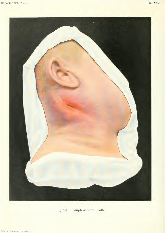
Bockenheinier, Atlas. Tab. XVII.
Fig. 24. Lyiiipliosarcoma colli.
Rcbnun Company, New-York.
LYMPHOSARCOMA COLLI {of Neck)Plate XVII, Fig. 24.
Clinically, the name lymphosarcoma is best
applied to those sarcomas which originate in lym-
phatic glands, whether their cells have the character
of lymph cells or arise from the stroma of the glands.
This is all the more indicated as both forms of sar-
coma can only seldom be distinguished from one
another, clinically or microscopically. The seat of
predilection of these tumors is the region of the
neck, where the lymphatics are abundant. A diffuse
nodular tumor quickly develops from a group of
small, hard, movable glands.
The malignancy of these tumors is shown, espe-
cially in young individuals, by the continual formation
of fresh nodules at the periphery, which coalesce with
the main tumor and cause it to attain a considerable
size. The unlimited growth into the neighboring
tissues is characteristic. The capsule of the glands
is quickly broken through, thus differing from lym-
phoma. The cervical fascia is destroyed and the
sterno-mastoid muscle invaded. The skin is at first
reddish, then bluish red or livid; it then becomes
thin and gives way over the tumor. The exposed
parts of the tumor rapidly break down from inflam-
mation. The sarcoma grows into the deeper parts,
especially into the internal jugular vein, giving rise
to fatal organic metastases. The vagus nerve and
the common carotid also become enveloped and
destroyed by the tumor. Dyspnoea and dysphagia
may be caused by pressure on the larynx and esoph-
agus. The tumor extends downwards into the
mediastinum and may even destroy the vertebrae.
39
Lymphosarcoma is distinguished from other tumors
of the neck by its rapid growth in all directions, its
breaking through to the exterior, and its sanious dis-
integration.
The diagnosis is usually not established in the
early stages as the growth is hard and limited to the
glands; microscopic examination is also inconclu-
sive.
Differential Diagnosis. Malignant lymphoma(Hodgkin's disease, pseudoleukjemia) which usually
begins in the neck, consists of small, multiple, encap-
suled nodules which do not break down nor extend
to the neighboring organs. There are generally also
glandular enlargements in the axillae, groins and
mediastinum, and changes in the spleen and bone-
marrow.
Leuksemic IjTnphoma can be diagnosed by the
blood changes.
Tuberculous glands are characterized by the iso-
lated groups of glands of different consistence—hard,
soft, or fluctuating.
Syphilitic glands are at first hard, later on soft;
but are not so extensive.
Branchiogenous carcinoma (t\ Volhnann), arising
from the remains of the epithelium of the branch-
ial clefts, is very rare and appears as very hard,
spherical tumors in the carotid fossa.
Metastatic carcinoma and sarcoma can be diag-
nosed by the presence of the primary tumors (scalp,
esophagus, parotid, maxilla).
Actinomycosis may also cause hard infiltration of
the neck, but the infiltration is diffuse and uniform,
not nodular, and extends over the whole region of
the neck.
The tumors affecting the sheaths of the blood-
vessels, first described by Langenbech, are to be
regarded as lymphosarcomas which have involved
the vascular sheaths at an early period.
40
Treatment. Extirpation of lymphosarcoma hasonly a chance of success by early diagnosis, and eventhen recurrence is frequent. For the removal ofsuch extensive non-encapsuled tumors much inter-
vention is necessary, in some cases including tem-porary ligature of the common carotid. As theinternal jugular vein and vagus nerve are usuallyremoved with the common carotid, with consequentdisturbances (encephalomalacia, pneumonia), manyprefer internal treatment with high doses of arsenic,or by the X-rays, by which means transient improve-ment may be obtained.
Fig. 24 shows an extensive lymphosarcoma of theneck. The tumor extends diffusely over the wholeof the right side of the neck and is constituted byseveral nodular, irregular formations. The skin is
broken in one place, in others it is thin and of abluish-red color. There is a sanious discharge fromthe fistula. Pressure of the tumor on the large ves-
sels has caused severe cyanosis, and pressure on therecurrent nerve hoarseness and asph>-xia. In spite
of treatment by arsenic and the X-rays the patient
continued in a state of cachexia.
41
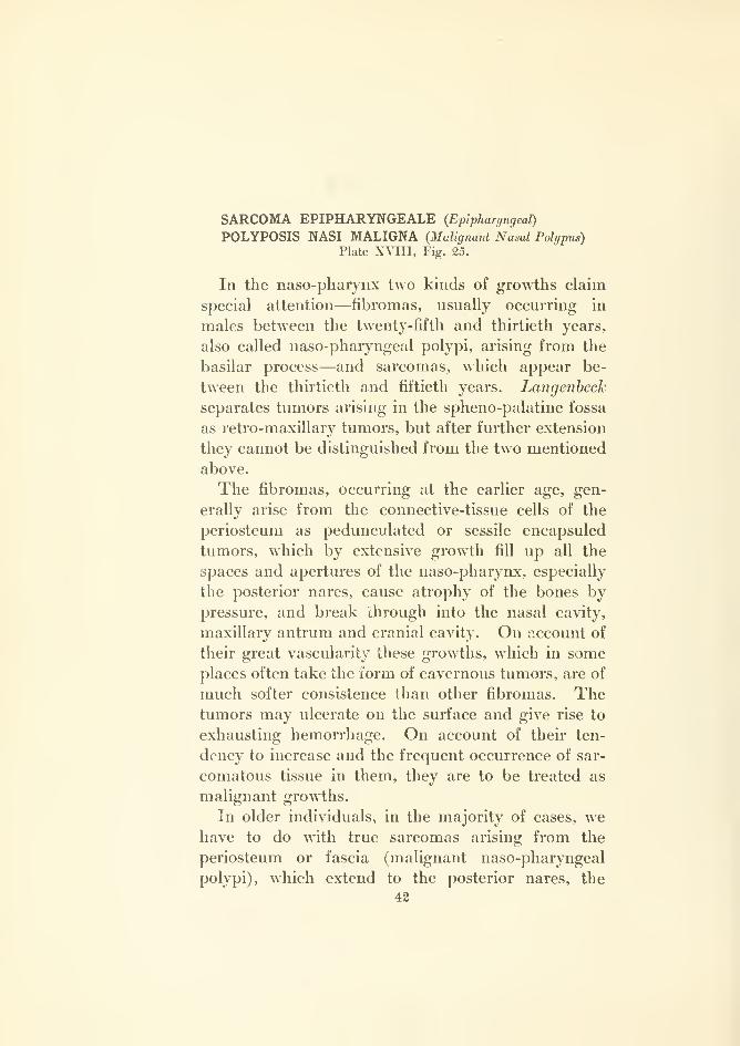
SARCOMA EPIPHARYNGEALE (Epipharyngeal)
POLYPOSIS NASI MALIGNA (Malignant Nasal Polypus)
Plate XMII, Fig. 25.
In the naso-pharynx two kinds of growths claim
special attention—fibromas, usually occurring in
males between the twenty-fifth and thirtieth years,
also called naso-pharyngeal polypi, arising from the
basilar process—and sarcomas, which appear be-
tween the thirtieth and fiftieth years. Langenbeck
separates tumors arising in the spheno-palatine fossa
as retro-maxillary tumors, but after further extension
they cannot be distinguished from the two mentioned
above.
The fibromas, occurring at the earlier age, gen-
erally arise from the connective-tissue cells of the
periosteum as pedunculated or sessile encapsuled
tumors, which by extensive growth fill up all the
spaces and apertures of the naso-pharynx, especially
the posterior nares, cause atrophy of the bones by
pressure, and break through into the nasal cavity,
maxillary antrum and cranial cavity. On account of
their great vascularity these growths, which in someplaces often take the form of cavernous tumors, are of
much softer consistence than other fibromas. Thetumors may ulcerate on the surface and give rise to
exhausting hemorrhage. On account of their ten-
dency to increase and the frequent occurrence of sar-
comatous tissue in them, they are to be treated as
malignant growths.
In older individuals, in the majority of cases, wehave to do with true sarcomas arising from the
periosteum or fascia (malignant naso-pharyngeal
polypi), which extend to the posterior nares, the
42
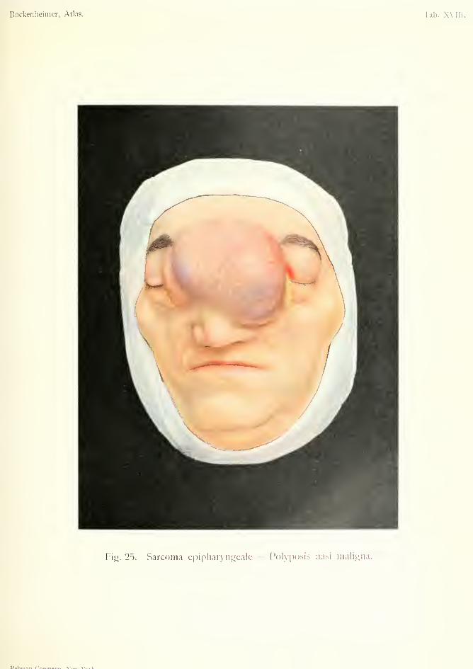
Bockeiiliciiiier, Atlas. Tab. XVIII
Fig. 25. Sarcoma cpipliaryiiscale - Polyposis nasi maligna.
lJ..Knii., r"«„,«-..,, \.T..._. \/.

/
spheno-maxillary fossa, Eustachian tubes and larynx;not, however, as encapsuled tumors hke the fibro-mas, but as soft, fungoid, sessile, firmly attachedgrowths with irregular boundaries. Later on theygrow very rapidly, causing destruction of the neigh-boring bones, and extend to the surface through thefrontal sinus, nasal cavity and orbit, and internallyto the brain. (Figs. 25 and 27).
Disintegration of the growth goes hand in handwith the advancing growth and the patient succumbsfrom the results of hemorrhage, septic infection,
anaemia and organic metastases.
The clinical symptoms in fibroma and in com-mencing sarcoma arise from obstruction of thenaso-pharynx. Continually keeping the mouth opensuggests disease of the naso-pharynx. Owing toobstruction of the posterior nares the patients snoreduring sleep; they acquire nasal catarrh (often atro-phic rhinitis) and have a nasal voice. As the tumorextends, obstruction of the Eustachian tubes causesdeafness and pain in the ear; extension to the cranialcavity causes headache, somnolence and choked opticdisk; extension to the orbit causes disturbance ofvision, e.g. diplopia. Pressure on the facial nerveand trigeminal causes paralysis and severe neuralo-ia.
The diagnosis of these advanced sarcomas pre-sents no difficulty. The soft, fungoid consistence ofthe whole tumor, the tendency to bleeding and therapid growth are characteristic. In extensive sar-
comas with commencing disintegration and dis-
charge soft glandular metastases are found. Thecommencing sarcomas can be recognized by digital
and rhinoscopic examination as irregular, rough,infiltrating tumors, which differ from the nodularencapsuled fibromas.
Differential Diagnosis. It is only in youngindividuals that other lesions can be confounded withtrue tumors of the naso-pharynx. Hypertrophied
43

tonsils and extensive adenoids cause similar symp-
toms, but digital examination and rhinoscopy will
make the diagnosis clear. In very young children
teratomas are seen (Fig. 146), which may be mis-
taken for sarcoma arising from the basilar process
and extending to the face. However, teratomas are
usually more or less encapsuled and only appear
on one half of the face.
Retro-maxillary tumors manifest themselves at
first by unilateral pain in the face, swelling of the
cheek and fixation of the corresponding maxillary
joint, but on further extension they cannot be distin-
guished from advanced tumors of the naso-pharynx,
or from large tumors of the upper maxilla or orbit.
Treatment. The removal of adenoid vegeta-
tions is best efi^ected by Gottstein's curette. Evenextensive adenoid growths may disappear sponta-
neously at the age of puberty. Hypertrophied ton-
sils are to be removed by the tpnsillotome. For
small fibromas an oral method may be employed, by
means of division of the soft palate and part of the
hard palate {Nclaton, Gussenhauer) or by temporary
division of the lower maxilla. The tumors should
always be removed by incision into healthy tissues
with the knife. In extensive fibromas and all tu-
mors suspected of sarcoma, the naso-pharynx mustbe freely laid open, by temporary resection of the
hard palate together with the alveolar process
(Partsch), or by temporary resection of both upper
maxillse and raising up the nose (v. Bergviann).
Previous tracheotomy and ligation of the external
carotid on one or both sides (Kocher, Konig), is ex-
pedient in these sanguinary operations. That very
large tumors can be removed by extensive operations
with good results is shown by the experience of
V. Bergmann's clinic. Even tumors which had ex-
tended through the base of the skull and caused
symptoms of cerebral pressure were successfully
44
removed. Naturally, the earlier diagnosis is madeby digital examination and rhinoscopy (excision for
examination is dangerous on account of severe
hemorrhage, and also useless) the more can these
complicated operations be avoided, and the morefrequent are radical cures. Inoperable tumors (Fig.
'27) may be treated by the X-rays or by the adminis-
tration of arsenic and morphia. When the tumors
fungate externally the ulcerated parts must be cau-
terized and treated with moist disinfectant dressings.
In the last stages tracheotomy must be performed, to
save the patient from death by asphj^xia.
Fig. 25 shows a malignant naso-pharyngeal poly-
pus which arose from the basilar process and was
at first covered by the mucous membrane of the
epipharynx. The disease was of ten years' duration.
Various polypi were removed, and also a larger
tumor, after partial resection of the upper maxilla,
without success. The sarcoma then grew almost
exclusively forwards through the posterior nares and
destroyed the whole bony framework of the nose.
The fairly symmetrical growth on both sides of the
middle line shows the origin from the basilar process,
in distinction to the more lateral swelling of retro-
maxillary tumors. It forms a soft, partly fluctuating
growth with fungatino; borders which has begun to
extend over both eyes. In some places the skin is
so thin that it appears livid and transparent ; in other
parts it shows the great vascularity of the skin char-
acteristic of sarcoma. The tumor, already disinte-
grating, is on the point of breaking through. Thewhole nasal cavity and the whole naso-pharynx on
digital examination were found to be filled with soft,
infiltrating tumor masses, which had displaced the
soft palate downwards and forwards, so that the
growth could onlv have been removed by very
extensive interference. The tumor had also extended
through the base of the skull.
45
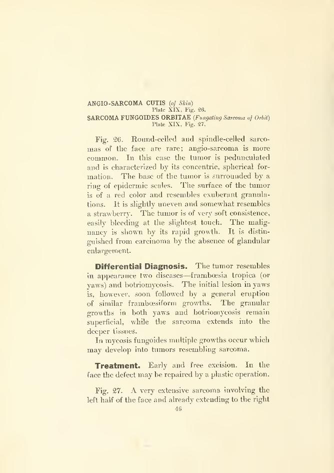
ANGIO-SARCOMA CUTIS (of Skin)
Plate XIX, Fig. 26.
SARCOMA FUNGOIDES ORBITAE {Fungating Sarcoma of Orbit)
Plate XIX, Fig. 27.
Fig. 26. Round-celled and spindle-celled sarco-
mas of the face are rare; angio-sarcoma is more
common. In this case the tumor is pedunculated
and is characterized by its concentric, spherical for-
mation. The base of the tumor is surrounded by a
ring of epidermic scales. The surface of the tumor
is of a red color and resembles exuberant granula-
tions. It is slightly uneven and somewhat resembles
a strawberry. The tumor is of very soft consistence,
easily bleeding at the slightest touch. The malig-
nancy is shown by its rapid growth. It is distin-
guished from carcinoma by the absence of glandular
enlargement.
Differential Diagnosis. The tumor resembles
in appearance two diseases—framboesia tropica (or
yaws) and botriomycosis. The initial lesion in yaws
is, however, soon followed by a general eruption
of similar frambcesiform growths. The granular
growths in both yaws and botriomycosis remain
superficial, while the sarcoma extends into the
deeper tissues.
In mycosis fungoides multiple growths occur which
may develop into tumors resembling sarcoma.
Treatment. Early and free excision. In the
face the defect may be repaired by a plastic operation.
Fig. 27. A very extensive sarcoma involving the
left half of the face and already extending to the right
46
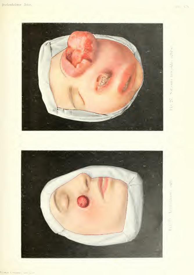
Bockenheinier, Atlas.Tab. XIX.
-.n
r,
bi
'-.t
zr,
t>j
Rebman Company, Ncvi-York.


half. Protruding from the orbit as a fungoid massthe tumor is characteristic of sarcoma (sarcoma
fungoides). The soft edges have the typical reddish-
brown color of sarcoma. In the places where the skin
is destroyed soft masses with a fairly regular surface
protrude, which differ from the ragged irregular ulcer
of carcinoma. The whole of the tumor situated in
the orbit is of soft, almost fluctuating consistence.
In some parts the fungoid masses are breaking downand covered with sanious dischai-ge. Blood crusts
form on the ulcerations owing to the fi'equent hem-orrhages in the tumor. The brown-colored skin is
almost atrophied from pressure of the tumor. Sar-
comatous masses protrude from both nostrils, andthe whole buccal cavity and naso-pharynx is full of
tumor masses, which have caused complete destruc-
tion of the bones of the face. The tumor has also
extended through the base of the skull, causing: ex-
treme somnolence. It is no longer possible to decide
whether it is a case of malignant naso-pharyngeal
pol}^us, a retro-maxillary tumor, a maxillary tumor,
or a periosteal sarcoma of the orbit. The last is the
most probable, as the tumor was first observed in the
orbit.
Treatment. Cf. Plate XVII.
47
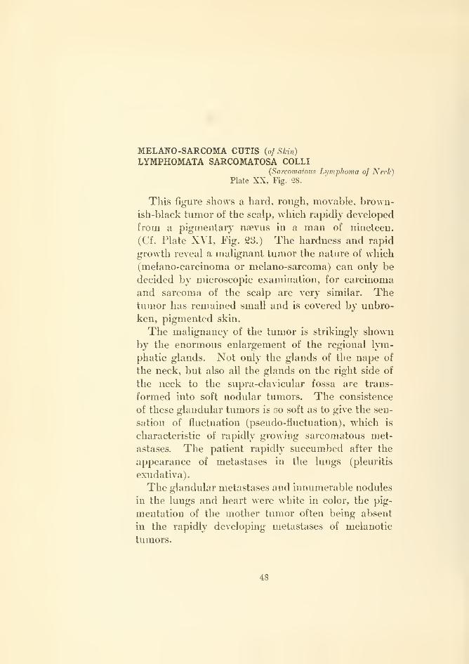
MELANO-SARCOMA CUTIS {of Shin)
LYMPHOMATA SARCOMATOSA COLLI{Sarcomatous Lymphoma of Neck)
Plate XX, Fig. 28.
This figure shows a hard, rough, movable, brown-
ish-black tumor of the scalp, which rapidly developed
from a pigmentary nsevus in a man of nineteen.
(Cf. Plate XVI, Fig. 23.) The hardness and rapid
growth reveal a malignant tumor the nature of which
(melano-carcinoma or melano-sarcoma) can only be
decided by microscopic examination, for carcinoma
and sarcoma of the scalp are very similar. Thetumor has remained small and is covered by unbro-
ken, pigmented skin.
The malignancy of the tumor is strikingly shownby the enormous enlargement of the regional lym-
phatic glands. Not only the glands of the nape of
the neck, but also all the glands on the right side of
the neck to the supra-clavicular fossa are trans-
formed into soft nodular tumors. The consistence
of these glandular tumors is so soft as to give the sen-
sation of fluctuation (pseudo-fluctuation), which is
characteristic of rapidly growing sarcomatous met-
astases. The patient rapidly succumbed after the
appearance of metastases in the lungs (pleuritis
exudativa).
The glandular metastases and innumerable nodules
in the lungs and heart were white in color, the pig-
mentation of the mother tumor often being absent
in the rapidly developing metastases of melanotic
tumors.
48
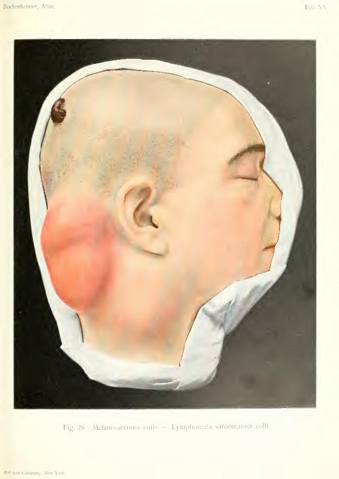
Bockenheimer, Atlas.
Fie. 28. Melanosarcoma cutis - I.viimliomata sarconiatosa colli.
Rebman Company, New-York


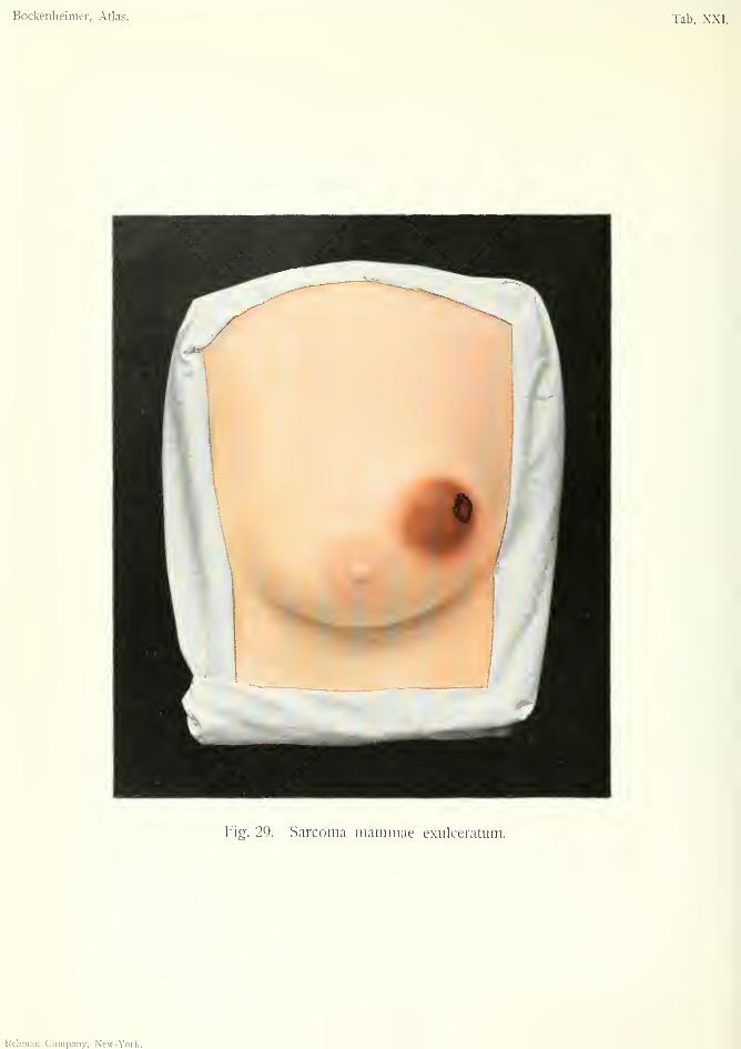
Bockenheimer, Atlas. Tab. XXI.
h'ig. 20. Sarcoma mammae e.xulceratum.
Rcbmaii Company, New-York.
SARCOMA MAMMAE EXULCERATUM{Ulcerating Sarcoma of Breast)
Plate XXI. Fig. 29.
SARCOMA MAMMAE CYSTICUM {Cystic Sarcoma of Breast)
Plate XXII, Fig. 30.
Sarcoma is much less common in the mammarygland than carcinoma (one hundred carcinomas to
ten sarcomas, and half of these cysto-sarcomas, v.
Angerer). All cell forms of sarcoma may be repre-
sented as well as mixed forms, such as myxo-, angio-,
and melano-sarcoma.
They occur most often in young women. Accord-
ing to their composition they have different clinical
signs. Spindle-celled sarcomas are of firm consist-
ence and of slower gro^i;h than the soft, malig-
nant, round-celled sarcomas and melanosarcomas.
Cysto-sarcomas soon lead to extensive tumors which
transform the breast into a large sac with fluid
contents. The typical characteristics of sarcoma are
generally present in the mammary tumors (Fig. 29).
Differential Diagnosis. Carcinoma is distin-
guished by the absence of any demarcation from the
mammary tissue, while sarcoma is often encapsuled.
Moreover, the clinical signs of carcinoma are so char-
acteristic (cf. Plates V-XI) that confusion is hardly
possible. Cysts of the mamma are usually situated
behind the mammilla, multiple (in one or both
mammte), and not so large as true cysto-sarcoma.
Fibroadenoma (to which the tumors incorrectly
designated by J. Miiller as cystosarcoma pajjilli-
ferum phyllodes, rightly belong), which originate
from the glandular tissue and show an abundant
49
development of connective-tissue cells, are slow grow-
ing, movable tumors, and are always encapsuled (cf
.
Plate XXVIII).
Treatment. Extirpation of the whole mammaas early as possible, with free exposure of the axilla.
After early and extensive operations local recurrence
is rare, and permanent cures more frequent than in
carcinoma.
Fig. 29 shows a rapidly growing, round-celled sar-
coma in a young girl. The tumor forms a soft,
fairly circumscribed nodule in the mammary gland.
That the part of the tumor lying in the mamma is
considerably larger than the external appearance in-
dicates is shown by the prominent veins. The tumor
is near the mammilla but has caused no retraction of
the nipple. It is freely movable over the pectoralis
fascia. Externally it has involved the skin, which
has the usual brownish-red color of sarcoma, has be-
come very thin and is already ulcerated in one spot,
from which repeated hemorrhage has taken place.
The fungoid tumors, in distinction to carcinoma,
have a smooth, uniform surface and resemble exuber-
ant granulation tissue. There were no glands to be
felt in the axilla. Treated by extirpation of the
mamma and free exposure of the axilla.
Plate XXII, Fig. 30.
A cystic tumor occurring in a young woman,which has begun to displace the whole breast. There
is no alteration in the nipple. The tumor is movable
over the pectoral fascia, and in several places dis-
tinctly separate from the mammary tissue. The veins
are enlarged from pressure of the tumor. The tumor
has already invaded the skin, which has become
very thin, and in some places fluctuating. The skin
is colored brownish red and bluish green, and shows
a network of vessels. As long as the skin is
50
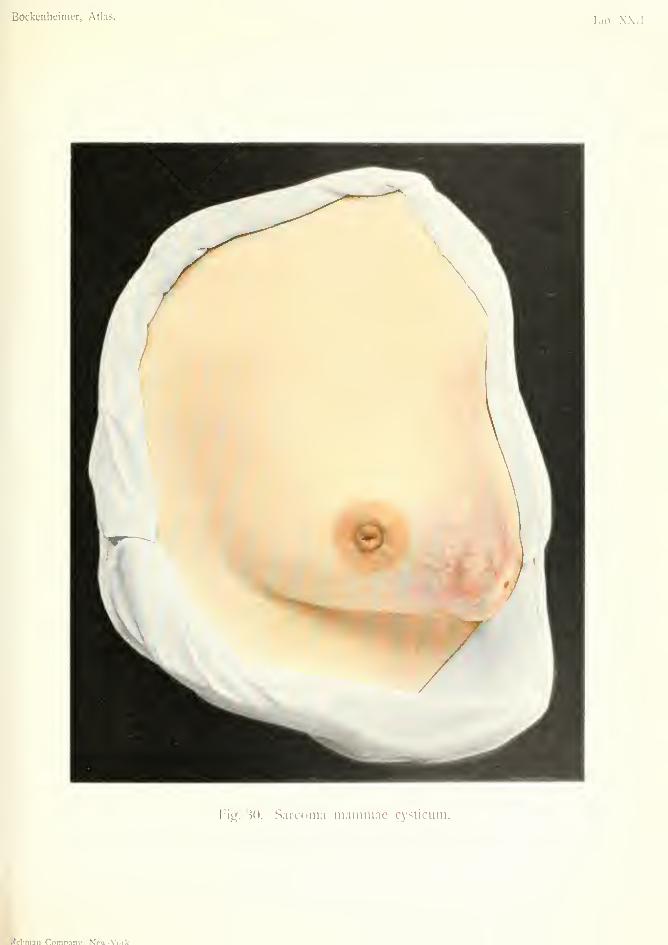
Bockenheiiner, Atlas. Tab. X.XII
t'ig. 30. Sarcoma mammae cysticiim.
tn-in Coninanv. NpT-Vorlc.


intact it can never be definitely ascertained whetherit is a case of actual cavities filled with fluid, or thepseudofluctuation of gelatinous or mucoid sarcoma.Rapid growth and commencing soft glandular swell-
ings in the axilla point to the diagnosis of a cysto-sarcoma.
Treatment. Extirpation of the mamma andremoval of the axillary glands.
51
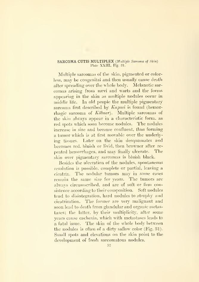
SARCOMA CUTIS MULTIPLEX (Multiple Sarcoma of Skin)
Plate XXm, Fig. 31.
Multiple sarcomas of the skin, pigmented or color-
less, may be congenital and then usually cause death
after spreading over the whole body. Melanotic sar-
comas arising from naevi and warts and the forms
appearing in the skin as multiple nodules occur in
middle life. In old people the multiple pigmentary
sarcoma first described by Kaposi is found (hemor-
rhagic sarcoma of Kbhner). Multiple sarcomas of
the skin always appear in a characteristic form, as
red spots which soon become nodules. The nodules
increase in size and become confluent, thus forming
a tumor which is at first movable over the underly-
ing tissues. Later on the skin desquamates and
becomes red, bluish or livid, then browner after re-
peated hemorrhages, and may finally ulcerate. Theskin over pigmentary sarcomas is bluish black.
Besides the ulceration of the nodules, spontaneous
resolution is possible, complete or partial, leaving a
cicatrix. The nodular tumors may in some cases
remain the same size for years. The tumors are
always circumscribed, and are of soft or firm con-
sistence according to their composition. Soft nodules
tend to disintegration, hard nodules to atrophy and
cicatrization. The former are very malignant and
soon lead to death from glandular and organic metas-
tases; the latter, by their multiplicity, after some
years cause cachexia, which with metastases leads to
a fatal issue. The skin of the whole body between
the nodules is often of a dirty sallow color (Fig. 31).
Small spots and elevations on the skin point to the
development of fresh sarcomatous nodules.
53
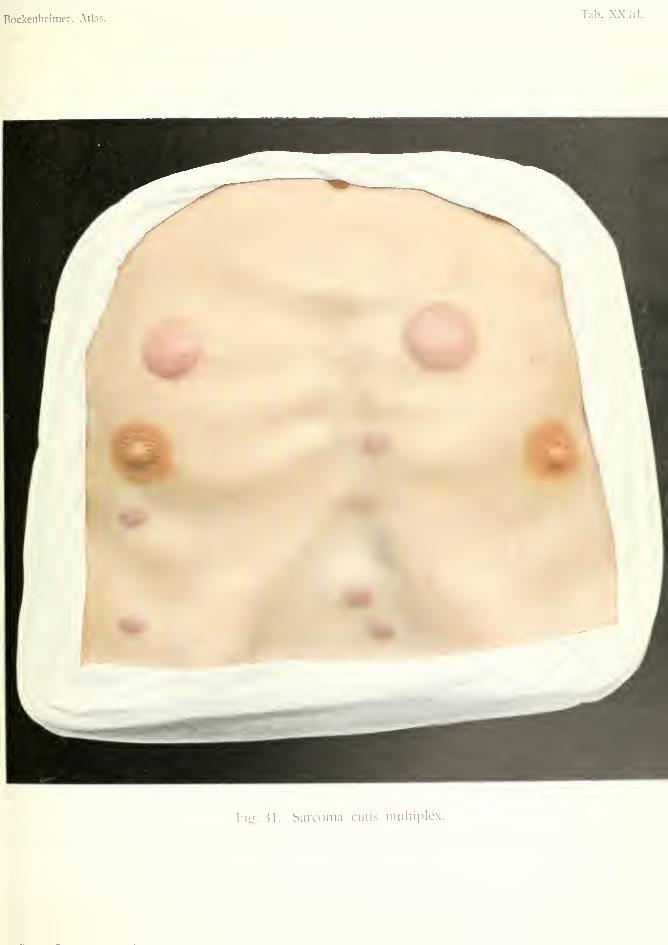
Bockenheimer, Atlas.
/
Fitr. 31. Sarcoma cutis multiplex.

Sarcoma multiplex hemorrhagicum pigmentosumappears in the form described above, but first of all
on the lower extremities, in the form of reddish
nodules which often cause much itching. Tumorformation goes hand in hand with oedematous infiltra-
tion which extends over the whole leg and prevents
the patient from walking. Desquamation of the skin
on the surface of the nodules occurs along with corni-
fication of the epidermis. Cicatrices form in theskin from atrophy of the nodules. Other regions of
the body are unaffected, except the peripheral parts
of the upper extremity. There is no enlargement of
the lymphatic glands. The disease runs a progres-
sive course, and in spite of the spontaneous resolution
of some of the tumors, finally causes death bymarasmus.
Microscopic examination shows a pure sarcomawith abundant blood-vessels, which often gives rise to
organic metastases. As this form occurs exclusively
in old people, arteriosclerosis may, perhaps, accountfor the origin and course of the disease. {Kobner,
Schlesinger)
.
Differential Diagnosis. Primary multiple sar-
comas must not be confounded with secondarysarcomatous growths in connection with a primarycutaneous sarcoma or a sarcoma of the internal
organs. The tumors of mycosis fungoides are morelikely to be mistaken for sarcoma, as they also de-
velop from red, uneven spots, and form granulation
tumors of a brownish-red color which in the later
stages tend to ulceration and cachexia; but mycosis
fungoides is of much slower growth than sarcoma.
Syphilitic and tuberculous granulomas can hardly beconfounded with sarcoma on careful examination.
Treatment. Preventive treatment of multiple
sarcoma consists in the removal of all msvi which
begin to take on rapid growth. In already existing
53
multiple pigmentary sarcomas excision is generally
useless, and should only be performed when the
tumors are few in number and the blood-vessels free
from melanin. After excision of multiple sarcomas,
especially melanosarcomas, death often follows from
rapid dissemination and organic metastases. Hencethe X-rays, large doses of arsenic (internally or
subcutaneously) have been employed for multiple
cutaneous sarcomas, in the same way as for mycosis
fungoides. A permanent cure, however, is not to be
expected as the prognosis of these multiple sarcomas
is always bad.
Fig. 31 shows a case of multiple sarcoma of the
skin affecting the whole of the thorax, abdomen andback. Some of the nodules have already atrophied
leaving cicatrices. The new growth of nodules, how-
ever, exceeds the atrophy so that the patient becamemore and more cachectic in spite of treatment.
54


Bockenlieimer, Alias.Tab. XX 1\'.
Fig. 32. Sarcoma huineri periphericum.
Rcbman Company, New-York.

SARCOMA HUMERI PERIPHERICUM(Peripheral Sarcoma of Humerus)
Plate XXrV\ Fig. 32.
Sarcomas arising from the bones are of special
interest on account of their frequency.
Osteo-sarcomas are best divided into peripheraland central; the latter may arise from the cortical,
spongy or medullary portions. Division into perios-
teal and myelogenous tumors is clinically impossi-ble, and the word myelogenous may be replaced byosteal. Tumors which appear clinically to be peri-
osteal often arise from the superficial layers of thecortex. By the use of the X-rays it is more easy to
divide them into peripheral and central tumors; this
leaves open the possible origin of the sarcoma fromany part of the bone, and this can only be conclu-
sively settled by section of the bone after removal.This classification is all the more rational becausesections of preparations which were clinically re-
garded as periosteal sarcomas show that these arose
from small foci in the medullary cavity. Periosteal
tumors may extend into the medullary cavity and so
simulate osteal tumors. In extensive tumors the
origin of the tumor from any definite part of the
bone cannot as a rule be established.
Both forms have special seats of predilection:
in the long bones, the neighborhood of the epiphyses
e.g. the upper end of the humerus (Fig. 3-2), the lowerend of the femur, especially the internal condyle, the
head of the tibia, the lower end of the radius; the
flat bones, especially the scapula and bones of the
skull. Both forms also grow in a globular forminvolving the whole circumference of the bone and
55
finally its whole thickness. They appear at puberty
and during the whole period of growth, generally in
young and robust individuals.
Both forms are distinguished by the fact that they
soon break through their own capsule and that of
the bones and then extend into the neighboring joints
and muscles, especially the muscular insertions into
the bones, and into the veins, forming eventually
enormous tumors which break through the skin and
appear as fungoid masses. The superficially situ-
ated tumors have a tendency to frequent hemorrhage
and destructive inflammation. Primary sarcomas of
bone are very rare and are sometimes confounded
with sarcoma-like changes in the bones which result
from ostitis fibrosa; also with formations which do
not belong to tumors in the strict sense, but are
known as myelomas (especially in the blood-forming
vertebral bodies).
Microscopically, spindle cells are often found in
peripheral sarcoma, and giant cells in central sar-
coma. The other forms of sarcoma cells are also
present.
The X-rays, in peripheral sarcoma, show little
change in the cortex. In central tumors, especially
those arising from the meduUaiy cavity, they often
show spherical transparent spaces in the interior,
while the cortex is very thin and excavated—forming
a shell—in the same way as in bony cysts, osteo-
myelitic abscesses, isolated tuberculosis and gumma.In the early stages the diagnosis of osteo-sarcoma
is difficult. The peripheral tumors are naturally
more easy to diagnose, as they present a rapidly
growing tumor firmly attached to the bone, with
irregular boundaries towards the muscles. Rheu-
matic pains and effusion into the joints frequently
occur when the tumors are situated near the joints.
The nearer the sarcoma approaches the skin the
easier it is to recognize the superficial tumor masses,
which infiltrate the soft tissues, and consist of cells
56
only without bony infiltration. Swelling of the cuta-neous veins occurs early from pressure of the tumoron the vessels (Fig. 32), while the skin becomes red-dish brown, thin and almost transparent, especiallywhen the tumor is attached to it.
Slow-growing central sarcomas can only at first
be diagnosed by the X-rays, later on they presentthemselves as hard spheroidal swellings like billiard
balls. The more they extend and approach theskin, the thinner becomes their bony shell, whichfinally gives the sensation of parchment crepitation,first described by Dupuytren. Central tumors areoften first diagnosed by the occurrence of sponta-neous fracture. Extensive forms, which assume amore spindle-celled formation are easy to recognize.Through growth of the tumor into the joints andmuscles, typical functional derangements are pro-duced, and separation of the epiphyses. Metastasesin the lungs develop early. Disintegration of thetumor cells gives rise to fever, especially in rapidlygrowing, small, round-celled sarcomas.
Differential Diagnosis. Parosteal sarcomas areeasily mistaken for peripheral sarcoma, and are oftenimpossible to distinguish by the X-rays. They areoften of very soft consistence, and were formerlycalled encephaloid.
Chondrosarcoma only occurs in the neighborhoodof the joints and forms irregular nodular tumors(Fig. 34).
Sarcomas situated near the large vessels and pul-sating with them, may be mistaken for aneurism,but the X-rays will assist the diagnosis. Central sar-
comas have been wrongly considered as aneurism of
the bone, owing to their vascularity and their red-
dish-brown color on section, which is due to frequent
hemorrhatres.
Myelomas are multiple and occur chiefly in the
vertebrae.
Metastatic carcinomas, which occur especially in
the neck of the femur after mammary carcinoma in
women, and in the head of the humerus after carci-
noma of the thyroid gland {v. Eiselsberg), must be
diagnosed by the primary growth.
Osteo-sarcomas may possibly be confounded with
inflammation of joints, rheumatism, osteo-myelitis,
syphilitic and tuberculous processes; but in most
cases the diagnosis can be made by the history of
the case, by the X-rays, by anti-syphilitic treat-
ment, and in osteomyelitis by search for hemolysin
(Bruck, Michaelis, Schultze). The uninterrupted
diffuse growth should always raise the suspicion of
malignant tumor. In doubtful cases an exploratory
incision may be made.
In all cases the prognosis is very bad. The harder
forms of sarcoma (spindle-celled and giant-celled)
sometimes have a better prognosis. The soft, round-
celled sarcomas are the most malignant on account
of their rapid growth and early metastasis.
Treatment. The earlier operative treatment is
undertaken, the more likely is a radical cure.
Small, central sarcomas can be removed by the
chisel, and the medullary cavity scraped. Larger
circumscribed tumors still confined to the bone can
be removed by free resection of bone. The defect
can be repaired by bone grafting (auto- or hetero-
plastic).
If the sarcoma has already invaded the muscles
amputation must be performed. When the tumor is
near the joint of one of the bones of the extremities,
disarticulation is necessary; when in a flat bone
total extirpation.
Inoperable sarcomas are to be treated according
to the rules for inoperable tumors (cf. Plate XVII).
Fig. 32 shows a peripheral sarcoma of the upper
end of the humerus in a young individual. The58

soft tumor has extended under the skin, in which
the brown coloring and extensive network of dilated
veins are very marked. The lower borders of the
fusiform tumor are irregular and send processes here
and there into the muscles. The tumor has destroyed
the head of the humerus and has broken through
into the joint, in which there is effusion. The func-
tion of the joint and upper arm is destroyed. Thesupra-clavicular glands are enlarged. Posteriorly
the tumor has extended to the scapula region. TheX-rays showed complete destruction of the upper
part of the humerus. As there was no evidence of
organic metastases, the arm and shoulder girdle
(scapula and outer half of the clavicle) were re-
moved after section through the middle third of the
clavicle and ligation of the subclavian artery and
vein. The axillary and supra-clavicular glands were
also removed.
On section, the whole of the upper portion of the
humerus was found to be transformed into a large
tumor, the central parts of which were hard from
bony infiltration, while the periphery was soft and
fungoid. The tumor was a round-celled sarcoma,
but it was too extensive to decide from which part
of the bone it originated.
59
SARCOMA FASCIAE EXULCERATUM(Ulcerating Sarcoma of Brachial Fascia)
Plate XXV, Fig. 33.
Fascia and the sheaths of blood-vessels are often
the starting point of sarcomas; not only of pure
round-celled and spindle-celled sarcomas, but more
often of mixed forms—myxosarcoma and fibro-
sarcoma. Fibrosarcomas are characterized by their
firm consistence and slow growth; they are fre-
quently circumscribed and partly encapsuled. Myxo-sarcomas are characterized by their softness and
rapid growth without encapsulation. The pure sar-
comas appear as soft, many-celled, rapidly growing
tumors, or in a harder form which is of slower
growth and not so malignant.
In the early stages of fascial sarcomas (fascia of
the arm, fascia lata, abdominal fascia) we find small
tumors fixed to the fascia, but movable over subja-
cent tissues and under the skin. The skin is soon
involved and becomes tightly stretched over the
tumor and pigmented, and finally the tumor breaks
through it. At the same time the muscles and
eventually the whole section of the body are infiltra-
ted with tumor substance (bones, joints, peritoneal
cavity). The chief growth, however, takes place on
the external surface in the form of nodular fungoid
tumors which exhibit all the characteristics of sar-
coma. They are of soft consistence, both in the
center and at the periphery; the surface is muchsmoother than in carcinoma, bleeds easily on account
of its numerous blood-vessels, and is covered with
sanious discharge. Nodules succeed one another till
an enormous cauliflower growth is formed (Fig. 33).
60
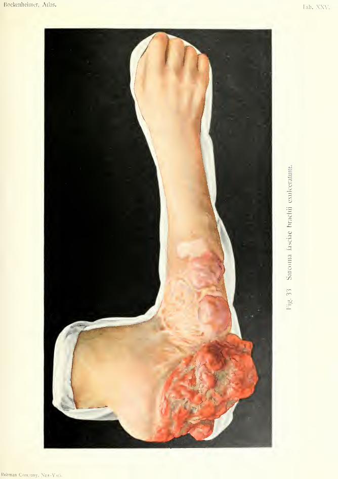
Bockenheimer, Atlas.Tab. XXV.
'X)
CO
U
Rebman Com.iany, New-York.


Ulceration of the tumor is followed by regionalglandular metastases, organic metastases, fever andsevere anaemia.
Differential Diagnosis. These rapidly growingmalignant tumors are so typical in their situation anddevelopment that it is only on the scalp that they canbe mistaken for ulcerating carcinoma. Sarcomas ofthe scalp often have hard borders with deep fissures
as in carcinoma, and also give rise to early glandularenlargement.
Treatment. Small, slow-growing sarcomas canbe removed by free excision, but local recurrence is
frequent. In extensive, and especially in ulcerated,
tumors of the extremities amputation is indicated.
Tumors which arise in the abdominal fascia often
become inoperable owing to extension to the peri-
toneal cavity.
Fig. 33 shows a rapidly growing, recurrent, ulcer-
ated sarcoma of the fascia of the arm. The youngernodules are covered by livid skin, which is intact in
some parts and thin in others. In other parts there
are white cicatrices left by former operations. TheX-rays showed that the sarcoma had extended to the
bone. Owing to the growth having broken into the
elbow joint, this was fixed in the rectangular position.
There were some small, soft, enlarged glands in the
axilla. Amputation through the arm was performed,with removal of the axillary glands.
61
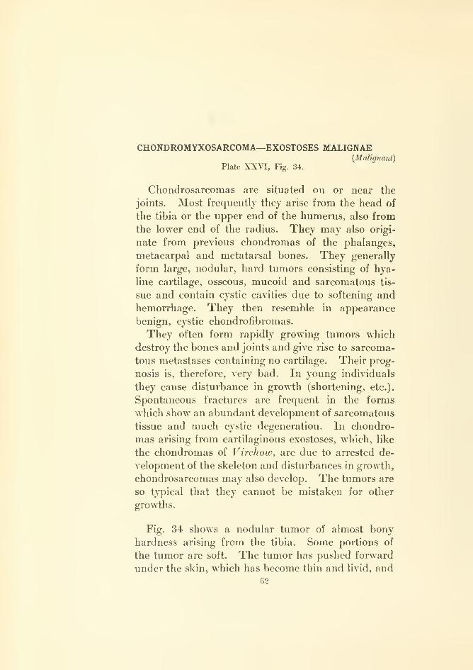
CHONDROMYXOSARCOMA—EXOSTOSES MALIGNAE{Malignant)
Plate XX^^, Fig. 34.
Chondrosarcomas are situated on or near the
joints. Most frequently they arise from the head of
the tibia or the upper end of the humerus, also from
the lower end of the radius. They may also origi-
nate from previous chondromas of the phalanges,
metacarpal and metatarsal bones. They generally
form large, nodular, hard tumors consisting of hya-
line cartilage, osseous, mucoid and sarcomatous tis-
sue and contain cystic cavities due to softening andhemorrhage. They then resemble in appearance
benign, cystic chondrofibromas.
They often form rapidly growing tumors which
destroy the bones and joints and give rise to sarcoma-
tous metastases containing no cartilage. Their prog-
nosis is, therefore, very bad. In young individuals
they cause disturbance in growth (shortening, etc.).
Spontaneous fractures are frequent in the forms
which show an abundant development of sarcomatous
tissue and much cystic degeneration. In chondro-
mas arising from cartilaginous exostoses, which, like
the chondromas of Virchoiv, are due to arrested de-
velopment of the skeleton and disturbances in growth,
chondrosarcomas may also develop. The tumors are
so typical that they cannot be mistaken for otlier
growths.
Fig. 34 shows a nodular tumor of almost bony
hardness arising from the tibia. Some portions of
the tumor are soft. The tumor has pushed forward
under the skin, which has become thin and livid, and62

Bockenheimer, Atlas. Tab. XXVI.
Fig. 34. Cliondroniy.xusarcoma — Exostoses malignac.


is broken through in some places through which thetumor is beginning to discharge. The movements ofthe knee joint are very hmited. No glandular ororganic metastases were found.
Treatment. Removal of the sarcomatous exos-toses, and resection of the joint, if necessary. Inlarge tumors, amputation and disarticulation. Pro-phylactic treatment consists in the removal of rapidlygrowing exostoses and chondromas.
63
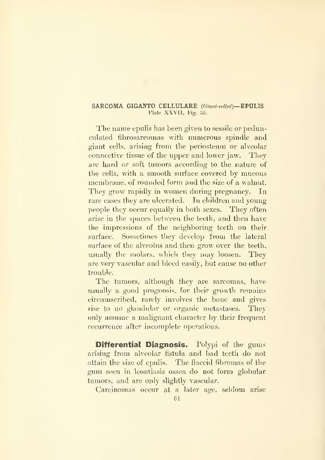
SARCOMA GIGANTO CELLULARE (Gtant-celM)—EPULISPlate XXVII, Fig. 35.
The name epulis has been given to sessile or pedun-
culated fibrosarcomas with numerous spindle andgiant cells, arising from the periosteum or alveolar
connective tissue of the upper and lower jaw. Theyare hard or soft tumors according to the nature of
the cells, with a smooth surface covered by mucousmembrane, of rounded form and the size of a walnut.
They grow rapidly in women during pregnancy. In
rare cases they are ulcerated. In children and young
people they occur equally in both sexes. They often
arise in the spaces between the teeth, and then have
the impressions of the neighboring teeth on their
surface. Sometimes they develop from the lateral
surface of the alveolus and then grow over the teeth,
usually the molars, which they may loosen. Theyare very vascular and bleed easily, but cause no other
trouble.
The tumors, although they are sarcomas, have
usually a good prognosis, for their growth remains
circumscribed, rarely involves the bone and gives
rise to no glandular or organic metastases. Theyonly assume a malignant character by their frequent
recurrence after incomplete operations.
Differential Diagnosis. Polypi of the gumsarising from alveolar fistula and bad teeth do not
attain the size of epulis. The flaccid fibromas of the
gum seen in leontiasis ossea do not form globular
tumors, and are only slightly vascular.
Carcinomas occur at a later age, seldom arise
64
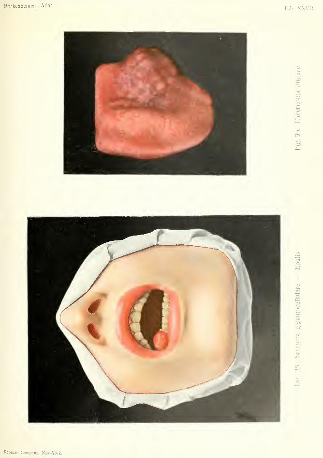
Bockenheimer, Atlas.Tab. XXVII
So
O
>UoCO
LT,
in
ti
Rebraan Company, New-York.

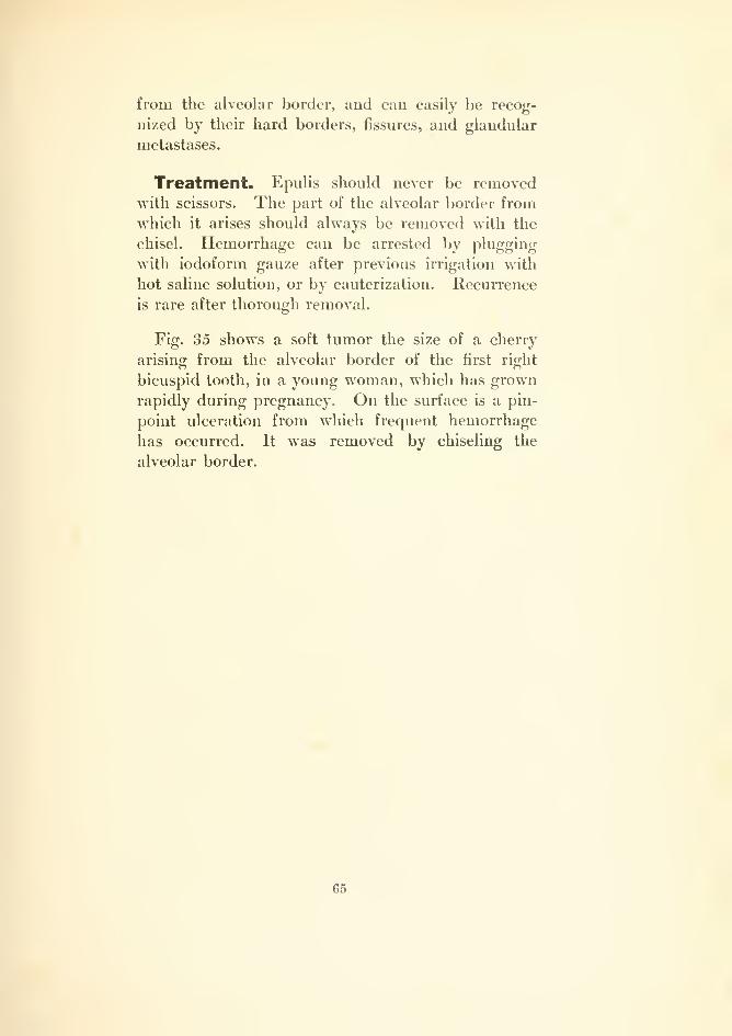
from the alveolar border, and can easily be rccotr-
nized by their hard borders, fissures, and glandular
metastases.
Treatment. Epulis should never be removed
with scissors. The part of the alveolar border from
which it arises should always be removed with the
chisel. Hemorrhage can be arrested by plugging
with iodoform gauze after previous irrigation with
hot saline solution, or by cauterization. Recurrence
is rare after thorough removal.
Fig. 35 shows a soft tumor the size of a cherry
arising from the alveolar border of the first right
bicuspid tooth, in a young woman, which has grown
rapidly during pregnancy. On the surface is a pin-
point ulceration from which frequent hemorrhage
has occurred. It was removed by chiseling the
alveolar border.
65
HEMANGIOMA CAVERNOSUM LINGUAE{Cavernous Hemangioma of Tongue)
Plate XXVII, Fig. 36.
Hemangioma cavernosum (cavernoma linguse
generally develops from a previous congenital hem-
angioma simplex, a slightly raised red spot which
often remains unnoticed. It may also occur as a
congenital tumor which becomes fully developed in
adolescence or sometimes later, and extends more
deeply than simple hemangioma into the mucous
membrane and sub-mucous tissue. The tumor con-
sists of new blood-vessels, especially capillaries, and
cavities lined by endothelium and filled with blood.
The cavernoma presents itself as a tumor with sev-
eral small nodular projections on its surface, which
have a bluish, glistening appearance. The mucous
membrane in the region of the tumor is so thin that
a dark fluid mass appears to be seen through it.
Apart from this characteristic appearance, the soft-
ness of the tumor, and the fact that it can be emptied
by pressure and made tense by bending the head
are worthy of notice. It thus consists of cavernous
tissue, such as is found normally in the corpora
cavernosa penis, and on this account the nameerectile tumor has been applied to it. Besides the
superficial growth there is also a deeper growth into
the mucous membrane, so that the tumor may in-
volve the whole tongue, the floor of the mouth, the
soft palate, the lips and the cheeks. Eventually the
tumor may involve the whole side of the face
and extend through the orbit to the brain. In
other cases the tumors are encapsuled. Sometimesthere are multiple encapsuled cavernomas lying
66
close together, but without any direct connection.
Tumors which, arising from the buccal mucous mem-brane, appear under the skin of the face, give rise
to thinning and a bluish glistening coloration of the
skin. Apart from the deformity large cavcrnomas
are dangerous, as they may rupture and give rise to
profuse and sometimes fatal hemorrhage, as often
occurs in cavernomas of internal organs (alimentary
canal and liver). Sometimes ulceration occurs at
the points of rupture, which may cause general sep-
tic infection, and in the tongue acute glossitis andoedema of the glottis.
fe'
Differential Diagnosis. Cavernous lymphan-
giomas are composed of larger protuberances andhave a greenish surface. Moreover, lymphangioma,
though diminished by pressure, remains independent
of the circulation and is not increased by pressure,
stooping or coughing. As the result of inflammatory
changes, hard nodules form in these tumors, which
are disseminated in the soft parts. Sarcomas are
rare and can generally be recognized by their smooth
surface and rapid growth. Retention cysts of the
mucous membrane of the tongue are smaller, circum-
scribed, and have a uniform surface. On the other
hand, they are also covered by thin, bluish, glistening
mucous membrane.
Treatment. Simple hemangiomas of the mu-cous membrane should be removed by caustics or
cauterization. Cavernous hemangiomas can be ex-
tirpated if they are encapsuled. Injection of per-
chloride of iron renders the boundaries of the tumor
visible and prevents hemorrhage, but is dangerous on
account of possible embolism.
Diffuse cavernous angiomas are best incised and
scraped with the sharp spoon (i'. Bergmann). Large
vessels can be ligatured and the bleeding surface cau-
terized, treated with hot saline solution or tamponed

with iodoform gauze or sterile sponges. The opera-
tion must be repeated if recurrence takes place.
Inoperable tumors are best treated with injections
of alcohol, or with Payr's magnesium. Both meth-
ods aim at thrombosis, after which shrinking of the
tumor takes place. Injections must be made deeply
under the mucous membrane to avoid necrosis.
Fig. 36 shows an encapsuled hemangioma arising
from a simple cavernoma after puberty, with the
characteristic changes described above. The tumorwas treated by incision and scraping.
68


Bockenheiiiier, Atlas.Tab. XXVI II.
-a
M
1?^\^^'>„ r««,.%...... VBiv^Vorl-
FIBRO-ADENOMA MAMMAE CYSTICUM(Cystic Fibro-adenoma of Breast)
Plate XXVIII, Fig. 37.
Adenomas, distinguished as true tumors (from
hyperplasias) by the irregular arrangement of the
newly formed glands, are rare in the breast, like pure
fibromas. Of the benign tumors of the breast only
fibro-adenomas come into consideration, as other tu-
mors are very rare (myxoma, angioma, chondroma,
and mixed tumors).
Fibro-adenomas usually develop in the peripheral
portions of the mammary gland in young women, in
the form of slow-growing, nodular tumors, which are
so well encapsuled that they are freely movable
within the breast. They are rarely multiple and sel-
dom affect both breasts. When there is an abun-
dant development of connective tissue the tumors are
firm; when cystic cavities develop they are soft and
fluctuating (fibroadenoma cysticum).
The tumor described as cystadenoma papilli-
ferum, fibroma intracanaliculare, and incorrectly as
sarcoma phyllodes, which is formed by connective
tissue processes covered by epithelium projecting into
the cavity of the cyst, belongs to the group of benign
mammary tumors. In older women, especially at
the menopause, small multiple cystadenomas occur,
chiefly in the region of the nipple, without causing
retraction; sometimes in both breasts. These feel
like solid tumors owing to their thickened walls. The
name of chronic cystic interstitial mastitis has been
given to these tumors by Konig.
The benign nature of these tumors is shown by the
fact that they cause neither glandular nor organic
69
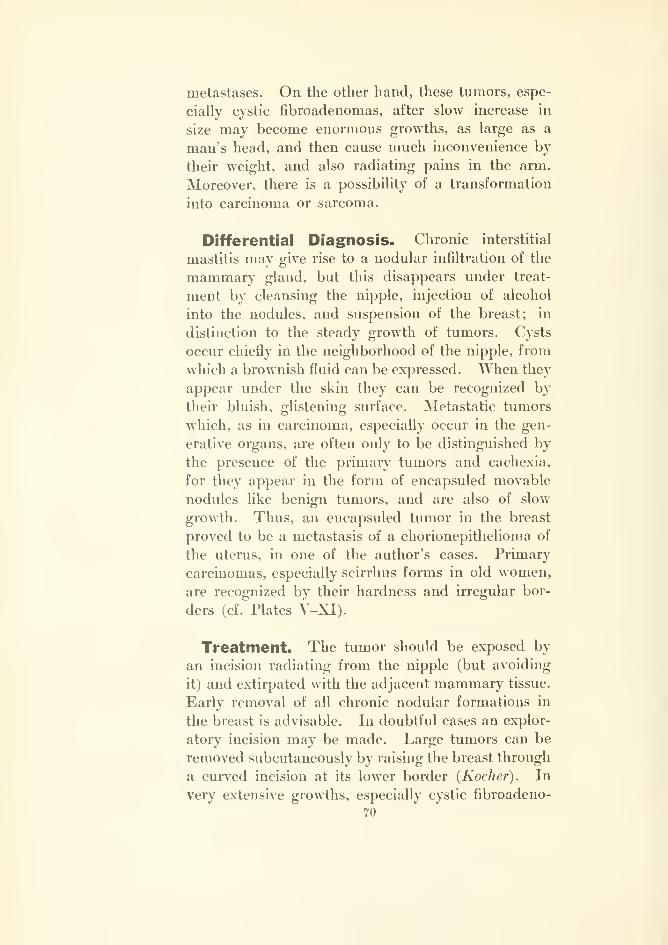
metastases. On the other hand, these tumors, espe-
cially cystic fibroadenomas, after slow increase in
size may become enormous growths, as large as a
man's head, and then cause much inconvenience by
their weight, and also radiating pains in the arm.
Moreover, there is a possibility of a transformation
into carcinoma or sarcoma.
Differential Diagnosis. Chronic interstitial
mastitis may give rise to a nodular infiltration of the
mammary gland, but this disappears under treat-
ment by cleansing the nipple, injection of alcohol
into the nodules, and suspension of the breast; in
distinction to the steady growth of tumors. Cysts
occur chiefly in the neighborhood of the nipple, from
which a brownish fluid can be expressed. When they
appear under the skin they can be recognized by
their bluish, glistening surface. Metastatic tumors
which, as in carcinoma, especially occur in the gen-
erative organs, are often only to be distinguished by
the presence of the primary tumors and cachexia,
for they appear in the form of encapsuled movable
nodules like benign tumors, and are also of slow
growth. Thus, an encapsuled tumor in the breast
proved to be a metastasis of a chorionepithelioma of
the uterus, in one of the author's cases. Primary
carcinomas, especially scirrhus forms in old women,
are recognized by their hardness and irregular bor-
ders (cf. Plates V-XI).
Treatment. The tumor should be exposed by
an incision radiating from the nipple (but avoiding
it) and extirpated with the adjacent mammary tissue.
Early removal of all chronic nodular formations in
the breast is advisable. In doubtful cases an explor-
atory incision may be made. Large tumors can be
removed subcutaneously by raising the breast through
a curved incision at its lower border {Kocher). In
very extensive growths, especially cystic fibroadeno-
70
mas and multiple cystic formations, the whole breastshould be removed.
Fig. 37 shows the right breast of a woman (at themenopause) much more projecting than the left.
The upper half of the right breast is involved in atumor, the irregular surface of which can be recoo--
nized by the bulging of the skin. The skin is thfnand reddened. The tumor, which was at first re-
mote from the nipple in the inner and upper quad-rant of the breast, has grown towards the nipplewithout causing retraction. The tumor is com-pletely encapsuled, freely movable, and of moderatelyhard consistence. It was removed through a radial
incision, together with the adjacent mammary tissue.
71

CORNU CUTANEUM (Cutaneous Horn)
ADENOMA SEBACEA (Sebaceous)
Plate XXIX, Fig. 38.
Cutaneous horns occur more frequently in old peo-
ple (senile keratoma), and in those subject to expo-
sure (sailors, etc.). They arise on the basis of
sebaceous and dermoid cysts and warts, and occur
on the eyelids, nose, lips, cheeks and ears, also on the
scalp and genital organs. They are seldom multiple.
They generally form sessile, freely movable, curved
or spiral structures which have an irregular, grooved,
yellowish-brown surface and a horny consistence.
These benign formations, which may attain the
length of several centimeters, are formed by a pro-
liferation of the horny layer of the epidermis. Thepapillae are also lengthened, which accounts for the
soft consistence of the interior.
Differential Diagnosis. In young people mul-
tiple nsevi with cornification occur, but these have a
wider base, and a flatter and more prickly surface,
Treatment. As about 10 per cent, of cutaneous
horns develop into carcinoma, excision by the knife
into the healthy skin is indicated. Recurrence takes
place after removal by ligature.
Fig. 38 shows a slightly curved cutaneous horn
about one and one-half centimeters long, occurring
in an old countrywoman, in the zygomatic region,
with all the characteristic features. The skin at the
base of the growth is scaly and somewhat reddened.
Adenoma of the skin is another form of growth
often occurring in women, both young and old.

Bockenheimer, AtlasTab. XXIX.
o
lU
o/)
o
CO
t£
Rebman Company, New-York.

usually on the face. Adenomas are benign tumorswhich develop from normal glandular tissue, andmay, therefore, occur in all glands. Adenomas whichdevelop in places where glands are normally absentmust be assumed to develop from congenital rudi-
ments of supernumerary glands.
In the skin, adenomas often develop from the se-
baceous glands (adenoma sebaceum). They oftenoccur in many places as small, round, flat, circum-scribed, encapsuled, movable tumors, of firm con-sistence, and with a dirty gray surface. The lymphaticglands are never affected, and there is no recurrenceafter extirpation. Adenomas which have becometransformed into carcinomas have been incorrectly
termed malignant adenomas.
Differential Diagnosis. Intercurrent cystic
formation may cause confusion with endothelioma of
the skin, and ulceration with carcinoma. The oc-
currence of calcification in the adenoma may makeit as hard as carcinoma. Doubtful cases must besettled by microscopic examination.
Treatment. Small multiple adenomas can betreated by cauterization or X-rays. Larger onesshould be extirpated.
Adenomas arising from the sweat glands (adenomasudiporum) generally form larger, more nodular tu-
mors, which after ulceration simulate carcinomas.The treatment consists in excision.
Fig. 38 shows multiple pin-point adenomas of the
sebaceous glands, which disappeared to a great
extent under treatment by X-rays. Characteristic
smegma-like matter can be expressed from larger
adenomas.
73
ENDOTHELIOMA CUTIS (0/ Skin)
Plate XXIX, Fig. 39.
Endotheliomas (Golgi) arise from the endothelium
of the blood-vessels and lymphatics, which, accord-
ing to Borst consists of specially modified connective-
tissue cells. Owing to the double nature of the en-
dothelium, it is not surprising that those who regard
the endothelial cells as epithelial cells give the nameof endothelial cancer or connective-tissue cancer to the
tumors arising from it, while others, who regard the
endothelial cells as connective-tissue cells, call these
tumors endothelial sarcomas, plexiform angiosarco-
mas {Waldeyer) and angiosarcoma {KoUaczek).
If we hold with Borst that the endotheliomas arise
from the endothelium, i.e. from the connective-tissue
cells, which may assume all kinds of modifications,
it follows that tumors of varied structure may arise
from these difterent varieties of endothelium, which
have the appearance of fibroma, sarcoma or carci-
noma, as the latter forms stratified globes, but with-
out cornification. By this means we avoid the end-
less number of names given to these tumors, and have
clinically only the term endothelioma, to be dis-
tinguished microscopically as hemangio-endothelio-
ma and lymphangio-endothelioma, which we can
designate as alveolar, plexiform or vascular, accord-
ing to their microscopic structure. Borst also in-
cludes the basal-cell cancers (regarded as carcinoma
by Krompecher and Conen) among the endotheliomas
as these tumors have no cornification (cf. Plate II,
Fig. 4).
It is no wonder that these tumors may appear
clinically in the most varied forms and be confounded74

with fibromas, adenomas, sarcomas and carcino-
mas.
The tumors may arise from all kinds of endothe-
lium and are most frequently observed in the skin
of the face, the mucous membrane of the mouth andpharynx, the bones of the face and skull, the perito-
neum, the pia mater of the brain and spinal cord,
and the parotid gland.
Occurring at any age, they form encapsuled, gen-
erally slow-growing, comparatively benign tumors
which seldom cause glandular or organic metastases,
but have a tendency to local recurrence.
As the shape, surface and consistence of the tu-
mors may assume all possible varieties, the clinical
signs of endotheliomas are very indefinite. Theshape is often irregular, especially in endothelioma
of the face (Fig. 39, horseshoe shape). The surface
may be smooth, iiTegular or ulcerated. The con-
sistence may be hai'd, soft or cystic. Sometimes the
tumors are very vascular and the epidermis assumes
the reddish-brow-n coloration which is seen in
sarcoma, at other times they are poor in vessels.
Although they are at first encapsuled they may later
on give rise to a diffuse infiltration of the tissue
along the endothelial clefts, and then have irregular
boundaries.
Differential Diagnosis. Sarcoma and carci-
noma are most often confounded with endothelioma,
also fibroma and adenoma, especially when they
undergo cystic degeneration or ulceration. Thediagnosis can often only be made by microscopic
examination.
Treatment. Early excision is indicated, as trans-
formation is possible in rapidly growing tumors. In
the diffuse forms, which represent malignant tumors
like carcinoma and sarcoma, extensive operations are
necessary. When multiple nodules develop in the
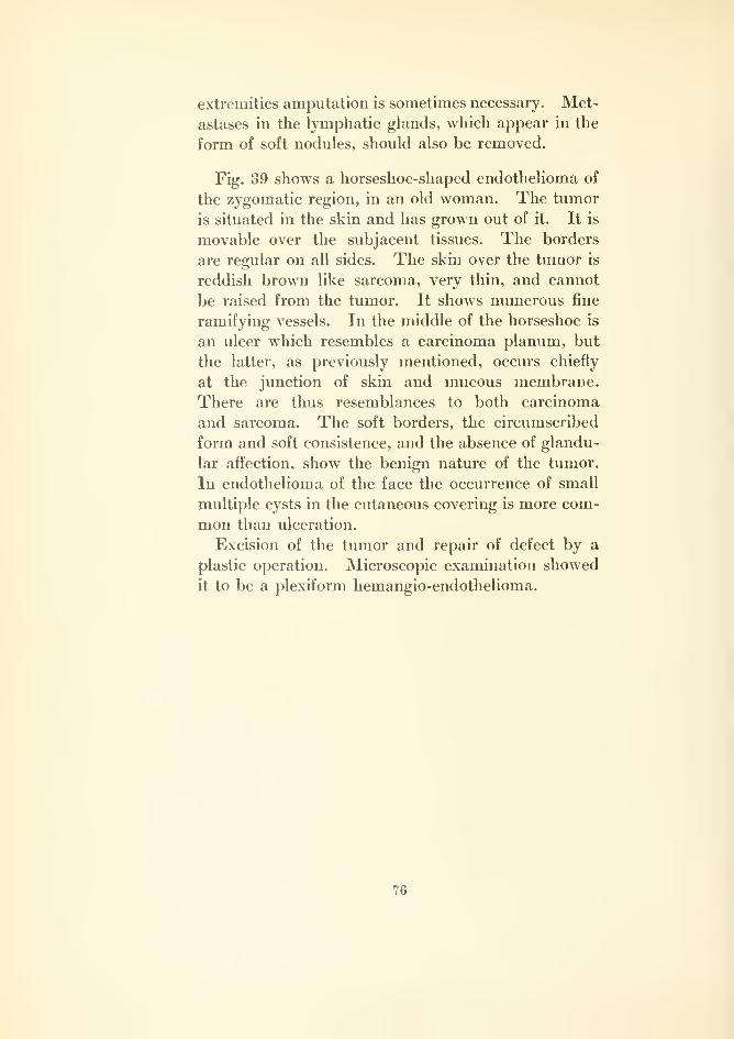
extremities amputation is sometimes necessary. Met-
astases in the lymphatic glands, which appear in the
form of soft nodules, should also be removed.
Fig. 39 shows a horseshoe-shaped endothelioma of
the zygomatic region, in an old woman. The tumor
is situated in the skin and has grown out of it. It is
movable over the subjacent tissues. The borders
are regular on all sides. The skin over the tumor is
reddish brown like sarcoma, very thin, and cannot
be raised from the tumor. It shows numerous fine
ramifying vessels. In the middle of the horseshoe is
an ulcer which resembles a carcinoma planum, but
the latter, as previously mentioned, occurs chiefly
at the junction of skin and mucous membrane.
There are thus resemblances to both carcinoma
and sarcoma. The soft borders, the circumscribed
form and soft consistence, and the absence of glandu-
lar affection, show the benign nature of the tumor.
In endothelioma of the face the occurrence of small
multiple cysts in the cutaneous covering is more com-
mon than ulceration.
Excision of the tumor and repair of defect by a
plastic operation. Microscopic examination showed
it to be a plexiform hemangio-endothelioma.
76

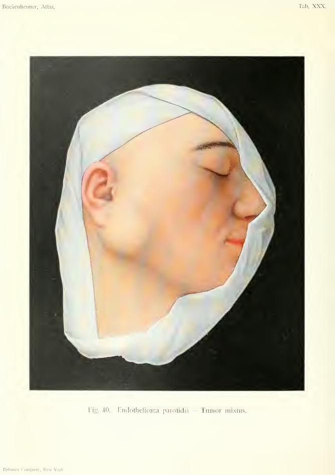
Bockenheimer, Atlas. Tab. XXX.
Fig. 40. Endothelioma parotidis — Tumor mixtus.
Rebnian Company, New-York.
ENDOTHELIOMA PAROTIDIS (of Parotid)
TUMOR MEXTUS (Mixed Tumor)Plate XXX, Fig. 40.
Mixed tumors occur frequently in the parotid,
less often in the other salivary glands. These par-
otid tumors are regarded as endotheliomas by Kanf-
mann, Nasse and Volkmann, which is intelligible
after the explanation of endothelioma given in Plate
XXIX, Fig. 39, if we assume that the epithelioid
tracts occurring in the tumors arise from endothelium,
the latter, according to Volkmann, being also capable
of forming cartilaginous, mucoid and connective
tissue. Others hold that these mixed tumors, which
also occur in the breast, kidneys and testicles, arise
from epithelial and connective-tissue cells (Wilms
and Hinsberg).
On section, the tumors show a very variegated
structure, in which are found parts resembling car-
cinoma and sarcoma, mucoid tissue, cartilage, cysts,
calcification and ossification.
Parotid tumor occurs more often in young indi-
viduals, and appears as an encapsuled, smooth or
nodular tumor, movable over subjacent parts, lying
under the fascia, and covered by intact non-adherent
skin. The rare tumors which lie above the parotid
fascia originate in aberrant parotid rudiments, ac-
cording to Bergmann. The consistence of parotid
tumors may be hard, soft or cystic, according to their
composition, and may differ in different parts of the
same tumor. At first they are of slow growth, but
may suddenly take on rapid growth, rupture their
capsule, infiltrate the surrounding parts like malig-
nant tumors, and finally perforate the skin and
77
ulcerate. In such cases there are glandular and
organic metastases.
Tumors arising from the anterior part of the
parotid cause swelling of the cheek; those arising
from the posterior part of the gland raise up the
external ear. Larger tumors may extend towards
the chin, the nape of the neck and the clavicle.
Small tumors cause hardly any pain, but some-
times salivation. Extensive tumors may give rise to
pain in the ear, deafness and facial paralysis.
Differential Diagnosis. The more common car-
tilaginous tumors with uneven surface are easy to dis-
tinguish from other growths, but the soft tumors with
smooth surface may be confounded with lymphomas,
cavernomas, lipomas and cysts. Extensive endothe-
liomas are often indistinguishable from sarcomas or
carcinomas.
Mixed tumors should be extirpated as early as
possible, on account of the possibility of their taking
on malignant growth. Both benign and malignant
recurrence may take place from the remains of the
capsule after removal of tumor. The capsule must,
therefore, be completely removed during extirpation,
taking care to avoid large branches of the facial
nerve, while the part of the gland which is unaffected
can be left behind. In extensive malignant endothe-
liomas of the parotid it is hardly possible to save the
facial nerve, for in these cases the whole gland must
be removed. In tumors of the submaxillary gland
the whole gland should always be removed.
Fig. 40 shows a mixed tumor of the parotid which
slowly developed during three years in a womanaged thirty. Profuse salivation, and latterly rapid
growth of the tumor, led the patient to seek advice.
The skin is freely movable over the tumor and shows
a fine network of vessels. The tumor lies under the
fascia and has spread to the anterior and lower
78

region of the ear. The surface of the tumor is irregu-
lar; the consistence of the posterior portion, where
the surface is uneven, is hard; soft and fluctuating in
the anterior portion, where the surface is smooth.
There is no projection of the tumor into the buccal
cavity. The tumor is freely movable over the sub-
jacent parts, and there is no glandular enlargement.
The tumor was extirpated with its capsule, and
the facial nerve avoided. Part of the parotid gland
was left behind. On section, cartilage, cysts, calcifi-
cation, and fibrous and sarcomatous tissue were
found.
79

GANGLION CARPALE {of Wrist)
Plate XXXI, Fig. 41.
Ganglions occur especially in connection with the
joints of the hand, most often on the dorsal surface
between the extensor carpi radialis and extensor
indicis, less commonly on the palmar side near the
flexor carpi radialis (especially in pianists); also on
the dorsum of the foot at the joints of the cuboid
bone and in the neighborhood of the knee joint.
Colloid degeneration of the joint capsule and the
periarticular connective tissue gives rise first to mul-
tilocular, then unilocular cystomas, which were for-
merly regarded as retention cysts. Ganglions of the
tendon sheaths arise in a similar manner, but are
smaller; they occur chiefly in the sheaths of the
flexor tendons over the metacarpo-phalangeal joints,
and cause neuralgic pain by pressure on the digital
nerves. They often occur after rowing and fencing,
i.e. from traumatic causes.
Spherical ganglia occur most commonly on the
dorsal aspect of the hand in young women, and re-
semble exostoses on account of their hardness.
They often cause neuralgic pains and slight trouble
in the movements of the joints.
Ganglions are of slow growth, the skin is unaltered
and movable over them; the surface is smooth or
slightly wrinkled. The consistence is hard in small
ganglions, soft and fluctuating in larger ones. In
pedunculated ganglions there is slight mobility over
the joint.
Differential Diagnosis. In the knee joint they
may be mistaken for affections of burste; in the
80
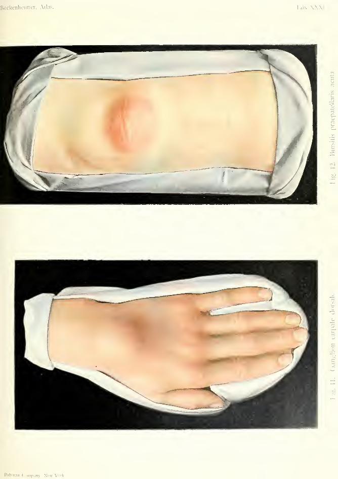
Bockenheinier, Atla?. Tab. XXXI.
r,
Rfbnian Company, New-York.

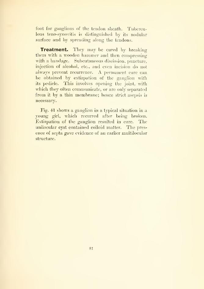
foot for ganglions of the tendon sheath. Tubercu-lous teno-synovitis is distinguished by its nodular
surface and by spreading along the tendons.
Treatment. They may be cured by breaking
them with a wooden hammer and then compressing
with a bandage. Subcutaneous discission, puncture,
injection of alcohol, etc., and even incision do not
always prevent recurrence. A permanent cure can
be obtained by extirpation of the ganglion with
its pedicle. This involves opening the joint, with
which they often communicate, or are only separated
from it by a thin membrane; hence strict asepsis is
necessary.
Fig. 41 shows a ganglion in a typical situation in a
young girl, which recurred after being broken.
Extirpation of the ganglion resulted in cure. Theunilocular cyst contained colloid matter. The pres-
ence of septa gave evidence of an earlier multilocular
structure.
81
BURSITIS PRAEPATELLARIS ACUTA(Acute prepatellar Bursitis)
Plate XXXI, Fig. 42.
Affections of the bursse may be divided into acute
and chronic inflammations, and further into purulent
and non-purulent (serous, fibrinous, hemorrhagic).
An acute bursitis occurs especially after injuries
and inflammation extending from neighboring regions
(furunculosis, arthritis). It may also arise from for-
eign bodies inside the bursa.
In acute serous bursitis the skin is unchanged,
while in purulent bursitis it is red and oedematous.
In the latter, suppuration often extends beyond the
limits of the bursa and is accompanied by fever,
pain and difiiculty in movement. Under the mova-
ble skin, in the case of superficial burste {e.g. the
prepatellar), a hemispherical, tense, sometimes fluc-
tuating, slightly movable swelling with a smooth
surface can be felt, limited to the anatomical position
of the bursa (Fig. 42).
Chronic bursitis, also called hygroma, occurs more
after chronic irritation, in the prepatellar bursa, in
housemaids, for instance (housemaid's knee), and
in the olecranon bursa in miners (miner's elbow).
Villous proliferations in the wall of the bursa lead
to thickening, and to the formation of rice bodies.
The skin over the bursa is movable and thickened.
The hygroma is almost spherical, with a rough,
uneven surface.
Hygroma may also develop in adventitious bursse,
especially in places where a bone is subjected to
pressure, for instance, on the toe over a clavus.
Hygromas give rise to little inconvenience, and only
82

hinder movement when of large size. In the case of
the elbow there is sometimes neuralgic pain from
pressure on the ulnar nerve.
Differential Diagnosis. The different forms of
bursitis may be mistaken for arthritis of the adjacent
joint, owing to limitation of movement, e.g. sub-
deltoid and sub-trochanteric bursitis. The strict
localization of the affection to the anatomical position
of the bursse should make the diagnosis easy. Dis-
ease of several bursas is chiefly obser\'ed in tuber-
culosis, syphilis, gonorrhea and gout.
Treatment. Acute purulent bursitis requires
early incision and plugging, as infection of the joint
may take place. In acute serous or hemorrhagic
effusion, puncture and injection of 4 per cent, car-
bolic lotion, 1 per cent, iodoform-glycerin or abso-
lute alcohol may be tried. In chronic bursitis, paint-
ing with iodine is generally useless. It is best to
extirpate chronic hygromas, especially when they are
large or have thick, hard walls, or when fistulte
develop, taking care to avoid the joints.
Fig. 42 shows an acute purulent prepatellar bur-
sitis. The skin is red and hot and the movements of
the knee joint are painful and limited. The tense,
fluctuating, spheroidal swelling is clearly situated in
front of the patella. The surface is smooth and reg-
ular, but the tumor is almost immovable over the
subjacent structures. It was shown by incision that
all three bursa?—subcutaneous, subfascial and sub-
aponeurotic—were full of pus and in communication
with each other.
83
HYGROMA GENUS MULTILOCULARE(Multilocular Hygroma of the Knee)
BURSITIS PRAEPATELLARIS ET BURSITIS PRAETIBIALIS{Prepatellar and Pretibial Bursitis)
Plate XXXII, Fig. 43.
This figure shows a case of chronic inflammation
of the prepatellar bursa and the lower half of the
pretibial bursa, occurring in a man who had to do
his work in the kneeling position. The skin over
the prepatellar bursa is thickened and movable over
the cystic swelling. The walls of both hygromas are
thickened. They are only slightly movable over the
subjacent structures. Pressure on one hygroma
causes some of its fluid to pass into the other, so
that the two bursse communicate. Total extirpation
was performed owing to the extent of the hygromaand the thickened walls.
84
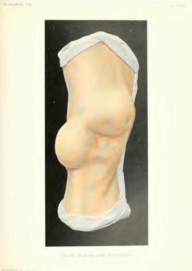
Bockenheinier, AtlasTab. XXXII.
Fig. 43. Hygroma genus miiltilociilaiT.
Rebm;in Coniti.Tiiv Mpvp-Vcirlt.


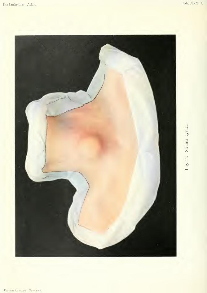
Bockenheimer, Atlas. Tab. XXXIII.
u^
Rebman Company, New-York.
STRUMA CYSTICA (Brmchocek)Plate XXXIII, Fig. 44.
Goitre occurs endemically (Switzerland and otherregions) and epidemically in barracks and boardinghouses (Strumitis acuta first observed by Kussmaul).Heredity, frequent congestion of the blood-vessels ofthe head, pregnancy, the nature of the soil, water andatmosphere have all been suggested as causes ofbronchocele.
Bronchoceles occur twice as often in females asin males (Schrotter). Clinically, they are dividedinto diffuse and circumscribed forms, and patho-logically into follicular, colloid, vascular and cystic
bronchoceles.
In all cases the typical situation corresponds tothe anatomical position of the thyroid gland, andthe symptoms are definite. Even small bronchocelescause marked and early deformity. Further exten-sion results in pressure on the veins, causing promi-nence of the cutaneous veins and a cyanotic appear-ance of the face. Pressure on the trachea may giverise to displacement, stricture and changes in its
walls, causing it to assume the form of a saber-sheath. This dangerous condition can be seen bythe X-rays. Eventually the wall of the trachea maybecome so much destroyed as to give way after
violent movement of the head. Difficulty in breath-ing during inspiration, causing stridor, is the neces-
sary result, and sudden asthmatic attacks, occurringduring violent movements of the patient or duringsleep may prove fatal. Pressure on one recurrent
nerve is of little consequence and often unnoticed, for
unilateral paralysis of the recurrent is compensated,85

so that hoarseness is often absent, and the condition
is only shown by laryngoscopic examination. Bilat-
eral paralysis of the reeurrents is, however, very
dangerous, as it may give rise to asphyxia or pneu-
monia. Small fibrous tumors arising in the middle
line from the isthmus of the thyroid, and those
lying behind the sternum cause more characteristic
symptoms than large, soft tumors, which often cause
little trouble.
Every bronchocele moves with the thjToid on swal-
lowing and is thus distinguished from other affections.
The simplest form of bronchocele, which consists
in a hypersemia of the whole organ, is common in
young girls at the onset of menstruation, or at the
first sexual intercourse, and appears as a soft, uni-
form swelling of the whole gland, which may disap-
pear spontaneously.
According to v. Eiselsberg, this simple form mayoften give rise to follicular hypertrophy. The latter
also occurs in young individuals in the form of hard
nodules in the gland, which may also disappear.
More marked enlargement of the thyroid gland,
developing gradually in middle age, and leading to
the formation of a horseshoe-shaped tumor involving
the whole gland and consisting of a number of large
nodules, is diagnostic of colloid bronchocele, while
the vascular bronchocele is characterized by pulsa-
tion and compressibility. The cystic bronchocele
(Fig. 43), arising from several colloid nodules owing
to hemorrhage and liquefaction, forms small, hemi-
spherical tumors with a smooth surface and distinct
fluctuation. When the cyst walls are hardened bycalcification the diagnosis is more difficult, but
differs from the irregular, nodular formation of
fibrous bronchocele. Cystic bronchoceles may attain
the size of a man's head.
The different varieties—colloid, cystic, vascular
and fibrous—may all occur in the same tumor.
The diagnosis of the different kinds of bronchocele
86
is important with regard to treatment, which shouldbe begun early, as cardiac symptoms occur iu con-nection with long-standing large bronchoceles.
Differential Diagnosis. An accessory broncho-cele is easily diagnosed when it is fixed to the thyroidby a pedicle, as it then gives rise to the same symp-toms. Bronchoceles which arise from free accessoryglands may be mistaken for other tumors of the neck—lymphoma, sebaceous cyst, dermoid or malio--
nant tumor.
Carcinoma of the thyroid gland occurs in oldpeople and forms a nodular, very hard, rapidlygrowing tumor, which soon surrounds the whole neckwith a hard ring. The diagnosis is settled by theglandular metastases, the early appearance of paraly-sis of the vocal cords and cachexia. In old people,the sudden occurrence of rapid growth in an old-
standing bronchocele always suggests malignanttransformation.
Sarcomas, which occur in young people as rapidlygrowing tumors, are distinguished by their soft
consistence and by their diffuse infiltration. Theyoften break through the capsule and give rise to
severe hemorrhage.
Basedow's disease (Grave's disease, exophthalmicgoitre), which, according to Mobiiis, consists iu
hypersecretion of the thyroid gland resulting in intox-
ication of the organism, is distinguished from ordi-
nary bronchocele by the presence of tachycardia,tremor, exophthalmus and neuropathic conditions.
The swelling in Basedow's disease is always veryvascular and often pulsates. In long-standing bron-choceles symptoms of Basedow's disease may appear,but they are never so marked as in the genuine form;all the other characteristic symptoms of bronchoceleare also present.
Mediastinal tumors and aneurisms are occasion-
ally mistaken for retrosternal bronchocele.
Treatment. The treatment varies according to
the nature of the bi'onchocele.
In countries where goitre is epidemic, prophylaxis
plays the chief role. Water should only be drunk
after boiling. Violent exertion should be avoided,
on account of causing a determination of blood to
the head.
In acute hyperaemia and follicular hypertrophy
iodine preparations are most useful — iodide of
potassium, or thyroid tabloids containing iodine
(to be given carefully on account of tachycardia).
Iodine preparations should not be continued too
long.
In cystic and colloid bronchoceles iodine treatment
gives no results, and operation is indicated—partial
extirpation in the case of colloid bronchocele. Suffi-
cient thjToid gland tissue must be left otherwise
tetania strumipriva or m;yxoedema may follow.
Cretinism, which is only observed in countries
where goitre is endemic, and causes changes in the
skin, disturbance in growth and idiocy, is also due
to degeneration of the greater part of the thyroid
eland or absence thereof in the cretins themselves or
in their parents.
In post-operative tetany, cachexia strumipriva,
mj^oedema and cretinism, implantation of a piece
of human thyroid gland in the diseased subject maybe attempted {Kohn, v. Eiselsherg, et al.). It is
best to transplant a large piece into the spleen
{Payr).
Isolated cysts and nodules can be enucleated.
Recurrence after operation is rare on the whole and
then usually causes no trouble.
In Basedow's disease ligation of the superior and
inferior th}Toid arteries has been successfully tried.
{Rehn, v. Bergmann). The operation is not without
danger, so that others have preferred internal treat-
ment with arsenic, or by the galvanic current, etc.,
often successfully.

Fig. 43 shows a tumor the size of a walnut, in anold woman, which is easily recognized as a cystic
bronchocele by its rounded form, regular outline,
situation in the isthmus of the thyroid and its move-ment during swallowing. The tumor was enucleatedon account of its causing considerable difficulty in
respiration.
89
PAPILLOMA CUTIS INFLAMMATORIUM{Inflammatory Papilloma of Skin)
Plate XXXIV, Fig. 45.
Papillomas or villous tumors, also occurring on
mucous membranes as villous polypi, belong to the
group of fibro-epithelial tumors (Borst). They con-
sist of vascular connective tissue and epithelial
proliferation (squamous moi-e often than cylindrical)
and simulate in structure the papillae of the skin and
mucous membrane. These growths represent a spe-
cial group of tumors, and must not be confounded
with papillomatous proliferations found in a similar
form in ntevi, carcinomas, sarcomas and endothelio-
mas. Condylomata acuminata are also very similar
to papilloma; however, these are not true tumors,
but are due to hyperplasia of the papillary body and
its epithelial covering. These generally occur as the
result of chronic inflammatory irritation from gon-
orrheal discharge, on the penis, vagina and anus.
True papillomas generally form small superficial
tumors of a warty or conical form, single or multiple,
occurring at any age, in places exposed to much irri-
tation (skin, genitals, thighs, back, tongue, rectum,
bladder and larynx). They are slow-growing, cir-
cumscribed, sessile or pedunculated, freely movable,
non-infiltrating growths. Papillomas of the skin are
yellowish-white dry growths, hard from cornification
of the superficial epithelial layers, and form conical
or wart-like projections.
Papillomas of the mucous membrane have a red-
dish fleshy appearance, and on account of their vas-
cularity, bleed easily and are of soft consistence. In
the larynx, they occur especially in the region of the
90
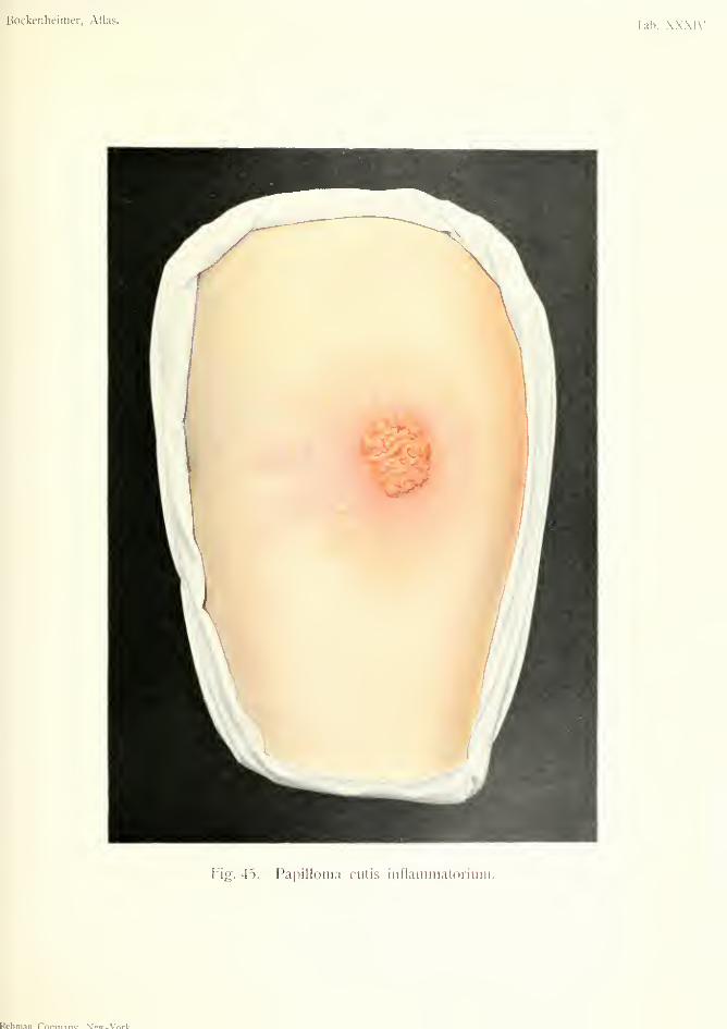
Bockenheimer, Atlas. Tah. XXXIV.
["ig. 45. Papilloma cutis inflammatorium.
Rchmnn Pnmnanv Ncvr.VnrV.

vocal cords; they are often multiple in young indi-
viduals, prone to recur, and may lead to stenosis.
Transition into carcinoma may occur, and is recog-
nized by rapid growth, ulceration, infiltration, andgrowth into the deeper parts.
Differential Diagnosis. Small papillomas of
the skin may be mistaken for common warts. In dis-
tinction to carcinoma they present the usual charac-
teristics of benign tumors—soft consistence, free
mobility, and no glandular metastases. Papillomasof mucous membranes are usually characteristic for-
mations. It is only in villous polypi of the bladder,
which may become transformed into villous cancer,
that the diagnosis is difficult.
Treatment. Excision. Extensive operations are
often necessary for the removal of papillomas of the
mucous membranes (tracheotomy, colostomy, etc.).
Fig. 45 shows a cutaneous papilloma, freely mova-ble over subjacent parts, of moderately soft consist-
ence, and covered with warty projections. Thehorny layer and the surface of the skin has beendestroyed by frequent cauterization. The surface is
covered with a yellowish fetid secretion, and betweenthe villous projections are deep depressions caused byulceration, so that the appearance in some places
resembles carcinoma; but the borders are not hard.
The skin round the tumor is red and painful from
cauterization.
After disinfection of the surface and arrest of the
discharge, the tumor was excised in healthy tissue
and the wound closed by suture.
91
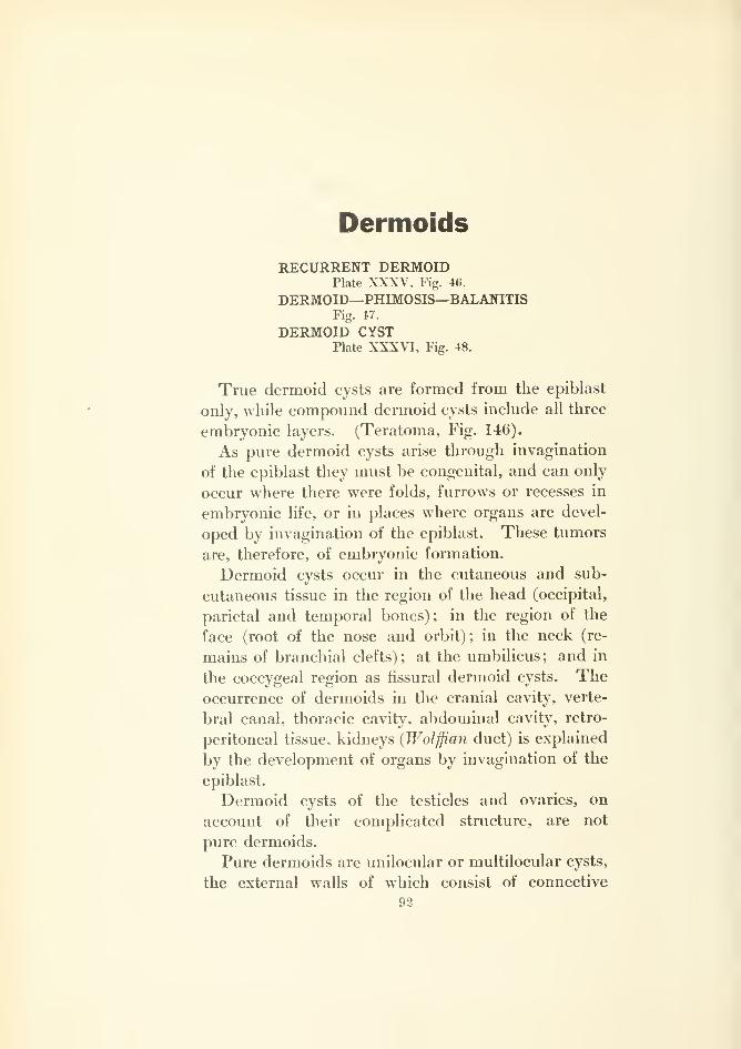
Dermoids
RECURRENT DERMOIDPlate XXXV, Fig. 46.
DERMOID—PHIMOSIS—BALAIHTISFig. 47.
DERMOID CYSTPlate XXXVI, Fig. 48.
True dermoid cysts are formed from the epiblast
only, while compound dermoid cysts include all three
embryonic layers. (Teratoma, Fig. 146).
As pure dermoid cysts arise through invagination
of the epiblast they must be congenital, and can only
occur where there were folds, furrows or recesses in
embryonic life, or in places where organs are devel-
oped by invagination of the epiblast. These tumors
are, therefore, of embryonic formation.
Dermoid cysts occur in the cutaneous and sub-
cutaneous tissue in the region of the head (occipital,
parietal and temporal bones); in the region of the
face (root of the nose and orbit); in the neck (re-
mains of branchial clefts) ; at the umbilicus ; and in
the coccygeal region as fissural dermoid cysts. Theoccurrence of dermoids in the cranial cavity, verte-
bral canal, thoracic cavity, abdominal cavity, retro-
peritoneal tissue, kidneys {Wolffian duct) is explained
by the development of organs by invagination of the
epiblast.
Dermoid cysts of the testicles and ovaries, on
account of their complicated structure, are not
pure dermoids.
Pure dermoids are unilocular or multilocular cysts,
the external walls of which consist of connective
92
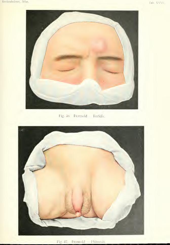
Bockenheimer, Atlas.Tab. X.WV,
Fig. 46. Dermoid Recidi
Fipr. 47. DcrniDiil ^'llilH()^i^.


tissue, and are connected with the surroundintr tis-
sues while the internal surface resembles skin
(hence the term dermoid), and presents papilla^,
squamous epithelium and hair. Those dermoids
which contain bone, cartilage and teeth are formed
at a very early embryonic period, before differentia-
tion has taken place.
The contents of the cyst consist of a yellowish-
white, caseous, odorless, fatty mass, mixed with
numerous hairs, the appearance of which varies
according to the situation of the dermoid (in the
region of the eye, eyelashes, etc.). The contents
are rarely serous or hemorrhagic. In the cutaneous
or subcutaneous tissue the cysts form spherical or
hemispherical tumors with a smooth surface and
tallowy consistence. They are covered by intact
skin, and are often attached to the bones. The super-
ficial dermoids usually occur in youth. They are
slow-growing and painless, and about the size of a
walnut. Sometimes fistulfe form from which hairs
protrude. The diagnosis of superficial dermoids is
easy to establish by the above signs.
Differential Diagnosis. Superficial dermoids
may be mistaken for sebaceous cysts, but the con-
tents of the latter are foul smelling and more dirty
yellow. If scars are present (e.g. after operations,
Fig. 46), the history or microscopic examination only
can decide whether it is a traumatic (post embryonic)
formation caused by proliferation of an involuted
part of the skin—the so-called epithelial cysts.
These may also form round a foreign body. Epider-
moids can often only be distinguished from dermoids
microscopically, the former being lined with squam-
ous epithelium, but containing no sebaceous or sweat
glands or hair. At the root of the nose there is a
similarity to encephalocele (Fig. 46). In the neck,
dermoids may be mistaken for lipomas, lymphomas
and branchiogenous cysts.
93
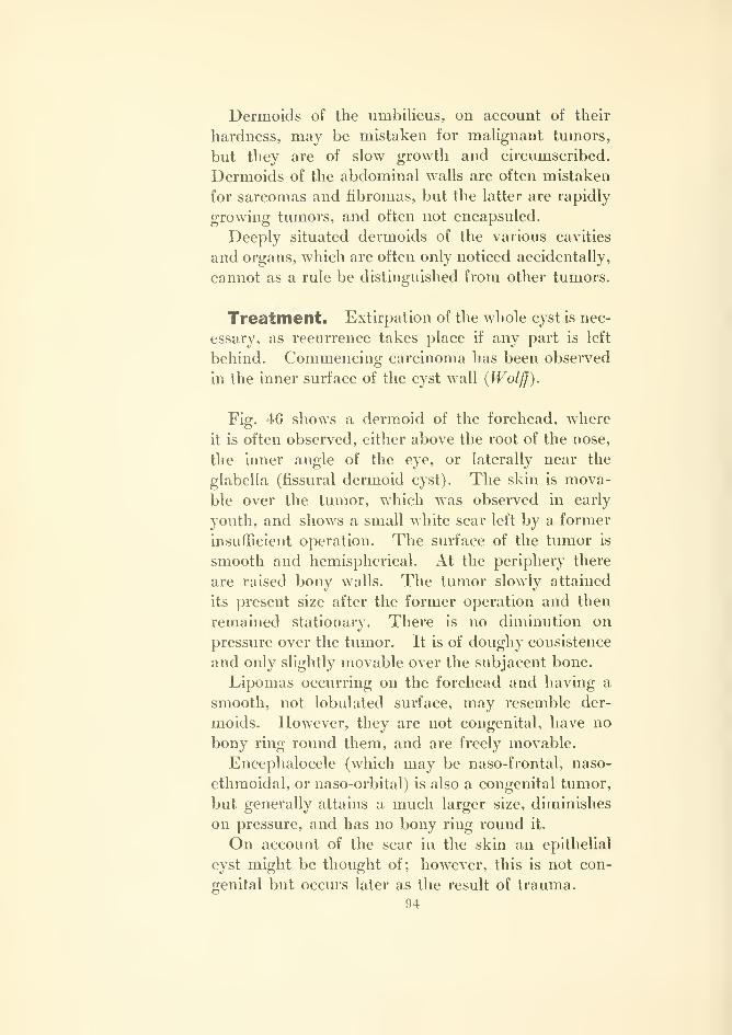
Dermoids of the umbilicus, on account of their
hardness, may be mistaken for malignant tumors,
but they are of slow growth and circumscribed.
Dermoids of the abdominal walls are often mistaken
for sarcomas and fibromas, but the latter are rapidly
growing tumors, and often not encapsuled.
Deeply situated dermoids of the various cavities
and organs, which are often only noticed accidentally,
cannot as a rule be distinguished from other tumors.'&^
Treatment. Extirpation of the whole cyst is nec-
essary, as recurrence takes place if any part is left
behind. Commencing carcinoma has been observed
in the inner surface of the cyst wall (Wolff).
Fig. 46 shows a dermoid of the forehead, where
it is often observed, either above the root of the nose,
the inner angle of the eye, or laterally near the
glabella (fissural dermoid cyst). The skin is mova-
ble over the tumor, which was observed in early
youth, and shows a small white scar left by a former
insufficient operation. The surface of the tumor is
smooth and hemispherical. At the periphery there
are raised bony walls. The tumor slowly attained
its present size after the former operation and then
reinained stationary. There is no diminution on
pressure over the tumor. It is of doughy consistence
and only slightly movable over the subjacent bone.
Lipomas occurring on the forehead and having a
smooth, not lobulated surface, may resemble der-
moids. However, they are not congenital, have no
bony ring round them, and are freely movable.
Encephalocele (which may be naso-frontal, naso-
ethmoidal, or naso-orbital) is also a congenital tumor,
but generally attains a much larger size, diminishes
on pressure, and has no bony ring round it.
On account of the scar in the skin an epithelial
cyst might be thought of; however, this is not con-
genital but occurs later as the result of trauma.
94

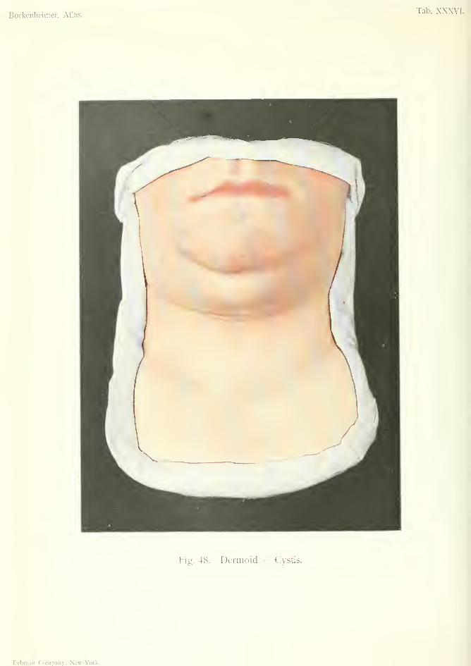
Bockeiilieimer, Atlas.Tab. XXXVl.
Fifj. 48. Dermoid — Cvstis.
Rcbiiiaii Company, Nevr-York.
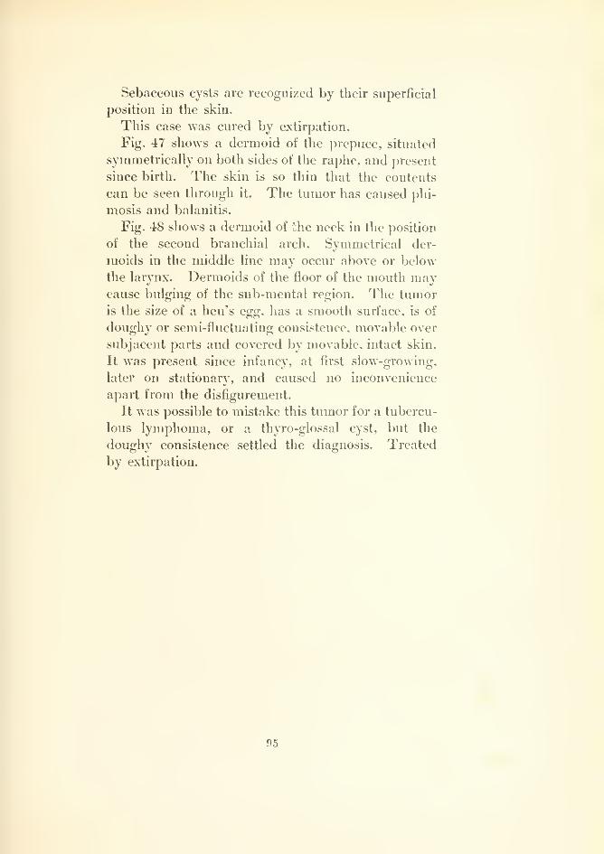
Sebaceous cysts are recognized by their superficial
position in the skin.
This case was cured by extirpation.
Fig. 47 shows a dermoid of the prepuce, situated
symmetrically on both sides of the raphe, and present
since birth. The skin is so thin that the contents
can be seen through it. The tumor has caused phi-
mosis and balanitis.
Fig. 48 shows a dermoid of the neck in the position
of the second branchial arch. Symmetrical der-
moids in the middle line may occur above or below
the larynx. Dermoids of the floor of the mouth maycause bulging of the sub-mental region. The tumor
is the size of a hen's egg, has a smooth surface, is of
doughy or semi-fluctuating consistence, movable over
subjacent parts and covered by movable, intact skin.
It was present since infancy, at first slow-growing,
later on stationary, and caused no inconvenience
apart from the disfigurement.
It was possible to mistake this tumor for a tubercu-
lous lymphoma, or a thyro-glossal C5'st, but the
doughy consistence settled the diagnosis. Treated
by extirpation.
95
FIBROMA VAGINAE TENDONIS {Fibroma of Tendon-Sheath)
Plate XXXVII, Fig. 49.
Fibroma belongs to the benign connective-tissue
tumors, and consists of connective-tissue cells, fibril-
lar, inter-cellular substance and a variable amount
of blood-vessels and lymphatics. When the matrix
is hard and abundant, with slight development of
spindle-cells, the fibroma is hard, while soft fibroma
is formed by spongy tissue with numerous blood-
vessels.
Fibromas, which consist of fibrous connective tis-
sue with few nuclei, are also termed desmoids, and
occur especially in the fascia of the abdominal walls,
while the term fibrosarcoma is applied to tumors
which consist of irregularly arranged spindle cells
with little intercellular substance, and show degen-
erative changes and an absence of mature tissue.
Transitional forms from fibroma to fibrosarcoma
and sarcoma are especially observed in the tumors
occurring in fascia. Mixed forms are often found,
such as fibro-lipoma, fibro-myoma, fibro-adenoma
and fibro-myxoma. Cystic formation is also seen
in fibromas.
Fibromas occur in all situations where fibrillar
connective tissue is present—in the cutaneous and
subcutaneous tissue (back and thigh), in intermus-
cular, intertendinous (Fig. 49), submucous and
subserous tissue (alimentary canal, uterus, larynx).
They may also develop in fasciae and aponeuroses,
nerve sheaths and periosteum (naso - pharyngeal
tumors, Fig. 25, and epulis. Fig. 35), and also in
the organs.
96
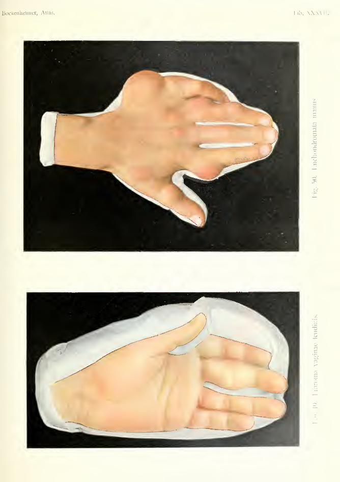
Bockeiilieiiiier, Atlas. Tab. XXWl
iJD
':^
p3
ti


They form circumscribed tumors of firm consist-
ence and smooth surface, often encapsuled, slow-
growing, sessile or pedunculated (fibrolipoma pendu-lum, Fig. 52). Pedunculated submucous fibromas
often occur in the laiynx in singers. Fibromas formrounded or polypoid growths, which may occur at
any age, but are seldom congenital. After meta-plastic changes (ossification) they may become hardtumors.
In the skin and subcutaneous tissue they have ayellowish-white surface (Fig. 49). On section they
show stratification and a glistening appearance like
tendon.
Differential Diagnosis. Superficial hard fibro-
mas of the skin and subcutaneous tissue are easily
recognized by their form, consistence, clear demarca-tion and solitary appearance. It is only transitional
forms between fibrosarcoma and sarcoma that pre-
sent any difficulty. Deep fibromas which often
attain a large size {e.g. in the abdominal cavity) are
recognized by their nodular surface, hardness andencapsulation.
Treatment. Excision of the tumor with its cap-
sule. For the removal of deep fibromas extensive
operations are necessary. Sometimes they are so
firmly attached to the neighboring tissues or organs
that a portion of the latter must be removed with
them.
Fig. 49 shows a fibroma of the sheath of the flexor
tendon of the finger, the yellowish-white surface of
which shows through the skin. The skin is slightly
movable over the hard nodular tumor. The tumor
itself is movable over the subjacent structures, and
remains unaltered in position on moving the finger.
Fibromas of tendon sheaths are rare on the whole,
and are due to traumatic causes. The tumor was
excised.
97

After injuries and stretching of tendons similar
growths occur, sometimes multiple; they are due to
proliferation of the cellular tissue. In Dupuytrens
contraction (Fig. 60) nodules also develop in the
palmar aponeurosis, which have a resemblance to
fibromas.
Thickenings which occur in tendons and tendon
sheaths, and lock the movements of the fingers in
certain positions, are not true fibromas.
98
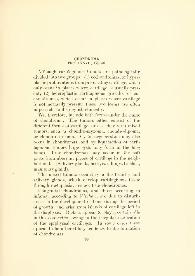
CHONDROMAPlate XXXVII, Fig. 50.
Although cartilaginous tumors are pathologically
divided into two groups: (1) ecchondromas, or hyper-
plastic proliferations from pre-existing cartilage, which
only occur in places where cartilage is usually pres-
ent; (2) heteroplastic cartilaginous growths, or en-
chondromas, which occur in places where cartilage
is not normally present; these two forms are often
impossible to distinguish clinically.
We, therefore, include both forms under the nameof chondroma. The tumors either consist of the
diflferent forms of cartilage, or else they form mixed
tumors, such as chondro-myxoma, chondro-lipoma,
or chondro-sarcoma. Cystic degeneration may also
occur in chondromas, and by liquefaction of carti-
laginous tumors large cysts may form in the long
bones. True chondromas may occur in the soft
parts from aberrant pieces of cartilage in the neigh-
borhood. (Salivary glands, neck, ear, lungs, trachea,
mammary gland).
The mixed tumors occurring in the testicles and
salivary glands, which develop cartilaginous tissue
through metaplasia, are not true chondromas.
Congenital chondromas, and those occurring in
infancy, according to Virchow, are due to disturb-
ances in the development of bone during the period
of growth, and arise from islands of cartilage left in
the diaphysis. Rickets appear to play a certain role
in this connection owing to the irregular ossification
of the epiphyseal cartilages. In some cases these
appear to be a hereditary tendency to the formation
of chondromas.
99

True chondromas, or enchondromas, develop from
the periosteum or medulla, most commonly in the
phalanges and metacarpal or metatarsal bones;
usually multiple. Isolated chondromas also occur
in the upper end of the humerus, the lower end of
the radius, the head of the tibia, the pelvic bones
and the scapula, often combined with cartilaginous
exostoses (ossified ecchondromas with a cartilaginous
covering)
.
Chondromas form slow-growing, hard, nodular,
circumscribed tumors, which may cause pressure
atrophy of neighboring parts (Fig. 50). Multiple
tumors, especially in the hands, cause considerable
deformity by disturbance of growth (shortening and
twisting). Pathological fracture may occur from
destruction of the cortex, in tumors growing from
the medullary cavity.
The softer forms of chondroma must be regarded
as malignant, because they take on an infiltrating
growth, extend to the veins and give rise to metas-
tases. (Chondro-sarcoma).
Differential Diagnosis. Central medullary
chondromas have to be diagnosed from osteomyelitic
abscesses and from central sarcoma. The former,
on X-ray examination, show thickening of the perios-
teum; the latter can often only be distinguished by
operation, as the X-ray appearances are very similar
in chondroma and sarcoma (when the chondroma is
single). Large chondromas of the head of the tibia
or upper end of the humerus are easily recognized by
their nodular surface and hard consistence.
Treatment. Isolated chondromas should always
be extirpated, as they may develop into sarcoma.
Multiple chondromas may be incised and scraped.
If rapidly growing recurrence takes place, resection
or amputation must be performed.
100

Fig. 50 shows a case of multiple chondromas ofthe fingers in a young man, which had been presentsince childhood. The nodular tumors are situatedin the phalanges and metacarpal bones, and havecaused thinning and reddening of the skin by pres-sure. The X-rays showed the origin to be in themedullary cavity. The tumors on the first, secondand fourth fingers were incised and scraped. Thelittle finger was removed with its metacarpal bone,on account of the multiplicity of the tumors.
101

HEMORRHOIDES ET FIBROMATA ANI(Jrlemorrhoids and Fibromas of Anus)
Plate XXX^^II, Fig. 51.
Among the benign growths of the anus, hemor-
rhoids are the most common. According to the
latest researches these must be regarded not only as
varicose veins, but as vascular growths or angiomas.
Hemorrhoids are called external or internal, accord-
ing as they are situated in the anus or rectum.
External hemorrhoids are due to the formation of
new blood-vessels and dilatation of the veins of the
inferior hemorrhoidal plexus. Certain races seem
to be predisposed to this affection; constipation and
pelvic engorgement may also give rise to it.
These subcutaneous hemorrhoids form bluish,
compressible, nodular, sessile growths covered by thin
skin, and situated around the anal orifice. There
is often moist eczema in the neighborhood (Fig. 51).
Through eczema and ulceration the nodules may be
transformed into fibrous structures (Fig. 51). In
their inflammatory state they cause much itching and
pain with tenesmus; while the nodules become hard
from thrombophlebitis, and bleed easily. Multiple
internal hemorrhoids of the lower part of the rectum
bleed easily without becoming inflamed, and have
a tendency to prolapse. When they are situated
higher up the rectum, diagnosis can be made by
digital examination or by the rectoscope.
Differential Diagnosis. External hemorrhoids
may be confounded with condylomata acuminata,
which are common round the anus in women suffer-
ing with gonorrhea. These are often as thick as the
103

Bockenheimer, Atlas. lab. XX.XVl
Fig. 51. I laniorrhoides et Fibromata ani.
Rebman Company, Nc«-York.

finjier, and form similar cockscomb growths onaccount of their papillomatous structure. Fibromas
are rare, generally smaller, pedunculated and solitary.
Carcinomas, of the papillomatous type, are recog-
nized by their rapid growth, inguinal glandular
metastases, early ulceration with hard borders, andirregular boundaries. In all cases of hemorrhoids
the rectum should be digitally explored for carcinoma.
Treatment. Laxatives should be given to create
soft stools, and the anus should be w'ashed after
defecation. During an attack, rest in bed with the
pelvis raised and the introduction of pessaries are
useful. Suppurating hemorrhoids must be incised.
In cases with frequent hemorrhages and severe pain,
a radical operation is indicated, either by cautery or
by excision of the nodules with subsequent suture.
Fibromas and condylomas can be removed byscissors, while carcinoma requires more extensive
operative interference.
Fig. 51 shows moist eczema in the region of the
anus. Round the anus are yellowish, nodular, hem-
orrhoidal growths, which have a resemblance to
fibromas on account of inflammatory changes and
ulceration. In one place is a bluish, glistening nod-
ule covered by thin skin. The growths were removed
by the thermo-cautery.
103

Lipoma
FIBROLIPOMA PENDULUM SUBCUTANEUM{Pendulous Fibrolipoma)
Plate XXXIX, Fig. .52.
LIPOMA DIFFUSUM SUBCUTANEUM(Diffuse Subcutaneous Lipoma)
Plate XL. Fig. 5.'?.
LIPOMATA SYMMETRICA SUBCUTANEA{Symmetrical Subcutaneous Lipomata)
Plate XLl/Fig. 5i.
Lipomas are tumors formed of fatty tissue, and
have, therefore, the yellowish-white color, soft con-
sistence, and lobular structure of fatty tissue. Theindividual fat lobules are separated by more or less
strongly developed connective-tissue septa, and the
whole tumor is demarcated from the surrounding
tissues by a thin capsule. Lipomas are of soft con-
sistence, often with pseudo-fluctuation; in rare cases
harder, from the development of more connective
tissue. They are slow-growing globular tumors,
which sometimes attain an enormous size, and are
usually supplied by a single vessel at the base of the
tumor. At the base of the larger tumors the skin is
generally drawn out into a pedicle, and is often
cedematous. Lipomas are essentially benign tumors;
they do not recur or give rise to metastases, nor do
they become transformed into malignant tumors.
Besides the fatty tissue, other tissues may be
developed (fibro-lipoma, myxo-lipoma, angio-lipoma,
chondro-lipoma). Cystic degeneration may give rise
to so-called oil-cysts in the interior of lipomas.
Multiple, usually symmetrical lipomas, are due
to disturbances in development. They may be
104
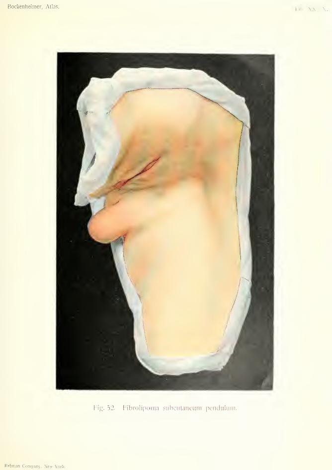
Bockenheimer, Atlas.Tab. .\.\.\i.\.
Fig. 52. Fibrolipoma subcutancum pendulum.
Rebman Company, Nc«--York.

connected with nerves (multiple lipomas are often
painful) or with lymphatic glands, which have beenfound in multiple lipomas. Congenital lipoma is
found especially in spina bifida, which arises as amyelo-cystocele, and usually as a myxolipoma (Fig.
144). That lipomas are true tumors is shown bytheir persistence in severe emaciation. Long-con-tinued pressure on a lipoma may cause suppurationof the fatty tumor through ulceration of the skin.
That chronic irritation plays a part in the develop-
ment of lipomas is shown by the occurrence of these
tumors on the backs of carriers, and on the foreheads
of persons who wear hard hats. Middle-aged womenare especially affected by these tumors, which maygrow considerably during pregnancy.
Lipomas are most often found in the subcutaneoustissue (Figs. 52, 53 and 54), where they appear as
soft, encapsuled tumors with a lobulated surface,
covered by non-adherent skin. The skin over the
tumor becomes dimpled when pinched up, owing to
its connection with the tumor by connective tissue
(Fig. 53) . The seats of predilection for subcutaneouslipomas are the back, nape of the neck, axilla, shoul-
der, upper arm, thigh, buttocks and scrotum.
Sub-fascial lipomas are very rare. They mayoccur under the fascia of the forehead (where they
may be mistaken for dermoids) and under the palmarfascia. Intermuscular lipomas occur behind the pec-
toralis major and in the tongue. In the knee joint
arborescent lipoma occurs, which has the typical
structure of fatty tissue. Lipomas may also arise
from the sub-mucous and sub-serous tissue (gut andlarynx) ; sub-peritoneal lipomas may give rise to
hernia through the linea alba. Sub-serous lipomas
also sometimes appear in the inguinal and femoral
canals; in the omentum and mesentery; in the
retroperitoneal tissue, and in the glandular organs
(breast and kidney).
All lipomas, especially sub-cutaneous, sub-fascial
105
and intermuscular, have a tendency to send processes
into the surrounding parts.
Differential Diagnosis. Superficial lipomas are
distinguished from fibiomas, lymphomas, dermoids,
sebaceous cysts, hygromas and other tumors by their
lobular surface and the puckering of the skin. Whenthey cannot be palpated, lipomas cannot always be
distinguished from other tumors.
Treatment. Incision through the skin and re-
moval of the tumor with its processes.
Diffuse lipomas, which consist in an infiltration of
the sub-cutaneous tissue with fatty masses without
any capsule, are not to be regarded as true tumors
(lipomatosis of Billroth). In the neck they may be
dangerous from pressure on the larynx, so that
removal is necessary, although this must generally
be incomplete. The fatty masses may also be madeto shrink by the injection of alcohol and ether.
Fig. 52 shows a pendulous fibro-lipoma in a middle-
aged woman. The skin is somewhat reddened, but
non-adherent. The surface of the tumor is smooth,
the consistence moderately hard. The tumor is
movable over the fascia. The base of the tumor is
broad, on account of its small size. The tumor was
removed by an oval incision and suture.
Fig. 53 shows a sub-cutaneous lipoma the size of
the fist in a common situation in a middle-aged
woman. The puckering of the skin is clearly seen.
These puckerings (white spots in the figure) are also
found in the breast, and are due to processes of the
lipoma extending into the breast. The tumor with
its processes was extirpated.
Fig. 54 shows symmetrical lipomas in the region
of both parotids, in the upper eyelids, and in various
parts of the neck (both sides of sub-maxillary region
and in sub-lingual region) in an old man. The pain-
106
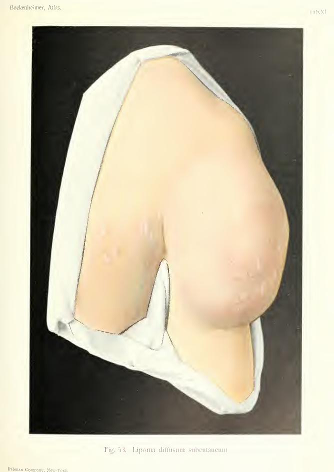
Bockenlieimer, Atlas.
Fig. 53. Lipoma diffusum siibcutanciim.
Rebman Company, New-Vork.


Bockenheimer, Atlas. Tab. XLI.
Fig. 54. Liponiata subcutanea symmetrica.
Rcbman Company, N'cw-York.


less tumors had not increased in size for some years.Their lobular surface and their consistence distin-guish these solid tumors from symmetrical cysticformations in the salivary glands, which cause simi-lar swellings in the face and neck. The disease is
distinguished from lipomatosis by consisting of mul-tiple, separate, encapsuled tumors. There were noother lipomas in other parts (in distinction to cases inwhich lipomas occur over the whole body). Thetumors were removed at several sittings.
107
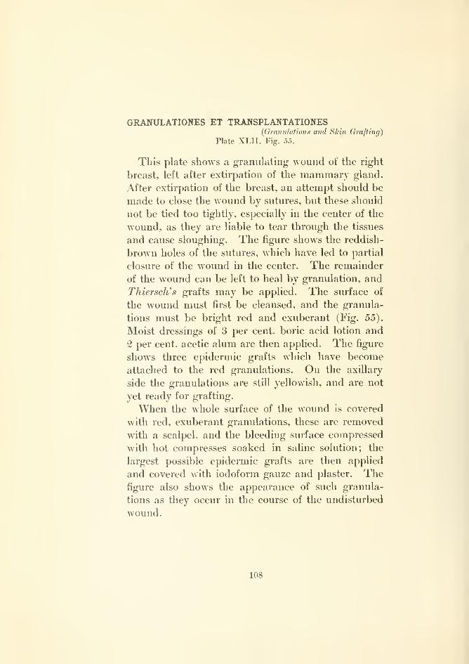
GRANULATIONES ET TRANSPLANTATIONES{Graiudations and Skin Grafting)
Plate XLII, Fig. 55.
This plate shows a granulating wound of the right
breast, left after extirpation of the mammary gland.
After extirpation of the breast, an attempt should be
made to close the wound by sutures, but these should
not be tied too tightly, especially in the center of the
wound, as they are liable to tear through the tissues
and cause sloughing. The figure shows the reddish-
brown holes of the sutures, which have led to partial
closure of the wound in the center. The remainder
of the wound can be left to heal by granulation, and
Thiersch's grafts may be applied. The surface of
the wound must first be cleansed, and the granula-
tions must be bright red and exuberant (Fig. 55).
Moist dressings of 3 per cent, boric acid lotion and
2 per cent, acetic alum are then applied. The figure
shows three epidermic grafts which have become
attached to the red granulations. On the axillary
side the granulations are still yellowish, and are not
yet ready for grafting.
When the whole surface of the wound is covered
with red, exuberant granulations, these are removed
with a scalpel, and the bleeding surface compressed
with hot compresses soaked in saline solution; the
largest possible epidermic grafts are then applied
and covered with iodoform gauze and plaster. Thefigure also shows the appearance of such granula-
tions as they occur in the course of the undisturbed
wound.
108
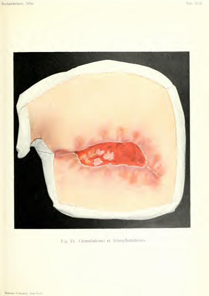
Bockenheimer, Atlas.
Fig. 55. Oranulationes et Transplantationes.
Rebman Company, New-York.


Bockenheimer, Atlas. Tab. XL
Fig. 56. Fistula ex corpore alieno.
Pt^l^rn^n f'nynn^nv W*»of_Vr\rlf

FISTULA EX CORPORE ALIENO {Fistula from foreign bodies)
Plate XLIII, Fig. 56.
As the result of incision of a paranephritic abscess,
a fistula has remained, which, in spite of drainage,
tamponage and repeated scraping, has not healed.
The surrounding skin is inflamed and oedematous.The granulations at the opening of the fistula are
unhealthy, dirty-brown and purulent. Shreds of
tissue with a fetid odor are discharged from the fistula.
Such an appearance of the fistula and its sur-
roundings is typical of all cases where, either the
external opening is too small, so that an abscess in
connection with it is not sufficiently drained, or
where necrosed pieces of tissue in the deeper parts
are cast off and act as foreign bodies {e.g. bony seques-
tra in coxitis, etc. (Figs. 95 and 96). Similar fistu-
las, with an offensive sanious discharge, sometimesresult from tampons, drains, or instruments being left
behind after operations.
In pyogenic lesions which have been insufficiently
incised, the presence of unhealthy, purulent granula-
tions shows that the pus has not a free outlet, or that
the lesion is extending. When a local pyogeniclesion gives rise to general pysemia the wound showssimilar changes, but the granulations besides having
a dirty-yellow appearance are quite dry.
Treatment must be directed to the cause of the
fistula. The latter should be laid open freely, andforeign bodies or pieces of necrosed bone removed,after which healing will take place.
In the case represented in Fig. 56, the kidney wasfound to be almost completely destroyed by suppura-
tion. Healing quickly took place after removal of
the kidney.
109

FISTULA COLLI MEDIANA (Median Fistula of the Neck)Plate XLIV, Fig. 57.
Median fistula of the neck is due to the persistence
of the thyro-glossal duct, which in embryonic life
leads from the foramen caecum at the back of the
tongue to the middle lobe of the thyroid gland.
Lateral fistulse of the neck are due to imperfect
closure of the second branchial cleft.
The lateral fistulse may also open in the middle
line, so that their true nature can only be made out
by tracing their course. This can be done by pal-
pation, by the passage of a probe, or by injection of
milk. The lateral fistulee of the neck deviate from
the middle line, perforate the superficial fascia of
the neck parallel to the sterno-mastoid muscle
behind the greater cornu of the hyoid bone, and open
into the side of the pharynx near the tonsil, while
the course of median fistulse remains in the middle
line, passing behind or through the hyoid bone to
the base of the tongue, and opening at the foramen
cfecum. If the internal opening of a fistula is open
and the outer opening closed, it is an internal incom-
plete fistula; if the outer opening is open but the
inner one closed, it is an external incomplete fistula.
If both openings are closed, branchial cysts are
formed in the case of lateral fistulae, and median
cysts (from the thyro-glossal duct) in the case of
median fistula.
Median fistula of the neck (Fig. 57), although of
congenital origin, is not usually noticed for several
years, for it is formed by an internal incomplete
fistula which gradually perforates the skin of the
neck. The fistula generally opens in the middle line
110

Bockenheimer, Atlas. Tab. XI IV.
M.>.Tf_V/M-^

between the hyoid bone and the sternum, and is
characterized by certain signs which are also foundin lateral fistula which opens on the inner border of
the sterno-mastoid muscle. The latter are moreoften congenital. In both cases there is a small
button-shaped opening, which is sometimes glued
together, sometimes discharges a drop of clear whit-
ish fluid. There are regularly arranged radiating
cicatrices round the fistula. If there is much secretion
the skin may be eczematous. On palpation, a hard
cord, as thick as a quill pen, can be felt passing
towards the middle line or laterally, according to the
nature of the fistula. Above the hyoid bone the cord
cannot be felt. The direction of the fistula is shownbetter by probing; the probe can hardly ever be
passed beyond the hyoid bone. However, if milk is
injected it can be seen to flow out near the tonsil in
the case of lateral fistula, and at the foramen caecum
at the base of the tongue in the case of median fistula.
Narrow fistulas cause little trouble to the patient,
but in wide, lateral fistulas accumulation of food
may cause inflammation and abscess. Carcinomamay arise from fistulas and cysts of the neck; it is
called branchiogenous, as it is derived from the
epithelium of the branchial clefts.
Differential Diagnosis. Fistulas arising from
tuberculous or inflammatory processes differ both in
their external appearance and in the course of the
fistulous track. In doubtful cases microscopic exam-ination may be made.
Treatment. Injections with the object of causing
obliteration of the fistula are useless. The only
rational treatment is total extirpation of the fistula
through a long incision, bearing in mind the anatomy
of the parts. In lateral fistula it is best to remove
the internal orifice together with the tonsil. In
median fistula, it is sometimes necessary to remove
111
the middle part of the hyoid bone, in order to follow
the track to the foramen csecum. Recurrence is fre-
quent if the smallest part of the fistulous track is left
behind. Microscopic examination of both median
and lateral fistulas shows squamous epithelium in
distal sections, cylindrical epithelium in proximal
sections. The presence of lymphoid tissue in the
wall of the fistula is characteristic.
Fig. 57 shows a median fistula of the neck in a girl
aged nineteen. The fistula first appeared at the age
of fifteen, and was treated by injection and incision,
without any result. A drop of secretion is seen at
the orifice of the fistula. Radiating cicatrices are
also visible. The fistulous track could be felt as a
cord as far as the hyoid bone, but its further course
could not be made out by injection of fluid. Theforamen csecum was deep. After an incision round
the opening of the fistula together with the scar tissue,
the track was dissected out. The center of the hyoid
bone, through which the track penetrated, was
removed, so as to continue the extirpation to the base
of the tongue. Microscopic examination showed
squamous epithelium in the lower part of the fistula
and ciliated, cylindrical epithelium in the upper part.
112

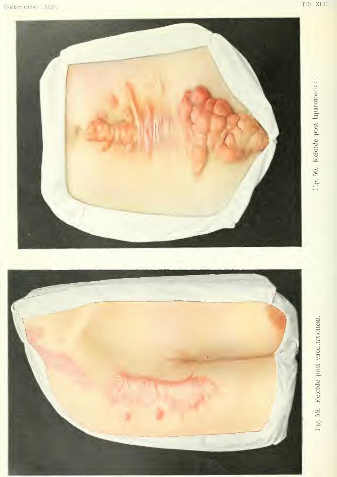
Bockenheimer. Atlas.Tab. XLV.
n
C3
•a
in
to
5
'u
>"55
o
T3
lis:
00in
bJ3
Keloid
Plate XLV.
KELOID POST VACCmATIONEM {Keloid after Vaccination)
Yi'r. 5S.
KELOID POST LAPAROTOMIAM (Keloid after Laparotomij)
Fig. 59.
This disease, the etiology of which is still obscure,
consists in the formation of homogenous, fibrous
tumors in the skin which are formed of hypertrophic
scar tissue with thickened blood-vessels. The chief
part of the growth consists of dense, hyaline, often
interlacing bundles of connective tissue, while cells
and elastic fibers are few in number. Only a few
cases can be spoken of as true tumors.
The papillary bodies are unchanged, but lying
under them are nodules or lamella% more or less rich
in cells (keloid-nodular cancer). In the lamellar
form (Fig. 58) there are radial processes at the periph-
ery which are often prolonged as fine processes into
the skin. The keloid presents itself as a tumor of
hard consistence, with a smooth, glistening surface,
of reddish (Fig. 58) or yellowish-white color (Fig.
59), situated in the skin and movable over subjacent
structures. Keloids are painless, of slow growth,
and then remain the same size for some time. They
are common in young women. Pain and irritation
may be caused by pressure on the tumors, especially
when they are of large size, or situated in places
exposed to frequent pressure or contact.
It is now believed that keloids occur exclusively
after injuries (operations, vaccination, scars caused
by flagellation, burns, chronic ulcers, etc.), and that
113
there is a local or general disposition to keloid for-
mation in the individual affected. It has not been
proved that infected wounds are more liable to form
keloids. Certain parts of the body are more affected
than others—the shoulders, face, abdomen and exter-
nal ears—while the extremities are seldom affected.
Dark races have a special tendency to keloid forma-
tion.
Glandular enlargement and metastases are not
observed, but large keloids may ulcerate, and cancer
may develop from the ulcers.
Differential Diagnosis. Hypertrophic scars
may be mistaken for incompletely developed keloids,
but the former are usually very sensitive, are not so
extensive as keloid, occur especially after infected
wounds, and nearly always undergo partial resolution
after some years.
Treatment. Operation is to be avoided, as recur-
rence nearly always takes place after extirpation, with
or without a plastic operation, after cauterization
and scraping, and the recurrent growth is often more
extensive than the original. Electrolysis, or injection
of a 10 per cent, solution of thiosinamin sometimes
cause improvement.
Fig. 58 shows a keloid in a young girl, which arose
from a vaccination scar and recurred extensively
after extirpation. An extensive flat growth is seen
with radiating processes ; also smaller nodular growths
in the neighborhood.
Fig. 59 shows an extensive nodular keloid in a
woman of twenty, which developed in the scar of a
laparotomy. At each suture hole a nodule has devel-
oped. At the lower part are hard, cauliflower
nodules, freely movable and covered by epidermis.
Injection of thiosinamin solution into the nodules
caused partial disintegration, but later on further
recurrence took place in the parts treated.
114


Bocketilieinier, Atlas. Tab. XLVl
h"ig. bO. Coiitractura aponeurosis pahnaris (Uupuytreii).
Rcbman Company, Ne^x-York.

CONTRACTURA APONEUROSIS PALMARIS (Dupuylren)
{Dupmjtren s Contrartttre oj Palmar aponeurosis)
Plate XLVI, Fig. 60.
The palmar aponeurosis, the continuation of the
palmaris longus muscle, which spreads over the palm
and sends processes to the proximal phalanges of all
the fingers, and is also connected with the skin, may
be affected by chronic inflammation leading to con-
nective-tissue formation and subsequent contracture.
Hard nodules develop in the aponeurosis and skin,
which finally become hard cords. These cord-like
thickenings occur not only in the palm, but even
more commonly in the processes of the aponeurosis
connected with the second, third, fourth and fifth
fingers. Contraction of these cords, which at the
base of the phalanges are connected with the tendon
sheaths, gives rise to an abnormal position of the
fingers, called Dupmjtren's contracture. This term
signifies limitation of movement in the joints which
may be of arthrogenous, neurogenous, myogenous,
tendogenous, or dermatogenous origin.
The fourth and fifth fingers are those most often
affected by Dupuytrens contracture, the second and
third less often, and the thumb least often. The dis-
ease usually begins in the fourth or fifth finger and
may spread to all the others. It is often symmetrical,
affecting both hands at the same time and to the
same extent. Before the commencement of contrac-
tion, nodular, fibrous thickenings can be felt in the
skin, later on fibrous cords are formed, by which
first the proximal phalanges, later on the middle
phalanges become fixed in a position of flexion,
while the terminal phalanges maintain their power
115
of extension. After some years the contraction
becomes so severe that the finger is completely
doubled on itself into the palm, and cannot be
extended. There is generally some power of exten-
sion of the middle and terminal phalanges, but as
this is painful it is avoided by the patient.
The affection occurs exclusively in men and was
hence attributed to traumatic influence by Diqmy-
tren. At any rate the affection is often found in
people in whom the palm of the hand is exposed to
continued pressure (in post-office clerks, as the result
of stamping, persons who carry guns, carpenters, etc.).
Some authorities attach little importance to the action
of trauma, and the disease often occurs in gouty
people. As the contraction is often symmetrical and
equally developed on both sides, a central nervous
origin is possible.
Differential Diagnosis. Dupui/tren's contrac-
ture differs from contracture due to cutaneous scars,
by the skin over it being intact. Fibromas of ten-
dons or tendon sheaths (Fig. 49) form rounded
swellings. In occupational contracture of the fingers,
there are no hard cords in the palm, and the pha-
langes of all the fingers are usually equally flexed.
In arthrogenous contracture the joints are obviously
affected.
Treatment. Mechanical treatment and massage
are incapable of arresting the progress of the disease.
In severe cases operation is indicated, according to
Kocher excision of the affected parts of the palmar
aponeurosis. Those parts of the skin which show
fibrous changes should also be removed, and the
wound repaired by skin flaps. Massage, commencedsoon after the operation, may give good functional
results. Treatment by injection of thiosinamin is
at present inconclusive.
116

Fig. 60 shows a case of Dupuytreri's contracture of
the fourth and fifth fingers in a man of fifty. Thehttle finger is considerably contracted, and only the
last phalanx can be freely extended. The fourth
finger shows contracture of the first phalanx and
commencing contracture of the second. Contrac-
ture is also beginning in the third finger. Theafl"ection was of several years duration, and caused
so little trouble that operation was refused.
117
CONTRACTURA POST PANARITIUM TENDINOSUM{Contrarture offer Tendon Sheath Suppuration)
Plate XLMI, Fig. 61.
Cutaneous contractures affect chiefly the flexor
surface of the fingers and palm, and originate in the
scars of operations, wounds, burns and inflamma-
tions. Tendon contracture is often associated with
cutaneous contracture, especially when there is sup-
puration within the tendon-sheaths, so that the finger
becomes stiff and fixed firmly in a contracted posi-
tion. Fig. 61 shows a hard, slightly movable sear,
extending from the flexor surface of the last joint of
the middle finger to the center of the palm, arising
from an incision for suppuration of the tendon
sheath (cf. Fig. 93). The nature of the lesion, and
the fact that there is no power of motion in the
finger, shows that the flexor tendon is destroyed.
Hence, the contracture is both dermatogenous and
tendogenous, i.e. caused by contraction of both skin
and tendon.
In cases where the tendon is partly destroyed, or
very firmly connected with the hypertrophic cuta-
neous scar, operative treatment is not successful.
After excision of the scar, contracture occurs in the
new scar, in spite of extension of the finger, length-
ening of the tendon, transplantation of tendon or
catgut, or plastic operations. If the patient is inca-
pacitated from work by the contracture, exarticula-
tion of the fingers gives the most useful result, the
use of the thumb being cultivated to take their place.
In cases of tendon-sheath suppuration, contracture
may be prevented by making small lateral incisions
in the finger. If the tendon is not destroyed by sup-
118
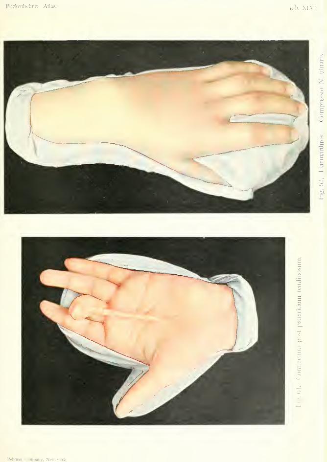
Hockenlieinier, Atlas. Tab. XIAII.
/:
o
~r.
Rebman Company, Ne^» -\'ork.

puration, the skin contracture can then be prevented
by early, active and passive movements, massage,baths, etc.
In cases of contracture limited to the skin, such as
those after cuts and burns, keloid scars, superficial
suppuration, etc., the prognosis is much better. Themobility of the scar over the deeper structures andthe power of moving the individual phalanges, showthat the tendon is not implicated. Excision of the
scar, extension of the finger, in some cases lengthen-
ing of the tendon, and repair of the wound by skin
flaps, in these cases restores the function of the
finger. In young persons good results can beobtained by orthopedic treatment, when the scar is
not very extensive, nor hypertrophic, nor of too long
standing.
119
HAEMARTHROS COMPRESSIO N. ULNARIS{Hemarthrogis Compress-ion of Ulnar Nerve, Neurogenous CordToclure)
Plate XLVII, Fig. 6'2.
Neurogenous contractures affect the hand and
fingers, and result from injuries to the radial, ulnar
and median nerves. They may be of peripheral or
central nervous origin. In contractures of central
origin, especially in the paralytic contractures due to
anterior poliomyelitis, nerve transplantation, and
shortening or transplantation of tendons may be
performed. Treatment by massage, electricity and
orthopedic apparatus is also useful. In contractures
due to lesions of the peripheral nerves (division of
nerve; pressure from badly united fracture, effusion
of blood, or tumors on the nerve), exposure of the
nerve, with excision of the injured part and subse-
quent suture is sometimes successful.
Fig. 62 shows a reflex contracture resulting from a
blow on the ulnar side of the wrist joint, causing
effusion of blood into the joint (hemarthrosis) which
pressed on the ulnar nerve. Compression of the
ulnar nerve by the joint effusion gave rise to "claw
hand"—by hyperextension of the proximal pha-
langes and flexion of the second and third phalanges.
There was slight swelling on the back of the wrist
joint, chiefly on the ulnar side. Fluctuation was
present. The sign of "snowball crunching" indi-
cated the presence of blood clots, and therefore of
hemarthrosis. The movements of the joints were
limited and very painful. The joint was in a posi-
tion of slight flexion, but could be easily extended.
130

The hand was fixed on a splint and recover^' took
place after absorption of the blood.
The diagnosis between the different kinds of neu-
rogenous contractures, and between these and other
contractures often requires an examination of the
whole nervous system. Hysterical contracture of the
knee and hip joints, which is common in children,
disappears under an anaesthetic.
121
CONTRACTURA ISCHAEBnCA BRACHH(Ischaemic contracture of the arm)
Plate XLVIII. Fig. 03.
Myogenous contractures occur most commonly in
the upper extremity, as a result of injuries and sup-
puration in the muscles, which cause shortening of
the muscles and their tendons. They also occur in
diseases of the nervous system, both peripheral and
central. Contracture also results from too long
immobilization of a limb, the over-action of the
flexor muscles causing flexion contracture of the arm,
wrist and fingers.
These contractures are most marked in ischaemic
muscular contracture (Volkmann), which is gener-
ally observed in the upper extremity of young per-
sons. The causes of this condition include fractures
{e.g. supra-condyloid fracture of the humerus), rup-
ture of the intima of blood-vessels, obstruction of
large vessels, exposure to cold, prolonged action of
Esmarch's elastic bandage, and constriction by
plaster of Paris bandages. A constricting bandage
is sufficient to cause ischaemia in the arm.
The greater frequency of contracture in the upper
extremity is explained by the fact that, owing to
there being less muscle in the arm than in the thigh,
the vessels are more easily compressed. Out of
thirty-five cases collected by Bardenheuer, there was
only one affecting the leg. The greater frequency
of ischaemic contracture in young individuals is due
to the greater compressibility of their muscles and
vessels. In older persons great pressure on the ves-
sels is liable to cause gangrene owing to arterio-
sclerosis; even slighter pressure may give rise to
obliterative thrombosis and consequent gangrene.
122
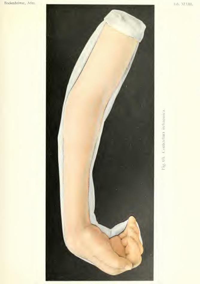
Bockenheimer, Atlas. Tab. XLVIIF.
o
U

It must be borne in mind that muscular tissue is
more affected than skin and bone even by short inter-
ruption of the blood supply, because the compressed
vessels are terminal branches.
The affection begins in the peripheral parts of the
extremities. The fingers become blue, swollen, cold
and moist, painful on movement, which can only bedone passively, and flexed. In cases where the
affection is due to tight bandaging, after early
removal of the bandage the skin appears white,
while the muscles feel as hard as a board and immo-bile, but recover after proper treatment. If the con-
stricting bandage is allowed to remain, in a few
hours the muscles become bloodless and undergo
degeneration, having a waxy-yellow appearance as
in tj'phus. As the result of extensive muscular atro-
phy, shrinking of the muscles takes place and causes
contracture. The patients suffer severe pain for a
long time after removal of the bandage. The skin
of the fingers gradually becomes yellowish-white like
parchment. The swelling of the fingers is followed
by shrinking. First of all the fingers, then the meta-
carpal bones, and finally the wrist become fixed in a
position of flexion. The fingers are eventually so
strongly flexed that the hand becomes useless. Themovements of the wrist are also very limited, and
the muscles of the forearm become atrophied and
are covered by pale skin. Sensory disorders mayoccur from pressure of the shrunken muscles on the
nerves, and in some cases ischaemic muscular con-
tracture is followed by ischaemic paralysis.
The clinical appearance of myogenous contrac-
ture, especially ischaemic muscular contracture, is
so characteristic that it can hardly be mistaken for
other forms of contracture.
Treatment. Myogenous contracture, when not
of too long standing, may be improved by massage,
electricity, baths and hot-air treatment. Prophy-
123
lactic treatment consists in avoiding the use of too
tight bandages, and too long fixation of the limb.
In the application of plaster bandages to fractures
of the upper extremity certain definite rules must be
observed. The limb must be well wrapped in cotton
wool, which must be loose at the extremity, and the
limb should be suspended to assist the venous circu-
lation. The bandage must be removed if the fingers
become blue, swollen or painful. Patients with
plaster of Paris bandaging must be kept under con-
tinuous observation. The bandage should always
be changed on the eighth day, when light massage of
the muscles and movement of the joints can be
carried out. After this movable plaster casing is
used (;i.e. plaster casing cut through on both sides
after fixation, then removed and reapplied with ban-
dages) . In every fracture careful examination should
be made to see if there is any injury to the nerves, so
that paralysis appearing later on may not be
unjustly attributed to the bandages.
In severe cases of ischaemic muscular contracture,
resection of several centimeters of the radius and
ulna may be performed, whereby the flexed position
of the fingers and hand is corrected and a certain
amount of function is restored.
In cases where the nerves are implicated, transpo-
sition of the large nerve trunks from the shrunken
muscles above the fascia has been successfully per-
formed.
Fig. 63 represents a case of ischaemic muscular
contracture without implication of the nerves, result-
ing from the application of plaster of Paris bandages
to a supracondyloid fracture of the humerus. Thebandages were left on for four weeks, in spite of
pain, swelling and blueness of the fingers occurring
soon after their application. After removal of the
bandages, the muscles of the forearm were found to
be much atrophied. The hand and fingers gradually
124

assumed the form of claw-hand, so that the patient
could not use his arm. Extensive resection of the
radius and ulna with subsequent suture corrected
the flexed position of the hand and restored the
function of the limb to a certain extent.
125
HALLUX VALGUS {Hammer-toe—Arthrogenous Contracture)
Plate XLIX, Fig. 64.
In the foot contractures occur which are generally
limited to the first and second toes. Pointed shoes
cause external deviation of the great toe, known as
hallux valgus. The deviation may be as much as
fifty degrees, so that the great toe lies over or under
the second toe. As the result of changes in the
joint (atrophy, inflammation, arthritis deformans),
arthrogenous contracture takes place in the meta-
tarso-phalangeal joint, so that in advanced cases
the deformity cannot be corrected. Over the pro-
jecting metatarso-phalangeal joint exostoses, clavus
and bunions may develop, while an ingrowing toe-
nail usually forms on the outer side of the great toe
(Fig. 99). Clavus most commonly forms a circum-
scribed thickening of the horny layer of the epider-
mis, causing pain by pressure on the papillary nerve
endings. Underneath the clavus a bursa generally
forms which may suppurate (bunion) and perforate
externally or into the joint. Clavus most often occurs
on the first and fifth toes. In hallux valgus and in
hammer-toe clavi are always found, often between
two toes or under the toe-nails. Subungual exos-
toses also occur in these cases (Fig. 140).
Hammer-toe is an arthrogenous flexion contrac-
ture usually aflFecting the second toe, as the result of
wearing too short boots, or secondary to hallux
valgus. The first phalanx is extended, the second
and third flexed. The third toe is rarely affected.
Hallux valgus and hammer-toe are often com-
bined with flat foot, and then render walking still
more awkward and painful.
126
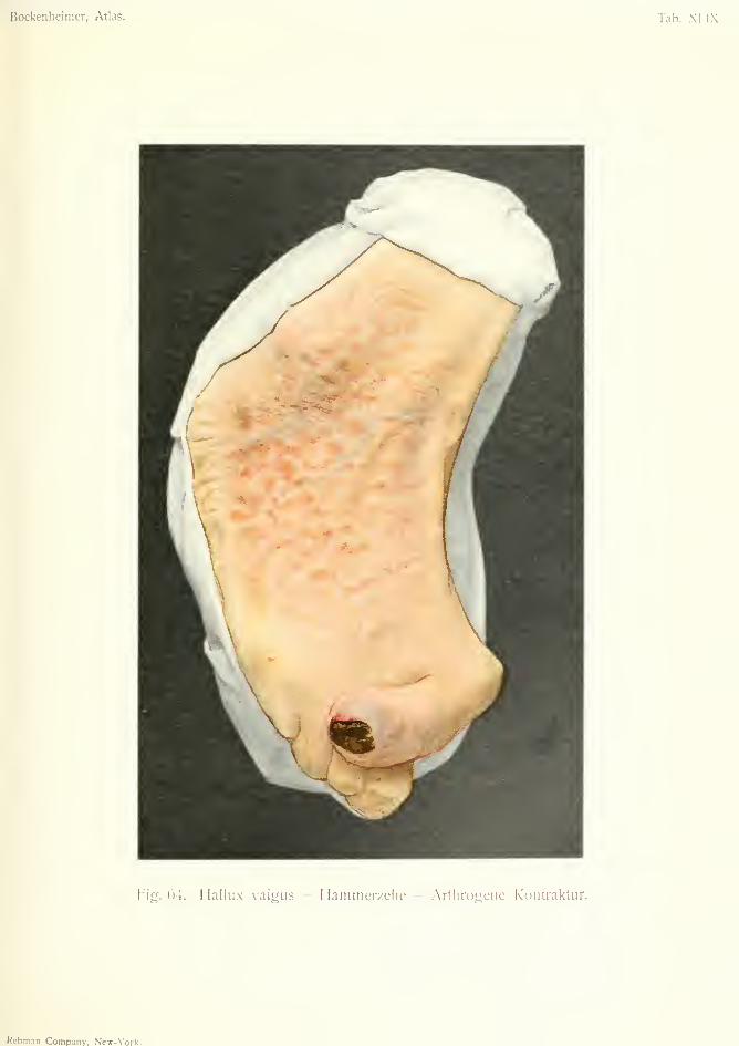
Bockenheimer, Atlas. Tab. XLIX.
Fig. 64. Hallux valgus — Hamtnerzehe — Arthrogciie Kontraktur.
Rcbm.in Corap.nny, New-York.

Treatment. Prophylactic treatment consists inattention to tlie feet, baths, cutting the toe-nails
straight instead of curved, properly made boots, etc.
Hallux valgus, if it gives much trouble, is besttreated by cuneiform osteotomy of the metatarsusand subsequent correction in plaster of I*aris.
Hammer-toe is often treated by fixation to a splint,
after correction of the deformity, but this is unsatis-factory. It is better to cut through the soft parts atthe seat of flexion, and resect the joint from theextensor surface; or in bad cases to disarticulate
the toe.
Exostoses can be chiseled; subungual exostosesafter removal of the nail.
Clavi are best removed by the knife. In subun-gual clavus the nail must be removed first. Fistulafrom a bunion should be freely incised and cau-terized; or the whole bursa may be extirpated.
(For the treatment of ingrowing toe-nail and flat
foot see Figs. 99 and 83).
Fig. 64 shows the result of neglect and badlyfitting boots. The great toe shows typical halluxvalgus. On the inner side of the metatarso-pha-langeal joint is a clavus, on which opens a fistula
from a bunion lying under it. On the outer side of
the great toe the nail is ingrowing. The second toe
is affected with hammer-toe and also clavus. Theback of the foot is covered with dry eczema, due to
uncleanliness. Owing to these disorders and a con-
siderable degree of flat foot the patient could hardlywalk. The hallux valgus was corrected by cunei-
form osteotomy of the metatarsus. The cla\Tis andbunion were excised, and the second toe disarticu-
lated. The eczema healed quickly with Hehra'sointment. After this the patient could walk nor-
mally, with a well-made boot.
127
RHACHITIS—IWFRACTIONES CRURIS UTRIUSQUE{Rickets, Greenstick, Fractures of Both Legs)
Plate L, Fig. 65.
Rickets, which is also known as the EngUsh dis-
ease, is a disturbance of growth affecting the whole
skeleton. It consists in softening of the bones in the
course of their growth, from defective ossification
due to deficiency in calcium and magnesium phos-
phates. In the epiphyses there is abnormal prolif-
eration of cartilage, and at the same time imperfect
calcification of the cartilage. This causes thicken-
ing of the epiphyses and interference with the growth
of the long bones in rickety children. Irregularity
in the formation of the medullary spaces also plays a
certain part. In the flat bones growth in thickness
is hindered.
In the skull the disease affects chiefly the frontal
and parietal bones. The bony substance may be so
poorly developed that the bones are soft and flattened,
yielding to pressure (cranio-tabes) . In other places,
especially the frontal and parietal eminences, the
bones are thickened and prominent from the over-
formation of bony tissue. The cranial sutures and
fontanelles remain open for a long time, and hydro-
cephalus is often present. The upper and lower
maxillae are flattened and irregularly developed, and
the implantation of the teeth is irregular and abnor-
mal.
The weight of the body causes bending of the
softened bones; the spine becomes kyphotic or
scoliotic; the thorax is constricted laterally, and the
junctions of the cartilage and bone of the ribs becomethickened (beaded ribs or rickety rosary) . The pel-
128
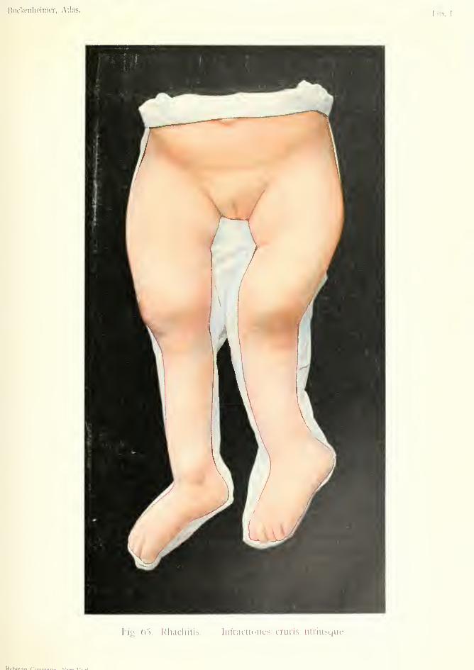
Bockenlieinier, Atlas.Tib. I.,
Fig. 05. Rliacliilib. Inlraclioiieb cruris iitriubL|iie.
P(.hTn.in r'ft«. XI \'__1.

vie bones remain small, so that the rickety pelvis is
a cause of obstructed labor. Lastly, in severe cases,
the lower extremities become extremely bent and the
bones are liable to greenstick fracture.
In the second year there is usually thickening of
the epiphyses of the bones of the limbs, especially
the lower ends of the ulna, radius and tibia; while
the diaphyses, especially of the femur, tibia and
fibula are curved. The femurs are bent outwards,
the bones of the leg outwards and forwards (Fig.
65). Genu valgum occurs in the knees. The arch
of the foot sinks in, causing flat foot. In severe
cases of rickets the children remain so backward in
growth that they become dwarfs. The so-called
fetal rickets, according to recent investigations, has
nothing to do with rickets. True rickets occurs
exclusively in children between the first and sixth
years, especially in the second year, and at puberty
as late rickets, especially when heavy weights act on
the limbs (genu valgum, coxa vara, scoliosis, pes
valgus).
The origin of rickets and its absence in certain
countries (China, Japan, Australia) is not yet quite
clear, but bad hygienic conditions and especially
improper feeding play an important part. Hered-
itary syphilis is a predisposing cause.
The disease often begins with anaemia, digestive
troubles and diarrhea, while spasm of the larynx
(laryngismus stridulus) or lung affections often
occur and may be fatal.
Differential Diagnosis. Osteomalacia, which
consists in softening of normally developed bones,
occurs at a later age, more often in women. Hered-
itary syphilis affects fewer bones, especially the
tibia, and is almost always associated with other
signs of congenital syphilis—interstitial keratitis,
notched teeth, etc.).
Rickety scoliosis and kyphosis are distinguished
129
from tuberculous spinal disease by the presence of
rickety changes in other parts of the body.
The prognosis is favorable on the whole. Calcifi-
cation may take place in the osseous tissue and the
bones may assume a sclerotic condition, without a
trace of shortening or bending being left. Accord-
ing to the researches of von Schlauge and Veil this
occurs in the course of four years in all children whodo not remain markedly backward in growth. Thedisease generally comes to an end about the sixth year,
but it may recur afterwards, especially in the winter.
Treatment. In the first place hygienic condi-
tions must be improved. Infants should be suckled
by the mother. Later on meat, eggs and vegetables
should be prescribed. Fresh air, high altitudes andsea bathing are all beneficial. Internally cod-liver
oil and phosphates. The children should be kept
off their feet, and sleep on hard beds. Surgical
treatment consists in the treatment of green-stick
fractures and in correcting the curvature of the bonesof the limbs. Complicated apparatus only leads to
atrophy of the limbs. Ricketty spine should betreated by a strong corset.
Curvatures of the bones should only be operated
on when they are severe, and then only when the
disease has come to a standstill. An X-ray exam-ination is useful; in active rickets the epiphyseal
lines appear wide and irregular, sometimes with
incomplete fractures and irregular arrangement of
cartilage, and the cortex appears much thinned;
while, in quiescent rickets, the epiphyseal lines havebecome regular, and the cortex appears the samethickness as the deeper parts.
As a rule, operation should not be performedbefore the sixth year. The curvature can be cor-
rected manually or by the osteoclast; better still bylinear or cuneiform osteotomy, followed by plaster
of Paris.
130

Operation is also indicated in cases where there is
early sclerosis of the bone, which is shown by the
X-rays. In this case the curvature must be cor-
rected by osteotomy, otherwise the bones will bearrested in growth.O'
Fig. 65 shows rickets affecting the whole skeleton
in a girl aged four years. The left femur was so
much curved and sclerosed that osteotomy was per-
formed, while the curvature of the right femur under-
went spontaneous cure. The epiphyses of the kneeand ankle joints are much thickened, the upper andlower ends of the tibias are much bent. At the lower
ends the X-rays showed green-stick fractures. Oper-ation here was contra-indicated, as the X-rays showedthe disease to be still in an active state. In the hip
joints the X-rays showed coxa vara of the neck of
the femur. The child was very feeble and backwardin growth.
131
LUXATIO CUM FRACTURA CRURIS{Fracture-dislocation of the leg)
PSEUDARTHROSISPlate LI, Fig. 66.
False joints (pseudarthrosis) occur in the leg,
chiefly after oblique fractures with dislocation, or
comminuted fractures; in the thigh and upper
arm after transverse fractures also, as the result of
interposition of the soft parts, chiefly the muscles.
Advanced age, pregnancy, rickets, syphilis, tuber-
culosis, may delay union of the fragments in a frac-
ture-
Extension treatment is the best to obtain rapid and
sure union. Delayed union may be accelerated by
percussion of the fragments, injection of iodine and
other preparations, or of blood, into the callus; by
passive hyperaemia, or by the administration of
phosphate of lime.
Badly united fractures can be brought into better
position by the osteoclast or by osteotomy.
In the treatment of pseudarthrosis situated close
to a joint resection comes into question. Pseudar-
throsis in the shaft can be repaired by bone suture.
When the ends of the fragments are much atrophied
(X-ray examination) they must be resected before
suturing. The fragments may be resected so as to
overlap each other (dovetailed). The periosteum
must always be spared as much as possible.
Transplantation of bone has sometimes proved
successful. If no union occurs after these methods,
apparatus must be worn, or amputation must be
performed.
132
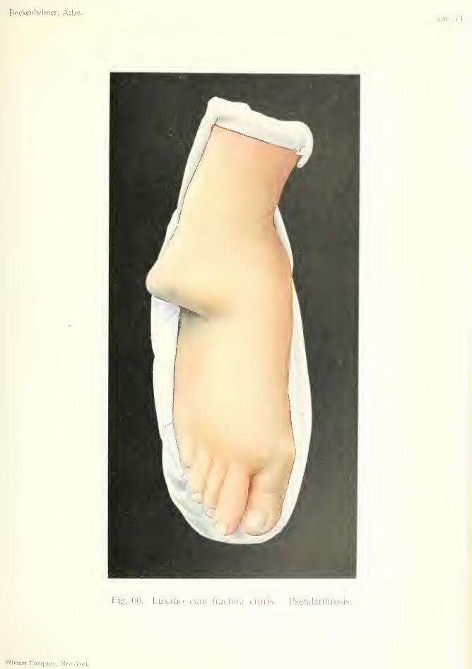
Bockenheinier, Atlas.
Fig. 66. Luxatio cum liactura cruris — Pseudartlirosis.
Rfbman Company, Ncw-Vork,

In the treatment of fractures and dislocations,
especially in fracture-dislocations, the X-rays are
especially useful in making an early diagnosis.
Fig. 66 shows marked deformity of the lower part
of the right leg as far as the ankle joint. On the
outer side there is slight outwai'd curvature of the
fibula above the external malleolus. The peripheral
end of the fibula is dislocated, so that the external
malleolus projects and the skin bulges on the outer
side of the ankle joint. There is an outward curva-
ture of the right tibia above the inner malleolus. Thefoot is in the position of advanced flat-foot.
The nature of the injury is an ununited supra-
malleolar oblique fracture of the tibia. The distal
part of the tibia is freely movable, although the frac-
ture is of two years standing. X-ray examination
shows that the fragments have overlapped, and that
there is a united fracture of the distal end of the
fibula a few centimeters above the external malleolus,
in the position of the above-mentioned projection.
The patient (aged sLxty), owing to effusion into the
left knee and left flat foot, depended entirely on the
right leg; the injury being due to the giving way of
the right foot in a position of supination. The frac-
ture dislocation had not been diagnosed, and the
patient had been treated with poultices, etc. The
foot was brought into proper position by resection of
the lower ends of the tibia and fibula and freshening
the head of the astragalus.
133
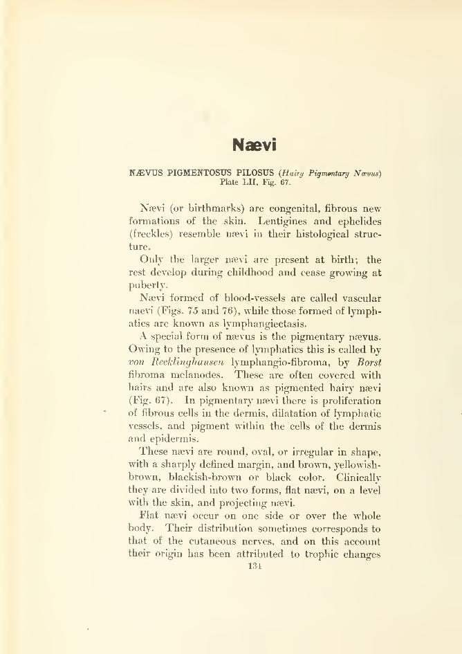
Naevi
N^VUS PIGMENTOSTJS PILOSUS {Hairy Pignwntary Nwvus)Plate LII, Fig. 67.
Nsevi (or birthmarks) are congenital, fibrous newformations of the skin. Lentigines and ephelides
(freckles) resemble nsevi in their histological struc-
ture.
Only the larger nsevi are present at birth; the
rest develop during childhood and cease growing at
puberty.
Naevi formed of blood-vessels are called vascular
naevi (Figs. 75 and 76), while those formed of lymph-
atics are known as lymphangiectasis.
A special form of nsevus is the pigmentary nsevus.
Owing to the presence of lymphatics this is called byV071 Recklinghausen lymphangio-fibroma, by Borst
fibroma melanodes. These are often covered with
hairs and are also known as pigmented hairy nsevi
(Fig. 67). In pigmentary nsevi there is proliferation
of fibrous cells in the dermis, dilatation of lymphatic
vessels, and pigment within the cells of the dermis
and epidermis.
These naevi are round, oval, or irregular in shape,
with a sharply defined margin, and brown, yellowish-
brown, blackish-brown or black color. Clinically
they are divided into two forms, flat nsevi, on a level
with the skin, and projecting nsevi.
Flat nsevi occur on one side or over the wholebody. Their distribution sometimes corresponds to
that of the cutaneous nerves, and on this account
their origin has been attributed to trophic changes134

Bockenheimer, Atlas. Tab. I.II.
Fig. 67. Naevus pijj;mentosus pilosus.

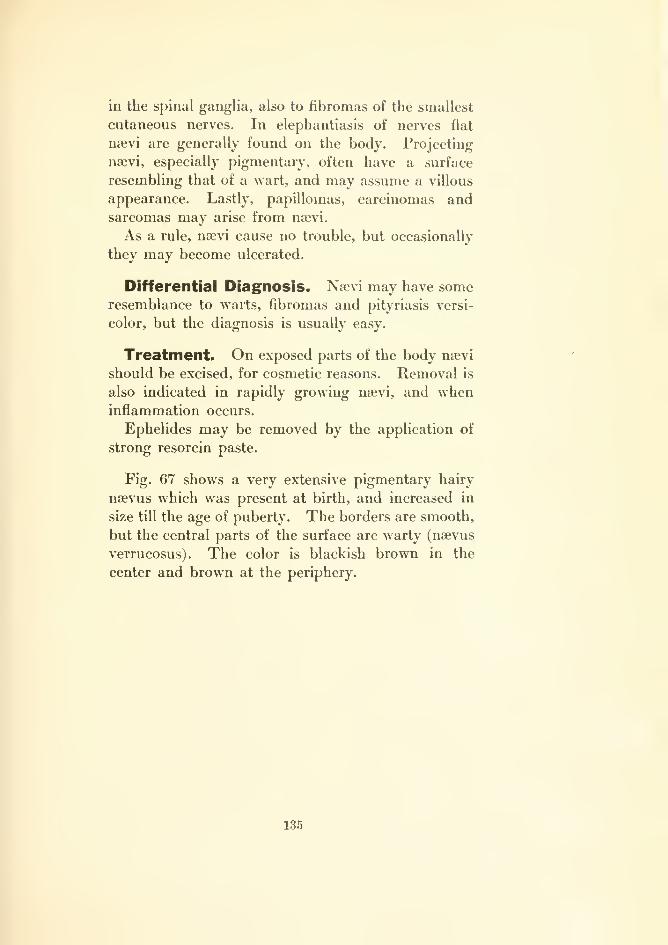
in the spinal ganglia, also to fibromas of the smallest
cutaneous nerves. In elephantiasis of nerves flat
nsevi are generally found on the body. Projecting
nsevi, especially pigmentary, often have a surface
resembling that of a wart, and may assume a villous
appearance. Lastly, papillomas, carcinomas andsarcomas may arise from nievi.
As a rule, nsevi cause no trouble, but occasionally
they may become ulcerated.
Differential Diagnosis. Nsevi may have someresemblance to warts, fibromas and pityriasis versi-
color, but the diagnosis is usually easy.
Treatment. On exposed parts of the body nsevi
should be excised, for cosmetic reasons. Removal is
also indicated in rapidly growing naevi, and wheninflammation occurs.
Ephelides may be removed by the application of
strong resorcin paste.
Fig. 67 shows a very extensive pigmentary hairy
naevus which was present at birth, and increased in
size till the age of puberty. The borders are smooth,
but the central parts of the surface are warty (nsevus
verrucosus). The color is blackish brown in the
center and brown at the periphery.
135
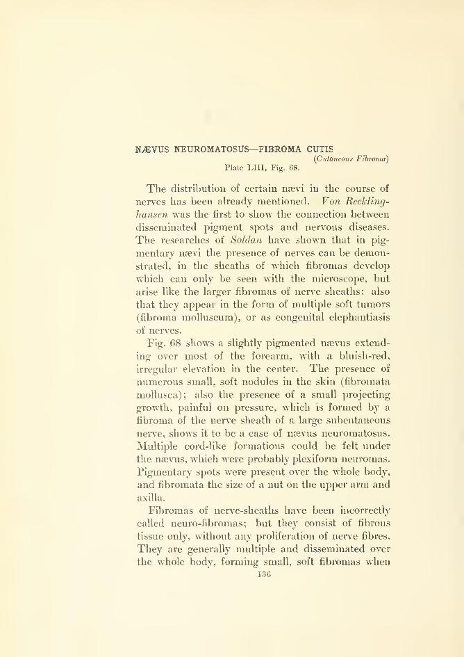
N^VUS NEUROMATOSUS—FIBROMA CUTIS{Cutaneous Fibroma)
Plate LIU, Fig. 68.
The distribution of certain nsevi in the course of
nerves has been already mentioned. Von Reckling-
hausen was the first to show the connection between
disseminated pigment spots and nervous diseases.
The researches of Soldan have shown that in pig-
mentary nfevi the presence of nerves can be demon-
strated, in the sheaths of which fibromas develop
which can only be seen with the microscope, but
arise like the larger fibromas of nerve sheaths; also
that they appear in the form of multiple soft tumors
(fibroma moUuscum), or as congenital elephantiasis
of nerves.
Fig. 68 shows a slightly pigmented naevus extend-
ing over most of the forearm, with a bluish-red,
irregular elevation in the center. The presence of
numerous small, soft nodules in the skin (fibromata
mollusca); also the presence of a small projecting
growth, painful on pressure, which is formed by a
fibroma of the nerve sheath of a large subcutaneous
nerv'e, shows it to be a case of noevus neuromatosus.
Multiple cord-like formations could be felt under
the naevus, which were probably plexiform neuromas.
Pigmentary spots were present over the whole body,
and fibromata the size of a nut on the upper arm and
axilla.
Fibromas of nerve-sheaths have been incorrectly
called neuro-fibromas; but they consist of fibrous
tissue only, without any proliferation of nerve fibres.
They are generally multiple and disseminated over
the whole body, forming small, soft fibromas when136
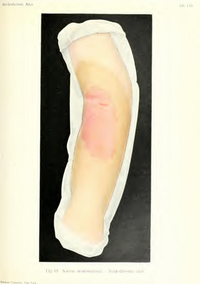
Bockenheimer, Atlas. Tab. LI I
Fig. 68. Naevus neuromatosus — Neurofibroma cutis.
Rcbman Company, New-York.

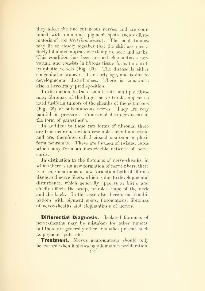
they affect the fine cutaneous nerves, and are com-bined with numerous pigment spots (neuro-fibro-
matosis of von Recklinghausen) . The small tumorsmay He so closely together that the skin assumes afinely lobulated appearance (temples, neck and back).
This condition has been termed elephantiasis ner-
vorum, and consists in fibrous tissue formation with
lymphatic vessels (Fig. 69). The disease is either
congenital or appears at an early age, and is due to
developmental disturbances. There is sometimes
also a hereditary predisposition.
In distinction to these small, soft, multiple fibro-
mas, fibromas of the larger nerve trunks appear as
hard fusiform tumors of the sheaths of the cutaneous
(Fig. 68) or subcutaneous nerves. They are very
painful on pressure. Functional disorders occur in
the form of paraesthesia.
In addition to these two forms of fibroma, there
are true neuromas which resemble cirsoid aneurism,
and are, therefore, called cirsoid neuroma or plexi-
form neuromas. These are formed of twisted cords
which may form an inextricable network of nerve
cords.
In distinction to the fibromas of nerve-sheaths, in
which there is no new formation of nerve fibers, there
is in true neuromas a new formation both of fibrous
tissue and nerve fibers, which is due to developmental
disturbance, which generally appears at birth, andchiefly affects the scalp, temples, nape of the neck
and the back. In this case also there occur combi-
nations with pigment spots, fibromatosis, fibromas
of nerve-sheaths and elephantiasis of nerves.
Differential Diagnosis. Isolated fibromas of
nerve-sheaths may be mistaken for other tumors,
but there are generally other anomalies present, such
as pigment spots, etc.
Treatment. Naevus neuromatosus should only
be excised when it shows papillomatous proliferation,
137
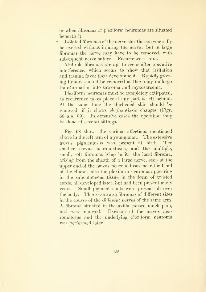
or when fibromas or plexiform neuromas are situated
beneath it.
Isolated fibromas of the nerve sheaths can generally
be excised without injuring the nerve; but in large
fibromas the nerve may have to be removed, with
subsequent nerve suture. Recurrence is rare.
Multiple fibromas are apt to recur after operative
interference, which seems to show that irritation
and trauma favor their development. Rapidly grow-
ing tumors should be removed as they may undergo
transformation into sarcoma and myxosarcoma.
Plexiform neuromas must be completely extirpated,
as recurrence takes place if any part is left behind.
At the same time the thickened skin should be
removed, if it shows elephantiasic changes (Figs.
68 and 69). In extensive cases the operation maybe done at several sittings.
Fig. 68 shows the various affections mentioned
above in the left arm of a young man. The extensive
nsevus pigmentosus was present at birth. Thesmaller nsaevus neuromatosus, and the multiple,
small, soft fibromas lying in it; the hard fibroma,
arising from the sheath of a large nerve, seen at the
upper end of the nsevus neuromatosus near the bend
of the elbow; also the plexiform neuroma appearing
in the subcutaneous tissue in the form of twisted
cords, all developed later, but had been present manyyears. Small pigment spots were present all over
the body. There were also fibromas of different sizes
in the course of the different nerves of the same arm.
A fibroma situated in the axilla caused much pain,
and was removed. Excision of the nsevus neu-
romatosus and the underlying plexiform neuroma
was performed later.
138


Bockenheimer, Atlas. Tab. LIV.
o
ta
no
c>
o
Rebman Company, New-York.

ELEPHANTIASIS NERVORUM {of the Nerves)
—FIBROMATA MOLLUSCAPlate LIV, Fig. 69.
Fig. 69 shows a similar case in a girl, aged twenty.
The whole of the right half of the scalp, the right side
of the forehead and the ear are the seat of a lobulated
growth (elephantiasis nervorum) fixed on the headlike a cap. The growth was congenital, and on its
surface are numerous pigment spots and soft, small,
painless tumors (fibromata mollusca). Numerouscord-like formations were found in it by palpation
(plexiform neuroma). The tumor was partially
removed by a curved incision, the scar of which is
shown in the figure. Total extirpation was per-
formed subsequently at several sittings. Microscopic
examination confirmed the above-mentioned explana-
tion of the affection, the lymphatic vessels being
increased and dilated in the region of the tumor.
139
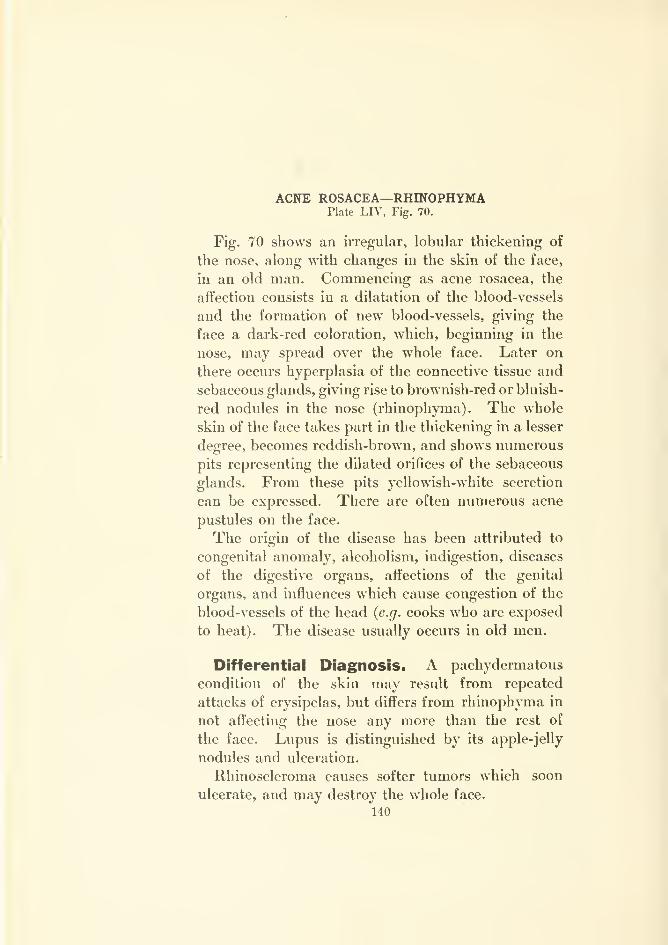
ACNE ROSACEA—RHmOPHYMAPlate LIV, Fig. 70.
Fig. 70 shows an irregular, lobular thickening of
the nose, along with changes in the skin of the face,
in an old man. Commencing as acne rosacea, the
affection consists in a dilatation of the blood-vessels
and the formation of new blood-vessels, giving the
face a dark-red coloration, which, beginning in the
nose, may spread over the whole face. Later on
there occurs hyperplasia of the connective tissue and
sebaceous glands, giving rise to brownish-red or bluish-
red nodules in the nose (rhiuophyma) . The whole
skin of the face takes part in the thickening in a lesser
degree, becomes reddish-brown, and shows numerous
pits representing the dilated orifices of the sebaceous
glands. From these pits yellowish-white secretion
can be expressed. There are often numerous acne
pustules on the face.
The origin of the disease has been attributed to
congenital anomaly, alcoholism, indigestion, diseases
of the digestive organs, affections of the genital
organs, and influences which cause congestion of the
blood-vessels of the head {e.g. cooks who are exposed
to heat). The disease usually occurs in old men.
Differential Diagnosis. A pachydermatous
condition of the skin may result from repeated
attacks of erysipelas, but differs from rhinophyma in
not affecting the nose any more than the rest of
the face. Lupus is distinguished by its apple-jelly
nodules and ulceration.
Rhinoscleroma causes softer tumors which soon
ulcerate, and may destroy the whole face.
140

Treatment. In the early stages massage of the
face and iuuuction of ichthyol-resorciii ointment (one
to ten per cent.) are useful. Attention should be
paid to the diet and all exciting causes avoided. In
rhinophyma the tumors may be excised or treated
with Pacquelin's thermo-cautery. Good results have
been obtained by peeling off the nodules with a
sharp knife (decortication). The wound is soon
covered by new epidermis, and the cosmetic results
are very satisfactory.
141

ELEPHANTIASIS PENIS LYMPHANGIECTATICA{Lymphangiedaiic elephantiasis of the penis)
Plate LV, Fig. 71.
In distinction to congenital elephantiasis of nerves
there is a second form of elephantiasis arabum, which
for various reasons chiefly affects the lower extremi-
ties, and is known as acquired elephantiasis or pachy-
dermia. It consists in a chronic, inflammatory
hyperplasia, and there is no formation of true
tumors. There is diffuse thickening of the connec-
tive tissue (fibromatosis), both in the cutis and in the
subcutaneous tissue. Finally the muscles are
attacked and replaced by hyperplastic connective
tissue. The periosteum of the bones may present
osteophytic deposits. Lastly, the epidermis takes
part in the proliferative process, so that the skin
becomes thickened and horny, or eczematous.
The affected parts thus become greatly thickened.
The thickening may be uniformly distributed, or mayassume a lobulated formation as in elephantiasis ner-
vorum. In addition to the proliferation of connective
tissue there is always dilatation of the blood-vessels
and lymphatics. The disease thus appears to origi-
nate in lymphatic engorgement, and the proliferation
of connective tissue results from lymphatic infiltration
of the tissues.
All processes which give rise to lymphatic engorge-
ment may, in certain cases, lead to elephantiasis.
For this reason, in the endemic form of this elephan-
tiasis which occurs especially in Arabia, Egypt, Aus-
tralia, and generally in tropical countries, it has been
assumed that the parasites (filaria sanguinis) block
up the lymphatic vessels, causing lymphatic varices
which rupture and deluge the tissues with lymph,142
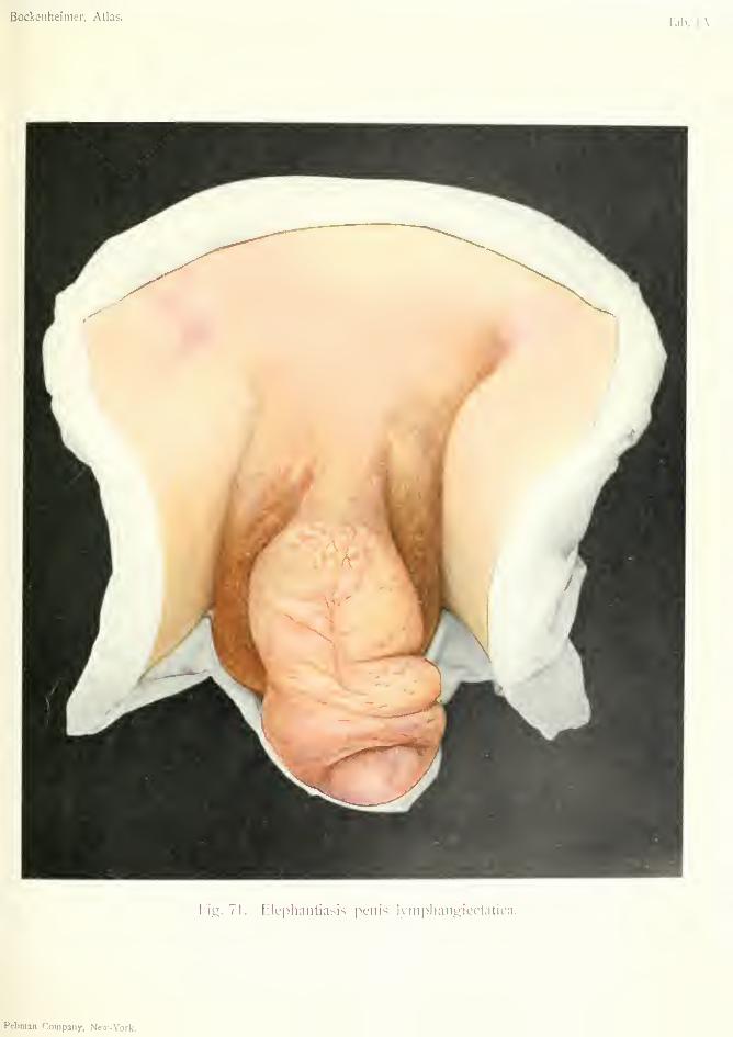
Bockenheinier, Atlas.
Fig. 7]. Elepliaiiliasis penis lympliaiigiectatica.
Rebman Company, Neu-Vork.

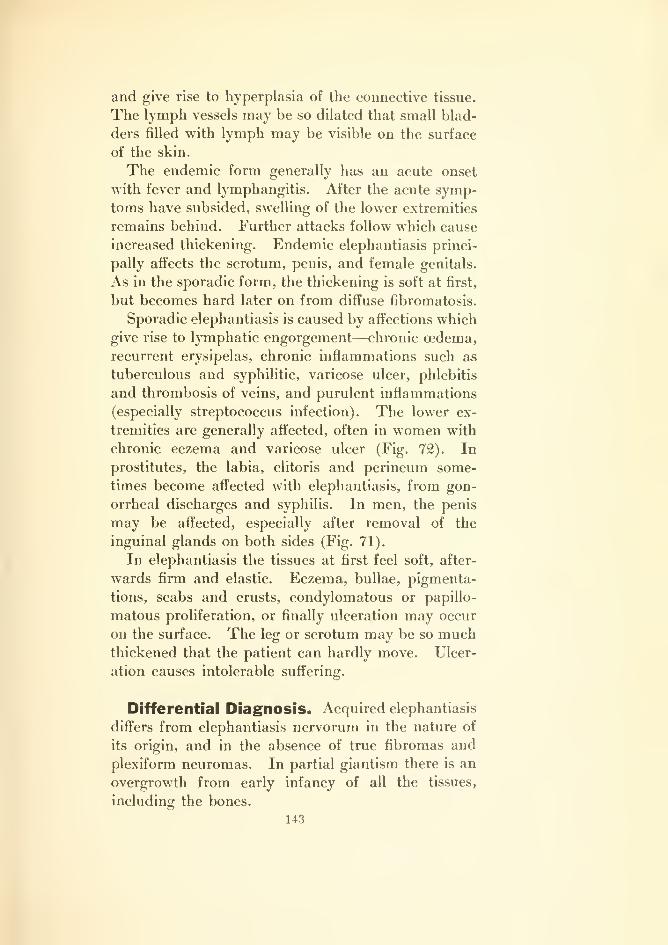
and give rise to hyperplasia of the connective tissue.
The lymph vessels may be so dilated that small blad-
ders filled with lymph may be visible on the surface
of the skin.
The endemic form generally has an acute onset
with fever and lymphangitis. After the acute symp-
toms have subsided, swelling of the lower extremities
remains behind. Further attacks follow which cause
increased thickening. Endemic elephantiasis princi-
pally affects the scrotum, penis, and female genitals.
As in the sporadic form, the thickening is soft at first,
but becomes hard later on from diffuse fibromatosis.
Sporadic elephantiasis is caused by affections which
give vise to lymphatic engorgement—chronic oedema,
recurrent erysipelas, chronic inflammations such as
tuberculous and syphilitic, varicose ulcer, phlebitis
and thrombosis of veins, and purulent inflammations
(especially streptococcus infection). The lower ex-
tremities are generally affected, often in women with
chronic eczema and varicose ulcer (Fig. 72). In
prostitutes, the labia, clitoris and perineum some-
times become affected with elephantiasis, from gon-
orrheal discharges and syphilis. In men, the penis
may be affected, especially after removal of the
inguinal glands on both sides (Fig. 71).
In elephantiasis the tissues at first feel soft, after-
wards firm and elastic. Eczema, bullae, pigmenta-
tions, scabs and crusts, condylomatous or papillo-
matous proliferation, or finally ulceration may occur
on the surface. The leg or scrotum may be so muchthickened that the patient can hardly move. Ulcer-
ation causes intolerable suffering.
Differential Diagnosis. Acquired elephantiasis
differs from elephantiasis nervorum in the nature of
its origin, and in the absence of true fibromas and
plexiform neuromas. In partial giantism there is an
overgrowi;h from early infancy of all the tissues,
including the bones.
143
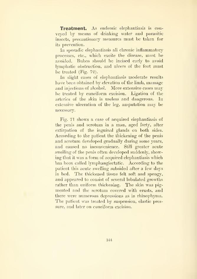
Treatment. As endemic elephantiasis is con-
veyed by means of drinking water and parasitic
insects, precautionary measures must be taken for
its prevention.
In sporadic elephantiasis all chronic inflammatory
processes, etc., which excite the disease, must be
avoided. Bubos should be incised early to avoid
lymphatic obstruction, and ulcers of the foot must
be treated (Fig. 72).
In slight cases of elephantiasis moderate results
have been obtained by elevation of the limb, massage
and injections of alcohol. More extensive cases maybe treated by cuneiform excision. Ligation of the
arteries of the skin is useless and dangerous. In
extensive ulceration of the leg, amputation may be
necessary.
Fig. 71 shows a case of acquired elephantiasis of
the penis and scrotum in a man, aged forty, after
extirpation of the inguinal glands on both sides.
According to the patient the thickening of the penis
and scrotum developed gradually during some years,
and caused no inconvenience. Still greater acute
swelling of the penis often developed suddenly, show-
ing that it was a form of acquired elephantiasis which
has been called lymphangiectatic. According to the
patient this acute swelling subsided after a few days
in bed. The thickened tissue felt soft and spongy,
and appeared to consist of several lobulated growths
rather than uniform thickening. The skin was pig-
mented and the scrotum covered with crusts, andthere were numerous depressions as in rhinophyma.
The patient was treated by suspension, elastic pres-
sure, and later on cuneiform excision.
144

V.'
/
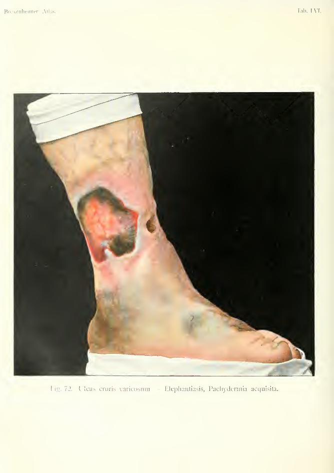
Bockenheimcf, Atlas. lab. LVI.
Fig. 72. Ulcus cruris varicosum — Elephantiasis, Pachydermia acquisita.
ULCUS CRURIS VARICOSUM {Varicose ulcer of the leg)
ELEPHANTIASIS S. PACHYDERMLA ACQUISITA{Acfjuired elcphaniias-is or pachydermia)
Plate LM, Fig. 72.
In this case an elephantiasic thickening of the
toes has developed in connection with a varicose
ulcer of the leg; which, as already explained (Plate
LV), is due to connective-tissue hyperplasia of the
skin resulting from lymphatic engorgement (acquired
lymphangiectatic pachydermia). The toes are enor-
mously thickened, and constricted in places; the
whole foot is also enlarged, and the arch of the foot
is obliterated. The thickening of the foot contin-
ually increased, and extended to the ankle. Frequent
attacks of erysipelas aggravated the affection.
At the lower third of the leg, on the inner side, is
an ulcer extending over nearly the whole circum-
ference of the leg. Ulcers develop in this situation
from various causes—blows on the leg, chronic ecze-
ma, abscess, erysipelas, thrombo-phlebitis, varicose
veins, burns and frost-bite.
These ulcers are most commonly connected with
disturbance in the blood and lymphatic circulation
both as regards their origin and chronic progress.
They generally occur in old people of the poorer
classes who have to do much standing, and are
especially aggravated by uncleanliness. They often
occur on both legs. Arteriosclerosis, diabetes, and
diseases of the central nervous system give rise to
especially obstinate and extensive ulcers (trophic
ulcer)
.
Varicose ulcer of the leg is characterized by its
irregular slightly raised edges, while the parts round
145
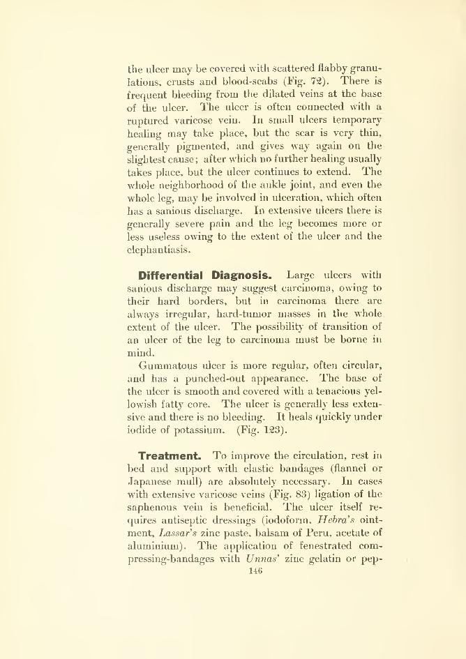
the ulcer may be covered with scattered flabby granu-
lations, crusts and blood-scabs (Fig. 72). There is
frequent bleeding from the dilated veins at the base
of the ulcer. The ulcer is often connected with a
ruptured varicose vein. In small ulcers temporary
healing may take place, but the scar is very thin,
generally pigmented, and gives way again on the
slightest cause; after which no further healing usually
takes place, but the ulcer continues to extend. Thewhole neighborhood of the ankle joint, and even the
whole leg, may be involved in ulceration, which often
has a sanious discharge. In extensive ulcers there is
generally severe pain and the leg becomes more or
less useless owing to the extent of the ulcer and the
elephantiasis.
Differential Diagnosis. Large ulcers with
sanious discharge may suggest carcinoma, owing to
their hard borders, but in carcinoma there are
always irregular, hard-tumor masses in the whole
extent of the ulcer. The possibility of transition of
an ulcer of the leg to carcinoma must be borne in
mind.
Gummatous ulcer is more regular, often circular,
and has a punched-out appearance. The base of
the ulcer is smooth and covered with a tenacious yel-
lowish fatty core. The ulcer is generally less exten-
sive and there is no bleeding. It heals quickly under
iodide of potassium. (Fig. 1:23).
Treatment. To improve the circulation, rest in
bed and support with elastic bandages (flannel or
Japanese mull) are absolutely necessary. In cases
with extensive varicose veins (Fig. 83) ligation of the
saphenous vein is beneficial. The ulcer itself re-
quires antiseptic dressings (iodoform, Hebra's oint-
ment, Lassar's zinc paste, balsam of Peru, acetate of
aluminium). The application of fenestrated com-
pressing-bandages with Unnas' zinc gelatin or pep-
146

tonated paste is also recommended. In out-patient
practice compressing bandages of mastich or starch
may be used. Compressing bandages should be left
on for several weeks, and the ulcer can be treated
daily through the hole in the bandage.
In very obstinate ulcers incisions above the ulcer
have been recommended to improve the circulation.
Other measures are scraping, cauterization, or exci-
sion of the whole ulcer followed by skin grafting.
Very severe cases, and those suspected of carcinoma,
may require amputation.
147
DECOLLEMENT DE LA PEAU {Detachment 0} the Skin)
Plate LVn, Fig. 73.
Detachment of the skin is a term applied by
Morel-Lavallee and Kbhler to a lesion which consists
in subcutaneous separation of the skin from the sub-
jacent tissues and fascia. The skin itself is unin-
jured, as the lesion is produced by a force acting at a
tangent which separates the skin from its foundations.
The lesion is more liable to occur in the neighborhood
of the elbow joint, and over the tibia {e.g. after being
run over) . Besides the detachment of skin the deeper
structures may be severely injured and the bones
fractured. The blood-vessels and lymphatics are
injured, giving rise to effusion into the newly formed
subcutaneous space and bulging of the skin. If
the larger blood-vessels are torn there is subcutane-
ous effusion of blood and dark-red discoloration
of the skin, forming an extensive tense swelling
which generally disappears quickly. If the larger
lymphatic vessels are torn, as usually happens,
the lymphatic effusion often appears several hours
after the injury. The skin is hardly altered, per-
haps somewhat livid and excoriated, while the
subcutaneous swelling subsides slowly, owing to the
long, continual effusion of lymph.
The lymphatic effusion, which is generally more or
less mixed with blood, accumulates in the dependent
parts of the injured region. Fluctuation of the fluid
in the subcutaneous cavity can be felt.
Treatment. Subcutaneous effusion of blood soon
undergoes spontaneous absorption. The lymphatic
effusion gradually disappears after repeated punc-
148
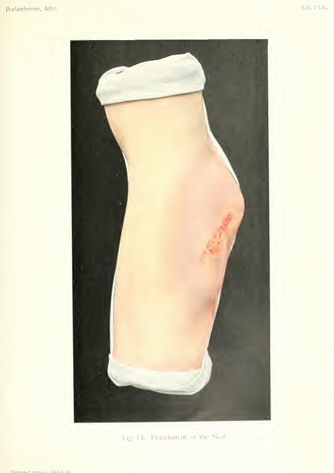
Bockeiiheimer, Atlas. Tab. LVl
I'igf. 73. Dclacluiicnt of tlie Skin.
P^hmsn rrtmn^ni- M^^-\'nrlf

ture, injection of tincture of iodine and compression
by bandages. Incision should only be performed if
there is suppuration.
Fig. 73 shows a detachment of the skin resulting
from a blow on the left elbow. A few days after the
injury effusion took place in the subcutaneous cavity,
chiefly in the forearm. The cavity was not com-pletely filled so that several swellings are shown.
There is a slight abrasion of the skin over the ole-
cranon, the appearance and direction of which showthat the blow was a tangential one. The skin is livid
over the whole swelling. Yellowish fluid was evacu-
ated by puncture, showing very slight mixture with
blood.
Submucous effusion in the nasal septum and in the
larynx may also be caused by the action of tangential
force (generally foreign bodies). Here also the effu-
sion only occurs where the submucous tissue is
situated over a hard substratum of cartilage.
149
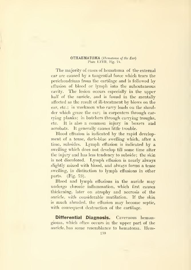
OTHAEMATOMA (Hematoma of the Ear)
Plate LVIII, Fig. 74.
The majority of cases of hematoma of the external
ear are caused by a tangential force which tears the
perichondrium from the cartilage and is followed byeffusion of blood or lymph into the subcutaneous
cavity. The lesion occurs especially in the upper
half of the auricle, and is found in the mentally
affected as the result of ill-treatment by blows on the
ear, etc.; in workmen who carry loads on the shoul-
der which graze the ear; in carpenters through car-
rying planks; in butchers through carrying troughs,
etc. It is also a common injury in boxers andacrobats. It generally causes little trouble.
Blood effusion is indicated by the rapid develop-
ment of a tense, dark-blue swelling which, after a
time, subsides. Lymph effusion is indicated by a
swelling which does not develop till some time after
the injury and has less tendency to subside; the skin
is not discolored. Lymph effusion is nearly always
slightly mixed with blood, and always forms a tense
swelling, in distinction to lymph effusions in other
parts. (Fig. 73).
Blood and lymph effusions in the auricle mayundergo chronic inflammation, which first causes
thickening, later on atrophy and necrosis of the
auricle, with considerable mutilation. If the skin
is much abraded, the effusion may become septic,
with consequent destruction of the cartilage.
Differential Diagnosis. Cavernous heman-gioma, which often occurs in the upper part of the
auricle, has some resemblance to hematoma. Hem-150

Bockenheimer, Atlas. Tab. LVIII.
i;3
in
zr.
SjO
Rebman Company, N'cw-York.

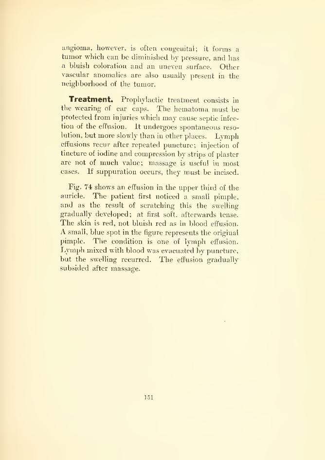
angioma, however, is often congenital; it forms atumor which can be diminished by pressure, and hasa bluish coloration and an uneven surface. Othervascular anomalies are also usually present in the
neighborhood of the tumor.'ti^
Treatment. Prophylactic treatment consists in
the wearing of ear caps. The hematoma must beprotected from injuries which may cause septic infec-
tion of the effusion. It undergoes spontaneous reso-
lution, but more slowly than in other places. Lympheffusions recur after repeated puncture; injection of
tincture of iodine and compression by strips of plaster
are not of much value; massage is useful in mostcases. If suppuration occurs, they must be incised.
Fig. 74 shows an effusion in the upper third of the
auricle. The patient first noticed a small pimple,
and as the result of scratching this the swelling
gradually developed; at first soft, afterwards tense.
The skin is red, not bluish red as in blood effusion.
A small, blue spot in the figure represents the original
pimple. The condition is one of lymph effusion.
Lymph mixed with blood was evacuated by puncture,
but the swelling recurred. The effusion gradually
subsided after massage.
151

HEMANGIOMA SIMPLEX (Simple Hemangioma)
Plate LVIII, Fig. 75 (of. also Figs. 76 and 81).
The term Angioma includes new growths arising
from blood-vessels and lymphatics; the former are
called hemangiomas, the latter lymphangiomas.
Hemangiomas may be simple or cavernous (cav-
ernoma, Figs. 36 and 80).
The red spots formed by tortuous and dilated
blood-vessels (telangiectasis, nsevus vasculosus) are
by some classed as tumors and included among the
simple hemangiomas; by others they are considered
as hypertrophic formations, and not as true tumors,
as they consist in dilatation, lengthening and tor-
tuosity of the vessels, rather than new formation of
vessels. The form known as racemose or plexiform
angioma also almost always consists in a dilatation
of a vascular region, not a true, new formation of
vessels. It is, therefore, better to give the namecirsoid aneurism to these formations, which are
usually congenital and due to fetal remains, but
sometimes traumatic. Lastly, neither aneurisms nor
varices belong to true vascular tumors. The red
spots with more or less regular outlines, often only
punctiform, which occur in the skin of old persons,
are also not true tumors but only dilated and tor-
tuous blood-vessels (telangiectases). A form de-
scribed by Ziegler as hypertrophic angioma is best
named hemangio-endothelioma, as, in addition to
new formation of vessels, there is extensive prolifera-
tion of the endothelium.
Clinically, we distinguish telangiectases, which are
situated superficially in the skin, from simple hem-152

angiomas which appear in the skin and subcutaneous
tissue. The latter tumors, also called angiomas(Fig. 75), appear as raised growths with well-defined
borders. The overlying skin is thin and adherent,
and of a reddish-blue color. In places there are
islands of normal skin. The edges of cutaneous
angiomas are dark-red, slightly raised, and often
bordered by an areola of fine ramifying blood-vessels.
The tumors are soft, spongy, somewhat compres-
sible, and easily movable over subjacent parts. Theyare sometimes present at birth and are often situated
on the face, lips, cheeks and neck, in the regions of
the fetal clefts. In other cases they appear soon
after birth, usually in the form of slow-growing red
spots. Angiomas distributed in the region of the
trigeminal nerve have been called neuropathic angio-
mas. Angiomas may also develop in scars.
More extensive growth may form large, nodular
lobulated tumors, which when situated in the orbit
may be dangerous from extension to the brain; but
they cannot be regarded as malignant tumors, be-
cause they give rise to no metastases.
Involution of angioma has been observed as the
result of inflammation. Angiomas are usually mul-
tiple, cutaneous or subcutaneous. They may also
occur in the muscles, bones, brain, breast and liver,
generally in the form of cavernous hemangioma.
They cause no trouble apart from that due to their
disfigurement.
Differential Diagnosis. The tumors are so
typical that they cannot easily be mistaken. Sub-
cutaneous hemangioma generally appears later
under the skin, which gradually assumes a bluish
coloration.
Cavernous hemangioma (cf. Figs. 36 and 80)
appears as a multilobular swelling, which diminishes
on pressure. When it forms in the skin, the latter
is colored bluish green (Fig. 81).
153
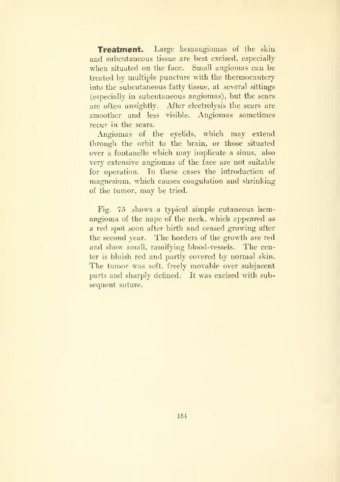
Treatment. Large hemangiomas of the skin
and subcutaneous tissue are best excised, especially
when situated on the face. Small angiomas can be
treated by multiple puncture with the thermocautery
into the subcutaneous fatty tissue, at several sittings
(especially in subcutaneous angiomas), but the scars
are often unsightly. After electrolysis the scars are
smoother and less visible. Angiomas sometimes
recur in the scars.
Angiomas of the eyelids, which may extend
through the orbit to the brain, or those situated
over a fontanelle which may implicate a sinus, also
very extensive angiomas of the face are not suitable
for operation. In these cases the introduction of
magnesium, which causes coagulation and shrinking
of the tumor, may be tried.
Fig. 75 shows a typical simple cutaneous hem-
angioma of the nape of the neck, which appeared as
a red spot soon after birth and ceased growing after
the second year. The borders of the growth are red
and show small, ramifying blood-vessels. The cen-
ter is bluish red and partly covered by normal skin.
The tumor was soft, freely movable over subjacent
parts and sharply defined. It was excised with sub-
sequent suture.
154

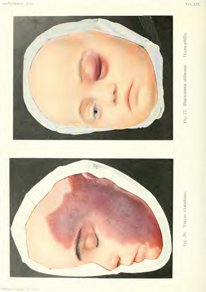
Bockenheimer, Atlas. Tab. LIX.
o
o
>
>
t/3
Rebman Company, New-Vork.
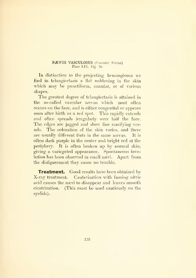
HMVUS VASCULOSUS (Vascular Nannui)Plate LIX, Fig. 76.
In distinction to the projecting hemangioma wefind in telangiectasis a flat reddening in the skin
which may be punctiform, annular, or of various
shapes.
The greatest degree of telangiectasis is attained in
the so-called vascular naevus which most often
occurs on the face, and is either congenital or appears
soon after birth as a red spot. This rapidly extends
and often spreads irregularly over half the face.
The edges are jagged and show fine ramifying ves-
sels. The coloration of the skin varies, and there
are usually different tints in the same naevus. It is
often dark purple in the center and bright red at the
periphery. It is often broken up by normal skin,
giving a variegated appearance. Spontaneous invo-
lution has been observed in small naevi. Apart from
the disfigurement they cause no trouble.
Treatment. Good results have been obtained byX-ray treatment. Cauterization with fuming nitric
acid causes the naevi to disappear and leaves smooth
cicatrization. (This must be used cautiously on the
eyelids)
.
155
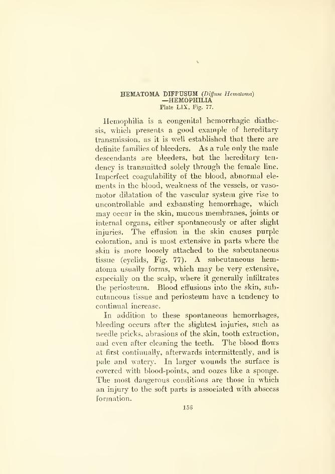
HEMATOMA DIFFUSUM {Diffuse Hematoma)—HEMOPHILIAPlate LIX, Fig. 77.
Hemophilia is a congenital hemorrhagic diathe-
sis, which presents a good example of hereditary
transmission, as it is well established that there are
definite families of bleeders. As a rule only the male
descendants are bleeders, but the hereditary ten-
dency is transmitted solely through the female line.
Imperfect coagulability of the blood, abnormal ele-
ments in the blood, weakness of the vessels, or vaso-
motor dilatation of the vascular system give rise to
uncontrollable and exhausting hemorrhage, which
may occur in the skin, mucous membranes, joints or
internal organs, either spontaneously or after slight
injuries. The effusion in the skin causes purple
coloration, and is most extensive in parts where the
skin is more loosely attached to the subcutaneous
tissue (eyelids, Fig. 77). A subcutaneous hem-
atoma usually forms, which may be very extensive,
especially on the scalp, where it generally infiltrates
the periosteum. Blood effusions into the skin, sub-
cutaneous tissue and periosteum have a tendency to
continual increase.
In addition to these spontaneous hemorrhages,
bleeding occurs after the slightest injuries, such as
needle pricks, abrasions of the skin, tooth extraction,
and even after cleaning the teeth. The blood flows
at first continually, afterwards intermittently, and is
pale and watery. In larger wounds the surface is
covered with blood-points, and oozes like a sponge.
The most dangerous conditions are those in which
an injury to the soft parts is associated with abscess
formation.
156
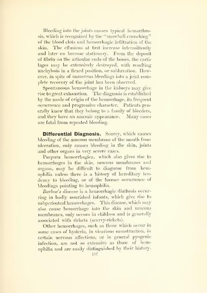
Bleeding into the joints causes typical honiarthro-
sis, which is recognized by the "snowball crunching"
of the blood clots and hemorrhagic infiltration of the
skin. The effusions at first increase intermittently
and later on become stationary. From the deposit
of fibrin on the articular ends of the bones, the carti-
lages may be extensively destroyed, with resulting
anchylosis in a flexed position, or subluxation. How-ever, in spite of numerous bleedings into a joint com-
plete recovery of the joint has been observed.
Spontaneous hemorrhage in the kidneys may give
rise to great exhaustion. The diagnosis is established
by the mode of origin of the hemorrhage, its frequent
occurrence and progressive character. Patients gen-
erally know that they belong to a family of bleeders,
and they have an anaemic appearance. Many cases
are fatal from repeated bleeding.
Differential Diagnosis. Scur\7, which causes
bleeding of the mucous membrane of the mouth from
ulceration, only causes bleeding in the skin, joints
and other organs in very severe cases.
Purpura hemorrhagica, which also gives rise to
hemorrhages in the skin, mucous membranes and
organs, may be difficult to diagnose from hem-
ophilia unless there is a history of hereditary ten-
dency to bleeding, or of the former occurrence of
bleedings pointing to hemophilia.
Barlow's disease is a hemorrhagic diathesis occur-
ring in badly nourished infants, which give rise to
subperiosteal hemorrhages. This disease, which mayalso cause hemorrhage into the skin and mucous
membranes, only occurs in children and is generally
associated with rickets (scurvy-rickets).
Other hemorrhages, such as those which occur in
some cases of hysteria, in vicarious menstruation, in
certain nervous affections, or in general pyogenic
infection, are not so extensive as those of hem-
ophilia and are easily distinguished by their history.
157
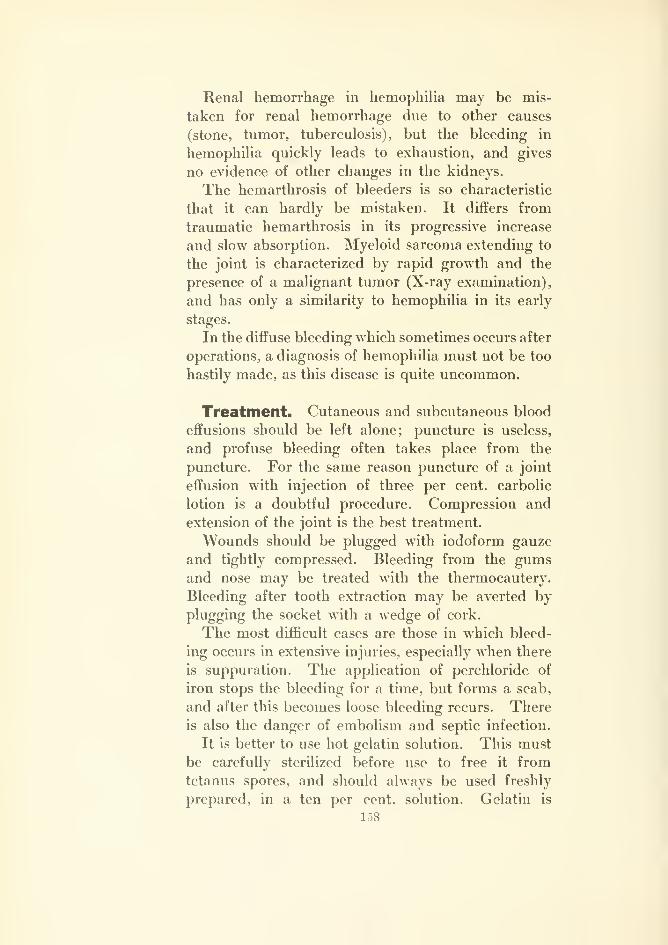
Renal hemorrhage in hemophilia may be mis-
taken for renal hemorrhage due to other causes
(stone, tumor, tuberculosis), but the bleeding in
hemophilia quickly leads to exhaustion, and gives
no evidence of other changes in the kidneys.
The hemarthrosis of bleeders is so characteristic
that it can hardly be mistaken. It differs from
traumatic hemarthrosis in its progressive increase
and slow absorption. Myeloid sarcoma extending to
the joint is characterized by rapid growth and the
presence of a malignant tumor (X-ray examination),
and has only a similarity to hemophilia in its early
stages.
In the diffuse bleeding which sometimes occurs after
operations, a diagnosis of hemophilia must not be too
hastily made, as this disease is quite uncommon.
Treatment. Cutaneous and subcutaneous blood
effusions should be left alone; puncture is useless,
and profuse bleeding often takes place from the
puncture. For the same reason puncture of a joint
effusion with injection of three per cent, carbolic
lotion is a doubtful procedure. Compression and
extension of the joint is the best treatment.
Wounds should be plugged with iodoform gauze
and tightly compressed. Bleeding from the gumsand nose may be treated with the thermocautery.
Bleeding after tooth extraction may be averted byplugging the socket with a wedge of cork.
The most difficult cases are those in which bleed-
ing occurs in extensive injuries, especially when there
is suppuration. The application of perchloride of
iron stops the bleeding for a time, but forms a scab,
and after this becomes loose bleeding recurs. There
is also the danger of embolism and septic infection.
It is better to use hot gelatin solution. This must
be carefully sterilized before use to free it from
tetanus spores, and should always be used freshly
prepared, in a ten per cent, solution. Gelatin is
158

also useful administered internally or by subcuta-
neous injection. In extensive, uncontrollable bleed-
ing affecting the extremities amputation may have
to be considered; in this, all the vessels must be
carefully ligatured.
In renal hemophilia nephrotomy and nephrectomy
has proved successful.
During the bleeding, which often ceases sponta-
neously after a time, the patient's general condition
must be kept up by forced nourishment. Bleeders
must naturally avoid everything which may cause
bleedinsr.'t)'
Fig. 77 shows blood effusion into the subcutaneous
and subconjunctival tissue of both eyelids, and an
extensive hematoma on the left side of the forehead
in a child aged six years, who belonged to a family of
bleeders. The effusions occurred spontaneously; the
one on the forehead occurred intermittently for a
time and then gradually subsided. There was no
bleeding in any other part of the body.
159
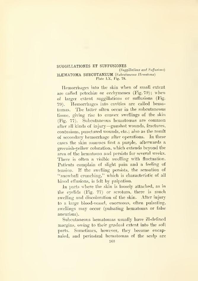
SUGGILLATIONES ET SUFFUSIONES{Suggillations and Snffrmons)
HiEMATOMA SUBCUTANEUM (Subcutaneous Hematoma)Plate LX, Fig. 78.
Hemorrhages into the skin when of small extent
are called petechise or ecchymoses (Fig. 79) ; when
of larger extent suggillations or suffusions (Fig.
79). Hemorrhages into cavities are called hema-
tomas. The latter often occur in the subcutaneous
tissue, giving rise to convex swellings of the skin
(Fig. 77). Subcutaneous hematomas are commonafter all kinds of injury—gunshot wounds, fractures,
contusions, punctured wounds, etc. ; also as the result
of secondary hemorrhage after operations. In these
cases the skin assumes first a purple, afterwards a
greenish-yellow coloration, which extends beyond the
area of the hematoma and persists for several weeks.
There is often a visible swelling with fluctuation.
Patients complain of slight pain and a feeling of
tension. If the swelling persists, the sensation of
"snowball crunching," which is characteristic of all
blood effusions, is felt by palpation.
In parts where the skin is loosely attached, as in
the eyelids (Fig. 77) or scrotum, there is muchswelling and discoloration of the skin. After injury
to a large blood-vessel, enormous, often pulsating,
swellings may occur (pulsating hematoma or false
aneurism)
.
Subcutaneous hematomas usually have ill-defined
margins, owing to their gradual extent into the soft
parts. Sometimes, however, they become encap-
suled, and periosteal hematomas of the scalp are
160

Bockenlieimer, Atlas. Tab. LX.
1
•Tt
CO


surrounded by a wall of bony hardness (also in
cephalhematoma)
.
Treatment. Light compression by bandages
soon causes resorption of the effusion. In delayed
resorption the fluid may he evacuated by puncture.
If suppuration occurs an incision must be made.
Fig. 78 shows extensive suggillations and suffu-
sions of the skin of the whole arm, which is colored
purple, brownish-red, green and yellow. Thepresence of a subcutaneous hematoma is shown byswelling and fluctuation.
It is a typical case of gunshot injury to the soft
parts, in which the apertures of entry and exit are
characteristic. The aperture of entry is smaller than
that of exit and shows radiating processes in the skin.
The skin is colored black and contains granules of
powder, owing to the shot being fired at close quarters.
In shot wounds of the face these powder granules
remain for a long time after the wound has healed,
and cause an unsightly appearance. The aperture
of exit is larger with irregular everted borders. Thesewounds are typical of modern projectiles with great
penetrating power.
Septic infection does not usually occur in gunshot
wounds, as the bactericidal power of the organism is
sufficient to counteract the slight infection caused by
projectiles. Even infected foreign bodies, such as
shreds of cloth, may heal up in the body.
The prognosis of gunshot wounds of the soft parts
is good if undue interference is avoided. All prob-
ing of the wound and search for the bullet is to be
condemned, as it generally sets up virulent infection
of the wound. Disinfection of the wound is also
unnecessar}\ The best treatment is to apply an
antiseptic sterilized gauze dressing (iodoform gauze
if there is much bleeding) and keep the part at rest;
in the extremities by the aid of plaster of Paris. By161
this simple treatment, first introduced by von Berg-
viann, the best results are obtained, not only in gun-
shot wounds of the soft parts, but also in wounds of
the joints and bones, even comminuted fractures.
In gunshot injuries of large blood-vessels opera-
tive interference is necessary; e.g. ligation of the
middle meningeal artery.
If the wound becomes infected, as often happens
after injuries with explosive bullets (dum-dum bul-
lets, etc.), a free incision must be made to give outlet
to the pus. Bullets and pieces of clothing which
have become healed over may give rise to abeess
after some years.
As a rule bullets should be left alone ; a bullet has
even remained in the apex of the heart without caus-
ing trouble {Trendelenburg) . Only superficially situ-
ated bullets should be removed, after locating them
by means of the X-rays. Bullets in the frontal or
maxillary sinuses, or in the mastoid process should
be removed, as they give rise to pain and chronic
catarrh. Bullets should also be removed which
cause pressure on tendons and nerves, or are situated
in the phalanges, or prevent union of fractures.
Blank cartridges, in which tetanus spores are often
present, should be removed on account of the danger
of tetanus. In war, there is always a danger of
tetanus infection of every large bullet wound, from
the presence of tetanus bacilli in the ground on which
the wounded lie. As the treatment of antitoxin is
only efficacious before the tetanus appears and is too
complicated to be used in warfare, the author recom-
mends, on the strength of experimental research, the
application of fat to wounds suspected of tetanus
infection, as fatty substances attenuate the tetanus
toxin (Surgical Congress, 1907 Bockenheimer's anti-
tetanus ointment).
The search for deep-seated bullets in the brain
causes much injury. Accumulation of blood or cere-
brospinal fluid, may abolish the reflexes for a time,
162
and paralysis may appear, but in spite of this recoverymay take place after a time.
Bullets situated outside the cortex of the brainmust be removed when convulsions occur from pres-
sure of the bullet, or a splinter of bone, or an accu-mulation of blood or pus.
Effusion of blood in the thorax through wound of
the lung should be left to be resorbed. If it becomesso extensive as to displace the heart puncture mustbe performed, and if suppuration occurs resection of
the ribs.
In gunshot wounds of the heart, free exposure of
the organ may be performed in some cases.
Gunshot wounds of the abdomen require laparot-
omy at the earliest possible opportunity.
In wounds of the larj-nx immediate tracheotomy is
necessary to avoid death from asphyxia.
In the above-mentioned cases resorption of the
blood effusion and healing of the wounds takes place
in a few weeks under the application of fixed aseptic
dressings.
163
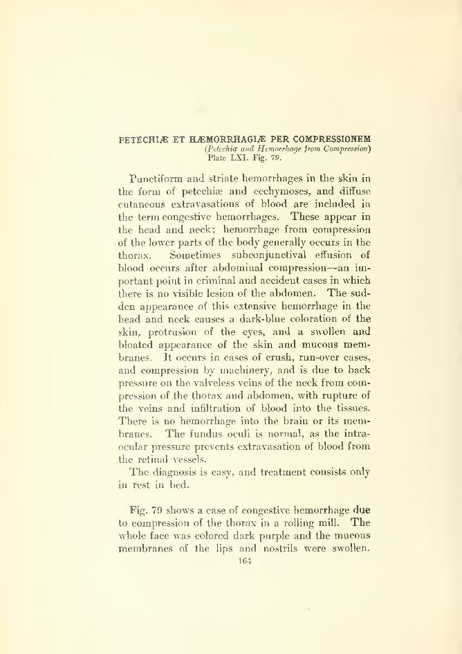
PETECHLS ET affiMORRHAGLffi) PER COMPRESSIONEM{Petechia and Hemorrhage from Compression)
Plate LXI, Fig. 79.
Punctiform and striate hemorrhages in the skin in
the form of petechise and ecchymoses, and diffuse
cutaneous extravasations of blood are included in
the term congestive hemorrhages. These appear in
the head and neck; hemorrhage from compression
of the lower parts of the body generally occurs in the
thorax. Sometimes subconjunctival effusion of
blood occurs after abdominal compression—an im-
portant point in criminal and accident cases in which
there is no visible lesion of the abdomen. The sud-
den appearance of this extensive hemorrhage in the
head and neck causes a dark-blue coloration of the
skin, protrusion of the eyes, and a swollen and
bloated appearance of the skin and mucous mem-branes. It occurs in cases of crush, run-over cases,
and compression by machinery, and is due to back
pressure on the valveless veins of the neck from com-
pression of the thorax and abdomen, with rupture of
the veins and infiltration of blood into the tissues.
There is no hemorrhage into the brain or its mem-branes. The fundus oculi is normal, as the intra-
ocular pressure prevents extravasation of blood from
the retinal vessels.
The diagnosis is easy, and treatment consists only
in rest in bed.
Fig. 79 shows a case of congestive hemorrhage due
to compression of the thorax in a rolling mill. Thewhole face was colored dark purple and the mucous
membranes of the lips and nostrils were swollen.
164
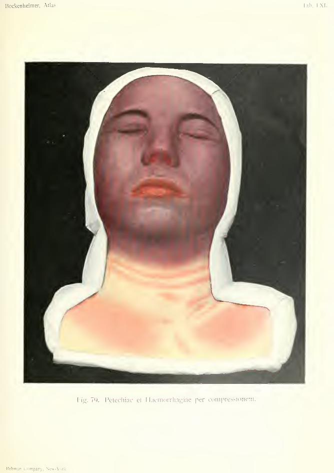
Bockenheimer, Atlas.
big. 7Q. Petecliiae et Haemorrliagiae per comprcssionem.
Rcbman Company, New-York.


There was also subconjunctival effusion of blood.
In the neck, the continuous purple coloration of the
face was replaced by a brighter red coloration in the
form of stripes (petechiie and ecchymoses). Thepetechise were situated over the shoulder and the
upper part of the back; also in the auditory canal
and the tympanic membrane. No visible lesion waspresent. The swelling of the face disappeared in a
few days, and the purple coloration subsided in the
course of time without any treatment. The discol-
oration remained longest in the eyelids and con-
junctiva.
165

HEMANGIOMA CAVERNOSUM SUBCUTAmEUM{Subcutaneous Cavernous Hemangioma)
Plate LXn. Fig. 80.
Fig. 80 shows a subcutaneous cavernous heman-gioma, which often occurs in the region of the rectus
abdominis muscle, sometimes in the muscle itself.
Mention has already been made of cavernoma in
Plate XXVII. They occur most frequently in the
skin and subcutaneous tissue, where their purple
color and lobulated surface has somewhat the appear-
ance of a mulberry. They are often combined with
simple hemangioma or with telangiectases, and often
appear soon after birth. In cutaneous hemangiomathe skin is much thinned and appears lobulated andof a bluish-black color. In subcutaneous heman-gioma the skin may be unaltered at first, or slightly
irregular and marked by telangiectases. Afterwardsthe skin becomes thinned or destroyed by pressure of
the subcutaneous growth, and assumes various colors
(Fig. 80). In the case represented in the figure the
skin is already destroyed over the blue parts of the
growth, and is of a livid color at the peripherj'. Thegrowth is encapsuled and freely movable over
the abdominal fascia (in distinction to infiltrating
cavernoma). In some parts the cavernous spaces
can be seen through the surface. In the center of the
growth the skin is yellow in some parts and brown in
others. The growth was soft, elastic and com-pressible; in some places there was thrombosis with
consequent shrinking. The growth had remainedstationary for a year.
Subcutaneous cavernomas of the scalp require
special mention, as they may communicate with a
166
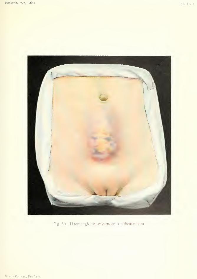
Bockenheimer, Atlas. lab. LXII
Fig. 80. Haemangioma cavernosum subcutaneum.
Rebman Company, New-York.


sinus through the emissary vessels, without the scalp
showing much change.
For Differential Diagnosis and Treatmentsee Plate XXVII, Fig. 3(i.
On account of the danger of rupture and hemor-
rhage, the case in Fig. 80 was extirpated and the
wound closed by suture. Recurrence sometimes
occurs after total extirpation.
167

HEMANGIOMA CUTANEDM ET SUBCUTAWEUM(Subcutaneous and Cutaneous Hemangioma)
TELEANGIEKTASIAE {Teleangiectases)
Plate LXIII, Fig. 81.
Fis. 81 shows a combination of cutaneous and sub-
cutaneous hemangiomas with telangiectases, affect-
ing the leg. The telangiectases are seen as red spots,
in some places arranged in the form of a wreath.
There is also an extensive subcutaneous heman-
gioma, of a bluish-red color, with more or less nor-
mally colored skin in the central parts. These
growths may remain covered by intact skin for a
long time, while the growth seen through it gives it a
bluish coloration. In this case, at the lower part of
the subcutaneous hemangioma, there were cutaneous
hemangiomas in the form of more elevated, round for-
mations in the skin, resembling the simple cutaneous
hemangioma represented in Fig. 75. In the whole
region of the subcutaneous hemangioma fine ramify-
ing blood-vessels can be seen. In the face, combina-
tions of cutaneous and subcutaneous hemangiomas
sometimes form a characteristic appearance, the sub-
cutaneous growth giving a blue color to the skin,
while the cutaneous angioma appears in the form of
lobulated growths or of bluish-red nodules projecting
from the surface. In Fig. 81 the difference in color
between the cutaneous and the subcutaneous heman-
giomas is very marked, the former being red, the
latter bluish in color. A combination of sub-
cutaneous with cutaneous hemangioma and telangi-
ectases is not very rare. The cutaneous heman-
gioma sometimes develops when the subcutaneous
growth appears under the skin.
For Differential Diagnosis and Treatmentsee Fig. 75.
168
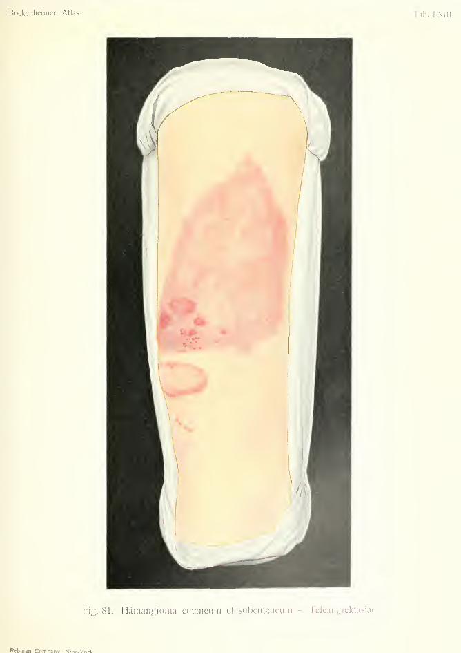
Bockenheimer, Atlas. Tab. LXIII.
Fiy. 81. Hamangioma cutaiieiim et subcutancum — Tclcangiektasiae.
Rebman Comnanv. Kpw.VnrV


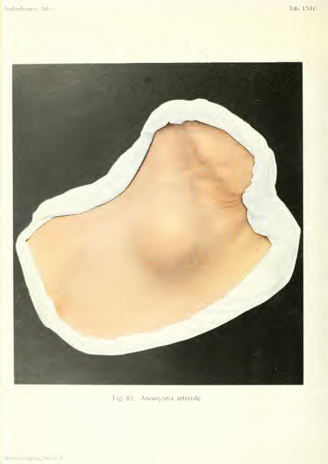
Tab. LXIV.
Fig. 82. Aiieurysiiia arteriale.
ANEURISMA ARTERIALE (Arterial Aneurism)Plate LXIV. Fig. 8i.
An aneurism is a partial dilatation of an artery.
The term true aneurism is applied to those dilatations
which are formed by all the three coats of the artery.
Through wearing away of the arterial wall, the blood
escapes from the vessel and is enclosed by the neigh-
boring soft parts, forming a false aneurism. A form
of false aneurism has already been mentioned as
pulsating hematoma (Fig. 78) ; in this case there is
a subcutaneous injury to large blood-vessels.
In both true and false aneurism we distinguish a
circumscribed and a diffuse form, but the classifica-
tion of aneurisms into cylindrical, saccular and fusi-
form is of no importance, and only has a clinical
interest in cirsoid aneurisms (Fig. 75).
True aneurisms are caused by disease of the
arterial walls from infective diseases, chiefly syphilis.
When the morbid processes extend over large areas
of the arterial system the aneurisms may be multiple.
These occur especially in the small arteries of the
brain, sometimes also in the lungs, and by their rup-
ture give rise to multiple apoplexy. This occurs
chiefly in syphilitic disease of the arteries, and in the
arteriosclerosis of young people.
True aneurisms are often situated in the ascending
aorta (syphilis), also in places where the arteries are
liable to traction or pressure from flexion of the
extremities: e.g. aneurism of the femoral artery from
pressure of an osseous growth ("rider's bone") in
the adductor muscle; aneurism at the entrance of
the femoral artery in Hunter's canal; popliteal
aneurism, etc.
169
False aneurisms may arise from true aneurisms
(consecutive false aneurism), or from injury to an
artery, causing pulsating hematoma (traumatic false
aneurism). Aneurisms only attain large dimensions
when they are surrounded by soft tissues (skin, muscle,
and fat). They are at first diffuse and ill-defined,
but eventually become circumscribed swellings, owing
to the formation of a connective-tissue capsule from
the surrounding tissues.
If both arterj' and vein are injured, which happens
in the majority of cases, an arterio-venou^ aneurism
is produced. This is called aneurismal varix when
there is direct communication between the artery and
vein, and a varicose swelling of the latter; varicose
aneurism when the two vessels communicate through
a sac which is formed between them. However, this
distinction is not always evident clinically, especially
when a series of inextricable sacs and communica-
tions is formed through multiple perforations of the
artery and vein. Traumatic aneurisms, both arte-
rial and arterio-venous, were formerly common in
the bend of the elbow as the result of phlebotomy.
They generally arise from punctured wounds, or
gunshot wounds with modern bullets.
Clinically, both true and false aneurisms are of
gradual development, as in traumatic aneurism there
is also a long interval before the sac is formed. Thesac may attain the size of a man's head, forming a
visibly pulsating swelling, the pulsation ceasing after
compression of the artery on the side next the heart.
The pulsation may be absent when the sac wall has
become thickened by thrombosis. The swelling can
be diminished by pressure. On auscultation of the sac
a bruit is heard, which is synclironous with systole of
the heart in arterial aneurism; irregular during both
systole and diastole, in arterio-venous aneurism. In
the latter condition there is congestion in the region
of the vein, with consecutive disturbance of nutrition,
eczema, ulcers, and abscess formation.
170
Aneurisms as a rule have a slow but persistent
growth, and tend to eventual rupture. In arterial
aneurism a cure sometimes occurs from thrombosis.
Aneurisms often cause severe symptoms frompressure on the neighboring organs; e.g. paraesthe-
sias, neuralgia and paralysis from pressure on the
ner\-es; congestion and elephantiasis from pressure
on the veins. A large aneurism may cause atrophy
of the bones from pressure (sternum and vertebrae).
Differential Diagnosis. True aneurisms can bedistinguished from false traumatic aneurisms by care-
ful examination. Abscesses, or benign and malig-
nant tumors, especially sarcoma, when they receive
pulsation from an underlying vessel, may be mis-
taken for aneurism. Aneurisms in which there is nopulsation or bruit, owing to thickening of their walls
from thrombosis, and which have caused inflamma-
tory changes in the skin by pressure, may be mistaken
for abscesses and be incised.
In cavernoma there is dilatation of the vessels but
no pulsation. Racemose aneurism presents itself as
an irregular serpentine arterial swelling caused bythe tortuous dilatation of a vascular area.
In many cases the X-rays are useful in the diag-
nosis of aneurism, which gives a dark shadow in the
X-ray picture.
The prognosis of aneurism is always unfavorable.
Treatment. For large spontaneous subcutaneous
aneurisms, the injection of coagulating fluids has been
recommended, but these are not free from danger.
The best is injection of solution of gelatin into the
sac. Other methods, which also aim at coagulation,
are the introduction of needles or magnesium into the
sac, and electropuncture. In the extremities, digital
compression or compression by instruments generally
causes only temporary improvement. Compression
of the common carotid artery and the internal carotid
1:1

are not without danger, as they may cause convul-
sions and unconsciousness.
The most certain method is ligation of the vessel
above and below the sac and removal of the sac.
In arterio-venous aneurism all the sacs must be
removed after ligation of all the vessels connected
with them. Ligation of the common carotid, which
may lead to softening of the brain, may be performed
if a temporary ligature of the carotid is well borne.
The ideal method is extirpation of the aneurism
with restoration of the blood-stream by suture of the
vessel {Paijr) with the aid of prothesis, which avoids
such complications as softening of the brain and gan-
grene of the extremities after ligation of the main ves-
sel. Lexer recommends lateral suture of the vessels,
circular suture, or transplantation of vessels.
In some cases peripheral ligation only is possible;
e.g. in aneurism of the subclavian artery. In the
extremities, when there is much disturbance of nutri-
tion, the question of amputation arises.
Internal medication consists in the administration
of iodide of potassium, with a view to the syphilitic
origin of aneurism.'&'
Fig. 82 shows a visibly pulsating swelling in the
region of the sterno-clavicular joint in a middle-aged
man with a probable history of syphilis. It consists
in a circumscribed arterial aneurism, and presented
all the clinical symptoms of arterial aneurism—pul-
sation, diminution on pressure, systolic bruit and
buzzing over the swelling. The swelling increased
in size slowly but continually, and was shown by the
X-rays to be an aneurism of the aorta. There was
paralysis of the left recurrent laryngeal nerve from
pressure of the dilated aortic arch, a characteristic
symptom of aortic aneurism, which sometimes mani-
fests itself by hoarseness ; but, when there is compen-
sation of the paralysis, it can only be recognized by
laryngoscopic examination. An early symptom of
172

aortic aneurism is also the phenomenon first described
by Oilier—pulsation of the larynx. When the larynx
is pulled upwards there is a sensation of traction frombelow ("tracheal tugging").
In this case, pressure on the brachial plexus caused
parsesthesias in the right arm; pressure on the veins
caused cyanosis of the face and neck; while the dys-
phagia from pressure on the esophagus, and dysp-
noea, which frequently occur in such aneurisms,
were absent.
Non-pulsating aneurisms may be mistaken for
gumma, which is common in this situation. Aneu-
risms of the aorta are often unrecognized till they
rupture, an event which may occur after sounding a
stricture of the esophagus caused by the aneurism
itself.
173
VARK CmSOIDES—PES VALGUS (Cirsoid Varix—Fhi Foot)
Plate LXV, Fig. S3.
The term phlebectasis is applied to dilatations and
tortuosities of veins. They may occur in various
parts of the body; e.g. in the inferior hemorrhoidal
plexus of veins, as hemorrhoids; in the pampiniform
plexus, as varicocele (this is more common on the
left side owing to the fact that the left spermatic vein
opens at right angles into the renal vein and is thereby
more liable to backward pressure and congestion);
more commonly in the veins of the leg (large and
small saphenous veins), where they are known as
varicose veins or varix.
Phlebectases appear as multiform tortuous blue
cords (cirsoid varix) clearly visible under the thinned
skin, on the inner side of the leg in the region of the
large saphenous vein. Varices of the small saph-
enous vein on the outer side of the leg and calf are
less common.Nodular swellings occur in places where the veins
have valves.
In the upper extremity phlebectasis is less often
observed, but may occur in connection with tumors
of the neck and shoulder. Phlebectases on the abdo-
men (called caput medusae) are due to obstruction of
the portal circulation. Submucous varices occur in
the esophagus and alimentary canal. Varicose veins
also occur in the brain, especially in the Sylvian
fissure.
Phlebectases in the legs are usually due to disturb-
ance in the circulation; e.g. from the pressure of
pelvic tumors. Phlebectases may occur on both sides
174
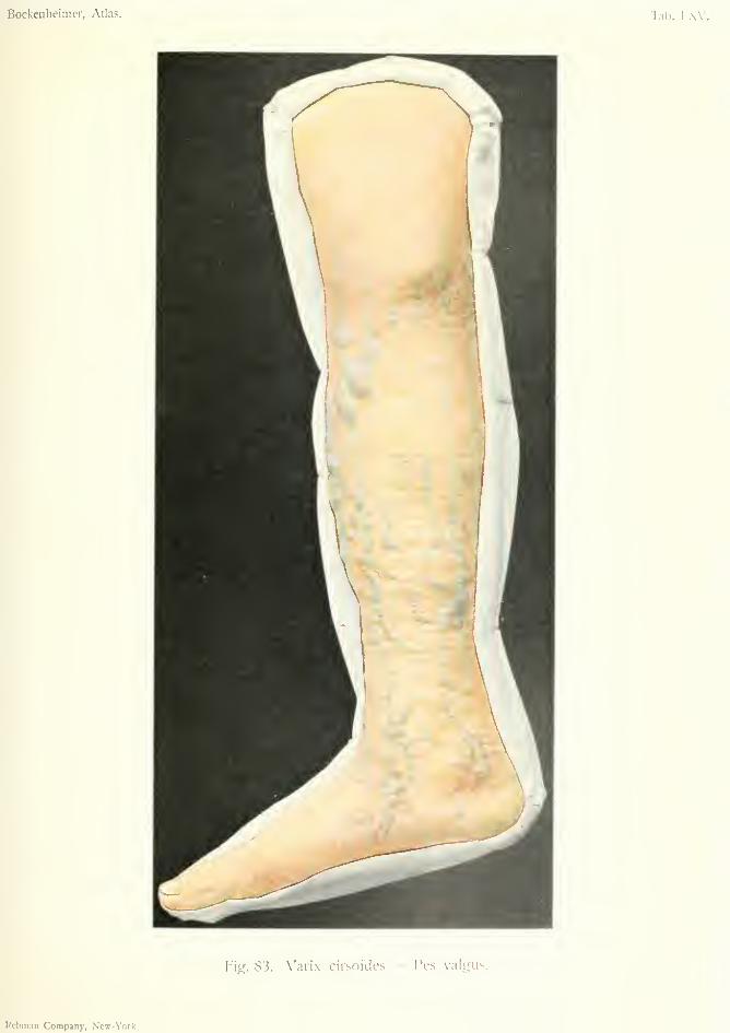
Bockenheiiiicr, Atlas. Tab. I.XV.
Fig. 83. V'aiix cirsoides — Pes valgus.
Rcbm.Tii Company, New-York.

and be combined with hemorrhoids, especially in
women who have had many pregnancies.
Under the thinned skin hard lumps can be felt
where thrombosis has occurred. Sometimes the
thrombi are calcified, and are then known as phlebo-
liths. At the commencement of the affection, before
the varices become prominent, fine ramifying vessels
are found under the skin, which later on appear
between the veins. These ramifying vessels give the
skin a brownish appearance.
Varices which extend in the form of ramifying
anastomoses and networks over the whole leg are
connected both with the skin and subcutaneous
tissue, and become very extensive when the valves of
the large saphenous vein are destroyed, thus imped-
ing the circulation. The insufficiency of the valves
can be shown by raising the limb till the varices have
emptied themselves of blood; then compress the
saphenous vein at its opening into the femoral vein
in the thigh, lower the limb and suddenly removepressure on the saphenous vein; the varices then
become again filled with blood from the femoral vein.
The patients suffer more when standing than whenwalking. The chief symptoms are tingling and
numbness in the limb, cramps in the calves, especially
when the deeper veins are affected, swelling of the
feet, eczema, ulceration and even elephantiasis.
These troubles often cause much suffering.
Varices may be dangerous from rupture and
hemorrhage. As a rule the small, thin, ramifying
peripheral vessels rupture, sometimes the larger
trunks. The blood being under considerable pres-
sure spurts out in a jet. Fatal hemorrhage may take
place unless the limb is elevated and the bleeding
stopped by pressure. Death may occur in rupture of
subcutaneous varices in the leg and in the internal
organs {e.g. brain and liver). The second danger
is thrombo-phlebitis which may lead to embolism,
especially when it becomes purulent (Fig. 84).
1(5
Differential Diagnosis. Varicose veins are so
typical in appearance that they cannot be mistaken
for the vascular formations, such as aneurism, cir-
soid aneurism or cavernoma. Primary phlebectasis
must not be confounded with the dilatation of super-
ficial veins caused by thrombosis of the deeper veins;
e.g. after infective diseases.
Treatment. Prophylactic treatment consists in
avoiding long standing, in cleanliness and massage.
In slight cases the circulation of the limb can be im-
proved by the application of flannel bandages from
the toes upwards {Martin's rubber bandage is liable
to cause eczema). If the varix is caused by pressure
of a tumor, this must be removed when possible.
The most radical treatment consists in extirpation
of the varices, especially w-hen very tortuous. If the
valves of the vein are destroyed (shown by the method
mentioned above), it is best to ligature the saphenous
vein near its opening into the femoral vein, and to
resect a part of it as well. After the operation small
varices and eczema quickly disappear, but elastic
bandages should be worn for some time. The extir-
pation of secondary varices due to thrombosis of the
deeper veins is useless.
Varicocele should be excised in its whole extent;
the testicle can be drawn up by suture.
Submucous varices of the esophagus and varices
in the brain and liver are inaccessible to treatment.
Fig. 83 shows somewhat extensive varices in the
region of the large saphenous vein in the leg, in a
woman of forty, after many pregnancies. The above-
mentioned ramifying vessels are seen between the
varices, giving the skin a reddish-brown appearance.
In this case the foot was in a position of pronation
and abduction (pes valgus or flat-foot)
176
PES VALGUS OR FLAT-FOOT
The treatment of pes valgus depends on its cause.
The deformity may be congenital or acquired (trau-
matic, paralytic, rickety (Fig. 65), or due to long
standing).
In all these forms the foot is more or less in a posi-
tion of pronation and abduction, and eventually there
is displacement at the astragalo-scaphoid articula-
tion. Along with changes in the bones and destruc-
tion of cartilage in the joints, the ligaments, tendons
and muscles are also affected.
Traumatic flat-foot occurs not only after fractures
of the leg and ankle, but also as the result of rupture
of the ligaments from twisting of the foot, especially
when the injury is not treated by fixation. Paralytic
flat-foot occurs after acute anterior poliomyelitis, in
which the plantar flexors are paralyzed and there is
over-action of the extensors.
Rickety flat-foot is due to sinking of the arch of
the foot owing to softness of the bones.
The commonest form is static flat-foot, which
occurs in persons of weak muscular power, as the
result of prolonged standing (waiters, etc.). It gen-
erally develops at the age of puberty. The symp-
toms are fatigue, pains in the ankle and tarsal joints,
and on the outer side of the leg. The pains are often
cramp-like (tarsalgia).
Differential Diagnosis. Pes valgus must not be
confounded with the flat-foot which occurs in certain
races (Jews, negroes). The latter is due to imperfect
development of the arch of the foot, but there are no
changes in the mid-tarsal joint, and the condition
causes little trouble.
Treatment. In congenital flat-foot the position
can be corrected by manipulation and massage. In
traumatic flat-foot caused by fractures and sprains,
the patients should not walk too soon, and then only
with a well-made boot provided with a flat-foot sole.
In more severe degrees of traumatic flat-foot, the
question of cuneiform osteotomy of the scaphoid
bone or head of the astragalus, or linear osteotomy
of the tibia and fibula may arise. These operations
may be considered in cases where manipulation has
failed to correct the position. If the tendon Achilles
is much shortened it should be tenotomized before
manipulation. After manipulation the foot should
be put up in plaster of Paris in an over-corrected
position.
In paralytic flat-foot tendon-transplantation is use-
ful. The peripheral end of the divided tendon of the
paralyzed tibialis anticus muscle can be connected
with the tendon of the healthy extensor longus hallucis
muscle.
Inflammatory flat-foot, which causes painful con-
tracture, should be treated by rest in bed and hot
fomentations. If the pain is very severe cocaine maybe injected.
General treatment consists in strengthening the
muscles (tibialis anticus and posticus, and calf mus-
cles) ; active movements and massage. When stand-
ing the toes should be turned inwards, and whenwalking the foot should not be turned outwards.
The boots should be well-made with flat-foot pads;
the latter are made after an impression of the foot
taken on smoked paper, and should extend from heel
to toes over the whole sole.
178
PYOGENIC mPECTIONSPlate LXVI et seq.
The bacterial invasion of injured or uninjured
parts of the body plays a great part in surgery, as
there is always the possibility of bacterial infection
in every injury and operation.
According to the nature of the infection, definite
clinical pictures are produced which are generally
represented by various degrees of inflammation and
reaction of the body. These processes may be
mcited not only by bacterial irritation but by mechan-
ical irritation, such as trauma without infection, also
by chemical irritation (e.g. poisons of all kinds,
animal poisons such as snake poison), and by the
action of heat and cold (burns and freezing).
In bacterial infection the inflammation is most
marked, as it does not remain limited to the place of
origin, but extends more or less rapidly in the sur-
rounding parts, and may eventually reach remote
parts of the body by way of the blood and lymphatic
vessels (general infection). According to the rate of
its extension, the inflammation may be acute, chronic
or subacute. All three forms may pass into each
other.
Bacterial infection causes various clinical phe-
nomena according to the nature, number and viru-
lence of the bacteria, and according to the parts of
the organism which are invaded, and the power of
resistance of the individual. Old, feeble and dis-
eased bodies (e.g. diabetes) are less capable of com-
bating bacterial invasion, while a healthy body
shows a strong reaction against it. This reaction
179
manifests itself by inflammation at the point of
infection.
This inflammatory reaction is manifested by the
cardinal symptoms—redness, heat, swelling and
pain. The redness and heat are due to dilatation of
the blood-vessels from irritation of the tissues (active
or arterial hypera?mia); the swelling and pain are
due to the transmigration of blood elements, espe-
cially leucocytes, owing to the slowing of the blood
stream. In every severe infection the function of the
part concerned is also interfered with.
The exudation varies in degree according to the
nature of the infection. It may be serous, fibrinous,
sero-fibrinous, or purulent; and when mixed with
red blood corpuscles becomes hemorrhagic. Puru-
lent exudation is the most common, and recurs in its
simplest form in wounds which do not heal by pri-
mary union.
Pyogenic infections are also distinguished accord-
ing to their situation and extent. They may thus be
superficial or deep; circumscribed or diffuse; cuta-
neous, subcutaneous, muscular, glandular, or osse-
ous, etc.
Besides the local inflammatory reaction of the part
of the body attacked, there is a general reaction
shown by considerable and prolonged rise of tempera-
ture. This must be distinguished from the slighter
degree of so-called aseptic fever which occurs during
resorption of blood effusions. The temperature
chart in pyogenic infections, together with the local
reaction and the general symptoms (rigors, pains in
the joints, dry tongue, sweating, diarrhea and vom-
iting) are of the greatest importance in estimating
the degree of wound infection.
After the first stage of inflammation, which causes
more or less destruction of tissue, comes the stage of
regeneration.
Owing to the formation of granulation tissue from
the fixed connective tissue cells the inflammatory
180
area becomes isolated and demarcated, the necrosedtissue becomes separated and is discharged with the
pus, and the wound eventually heals by scar tissue
which is developed from the vascular granulations.
As the stage of reparation proceeds, the clinical
symptoms of inflammation subside.
If the infection is very virulent, the body cannotovercome the bacteria and their products of meta-bolism. From the local infection arises a general
infection which the defensive power of the body is
generally unable to combat.
The researches of Ehrlich and Morgenroth havethrown much light on this complicated process.
This is not sufficiently explained by the presence of
a substance (called alexin) present in ihe blood-
serum, nor by Metchnikofj's theory o: phagocytosis
(destruction of bacteria by the white blood corpus-
cles), but depends on the combined action of several
factors. Also, the still more complicated processes
of the formation of antitoxins, and the immunization
of the organism, have been made comprehensible byEhrlich's "side-chain" theory.
Again, the knowledge of surgical infections due to
bacteria has been extended by numerous observers
{Koch, Fehleisen, Rosenbach and others). The harm-ful action of bacteria is due to their multiplication in
the organism, and to the formation of products of
metabolism, the most dangerous of which are the
toxalbumins (or toxins) excreted by living bacteria;
while the poisons found within the bacteria, which
lead to their destruction, are known as endotoxins
and are of less importance.
While the normal skin and mucous membranes only
rarely harbor bacteria, every wound forms a favora-
ble soil for their development, and from this they
spread by the blood and lymphatic vessels. Theorganism may be infected by one or several kinds of
bacteria (mixed infection).
The most important bacteria from the surgeon's
181
point of view are those which cause pyogenic infec-
tions—the staphylococcus aureus and albus and the
streptococcus pyogenes. Most acute inflammatory
processes, whether a wound is present or not, are
due to these forms of bacteria.
Staphylococcal infections are very common (fur-
uncle, carbuncle, osteomyelitis, etc.), and generally
lead to circumscribed purulent inflammations. Strep-
tococcal infections are more diftuse and often cause
general infection.
Both these forms of bacteria are especially virulent
when they give rise to pyogenic infection of the
human body. Other bacteria only cause a slighter
degree of inflammation; generally serous or sero-
fibrinous, only occasionally purulent (pneumococcus,
typhoid bacillus, bacterium coli commune, gonococ-
cus, bacillus pyocyaneus, tubercle bacillus, diphtheria
bacillus).
Differential Diagnosis. Pyogenic infections
present such characteristic clinical symptoms that a
general diagnosis is not difiicult. A stricter diagnosis
depends on the history of the case, the local and gen-
eral condition and bacteriological examination.
Prognosis. With early diagnosis and appropri-
ate treatment the prognosis is favorable as regards
life, but doubtful as regards function in certain
regions. There is always danger to life in every
pyogenic infection, as a circumscribed inflammatory
focus may become diffuse and set up general infec-
tion. In consideration of this fact, every apparently
insignificant pyogenic affection must be treated with
the greatest care.
Treatment. In the first place all sources of irri-
tation must be removed (foreign bodies, stone, etc.).
Whenever signs of suppuration appear, the affected
part must be kept at rest; in the extremities by sus-
182
pension. When there is inflammatory infiltration of
the skin without any formation of pus, it may besmeared with ointment; but the apphcation of anice-bag is injurious, as it delays the locahzation of
the process in the form of a circumscribed collection
of pus, which is the object desired. Hot, moist fo-
mentations are best avoided, as they favor the
growth of bacteria. When a circumscribed collec-
tion of pus has formed, it must be evacuated by a free
incision (pyogenic conditions which require earlier
incision will be mentioned later). Small abscesses
can be opened under local anaesthesia, but moreextensive ones require a general aneesthetic. Localanjesthetics should never be injected into inflamma-
tory tissue, as they are very painful and may also
give rise to general infection.
Large incisions, made so as to give the best outlet
for the pus, lead to more rapid healing than small
incisions. The after-treatment is rendered muchsimpler by large incisions, while small incisions
often require further incision. For the same reason,
evacuation of pus by an aspirator is more uncertain
and uncleanly.
After-treatment consists in loosely plugging the
wound with dry iodoform gauze, and later with
sterile gauze, applied daily. Immobilization should
be continued till all signs of inflammation have sub-
sided.
In cases where dry tampons cause pain they maybe replaced by moist tampons with two per cent,
boric acid lotion, one per cent, aluminium acetate,
or three per cent, oxygenated water, renewed two or
three times a day. Tampons should not be left in
too long, as they cause irritation of the tissues. Theymust, therefore, be managed as carefully as possible,
if necessary, under an anaesthetic. The application
of alcohol, iodine, carbolic acid, balsam of Peru to
infected wounds (cf. treatment of tetanus. Fig. 78),
is not to be recommended, as they cause much irrita-
183
tion in the wound. Von Bergmann's method of dry
antiseptic dressings is the simplest and most practical
method of dealing with pyogenic infections.
Granulation tissue should be treated by ointments
of zinc oxide or nitrate of silver, and by baths. Later
on, massage, active and passive movements and
electricity are indicated, according to the situation
and nature of the affection. The general condition
also requires treatment in every pyogenic infection,
by tonics and nourishing diet. When necessary sub-
cutaneous injections of normal saline solution and
nucleinic acid should be given (20 cc. of nucleinic
acid in 200 cc. of normal saline solution). Antitoxic
or bactericidal serums have so far given no result in
pyogenic infections.
The method of passive hypersemia advocated by
Bier for the treatment of acute pyogenic infections
has, after the experimental and clinical research of
Lexer, Wrede, WoUf-Eisner and others, proved itself
to be "a double-edged sword." (Discussion at the
Surgical Congress, 1906). It cannot be recom-
mended as a practical method, as it necessitates
prolonged internment of the patient in hospital. It
is true that an increase in the power of defense takes
place at the seat of infection after passive venous
hypergemia, as it does after active hypersemia induced
by painting with iodine or hot-air treatment. Onthe other hand, nutrition is impaired, and the resorp-
tion of the bacteria and their poisons delayed by the
venous hypersemia, which may result in further
destruction of tissue at the seat of infection. Again,
if the infection is a virulent one, especially strepto-
coccal, there may be rapid resorption of bacterial
poisons in the organisms after removal of the elastic
compression, which may be fatal. In infection by
gas-forming bacteria, which may cause gangrene of
the tissues by pressure of gases, passive hypersemia
only aggravates this action.
We, therefore, consider treatment by passive hyper-
184
aemia (which cannot be endured by many patients)as unnecessary in the milder forms of pyogenic infec-
tion. The above-mentioned treatment is sufficient
in these cases, especially when combined with immo-bilization. Again, treatment by passive hypersemiaoften obscures the indications for incision. Smallincisions are often insufficient even in mild cases, andrequire to be enlarged or repeated, thus complicatingand lengthening the treatment. (For the treatmentof suppuration in tendon-sheaths, see Fig. 96).
In the more acute pyogenic infections, which pre-sent severe clinical symptoms and have a tendencyto progress, treatment by passive hypersemia is
unsafe, and has often aggravated the condition;e.g., by thrombo-phlebitis of the small veins, multipleabscesses, and even general infection.
Finally, the treatment of acute pyogenic infections
by passive hyperaemia has not a scientific foundationon bacteriological research, nor is it supported bythe results of clinical experience.
185
THROMBOPHLEBITIS ACUTA PURULENTA{Acute purulent Thrombo-phlebitis)
Plate LXVI, Fig. 84.
Acute purulent thrombo-phlebitis may arise from
infection of the neighboring parts. In every pyogenic
infection purulent thrombi are found in the smaller
veins. In the larger veins it arises from periphlebi-
tis, in which there is infection of the wall of the vein.
Infection of the walls of veins may also result from
internal infection by the blood. Purulent phlebitis
always results in the formation of a thrombus which
may cause complete occlusion of the vessel. Thethrombus generally contains pus (thrombo-phlebitic
abscess) ; it may extend and infect larger areas, or
may disintegrate and give rise to general infection
by embolism (cf. Fig. 108).
Various pyogenic affections may give rise to
thrombo-phlebitis (lymphangitis, furuncle, carbun-
cle, erysipelas, varicose ulcer of the leg). Otitis
media may cause thrombo-phlebitis of the lateral
sinus. In the portal vein, infection by the blood
may cause pylephlebitis and subsequent multiple
abscesses in the liver. Carbuncle of the lips maycause meningitis through thrombo-phlebitis of the
facial and ophthalmic veins. When the lesion is
superficial, it gives rise to all the symptoms of puru-
lent inflammation—redness, swelling and cedema of
the skin and subcutaneous tissue, pain, fever andrigors. The skin is often tense and hard. Theinfiltration extends along the course of the veins, in
the form of hard cords. The presence of pus and the
formation of abscess is indicated by yellowish coloring
of the skin (Fig. 84), and later by fluctuation.
186

HoikenheiiiuT, Atlas.Tab. I.WI
Fig. 8-1. riiioiiibophlebitis puiulciila acuta.
Hebman tJnnipaiiy, Nc«-N'nrk.

Thrombo-plilebitis of the deeper veins gives rise
to severe symptoms—pain, high fever, rigors and
change in the general condition.
Thrombo-phlebitis of the femoral vein, occurring
in women as the result of puerpural parametritis, is
known as phlegmasia alba dolens (white leg). In
this affection the whole leg is affected by painful,
hard oedema, preventing any movement. The throm-
bosis may be so extensive as to cause gangrene of
the extremity.
In every case of thrombo-phlebitis the walls of the
veins remain thickened causing congestion which, in
the lower extremities, leads to deficient nutrition
(ulcer, eczema, elephantiasis). Thrombi may be-
come transformed into hard, painful phleboliths, by
deposit of calcareous salts.
Differential Diagnosis. Superficial thrombo-
phlebitis differs from lymphangitis in the veins being
thicker and harder. Deep thrombo-phlebitis is often
impossible to distinguish from other pyogenic affec-
tions.
The prognosis is always doubtful, owing to the
possibility of general pyogenic infection.
Treatment. In the early stages suppuration of
the thrombi may be avoided by rest. In the extrem-
ities, these should be suspended. The treatment
must be conducted according to the general rules for
pyogenic affections. Abscesses must be incised;
there is no fear of hemorrhage owing to thrombosis
of the vessels for some distance from the seat of
inflammation. If general infection appears to be
imminent the vein should be resected after double
ligation of the diseased section. For example, liga-
tion of the internal jugular vein is indicated in otitis
media, and in furuncle of the lips (in the latter, also,
ligation of the anterior facial vein).
Phlegmasia alba dolens does not suppurate as a
187

rule and can be treated by rest in bed and the appli-
cation of mercurial or silver ointments (unguentum
cinereum and unguentum Crede).
Fig. 84 shows acute purulent thrombo-phlebitis in
a woman, affecting a varicosity of the saphenous vein,
which developed after pregnancy. There is diffuse
redness, with yellowish nodules indicating the com-
mencement of abscesses in connection with the
infiltrated and thrombosed vein.
188

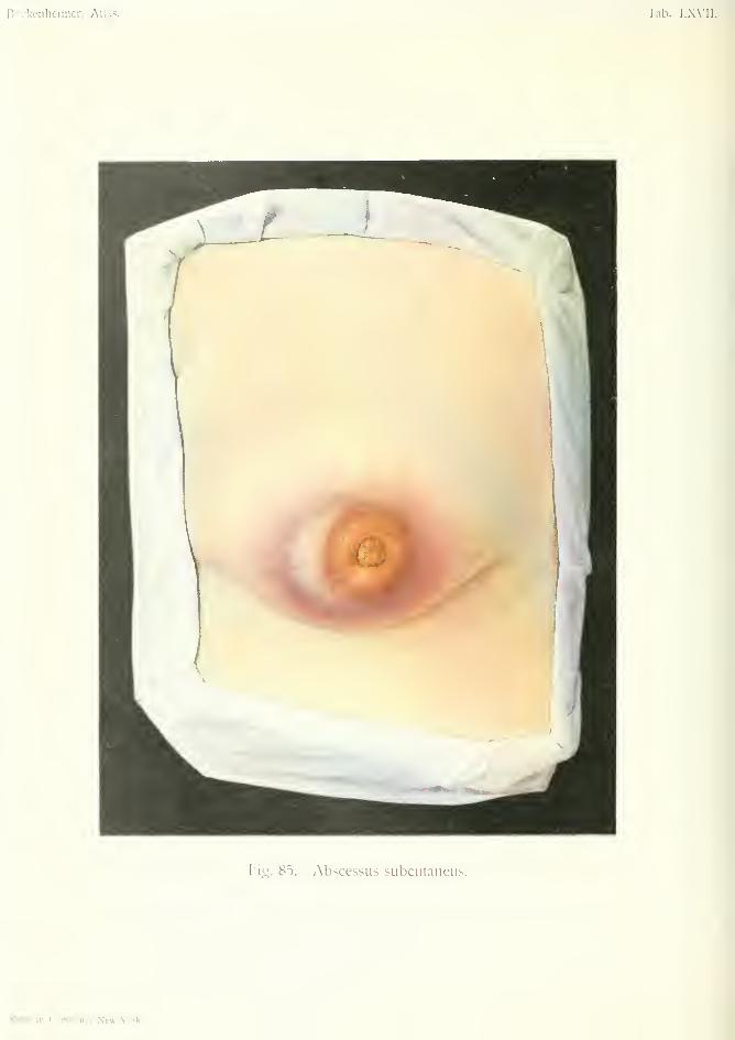
Bockenheinicr, Atlas. Tab. I Wll
Ficr. 85. Abscessus subcutaneus.
Rebman Company, New-York.
ABSCESSUS SUBCUTAKEUS PARAMAMMILLARroS(Subcutaitcous paramammillary abscess)
Plate LXVn, Fig. 83.
The term abscess is applied to a circumscribed
collection of pus which arises from loss of tissue.
The terms purulent exudation or empyema are
applied to collections of pus which form in pre-
existing cavities (maxillary antrum, pleura, abdo-
men). Abscesses may occur in the skin, subcuta-
neous tissue, muscles, bones, and also in the internal
organs (liver, lungs, brain).
Cold abscesses, which are due to chronic infec-
tions, such as tuberculosis (Fig. 12.5), must be dis-
tinguished from acute abscesses, which, in most
cases, occur in the subcutaneous tissue as the result
of acute pyogenic inflammation, due to staphy-
lococci and streptococci. The formation of an
abscess may usually be considered a favorable sign,
as it arrests the progress of infection in the organism
by damming up the inflammation. After the difi'use
inflammation has become circumscribed in the form
of abscess, the severe inflammatory symptoms sub-
side. Granulation tissue formed by the fixed con-
nective-tissue cells forms a continuous boundary
known as the abscess membrane.
The majority of abscesses arise from diffuse, infil-
trating, purulent inflammation of the subcutaneous
tissue. Abscesses also occur in the various organs
of the body in all other pyogenic affections (erysipe-
las, lymphangitis, osteomyelitis, lymphadenitis, my-
ositis). The abscesses may spread from the deeper
parts to the surface, or inversely.
189
Blood efiFusions may suppurate and form abscesses
if another part of the body is invaded by bacteria
{e.g. furuncle). The so-called embolic or metastatic
abscesses are formed by way of the blood stream in
general infection, and may occur in any part of the
body.
The clinical symptoms are those already men-
tioned. In subcutaneous abscess the skin is at first
red, and shows diffuse inflammatory infiltration.
There is pain, tension and fever. The red color of
the skin becomes gradually darker and more circum-
scribed. The skin becomes thinner and yellowish
and bulging at one spot, through which the abscess
bursts. The deeper the abscess, the more diffuse
and extensive are the infiltration and inflammatory
oedema {e.g. in osteomyelitis. Fig. 82).
Erysipelas, lymphangitis, and other pyogenic affec-
tions may be present along with abscess formation.
The part of the body affected is stiff and painful on
movement, and as every abscess may lead to general
infection all movements should be avoided.
Differential Diagnosis. Acute abscess is recog-
nized by the presence of all the symptoms of acute
inflammation. The cause of the abscess must be
found, and the occurrence of metastatic abscesses
must be borne in mind.
Treatment. As soon as an acute abscess is diag-
nosed by the presence of fluctuation, or by an explor-
ing syringe in the case of deep abscess, it must be
freely opened. When the suppuration is once cir-
cumscribed, early incision prevents further destruc-
tion of tissue, leads to quicker healing and leaves less
scar. Treatment by hot fomentations or poultices,
to cause spontaneous bursting of the abscess, causes
more destruction of tissue and delays healing.
After incision the abscess should be plugged with
sterile gauze, after which granulation tissue is quickly
190
formed. Treatment by aspiration is not so good asit does not remove the abscess membrane.Deep abscesses must be freely opened, plugged
and drained. In large abscesses a counter incision
should be made at the deepest part of the abscesscavity, and all recesses should be opened up. Theaffected part should be then immobilized.
Fig. 85 shows a subcutaneous abscess surroundingthe nipple in a lying-in woman, arising from a crackednipple, which gave entrance to bacteria. The skin
round the nipple is bluish red and swollen. The pres-
ence of fluctuation indicates a collection of fluid in
the subcutaneous tissue. The inflammation hasalready become circumscribed. In spite of the
apparently slight extent of the abscess, the patient
suffered from severe pain, fever and general malaise.
The abscess healed quickly, after incision and plug-
ging and suspension of both breasts.
191
MASTITIS PUERPERALIS PURULENTA(Purulent puerperal mastiiis)
Plate LXVIII, Fig. 86.
Bacterial inflammation of the breast (phlegmonous
mastitis) ending in suppuration (purulent mastitis),
occurs almost exclusively in women during the puer-
perium, as the result of direct infection of the
lactiferous ducts with bacteria (mostly staphylo-
cocci), through cracks and fissures of the nipple. Theclinical symptoms are those of pyogenic infection, with
the formation of a hard, painful infiltration, usually
in the lower and outer quadrant of the breast. Theskin is tense, oedematous, reddened and often glisten-
ing. The redness qviickly extends over the whole
mamma and beyond it. The patients suffer from a
feeling of tension in the breast, and radiating pain
in the arm of the affected side. There is also general
malaise. The affection is often ushered in by rigors
and high temperature.
The axillary glands may be enlarged and painful.
In severe cases there is diftuse infiltration of the
whole mammary gland, which may extend into the
lymphatic vessels round the breast. Abscesses form
in one or more places; the superficial ones being
recognized by fluctuation, the deeper ones by the
extensive nature of the lesion. Purulent inflamma-
tion of the mamma may occur in general infection;
on the other hand, it may also give rise to general
infection by thrombo-phlebitis.
Differential Diagnosis. A non-bacterial in-
flammation of the breast occurs in sucklings soon
after birth (mastitis neonatorum). This is a physio-
192
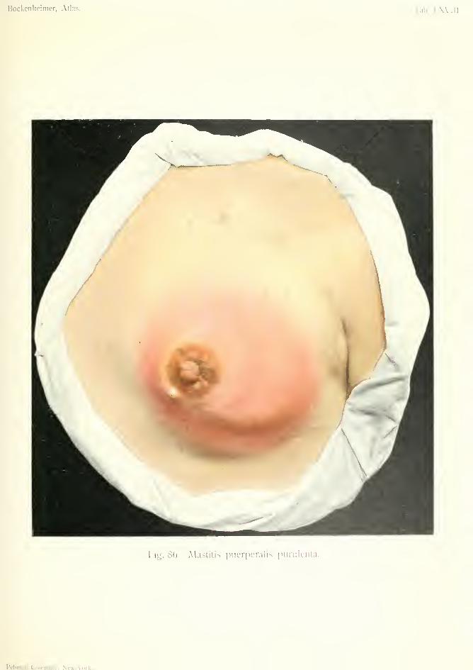
Bockenlieimer, Atlas. Tab. I..Will.
Fig. 80. MasUlis puer|ieralis iiurulciita.
Rcbman Coiiinnnv. Nc'»-\'ork.


logical swelling of the gland with excretion of a
secretion resembling milk. In some cases there is
circumscribed abscess formation, which soon heals
after incision. The inflammation, however, usually
subsides under ointments and moist fomentations.
Similar mastitis may occur at the age of puberty,
both in boys and girls, which yields to the same
treatment and seldom leads to abscess. Pigmenta-
tion of the areola remains after these cases of
mastitis.
During the period of lactation, accumulation of
milk due to stopping its outflow may cause hard in-
flammatory infiltration of the breast (milk abscess)
which disappears after removal of the milk by a
breast pump, etc. In these cases both breasts
should be supported by a suspensory bandage.
Mastitis may be caused by trauma, by suppuration
in a blood eft'usion caused by injury. In cases of
furunculosis and diabetes mastitis may occur, with
the formation of hard, deeply situated abscesses
resembling malignant tumors.
Tuberculous mastitis is generally due to extension
from tuberculous axillary glands and is characterized
by its chronic course. Actinomycosis gives rise to
hard swellings (cf. Fig. 115). Syphilis may also
cause interstitial mastitis, but there is no suppura-
tion. Gonorrheal infection of the lactiferous ducts
has also been described, as the result of uncleanliness
of the mother, or gonorrheal stomatitis in the infant.
Interstitial mastitis and chronic cystic mastitis
which form tumor-like nodules in the mamma, can-
not be mistaken for phlegmonous mastitis as they
cause no acute inflammatory symptoms.
Superficial abscesses in the region of the nipple
(Fig. 85) are easily distinguished from purulent mas-
titis, and are only of limited extent. Retro-mam-
mary abscesses may cause difficulty in the diagnosis
when there are also signs of inflammation in the
mamma. In these eases the skin is usually intact,
193

the whole breast is raised from the thorax, and pal-
pation of the breast causes no pain ; but there is pain
on pressing the breast against the thorax. There is
generally acute adenitis of the axillary glands and
pain on moving the arm in retro-mammary abscess.
Treatment. As soon as suppuration in the breast
is diagnosed it must be incised. The earlier incision
is made the more rapidly do the symptoms subside.
The case should not be left till the abscess points
under the skin, but a radial incision should be made,
under an anaesthetic, through the breast tissue, if nec-
essary as far as the pectoral fascia. All recesses andpockets must be opened up, and counter-openings
made if necessary. Glandular tissue destroyed by
suppuration can be removed with the sharp spoon.
The after-treatment consists in plugging and drain-
age, and must be carefully carried out, otherwise
there may be purulent infiltration of the neighboring
gland lobules and further extension in the form of
diffuse inflammation. Large incisions are indicated,
as they lead to more rapid healing, and enable the
mammary gland to retain its function of lactation.
Both breasts should be suspended, and the child
removed from the breast. Purgatives and iodide of
potassium may be given to diminish the formation
of milk. Treatment by moist fomentations is not
to be recommended, as it may lead to destruction of
the whole glandular tissue.
Treatment of the abscess by aspiration, whichaims at the least possible destruction of the mam-mary tissue, is only indicated in the rare cases wherethe inflammation and abscess formation is circum-
scribed. In the more common phlegmonous formthis method is dangerous, and has in more than oneinstance necessitated amputation of the breast. Aspi-
ration has also the disadvantage of being uncleanly.
Fig. 86 shows a case of acute purulent mastitis in
a lying-in woman, situated in the lower and outer
194

quadrants of the breast. It may be mentioned, bythe way, that congestive mastitis of the lower quad-rants of the breast may predispose to infective mas-titis. In Fig. 86 the inflammatory signs are very
marked. The skin is reddened, tense and infiltrated
;
the whole of the outer and lower part of the mammais hard and painful. Fluctuation was nowherepresent. The case healed rapidly after incision,
plugging and suspension.
Persistent fistulas of the breast with unhealthy
granulations (cf. Fig. 56) may be due to deep col-
lections of pus which have not been opened up, or
to tampons or drainage tubes which have been left
behind. They often require multiple incisions.
195
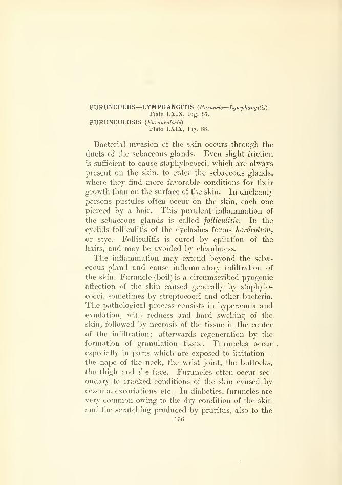
FURUNCULUS—LYMPHANGITIS {Furuncle—Li/mphangUis)Plate LXIX, Fig. 87.
FURHNCULOSIS (Fnruneulosis)
Plate LXIX, Fig. 88.
Bacterial invasion of the skin occurs through the
ducts of the sebaceous glands. Even slight friction
is sufficient to cause staphylococci, which are always
present on the skin, to enter the sebaceous glands,
where they find more favorable conditions for their
growth than on the surface of the skin. In uncleanly
persons pustules often occur on the skin, each one
pierced by a hair. This purulent inflammation of
the sebaceous glands is called folliculitis. In the
eyelids folliculitis of the eyelashes forms hordeolum,
or stye. Folliculitis is cured by epilation of the
hairs, and may be avoided by cleanliness.
The inflammation may extend beyond the seba-
ceous gland and cause inflammatory infiltration of
the skin. Furuncle (boil) is a circumscribed pyogenic
affection of the skin caused generally by staphylo-
cocci, sometimes by streptococci and other bacteria.
The pathological process consists in hyperaemia andexudation, with redness and hard swelling of the
skin, followed by necrosis of the tissue in the center
of the infiltration; afterwards regeneration by the
formation of granulation tissue. Furuncles occur
especially in parts which are exposed to irritation
—
the nape of the neck, the wrist joint, the buttocks,
the thigh and the face. Furuncles often occur sec-
ondary to cracked conditions of the skin caused byeczema, excoriations, etc. In diabetics, furuncles are
very common owing to the dry condition of the skin
and the scratching produced by pruritus, also to the
196
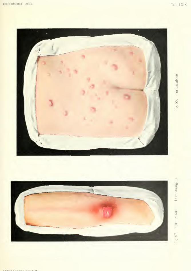
Bockenheimer, Atlas. Tab. I.XIX
CO
i;:
P^hman Pr. Ma... V'n..U

body being especially vulnerable to bacterial inva-
sion (Fig. 140). Furuncles may also appear in all
cases where the bodily resistance is impaired—in chil-
dren, old people, and the tuberculous.
The clinical appearance of furuncle is typical.
From a small punctiform redness develops a hard,
redder, painful nodule in the skin, which extends at
its periphery and also deeply towards the fascia.
The epidermis is at first intact, but afterwards rup-
tures at the apex of the projecting furuncle, exposing
a yellowish center which becomes more and moredemarcated from the hard, red infiltration. In this
way a round, crateriform ulcer is produced with a
central yellowish core (Fig. 87). Sometimes a hair
is situated in the center of the furuncle. Large fur-
uncles are extremely painful, especially on move-
ment, and are often accompanied by fever and
general debility. The symptoms subside when the
central core becomes loosened by suppuration. Thecavity is then cjuickly filled by granulation tissue,
which may form a cicatrix in a few days. The hard
infiltration remains for a long time and generally
causes unpleasant itching of the skin. The scar,
which is always hypertrophic in all inflammatory
processes, may also cause trouble.
Complications may increase the severity of fur-
uncle. There is always lymphangitis, especially in
the extremities, and often lymphadenitis. Early im-
plication of the lymphatics signifies extensive inflam-
mation and virulent bacteria.
Several furuncles are sometimes found close to-
gether, either from simultaneous infection of several
sebaceous glands or from secondary infection from
the primary furuncle. This often occurs after the
application of plaster or other measures with the
object of "drawing out" the furuncle.
In individuals with a feeble power of resistance
(diabetics, infants and old people), there maybe an
outbreak of furuncles over the whole body, a condi-
197
tion known as jurunculosis (Fig. 88). In children
this process often consists in the formation of multiple,
small nodular infiltrations in the skin, in which there
is no central core but a small abscess. Extensive
furunculosis may be fatal from exhaustion. As in
every pyogenic infection, furunculosis may lead to
purulent thrombo-phlebitis and general pyogenic in-
fection. Furuncle of the lip may cause meningitis bythrombo-phlebitis of the facial vein, and general
infection may be caused by thrombo-phlebitis of the
veins of the neck (Fig. 108). Furuncles may lead to
renal abscess and osteomyelitis (Fig. 104), especially
when not properly treated.
Differential Diagnosis. Furuncles arising from
sebaceous glands are so characteristic that they can-
not be mistaken. Metastatic furuncles in general
infection are multiple, and are associated with other
pyogenic affections.
Furuncles arising from the sweat glands develop
under the skin and form subcutaneous abscesses.
These occur in hairy regions where there is muchexcretion of sweat, such as the axilla. They must
not be confounded with the more deeply situated
glandular abscesses. They generally affect several
sweat glands and form multiple superficial abscesses,
in distinction to lymphadenitis, which either assumes
a diffuse phlegmonous form, or is converted into a
large abscess.
Treatment. Individuals who have a tendency to
furunculosis should take precautions against infec-
tion, by careful attention to hygiene; frequent baths,
rubbing ointment into dry, cracked skin, etc.
Small furuncles can sometimes be aborted by fre-
quent friction with sulphuric ether, or spraying with
ethyl chloride. When painful infiltration of the skin
has developed, the best method is an incision extend-
ing through the whole depth and breadth of the infil-
198
tratiou, after careful disinfection of tlie skin, underlocal anaesthesia. There is no need to wait for com-plete separation of the core, but the incision may bemade as soon as necrosis is commencing, which is
shown by rupture of the skin in the center. Earlyincision diminishes pain and lymphangitis and has afavorable influence on the whole process. Largerfuruncles require a crucial incision. After incision
the wound should be loosely plugged with iodoformgauze. The core generally separates within twenty-
four hours. The core must never be forcibly ex-
pressed, as this causes irritation of the inflamed
tissues, suppuration in the lymphatics, and delay in
healing. Friction of the skin with ether is useful at
each change of the dressings. As soon as granula-
tions appear the plugging should be left off, and the
formation of granulations promoted by ointments
and the nitrate of silver crayon. In the extremities
absolute immobilization with suspension is necessary
till complete healing has taken place, otherwise heal-
ing is delayed or fresh infection follows. The cica-
trices may be treated with iodide of potassium oint-
ment.
Incision by the thermo cautery is not to be recom-
mended, as the formation of eschars hinders the
exit of infectious secretion. Moist fomentations are
also to be avoided, as they cause greater destruction
of tissue and often lead to extensive furunculosis.
Dry cupping has been recommended both as an
abortive method, and also for removal of the core.
Furunculosis of young children should be treated
by incision of the multiple abscesses, followed by
antiseptic baths. The skin must be kept clean to
avoid recurrence. In adults the general health
requires treatment, by purgatives, etc. Yeast prepa-
rations have also been recommended. Diabetic fur-
uncle requires special treatment.
199

LYMPHANGITIS
In pyogenic affections the lymphatic vessels and
glands exercise a beneficial function by harboring
and destroying bacteria and their products. If the
bacterial invasion is very severe, or the bacteria very
virulent, the lymph is coagulated and inflammation
takes place in the walls of the lymphatics, first as
hypersemia, later as small-celled infiltration of the
walls of the vessels. Virulent bacteria may give rise
to lymphangitis and lymphadenitis (Fig. 110) through
slight abrasions of the skin, or in connection with
pyogenic affections, such as whitlow, furunculosis, etc.
Lymphangitis is most clearly observed in the super-
ficial lymphatics of the extremities, in the form of
red, diffuse patches, which soon develop into irregular
red cords extending from the periphery to the root of
the limb. The number of cords diminishes in the
upper part of the limb, and eventually only one large
cord remains in the region of the lymphatic glands
(inguinal or axillary). These signs are most marked
in infection by virulent bacteria.
The lymphatic cords are somewhat raised above
the level of the skin and feel hard. They are painful
to touch and on movement. There is also itching
and a feeling of tension in the whole limb. Theregional lymphatic glands are at the same time
swollen and painful. In some places abscesses form
in the hard cords. There is generally fever and
rigors.
Lymphangitis of the deep IjTiiphatics of the extrem-
ities can be recognized by the feeling of tension and
the general symptoms. Peritonitis may give rise to
200
picuritis through the lymphatic vessels of the dia-
phragm.
The prognosis of lymphangitis is generally favor-
able, as it disappears after removal of the cause.
Chronic lymphangitis, caused by long-continued
irritation of the skin, eczema, ulcers, etc., gives rise to
hard, cord-like formations, which persist for a long
time. Obliteration of the lymphatics may cause
elephantiasis.
Differential Diagnosis. Similar symptoms are
caused by acute purulent thrombo-phlebitis (Fig.
84), but the cords are thicker and not so numerous.
Treatment. This consists in treatment of the
primary affection which causes the lymphangitis
(furuncle, etc.) and in absolute immobilization of the
limb, with suspension. The thickened lymphatic
cords may be painted with mercury or silver oint-
ments (unguentum cinereum, unguentum Crede),
but these should not be forcibly rubbed in. In
chronic lymphangitis, baths and massage are indi-
cated. Abscesses must be incised.
Fig. 87 shows a furuncle with lymphangitis. It
was cured in eight days by incision, iodoform gauze,
plugging and suspension of the arm.
Fig. 88 shows a case of furunculosis in a young
child. Abscess formation is seen in the center of
the furuncles. The case was cured by incisions and
almond bran baths.
201
CARBITNCULUS (Carbuncle)
Plate LXX, Fig. 89.
Carbuncle, which generally occurs in middle life,
differs from furuncle only its greater extent, both
superficially and deeply. It consists of an infection
of several sebaceous glands, thus forming an agglom-
eration of furuncles. The skin gives way in several
places and there are several yellow cores. Com-mencing as a small, red nodule, it quickly develops
into a hard infiltration, extending to the fascia, and
may eventually attain the size of a hand, and cause
more or less diffuse inflammatory infiltration of the
neighboring parts. Lymphangitis and lymphade-
nitis are generally present. The affection is accom-
panied by severe pain, high fever and rigors.
Carbuncle is generally caused by streptococcal
infection. Eczema and other affections of the skin
which cause furuncle, may also give rise to carbuncle.
Moreover, furuncle may develop into carbuncle,
especially when the core has been forcibly expressed,
or when hot fomentations have been applied. In
diabetics carbuncle is still more common than fur-
uncle, and leads to extensive necrosis of the fascia;
it often causes death from exhaustion. Carbuncle of
the face is dangerous owing to its liability to cause
general infection, or meningitis by infection of the
facial vein. Carbuncle of the nape of the neck mayattain enormous size, and extend from one ear to the
other.
Differential Diagnosis. Anthrax (malignant
pustule) differs from carbuncle in the presence of
small vesicles .filled with turbid fluid and early cen-
202
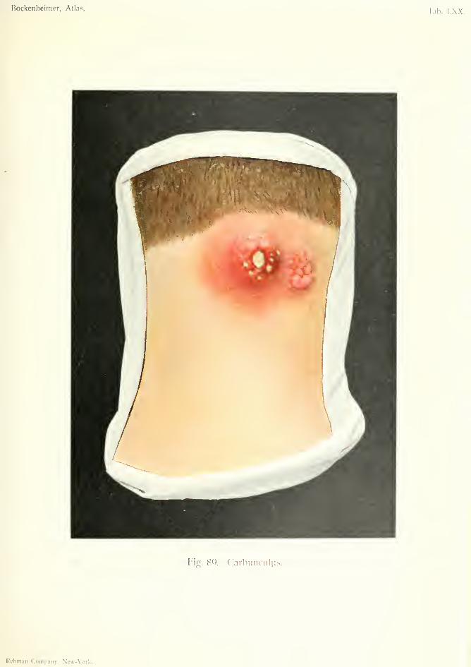
Bockenheimer, Atlas. Tab. LXX.
Fig. 8Q. Carbunculus.
Rcbman Company, New-York.

tral necrosis of the skin, and in the absence of cores.
In doubtful cases a bacteriological examination must
be made.
Treatment. Under an antesthetic, a crucial inci-
sion is made through the whole extent and depth of
the carbuncle, and the central necrosed parts excised.
The wound is plugged with iodoform gauze.
In diabetic carbuncle, progressive neci'osis of the
fascia often necessitates counter-incisions. Iodoform
gauze should not be used in these cases, but sterile
gauze. Special treatment is required for the diabetes.
In every carbuncle there is severe constitutional
disturbance which requires general treatment by
nourishing diet, etc.
Fig. 90 shows a carbuncle of the nape of the neck
in a patient of forty. The infiltration is very exten-
sive. In the central parts the skin is ruptured in
several places, and shows the deeply situated, ne-
crotic cores. Round this is a zone of reddish-blue
skin, and beyond this zone an area of hard, red infil-
tration. There was high fever. The case healed
under the above-mentioned treatment.
203
ERYSIPELAS ERYTHEMATOSUM (Erysipelas)
Plate LXXI, Fig. 90.
While in lymphangitis the deeper and larger
lymphatics are infected, in erysipelas the smaller
lymphatic spaces of the skin and subcutaneous tissue
are plugged with streptococci. A similar condition
may occur in the superficial layers of the mucous
membranes. The causes of this bacterial infection
are streptococci (Fehleisen), but their identity with
the streptococcus pyogenes is not yet agreed upon.
The affected skin is red, tense, somewhat glistening
and slightly raised above the level of the rest of the
skin. The borders are well-defined, distinctly raised
and zigzag, so that the extension of erysipelas, espe-
cially on the face, has been compared to lambent
flames. When the disease spreads over the whole
body, it is spoken of as migratory erysipelas.
Erysipelas may occur wherever there is a solution
of continuity in the skin—after scratches and excoria-
tions, after all injuries and operation wounds. It
may also be combined with various pyogenic affec-
tions—whitlow and phlegmon (especially staphylo-
coccal phlegmon). Conditions which give rise to con-
stant irritation of the skin, such as lupus, tuberculous
fistula, ulcer of the leg, foreign bodies, etc., may also
give rise to erysipelas, which is then often relapsing.
Relapsing erysipelas of the face and leg may cause
elephantiasis. Lastly, erysipelas may arise in gen-
eral streptococcal infection, and is then always com-bined with other pyogenic conditions—abscess, phleg-
mon, etc.
The common form of erysipelas, which consists in
a red elevation of the skin, is called erythematous
204
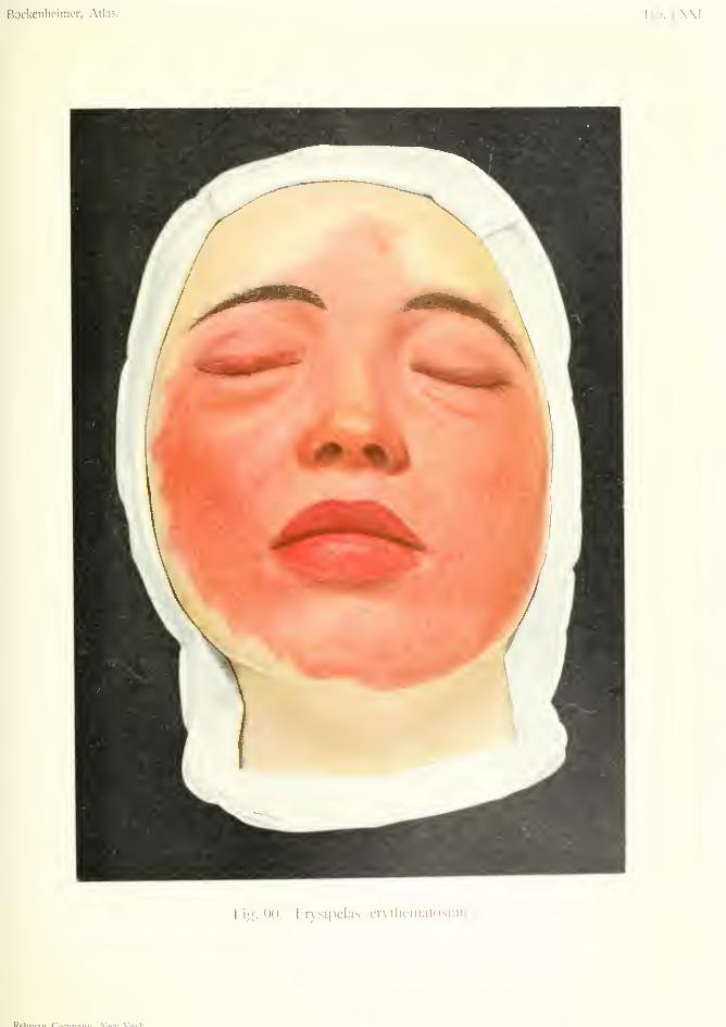
Bockenheimer, Atlas. Tab. 1 .\.\l
Fiy. QO. Hrvsipelas crvlliematosiiin.
Dffhmin Cnw^^.^^,. NTai.- Va.-!/

erysipelas (Fig. 90). In bulbous erysipelas the skin
is covered with vesicles (Fig. 91). In hemorrhagicerysipelas there is hemorrhage in the skin (Fig. 91).
In the great majority of cases there is resolution, but
sometimes erysipelas may cause cutaneous abscesses,
and in the form of gangrenous, phlegmonous erysipe-
las may give rise to ulceration and extensive destruc-
tion of the skin.
The clinical sjTnptoms of erysipelas are character-
istic. The disease usually commences by a rigor,
high temperature (40°-42° C.) and redness of the
skin. There is itching and tension in the skin, andtenderness on pressure. There is considerable con-
stitutional disturbance owing to high fever, head-
ache and vomiting which continue while the disease
progresses. The temperature falls suddenly, the
redness ceases to extend, and the skin, after slio;ht
desquamation resumes its normal condition in about
a week from the onset of the disease. In relapsing
erysipelas the whole process may take place within
one or two days. Erysipelas occurs most frequently
on the face, after this on the exti'emities and genital
organs. In places where the skin is loosely attached
(eyelids, scrotum), there may be considerable swell-
ing and oedema.
Erysipelas of the mucous membranes is generally
difficult to recognize, except when it is an extension
from erysipelas of the skin. The mucous membraneis swollen, oedematous, sodden and of a deep-red
color. Constitutional disturbance is o;enerallv severe.
Erysipelas of the buccal mucous membrane mayoccur after tooth extraction with dirty instruments.
It may cause death by meningitis or oedema of the
glottis. The average mortality of erysipelas is ten
per cent.
Differential Diagnosis. Erythematous erysipe-
las is so characteristic that it can hardly be mistaken
for other affections. The advancing, irregular, raised
205
edge distinguishes it from other inflammatory condi-
tions.
Treatment. The affected parts must be covered
with antiseptic ointments to prevent infection and
auto-infection. If pain is very severe scarifications
are useful. In erysipelas of the extremities the
healthy skin, at the upper limit of the lesion, may be
painted with a single application of pure carbolic
acid, which destroys the superficial layers of the skin.
However, in spite of this procedure, the erysipelas
often extends further up the limb. The induction
of passive hypersemia, by surrounding the limb with
adhesive plaster, has also been recommended. Amongother methods, painting with iodine may be men-
tioned. The patient should always be kept in bed.
Serum therapy has so far proved useless, and is
likely to remain so, since repeated attacks of the
disease do not confer immunity.
The formerly extolled curative action of erysipelas
on tumors has proved illusory. If erysipelas extends
over a malignant tumor (carcinoma or sarcoma), the
tumor may diminish in size owing to destruction of
its cells, but it soon begins to grow again. The same
thing occurs after injection of the fluid, and this
explains the temporary action of the so-called cancer
serum.
On account of the infectious nature of the disease,
the patient should be isolated, and the room disin-
fected with formalin vapor. The same disinfection
must be carried out in operation theaters when an
epidemic of erysipelas occurs. However, it is more
often the hands of the surgeon which convey infec-
tion; hence great care must be taken in avoiding
contact with the patient as much as possible, and
in disinfecting the hands.
Fig. 90 shows a typical case of erythematous ery-
sipelas of the face, which originated from a fissure on
206

the nose. In a few days there occurred high fever
and rigors, followed by erysipelas, first on one side
of the face, then on the other. The skin was tense,
purple and somewhat raised. There was considera-
ble pain and itching. The eyelids were so oedema-
tous that the patient could hardly open them. Thelips were also much swollen, and there was com-mencing erysipelas of the buccal cavity. The sharp
zigzag borders are seen towards the scalp and the
neck.
207
ERYSIPELAS BULLOSUM H^MORRHAGICUM(Hemorrhagic Bullous Erysipelas)
Plate LXXII, Fig. 91.
This case is interesting on account of the origin of
the infection from a horse bite in the arm. Round
the three wounds (which were only superficial abra-
sions) the skin is dark red and there are annular
extravasations of blood. There are also several
vesicles filled with turbid fluid. There is extensive
diffuse reddening, especially on the forearm, and a
brownish coloration due to numerous extravasations
of blood from the smaller blood-vessels situated
round the lymphatic vessels. In the upper arm
there is macular and cord-like reddening due to
lymphangitis. The axillary glands are much swollen
and painful.
Wounds caused by bites from animals or men tend
to become severely infected. In this case, the swell-
ing of the forearm was so extensive that a deep
phlegmon was suspected. The symptoms quickly
subsided after suspension of the arm. In the place
where the erysipelas was hemorrhagic and bullous,
there occurred a superficial phlegmonous inflamma-
tion, which led to gangrene of the skin.
Differential Diagnosis. This has to be madefrom several other affections. Anthrax also com-
mences with redness of the skin and the formation of
vesicles (Fig. 112), fever and rigors, and may, in its
early stage, be confounded with this form of erysipe-
las. But the redness is not so extensive in anthrax,
nor so rapidly developed. Anthrax always causes
early gangrene of the skin. In doubtful cases
308
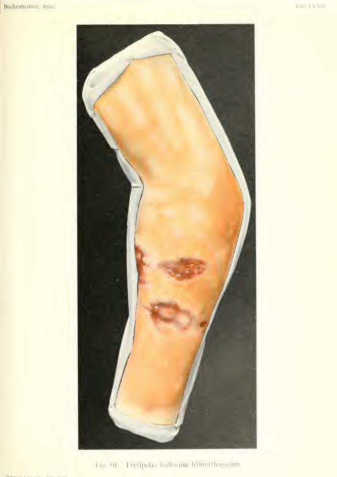
Bockenheimer, Atlas. Tab. LXXII.
hig. 91. Erysipelas biillosum iiaiiioiThagiciim.

anthrax bacilli must be looked for in the contents of
the vesicles.
In this case, which arose from a horse bite, there
was a suspicion of glanders. But, in the latter the
redness is punctiform or macular; the vesicles are
larger and purulent, and soon rupture, giving rise to
gangrenous ulcers.
Subcutaneous phlegmons, which arise from very
virulent streptococci, may cause an erysipelatous
redness of the skin, but this only occurs in the region
of the phlegmon, and does not extend so rapidly as
erysipelas. Vesicles may also form on the skin in
virulent streptococcal infection.
Phlegmons due to gas-forming bacteria (e.g. malig-
nant oedema. Fig. 109) cause rapid redness and swell-
ing of a whole limb. Increase of pressure in the
tissues from the formation of gas also gives rise to
the formation of vesicles, but these are very large
and often raise the epidermis over the whole part
afi'ected (Fig. 109). In these severe forms of phleg-
mon there are signs of general infection from the be-
ginning—rigors, delirium, diarrhea, dry tongue, andbacteria in the blood.
In all the above-mentioned cases the clinical pic-
tures may be very similar, and the diagnosis should
always be established by bacteriological examination.
Correct diagnosis is all the more important to estab-
lish, as the treatment differs in the different afi'ections.
In erysipelas, anthrax and glanders conservative
treatment is indicated, while streptococcal phlegmon
requires early incision to prevent general infection
and in gas-phlegmon very extensive incisions, or even
early amputation of the limb, may be necessary to
save the patient's life.
In Fig. 91 streptococci were found in the vesicles,
and from this, together with the clinical symptoms
the diagnosis was made of hemorrhagic bullous
erysipelas; but the possibility of a deep phlegmon209

due to the bite still remained. However, the mild-
ness of the constitutional disturbance, and the rapid
disappearance of the swelling showed it to be a case
of erysipelas only. Recovery took place in the course
of three weeks, with cicatrization of the gangrenous
part.
210

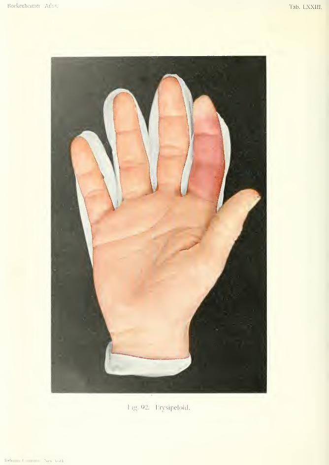
Bockenheimer, Atlas. Tab. LXXIII.
Vlli. 92. Iirysipcloid.
Rfbnian CoiiiDaiiy, New-York.
ERYSIPELOIDPlate LXXIII, Fig. 92.
An affection very similar to erysipelas, called
chronic erysipelas by Rosenbarh, is now known by
the term erysipeloid. This is also a bacterial infec-
tion of the skin (according to Tavel, also of tendon-
sheaths and joint capsules) but of a very harmless
nature. The specific cause of erysipeloid is unknown
;
in some cases the staphylococcus albus has been
found.
The affection begins with redness and swelling of
the fingers. Like erysipelas, the redness has sharp,
irregular borders. The redness spreads slowly but
continuously over the whole finger, and may extend
to the next finger and as far as the wrist. At this
point the inflammation stops. There are no consti-
tutional symptoms; no fever nor rigors. The pa-
tients only complain of itching and a feeling of ten-
sion in the skin. In some cases there is lymphangitis,
generally on the extensor surface, as far as the axilla.
In rare cases lymphadenitis with high temperature
has been observed.
Erysipeloid generally occurs after injuries to the
fingers, especially by fish and game. It is, therefore,
more common in venders of fish and game, cooks,
butchers, curriers, etc. Sometimes the injured spot
is invisible, as the redness and swelling generally
appear a few days after the injury. In other cases
foreign bodies are found in the skin. The affection
has been observed in doctors after operating upon
infected persons. The symptoms generally subside
in a week, but relapses are common. The disease is
more common in the autumn.
211

Differential Diagnosis. Erysipeloid diflfers from
erysipelas in its chronic course, absence of fever, paler
color, and demarcation at the wrist.
Treatment. Ointments, rest and support on
splints. Movements must be restricted after removal
of splints, to avoid relapses. Foreign bodies must be
removed. Suppuration has never been observed.
Baths and iodide ointment may be used if swelling
persists.
Fig. 92 shows erysipeloid in a cook, which appeared
soon after handling game. A few days after a slight
wound, redness and swelling developed at the tip of
the right forefinger, and gradually extended over the
whole finger. At the base of the finger the edge of
the redness is irregular and zigzag.
212
Panaritium or Panaris(WhUlou^
PANARITIUM SUBEPIDERMOIDALE {Sub-epidermic whitlow)
Plate LXXIV, Fig. 93.
PANARITIUM SUBCUTANEUM (Subnitaneous whitlow)
Plate LXXV, Fig. 94.
PANARITIUM OSSALE ET ARTICULARE{Ossrmifi and ariicnlar whitloui)
Plate LXX\^, Fig. 95.
PANARITIUM TENDINOSUM (Tendon-sheath ivhithw)
Plate LXXVII, Fig. 96.
PANARITIUM INTERDIGITALIS (Inlerdigital whitlow)
Plate LXXVII, Fig. 97.
Subcutaneous suppuration in the fingers and toes
is called ivhitlow. Although various forms of whit-
low are distinguished, this usually begins as an infec-
tion of the subcutaneous tissue (primary subcuta-
neous whitlow), from which may arise tendinous,
periosteal, osteal or articular whitlow, according to
the extent of the inflammatory process.
Subcutaneous whitlows occur most often in the
fingers, especially among the working classes wlio are
subject to cracks and fissures of the skin. They often
occur after punctured wounds, through which staphy-
lococci, or more rarely streptococci, gain entrance
to the subcutaneous tissue.
The anatomical formation of the subcutaneous
tissue is peculiar, vertical connective-tissue septa sep-
arating the fatty connective tissue into a number of
distinct compartments. If bacteria gain an entry
into such enclosed chambers the inflammation they
cause is at first circumscribed.
As in all infections, there is hypersemia, exudation
and necrosis of tissue; the latter occurs rapidly, owing
213
to the impairment of nutrition from pressure in the
inflamed area. In this way a necrotic core is formed,
as in furuncle. The increase of tension in the tis-
sues causes severe pain, and the finger becomes red
and swollen. In horny-handed workmen the seat of
infection is at first difficult to see, and is only madeevident by the great pain on pressure. Later on,
when the suppuration has extended further, the pain
is not so circumscribed. In a few cases only, the
skin gives way and a yellow core becomes loosened
and cast off, after which healing takes place by gran-
ulation tissue. The hard skin on the palmar surface
of the fingers prevents escape of pus, so that the latter
takes paths of less resistance. The vertical connec-
tive tissue septa, mentioned above, direct the pus
towards the peritendinous tissue, where it may spread
along the whole length of the tendon. The pus mayalso reach the loose connective tissue on the dorsal
surface, and give rise to redness, swelling andoedema, while inflammatory signs may be absent at
the seat of infection on the flexor surface. If the
tendon sheath is bathed in pus for some time it be-
comes perforated, and the pus extends within the
tendon sheath (tendinous whitlow, Fig. 96). In the
same way the periosteum, bony cortex, medullary
cavity and joint may become infected from a sub-
cutaneous whitlow (Fig. 95).
A further danger of whitlow is spreading of pus
to the hand and forearm along the tendon sheaths.
General infection may also occur.
The clinical symptoms vary according to the dura-
tion and extent of infection, and the virulence of the
bacteria. In sub-epidermic whitlow (Fig. 93), a puru-
lent vesicle develops, generally on the dorsal surface,
with slight redness of the surrounding skin. Theraised epidermis sometimes shows several yellow
spots, where the pus breaks through. Pain andfunctional disturbance are slight, the inflammation
remaining local. There is seldom lymphangitis, no214

tendency to spread, and little or no constitutional
disturbance.
In subcutaneous whitlow it is quite otherwise
(Fig. 94). The whole finger is red, swollen, flexed
and extremely painful, especially at one spot. Red-
ness, swelling and oedema are often more marked on
the dorsal surface, together with lymphangitis of the
hand and forearm. There is moderate fever (39° C.)
and some constitutional disturbance.
The symptoms are most severe in tendinous whit-
low (Fig. 96). There is more swelling of the finger,
and the latter is more flexed. There is pain on
pressure along the whole tendon sheath, and usually
over the whole palm. Movement of the tendon
causes great pain, and extension is almost impossible.
L\Tuphangitis and erysipelatous reddening often ex-
tend far beyond the seat of infection. There are
rigors and rise of temperature (40° C), sleepless-
ness, and considerable malaise.
If the tendon sheath of the thumb or little finger is
infected, the pus may extend along the course of these
sheaths as far as the wrist; whereas, suppuration in
the tendon sheaths of the second, third and fourth
fingers does not extend beyond the metacarpo-
phalangeal joints, where these tendon-sheaths end.
In the wrist the tendon-sheaths become widened
and lie so close together that suppuration mayextend from one to the other. In this way, infection
of the tendon-sheaths of the thumb may result from
a lesion of the tendon of the little finger ; and inversely,
infection of the little finger from the thumb. This
has been called V-shaped whitlow. It is obvious
that infection of both tendon-sheaths causes severe
symptoms—high fever and much constitutional dis-
turbance. The thumb and little finger are flexed,
swollen and very painful on pressure. The pus often
breaks through the tendon-sheaths and extends
between the muscles of the forearm up to the elbow
joint, in the form of deep, progressive suppuration.
215
In other cases the wrist-joint is infected. Such cases
may give rise to general infection.
The V-shaped whitlow is recognized by its severe
clinical symptoms and typical appearance. In the
early stages there is often pain, redness and swelling
in the palm, or on the flexor surface of the wrist.
When suppuration has existed some time and become
extensive it seeks a way to the surface. In this way
fistulas are formed in the course of the tendon-
sheaths, discharging much pus, and often exposing
the greenish-yellow remains of the necrosed tendon
(Fig. 96). The orifices of these fistulas are sur-
rounded by flabby, unhealthy granulations which, as
mentioned before (Fig. 56), indicate necrosis in the
deeper parts.
In periosteal and osteal whitlows, which generally
occur at the ends of the fingers, the periosteum and
bone are surrounded by pus and destroyed. In the
terminal phalanx total necrosis may occur. A fistula
forms and discharges the fetid, slimy pus, which is
characteristic of necrosed bone. Eventually dead
bone is discharged. (Fig. 95). Parts of the skin
may become necrosed, so that, eventually, the whole
finger-joint may be lost. Commencing with sharp
pain, the acute stage gradually becomes more
chronic, and in this stage infection of the bones
may be overlooked.
In the first and second phalanges there is often
infection of the joints, either secondary to infection
of the periosteum, or directly from the surface.
Articular whitlow generally manifests itself by rigors.
The joint is fixed in a position of flexion and is very
painful on movement. The capsule and ligaments
are soon destroyed, and destruction of the cartilage
causes grating on movement. Articular whitlow maygive rise to general infection.
It is not always easy to diagnose the stage of the
whitlow. Patients of the working class generally
come so late for treatment that there is often infection
316

of the tendon-sheath, periosteum and joint. In other
cases the pain is so severe as to suggest tendinous
whitlow, while it is only subcutaneous. A correct
diagnosis can often only be made after incision.
Differential Diagnosis. Tuberculous and syph-
ilitic inflammations are more chronic and cause less
pain and fever. They do not heal after incision, but
require specific treatment.
Treatment. All whitlows require early incision.
In sub-epidermic whitlow the purulent bulla must be
opened and dressed with antiseptic dressings, and
the arm suspended in a sling. Sub-epidermic whit-
low may cause infection of the deeper tissues, and
there is also the danger of erysipelas. Hence, plenty
of dressing should be used.
Subcutaneous whitlows should be incised as soon
as possible, under an anaesthetic. Schleich's infiltra-
tion anaesthesia is dangerous and painful in infected
areas. However, endoneural injection of one per
cent, cocaine may be made in the first phalanx, ac-
cording to the method of Oberst-Corning, if there is
no sign of inflammation at this place. But general
anaesthesia should be employed in all cases where the
extent of the suppuration is not clear. Incision
should be made into the subcutaneous tissue on the
palmar surface away from the middle line, and
between the joints. The wound is then held open
by retractors and examination made for pus in the
tendon-sheath or under the periosteum. This exam-
ination can only be made by a free incision, after the
hand is made bloodless by the elastic tourniquet.
This is the safest method of dealing with whitlows;
for although some cases may be cured by evacuation
of the pus through a small incision, suppuration in
the tendon-sheath may be overlooked, and this maylead to spread of suppuration, destruction of tendon,
etc., and even death from general infection.
217
No doubt, infected wounds of the finger often sub-
side with rest in a shng; but sometimes the perios-
teum is infected, and this conservative treatment then
results in necrosis of the phalanx. Therefore, wemust urge the treatment of all such infected wounds
by early incision, especially in doctors who are liable
to virulent infections.
Tendon-sheath whitlows require very careful treat-
ment, in order to preserve the tendon and the function
of the finger. Some cases come too late for treatment
for the tendon to be preserved. Many cases of
tendon-sheath whitlow extend so rapidly, and so
often lead to general infection, that they require free
incision of the whole area of suppuration. In some
cases, no doubt, this may cause injury to or loss of
the tendon; but it is far worse to be responsible for
a general infection which might have been avoided
by more extensive incision. Therefore, in extensive
tendon-sheath whitlows, especially in V-shaped whit-
lows, free incisions are necessary, but these should
always be made laterally. In V-shaped whitlows
care must be taken to preserve the palmar carpal
ligament; this must only be divided when there is
threatening infection of the wrist joint, or extension
of suppuration up the forearm.
Better functional results are obtained by several
smaller incisions instead of one continuous incision.
Too much plugging of the wound is to be avoided, as
it interferes with the nutrition of the tendon. After-
treatment consists in early passive movements.
In osteal whitlow necrosed bone must be removed
if present. In the terminal phalanx it is often suffi-
cient to remove the peripheral end. If the joint is
much destroyed resection of the bone, or even ampu-tation may be required.
Progressive suppurations, due to infection by viru-
lent bacteria or to extensive injuries, must be freely
laid open, sometimes as far as the bone. If general in-
fection supervenes the question of amputation arises.
218
Although early incision removes the danger of the
pyogenic condition spreading by subcutaneous sup-
puration, this danger may recur if the after-treatment
is neglected.
The incisions should be lightly plugged with iodo-
form gauze, which best absorbs the discharge. After
the first dressing this should be replaced by small
pieces of sterilized gauze, sufficient to keep the edges
of the wound open and allow the pus to escape.
The hand and forearm should be immobilized on a
splint. Under this treatment even deep cavities be-
gin to granulate in a few days, when the plugging
should be left off and replaced by baths and ointment.
To decide the time when plugging may be left off is
a matter of experience. If it is kept on too long the
nutrition of the tendon (in the case of tendinous whit-
low) is impaired. If it is left off too soon, suppura-
tion may extend into the deeper parts; this is mani-
fested by further redness, swelling and pain, and by
a fetid, slimy discharge from the wound and the for-
mation of flabby, unhealthy granulations. Increase
of pain is often a sufficient sign of fresh infection. If
the extension of infection is not immediately noticed
it may cause severe complications and general infec-
tion, even during the period of after-treatment. This
reinfection may be avoided by several measures.
First of all, patients with severe forms of whitlow
should be treated in hospital, where they can be kept
under observation and treated under more favorable
conditions.
The temperature, in severe cases of whitlow, should
be taken every four hours. The dressings should be
changed every day, if necessary under an anaesthetic,
so that the local condition can be examined. Thegauze tampons should be carefully removed during
irrigation with peroxide lotion. The wound should
then be irrigated with normal saline solution under
very slight pressure, and the dressing renewed. It is
often necessary to hold the edges of the wound apart
219

by retractors, so as to obtain a better view of the con-
dition of the wound, and drain all suspicious pockets.
Drains should be only retained after the first change
of dressing in extensive whitlows. When the dressings
are changed examination must be made for inflam-
mation and suppuration in parts remote from the
wound—in the palm in tendinous whitlow, and in
the wrist and elbow joints in V-shaped whitlow. Even
in the slighter forms of whitlow the dressings should
be changed every day, especially in out-patients
(polyclinic). This avoids stiffening of the fingers by
prolonged immobilization, also the troublesome con-
dition called "glossy skin."
After-treatment is begun when the suppuration has
ceased and the temperature has become normal.
This consists in performing passive movements of the
fingers each time the dressings are changed. In out-
patient practice (polyclinic), large immobilizing dress-
ings should be applied after these passive movements
have been performed. This is especially necessary in
alcoholic patients, in whom the inflammation is muchaggravated. Moreover, small dressings may be re-
moved by the patient himself. The application of
large immobilizine; dressings has a favorable influence
on the inflammation, and renders the after-treatment
easier and shorter, while the disadvantage of immo-bilization is removed by daily passive movements
when the dressings are changed.
In the treatment of whitlows it is best to pursue a
middle course. On the one hand, too large incisions,
too much plugging and too long immobilization cause
impairment of function; on the other hand, small
incisions, too little plugging and too free movementmay lead to general infection. Radical treatment is
best for the beginner, although more conservative
methods may be adopted with further experience.
After-treatment must be commenced directly sup-
puration has ceased (massage, baths and passive
movement).
220

Cicatricial contractions sometimes yield to gradual
extension; but some cases may require excision of
the scar, followed by a plastic operation.
As already mentioned Bier's passive hypersemia
treatment is contra-indicated in acute pyogenic affec-
tions; in mild cases it is unnecessary, and in severe
cases it is dangerous.
Other methods, such as injection of carbolic acid
lotion, staphylococcal serum, carbolic and alcoholic
fomentations generally do harm.
221

PANARITIUM SUBEPIDERMOIDALE {Subepidermis Whitlow)Plate LXXIV. Fig. 93.
In this case a circumscribed redness developed on
the dorsal surface of the left forefinger, without any
obvious injury. This was followed by the formation
of a purulent blister. The epidermis is raised and
shows several yellow points due to the presence of
pus. The movement of the finger was not impaired.
The blister was opened, the pus evacuated and the
thin epidermis removed. The wound was dressed
with sterilized gauze, and the finger put up on a
splint extending above the wrist.
223
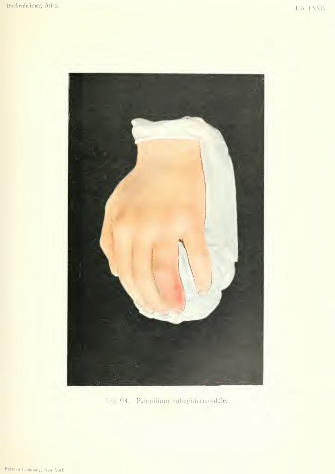
Borkeiilifiiiier, Atlas.Tab. I.N\I\-.
I'l'p. 9'i. Panaritium subepidennoidale.
I'rbniaii Conipany, New-\'ork.


Bockeiiliciiiier, Alias. Tab. LXXV.
l"ig. 94. Faiiaritiuni subcutaneum — Lymphaiiyilis acuta.
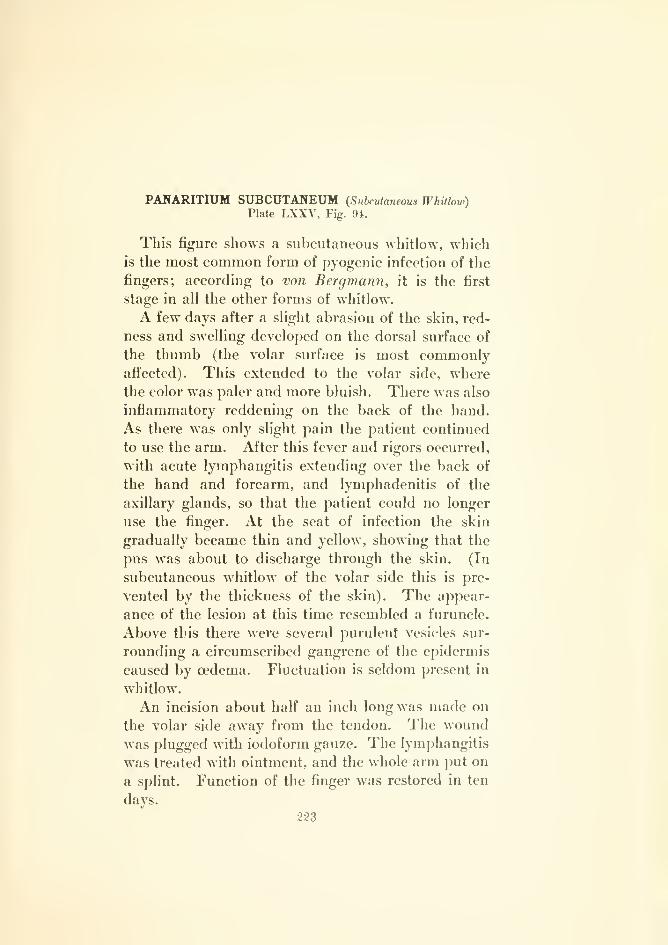
PANARITIUM SUBCUTANEUM {SiibnUaneoM Whitlow)Plate LXXV, Fig. 94.
This figure shows a subcutaneous whitlow, whichis the most common form of pyogenic infection of the
fingers; according to von Bergmann, it is the first
stage in all the other forms of whitlow.
A few days after a slight abrasion of the skin, red-
ness and swelling developed on the dorsal surface of
the thumb (the volar surface is most commonlyafi'ected). This extended to the volar side, where
the color was paler and more bluish. There was also
inflammatory reddening on the back of the hand.
As there was only slight pain the patient continued
to use the arm. After this fever and rigors occurred,
with acute lymphangitis extending over the back of
the hand and forearm, and lymphadenitis of the
axillary glands, so that the patient could no longer
use the finger. At the seat of infection the skin
gradually became thin and yellow, showing that the
pus was about to discharge through the skin. (In
subcutaneous whitlow of the volar side this is pre-
vented by the thickness of the skin). The appear-
ance of the lesion at this time resembled a furuncle.
Above this there were several purulent vesicles sur-
rounding a circumscribed gangrene of the epidermis
caused by oedema. Fluctuation is seldom present in
whitlow.
An incision about half an inch long was made on
the volar side away from the tendon. The woundwas plugged with iodoform gauze. The lymphangitis
was treated with ointment, and the whole arm put on
a splint. Function of the finger was restored in ten
days.
223
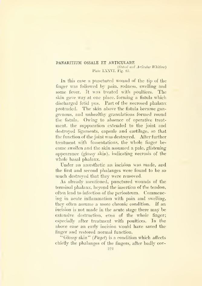
PANARIXroM OSSALE ET ARTICULARE(Osteal and Articular Whitlow)
Plate LXXM, Fig. 93.
In this case a punctured wound of the tip of the
finger was followed by pain, redness, swelling and
some fever. It was treated with poultices. Theskin gave way at one place, forming a fistula which
discharged fetid pus. Part of the necrosed phalanx
protruded. The skin above the fistula became gan-
grenous, and unhealthy granulations formed round
the fistula. Owing to absence of operative treat-
ment, the suppuration extended to the joint and
destroyed ligaments, capsule and cartilage, so that
the function of the joint was destroyed. After further
treatment with fomentations, the whole finger be-
came swollen and the skin assumed a pale, glistening
appearance (glossy skin), indicating necrosis of the
whole basal phalanx.
Under an anaesthetic an incision was made, and
the first and second phalanges were found to be so
much destroyed that they were removed.
As already mentioned, punctured wounds of the
terminal phalanx, beyond the insertion of the tendon,
often lead to infection of the periosteum. Commenc-ing in acute inflammation with pain and swelling,
they often assume a more chronic condition. If an
incision is not made in the acute stage there may be
extensive destruction, even of the whole finger;
especially after treatment with poultices. In the
above case an early incision would have saved the
finger and restored normal function.
"Glossy skin" (Paget) is a condition which affects
chiefly the phalanges of the fingers, after badly cov-
224:
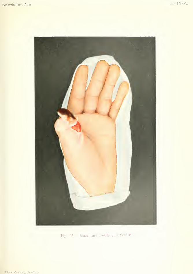
Bockenheimer, Atlas. Tab. I.XXVI.
Fig. 95. I'anaritium ossale et articulare.
Rebman Company, New- York.


ered amputation stumps, or after too-long immo-bilization. This condition may extend over the wholefinger. The skin is at first thickened, bluish red,
and cold to the touch; later on it becomes pale yel-
low and has an appearance like parchment. The cir-
culation is bad and there are often neuralgic pains
and a feeling of coldness. It may finally lead to trau-
matic neurasthenia. This condition can be pre-
vented by avoiding too long immobilization and byproviding the amputation stumps with sufficient well-
nourished flaps.
225

PANARITIUM TENDINOSUM (Tendincms Whitlow)
Plate LXXMI, Fig. 96.
This is a case of subcutaneous whitlow, following
a punctured wound, which rapidly spread to the
tendon-sheath of the thumb. A severe form of
infection was indicated by the acute redness and
swelling, severe pain, high temperature and consti-
tutional disturbance. As no incision was made, the
tei'minal phalanx continued to swell and finally gave
way, forming a fistula discharging pus and parts of
necrosed tendon. Apart from this, the diagnosis of
tendinous whitlow could be made from the severity
of the symptoms; from the complete loss of move-
ment in the thumb, the great pain on pressure over
the course of the tendon, the swelling and redness of
the ball of the thumb, and the discharge of pus from
the fistula on pressure over this part. The tendon
sheath of the little finger was unaffected, and there
was no sign of abscess above the wrist.
Under an anaesthetic an incision was made along
the whole of the terminal phalanx and pus evacuated
from the tendon-sheath. A second incision was
made in the palm, a little below the wrist, and the
tendon-sheath opened again at this point. By this
means the suppuration ceased and infection of the
tendon-sheath of the little finger was avoided. As the
tendon of the thumb was already partly destroyed,
the end joint remained functionless. In spite of a
certain degree of contracture, the patient could
use the thumb, by movement at the metacarpo-
phalangeal joint.
236
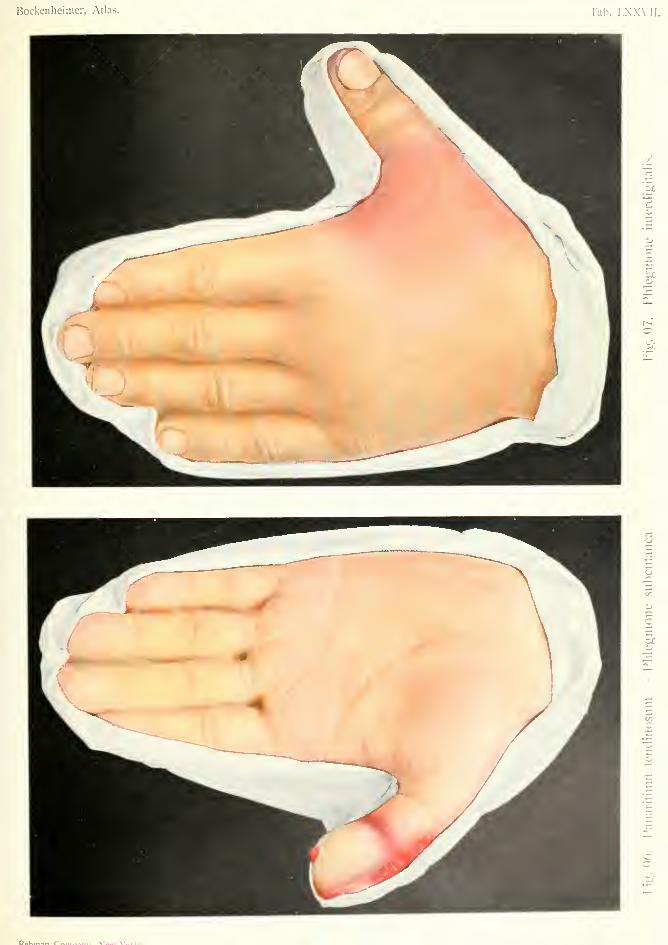
Bockenheimer, Atlas. Tab. I.XXVIL
ti3
onc
o
So
D»kni.*« r^ M...» V'».-l.

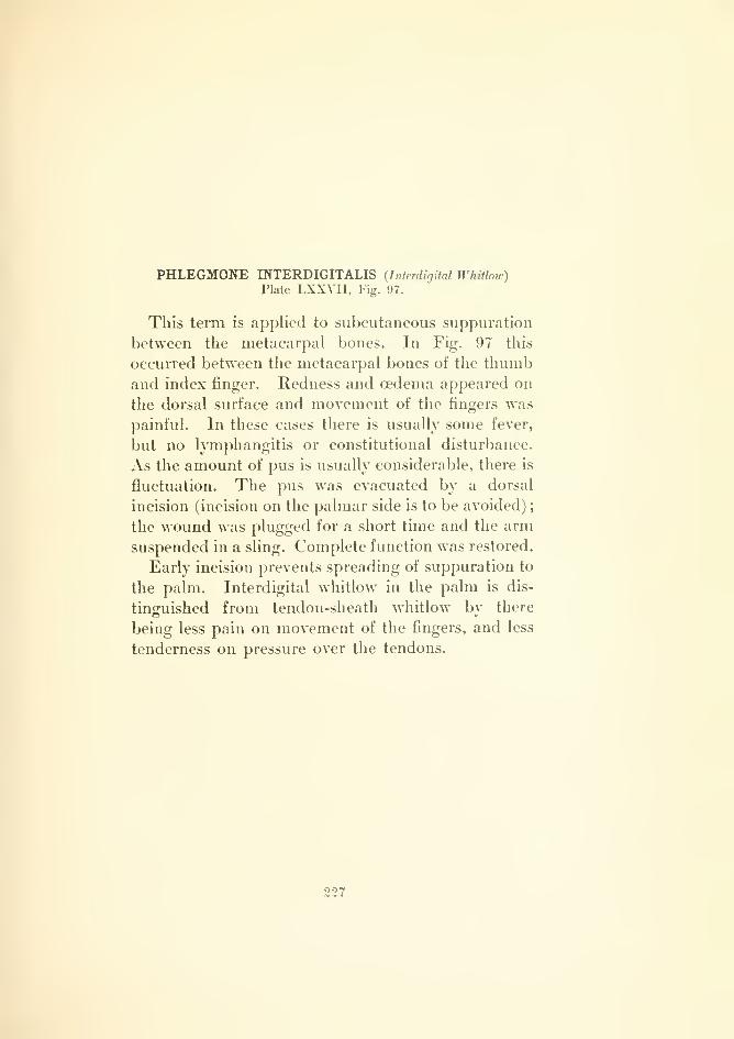
PHLEGMONE INTERDIGITALIS {Interdigiial Whitlow)
Plate LXXVIl, Fig. 97.
This term is applied to subcutaneous suppuration
between the metacarpal bones. In Fig. 97 this
occurred between the metacarpal bones of the thumband index finger. Redness and oedema appeared on
the dorsal surface and movement of the fingers was
painful. In these cases there is usually some fever,
but no lymphangitis or constitutional disturbance.
As the amount of pus is usually considerable, there is
fluctuation. The pus was evacuated by a dorsal
incision (incision on the palmar side is to be avoided)
;
the wound was plugged for a short time and the arm
suspended in a sling. Complete function was restored.
Early incision prevents spreading of suppuration to
the palm. Interdigital whitlow in the palm is dis-
tinguished from tendon-sheath whitlow by there
being less pain on movement of the fingers, and less
tenderness on pressure over the tendons.
227
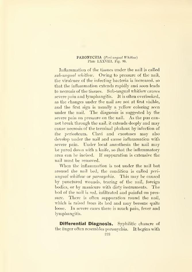
PARONYCHIA (Pen-ungual Whitlow)
Plate LXXVIII, Fig. 98.
Inflammation of the tissues under the nail is called
suh-ungual wJdtloiv. Owing to pressure of the nail,
the virulence of the infecting bacteria is increased, so
that the inflammation extends rapidly and soon leads
to necrosis of the tissues. Sub-ungual whitlow causes
severe pain and lymphangitis. It is often overlooked,
as the changes under the nail are not at first visible,
and the first sign is usually a yellow coloring seen
under the nail. The diagnosis is suggested by the
severe pain on pressure on the nail. As the pus can-
not break through the nail, it extends deeply and maycause necrosis of the terminal phalanx by infection of
the periosteum. Clavi and exostoses may also
develop under the nail and cause inflammation with
severe pain. Under local anaesthesia the nail maybe pared down with a knife, so that the inflammatory
area can be incised. If suppuration is extensive the
nail must be removed.
When the inflammation is not under the nail but
around the nail bed, the condition is called pcri-
ungval whitlow or paronychia. This may be caused
by punctured wounds, tearing of the nail, foreign
bodies, or by manicure with dirty instruments. Thebed of the nail is red, infiltrated and painful on pres-
sure. There is often suppuration round the nail,
which is raised from its bed and may become quite
loose. In severe cases there is much pain, fever andlymphangitis.
Differential Diagnosis. Syphilitic chancre of
the finger often resembles paronychia. It begins with
228
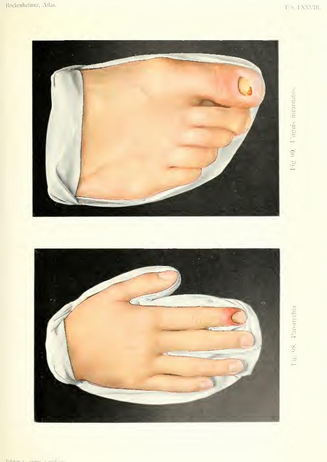
Bockenheimer, Atlas.Tah. I.X.W;
CO
W)
W.,, \'^..t.

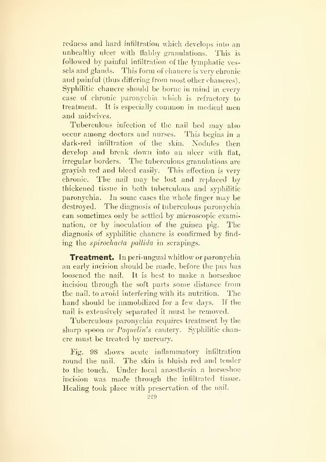
redness and hard infiltration which develops into anunhealthy ulcer with flabby granulations. This is
followed by painful infiltration of the lymphatic ves-
sels and glands. This form of chancre is very chronic
and painful (thus dift'ering from most other chancres).
Syphilitic chancre should be borne in mind in every
case of chronic paronychia which is refractory to
treatment. It is especially common in medical menand midwives.
Tuberculous infection of the nail bed may also
occur among doctors and nurses. This begins in a
dark-red infiltration of the skin. Nodules then
develop and break down into an ulcer with flat,
irregular borders. The tuberculous granulations are
grayish red and bleed easily. This affection is very
chronic. The nail may be lost and replaced bythickened tissue in both tuberculous and syphilitic
paronychia. In some cases the whole finger may be
destroyed. The diagnosis of tuberculous paronychia
can sometimes only be settled by microscopic exami-
nation, or by inoculation of the guinea pig. Thediagnosis of syphilitic chancre is confirmed by find-
ing the spirochacta pallida in scrapings.
Treatment. In peri-ungual whitlow or paronychia
an early incision should be made, before the pus has
loosened the nail. It is best to make a horseshoe
incision through the soft parts some distance from
the nail, to avoid interfering with its nutrition. Thehand should be immobilized for a few days. If the
nail is extensively separated it must be removed.
Tuberculous paronychia requires treatment by the
sharp spoon or Paquelin's cautery. Syphilitic chan-
cre must be treated by mercury.
Fig. 98 shows acute inflammatory infiltration
round the nail. The skin is bluish red and tender
to the touch. Under local anaesthesia a horseshoe
incision was made through the infiltrated tissue.
Healing took place with preservation of the nail.
229
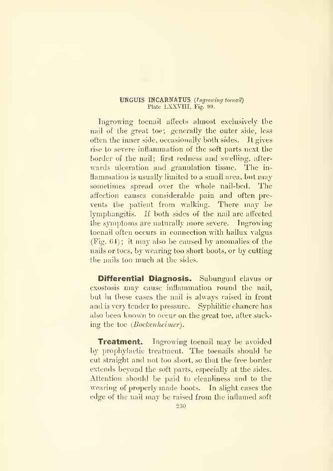
UNGUIS mCARNATUS (Ingromng toenail)
Plate LXXVIII, Fig. 99.
Ingrowing toenail aflPects almost exclusively the
nail of the great toe; generally the outer side, less
often the inner side, occasionally both sides. It gives
rise to severe inflammation of the soft parts next the
border of the nail; first redness and swelling, after-
wards ulceration and granulation tissue. The in-
flammation is usually limited to a small area, but maysometimes spread over the whole nail-bed. Theaffection causes considerable pain and often pre-
vents the patient from walking. There may be
lymphangitis. If both sides of the nail are afi^ected
the symptoms are naturally more severe. Ingrowing
toenail often occurs in connection with hallux valgus
(Fig. 64) ; it may also be caused by anomalies of the
nails or toes, by wearing too short boots, or by cutting
the nails too much at the sides.
Differential Diagnosis. Subungual clavus or
exostosis may cause inflammation round the nail,
but in these cases the nail is always raised in front
and is very tender to pressure. Syphilitic chancre has
also been known to occur on the great toe, after suck-
ing the toe (Bockenheimer).
Treatment. Ingrowing toenail may be avoided
by prophylactic treatment. The toenails should be
cut straight and not too short, so that the free border
extends beyond the soft parts, especially at the sides.
Attention should be paid to cleanliness and to the
wearing of properly made boots. In slight cases the
edge of the nail may be raised from the inflamed soft
230

parts by an iodoform tampon, or partial excision of
the nail may be performed. In severe cases these
methods are useless. Excision of the nail, which wasformerly practiced, is useless, as the condition recurs
after. The most rational method consists in excision
of the whole lateral border of the nail together with
the inflamed soft parts, down to the bone; taking
care to include the posterior part of the matrix, so
that recurrence cannot take place. The woiuid is
dressed with iodoform powder and sterilized gauze
and immobilized for a week, after which the woundis usually healed. In ingrowing toenail affecting
both sides the same operation is performed on each
side, leaving the center part of the nail in place.
Fig. 99 shows an ingrowing toenail on the outer
side of the right great toe. The thickened soft parts
have grown over the border of the nail. There is a
purulent discharge from unhealthy granulations. Thenail is so imbedded in the swollen soft parts that it is
only partly visible. The above operation was per-
formed with good result.
231
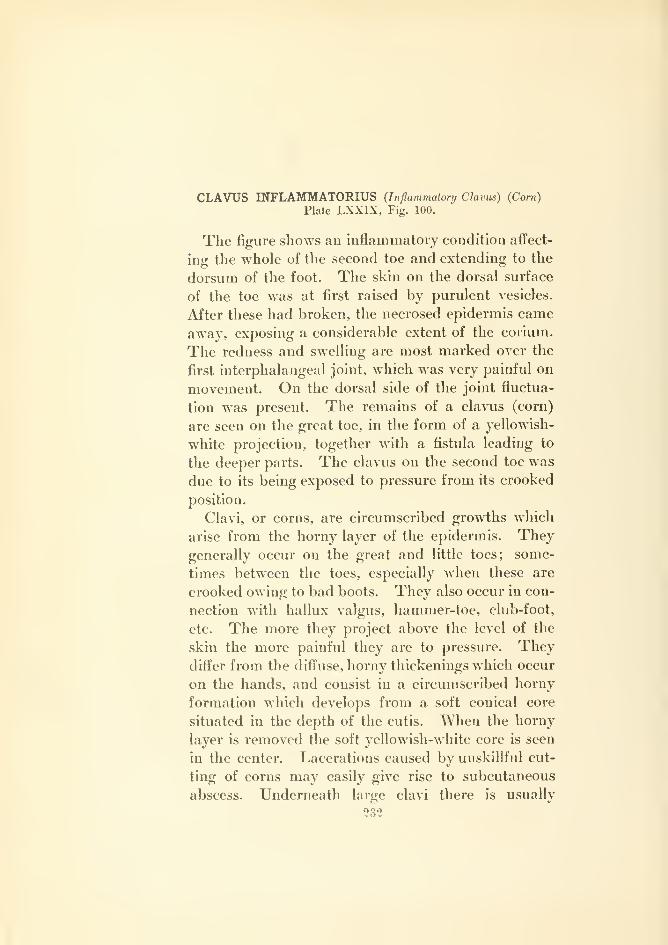
CLAVUS nOi'LAMMATORrUS (Inflammatory Clavus) (Com)Plate LXXIX. Fig. 100.
The figure shows an inflammatory condition affect-
ins the whole of the second toe and extendiuo; to the
dorsum of the foot. The skin on the dorsal surface
of the toe was at first raised by purulent vesicles.
After these had broken, the necrosed epidermis came
away, exposing a considerable extent of the corium.
The redness and swelling are most marked over the
first interphalangeal joint, which was very painful on
movement. On the dorsal side of the joint fluctua-
tion was present. The remains of a clavus (corn)
are seen on the great toe, in the form of a yellowish-
white projection, together with a fistula leading to
the deeper parts. The clavus on the second toe was
due to its being exposed to pressure from its crooked
position.
Clavi, or corns, are circumscribed growths which
arise from the horny layer of the epidermis. Theygenerally occur on the great and little toes; some-
times between the toes, especially when these are
crooked owing to bad boots. They also occur in con-
nection with hallux valgus, hammer-toe, club-foot,
etc. The more they project above the level of the
skin the more painful they are to pressure. Theydiffer from the diffuse, horny thickenings which occur
on the hands, and consist in a circumscribed horny
formation which develops from a soft conical core
situated in the depth of the cutis. When the horny
layer is removed the soft yellowish-white core is seen
in the center. Lacerations caused by unskillful cut-
ting of corns may easily give rise to subcutaneous
abscess. Underneath large clavi there is usually
232
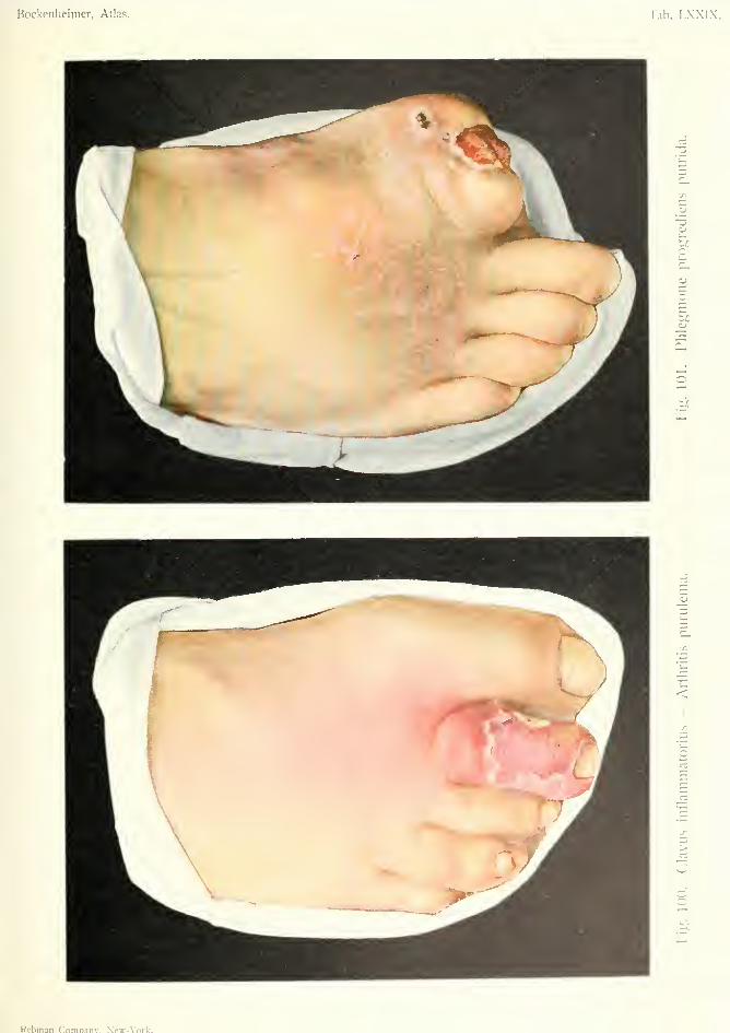
Bockenheimer, Atlas. Tab. I.XXIX.
•a<u
heo
3
oo
Kcbman Comnanv. Neip-\'ork-.

develoj)ed a bursa, which is Hable to become inflamed
from external pressure. The inflammatory exuda-
tion from the bursa generally discharges by a fistula
near the clavus (Fig. 100). Septic infection of the
bursa may be caused through the fistula, and this mayextend to the neighboring tendon-sheath or joint.
Joint infection is especially frequent when the bursa
communicates with the joint; and is manifested by
severe local inflammation, fever, rigors and constitu-
tional disturbance. The purulent arthritis may even
give rise to general infection.
Treatment. Prophylactic treatment of clavus
consists in cleanliness and the wearing of proper
boots. If a clavus forms it should be removed with
a sterilized knife. It is not sufficient to remove the
horny layer; the deeply situated core must also be
removed, otherwise recurrence takes place. Other
methods, such as the application of salicylic collo-
dion, only loosen the horny layer and do not prevent
recurrence.
If a bursa forms under the clavus it must either be
incised and plugged, or excised. If suppuration
extends to the joint this must be opened; in some
cases resection or disarticulation may be necessary.
In Fig. 100 there was inflammation of a bursa
which communicated with the joint. The bursa
discharged through a fistula, and infection through
the fistula gave rise to suppuration and to inflamma-
tion of the joint. Severe sjTQptoms developed, with
rigors and fever, and lymphangitis of the foot and
leg. The joint was opened on the dorsal surface by
a transverse incision, and the superficial suppuration
by another incision on the dorsum of the foot. The
clavus and the bursa were excised subsequently.
233
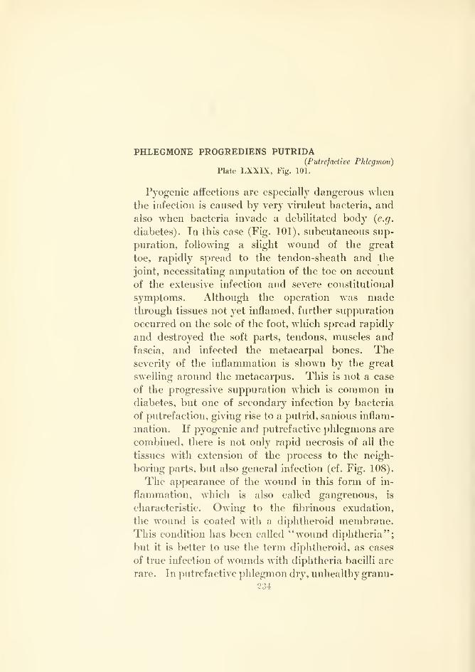
PHLEGMONE PROGREDIENS PUTRIDA(Putrejadive Phlegmon)
Plate LXXIX, Fig. 101.
Pyogenic affections are especially dangerous whenthe infection is caused by very virulent bacteria, and
also when bacteria invade a debilitated body {e.g.
diabetes). In this case (Fig. 101), subcutaneous sup-
puration, following a slight wound of the great
toe, rapidly spread to the tendon-sheath and the
joint, necessitating amputation of the toe on account
of the extensive infection and severe constitutional
symptoms. Although the operation was madethrough tissues not yet inflamed, further suppuration
occurred on the sole of the foot, which spread rapidly
and destroyed the soft parts, tendons, muscles and
fascia, and infected the metacarpal bones. Theseverity of the inflammation is shown by the great
swelling around the metacarpus. This is not a case
of the progressive suppuration which is common in
diabetes, but one of secondary infection by bacteria
of putrefaction, giving rise to a putrid, sanious inflam-
mation. If pyogenic and putrefactive phlegmons are
combined, there is not only rapid necrosis of all the
tissues with extension of the process to the neigh-
boring parts, but also general infection (cf. Fig. 108).
The appearance of the wound in this form of in-
flammation, which is also called gangrenous, is
characteristic. Owing to the fibrinous exudation,
the wound is coated with a diphtheroid membrane.This condition has been called "wound diphtheria";
but it is better to use the term diphtheroid, as cases
of true infection of wounds with diphtheria bacilli are
rare. In putrefactive phlegmon dry, unhealthy granu-
234
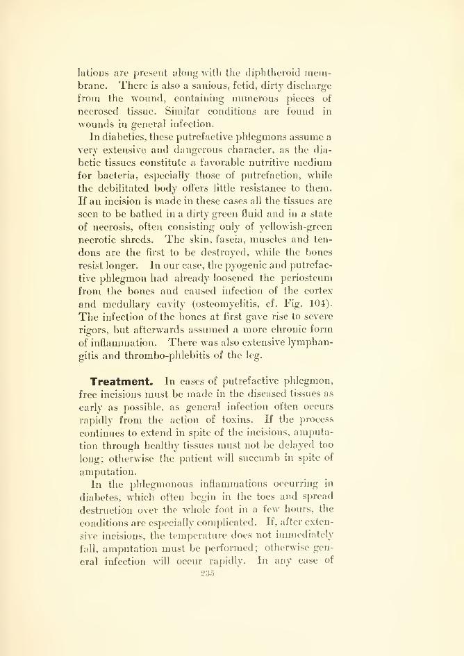
lations are present along with the diphtheroid mem-brane. There is also a sanious, fetid, dirty discliarge
from the wound, containing numerous pieces of
necrosed tissue. Similar conditions are found in
wounds in general infection.
In diabetics, these putrefactive phlegmons assume a
very extensive and dangerous character, as the dia-
betic tissues constitute a favorable nutritive mediumfor bacteria, especially those of putrefaction, while
the debilitated body offers little resistance to them.
If an incision is made in these cases all the tissues are
seen to be bathed in a dirty green fluid and in a state
of necrosis, often consisting only of yellowish-green
necrotic shreds. The skin, fascia, muscles and ten-
dons are the first to be destroyed, while the bones
resist longer. In our case, the pyogenic and putrefac-
tive phlegmon had already loosened the periosteum
from the bones and caused infection of the cortex
and medullary cavity (osteomyelitis, cf. Fig. 104).
The infection of the bones at first gave rise to severe
rigors, but afterwards assumed a more chronic form
of inflammation. There was also extensive lymphan-
gitis and thrombo-phlebitis of the leg.
Treatment. In cases of putrefactive phlegmon,
free incisions must be made in the diseased tissues as
early as possible, as general infection often occurs
rapidly from the action of toxins. If the process
continues to extend in spite of the incisions, amputa-
tion through healthy tissues must not be delayed too
long; otherwise the patient will succumb in spite of
amputation.
In the phlegmonous inflammations occurring in
diabetes, which often begin in the toes and spread
destruction over the whole foot in a few hours, the
conditions are especially complicated. If, after exten-
sive incisions, the temperature does not immediately
fall, amputation must be performed; otherwise gen-
eral infection will occur ra{>idly. In any case of
235

phlegmonous inflammation in a diabetic patient death
may occur from coma or heart failure.
In Fig. 101 there was a combination of pyogenic
and putrefactive phlegmon of a progressive character
in a diabetic patient. High temperature, rigors, dry
tongue and somnolence suggested the commence-ment of general infection. Amputation was performed
above the knee, owing to the presence of lymphan-gitis, thrombo-phlebitis in the leg, and also advanced
arterio-sclerosis. The operation was performed un-
der lumbar anaesthesia and led to healing.
236

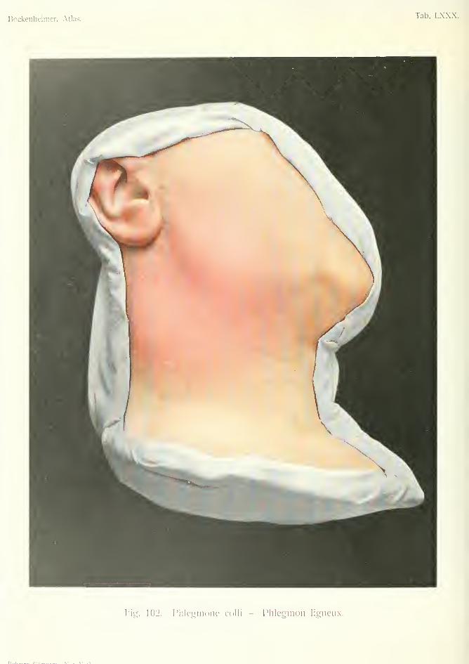
Bockenheimer, Atlas.Tab. LXXX.
I'l'^. 102. I'lilcgmone colli — Phlegmon ligneu.x.

PHLEGMONE COLLI (PJikgman of the neck)
riate LXXX, Fig. 102.
In the region of the neck, subcutaneous and sub-
fascial phlegmons are common, owing to the numerousgroups of lymphatic glands in this situation. Suppu-
rative inflammation of these glands may be caused
by affections of the mouth and pharynx, carious
teeth, angina, otitis media, alveolar periostitis, foreign
bodies, etc. Eczema and other affections of the head
and face may also cause suppuration in the glands of
the neck, especially in young individuals. The infec-
tion is generally due to staphylococci, sometimes
streptococci and other bacteria. In lesions of the
mouth and pharynx putrefactive bacteria are some-
times found in the buccal cavity.
Subcutaneous phlegmon in the neck manifests
itself by redness of the skin, inflammatory infiltration
and fever; later on fluctuation can be made out. In
nearly all cases a circumscribed abscess forms on one
side of the neck. Large abscesses may cause dysp-
noea by pressure on the larynx, and dysphagia by
pressure on the esophagus.
In the submaxillary region the inflammation oc-
curs most commonly in the subcutaneous l;yTnphatic
glands, and the abscess is situated outside the cap-
sule of the submaxillary gland. This must be dis-
tinguished from intracapsular suppuration of the
submaxillary gland itself, which is called Ludwig's
angina (angina Ludovici). In this case the symp-
toms are much more severe—fever, rigors, swelling
in the buccal cavity and pharj'nx, causing difficulty
in respiration and swallowing.
Infection of the sub-mental honphatic glands gives
237
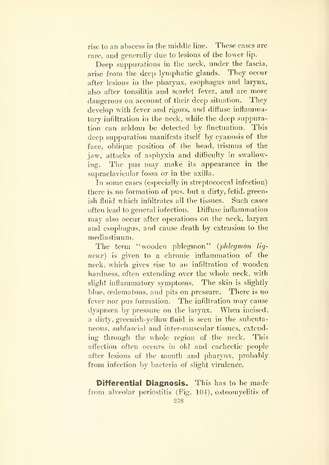
rise to an abscess in the middle line. These cases are
rare, and generally due to lesions of the lower lip.
Deep suppurations in the neck, under the fascia,
arise from the deep lymphatic glands. They occur
after lesions in the pharynx, esophagus and larynx,
also after tonsilitis and scarlet fever, and are more
dangerous on account of their deep situation. They
develop with fever and rigors, and diffuse inflamma-
tory infiltration in the neck, while the deep suppura-
tion can seldom be detected by fluctuation. This
deep suppuration manifests itself by cyanosis of the
face, oblique position of the head, trismus of the
jaw, attacks of asphyxia and diflSculty in swallow-
ing. The pus may make its appearance in the
supraclavicular fossa or in the axilla.
In some cases (especially in streptococcal infection)
there is no formation of pus, but a dirty, fetid, green-
ish fluid which infiltrates all the tissues. Such cases
often lead to general infection. Diffuse inflammation
may also occur after operations on the neck, larynx
and esophagus, and cause death by extension to the
mediastinum.
The term "wooden phlegmon" (phlegmon lig-
neux) is given to a chronic inflammation of the
neck, which gives rise to an infiltration of wooden
hardness, often extending over the whole neck, with
slight inflammatory symptoms. The skin is slightly
blue, cedematous, and pits on pressure. There is no
fever nor pus formation. The infiltration may cause
dyspnoea by pressure on the larynx. When incised,
a dirty, greenish-yellow fluid is seen in the subcuta-
neous, subfascial and inter-muscular tissues, extend-
inor throuarh the whole refrion of the neck. This
affection often occurs in old and cachectic people
after lesions of the mouth and pharynx, probably
from infection by bacteria of slight virulence.
Differential Diagnosis. This has to be madefrom alveolar periostitis (Fig. 104), osteomyelitis of
238
the lower jaw (Fio;. 105), tuberculous adenitis, andcystic tumors in the neck (blood cysts, dermoidssebaceous cysts, branchial cysts). Changes in the
bone are revealed by an incision in the case of
periostitis and osteomyelitis. Acute symptoms andfever are absent in the other formations, but sujjpu-
ration of a cystic tumor may resemble glandular sup-
puration. In cases of deep suppuration in the neck,
retro-phar^Tigeal abscess must be borne in mind.
Wooden phlegmon of the neck may be mistaken
for commencing actinomycosis, but the latter soon
gives rise to a fistula vi'hich discharges pus mixed with
the characteristic yellow* bodies (Fig. 115).
Treatment. Poultices are contra-indicated, as
they cause considerable destruction of tissue, andallow the right time for incision to be passed by.
Early incision is indicated in most cases. In sub-
cutaneous phlegmons with a tendency to becomecircumscribed, incision should not be made until anabscess forms. Under local anaesthesia an incision
is made through the skin at the lowest part of the
abscess, and the pus evacuated by means of blunt
dressing forceps. In the submaxillary region the
facial nerve and vessels must be avoided.
In intracapsular inflammation of the submaxillary
gland, the gland must be freely incised before sup-
puration occurs, otherwise general infection mayoccur from increased virulence of the bacteria due
to pressure of the capsule.
In all cases of phlegmons in the neck in which
there is much infiltration of the floor of the mouth
with difficulty in breathing and swallowing, it is
advisable to perform a preliminary tracheotomy, as
death may occur from sudden oedema of the glottis
during anaesthesia.
In deep suppurations of the neck we must not wait
for the appearance of a superficial abscess. A free
incision must be made along the median border of
239

the sternomastoid muscle. Extensive cases require
counter-incisions. The wounds should be drained
by gauze tampons, as drainage tubes may injure the
large vessels.
Wooden phlegmon of the neck sometimes requires
multiple deep incisions, laterally and in the middle
line.
Fig. 102 shows acute inflammation of the sub-
maxillary lymphatic glands, with the formation of
an abscess under the skin. It was treated under
local anaesthesia by incision and drainage.
240

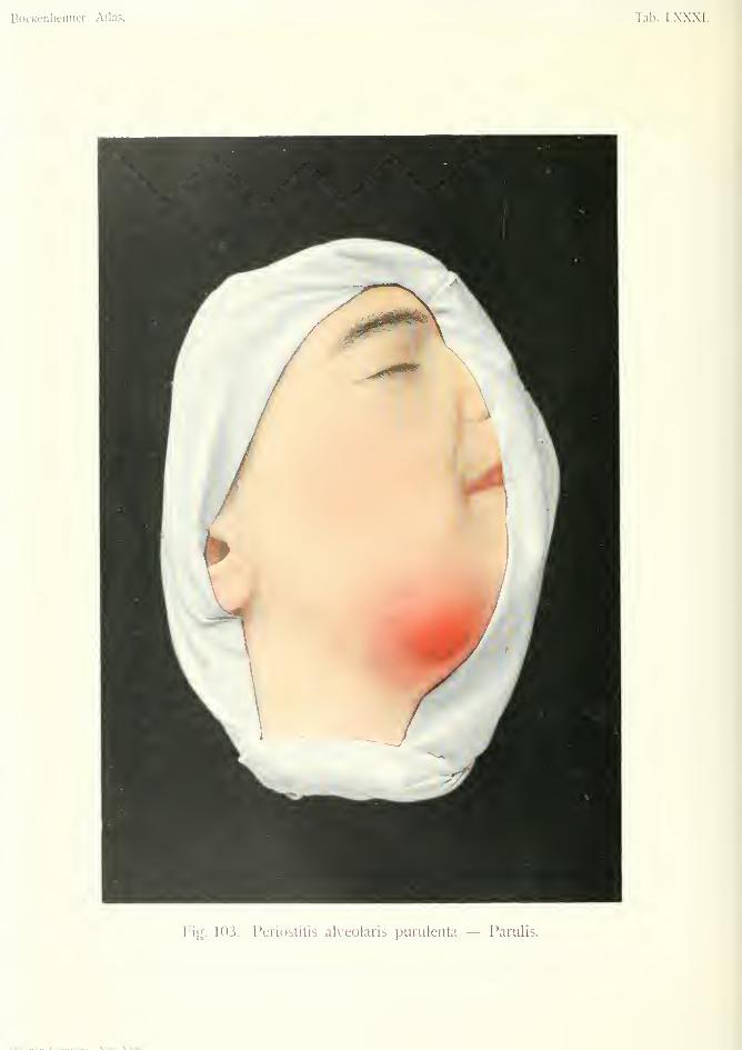
Bockeiilieimer, Atlas. Tab. LXXXI.
rig. 103. Periostitis alveoians innulciita — Parulis.
r>„i .. r*^.„«„..., \i«,». \'^^i.
PERIOSTITIS ALVEOLARIS PURULENTA—PARULIS(Purulent alveolar Periostitis)
Plate LXXXI, Fig. 103.
Parulis i.s a name given to purulent alveolar perios-
titis of the lower jaw, which usually gives rise to a
subcutaneous abscess. It may be caused by lesions
of the gums (e.g. after tooth-extraction with dirty
instruments), fractures of the jaw, operations on the
jaw, caries of the teeth, fistulas from the stumps of
teeth. Infection of the periosteum of the alveolar
portion of the lower jaw gives rise to a circumscribed
subperiosteal accumulation of pus which descends
to the submaxillary region and lies over the fascia
covering the submaxillary gland. The signs of puru-
lent inflammation are most apparent in this region,
while symptoms at the seat of infection are often slight.
The symptoms commence with fetor of the breath,
fever and rigors, and inflammatory infiltration in the
submaxillary region. Soon afterwards the presence
of fluctuation indicates abscess formation, after which
the symptoms diminish. In most cases the suppu-
ration is circumscribed, but sometimes there is dift'use
inflammation, causing considerable infiltration of the
soft parts and swelling and redness of the side of the
face. There is then often trismus and oedema of the
mucous membrane of the mouth, with difficulty in
mastication and often difficulty in breathing. In
these diffuse forms there are severe constitutional
symptoms—rigors, fever, headache, etc.
Although the circumscribed form is harmless, the
diffuse form may be dangerous to life, especially
when improperly treated. Treatment of the circum-
scribed form by poultices may give rise to the diffuse
form. If the pus is allowed to remain for long under
241
the periosteum, it may cause osteomyelitis of the
jaw and all its consequences (Fig. 104). Meningitis
and general infection may also occur from thrombo-
phlebitis.
In the upper jaw, infection of the periosteum mayalso cause subperiosteal suppuration, which has not
such favorable conditions for extension to the sub-
cutaneous tissue as in the case of the lower jaw.
Small abscess caused by morbid conditions of the teeth
may burst into the mouth and cause no trouble, but
more virulent infection may cause osteomyelitis of
the upper maxilla, which rapidly extends over the
whole of the bones of the face, and often causes
death by general infection. In these cases there is
infiltration of the upper part of the face, oedema of
the eyelids, high temperature, rigors, headache, etc.
In these morbid conditions pyogenic inflammation
generally staphylococcal, is often combined with
putrefactive inflammation from bacteria in the
mouth. We, therefore, find the fetid, dirty, reddish-
brown pus, mixed with broken-down tissue, which is
characteristic of putrefactive inflammation.
Differential Diagnosis. Although parulis of the
lower jaw may cause swelling of the neck resembling
glandular abscess, it can usually be distinguished bythe history, and by inspection of the mouth. If the
parulis has been present some time the bone becomes
to a considerable extent denuded of its periosteum
which distinguishes it from glandular abscess. In
the upper jaw empyema of the antrum of Highmaremay be mistaken for parulis, especially when the
empyema has broken through the bony wall of the
antrum and appears as an abscess under the gum.
If the antrum of Highmare is translucent to light
there is no pus in it; on the other hand, absence of
translucency does not necessarily indicate the pres-
ence of pus, as this sometimes occurs in the normal
condition.
243I
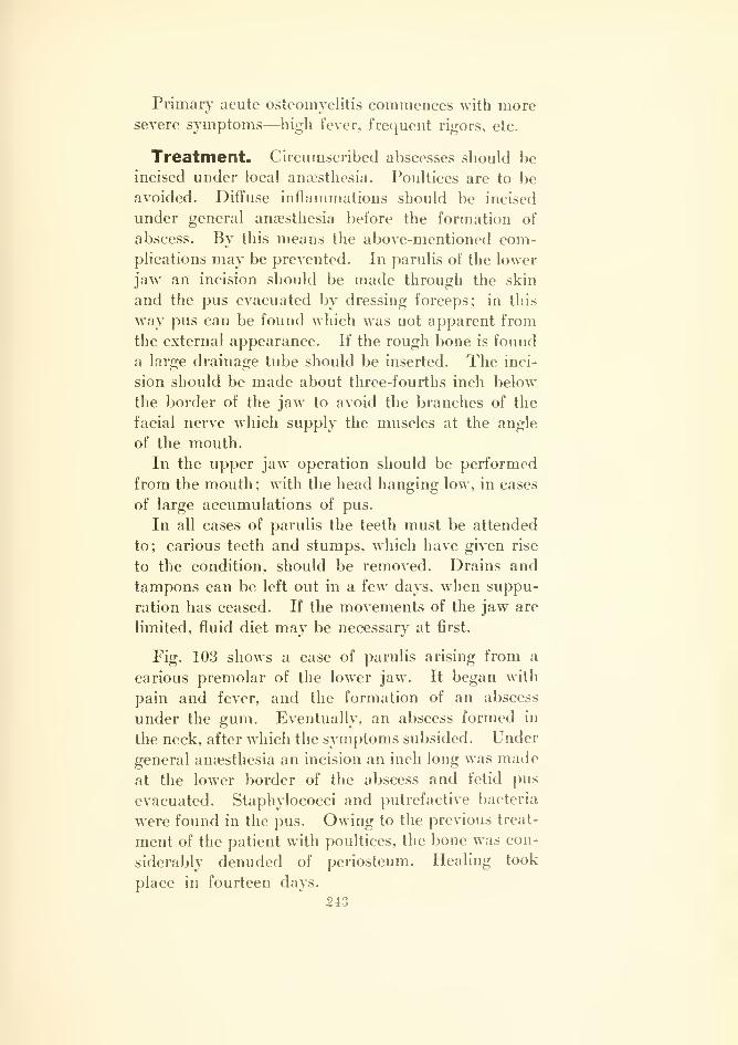
Primary acute osteomyelitis commences with moresevere symptoms—high fever, frequent rigors, etc.
Treatment. Circumscribed abscesses should be
incised under local antesthesia. Poultices are to be
avoided. Diffuse inflammations should be incised
under general anaesthesia before the formation of
abscess. By this means the above-mentioned com-plications may be prevented. In parulis of the lower
jaw an incision should be made through the skin
and the pus evacuated by dressing forceps; in this
way pus can be found which was not apparent from
the external appearance. If the rough bone is found
a large drainage tube should be inserted. The inci-
sion should be made about three-fourths inch below
the border of the jaw to avoid the branches of the
facial nerve which supply the muscles at the angle
of the mouth.
In the upper jaw operation should be performed
from the mouth ; with the head hanging low, in cases
of large accumulations of pus.
In all cases of parulis the teeth must be attended
to; carious teeth and stumps, which have given rise
to the condition, should be removed. Drains and
tampons can be left out in a few days, when suppu-
ration has ceased. If the movements of the jaw are
limited, fluid diet may be necessary at first.
Fig. 103 shows a case of parulis arising from a
carious premolar of the lower jaw. It began with
pain and fever, and the formation of an abscess
under the gum. Eventually, an abscess formed in
the neck, after which the symptoms subsided. Under
general an:esthesia an incision an inch long was madeat the lower border of the abscess and fetid pus
evacuated. Staphylococci and putrefactive bacteria
were found in the pus. Owing to the previous treat-
ment of the patient with poultices, the bone was con-
siderably denuded of periosteum. Healing took
place in fourteen days.
943
Osteomyelitis
OSTEOMYELITIS MAXILLA INFERIORIS(Osteomyelitis of the lower jaw)
Plate LXXXII, Fig. 104.
OSTEOMYELITIS SCAPULA ACUTA(Acute osteomyelitis of tlie scapula)
Plate LXXXIII, Fig. 105.
OSTEOMYELITIS HUMERI CHRONICA(Chronic osteomyelitis of the Jnimerns)
Plate LXXXIV, Fig. 106.
OSTEOMYELITIS TEBL^—NECROSIS TOTALIS(Osteomyelitis and necrosis of the tibia)
Plate LXXXV, Fig. 107.
The term osteomyelitis is applied to pyogenic
affections of bone in general, while in the stricter
sense these are divided into purulent periostitis,
osteitis and osteomyelitis. Since all three parts of
the bone are generally the seat of suppuration and
the process can only be localized clinically to the
bones as a whole, and as the majority of cases begin
with infection of the bone-marrow, the name osteo-
myelitis is rational.
Infection of the bones may result from lesions of
the soft parts, compound fractures, operations (this
was common after amputations in the pre-antiseptic
days); after pyogenic affections of the neighboring
parts (subcutaneous abscess, whitlow, otitis media).
In the latter cases the periosteum is first infected,
the cocci then invade the Haversian canals in the
cortex and infect the medullary cavity. As in all
pyogenic infections, the great majority of cases are
caused by the staphylococcus pyogenes aureus; while
the staphylococcus albus, pneumococcus and strepto-
cocci only in rare cases cause infection of bone.
244
Apart from the above-mentioned modes of infec-
tion this may take place through the blood; themedulla is then first infected, and the suppurationspreads to the cortex and periosteum, finally appear-ing as a subcutaneous abscess.
In all pyogenic affections (furunculosis, whitlow,quinsy, otitis media) the bone marrow is infected bystaphylococci, but the power of resistance of thebody is generally sufficient to withstand their action.
The cocci remains harmless till the power of resistance
of the body is weakened by some exciting cause, suchas fracture, overexertion, exposure to cold, etc. Osteo-myelitis may thus occur after injury to a bone, even
'
after a slight contusion. In this case the result-
ing eflFusion of blood favors further growth of the
cocci and leads to infection. It follows from this
that, according to the circumstances, purulent infec-
tion of the bones may develop sometimes directly
after and sometimes a long time after purulent inflam-
mation in other organs of the body; also that, accord-
ing to the number and virulence of the bacteria, it
may assume an acute or chronic form, with corre-
sponding violent or mild s^Tnptoms. Like all puru-lent inflammations, the process begins at the seat of
infection with hypersemia, exudation, suppuration,
degeneration and regeneration; these processes as-
suming a special form corresponding to the structure
of the bone. Thrombo-phlebitis may occur and give
rise to metastatic infection by embolism in other
parts of the body (bones, endocardium, meninges,
etc.)
As the great majority of cases arise from blood
infection, it is clear that the bones most liable to
infection are those which are most richly supplied
with blood-vessels, especially during their period of
growth when they are most vascular. The diaphyses
of the long bones are thus most often affected at their
junction with the epiphyses. The lower ends of the
femur and radius and tibia, and the upper ends of
245
the humerus and tibia are the places of predilection.
Osteomyelitis is rare in the short bones and in the
flat bones. It is also rare after the thirtieth year.
According to the statistics of Garres, in one-fifth of
the cases several bones are affected simultaneously.
The symptoms of acute purulent osteomyelitis are
more severe than in any other pyogenic affection.
The deeper the infection, the greater is the virulence
of the bacteria. Bacteria in the bone-marrow are
under greater pressure than in any other tissue, and
this increases their virulence. In young individuals
osteomyelitis often occurs suddenly after an injury,
with high fever, rigors, pains in the joints and severe
constitutional disturbance. Pain on pressure and
movement, and loss of function point to an affection
of the bones. Serous effusion soon takes place in the
nearest joint. Changes first appear under the skin
when pus forms under the periosteum. The sub-
periosteal abscess appears as a sharply defined fluc-
tuating swelling with hard borders, and the skin over
it is tense and reddish blue. If the subperiosteal
abscess bursts, it gives rise to intermuscular and sub-
cutaneous infiltration, with redness and swelling of
the skin, and oedema of the soft parts; the regional
lymphatic glands are swollen and painful.
Although operation often only reveals a sub-
periosteal abscess, especially in children, in cases of
hematogenous origin (blood infection) the cortex and
medulla of the bone are also affected. Infection of
the cortex is shown by the presence of yellow spots
on the surface, which correspond to small holes dis-
charging pus. After removal of the cortex, the
infected medulla shows reddish-brown or yellowish
spots, which may lead to the formation of a circum-
scribed abscess, or to diffuse suppuration in the
medullary cavity. If the condition is not recognized
early and the spread of infection arrested by opera-
tion, separation of the epiphyses or infection of the
joint may occur, or general infection with death in
246

a few days. In extensive disease the whole bone is
whitish-yellow; white from bloodlessness due to
thrombo-phlebitis, and yellow from pus formation.
Numerous pits are seen from which pus has been
discharged under the periosteum.
The amount of necrosis corresponds to the degree
and extent of infection. In subperiosteal necrosis
the infected cortex and medulla may regenerate
without loss of substance, especially when the pus
has obtained an early exit. If the cortex has been
for some time the seat of extensive purulent inflam-
mation necrosis must result with the formation of a
sequestrum. According to the extent of the inflam-
mation this necrosis will be limited to one part of the
bone or extend through the thickness and length of
the bone partially or completely. In disease of the
cortex the sequestrum is generally lamelliform,
slightly corroded and pitted; in disease of the
medullary cavity the sequestrum is, to a certain
extent, a cast of the cavity, and in the form of a
trough.
The sequestrum in osteomyelitis is large and con-
tinuous and may include the whole length and
thickness of the diaphysis (Fig. 107), thus differing
from the sequestra in tuberculous bone disease,
which are generally multiple, small and much cor-
roded. Such complete necrosis occurs in acute cases
which have been operated upon too late and in
chronic cases. The dead bone (sequestrum) be-
comes separated from the healthy bone by a zone of
inflammatory' demarcation, more or less rapidly
according to its size.
In extensive necrosis the demarcation process maycontinue for months, so that patients who escape
death from general infection may succumb from
exhaustion, albuminuria or amyloid degeneration of
the kidneys. Spontaneous expulsion of the dead
bone should be assisted by operation (sequestrot-
omy).247
The regenerative or osteoplastic process goes hand
in hand with the degenerative. The purulent in-
flammation not only causes necrosis, but causes
irritation which stimulates the periosteum to form
new bone (osteoplastic periostitis). This results in
thickening of the cortex at the seat of necrosis; and
in cases of total necrosis, complete repair of the
destroyed bone. This irregular formation of newbone is sometimes called the "sequestral capsule."
There are numerous holes (cloacas) in this capsule
where the periosteum has been destroyed. Fromthese holes pus is discharged from the zone of inflam-
matory demarcation, and eventually the sequestrum,
through a fistula in the skin (Fig. 107). The X-rays
are useful in showing the extent of necrosis, and also
separation of the epiphyses.
The whole process of degeneration and regenera-
tion take much longer than in purulent inflammation
of the soft parts, and the acute stage is followed by a
chronic stage after the pus has been evacuated spon-
taneously or by operation. However, an acute
relapse may occur at any time during the chronic
stage, especially after improper treatment, or after
an injury.
In distinction to this form of acute osteomyelitis
there is a subacute form which is chronic from the
beginning. In these cases there is often a history of
previous acute inflammation of the bone, and the
condition is really one of recurrence in a milder form,
often at the age of puberty. Recurrence may also
occur later in life, hence bones which have been pre-
viously affected with osteomyelitis must be regarded
as places of less resistance and must be protected
from the action of trauma and over-exertion.
The clinical symptoms in these cases often resemble
rheumatic pains, but the pain is localized to one bone,
or sometimes a definite part of a bone. There is
often a history of pyogenic disease in youth, andscars and fistulas may be found in the bone con-
248
cerned or in other bones. The affected bone is often
very tender to pressure at certain points. In the
course of time the bone becomes thickened, and the
diaphysis lengthened. The growth in thickness maybe enormous at the seat of disease, the thickening
being both periosteal and cortical.
The changes in the bone in chronic osteomyelitis
are as follows: Sometimes there is a small sequestrum
in the interior of the bone, shown as a clear spot sur-
rounded by bony proliferation in an X-ray picture;
sometimes a circumscribed abscess in the medullary
cavity, shown by the X-rays as a round space sur-
rounded by bone. If bony proliferation is absent
the X-ray pictures resemble tumors or cysts in the
bone. The diagnosis of chronic osteomyelitis is,
therefore, sometimes difficult when there is no his-
tor}- or evidence of former osteomyelitis. Pain on
pressure suggests the infective nature of the disease.
In doubtful cases search may be made for staphylo-
lysin, according to the method of Bruch, Michaelis
and Schultze.
If large portions of the cortex and medulla are
affected by chronic osteomyelitis large sequestra are
formed, which seek a way to the surface in spite of
the considerable formation of new bone. In these
cases we find numerous cloacas in the bony capsule,
subcutaneous abscess and fistulas (Fig. 106); while
the whole bone is thickened, and the X-rays show
changes in the periosteum, cortex and medulla.
A third form of chronic osteomyelitis is limited to
the periosteum, under which a hyaline sero-mucoid
fluid develops, forming a sharply defined, fluctuating
swelling with hard borders. This has been called
albuminous periostitis but is a form of osteomyelitis.
Staphylococci are present in the fluid.
All these chronic forms are due to infection by less
virulent staphylococci. However, every chronic os-
teomyelitis may become acute, especially when the
bones are exposed to the effects of overexertion,
249
injury, or massage (performed on account of wrong
diagnosis). Chronic fistulas in osteomyelitis maygive rise to carcinoma (cf. Plate XIV). In the
long bones both acute and chronic osteomyelitis maycause disturbance in growth, pseudarthrosis and con-
tractures. Although the great majority of cases of
acute and chronic osteomyelitis affect the long bones,
both forms may occur in the short and flat bones; in
the skull, after compound fractures, incised and
punctured wounds; in the scapula, pelvic bones and
vertebrae; in the bones of the face (after tooth
extraction). In Frbhnefs statistics, four hundred
and seventy cases of osteomyelitis affected the long
bones and thirty-four the short and flat bones. As
the cortex is thin in these bones, there is greater
destruction. Osteomyelitis of the cranial bones mayspread through the diploe to half the skull, form large
sequestra of the inner table, and epidural abscess.
In the scapula the whole bone may be destroyed by
multiple abscesses and sequestra, necessitating com-
plete removal of the bone. In osteomyelitis of a
facial bone, infection may spread to all the bones of
the face, causing extensive destruction and conse-
quent deformity. Osteomyelitis of the cranial and
facial bones may give rise to meningitis.
In streptococcal osteomyelitis the pus is thinner
and very abundant, and the disease is more severe
like all streptococcal infections. In these cases the
skin usually shows erysipelatous reddening.
Osteomyelitis after infection by typhoid bacilli or
pneumococci can only be distinguished from the
other forms by the history and by bacteriological
examination.
Differential Diagnosis. Acute osteomyelitis
may be mistaken for deep abscess, but this is madeclear by incision. The redness of the skin in osteo-
myelitis resembling erysipelas is limited to the
affected part and gradually diminishes. Acute osteo-
250
myelitis of the diaphyses is characterized by the
severity of the symptoms, the marked swelUiag and
the loss of power in the limb.
Chronic forms are most often mistaken for tuber-
culous bone disease, but the latter generally affects
the epiphyses, while osteomyelitis attacks the dia-
physes. Osteomyelitic fistula has hard borders and
bright red granulations, and passes directly to the
bone, while tuberculous fistula has yellow, slimy
granulations, irregular borders and an irregular
course through the deep parts (Figs. 125 and 130).
In osteomyelitis the pus is reddish brown, in tuber-
culosis it is thin and greenish yellow. In doubtful
cases an incision will decide the diagnosis; in osteo-
myelitis the periosteum and cortex will be found
thickened and the sequestrum large and continuous;
in tubercular bone disease there are multiple, small
corroded sequestra.
Chronic osteomyelitis causing much swelling of the
bone may be mistaken for syphilitic bone disease,
especially in the tibia. In syphilitic bone disease the
X-rays show a diffuse thickening of all layers of the
bone, and a uniform dark shadow with irregular
borders, corresponding to the periosteum; while, in
osteomyelitis, dark shadows together with clear
spaces are shown, corresponding to sequestra and
abscesses respectively. If fistulas form in syphilitic
bone disease they present the characteristic sharp
borders and prolific granulation tissue round them
(Fig. 122).
Osteitis deformans (Paget's disease) is characterized
by affecting the whole extent of both tibias, and by
the early appearance of marked curvature.
Osteomyelitic abscesses in the diaphysis, when
they extend to the epiphyses may be mistaken for
tuberculosis, but the pronounced new bone-formation
is absent in the latter. Sarcoma and bone-cysts may
also in some cases be difficult to distinguish from
chronic osteomyelitic abscess, even by the X-rays.
251
In doubtful cases an exploratory incision may be
made, or staphylolysin looked for.
In the majority of cases, however, the diagnosis of
osteomyelitis is established by the history and the
typical appearance, situation and course of the
disease.
The earlier diagnosis is made and treatment com-
menced, the better the prognosis.
Treatment. In the most acute cases with puru-
lent joint-etfusion and signs of general infection (dry
tongue, delirium, presence of bacteria in the blood)
amputation is sometimes the only means of saving
life.
In acute osteomyelitis incision must be made as
soon as possible, before the abscess has broken into
the subcutaneous tissue. After opening the abscess
the bone must be examined; if it is unaltered it can
be left alone. If the temperature does not fall after
opening the abscess and the condition becomes worse,
with rigors, etc., the bone must be laid open as far
as the medullary cavity. This should be performed
freely with a gouge ; it is useless simply to bore holes
as they do not give sufficient outlet for pus, nor for
subsequent necrosed pieces of bone. On the other
hand, in cases with severe constitutional symptoms,
especially in children, the whole extent of bone
should not be gouged at one sitting, owing to the
severe shock, and the possibility of general infection;
the gouging should be performed at several sittings.
After gouging, the infiltrated bone-marrow must be
scraped with the sharp spoon and the cavity drained
with iodoform gauze. The wound must be kept
open by a drainage tube to allow pus and sequestra
to escape.
The after-treatment is sometimes hindered by nar-
rowing of the opening in the bone from the formation
of callus; if there is no suspicion of necrosis, this
callus must be removed with the knife, to establish
252
sufficient communication with the medullary cavity.
Complete immobilization is necessary in the extremi-
ties, to avoid spread of inflammation and the possi-
bility of fracture.
Serous effusion into a joint must be punctured
when extensive. Purulent effusion requires incision,
and sometimes resection of the joint. If there is
purulent arthritis with high fever and rigors, resection
must not be delayed, or general infection may follow.
In chronic osteomyelitis it is best to wait till the
sequestrum is complete and new bone has begun to
form round it (X-ray examination) before performing
sequestrotomy. If there are subcutaneous abscesses
these must be opened. As small sequestra and ab-
scesses often cause considerable pain, in some cases
the bone must be gouged when the X-ray examination
shows no changes. The operation is troublesome, as
the small sequestrum or abscess is often situated in
the middle of hardened sclerotic bone. The fistulas
in chronic osteomyelitis must be freely opened upand the callus removed. The cavity in the bone left
after gouging must be left open and drained till heal-
ing takes place from the bottom. Immediate plug-
ging of the bone cavity with iodoform is only of use
in a few cases of circumscribed chronic osteomyelitis,
as in extensive cases the plugs are often expelled
through a fistula; but when the cavity is filled with
fresh granulations, all cases of osteomyelitis can
quickly be made to heal with plugging. The cavity
is then scraped, disinfected with peroxide lotion,
dried with Hollander's hot air apparatus, and filled
with a mixture of iodoform, glycerin and spermaceti.
^Mienever possible, the periosteum should be united
over the plug and a covering of skin made over the
cavity. Strict asepsis is necessary.
Frequent recurrences in chronic osteomyelitis, with
emaciation, albuminuria, etc., necessitate amputa-
tion. Contractures must be treated by extension on a
splint, or when they cannot be extended, by resection.
253

Large defects in the skin can be covered by peduncu-
lated flaps.
In the flat bones subperiosteal removal of the whole
bone is often necessary {e.g. scapula). This may be
followed by complete regeneration and restoration of
function. In osteomyelitis of the cranium sequestra
and epidural abscesses must be evacuated through a
large trephine hole, which can afterwards be repaired
by bone grafting.
Treatment of acute osteomyelitis by passive hyper-
femia is to be condemned, as it obscures the signs and
symptoms. It may also lead to diffuse suppuration
by thrombo-phlebitis, rendering amputation neces-
sary; but its chief danger is general infection.
254

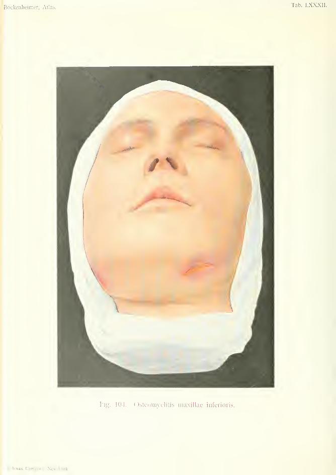
Bockenheimer, Atlas.Tab. LXXXII.
Pig. 104. Osteonivflitis maxillae inferioris.
Rfbman Coni|)aiiy, New-Vork.

OSTEOMYELITIS MAXILLAE INFERIORIS(Ostcomi/clitis of the Loirer Jaio)
Plate LXXXII, Fig. 104.
This figure shows chronic osteomveHtis in a eirl of
nineteen, which occurred in connection with tooth
extraction. Osteomyelitis of the lower jaw often
occurs after tooth extraction, when there is muchinflammation of the gum and periosteum, or whenthe alveolus is extensively injured. It may also fol-
low injuries to the jaw. Osteomyelitis of the lower
jaw% due to blood infection, is generally combinedwith disease of other bones, and occurs especially in
children. Acute osteomyelitis of the lower jaw com-mences with high fever, rigors, oedema of the face
and mucous membrane of the mouth, difficulty in
breathing and swallowing, headache and delirium.
It is often fatal from meningitis or general infection.
In some cases the whole of the lower jaw may becomenecrosed.
In the chronic form (Fig. 104) a painless circum-
scribed or diffuse painless swelling slowly develops in
the lower jaw. The skin gradually becomes tense,
red and cedematous; one or more fistulas develop,
and later on necrosis takes place. In extensive cases
the teeth become loosened and trismus may occur.
In the stage of painless swelling the case may resem-
ble cystic adenoma. In actinomycosis the swelling
is situated in the floor of the mouth and in the mus-
cles, and only extends to the bones later on.
Osteomyelitis of the lower jaw should be treated
by early incision down to the bone, at the lower
border of the jaw. Healing without necrosis occurs
more often than in the long bones. If necrosis
occurs it is best to wait, in chronic cases, till sufficient
255
new bone is formed, so as to avoid fracture of the jaw
during removal of the sequestrum.
Sequestra are best removed by external incisions.
The cavity should be plugged with iodoform gauze
for a long time. In extensive necrosis, bone grafting
may be tried, or the patient may wear a prothesis.
In Fig. 104 the fistula was opened up; after which
the discharge diminished, but the swelling of the
bone remained and the fistula did not heal, indicating
necrosis. The X-rays showed diffuse swelling of
the jaw.
A condition affecting the bones, observed by
Billroth in workers in mother-of-pearl, which resem-
bles osteomyelitis, and chiefly affects the lower jaw,
undergoes spontaneous resolution; so long as the
patients are not exposed to fresh injury through their
work.
Phosphorous necrosis of the lower jaw, which occurs
in workers in yellow phosphorus, is probably due to
infection of the bone. The phosphorous vapor causes
ulceration of the gums, through which the periosteum
and bone are infected. This condition gives rise to
great swelling of the whole of the lower jaw. Theteeth become loose and fall out. The gums become
ulcerated and fetid, so that many patients succumb
to septic pneumonia or to general septic infection.
The bone becomes both sclerosed and brittle. After
some years total necrosis occurs with a row of fistulas
along the lower border of the jaw.
As there is generally total necrosis in these cases,
partial resection is useless, and subperiosteal resec-
tion of one or both sides of the jaw should be per-
formed. After this regeneration of the jaw takes
place if the periosteum has been preserved, and
relapses are avoided.
Phosphorous necrosis (which is fatal in fifty per
cent, of the cases) has been prevented by the prohi-
bition of the use of the dangerous yellow phosphorus
in the manufacture of matches.
256

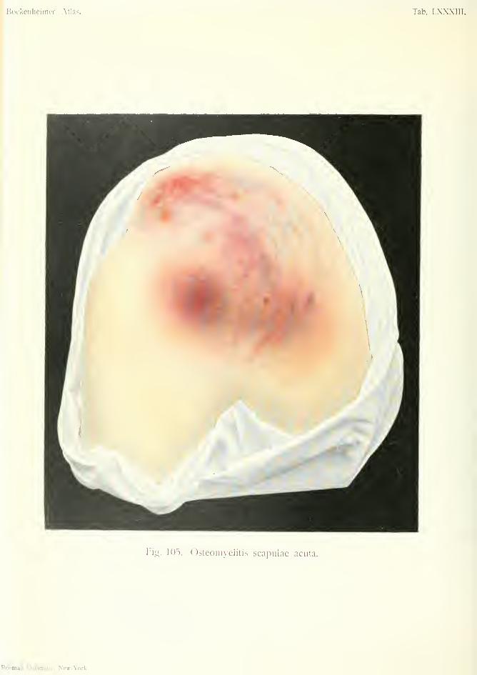
Bockenheimer, Atlas. Tab. LXXXIII.
Fig. 105. Osteomyelitis scapulae acuta.
Pcbman Company, New-York.
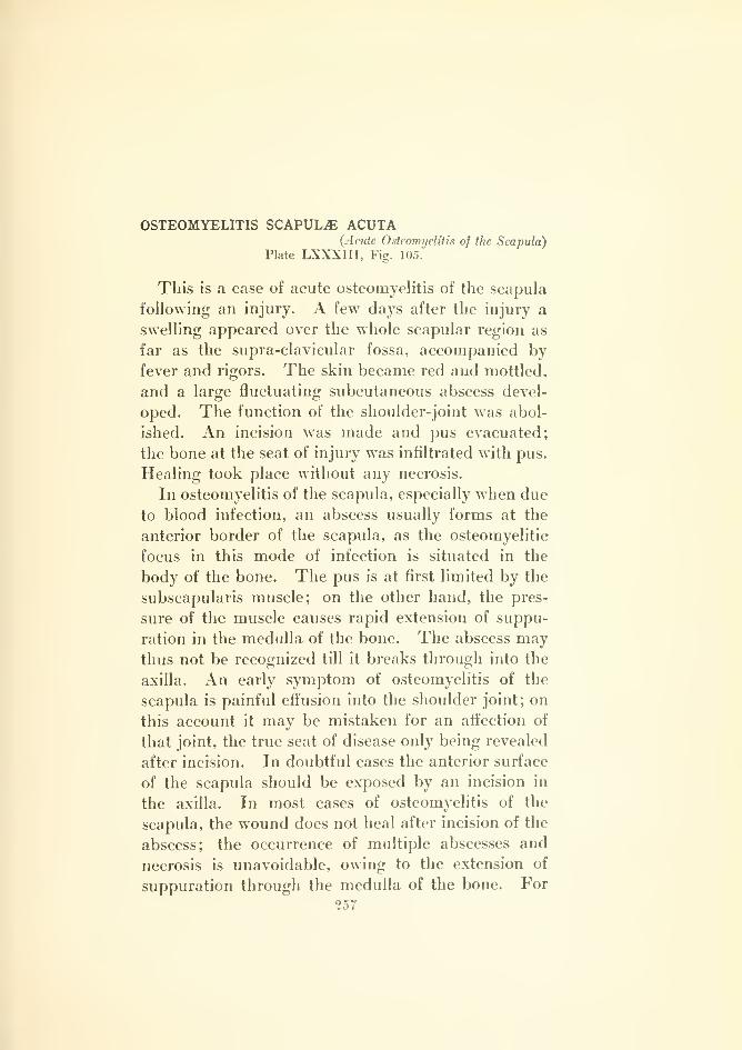
OSTEOMYELITIS SCAPULA ACUTA(Acute Osteomyelitis of the Scapula)
Plate LXXXIII, Fig. 105.
This is a case of acute osteomyelitis of the scapula
following an injury. A few days after the injury a
swelling appeared over the whole scapular region as
far as the supra-clavicular fossa, accompanied by
fever and rigors. The skin became red and mottled,
and a large fluctuating subcutaneous abscess devel-
oped. The function of the shoulder-joint was abol-
ished. An incision was made and pus evacuated;
the bone at the seat of injury was infiltrated with pus.
Healing took place without any necrosis.
In osteomyelitis of the scapula, especially when due
to blood infection, an abscess usually forms at the
anterior border of the scapula, as the osteomyelitic
focus in this mode of infection is situated in the
body of the bone. The pus is at first limited by the
subscapularis muscle; on the other hand, the pres-
sure of the muscle causes rapid extension of suppu-
ration in the medulla of the bone. The abscess maythus not be recognized till it breaks through into the
axilla. An early symptom of osteomyelitis of the
scapula is painful effusion into the shoulder joint; on
this account it may be mistaken for an affection of
that joint, the true seat of disease only being revealed
after incision. In doubtful cases the anterior surface
of the scapula should be exposed by an incision in
the axilla. In most cases of osteomyelitis of the
scapula, the wound does not heal after incision of the
abscess; the occurrence of multiple abscesses and
necrosis is unavoidable, owing to the extension of
suppuration through the medulla of the bone. For
257

this reason the disease may last for years. In these
cases, and also in acute cases where incision shows
extensive destruction of the bone, subperiosteal total
extirpation of the scapula is indicated, taking care to
preserve the muscular attachments and the important
nerves. This is especially indicated in acute osteo-
myelitis of the flat bones, which often gives rise to
early general infection. After total extirpation of
the scapula relapses are avoided, and complete regen-
eration of bone with normal function is possible
{Bockenheimer)
.
258

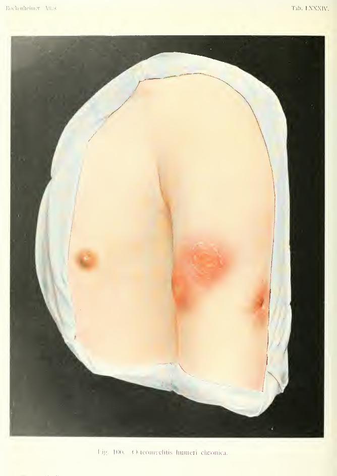
Bockenheimcr Atlas. Tab. LXXXIV.
I'ig. 100. Ostcoiiivelili^ liimicn ciiroiiica.

OSTEOMYELITIS HUMERI CHRONICA(Chronic onteomi/elitis of the Humerus)
Plate LXXXIV, Fig. 106.
Fig. 106 shows a painful club-shaped swelling of
the left humerus, which gradually developed at the
age of puberty, in a patient who had frequently
suffered from tonsilitis in childhood. The patient
attributed it to over-exertion at his work as a black-
smith. A year after the onset, a fistula formed at
the posterior and external side of the arm, with hard
borders and red granulations at its orifice. A probe
passed down the fistula discovered rough bone,
denuded of periosteum. Subcutaneous abscesses
formed at the front of the arm, where the skin was
thin and reddened. Examination by the X-rays
showed a sequestrum, along with new bone forma-
tion. Chronic osteomyelitis of the diaphysis of the
humerus was diagnosed. An incision was madedown to the bone in the lower third of the outer side
of the arm, avoiding the radial nerve. The perios-
teum was destroyed at one place and a cloaca was
found leading to a sequestrum. The sequestrum was
removed by carefully gouging the bone; the cavity
was scraped and plugged, and the fistulous track
with its hardened walls excised. The subcutaneous
abscesses were opened and scraped. The arm was
immobilized for a long time. Healing took place
after some months, and the patient was told to
choose a lighter occupation in order to avoid recur-
rence of the disease.
359
OSTEOMYELITIS TIBLE—NECROSIS TOTALIS(Acute Osteomi/elitiii and Xecrosis oj the Tibia)
Plate LXXXV, Fig. 107.
In this case acute osteomyelitis of the tibia in a
child, aged nine years, commenced with severe pain
in the leg and knee joint, accompanied by high fever
and rigors. There was no history of a previous
attack. A few days before the onset the child re-
ceived a blow on the tibia. In spite of the severe
clinical symptoms and the marked swelling of the
knee joint, operative treatment had been neglected,
and, only when a subcutaneous abscess developed,
was an incision made. Although the acute symp-
toms gradually subsided after this, the swelling of the
leg remained, and the wound discharged fetid pus.
In a few months almost the whole shaft of the tibia
became necrosed. Fig. 107 shows the yellow ne-
crosed bone, with the open medullary cavity contain-
ing slimy granulations. Between the necrosed bone
and the healthy bone are granulation tissue and pus.
As the leg had not been properly fixed, a fracture
occurred at the lower part of the tibia. The condi-
tion of the child on admission to hospital was very
bad, owing to the prolonged suppuration. Examina-
tion by the X-rays showed that the sequestrum
extended further down, and that a thick, bony cap-
sule had already formed behind and at the sides.
Under an ansesthetic, the wound was extended
downwards, the necrosed bone removed, the cavity
scraped and plugged, and the leg put up on a splint
with extension, to correct the position of flexion.
The equinus position of the foot, due to insufficient
fixation, was gradually corrected.
Such extensive necrosis could have been avoided
by early gouging of the bone and proper after-
treatment.
260
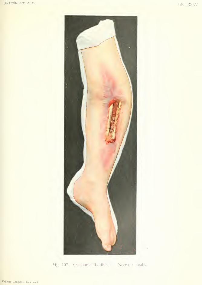
Bockenheimer, Atlas.Tab. l.XXXV.
Fig. 107. Osteonnclitis tibiae - Necrosis totalis.
Rcbman Company, New-York.


Bockenheinier, Atlas. Tab. LXXXVI.
Fig. 108. Infectio eeneralisata.
Rebmm Company, New-York.
INFECTIO GENERALISATA {General Infection)
Plate LXXXVI, Fig. 108.
In the description of the various local pyogenic
infections, mention has already been made of gen-
eral infection. In every pyogenic and putrefactive
infection there is a certain degree of general infection,
but this is not generally sufficient to be recognized
clinically or bacteriologically. In apparently benign
pyogenic affections, such as furuncle, bacteria maybe found in the blood. This explains the occasional
occurrence of metastatic osteomyelitis in connection
with such affections; and also the fact that the im-
pairment in general health is often out of proportion
to the local inflammation.
General infection assumes different clinical forms,
but it is impossible to make a classification of these
which is free from objection. Moreover, such a
division is of little practical value, as the same meas-
ures must be employed against different forms of
general infection. From the clinical point of view,
it is, therefore, best to speak only of general infec-
tion, and abandon the old, and often inappropriate,
terms sepsis, septicaemia and pyaemia. In any case
the term sepsis should be confined only to that form
of general infection which is caused by the putre-
factive bacteria; but this form is rare, and it is gen-
erally a question of mixed infection with putrefactive
bacteria and streptococci.
Again, the distinction into metastatic and non-
metastatic general infection, proposed by Lexer, is
practically without value and does not hold good
for all cases. In many cases non-metastatic cannot
be distinguished from metastatic general infection,
261
especially as both often co-exist, or one may merge
into the other. Lastly, when only small metastases
are present in the internal organs, and they remain
unrecognized, such a metastatic form may be wrongly
regarded as non-metastatic.
Bacteriological research has shown that general in-
fection is not due to one specific cause; staphylo-
cocci, streptococci, pneumococci, typhoid bacilli, bac-
terium coli commune give rise to p?jogenic general
infection, while putrefactive bacteria (proteus vul-
garis, etc.) cause putrefactive general infection. These
two forms are often combined, and clinically indis-
tinguishable, so that the designation general infection
is sufficient for practical purposes. On the other
hand, the nature of individual cases should be made
clear by bacteriological investigation.
It has been shown that in general infection caused
by staphylococci there are usually metastasic forma-
tions (ninety-five per cent, according to Lehnartz).
Local infection with staphylococci is generally cir-
cumscribed, while streptococcal infection is more
diffuse. Thismay depend on the fact that the staphylo-
cocci are accumulated in large masses, but it has not
been proved.
In streptococcal general infection, on the other
hand, there are hardly ever any metastatic forma-
tions. As cases without metastases are clinically the
most severe, and almost always fatal, a division into
metastatic and non-metastatic general infection is
identical with less-severe and more-severe infection.
However, just as infection with very virulent staphylo-
cocci may be fatal without metastatic formation, so
may infection with less virulent streptococci cause
metastatic formation and end in recovery. In prac-
tice, we know that streptococcal local infection is
more severe than staphylococcal, and this usually
holds with general infection.
It is impossible to introduce the ideas of bacteri-
semia and toxinaemia into clinical nomenclature, for
262
the characterization of general infection. Although
general infection may exist without the presence of
bacteria in the blood being capable of demonstration
by the present methods of bacteriological research,
bacteria are no doubt present in the blood in every
case of general infection, but are quickly destroyed
by the bactericidal substances. On the other hand,
bacteria may be found in the blood in cases of gen-
eral infection in which toxinsemia is not recognizable.
If toxins are always present in the blood, they are
not easy to find, especially as they have a tendency
to form combinations with the organs. In general
infection there is always bacterisemia and toxin-
semia, but in practice we only speak of general infec-
tion which is most often acute, rarely chronic.
Acute general infection may be primary or sec-
ondary; mild or severe. The severity of general
infection depends on the number and virulence of
the bacteria, and on the power of resistance of the
body. The severest forms of general infection
appear so rapidly after the local infection that the
latter remains in the background; these forms are
often fatal before the typical inflammatory processes
have developed at the seat of infection. Such cases
include those which often occur in doctors from
infection during operations or post-mortem examina-
tions (streptococcal infection) ; also from infection
by putrefactive bacteria; or by mixed infection by a
symbiosis of streptococci and putrefactive bacteria.
To this class belong cases formerly called crypto-
genetic pyaemia, which are better considered as
latent general infection arising from unrecognizable
foci of infection. Virulent bacteria must often invade
the intact mucous membrane and give rise to general
infection.
In the great majority of cases, however, general
infection is of gradual onset, arising from a local
infection; but it is often in an advanced condition
before it is recognized. It may occur by direct
263
extension of suppuration, or may arise without
further extension of the local disease.
The more rapidly virulent bacteria enter the blood
from the local seat of infection, the more severely is
the organism affected. This is shown in the tem-
perature chart.
In the most severe forms the temperature rises
rapidly and remains at 40° or 42° C. (104°-107° F.)
;
such cases generally cause death in a few days with-
out metastatic formations. In less severe forms the
temperature does not remain high, but is intermittent.
This may be due to the intermittent entry of the
bacteria and their toxins into the blood from the
seat of infection, or to smaller quantities of them.
When the organism conquers the bacteria and their
toxins by the formation of antitoxin, the temperature
falls; when the bacteria gain the upper hand the
temperature rises.
The longer the process continues, the more fre-
quent are the rigors, with intermissions of tempera-
ture. When these variations in temperature follow
each other rapidly (as occurs in the severest cases)
the temperature becomes continuous, the rigors cease
and there are no metastases. If the organism gains
the upper hand, the infection expends its energy in
the formation of local metastatic formations in
various places. This, in a way, may be regarded as
a victory of the organism over the bacteria.
In the milder forms of general infection we there-
fore find metastatic formations in those parts of the
body which are specially constituted to absorb bac-
teria and render them harmless (peritoneum, pleura,
endocardium joint cavities). Metastatic formation
is to be regarded as a curative process, as the bacteria
are to a great extent destroyed. These metastases
caused by bacteria in the blood must be distinguished
from metastases propagated from purulent thrombo-
phlebitis, or emboli containing bacteria. In all these
cases the blood-stream plays the principal part in
264

general infection, the role of the lymphatics being
subordinate.
As in local infection, general infection is predis-
posed to by debilitation of the organism by exhaus-
tion, hunger, and exposure to cold, and by diseases
such as diabetes and tuberculosis, etc. Along with
general predisposition, there is a local predisposition
depending on the nature and seat of the lesion.
Foreign bodies often lead to general infection, also
machine-injuries, compound fractures, bites, andwounds of the mouth and rectum.
The deeper the infection and the greater the pres-
sure on the bacteria, the greater is their virulence,
and therefore the more frequent is general infection.
This accounts for the frequency of general infection
in deep suppurations, such as those under the cervical
fascia, and in the bones and joints. It is well knownthat the internal surface of the uterus durinjr the
puerperium is especially liable to infection, which
may become general, and that the retention of pus
and blood effusions are dangerous. Lastly, the treat-
ment of infected wounds with strong caustics, such as
carbolic acid, may give rise to general infection.
As regards the clinical symptoms of general infec-
tion, various clinical pictures may be produced
according to the kind of infection, but the morbid
condition is uniform as regards its most essential
points. Bacteriological examination must decide
which bacteria have caused the infection, whether
one or more different kinds are present, and whether
they are present in the blood (bacterisemia). Asregards metastases, we can only speak of metastatic
general infections when metastases are found during
life; while cases in which no metastases are found
cannot be called non-metastatic till the absence of
metastases has been established by post-mortem
examination. Small metastatic foci are often found
post-mortem (especially in the kidneys), which were
not recognizable during life.
265
The symptoms of general infection differ according
as the onset is sudden and acute, or gradual and
chronic. In the most acute forms the symptoms
appear suddenly, while in the other forms there is a
latent stage with disturbances in the general condi-
tion (insomnia, loss of appetite, headache, pain at
the seat of infection) which are premonitory of gen-
eral infection. A frequent small pulse points to the
onset of general infection, before the rise of tempera-
ture. The temperature then rises suddenly to 39°
or 41° C. (102°-106° F.), with rigors. We have
already pointed out that in the most severe cases, in
which numerous virulent bacteria remain in the
blood, the body is only able to offer a slight degree
of resistance. In these cases there is no fall in tem-
perature and no formation of abscesses, and the
infection is often fatal in twenty-four hours or a few
days, with high, continued fever.
On the other hand, if the bacteria only enter the
blood intermittently, there may be periods during
which fever is absent {e.g. after evacuation of retained
virulent secretion by incision). With fresh infection
of the blood there is at the same time a rise of tem-
perature. Hence the variations in the temperature
chart. Although remissions in temperature are char-
acteristic of mild, general infection, this remittent
fever after some days may become continuous and
fatal. For example, when an extremity has been
amputated for progressive suppuration, the tempera-
ture falls; but it may rise again after a time, showing
that the organism was already saturated with bac-
teria and their toxins, and that the operation was
performed too late to save life. It is noteworthy
that the pulse in remittent fever remains small and
rapid during the fall of temperature, even after com-
plete cessation of the fever, showing how much the
heart is affected by the process.
If, after extensive operative interference, the tem-
perature approaches normal, this may be regarded
266

as a good sign for the further progress of the case.
A subnormal temperature is sometimes observed in
the most severe cases of general infection, and sig-
nifies complete collapse of the organism.
The respiration is rapid, as in all feverish condi-
tions, and may become stertorous in severe cases
with loss of consciousness.
Besides sudden rise of temperature and rapidity of
the pulse, the tongue shows conditions which are
characteristic of general infection. Changes in the
tongue are observed even in slight disturbance in the
wound. The tongue is at first smooth, dry and sal-
mon colored; later on it becomes rough, fissured andbrownish black. In severe cases of general infection
the teeth are also dry and coated with sordes. Theconjunctivae are yellow, and in severe cases there
may be jaundice of the whole body (hematogenous
icterus). The patients are continually tormented bysweating and thirst.
These characteristic symptoms are diagnostic of a
general infection whose point of origin is concealed.
On the other hand, in general infection arising from
infected wounds, the earliest signs pointing to gen-
eral infection are often observed in the wound itself.
As every pyogenic condition may lead to general
infection, the wound must be continually watched
by frequent changing of the dressings. The expe-
rienced can often foresee the onset of general infec-
tion, from the appearance of the wound. Apart from
cases of general infection following a slight abrasion
of the skin or mucous membrane, the wound generally
becomes painful and oedematous; the granulations
become unhealthy and flabby; the discharge of pus
subsides and gives place to a scanty, dii'ty, often
fetid secretion; the surface of the wound l)ecomes
dry and often covered by diphtheroid membrane(Fig. 101). Retention of pus, necrosis, extension of
suppuration, lymphangitis and lymphadenitis are
often concomitant signs. In infection by putrefactive
267
bacteria (Fig. 109) there are bullie in the infiltrated
skin and crepitation due to the formation of gas, and
bubbles of gas in the secretion. Unfortunately, these
characteristic signs are often overlooked; operative
interference which could prevent extension of the
already commencing general infection is neglected,
and the condition passes into a stage which is almost
always incurable.
In no other condition is the organism so muchaltei'ed as in advanced general infection, so that the
clinical symptoms become indelibly imprinted on the
memory of the observer. All the already-mentioned
symptoms of commencing general infection become
intensified in advanced cases. The patients at first
become light-headed, then delirious, and finally
unconscious. The indifference of patients in the
advanced stage is in marked contrast to their feeling
of fear in the early stage of infection, and is an unfa-
vorable sign. In the final stage, shortly before death,
if the patient has not permanently lost consciousness,
he often has attacks of fear, or even maniacal attacks,
followed by collapse. In this stage the patient can
hardly be kept in bed, as he makes repeated attempts
to go home, etc.
The gastro-intestinal canal is severely affected;
vomiting of blood from submucous hemorrhage,
vomiting of bile and uncontrollable diarrhea result
from the action of toxins. The skin is pale and cold,
and may present morbilliform eruptions, erythema,
erysipelatous reddening, vesicular eruptions, puncti-
form hemorrhages or more extensive blood-effusions.
Bedsores are also common.Almost all the internal organs are saturated with
bacteria and their toxins, and react in their special
manner. Nephritis is manifested by albuminuria;
meningitis gives rise to stiffness of the neck; pleuritis
causes blood-spitting; pericarditis is manifested by
pericardial friction, and endocarditis (which is very
common and often ulcerative) by cardiac murmurs.268
As in every severe infection, the spleen is enlarged,
and sometimes there is acute bronchocele.
In streptococcal general infection there is nearly
always suppuration in the joints; in staphylococcal
infection, suppuration in the bones. Lastly, infec-
tive emboli or propagated thrombo-phlebitic abscesses
(metastatic) may occur in all the organs, especially
in staphylococcal infection. In this way multiple
abscesses may appear in the skin. Metastatic ab-
scesses may be cold and painless, and often contain
few bacteria. Deeply situated subfascial and inter-
muscular abscesses often escape observation. Lastly,
small multiple or large abscesses may occur in the
lungs, heart, liver, kidneys, etc. According to Wal-
deyer, these abscesses are due to plugging of the
smallest vessels. For example, plugging of the
central artery of the optic nerve causes panophthal-
mitis, while plugging of a terminal artery in the lung
causes an infarct. In this advanced stage of general
infection, there is often frequent bleeding from the
wound at the seat of the local infection, due to affec-
tion of the arteries. In the pre-antiseptic period
many cases of amputation were fatal owing to this
so-called septic secondary hemorrhage. In this stage
bacteria are nearly always found in the blood. While
an increase of bacteria in the blood is a bad sign,
their disappearance is not always a good sign for the
further progress of the case; for after the disap-
pearance of bacteria from the blood in many diseases,
the action of their toxins (toxinsemia) becomes mani-
fest. Streptococci are more easily demonstrated in
the blood than staphylococci.
According as the onset is gradual or sudden, and
according to the degree and the course of the general
infection, a many-sided but unmistakable clinical pic-
ture is produced.
The chronic forms of general infection, which occur
after long-standing fistulas, suppuration and necrosis,
are characterized Ijy their gradual development and by
369

the slight severity of the symptoms. Many cases,
however, are fatal from heart failure or albuminuria;
or the chronic form may become acute. In chronic
general infection there are often long periods free
from fever, followed by rigors and rise of temperature.
In the chronic forms metastatic abscesses are more
common. In such cases recovery may take place
after removal of the primary cause, but it requires
several months to restore the weakened body. Again,
acute general infection may become chronic, and
occasionally end in recovery.
It is only young and robust bodies that can offer
an effective resistance against such a destructive mor-
bid condition, and then only in the early stages of
infection. The organism cannot withstand the de-
structive action of a fully developed general infection.
These cases are all fatal. Even in the early stages of
general infection the heart may become so weak by
the action of toxins, that death occurs from collapse
before the full development of the clinical picture.
Staphylococcal infection, with its tendency to metas-
tatic formation and its remittent type of fever, is more
likely to recover than streptococcal infection; this is
generally fatal in a few days, with continued fever
and increase of all symptoms, but without metastatic
formation.
Differential Diagnosis. Although the clinical
symptoms of a typical case of general infection are
unmistakable, cases in which the origin of infection
remains unrecognized, or cases of chronic general
infection may be mistaken for typhoid, miliary tuber-
culosis or acute rheumatism. Severe inflammations,
erysipelas (especially hemorrhagic bullous erysipelas)
may be associated with such high temperature and
rigors, etc., that it is difficult at first to distinguish
whether the symptoms are due to the local condition,
or to the commencement of general infection. Theprogress of the disease will decide. It must, how-
270
ever, be borne in mind that in these cases there is
generally already commencing general infection,
especially in cases of progressive inflammation.
Treatment. Apart from cases in which the mostacute form of general infection arises from com-paratively slight lesions, some cases may be curedby proper treatment of infected wounds (Fig. 93),
and by early diagnosis of commencing general infec-
tion. Special attention must be devoted to the place
of entry of the infection. Free incisions are here
required. Infected joints must be resected. In
some cases of severe general infection and progressive
suppuration in the extremities, amputation should
not be too long delayed.
In threatening general infection from purulent
thrombo-phlebitis of the large veins, ligation should
be performed; for instance, of the jugular vein andanterior facial vein in carbuncle of the face; of the
internal jugular vein in otitis media. Metastatic ab-
scesses must be opened early. Metastatic joint effu-
sions should be incised. Pleural effusions require
aspiration or resection of the ribs. Suppuration in
the internal organs (liver, brain, kidneys) require
operative interference.
Antipyretics are best avoided on the whole, as they
obscure the symptoms and weaken the heart. In
severe cases high temperature may be reduced bytepid sponging. The heart must be supported bystimulants. Nourishing diet is required (if neces-
sary by esophageal tube). Subcutaneous or intra-
venous injections of saline solution are often useful.
Subcutaneous injections of nucleinic acid with salt
solution have been recommended. Not more than
two hundred to three hundred cubic centimeters of
solution should be injected at one time into the veins.
Injections of colloidal silver, anti-streptococcal serum
and polyvalent serum (Aronsohn) have generally no
influence on the disease.
271
On account of the danger of infection to others, the
patients should be isolated and treated by special
attendants. The body must be frequently bathed
with alcohol or spirit of camphor to prevent the for-
mation of bedsores, especially on the back and but-
tocks. The wound at the seat of infection must be
dressed at least once or twice a day to prevent accu-
mulation of pus. Frequently changed moist dress-
ings are the best. Iodoform gauze should be avoided,
as it is rapidly decomposed by the secretion and gives
rise to toxic symptoms. Disinfection of the woundwith strong antiseptics is to be avoided on account of
its injurious effect on the tissues. The affected parts
of the body should be completely immobilized. Rub-
ber gloves should be worn when dressing the wounds,
and no aseptic operation should be performed on the
same day by the surgeon who dresses them.
After recovery from general infection great care
must be taken of the body, in order to give encap-
suled traces of the disease an opportunity to heal.
These encapsuled metastatic foci may at any time
(even after some years) become virulent from some
exciting cause, and give rise to fresh infection.
Patients often succumb, after some years, to nephri-
tis, endocarditis, pleurisy or pneumonia. In these
cases strychnine is useful.
Fig. 108 shows a case of acute general infection
arising from a subcutaneous whitlow, which was
insufficiently incised and extended to the tendon-
sheath and the joint. The temperature rose to
41° C. (106° F.), with rigors; remained high for a few
days and then became remittent, during the forma-
tion of several subcutaneous metastatic abscesses.
An abscess developed gradually in the thigh; this
was incised, and thin pus containing a few staphylo-
cocci evacuated. Staphylococci were also present in
the blood for some time. Other symptoms were
—
dry tongue, jaundice, slight delirium, and diarrhea.

The wound in the finger was dry and unhealthy.
After disarticulation of the finger there was no exten-
sion of infection to the hand, and the whole condition
improved. Under the above-mentioned treatment,
with injection of saline solution, etc., recovery took
place in a few months. Several metastatic abscesses
required incision during the course of the disease.
After removal of the finger, bacteria were no longer
found in the blood^a proof that the virulent bacteria
in the blood were derived from the seat of infection.
The pulse remained rapid for a long time after
recovery.
273
GASPHLEGMONE (Gaseotis phlegmon)
(EDEMA MALIGNUM (Malirjnant (Edema)
PHLEGMONE EMPHYSEMATOSA—GANGRAENOSA{Gangrenous, emphi/sematous phlegmon)
Plate LXXXVII, Fig. 109.
We have already mentioned (Fig. 101) the pro-
gressive putrefactive inflammation which often occurs
in necrosed tissues, and in the wounds of diabetics.
Similar conditions of progressive inflammation, under
various names, accompanied by rapid necrosis and
the formation of gases in the tissues, give rise to gen-
eral infection, and run an unfavorable course.
Pirogoff described these cases as acute purulent
oedema, Maisonneuve as fulminating gangrene, others
as gasphlegmon, gangrenous phlegmon, etc. Theputrid necrosis of wounds known as "hospital gan-
grene," which was so common in the pre-antiseptic
days, appears to be nothing more than putrefactive
inflammation due to gas-forming bacteria. All these
conditions are best included under the name jprogres-
sive gaseous ^phlegmon. The causes of these phleg-
mons are not well known, as they are anterobic
bacteria which have not yet been well differen-
tiated from each other by bacteriological methods.
They are found most often in dust, manure andputrid flesh.
The bacillus of malignant oedema, the bacillus
emphysematosus and the proieus vulgaris are the
bacteria at present found, generally in symbiosis
with the ordinary pus-forming bacteria, especially
streptococci. By this symbiosis the growth of the
anaerobic bacteria is at first made possible in open
wounds, and through the combined action of both
274
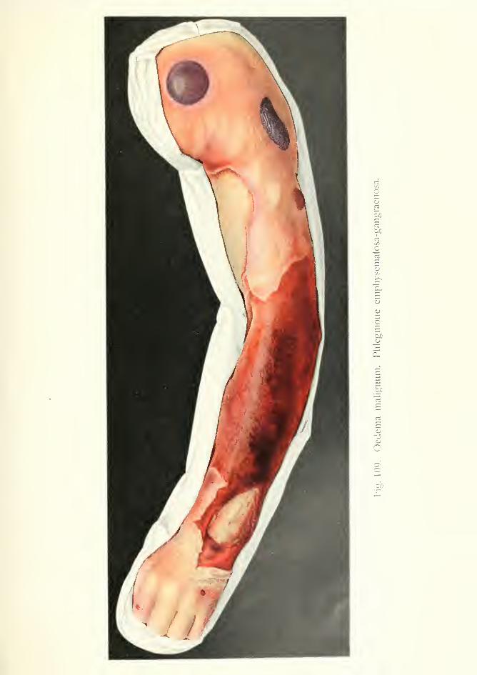
n
5
CI
U)
o
to


forms of bacteria rapid and extensive destruction of
tissue may be caused. Sometimes gaseous phlegmonis found after quite harmless lesions of the skin
(Fig. 109), also after compound complicated frac-
tures with small wounds.
Gaseous phlegmons occur in the extremities; onthe back, in connection with bedsores; in operative
wounds on the rectum, through infection by faeces;
in the penis, scrotum and perineum, from lesions of
the urethra with extravasation of urine; in the neck,
after lesions of the esophagus and pharynx. Theprogress of gaseous phlegmon is extremely rapid;
in a few hours large portions of the body are affected
by the rapid formation of gas. As gaseous phleg-
mon may occur after apparently slight injuries, it
is necessary to emphasize the necessity of frequent
dressings in order to control the progress of infection.
The wound becomes dry, coated and fetid, and
extensive swelling rapidly extends from it on all sides.
The discharge from the wound is brownish or green-
ish, fetid, and mixed with necrotic shreds of tissue.
High temperature, rigors, severe pain, anxiety and
later on delirium and frequent pulse indicate the
onset of general infection.
The circulation is obstructed by the great pressure
of gas in the tissues. The skin of the extremities
becomes pale and cold, and presents brown and
green spots, and punctiform hemorrhages. Small
vesicles filled with dark fluid then appear, which later
on become larger; finally the whole epidermis of the
affected parts is raised, and underneath it is offensive,
dirty fluid. In other places the skin is reddish brown,
hard, and infiltrated. There is no formation of a
circumscribed fluctuating collection of fluid, but the
tissues are saturated with fetid, sanious fluid contain-
ing bubbles of gas. On pressure the characteristic
crepitation of cutaneous emphysema is heard. The
infiltration is seen best after incision. The tissues
cannot be distinguished from each other—muscles,
2r5
fascia and periosteum are transformed into sodden,
homogenous, greenish shreds. If the medullary
cavity of a bone is opened, it is filled with sanious
fluid. Sometimes circumscribed cavities containing
fluid and gas are found under the skin. Pressure of
gas may cause gangrene of the peripheral parts of
the extremities, resembling the putrefaction of a
corpse (Fig. 109). At the same time there is rapidly
extending lymphangitis, in the form of reddish-blue
or reddish-brown cords; the color being due to con-
gestion in the tissues. The lymphatic glands are
infiltrated and painful. The veins are aft'ected
with thrombo-phlebitis. Finally, the arteries are
destroyed, and severe hemorrhage ensues. Theneighboring joints are filled with sanious fluid {e.g.
the hip joint after extravasation of urine.
The formation of gases in the subcutaneous tissue
may extend to large portions of the body ; for instance,
from the neck to the thorax and abdomen, and from
the coccyx over the whole of the back. Death gen-
erally occurs from general infection, when the forma-
tion of gases is found in the internal organs at the
autopsy. Gaseous phlegmon in the neck maycause death from oedema of the glottis or from
mediastinitis. In spite of the general infection bac-
teria are not usually found in the blood.
Differential Diagnosis. Gaseous phlegmon
in the early stages may be mistaken for progressive
streptococcal inflammation. Hemorrhagic bullous
erysipelas (Fig. 91) and anthrax (Figs. 112 and 113)
may also cause great swelling of the skin with for-
mation of bullae. However, gaseous phlegmon is
distinguished from the above by its rapid course, by
the necrosis of the tissues, by the fetid secretion con-
taining gases, and by the crepitation in the oede-
matous parts. In doubtful cases bacteriological
examination must be made.
276

Treatment. Early and free incisions are indi-
cated to open up the tissues and de{)rive the anairobic
bacteria of their conditions for existence. In com-pound fractures with infection of the bones andjoints, amputation is necessary to save life. If the
gaseous infiltration has already extended above the
seat of fracture, amputation may be performed a
short distance above this point, and the infiltrated
tissues of the stump freely incised. The woundshould be dressed with dry aseptic tampons (not
iodoform), or moist dressings with mild antiseptic
lotions. Disinfection with strong lotions is injurious.
In extravasation of urine external urethrotomy is
required, besides free incisions. In gaseous phleg-
mon of the neck a preliminary tracheotomy is nec-
essary before making incisions, on account of the
danger of oedema of the glottis.
Fig. 109 shows a characteristic case of gaseous
phlegmon. In a young man two small abrasions
were caused by a meat-knife, one on the index finger
and one over the fifth metacarpo-phalangeal joint.
In a few hours the forearm became enormously
swollen, and in a few days the swelling extended over
the whole arm. The patient became delirious and
finally completely comatose. After incision, the tis-
sues were found infiltrated with fetid sanious fluid
containing numerous necrotic shreds. The elbow
and shoulder joints were full of sanious fluid. Thefinsers were cold. Bacteriological examination
showed the presence of putrefactive bacteria and
streptococci. There were no bacteria in the blood.
There were the usual signs of severe general infec-
tion (dry tongue, jaundice, etc.). In spite of free
incisions, and disarticulation at the shoulder joint on
the third day, the patient died.
277
LYMPHADENITIS (BUBO) INGULNALIS DIFFUSA(Diffuse Inguinal Adenitis (Bubo)
Plate LXXXVIII, Fig. 110.
Pyogenic affection of the lymphatic glands has
already been mentioned in the case of glandular in-
flammation in the neck (Fig. 102). The lymphatic
glands act as barriers which stop the bacteria brought
to them by the lymphatic vessels and destroy them,
unless they are too numerous and virulent, when they
become themselves affected. Besides the commonpyogenic affections of the glands of the neck, the
axillary and inguinal glands are often affected. Theinflammation may be acute or chronic. Injuries,
eczema, and pyogenic affections such as whitlow,
abscess, lymphangitis or erysipelas may give rise to
an acute purulent lymphadenitis or to chronic lymph-
adenitis, usually staphylococcal. The point of origin
is often invisible, for a small excoriation of the skin
may heal before the lymphangitis to which it gives
rise becomes apparent.
Abscesses of unknown origin (e.g., in the abdominal
wall) generally arise from suppurating aberrant
lymphatic glands. The inguinal glands (inguinal
bubo) may be affected after ingrowing toenails, exco-
riations (Fig. 110), soft chancre or gonorrhea. In
the last case gonococci are found in the pus.
The acute forms are very painful and prevent move-
ment of the limb. The skin becomes red, and is at
first movable over the inflamed glands; but it grad-
ually becomes infiltrated and bluish red in color.
Pyogenic infection of the lymphatic glands may give
rise to diffuse suppuration of the surrounding tissue
(periadenitis) which may extend rapidly in the sub-
278
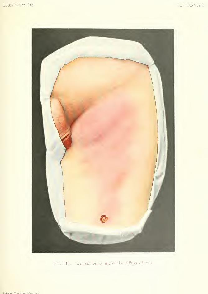
Bockenheinier, Atlas. Tab. LXXXVIII.
F-ig. 110. l.ymphadenitis inguinalis diffusa (Bubo)
Rrhman f^nmn^nv V»wf_Vrtrlf

cutaneous tissue, both superficially and deeply (Fig.
110). In this form there are rigors, fever and consti-
tutional disturbance. More often the inflammation
is localized and gives rise to a circumscribed abscess
(Fig. 114). The skin becomes thin and the pus is
discharged through a fistula. After this the pain
subsides; but the fistula does not heal, because the
whole gland is generally necrotic and is gradually
cast off, giving rise to infection of the neighboring
lymphatic glands and the formation of multiple
fistulas.
Diffuse suppurative lymphadenitis causes still
greater destruction for there is not only necrosis of
the glands themselves but also of the periglandular
tissue, and even of the subcutaneous tissue in exten-
sive cases. Moreover, burrowing abscesses may de-
velop in remote places; for instance, in the pelvis
after inguinal adenitis, and in the retro-pharyngeal
tissue after cervical abscesses. Again, general infec-
tion may occur from thrombo-phlebitis {e.g., from
thrombo-phlebitis of the pelvic veins after inguinal
bubo. All these complications can be avoided by
early incision.
In the chronic forms inflammatory symptoms are
absent. A slightly painful thickening develops in one
or more glands, after long-continued irritations, in-
flammation in the neighboring parts, eczema, pedicu-
losis, ulcers, etc. Finally, a small, irregular, movable
swelling is formed in the subcutaneous tissue, covered
by normal skin. Recovery takes place after removal
of the cause; but in long-standing cases a perma-
nent swelling may remain (fibrous hyperplasia).
Differential Diagnosis. Acute lymphadenitis
is characteristic and easy to diagnose by occurring
in the situation of the various groups of lymphatic
glands. The diagnosis of submaxillary and cervical
lymphangitis from alveolar periostitis, dermoids,
sebaceous cysts and tuberculous abscesses has al-
279

ready been given (Fig. 10''2). Acute lymphangitis
may be mistaken for sweat-gland abscesses, especially
in the axilla, but these are usually small, multiple and
circumscribed. In the inguinal region a hernia maybe mistaken for a bubo, especially when the sac of
the hernia is inflamed and is situated over the glands.
This error is more likely to occur in incomplete her-
nias in women. In these cases diagnosis is often only
made after incision. Suppuration arising from neigh-
boring bones or joints may also simulate lymphade-
nitis. Tuberculous lymphangitis may cause inflam-
matory infiltration, and painful enlargement of the
glands accompanied by fever; but the glands are
softer and of less uniform consistence. If a fistula is
present the diagnosis is more easy (Fig. 125). Tuber-
culous abscess is of slower development, and is gen-
erally associated with various degrees of infiltration
of neighboring glands. Lastly, the thin greenish pus
is characteristic.
Chronic lymphadenitis may be mistaken for metas-
tatic carcinomatous disease of the glands; e.g., of the
inguinal glands after cancer of the anus. These
glands, however, are hard and more or less fixed.
Syphilis gives rise to multiple hard infiltrations of the
lymphatic glands in various parts of the body.
Treatment. Pyogenic infection of the lymphatic
glands can often be avoided by removal of the pri-
mary cause. In acute lymphadenitis early incision
will prevent the complications mentioned above. If
this is neglected, not only the whole of the lymphatic
glands of the aft'ected region, but also the subcuta-
neous tissue may undergo necrosis; also oedema or
elephantiasis of the extremity may develop, owing to
the obstruction of the lymphatic circulation. Ele-
phantiasis may also occur after total extirpation of
the lymphatic glands (Fig. 71). This may be avoided
by taking care not to remove too much of the fatty
connective tissue along with the glands; this tissue
280
carries on the lymphatic circulation after removal of
the glands, and new glands are also formed from it.
In the groin and axilla a careful dissection of the
glands must be made, avoiding the great vessels.
Circumscribed abscesses are best opened by a free
incision. Treatment by poultices or icebags, aspira-
tion, puncture and injection of various fluids, mas-
sage and inunction of mercurial ointment are best
avoided. The affected parts should be immobilized
to prevent extension of the infective process. Patients
should, therefore, stay in bed. Commencing infec-
tion of the lymphatic glands often undergoes sponta-
neous resolution. Acute lymphadenitis of the neck
caused by infections such as diphtheria, may subside
spontaneously; so may chronic lymphangitis whenit is not of too long standing, and when the cause is
removed. Inunction with iodide of potassium or
iodine-vasogen ointments is useful in chronic lymph-
angitis. In cases of large glands causing pain, or of
multiple fistulas connected with chronic lymphade-
nitis, the glands should be extirpated, and the wounds
plugged for a long time with iodoform gauze to pre-
vent relapse.
Fig. 110 shows a case of acute lymphadenitis of
the inguinal region, occurring after an excoriation of
the skin of the thigh, which has already scabbed over.
Infiltration of the skin and subcutaneous tissue
extends from the genito-crural fold down the thigh.
The symptoms were pain and difficulty in walking,
followed by fever and rigors. Under an anaesthetic
an incision was made below and parallel to PouparVs
ligament. The inguinal glands were swollen, and
contained numerous foci of suppuration; but there
was no extensive necrosis, nor any large collection of
pus. The wound was j)lugged with iodoform gauze
and the leg immobilized on a splint. The wound
healed after a part of the gland which had necrosed
came away.281
ARTHRITIS GONORRHOICA PHLEGMONOSA{Phlegmonous Gonorrheal Arthritis)
Plate LXXXIX, Fig. 111.
In the course of both acute and chronic gonorrhea
the joints may be affected by general gonococcal in-
fection of the blood. In the acute stage of gonorrhea,
arthritis may be caused by the passage of bougies or
by overexertion, etc. In women it may occur during
pregnancy. In chronic gonorrhea it may be caused
by sexual excess. Gonococci may remain for a long
time in a latent state encapsuled in the mucous mem-brane, and when set free by mechanical irritation mayagain become virulent. Recurrence of gonorrheal
arthritis may take place in cases of neglected gonor-
rhea, also after a fresh attack of gonorrhea.
Through invasion of the joints by the gonococci
and their toxins inflammation is set up which maybe serous, fibrinous or purulent. Most commonly
the arthritis is fibrinous, suppurative arthritis being
rare and generally caused by mixed infection. Oneor several joints may be affected at the same time, or
successively. Acute gonorrheal arthritis is very sud-
den in its onset, and characterized by severe pain,
preventing any movement of the affected joint. In
a few hours the soft parts become infiltrated and
cedematous, the infiltration remaining more or less
limited to the region of the joint, or spreading to the
neighboring muscles and tendons. The skin is red
and tense (Fig. 111). In severe cases there is high
fever and complete loss of function. In chronic
gonorrheal arthritis there are usually aching pains
in the joint befoi'e the arthritis becomes evident.
282
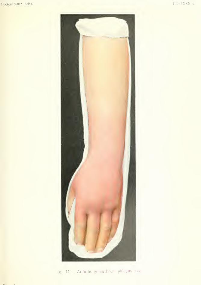
Bockenheimer, Atlas. Tab. LXXXIX.
Fig. 111. .Artiiiitis gonorrhoica phlcgmonosa.

The knee joint is most often affected in men; the
elbow and wrist in women. The hip, ankle andtemporo-maxillary joints are also often affected.
In cases of serous or sero-fibrinous effusion, limited
to the joint, the swelling generally subsides in one or
two weeks, and recovery takes place without loss of
function. In the more common form of fibrinous
arthritis, however, the process is more severe, espe-
cially when the infiltration extends to the periarticular
tissue and the soft parts. In these cases the arthritis
is accompanied by fever and rigors, and there is sero-
fibrinous effusion into the neighboring parts, but no
formation of pus. The inflammation affects not only
the synovial membrane, but may cause destruction of
the cartilage and extend to the bone. This may result
in fibrous, cartilaginous or bony anchylosis (X-ray
examination). Destruction of the capsule of the
joint may cause subluxations or dislocations, and the
prolonged immobility may lead to muscular atrophy.
In the rarer forms of suppurative arthritis there is
continued high fever with rigors, and severe consti-
tutional disturbance. The skin is red and there is
great swelling of the affected parts.
If several joints are affected different forms of gon-
orrheal arthritis may occur in the various joints.
Multiple relapsing arthritis may reduce the patients
to a deplorable condition, as they often cannot walk
or use their arms. Such cases may be fatal from
gradual exhaustion.
Differential Diagnosis. Gonorrheal arthritis
may be mistaken for acute rheumatism; but the latter
usually affects a greater number of joints, and the
acute stage of inflammation is not so prolonged as in
gonorrheal arthritis. Purulent gonorrheal arthritis
must be diagnosed from other suppurations in joints
by the history, by the presence or history of acute or
chronic gonorrhea, or by bacteriological examination
after puncture of the joint.
283

Chronic gonorrheal arthritis is often difficult to
distinguish from certain forms of syphilitic arthritis,
especially when both diseases have been contracted
together.
Tuberculous arthritis is usually easy to distinguish
by its characteristic signs (Fig. 125).
Treatment. On account of the severity of the
disease and the possibility of a fatal ending, espe-
cially from endocarditis, the prophylactic treatment
of gonorrhea is important. Washing immediately
after coitus, vaginal injections in women, the instilla-
tion of a few drops of weak silver nitrate solution into
the urethra after coitus, and the avoidance of any
kind of irritation (alcohol, etc.), will often prevent
gonorrheal infection. Gonorrhea should be regarded
as a serious disease and treated accordingly. In gon-
orrheal arthritis the urethra should always be exam-
ined, and treated if gonorrheal urethritis is present.
The subacute or serous forms of gonorrheal arthri-
tis subside in one or two weeks after rest in bed; but
too early movement may cause relapse. In fibrinous
arthritis, on the other hand, too long immobilization
may lead to anchylosis. Immobilization (by plaster
of Paris bandages, or better by extension splints)
should, therefore, not be continued longer than one
or two weeks; after which gentle massage, active and
passive movements or hot air treatment should be
tried. Langenbeck has recommended "animal baths"
for cases of stiffness; i.e., placing the affected part in
the viscera of a freshly killed animal, to obtain the
effect of animal heat. Sandbaths are also worth a
trial. If an acute relapse occurs in the course of the
disease the joint must be again immobilized. In any
case, movements of the joint must be carried out after
two or three weeks; otherwise bony anchylosis mayoccur. Injections of morphine may be given before
the performance of massage or passive movements,
or cocaine may be injected into the joint (0.05 cubic
284
centimeters of a five per cent, solution). Injections
of carbolic acid and protargol solution into the joint
have also been recommended. The best method of
treatment would be injection of antitoxin, as the
inflammation is primarily caused by the gonotoxin.
[In one case a good result was obtained by injection
of meningococcus-serum Bockenheimer.]
By careful after-treatment complete function cangenerally be restored even in severe forms of gonor-
rheal arthritis.
Bier's treatment by passive hyperaemia has a goodeffect in these cases, and may be tried in all cases of
gonorrheal arthritis where there is no suppuration.
After application of the elastic bandages, the joints
become painless (in about fifty per cent, of cases), so
that the patients do not hold them so stiffly, and early
movements can be performed, thus giving a better
functional result. In severe cases the joints should
be bandaged to protect them against injury, the ban-
dages being frequently removed and movements per-
formed. The elastic compression bandages should
be applied at first for two or three hours, later on for
twenty hours. This treatment may be carried out
without danger in out-patient practice (polyclinic).
If there is much destruction of the joint, with sub-
luxation or anchylosis in a faulty position, resection
may be required. Fibrous contractures are commonafter gonorrheal arthritis; these can be corrected
under an anaesthetic, and that function restored bv
appropriate after-treatment. In suppurative arthri-
tis, which is often complicated by lymphangitis,
lymphadenitis and other pyogenic conditions, arthrot-
omy or resection of the joint must be performed to
avoid general infection. These cases require longer
immobilization of the joint.
Fig. Ill shows a case of acute and painful swelling
in the region of the wrist joint in a woman. The skin
was red and tense. The swelling rapidly extended
285

to the forearm and to the fingers, so that the patient
could not use the arm. The wrist joint and the meta-
carpo-phalangeal joints could not be moved. Exam-
ination of the genitals showed gonorrhea. On the
following day the joint effusion increased and was
partly evacuated by puncture. Gonococci were found
in the fluid. Under treatment by passive hypersemia
the pain subsided in a few days and the acute inflam-
mation became chronic. Massage, active and passive
movements, combined with passive hypersemia,
restored the function in four weeks.
286

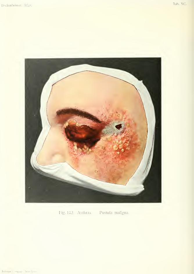
Bockenheimer, Atlas.Tab. \C.
Fig. 112. Antlira.x Pustuia maligna.
Rcbman Company, iS'ew-Vork.
ANTHRAX — PUSTULA MALIGNA(Anthrax—Malignant Pustule)
Plate Xr, Fig. 11-2.
ANTHRAX — NECROSIS (Anthrax necrosis)
Plate XCI, Fig. 113.
Anthrax (splenic fever) is a bacterial disease which
occurs externally on the skin, and internally in the
lungs and alimentary canal. The bacteria have a
characteristic appearance. They consi.st of immo-bile rods (bacilli) with sharp, angular corners, andare often arranged in a row in long chains. In the
center of the rods are clear spaces corresponding to
spores, which are very resistant to dryness and heat.
The anthrax bacillus was carefully studied by Koch,
while Pasteur originated the protective inoculation of
animals with attenuated cultures. The bacilli and
spores are found in the alimentary canal of animals
(horses and cattle) ; also in damp soil on which these
animals graze, and in the skin, fur and excrements of
animals infected with splenic fever (Rinderpest or
cattle fever). Epidemics of anthrax are common in
Egypt, as the excrements of animals are used as fuel
for cooking purposes. The disease is common in
Siberia in the skin trade and is known as Siberian
plague. Butchers, skinners, ragsorters, tanners,
paper makers and workers in horsehair are liable to
anthrax infection. The disease has been observed
in farmers, owing to the custom of treating horses and
cows affected with colic by passing the hand into the
rectum. The disease may also be transmitted by
earthworms and flies. The bacilli may remain local-
ized at the seat of infection, or may enter the blood-
stream and give rise to metastatic foci in other places
;
while their toxins play a subordinate part.
287
The external form of anthrax occurs on the skin of
the neck or face after small abrasions of the skin,
through which the bacilli enter. The infection maybe conveyed to the mouth by the finger, and the
spores may thus be inhaled or swallowed, and give
rise to anthrax of the lung or intestines respectively.
Anthrax of the skin has a very characteristic ap-
pearance. A small, red spot first appears, with fever
and often rigors; this develops into a small vesicle
with yellowish or turbid fluid containing anthrax
bacilli (malignant pustule). The pustule ruptures
and is replaced by a scab. At the same time the sur-
rounding skin becomes green—a sign of commencing
necrosis. The early appearance of necrosis of the
skin is characteristic of anthrax (Fig. 112). Thetissues become infiltrated in the same way as in car-
buncle (cf. Fig. 89), and oedema occurs where the
skin is loosely attached to the subcutaneous tissue
{e.g. eyelids). The redness of the skin extends rap-
idly and irregularly, resembling erysipelas. Other
vesicles appear and rupture, after which there is
extensive necrosis of the skin (Fig. 113).
In the extremities, along with the above symptoms,
there is always lymphangitis and lymphadenitis,
which may form abscesses by mixed infection.
There is always considerable constitutional disturb-
ance, with fever, rigors, headache and rapid pulse.
Multiplication of the bacilli in the blood gives rise
to symptoms of general infection—dry tongue, jaun-
dice, diarrhea and swelling of the spleen. Death mayresult from collapse in a few days.
External anthrax has a more favorable prognosis
than internal (mortality twenty-five per cent.), but
anthrax of the face is very dangerous. The moremarked are the local symptoms the more likely is
general infection. Moreover, anthrax of the face
may easily infect the mouth, and thereby cause infec-
tion of the lungs or alimentary canal. In the milder
forms of general infection metastatic inflammations
288
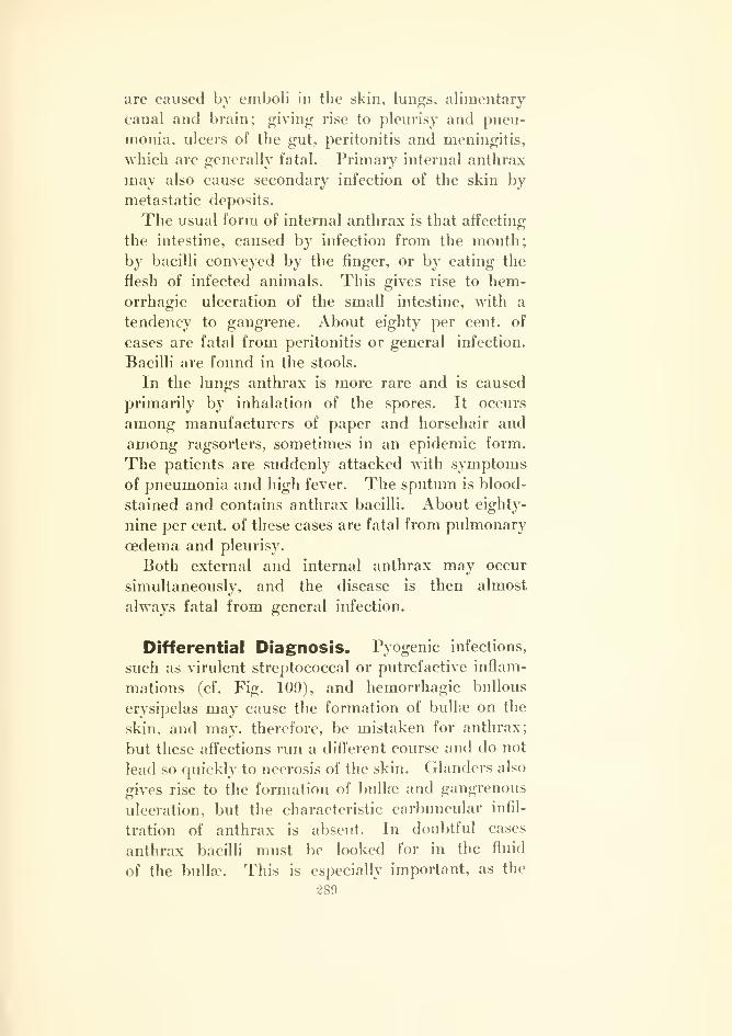
are caused by emboli in the skin, lungs, alimentary
canal and brain; giving rise to pleurisy and pneu-
monia, ulcers of the gut, peritonitis and meningitis,
which are generally fatal. Primary internal anthrax
may also cause secondary infection of the skin by
metastatic deposits.
The usual form of internal anthrax is that affecting
the intestine, caused by infection from the mouth;
by bacilli conveyed by the finger, or by eating the
flesh of infected animals. This gives rise to hem-
orrhagic ulceration of the small intestine, with a
tendency to gangrene. About eighty per cent, of
cases are fatal from peritonitis or general infection.
Bacilli are found in the stools.
In the lungs anthrax is more rare and is caused
primarily by inhalation of the spores. It occurs
among manufacturers of paper and horsehair and
among ragsorters, sometimes in an epidemic form.
The patients are suddenly attacked with symptoms
of pneumonia and high fever. The sputum is blood-
stained and contains anthrax bacilli. About eighty-
nine per cent, of these cases are fatal from pulmonary
cedema and pleurisy.
Both external and internal anthrax may occur
simultaneously, and the disease is then almost
always fatal from general infection.
Differential Diagnosis. Pyogenic infections,
such as virulent streptococcal or putrefactive inflam-
mations (cf. Fig. 109), and hemorrhagic bullous
erysipelas may cause the formation of bullae on the
skin, and mav. therefore, be mistaken for anthrax;
but these affections run a different course and do not
lead so quickly to necrosis of the skin. Glanders also
gives rise to the formation of bullae and gangrenous
ulceration, but the characteristic carbuncular infil-
tration of anthrax is absent. In doubtful cases
anthrax bacilli must be looked for in the fluid
of the bullae. This is especially important, as the
289
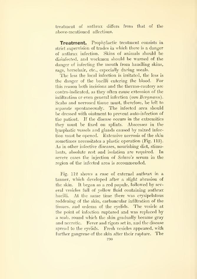
treatment of anthrax differs from that of the
above-mentioned affections.
Treatment. Prophylactic treatment consists in
strict supervision of trades in which there is a danger
of anthrax infection. Skins of animals should be
disinfected, and workmen should be warned of the
danger of infecting the mouth from handling skins,
rags, horsehair, etc., especially during meals.
The less the local infection is irritated, the less is
the danger of the bacilli entering the blood. For
this reason both incisions and the thermo-cautery are
contra-indicated, as they often cause extension of the
infiltration or even general infection {von Bergmann).
Scabs and necrosed tissue must, therefore, be left to
separate spontaneously. The infected area should
be dressed with ointment to prevent auto-infection of
the patient. If the disease occurs in the extremities
they must be fixed on splints. Abscesses in the
lymphatic vessels and glands caused by mixed infec-
tion must be opened. Extensive necrosis of the skin
sometimes necessitates a plastic operation (Fig. 113).
As in other infective diseases, nourishing diet, stimu-
lants, absolute rest and isolation are required. In
severe cases the injection of Sclavo's serum in the
region of the infected area is recommended.
Fig. 112 shows a case of external anthrax in a
tanner, which developed after a slight abrasion of
the skin. It began as a red papule, followed by sev-
eral vesicles full of yellow fluid containing anthrax
bacilli. At the same time there was erysipelatous
reddening of the skin, carbuncular infiltration of the
tissues, and oedema of the eyelids. The vesicle at
the point of infection ruptured and was replaced by
a scab, round which the skin gradually became gray
and necrotic. Fever and rigors set in, and the disease
spread to the eyelids. Fresh vesicles appeared, with
further gangrene of the skin after their rupture. The290

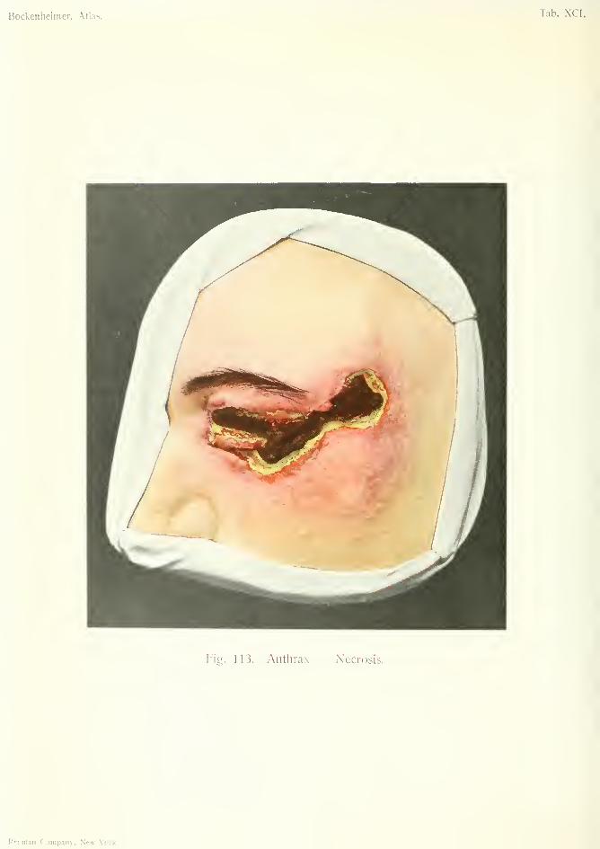
Bockenheimer, Atlas. Tab. XCI.
Fig. 113. Antlirax — Necrosis.
Rfbman Company, New- York.

affected area was covered with ointment and the
symptoms gradually subsided, without internal an-
thrax or general infection supervening.
Fig. 113 shows the same case a few weeks after
infection. The leathery, blackened, necrosed skin is
separated by a zone of pus and slimy granulation
tissue from the surrounding skin, which is still red
and infiltrated. The necrosed skin is firmly adherent
to the subjacent tissues. Removal of this by the
knife or sharp spoon would only cause a further out-
break of infection. It was, therefore, allowed to sep-
arate gradually under treatment by moist dressings of
peroxide and boric acid and ointments. In this case,
after separation of the necrosed skin, the defect wasrepaired by a plastic operation, and the upper eyelid
restored by a pedunculated flap. The patient recov-
ered, in spite of the unfavorable prognosis in anthrax
of the face and the severity of the local infection.
291
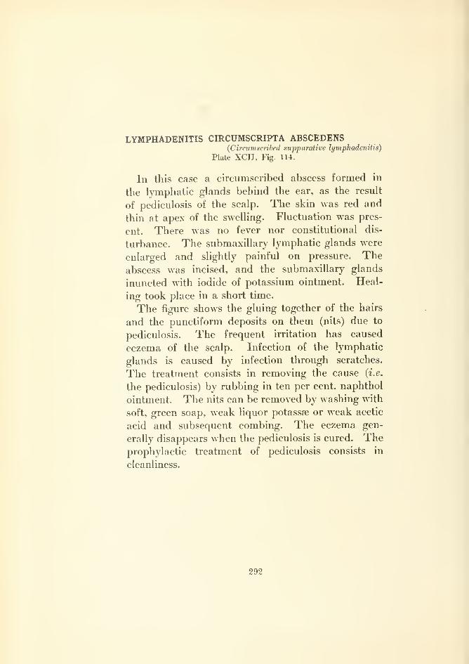
LYMPHADENITIS CIRCUMSCRIPTA ABSCEDENS(Circum.scribed suppurative lymphadenitis)
Plate XCn, Fig. 114.
In this case a circumscribed abscess formed in
the lymphatic glands behind the ear, as the result
of pediculosis of the scalp. The skin was red and
thin at apex of the swelling. Fluctuation was pres-
ent. There was no fever nor constitutional dis-
turbance. The submaxillary lymphatic glands were
enlarged and slightly painful on pressure. Theabscess was incised, and the submaxillary glands
inuncted with iodide of potassium ointment. Heal-
ing took place in a short time.
The figure shows the gluing together of the hairs
and the punctiform deposits on them (nits) due to
pediculosis. The frequent irritation has caused
eczema of the scalp. Infection of the lymphatic
glands is caused by infection through scratches.
The treatment consists in removing the cause {i.e.
the pediculosis) by rubbing in ten per cent, naphthol
ointment. The nits can be removed by washing with
soft, green soap, weak liquor potassse or weak acetic
acid and subsequent combing. The eczema gen-
erally disappears when the pediculosis is cured. Theprophylactic treatment of pediculosis consists in
cleanliness.
292

Bockenheimer, Atlas.Tab. XCII.
W)
•a
to
l^f.Km'.n i"»

Actinomycosis
ACTINOMYCOSIS INCIPIENS {Incijnent AcHnomycods)Plate XCII, Fig. 115.
ACTINOMYCOSIS PROGRESSIVA {Progressive Actirumycosis)
Plate XCm, Fig. 116.
Actinomycosis is a chronic infective disease caused
by a fungus (actinomyces) , which is called the ray-
fungus on account of the radiating arrangement of
its mycelium. Actinomycosis was first described by
Langenbeck, and later by Bollinger, in the form of
new growths in the lower jaw of cattle and horses.
In 1878 Israel found yellow bodies in the pus from a
patient who was supposed to have died of chronic
pyaemia; the yellow bodies were found to be actino-
myces. Later researches have shown that there are
different forms of actinomyces. The fungus is best
stained with Levaditi's silver nitrate method.
The fungus is found in corn, straw and flour. In
countrymen who have the habit of chewing corn the
mouth may become infected; either through a
carious tooth, leading to infection of the bone, or
through the parotid duct, leading to infection of the
cheek. In the great majority of cases, therefore, we
find actinomycosis in the mucous membrane of the
cheek, the tongue, the jaw, the pharynx and the neck.
It forms a stringy, nodular infiltration which, by be-
coming confluent, causes a swelling of wooden hard-
ness. Acute inflammatory symptoms are absent.
The skin becomes bluish red when the infiltration
extends through the cheek or into the neck (Figs.
293
115 and 116). The infiltration extends gradually into
the neighboring tissues, and by its unlimited progress
resembles a malignant tumor. At the same time
there is softening in the center of the infiltration with
the formation of an abscess which discharges through
several ramifying and anastomosing fistulas. The
pus contains the characteristic yellow bodies, about
the size of a pin's head. There is much induration
around the fistulas which often prevents the discharge
escaping. Granulation tissue is scanty, yellowish
red in color, and rapidly disintegrated. The forma-
tion of abscesses is accompanied by a slight rise of
temperature. Large abscesses may result from mixed
infection; the yellow bodies are then often absent,
the fungus being destroyed by the pus cocci.
In actinomycosis of the cheek a fistula is formed
externally. If the infiltration is situated in the mas-
ticatory muscles there is trismus. The fungus mayextend to the bones and give rise to enormous tumors.
If the upper maxilla is invaded it may extend to the
base of the skull and lead to meningitis or cerebral
abscess. If the tongue is infiltrated it cannot be
moved. When actinomycosis extends to the root of
the tongue or to the pharynx there is difficulty in
swallowing and later on in breathing. In these cases
abscesses form which generally discharge through
fistulas in the neck, and give rise to secondary' actino-
mycosis of the skin. Primary actinomycosis of the
skin has been observed through infection through
lesions of the skin (vo7i Bergmann).
The prognosis of actinomycosis of the buccal
cavity is comparatively favorable, and by appropriate
treatment two-thirds of the cases recover. On the
other hand, by extension to the retropharyngeal tissue
it may descend to the thorax or abdomen. Inspira-
tion of secretion containing the fungus may infect the
lunss. Invasion of the larwe veins of the neck result-
ing in metastatic foci has also been observed. Fromthis circumstance the affection was formerly regarded
294
as chronic pyaemia especially as actinomycotic gen-eral infection gives rise to similar clinical appearancesand eventually causes death by cachexia.
Actinomycosis of the lungs may occur from direct
inspiration of substances carrying the fungus, besides
infection from actinomycosis of the mouth. Theprognosis is very bad. The symptoms are those of
commencing phthisis. The lung becomes indurated
and the pleura infiltrated, and abscesses discharge
through the skin of the thorax. The disease mayspread from the pleura to the pericardium, the ver-
tebrae, the diaphragm and the abdominal cavity
The patient becomes exhausted from empyema andmultiple burrowing abscesses. The fistulas are diffi-
cult to follow owing to the hardness of their walls, so
that relapses are common after incision, and the cases
are usually fatal. Cases of recovery from actinomy-
cosis of the lung have, however, been observed.
Infiltration of wooden hardness between the ribs is
always suggestive of actinomycosis.
The intestine may also be the seat of actinomycosis
when material containing the fungus is swallowed.
The ileo csecal region is the part most often affected,
in the form of hard tumor-like infiltration which maybe so extensive as to prevent the passage of faeces.
The disease may spread to the vertebrae, pelvic bones,
abdominal organs, and may extend through the dia-
phragm to the thorax. There is often secondary
actinomycosis of the skin. A fistula often forms near
the umbilicus, discharging pus and sometimes faeces.
The prognosis is somewhat more favorable than that
of actinomycosis of the lung, but cases are often fatal
from general infection.
\\Tien actinomycosis is visible externally the diag-
nosis is not usually difficult; the wooden infiltration,
the multiple fistulas, the yellow granulations, and the
yellow bodies mixed with the pus are characteristic.
The diagnosis should always be confirmed by micro-
scopic examination.
295
Differential Diagnosis. Actinomycosis of the
cheek may, at first suggest kipus; but when the
nodules have broken through, this mistake is no
longer possible. Extensive infiltration of the cheek
may be mistaken for tumors, especially when the jaw
and the tongue are also afl^ected; but the history of
the formation of a cord extending often from a
carious tooth, followed by swelling of the cheek will
lead to the diagnosis. Actinomycosis of the tongue
is distinguished from abscess or gumma by extending
to the base of the tongue and causing immobility;
also, in actinomycotic abscess the pus contains the
characteristic yellow bodies. Actinomycosis of the
neck may be mistaken for "wooden phlegmon," but
the latter is generally unilateral and uniform, and
does not form fistulas; actinomycotic infiltration
extends round the whole neck, at first as a narrow
zone, later on as several zones in the form of terraces
one above another; the infiltration is also irregular.
Actinomycosis of the lungs and pleura may be mis-
taken for tuberculosis, but the more advanced cases
with fistulas are unmistakable. Actinomycosis of
the intestine may be mistaken for tuberculosis or
malignant growths, especially when it forms a tumor-
like mass in the ileo-caecal region.
Treatment. In extensive cases of antinomycosis
of the buccal cavity attempts at total extirpation are
useless, but healing may take place after free incision
of abscesses and laying open all fistulas. Granula-
tion tissue must be scraped away, and indurated tis-
sue removed as far as possible. The incisions should
be kept open for a long time by tampons. Carious
teeth must be removed. In actinomycosis of the
lung extensive resection of ribs is often necessary.
In actinomycosis of the ileo-csecal region resection
of the gut may be necessary on account of intestinal
obstruction or fistula. In other cases intestinal acti-
nomycosis comes to the surface and then only requires
296

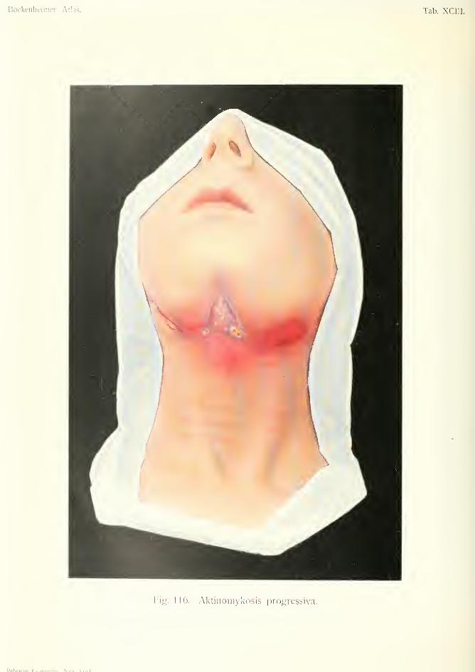
Bockenlieiiiicr. Atlas. Tab. XCIII.
Fiy. 116. yXktinomykosis progressiva.
Pplnlinn r\>.n.i'...i> M..»^.V..rl/
free incisions. Metastatic deposits in the bones
(which can be detected by the X-rays) may require
resection. General treatment consists in nourishins:
diet and the administration of iodide of potassium
and arsenic.
Fig. 115 shows a case of actinomycosis of the cheek
in an old countrywoman. Infection took place from
a carious molar tooth. A cord-like growth extended
from the root of the tooth to the gum, and thence to
the mucous membrane and muscles of the cheek, giv-
ing rise to diffuse infiltration. The skin becamebluish red and several small fistulas developed which
discharged pus containing yellow bodies. The latter
were found by microscopical examination to be acti-
nomyces. A circumscribed patch of gangrene wascaused over the malar bone by pressure of the infil-
tration. There was no fever and little trouble except
a slight degree of trismus. Treatment by free inci-
sion and plugging.
Fig. 116 shows a case of extensive actinomycosis of
the neck in a young countryman. The point of infec-
tion was not ascertained, and no changes w ere present
in the mouth or pharynx. Hard, painless infiltration
extended from one angle of the jaw to the other,
finally spreading over the whole region of the neck.
The skin was at first unaltered, but afterwards
became dark red. A circumscribed abscess formed
in the submaxillary region, which discharged pus
mixed with yellow bodies through several fistulas,
with yellow granulation tissue at their orifices. Both
actinomyces and cocci were found in the pus, show-
ing it to be a case of mixed infection.
The patient suffered from difficulty in breathing
and in sw^allowing. Free incisions were made in the
infiltration, the abscess was evacuated and the fis-
tulas scraped.
297
LINGUA GEOGRAPHICA{Marginate Glossitis—Geographical Tongue)
Plate XCIV, Fig. 117.
This affection is chiefly of interest on account of
the possibility of its being mistaken for other afi'ec-
tions of the tongue. The dorsal surface of the tongue
is covered with segments of circles of a gray color,
arranged irregularly and of various sizes. The inter-
section of these segments gives rise to an irregular
polycyclic or "geographical" pattern. The condi-
tion is caused by patches of hyperkeratosis of the
filiform papillae which spread at the periphery and
become normal in the center. The peripheral parts
form the segments of circles and consist of an accu-
mulation of desquamated epithelium. The condition
occurs most commonly in infants, but also in young
adults. It runs a benign course, and its cause is
unknown. It has been attributed to a syphilitic
origin by Kaposi, but this is doubtful.
Differential Diagnosis. Marginate glossitis
must not be mistaken for leucoplakia. The two
conditions have entirely different appearances. (Cf.
Fig. 9.)
Treatment. No special treatment is required
beyond mouth washes, painting with tincture of
myrrh and avoidance of spicy foods.
Fig. 117 shows a case of marginate glossitis affect-
ing the anterior two -thirds of the tongue. Thewhole tongue is divided into a series of projecting
areas of a yellowish-white color. Between these areas
are the gray segments filled with the secretions of the
mouth. At the back of the tongue the surface is
normal.
298
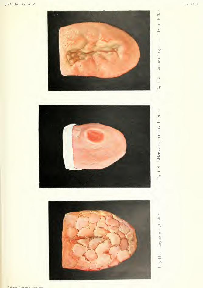
Bockcnheinier, Atlas. Tab. XC1\'.
tr.
tc
zr.
o
U
oo
tjO
M^w.Vrtrl/


Syphilis
SCLEROSIS SYPHILITICA LINGUA(Syphilitic Chancre of tongue)
Plate XCIV, Fie;.' 118.
GUMMA LINGUJE—LINGUA BIFIDA (Gumma of tongue)
Plate XCIV, Fig. 119.
GUMMA LABII SUPERIORIS ET NASI(Gumma of upper lip and nose)
Plate XCV, Fig. 120.
ABSCESSUS GUMMOSI (Gummatous Abscess)
Plate XCV, Fig. 121.
OSTITIS GUMMOSA (Gummatotis Osteitis)
Plate XCVI, Fig. 122.
ULCUS GUMMOSUM (Gummatous Ulcer)
Plate XCMI, Fig. 123.
Syphilis is a specific infectious disease which, in
the great majority of cases, is contracted by sexual
intercourse between human beings. It is probably
caused by the sjnrochaeta pallida, which was discov-
ered in 1905 by Schaudinn and Hoffmann, and has
since been found in all the products, and also in the
blood, in both acquired and hereditary syphilis. Thespirochaeta pallida is a delicate, thin organism with
corkscrew-like spirals, only visible under high mag-
nification. It is best stained by Giemsa's stain or
Levaditi's silver nitrate method.
Syphilitic infection takes place through slight
excoriations or fissures of the skin or mucous mem-brane. In this way extragenital infection may occur
in various parts of the body (lips, eyelids, tongue,
nipple, fingers, etc.). Indirect contagion may also
be caused by contaminated towels, linen, drinking-
glasses, cigars, tobacco-pipes, shaving brushes, etc.
Congenital or hereditary syphilis is the result of
299

syphilis in one or both of the parents. This often
causes abortions or stillbirths.
In acquired syphilis, after an incubation period of
three to five weeks, a circumscribed, hard, painless
infiltration of the skin or mucous membrane develops
at the point of infection, called the initial sclerosis or
hard chancre. This forms a flat erosion with a
smooth, dark-red surface, regular smooth borders
and an indurated base. The chancre forms a hard
nodule movable over the subjacent tissues. In geni-
tal infection it occurs on the prepuce, glans penis and
labia, more rarely in the urethra; in extragenital
infection it occurs at the part of the body inoculated.
In about ten per cent, of cases the chancre is not
discovered, but in the genital organs of women it is
often overlooked. The chancre generally heals in a
few weeks (with or without treatment) and leaves a
white scar which usually disappears in course of
time. Suppuration only takes place when the chan-
cre is infected by pus cocci. Sometimes the chancre
becomes gangrenous (phagedenic chancre). Mixedchancre is due to simultaneous infection with syph-
ilis and soft chancre; in these cases the soft chancre
appears first and becomes indurated later on. Theinduration of hard chancre is due to round-celled
infiltration chiefly arising in and around the walls of
the small blood-vessels.
The diagnosis of chancre is usually easy when it is
situated in the genital organs, but extragenital chan-
cres are often overlooked. Chancre of the fingers
often resembles a chronic whitlow or paronychia
(Figs. 93 and 98) ; but the sore has hard borders anda smooth surface and the acute inflammatory symp-toms of whitlow are absent. About a week after the
appearance of the chancre the regional lymphatic
glands become enlarged, forming hard, painless,
movable swellings (indolent bubo). In chancre of
the genitals the inguinal glands are aft'ected; in extra-
genital chancre the regional glands corresponding to
300
the part infected. Suppuration may occur in the
glands if the chancre is infected with pus cocci.
Secondary symptoms appear after a second incu-
bation period of six to twelve wrecks. They often
begin with malaise, headache and pains in the joints,
accompanied by a rise of temperature. A rose-red
macular rash (syphilitic roseola) develops on the
abdomen and thorax. Later on various syphilitic
eruptions develop (secondary syphilides), the most
common of which is an eruption of flat, rounded,
reddish-brown or ham-colored papules situated on
the trunk, face and limbs. On the forehead these
papules form the so-called "corona veneris." Onthe genital organs and around the anus these papules
become sodden and white, and are known as condyl-
omata lata, which are liable to ulcerate. In some
cases pustular eruptions form, and in severe or neg-
lected cases the pustules become ulcers covered with
limpet-shaped crusts (syphilitic rupia). Acneiform
eruptions are common on the scalp, and scaly or
psoriasiform syphilides on the palms and soles.
Most secondary eruptions disappear without leaving
any trace, but the ulcerative forms (rupia) leave pig-
mented scars, which later on become white in the
center. Syphilitic eruptions are characterized by
their reddish-brown or ham color, their polymor-
phous tendency and the absence of itching.
The mucous membranes, especially of the mouth,
are affected by papular, erosive or ulcerative syph-
ilides which are know^n as mucous patches. These
develop on the tonsils, fauces, tongue, and inside the
lips and cheeks, in the form of grayish-white patches
or streaks, with a red border. Later on they may
become eroded or ulcerated in their central parts,
and then appear as red erosions with a gray border.
In early secondary syphilis the tonsils and fauces may
be acutely swollen (syphilitic angina), but more often
there is dark-red coloration of the tonsils, fauces and
soft palate. In secondary syphilis there is often loss
301

of hair, sometimes due to acneiform syphilides of the
scalp, but more often appearing without any apparent
lesion. The nails are sometimes affected with
onychia or paronychia.
Secondary syphilis may last several years, and is
liable to recurrences. The most contagious lesions
are condylomata and mucous patches, even more
contagious than the chancre.
Tertiary syphilis occurs in about twenty per cent,
of cases, usually before the fifth year, sometimes later,
even up to the thirtieth year after infection. Thechief causes of tertiary syphilis, apart from specially
virulent forms of the virus, are absence of or insuflS-
cient treatment, and abuse of alcohol.
The characteristic feature of tertiary sj-philis is
the formation of circumscribed or diffuse infiltrations
called gummaia. The gumma is formed of round
cells, epithelioid cells and giant cells, and contains
blood-vessels thickened by syphilitic arteritis. Ow-ing to the changes in these vessels, the nutri-
tion of the gumma is interfered with and the
central parts undergo fatty degeneration or caseation.
A mature gumma shows on section three zones—
a
central zone of caseation, a middle zone of round
cells and an outer zone of fibrous tissue. Gummatamay cicatrize by the formation of fibrous tissue, or
they may suppurate and form an abscess. If the
abscess is superficial, it breaks through the skin andgives rise to a gummatous ulcer.
It must be borne in mind that the secretion from
gummatous ulcers may be contagious. (The spiro-
chaeta pallida has been found in gummata, and anylesion containing this organism is contagious).
Gummata develop in the skin and subcutaneous
tissue in the form of circumscribed nodules. Theskin becomes reddened and may suggest a furuncle,
especially in the ease of a single gumma. When the
gumma breaks through the skin the resulting gum-matous ulcer is characteristic. The borders are hard,
302
smooth, not undermined but circular and sharply cut,
as if punched out; the surface is covered by a tough,
tenacious, yellowish deposit, or core. In the skin
several gummata usually occur close together; these
break down in some places and heal in others, thus
giving rise to an irregular or serpiginous appearance
which is characteristic of tertiary syphilitic ulceration.
Gummata sometimes occur on the penis and maysomewhat resemble chancres, but there is no enlarge-
ment of the lymphatic glands in gummata. Gum-matous ulcers generally emit a disagreeable odor,
especially when they are situated in the pharynx or
nose (ozaena).
Gummata of the skin may be secondary to exten-
sion from gummata in the muscles or bones. Onthe other hand gumma of the skin may extend to the
deeper tissues. Diffuse gummatous infiltration of
the skin and subcutaneous tissue gives rise to multiple
fistulas which discharge a scanty secretion. Gum-mata may cause extensive deformity by destruction of
tissue, especially in the face (Fig. VZO). Gummata of
the scalp leave deep, smooth, glistening scars. Gum-ma of the tongue is usually situated in the center, and
may divide the tongue into two parts (Fig. 119).
Gummata and gummatous infiltration often affect
the soft palate and pharynx, giving rise to considera-
ble destruction of tissue and cicatricial stenosis. Thelarynx is also often affected. Gummatous infiltra-
tion of the rectum gives rise to stricture.
Gumma of the bones may develop in the perios-
teum, cortex or medulla, in the form of circumscribed
growths or diffuse infiltration. Generally, all three
parts of the bone are affected with simultaneous bone
destruction and bone proliferation, causing an irregu-
lar, corroded appearance. Gumma of bone mayundergo fibrous transformation, or may suppurate
and cause necrosis. Necrosis of the cranial bones
often leaves circular cavities to which the smooth,
glistening skin is firmly adherent.
303

The nose and hard palate are often extensively
destroyed by gummatous infiltration, suppuration
and necrosis. The sternum and clavicle are some-
times affected. In extensive disease of the long bones
curvature may result, especially outward curvature
of the tibia from the weight of the body. There is
also brittleness of the bones. Examination by X-
rays shows irregular shadows in the periosteal region,
while the cortex and medulla cannot be distinguished
from one another. The whole bone is thickened and
irregular.
Patients often complain of pain in the bones (osteo-
copic pains) before any changes are visible. Palpi-
tation of the anterior surface of the tibia often reveals
an irregular, uneven surface. The ulna, radius and
fibula may also be the seat of syphilitic osteitis.
Serous effusion may occur in the joints and bursas
in the course of syphilis. Extensive disease of the
joints may also arise from gummatous infiltration of
the perisynovial tissue, or from gummatous osteitis
of the articular ends of the bones. If a gumma of the
bone breaks into the joint, suppurative arthritis gen-
erally follows. The knee joints are most often
affected by syphilitic arthritis.
Gummata may occur in the muscles, and may be
mistaken for tumors. They usually occur in the
tongue, calf muscles and sterno-mastoid. Gummain the brain gives rise to symptoms of cerebral
tumor. Gummata are common in the liver and
testicles, and may occur in the lungs, heart and
other organs.
The blood-vessels are affected in all three stages
of syphilis (syphilitic arteritis). The changes affect
both the inner and outer coats of the vessels (endar-
teritis and periarteritis). Extensive proliferation
of the intima may cause complete occlusion of the
vessel; this occurs especially in the vessels of the
brain and leads to foci of softening. Syphilitic
arteritis of the aorta and other large arteries causes
304
aneurism. Syphilitic arteritis of the cerebral arteries
causes cerebral hemorrhage.
Each of the three stages of syphilis may be absent.
The chancre is undiscovered in ten per cent, of cases,
and may sometimes be absent. Tertiary syphilis is
said to occur in only twenty per cent, of cases; at
any rate it is frequently absent. The secondary stage
may also be absent in cases of severe infection in
which tertiary lesions appear soon after infection
(malignant syphilis). It is also possible that somecases of syphilis undergo spontaneous abortion after
the chancre.
In some cases of congenital syphilis the symptomsdo not appear till the eighth to sixteenth year. This
is known as late or delayed hereditary syphilis, to
distinguish it from early hereditary syphilis whichappears at or soon after birth.
Among the characteristic signs of early hereditary
syphilis are bullous syphilides of the palms andsoles (syphilitic pemphigus), and epiphysitis. Thelatter consists in a form of osteochondritis affecting
the epiphyses of the long bones, and causing thick-
ening. It is more common in the arm and gives rise
to paralysis of the limb. Epiphysitis may cause
interference with growth of the limb.
In late hereditary syphilis the bones are frequently
affected with gummatous processes identical with
those of acquired syphilis. The tibias are often
curved forwards and outwards owing to osteoplastic
periostitis. This condition is known as "saber
blade tibia," and is a characteristic sign of late hered-
itary syphilis. The skin over the bones is often
ulcerated.
Syphilitic dactylitis may occur in both early and
late hereditary syphilis. It causes thickening of the
phalanges, usually the basal ones. It is generally
multiple, sometimes bilateral, and tends to sponta-
neous resolution without suppuration.
The bones in hereditary syphilis are often very
305
brittle. Other signs of hereditary syphiHs are inter-
stitial keratitis, deafness due to disease of the internal
ear, notching of the incisor teeth {Hutchinson's teeth).
These three signs have been called the "Triad of
Hutchinson.''' Radiating scars round the mouth left
by former ulcerations are also characteristic.
Acquired syphilis may also occur in infants, but
differs in the absence of the characteristic features
mentioned above.
Differential Diagnosis. Syphilis is so wide-
spread among all classes of society that it must always
be borne in mind in cases of doubtful diagnosis.
Although the disease is fairly characteristic in all
three stages, it is possible to mistake it for other
affections, especially as the history can never be
relied upon.
Hard chancre, when ulcerated, may be mistaken
for soft chancre, but diagnosis can be established by
finding the spirochaeta pallida in scrapings. Extra-
genital chancres may be mistaken for epithelioma,
especially in the tongue and nipple, but the smooth
surface of the chancre differs from the irregular
ulcerated surface of epithelioma (cf. Fig. 1); the
regional lymphatic glands are affected early in chan-
cre. Chancre of the fingers is often mistaken for
whitlow, but difters in its chronic character and
absence of acute inflammatory symptoms. Sec-
ondary syphilis of the skin and mucous membranes
may be mistaken for various affections, and the
diagnosis often depends on the situation and general
course of the lesions, and on the presence of other
signs of syphilis. Gummatous ulcerations of the
skin may be mistaken for tuberculous ulcers or for
furuncle, but differ in the characters mentioned
above. Diffuse gummatous infiltration of the skin
with fungoid proliferation may suggest sarcoma (cf.
Figs. 24 and 26), but differs in the absence of any
tendency to bleeding, in the presence of circular scars
306
and brown pigmentation in the surrounding skin,
and in the presence of other signs of syphiHs, espe-
cially changes in the bones. Gumma in a muscle is
often at first indistinguishable from a tumor. Gum-ma in the testicle may be mistaken for tuberculosis,
but the former begins in the testicle while tubercle
begins in the epididymis. The diagnosis is easy
when the skin of the scrotum is perforated.
In the brain, liver, spleen and other organs the
diagnosis of gumma depends on other signs of
syphilis. Central gumma of bone may resemble
central sarcoma or bone cyst, and may give the sameappearance on X-ray examination, but gummatouschanges in bone are characterized by implication of
the periosteum. In doubtful cases antisyphilitic
treatment should be tried. If the diagnosis hesitates
between gumma and malignant tumor antisyphilitic
treatment should not be continued too long, as a
malignant tumor may thus become inoperable. In
such cases an exploratory incision with microscopical
examination is to be preferred. It must, however, be
borne in mind that long-standing gumma of the skin
may develop into carcinoma.
The earlier the diagnosis and the sooner the com-
mencement of treatment, the quicker is the cure of
syphilis. On the w-hole it may be assumed that the
majority of cases become cured, but the marriage of
syphilitics should not be allowed before five years
after infection, and then only after thorough and pro-
longed treatment, with an additional course of treat-
ment shortly before marriage. The danger of trans-
mission to the children is diminished by time and
treatment.
The disease generally runs a chronic course, and
cases of acute malignant syphilis are rare except in
persons who are broken down in health from other
causes (tuberculosis, alcohol, etc.). In the tropics,
however, syphilis is more severe and often fatal. It
is also more severe in races who are attacked for the
307
jBrst time and whose ancestors have been free from
the disease.
In a certain number of cases syphilis causes death
by gummatous disease of the internal organs, or by
diseases of the nervous system, such as tabes and
general paralysis, which, according to the latest
researches, are always of syphilitic origin.
[Reinfection in syphilis is rare, but may sometimes
occur after both the acquired and hereditary disease.
Immunity in hereditary syphilis does not appear to
last much beyond the age of puberty, after which
acquired syphilis may be contracted, usually in an
attenuated form. No doubt a soft chancre in a
syphilitic subject may become indurated by the
syphilitic process and be mistaken for reinfection;
so may a chancriform gumma of the penis; but a
considerable number of cases have been recorded in
which patients passed through two distinct attacks
of secondary syphilis, separated by an interval of
several years. These cases must of course be dis-
tinguished from cases of relapsing secondary syphilis
due to the primary infection.
Treatment. Infection can often be avoided by
cleanliness—by using ointment before coitus and
soap and water afterwards. Any abrasion of the
epithelium of the penis, caused by balanitis, etc., maylead to infection. Antisyphilitic treatment should be
commenced as soon as primary syphilis is diagnosed;
it should only be delayed till secondary symptoms
appear in cases of doubtful chancre. Excision of the
chancre has been often tried, but it cannot prevent
constitutional infection which is already present;
moreover, an ulceration may occur at the place of
excision. The chancre must be kept clean and
dressed with iodoform, xeroform or mercurial oint-
ment. Phagedenic chancre should be treated by
prolonged immersion in mild antiseptic baths.
Treatment by mercurial inunction is one of the
308
best methods, and can be carried out by the patient
himself. From three to five grammes (about adrachm) of unguentum cinereum is rubbed into the
skin for about twenty minutes daily, varying the seat
of inunction from day to day (inner side of arms andthighs and sides of body). This is best done at night,
the patient sleeping in a flannel nightshirt and taking
a hot bath in the morning. On the seventh day the
patient omits the inunction. The whole course lasts
six weeks. In the first year two energetic courses of
inunction should be taken; in the second year twomilder courses; and in the third year one course.
Treatment by intramuscular injections may be
employed instead of inunction. For instance, injec-
tions of one cubic centimeter of a two to five per
cent, solution of perchloride of mercury with sodiun
chloride every two or three days. [Injections of
perchloride of mercury are painful and have been
replaced by other preparations of mercury, those
most generally used being the biniodide and gray oil.
Biniodide is a soluble injection given in daily injec-
tions of one-third grain. Gray oil is a preparation
of metallic mercury suspended in liquid paraffin and
lanolin, and is given in weekly injections of one to
one and one-half grains. Injections are usually
made in the gluteal muscles, but some inject into the
subcutaneous tissue of the back. The treatment of
average cases of syphilis can also be carried out per-
fectly well by internal medication in the form of
pills—blue pill, proto-iodide, etc.]
Erosions and ulcerated mucous patches in the
mouth may be painted with chromic acid (five to
ten per cent.). To avoid mercurial stomatitis the
teeth should be cleansed with carbolic tooth powder,
and chlorate of potash mouth washes used.
In the tertiary stage iodide of potassium is indi-
cated for the treatment of gummatous formations
(thirty to sixty grains daily). It may be given in
milk. If iodism occurs the drug should be discon-
309
tinued, and fifteen to thirty grains of antipyrin given
daily (Jadassohn). If iodide cannot be borne,
Zittmanns decoction may be tried. Hot baths and
vapor baths are useful in improving metabolism, and
favor the elimination of large doses of mercury.
Gummatous ulcers may be treated with iodoform,
calomel ointment, or gray ointment. Gummatousabscesses may be incised and scraped. Deformities
of the lips, nose, etc., caused by gummata recjuire
plastic operations. Extensive stricture of the rectum
may necessitate resection of the gut. Cases of cere-
bral gumma, which do not yield to energetic treat-
ment with mercurial inunction or injection and large
doses of iodide of potassium, may be treated by tre-
phining, when they cause symptoms of a circum-
scribed cerebral tumor. Extensive gummatous dis-
ease of the testicle may require castration. Gum-mata in muscles may be incised, scraped and treated
locally with mercurial ointment, if they do not yield
to general antisyphilitic treatment. The same applies
to gummata in the bones, especially when they cause
severe pain. Gummatous periostitis and osteitis
often heal under energetic antisyphilitic treatment,
but sometimes require operative treatment for the
removal of sequestra. In cases of delayed union of
fractures, iodide of potassium is often useful whenthere is a history of previous syphilis. The same
applies to all badly healing wounds in syphilitic
patients, especially operation wounds. In hereditary
syphilis, osteochondritis can be treated by splints,
and gummatous osteitis may eventually require
operative interference.*
* For further information on this subject the reader is referred to
Marshall's "Syphilology and Venereal Disease," London. Bailiere,
Tindall and Cox; Marshall's "Golden Rules of Venereal Disease,"
Bristol. John Wright and Co. : Marshall's translation of Foiirnier's
"Treatment and Prophylaxis of Syphilis," New York. Rebman Co.
310

SCLEROSIS SYPHILITICA LINGUA(Si/phililic chancre of the tongue)
Plate XCIV, Fig. 118.
This is a case of extragenital chancre affecting the
tongue. The sore is sHghtly raised above the sur-
face; it has a round form with hard, sHghtly raised
not undermined borders, and a smooth, varnished
surface. The lymphatic glands in the submaxillary
and occipital regions were hard and movable. Car-
cinoma of the tongue differs from this in its irregular
surface, from which epithelial plugs can be expressed,
and in the glandular affection occurring later.
As already mentioned, syphilitic contagion maytake place through intermediate objects. Vo7i Berg-
mann has observed a case in which contagion was
due to smoking the fag end of a cigarette thrown
away by a syphilitic person.
311
GUMMA LmCUiE—LINGUA BIFIDA {Gumma of the tongue)
Plate XCIV, Fig. 119.
Gumma of the tongue is usually situated in the
center of the tongue, while carcinoma generally
affects the posterior part of the side of the tongue
(Fig. 9). A breaking-down gumma may divide the
tongue into two parts (bifid tongue). The figure
shows a broken-down gumma with its characteristic
tenacious, yellowish-brown deposit. Syphilitic infec-
tion was denied in this case, but it was cured byantisyphilitic treatment.
312

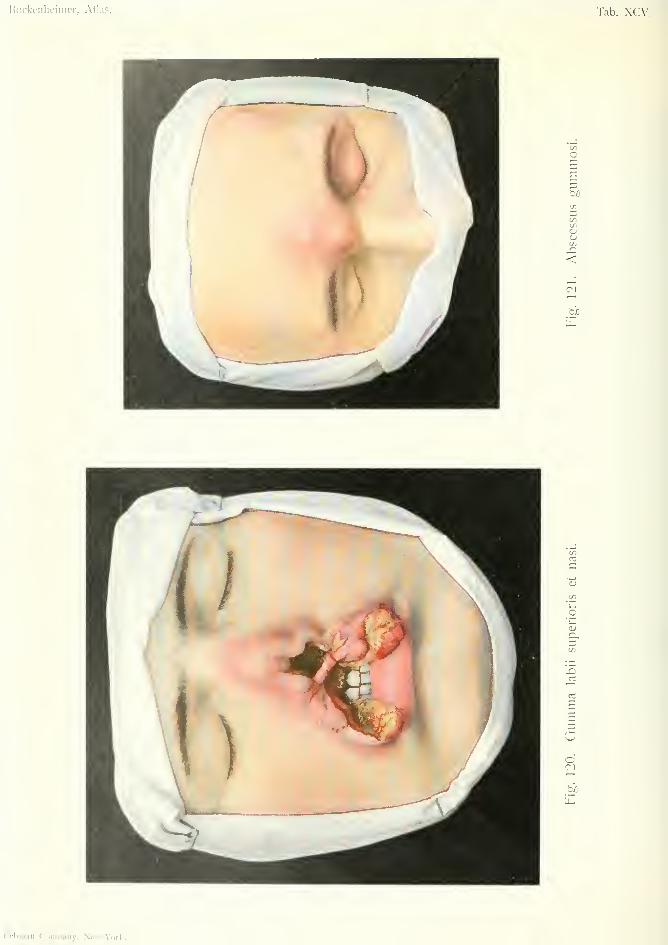
Hockenheinicr, Atlas. Tab. XCV.
OSS
IDOenXi<
o5
r3
On
Rcbman Company, New-York.
GUMMA LABII SUPERIORIS ET NASI{Gumma of the upper lip and nose)
Plate XCV, Fig. liO.
This case shows extensive destruction of the upperlip, the cartilaginous portion of the nose, the nasal
septum, and the bony framework of the nose, due to
gummatous ulceration. There is also perforation of
the hard palate. The upper lip shows the character-
istic yellow, tenacious deposit, which can be removedwithout bleeding. The surface of the ulceration is
fairly smooth and the borders soft, as compared withthe irregular surface and hard borders of carcino-
matous ulceration. The patient had syphilis ten
years previously.
The patient was treated with iodide internally andmercurial ointment locally. After the ulcerated sur-
face had become clean, the borders were excised andunited by sutures. The defect in the nose wasrepaired by a plastic operation, and an obturator
was worn for the perforation in the palate.
313
ABSCESSUS GUMMOSI (Gummatous Abscess)
Plate XCV, Fig. 121.
This is a case of multiple gummata in the skin of
the face, situated at the root of the nose, in the left
eyelid and in the temporal region. The skin is thin
and red. Fluctuation was felt on palpation. Thepatient could not remember contracting sj-philis, but
his wife had had frequent abortions and several
syphilitic children. There were gummatous pro-
cesses in the skin of various parts of the body; also
limitation of movement in the elbow joint due to
previous gumma of the bone. The surface of both
tibias was irregular, and there were circular scars
on the legs. The patient also suffered from severe
headache and attacks of giddiness, due to syphilitic
disease of the cerebral arteries.
The abscesses were incised and scraped, and
healed under antisyphilitic treatment. The cerebral
symptoms also improved.
314

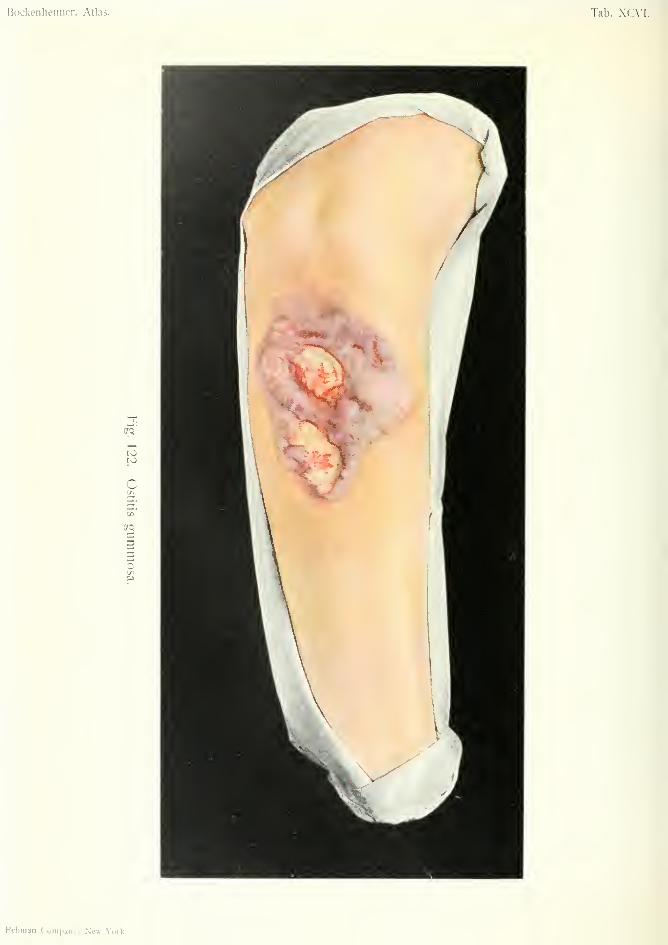
Bockeniieimer, Atlas. Tab. XC\'I
trq
toto
C
5
Q
Kcbinan Company, Ne«-\'aik.
OSTITIS GUMMOSA (Gvmmatous OsteUis)
Plate XC\'I, Fig. 1>22.
This patient acquired sypiiiiis twenty years ago,
and suffered for some years from pain in the right
forearm, especially at night. The bones of the fore-
arm gradually became thickened, and the skin red
and swollen. Two irregular ulcers developed, cov-
ered with yellow, tenacious deposit. Round the
ulcers soft proliferations formed resembling sarcoma-
tous tissue. There were several small fistulas lead-
ing to the bones, in which the X-rays showed irregular
proliferation of the periosteum and irregular thick-
ening of the cortex. Healing gradually took place
under treatment by iodide internally and mercurial
ointment locally. There were no other signs of
syphilis.
315
ULCUS GUMMOSUM {Gummatous Ulcer)
Plate XCVII. Fig. 123.
In this case a gumma occurred in the skin over the
internal malleolus after a kick (trauma is sometimes
an exciting cause of gumma). The skin became
infiltrated, swollen and red, and gradually broke
down, forming an ulcer with sharply cut edges and
a base covered with tenacious, yellow deposit. Thepatient contracted a sore on the penis some years
previously, which was diagnosed as a soft chancre,
and received no specific treatment. Three years
after infection a gumma developed in this situation
and was treated for a long time with poultices, but
was afterwards healed by iodide of potassium inter-
nally and mercurial ointment locally. The patient
was recommended further treatment by mercurial
inunction or injections.
316
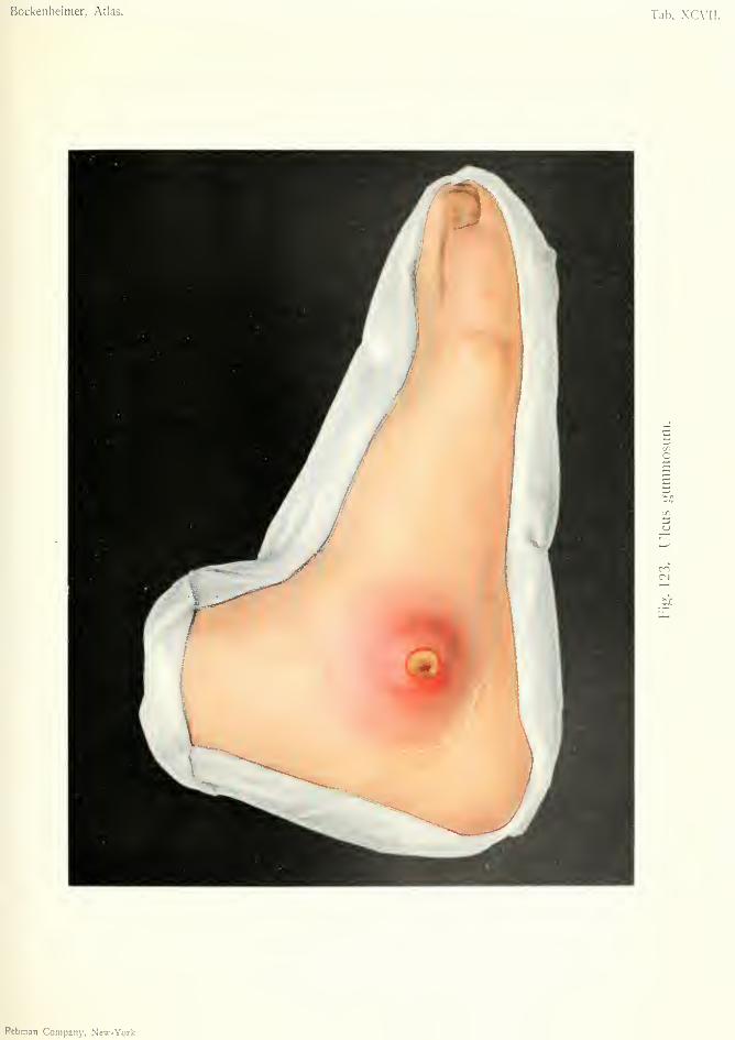
Bockenheimer, Atlas. Tab. XCVII.
n
D
CO
Rebman Company. New-York

Tuberculosis
LYMPHOMATA COLLI TUBERCULOSA(Ti/hrrciiloiix lymphoma of the Neck)
Plate XCVIII. Fig. 134.
ARTHRITIS TUBERCULOSA FUNGOSA(Fungaling Tuberculouji Arthritu)
ANKYLOSIS GENUS FIBROSAABSCESSUS FRIGIDUS
(Fibrous Anchylosis of the Knee—Cold Abscess)
Plate XCIX, Fig. 125.
ARTHRITIS TUBERCULOSA PURULENTA(Purulent Tuberculous Arthritis)
Plate C, Fig. 126.
ARTHRITIS TUBERCULOSA FIBROSA(Fibrous Tuberculous Arthritis)
ANKYLOSIS OSSEA—SUBLAXATIO(Bonij Anchylosis—Subluxation)
Plate C, Fig. 127.
ARTHRITIS TUBERCULOSA FIBROSA(Fibrous Tuberculous Arthritis)
TUMOR ALBUS (nidtc swelling)
Plate CI, Fig. 128.
TUBERCULOSIS TESTIS (Tuberculous Testicle)
Plate CII, Fig. 129.
TUBERCULOSIS MANUS (Tuberculosis of the Hand)Plate cm. Fig. 130.
OSTITIS TUBERCULOSA (Tuberculous Osteitis)
SPINA VENTOSA (Spina reniosa)
Plate CIV, Fig. 131.
GANGRjENA PEDIS HUMIDA (Moist Gangrene of the Foot)
Plate CIV, Fig. 132.
Since the discovery of the tubercle baciUus by
Robert Koch, in 1881, it is known that tuberculous
affections are solelv due to the invasion of these
bacilli; although tuberculosis was regarded as an
infective disease by several investigators before
the time of Koch. Baumgarten also discovered the
tubercle bacillus almost at the same time as Koch.
317
The tubercle bacilli are straight or slightly curved
rods. They are easily stained by the Ziehl-Neelsen
method, or by Gram's method, and are not decolor-
ized by nitric acid solution (acid fast bacilli). The
bacilli retain their virulence for a long time in the
dry state, but are destroyed by boiling and by sun-
light. Besides microscopic examination and culture
oi the bacilli, inoculation of the guinea pig is useful
for establishing diagnosis. Recent researches by
Friedrich have shown that tubercle bacilli in cultures
assume the form of club-shaped radiating filaments
similar to actinomyces; so that the bacillary nature
of the tubercle bacillus is doubtful, and it may belong
to the hvphomycetes. In any case tuberculosis and
actinomycotic affections are often very similar.
Tubercle bacilli are present in dust and on the
walls of rooms. Patients with tuberculosis of the
lunsrs infect the air with small vesicles of fluid con-
taining tubercle bacilli. Tubercle bacilli may also
penetrate the unbroken skin and mucous membrane
and cause infection of the lymphatic glands; but this
form of infection is comparatively rare. Wounds are
easily infected with tubercle bacilli, especially when
the sputum of a tuberculous subject comes in con-
tact with a wound {e.g. tattooing). Although, accord-
ing to Koch, there may be a distinction between
human and bovine tuberculosis, the latter may be
transmitted to man, and infection may occur from
the meat and milk of animals infected with bovine
tuberculosis; [especially by milk from cows with
tuberculous udders].
As regards the hereditary transmission of tuber-
culosis, it is certain that the children of tuberculous
parents are more predisposed to tuberculosis than
the children of healthy parents; but. whether the
bacilli can be transmitted from the mother to the
fetus and remain for a long time latent in the tissues
of the child, and whether transmission can take place
through the semen of the father, are points which are
318
still unsettled. In distinction to hereditary predis-
position, there is acquired predisposition; after cer-
tain diseases, such as influenza, measles, bronchial
catarrh and glandular swellings. The stronger the
body at the time of infection, the more it is able to
resist the disease. It is said that signs of former
tuberculosis can be found in almost ninety per cent,
of all men, in a great many of these cases the
tuberculous foci having become encapsuled or cal-
cified. Bad feeding, unhealthy dwellings, sedentary
occupations and alcoholism predispose to infection.
In the great majority of cases primary tuberculosis
affects the lungs, either by direct inhalation of bacilli,
or by bacillary infection of the lymphatic glands
(tonsils, bronchial glands, nasopharyngeal glands).
If the bacilli remain in the lungs they give rise
to phthisis. Tuberculosis of the mouth, pharynx,
larynx, trachea, bones and all other tissues, is in
most cases due to secondary metastatic infection bythe blood. Tuberculous embolism may be caused
by a tuberculous focus breaking through a large
vessel.
Tuberculous lesions which interest the surgeon are
in the majority of cases secondary. Tuberculosis
may attack any of the tissues, but has a predilection
for certain ones—primary tuberculosis for the lymph-
atic glands and lungs; secondary tuberculosis for
the bones and joints. Tuberculosis of the intes-
tine, which generally affects the small intestine and
ileocaecal region, is rarely primary but generally
secondary to tuberculosis of the lung (by swallowing
phthisical sputum) or the mesenteric glands. Tuber-
culosis may occur at any age.
The tubercle bacilli give rise to small nodular
infiltrations known as tubercles or granulomas. Thegranuloma is characterized by the presence of several
forms of cells, the majority of which are round cells,
a smaller number epithelioid cells, in fresh tubercles;
while in older tubercles giant cells are present,
319
especially in tubercles with a tendency to heal. Thegiant cells of tubercle differ from other giant cells in
the fact that the nuclei are situated at the periphery
of the cell round a central homogeneous mass, and
that in some parts of the circumference of the cell
there is a double arrangement of nuclei. Owing to
the absence of blood-vessels in the center of the tuber-
cle there is caseous degeneration of the central cells.
The giant cells often contain tubercle bacilli and are
believed to take part in the process of healing, byacting as phagocytes.
The tubercle sets up inflammatory reaction in the
surrounding tissues, resulting in the formation of
granulation tissue and pus, the latter being discharged
by a fistula, or forming an ulcer when the process is
in the skin. In most cases the body tries to expel
the tuberculous focus, but in some cases the latter
becomes encapsuled by connective tissue. This con-
nective-tissue capsule may at any time be ruptured,
by trauma, etc., and give rise to fresh tuberculous
infection. The majority of cases of tuberculosis fol-
lowing an injury are explained by the setting free of
encapsuled foci of tubercle; this not only causes a
fresh outbreak of tubercle at the seat of the injury,
but also spread of the previously encapsuled focus of
disease to other organs.
Surgical cases of tuberculosis are generally charac-
terized by the formation of typical granulations, fis-
tulas and specific pus. The granulations are pale
and vitreous. The fistulas run an irregular course,
and, in cases of tuberculous bone disease, open at
more or less distant points in the skin; the walls of
the fistula are soft and bleed easily. The pus is thin
and mixed with fibrin, caseous masses and shreds of
tissue. The tuberculous ulcer is characterized bythin, soft, ragged, undermined borders, and a base
covered with yellow caseous masses, or pale-red or
gray granulations. Tuberculous granulations maydestroy all the surrounding tissues (bones, cartilage
320
and muscles) and the necrosed parts are expelled
from the body. In the majority of cases there is a
formation of soft, spongy granulations, and little
fibrous tissue formation. Tuberculous processes
often continue for years before an abscess forms, or
a cavity from destruction of the tubercle.
Tuberculosis affects the different tissues in char-
acteristic ways, which we shall describe later whendealing' with the different cases. As a rule it runs a
chronic course with intermittent fever, without acute
inflammatory symptoms. The diagnosis can often
be made from the appearance of the ulcer, fistula,
pus or granulation tissue, and by the X-rays in the
case of bone disease. In many cases tuberculosis of
the luns leads to tuberculous disease of other tissues.
Diagnosis can be confirmed by microscopic examina-
tion; or by inoculation of suspected tissue into the
peritoneum of the guinea-pig, which gives rise to
tuberculosis of the mesenteric glands in a few weeks.
The walls of tuberculous abscesses are very suitable
for inoculation, as they contain many tubercle bacilli.
The prognosis depends on the situation and extent
of the disease. Small, circumscribed foci can be
radically removed by operation—for instance, cir-
cumscribed tuberculosis of the skin, or tuberculous
jrlands in the neck which are common in children.
However, as tubercle is generally present in the lungs,
many patients succumb to this sooner or later. In
tuberculosis of bones and joints, complete restitution
is seldom possible, owing to the extensive destruction
of tissue. Long-standing disease of bones and joints,
which may occur at an advanced age, is often fatal
from exacerbation of tuberculosis of the lungs.
Treatment. The spread of the disease should be
checked by prophylactic measures. Tuberculous
patients should be warned against spitting into hand-
kerchiefs or on the ground, and should use spitting-
cups. Meat and milk from tuberculous cattle should
321
not be consumed. The general treatment of tuber-
culous patients consists in nourishing diet (plenty of
milk, meat and butter), cod-liver oil and extract of
malt; administration of preparations such as creo-
sote and guiacol; residence at high altitudes; sea
baths; sanatorium treatment. Inunction of the
whole body with green soap, which is allowed to
remain on the skin for half an hour, is said to be
beneficial. Brine baths and sulphur baths are useful.
Tuberculin treatment has not been successful, and
cannot be recommended in practice.*
As regards local treatment, the object of modernsurgery is to remove the focus of disease when it is
within reach. By this means not only is the local
disease often cured, but the primary lung disease is
often improved. In some cases a whole organ, such
as the kidney or testicle, must be removed when it is
extensively diseased. A ten per cent, emulsion of
iodoform in glycerin is useful for application to
tuberculous ulcers and fistulas, and for injection into
tuberculous joints and abscesses. In the treatment
of tuberculous bone and joint disease immobilization
is essential.
Tuberculosis of the Skin. This has already
been mentioned in the case of lupus. (Plate III.)
Local tuberculosis of the skin may occur from infec-
tion from dead bodies affected with tuberculosis.
This form is common on the fingers in doctors and
hospital attendants after post-mortem examinations,
and in butchers from handling tuberculous meat.
It is known as cadaveric tubercle or post-mortem
wart. It commences as a small red spot which
develops into a raised nodule with slight sanious
discharge. Several nodules may develop close to-
gether and form a warty growth. A more extensive
* Sir Almroth Wright's method by injection of the new tubercuh'n,
under control of the opsonic index, is apparently successful in suit-
able cases.
322
form of warty cutaneous tuberculosis is known as
tuberculosis verrucosa, and is common in the poorer
classes. In these cases the tubercles have little ten-
dency to break down and undergo caseous degenera-
tion, but become warty by cornification of the epi-
dermis.
The treatment of these forms of cutaneous tuber-
culosis is the same as for lupus; viz. excision of
small lesions; scraping with sharp spoon, cauteriza-
tion, or treatment by Finsen's light in the case of
larger growths.
Another form of cutaneous tuberculosis commencesin the subcutaneous tissue, and gradually extends to
the skin in the form of red nodules resembling fur-
unculous abscesses. The skin becomes thin, the
nodules suppurate and discharge pus on the surface.
This condition is common in the neck in tuberculous
children. Before the skin breaks down the nodules
may be mistaken for gummata, but afterwards typical
tuberculous ulcers are formed. This condition has
been called scrofuloderma, but is due to the action
of tubercle bacilli. The treatment consists in scrap-
ing and iodoform dressings.
Tuberculous Lymphangitis and Lymphade-nitis. Tuberculous lymphangitis is rare, and only
occurs in connection with tuberculosis of the skin
and lymphatic glands, in the form of nodular cords.
Tuberculous lymphadenitis, on the other hand, is
very common (Fig. 124). It occurs especially in
children in the glands of the neck, the tubercle
bacilli easily penetrating the soft walls of the lym-
phatic vessels. The glands may be affected by wayof the blood or lymph, after eczema, ulcers or tuber-
culosis of the neighboring tissues. Through slight
lesions of the mucous membrane of the mouth or
pharynx, the tubercle bacilli enter the lymphatics,
and infect the glands of the neck and submaxillary
region. Some authorities maintain that tuberculosis
323
of the lunffs is secondarv to tuberculous disease of the
bronchial glands, and intestinal tuberculosis to dis-
ease of the mesenteric glands. The tubercle bacilli
cause inflammatory swelling, and the formation of
miliary tubercles in the glands. Several miliary
tubercles become confluent and form larger nodules
which undergo caseous degeneration and softening,
and finally suppurate. The tuberculous process
is not usually limited to a single gland, but extends
through the capsule to the surrounding tissue,
and finally to the skin. The glandular tumor, at
first circumscribed and covered with intact skin, soon
implicates the skin and breaks through it in one or
more places, forming fistulas which discharge thin,
greenish pus. The pus often burrows under the
skin and breaks through in more or less remote
places. The axillary and inguinal glands are seldom
the seat of primary tuberculosis.
Differential Diagnosis. Tuberculous glands are
characterized by the variation in their consistence;
some glands being soft and fluctuating, others hard.
In the absence of fistulas or other signs of tubercu-
losis, an isolated tuberculous gland may be mistaken
for a suppurating sebaceous cyst or dermoid. Thedifferential diagnosis from malignant tumors has
already been described (Fig. 24). In doubtful cases
microscopic examination, or inoculation in the guinea
pig will establish the diagnosis.
Treatment. The primary cause (eczema, ulcers,
etc.), must, of course, be treated. Circumscribed
glandular abscesses may be evacuated by puncture
and injected with ten per cent, iodoform emulsion.
Larger groups of glands should be freely laid open
and removed. Removal of tuberculous glands in
the neck requires an accurate knowledge of anatomy,
as these glands are often situated around the large
324

vessels from the mastoid process to the suprachivicii-
hir fossa, and lie behind the sterno-mastoid muscle
and sometimes under the trapezius. After extirpa-
tion, the wound should be plugged with iodoform
gauze and the wound closed, leaving a small space
for drainage. In children especially, there is a rise
of temperature for the first few days after extensive
removal of glands, which is probably due to the
entrance of tubercle bacilli into the blood. Miliary
tuberculosis may develop after extensive removal of
tuberculous glands. It is, therefore, better in exten-
sive glandular disease, occurring in feeble patients,
to limit operative interference to incision and scrap-
ing-
Tuberculous lymphadenitis of the neck, especially
when associated with eczema of the eyelids, otitis
media and ulcers of the cornea, is often wrongly
called scrofula. Staphylococci are often found along
with tubercle bacilli. In cases where no tubercle
bacilli are found it is possible that they have been
destroyed by the pus cocci. The term scrofula
should, therefore, be avoided, especially when typical
tuberculous disease is present in other parts of the
body. Predisposing causes of tuberculous lympha-
denitis are—measles, influenza, whooping-cough, un-
cleanliness and improper feeding. General treat-
ment consists in the measures already mentioned,
especially sea and sulphur baths.
Tuberculosis off Bone. Tuberculous disease of
bones is secondary, and caused by the spread of
tuberculous material by way of the blood. For this
reason the bones are generally aft'ected in certain
places corresponding to the distribution of their
blood-vessels. Lexer found, by X-ray examination
after injection of the vessels of bones with mercury,
that the nutrient artery of the long bones terminated
in the epiphyses. This explains the frequency with
which the epiphyses of the long bones are affected
325
with tuberculous deposits, by plugging the terminal
branches of the nutrient artery in the epiphysis with
tuberculous infarcts. In the short bones the nutrient
artery terminates soon after its entrance in the middle
of the diaphysis; hence tuberculous disease of these
bones affects the diaphysis. Tuberculosis also affects
the vertebrae, the bones of the hand and foot, the
cranial bones, the sternum, ribs and ilium.
In most cases there is circumscribed disease in the
form of a caseous sequestrum. Around this form
granulation tissue and pus, which seeks a way to the
surface by the formation of a fistula. Small sequestra
often give rise to large abscesses which become visible
under the skin, often at some distance; these are
known as "cold abscesses" (Fig. 125). Tuberculo-
sis of the vertebrae may thus cause abscesses which
appear in the thigh. In tuberculous bone disease
there is little tendency to the formation of new bone.
In some cases the focus of disease may become encap-
suled in the bone, but is always liable to recrudes-
cence, especially after an injury. More commonlythe sequestrum is discharged piecemeal through a
fistula, thus differing from the large sequestrum of
pyogenic osteomyelitis. Multiple foci of disease often
occur in one or more bones. When the bone is
exposed by incision, irregular, caseous fragments
are seen, together Avith pus. When the disease
occurs in the epiphyses of the long bones it maybreak into the joint, giving rise to suppurative
arthritis.
Although the foci of disease are usually small, andthere is seldom the necrosis of large portions of bonewhich occurs in pyogenic osteomyelitis, there may be
extensive disease of the medullary cavity whentuberculous disease of a joint extends to the bones.
The phalanges may also be extensively diseased.
Sometimes no changes are found in the bone, andthe disease is confined to the periosteum, giving rise
to subperiosteal abscess, especially in the ribs.
326
Differential Diagnosis. In some cases tuber-
culosis of bone may be mistaken for the chronic forms
of pyogenic osteomyehtis. However, tuberculous
bone disease can nearly always be recognized by its
typical situations, its characteristic pus, its small
sequestra, its slight tendency to new bone formation,
and by the presence of tuberculosis of the lungs. In
many cases the diagnosis is assisted by the X-rays.
Some cases may be mistaken for syphilitic bone
disease. In doubtful cases diagnosis can be settled
by incision.
Treatment. As soon as tuberculous disease of
bone is diagnosed (by the X-rays early diagnosis can
be made), the disease must be radically removed,
without interfering too much with function. Thebone must be freely exposed, the diseased parts
removed by the gouge or sharp spoon, and the woundplugged for some time with iodoform gauze. In the
extremities immobilization is necessary. Later on,
iodoform-glycerin emulsion may be injected into the
bone cavity. Tuberculous foci can be treated in
this way in the bones of the face, the cranial bones,
the sternum and the ilium.
In cases of tuberculous disease of the vertebrae
(tuberculous spondylitis or Pott's disease) operative
interference should be limited to the evacuation of
abscesses, which often point on the inner side of the
thigh below Poupart's ligament, and injection of
iodoform-glycerin emulsion. Operation on the ver-
tebrae themselves is likely to injure the spinal cord or
nerves. These cases often undergo spontaneous
cure by sinking of the bodies of the vertebra?, result-
ing in kyphosis. These cases require immobiliza-
tion by extension splints and later on by plaster of
Paris jackets. Extensive bone disease of the extremi-
ties in old people may require amputation to save the
patient from death b\ pulmonary tuberculosis; on
which amputation often has a favorable influence.
327
Treatment of tuberculous bone disease by passive
hypersemia is only of use when combined with other
methods of treatment. In cases of pain and fatigue
in the Hmbs of young people, occurring without
apparent cause, the possibility of commencing tuber-
culous disease of the bones or joints must always be
borne in mind. Early cases often recover after pro-
longed immobilization without operation. Bones
which have been affected by tuberculosis must be
protected from injury, which may start the disease
afresh.
Tuberculosis of Joints. The joints are often
affected with tuberculosis, generally by extension
from tuberculosis of the bones. Infection of the
joints may also take place through the blood, but
primary tuberculosis of joints is rare. In most cases
both the synovial membrane and the articular ends of
the bones are affected. The knee and hip joints are
most often attacked; after these the wrist and elbow.
Tuberculous joint disease is most common before
puberty, but it also occurs at an advanced age.
The tubercle bacilli give rise to the formation of
granulation tissue and effusion in the joint. In the
mildest forms there may be only serous effusion
(hydrops), but more commonly the effusion is sero-
fibrinous. The fibrin forms villous deposits on the
synovial membrane and cartilage, and the so-called
"rice bodies," which are lumps of loose fibrin in the
joint. These milder forms of joint disease may be
included under the name of articular hydrops.
The second form of tuberculous arthritis is knownas fungoid arthritis, owing to the formation of fun-
goid or spongy granulation tissue, which gives rise to
globular swelling of the joint. In these cases the
whole joint is filled with grayish-red or yellowish-
white granulations, and there is only slight exudation.
The fungous granulations tend towards caseous
degeneration, and after a time to suppuration. This
328
form of tuberculous arthritis does not remain limited
to the joint but soon extends to the Hgaments andperiarticular tissue, and eventually to the subcuta-
neous tissue and skin (Figs. 125 and 126).
A third form is fibrous arthritis, in which there is a
formation of hard fibrous tissue in the joint. Thisform is called caries sicca by Volkmann. It is com-mon in the shoulder and hip joints, and is character-
ized by a great tendency to cause atrophy, of the
articular end of the bone, giving rise to dislocations
and also to muscular atrophy.
In distinction to the above atrophic form, there is
another form of fibrous arthritis causing globular
swelling of the joint from the abundant formation of
fibrous tissue. This is especially common in the
knee joint and may be mistaken for bone tumor. It
is known as "white swelling" or tumor albus, owingto the white anaemic appearance caused by pressure
of the fibrous tissue on the skin.
A fourth form of tuberculous joint disease is furu-lent arthritis. This is often due to mixed infection
of one of the above-mentioned forms with staphylo-
cocci—for example, through a fistula in the skin.
However, purulent arthritis sometimes occurs quite
suddenly, especially in children.
In all these forms of tuberculous arthritis the car-
tilage may be destroyed by the fibrinous exudation.
In cases of fibrinous hydrops, and in caries sicca, the
destructive action is generally limited to the cartilage;
but in the fungoid and purulent forms of arthritis the
whole epiphysis may be destroyed, and the infection
may spread to the diaphysis. Besides this, multiple
abscesses often develop at some distance from the
joint. The greater the destruction of the joint the
more abnormal are the positions of the afi'ected limb.
The affected joint assumes the position in which its
capsule has the greatest capacity {i.e. the position in
which the capsule is fully distended). For this rea-
son the knee joint is in the position of flexion, the hip
329

joint in the position of abduction and flexion, the
elbow joint in the position of flexion, and the shoulder
joint in the position of external rotation. Fibrous or
bony anchylosis may occur in these positions ; also in
positions of subluxation or dislocation.
Tuberculous arthritis generally begins with pain,
which is often remote from the affected joint; e.g. in
disease of the hip joint pain is referred to the inner
side of the knee. This is followed by slight rises of
temperature and pain in the region of the affected
joint. Movement of the joint is avoided, the whole
joint becomes swollen, and characteristic positions
are assumed by the different joints. In hydrops there
is fluctuation. In fungoid arthritis the whole joint
is filled with soft, spongy tissue, causing balloon-like
swelling of the joint (ballooning) ; this spongy tissue
extends to the periarticular tissue and reaches the skin,
which becomes reddish blue, and later on breaks
down into tuberculous ulcers and fistulas (Fig. Via).
Diagnosis is generally easy in cases with a fistula
discharging characteristic thin tuberculous pus mixed
with caseous debris and fragments of sequestrum.
In other cases there is evidence of tuberculosis in
the lungs or other organs. The fibrous forms
(caries sicca) are characterized by the marked atro-
phy of the joint, the abnormal positions, and the
muscular atrophy and complete loss of function.
White swelling is recognized by the extensive tumor-
like swelling covered by white skin (Fig. 1'28). In
purulent arthritis there is redness and swelling of the
skin with high temperature. In doubtful cases an
incision will make the diagnosis clear.*o"
Differential Diagnosis. Tuberculous hydrops
may be mistaken for traumatic effusion, gonorrheal
arthritis or syphilitic arthritis. The diagnosis
depends on the historv' of the case and thorough
examination of the whole body. In doubtful cases
the joint may be punctured, or inoculation of the
330
t>
guinea pig may be performed. Acute forms of fun-
goid tuberculous arthritis can hardly be mistaken
for other affections. In cases where complete heal-
ing of the joint has taken place, with bony anchylosis,
it is sometimes impossible to distinguish tuberculous
cases from joint disease secondary to pyogenic osteo-
myelitis of the diaphysis. In old people healed
tuberculous joints may be mistaken for arthritis
deformans or chronic rheumatism. Purulent tuber-
culous arthritis often resembles acute pyogenic osteo-
myelitis. In young children especially, when the
disease begins with rigors, high fever and constitu-
tional disturbance, diagnosis is often only made after
incision.
The prognosis of tuberculous arthritis is more
favorable in young individuals than in old people.
Chronic tuberculous arthritis may give rise to miliary
tuberculosis, or to amyloid degeneration of the inter-
nal ortrans.&•
Treatment. In its early stages tuberculous
arthritis may be cured by immobilization by means
of extension splints or plaster of Paris casings. Con-
serv'ative treatment should always be adopted in the
early stages. Hydrops may be treated by repeated
puncture, injection of iodoform-glycerin emulsion or
alcohol and immobilization of the joint. Recurrence
is common, and complete restoration of function sel-
dom occurs. The joints should, therefore, be
allowed to anchylose in the most useful position.
When abscesses and fistulas form, and when an
extensive focus of bone disease is shown by the
X-rays, conservative treatment must be abandoned.
In fibrous arthritis, caries sicca and white swelling,
resection of the joint should be performed as early as
possible, to prevent muscular atrophy. In the
shoulder joint resection gives good results; but in
the knee joint, bony anchylosis in the straight position
is the only possible result. In fungous arthritis,
331
especially in young patients, operation may be
limited to opening the joint and carefully removing
all tuberculous disease (arthrectomy). The capsule
of the joint must be excised wherever it is diseased,
and tuberculous foci in the cartilage and bone
removed with the gouge. In young subjects a
typical resection of the joint is to be avoided, owing
to interference with the growth of the limb by exten-
sive removal of the epiphyses.
In adults, on the other hand, the joint may be
resected and all diseased parts carefully removed.
If the medullary cavity is found to be diseased, after
resection of the epiphysis, it must be scraped out.
Abscesses and fistulas require incision and scraping.
In purulent arthritis the joint must be freely opened
;
in advanced cases resection is necessary. In exten-
sive tuberculous arthritis with tuberculous disease of
the neighboring bones and soft parts, amputation
may be necessary, especially in old people (Fig. 130).
After operation the joint must be plugged with iodo-
form gauze, drained, and immobilized. Joints which
have become healed in abnormal positions may be forc-
ibly corrected under an anaesthetic when the anchylosis
is fibrous ; but there is danger of rupture of the vessels
and consequent gangrene (Fig. 132). It is better to treat
fibrous anchylosis by gradual extension; while bony
anchylosis in a bad position may require resection.
After operations on joints, these should be pro-
tected by light splints {e.g. poroplastic casings) till
the end of the period of growth in children, and for
some years in adults. The disadvantage of this
apparatus is the causation of muscular atrophy. Onthe other hand, after resection of the knee joint in
young subjects, the knee often becomes flexed, even
after bony anchylosis, requiring further resection.
Tuberculosis of other Tissues. Tuberculosis
of the mucous membranes occurs in the buccal cavity,
the tongue, lips, larynx, small intestine and rectum,
332
and is generally secondary to tuberculosis of the
lungs. Von Bergmann has observed a case of tuber-
cular infection of the mouth, from a culture of
tubercle bacilli, which was cured by excision.
Tuberculosis of the mucous membranes develops in
the form of small, reddish-gray nodules, which break
down into small easily bleeding ulcers with ragged
edges and a yellow caseous surface. These are best
treated by cauterization with strong lactic acid.
Fistula of the rectum, which is common in intestinal
tuberculosis, requires incision. Tuberculosis of the
ileo-caecal region, causing fibrous stricture, mayrequire resection of the gut.
Tuberculous peritonitis, which gives rise to exuda-
tion and the formation of extensive adhesions, is im-
proved by laparotomy and removal of the exudation.
Purulent tuberculous effusion into the pleural cavity
should be evacuated by resection of the ribs. Tuber-
culosis of the testicles and kidneys necessitates
removal of these organs. Tuberculosis of the blad-
der should be treated by irrigation and the internal
administration of guiacol. It has been attempted to
remove isolated foci of tuberculosis in the lungs by
operation.
Treatment of the general condition of the patient
is necessary in all forms of tuberculous disease.
Miliary tuberculosis, which may develop after
extensive operations, such as removal of tuberculous
glands in the neck, or after breaking down joint
adhesions, is due to dissemination of tubercle bacilli
in the blood, and may take the form of a typhoid
condition, pulmonary disease or meningitis. It is
not amenable to surgical treatment.
333
LYMPHOMATA COLLI TUBERCULOSA{Tuherculmis LympJioma of tJie Neck)
Plate XC\T:n, Fig. 124.
This is a case of tuberculosis of the submaxillary
and cervical glands. The patient suffered since youth
from eczema of the face and inflammation of the
eyelids. A swelling gradually formed in the neck
over which the skin became livid. A series of swell-
ings of different sizes were felt under the skin, which
was movable over them. Some of these were hard,
others soft and fluctuating. There was no sign of
pulmonary tuberculosis. The glands were removedthrough an incision along the inner border of the
sterno-mastoid. In removing glands with suppura-
tion in their interior, care must be taken not to break
into them and thus infect the wound. The woundwas plugged with iodoform gauze and sutured, leav-
ing a space for drainage at the lower end.
334
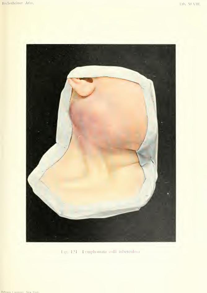
Bockenlieimer, Atlas. Tab. XCAIII.
Tig. 124. Lyniplioniata culli tuberculosa.
Rcbman C'oiiin.Tiu' NVw-Vnrif.


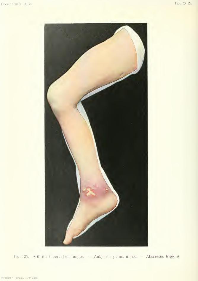
Bockenheimer, Atlas. Tab. XCIX.
Fig. 125. Artliritis tuberculosa fungosa — Ankylosis genus fibrosa — Abscessus frigidus.
Rcbman Company, New-Vork.
ARTHRITIS TUBERCULOSA FUNGOSA(FiDKioid fiibcretdous arthritis)
ANKYLOSIS GENUS FIBROSA (Fibmu.'s anchylosis)
ABSCESSUS FRIGIDUS {Cold Abscess)
Plate XCIX, Fig. 1-23.
This is a case of multiple tuberculosis of the joints,
bones and soft parts, together with pulmonary tuber-
culosis, occurrinfj in a vounc; individual. The rijjht
leg was useless owing to extensive disease of the hip
joint. The thigh was flexed, and X-ray examination
showed destruction of the upper margin of the
acetabulum and displacement of the head of the
femur onto the ilium. In the middle of the flexor
surface of the thigh is a healed fistula due to a bur-
rowing abscess. In the middle of the extensor sur-
face of the thigh is a clearly visible swelling due to a
burrowing abscess, which is common in this situation
in tuberculous arthritis of the hip joint, and in tuber-
culous disease of the vertebrae; in the latter case the
abscess burrows along the psoas muscle. Fluctua-
tion was present, but the skin was intact (cold
abscess). The abscess was evacuated by puncture
and injected with iodoform-glycerin. Resection of
the hip joint was postponed till the general condition
of the patient was improved.
The knee joint was also the seat of old tuberculous
arthritis of the fibrous type, which had led to anchy-
losis at right angles. This was corrected under an
anaesthetic by forced movement and an extension
splint.
On the inner side of the ankle joint are character-
istic tuberculous ulcers, with irregular undermined
borders and yellow caseous surface. Thin, greenish
pus was discharged by pressure. The X-rays showed335

a focus of tuberculous disease in the astragalus, which
had broken into the joint. Tuberculous arthritis of
the ankle joint more often breaks through on the
outer side. Hydrops is rare in this situation. Thejoint is usually filled with fungoid tuberculous tissue
which extends to the periarticular tissues. In Fig.
125 the foot w'as in the position of equinus owing to
absence of function and neglect of treatment. Owingto the extensive nature of the disease conservative
treatment was out of the question. The joint wasfreely laid open and all tuberculous matter removed(arthrectomy). The limb was immobilized by plas-
ter of Paris bandages and extension applied.
336

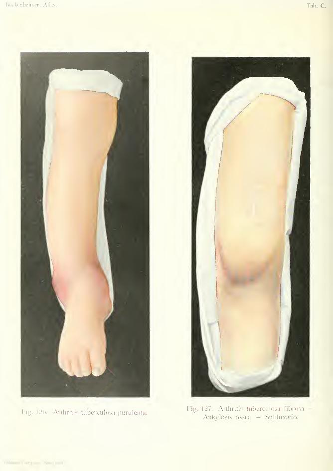
fiockenheimer, Atlas. Tab. C.
Fig. 120. Arthritis tuberculosa-puruleiita.Fig. 127. Arthritis tuberculosa fibrosa
Anlsvlosis ossea — Subluxatio.
Kebman Company, Ncw-\'ork.
ARTHRITIS TUBERCULOSA PURULENTA{Piiruhnt Tuberculous Arthritis)
Plate C, Fig. 12b.
This figure shows a case of purulent tuberculous
arthritis of the ankle joint. This form of arthritis is
common in children, more often affecting the knee-
joint. It begins with fever and rigors, and the rapid
formation of abscess, and may be mistaken for
arthritis due to staphylococci or other pus cocci.
Two incisions were made on the outer and inner
sides of the joint, and characteristic thin pus mixedwith fibrin was evacuated. The joint was then put
up in plaster of Paris. Purulent tuberculous arthritis
in children often recovers after early incision; but
there is generally some stifl'ness in the joints, so that
these must be put up in the most suitable position
for future use.
337
ARTHRITIS TUBERCULOSA FIBROSA{Fibrous Tuberculous Arthritis)
ANKYLOSIS OSSEA (Bony Anchylosis)
SUBLUXATIO (Sub-luxation)
Plate C, Fig. 127.
This is a case of old-standing fibrous tuberculous
arthritis of the knee joint with bony anchylosis, as
shown by the X-rays. Owing to neglect of prolonged
fixation of the joint in the straight position, flexion
contracture wuth backward displacement of the tibia
has taken place. This was corrected by cuneiform
osteotomy, plaster of Paris bandages, and later on a
celluloid casing.
338

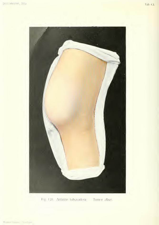
Bockenlic'iiiier, Atlas. lab. CI.
Fig. 128. Arthritis tuberculosa Tumor albus.
Kebm.in Coinp.aiiy, Nc\\-\'ork.
ARTHRITIS TUBERCULOSA FIBROSA(Fibrom Titherculous Arthritis)
TUMOR ALBUS (White Swelling)
Plate CI, Fig. 128.
This form of tuberculous arthritis is common in
the knee joint in adults. It consists in the formation
of hard, fibrous tissue in the joint and periarticular
tissue, and gives rise to a tumor-like swelling of the
knee and adjacent parts. The skin is white from
pressure of the subjacent mass; hence the namewhite swelling, or tumor alhus. In Fig. 128 the
disease was of several months' duration, and was as-
sociated with tuberculosis of the lungs. The patient
attributed the affection of the knee to an injury.
The X-rays showed tuberculosis of the bones, as well
as of the synovial membrane—a common combination
in tubercle of the knee joint. Similar swelling occurs
in tuberculous hydrops, the simplest form of tubercu-
lous joint disease. Effusion into the joint often pre-
cedes the arthritis and is recognized by halottement
of the patella, which is raised from the femoral
condyles by the fluid in the joint. The fluid is
generally sero-fibrinous, with numerous free "rice
bodies." More common than the fibrous form is
fungoid arthritis, which may go on to suppuration
and cause much destruction in and around the joint.
In all forms of tuberculous arthritis of the knee, the
joint is in a position of flexion and valgus. Themuscles of the leg become atrophied, and there is
retarded growth of the leg.
In Fig. 128, the joint was resected and all tuber-
culous tissue removed. The articular ends of both
339

bones were extensively diseased and the cartilages
destroyed. In resection of the articular surfaces it
is necessary to saw the bones so that the limb can bebrought into a straight position.
340

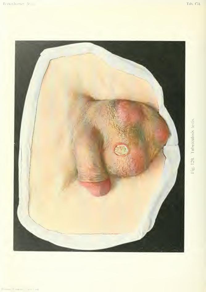
Bockenlieimer, Atlas. Tab. CII.
Rrbiii.111 roiiip.iiiy, Nmv \'oik.
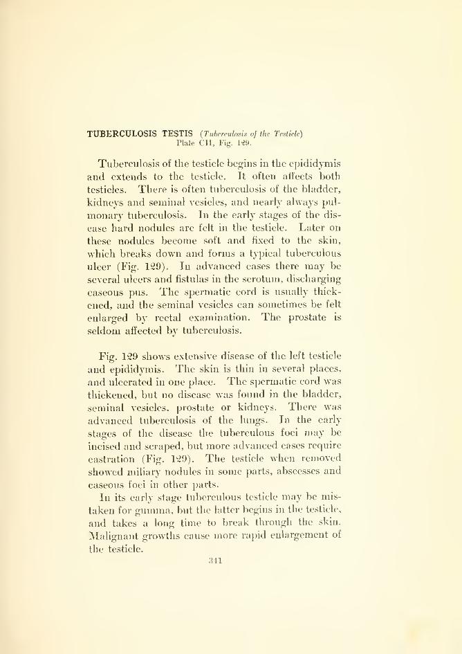
TUBERCULOSIS TESTIS (Tubercuhxh of the Testicle)
Plate CII, Fig. Ui).
Tuberculosis of the testicle begins in the epididymis
and extends to the testicle. It often affects both
testicles. There is often tuberculosis of the bladder,
kidneys and seminal vesicles, and nearly always pul-
monary tuberculosis. In the early stages of the dis-
ease hard nodules are felt in the testicle. Later on
these nodules become soft and fixed to the skin,
which breaks down and forms a typical tuberculous
ulcer (Fig. 129). In advanced cases there may be
several ulcers and fistulas in the scrotum, discharging
caseous pus. The spermatic cord is usually thick-
ened, and the seminal vesicles can sometimes be felt
enlarged by rectal examination. The prostate is
seldom affected by tuberculosis.
Fig. 129 shows extensive disease of the left testicle
and epididymis. The skin is thin in several places,
and ulcerated in one place. The spermatic cord was
thickened, but no disease was found in the bladder,
seminal vesicles, prostate or kidneys. There was
advanced tuberculosis of the lungs. In the early
stages of the disease the tuberculous foci may be
incised and scraped, but more advanced cases require
castration (Fig. 129). The testicle when removed
showed miliary nodules in some parts, abscesses and
caseous foci in other parts.
In its early stage tuberculous testicle may be mis-
taken for gumma, but the latter begins in the testicle,
and takes a long time to break through the skin.
IVIalignant growths cause more rapid enlargement of
the testicle.
341
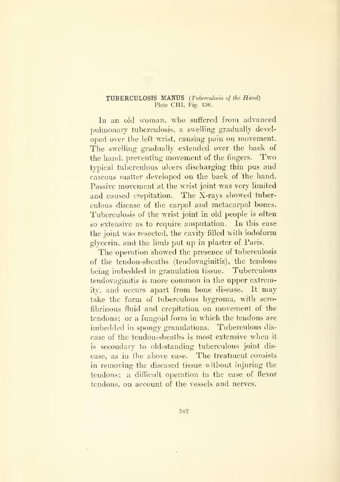
TUBERCULOSIS MANUS (Tubereidofds of the Hand)Plate cm. Fig. 130.
In an old woman, who suffered from advanced
pulmonary tuberculosis, a swelling gradually devel-
oped over the left wrist, causing pain on movement.
The swelling gradually extended over the back of
the hand, preventing movement of the fingers. Twotypical tuberculous ulcers discharging thin pus and
caseous matter developed on the back of the hand.
Passive movement at the wrist joint was very limited
and caused crepitation. The X-rays showed tuber-
culous disease of the carpal and metacarpal bones.
Tuberculosis of the wrist joint in old people is often
so extensive as to require amputation. In this case
the joint was resected, the cavity filled with iodoform
glycerin, and the limb put up in plaster of Paris.
The operation showed the presence of tuberculosis
of the tendon-sheaths (tendovaginitis), the tendons
being imbedded in granulation tissue. Tuberculous
tendovaginitis is more common in the upper extrem-
ity, and occurs apart from bone disease. It maytake the form of tuberculous hygroma, with sero-
fibrinous fluid and crepitation on movement of the
tendons ; or a fungoid form in which the tendons are
imbedded in spongy granulations. Tuberculous dis-
ease of the tendon-sheaths is most extensive when it
is secondary to old-standing tuberculous joint dis-
ease, as in the above case. The treatment consists
in removing the diseased tissue without injuring the
tendons; a difficult operation in the case of flexor
tendons, on account of the vessels and nerves.
342
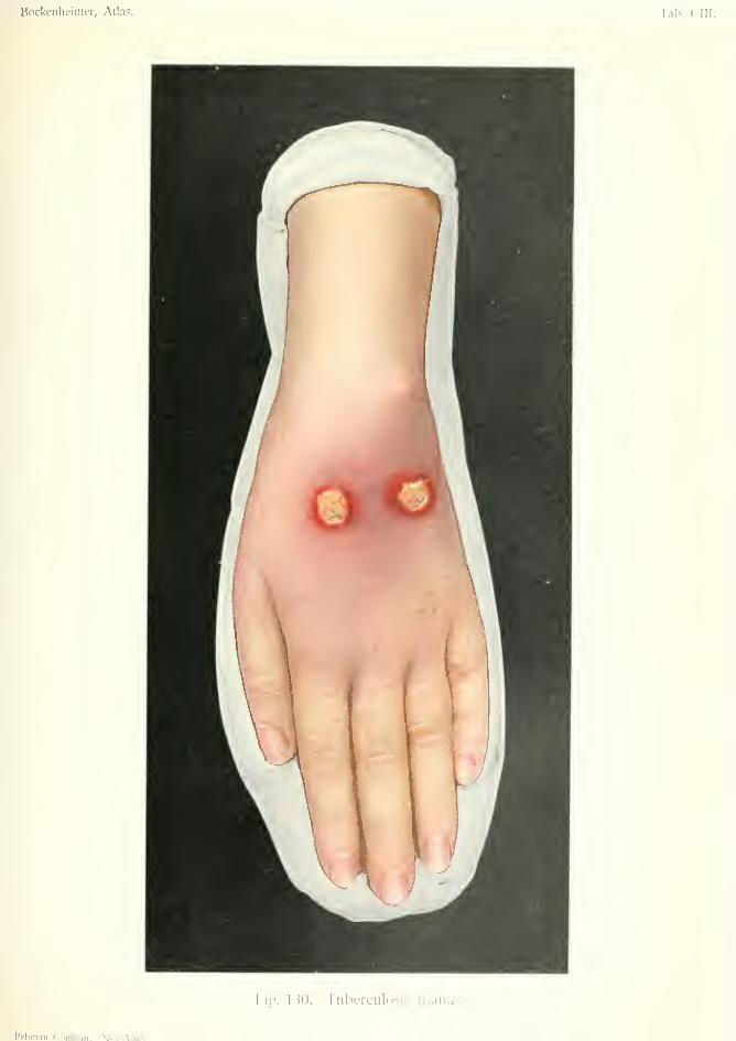
Bockenheiiiier, Atlas. lab. cm
I'ig. ]'W. Tuborculnsis in.inus.
Rebnnn fnninnnv. Mpt- \'nrl.-


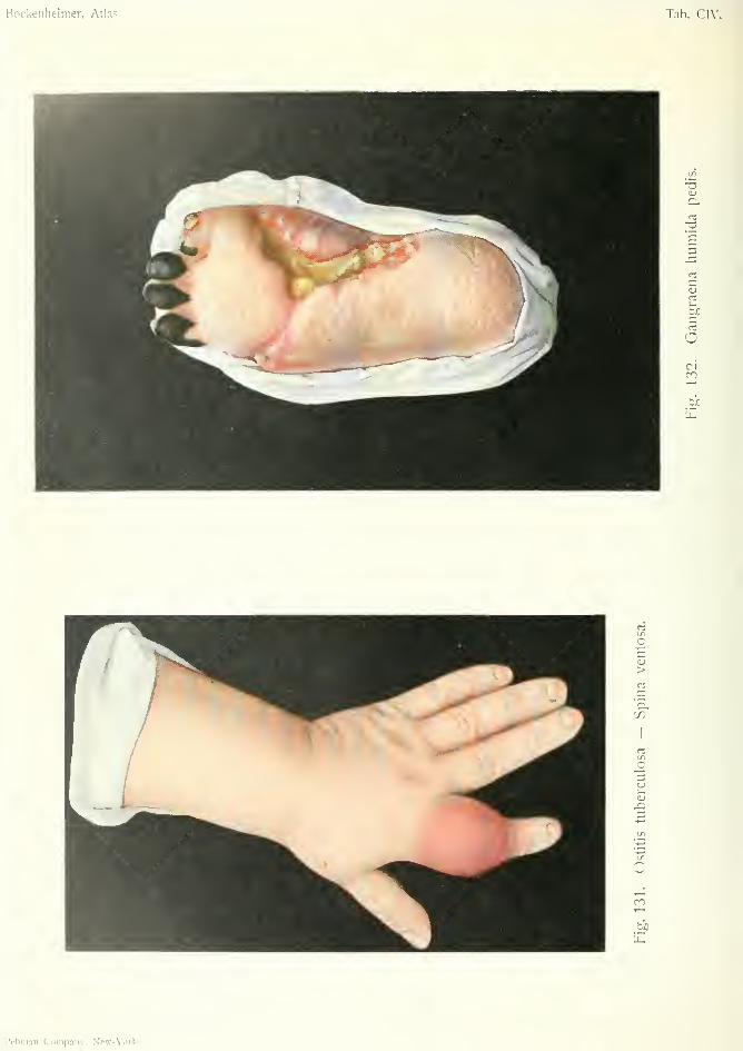
Bockeiiheimer, Atlas. Tab. Cl\'.
o
CICO
CO
I
OJ
o
CO
l?ebninn Company, New-Vork
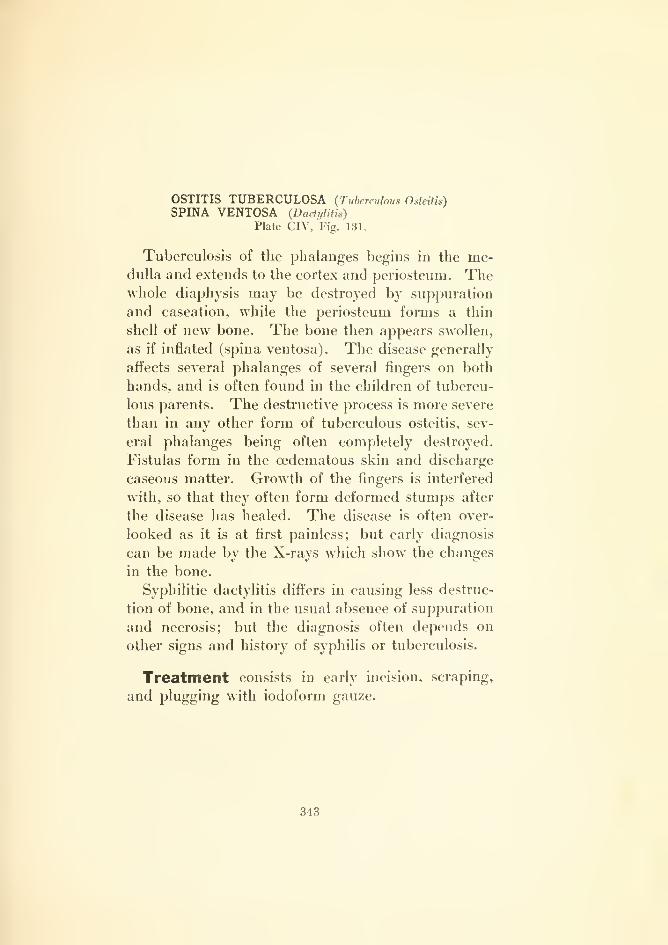
OSTITIS TUBERCULOSA (Tuberculous Osteitis)
SPINA VENTOSA {Dadyliih)
Plate CIV, Fig. 131.
Tuberculosis of the phalanges begins in the me-dulla and extends to the cortex and periosteum. Thewhole diaphysis may be destroyed by suppuration
and caseation, while the periosteum forms a thin
shell of new bone. The bone then appears swollen,
as if inflated (spina ventosa). The disease generally
aflFects several phalanges of several fingers on both
hands, and is often found in the children of tubercu-
lous parents. The destructive process is more severe
than in any other form of tuberculous osteitis, sev-
eral phalanges being often completely destroyed.
Fistulas form in the oedematous skin and discharge
caseous matter. Growth of the fingers is interfered
with, so that they often form deformed stumps after
the disease has healed. The disease is often over-
looked as it is at first painless; but early diagnosis
can be made by the X-rays which show the changes
in the bone.
Syphilitic dactylitis difi^ers in causing less destruc-
tion of bone, and in the usual absence of suppuration
and necrosis; but the diagnosis often depends on
other signs and history of syphilis or tuberculosis.
Treatment consists in early incision, scraping,
and plugging with iodoform gauze.
343
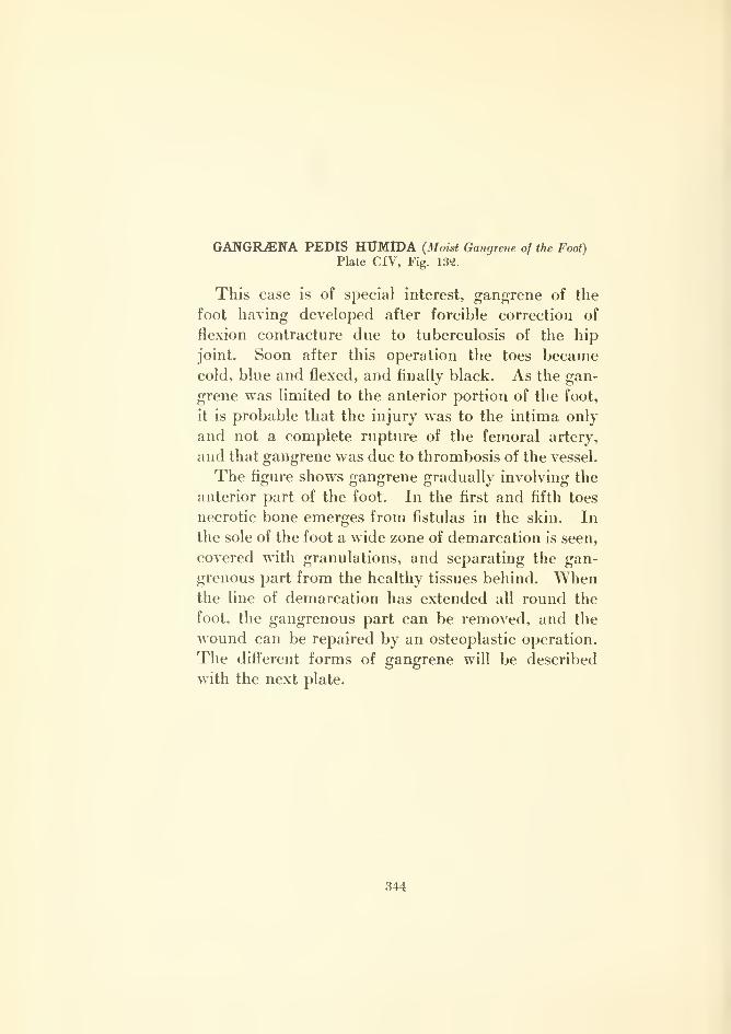
GANGILENA PEDIS HUMroA (Moist Gangrene of the Foot)
Plate CIV, Fig. 132.
This case is of special interest, gangrene of the
foot having developed after forcible correction of
flexion contracture due to tuberculosis of the hip
joint. Soon after this operation the toes becamecold, blue and flexed, and finally black. As the gan-
grene was limited to the anterior portion of the foot,
it is probable that the injury was to the intima only
and not a complete rupture of the femoral artery,
and that gangrene was due to thrombosis of the vessel.
The figure shows gangrene gradually involving the
anterior part of the foot. In the first and fifth toes
necrotic bone emerges from fistulas in the skin. In
the sole of the foot a wide zone of demarcation is seen,
covered with granulations, and separating the gan-
grenous part from the healthy tissues behind. Whenthe line of demarcation has extended all round the
foot, the gangrenous part can be removed, and the
wound can be repaired by an osteoplastic operation.
The different forms of gangrene will be described
with the next plate.
344

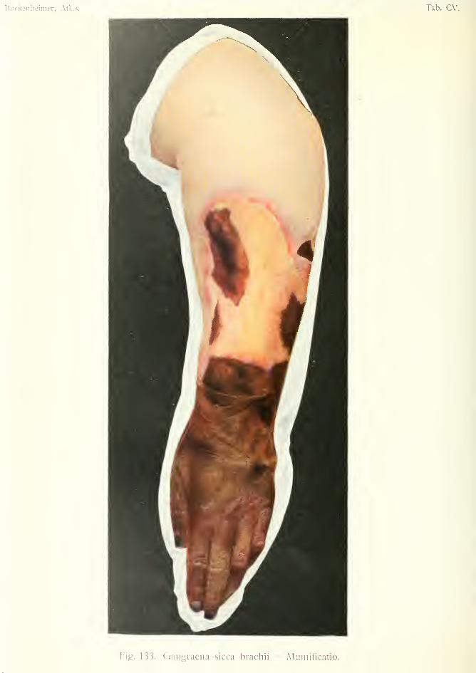
Bockenheimer, Atlas. Tab. CV.
Fig. 133. Oangraena sicca brachii -- AUuiiificatio.
GAWGIL«NA SICCA BRACHII—MUMMIFICATIO{Dri/ Gangrene of the Arm)—(Mummification)
Plate CV, Fig. 133.
The term gangrene is applied to extensive, pro-
gressive death of the superficial tissues of the body;
the term necrosis to death of the deeper structures
(fascia, muscle and bone). The bones have a greater
power of resistance than the skin, which may become
gangrenous after slight disturbance in the circulation.
Under certain conditions, e.g. after cutting off the
blood supply, the whole peripheral part of a limb
may become destroyed; but, as the death of the
tissues is first noticed in the skin, it is spoken of as
gangrene. ^Vhen the process consists in desiccation
of the tissues it is called dry gangrene; when it ends
in liquefaction from the invasion of putrefactive bac-
teria, it is called moist or infective gangrene. Dry
gangrene may change to moist gangrene, and both
processes may occur simultaneously in different parts
of the same limb, when one part becomes infected
and the other does not.
The extent of the gangrene varies according to
the cause; it may be circumscribed (after local appli-
cations, such as carbolic acid), or progressive (after
embolism). In both forms the dead tissue becomes
separated from the living by a zone of demarcation.
The zone of demarcation forms a groove filled with
granulation tissue (Fig. 13^2). It may be circular
(Fig. 135) or irregular (Figs. 133 and 134).
In the early stage of dry gangrene the condition
resembles that of ischajmic muscular contracture
(Fig. 63), especially when the condition is due to
plugging of the blood-vessels. The skin becomes
345
dry, shrunken and parchment-like. In the extremi-
ties the peripheral parts are flexed and immovable.
The skin becomes gradually yellowish brown and
finally black (Fig. 133). All the subjacent structures
may undergo dry atrophy. The dead tissue is grad-
ually separated by the zone of demarcation, and the
whole of an extremity may undergo spontaneous
separation.
While in dry gangrene there is diminution in vol-
ume and charring of the affected part; in moist
gangrene there is increase in volume, due to preceding
oedema. In moist gangrene there is more or less
liquefaction, decomposition or putrefaction, due to
putrefactive bacteria. The skin is cool and moist,
and the epidermis becomes raised in bullae containing
blood-stained fluid. After rupture of the bullae the
skin is reddish brown (Fig. 109). Finally the tissues
become disintegrated and smell horribly; lymphan-
gitis, lymphadenitis and general infection then follow.
In both forms of gangrene the skin is at first pale
and cold, and then shows bluish patches in various
places, often without any direct connection. Con-
tractures and loss of movement indicate the occur-
rence of total gangrene, whether moist or dry. How-ever, the difference in volume between the two forms
is apparent from the beginning.
A deep groove of demarcation also forms in moist
gangrene, separating the dead from the livmg tissue,
and spontaneous separation may occur if the patient
does not succumb to general infection. In less
extensive cases of moist gangrene we can wait for
the line of demarcation to form; but the gangrenous
part must be removed if there are rigors and high
temperature.
The etiology of gangrene is complex, but it is
always due to disturbance of the circulation. Theblood-vessels may be affected directly or indirectly.
Senile gangrene in old people is due to arterioscle-
rosis. The loosened intima of the small terminal
34G
vessels (also in the larger vessels) gives rise to throm-bosis, causing death of the peripheral tissues sup-
plied by these vessels, especially when the vessels
which carry on collateral circulation are themselves
diseased. In this way the toes or the whole leg maybecome gangrenous. In these cases the typical
changes of gangrene are preceded by pain. Gan-grene of the lower extremities in diabetic subjects is
generally caused by disease of the vessels.
In younger people gangrene of the peripheral
parts of the extremities may be caused by disease of
the intima of the smaller vessels (endarteritis oblit-
erans). This is usually of syphilitic origin. In
these cases there are severe intermittent pains, caus-
ing the patient to limp (intermittent claudication).
Both feet are usuallv affected, and become bluish
red. After some years gangrene gradually super-
venes, often taking months to develop (angioscle-
rotic gangrene). The patients suffer severe pain,
especially on contact or exposure to cold.
Embolism of the main arteries {e.g. from heart
disease) causes sudden and extensive gangrene of
the upper or lower extremities. (Embolic gangrene).
Sudden gangrene may also be caused by rupture or
ligation of a main artery. Certain nervous diseases
may cause gangrene by vaso-motor constriction of
the vessels (angio-neurotic gangrene). The latter
affection occurs symmetrically in both feet and is
known in its early stages as Raynaud's disease. It
is generally preceded by parsesthesias and diminution
in the sense of temperature.
Gangrene may also occur after extensive burns
and frostbite; after local application of carbolic acid,
lysol and alcohol; after injection of adrenalin into
the tissues, and after the internal administration of
ergotin (hands, feet and ears). In all these cases
gangrene is caused by thrombosis of the vessels. In
the same way erysipelas and phlegmonous inflamma-
tion may cause gangrene of the skin and deeper
347
tissues. Gangrene of the skin may also be caused
by the X-rays and by radium.
Differential Diagnosis. The appearance of gan-
grene, when fully developed, is so characteristic that
it can hardly be mistaken for any other condition.
The two forms of gangrene are also sharply defined
from each other. Dry gangrene might be mistaken
for burns of the third or fourth degrees, if signs of
the first and second degree of burn were not always
present in the neighborhood. Moist gangrene might
be mistaken for putrefactive phlegmon, especially
with progressive gaseous phlegmon (Fig. 109), if
the signs of general infection were not present at an
early stage. The history and a thorough examina-
tion will not only establish the diagnosis, but in most
cases will decide the cause of the gangrene.
The prognosis naturally depends on the cause and
on the extent of the gangrene. Angiosclerotic gan-
grene extends very slowly; it may remain stationary;
or parts w'hich appeared to be affected may recover.
Plugging of a large vessel causes extensive gangrene
of the part supplied by the vessel. Diabetic gangrene
and senile gangrene are characterized by their pro-
gressive course. Gangrene is more extensive whenthere is much cedema.
Treatment. Extensive gangrenous parts should
be removed after a zone of demarcation has formed.
Before this takes place the part should be dressed
with aseptic dressings or ointments. In moist gan-
grene of an extremity early removal may be indicated
in order to prevent general infection. In gangrene
due to syphilitic endarteritis, iodide of potassium
and mercury should be given; the limb should be
raised and enveloped in wool; hot-air treatment is
useful for the pains; alcohol should be avoided:
after demarcation has formed, amputation should be
performed in the most conservative way possible.
348
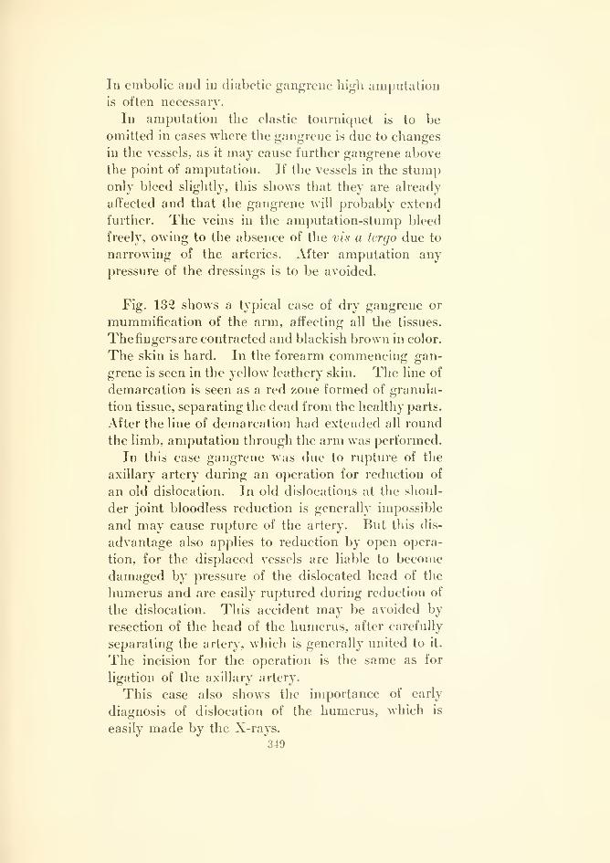
In embolic and in diabetic gangrene high amputation
is often necessary.
In amputation the elastic tourniquet is to be
omitted in cases where the gangrene is due to changes
in the vessels, as it may cause further gangrene above
the point of amputation. If the vessels in the stumponly bleed slightly, this shows that they are already
affected and that the gangrene will probably extend
further. The veins in the amputation-stump bleed
freely, owing to the absence of the vis a tergo due to
narrowing of the arteries. After amputation any
pressure of the dressings is to be avoided.
Fig. 132 shows a typical case of dry gangrene or
mummification of the arm, affecting all the tissues.
The fingers are contracted and blackish brown in color.
The skin is hard. In the forearm commencing gan-
grene is seen in the yellow leathery skin. The line of
demarcation is seen as a red zone formed of granula-
tion tissue, separating the dead from the healthy parts.
After the line of demarcation had extended all round
the limb, amputation through the arm was performed.
In this case gangrene was due to rupture of the
axillary artery during an operation for reduction of
an old dislocation. In old dislocations at the shoul-
der joint bloodless reduction is generally impossible
and may cause rupture of the artery. But this dis-
advantage also applies to reduction by open opera-
tion, for the displaced vessels are liable to become
damaged by pressure of the dislocated head of the
humerus and are easily ruptured during reduction of
the dislocation. This accident may be avoided by
resection of the head of the humerus, after carefully
separating the artery, which is generally united to it.
The incision for the operation is the same as for
ligation of the axillary artery.
This case also shows the importance of early
diagnosis of dislocation of the humerus, which is
easily made by the X-rays.
349
GANGRiENA CUTIS HUMIDA—NECROSIS FASCLE{Moist Gangrene of the Sinn)
ULCUS DECUBITALE (Decubital Ulcer—Bedsore)Plate CVI, Fig. 134.
The skin, being the most superficial part of the
body, is most liable to injuries which may cause
gangrene. It has also less power of resistance than
other tissues. Long-continued pressure, especially
in places situated over the bones, may cause gan-
grene of the skin. In this way gangrene may be
caused by the pressure of tight bandages or splints;
also by a displaced piece of bone in fractures; by
pressure on the outer side of the foot in pes varus;
by tight sutures, e.g. after amputation of the breast,
leaving a wide space to be closed.
Uncleanliness, loss of consciousness, nervous
diseases (trophoneuroses, syringomyelia, hemiplegia,
paraplegia, tabes), cachexia, diabetes, typhoid fever,
osteomyelitis, phlegmonous inflammation, general
infection and comatose conditions, all predispose to
gangrene, which, in emaciated persons, may become
very extensive. Gangrene of the skin caused by the
pressure of oedema and gaseous formation in the
tissues has already been mentioned (Figs. 91 and
109). After operations, gangrene of the skin (bed-
sores) may occur over the heels, buttocks, spinous
processes, shoulder blades and back of the head, if
care is not taken to change the position of the patient
and apply soft, smooth, protective coverings.
Gangrene of the mucous membranes may occur
from the pressure of foreign bodies; for instance, in
the esophagus, from the passage of bougies; in the
intestine, from the pressure of Murphy's buttons; in
350

Bockenheimer, Atlas.Tab. C\'l.
r\g. 134. Oangnieiia liumida cutis - Necrosis fasciae — Ulcus decubitale.
Rebman Company. New-York.

the larynx, after intubation; also after resection of
the intestine or esophagus when the united ends are
under great tension.
Gangrene of the skin begins with pain and redness;
then slight swelling and blue coloration; finally, rais-
ing of the epidermis in bullte. The epidermis then
separates leaving the corium exposed; this is at first
greenish yellow, afterwards blackish brown and
leathery. At the edge of the gangrenous part the
skin becomes inflamed, and by the formation of pus
and granulation tissue a gutter-shaped, often circular
space is gradually formed—the zone of demarcation.
The more severe the injury the deeper is the gangrene
so that subcutaneous tissue, fascia (Fig. 134), muscles
and bone may become necrosed and cast off.
After separation of the gangrenous part an ulcer is
left, called decubital ulcer, which is covered with
slimy, greenish-yellow connective-tissue shreds and
fetid pus. A neglected decubital ulcer may give rise
to extensive putrid inflammation or gaseous phleg-
mon, as the pus always contains putrefactive bacteria,
especially in decubital ulcer over the sacrum which is
infected from the faeces. Erysipelas may also occur
in decubital ulcer. In neglected cases the gangrene
may also extend deeply and cause extensive destruc-
tion.
Pressure-necrosis in the internal organs (larynx,
esophagus, intestine) is dangerous from perforating
ulceration or hemorrhage; also from stenosis after
healinor.'O
Treatment. Gangrene of the skin may, in manycases, be prevented, or, at any rate limited, by
prophylactic treatment. Decubital ulcers (bedsores)
may be prevented by applications of spirit of cam-
phor to the skin of the parts exposed to pressure, by
air cushions and frequently changing the patient's
position. If the skin is discolored an ointment dress-
ing should be applied, and this should be changed if
351
the patient complains of pain. As the pain also
subsides in a few days under continuous pressure of
a dressing, its removal is often neglected, and then
when it is removed there may be gangrene down to
the bone. In emaciated patients the bony promi-
nences should, therefore, be well padded, and the
skin disinfected before applying the dressing.
If gangrene has developed the skin must be pro-
tected against infection by a dressing. Separation of
the gangrenous part may be hastened by moist dress-
ings with two per cent, boric acid lotion, three per
cent, peroxide lotion, or camphor liniment, applied
several times daily. Forcible removal of the gan-
grenous parts while they are firmly attached is not
advisable; they should be removed by scissors whenalmost completely loose. The ulcer may be treated
with moist dressings or ointments, and with caustics
when granulations have sprung up. After extensive
gangrene of the skin the space may be closed
by undermining the skin and suturing; or, if this
is impossible, by a plastic operation by means of
pedunculated flaps.
Fig. 134 shows a case of moist gangrene of the
skin with necrosis of the abdominal fascia. Part of
the skin is separated from the healthy, somewhatreddened and inflamed skin around it, by a zone of
demarcation. The gangrenous part is still firmly
attached to the subjacent structures. In some places
the skin has separated, exposing the abdominal
fascia, the yellowish color of which shows that it
has already undergone necrosis. The borders of
the ulcer were undermined, and it discharged fetid
pus.
In this case the gangrene was caused by a sub-
cutaneous injection of salt solution, performed on a
patient in a state of collapse. Gangrene of the skin
may occur after injection of large quantities of salt
solution when the injection is made intracutaneously

instead of subcutaneously; also when the fluid is too
hot, or not steriHzed.
The ulcer became clean under dressings of peroxide
lotion; the gangrenous skin and necrotic fascia sep-
arated; the edges of the fascia and the skin were
sutured separately, and primary union took place.
As sutures in fascia often do not hold, the patient
was ordered an abdominal belt to prevent abdominal
hernia.
353
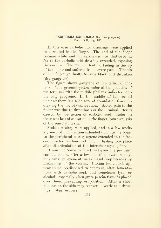
GANGRjENA CARBOLICA (Carbolic gangrene)
Plate C\1I, Fig. 135.
In tliis case carbolic acid dressings were applied
to a wound in the finger. The end of the finger
became white and the epidermis was destroyed as
far as the carbolic acid dressing extended, exposing
the corium. The patient had no feeling in the tip
of the finger and sufl^ered from severe pain. The tip
of the finger gradually became black and shrunken
(dry gangrene).
The figure shows gangrene of the terminal pha-
lanx. The greenish-yellow color at the junction of
the terminal with the middle phalanx indicates com-mencing gangrene. In the middle of the second
phalanx there is a wide zone of granulation tissue in-
dicating the line of demarcation. Severe pain in the
finger was due to thrombosis of the terminal arteries
caused by the action of carbolic acid. Later onthere was loss of sensation in the finger from paralysis
of the sensory nerves.
Moist dressings were applied, and in a few weeks
a groove of demarcation extended down to the bone.
In the peripheral part gangrene extended to the fas-
cia, muscles, tendons and bone. Healing took place
after disarticulation at the interphalangeal joint.
It must be borne in mind that even one per cent,
carbolic lotion, after a few hours' application only,
may cause gangrene of the skin and deep necrosis bythrombosis of the vessels. Certain individuals ap-
pear to be predisposed to gangrene after fomenta-
tions with carbolic acid, and sometimes lysol or
alcohol ; especially when gutta percha tissue is placed
over them, preventing evaporation. After a short
application the skin may recover. Acetic acid dress-
ings hasten recovery.
354
Bockenheiiner, Alias.lab. (A'
I
Fig. 135. Oangraena carbolica.


Bockenheimer, Atlas. Tab. CVm.
Fig. 136. Combustio entheniatnsa - bullosa ~ escliarotica.
Rcbman Company, Neve-York.
COMBUSTIOERYTHEMATOSA-BULLOSA-ESCHAROTICA{Hums)
Plate CVin, Fig. 136.
Burns may be caused by the action of radiant heat;
e.g. prolonged exposure to hot sun or a hot fire. Theheat may arise from solids, liquids or gases. Elec-
tricity (lightning stroke) and the X-rays may also
cause burns; also strong acids and alkalis (sulphuric
and nitric acids, caustic potash and soda).
Burns of the mucous membrane of the mouth,
tongue, pharynx, esophagus and intestine are caused
by certain chemicals swallowed as poisons. Thesemay cause death by oedema of the glottis, or later on
by perforating ulceration of the gut and peritonitis.
If the ulcers heal, they lead to stenosis of the gut.
Burns of the skin may be caused by strong caus-
tics; such as trichloracetic acid, for removal of warts;
Vienna paste, tartar emetic ointment, etc.
Tender skins {e.g. children) react to slight degrees
of heat; e.g. after the application of poultices,
fomentations.
The mildest degree of burn—also called the first
degree—consists in arterial hyperaemia, causing red-
ness and slight swelling: of the skin. There is more
or less pain or tenderness, itching and tension of the
skin. In this form there is early and complete resti-
tution to normal, sometimes after desquamation of
the epidermis.
The second degree of burn is characterized by the
formation of bulhe. Besides redness caused by the
first degree, the epidermis is raised in blisters by
exudation of lymph between the epidermis and the
corium. The blisters contain yellowish fluid or
gelatinous masses, and may develop twenty-four
355

hours after the injury. In severe burns of the second
degree some of the blisters rupture, exposing the red
corium, which is very painful to touch. This form
of burn is common after boiler explosions, gas explo-
sions, and scalding with steam or hot water. Heal-
ing by epidermization of the corium takes two or
three weeks, but the skin is restored to the normal
condition without scarring, provided the process has
not been complicated by suppuration.
In the third degree of burn the epidermis andcorium are destroyed, to a greater or less extent
according to the severity of the injury. The resulting
gangrene of the tissues is due to three factors ; loss of
water from the tissues; loss of blood supply from
acute thrombosis of the vessels; and coagulation of
albumen in the tissues. The skin becomes black
and gangrenous. Sensation is lost at this part; but
there is always pain due to burns of the first andsecond degrees in the surrounding parts.
In severe burns the fascia, muscles and bones mayundergo necrosis, as well as the skin. The separa-
tion of the necrosed parts takes place in the usual
way by an inflammatory zone of demarcation.
\ Burns of the third degree are liable to infection of
the exposed tissues by pyogenic and putrefactive
bacteria, so that the wounds take months to heal,
with hypertrophic scars which cause contractures
of the joints and form adhesions with neighlioring
parts. The scars of burns of the third degree are
easily lacerated and may give rise to carcinoma (Fig.
20). In burns of the third degree there is also the
danger of general infection from prolonged suppura-
tion. These cases may also be fatal from exhaustion,
hemorrhage from erosion of vessels, or amyloid dis-
ease of the kidney, liver, etc.
In burns of the fourth degree there is complete
charring of all the tissues, which fall to ashes whentouched.
It is obvious that the effect of a burn on the organ-
356

ism depends on the degree of the burn, the extent of
surface involved, the part of the body affected, and
the condition of the patient beforehand. Burns of
the first and second degrees when they are not very
extensive are not serious; but if a third of the bodyis affected, even in burns of the first degree, there is
fatal constitutional disturbance, especially in chil-
dren. Apart from the severe pain, rapid collapse
sets in. The skin becomes cold, pale and covered
with sweat; the pulse is small and i-apid; the patient
complains of thirst; consciousness is retained till
death occurs in two or three days. As the patients
are fully conscious and in good spirits, and do not
complain of any more pain, it is necessary to explain
to the relatives and friends that death after extensive
burns is almost inevitable.
In these severe cases the temperature is subnormal.
There is sometimes delirium and coma. In extensive
burns of the second degree, complicated by general
infection, there is high temperature, delirium, diar-
rhea and fetid discharge from the wounds. Duodenal
ulcer may also occur. Death may occur from
urjemic coma following anuria. The autopsy shows
ecchymoses and thromboses in all the organs, paren-
chymatous nephritis, etc.
In extensive burns death may be caused by shock,
which may be due to great pain, sudden cooling of
the skin, or overheating of the blood, as in heat-
stroke. Accumulation of poisonous substances in
the blood may also cause death.
Burns of the third degree, when affecting certain
regions, give rise to various disfigurements and con-
tractures. The eyelids and mouth may be disfigured
by contracting scars (ectropion). The head may be
flexed on the thorax; the fingers may become
united, etc.
Differential Diagnosis. Burns may be con-
founded with frostbite, in the absence of history.
357
Treatment. In burns of the first degree the sur-
rounding healthy skin should be disinfected, and
ointment applied to the burnt part. In burns of the
second degree small blisters can be left to dry up;
large blisters should be opened at the base, the lymph
evacuated and the epidermis replaced. The loosened
epidermis then generally becomes attached. If the
blisters are already broken, the loose epidermis
should be removed and the exposed corium pow-
dered with rice powder, talc or flour. Bismuth
dressings are useful; but oil and lime water applica-
tions should not be employed, as they favor infection.
Antiseptic gauze is to be avoided, on account of the
danger of poisoning.
Morphia may be required if there is much pain,
especially when the dressings are changed. Thelatter should be covered with plenty of wool.
The more infection of the surface is prevented bycareful treatment, the less is the scar tissue. If
there is much scar tissue this may be excised and
the wound covered by skin flaps. Injection of ten
per cent, thiosinamin solution may be tried to absorb
scar tissue.
In the extremities, resection of joints, amputation
or disarticulation may be necessary when the limbs
are useless, or the seat of exhausting suppuration, or
when there is threatening general infection. Such
operations should not be performed till the patient
has somewhat recovered from shock. Burns of the
neck and mouth may require tracheotomy. In exten-
sive burns of the second and third degrees with muchdischarge permanent baths are useful.
In all severe burns the general condition of the
patient requires attention. To support the heart,
digitalis, camphor injections, subcutaneous injections
of salt solution may be indicated. The whole bodymust be well protected by wool. The function of the
kidneys should be stimulated by diuretics (caffein,
acetate of potash, etc.).
358
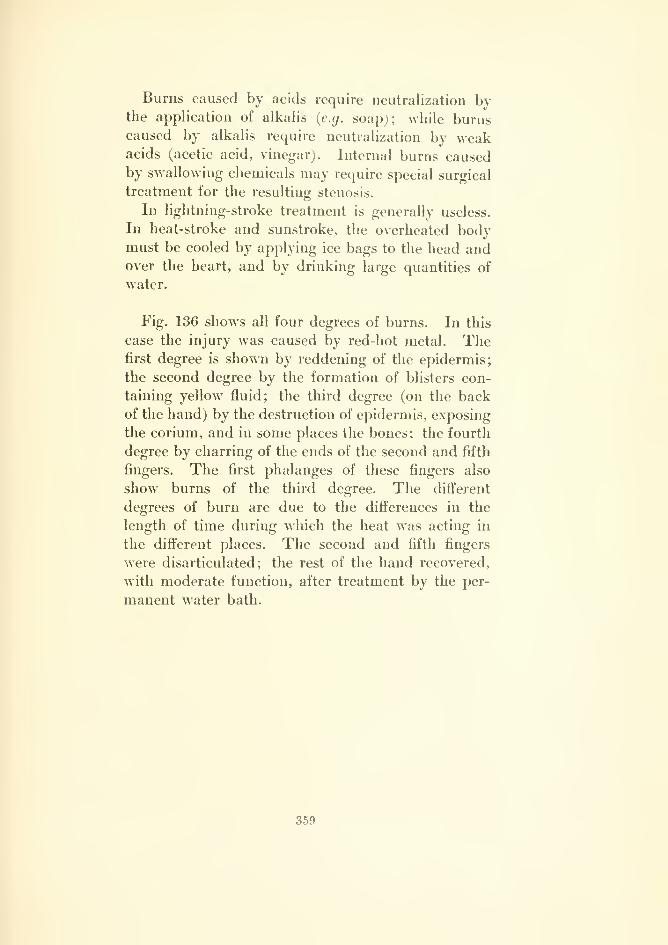
Burns caused by acids require neutralization bythe application of alkalis [^e.g. soap); while burnscaused by alkalis require neutralization by weakacids (acetic acid, vinegar). Internal burns causedby swallowing chemicals may require special surgical
treatment for the resulting stenosis.
In lightning-stroke treatment is generally useless.
In heat-stroke and sunstroke, the overheated bodymust be cooled by applying ice bags to the head andover the heart, and by drinking large quantities of
water.
Fig. 136 shows all four degrees of burns. In this
case the injury was caused by red-hot metal. Thefirst degree is shown by reddening of the epidermis;
the second degree by the formation of blisters con-
taining yellow fluid; the third degree (on the back
of the hand) by the destruction of epidermis, exposing
the corium, and in some places the bones; the fourth
degree by charring of the ends of the second and fifth
fingers. The first phalanges of these fingers also
show burns of the third degree. The different
degrees of burn are due to the differences in the
length of time during which the heat was acting in
the different places. The second and fifth fingers
were disarticulated; the rest of the hand recovered,
with moderate function, after treatment by the per-
manent water bath.
359
COWGELATIO ERYTHEMATOSA—BULLOSA (Frostbite)
Plate CIX, Fig. 137
Extreme degrees of cold may cause destruction of
the tissues, in the same way as burns. Here again,
the extent of injury depends on the degree of cold,
the duration of its action, and the condition of the
patient. Dry cold is better borne than moist cold.
Certain individuals are especially liable to the effects
of cold—persons in a state of alcoholic intoxication,
angemic individuals, children and old people, cooks
and others who are exposed to rapid changes of tem-
perature. Frostbite may be caused by the action of
snow, ice or liquid air.
Pernio or chilblain may be regarded as a chronic
form of frostbite, affecting the fingers, toes andears. It is especially common in chlorotic indi-
viduals and causes swelling and blueness of the skin
with numerous bluish-red nodules. These often
cause unbearable itching and burning sensations,
and, when scratched, give rise to intractable ulcers.
Acute frostbite appears in different degrees accord-
ing to the degree of cold, in the same way as burns.
The parts of the body usually affected are the fingers,
ears, nose and toes. In the first degree of frostbite
there is redness of the skin from hypersemia (erji;he-
matous congelation). This is usually followed in a
short time by the formation of a blister. The red-
ness increases when the patient comes into a warmroom, or takes alcoholic drinks. It is accompaniedby burning and itching pains, which may continue
for a long time. The redness may even last for life
after a single frostbite of the first degree; for instance
in the nose of a chlorotic woman. In most cases of
360
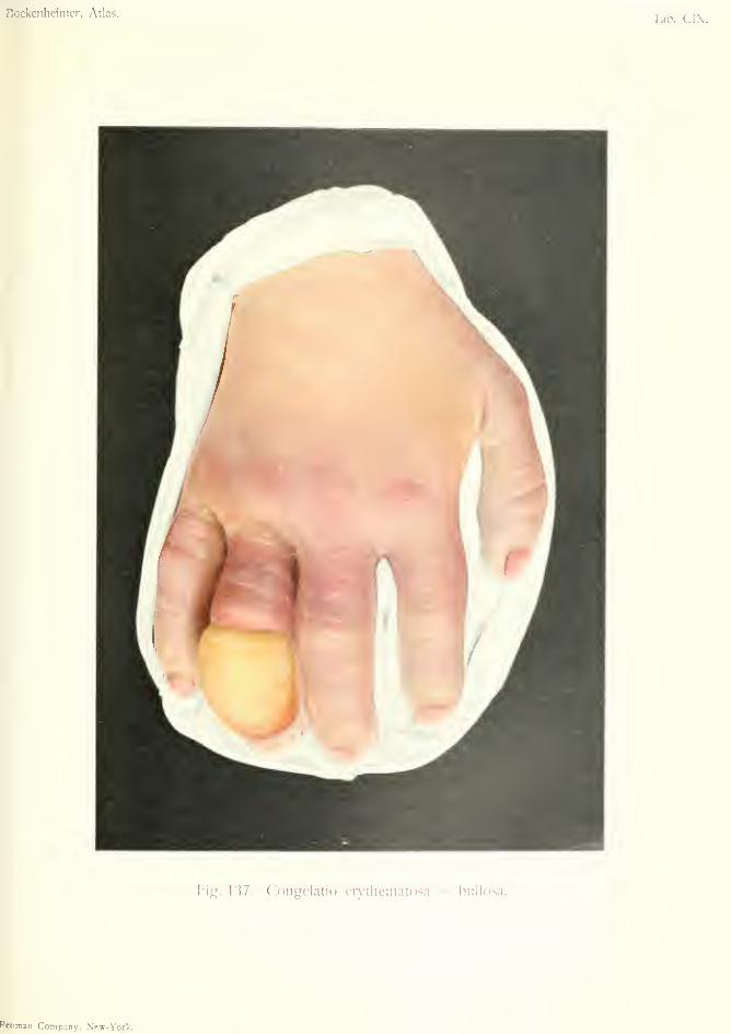
Bockenlieinier, Atlas.Tab. CI.X.
Fig. 137. Congelatio eiytliemato,sa — bullosa.
Rfbman Company, N'ew-York.

frostbite of the first degree, however, there is com-plete recovery.
Longer exposure to cold, or exposure to moresevere cold, causes venous congestion, cedema, andthe formation of blisters. The skin becomes blue or
white, cold and insensitive, and is often covered with
numerous blisters, with bluish-black contents; after
their rupture the exposed corium is dark in color and
very painful. Infection is liable to occur, causing
extensive ulceration with little tendency to heal, andleading to cicatricial contraction. Pain is more severe
and continuous in frostbite of the second degree.
In frostbite of the third degree, in the same way as
in burns of the third degree, there is gangrene of the
skin and necrosis of the deeper tissues, due to throm-
bosis of the vessels. The skin is at first bluish black,
cold and insensitive, later on quite black. Separa-
tion of the frozen tissues may take place either by dry
or moist gangrene. The zone of demarcation has
often a putrid character. Progressive phlegmonous
inflammation may spread from the borders of the
frozen area, and may lead to general infection.
Along with frostbite of the third degree the neighbor-
ing parts are affected in the first and second degrees,
and other parts are ulcerated; so that the clinical
picture is variegated. The gangrenous and necrotic
parts, after some months, are cast off spontaneously.
The nails soon fall off in frostbite of the hand. In
frostbite of the third degree, parts which at first
showed signs of the second degree only, may after-
wards become gangrenous.
Healing eventually takes place by the formation of
very unsightly hypertrophic scars, which may cause
contractures. Contractures may also be caused by
paralysis of nerves, or by waxy degeneration of muscle
fibres. Frostbite is said to cause changes in the
blood-vessels which may lead to secondary gangrene.
The general condition of the patient is little impaired
in acute local frostbite of circumscribed regions.
361
The period of healing varies according to the degree
of the frostbite, but is usually longer than in burns
and causes more severe after effects.
General frostbite is common in severe winters
among weary wayfarers who weaken their power of
resistance to cold by alcoholic drinks. After the pre-
liminary feeling of cold they become overcome by
fatigue, fall down and become frostbitten. People
may even fall unconscious without any previous
symptoms. The body, lying on the ground, becomes
cooled to below 20° C. (68° F.). Exposed parts mayeven become frostbitten by slight degrees of cold,
acting continuously on the recumbent body. Thenose, ears and hands then become frozen to ice and
fall off when touched, while the blood becomes
decomposed and contains ice crystals. This condi-
tion may last for days before death takes place. Only
early attempts at resuscitation can do any good in
these cases. The heart is, however, so weakened
that, even if the patients recover consciousness, they
succumb some days later with delirium, coma and
heart failure. The prolonged action of intense cold
may freeze not only the external parts of the body
but may convert all the fluid parts to ice. Theexpansion caused by the conversion of liquids to ice
then ruptures the surface of the body.
Patients who have been exposed to general frost-
bite must not be suddenly warmed, as this may cause
death from shock. The stronger the patient's con-
stitution the better is the chance of recovery ; but the
prognosis of general frostbite is very unfavorable.
Extensive paralysis (hemiplegia and paraplegia) mayremain after recovery from the immediate effects,
and the patients may suffer for years from headache,
pains in the joints, and a tendency to local frostbite
due to changes in the arteries. [These secondary
phenomena may be due to frostbite acting as an
exciting cause on pre-existing latent disease, espe-
cially disease of the arteries.
362
Differential Diagnosis. Frostbite may be mis-
taken for burns in the absence of any history.
Treatment. Chilblains may be treated by hot
air apparatus or hot sandbaths, together with general
treatment of chlorosis by iron and arsenic. Theirritation may be relieved by painting with tincture
of iodine, balsam of Peru, or by inunction with
bromocoU ointment. Ulcers are best treated with
Hehra's diachylon ointment. Recurrence can be
limited by prophylactic measures.
In acute local frostbite the parts must be warmedgradually—by rubbing with snow or cold applica-
tions. Early treatment in this way may restore the
frozen skin. In frostbite of the second degree, large
blisters should be opened and broken blisters re-
moved. Ulcers should be treated with strict asepsis,
and dressed with sterile gauze or ointment. Theextremities should be suspended on splints, avoiding
all pressure.
In cases with moist gangrene and putrefactive
phlegmonous inflammation, early amputation is often
necessary to prevent general infection. In dry gan-
grene, amputation may be deferred till a zone of
demarcation has formed. Plastic operations are
often required after spontaneous separation of gan-
grenous parts of the fingers or toes. IMorphia injec-
tions may be necessary for the severe pain in the
early stages of frostbite. Paralysis may be improved
by electricity, and contractures by massage; but the
latter more often require a secondary operation.
In general frostbite the body must be very grad-
ually warmed. The patient is placed in a cool room
and rubbed down with cold water. He is then put
in a tepid bath the temperature of which is gradually
raised in the course of several hours. If respiration
has stopped, artificial respiration must be performed.
Injections of camphor and subcutaneous infusion of
salt solution is useful to stimulate the action of the
363
heart. When the patient recovers consciousness hot
alcoholic drinks should be given. Local gangrene
resulting from general frostbite is identical with that
occurring in severe local frostbite, and requires the
same treatment.
Fig. 137 shows a case of frostbite of the first and
second degrees in a workman who had had repeated
milder attacks in the winter, after exposure of his
hands to cold water during his work. The hands
were permanently blue, and in the winter painful
chilblains developed on the fingers, especially on the
extensor surface. He finally developed frostbite of
the second degree, which is shown by the whiteness
of the ends of the fingers, and other changes in the
fourth finger. The skin over the first joint of the
fourth finger is blue, and a large blister containing
yellow lymph has developed on the extensor surface
of the last joint. The patient complained of severe
burning pains in the tips of the fingers, especially in
the fourth. The blister was opened and the epider-
mis replaced on the corium, the hand was dressed with
ointment and put on a splint. Under this treatment
the skin ouickly recovei'ed.
364

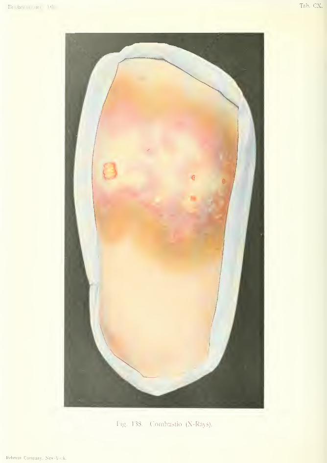
Bockenlieinier, Atlas. Tab. ex.
Fig. 138. Combuslio (X-Rays).
Rebman Company, New-York.

X-Ray BumPlate CX, Fig. 138.
The X-rays have been used for the treatment of
various diseases; sometimes with good results, as in
lupus, chronic eczema, etc.; sometimes with noresult, as in malignant tumors. A single exposure,
properly performed, causes no injury to the skin;
but repeated exposures sometimes give rise to changes
in the skin, especially when the tubes are placed
nearer than thirty centimeters from the skin, andwhen the exposures are too long or too frequent.
The changes produced take the form of a dermatitis,
and certain individuals appear to be predisposed
to it.
The first signs are redness, swelling and tension
of the skin, accompanied by itching and burning
sensations. This condition is followed by fissures
in the skin and finally ulceration, which is character-
ized by its chronic and progressive nature. Workers
in X-ray laboratories are subject to a chronic form
of dermatitis of the hands, unless they protect them-
selves with gloves, lead-foil, etc. The skin becomes
dr}% cracked and fissured; the nails become brittle
and are often shed. Some cases become gangrenous,
and the necrotic tissue is separated by a zone of
demarcation. Other cases develop into carcinoma
and require amputation of the hand. Some cases
are fatal from exhaustion.
X-ray dermatitis can be prevented by placing the
tube not less than thirty centimeters from the skin,
and by avoiding too long or too frequent exposures.
365
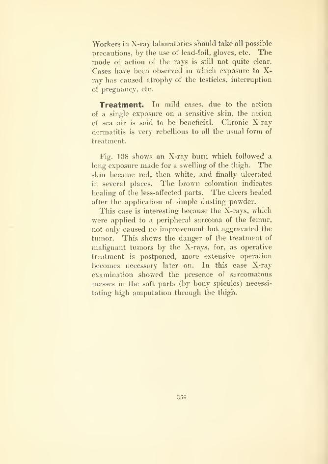
Workers in X-ray laboratories should take all possible
precautions, by the use of lead-foil, gloves, etc. Themode of action of the rays is still not quite clear.
Cases have been observed in which exposure to X-
ray has caused atrophy of the testicles, interruption
of pregnancy, etc.
Treatment. In mild cases, due to the action
of a single exposure on a sensitive skin, the action
of sea air is said to be beneficial. Chronic X-ray
dermatitis is very rebellious to all the usual form of
treatment.
Fig. 138 shows an X-ray burn which followed a
long exposure made for a swelling of the thigh. Theskin became red, then white, and finally ulcerated
in several places. The brown coloration indicates
healing of the less-affected parts. The ulcers healed
after the application of simple dusting powder.
This case is interesting because the X-rays, which
were applied to a peripheral sarcoma of the femur,
not only caused no improvement but aggravated the
tumor. This shows the danger of the treatment of
malignant tumors by the X-rays, for, as operative
treatment is postponed, more extensive operation
becomes necessary later on. In this case X-ray
examination showed the presence of sarcomatous
masses in the soft parts (by bony spicules) necessi-
tating high amputation through the thigh.
366


F^)Ockeiilicimer, Atla?.Tab. CXI.
Fig. 139. Alal perlorant du jiied - Qangraena Ra\iiaud.
Rebman f'mnpany, Ncvr-Yoik.
MALUM PERFORANS PEDIS (Perforating Ulcer of the Foot)
GANGR^NA RAYNAUD (Raynaud:i gangrene)
Plate CXI, Fig. 139.
Perforating ulcer of the foot commences as a hard
horny thickening of the epidermis over the heads of
the third and fifth metatarsal bones, somewhat re-
sembHng a clavus but much more extensive. Theepidermis becomes fissured and finally ulcerated in
the center. The ulcer is characterized by its ten-
dency to extend deeply, and by its persistence in spite
of all kinds of treatment. The disease is essentially
chronic and leads to destruction of muscles, tendon-
sheaths, bones and joints, by continuous crateriform
extension of the ulcer into the deeper tissues. Theepidermis always remains thickened at the border of
the ulcer, and is sometimes undermined. The visible
surface of the ulcer is small and is covered with
flabby granulation tissue. Necrotic shreds often pro-
trude, indicating extensive necrosis of the fascia and
tendons. There is often loss of sensation in the skin
for some distance round the ulcer. As a rule there is
little pain, but sometimes parsesthesia. The general
health may suffer from prolonged suppuration, or the
condition may be aggravated by acute progressive
phlegmonous inflammation.
Perforating ulcer is of trophoneurotic origin and
due to disease of the nervous system. It occurs in
tabes, sjTingomyelia, certain forms of spina bifida
(Figs. 143 and 144), and also in diseases where sen-
sation is lost in the lower extremities. Owing to the
loss of sensation the patient does not notice the
injury to the sole of the foot caused by pressure, and
in this way a trophoneurotic ulcer develops, charac-
367
terized by hard borders due to the horny epidermis
which is normally present in the sole of the foot.
These ulcers may also develop on the outer border
of the foot in cases of paralytic pes varus (Fig. 143).
Some authors attribute the condition to disease of the
blood-vessels (arteriosclerosis, endarteritis obliterans)
as well as to trophoneurotic disorder, and in manycases both conditions are probably present. Thatthe blood-vessels play a part in the pathology of per-
forating ulcer is supported by the fact that this con-
dition is often met with in syphilitics and alcoholics
with vascular disease.
Raynaud's gangrene—which is better called local
asphyxia, as it only consists in the first stage of gan-
grene—is usually symmetrical, and affects the feet
more often than the hands. After a short premon-
itory stage during which the digits become cold
and white (vaso-motor constriction), the tips of the
fingers or toes become dark-purple and the proxi-
mal parts red (vaso-motor paralysis). The disease is
due to vaso-motor disturbance depending on disease
of the peripheral or central nervous systems. Thesymptoms consist in parsesthesias and disturbance
in the temperature sense, and pain on changes of
temperature.
Differential Diagnosis. A commencing per-
forating ulcer may be mistaken for a clavus compli-
cated by a mucous bursa and central fistula; but the
latter does not extend so deeply.
Raynaud's disease may be confounded with the
early stages of other forms of gangrene (Figs. 132,
133 and 140), or frostbite; but the changes in Ray-
naud's disease are diffuse and symmetrical.
Treatment. Even in the early stage of perforat-
ing ulcer, removal of the callosity and necrosed tissue
gives little result. In the later stages no treatment is
of any use. The wound must be protected from368
infection by aseptic dressings. In some cases partial
amputation of the foot is necessary, especially when
there is extensive necrosis of the plantar fascia
{Lisfranc's, Choparfs or Pirogojfs amputations). If
there is phlegmonous inflammation free incisions
must be made down to the bone. Amputation
through the leg may be necessary in cases of pro-
gressive phlegmon or general infection. Internally
iodide of potassium should be administered, and
other treatment for arteriosclerosis (Fig. 140).
In Raynaud's disease exposure to cold must be
avoided. Treatment by hot air, hot sand-baths and
massage is useful.
Fig. 139 shows a case of perforating ulcer of the
foot in a typical position, over the head of the third
metatarsal bone. The epidermis is fissured and
thickened round the small ulcer, which is covered
with granulations. A piece of necrosed fascia is
seen protruding from the ulcer. The peripheral part
of the foot shows diffuse bluish-red coloration, which
was also present symmetrically on the other foot
(Raynaud's disease). The ulcer showed no tendency
to heal under treatment by aseptic dressings and rest
in bed, so amputation was performed at the tarso-
metatarsal joint.
369
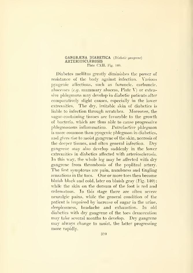
GANGRiENA DIABETICA (Diabetic gangrene)
ARTERIOSCLEROSISPlate CXII, Fig. 140.
Diabetes mellitus greatly diminishes the power of
resistance of the body against infection. Various
pyogenic affections, such as furuncle, carbuncle,
abscesses (e.g. mammary abscess, Plate V) or exten-
sive phlegmons may develop in diabetic patients after
comparatively slight causes, especially in the lower
extremities. The dry, irritable skin of diabetics is
liable to infection through scratches. Moreover, the
sugai'-containing tissues are favorable to the growth
of bacteria, which are thus able to cause progressive
phlegmonous inflammation. Putrefactive phlegmon
is more common than pyogenic phlegmon in diabetics,
and gives rise to moist gangrene of the skin, necrosis of
the deeper tissues, and often general infection. Drygangrene may also develop suddenly in the lower
extremities in diabetics affected with arteriosclerosis.
In this way, the whole leg may be affected with dry
gangrene from thrombosis of the popliteal artery.
The first symptoms are pain, numbness and tingling
sensations in the toes. One or more toes then becomebluish black and cold, later on bluish gray (Fig. 140)
;
while the skin on the dorsum of the foot is red andoedematous. In this stage there are often severe
neuralgic pains, while the general condition of the
patient is impaired by increase of sugar in the urine,
sleeplessness, headache and exhaustion. In old
diabetics with dry gangrene of the toes demarcation
may take several months to develop. Dry gangrene
may always change to moist, the latter progressing
more rapidly.
370
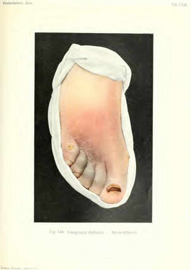
Bockenheimer, Atlas.Tab. CXII.
Fig. 140. Gangraena diabetica ^ Artcriosl<lerosis.
Rcbman Company, New-York.

The prognosis in these cases is bad, especially
when there is much sugar in the urine. Death mayoccur from heart failure, general infection or diabetic
coma. A\Tien the general treatment of diabetes fails
to act, the gangrene usually extends, and leads to
death.
Prophylaxis consists in the early diagnosis andtreatment of diabetes. It is, therefore, important
to examine the urine for sugar in all cases of pyogenic
and putrefactive infections. Diabetic patients should
pay strict attention to bodily cleanliness and try to
avoid all kinds of infection. They should also avoid
the causes which lead to arteriosclerosis.
Differential Diagnosis. Diabetic gangrene is
distinguished from other forms of gangrene by exam-ination of the urine. Extensive calcification of the
arteries can sometimes be seen by X-ray examination.
Treatment. In dry gangrene it is best to wait
for demarcation, unless extensive arteriosclerosis is
present. If, however, the popliteal artery is pulse-
less, amputation of the leg is the only remedy. If
there is no arteriosclerosis the gangrene may slowly
extend for months. When demarcation is complete
amputation may be performed directly above the line
of demarcation. Before demarcation the parts should
be treated with dry aseptic dressings (moist dressings
cause putrefaction), and be suspended. In slowly
extending moist gangrene demarcation may be waited
for if the temperature does not remain high. In rap-
idly extending moist gangrene with high temperature
early amputation is indicated some distance above the
gangrene. In gangrene of the lower extremity with
arteriosclerosis it is better to amputate through the
thigh; for the flaps after amputation through the leg
are badly nourished even in healthy individuals, and
in diabetics they are liable to become gangrenous.
Amputation through the thigh is best performed above371
the condyles (supracondylar amputation), or through
the epiphyseal line. Epiphyseal stumps have con-
siderable supporting power. As a rule, amputation
may be conservative in slowly progressing cases which
are not complicated by phlegmonous inflammation,
arteriosclerosis or high temperature. On the other
hand, rapidly extending gangrene complicated by
arteriosclerosis and phlegmon always requires high
amputation.
It is best to give an injection of scopomorphine
{Riedel's preparation) before the operation ; less quan-
tities of chloroform or ether are then required. In
these cases both general anaesthesia and lumbar
anaesthesia are badly borne, and infiltration anaesthe-
sia is contra-indicated, as it causes inflammation of
the weakened tissues. When the vessels are afl'ected
with arteriosclerosis they should be compressed by the
fingers of assistants during the operation, as the appli-
cation of the elastic tourniquet may cause thrombosis.
The wound should be dressed with sterile gauze;
iodoform is contra-indicated on account of the danger
of iodoform poisoning. Primary suture of the flaps
should not be attempted, and these should, therefore,
be made larger than usual. Secondary suture of the
flaps may be performed after a few days if the pro-
gress of the case is satisfactory. Ligatures must not
be applied too tightly to vessels affected with arterio-
sclerosis, as the coats of the vessel may give way and
cause secondary hemorrhage. The operation must
be performed under the strictest aseptic precautions,
as the diabetic tissues are easily infected, and osteo-
myelitis may occur in the bone stump or phlegmonous
inflammation in the soft parts.
After the wound has healed ulceration is commonin the amputation stump. This must be treated with
aseptic dressings to avoid fresh phlegmonous inflam-
mation. In some cases amputation of both legs maybe necessary for gangrene of both the feet. Only
about fifty per cent, of cases of diabetic gangrene
372

recover after amputation, a great many cases suc-
cumbing to diabetic coma. Wlienever possible the
iimount of sugar should, therefore, be reduced bygeneral treatment of the diabetes before operation.
Cases where acetone is present, and which give a
positive result with the perchloride of iron reaction,
have an unfavorable prognosis. The general con-
dition requires treatment by strict diet and the admin-
istration of salicylate or bicarbonate of soda in large
doses. Subcutaneous injection of saline solution maybe tried in diabetic coma. Thirst may be relieved bytincture of opium or by von Bergmamis diabetic
drink (citric acid 10; glycerin, 100; distilled water,
1.000).
373
ARTERIOSCLEROSIS (Atheroma)
This disease consists in the thickening of the walls
of the vessels by connective tissue formation, with sub-
sefjuent fatty degeneration of the inner and middle
coats (atheroma) and the deposition of calcareous
plates, causing roughening of the inner surface of
the vessel and leading to thrombosis. The disease
is more common in the male sex. Central and
peripheral nervous affections, especially those causing
vaso-motor disturbances ; infective diseases, including
typhoid, malaria, syphilis, general infection, leprosy,
cout and diabetes; the action of alcohol, nicotin and
lead; overexertion and sudden exposure to cold have
all been cited as causes of arteriosclerosis. [The
term arteriosclerosis is here used to describe what is
generally known in England as atheroma. The fun-
damental cause of this is generally considered to be
syphilis, though other causes mentioned above proba-
bly contribute. General arteriosclerosis, character-
ized by a general fibroid thickening of all the arteries,
is of more complex etiology, the chief factors being
probably syphilis, chronic alcoholism, infective fevers,
gout, and microbial toxaemias].
A tortuous condition of the temporal and radial
arteries is ofteia present in arteriosclerosis, along with
differences in the pulse in different arteries. Exten-
sive calcification is sometimes visible by X-ray exam-
ination.
The symptoms begin with pains of a rheumatic
character. The feet, in which the disease often be-
gins, are blue, cold and dry. Sensations of numb-ness and tingling are often present. There may be
severe pain in the heels, preventing the patient from374
walking {Charcot's intermittent claudication). Ex-tensive arteriosclerosis may cause gangrene of the
lower extremities. In women arteriosclerosis moreoften affects the hands causing great pain and loss of
function; but gangrene in the hands is very rare.
Arteriosclerosis of the cerebral arteries causes severe
headaches, attacks of loss of consciousness, or cerebral
hemorrhage.-'to^
Differential Diagnosis. Commencing arterio-
sclerosis of the extremities with no visible change in
the vessels may be mistaken for gout or rheumatism,
etc. In advanced cases the diagnosis is easy, owing
to the hardness of the vessels.
Treatment. Prophylactic treatment consists in
avoiding, as far as possible, the causes which maylead to arteriosclerosis. The best therapeutic meas-
ures are those which promote metabolism andstrengthen the heart; for instance, light gymnastics,
massage, mud baths, sand baths, ^Yiesbaden hot
springs, etc. Internally iodide of potassium should
be administered. Hot air treatment and hot potash
baths are useful for the pains in the heel. In severe
cases morphia may be necessary.
Fig. 140 shows commencing gangrene of the right
foot in a man of fifty-six, suffering from diabetes for
some years. The toes are bluish red in some parts,
grayish black in others, while the dorsum of the foot
is red. The skin was pale and cold. The discolora-
tion appeared in the course of a few hours, and in a
few days extended to the ankle joint. jNIoist gangrene
spread rapidly from the toes, and lymphangitis
extended up the leg.
The X-rays showed numerous calcareous deposits
in the anterior and posterior tibial arteries. Ampu-tation was performed above the knee joint, after the
sugar had been reduced from five to two per cent, by
375

three days' treatment of the diabetes. After opera-
tion the sugar diminished still further, and the tem-
perature fell—two favorable signs. Secondary suture
of the stump was performed on the fifth day and the
wound healed in four weeks. After general treat-
ment of the diabetes the sugar disappeared from the
urine.
The figure also shows other changes. On the
inner side of the foot over the metatarsophalangeal
joint is a large clavus, and another on the fifth toe.
The nail of the great toe is affected with onychogry-
posis, a common condition in old people who neglect
their feet. As the nail caused trouble in walking, it
was removed under local anaesthesia.
376

y

L^eiilieiiner, Alias. Tab. CXI II
Fie. 141. Arthritis urica.
Rcbnian Company, Ncw^olk.
ARTHRITIS URICA (Gout;/ Arthritis)
Plate CXIII, Fig. 141.
Gout is a disorder of metabolism which is often
transmitted from father to son for generations. It
therefore usually occurs among people with a hered-
itary predisposition. It most often affects middle-
aged men who indulge in high living and who take
too little exercise.
The disease is due to the deposit of urate of soda
in various places, especially in the cartilages of the
joints. According to Pfeiffer there is no increase in
the formation of urate of soda, but only deficient
elimination. The urate of soda deposits form yel-
lowish-white masses in the cartilage, synovial mem-brane, tendons, subcutaneous and periarticular tis-
sue, bursse, bronchi, intestinal mucous membraneand kidneys—in fact, in all the tissues and organs of
the body. An acute attack of gout is caused by
deposit of urate of soda in a joint, usually the meta-
tarso-phalangeal joint of the great toe (Podagra).
The symptoms are great pain in the affected joint,
slight rise of temperature and a certain amount of
constitutional disturbance (gastric pain, nervous phe-
nomena, rheumatic pains, etc.). The first attack is
sometimes excited by an injury to the foot. Theregion of the joint is swollen and oedematous, and
the skin shows erysipelatous reddening and phleg-
monous infiltration. The slightest touch or move-
ment causes intense pain. There is slight effusion
in the joint. After some hours the pain subsides, but
senerallv recurs on the second night; and so on for
about two weeks, till the attacks gradually become
less painful and finally disappear. Slight swelling of

the affected joint remains. Later on fresh attacks
may occur, often after many years. During the
attacks there is always a heavy sediment in the urine.
Repeated attacks may give rise to a permanent nodu-
lar swelling of the joint, and slight trauma may bring
on another acute attack {e.g. hand pressure on gouty
fingers).
Chronic gout, which is rarely primary and generally
results from the acute form, is observed also amongthe poorer classes. It often affects the joints, but is
less painful. The frequency with which the meta-
tarso-phalangeal joint is attacked is perhaps due to
bad circulation of the blood, owing to its peripheral
position. This joint is also affected by arthritis
deformans in old people. Large deposits of urate of
soda give rise to gouty nodules or tophi, which occur
in the joints of the fingers, hand, foot and elbow.
They also occur in the cartilages of the ear, nose andeyelids in the form of small, yellowish nodules, which
become hard and painful. In advanced cases of
gout these nodules may be found in all the joints andcartilages, joint capsules, tendon-sheaths, cartilages
of the ribs, and in other tissues.
Microscopic examination of gouty deposits shows
the presence of crystals of urate of soda. These crys-
tals act on the tissues like foreign bodies, and cause
not only pain but gradual necrosis by pressure. Thenecrosed tissues are expelled by the formation of fis-
tulas, and through the latter infection of the joints
may take place. Joint infection may also occur by
way of the blood (staphylococcal or streptococcal in-
fection), without communication with the exterior.
Suppuration in a gouty joint is always serious, as it
easily leads to general infection. The cartilages of
the joint may be destroyed by the gouty deposits,
without the occurrence of suppuration, and lead to
subluxation and anchylosis. Tophi, especially whensituated in the subcutaneous tissue, may give rise to
ulceration, venous thrombosis and phlebitis, espe-
378
cially in the lower extremities. Eczema of the skin is
common in gouty subjects.
Although in most cases of gout the joints are
affected, and the symptoms are those of joint inflam-
mation, gouty deposits in other tissues and organs
may give rise to the most diverse symptoms. Depos-
its in the tendo Achillis causes achylodynia with pain
in the heel; deposits in other places may cause
sciatica and lumbago, asthma and bronchitis, iritis
and other affections of the eye, disorders of the intes-
tine, etc.
In all long-standing cases of gout there is a danger
of complications affecting the internal organs. Thechief of these is chronic interstitial nephritis, in which
numerous deposits of urate of soda are found in the
kidneys, which may give rise to renal calculus.
Gouty subjects are also liable to emphysema of the
lungs. The prognosis in cases of pronounced gout
is always doubtful.
Differential Diagnosis. Gouty arthritis is most
often confounded with chronic rheumatism, but in
the latter the skin over the joints is unchanged. In
purulent arthritis there is high temperature and rigors
while the temperature in gout does not exceed 38° C.
(100° F.) provided no suppuration is present. En-
chondromas of the fingers (Fig. 50) differ from gouty
deposits by the absence of pain. Gout of other
organs must be diagnosed by the history of the case.
Large deposits of urate of soda can be seen by X-ray
examination; e.g. in bursae.
Treatment. Persons who are predisposed to
gout should try to avoid it by careful living, exercise,
etc. In acute gout, tincture of colchicum should be
given in large doses (fifty to one hundred drops daily).
The affected joint should be wrapped in wool and
suspended on a splint. Hot air treatment is also
useful. If suppuration occurs in the joint (with high
379
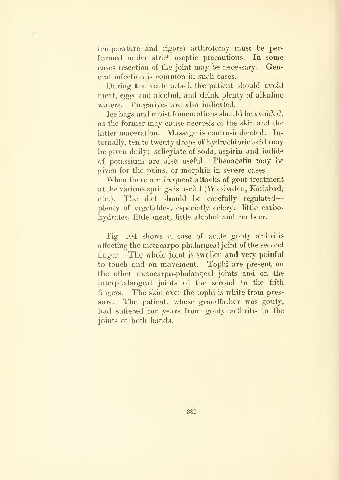
temperature and rigors) artlirotomy must be per-
formed under strict aseptic precautions. In some
cases resection of the joint may be necessary. Gen-
eral infection is common in such cases.
During the acute attack the patient should avoid
meat, eggs and alcohol, and drink plenty of alkaline
waters. Purgatives are also indicated.
Ice bags and moist fomentations should be avoided,
as the former may cause necrosis of the skin and the
latter maceration. Massage is contra-indicated. In-
ternally, ten to twenty drops of hydrochloric acid maybe given daily; salicylate of soda, aspirin and iodide
of potassium are also useful. Phenacetin may be
given for the pains, or morphia in severe cases.
When there are frequent attacks of gout treatment
at the various springs is useful (Wiesbaden, Karlsbad,
etc.). The diet should be carefully regulated
—
plenty of vegetables, especially celery; little carbo-
hydrates, little meat, little alcohol and no beer.
Fig. 104 shows a case of acute gouty arthritis
affecting the metacarpo-phalangeal joint of the second
finger. The whole joint is swollen and very painful
to touch and on movement. Tophi are present on
the other metacarpo-phalangeal joints and on the
interphalangeal joints of the second to the fifth
fingers. The skin over the tophi is white from pres-
sure. The patient, whose grandfather was gouty,
had suffered for years from gouty arthritis in the
joints of both hands.
380
Malformations
ENCEPHALOCELE OCCIPITALIS (Occipital Encephahcele)
RHACHISCHISISPlate CXIV, Fig. 142.
MYELOCELE—PES VARUSPlate CXV, Fig. 143.
MYELOCYSTOCELE—MYXOLIPOMAPlate CXVl, Fig. 144.
LYMPHANGIOMA (Congenital multiple)
Plate CXM, Fig. 145.
TERATOMA MOROGERMINALE (Monogerminal Teratoma)
Plate CXMI, Fig. 146.
DUCTUS OMPHALO-MESENTERICUS PERSISTENS(PersiMcttt omphalo-mesenteric duct)
Plate CXVIII. Fig. 147.
HERNIA FUNICULI UMBILICALIS CONGENITA(Congenital Umbilical Hernia)
Plate CXVIII, Fig. 148.
AMPUTATIONES AMNIOTICS (Amniotic Amputations)
Plate CXIX, Fig. 149.
AKROMEGALLA (Acromegaly)
MAKROMELLAMAKROGLOSSLA
Plate CXX, Fig. 150.
The study of malformations (teratology) is of great
interest to the surgeon, because many of these can be
improved by surgical intervention. A knowledge of
embryology is necessary in order to understand mal-
formations. We distinguish between primary mal-
formations which affect the embryo in its early
stages of development, and secondary malformations
which affect a part already formed, by some influence
actins on it during intra-uteriiie life. The latter are
spoken of as arrested development. Slight disturb-
ances in development are called anomalies; greater
deformities, malformations. The greater the mal-
381

formation, the earlier was its origin. The causes
which lead to malformation may be already present
in the embryo, or arrested development may be due
to external causes. Experimental observations on
animals have shown that malformations may be
caused by injury. In the lower extremities malfor-
mations may be caused by pressure or by abnormal
positions of the fetus in the uterus (various forms of
talipes—pes varus, pes valgus, pes calcaneus). Pres-
sure on the fetus may be caused by a uterine tumor
or by deficiency in the liquor amnii, and signs of such
pressure can often be seen after birth of the child.
Many malformations are due to anomalies in the
membranes; e.g. amniotic adhesions. All malfor-
mations caused in this way are cases of arrested
development. These amniotic adhesions or bands
may prevent the union of parts which should nor-
mally become united (branchial clefts) or may cause
duplication of parts, or partial or complete separa-
tion (amniotic amputations, aberrant glands).
382

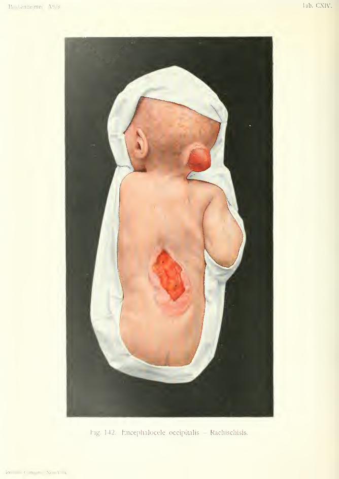
Bockenheimer, Atlas. Tab. CXIV.
Fig. 142. Encephalocele occipitalis — Rachischisis.
Rfbman Company, New-York.

ENCEPHALOCELE OCCIPITALIS {Occimtal EncenhaloceU)RHACHISCHISIS
Plate CXIV Fig. 142.
Encephalocele, or cephalocele, is a malformation
due to arrested development, and occurs in tworegions—the region of the nose (syncipital en-
cephalocele) and the occipital region (occipital
encephalocele). The former is subdivided into naso-
ethmoidal, uaso-frontal and naso-orbital; the latter
into superior and inferior occipital encephalocele, ac-
cording as it is situated above or below the occipital
protuberance. According to Miiller, one case of en-
cephalocele occurs in thirty-six hundred births. Thedeformity is due to more or less extensive deficiency
in the closure of the cerebro-spinal canal, caused by
trauma or by amniotic bands. The earlier this
occurs in fetal life the more extensive is the cleft in
the cerebro-spinal canal. In extensive cases there
may be acrania or anencephalus, while in slighter
degrees there is only a defect in the bone and dura
mater. Owing to the defect in the dura mater there
may be prolapse of the brain through the bone, gen-
erally a hernial protrusion of one of the ventricles.
According to von Berc/ma?in the existence of a true
congenital meningocele in which the dura is intact,
and there is only a hernial protrusion of the mem-branes through the gap in the bone, must be regarded
as doubtful. The author's observations on myelo-
cele (Fig. 134) have also shown that the inner cover-
ing of the protrusion, which is said to be dura, often
consists of connective tissue only, and that the inner
wall is often formed of ciliated columnar epithelium,
and, therefore, represents the degenerated ventricle
383

of the brain. Hence the so-called meningocele is a
true encephalocele or myelocystocele (Fig. 144).
As the subdivision of the different forms into
meningoceles, encephaloceles, encephalomeningoceles
encephalocystoceles and encephalocysto-meningo-
celes depends on pathological anatomy, and cannot
be distinguished clinically, it is sufficient for all prac-
tical purposes to use the term encephalocele or ceph-
alocele for all hernial protrusions through the skull,
especially as they mostly contain a protrusion of the
ventricle. For instance, the so-called encephalo-
meningocele has been shown to be not a true menin-
gocele, but a cystic formation which has becomegradually cut off from a primary hernia cerebri or
encephalocele.
Cephaloceles occurring at the sagittal suture, the
fontanelles or other parts of the skull are, according
to von Bergmann, either dermoids or caused by
trauma after birth (spurious traumatic acquired
cephalocele) . Congenital cephaloceles are either syn-
cipital or occipital. Syncipital cephaloceles have gen-
erally a wide base, while occipital cephaloceles are
pedunculated. Occipital cephaloceles may attain a
large size—as large as the child's head. The skin
at the base of the tumor is thickened and covered
with radially arranged hair. The tumor may be cov-
ered with normal skin, but more commonly most of
the surface resembles fresh scar tissue; or, whenulceration is present, it resembles the mucous mem-brane of the intestine. Vascular anomalies—telan-
giectases and angiomas—are often present. Thetumor is diminished by pressure, and can be com-
pletely emptied in cases when it apparently con-
sisted of a collection of fluid only. After the tumor
has been emptied by pressure the hole in the skull
can be felt, situated symmetrically in the middle line.
It is generally small and circular, and can sometimes
be shown by X-ray examination. As the tumor can
be diminished by external pressure, so is it increased
384

by internal pressure; e.g. when the child cries.
Cystic cephaloceles may be translucent. In other
eases there is little diminution on pressure. Irregular
partitions can then be felt in the interior of the sac.
Firm pressure then usually causes bulging of the
fontanelle, or sometimes convulsions. Sometimes
pulsation is observed in cephaloceles. The skull in
these cases is generally very small, and often flat-
tened. Other malformations are often present. Theinfants are weakly and have a subnormal tempera-
ture. The prognosis is generally unfavorable, but
is better in cases where the cephalocele can be com-
pletely emptied of fluid by pressure, and when no
brain substance can be felt in the sac after evacuation
of the fluid. Cases of occipital cephalocele with a
large gap in the bone, often extending to the vertebrae
of the neck, and protrusion of both occipital lobes
and the whole of the cerebellum, are soon fatal.
Differential Diagnosis. Syncipital cephalocele
may be mistaken for dermoid or lipoma. Diagnosis
depends on the presence of a gap in the bone, diminu-
tion of the tumor on pressure and the presence of
other deformities. Occipital cephalocele may be
mistaken for cephalhematoma, which sometimes
occurs on the occipital bone, especially as cephal-
hematoma may be surrounded by a hard ring at its
base caused by the raised periosteum. Cephalhema-
toma is not diminished by pressure. However, di-
minution by pressure may be absent in cephalocele if
the gap in the bone is occluded. In doubtful cases
an operation will settle the diagnosis.
Treatment. Puncture and injection is useless
and dangerous in cephalocele. The only rational
treatment is a radical operation. The sac is exposed
by incision through the skin, separated down to the
bone, ligatured and removed. The defect in the bone
may be covered in by suturing the periosteum over
385
it, by a pedunculated bone flap, or by a celluloid
plate. In cases where brain substance is present in
the sac, the operation can only be performed whenthe brain substance can be reduced through the gap
in the bone without producing symptoms of cerebral
compression. Removal of portions of brain still
possessed of function may cause dangerous symp-
toms, but a functionless dropsical protrusion may be
removed without danger. Cases of large defect in
the skull, with defect in the cervical vertebrae, or cases
combined with other extensive malformations, are
inoperable. The after-treatment is complicated by
the escape of cerebro-spinal fluid, which is always
abundant, even after the most careful closure of the
bone defect. The dressings therefore require chang-
ing several times daily to prevent infection of the
wound.
Fig. 142 shows a cephalocele situated symmetri-
cally in the middle line under the occipital protuber-
ance. The skin at the base of the tumor was thick-
ened ; over the greater part of the surface it resembled
fresh scar tissue, and presented numerous fine rami-
fying vessels. The tumor could be completely
emptied of its fluid contents by pressure, without
causing symptoms of cerebral pressure. After this
a circular hole in the bone could be felt about one-
half centimeter in diameter. This cephalocele could
have been completely removed by radical operation,
but for the presence of another malformation of the
spine which made the condition of the infant hopeless.
In the dorso-lumbar region from the twelfth dorsal
to the third lumbar vertebra is a condition known as
rhachischisis (spina bifida). This is a condition of
arrested development of the spine in which there is
absence of closure of the embryonic medullary canal
affecting the bones, soft parts, spinal cord and mem-branes. This malformation may extend the whole
length of the spine, and is then known as total pos-
386
terior rhachischisis; or it may be limited to one
portion only.
Rhachischisis represents the most extreme degree
of spina bifida (Figs. 143 and 144). It is most com-mon in the himbo-sacral region, because the medul-
lary groove closes last in this region to form the
neural canal. Rhachischisis is usually associated
with other extensive malformations such as anen-
cephalus, acrania, absence of vertebral bodies, etc.
Three typical zones can be distinguished situated
symmetrically on each side of the vertebral column:
(1) a circular, peripheral zone of thickened skin,
often covered with abundant hair; (2) a middle zone
which resembles fresh cutaneous scar tissue, or the
serous coat of the intestine, and has hence been called
the epithelio-serous zone; a central zone of flabby
granulations with a depression at the upper and
lower ends, which represents the open and exposed
spinal cord. The depressions at each end of the
central zone lead to the central canal of the spinal
cord. In cases where the spinal cord is muchexposed, death soon occurs from meningitis.
387
Spina Bifida
MYELOCELE—PEDES VARIPlate CXV, Fig. U3.
MYELOCYSTOCELE—MYXOLIPOMAPlate CXVL, Fig. Hi.
As already mentioned, rhachischisis represents the
most extreme degree of spina bifida. If the arrest
of development is limited to one, two or three verte-
bral arches, the cleft spinal cord is not exposed in the
vertebral groove as in rhachischisis, but projects in
the form of a tumor through the small cleft in the
vertebrae, owing to pressure of fluid on its ventral
surface. It thus forms a symmetrical tumor in the
middle line, with the same three characteristic zones
as in rhachischitis, and is known as a myelocele
(Fig. 143).
There are four kinds of spina bifida, differing in
degree according to the date of their appearance in
embryonic life. The first and most extensive formis rhachischisis, which has already been mentioned.The second form (myelocele) appears later and is
limited to a smaller extent of the spine, although it
may include the soft parts, bones and spinal cord;
this forms a tumor-like swelling. The third form{myelocystocele) occurs still later in embryonic life, at
a time when the spinal cord and the skin have already
closed on the dorsal surface of the embryo, but the
dura mater and bone have not yet united. Thefourth form [meningocele) only occurs in the lumbosacral region where the spinal cord has become the
filum terminale. Spina bifida occulta, which also
388

Bockenheimer, Atlas. Tab. CXV.
Fig. 143. Myelocele - Pedes vari.
Rebman Company, New-York.

occurs at the lower extremity of the vertebral column,is not to be regarded as a special form, but as a men-ingocele.
The subdivision of spina bifida into the three chief
forms—myelocele, myelocystocele and meningocele
—is the most suitable for practical purposes. Spina
bifida is a comparatively rare malformation, occur-
ring in one or one and five-tenths out of one thousand
infants.
1. Myelocele. By far the most common form is
myelocele. According to von Recklinghausen this
occurs before the twelfth day of embryonic life, as
after this time the medullary groove closes to formthe neural canal. The arrest of development con-
cerns the dorsal part of the spinal cord and mem-branes, the vertebral arches, the muscles and the
skin. A tumor-like swelling is then formed by the
formation of hydrops on the ventral side of the spinal
cord which continually pushes the cord out of the
vertebral canal through the preformed cleft. Ac-
cording to von Bergmann the occurrence of hydrops
is due to the absence of dura mater.
Myelocele forms a characteristic swelling with a
wide base, situated symmetrically in the middle line,
with the three zones already mentioned in the case
of rhachischisis; viz. an outer zone of thickened
skin with abundant hair, and often telangiectases; a
second zone of a pink color resembling new scar
tissue, with a deep network of ramifying vessels; a
third zone of an oval form at the summit of the
swelling, red and tumid like intestinal mucous mem-brane, very vascular, and covered with pus a few
days after birth. This third or central zone repre-
sents the remains of the cleft spinal cord, and is called
the vasculo-medullary zone in distinction to the
epithelio-serous or second zone. At the upper and
lower ends of the third zone is a depression through
which a probe can be passed into the central canal of
the spinal cord. These cases generally die from
389
meningitis through infection of the vasculo-medul-
lary zone. Operative treatment is useless. Thespinal nerves become dragged upon by the formation
of the protruding myelocele, causing motor paralysis
of the lower extremities, bladder and rectum (paraly-
sis of the upper extremities when the myelocele is
situated in the upper part of the spine).
The common occurrence of pes varus in these cases
(Fig. 143) is due to the myelocele being usually
situated at the junction of the lumbar vertebrae with
the sacrum where the nerves arise which supply the
anterior and posterior tibial muscles; viz. the fourth
and fifth lumbar and the first and second sacral
nerves. Sensory disorders are rare in myelocele, but
trophoneurotic disorders occur in the form of exten-
sive eczema and decubital ulcers, especially on the
feet; in pes varus on the outer border of the foot.
Diagnosis is easily made by the characteristic ap-
pearance, the presence of fluctuation and the cleft in
the bone. There is no diminution in the swelling bypressure owing to the absence of communication with
the subarachnoid space. Myelocele is most commonin the lumbo-sacral region; after this in the cervical
and thoracic. It is often associated with other mal-
formations, such as umbilical hernia, etc., and the
infants seldom survive.
2. Myelocystocele. This form consists in arrested
development of the vertebral arches and dura mater.
It appears in the third week of embryonic life, at a
time when the medullary groove has closed to form
the neural tube, and the epiblast has grown over it.
Hydrops of the central canal causes bulging of the
posterior part of the spinal cord through the gap in
the vertebral arches, giving rise to a tumor-like swell-
ing of the spinal cord covered by the soft parts. Thesubstance of the spinal cord soon undergoes degen-
eration and can only be identified by the presence of
ciliated cylindrical epithelium on the inner surface
of the cavity (the remains of the ciliated epithelium
390

of the central canal of the spinal cord). In the
external coverings of myelocystocele there is often
lipoma, myxoma, lymphangioma or teratoma. Thetumor has a wide base and is covered with normalskin, which is thickened at the base of the tumor.
Sometimes small depressions are present in the skin
caused by the remains of amniotic bands (Fig. 144).
The tumor is of soft consistence, and fluctuation is
always present. The fluid contents of the tumorcan be completely reduced by pressure, as there is
direct communication with the central canal, and also
with the subarachnoid space. By pressing on the
tumor the transmission of fluid pressure can be felt
at the fontanelle.
Myelocystocele is often combined with hydroceph-
alus. Paralyses are rare, as the motor nerves are
not displaced by the malformation ; at the most there
may be pes varus or valgus on one side, due to the
tumor being situated unsymmetrically more to one
side of the middle line, and thus dragging on a motor
nerve. However, extensive myelocystocele of the
lumbo-sacral region may cause paralysis of the
bladder and rectum. Trophoneurotic disorders are
common. Sometimes paralysis occurs at a later age,
the tumor increasing gradually in size and dragging
on the spinal cord and nerves. Defective bone for-
mation is often associated with myelocystocele
—
absence of vertebral bodies, unilateral defects in the
vertebral laminae, absence of ribs or patella, sco-
liosis, etc.
3. Meningocele, x^ccording to recent observations
meningocele can only occur in places where the spinal
cord is absent (von Bergmann). In this condition
there is defective formation of the vertebrae and dura
mater, so that the pia mater protrudes posteriorly,
inclosing the filum terminale. In this way a pedun-
culated swelling is formed, covered by normal skin,
which may attain the size of a child's head as the
amount of cerebro-spinal fluid in the sac increases.
391
Paralysis only occurs when the meningocele is large,
and is then generally of limited extent. There is
sometimes abundant hair on the summit of the swell-
ing. Fluctuation is always present, but there is only
slight diminution on pressure. The space in the bone
is generally smaller than in myelocele. Meningocele
occurs most often in the sacral region.
Spina Bifida Occulta, according to the most
recent observations, is a form of meningocele which
becomes ruptured and undergoes spontaneous heal-
ing under the skin. The pressure of the cicatrix maycause disturbances which are not noticed till the
child grows older.to"
Differential Diagnosis. Myelocele, when the
vasculo-medullary zone is very extensive, may some-
times be mistaken for cavernoma. In rare cases
where epidermization of the second zone leads to
cicatrization of the third zone, myelocele may be mis-
taken for a myelocystocele in which the skin has
become cicatrized after ulceration. In such cases
diminution of the tumor on pressure points to myelo-
cystocele. In lipoma, lymphangioma and teratoma
there is no diminution in the tumor on pressure unless
there is a myelocystocele underneath it ; which, how-ever, is often the case. Meningocele may be mis-
taken for myelocystocele when it is not situated in
the sacral region (where the spinal cord is absent).
It may also be mistaken for sacral tumors, dermoids
and teratomata.
The prognosis is not unfavorable in myelocystocele
and meningocele provided other malformations are
absent and the infant has a strong constitution.
Treatment. In myelocele a radical operation is
useless, because by removal of the cystic sac the
spinal cord is divided and unites with the cicatrix.
Reduction of the infected vasculo-medullary zone byoperation always leads to meningitis. Palliative
393
treatment, by puncture of the sac, is all that can be
done in these cases.
Myelocystocele, owing to its covering of intact skin,
is more suitable for operation. In this case the
operation is similar to that for hernia. The sac, con-
sisting of degenerated spinal cord, is exposed by anincision through the skin, dissected down to the bone,
ligatured and removed. The cleft in the bone is
repaired by a plastic operation. The sac is often
covered by a fatty tumor which also requires removal.
Removal of the sac after ligature is not dangerous in
these cases, as it consists only of functionless degen-
erated spinal cord. Meningitis sometimes follows
these operations, but most cases recover and maygrow up.
Meningocele offers the best chances for operation.
The sac is opened and the nerves replaced in the ver-
tebral canal. The sac is then ligatured and removed
and the space in the bone closed by suture of the soft
parts, or by bone grafting. The prognosis is good
after these operations.
In spina bifida occulta with disturbances due to
pressure of the cicatrix, the latter may be removed
and the space in the bone repaired.
The development of hydrocephalus, which mayoccur after operation on all forms, is an unfavorable
sign.
Fig. 143 shows a myelocele of the lumbo-sacral
region. The tumor is situated symmetrically in the
middle line and has a wide base. At the base the
skin is thickened (first zone) ; the second zone (epi-
thelio-serous) shows numerous ramifying vessels; the
third zone (vasculo-meduUary) is not typical and
resembles the second zone, owing to epidermization
of the latter (cf. Fig. Ii2). It only differs from the
second zone in its bluish color. The diagnosis of
myelocele depended on the absence of diminution on
pressure, and the presence of paralysis of the bladder
393
and rectum, and pronounced pes varus of both feet.
Death occurred soon after birth.
Fig. 144 shows a myelocystocele situated in the
lumbar region, and covered with normal skin. Asmall depression in the surface is due to amniotic
adhesions. Under the skin is a mass of fatty tissue,
while a deep cystic tumor could be felt more deeply
situated. The latter could be almost completely
emptied by pressure. There were no motor or sen-
sory disorders present, and no other malformations.
The X-rays showed a small cleft in one of the verte-
bral arches situated a little to one side of the middle
line. The superficial fatty tumor was removed and
found to be a myxolipoma. The myelocystocele was
then separated down to the bone, ligatured and
removed. The gap in the vertebra was closed by
transplantation of a piece of bone from the iliac crest.
Microscopic examination showed the pressure of cyl-
indrical epithelium in the inner wall of the cyst, thus
confirming the diagnosis.
394
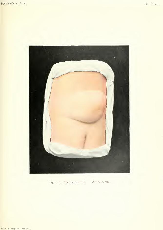
Bockenheinier, Atlas. Tab. CXVI.
Fig. 144. .\\\elocystocele Myxolipoma.
Rebman Company, New- York.

PES VARUS
Pes varus may be congenital or acquired. Thecongenital form may be caused by arrested develop-
ment, or may be secondary to pressure caused byamniotic adhesions, etc. Congenital pes varus is
common in connection with myelocele, and is due to
paralysis of the nerves, as already explained. Ac-quired pes varus occurs in rickets, and as the result
of poliomyelitis which causes paralysis of the pro-
nators and dorsal flexors of the foot. The chief effect
takes place at the midtarsal joint and consists in
supination, plantar flexion, internal rotation andadduction. Changes also occur in the astragalus
and OS calcis, especially in long-standing cases.
These changes can be seen by the X-rays. Thereis also shortening of the muscles, tendons, fascia
and ligaments, especially shortening of the tendo
Achillis (talipes equino-varus). Decubital ulcers
may form on the outer border of the foot.
Treatment. In congenital clubfoot treatment
should be begun as early as possible, by repeated
manual correction to the normal position, followed
by fixation in an over-corrected position by means of
plaster of Paris bandages. In sucklings, thin strips
of cotton bandages soaked in mastic solution (tur-
pentine 15, mastic 12, resin 28, alcohol (90 per cent.)
180, ether 20) may be used, applied to the foot and
leg so that the foot is fixed in the over-corrected posi-
tion. This treatment should be kept up for six
months, after which elastic traction may be ap{)lied
to the foot for another six months. In older children
manipulation must be performed under an anaes-
395

thetic. After the ninth month preliminary tenotomy
of the tendo Achillis is necessary, before the foot
can be brought into the proper position. To prevent
relapse boots should be worn with the sole raised on
the outer side, but care must be taken to avoid pro-
ducing flat foot. In pes varus due to poliomyelitis
tendon transplantation may be performed. Old-
standing cases of clubfoot in adults require oste-
otomy, or sometimes more extensive operations such
as disarticulation.
396


Bockenheimer, Atlas. Tab. CXVIl.
O
t/J
Xo
Oo
O
n
M
Rcbman Comnanv. W\»-Vorl(.
Lymphangioma
LYMPHANGIOMA CONGENITUM CYSTICUM MULTIPLEX{Cotigenilal Multiple ajstic lymphangioma)
Plate CXMI, Fig. 145.
The term lymphangioma should be limited to
those tumors in which there is a new formation of
lymphatic vessels. A number of growths have been
included in the term lymphangioma which are only
formed of dilated lymphatics, without any newlymphatic vessels. Microscopic examination is there-
fore important in these cases. Clinically, we distin-
guish simple, cavernous and cystic lymphangiomas;
also single and multiple. In the great majority
of cases the growths are congenital. All three forms
are often present in the same patient. Lymphan-giomas may occur in the skin, but more often in the
subcutaneous tissue; also between the muscles and
in the subserous tissue. The term simple lymph-
angioma is WTongly applied to lymphangiectases,
which form lobulated growths covered by thickened
skin and occur on the head, trunk and extremities.
Simple lymphangioma occurs most commonly in the
tongue or lips as a circumscribed growth. The skin
or mucous membrane is always somewhat thickened
and adherent to the growth. The isolated circum-
scribed form of simple lymphangioma may be mis-
taken for other kinds of tumor, but transitional stages
to cavernous lymphangioma are often found, the
diagnosis of which is easier.
Cavernous lymphangioma is always a diffuse for-
397
mation, of soft consistence and always united with
the skin or mucous membrane over it. The tumorcan be gradually diminished by pressure, as the
cavernous spaces, filled with lymph and lined with
epithelium, communicate with the neighboring lym-
phatic vessels. Cavernous lymphangioma forms a
painless, diffuse, slow-growing tumor with a smoothsurface and irregular borders. WTien they are visible
under the skin or mucous membrane they have a pale-
green color, in distinction to the reddish-blue color
of cavernous hemangioma. They occur most often
in the cheeks, tongue and lips, giving rise to enlarge-
ment of these parts, known as macromelia, macro-
glossia and macrocheilia. They are generally con-
genital, or appear soon after birth. Cavernouslymphangiomas also occur in the neck, causing a
dimpled swelling of the skin by their numerous pro-
cesses, which extend in all directions (Fig. 145). Asalready mentioned, lymphangiomas may be situated
over encephaloceles or myelocystoceles. Gradualatrophy of the bones may be caused by the pressure
of extensively progressing lymphangiomas.
Cystic lymphangioma occurs in the subcutaneous
or intermuscular tissue, most often in the side of the
neck (Fig. 145). It is composed of large, cystic
cavities lined by epithelium and containing whitish
or brownish fluid (cystic hygroma). Cystic lymph-angioma is almost always congenital and is character-
ized by its slow growth, which may cease after someyears. The skin is unchanged and can be raised
from the tumor. Fluctuation is present, but there is
no diminution of the tumor on pressure. Extensive
lymphangioma of the neck may be dangerous frompressure on the trachea. Besides the neck, the growthsmay also occur in the axilla, the popliteal space, the
bend of the elbow, the groin and the sacral region.
Infants with congenital lymphangioma sometimesshow other malformations, and are often incapable
of life.
398
Differential Diagnosis. Simple lymphangiomawhich occurs in the form of a small, soft, circum-
scribed tumor, may be mistaken for fibroma, lipomaor hemangioma. Cavernous lymphangioma can only
be mistaken for hemangioma, as no other tumordiminishes on pressure. It differs from hemangiomain its greenish color and in the nature of its contents.
Cystic lymphangioma, when it occurs in the form of
an isolated unilocular cyst, may be mistaken for
various tumors ; in the neck, for blood cyst, branch-
ial cyst, lipoma or dermoid.
The prognosis of lymphangioma is, on the whole,
not unfavorable, on account of its limited growth
and occasional spontaneous resolution.
Treatment. Circumscribed lymphangioma can
be excised. In diffuse cavernous lymphangiomas
(macrocheilia, macroglossia, macromelia) cunei-
form excision may be performed. The introduction
of magnesium may be tried, to cause thrombosis and
shrinking of the tumor. After this extirpation is
easier and infection through a lymph fistula, which
so often occurs after the usual operation, is avoided.
Cystic hygroma is best treated in this way. Radical
operations should not be performed unless the child
is in good condition. Puncture and injection of
tincture of iodine are unsafe measures, while lymph
fistula often remains after incision and plugging.
Lymph fistulas must always be removed by a radical
operation, on account of the danger of infection
through them. Lymph fistulas, which occur from
injury to the thoracic duct after extensive extirpation
of the breast, can be healed by plugging with acetate
of aluminium.
Fig. 145 shows a congenital tumor involving the
lower part of the right cheek, the whole of the right
side of the neck and the greater part of the left
side of the neck. The skin was unchanged and399
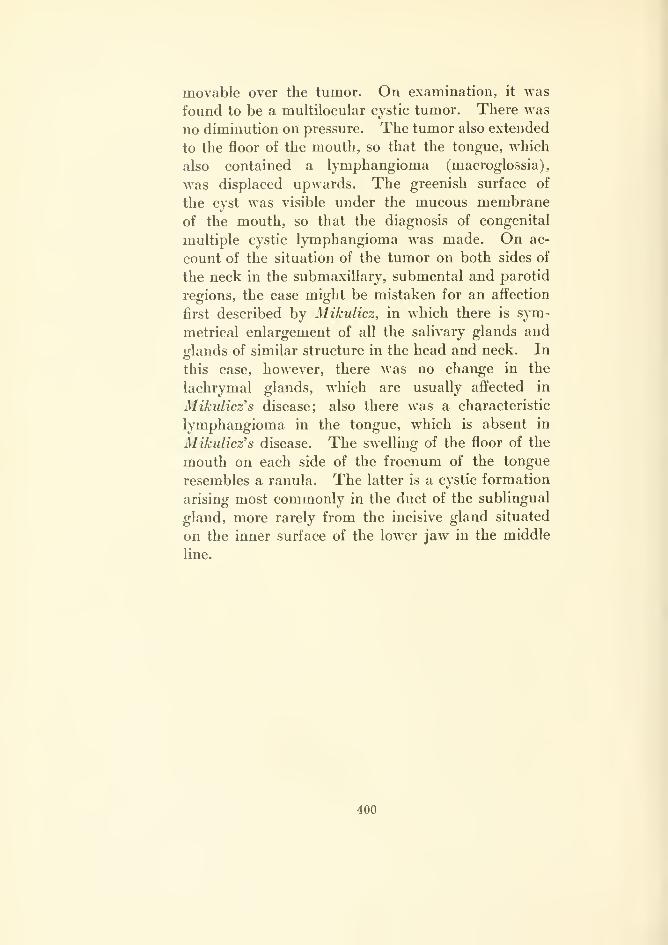
movable over the tumor. On examination, it was
found to be a multilocular cystic tumor. There was
no diminution on pressure. The tumor also extended
to the floor of the mouth, so that the tongue, which
also contained a lymphangioma (macroglossia),
was displaced upwards. The greenish surface of
the cyst was visible under the mucous membraneof the mouth, so that the diagnosis of congenital
multiple cystic lymphangioma was made. On ac-
count of the situation of the tumor on both sides of
the neck in the submaxillary, submental and parotid
regions, the case might be mistaken for an affection
first described by Mikulicz, in which there is sym-
metrical enlargement of all the salivary glands and
glands of similar structure in the head and neck. In
this case, however, there was no change in the
lachrymal glands, which are usually affected in
Mikulicz's disease; also there was a characteristic
lymphangioma in the tongue, which is absent in
Mikulicz's disease. The swelling of the floor of the
mouth on each side of the froenum of the tongue
resembles a ranula. The latter is a cystic formation
arising most commonly in the duct of the sublingual
gland, more rarely from the incisive gland situated
on the inner surface of the lower jaw in the middle
line.
400
Teratomata
TERATOMA MONOGERMINALE {Monogerminal teratoma)
Plate CXVII, Fig. 146.
Teratomas may be bigerminal or monogerminal. In
bigerminal teratoma there is a true double formation
—a fetus within a fetus. In monogerminal teratoma
there is perverted development in one embryo, and
all the tissues are derived from one embryo only.
The latter includes all kinds of mixed tumors which
are formed of all three embryonic layers (epiblast,
mesoblast and hypoblast). Dermoid cysts, which are
formed by all three embryonic layers, belong to the
teratomata. A distinction between monogerminal
and bigerminal teratomata is not always possible,
and is of little clinical importance. Teratomas are
rare on the whole, and are always congenital. They
are most often found in the buccal cavity, where they
may be mistaken for naso-pharyngeal polypi (Fig.
25). They also occur in the face, neck and coccyg-
eal region, and have been observed in the mediasti-
num and abdominal cavity. They may attain enor-
mous dimensions, and have then an irregular, uneven
surface. The consistence also varies, some parts
being cystic, others soft and others hard. Teratomas
often form encapsuled tumors. They may cause
extensive destruction by pressure on the neighboring
parts. A distinction between teratomata and tera-
toid mixed tumors is clinically impossible. Diagnosis
in many eases is only made after examination of the
extirpated tumor.
401
Differential Diagnosis. Teratomas which ap-
pear as large, congenital tumors can generally be
recognized by the above-mentioned characteristics,
especially by their situation in the embryonic fissures.
Diagnosis is assisted by the X-rays which may reveal
bones and teeth, which ai-e often present in tera-
tomas. Teratomas occurring in the thorax, abdo-
men and pelvis, especially when they do not assume
a tumor growth till later years, can often only be
diagnosed by operation.
Treatment. Teratomas have been successfully
removed both in children and in adults. Extensive
teratomas (Fig. 146) cannot be removed by operation.
The presence of other deformities, such as spina
bifida, and the feeble condition of the infants often
renders operative treatment impossible.
Fig. 146 shows a teratoma of the left side of the
face, almost as large as the fist, involving the left
orbit and almost the whole of the buccal cavity, and
covered by livid, movable skin. It was covered by a
connective-tissue capsule. Further examination
showed that it arose from the base of the skull, but
did not communicate with the cranial cavity. Thetumor was soft and fluctuating in some places, hard
in others. Examination by the X-rays showed the
presence of a piece of bone, which was afterwards
found to be part of the upper jaw. Further exam-
ination showed that the tumor consisted of neuroglia,
neuroepithelium and cysts lined with epithelium. As
it consisted of epiblastic products only it must be
regarded as a monogerminal tumor which, in this
case, originated from a separated portion of the
epiblast. This view is supported by the fact that
the tumor developed in a region (base of the skull)
where separation of the epiblast is possible. Onthe other hand, it appears far-fetched to consider
the tumor as a bigerminal teratoma (fetus within
402

fetus by inclusion) simply because of its large size
at birth.
There were no other malformations present except
mutilation of the right ear. Death occurred soon
after birth.
403
DUCTUS OMPHALO-MESENTERICUS PERSISTENS(Persistent OntpJialo-mesenteric Duct)
Plate CX\1II, Fig. U7.
The omphalo-mesenteric duct, or vitelline duct, is
the communication between the alimentary canal
and the umbilical vesicle or yolk-sac. It usually dis-
appears about the eighth week of fetal life. In some
cases this duct may persist and is then known as
Meckel's diverticulum, which arises from the small
intestine about ten inches above the ileocsecal valve.
This diverticulum may lie free in the abdominal
cavity, where it may cause intestinal obstruction by
becoming entangled with the intestines; or it maybecome attached to the umbilicus, or extend a short
distance into the umbilical cord. In the latter case
it may become opened after birth when the umbilical
cord has separated, thus giving rise to an umbilical
fistula, discharging faeces from the umbilicus whenthe whole length of the duct is open as far as the
intestine. When the intestinal end of the duct is
closed, the remainder may persist as a small fistula
discharging mucoid secretion; or it may become
dilated into cystic formations.
In umbilical fistula there is a red globular swelling
with a small depression at its apex, situated at the
navel. The surface of the swelling is formed by
mucous membrane. A probe can be passed through
the depression as far as the small intestine, and the
greater part of the faeces are discharged through the
fistula, causing inflammation of the skin surrounding
the navel. Death often occurs from prolapse of the
small intestine.
404
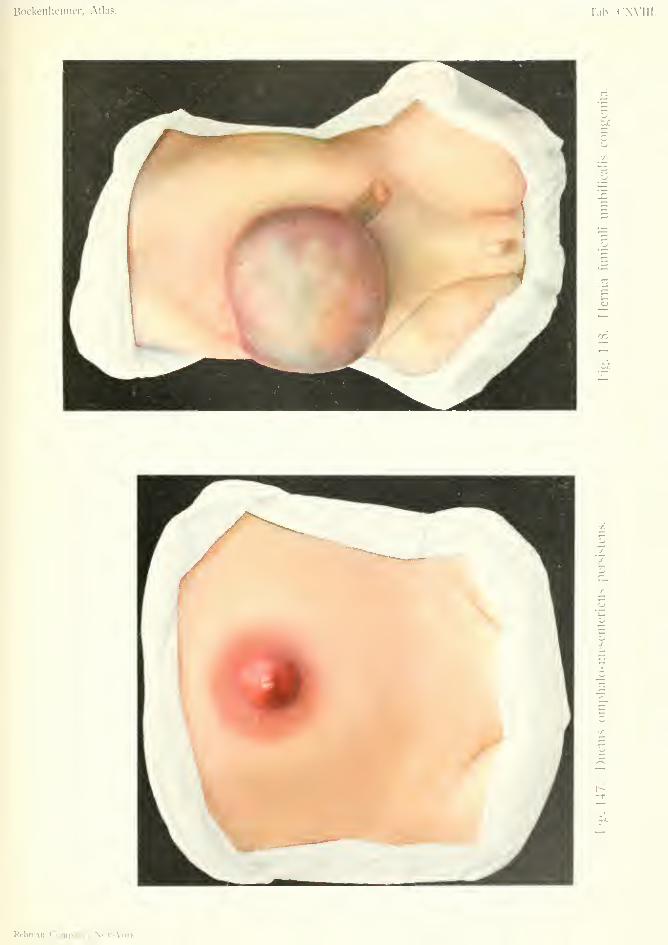
Bockenheimer, Atlas. Tab. CWIII.
5o
zr.
u
Rebman Company, Neir-York.

Differential Diagnosis. Infection of the navel
with the formation of granuhition tissue may resem-
ble the above condition. Other fistulas may also
occur in the umbilicus. The urachus, which repre-
sents the remains of the communication between the
bladder and the allantois in fetal life, may remain
open and form a fistula at the umbilicus. Normally
the urachus becomes obliterated and forms the medianligament of the bladder. Fistula of the urachus is
diagnosed by discharging urine. Like fistula of the
vitelline duct, fistula of the urachus usually appears
after separation of the umbilical cord. The nature
of the fistula is not always determined by probing;
a more certain method of diagnosing fistula of the
vitelline duct is by feeding with powdered charcoal,
which then appears at the navel. The diagnosis can
sometimes be made by chemical and microscopical
examination of the secretion. Tuberculosis of the
intestine, actinomycosis, peritonitis, empyema of
the gall bladder, injuries of the bladder, and der-
moids may all give rise to fistula at the navel.
Treatment. Fistula of the vitelline duct can some-
times be prevented by discovering the condition before
tying the umbilical cord. The cord is then thicker than
usual at its base. The end of the duct can then be re-
duced and the cord tied further away from the navel.
In cases of complete fistula leading to the intestine
laparotomy is necessary, with resection of the diver-
ticulum and suture of the intestine. Fistula of the
urachus must be separated down to the bladder and
removed, and the bladder sutured.
Fig. 147 shows a case of complete fistula of the
vitelline duct. The infant was in a bad condition
from prolapse of the gut, evacuation of faeces from
the navel, and inflammation of the surrounding skin.
Laparotomy was performed but the operation was
unsuccessful.
405
HERNIA FUinCULI UMBILICALIS CONGENITA(Congenital Umbilical Hernia)
Plate CXVIII, Fig. Us.
Congenital umbilical hernia must be regarded as a
malformation, and forms a large tumor, containing
intestine and often also the liver. It is often asso-
ciated with various forms of spina bifida, or with
ectopia of the bladder. Cases of extensive umbilical
hernia are due to arrested development causing in-
complete closure of the abdominal walls. Umbilical
hernia may also be acquired. In this case the ab-
dominal walls are closed, the umbilical ring is small,
the hernia is smaller and more cylindrical, and the
contents consist of small intestine. Acquired hernia
may be so small as to be overlooked at birth, and maythen be included in the ligature of the umbilical cord.
The base of the cord should, therefore, always be
examined to see if it contains intestine.
Congenital umbilical hernia forms a large globular
swelling in the region of the navel (Fig. 148). Thesurface is destitute of cutaneous covering and shows
the greenish-yellow remains of the amnion. Theremains of the umbilical cord is generally seen at one
side of the swelling. In rare cases epidermization
takes place at the borders; more commonly the
swelling ruptures from pressure, with consequent
prolapse of the viscera and death from peritonitis.
Differential Diagnosis. Both the congenital
and the acquired forms of umbilical hernia are so
characteristic that they cannot be mistaken for any
other condition.
406
Treatment. The occurrence of symptoms of
intestinal obstruction, or tlireatening perforation of
the sac indicate immediate laparotomy, with excision
of the sac, reduction of its contents and closure of the
abdominal walls. In some cases the viscera are
adherent to the sac and require separation. Reduc-
tion of the visceral contents is sometimes difficult or
even impossible, especially when the liver is con-
tained in the sac. If operation is not urgent it maybe postponed till the child is stronger, the sac being
supported by bandaging in the meantime.
Acquired umbilical hernia may occur during the
first month after birth, as the umbilical ring takes
several weeks to close completely. Anything which
causes the infant to cry may be an exciting cause for
hernia, also straining from phimosis, etc. Manycases become cured without treatment. Non-opera-
tive treatment consists in placing a metal disk
wrapped in plaster over the umbilical ring, after
reduction of the hernia, and bringing the skin of the
abdomen together over it by means of plaster. Thedisk must be larger than the hernial opening. Small
openings may be closed in this way after nine months'
treatment. Larger openings with separation of the
recti muscles above the umbilical ring require lapa-
rotomy. In older children, especially girls, this
should always be performed. The operation con-
sists in extirpation of the whole umbilical ring and
suture of the abdominal walls with wire.
In Fig. 148 the hernial sac contained the intestine
and liver, which were reduced with great difficulty,
so that the abdominal walls when sutured were under
great tension. The infant died soon after the opera-
tion.
407

AMPUTATIONES AMWIOTICiE (Amniotic Amputations)
Plate CXIX, Fig. 149.
Malformations of the extremities include amelus
and phocomelus. In amelus the extremities are
absent or only represented by stumps. This condi-
tion may affect all four extremities, both arms or
legs, or one arm or leg. In phocomelus there is
arrested development of the proximal segments of
the arms or legs, or of all four extremities. Thehands or feet are then situated directly on the trunk.
Some of these cases attain adult age, and one has
been known to live to sixty-two. [Several such cases
were among Bartium's freaks.]
The so-called spontaneous amputations of various
parts of the extremities are caused by pressure of
amniotic bands or the umbilical cord. The ends of
the amputations are then pointed. In other cases
there is not complete amputation but constriction,
resulting in deep, circular grooves extending to the
bone (Fig. 149). In spite of the depth of the grooves,
the circulation remains normal, but there is often
elephantiasic thickening from lymphatic congestion.
In some cases the bones are constricted, as shown by
the X-rays. The remains of the amniotic bands are
often present in the constricted places.
Other malformations, also due to tightness of the
embryonic membranes, are synechia of the fingers
(webbed fingers), hare-lip, cleft-palate, transverse
fissure of the cheek, and fissure of the tongue.
Treatment. When the constricted parts are
functionless they should be amputated. Elephanti-
asis may be treated by cuneiform excision.
408
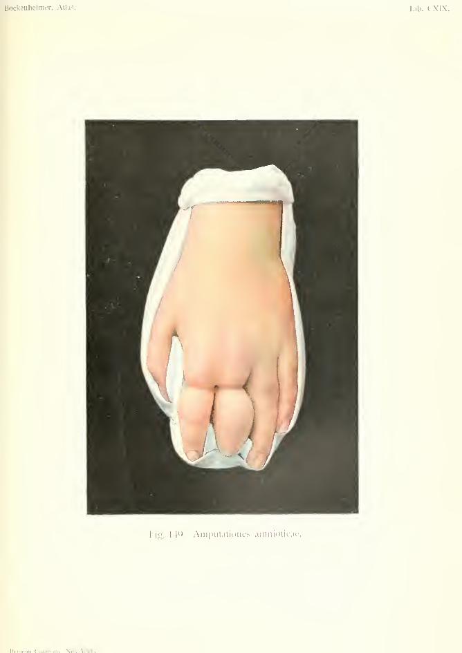
Bockenheimer, Atlas. lab. CXIX.
hig. 14y. Ainputalioiies aiiiiiioticae.
Pfhmnn Pnninanv. Npw-York.


In Fig. 149 the function of the fingers was normal
so that no operation was necessary. In this case
there was also hare-lip and cleft-palate, which were
operated upon.
409
AKROMEGALIA (Acromegaly)
MAKROMELIA {Macwmelia)MAKROGLOSSIA (Macroglossia)
Plate CXX, Fig. 150.
The term Acromegaly is applied to a condition in
which there is enlargement of the terminal portions
of the body—the hands, feet, nose, cheeks, tongue
and ears. The enlargement affects all the tissues
(true giantism) and does not appear till after the
termination of the period of growth, thus differing
from congenital giantism. In some cases there is
increased growth of hair, and curvature of the verte-
bral column. The disease causes considerable dis-
figurement of the face. It generally appears between
the twentieth and fortieth years and may remain
stationary. In many cases there is, first of all,
hypertrophy of the bones of the hands, feet and face.
The disease has been attributed to changes in the
pituitary body (hypertrophy, adenoma, sarcoma,
cyst) ; to changes in the thyroid gland, pancreas,
genital glands; to persistence of the thymus; to
nervous influence, since nervous disorders have been
observed in the hypertrophied extremities; also to
a congenital condition. The most probable of these
is enlargement of the pituitary body, which can be
demonstrated by widening of the sella turcica, shownby the X-rays. Large tumors of the pituitary body
may press on the optic nerve and nerves of the
ocular muscles.
The prognosis is not unfavorable, as severe dis-
turbances only occur after the disease has existed
for many years.
Differential Diagnosis. Partial giantism, which
also begins in the hands and feet, differs from acro-
410
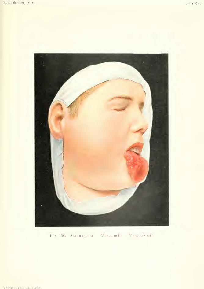
Bockeiiheimer, Atlas. Tab. CXX.
Fiu. IJO. Akiomeyalia — Alakroniclia A\akroglossia.
Rebiiian Comnanv. NeT-N'ork

megaly by being congenital. In ieontiasis ossea there
is enlargement of the bones, while the soft parts are
more often atrophied. Acromegaly affecting one ex-
tremity only might be mistaken for osteitis deformans,
or for chronic osteomyelitis, as there may be length-
ening of the bone in both these diseases. Acro-
megaly differs from elephantiasis in the presence of
enlargement of the bones, which can be shown by
the X-rays. Acromegaly commencing in the face
might possibly be mistaken for tumor of the upper
maxilla, but there is usually early hypertrophy of the
cheeks (macromelia), lips and tongue (macroglossia),
and of the hands and feet.
Treatment. Thyroid extract and extract of
pituitary gland have been recommended. Tumorof the pituitary gland may be removed by operation.
Extensive enlargement of the soft parts may be
diminished by cuneiform excision.
Fig. 150 shows marked hypertrophy of the right
side of the face. The right ear is considerably
larger than the left, and there is hypertrophy of all
the tissues of the cheek. The right side of the
tongue is enlarged, somewhat resembling cavernous
lymphangioma, but differing in being unilateral.
X-ray examination showed unilateral enlargement of
the upper and lower maxillary bones. The fingers
and toes on the right side had increased in size for
some years. The X-rays showed widening of the
sella turcica indicating the presence of a tumor of
the pituitary body. As the patient suffered no
trouble from the disease, he refused operation.
411


Complete IndexAbscess. 19
bone, 189burrowing, 191cold, 1S9, 335embolic, 190epidural, 257gummatous, 299, 314hot, 189lymphadenitis, 189milk, 193metastatic, 190paramammillarj-. 189subcutaneous, 189subperiosteal, 256thrombo-phlebitis, 186
Achylodynia, 378Acne rosacea, 140Acrania, 384Acromegaly, 381, 410Actinomycosis, 40, 293Adenoids, 44Adenoma, 72
malignant, 72sebaceous, 72
Adenophlegmon, 237Amelus, 408Amniotic amputation, 381, 408Anchylosis, fibrous, 335
osseous, 338tuberculous, 329
Anencephalus, .381
Aneurism, 57, 87arterio-venous, 169cirsoid, 137consecutive, 170false, 169pulsating, 172racemose, 171traiunatic, 170true, 169varicose, 170
Angina Ludovici, 237Angina, syphilitic, 301Angiolipoma, 104Angioma, cavernous, 8, 152
fissural, 152hypertrophic, 152neuropathic, 1.53
plexiform, 152rae<>mose, 1.52
Angiosarcoma, 34, 74cutaneous. 46, 74plexiform, 74
Animal baths, 284
Anthrax, 202, 287Anti-streptococcus serum, 271Antitetanin, 162Anus, hemorrhoids of, 102Arteritis, sj-philitic, 304Arteriosclerosis, 370, 374Arthritis, 82, 337
fibrinous, 283, 329, 338fungoid. 328, 335gonorrhceal, 282gouty, 377phlegmonous, 282purulent, 283. 329, .337
rheumatic, 283tuberculous, 284, 335, 337urica, 377
Asphyxia, local. 368Atheroma, 24, 374
carcinoma, 24
Bacteriaemia, 261Balanitis, 92Ballottenient of patella, 339Balsam of Peru, 146Barlow's disease, 157Basal-celled cancer, 4, 74Basedov's disease, 87Birth marks, 134Boil. 196Bone plugging, 253Botrioraycosis, 46Breast cancer, 16Bronchocele, 85Bubo, mdolent, 300
inguinal. 278Bullet wounds. 162Burn, X-ray, 305Burns, 30, 355
internal, 359Biu-sitis, 82
prepatellar, 82, 84
Cadaveric tubercle, 322Cancer en cuirasse, 23Cancroid, 3Capsule, sequestral, 248Caput medusa, 174Carbolic gangrene, 345, 354Carcinoma, branchial, 40Carbuncle, 202
diabetic. 202facial, 202buccal, 10disseminated, 22
413

Carbuncle, glandular, 16inoperable, 4, 20metastatic, 40, 58naevus. 24papillary, 30paramammary, 18of basal cell, 5of breast, 16
of cicatrLx, 30of connective tissue, 20of face. 1
of forehead, 1
of hand. 31of lip, 6of mamma. 16, 22of nipple, 16of nose, 1
of penis. 27of skin, 4, 6. 24, 30of tongue, 6, 13
Caries sicca, 329Cattle fever, 2S7Cavernoma, 66
of tongue, 66Cephalhematoma, 161Cephalocele, 383Cephalocele, see EncephaloceleChancre, phagedenic, 300
syphilitic, 299tongue, 311
ChilbLains, 362Chimney-sweep's cancer, 30Chiragra. 377Chonilrofibroma, 69Chondro-lipoma, 99, 104
myxoma, 99sarcoma, 99
Chondroma, 35, 99Chondromyxonia , 99Chondromyxosarcoma, 62Chondrosarcoma, 57, 62Claudication, intermittent, 375Cla\'us, 232Claw-hand, 120Combust io, 355Compression of ulnar nerve, 120Condylomata, 302Condylomata lata, 301Congelatio, .360
Contracture, of arm, 122arthrogenous, 126cicatricial, 116dermatogenous, 118Dupwjtren's . 115ischaemic, 122hysterical, 120myogenous, 123neurogenous, 120palmar. 115paralj'tic, 120tendogenous, 118
Corn, 232Corona veneris. 301Corpus cavernosum, 28Coxa vara. 129Craniotabes, 128Cr^de, unguentum, 188Cretinism, 88
Cutaneous cancer, 72fibroma, 136horn, 72
Cystadenoma, 69Cysts, bone, 19
branchial, 92dermoid. 92
Cystic goitre, 85hygroma, 84
Cysto-sarcoma (of breast), 49
Dactylitis, syphilitic, 305, 317, 343Decollement de la peau, 148Decubital ulcer, 350Delirium, 209Dermoid cysts, 92
reciurent. 92Detachment of skin, 148Diabetes, 370Diathesis, hemorrhagic, 156Diffuse hematoma. 156Dislocation of shoulder, 259Dislocation with fractm-e of leg, 132Dissemination, cancerous, 22Duct, persistent omphalo -mesenteric,
387, 404Dupuytren's contracture, 115
Ecchondroma, 99Ecchondroses, 99EcchjTiioses, 160Eczema of nipple, 16. 22Elephantiasis, 142, 145
acquired, 142hard, 142IjTnphangiectatic, 142nervorum, 137, 139penis, 142soft, 142
Embolism, tuberculous, 319Empyema, 179
of antrum, 241Encephalocele, 94, 381, 383Encephalomalaeia, 41Enchondroma. 99Endarteritis. 304. 347Endocarditis, ulcerative, 266Endothelioma, 32. 74
of parotid, 77Endothelial cancer, 77
sarcoma, 77English disease, 128Ephelides. 134Epidermoids, 92Epithelial cysts, 92Epithelioma, 21Epulis, 34, 64Erysipelas. 204
bulbous, 205chronic, 211curative, 206erythematous, 204gangrenous, 204hemorrhagic, 208migrating, 204phlegmonous, 204recurrent. 204
Erysipeloid, 211
414

Exostoses, cartilaginous, 62malignant, 62subungual, 126
False joints. 132Fibro-adenoma, 19, 49, 69
cystic, 69FibroUpoma, 97, 104
pendulous. 104Fibroma, 96. 116
cutaneous, 136intracanalicular, 69melanodes. 134vaginae tendinis, 96
Fibromata ani, 102mollusca, 136, 139
Fibromatosis. 137, 142Fibrolipoma, 104Fibromyoma, 96Fibromyxoma, 96Fibrosarcoma, 36, 60, 64Fistula, IjTnph, 399
from foreign body, 109of neck. 110mammarj'. 195osteomvelitic, 259rectal. 333umbilical, 404vesical. 294
Flatfoot, 174. 177Folliculitis. 196Fracture, treatment of, 128
pathological, 57rickety, 128
Fracture-dislocation, 132Framboesia, 46Freckles, 134I'rost^bite, 360Furuncle, 196Furunculosis, 82, 196
Ganglion of wrist, 80Gangrene, angioneurotic, 347
angiosclerotic, 348carbolic. 345, 354diabetic, 3.50. 370dry, 345, 370embolic, 347infective, 350moist, 317, 344, 350progressive, 345Raynaud's, 347, 367senile, .346
spontaneous, 350Gaseous phlegmon, 274General infection, 261Genu valgum, 129Geographical tongue, 298Giantism, 64, 143, 410Glanders, 287Cilands, tuberculous, 324Gliosarcoma, 35Glossitis, 298Glossy skin. 220Glottis, oedema of, 239Goitre, 85
exophthalmic, 87Gonococcal metastases, 282
41.5
GonorrhoDal arthritis, 282Gout, 377Grafting, 108Granulations, 108Grave's disease, 87Greenstick, 128Gumma, 299, 302
of lip and no.se, 299, 313of tongue, 299, 312
Gummatous abscess, 314astoitis, 315ulcer, 316
Hallux valgus, 126Hammer-toe, 126Hebra's ointment, 146Heat stroke. 359Hemangio-endothelioma, 152Hemangioma, cavernous, 66, 150, 166
simple. 152subcutaneous, 166, 168
Hemarthrosis. 120, 158Hematoma, diffuse, 156
ear, 150pulsating, 160subcutaneous, 156, 160
Hemophilia, 1.56
Hemorrhage from compression, 164Hemorrhoids, 102Hernia, abdominal, 406
mnbilical. .381,406Hodgkin's disease, 40Hordeolum, 196Horn, cutaneous, 72Hospital gangrene, 345Housemaid's knee, 82lluichinson's teeth, 306Hydrocephalus, 393Hydrops, tuberculous, 328, 330, 389Hygroma, 82
cystic, 399multilocular, 84tuberculous, 342
Hyperkeratosis, 10
Icterus, hematogenous, 267Implantation carcinoma, 6Infection, generalized, 261
pyogenic, 179Infective diseases, 350Inflammation. 90Interdigital whitlow. 227Intestine, actinomycosis of, 293
anthrax of, 287tuberculosis of, 341
Iodoform bone plugs, 253Ischaemic contracture of arm, 122
Joint effusion, 248
Keloid, after laparotomy, 113after vaccination, 113
Keratitis, parenchymatous, 306Keratoma, senile. 72Knee, hygroma of, 84Kyphosis, 129
Lassar's zinc paste, 146Laparotomy keloids, 113

Leg. fracture of, 128, 132varicose ulcer, 145
Lentigines, 134Leontiasis ossea, 410Leucoplakia, 11
preputial, 27Lightning stroke, 355Lingua bifida, 312Lingua geographica, 298Lipoma, diffuse, 104
symmetrical, 94, 104Lipomatosis, 106Lung, actinomycosis of, 293
anthrax of, 289ca\itation of. 268tuberculosis of, 323
Lupus, 8exfoliating, 10exulcerans, 10hypertrophic, 10of face, 9ulcerative, 10
Lupus-carcinoma. 9Luxatio, 132Lymphadenitis, acute, 278, 323
axillary, 286cerv'ical, 279, 325chronic, 280circumscribed, 281, 292difi'use, 278inguinal, 278ne~ck, 325tuberculous. 280, 325
Lymphangio-endothehoma, 74Lymphangio-fibroma, 134Lymphangioma, cavernous, 67, 397
circumscribed, 399cystic, 67, 397multiple, 381, 397simple, 67, 397
Lymphangitis, 196, 200, 213, 223carcinomatous, 16chronic, 23tuberculous. 323
Lymphatic glandular abscess, 281phlegmon, 278tuberculosis, 334
Lymph fistulas, 399Lymphoma, carcinomatous, 16
leukaemic, 40malignant, 39sarcomatous, 34, 39, 48syphilitic, 39tuberculous, 317, 334
Lymphosarcoma, 34, 39, 40, 48Lymphvarix, 142
Macrocheilia, 410Macroglossia, 381, 410Macromelia, 381, 410Malformations, 381Malignant oedema, 274
pustule, 287Marginate glossitis, 298Malum perforans, 367Mastitis, 192
carcinomatous, 23clironic cystic, 70
41G
Mastitis, interstitial, 70, 193neonatorum, 192phlegmonous, 192puerperal. 192superficial, 193tuberculous, 193
Meckel's diverticulum, 404Median fistula of neck, 110Mediastinitis, 87Melanocarcinoma, 32, 39, 48Melanoma, 32Melanosarcoma, 32, 48Meningitis, 392Meningocele, 390Meningococcus serum, 285Mesenteric tuberculosis, 381, 404Metastases, 58Mikulicz's disease, 94, 399Miliary tuberculosis, 333Milk fistula, 195Miner's elbow, 82Mixed infection, 181Mixed tumor, 19, 77Mucous membrane, erysipelas of, 204
carcinoma of, 13papilloma of, 11tuberculosis of, 332
Mucous patch, 301Multilocular hygroma of knee, 84Mummification, 345Myelocele, 381, 388Myelocystocele, 381, 388Myeloma, 57Myxcedema, 88Myxolipoma, 104, 381, 388Myxosarcoma, 60
Nffivi, 1.34
NsBvus carcinoma, 24hairy. 134lymphangiectatic, 134neuromatous, 136pigmentary, 134vascular, 134, 155verrucosus, 135vinous, 155warty, 24
Nasopharyngeal polypi, 42, 45Necrosis of bone, 287, 371
fascia, 350Neck, fistulas of, 110
]ihlegmon of, 237Nephritis, 265Nerve, elephantiasis, 139
compression of, 120Neurasthenia, traumatic, 214Neurofibroma, 136Neurogenous contractiu'e, 120Neuroma, 137, 139
CEdema, acute, 274malignant, 209, 274
Omphalo-mesenteric duct, 381Onychogryposis, 376Osteitis, deformans, 251
fibrous, 56gummatous, 299, 315purulent, 2.50

Osteitis, tuberculous, 317, 343Osteochondritis, syphilitic, 305Osteocopic paiiis, 304Osteomalacia, 129Osteomyelitis, 58, 244, 250, 259, 327
acute, 244, 252, 257chronic. 253. 259of humerus, 259of jaw. 255of lower maxilla, 255of scapula, 257of tibia, 244. 260
Osteosarcoma, 55Othematoma, 150Otitis media, 245Ozoena, 303
Pachydermia. 142acquired, 145lymphangiectatic, 145
Paget's disease of nipple, 16, 22Panaritium (sec whitlow). 213Papilloma, inflammatory of skin, 90
of tongue, 6. Illmalignant. 91
Paramammillarj' abscess, 189Parotid, endothelioma of, 77Paronychia. 228Parulis, 241Payr's magnesium, 68Pediculosis, 292PeK-is, rickety, 128Pemphigus, 305Pendulous fibrolipoma. 97, 104Penis, elephantiasis, 142
carcinoma, 27Perforating ulcer of foot, 367Periadenitis, 278Periarteritis, 304Pericarditis, 350Periostitis, albuminous, 249
alveolar, 241gummatous. 241ossifying, 248purulent, 241
Periphlebitis. 186Peritoneal tuberculosis, 333Pernio, 360Pes planus, 177
valgus, 129. 174, 177varus, 381, 388, 395
Petechia, 160, 164Phagocytosis, 179Phimosis, 92Phlebectasis. 174Phlebitis, thrombo, 186Phleboliths, 130Phlegmasia alba dolens. 187Phlegmon, 234, 274
emphysematous, 274gaseous, 274gangrenous. 274neck, 237putrefactive, 234submaxillary, 237wooden, 2.38
Phocomelus, 408Phosphorous necrosis, 256
417
Pigmentary na;vi, 134carcinoma, 25
Plague. .Siberian, 289Pleuritis. 3.50
tuberculous, 333Podagra. 377Polypus, malignant nasal, 42. 45Polyvalent serum. 271Putt's disease, 327Prepatellar bursiti.s, 82, 84Pseudarthrosi.s, 132Pseudoleukaemia, 40Psoriasis. 301
bucail, 9lingual. 9
Puerperal mastitis. 192Purpura hemorrhagica, 157Pustule, malignant, 202, 287Pyaemia, 261
chronic. 294Pyogenic infections, 179, 200, 262
Quinsy, 245
Rag-sorters' disease, 287Raynaud's disease, 367Recklinghausen's disease, 389Rhachischisis, 381, 383, 387, 388Rhachitis. 128Rheumatism, 282Rhinitis, atrophic, 43RhinophjTna, 140Rhinoscleroma, 140Rickets, 128Rider's bone, 169Riedel's preparation, 372Rinderpest, 287Roentgen-ray burn. 365Rosary, rickety, 128Roseola, 301Round-celled sarcoma. 35, 46Rupia, syphilitic, 301
Saber blade tibia, 305Sand baths, 284Sarcocele, 34Sarcoma, cavernous, 46
central, 53cutaneous. 34, 52endothelial. 74epipharyngeal. 34, 42fascial. 34, 60
_
fungoid, 34. 46giant-celled. 35, 64hemorrhagic. 52inoperable, 58mamman,-. 34, 49myeloid, 1.58
multiple, 52osteo, .58
parosteal, 57peripheral (of humerus), 34, 55phyllodes, ()9
pigmentary, 53round-celled, 35, 46
Sarcomata, 34Scars, hypertrophic, 114
Scleroderma, 122

Sclerosis, 299, 311Scoliosis. 129Scrofula. 321Scrofuloderma, 319Scurvy—rickets, 157Sebaceous adenoma, 72
furuncle, 196Seborrhoea, senile, 1
Sepsis, 261Septicaemia, 261Septico-pysemia. 261Sequestrotomy, 247Serous effusion, 246Serum, polyvalent, 271Silver, colloidal. 271Skin, carcinoma of. 30
detachment of, 148endothelioma. 74gangrene of. 345grafting of. 108horns of, 72papilloma of, 90sarcoma of, 53tuberculosis of, 322
Snowball crunching, 120, 157, 160Spina bifida, 388
occulta, 389ventosa, 343
Spindle-celled sarcoma, 35, 46Spirochaeta pallida, 299Splenic fever, 287Spondylitis, tuberculous. 327Spontaneous gangrene. 350Staphylococcal infection, 182
mycosis, 262phlegmon, 204serum. 221
Staphylolysin, 249Stomatitis, 309Streptococcal infection, 263
mycoses, 263osteomyelitis, 250phlegmon, 276
Strictures, 303Struma cystica, 85Subcutaneous hemangioma, 166, 168
hematoma, 160Subluxation, 338Suffusions, 160Suggillations, 160Sunstroke, 359Sweat-gland abscess. 189
furuncle, 196Syphilide, macular, 301
papular, 301Syphilis, acquired. 299
congenital, 305malignant, 305
Teeth, syphilitic, 306Telangiectases, 152, 155Tendon, contraction, 118
whitlow, 218sheath, fibroma of, 96, 116, 118
Tendovaginitis, tuberculous, 342Teno-svnovitis, 81Teratology, 381Teratoma, 44, 381, 401
418
Testicle, tuberculosis, 317, 341Tetania, strumipriva, 88Thiersch's grafts, 108Thrombo-phlebitis, 186Toe-nails, ingrowing. 230Tonsillar hypertrophy, 44Tongue, abscess of, 12
actinomycosis of, 293bifid, 312carcinoma of. 6, 13chancre of, 299geographical. 298gumma of. 299sj-philis, 311
Tophi, 378ToxiuEemia, 263Triad of Hutchinson, 306Tuberculin, 322Tuberculosis, 317
of bladder, 333, 341of bones, 325, 335of glands. 324of hand, 317, 342of joints, 328of neck, 317of prostate, 341mUliary, 333of skin, 322surgical, 320of testicle, 317, 341
Tumor albus, 329, 339mixed, 77retromaxillary, 42
Typhoid, 270
Ulcer, decubital, 350gangrenous, 350gimamatous, 299, 316moUe, 299phagedenic, 28rodent, 1
varicose, 145lUcus rodens, 1
UmbUical hernia. 381Unguis incarnatus, 230Unna's zinc gelatin, 145Urachal fistula, 382Urinary phlegmon, 277
Vaccination keloids, 113Vagina, fibroma of. 96Valgus, pes, 174, 177Varicocele, 174Varicose ulcer of leg, 145Varix, 174
aneurismal, 170cirsoid, 174submucous, 174
Vascular n£e\'us, 155Villous cancer, 90
polj-pus, 90Von Recklinghausen's disease, 389V-phlegmon, 215
Wart, 30White leg, 187White swelling, 329, 339

Whitlow, 213 AVhitlow, tendinous, 226articular, 224 Wooden phlegmon, 238chronic, 229 Wound diphtheria, 234mterdigital, 227 Wounds, treatment of, 235osteal, 224 Wrist, ganglion of, 80perioste;U, 224peri-ungual, 228 Xeroderma pigmentosum, 1, 33subcutaneous, 223 X-ray burns, 365subepidermal, 222sub-ungual, 228 Yaws, 46
419


University of California
SOUTHERN REGIONAL LIBRARY FACILITY405 Hilgard Avenue, Los Angeles, CA 90024-1388
Return this material to the library
from which It was borrowed.
810W
Form
D 000 142 342

ii^:'';i:!!'iii)iif!iip!!il!ii|ii^^^^^^