Diagnosis - Springer
26
Tachycardia, supraventricular Diagnosis Definition • Supraventricular tachycardias include the following: Sinus tachycardia. Atrial fibrillation. Atrial flutter. Atrial tachycardia. Atrioventricular re-entrant tachycardia Atrioventricular nodal re-entrant tachycardia. • Atrioventricular re-entrant and nuda! re-entrant tachycardias. the two common supra- ventricular tachycardias arising from the atrioventlicular junction, are discussed here. Symptoms Palpitation: paroxysms of reguJar palpitation at 140-240 bcats/min, with sudden onset and offset. Syncope: palpitation may be associated with syncope or presyncope at onset of attack, when blood pressure is probably at its lowest. Chest pain: unusual but may occur during attacks, particularly in presence of ischemie heart disease. Pat·oxysmal attacks: occasionally prccipitated by postura} changes or may be associated with particular times in menstrual cycle. Diaphoresis, lightheadedness, and dyspnea. Signs Pul se Fast, regular pulse. Jugular venous pulse • In atrioventricular nodal re-entrant tachycardia, prominent jugular venous pulsations that match the rate of tachycardia, a feature referred to as the frog sign. Heart Signs of left ventricular failure: unusual unless stn1ctural heart disease is coexistent. • Atrioventricular dissociation is not evident (no cannon wavcs in neck). • Blood pressure is usually well maintained after the first few seconds of an attack. 1 nvestigations ECG: regular rhythm present, usually with narrow QRS complexes; occasionally, pre- existing or rate-related bw1dle branch block leads ta broad QRS complexes. Atrioventricular nodal re-entrant tachycardia: Usually the P waves are superimposcd in the QRS complex in II. lil, and aVF. In about 10% of the patients. clearly visible inverted P waves are seen in Il, III, and aVF and an RP interval during tachycardia that is much longer than the PR interval. Chest radiography: usually normal unless stntctural heart di.sease coexlstent. Electrophysiology: indicated if catheter ablation contemplated or for risk assessment in symptomatic patients with \Volff-Parkinson-White syndrome. Complications Left ventricular failure: caused by coexistent structural heatt disease: supraventricular tachycardia may cause ventricular failure in the absence of pre-existing structural heart disease only if tachycardia is incessant and has continued unintenupted for months or years. 428 Differential diagnosis Atriel tl<h)urdoa or atrial ta<hyurdtl " lf otrloventrlcular r.,_.,ntrant ta<hycardia " conducted woth b<an<h block • When histO!)' ls belng taken ... taJ>. h>hrng the p<eseno>"' ab>ence of structural dlsea5e, cardoomyopathy 01 pr..- voous myocardial infarction, k Important. A hostO!)' or known diagnosis of eithe< of the<e condotlom mak .. a dlagnosis of tac:hyurdoa muth p<obable than atrio- ventrkular re-entr .. ant or nodal re--entrant ta<hycardia 11 the QRS complex is b<oad and h"' a unhke lhal of classic left or right bundle branch bloci<, dtagnosis ls p<obably ventricular ta<hycardla Etiology Atrioventricvlal ..... uant !Khyun!Y • The structural substrete i congenital abnormahty conducting system of the hean. an extta connection exo$1> betwHn the atr ia and the .. • Tachycardla anses when an eleoctn<al 1mpulse passes from t he atrium to the ven· through the n01mal node bul returns 10 atria by the a<< .. • sorypathway Atrioventrlcula< noclal - ta<hyQnlla • PaUents have two functlonally 5eparate pathways withon or close 10 lhe atrloven· trteular node- . • TKhyci rdia •nses ln 11imilar way to that ansing m patienu w•th reentranl lachyurdla Epidemiology • These arrhythmias occur frequently and form most ol t he tachycardlas in pat lenu wilh structurally normal hearu • Men are more hkely to have atrioventrt<· ular tachycardia. and women atrloventncular nodal • Parooysms of palpitat lon may start In mfancy but mOfe usual 1n teenage years or twent irs • Alt hough AVNRT may appear al any "!!"· most seek medocal allentlon dunng fourth or fofth de<ade of hle. J. O. Woolliscroft (ed.), Current Diagnosis & Treatment © Springer Science+Business Media New York 2001
-
Upload
khangminh22 -
Category
Documents
-
view
0 -
download
0
Transcript of Diagnosis - Springer

Tachycardia, supraventricular
Diagnosis
Definition • Supraventricular tachycardias include the following:
Sinus tachycardia.
Atrial fibrillation.
Atrial flutter.
Atrial tachycardia.
Atrioventricular re-entrant tachycardia
Atrioventricular nodal re-entrant tachycardia.
• Atrioventricular re-entrant and nuda! re-entrant tachycardias. the two common supraventricular tachycardias arising from the atrioventlicular junction, are discussed here.
Symptoms Palpitation: paroxysms of reguJar palpitation at 140-240 bcats/min, with sudden onset and offset.
Syncope: palpitation may be associated with syncope or presyncope at onset of attack, when blood pressure is probably at its lowest.
Chest pain: unusual but may occur during attacks, particularly in presence of ischemie heart disease.
Pat·oxysmal attacks: occasionally prccipitated by postura} changes or may be associated with particular times in menstrual cycle.
Diaphoresis, lightheadedness, and dyspnea.
Signs Pul se Fast, regular pulse.
Jugular venous pulse • In atrioventricular nodal re-entrant tachycardia, prominent jugular venous pulsations that match the rate of tachycardia, a feature referred to as the frog sign.
Heart Signs of left ventricular failure: unusual unless stn1ctural heart disease is coexistent.
• Atrioventricular dissociation is not evident (no cannon wavcs in neck).
• Blood pressure is usually well maintained after the first few seconds of an attack.
1 nvestigations ECG: regular rhythm present, usually with narrow QRS complexes; occasionally, preexisting or rate-related bw1dle branch block leads ta broad QRS complexes.
Atrioventricular nodal re-entrant tachycardia: Usually the P waves are superimposcd in the QRS complex in II. lil, and aVF. In about 10% of the patients. clearly visible inverted P waves are seen in Il, III, and aVF and an RP interval during tachycardia that is much longer than the PR interval.
Chest radiography: usually normal unless stntctural heart di.sease coexlstent.
Electrophysiology: indicated if catheter ablation contemplated or for risk assessment in symptomatic patients with \Volff-Parkinson-White syndrome.
Complications Left ventricular failure: caused by coexistent structural heatt disease: supraventricular tachycardia may cause ventricular failure in the absence of pre-existing structural heart disease only if tachycardia is incessant and has continued unintenupted for months or years.
428
Differential diagnosis Atriel tl<h)urdoa or atrial flutt~ .
v~ntrkular ta<hyurdtl" lf otrloventrlcular
r.,_.,ntrant ta<hycardia " conducted woth bundl~ b<an<h block
• When th~ histO!)' ls belng taken ... taJ>.
h>hrng the p<eseno>"' ab>ence of structural h~art dlsea5e, ~.g . , cardoomyopathy 01 pr..
voous myocardial infarction, k Important. A hostO!)' or known diagnosis of eithe< of the<e
condotlom mak .. a dlagnosis of ~nlrocular
tac:hyurdoa muth mor~ p<obable than atrioventrkular re-entr .. ant or nodal re--entrant ta<hycardia 11 the QRS complex is b<oad and
h"' a patt~rn unhke lhal of classic left or right bundle branch bloci<, th~n dtagnosis ls p<obably ventricular ta<hycardla
Etiology
Atrioventricvlal ..... uant !Khyun!Y
• The structural substrete ~ i congenital abnormahty olth~ conducting system of the
hean. -~ an extta ~lecUical connection exo$1> betwHn the atria and the ~ntrocl ..
• Tachycardla anses when an eleoctn<al 1mpulse passes from the atrium to the ven· triei~ through the n01mal atrlo~ntricular
node bul returns 10 th~ atria by the a<< .. • sorypathway
Atrioventrlcula< noclal - ta<hyQnlla • PaUents have two functlonally 5eparate pathways withon or close 10 lhe atrloven·
trteular node-.
• TKhycirdia •nses ln 11imilar way to that ansing m patienu w•th ~J trlove:ntricul•r reentranl lachyurdla
Epidemiology
• These arrhythmias occur frequently and form most ol the tachycardlas in pat lenu
wilh structurally normal hearu
• Men are more hkely to have a trioventrt<· ular r~n1rant tachycardia. and women atrloventncular nodal re~entrant tachyardla.
• Parooysms of palpitatlon may start In mfancy but mOfe usual 1n teenage years or twentirs
• Although AVNRT may appear al any "!!"· most pato~nu seek medocal allentlon dunng th~ fourth or fofth de<ade of hle.
J. O. Woolliscroft (ed.), Current Diagnosis & Treatment© Springer Science+Business Media New York 2001

Tachycardia, supraventricular
Treatment
Diet and lifestyle • If the tachycardias are initiated by atrial premature beats, abstinence from caffeine may help.
• Otherwise, no special precautions are necessary.
Pharmacological treatment Acute treatment Standard dosage
Contraindications
Special points
Adenosine, i.v. bolus duse followed by saline flush, starting at 3 mg, with a second duse of 6 rug if tachycardia does not terminate after 60 seconds; a further dose of 12 mg is given if tachycardia does not terminate after another 60 seconds. Verapamil, 5 mg i.v. slowly (30 seconds); if tachycan.lia has not terminated after 5 minutes, a second 5-mg duse may be given, if hypotension has not occurred.
Adenosine: asthma. Verapamil: poor ventricular function.
Adenosine: although 12 mg is maximum adult dose recommended in product license, bolus doses of 18 mg may occasionally be need to terminate tachycardia; may exacerbate bronchoconstriction. Verapamil: negatively inotropic antl should not be given to patients with known abnormal ventricular function or with signs of cardiumegaly on chest radiography; best not given tu patients with a broad complex tachycardia because it may cause cardiovascular collapse if erroneously given tu patients with ventricular tachycardia; i.v. verapamil should not be givcn to patient taking oral beta-blockers because sinus arrest or dramatic hypotensiun may occur.
Main dntg interactions Adenosine: increased effect with dipyridamolc; decreased effect with theophylline.
Main side effects Adenosine: flushing and chest tightness (transient).
Prophylaxis • Beta-blockade (e.g., atenulol, 50-100 mg) i<; often effective in this rute, particularly if the attacks are exercise-induced.
• Diguxin, 0.125 mg, ;md verapamil, 180-360 mg, may be effective but are contraindicated in the presence of a delta wave because they may increase the ventricular rate if atrial fibrillation complicates the Wolff-Parkinson-\Vhite syndrome.
General references Ganz U, Fd~man PL Supraventficular tachycardia N EngiJ M~ 1995, 332.:162
Morady F Radoo.frequency ablatoon as
treatment for tardta( arrhythmtas.. N fngl J
M~ 1999, 340:534
42l)

Tachycardia, ventricular
Diagnosis
Symptoms • Symptoms are not always manifest.
Palpitatioru;.
Sudden shortness of breath.
Dizzy spells.
Blackouts.
Cardiac arrest.
Sudden death.
Signs • Signs are not always manifest.
Tachycardia: 100-300 beat•/min.
Hypotension and associated signs.
Cannon waves in jugular venous pressure, variable blood pressure, variation in intensity of îrrst heart sound: signs of atrioventricular dissociation.
lnvestigations 12-lead ECG: initially, to confirm diagnosis, after treatment, for comparison of sinus rhythm; reveals tachycardia with QRS complexes 140 ms duration, evidence of atrioventricular dissociation (independent P waves, fusion beats, cap ture beats, second-degree ventriculo-atrial block), marked left or right axis deviation during tachycardia, absence of RS complcxes in chest Jeads during tachycardia.
Adenosine test: initially, to distinguish from junctional ora triaJ tachycardia; if patient presents with stable tachycardia anct if 12-lead ECG cannot be interpreted as showing ventricular tachycardia, incremental boluses of adenosine 0.05-0.20 mg!kg i.v. should be given, which tcnninates almost aU junctional tachycardias, slows most atrial tachycardias. but affccts almost no vcntricular tachycardias cxcept thosc arisîng in the right vcntricular outflow tract.
Cardiac enzyme ana1ysis: after treatment. if history suggests înfarction.
Echocardiogram: to determine underlying cardiomyopathy or valvular disease. to assess left and right ventricular function.
Exercise test: under supcrvision of arrhytllmia specialist to look for coronary disease, to provoke arrhythmia, and to assess drug efficacy.
24-hour ambulatory ECG recording: to quantify frequency of ventricular arrhythmia and associated arrhythmias (e.g., ventricular premature beat).
Left ventricular and coronary angiography.
Programmed electrophysiologic testing: to detennine inducibiliry, morphology, and suppressibility of symptoms and to guide decisions among dntg, ablative, or ICD therJ.py.
Complications Cardiac arrest.
Sudden death.
Cardiogenic shock.
Pulmonary edema.
430
DlfferenU.I diagnosls Supt ~v~uicular t.chyc.rdoa: eotlwr atrial
or functoonal, woth erth« roght or leit
bundle branch block.
o Ali wide-<omplex t«hy<ardias lohould M tonJtdered ventricular in orlgtn unttl
pro.ed otherwow.
Etlology
Acute oKhemoa coronary ob<truction by
thtombus
~rge scar old onlarctoon: p<evoous surgety,
dysplasias.
Mo<roscopk sur lnloltratoon: fibrosk
No clonical dliease: e.g .. rightventro<ular
outllow tachycardla o Right ventro<ular dysplasia (where the roght venttlcular muscle os replaced by
lobrofany tossue and dlleted RV can be determoned by echoe<~rdlogroplly) .
o Hypertropllo< cardiomyopathy (Sumlned
VT In thew pato~H carri~ a hlgh ris!< of
sudden death) .
Voral myocarditls.
Funct1onat d1~easeo: •tasc..cular"' tac:hycard ...
o Fallot Tetralogy or VSD when succossfully corrii!Cied at mit for ~e _.opment of VT
Tumon, malformatoons. other Caus8 (rare)
Epldemiology
o 2.._ - 10.._ of patientJ sulferong a
myocardoallnfarction have a sustaoned
ventricular t.chy<ardia or cardiac arren wtthtn 12 months

Treatment
Diet and lifestyle • Avoid strenuous exercise and cat a nonnal diet unless evidence of coronary disca-,c.
Pharmacological treatment • TI1e underlying disea.;;e process or associated cardiogenic fluid retcntion mm;t be trcated.
Emergency Cardiopulmonary resuscitation if ncccssary.
Lidocainc 1 to 1.5 mg/kg intravcnously, followcd by infusion at 1 to 4 mglmin, supplemcnted by boluses of 0.5 to 0.75 mg/kg cvery 5 to 10 minutes ta a maximum of 3 mg/kg (in those with heart failure or liver damagc and in thc elderly, the infusion rate should be titrated to toxicity).
Procainamide 1 g intravenously over 30 minutes followed by 1 to 2 mg/min (decrea..;;e the infusion ratcs in patient.;; with renal insufficiency).
Amiodarone 150 mg intravenously ovcr 1 O minutcs followed hy 360 mg intravenously over 6 hours, then 0.5 mg/min infusîon.
Intravenous bretylium or lidocaine must be used with great caution and should be supervised by physicians well versed with these medications.
Direct--current cardiovcrsion in unstable paticnts (synchronizcd and starting at 100 J) or if dmg treatmcnt fails.
A succcssion of drugs is unwise, cspecially in patients with coronary discase and impaircd vcntricular function.
Vcrapamil must not be given: it may causc profound collapse and even death.
Long-term treatment • Drug treatment should be supervised by a cardiologist with a long-term interest in arrhythrnias.
• Ali antiarrhythmic dmgs ha ve proarrhythmic propcrtics cspccially whcn uscd in combination with othcr antiarrhythmic drugs.
• \Vhcther Iong-tcnn drug trcatment improves prognosis is not known. Currently amiodarone holds promise in the management of non-sustaincd ventricular tachycardia and the results of A.\1IOVERT, a randomizcd triat, is awaited.
lmplantable cardioverter defibrillator (ICD) • Implantation of automatic defibrillator device to terminate tachycardia: this also trcats ventricul ar tachycardia by rapid pacing.
• Two primary prcvcntion trials MADIT (Multiccnter Automatic Dcfihrillator Implantation Triat) and MUSIT (Multicentcr UnSustaincd Tachycardia TriaJ) substantiated irnproved survival with ICD thcrapy in coronary patient'i with nonsustaincd ventricular tachycardia.
• Two secondary prevention trials that focused on patients with aborted cardiac arrest ar life-threateoing cardiac arrhythmias ha ve also bcen completed: these are the A VID (Antiarrhythmic Vcrsus Implantablc Dcfibrillator) study and thc CIDS (Canadian Implantablc Dcfibrillator Study). A similar survival bcncfit was achicvcd with JCD thcrapy whcn comparcd with amiodaronc in both thcsc trials.
• Titc A VID data suggcstcd that paticnts with relativcly wcll prcscrvcd cjcction fraction P-0.35) do not have a bcttcr survival rate whcn trcatcd with thc ICD when compared with anti-arrhythmic drugs (mostly amiodarone). However, in patients with low ejcction fraction. the ICD was associated with improved survival when compared with antiarrhythmic dmgs. Currcntly two randomized trials I\1ADIT-II and SCD-HeFT (Suddcn Cardiac Death-Heart Failure Trial) are cvaluating thc survivaJ bcncfit of ICD therapy in patients with Jcft ventricular dysfunction.
• ICD is not stand-alo ne therapy for prcvcntion of sudden cardiac dcath. Thc rcduction of sudden dcath with bcta-hlockcrs and angiotcnsin convcning cnzymc inllibitors in paticnts with hcan failure is wcll substantiatcd and hypolipidemic therapy may a!so contribute additional henefit in patients with hcan failure duc to coronary diseasc.
Tachycardia, ventricular
General references
MouAJ ond the MAOIT lnvesi.J9.1Iors: lmp.-01/ed IUMVal woth an omplanted
defobflllatot In patlt'IIIS wolh <otonary dis<>""' at hlgh ri<lt. lot1A'ntn<ular arrhythm ... N fngl
JMI!d 19'16,llS:1933-1940.
8uxton Af and lhe MUSST tn~ogators· A
randomized study of the preventton of 1udden death 1n pauenn w1th coronary artery di.-,a<e. N Engl J MNJ 1999,
341: 1882- 1890.
Connolly SJ and the CIOS lnvostlgators
C..nadlan Implanta bie ~fibnllator Study· a
randomlzed mal of lmplantable <ArdOOVM;er
venus amiodaJonf!. c"cullltlon 2000. 101:1297-1]02.
Morady F Radoofr~uency ablatoon as
treatment of urdoa< arrhyt.hmoa-. N Engl J
MNJ 1999, 34&.534
-431

Thalassemia
Diagnosis
Symptoms • ţ3-Thalassemia is manifest during the ftrst year of life in 90% of patients; a few present at
3- 4 years Oate-onset J3-thalassemia major).
• 10% of patients with !3-thalassemia major ha ve a rrtild course (independent of transfusion).
• Patients may be asymptomatic; detection is fro m antenatal screening and ctiagnosis.
Poor weight gain.
Failure to thrive.
Fever.
Diarrhea.
Increasing pallor.
Distended abdomen.
Signs Pallor.
Heart failure.
Splenomegaly.
Jaundice.
lnvestigations Blood tests: low hemoglobin, mean corpuscular hemoglobin, mean cell volume;
absent ar reduced hemoglobin A (J3° or J3• thalassemia). variable hemoglobin F and
A2
(using elcctrophoresis).
Genetic analysis: defines specitk mutations. may help to predict disease severit)• and
prognosis and facilitate ftCSt-trimesler diagnosis.
Complications Splenomegaly Jeading to hypersplenlsm (neutropenia, thrombocytopenia.
anemla).
Anemia causing severe bone changes, short stature, and heart failure.
132
Diffenntlal dlagnosis
lron dofko ncy
Etlology
• Th•hmemla is calM<! by defectove
synthesis of the" or ~lobin thaln
(o · and ~thalassemla. respec\lvely).
• Tht! dtSOrder 1.s 1nh.r1t~ (~~han
receuive).
• It oc.curs In Med1terranean, Asian, Arabk.
and Chon...., groups be<ause of a selectove
advantage againn PlaJmOdlum f~fc,parum
Epidemiology [1)
• Thalass.em1a tS one of the most common
onhented dtSO<der> throughout 11\e world.
• Among blacks, app<oxlmately 30~ of
lhe populalton tS either heterozygous or
homozygous for the " ·1halassemla·2 deleuon.
• In southwest Europe. It "dec:llnong
b«ause of preventlon progra~ 1n consangUJneous m rriages. the b1rth
rate of IHhalaiSI!mla oncreases by 30%
Treatment
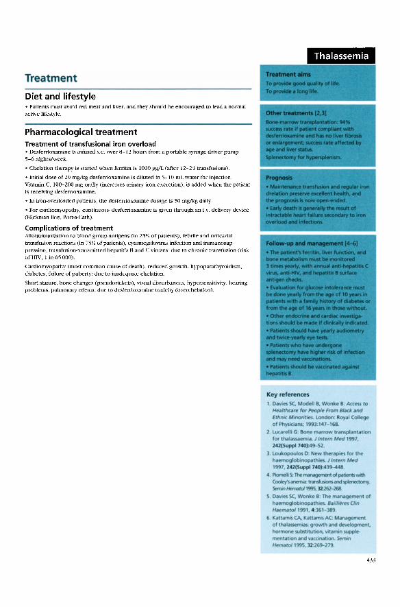
Diet and lifestyle • Patients must avoid rect meat anei liver, and they shoulcl be encouraged to lead a normal active lifestyle.
Pharmacological treatment Treatment of transfusional iron overload • Desferrioxamine is infused s.c. over 8-12 hours from a portable syringe driver pump 5-6 nights/week.
• Chelation therapy is started when ferritin is 1000 !Jg/L (after 12-24 transfusions).
• Initial dose of 20 mg/kg desferrioxamine is diluted in 5-10 mL water for injection. Vitamin C, 100-200 mg orally (increases urinary iron excretion), is added when the patient is receiving desferrioxamine.
• In iron-overloaded patients, the desferrioxamine dosage is 50 mg!kg daily.
• For cardiomyopathy, continuous desferrioxamine is given through an i.v. delivery device (Hickman line, Port-a-Cath).
Complications of treatment Alloimmunization to blood group antigens (in 25% of patients). febrile and urticarial transfusion reactions (in 75% of patients), cytomegalovirus infection and inmmnosuppression, transfusion-transmitted hepatitis B and C viruses: due to chronic transfusion (risk of HIV, 1 in 65 000).
Cardiomyopathy (most common cause of death), reduced growth, hypoparathyroidism, diabetes, failure of puberty: due to inadequate chelation.
Short stantre, bone changes (pseudorickets), visual disturbances, hypersensitivity, hearing problems, pulmonary edema: due to desfenioxarnine toxicity (overchelation).
Thalassemia
Key references 1. Oavl~ SC. Modell 8, Wonke 8 Ace~ to
1-/ea/rhcore for hople From Black and Ethn.c Mononro~ London· Royal College of Physlcians; 1993.147- 168.
2. Lucar~llt G: Bone marrow transpl~ntation for tl\alassaemla. 1 Intern M~ 1997, 242(Suppl 740):49- 52.
l Lookopoulos O New theraptes for the haemoglobtnopatht~ 1 Intern M~ 1997, 242(Suppl740):439-<148
4 l'lomolli S.lhe managoment of pauents"""' Cooley's anemia: translusoons and ~ Semn Hemaro/1995, 32262-268.
5. Oavies SC. Wonke 8 The management of l>aemogloblnopatht~ B<>ollo~r~ Clm Haemaro/1991, 4 ·361- 389
6 KattamtS CA. KanamiS AC Management of tl\alasse>mtas growth and developmenl. hormone subst•tutiOI\ Vltamon supplementauon and vaconatton. Semm Hemato/1995, 32 26~279
433

Thyroid carcinoma
Diagnosis
Symptoms • Patients may be asymptomatic (thyroid swelling is nored by someone else).
Neck swelling: rapid increase in size raises Jevel of concern.
Dysphagia, dyspnea, dysphonia: symptoms of local compression.
Pain in neck, radiating to jaw or ear.
Bone pain, hemoptysis, abdominal discomfort: may indicate metastases.
Symptoms of thyrotoxicosis: exceedingly rare.
Signs Single nodule: most common presemation; often firm or hard.
Fixation of thyroid swelling to local structures.
Cervicallymphadenopathy.
Evidence of metastases (unusual): spinal cord compression, spastîc pamparesis,
hepatomegaly, bone swellings, renden1ess.
lnvestigations • No blood test of radiological srudy can distinguish benign from malignant thyroid lesions.
Fine-needle aspiration cytology: test of choice to diagnose malignancy in
thyroid nodules.
Tbyroid function tests: serum-free thyroxine and thyroid-stimulating hormone; used to diagnose hypothyroidism or hyperthyroidism.
Isotope scanning: hor nodules (suppressing surrounding thyroid tissue) are rarely malignant; nodules that are not
hot should be cvaluated by fine· needle aspirJtion.
,_ ' ...-· ~" . ' ' . . . ! .' - ' : .. , .. ~--. . ~-·:1
• • 1 •. •' ... ·· •••• Fine-needle aspiration of a thyroid nodule, allowing
rapid cytological examination of aspirated material.
(See Color Plate.)
Ultrasonography: best for accurarely measuring size of nodules in patients who are being
followed; ca.n differentiate cystic vs. solid, but either may be malignant.
Serum calcitonin measurement: should be reserved for patients with k.nown meduUary
thyroid cancer (MTC) and for screening members of families with inherired tOnus of MTC.
Thyroglobullit evaluation: minimal value in diagnosis of d1yroid noduJes; extremely
useful for mon.itoring treared patients.
Antithyroid antibody tests: of little v:1lue in evaluating thyroid nodules.
Special considerations • Hyperthyroil.l patients probably should ha ve isotopc scanniog. If the nodule is hot
(autonomously functioning thyroid nodule), rnaligna.ncy is unlikely.
• A rapidly enlarging thyroid (diffuse or nodular) should raise the possibility of lymphoma.
Complications Local ittîtltration of trachea, esophagus, nerves.
Metastases of bone, liver, lung.
434
tffuse golle<,
Dlffentntial dlagnosls
lleotgn thyroid diw- d
s;ngle O< multtple nodules.
Hashlmoto"s dM-.
Other uuws of lymphade nop.~thy
Etlology (2]
• causes Include the follow.
E>tte<nal lrradtatton of he
Genetk. nsedullary CM1<e
endocrine neoplasla syndr
ng:
ad and neck . rIn mulltple 0-
Epiclemlology (21
fO< <0.5'111 of,_ • Thyroid unce< accounu
maltgnanc.es and <0.5% of canc:e< deaths.
d nodule., ls • Gotter, lncludlng thyrot
."_"tin up to 10,. of the populatlon .
• 'The f~le m•l• ratio t<l 1
t< increased wMt> • The rt<k of maltgnancy
nodules -lop tn chlldr
In adulu ~ >60 years en, young men. O<
Tumor pathology
lsthe mosi • Papillary thyroid can<er
frequently diagnosed ma
tumor (>80%).
ltgnant thyrold
r is a r~lat1vfly • Folltcular thyrOtd unce unusual mallgnancy (-5'111
thyroid cancors) - IO'H>ofall
• Medullary thyrold cance
5'111 - 10'111 of patt4!nU. r occun In
~ occurs rar~ty • AnaplastJ< thyroid canc
and has a~ poor prog nost<

Treatment
Diet and lifestyle • No evidence indicates that environmental factors affect the prognosis of established thyroid cancer.
• An iodine-replete diet and avoidance of extemal irradiation to head and neck reduce the risk of developing thyroid cancer.
Pharmacological treatment [1-41
• After surgecy, patients can be considered for radioiodine treatment.
• No consensus bas been reached on BIJ ablation of the d1yroid in patients with smaU solitary papillary lesions.
• Routine ablation of thyroid remnant is indicated in patîcnts with follicuJar thyroid cancer.
• Radioiodine has a clear role in the management of residual disease or established metastases.
• All patients with differentiated thyroid cancer need suppressive thyroxine treatment after defl1litive treatment; thyroxine is necessary in aU patients rendered hypothyroid
after treaunent.
Nonpharmacological treatment • The extent of surgery (simple lobectomy, near-total thyroidectomy, tota1 thyroidectomy) depends on pathology and surgical preference.
• Surgef)' is the primary mode oftreaunent for papîUary, fou.icuJar, and medullary thyroid cancers.
• Surgîcal removal of involved lymph nodes is indicated.
Thyroid carcinoma
~~· ~ ' - ,;- '. -: .
' ~ ' ~ 1 •
~: . . . .
:~- ' . •. -·
Key references
Kapfan MM, ec:t Thyro1d carcmoma.
Endocrinol Mo!Ob Clln North Am 1990, 19•741 - 760
2. DeGroot U, er al.: Natural hiflory. treat· rm.nt. and courw of papillary thyroid cardnoma J Clm Endoc.rinol Merabol 1990, 71 :414-424.
3 Utiger 110· Follow-up of patlenu w•th thyroid u.rcinoma N Engl J Ml!d 1997, 337 928
Samaan NA. era/ · The resulu of d•fferent modahtles of tre11tment of well d1ffere-n· tiatl!d thyroid u.rcJnoma· a retrospect•~ ri!Ylow of 1599 patienu. J Clm Endocrmol
Merab 1992, 75:714-720

Tobacco use disorder
Diagnosis
Symptoms and signs Daily use of cigarettes for at least several weeks.
Continued smoking of cigarettes despite knowledge of h ealth risk.
Potential for nicotine withdrawal symptonlS within 24 hours of abrupt ce..o;;sation
of smoking: dysphoria, insomnia, irritability, anxiety, resrlcssness, difficulty coocentrating,
increased appetite.
Impainnent in social or occupational functioning: may occur bccause of witl1drawaJ symptoms.
lnvestigations ASK 1 t]: must have S)'Stem in place to idcntify all tobacco users at every visit.
• Consider mbacco status as part of vital sîgns (e.g. , tobacco use status stickers on paticnt
charts) or inclusion of "tobacco use disorder" o n active problcm J.ist.
Fagerstrom Test for Nlcotine Dependence [2]: assists in establishing levcl of physical
dependence (see table). Correlation exists betwcen high level of dependencc (score >6)
and severity ofwithdrawal symptoms, difficulry with abstinence, and. rc lapse.
Fagerstrom test for ni coti ne dependence
Questions Score
1. How soon after you wake up do you smoke your first cigarette? <5 minutes 6-30 minutes 31-60 minutes >61 minutes
2. Do you find it difficult ta refrain from smoking in places where it is forbidden, e.g., church, libra ry, movie theater?
3. Which cigarette would you hate most ta give up?
4. How many cigarettes per day do you smoke?
YES NO
The first in the morni ng Any other
>31 21-30 11-20 <10
5. Do you smoke more frequently during the first hours after waking than du ring the rest of the day?
YES NO
6. Do you smoke if you are so ill that you are in bed most of the day?
Complications
YES NO
Cancer: causally Hnked 1'0 lung cancer; strong a'isociations with cancer of oral cavity ,
la.rynx, esophagus, bladder, kidney, panc rca-;, stomach, and cervix.
1 o
Chronic obstructivc pulmonary disea'ie: principal risk factor for chronic ohstmctive
pulmonary disease; current smokers havc Jower forced expiratory volume in J second
(FEV1) and accelerated decline in FEV
1 than nonsmokers.
Coronary heart disease: 2-4-fold increased incidence of coronary heart disease and
sudden death.
Cerebral vascular dL'iease: nearly 2-fold incrcased risk of srroke.
Pregnancy: pregnant smokers wi th increa'ied incidence of low birth weight bahies,
spontaneous abortio n, placenta previa, and placcntal abruption.
Differential diagnosis
Not apphcob~
Etiology
• Nk:otJn~ is the agent In t1garet1e-s
rHponSible for addt<t1ve propertiH
• ~pe-ndence occurs via lncreased expression of brain nitotlne rec:eptOf'S.
chang<'< '" b<a1n gluc:ose metabolism. and release of cate<holamlnes.
• Pos•ttve retnforcement O<CUr'$ w1th
dehvery of nkotlne via clgarettH and
negat1ve re1nforcement oc:curs via Wlthdrawal symptoms dunng qu11
attempts
Epidemiology
• There are 1 bilhon smoken worldw•de
• 25% of Ameran adult> are smokers.
• 3000 toenagers start to smoke each day;
most of thos<! who bec.ome hfelong
smokers start before h'9h school graduatiOI'I
• 440 000 deaths per y ar in the Unlted
StatH are !rom dlseaSM d~rt<tly I1nked
tosmokmg

Treatment
Diet and lifestyle • The major treatment for tobacco addiction is Jifestyle change: eliminate smoking.
• Patients should 1) gather support of family, friends, and coworkers; 2) remove cigarcttcs from daily environment; 3) anticipate difficult situations and plan a nonsmoking coping mechanism.
Pharmacological treatment •Some fonn of phannacological treatment should he offered to al! patients smoking > 10 cigarettes/day. • P:uient preterences, previous quit attempt expetiences, cost should he weighed in choosing thcrapy. • Bupropion hydrochlmide is contraindicated in any patient "With seizure disorder. • No phannacological agents well studied in pregnancy. FDA risk categories: buproprion hydrochloride, category B; nicotine gum, category C; nicotine transdennal spray and inhaler, category O.
Transdermal nicotine patch Standard dosage 5-10 cigarcttcs/d, mid·range dose (10-14 mg/24 h); >10
cigarettcs/d, usc highcst dose ofbrand (-21 mg/24 h). Use on hairlcss site, changc site each day. Remove before bcdtimc
Advantages Disadvantages
Nicotine gum Standard dnsage
Advantages
Disadvantages
Nicotine nasal spray Standard dosage
Advantages
Disadvantages
Nicotine inhaler Standard dosagc
Advantagcs
Disadvantagcs
if insomnia. Minimal instntction, greater compliance. Sidc cffects of skin rash, pruritus, insomnia; 2-3 days to achieve maximum serum levels.
<20 Cigarettes/d, use 2-mg stick; >cigarettes/d, use 4-mg t~tick; -1 stick gum/h; must chew and "park" between chcck and gum every 15 min; avoid acidic beveragcs. May use as needed; maximum nicotine level in 20-30 min.
Jaw fatigue, hiccups, belching.
1 spray (1 mg) every 30-60 min a.;; ncedcd for craving. Tip head slightly back and spray rnucosa; do not sniff.
Maximum nicotine levcls within 5-10 min.
Nasal irritation cornrnon but subsides after first wcek.
80 Puffs over 20-30 min, a.;; needed or every hour. Puff frequently (3 - 4 puff/min); max dose, !6 cartridges/d.
Mimics tactile stimula ti an of smoking.
Cough; mouth and throat irritation.
Buproprion hydrochloride Standard dosage 150 mg/d for 3 days, then 150 mg twice daily. Start l weck
bcfore quit date; second daily dose before 6 pm.
Advantages
Disadvantagcs
Trcats concomitant depression.
Dry mouth, insomnia.
Nonpharmacological treatment The "4 As" ofsmoking cessatîon counseling [11:
Step 1. ASK: idcntify aJI smokers at every visit.
Step 2. ADVISE: urge ali smokers to quit with a pcrsonalîzed statement related to paticnts' health, social, or economic issues.
Step 3. ASSIST: once patient is willing to makc quît attcmpt, set a quit date; advise total ahstinence; provide nicotine replaccmcnt thcrapy and supplemental matelials.
Step 4. ARRANGE: schedulc follow~up in person ar by phone, ideally during first week. Coogr.ttulate successcs and revicw potential reasons for failures.
Physician ar counselor can use thc "faur Rs" [ 1] to motivate smokers to quit: 1. RELEVANCE: to patîent's age, health, family or social situation, and so on.
2. RISKS: rcvicw health risks to both individual and family membcrs.
3. REW ARDS: have patient Iist rewards of quitting most relevant ta self.
4. REPETITION: review ahove as nccdcd.
Tobacco use disorder
Key references
1. The Smoking C"sation Clinocal Practice Guidel ine Panel and Stalf· The Agency for Health Care Policy and Research Smoking Ceuatlon Prae1~ee Guidelme JAMA 1996, 275:1270-1280.
2. Heatherton TF, Kozlowslu l T, Frecker RC, Fagerstrom KO The Fagersttom l.,t for n1<0tme de~ndenc.f": a rev1s.ion of the Fagerstrom Toleranc;e Ouest•onnatre. Br J Add1ct l!r.l1 , 86:1119-1127.
437

Toxic shock syndrome
Diagnosis
Definition • Defmition criteria requ.ire fever, rash, hypotension, clinica! or test evidence of
involvement ofthree systems, negative results from blood (except in the case of
Stapbylococcus aurettS) , CSF, and throat eul ture, and exclusion of measles, leptospirosis,
and Rocky Mountain sponed fever.
• M.ild or near-miss toxic shock syndrome that does not reach the clinical severity of the
fuU definition is almost certainly more prev-d.lent and poorJy recognized. It may resolve
because of general antibiotic use or end of me::nstruation.
Symptoms Hlgh temperature: often to 40"C.
Vomitlng and diarrhea: usuaUy watery.
FaintnC5S: especiaUy on standing.
Aching muscles.
Rash.
Signs Hypotension: systotic blood pressure <90 mm Hg or postur-A! drop of> 15 mm Hg; below
ftfth percentile for age in children < 16 years.
Rash: patchy erythema, likened to sunbum, especially on tnmk, thighs, palms, soks.
Tender muscles.
Reddened mucosal surfaces: conjunctival, oral, vaginal.
"Red strawberry" tongue.
Confusion: without focal neurological signs.
lnvestigations Hematology: thrombocytopenia usual.
Biochemistry: to assess renal function and liver inflammation; creatine kinase concent
ration often high.
Bacteriology: cultures of blood, stool, urinc, vagina, cervix, wounds; S. aureus isolates to
be forwarded for toxin pro<.luction and phage ryping.
Complications Renal failure, coma, peripheral gangrenc, acute respiratory distress: in val)'ing
combinations due to hypotension.
Marked desquamation: especially of palms, soles, and digits, 1-2 wteks after onset.
Hair loss~ occasional nailloss: after 2-3 months.
438
Dlfferential diagnosls lnfe<ttve ~roentrntb.
S.pti<eml.t, especlally gr am·negatlve X.rlatina IC-asakl syndrome
L!Optospirosis.
Etlology
• T oxino arr produced by S. aunus, mostly
toxic shod< syndrome toxin-1, but also
tmt..,-otoxin A. B, or C. (Str~ococuol toxino uon produce a similar lllnes..)
• They are absorbed into the body durlng menstru.ollon (~lly Wlth umpono) or at sote of onfectoon (wound or bum, slnusotis. em~. postonfl~nzal bronchoprleum· onla, C011JUnciÎVIIIS) .
Epldemiology
• In lhe UMed Sut~ wh..,-e illneu fulflllong doagnostlc aoteria has been notofi~. only 50% of lhe nollfiuotoons have been menwu.olly related
• Mfl>strualtoxic shock syndrome occun most often In hogh-a~ncy umpon u.en aged 15-25 years.
• The lndclen<e of menwu.oltoxic shod< syndrome has declined slnce the early 19110> on the Unhed Sutes, probably a< a result of inue..ed health educatlon. reduced umpon absorbency, and removal of acrylate fiben from tampono.
• Toxoc shock syndrome has also been assoclated Wlth the use of contr.ceptove doophr~ms and spon~
• Nonrnenstru.ol IOXIC shod< .-yndrome -lops In both sue. and at any age. • - 80% of adults "- toxoc shoclt syndrome toxln-1 antibodie, suggestong that mild or subcliniuol lmmunizong ovents happen frequently.
....,_
: 1 100
1980 1985 1990
- Mcnslrual Year
- Non-mensiOUBI
Incidente of toxic shock syndrome in the United
States 197!}-1993.

Treatment
Diet and lifestyle • To reduce the likelihood of menstrually related toxic shock syndrome, women must wash hands before and after inserting tampons, use the lowest absorbency tampons that cope with theit needs, change tampons regularly (at least every 4-6 hours), use pads overnight, remove and stop using tampons if acute symptoms develop during menstruation, and inform their physician of symptoms of current menstntation. Details of toxic shock syndrome are given on leaflets within tampon cartons, and a health waming is printed on the outside.
Pharmacological treatment General resuscitation • Severely il1 patients need management and monitoring in intensive care units.
• Intravenous fluids should be used, initially with inotropic support.
• Other modalities, e.g., dialysis, ventilation, should be used when indicated.
Antibiotics • Although nafcillin does not improve case fatality rate, it reduces relapse rate.
Standard dosage Nafcillin, 1 g i.v. every 4 hours X 10 days.
Contraindications Hypersensitivity, porphyria.
Special points Reduces recurrence rate in future menstrual ptriods. Third-genemtion cephalosporin can be used instead.
Main drug interactions None.
Main side effects Sensitivity reactions, jaundice.
Antitoxin • The therapeutic usefulness of toxic shock ~l'ndrome toxin-1 antibodies (found in normal immunoglobulin or plasma) has not been dctermined.
Toxic shock syndrome
General references
~ain ~C. S<hulzer M, Chow AW: Clinie<OI
spe<trum of nonmenstrual toxk shock syndrome. Clin Infect D1s 1993, 16:10G-106
Ste"en< Dl: The to•k <hod< syndromes. Infect Dis Clln North Am 1996, 10:727- 746.
439

Toxoplasmosis in AIDS
Diagnosis
Symptoms Primary infection Fever, neurological symptoms.
Secondary infection Reactivation of tachyzoites: in cysts in any tissue, particularly brain, also eyc, heart, and ltmg; symptoms are due to focal brain abscess, possibly with diffuse meningoencephalitis.
Focal symptoms • Occur in 70% of patients.
• May be subacute in onset, evolving to persistent focal neurological deficits, including the following:
Hemiparesis, hemisensory Ioss, ataxia, visual field deficits, cr.tnial nerve palsies, aphasia; cognitive changes may occur, depending on the intracraniallocation.
Focal and secondarily generalized seizures: in 38%.
Nonfocal symptoms • These may predominate in up to 40% of patients; headache, fever.
Fever and malaise ( variable ), disorientation, psychosis, neck stiffncss (in 5% ), increasing confusion and coma.
Signs • Focal signs, e.g., cerebeliar ataxia and hemiplegia may occur; they indicate multiple or fucal CNS lesions.
• Signs of infectîon (fever, meningism) may be absent.
lnvestigations Toxoplasnta serology: JgG serology will be present in >95% of cases, representing reacti· vation of disease.
CT of brain with contrast: urgent; in 70%-80% of patients, shows single or multiple ring-cnhancing Jesions with edema (mainly in basal ganglia and bilatcrally at corticomedullarr junction).
MRI scans of a patient with cerebral toxoplasmosis. Transverse section before {left) and after contrast (middle) shows multiple lesions; coronal section (right) shows several corticallesions and one brain stern lesion.
MRI of br.tin: as alternative to c:T scanning or for patients with single lcsion on cr or lesions in cerebellum that do nut appear on CT: demonstratiun of single lesion on MR strongly suggesL-; cause other than toxoplasmosis.
Lumbar puncture: most often contraindicatcd becausc of CNS mass.
Brain biopsy: indicated for single lesions on MRI or those patients who ha ve negative serology to Toxop!asma gmtdii.
Complications Seizures or hydrocephalus.
Panhypopituitarlsm, inappropriate antidiuretic honnone secretion, encephalopathy.
440
Differential diagnosis
Fo<al neurologkal dysfun<1lon
Pnmary CNS lymphoma, m~tastati< non· Hodgkon"s lymphoma. progre.sive multlfouolleukodystTophy (<Ommon).
Cytomegalovorus. herpe> slmplex virus. h~rpes zoster virus. cryptococuol menongotos, ab<esse (Tu~rculosis. No<ardoa. Candrd• spp.). <~rebrov<HCUiar dosease (rare) Pyog~nk ab><esses.
Dlffuse .n<ephalltls AIDS encephahtos. AJDS dementia syndrome. cytomegalovtrus or h~rpes simplex encephalltos
Etlology
• Toxoplasmosis is C.}uwd by infectton wtth
Toxoplasma gondli; spread " by the fe<ooral roule. • Si lent chrontc Infect ton ts chartKterized by t.achyzoite5 in ttuue ~ throughout the body.
• Acute reKttv<Jtion inv~va qs:t rupture, couslng an encephaloti> and a fouol ne<rotizing vaKuhti<: pro<ess
Epid~miology
• Toxoplasmosis ls ublquttous in human populatlons.
• The incodence reflecu the bad<ground seroprevalence In a given group. 10%-40% of adul In the Unoted States ar~ seroposotove (90% on France)
• Up to 30% of HIV-onfected people who are seropositlve for T. gondli dev~lop cerebral toxoplasmosos. usually woth C04 count> <100 x 10'/L
• OVerall. toxoplasma a<<ounu for 40% of known CNS inlectlon and 33% olontra· cerebral lesions on AIDS patoenu
Contrast-enhanced CT scan of the same patient shows a single lesion in the left temporallobe.

Treatment
Diet and lifestyle • No special precautions are necessary.
Pharmacological treatment • Treatment is begun empirically for a presumptive diagnosis of toxoplasma encephalitis based on clinical presentation and CT or .MRI evidence (usually) of multifocal rung~ enhancing lesions in a person who has positive serology to T. gondii. A brain biopsy is not indicated before initiation of therapy in this paticnt group.
• The maio regimen is pyrimethamine and folinic acid with suJJadiazine. Clindamycin or atovaquone is used in sulfa-intolerant patients.
Standard dosage
Contraindieations
Special points
Pyrimethamine, 75 rug to load (or 100-200 mg), then 50 mg orally daily. Folinic acid, 15 mg daily to prevcnt myelosuppression. Sulfadiazine, 4-6 g daily in divided doses. Clindamycin, 2400 mg daily in divided doses, if sul fa-intolerant. Atovaquone, 7;0 mg 4 times daily if cannot tolerate clindamycin.
Pyrimetbamine: hepatic or renal impairment. Sulfadiazine: pregnancy, renal or hepatic failure, jaundice, porphyria. Clindamycin: diarrheal states.
Careful monitoring of neurological condition and repeat CT or MRI to check .for improvement within 3 weeks. With clînical and radiogrdphic improvement, treatment is normally continued for 4-6 weeks; if no improvement occurs in the first 14-21 days, stercotactic bmin biopsy should be perfonned. Pyrimet!Jamine. sulfadiazine: dis1upt folie acid metabolism; blood count must be monitored weekly. Clindamycin: liver function and blood count must be monitored .
.Main drug intcraction._o;; Pyrimetbamine: increased antifolate effect with phenytoin and trimethoprim.
.Maiu side effects
Sulfadiazine: warfarin, phcnytoin, pyrimethamine, cyclosporine. Clindamycin: neostigmine, tuhocurarine .
Pyrimetbamine: nausea. vomiting, myelosupprcssion (folatc). Folinic acid: pyrexia. Su(fadiazine: nausea, vomiting, rashes, blood dyscrasias (treatment must be stopped immediately), nephrotoxicity. headache, hepatitis (rare); adverse reaction ra.te 40%. Clindamycin: nausea, vomiting, rash, diarrhea (rare but serious pseudomembranous colitis; treatment must be stoppcd immediately). Atol'aquone: diarrhea, rash.
• In patients with significant mass dfect or decreased consciousness level, steroids are indicatcd, e.g., high-dose dexamethasone, 8 mg orally or i.v. 4 tirncs daily.
• Steroid.s may complicate the interpretati an of clinical improvement and CI or MRI resolution during empirica[ antitoxoplasma treatment.
• TI1ey should he reduced and withdrawn as soon as is reasonable and repeat scans performed to check for exacerbation.
Toxoplasmosis in AIDS
T.-. .... ToaniCIIII--. ,.,._. ........ .......... ... ",......._.....,....._ ... .... ............ " ... d ....... ..., In 14-.lt.,., .... _. butone'IIIH ._ ....... -.logicii cllftct.
General references
Minkoff H, Rem1r>gton JS. Holman S. era/.
Vert1cal transmission of toxoplasma by human lmmunodeficten<y vtrus.-tnfected women Am J Obsret Gyne<o/1997,
176:555-559
Porter SB. Sande MA Toxop1awa..s
of the central nervous system in the acqutred lmmune defic1ency syndromeo N fngl J Me<l 1992, 327•1643-1647.
Wor>g S. l>raels1u o. Rem ngton J AIDS·
auoc:iate<ltoxoplasmOSI> In TM Me<lkal Man.ogement of AIOS Ed1te<l by Sande M,
Volberdlng P Philadelphla WB Saunders; 1995.
441

Transfusion medicine
lndications
Erythrocytes • A low hemoglobin level per se is not an indication for transfusion. Factors such as the patient's condition, the rate of fali of hemoglobin, and the cause of anemia must be considered to determine the correct treatment.
Whole blood For acute massive blood loss.
For exchange transfusion of infants to avoid multiple donors.
Erythrocyte concentrates For chronic blood loss or anemia.
For blood loss <1 blood volume in elective surgery (in additive solution).
Filtered blood To prevent or delay nonhemolytic febrile transfusion reactions in patients who are erytbrocyte dependent.
For patients with newly diagnosed a plastic anemia who are potential bonemarrow transplant recipients.
When cytomegalovirus-antibody-negative blood is indicated but not readily available.
For intrauterine transfusion.
Washed cells For patients with proven hypersensitivity reactions to plasma proteins.
For neonates who have necrotizing enterocolitis.
Cryopreserved erythrocytes For patients needing blood of rare phenotypes or blood compatible with multiple erytbrocyte alloantibodies.
For patients with anti·IgA in the absence of IgA-negative blood.
lrradiated erythrocytes For recipient."i at risk of graft-versus-host disease: in utero traosfusion, after bonc~ marrow tmnsplantation.
For rare cases of transfusions from relatives.
Platelets [1J
To prevent or treat bleeding in patients with thrombocytopenia and rarely to treat bleeding in patients with platelet function detects: the cause of thrombocytopcnia and significance of hemorrhage intluence the use of platelet transfusiom;.
For acute bone-marrow failure (e.g .• due to aplasia, chemotherapy ): if platelet count 10-50 X 10\.1/L, serious spontaneous bleeding i.o;;; unlikely, aldmugh minor bleediog (purpura, epistaxis) may occur, prophylactic platelet transfusions to maint:ain cotmt > 1 O X 109 /L reduce..<:~ risk of hemorrhage as effectively as keeping count higher and reduces morbidity but not mortality; patients \\rith fever, înfection, coagulopathy, or rapid fali in platelet count should be transfuscd to maintain platelet.;; > 20 X 1 OY /L.
For acute disseminated intravascular coagulation: when thrombocytopenia is assodated with bleeding (not indicated in chronic disseminated intravascular coagulation without bleeding).
For massive blood loss: to maintain platelets >50 X 10'/L.
Prophylaxis for surgery: to maintain platelets >50 X 109 IL or > 100 X 109 /Lfor surgery in a critical site. e.g., brain or eye (not routinely with cardiopulmonary bypass).
For autoinunune throtnbocytopenia; platelet transfusions rarely uscd.
Fresh frozen plasma [2J
To replace siogle coagulation factor deficiencies when a specific or combined factor concentrate is unavailable.
For inunediate reversal of warfarin effect.
For acute disseminated intravascular coagulation.
For thrombotic thrombocytopenic purpura.
For bleeding or disturbed coagulation associated with massive transfusion, liver disease, or cardiopulmonary bypass surgery.
442
Autologous transfusion 13.41
Adv~
• The pombilotin of allolmmunizat lon, immunosuppr..ulon. and tronsfuslontram:m•tt~ 1nfK110n ~r~ avo,ded.
DIY~~vanuves
• NOI aii pat .. nts OI@ @ligobl@.
• l'red@posoted blood may be uni.Md. ~ g . il surgery ls c.ncelled, Of may be Insul· flcoentto ITifttthe patient"s needs
• Collertong predeposoted autologous blood ls more ex.~nsiw th.n using n.ndard un1ts..
Optlons
Autologous predeposit. up to 5 U of blood colle<ted and stOfed durlng the we..ks befor@ surgery.
Acute normovolem1c hemodolutlon' blood drawn from patlent undef anesthetK and replaced by <rystallold so that blood lost durlng surgery has a lower hem.otouot. when blood loss starts Of at end of procedure, patlent's whol@ blood ls r~turned
Erythrocyte wlv~: blood lost at operatong sot@ recovered .,1(1 processed fa< tramfuslon.
OIN<Wd blood dotYtlons
• Blood donatlorn from relatlwes Of froench should be doscouraged unleu needed on medKal grounch
• EvicH.nce wggnts that "directe<!" don• tlons are generally leu wfe than blood supplied by the Ameriuon Red Cross

Treatment
Erythrocytes Whole blood: packed cell volume (PCV) 0.35-0.45, 1 U = 510 mL ± 10%; it blood loss aod replacement exceed twice the blood volume, thrombocytopenia and abnormalities of hemostasis may develop.
Fresh blood (<24 hours): blood that has not been microbiologically tested must not be transfused.
Erythrocyte concentrates: PCV 0.55-0.75, 1 U = 220-340 mL.
Erythrocyte concentrates in additive solution: PCV 0.5-0.7, 1 U = 280-420 mL; not tobe used for exchange or large-volume transfusion in neonates.
Filtered blood (leukodepleted <5 X 10° lenkocytes/U): PCV and quantity variable; rigorous validation ofblood processing and componcnt preparation needed [5].
Washed cells (residual protein <0.5 g!U): must be used within 24 hours of preparation.
Cryopresetved erythrocytes (thawed and washed): volume usualiy <200 ml; must be used within 24 hours of prepara.tion.
lrradiated erythrocytes (minimum dose, 25 Gy): must be used within 1 day if for intrauterine transfusion because of increased putassîum.
Platelets l6l
Administration • Platelets for transfusion should preferably be of the recipient ABO and RhD group.
• If RhD-positive platelets are transfused to a RhD-negative woman potentially capable of childbearing, 250 IU anti-D immunoglobulin should he given subcutaneously with each dose of platelets.
Dose and response • A standard dose of 300 X 109 may be issued as pooled or single platelet conccntrates derived from individual donations oras a single apheresis donation, respectively, to raise the platelet count (adult) by -40 X 109/L.
• Paticnt factors, including sepsis, certain drugs, disseminated intravascular coagulation, splenomegaly, uremia, and platelct antibodies, can reduce the expected platelet incremcnt.
• Patients who repeatedly under go transfusion with platelets may develop immunological refractoriness because of HLA alloimmunization and should receive platelets from HIAmatched donors.
• In some cases, platclet-specific alloantibodies devclop, requiring type-specific platelet transfusions.
Fresh frozen plasma l2l
Administration • Fresh frozen plasma (FFP) should hc ABO and RhD compatible, although compatibility testing is not required.
• Group O FFP should be transfused only ta group O recipients.
• If an RhD-negative woman potentially capable of childbearing has transfusiun with RhDpositive FFP, 50 IU anti-D immunoglobulin should be given per unit of FFP transfused.
Do se • A generally accepted starting dose is 12-15 mL/k:g.
• The clinica! and laboratory responses should be monitored to assess response and plan further management.
Transfusion medicine
Key references
1. Murphy MF, el al.: Guldellnt!$ for platelet transfuslon. TnmrfUf/011 Med 1992, 2:311- 318.
2. Conlreras M, et•l.: Gu1delinl!$ for the use of lresh frozen plasma Tranrfusi011 Med 1992, 2:57-63.
3. Conlreras M: A8C of T"'nrfUf/011, edn 2. London: 8ritish Medical As>ociatlon; 1992.
4. Au Buchon JP: Blood transfosion op110M: improvlng outcomes and redudng cosu. ArchPatho/LabMed 1997, 121:40-47.
5. lumadue J, Ne-ss PM: Current approach .. to red blood celltransfusion. 5emm Hematof 1996, ll:2n- 289
6. MoliiSIOR PL. Engelfriet CI', C011treras M: 8/ood Transfusion in Climca/ Medtcine, edn 9. Oxford: Blac:kwell SclentifK Publi<atiom; 1993.
General references
Goodnough LT, Brecher ME, Kanter MH, AuBuchon JP: Transfusion medi<ine. Part 1. N Engl J Med 1999, 438-447.
Goodnough LT, Brecher ME. Kanler MH, AuBuchon JP: Transfuslon medi<lne. Part 2. N Engl J Med 1999, 52!;-533.
443

Transient ischemie attacks
Diagnosis
Definition • A transient ischemie attack is abrupt loss of focal cerebral or monocular function with symptoms lasting <24 hours, which, after adequate investigations, is presumed to be due to embolie or thrombotic vascular disease.
Symptoms • Symptoms such as syncope, confusion, convulsions, incontinence, and isolated dizziness are not acceptable for tra.nsient ischemie attacks.
Carotid territory • This is the site of 80% of transient ischemie attacks.
Unilateral paresis: wcakness, heaviness, or clumsiness.
Unilateral sensory loss.
Aphasia.
Transient monocular visual loss: amaurosis fuga:x.
Vertebrobasilar territory • Tills is the site of 20% of transient ischemie attacks.
Bilateral or alternating weakness or sensory symptoms.
Vertigo, diplopia, dysphagia, ataxia: patients must have two or more simultaneously.
Sudden bilateral blindness: in patients aged >40 ycars.
Uncertain arterial distribution Hemianopia alone.
Dysarthria alone.
lnvestigations l1l • Investigations are of little help in the recognition of tra.nsient ischemie attacks; they are directed at determining the cause of the attack.
Serological Prothrombin tinte/partial thromboplastin time. fibrinogen, platclet count, renal and hepatic function tests, sedimentation rates, cholestcrol profile.
• In patients aged $50 years. add protein S & C, antithromhin Ul, antkardioJipin and antiphospholipid antibodies, homocystine levcls, lactate/pymvate, adrenoleukodystrophy and metachromatic leukodystrophy screen, wherc indicatcd.
Structural and etiological CT or MRI of brain (MRI required for posterior circulation event).
MR angiography: for large- and medium-vessel disease.
Carotid ultrasonography.
ECG.
Transesophageal echocardiogram.
Cholesterol emboli seen on fundoscopy in a patient with amaurosis fugax. (See
Color Plate.)
Angiogra.phy: for all patknts with hemorrhage ami needed for many without el car stroke etiology; often necessary tu exclude vasculitis or dissection in young adults.
Temporal artery biopsy: if indicatcd.
Lumbar puncture: for hemorrhage or suspected vasculitis.
Complications None, by defmition.
444
Oifferential diagnosis
M•grame wnh aur a.
Pan1al epilepţtc seizures.
Structurallntra<ranlalleslons: tvmor, vascular malformatlon. chronk subdural
hematoma. g1ant aneurysm.
Multiple s<lerosi>; In pa~enu ~ <40 yeat>.
Labyronthlne d1sorders: e.g . Men1 re's d1sease, ben1gn pc>sltlonalven•go
Penpheral nerve or root or ~p.nallesaon.
Metabohc dlsordet>: e.g., hypogly<emla
Etlology
C.Ouws Embol1sm (ompUcat ing atkerosc.lerm.1s of
aneroes to bra1n (50% of attacks). lntra(rania1 small·vessel dis-ea~ (l1pohyahnos•sl (20%}.
Embohsm !rom the hean (20%}.
lnflammatory anerlal d•sease (e.g., g lont· cellarterllis} Arterlal dissectlons Hematok>gocal doSO<der> (e. g. , polycythemia}.
Risk hocton
• Facton t.hat 1nc.rea~ the ri-de. of degenerat1ve anerial d1sease in general
•ncludethe followong:
Age, h~nenslon, dlabetes mellltus. ogarene smolclng, plasma cholesterol, plasma libflnogen.
• Markers for aneroal disease Include the follow•ng
lschem•c heart d•sease, pe11pheral vascular d•sease, cerv•cal anerial bru1t, leit ventrowlar hypenrophy.
• E111dence of carch< ernbolk source 1ncludes the followlng
Atrkll hbnllallon, rocem ~1 ~nf..a.on. valvular dMaw, prosthetic heart v•tves
Epidemiology (21
• 1S" of pat1ents sufferlng •heir f.r1t stroke have had p<e<edlng translent•s<l>eml< atac:lc.s; only hali of these att<><ks wlll have been seen or rec:ognlzed by a doctor
• The annual•ncldenc:e of pallenu wlth attadc$ who p<esent for lurtht.r '""""'gauon and t reatmentt~ about 0.5 in 1000.
• The inciden(e lntreas.es w1th age.
• More men are atfected than women

Treatment
Diet and lifestyle • Treating raised blood pressure reduces the risk of stroke by 50%, even after only a few years; the effect an coronary events is less impressive. Targets should be a systolic and diastolic pressure ofbclow about 180 mm Hg and 100 mm Hg, respectively.
• The effect of stopping smoking is most marked on reducing cardiac events, and aU patients should be encouraged vigorously ta stop.
• Reducing serum cholesterolleads to a reduced risk of cardiac events, but no reliable data for stroke are available. A diet low in saturated fats should be advised.
• Physical exercîse should be encouraged and probably helps by facilitating weight, cholesterol, and blood pressure control.
Pharmacological treatment Antiplatelet drugs • Antiplatelet drugs have shown clear evidencc of benetlt in a meta~analysis of ali trials: the risk of nonfatal myocardial infarction and stroke is reduced by one-third: the risk of aii fatal vao;;cular events is reduced by one-sixth.
• Aspirin is the most widely used agent.
Standard dosage Aspirin, 75-325 mg.
Contraindications Active peptic ulceration.
Main drug interactions Increased risk of bleeding with warfarin.
Main side effects Gastrointestinal hemorrhage.
• Ticlopidine or clopidogrel is useful in patients with small~vessel or postetior circulation disease, and often is used if aspilin fails.
Standard dosage
Contraindications
Special points
Main side effects
Anticoagulants
Ticlopidine, 250 mg orally twice daily; clopidogrel, 75 mg daily.
Bone~marrow disease.
Requires regular complete blood count analysis to exclude neutropenia.
Nausea/ga.'itrointestinal upset, neutropcnia.
• Anticoagulants are indicated when a definite cardiac source of emboli has been identified (e.g., mitra! valve disease with atrial fibrillation, prosthetic heart valve, rc::cent myocardial infarction, dilated cardiomyopathy).
• In nonrheumatic atrial fibrillation, warfarin is superior to aspirin.
• Short-term warfarin may be used empirically for symptomatic treatmc::nt of frequent attacks resistant to aspirin. \Varfarin may also be useful for intracranial stenosis and posterior circulation disease.
• Possihle benefits of warfarin must always be weighed agaiost definite si de effects in individual patients.
Standard dosage Warfarin sufficicnt to maintain INR at 2-4.
Contraindications Bleeding diathesis. active peptic ulceration.
Special points Requires regular blood monitoring.
Main drug interactions Alcoho1, NSAIDs, antkpileptics, antidepressants.
Main side effects Hemorrhage.
Transient ischemie attacks
Key references
1. Hankey GJ, Warlow CP: Ttan"..nr lsthaemJC An.Jcks of rhe Bram anei Eye london: WB Saunde"; 1!l'34 (Major Problems In Neurotogy 27 1
2. DenniS M, er al .. The prognosos of translent k.chemic anacks ln the Oxfordsh1re community stroke proje<t
Stroke 1990. 21 :848-853.
3. Eur"P"an carotld Surgery Trlalost's Collaboratove Group; MRC EurOP"an
Carotod Surgery Triat; lntenm results for symptomatk patienu w1th severe
(7o-99%) or woth mlld (0..29%) carotod
stenosls ~ncer 1991, 337·1235-1243
445

•·BUl·'' Diagnosis
Definition • Tremor is an involuntary, rhythmic, smooth, sinusoidal oscillation of a body part. Faster
tremors (6-12 Hz) are usuaUy of fine amplitude; slower tremors (2-5 Hz) are coarse and of
large amplitude. Tremors may be described according to che following:
Cause or underJying diagnosis. Clinical cîrcumstances of occurrence: rest (limb supponed), postural (limb out.'itretched),
kinetic (during voluntary movement), task-specific actioo (c.g., during writing), imention
(in terminaJ stages of movement). Affected body parc: e.g. . head, voi ce, hand, leg. Frequency (cycles/s or Hz, considerable overlap).
Symptoms Rhythmic shaking of hand"i, legs, trunk, or head: oft.cn resulting in cJumsiness
and loss of manual dextcrity. Slow voluntary movement (with rest tremor): suggesting Parkinson 's disea.;;e.
Unsteadin<.>SS when standing (shaking legs), rclieved by walking or sitting:
suggcsting primary onhostatic tremor.
SignS [1J Parkinsonian Rest ("pill-rolling"), possibly with postura! tremor: 4-5 and 5-6 Hz, respcctively;
arms affected more than lcgs, which are affected more than jaws or lips.
Midbrain (rubral) Rest, postural, intention tremor: 2-5 Hz; particularly affecting proxima! arms.
Cerebel! ar Postura!, intention, kinetic tremor: 3-6 Hz; arms and trunk more than lcgs; titubation
(head and truncal tremor).
Essential Postural, kinetic tremor: 5-8 Hz; anns more than head more than legs; isolated.
Neuropathic Postura!, kinetic tremor: 4-6 Hz; arms more than legs.
Dystonic Postural, kinetic tremor: 2-6 Hz; anns more than legs; cxacerbatcd in certain posturcs.
Primary orthostatic Tremoroflegs and trunk when standing: 14-16 Hz.
Physiological Postura) trcmor: 8-12 Hz; affecting arms; normal finding.
Exaggerated physiological Postural tremor: 8-1 2 Hz; affecting arms; larger amplîtude than physiological tremor.
Focal Postura! tremor: 4-8 Hz; affccting head. face, jaw, chin, tongue, voicc, trunk (alone).
Task-specific action Klnetic tremor during specific tasks: -6 Hz; affccting arms, lips, and head.
lnvestigations • lnvesrigations arc needcd to exclude symptomatic trcmor or identify the causc.
Serum ceruloplasmin, thyroid function, blood glucosc oteasurement: for Wilson's
disease, thyrotoxicosis , and hypoglycemia, respectivcly. Nerve conduction studies, electromyography, serum immunoglobulin
measuremeot: in patients with suspected neuropathy. Brain imaging: if clinical suspicion of structurallesion, e.g., hemitremor, foca!
neurological signs, midbrain o r cerehellar tremor.
Complications Clumsiness, Ioss of manual dexterity, difficulty writing, embarrassment.
446
Dlfferential diagnosls
Repetotow myodonus· broslt, abrupt jerlts.
Chorea llowong random variable
movernents
Oystonia: 1nt~rmittMt fixed postUres.
Etiology 12,31
Hend]Ury Esw.ntoaltremor (50% of ca""' inheroted aUIOSOINII-dominant)
ldiopoothk
Pllysoologotal tremor-.
Prlmary onhoruotlc tremor.
Tasl<-spec:ifk actlon tremo"
Symptonytlc Paritinson•s d•~ase
Akonetot rigid syndromes
Dystonic tremor
Thyrolo•ocosis
Cerebel Iar dosease: multiple sderosis,
deger>eralove atui1s
Midbraon lesions. multople "'"rosis, vaKut.r .
W1lson's di~aw
P.,ropher•l ""uropatl>y (espe<oally
dernY"Ionatong).
Eugger 11ed physlologk•l tremor.
Drug lnduced tremor.
Toxtns
Epidemiology
• Esw.ntia l tremor occurs In 300 in 100000
population.
• PaBinson's disease occurs tn 200 in 100000 population (oncreasong woth age)
Diagnostic difficulties 141
Drug-lnduced "-
8eta-2 agonisb, caff"'""· theophytliM, lrkydlc anti""pr"""nb, 5-HT reuptak" onhibolors. lithoum, neuroleploa,
omphetMnones, valpr-e, steroods. thyrC>XOne.
To•in-lnduteci"-MPTP (1-methyl-4-phenyl-1,2,3,6-
tetrahydropyrido""), rMrcury
Orug-withdr_., -Aloohol. ~ bonzocliazepine ~
Euggerated physiologlul "-Drug< (as abow), drug W1thdr~wal (as
above), rM~bolic disease: thyroloxkOStS. hypoglycemla, pheochromocytoma,
1ruciety, latig""

Treatment
Diet and lifestyle • Caffeine and fatigue may exacerbate tremo r.
• Alcohol may help essential tremor, but addiction is a possibility.
• Aids for subility when sunding (e.g. , shooting stick) help orthosutic tremor.
Pharmacological treatment For essential tremor Standard dosage Propranolol, 60-180 mg long-acting preparation daily.
Primidone, up 10 50 mg 2 times daily.
Contraindications Propranolol: obstructive airways disease, heart failure .
Main drug intecactions None.
Main side effects Propranolol: hypotension, bradycardia, bronchospasm. Pn'midone: fatîgue.
• Somctimes amantadine or anticholinergics are useful.
For parkinsonian tremor t-Dopa, anticholinergics, amantadine, dopamine agonists in doses as for Parkinson's disea"c
(see Parkinson 's diseasefor detaiLl).
Key references
1. We ner WJ, Land AE: McwemMI Di50rders. New York: Futura Publoshing
Company; 1989·221- 2S6.
2. CleevM L. Flndley U, Marsden CO. Odd
1remots. In Molll!mel>l Difordefl. edn 3. Edited by Marsden CO, Fahn S. Oxford:
Bunerwonh-Heinemann; 1994.434-'198
3. Elble RJ · Central mec.hanivns of tremor
J Clin Neurophyslol 1996, 13: 133-144
4. Findley U: Tremon: d1Het'entlal diagnosls
and pllarmacology In i>atklfrlD(l'S D<sNs..
andMovemMr~ Edltedby
JankOVIC J, Tolooa e. 8alttrnol'e Williams &
Wilklns; 1993'293-313
447

Tuberculosis, extrapulmonary
Diagnosis
Symptoms • Disease may be focal, disseminated, or multifocal. Proportion by site: lymphatic -35%; genitourinary, bone, joint -15% each; disseminated, abdominal, cerebral <10% each; cutaneous, other sites <5% each. • Systemic symptoms usually imply more widespread disease; focal disease may be acute or insidious (more usual).
Fever, malaise, fatigue, weight Ioss, night sweats. Pain: in bone or joint, abdomen, gastrointestinal tract, or meninges. Tissue swelling: in lymph node, joint, or peritoneum. Frequency, dysurla, loin pain: in renal disease. Cough, fever: in disseminated disease. Headache, vomiting, confusion: in CNS disease.
Signs Fever, wasting: due to ntberculous toxicity. Erythema nodosum: a hypersensitivity reaction. Choroidal tubercles: in miliary disease. Nontender, fluctuant lymph node: perhaps discharging (scrofula). Hematuria. Bone or joint deformity, cold abscess, spinal-cord signs. Chest signs, pleura! effusion, hepatosplenomegaly: in dissemination. Ascites, abdominal distension, bowel obstruction. Nuchal rigidity, obtundation, foca! signs: indicating meningitis.
Asian woman with multiple tuberculous lymph nodes. (See Color Plate.)
lnvestigations Chest radiography. Blood analysis. Tuberculin skin test. Sputum or urine culture. Biopsy ofbone marrow, liver, lymph node, joint, or bowel.
Complications Cryptic disseminated tuberculosis, acute millary disease, or meningitis.
Massive cerebral edema typical of advanced tuberculous meningitis. (See Color Plate.)
Focal damage. Hydrocephalus, spinal block. cerebral vasculitis.
Extrapulmonary tuberculosis. (See Color Plate.)
Acute respiratory distress syndrome. Ureteric obstruction or renal destruction by caseation. Spinal-cord involvement. Bone or joint deformity. Gastrointestinal stenosis, adhesions, obstruction. Cutaneous cold abcesses.
448
Differential diagnosis Lymphoma. to•oplasmools. atypi<al myc.obacterioses.
lnfeaion: ~-9· · endocard•ti~. brucellos•s.
pneumocyotosis
Nononfectove dlse...,: e. g .• lymphoma.
coiJagenoses
Chronk pyelonephritls
Othfr anhrttides. osteomyelit•s. neoplasm
Crohn's disease, narch or sarcoid periton1t1s..
Viral or lunga! infectoons or CNS neoplasm
Etlology
• Tuberculooos os caused by onfection by
Mycob.tcrenum tuberru/osos (now rarely
M. bovrs).
• Extrapulmonary dosease may be due to lymphohematogenous, contlguous. or
"downstream" mucosal spread
• Predlsposong f a<tors Include gene tia and ra<e (rate In ethnk groups from Indian subcontinent 50 tomes European rate).
immunocompromlse (due to disea..,, drugs. akohol). extr.,mes of age. and HIV lnfectoon.
Epldemiology
• Extrapulmonary tubfrcuiOSis develops In -25% of pauents; pulmonary lnfection
maycoe•ist.
• The annual not4fication rate ts 3- 10 u..,s/100000 in the Unlted States
Tuberculosi• and HIV infection
• ... J .S million people worldw•de are co-onfected.
• The infections may be mutualty synergrst1c..
• Otssemlnat10n is mU<h more pro.,.ble.
• Manypatoen 1\aveeoctrapul~d-.
• Patients are more likely to be anergi< on
skin testong
• Respon"' 10 >1andard tnple or quadruple treatmenl os usually good.
Strongly positive reaction to Mantoux test. (See Color Plate.)

Tuberculosis, extrapulmonary
Treatment
Diet and lifestyle • Weight loss and malnutrîtion must be reversed.
• Alcohol or drug addiction should be treated.
• Social circumstances, e.g., homelessness, should be improved.
Pharmacological treatment • The advice of a tuberculosis specialist must be sought and the case reported ta state authorities.
• In a seriously ill patient, empirical treatment is justified even when the diagnosis is unconfirmed; treatmcnt is invariably instituted before sensitivities are available.
• Efforts should bc macte to establish diagnosis.
• Paticnts must comply with the drug regimen for the duration of treatrnent.
Standard chemotherapy Standard dosage
Contraindications
Special points
Initial phase (2 months): 4 drugs, usually isoniazid, with pyridoxinc, rifampin, pyrazinamide, and ethamhutol. Continuation phase ( 4-12 months): two drugs, usually isoniazid and rifampin.
Isoniazid: drug-induced liver disea'ie, porphyria. RifamjJin: jaundice, porphyria. Pyrazinamide: liver damage, porphyria. Etbambutol: optic ncuritis.
For suspected rcsistance: addition of ethamhutol. For documcnted rcsistancc: drugs as indicated hy scnsitivities. For cercbraJ, banc and joint, and drug-rcsistant infections: Iongcr treatment (9 months). For unreliable compliancc: intcrmittcnt, supervised treatmcnt 2-3 times weekly.
Main drug interactions RzJampin: oral contraceptive.
Main side effects /soniazid: neuropathy (pyridoxine prophylaxis), hepatitis, hypcrscnsitivity reactions. Rifampin: hepatitis, gastrointcstinal upset, influenza symptoms, purpura. Pyrazinamide: hcpatitis, gastrointcstinal upset.
Agcnt'l ta consider for treatment of multidrug-rcsistant tuhcrculosis include: ciprofloxacin, cJcloserinc, clofazimine, and aminosalicylic acid. Thcsc should be used after sensitivities return. Trcatmcnt is continucd for 18 months for multidrug-rcsistant tuberculosis.
Steroids Paticnts with pcricarditis or meningitis should reccive steroids in order to prevent
obstructive hydroccphalus.
...... . ................ --..-of1heOiglft ............ ~ progllllh ..... .... .... afhciA ....,..., .......... ~ ....... (1......... ..... ~ ,..,.... .............. ...... ........ diallle ....... .... ........... _,..... ... ... ., lllhgJIII••n ln'flllllllllllnt.
............ P IJE•Int Dnlgtllllllllllltlllt..,.,._~ ................... ............. ~-
_ _. •• a •llldlllng4lml .......
General references
langdaleLA el al.: Tuberculosis and th surgeon . Am J Surg 1992, 163:505-509
Shafer Rw, er al.: Extrapulmonary
tuberculosl~ jn patients w1th human immunodefklency virus lnfection Medrcrne 1991 , 70.384-397
449

Tuberculosis, pulmonary
Diagnosis
Symptoms Primary infection • 90% of patients have no symptoms; the following are seen rarely:
Malaise, fcver, erythema nodosum, phlyctenular conjunctivitis.
Postprimary infection (reactivation tuberculosis) • Often no symptoms are scen.
Malaise, weight loss, fever, night sweats, anorexia., fatigue.
Cough, mucoid or mucopun.Uent sputum, bemoptysis, dyspnea, dull chest ache, pleuritic chest pain.
Malaise, weight loss, fever, meningism: symptoms of miliat-y tuberculosis; may be nonspecific, particularly in elderJy patients.
Signs • Physical examination is often unhelpful.
Evidence of weight loss, pyrexia, erythema nodosum: in primaty infection.
Signs of pulmonary collapse, consolidation, or effusion, crackles (upper zones, increased after coughing), amphoric breath sounds over a Iarge cavity.
lnvestigations Chest radiography: Primary: nom1al in 70% of patients; more often abnonnal in children <5 years of age;
typîcally shows unilateral hilar lymphadenopathy; bronchiaJ compression can produce segmenta) or lobar collapse or hyperinflation, particulari)' in lower, lingula, and middle Iabes; ulceration i.nto bronchia! tree, producing distal patchy consolidation, or into pleura, producing effusion. Postprimary: classically shows bilateral upper-zone consolidation progress:ing to cavitation, fibrosis, and upper-lobe contraction; chronic tubcrculosis lesions often calcified.
Tuberculin s kin testing: positive test implies current or past infection or previous bacille Calmette- Gul!rin \'accination; positivity increases with age in United States; useful in identifying primary diseasc in younger patients. The larger the reaction, the greater likelillood exists that infection is present. A negative test does not rule out diagnosis.
Bacteriology: sputum samples stained (fluorescent :mramine or ZiehJ-Neelsen) and cultured (e.g., in LOwenstein-Jensen medium); useful in establishing diagnosi..., in p ostprimary disease and in guiding management; sputum smear often positive in cavitating disease; examination of two good early-mon1ing sputum samples allows identification of 90% of smear-positive cases; smear-positive sputum impUes signilicant risk of infection; culture and sensitivity testing takes 4- 8 weeks. DNA probes for iHycobaderium tuhercu/o:::iis
assîst in the identification.
Com pl ications Lobar or segmenta! collapse or consolidation, pleura! effusion, pericardial involveme nt, ruberculoma formation: in primary tuberculosis.
Postprimary or miliary tuberculosis, later infection at distant sites: caused by blood dissemination at time of primat"}' infection.
450
Differentlal dlagnosis
• R..tiographlcal and oome dinlcalleatures can J>. mlmi<kod by the following;
lung c.n<er
Sarcoldosi•.
Some bacterial pneumonias.
Allergi< bforxhopul~ry aspergtllosiS.
Pr'teumoc.oniosis.
Actonomy<osls
Etlology _..._ ~tion of M ~!rom the cough of a patient w.th oputum·po51tove d...,..., .
PostpriiNry "__
Reactiv•tton of dor~nt M. tuMrculotis diswrnlnatod at the time of primary
lnfection; risk inaeased in patienu wlth do~._ c.ousong depressed ommunity (e.g., ~Ninutritoon, akohohsm, diabetes. AIOS)
Epidemiology
• The estJmatod ann.ul incidence in the
Unotod Sutes os 4-201100000 people.
• The ann.uloncodence In lndoan. PakiSUini, and Banglade<hi IO!hnoc groups;, - 170 on
100000
• Re<ently. the ~raii incidence ha<
increased shghtly.
• Persons infectod wllh HIV are at lnaeased
rosk of developlng tubercul051s
Atyplcal tuberculosis
• The frequency of atypiultuberrulosiS;, lnaeaslng in elderly ..,.,n wrth pre-existong
lungd...,...,.
• It is monly caused by Mycol»cr~ium
bnutli. m./~nse. XfflOPL avrum. 0<
allioJm.ontnc!Hiul<o,. (M. -un>-lntn<IH/u,.,.
panocularly In later n~ of AJOS).
• Presentatton is SJm1lar to th.lt of M
tubercu/05is infectlon: 10~-40~ of
patients may be asymptomaloc.
• Manag•ment os comphcatod, lnvolvong prolongod multodrug treatment. expert advi<e ;, nHdod

Treatment
Diet and lifestyle • Smoking must be stopped, and nutrition improved.
Pharmacological treatment • Treatment must be supervised by a physician expt:rienced in all aspects of tuberculosis management . Drug choice depends on susceptibility.
• Patients must be educated about the disease and the imponance of prolonged treatment.
• Sputum-positive patients must be advised that they remain infectious for the first 2 weeks
of treatment.
Standard 6-month unsupervised regimen • A faur-drug regimen consisting of isoniazid, ritampin, pyr.tzinamide, and cthambutol ar streptomycin is recommended. The latter may be discontinued if the organism is susceptible w isoniazid and rifampin. Pyrazinamide is stopped after 8 weeks.
Standard dosage lsoniazid, 300 mg daily (child, ; rng/kg daily; maximum, 300 mg) for 6 months,
ContraindicatJons
Special points
rifampin, 450-600 mg daily (child, 10 mg/kg daily) for 6months, pyrazinamide, 1.5-2 g daily (child, 15-30mg(kg daily) for 2 months, and ethambutol, 15 mg/kg daily, or streptomycin, 15 mg/kg daily.
Ali: previous severe adverse evem . /soniazid: drug-induced liver diseasc:=, porphyria; caution in
Liver or renal disease. Rtfamphz: jaundice, porphyria; caution in liver disease. Pyrazinatnide: liver disease, porphyria; caution in renal impairment, gout, or diabetes.
fsoniazid: isoniazid-resisrant M. tuberntlosis present in 4%-6%
of patients. Rifampin: colors urine and tears orange; may stain soft contact lenses.
Main drug intcractions Jsoniazid: binds tu p yriduxine and may produce deficienc-y; inhibits phenytoin metabolism .
Main side effects
Rtfampin: hepatic enzymc induction increases metabolism and reduces effect of anticunvu.lsants, oral contrJ.ceptive pill , steroids, and digoxin. Pyrazinamide: inte rfcrcs with renal tc::sting for ketones; reduces renal excrction of urie acid. Streptom.yt:in: may potentiarc:: ne uromuscular-blocking agents.
lsoniazid: gastrointestinal into le rance, exacerbation of acne. hepatotox.icity (in 1%- 2% of patients; can be severe), peripheral neuropathy (in 2%; prevented by p ytidoxine supplements 1 U mg daily). Rifampin: nausea, abnormalliver function tests (usualiy mild and reversible) , influenza-like reaction. Pyrazinamide: hepatotox.k:ity, arthra.lgia, precipitation of gout, urticaria. Streptomycin: 8th nen·e lhunagc, nephrotoxicity.
• Longer periods are ncedcd for meningitis and with extensive disease.
• Pyridoxine, 1 O mg daily, may be added when defi ciency is a possibility.
• Supervised regirnen and bi- or tri-wcckly thempy are available options.
Other drugs Cortîcostcroids: improve outcomc in pericard iris and meningitis; of vaJue in pariems with st:vere infection , persistent pyrexia, or weight loss; higher Oost:s needeU if rifampin useO.
Streptomycin, capreomycin, cycloserine, ethionamide for multiresistant or atypical infection: specialist advict must be sought. Dumtion of treatment must be prolonged in these setti.ngs.
Tuberculosis, pulmonary
General references
~ICiWl ThoracJc 5o<iety •nd Cent"" fOJ OM- Control .ne! Pre.entJO<r Dtagno<t.:
standards .ne! dassdic.ltJOn of tuberculows '" adulu .ne! chddren. Am J Re$pir Cnt C.,. M«< 2000, 161:1376-1395
Anonymous: Prewntlon .ndtreatment of tubtn:ulosls among pa~ts onfected woth
human unmunodefioency vorus: prinopll!$ of ther"flll .ne! ri!Wed rO<On'IITiendatoons. Centen fOJ Disc!- Control end Pre.entJon. MMWII Morb Mortii/ Wld)' Ropt1998, 47:1-58.
S...S JB Jr, farer LS, Hopewell PC. ~tai Treatment of tuberculosis and tubefculosis lnfectJon In adults and chlldren. ~K<In
Thor«JC 5o<iety end 1M Cente<S IOJ OM
Control and Preventoon. Am 1 R~ (III CM~
M«/1994, 149 1359-1374
0rugs fOJ tubercu!osos. M«J un Drugs n.... 1995, 37 67-70.
Stead WW MaNgement of health car~ WOfl<en •fter inadvertent exposure to tuberculosos. a guode fOJ the use of pre.entove theropy Ann lntlffll M«J 1995, 122~12

Typhoid & paratyphoid fevers
Diagnosis
Symptoms Persistent fever: developing after recent visit to endemic conntry.
Mounting fever, headache, malaise, anorexia, dry cough, constipation: abdominal pain in first week of illness.
Continuing fever, mental apathy: in second week.
Abdominal distension, "pea-soup" diarrhea: in third week.
Signs Pyrexia: in first week.
Rose spots: 2-4-mm erythematous maculopapules in crops on lower chest and upper abdomen in 30% of patients in second week.
Vagueness and withdrawal, splenomegaly, relative bradycardia: în second week.
Delirlum or stupor, distended tender abdomen, dehydration, poor-volume rapid pulse, signs of complications: in third week.
1 nvestigations
Rose spots in a patient with typhoid fever. (See Color Plate.)
• Definitive diagnosis need.;; positive blood or bone-marrow eul ture.
Blood culture: highest positivity during first week (80%), declining thereafter; often negative if patient has already been given antibiotic.
Bone-marrow culture: often remains positive despite antibiotic administration.
Stool and urine culture: often positive from third week onwards; diagnostic only if clinica! picture is compatible.
Widal's test: unreliable and difficult to interpret, so rarely perfom1ed.
Complete blood count: leukocyte count decreased, with relative lymphocytosis after first week.
Liver function tests: often mildly raised aspanate and alanine transaminase concentrations.
Complications • Complications occur only in untreated patients after the second week.
Intestina! hemorrhage: indicated by sudden drop in temperature, rise in pulse rate, and rectal bleeding.
Intestina! perforation: signs of peritonitis may be difficult to detect in an already tender and distended abdomen; presence of gas under diaphr.1gm and ascites may be the only signs.
Myocarditis: indicated by rapid, thready pulse and ECG abnormalities.
jaundice: may be due to cholangitis, hepatitis, or hemolysis.
Typhoid abscesses: in different organs (rare).
452
Differential dlagnosis
• Fever on a traveler returnong !rom endemk areas may also be due to the following:
MaiM~ (most impo<Unt). Tropocal Ylrallnfections. Typhu.. Amebk liwr abKess. Trypanosomi...,. anei leos.tomani...,.. Othef causes of fe~~er tllat are common worldwtde.
Etiology
• Ouses inc:lude the followong:
lnfectlon by Sai1710MI/a ryp/11 or pararyp/11
A. a. or c. Ptolonged bact,.,rmia Gallbladder lnfection. lnflamed Peyet's patcheo In small ontestone (may ulcerate during third week. leading to blftdlng or perforation).
Epidemiology
• The d...,a.., is en~mk In the lndoan subconlonent. southeast Asia. the Middle Ean. Afrka. and Central and South Am,.,oca; par atyphoid 8 aho occur> In southern and ecHtern Europe
• Transmiuion fs through food or wateor contamlnated by fe<.es or urone of a suffer~ or carrier

Typhoid & paratyphoid fevers
Treatment
Diet and lifestyle • Adequate nutrition and fluid replacement must be ensured.
• Chronic carriers, especially those handling foods, must practice strict hygiene because of the fecal-oral transmission of the disease; in such patients, attempts should be made ta
clear carriage .
Pharmacological treatment General and supportive care • Nutritional and fluid-electrolyte deficiencics should be corrected in patients
presenting late.
Specific treatment • Fluoroquinolones are currently the dmg of choice in aduJts.
• Defervescence occurs in 3-5 days.
• Treatment failure , relapse, and chronic carriage are rare.
• Other dmgs include th ird-generation cepbalosporins and corticostero ids (in severe toxemia).
Standard dosage
Contraindications
Special points
Ciprofloxacin, 500 mg orally twice daily for 14 days (200 mg i. v. with vorniting o r diarrhea). bt cbildren (but see Special points): 10 mg!kg orally or i.v. daily in 2 divided doses. Ceftriaxone , 3-4 g i.v. daily as single dose ar 75-100 mg/kg i.v. daily in 2 divided doses for 7-10 days (shorter duration may be
equally effective). Adjunctive dexamethasone, 3 mglkg i.v. initially, then l mg!kg
6-hourly for 48 hours.
Hypersensitivity. De.).:ametbasone: caution in pregnancy. renal insuffi.ciency, hypertension, and d iabetes.
Ciprofloxacin.: currently not recomrnended in pregnant women and children because of arthropathy in growing animals; this has not, however, been observed in children.
and ciprofloxacin should not be withhcld if benefits outweigh risks. Ceftriaxone: can be used in chîldren and pregnam women but
must be given intravenously.
Main drug interactions Ciprojloxacin: NSAIDs, oral iron, anticoagulants.
Main side effects
Ceftriaxone: probenecid. Dexametbasoue: phenytoin, barbiturmes, ephedrine or rifdlllpin may increase the metabolic clearance of corticosteroids.
Ciprof!oxacin: nausea, abdominal pain. c.liarrhe.1 (ali unusual). Ceftriaxone: he.adache. nausea . rash. Dexametbasone: tluid and electrolyte <.Hsturbances.
Treatment of chronic carriers • Ciprofloxacin, 750 mg. or norfloxacin, 400 mg twice daily for 28 days, is highly effecti\'e.
Drug resistance • Chloramphenicol. cotrimoxazole , and amoxicillin are highly effective it thc infecring
str.tins are sensitive to the above drugs.
~.·· ~ . . ' .. .
.c • '
i:' ' ,r
' '
·. -:. .
General references
Lassere R, Sangalang RP, Santiago L Thr~
day treatment of typhoid fever woth two
d1fferent dose. of celtn.-one, compared
to 14 day therapy w1th chlorampheni<ol
a randomised tnal J Anr•mKIOb
Chemothf!r 1991, 28 765-772.
Mandal BK Modern treatment of typhoid
fever J lnfecr 1991, 22 1-4
S..Ck RB, Rahman M, Yunus M, Khan EN
Ant ~microbta1 U!"Sistance In organ1sms causing d1arrheal d1sease Clm lnfecr Ors
1997, 24(suppl 1) 5102 SI OS
453